REVIEW ARTICLE Optimizing tumor immune response through combination of radiation and immunotherapy Alissar El Chediak 1 • Ali Shamseddine 1 • Larry Bodgi 2 • Jean-Pierre Obeid 3 • Fady Geara 2 • Youssef H. Zeidan 2 Received: 26 July 2017 / Accepted: 12 August 2017 / Published online: 21 August 2017 Ó Springer Science+Business Media, LLC 2017 Abstract Radiation therapy and immunotherapy are two highly evolving modalities for the treatment of solid tumors. Immunotherapeutic drugs can either stimulate the immune system via immunogenic pathways or target co- inhibitory checkpoints. An augmented tumor cell recogni- tion by host immune cells can be achieved post-irradiation, as irradiated tissues can release chemical signals which are sensed by the immune system resulting in its activation. Different strategies combining both treatment modalities were tested in order to achieve a better therapeutic response and longer tumor control. Both regimens act synergistically to one another with complimentary mechanisms. In this review, we explore the scientific basis behind such a combination, starting initially with a brief historical over- view behind utilizing radiation and immunotherapies for solid tumors, followed by the different types of these two modalities, and the biological concept behind their syner- gistic effect. We also shed light on the common side effects and toxicities associated with radiation and immunother- apy. Finally, we discuss previous clinical trials tackling this multimodality combination and highlight future ongoing research. Keywords Radiation Á Abscopal Á Immunotherapy Á SBRT Historical background Immunotherapy has emerged as a promising venue in cancer therapy. It is based on complementation or stimu- lation of the immune system to mount a response against cancer cells [1]. With that approach, immunotherapy aims to establish a tumor specific response with minimal toxicity [2]. Breakthrough work in immunotherapy started more than 100 years ago, with Dr. William Colley. Colley, a surgeon, injected streptococcal cultures (Colley’s toxin) into cancer patients in an effort to induce tumor regression after he noticed that some cancer patients had remissions after they developed skin infections [3]. Colley’s toxin was thought to trigger anti-bacterial phagocytes that might kill bystan- der tumor cells [4]. A similar observation has been made with intravesical injection of Bacillus Calmette–Gue ´rin (BCG) in non-muscle invasive bladder cancer, which was shown to prolong patient survival [3, 5]. In 1950, Burnet’s theory of immunosurveillance sup- ported the view that immunotherapy is possible. He sug- gested that tumor-associated antigens (TAA) could provoke an effective immunological reaction that would eliminate developing cancers [6]. This concept was further supported by the identification of many TAA [7–10]. Another milestone occurred in the 1950s but this time in the field of radiotherapy. Mole et al. described a novel phenomenon: the abscopal effect. The abscopal effect is when systemic effects in non-irradiated area (out-of-field) occur after treatment with localized radiation [11]. Several mechanisms to explain what induces such an effect were proposed [12, 13]. Demaria et al. [12] were the & Ali Shamseddine [email protected] 1 Division of Hematology/Oncology, Department of Internal Medicine, Data Management and Clinical Research Unit, Naef K. Basile Cancer Institute- NKBCI American University of Beirut Medical Center, P.O. Box 11-0236, Riad El Solh, Lebanon 2 Department of Radiation Oncology, American University of Beirut Medical Center, Beirut, Lebanon 3 Department of Internal Medicine, Beth Israel Deaconess Medical Center, Boston, MA, USA 123 Med Oncol (2017) 34:165 DOI 10.1007/s12032-017-1025-z

Welcome message from author
This document is posted to help you gain knowledge. Please leave a comment to let me know what you think about it! Share it to your friends and learn new things together.
Transcript
REVIEW ARTICLE
Optimizing tumor immune response through combinationof radiation and immunotherapy
Alissar El Chediak1 • Ali Shamseddine1 • Larry Bodgi2 • Jean-Pierre Obeid3 •
Fady Geara2 • Youssef H. Zeidan2
Received: 26 July 2017 / Accepted: 12 August 2017 / Published online: 21 August 2017
� Springer Science+Business Media, LLC 2017
Abstract Radiation therapy and immunotherapy are two
highly evolving modalities for the treatment of solid
tumors. Immunotherapeutic drugs can either stimulate the
immune system via immunogenic pathways or target co-
inhibitory checkpoints. An augmented tumor cell recogni-
tion by host immune cells can be achieved post-irradiation,
as irradiated tissues can release chemical signals which are
sensed by the immune system resulting in its activation.
Different strategies combining both treatment modalities
were tested in order to achieve a better therapeutic response
and longer tumor control. Both regimens act synergistically
to one another with complimentary mechanisms. In this
review, we explore the scientific basis behind such a
combination, starting initially with a brief historical over-
view behind utilizing radiation and immunotherapies for
solid tumors, followed by the different types of these two
modalities, and the biological concept behind their syner-
gistic effect. We also shed light on the common side effects
and toxicities associated with radiation and immunother-
apy. Finally, we discuss previous clinical trials tackling this
multimodality combination and highlight future ongoing
research.
Keywords Radiation � Abscopal � Immunotherapy � SBRT
Historical background
Immunotherapy has emerged as a promising venue in
cancer therapy. It is based on complementation or stimu-
lation of the immune system to mount a response against
cancer cells [1]. With that approach, immunotherapy aims
to establish a tumor specific response with minimal toxicity
[2].
Breakthrough work in immunotherapy started more than
100 years ago, with Dr. William Colley. Colley, a surgeon,
injected streptococcal cultures (Colley’s toxin) into cancer
patients in an effort to induce tumor regression after he
noticed that some cancer patients had remissions after they
developed skin infections [3]. Colley’s toxin was thought
to trigger anti-bacterial phagocytes that might kill bystan-
der tumor cells [4]. A similar observation has been made
with intravesical injection of Bacillus Calmette–Guerin
(BCG) in non-muscle invasive bladder cancer, which was
shown to prolong patient survival [3, 5].
In 1950, Burnet’s theory of immunosurveillance sup-
ported the view that immunotherapy is possible. He sug-
gested that tumor-associated antigens (TAA) could
provoke an effective immunological reaction that would
eliminate developing cancers [6]. This concept was further
supported by the identification of many TAA [7–10].
Another milestone occurred in the 1950s but this time in
the field of radiotherapy. Mole et al. described a novel
phenomenon: the abscopal effect. The abscopal effect is
when systemic effects in non-irradiated area (out-of-field)
occur after treatment with localized radiation [11].
Several mechanisms to explain what induces such an
effect were proposed [12, 13]. Demaria et al. [12] were the
& Ali Shamseddine
1 Division of Hematology/Oncology, Department of Internal
Medicine, Data Management and Clinical Research Unit,
Naef K. Basile Cancer Institute- NKBCI American
University of Beirut Medical Center,
P.O. Box 11-0236, Riad El Solh, Lebanon
2 Department of Radiation Oncology, American University of
Beirut Medical Center, Beirut, Lebanon
3 Department of Internal Medicine, Beth Israel Deaconess
Medical Center, Boston, MA, USA
123
Med Oncol (2017) 34:165
DOI 10.1007/s12032-017-1025-z
first to propose that this phenomenon is immune-mediated.
They suggested that radiation-induced cell death releases
cytokines, chemokines, and inflammatory stimuli, which
can promote the appropriate signals for dendritic cell
activation [12]. This systemic secretion of specific
cytokines and chemokines causes the immune system to
mount a response that can lead to a distant effect [14]. It is
thought that high-dose radiation can provoke the abscopal
effect. High-dose radiation causes necrotic cell death,
which releases TAA to activate the immune system
[11, 15, 16].
Overview of different types of cancerimmunotherapy
There is evidence that a competent immune system can
defend the body against cancer cells and even eliminate
them [17, 18]. On the other hand, tumors have developed
mechanisms to evade the immune system [19, 20]. This
interplay between the tumor and the host immune system
has been the subject of interest and the target of
immunotherapy.
T cell activation
To mount an immune response against foreign antigens, T
cells need to be activated. This occurs when antigenic
peptides (tumor or other origin) expressed on the cell
surface via the major histocompatibility complex (MHC)
engage with the T cell receptor (TCR) [21]. This consti-
tutes the primary signal. The secondary signal occurs upon
the interaction between costimulatory T cell surface
molecule CD28 and its target cell ligand B7 [22]. After
activation, T cells undergo clonal proliferation and
expansion to initiate a cytolytic response in the tumor
microenvironment [19].
Tumor evasion mechanisms
Tumors evade the immune system through several mech-
anisms with some even still unknown. They can lead to
impaired antigen processing and recognition by inactiva-
tion of the cellular machinery involved in MHC complex
[23–26]. Moreover, they create an immunosuppressive
microenvironment by recruiting inhibitory Treg cells and
myeloid-derived suppressor cells (MDSCs) that secrete
inflammatory cytokines that inhibit the cytolytic activity of
cytotoxic T lymphocytes (CTLs) [19, 27, 28]. Co-in-
hibitory signals that cause T cell downregulation are also
upregulated. These signals include the interaction between
T cell inhibitory receptors such as programmed death-1
receptor (PD-1) or cytotoxic T-lymphocyte antigen 4
(CTLA-4) and their ligand on tumor cells, PD-L1 or B7,
respectively [29]. These two pathways are targets of
immunotherapy as their inhibition prevents the suppression
of T cells and improves their anti-tumor effects [29].
Immunotherapeutic agents
Immunotherapeutic drugs can be broadly categorized into
two broad categories. The first category targets immune
tolerance of the tumor via co-inhibitory checkpoints: anti-
CTLA-4, anti-PD-1/PD-L1. The second category directly
stimulates immunogenic pathways: cytokines, CAR T
cells.
The binding of CTLA-4 on T cells to its receptor B-7 on
antigen-presenting cells downregulates T cell activation
and proliferation leading to inhibition of the immune
response [30, 31]. Ipilimumab, a CTLA-4 antibody,
antagonizes CTLA-4’s inhibitory action on T cells which
mobilizes them to mount a response against tumor cells
[32, 33].
T cell proliferation and functions are inhibited by the
PD-1 signaling pathway [34]. In addition, the binding of
PD-1 to its receptor (PD-L1) also induces cell cytolysis
[34]. Thus, targeting this pathway via PD-1 or PD-L1
inhibitors leads to activation of T cells.
Cytokines are small molecules that play a role in cell
signaling and regulation of both the innate and adaptive
immune system [1]. Currently, two cytokines, interleukin 2
(IL-2) and interferon (IFN), are approved by the Food and
Drug Administration (FDA) for the treatment of certain
cancers. In particular, IL-2 has been approved for the
treatment of renal cell carcinoma, leukemia and lymphoma
[35, 36], while IFNs have been approved as adjuvant
treatment in melanoma [19].
IL-2 primarily functions as a T cell growth factor and
central regulator of immune function [37]. Moreover,
stimulation of IL-2 induces proliferation and enhanced
cytotoxicity of natural killer (NK) cells and promotes dif-
ferentiation of B-cells [38]. Similarly, IFNs activate
effector T cells, NK cells, induce direct tumor apoptosis,
and alter tumor vasculature [39]. Nonetheless, these
cytokines were associated with only modest clinical effects
and significant side effects rendering their use to be further
compromised [40, 41].
Another promising approach for cancer immunotherapy
involves chimeric antigen receptor CAR-modified T (CAR
T) cells. Chimeric antigen receptors are proteins expressed
on the surface of T cells. CAR T cells contain an antigen-
binding moiety, a hinge region, a transmembrane domain,
and an intracellular costimulatory domain resulting in T
cell activation subsequent to antigen-binding [42]. More-
over, CAR T cells have the potential to manipulate cyto-
kine secretion and other parameters to improve passive and
165 Page 2 of 13 Med Oncol (2017) 34:165
123
active immunity [43]. While the use of CAR T cells was
associated with great results in the treatment of leukemia,
its utility in solid malignancies is still debatable [43]. In the
case of solid tumors, the number of infiltrating CAR T cells
needs to reach a certain threshold to be effective. This can
be accomplished when there is sufficient T cell extrava-
sation and tumor-induced immunosuppression is deacti-
vated. Tumor irradiation has been linked to increasing
tumor infiltration by T cells in addition to eliminating
immunosuppressive cell populations [44, 45], thus pointing
toward a potential of combination between CAR T cells
and radiation. Future studies are needed to determine the
efficacy of this combination.
Evolution of modern radiation therapy
Medical applications of radiation originated prior to the turn
of the twentieth century, with the discovery of X-rays by
Roentgen. Characterized by the emanation and irreversible
propagation of energy away from a source, radiation con-
sists of various compositions including electromagnetic
waves and particles [46]. Utilization of X-rays for purposes
of therapy was initiated imminently following their dis-
covered role in imaging. Such administration could be
classified into three general categories depending on the
source: external beam radiation therapy (EBRT/telether-
apy), sealed-source radiotherapy (brachytherapy) or
unsealed-source radiotherapy (molecular radiotherapy).
In France, Victor Despeignes delivered the first X-ray
treatment to an oncologic patient 5 months after their
discovery [47]. In subsequent years, Dr. Regaud another
French pioneer in radiation introduced the concept of
fractionation. This notion was based upon the observation
that repeated administration of lower radiation doses ster-
ilized a male ram with significantly less skin toxicity and
necrosis relative to a sufficiently large single dose [47].
Following further scientific investigations and the
advent of awareness toward the biologically harmful
effects of ionizing radiation, increased efforts were
undertaken to accurately deliver controlled quantities of
radiation [48]. EBRT experienced a significant enhance-
ment with the development of linear particle accelerators
(Linacs) capable of reproducibly generating megavoltage
X-rays for treatment of internal tumors [49]. Invention of
computed tomography (CT) technology by Hounsfield and
Cormack permitted the next radical transformation of
EBRT via three-dimensional conformal radiation therapy
(3DCRT). This 3D image-based treatment planning
methodology allowed delineation of target volumes and
susceptible structures via slice-based contours on CT
sequences in contrast to marking beam portals on radio-
graphs [50]. This compounded a significantly augmented
precision in treatment with a reduction in delivery of
dosage to organs at risk. A relatively recent refinement of
this method, applicable to certain tumors, is intensity
modulated radiation therapy (IMRT). Through computer-
aided inverse planning, a series of intensity modulated
beamlets are generated in constructing a set of beams
delivered from various gantry positions to optimize a tuple
of dosing constraints implemented by the oncologist. While
encompassing particular challenges, IMRT exhibits sig-
nificant potential for improving the therapeutic ratio in
certain tumors [50, 51]. Of the most current advances in
EBRT, stereotactic radiosurgery (SRS) and stereotactic
body radiation therapy (SBRT/SABR) focus on the
administration of high radiation doses in a substantially
accelerated or hypofractionated manner. Through this high
accuracy and dose conformational delivery, the involved
radiobiology diverges from that portrayed in conventional
fractionation and permits attaining previously unfeasible
biological equivalent doses [52]. This relatively novel
modality displays some degrees of promise in therapy
toward disease states as CNS metastases, pancreatic, oli-
gometastatic tumors, and others lacking an orthodox
solution [53–56].
Evolution of image guidance
As mentioned, technological advancements in radiological
imaging modalities exhibited very prominent and positive
impacts beyond their role for diagnostic purposes. Such
developments extended suitable applications in improving
pretreatment simulations in addition to in situ treatment
localization and guidance. The latter function is termed
image-guided radiation therapy (IGRT). Successful
implementation of IGRT has allowed for various beneficial
alterations secondary to the accurate delivery of radiation
including toxicity reduction, dose escalation, hypofrac-
tionation, voxelization, and adaptation [57].
IGRT is a broad term encompassing utilization of a
diverse array of modalities capable of measuring external
and internal structural positioning in patients. A set of
imaging techniques involve the attachment of a kilovoltage
(kV) X-ray tube source and opposing flat-panel detector on
an axis orthogonal to that of the port and beam on a Linac
gantry. This permits the acquisition of two-dimensional
planar radiographs and fluoroscopic images [58]. These
may then be compared with a digitally reconstructed
radiograph (DRR) of the original CT for matching patient
positioning. An advanced operation of this system termed
cone beam CT (CBCT) involves the time-series acquisition
of multiple kV radiographs traversing a complete revolu-
tion of the gantry and a filtered back-projection algorithm
to reconstruct a volumetric image [59]. A significant ben-
efit to this form of kV imaging is excellent spatial
Med Oncol (2017) 34:165 Page 3 of 13 165
123
resolution of soft tissue at reasonable radiation dosages
[59]. On some devices, the Linac port may further be a
source of megavoltage (MV) imaging [60]. This image is
acquired in alignment with the axis of therapy and uses
energies of higher orders of magnitude to better estimate
and verify the denser (typically bony) landmarks [58, 60].
In accordance with the significantly improved soft tissue
delineation obtained via magnetic resonance imaging
(MRI), the underlying principles of nuclear magnetic res-
onance are employed in the next generation of IGRT.
Implementation of MRI-guidance is not limited to EBRT,
but has demonstrated growth and quality outcomes in
brachytherapy [61, 62]. MR-guided RT is the latest tool in
cancer therapy and provides oncologists with continuous
high-resolution imaging for verification of both internal
structures and tumor positioning [63]. One issue arises
since magnetic fields are known to impose perpendicular
forces to each point on the trajectories of charged particles
[46]. A ramification of this influence is distortion of the
original trajectory. Thus, most current MR-guided RT
machines employ radioactive isotope gamma-ray sources
as Cobalt-60 in the avoidance of charged particle acceler-
ation. However, with works demonstrating progress in
combining MRI-IGRT with a Linac source, MR-guided RT
may harbor significant impacts to improving cancer treat-
ments in the upcoming future [63].
Biological basis of combination of immunotherapyand radiation
Tumors develop several mechanisms to evade the host’s
immune system. However, recent findings have shown that
a better tumor immune recognition can be initiated after
irradiation as irradiated tissues can release ‘‘danger sig-
nals’’ that can be sensed by the host’s immune system
[64–67].
In fact, one of the major reactions of the immune system
toward tumors is a cytotoxic response called the
immunogenic cell death (ICD) [68]. This process is very
dependent on both the intrinsic characteristics of the tumor
and the immune status of the patient [69]. RT is thought to
trigger the ICD pathway by activating key steps involved in
this process. This results in the translocation of the
cytosolic chaperon protein (CRT) to the cell surface (which
is an ‘‘eat me signal’’) but also the release of HMBG-1 and
ATP ‘‘danger signals’’, also known as damage-associated
molecular patterns (DAMPs), that can initiate pro-inflam-
matory events. HMBG-1 is a DNA-binding protein and
TLR4-mediated dendritic cell (DC) activator, while ATP is
an activator of the purinergic receptor P2RX7 [67, 70–74].
They act by promoting CD8? T cell anticancer response.
These cells have an essential role in eliminating cancer
cells from the tumor bed and at distant sites of the disease.
Thus, radio-induced cell death can lead to powerful anti-
tumor immune response.
Moreover, radiation-induced DNA damage or loss of
ataxia-telangiectasia mutated (ATM, a protein involved in
DNA repair) leads to the release of nuclear DNA in the
cytoplasm (cytosolic DNA) [75]. After sensing cytosolic
DNA, STING (stimulator of interferon genes) adaptor
protein will bind to Tank-binding kinase 1 (TBK1), which
will be followed by the activation of the transcription of
IFN regulatory factor 3 (IFN3) to induce type I interferon
(IFN) [76–78]. Type I interferon (IFN) is thought to be the
main link between innate and adaptive immunity [79]. In
fact, T cells are promoted by DC that is activated by type I
IFN signaling after irradiation. This makes type I IFN play
a major role in the anticancer immune response.
In fact, type I IFN was reported to be secreted in high
amounts only after fractionated irradiation [80, 81]. This is
mainly due to the fact that fractionated RT induces the
accumulation of double-stranded DNA in the cytoplasm,
leading to the activation of the STING pathway. On the
other hand, when a single high dose is applied, an exonu-
clease called TREX1 known to mediate anti-inflammatory
effects becomes upregulated [82]. This leads to a lower
type I IFN secretion, and therefore a less effective adaptive
immune response. The difference between a single dose
and fractionated RT was observed in vitro on TSA cultured
cell lines, but also in vivo with murine models of breast and
colorectal carcinoma [80, 82, 83]. The dose threshold at
which TREX1 induces the inhibition of type I IFN secre-
tion is thought to be dependent on cancer types and patients
[83]. Understanding this phenomenon will lead to treat-
ments with the dose per fraction that is likely to work the
best to elicit an immune response.
In addition to its direct effect on ICD, RT was shown to
upregulate other molecules that are involved in anti-tumor
immune response are also upregulated by RT. One example
is the increased cell surface expression of major histo-
compatibility complex (MHC) class I molecules [84–88].
These molecules have endogenous peptides to cytotoxic T
lymphocytes leading to the recognition of tumor cells [89].
This upregulation was shown to be dose dependent, with a
minimal dose of 4 Gy, but other studies are still required to
assess the effect of multiple doses irradiations fractionation
[86]. The alteration of the tumor bed by RT also supports
the expression of pro-inflammatory chemokines like
CXCL16 and endothelial adhesion factors VCAM and
ICAM-1 that recruit immune cells to the site of disease,
which also plays an important role in the immune response
[90, 91].
On the other hand, RT can create an immunosuppressive
environment by activating the expression of two T cell
surface proteins, CTLA-4 and PD-1. These two immune
165 Page 4 of 13 Med Oncol (2017) 34:165
123

checkpoints are associated with dysfunctional CD8? T
cells and immunosuppressive environment once they
interact with their cognate ligands [92–94]. There is also
increased evidence that ligands for CTLA-4 and PD-1 are
upregulated during cancer development, which facilitates
tumor growth [92, 95].
The abscopal effect
The effect of RT can potentially extend beyond the tar-
geted site of the disease. In fact, complete regression of
metastases at different sites was observed in some cancer
patients treated by a combination of anti-CTLA-4 and RT
[96, 97]. This abscopal effect is the subject of many recent
preclinical and clinical studies recently that helped reveal
its mechanism [66, 96–104]. DNA damages induced by RT
lead to the death of the tumor cells. Tumor antigens are
released in the microenvironment of the disease and acti-
vate the immune response subsequently [68, 105, 106].
Demaria and colleagues have investigated the abscopal
effect extensively over the past decade [107]. In their study
published in 2004, cell growth factor FlT3-L was used on
mice with mammary carcinoma in both flanks (67NR).
After irradiating the tumor in one flank, a significant tumor
regression was observed on the untreated tumor. They also
showed that the abscopal effect is tumor specific by irra-
diating the same mice with both mammary carcinoma and
A20 lymphoma. No significant regression was observed on
the A20 untreated lymphomas. Furthermore, CD8? cyto-
toxic T cells were shown to have an important role in the
abscopal effect: T cell-deficient nude mice presented a
negligible abscopal effect, which means that a good
immune system is required to observe this effect [107].
However, due to the escape mechanisms adopted by the
tumor, the abscopal effect of RT alone is rarely observed in
clinical cases. The focus was then shifted to combinational
therapies, involving RT and IT, to enhance the immune
direct mediated and abscopal anticancer response.
Principle of RT and IT combination
RT and IT are showing a synergistic effect able to improve
the therapeutic ratio and with a longer tumor response.
Different strategies combining RT and IT were studied
recently in several preclinical tumor models
[88, 101, 108, 109]. Using immune checkpoint inhibitors
along with RT was shown to be one of the most promising
strategies [110]. In 2005, Demaria’s team found that anti-
CTLA-4 antibody can induce an abscopal anti-tumor
response when combined with RT during the treatment of a
metastatic 4T1 breast cancer model [111]. Other studies
have pointed out that, generally, poorly immunogenic
tumors might need RT for the anti-tumor immune effect to
be observed [111, 112]. In another study on EL4 lym-
phoma cells and Lewis lung carcinoma (LL/C) cells on a
mice model, an anti-CTLA-4 antibody significantly
increased the anti-tumor activity of radiotherapy evidenced
by tumor growth delay change from 13.1 to 19.5 days
[113, 114]. The same effect can be found when anti-PD-1
was associated with RT. In a study conducted by Zeng
et al., anti-PD-1 and RT were either used alone or together
on glioblastoma mice model. As expected, the dual therapy
had a better median survival (53 days) than either therapy
alone (28 days for RT and 27 days for IT) [115].
A dual checkpoint blockade (anti-PD-1 and anti-CTLA-
4), along with RT, is also showing promising results in a
preclinical model. In a study conducted in 2015 on a
melanoma mice model, the PD-L1 allowed the tumor to
escape anti-CTLA-4-based therapy. A tri-therapy, com-
bining RT, anti-PD-L1 and anti-CTLA-4 showed an overall
better response when compared with dual or mono thera-
pies, with more than 80% of mice showing a complete
response to the treatment [116]. These preclinical data
suggest that combined therapy may have a better outcome
than monotherapies. However, additional studies and
clinical trials are required to assess the outcome of such
strategies.
Clinical trials tackling the combination of RTwith immunotherapy
Previous clinical trials involving immunotherapy
and radiation therapy
In accruing an eligible list of prior clinical trials involving
the combination of immunotherapy and radiation therapy,
we utilized the international database ClinicalTrials.gov
[64]. A search criterion was composed using the general
keyword ‘‘Immunotherapy’’ in addition to the interven-
tional specifier ‘‘Radiation’’ encompassing both treatments
listed under ‘‘Radiation’’ and ‘‘Procedure.’’ The search was
modified to filter for closed interventional studies with
results. In acquiring further trials regarding the utilization
of SBRT/SABR in conjunction with immunotherapy agents
or techniques, prior reviews were consulted [65, 66].
Published results and relevant NCT number are consoli-
dated in a summary table (Table 1).
Applications of this dual-regimen toward oncological
therapy span various clinical sites. Immunological modal-
ities involved include biological agents for targeted regu-
lation of immune control mechanisms, vaccines with
respective adjuvants and immune cell transplantations
among others. These techniques permit the introduction or
Med Oncol (2017) 34:165 Page 5 of 13 165
123

Table
1Summaryofoutcomes
from
priorclinical
trials
involvingim
munotherapyandradiationtherapyregim
enscategorizedbydisease
Disease
Interventionregim
enSeq.
Results
References
Glioblastomamultiform
eRTwithconcurrenttemozolomide,
dendriticcell
vaccine
RT,IT
Vaccineparticipants
analyzed(n
=10):median
PFSwas
9.5
monthsandOSwas
28months
NCT00323115[67]
Glioblastomamultiform
eRT,PEP-3
vaccine,
temozolomide
RT,IT
6-m
onth
PFSrate
aftervaccinationwas
67%
(95%
CI40–83%)
NCT00643097[68]
MedianOSwas
26.0
mo(21.0–47.7)
Melanoma—
metastatic
Ipilim
umab,palliativeRT
Con
50%
clinical
benefitofCR,PR
orSD
atMFU
of
55weeks
[73]
Melanoma—
metastaticto
brain
Ipilim
umab,SRS(15–24Gy,1Fx)
1.Con
OSsignificantlyassociated
withthetimingofSRS/
IT
[74]
2.SRS,IT
SRSduringorbefore
ipilim
umab
had
betterOSand
less
regionalrecurrence
than
did
those
treatedwith
SRSafterIT
(1-yearOS65vs.56vs.40%,
P=
0.008)
3.IT,SRS
Concurrentyielded
less
localrecurrence
than
SRS
before
orafterIT
(1-yearlocalrecurrence
0vs.13
vs.11%,P=
0.21)
Melanoma—
metastaticto
brain
Nivolumab,SRS(1Fx,except12BMstreatedwith
[1Fx)
1.SRS,IT
MedianOSfrom
thedateofSRSandnivolumab
initiationwas
11.8
and12.0
months,respectively
(un-resecteddisease)
[75]
2.Con
LocalBM
controlfollowingradiationat
6and
12monthswere91and85%,respectively
3.IT,SRS
Prostate—
metastatic,
castration-resistant,
progressions/pdocetaxel
PalliativeRT(8
Gy/1
Fx),±ipilim
umab
RT,IT
Ipilim
umab
(n=
399):medianOS=
11.2
months
(95%
CI9.5–12.7)
NCT00861614[69]
Placebo(n
=400):medianOS=
10.0
months
(8.3–11.0)
Prostate—
metastatic,
castration-resistants/p
docetaxel
153Sm-EDTMPradiation,±PSA/TRICOM
vaccine
Con
MedianPFSwas
3.7
(?)versus1.7
(-)months
NCT00450619[70]
PSA
decline[30%
was
19%
(?)versus0%
(-)
Toxicitiesweresimilar
SBRT—
advancedsolid
tumors
(liver/lung)
Treatmentcohort1:ipilim
umab
4xonday
1ofall
21day
cycles.SBRT50Gy/4
Fxto
1–4liver
lesion(s)ondays1–4
Con
n=
31:3patients(10%)exhibited
partial
response
and7(23%)experiencedclinical
benefit
NCT02239900[71]
Treatmentcohort2:ipilim
umab
onday
1ofcycles
1
and2.After
SBRTtreatm
ent50Gy/4
Fxto
1–4
liver
lesion(s)ondays29–33,ipilim
umab
given
on
day
1ofcycles
3and4
n=
35:2experienceddose-lim
itingtoxicityand12
grade3toxicity
165 Page 6 of 13 Med Oncol (2017) 34:165
123
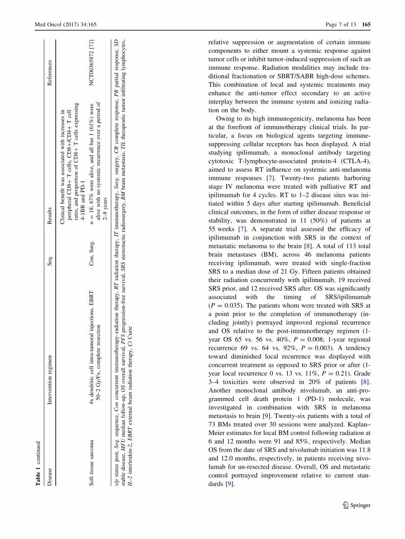
relative suppression or augmentation of certain immune
components to either mount a systemic response against
tumor cells or inhibit tumor-induced suppression of such an
immune response. Radiation modalities may include tra-
ditional fractionation or SBRT/SABR high-dose schemes.
This combination of local and systemic treatments may
enhance the anti-tumor effect secondary to an active
interplay between the immune system and ionizing radia-
tion on the body.
Owing to its high immunogenicity, melanoma has been
at the forefront of immunotherapy clinical trials. In par-
ticular, a focus on biological agents targeting immune-
suppressing cellular receptors has been displayed. A trial
studying ipilimumab, a monoclonal antibody targeting
cytotoxic T-lymphocyte-associated protein-4 (CTLA-4),
aimed to assess RT influence on systemic anti-melanoma
immune responses [7]. Twenty-two patients harboring
stage IV melanoma were treated with palliative RT and
ipilimumab for 4 cycles. RT to 1–2 disease sites was ini-
tiated within 5 days after starting ipilimumab. Beneficial
clinical outcomes, in the form of either disease response or
stability, was demonstrated in 11 (50%) of patients at
55 weeks [7]. A separate trial assessed the efficacy of
ipilimumab in conjunction with SRS in the context of
metastatic melanoma to the brain [8]. A total of 113 total
brain metastases (BM), across 46 melanoma patients
receiving ipilimumab, were treated with single-fraction
SRS to a median dose of 21 Gy. Fifteen patients obtained
their radiation concurrently with ipilimumab, 19 received
SRS prior, and 12 received SRS after. OS was significantly
associated with the timing of SRS/ipilimumab
(P = 0.035). The patients whom were treated with SRS at
a point prior to the completion of immunotherapy (in-
cluding jointly) portrayed improved regional recurrence
and OS relative to the post-immunotherapy regimen (1-
year OS 65 vs. 56 vs. 40%, P = 0.008; 1-year regional
recurrence 69 vs. 64 vs. 92%, P = 0.003). A tendency
toward diminished local recurrence was displayed with
concurrent treatment as opposed to SRS prior or after (1-
year local recurrence 0 vs. 13 vs. 11%, P = 0.21). Grade
3–4 toxicities were observed in 20% of patients [8].
Another monoclonal antibody nivolumab, an anti-pro-
grammed cell death protein 1 (PD-1) molecule, was
investigated in combination with SRS in melanoma
metastasis to brain [9]. Twenty-six patients with a total of
73 BMs treated over 30 sessions were analyzed. Kaplan–
Meier estimates for local BM control following radiation at
6 and 12 months were 91 and 85%, respectively. Median
OS from the date of SRS and nivolumab initiation was 11.8
and 12.0 months, respectively, in patients receiving nivo-
lumab for un-resected disease. Overall, OS and metastatic
control portrayed improvement relative to current stan-
dards [9].Table
1continued
Disease
Interventionregim
enSeq.
Results
References
Clinical
benefitwas
associated
withincreasesin
peripheral
CD8?
Tcells,CD8?/CD4?
Tcell
ratio,andproportionofCD8?
Tcellsexpressing
4-1BB
andPD-1
Softtissuesarcoma
4xdendriticcellintra-tumoralinjections,EBRT
50–2Gy/Fx,complete
resection
Con,Surg.
n=
18;67%
werealive,
andallbut1(61%)were
alivewithnosystem
icrecurrence
over
aperiodof
2–8years
NCT00365872[72]
s/pstatuspost,Seq.sequence,Conconcurrentim
munotherapy–radiationtherapy,RTradiationtherapy,IT
immunotherapy,Surg.surgery,CRcomplete
response,PRpartial
response,SD
stable
disease,MFUmedianfollow-up,OSoverallsurvival,PFSprogression-freesurvival,SRSstereotactic
radiosurgery,BM
brain
metastasis,TIL
therapeutictumorinfiltratinglymphocytes,
IL-2
interleukin-2,EBRTexternal
beam
radiationtherapy,CiCurie
Med Oncol (2017) 34:165 Page 7 of 13 165
123

In the setting of glioblastoma multiforme (GBM), a
severe and fatal intrinsic brain tumor composed of glial
cells, vaccine-based immunotherapy has provided
improvement over certain end points. Cervical intra-nodal
vaccination with autologous tumor lysate-loaded dendritic
cells (DCs) in patients with GBM was assessed after
radiation therapy and temozolomide (TMZ) [67]. A num-
ber of 10 vaccine participants were analyzed, yielding a
median PFS of 9.5 months and an OS of 28 months [67].
This suggests addition of DC vaccines to radiation therapy
and TMZ in the treatment of GBM to be safe and feasible
in the potential induction of immune responses. A second
trial evaluating the use of a vaccine in GBM employed an
epidermal growth factor receptor variant III-peptide (PEP-
3) vaccine in conjunction with RT and TMZ to promote
immunogenicity [68]. At 6 months, PFS rate after inter-
vention was 67% (95% CI 40–83%) and median OS dis-
played was 26.0 months (95% CI 21.0–47.7 months). OS
of the experimental arm was greater than the control
matched for eligibility criteria, prognostic factors, and
TMZ treatment upon adjustment for age and KPS (hazard
ratio, 5.3; P = 0.0013; n = 17) [68]. These results suggest
further investigation in a potential phase III trial of the
PEP-3 vaccine in the setting of GBM.
Metastatic prostate cancer has also been the subject of
investigation as to the administration of immunotherapy
and RT combinations. Ipilimumab was studied in patients
with metastatic castration-resistant (androgen-insensitive)
prostate cancer progression following therapy with doc-
etaxel [69]. A total of 799 patients were randomly assigned
to receive bone-directed radiotherapy (8 Gy in a single
fraction) then to ipilimumab or placebo. Median OS was
11.2 months with ipilimumab versus 10.0 months in the
control arm (HR 0.85, 0.72–1.00; P = 0.053) and not
deemed statistically significant. The most common grade
3–4 adverse events were immune-related, being diarrhea,
fatigue, anemia and colitis, and occurring in 101 (26%)
patients in the ipilimumab group and 11 (3%) of patients in
the placebo group [69]. Similarly, in the case of castration-
resistant metastatic prostate cancer, immunotherapy in the
form of a prostate specific antigen (PSA)-TRI ad of COs-
timulatory Molecules (B7-1, ICAM-1 and LFA-3) (TRI-
COM) was studied in combination with radiation from
Samarium-153-ethylene diamine tetramethylene phospho-
nate (Sm-153-EDTMP), a beta-emitter with affinity for
osteoblastic bone lesions [70]. Median PFS was 1.7 versus
3.7 months in the Sm-153-EDTMP exclusive and combi-
nation arms (P = 0.041, HR = 0.51, P = 0.046). No
patient in the Sm-153-EDTMP exclusive arm attained PSA
decline[30% compared with four patients (out of 21) in
the combination arm. Meanwhile, comparable toxicities
were observed between the arms [70]. These studies war-
rant further investigation in order to identify patients with
metastatic prostate cancer who could potentially benefit
from the combined approach.
Outside of these specific settings, clinical trials investi-
gating the role of combination therapy have demonstrated
results in other sites. SBRT was evaluated in combination
with ipilimumab in the context of advanced solid tumors,
particularly in liver and lung [71]. Thirty-five patients
initiated ipilimumab, among whom 2 experienced dose-
limiting toxicities and 12 (34%) grade 3 toxicities. Thirty-
one patients could be evaluated for response external to the
radiation fields. Three patients (10%) exhibited partial
response and 7 (23%) demonstrated clinical benefit (de-
fined as partial response or stable disease lasting
C6 months). Clinical benefit was found to be associated
with increases in peripheral CD8? T cells, CD8?/CD4? T
cell ratio, and proportion of CD8? T cells expressing
markers CD137 and PD-1. Liver (vs. lung) irradiation
produced greater T cell activation. Therefore, combining
ipilimumab with SBRT/SABR was shown to be safe with
signs of efficacy [71]. Finally, a neoadjuvant DC vaccine
was studied in combination with RT for effects in a cohort
of 18 newly diagnosed high-risk soft tissue sarcoma
patients [72]. Neoadjuvant treatment was 50 Gy in 25
fractions of EBRT, combined with four intra-tumoral
injections of DCs followed by complete resection. A total
of 12 out of 18 (67%) patients were alive, among which
11/18 (61%) were alive without systemic recurrence over
2–8 years. There were no unexpected toxicities, and
favorable immunological responses correlated with clinical
responses in some cases. This provides evidence for
potential effectiveness of DC vaccines in conjunction with
RT in soft tissue sarcomas as well [72].
Toxicity associated with combination
Both immunotherapy and radiation therapy have their own
toxicity profiles. Interestingly though, some adverse effects
of both treatments overlap. This stems from activation of
the immune system which can lead to its overstimulation
resulting in immune-mediated toxicities [76]. Perhaps the
most relevant adverse effect is pneumonitis. Radiation
pneumonitis is thought to reflect an immune-mediated
inflammatory reaction to radiation-induced lung damage
[77, 78]. Patients receiving immune check point inhibitors
are also at risk of developing pneumonitis even in the
absence of radiotherapy [79]. It is still unclear if the inci-
dence of pneumonitis from radiation therapy to the thorax
is higher if delivered concurrently or in close proximity to
immune check point inhibitors and if pneumonitis symp-
toms would be more severe with the treatment combination
[80]. A secondary analysis of the KEYNOTE-001 trial
provided insight on the pulmonary toxicity in patients with
165 Page 8 of 13 Med Oncol (2017) 34:165
123
non-small cell lung cancer who received radiotherapy
before pembrolizumab [81]. A total of 15 (63%) of 24
patients who had previously received thoracic radiother-
apy had any recorded pulmonary toxicity versus 29
(40%) of 73 patients with no previous thoracic radio-
therapy [81]. Three (13%) patients with previous thoracic
radiotherapy had treatment-related pulmonary toxicity
compared with one (1%) of those without; frequency of
grade 3 or worse treatment-related pulmonary toxicities
was similar [81]. A longer progression-free survival and
overall survival were obtained while maintaining an
acceptable safety profile.
Similarly, radiation to the abdomen causes changes in
the mucosa of the colon and rectum resulting in entero-
colitis [82]. Colitis is also reported with check point inhi-
bitors which cause T cell activation within the
gastrointestinal tract [83].
A phase I study was conducted to determine feasibility,
dose-limiting toxicities and maximum tolerated SBRT
fraction when given in conjunction with ipilimumab to
patients with metastatic melanoma [84]. Fifteen patients
(68%) developed different grade 3 toxicities with anemia
being the most common; no grade 4 toxicities were
observed [84]. Nonetheless, Barker et al. [85] reviewed
the records of melanoma patients treated with ipilimumab
and radiotherapy. The frequency of grade 3 or 4 adverse
events in irradiated organs was 15% for the combination
[85]. One grade 4 event was noted in a previously irra-
diated organ that was re-irradiated [85]. So, concurrent
ipilimumab and radiation was not associated with higher
than expected rates of adverse events, nor did it abrogate
palliative effects of RT or survival benefits of ipilimumab
[85].
Although clinical data on combining immunotherapy
and radiation are still lacking, some trials are revealing
positive results of this combination. Tang et al. [71]
showed the safety and efficacy of combining SBRT with
ipilimumab, where only 2 out of 35 (5.7%) patients
developed dose-limiting toxicities and twelve (34%)
developed grade 3 toxicities. Slovin et al. [86], examined
the combination of ipilimumab and radiotherapy in meta-
static castration-resistant prostate cancer. The most com-
mon reported adverse events in that trial were diarrhea
(54%), colitis (22%), rash (32%), and pruritus (20%); grade
3/4 immune-related adverse events included colitis (16%)
and hepatitis (10%) [86]. Thus, this combination resulted in
clinical anti-tumor activity with disease control and man-
ageable adverse events.
Trials examining the combination of immunotherapy
with radiation therapy must be conducted with prudence,
as it is yet to be determined whether the frequency and
severity of immunotoxicities increase with such a
regimen.
Assessing response to combination
The introduction of immunotherapy with its unique anti-
cancer mechanisms not only prompted discussions about its
efficacy but about means to assess the response of patients
receiving such a therapy. Predictors of response to
immunotherapy are currently under investigation.
Pretreatment tumor PD-L1 expression correlates with
response to anti-PD-1 therapy [87]. Taube et al. [88] ana-
lyzed different tumor specimens obtained before treatment
with anti-PD-1 therapy to determine whether PD-1 expres-
sion can be used as an indicator of anti-tumor immune
response. They concluded that the single factor most closely
correlated with response to anti-PD-1 blockade and with
clinical benefit is tumor PD-L1 expression [88]. Similarly,
Tumeh et al. [89] examined pretreatment melanoma sam-
ples. Patients who responded to anti-PD-1 treatment had
higher numbers of CD8, PD-1, and PD-L1 expressing cells
at the invasive tumor margin and inside tumors [89].
A higher mutational load was associated with better
responses to CTLA-4 blockade and to anti-PD-1 therapy
[90]. Somatic mutations and candidate neoantigens gener-
ated from these mutations were characterized for 64
patients with melanoma by Snyder et al. [91]. Mutational
load was associated with the degree of clinical benefit [91].
In two independent cohorts, higher nonsynonymous
mutation burden in tumors was associated with improved
objective response, durable clinical benefit, and progres-
sion-free survival in non-small-cell lung cancers treated
with anti-PD-1 [92].
Traditionally, response patterns to chemotherapeutic
agents are defined according to Response Evaluation Cri-
teria in Solid Tumors (RECIST) criteria [93]. It is based on
the concept of an overall assessment of tumor burden by
summing the products of bidimensional lesion measure-
ments and determines response to therapy by evaluation of
change from baseline while on treatment [94]. Nonetheless,
immunotherapy treatments display different kinetics in
comparison with cytotoxic agents [95]. They may activate
the immune system inducing a cellular response before
affecting tumor burden or patient survival [95, 96]. Thus,
responses seen with immunotherapeutic agents, whether
complete or partial, can occur after an increase in tumor
burden characterized as progressive disease according to
RECIST criteria [93, 97]. To accommodate these new pat-
terns of responses, the immune-related response criteria
(irRC) were developed by Wolchok et al. irRC were
developed after a series of large multinational studies of
patients with advanced melanoma who received ipilimumab
[93, 95]. Four distinct response patterns were seen [93]:
• irCR complete disappearance of all lesions (whether
measurable or not, and no new lesions) confirmation by
Med Oncol (2017) 34:165 Page 9 of 13 165
123
a repeat, consecutive assessment no less than 4 weeks
from the date first documented.
• irPR decrease in tumor burden C50% relative to
baseline confirmed by a consecutive assessment at
least 4 weeks after first documentation.
• irSD not meeting criteria for irCR or irPR, in absence
of irPD.
• irPD increase in tumor burden C25% relative to nadir
(minimum recorded tumor burden) confirmation by a
repeat, consecutive assessment no less than 4 weeks
from the date first documented.
As such, tumor burden is assessed as a continuous variable
which takes into account index lesions identified at base-
line together with new lesions as they make occur after
treatment commences [95]. Using irRC with agents that
can cause tumor shrinkage such as ipilimumab allows a
more comprehensive assessment of clinical activity and
explain why patients with progressive disease by RECIST
have a prolonged long term survival [20, 93]. However, the
importance of adopting the irRC to monitor the response
with agents that are less likely to cause tumor shrinkage
such as cytokines and vaccines is yet to be determined and
requires further prospective studies [93].
The use of irRC has implications on the continuation of
treatment. With cytotoxic agents, evidence of progression
(by RECIST) prompts the discontinuation of therapy.
However, evidence of increase in tumor burden in patients
receiving immunotherapy may not indicate cessation in the
use of the immunotherapeutic agent.
As the combination of immunotherapy and radiation is
novel, so are the appropriate clinical end points to monitor the
response to such a regimen. As clinical trials examining such a
combination emerge, they providemore data on the evaluation
of response to this treatment. For instance, a phase I trial by
Tang et al. [71] tested SBRT with ipilimumab in patients with
advanced malignancies. Clinical benefit was associated with
increases in peripheral CD8? T cells, CD8?/CD4? T cell
ratio, and proportion of CD8? T cells expressing PD-1 [71].
Other ongoing trials exploring the combination checkpoint
inhibitors with radiation are also using T cell counts tomonitor
the response to treatment. They are measuring absolute lym-
phocyte counts, Treg counts, and PD-1 expression [98]. Other
biomarkers are expected in the future as we learn more about
the mechanism of immune stimulation by radiation and the
synergistic action of immunotherapy.
Future research
Although significant progress has been made, several
questions remain to be addressed. First, the ideal patient
population who benefit from targeting the immune system
remains to be elucidated. In most studies, it is estimated
that only 10–20% of patients derive benefit. Some have
proposed predictive biomarkers as PDL-1 expression [99],
IFNc response [100, 101] and absolute lymphocyte count
[102]. Another question pertains to tumor sites that are
amenable to this therapy. For example, in a patient with
metastatic cancer to bones, liver and lung, it would be
prudent to select the anatomical site with highest
immunogenicity for targeting with SBRT. A third question
relates to the sequence and number of cycles of
immunotherapy in relation to SBRT. The ultimate time gap
between SBRT and immunotherapy remains unknown.
These questions should be addressed in future prospective
studies exploring the combination of immunotherapy and
radiation therapy.
Conclusion
The fields of radiotherapy and immunotherapy have
immensely evolved since their establishment. The combi-
nation of RT and IT provides a synergistic effect to induce
a longer tumor response. The combination is best studied
within clinical trials as we learn more about patient
selection, efficacy and toxicity. The novel approach has the
potential of transforming the role radiotherapy from mere
local control to systemic effect. The appropriate clinical
end points to monitor and assess the response to this
combination are still being studied. The use of radiotherapy
and immunotherapy together in the treatment of malig-
nancies constitutes a major breakthrough in oncology and
can potentially improve clinical outcomes in patients with
dismal prognosis.
Compliance with ethical standards
Conflict of interest The authors declare that they have no conflict of
interest.
Ethical approval This article does not contain any studies with
human participants or animals performed by any of the authors.
Informed consent No informed consent was obtained as no human
subjects were involved in this review.
References
1. Schuster M, Nechansky A, Kircheis R. Cancer immunotherapy.
Biotechnol J. 2006;1(2):138–47.
2. Helmy KY, et al. Cancer immunotherapy: accomplishments to
date and future promise. Ther Deliv. 2013;4(10):1307–20.
3. Mellman I, Coukos G, Dranoff G. Cancer immunotherapy
comes of age. Nature. 2011;480(7378):480–9.
4. Schwartz RS. Book review. N Engl J Med.
1997;337(16):1178–9.
165 Page 10 of 13 Med Oncol (2017) 34:165
123
5. Sylvester RJ. Bacillus Calmette–Guerin treatment of non-mus-
cle invasive bladder cancer. Int J Urol. 2011;18(2):113–20.
6. Dunn GP, Old LJ, Schreiber RD. The immunobiology of cancer
immunosurveillance and immunoediting. Immunity.
2004;21(2):137–48.
7. Rosenberg SA. A new era for cancer immunotherapy based on
the genes that encode cancer antigens. Immunity.
1999;10(3):281–7.
8. Boon T, et al. Tumor antigens recognized by T lymphocytes.
Annu Rev Immunol. 1994;12:337–65.
9. Boon T, van der Bruggen P. Human tumor antigens recognized
by T lymphocytes. J Exp Med. 1996;183(3):725–9.
10. Urbanski M, Cone RE. Appearance of T lymphocyte-derived
proteins specific for the immunizing antigen in serum during a
humoral immune response. J Immunol. 1992;148(9):2840–4.
11. Sologuren I, Rodrıguez-Gallego C, Lara PC. Immune effects of
high dose radiation treatment: implications of ionizing radiation
on the development of bystander and abscopal effects. Transl
Cancer Res. 2014;3(1):18–31.
12. Demaria S, et al. Ionizing radiation inhibition of distant
untreated tumors (abscopal effect) is immune mediated. Int J
Radiat Oncol Biol Phys. 2004;58(3):862–70.
13. Kaur P, Asea A. Radiation-induced effects and the immune
system in cancer. Front Oncol. 2012;2:191.
14. Schaue D, Kachikwu EL, McBride WH. Cytokines in radiobi-
ological responses: a review. Radiat Res. 2012;178(6):505–23.
15. Tesniere A, et al. Molecular characteristics of immunogenic
cancer cell death. Cell Death Differ. 2008;15(1):3–12.
16. Lumniczky K, Safrany G. Cancer gene therapy: combination
with radiation therapy and the role of bystander cell killing in
the anti-tumor effect. Pathol Oncol Res. 2006;12(2):118–24.
17. Willimsky G, Blankenstein T. Sporadic immunogenic tumours
avoid destruction by inducing T-cell tolerance. Nature.
2005;437(7055):141–6.
18. Dunn GP, et al. A critical function for type I interferons in
cancer immunoediting. Nat Immunol. 2005;6(7):722–9.
19. Velcheti V, Schalper K. Basic overview of current
immunotherapy approaches in cancer. Am Soc Clin Oncol Educ
Book. 2016;35:298–308.
20. Vatner RE, et al. Combinations of immunotherapy and radiation
in cancer therapy. Front Oncol. 2014;4:325.
21. Thaxton JE, Li Z. To affinity and beyond: harnessing the T cell
receptor for cancer immunotherapy. Hum Vaccines Immun-
other. 2014;10(11):3313–21.
22. Willemsen RA, et al. T cell retargeting with MHC class I-re-
stricted antibodies: the CD28 costimulatory domain enhances
antigen-specific cytotoxicity and cytokine production. J Im-
munol. 2005;174(12):7853–8.
23. Seliger B. Molecular mechanisms of MHC class I abnormalities
and APM components in human tumors. Cancer Immunol
Immunother. 2008;57(11):1719–26.
24. Aptsiauri N, et al. Role of altered expression of HLA class I
molecules in cancer progression. Adv Exp Med Biol.
2007;601:123–31.
25. Poschke I, Mougiakakos D, Kiessling R. Camouflage and sab-
otage: tumor escape from the immune system. Cancer Immunol
Immunother. 2011;60(8):1161–71.
26. Campoli M, Chang CC, Ferrone S. HLA class I antigen loss,
tumor immune escape and immune selection. Vaccine.
2002;20(Suppl 4):A40–5.
27. Gajewski TF, et al. Immune resistance orchestrated by the tumor
microenvironment. Immunol Rev. 2006;213:131–45.
28. Gajewski TF, Schreiber H, Fu YX. Innate and adaptive immune
cells in the tumor microenvironment. Nat Immunol.
2013;14(10):1014–22.
29. Dany M, et al. Advances in immunotherapy for melanoma
management. Hum Vaccine Immunother. 2016;12(10):2501–11.
30. Camacho LH. CTLA-4 blockade with ipilimumab: biology,
safety, efficacy, and future considerations. Cancer Med.
2015;4(5):661–72.
31. Linsley PS, et al. Intracellular trafficking of CTLA-4 and focal
localization towards sites of TCR engagement. Immunity.
1996;4(6):535–43.
32. Michielin O, Hoeller C. Gaining momentum: new options and
opportunities for the treatment of advanced melanoma. Cancer
Treat Rev. 2015;41(8):660–70.
33. Trinh VA, Hagen B. Ipilimumab for advanced melanoma: a
pharmacologic perspective. J Oncol Pharm Pract.
2013;19(3):195–201.
34. Topalian SL, et al. Survival, durable tumor remission, and long-
term safety in patients with advanced melanoma receiving
nivolumab. J Clin Oncol. 2014;32(10):1020–30.
35. Hurley KE, Chapman PB. Helping melanoma patients decide
whether to choose adjuvant high-dose interferon-alpha2b.
Oncologist. 2005;10(9):739–42.
36. Yang JC, et al. Randomized study of high-dose and low-dose
interleukin-2 in patients with metastatic renal cancer. J Clin
Oncol. 2003;21(16):3127–32.
37. Barker CA, Postow MA. Combinations of radiation therapy and
immunotherapy for melanoma: a review of clinical outcomes.
Int J Radiat Oncol Biol Phys. 2014;88(5):986–97.
38. Begley CG, et al. Human B lymphocytes express the p75
component of the interleukin 2 receptor. Leuk Res.
1990;14(3):263–71.
39. Wagner TC, et al. Interferon receptor expression regulates the
antiproliferative effects of interferons on cancer cells and solid
tumors. Int J Cancer. 2004;111(1):32–42.
40. Panelli MC, et al. Forecasting the cytokine storm following
systemic interleukin (IL)-2 administration. J Transl Med.
2004;2(1):17.
41. Markman M, et al. Phase 2 trial of interferon-beta as second-line
treatment of ovarian cancer, fallopian tube cancer, or primary
carcinoma of the peritoneum. Oncology. 2004;66(5):343–6.
42. Haji-Fatahaliha M, et al. CAR-modified T-cell therapy for
cancer: an updated review. Artif Cells Nanomed Biotechnol.
2016;44(6):1339–49.
43. Eckert F, et al. Beyond checkpoint inhibition – Immunothera-
peutical strategies in combination with radiation. Clin Transl
Radiat Oncol. 2017;2:29–35.
44. Lugade AA, et al. Local radiation therapy of B16 melanomatumors increases the generation of tumor antigen-specific
effector cells that traffic to the tumor. J Immunol.
2005;174(12):7516–23.
45. Matsumura S, et al. Radiation-induced CXCL16 release by
breast cancer cells attracts effector T cells. J Immunol.
2008;181(5):3099–107.
46. Griffiths DJ. Introduction to electrodynamics. 3rd ed. Upper
Saddle River: Prentice Hall; 1999. p. xv.
47. Laugier A. The first century of radiotherapy in France. Bull
Acad Natl Med. 1996;180(1):143–60.
48. Bernier J, Hall EJ, Giaccia A. Radiation oncology: a century of
achievements. Nat Rev Cancer. 2004;4(9):737–47.
49. Thwaites DI, Tuohy JB. Back to the future: the history and
development of the clinical linear accelerator. Phys Med Biol.
2006;51(13):R343–62.
50. Purdy JA. 3D treatment planning and intensity-modulated
radiation therapy. Oncology (Williston Park). 1999;13(10 Suppl
5):155–68.
51. Galvin JM, et al. Implementing IMRT in clinical practice: a joint
document of the American Society for Therapeutic Radiology
Med Oncol (2017) 34:165 Page 11 of 13 165
123
and Oncology and the American Association of Physicists in
Medicine. Int J Radiat Oncol Biol Phys. 2004;58(5):1616–34.
52. Macia IGM. Radiobiology of stereotactic body radiation therapy
(SBRT). Rep Pract Oncol Radiother. 2017;22(2):86–95.
53. Bhattacharya IS, et al. Stereotactic body radiotherapy (SBRT) in
the management of extracranial oligometastatic (OM) disease.
Br J Radiol. 1048;2015(88):20140712.
54. Greco C, et al. Spinal metastases: from conventional fraction-
ated radiotherapy to single-dose SBRT. Rep Pract Oncol
Radiother. 2015;20(6):454–63.
55. Linskey ME, et al. The role of stereotactic radiosurgery in the
management of patients with newly diagnosed brain metastases:
a systematic review and evidence-based clinical practice
guideline. J Neurooncol. 2010;96(1):45–68.
56. Trakul N, Koong AC, Chang DT. Stereotactic body radiotherapy
in the treatment of pancreatic cancer. Semin Radiat Oncol.
2014;24(2):140–7.
57. Jaffray DA. Image-guided radiotherapy: from current concept to
future perspectives. Nat Rev Clin Oncol. 2012;9(12):688–99.
58. Glide-Hurst CK, Chetty IJ. Improving radiotherapy planning,
delivery accuracy, and normal tissue sparing using cutting edge
technologies. J Thorac Dis. 2014;6(4):303–18.
59. Jaffray DA, et al. Flat-panel cone-beam computed tomography
for image-guided radiation therapy. Int J Radiat Oncol Biol
Phys. 2002;53(5):1337–49.
60. Huntzinger C, et al. Dynamic targeting image-guided radio-
therapy. Med Dosim. 2006;31(2):113–25.
61. Fields EC, Weiss E. A practical review of magnetic resonance
imaging for the evaluation and management of cervical cancer.
Radiat Oncol. 2016;11:15.
62. Tanderup K, et al. Magnetic resonance image guided
brachytherapy. Semin Radiat Oncol. 2014;24(3):181–91.
63. Pollard JM, et al. The future of image-guided radiotherapy will
be MR guided. Br J Radiol. 1073;2017(90):20160667.
64. NIH, U.S. ClinicalTrials.gov. 2017.
65. Bernstein MB, et al. Immunotherapy and stereotactic ablative
radiotherapy (ISABR): a curative approach? Nat Rev Clin
Oncol. 2016;13(8):516–24.
66. Shabason JE, Minn AJ. Radiation and immune checkpoint
blockade: from bench to clinic. Semin Radiat Oncol.
2017;27(3):289–98.
67. Fadul CE, et al. Immune response in patients with newly diag-
nosed glioblastoma multiforme treated with intranodal autolo-
gous tumor lysate-dendritic cell vaccination after radiation
chemotherapy. J Immunother. 2011;34(4):382–9.
68. Sampson JH, et al. Immunologic escape after prolonged pro-
gression-free survival with epidermal growth factor receptor
variant III peptide vaccination in patients with newly diagnosed
glioblastoma. J Clin Oncol. 2010;28(31):4722–9.
69. Kwon ED, et al. Ipilimumab versus placebo after radiotherapy in
patients with metastatic castration-resistant prostate cancer that
had progressed after docetaxel chemotherapy (CA184-043): a
multicentre, randomised, double-blind, phase 3 trial. Lancet
Oncol. 2014;15(7):700–12.
70. Heery CR, et al. Samarium-153-EDTMP (Quadramet(R)) with
or without vaccine in metastatic castration-resistant prostate
cancer: a randomized Phase 2 trial. Oncotarget.
2016;7(42):69014–23.
71. Tang C, et al. Ipilimumab with stereotactic ablative radiation
therapy: phase I results and immunologic correlates from
peripheral T cells. Clin Cancer Res. 2017;23(6):1388–96.
72. Raj S, et al. Long-term clinical responses of neoadjuvant den-
dritic cell infusions and radiation in soft tissue sarcoma. Sar-
coma. 2015;2015:614736.
73. Hiniker SM, et al. A prospective clinical trial combining radi-
ation therapy with systemic immunotherapy in metastatic mel-
anoma. Int J Radiat Oncol Biol Phys. 2016;96(3):578–88.
74. Kiess AP, et al. Stereotactic radiosurgery for melanoma brain
metastases in patients receiving ipilimumab: safety profile and
efficacy of combined treatment. Int J Radiat Oncol Biol Phys.
2015;92(2):368–75.
75. Ahmed KA, et al. Clinical outcomes of melanoma brain
metastases treated with stereotactic radiation and anti-PD-1
therapy. Ann Oncol. 2016;27(3):434–41.
76. Alatrash G, et al. Cancer immunotherapies, their safety and
toxicity. Expert Opin Drug Saf. 2013;12(5):631–45.
77. Roberts CM, et al. Radiation pneumonitis: a possible lympho-
cyte-mediated hypersensitivity reaction. Ann Intern Med.
1993;118(9):696–700.
78. Morgan GW, Breit SN. Radiation and the lung: a reevaluation of
the mechanisms mediating pulmonary injury. Int J Radiat Oncol
Biol Phys. 1995;31(2):361–9.
79. Abdel-Wahab N, Shah M, Suarez-Almazor ME. Adverse events
associated with immune checkpoint blockade in patients with
cancer: a systematic review of case reports. PLoS ONE.
2016;11(7):e0160221.
80. Alley EW, et al. Immunotherapy and radiation therapy for
malignant pleural mesothelioma. Transl Lung Cancer Res.
2017;6(2):212–9.
81. Shaverdian N, et al. Previous radiotherapy and the clinical
activity and toxicity of pembrolizumab in the treatment of non-
small-cell lung cancer: a secondary analysis of the KEYNOTE-
001 phase 1 trial. Lancet Oncol. 2017;18(7):895–903.
82. Qadeer MA, Vargo JJ. Approaches to the prevention and man-
agement of radiation colitis. Curr Gastroenterol Rep.
2008;10(5):507–13.
83. Gangadhar TC, Vonderheide RH. Mitigating the toxic effects of
anticancer immunotherapy. Nat Rev Clin Oncol.
2014;11(2):91–9.
84. Twyman-Saint Victor C, et al. Radiation and dual checkpoint
blockade activate non-redundant immune mechanisms in cancer.
Nature. 2015;520(7547):373–7.
85. Barker CA, et al. Concurrent radiotherapy and ipilimumab
immunotherapy for patients with melanoma. Cancer Immunol
Res. 2013;1(2):92–8.
86. Slovin SF, et al. Ipilimumab alone or in combination with
radiotherapy in metastatic castration-resistant prostate cancer:
results from an open-label, multicenter phase I/II study. Ann
Oncol. 2013;24(7):1813–21.
87. Hiniker SM, Maecker HT, Knox SJ. Predictors of clinical
response to immunotherapy with or without radiotherapy. J Ra-
diat Oncol. 2015;4:339–45.
88. Taube JM, et al. Association of PD-1, PD-1 ligands, and other
features of the tumor immune microenvironment with response
to anti-PD-1 therapy. Clin Cancer Res. 2014;20(19):5064–74.
89. Tumeh PC, et al. PD-1 blockade induces responses by inhibiting
adaptive immune resistance. Nature. 2014;515(7528):568–71.
90. Yuan J, et al. Novel technologies and emerging biomarkers for
personalized cancer immunotherapy. J Immunother Cancer.
2016;4:3.
91. Snyder A, et al. Genetic basis for clinical response to CTLA-4
blockade in melanoma. N Engl J Med. 2014;371(23):2189–99.
92. Rizvi NA, et al. Cancer immunology. Mutational landscape
determines sensitivity to PD-1 blockade in non-small cell lung
cancer. Science. 2015;348(6230):124–8.
93. Wolchok JD, et al. Guidelines for the evaluation of immune
therapy activity in solid tumors: immune-related response cri-
teria. Clin Cancer Res. 2009;15(23):7412–20.
165 Page 12 of 13 Med Oncol (2017) 34:165
123

94. Eisenhauer EA, et al. New response evaluation criteria in solid
tumours: revised RECIST guideline (version 1.1). Eur J Cancer.
2009;45(2):228–47.
95. Hoos A, et al. Improved endpoints for cancer immunotherapy
trials. J Natl Cancer Inst. 2010;102(18):1388–97.
96. Hoos A, et al. A clinical development paradigm for cancer
vaccines and related biologics. J Immunother. 2007;30(1):1–15.
97. Ratain MJ, Eckhardt SG. Phase II studies of modern drugs
directed against new targets: if you are fazed, too, then resist
RECIST. J Clin Oncol. 2004;22(22):4442–5.
98. Spiotto M, Fu Y-X, Weichselbaum RR. The intersection of
radiotherapy and immunotherapy: mechanisms and clinical impli-
cations. Sci Immunol. 2016. doi:10.1126/sciimmunol.aag1266.
99. Herbst RS, et al. Predictive correlates of response to the anti-
PD-L1 antibody MPDL3280A in cancer patients. Nature.
2014;515(7528):563–7.
100. Chow LQM, et al. Antitumor activity of pembrolizumab in
biomarker-unselected patients with recurrent and/or metastatic
head and neck squamous cell carcinoma: results from the phase
Ib KEYNOTE-012 expansion cohort. J Clin Oncol.
2016;34(32):3838–45.
101. Sheikh NA, et al. Sipuleucel-T immune parameters correlate
with survival: an analysis of the randomized phase 3 clinical
trials in men with castration-resistant prostate cancer. Cancer
Immunol Immunother. 2013;62(1):137–47.
102. Ku GY, et al. Single-institution experience with ipilimumab in
advanced melanoma patients in the compassionate use setting:
lymphocyte count after 2 doses correlates with survival. Cancer.
2010;116(7):1767–75.
103. Grass GD, Krishna N, Kim S. The immune mechanisms of
abscopal effect in radiation therapy. Curr Probl Cancer.
2016;40:10–24.
104. Postow MA, et al. Immunologic correlates of the abscopal effect
in a patient with melanoma. N Engl J Med. 2012;366:925–31.
105. Demaria S, Golden EB, Formenti SC. Role of local radiation
therapy in cancer immunotherapy. JAMA Oncol.
2015;1:1325–32.
106. Demaria S, Pilones KA, Vanpouille-Box C, Golden EB, For-
menti SC. The optimal partnership of radiation and
immunotherapy: From preclinical studies to clinical translation.
Radiat Res. 2014;182:170–81.
107. Demaria S, et al. Ionizing radiation inhibition of distant
untreated tumors (abscopal effect) is immune mediated. Int J
Radiat Oncol Biol Phys. 2004;58:862–70.
108. Dewan MZ, et al. Fractionated but not single-dose radiotherapy
induces an immune-mediated abscopal effect when combined
with anti-CTLA-4 antibody. Clin Cancer Res. 2009;15(17):
5379–88. doi:10.1158/1078-0432.CCR-09-0265.
109. Shi W, Siemann DW. Augmented antitumor effects of radiation
therapy by 4-1bb antibody (bms-469492) treatment. Anticancer
Res. 2006;26:3445–53.
110. Hiniker SM, Knox SJ. Immunotherapy and radiation. Semin
Oncol. 2014;41:702–13.
111. Demaria S, et al. Immune-mediated inhibition of metastases
after treatment with local radiation and ctla-4 blockade in a
mouse model of breast cancer. Clin Cancer Res.
2005;11:728–34.
112. Formenti SC, Demaria S. Combining radiotherapy and cancer
immunotherapy: A paradigm shift. J Natl Cancer Inst.
2013;105:256–65.
113. Ruocco MG, et al. Suppressing t cell motility induced by anti–
ctla-4 monotherapy improves antitumor effects. J Clin Invest.
2012;122:3718–30.
114. Yoshimoto Y, et al. Radiotherapy-induced anti-tumor immunity
contributes to the therapeutic efficacy of irradiation and can be
augmented by ctla-4 blockade in a mouse model. PloS One.
2014;9:e92572.
115. Zeng J, et al. Anti-pd-1 blockade and stereotactic radiation
produce long-term survival in mice with intracranial gliomas. Int
J Radiat Oncol Biol Phys. 2013;86:343–9.
116. Victor CT-S, et al. Radiation and dual checkpoint blockade
activates non-redundant immune mechanisms in cancer. Nature.
2015;520:373–7.
Med Oncol (2017) 34:165 Page 13 of 13 165
123
Related Documents
![Tumor Microenvironment Hijacking the Immune System [Read-Only]](https://static.cupdf.com/doc/110x72/61bf372f43ec6023e9684384/tumor-microenvironment-hijacking-the-immune-system-read-only.jpg)