Medical Technology Department, Faculty of Science, Islamic University-Gaza M MICRO B BIOLOGY Dr. Abdelraouf A. Elmanama Dr. Abdelraouf A. Elmanama Ph. D Microbiology Ph. D Microbiology 2008 Chapter 3 Chapter 3 Observing Microorganisms Through a Observing Microorganisms Through a Microscope Microscope

observing microorganisms_through_a_microscope
Aug 08, 2015
Welcome message from author
This document is posted to help you gain knowledge. Please leave a comment to let me know what you think about it! Share it to your friends and learn new things together.
Transcript
Medical Technology Department, Faculty of Science, Islamic University-Gaza
MMICROBBIOLOGY
Dr. Abdelraouf A. ElmanamaDr. Abdelraouf A. ElmanamaPh. D MicrobiologyPh. D Microbiology
2008
Chapter 3Chapter 3
Observing Microorganisms Through a Observing Microorganisms Through a MicroscopeMicroscope
2008
Units of Measurement
• 1 µm = 10-6 m = 10-3 mm
• 1 nm = 10-9 m = 10-6 mm
• 1000 nm = 1 µm
• 0.001 µm = 1 nm
2008
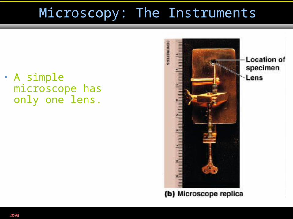
• A simple microscope has only one lens.
Microscopy: The Instruments
Figure 1.2b
2008
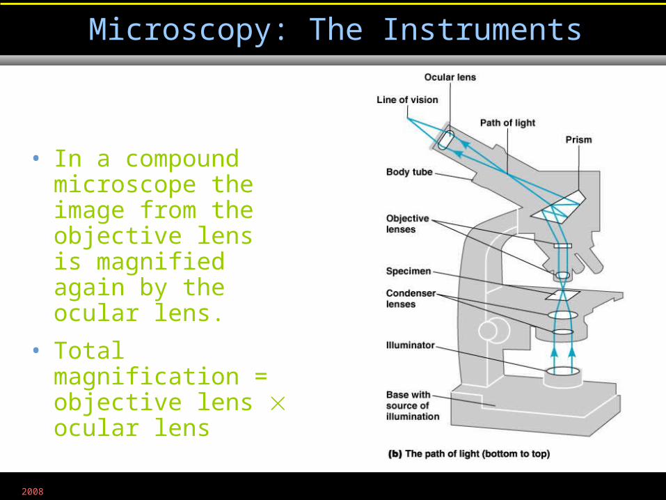
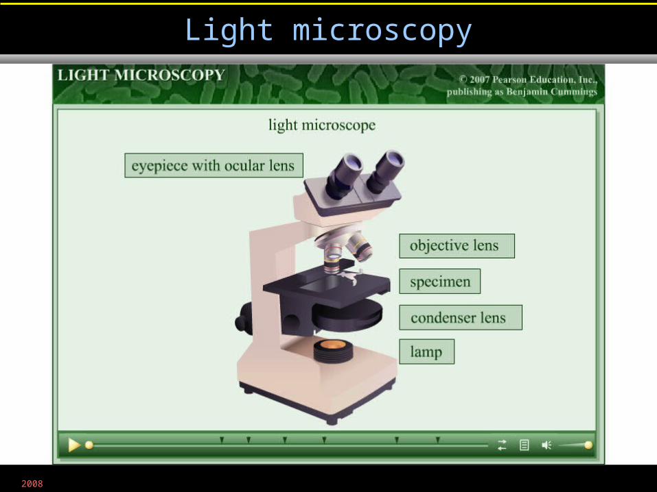
• In a compound microscope the image from the objective lens is magnified again by the ocular lens.
• Total magnification =objective lens ocular lens
Microscopy: The Instruments
Figure 3.1b
2008
• Resolution is the ability of the lenses to distinguish two points.
• A microscope with a resolving power of 0.4 nm can distinguish between two points ≥ 0.4 nm.
• Shorter wavelengths of light provide greater resolution.
Microscopy: The Instruments
2008
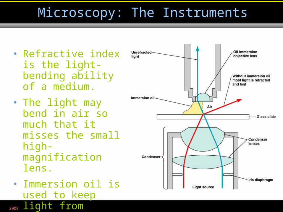
• Refractive index is the light-bending ability of a medium.
• The light may bend in air so much that it misses the small high-magnification lens.
• Immersion oil is used to keep light from bending.
Microscopy: The Instruments
Figure 3.3
2008
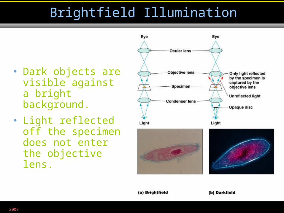
• Dark objects are visible against a bright background.
• Light reflected off the specimen does not enter the objective lens.
Brightfield Illumination
Figure 3.4a, b

2008
• Light objects are visible against a dark background.
• Light reflected off the specimen enters the objective lens.
Darkfield Illumination
Figure 3.4a, b
2008
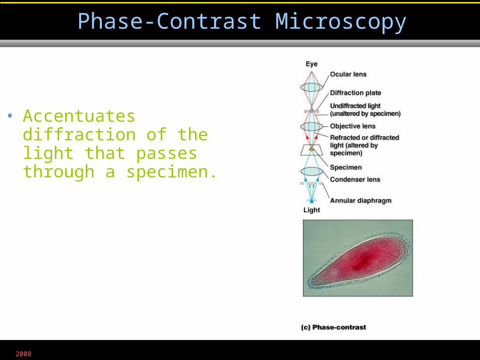
• Accentuates diffraction of the light that passes through a specimen.
Phase-Contrast Microscopy
Figure 3.4c
2008
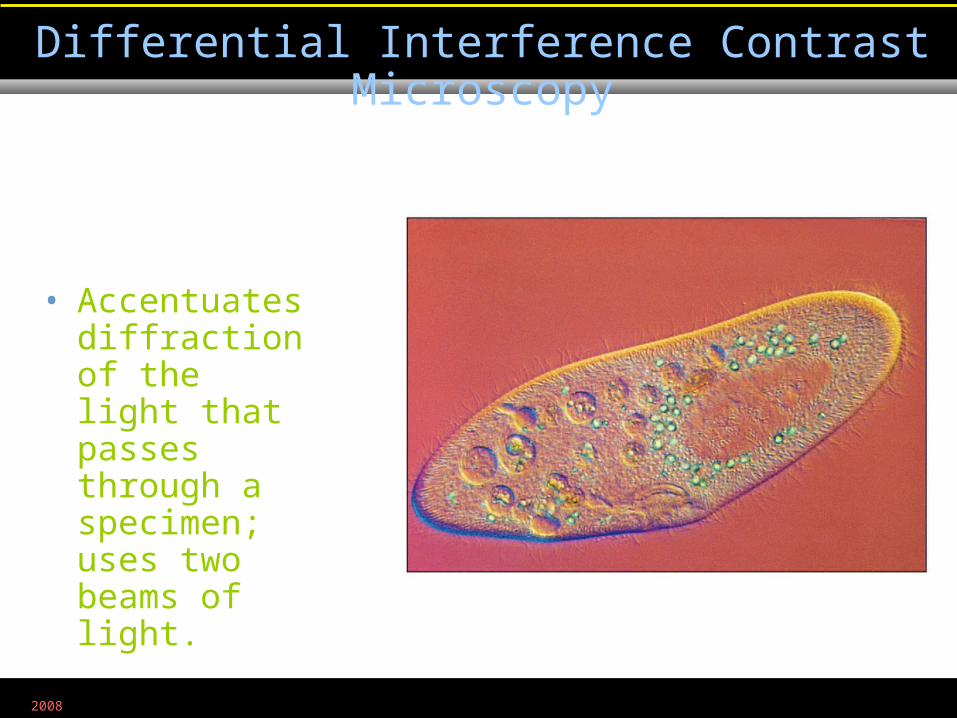
• Accentuates diffraction of the light that passes through a specimen; uses two beams of light.
Differential Interference Contrast Microscopy
Figure 3.5
2008
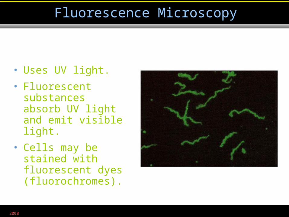
• Uses UV light.
• Fluorescent substances absorb UV light and emit visible light.
• Cells may be stained with fluorescent dyes (fluorochromes).
Fluorescence Microscopy
Figure 3.6b
2008
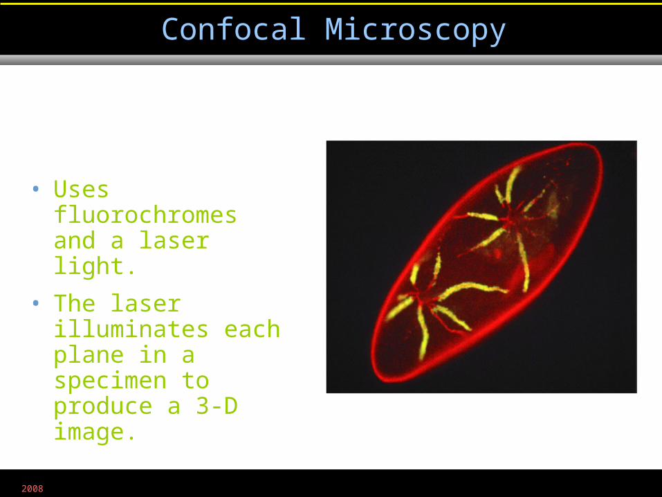
• Uses fluorochromes and a laser light.
• The laser illuminates each plane in a specimen to produce a 3-D image.
Confocal Microscopy
Figure 3.7
2008
• Uses electrons instead of light.
• The shorter wavelength of electrons gives greater resolution.
Electron Microscopy
2008
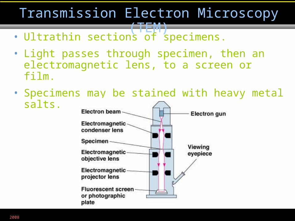
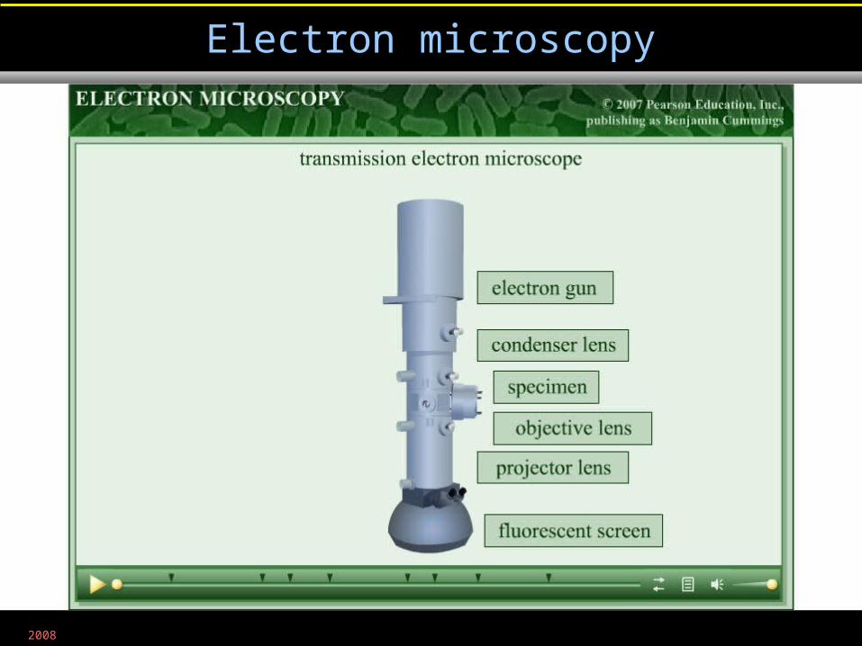
• Ultrathin sections of specimens.
• Light passes through specimen, then an electromagnetic lens, to a screen or film.
• Specimens may be stained with heavy metal salts.
Transmission Electron Microscopy (TEM)
Figure 3.8a
2008
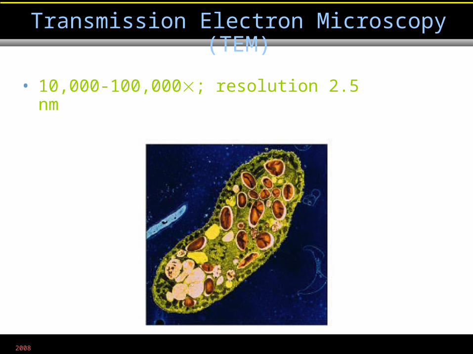
• 10,000-100,000; resolution 2.5 nm
Transmission Electron Microscopy (TEM)
Figure 3.8a
2008
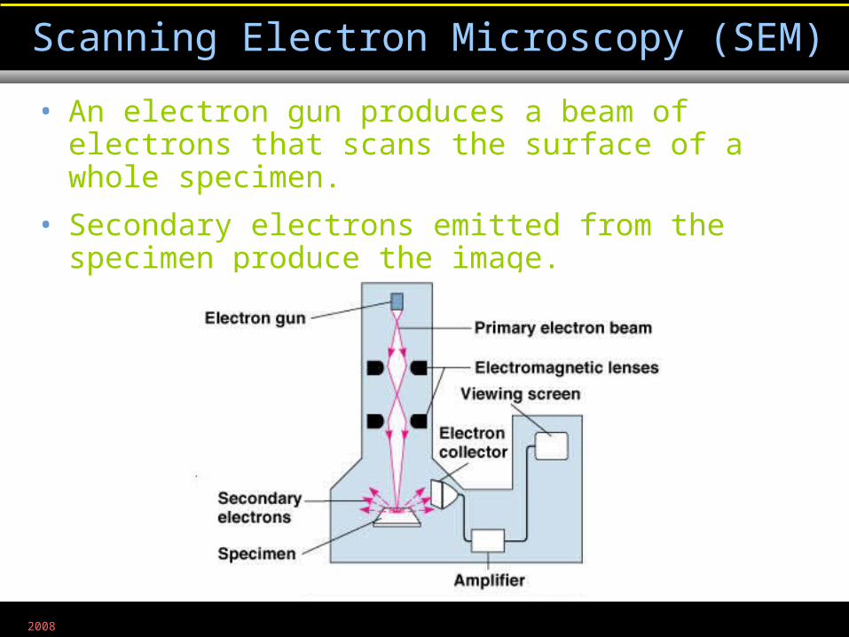
• An electron gun produces a beam of electrons that scans the surface of a whole specimen.
• Secondary electrons emitted from the specimen produce the image.
Scanning Electron Microscopy (SEM)
Figure 3.8b
2008
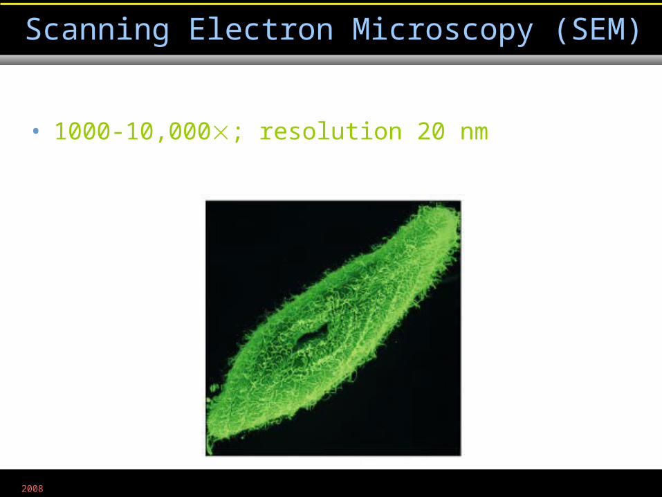
• 1000-10,000; resolution 20 nm
Scanning Electron Microscopy (SEM)
Figure 3.8b
2008
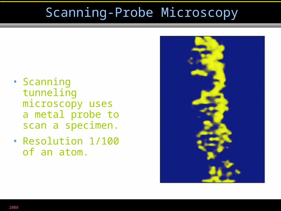
• Scanning tunneling microscopy uses a metal probe to scan a specimen.
• Resolution 1/100 of an atom.
Scanning-Probe Microscopy
Figure 3.9a
2008
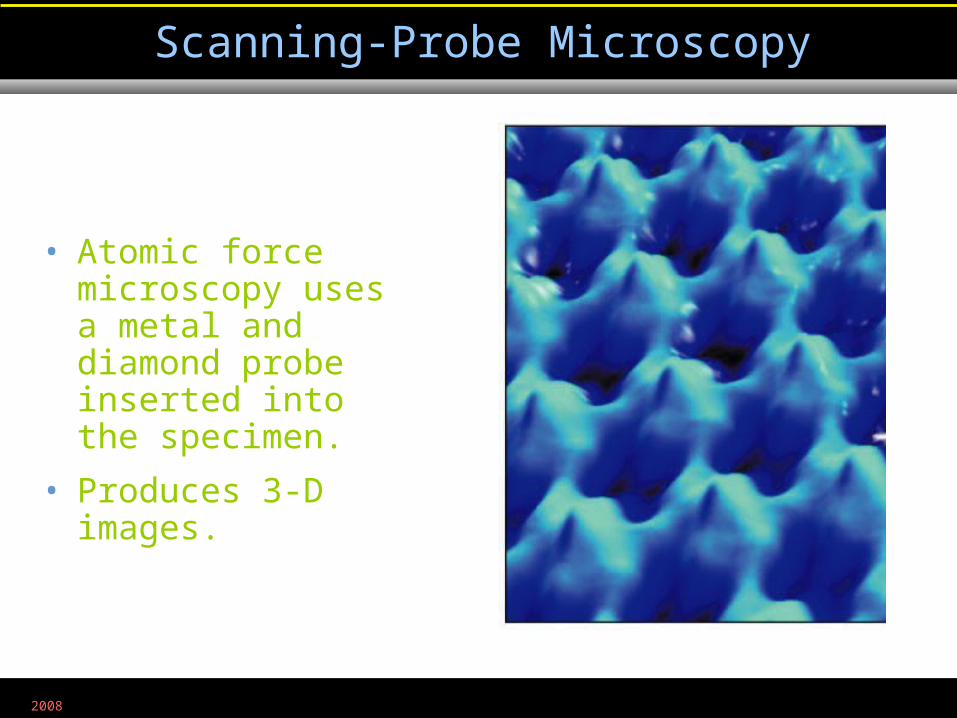
• Atomic force microscopy uses a metal and diamond probe inserted into the specimen.
• Produces 3-D images.
Scanning-Probe Microscopy
Figure 3.9b
2008
Light microscopy
2008
Electron microscopy
2008
Preparation of Specimens for Light Microscopy
• A thin film of a solution of microbes on a slide is a smear.
• A smear is usually fixed to attach the microbes to the slide and to kill the microbes.
2008
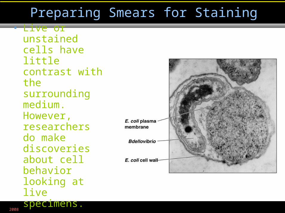
• Live or unstained cells have little contrast with the surrounding medium. However, researchers do make discoveries about cell behavior looking at live specimens.
Preparing Smears for Staining
2008
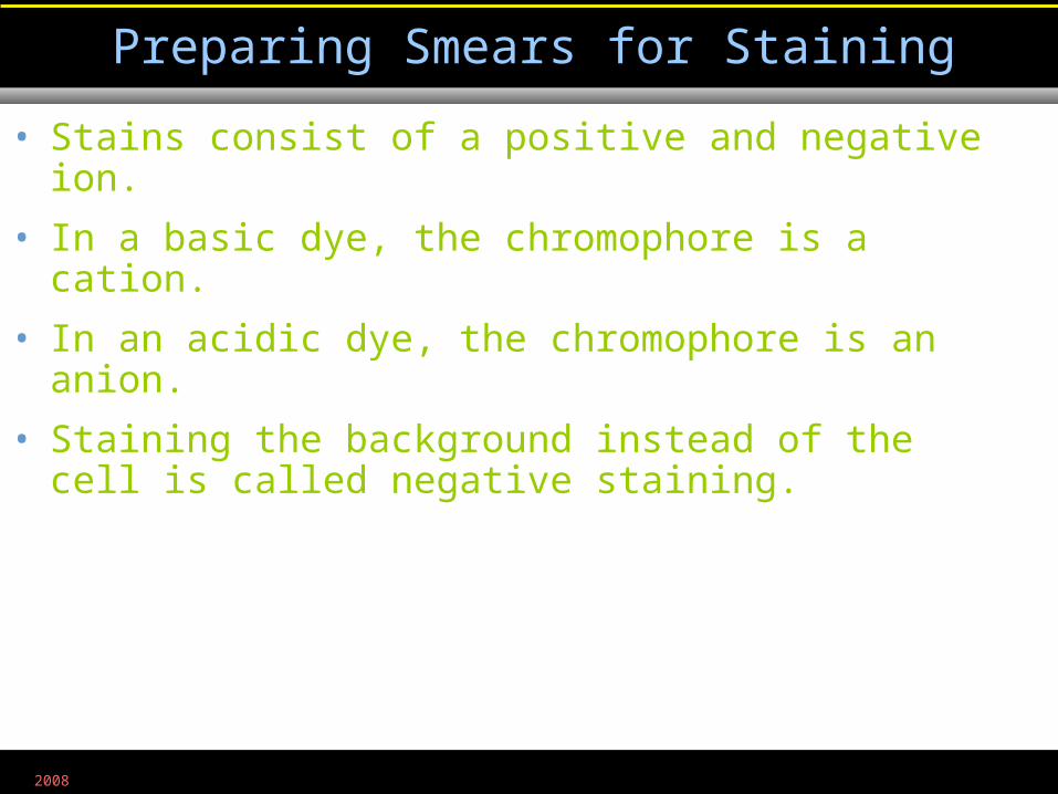
• Stains consist of a positive and negative ion.
• In a basic dye, the chromophore is a cation.
• In an acidic dye, the chromophore is an anion.
• Staining the background instead of the cell is called negative staining.
Preparing Smears for Staining
2008
• Use of a single basic dye is called a simple stain.
• A mordant may be used to hold the stain or coat the specimen to enlarge it.
Simple Stains
2008
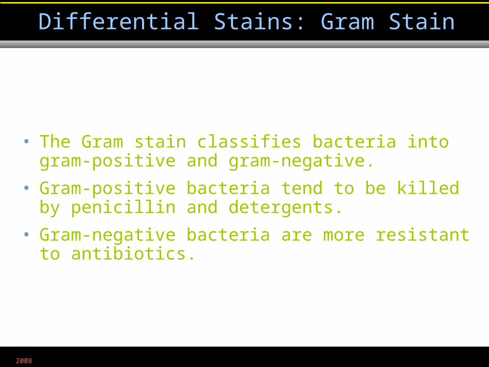
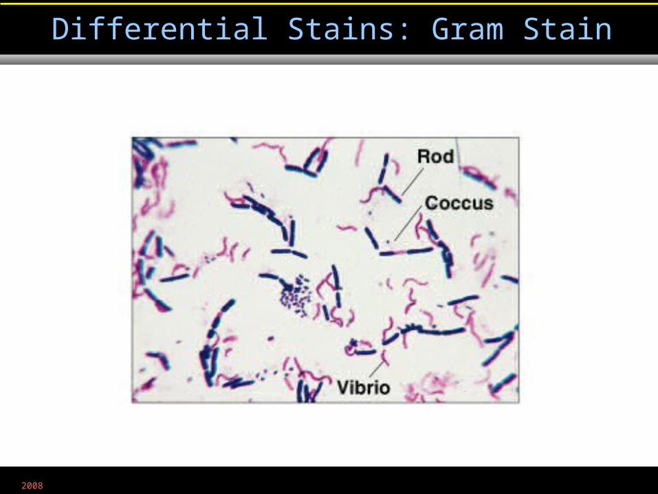
• The Gram stain classifies bacteria into gram-positive and gram-negative.
• Gram-positive bacteria tend to be killed by penicillin and detergents.
• Gram-negative bacteria are more resistant to antibiotics.
Differential Stains: Gram Stain
2008
Differential Stains: Gram Stain
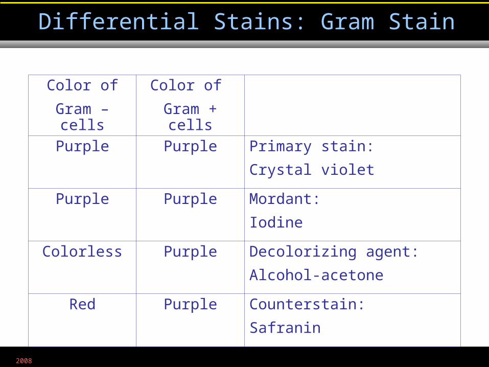
Color of
Gram + cells
Color of
Gram – cells
Primary stain:
Crystal violet
PurplePurple
Mordant:
Iodine
PurplePurple
Decolorizing agent:
Alcohol-acetone
PurpleColorless
Counterstain:
Safranin
PurpleRed
2008
Differential Stains: Gram Stain
Figure 3.10b
2008
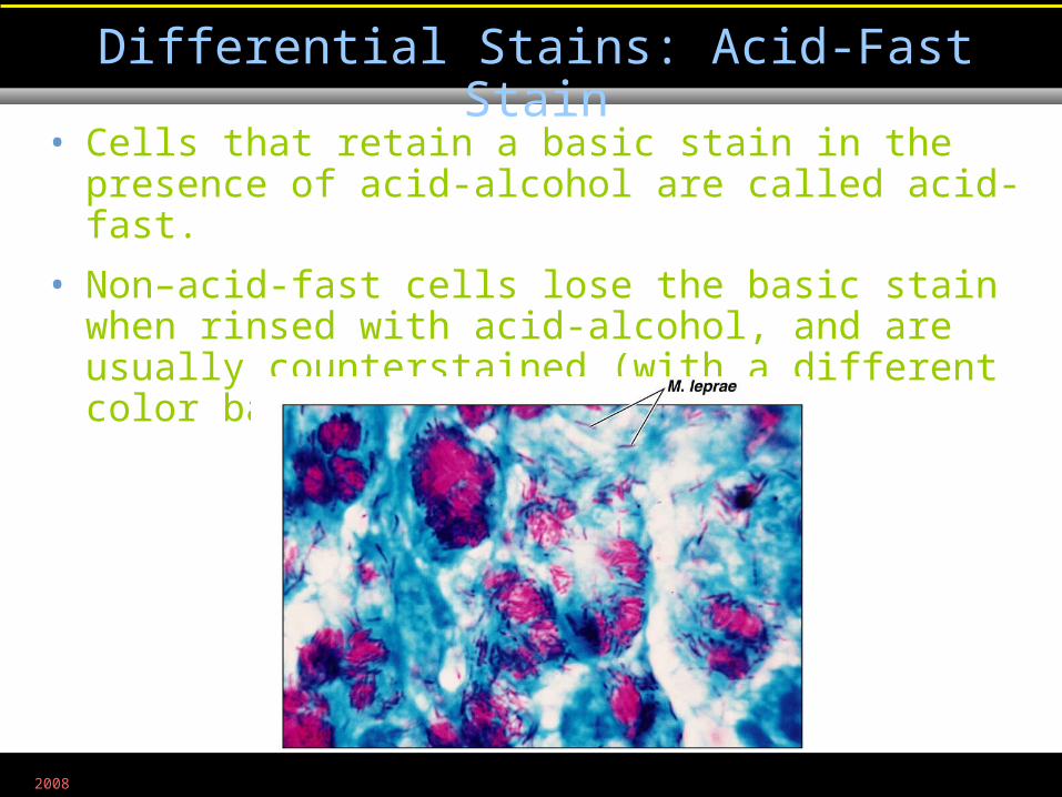
• Cells that retain a basic stain in the presence of acid-alcohol are called acid-fast.
• Non–acid-fast cells lose the basic stain when rinsed with acid-alcohol, and are usually counterstained (with a different color basic stain) to see them.
Differential Stains: Acid-Fast Stain
Figure 3.11
2008
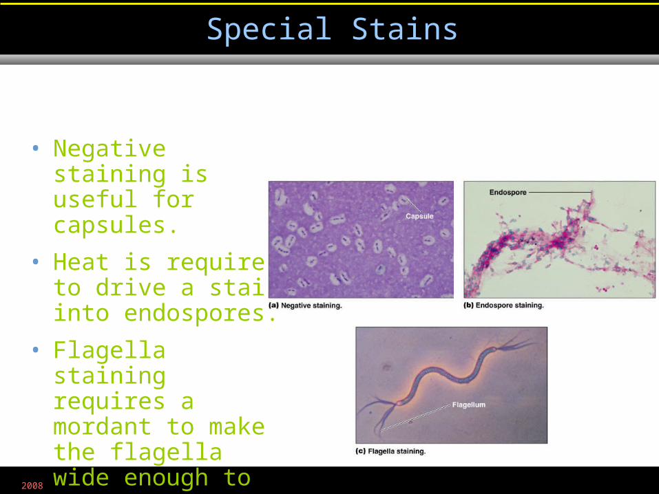
• Negative staining is useful for capsules.
• Heat is required to drive a stain into endospores.
• Flagella staining requires a mordant to make the flagella wide enough to see.
Special Stains
Figure 3.12a-c
2008
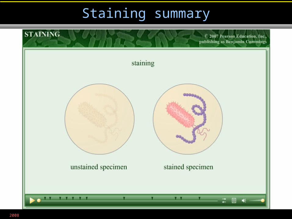
Staining summary
Related Documents