Abstract The sub-cellular location of enzymes of fatty acid b-oxidation in plants is controversial. In the current debate the role and location of particular thiolases in fatty acid degradation, fatty acid synthesis and isoleucine degradation are important. The aim of this research was to determine the sub-cellular location and hence provide information about possible func- tions of all the putative 3-ketoacyl-CoA thiolases (KAT) and acetoacetyl-CoA thiolases (ACAT) in Arabidopsis. Arabidopsis has three genes predicted to encode KATs, one of which encodes two polypeptides that differ at the N-terminal end. Expression in Arabidopsis cells of cDNAs encoding each of these KATs fused to green fluorescent protein (GFP) at their C-termini showed that three are targeted to peroxi- somes while the fourth is apparently cytosolic. The four KATs are also predicted to have mitochondrial tar- geting sequences, but purified mitochondria were un- able to import any of the proteins in vitro. Arabidopsis also has two genes encoding a total of five different putative ACATs. One isoform is targeted to peroxi- somes as a fusion with GFP, while the others display no targeting in vivo as GFP fusions, or import into isolated mitochondria. Analysis of gene co-expression clusters in Arabidopsis suggests a role for peroxisomal KAT2 in b-oxidation, while KAT5 co-expresses with genes of the flavonoid biosynthesis pathway and cytosolic ACAT2 clearly co-expresses with genes of the cytosolic mevalonate biosynthesis pathway. We conclude that KATs and ACATs are present in the cytosol and peroxisome, but are not found in mitochondria. The implications for fatty acid b-oxidation and for isoleucine degradation in mitochondria are discussed. Keywords Thiolase Mitochondria Peroxisomes b-oxidation Sub-cellular localization Abbreviations AOX Alternative oxidase ACAT Acetoacetyl-CoA thiolase KAT 3-Ketoacyl-CoA thiolase Rubisco SSU Small subunit of Ribulose 1,5- bisphosphate carboxylase/oxygenase Introduction It is widely accepted that the complete b-oxidation of medium- and long-chain fatty acids in plants takes place in the peroxisomes (Hooks 2002), as it does in yeast (van Roermund et al. 2003). However, some biochemical evidence suggests that plant mitochondria can also carry out such b-oxidation of fatty acids (Masterson and Wood 2001). It has also become clear recently that plant mitochondria catalyse at least the initial steps in the degradation of branched-chain a-keto acids, derived from leucine, isoleucine and Electronic supplementary material Supplementary material is available in the online version of this article at http:// dx.doi.org/10.1007/s11103-006-9075-1 and is accessible for authorized users. C. Carrie M. W. Murcha A. H. Millar S. M. Smith J. Whelan (&) ARC Centre of Excellence in Plant Energy Biology, University of Western Australia, MCS building M316, 35 Stirling Highway, Crawley 6009 WA, Australia e-mail: [email protected] Plant Mol Biol (2007) 63:97–108 DOI 10.1007/s11103-006-9075-1 123 Nine 3-ketoacyl-CoA thiolases (KATs) and acetoacetyl-CoA thiolases (ACATs) encoded by five genes in Arabidopsis thaliana are targeted either to peroxisomes or cytosol but not to mitochondria Chris Carrie Monika W. Murcha A. Harvey Millar Steven M. Smith James Whelan Received: 25 June 2006 / Accepted: 10 August 2006 / Published online: 21 November 2006 Ó Springer Science+Business Media B.V. 2006

Welcome message from author
This document is posted to help you gain knowledge. Please leave a comment to let me know what you think about it! Share it to your friends and learn new things together.
Transcript

Abstract The sub-cellular location of enzymes of
fatty acid b-oxidation in plants is controversial. In the
current debate the role and location of particular
thiolases in fatty acid degradation, fatty acid synthesis
and isoleucine degradation are important. The aim of
this research was to determine the sub-cellular location
and hence provide information about possible func-
tions of all the putative 3-ketoacyl-CoA thiolases
(KAT) and acetoacetyl-CoA thiolases (ACAT) in
Arabidopsis. Arabidopsis has three genes predicted to
encode KATs, one of which encodes two polypeptides
that differ at the N-terminal end. Expression in
Arabidopsis cells of cDNAs encoding each of these
KATs fused to green fluorescent protein (GFP) at their
C-termini showed that three are targeted to peroxi-
somes while the fourth is apparently cytosolic. The four
KATs are also predicted to have mitochondrial tar-
geting sequences, but purified mitochondria were un-
able to import any of the proteins in vitro. Arabidopsis
also has two genes encoding a total of five different
putative ACATs. One isoform is targeted to peroxi-
somes as a fusion with GFP, while the others display
no targeting in vivo as GFP fusions, or import into
isolated mitochondria. Analysis of gene co-expression
clusters in Arabidopsis suggests a role for peroxisomal
KAT2 in b-oxidation, while KAT5 co-expresses with
genes of the flavonoid biosynthesis pathway and
cytosolic ACAT2 clearly co-expresses with genes of
the cytosolic mevalonate biosynthesis pathway. We
conclude that KATs and ACATs are present in the
cytosol and peroxisome, but are not found in
mitochondria. The implications for fatty acid
b-oxidation and for isoleucine degradation in
mitochondria are discussed.
Keywords Thiolase Æ Mitochondria Æ Peroxisomes Æb-oxidation Æ Sub-cellular localization
Abbreviations
AOX Alternative oxidase
ACAT Acetoacetyl-CoA thiolase
KAT 3-Ketoacyl-CoA thiolase
Rubisco SSU Small subunit of Ribulose 1,5-
bisphosphate carboxylase/oxygenase
Introduction
It is widely accepted that the complete b-oxidation of
medium- and long-chain fatty acids in plants takes
place in the peroxisomes (Hooks 2002), as it does in
yeast (van Roermund et al. 2003). However, some
biochemical evidence suggests that plant mitochondria
can also carry out such b-oxidation of fatty acids
(Masterson and Wood 2001). It has also become clear
recently that plant mitochondria catalyse at least the
initial steps in the degradation of branched-chain
a-keto acids, derived from leucine, isoleucine and
Electronic supplementary material Supplementary materialis available in the online version of this article at http://dx.doi.org/10.1007/s11103-006-9075-1 and is accessible forauthorized users.
C. Carrie Æ M. W. Murcha Æ A. H. Millar Æ S. M. Smith ÆJ. Whelan (&)ARC Centre of Excellence in Plant Energy Biology,University of Western Australia, MCS building M316,35 Stirling Highway, Crawley 6009 WA, Australiae-mail: [email protected]
Plant Mol Biol (2007) 63:97–108
DOI 10.1007/s11103-006-9075-1
123
Nine 3-ketoacyl-CoA thiolases (KATs) and acetoacetyl-CoAthiolases (ACATs) encoded by five genes in Arabidopsisthaliana are targeted either to peroxisomes or cytosolbut not to mitochondria
Chris Carrie Æ Monika W. Murcha Æ A. Harvey Millar ÆSteven M. Smith Æ James Whelan
Received: 25 June 2006 / Accepted: 10 August 2006 / Published online: 21 November 2006� Springer Science+Business Media B.V. 2006
valine, through a branched chain a-keto acid dehy-
drogenase complex similar to the pyruvate and 2-oxo-
glutarate dehydrogenase complexes of the TCA cycle
(Fujiki et al. 2000; Graham and Eastmond 2002; Taylor
et al. 2004). In the case of the leucine carbon skeleton,
the later steps of degradation are carried out entirely
within the mitochondrion (Graham and Eastmond
2002; Taylor et al. 2004). In contrast, final degradation
of valine derivatives requires both mitochondrial and
peroxisomal steps (Lange et al. 2004). Meanwhile the
complete oxidation of the products from the isoleucine
carbon skeleton includes a b-oxidation step that
requires a 3-ketoacyl-CoA thiolase (KAT) for the
removal of an acetyl-CoA from 2-methylaceto-acetyl
CoA to form propionyl-CoA. However, it is unclear if
this thiolase catalysed b-oxidation of 2-methylaceto-
acetyl CoA occurs in the mitochondrion in plants, as it
does in mammals (Fukao et al. 2001), or whether it
occurs in the peroxisome in plants akin to the final
steps of valine metabolism (Lange et al. 2004).
Comparisons to Saccharomyces cerevisiae are not
informative as yeast degrades branched chain amino
acids not via the branched chain dehydrogenase com-
plex route in mitochondria, but via the Erhlich path-
way involving pyruvate decarboxylase to form the
corresponding aldehydes and an aldehyde dehydroge-
nase to form the corresponding alcohol in the cytosol
(Derrick and Large 1993). Thus yeast does not need a
thiolase for isoleucine degradation.
Two distinct forms of 3-ketoacyl-CoA thiolase are
known. Type 1 3-ketaoacyl-CoA thiolase (KAT; EC
2.3.1.16) is typically involved in the degradative pro-
cess of fatty acid b-oxidation. The Type II enzyme is an
acetoacetyl-CoA thiolase (ACAT; EC 2.3.1.9), typi-
cally involved in the mevalonate pathway where it
functions in the biosynthetic direction. However,
ACATs are not exclusively involved in mevalonate
synthesis. In mammals that undertake both fatty acid
b-oxidation and isoleucine catabolism in mitochondria,
the former is performed by a KAT while the later is
performed by an ACAT (Pereto et al. 2005).
In Arabidopsis, thiolase has been reported to be
associated with both peroxisomes and mitochondria in
sucrose density gradients (Footitt et al. 2002). Kruft
et al (2001) and Heazelwood et al (2004) have both
claimed the thiolase KAT2 encoded by At2g33150 to
be present in purified mitochondria in large-scale
proteome analyses. This thiolase has previously been
proposed to be a component of isoleucine catabolism
in mitochondria (Taylor et al. 2004). However, the
thiolase is question is a type I enzyme, while in mam-
mals it is the mitochondrial type II ACATs that have
been implicated in isoleucine catabolism (Pereto et al.
2005). The KAT2 thiolase encoded by At2g33150 has a
predicted type 2 peroxisomal targeting sequence
(PTS2) conforming to the consensus R-(X)6-H/Q-A/L/F
with a downstream cysteine residue required for pro-
teolytic cleavage (Baker and Sparkes 2005), and is
imported into peroxisomes in vitro (Johnson and
Olsen 2003). At2g33150 is well known to be essential
for peroxisomal b-oxidation (Germain et al. 2001).
However, the protein encoded by At2g33150 is also
predicted to be targeted to mitochondria by three dif-
ferent targeting prediction programs (Heazlewood
et al. 2004). Furthermore, changing a single amino acid
in the peroxisomal targeting signal of the KAT
precursor in mammals, a glutamic acid residue to any
non-acidic residue, resulted in targeting to both mito-
chondria and peroxisomes (Tsukamoto et al. 1994).
This raises the possibility that the thiolase encoded by
At2g33150 is targeted to peroxisomes and mitochon-
dria, representing a dual targeted protein.
This study was carried out to define the subcellular
localization of the products from the putative KATs
and ACATs in Arabidopsis. This was achieved by
identifying all possible thiolase genes in Arabidopsis
and comparing their sequences to known-location type
I and type II thiolases in yeast, mammals and fungi. We
then used transcript sequence data to produce all the
possible cDNAs for these gene products. These
cDNAs were used with in vivo and in vitro organelle
targeting assays to define subcellular localization of
type I and II thiolases in Arabidopsis and gene
expression profiling data was compared to define likely
functional links.
Materials and methods
Identification of genes and cDNAs encoding
thiolase
The predicted protein sequence encoded by At2g33150
previously shown to be a thiolase was used to define
other thiolase encoding genes in Arabidopsis (Germain
et al. 2001), and the sequences from other species as
previously published (Pereto et al. 2005). A similarity
tree was made using ClustalW multiple sequence
alignment and neighbour joining (Thompson et al.
1994, 1997). The gene structures were obtained from
The Arabidopsis Information Resource annotation
version 6 (TAIR6) and three individual cDNAs were
amplified for all possible cDNAs. The cDNAs pro-
duced from the genes were defined using the Arabid-
opsis genome tiling array (Mockler et al. 2005;
Yamada et al. 2003). Targeting predictions of the
98 Plant Mol Biol (2007) 63:97–108
123
encoded proteins were carried out using a variety of
prediction programs: TargetP (Emanuelsson et al.
2000), Mitoprot (Claros and Vincens 1996), Subloc
(Hua and Sun 2001), Ipsort (Bannai et al. 2002),
Predotar (Small et al. 2004), Mitpred (Kumar et al.
2006) and PeroxP (Emanuelsson et al. 2003). Percent-
age identity and similarity was calculated using Mat-
GAP v2.02 (Campanella et al. 2003).
Subcellular targeting of predicted thiolase proteins
The coding sequences of the predicted thiolase pro-
teins were cloned in frame with the coding region of
GFP in pGEM 3Zf(+) containing the 35S CaMV pro-
moter (Chew et al. 2003). The alternative oxidase
(AOX) coding region fused to GFP (Lee and Whelan
2004), and the red fluorescent protein (RFP) fused to a
type 1 peroxisomal SRL targeting signal from pumpkin
(Pracharoenwattana et al. 2005), were used as mito-
chondrial and peroxisomal controls respectively. The
constructs were used to transform Arabidopsis
suspension culture cells by biolistic transformation as
previously outlined (Thirkettle-Watts et al. 2003).
Fluorescence patterns were obtained 48 h after trans-
formation by visualization under an Olympus BX61
fluorescence microscope and imaged using the CellR
imaging software. In vitro protein import assays into
mitochondria isolated from Arabidopsis suspension
cell cultures were carried out as described in Lister
et al. (2004). In vitro mitochondrial uptake assays were
performed by adding precursor protein to 100 lg of
isolated mitochondria in 200 ll in the presence of
respiratory substrate (succinate 5 mM) and ATP
(1 mM) and ADP (200 lM) in import buffer (0.3 M
sucrose, 50 mM KCl, 10 mM MOPS pH 7.2, 5 mM
KH2PO4, 1% (w/v) BSA, 1 mM MgCl2, 1 mM methi-
onine and 5 mM DTT). Reactions were incubated at
24�C for 20 min then divided into two equal aliquots
and placed on ice. To one aliquot Proteinase K was
added to a final concentration of 40 lg/ml and incu-
bated for 15 min on ice, followed by the addition of
PMSF to 2 mM to terminate protease digestion. The
mitochondria were pelleted, washed twice in ice-cold
import buffer. The final pellet was resuspended in
SDS-PAGE sample buffer and proteins separated in
12% (w/v) polyacrylamide gels, then dried. Radiola-
belled proteins were visualized by exposing to a BAS
TR2040 imaging plate for 24 h and reading on a BAS
2500 Bio imaging analyser (Fuji, Tokyo). Outer
membrane ruptured mitochondria (Mit-OM) were
prepared after the import assay to test for the intra-
organelle location of imported protein. Rupture of the
outer membrane allowed access of added protease to
intermembrane space components or inner membrane
proteins exposed to the intermembrane space. Mit-OM
were prepared by resuspending 100 lg of mitochon-
drial protein in 10 ml SEH buffer (250 mM sucrose,
1 mM EDTA, 10 mM Hepes pH 7.4) and then adding
155 ll of 20 mM Hepes pH 7.4 and incubating on ice
for 20 min. To restore osmolarity 25 ll of 2 M sucrose
and 10 ll of 3 M KCl was added and mixed, re-pelleted
and washed in import buffer. Valinomycin was added
to a final concentration of 1 lM where indicated prior
to the addition of the precursor protein to mitochon-
dria and commencement of the import assay. AOX was
used as a positive control and the small subunit of
Ribulose 1, 5-bisphosphate carboxylase/oxygenase
(Rubisco SSU) as a negative control. As some pre-
cursor proteins displayed protease insensitivity even in
the presence of valinomycin the sensitivity of the pre-
cursor proteins alone to added protease was tested.
Sensitivity of the in vitro synthesized radiolabelled
proteins was tested by adding proteinase K to the
synthesized protein alone in the absence of mitochon-
dria to ensure that the added protease could digest the
protein.
In silico expression analysis
Expression correlation for genes encoding KATs and
ACATs was performed using the Expression Angler
program on the Botany Array Resource (Toufighi
et al. 2005). The Genevestigator Arabidopsis micro-
array database was used to analyse the response of the
genes of interest in this study in a variety of tissue types
(Zimmermann et al. 2004). The meta-analyser tool was
the function utilized, ATH1 22k array wild type only
arrays were chosen and the genes of interest were
selected. The data was visualized using a linear scale
from a total of 1860 array experiments. TMeV (TIGR
Multiple Experiment Viewer) programme was used to
cluster the genes and stresses, and Euclidean distance
and complete linkage were chosen for the hierarchal
clustering (Eisen et al. 1998; Saeed et al. 2003).
Results
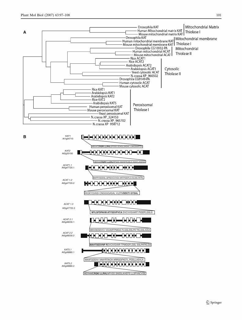
The Arabidopsis thiolase gene families
Searches of the Arabidopsis genome identify five loci
with sequence similarity to genes encoding known
thiolase proteins (Germain et al. 2001). Comparison of
predicted amino acid sequences shows that they fall
into two classes. Three loci encode the Type I class of
enzyme, KAT 1, 2 and 5, typically involved in
Plant Mol Biol (2007) 63:97–108 99
123
acetyl-CoA formation in fatty acid b-oxidation by
removal of a 2-carbon chain from a 3-ketoacyl-CoA.
The other two genes encode the type II class of
enzyme, ACAT 1 and 2, typically involved in aceto-
acetyl-CoA formation from two molecules of acetyl-
CoA (Fig. 1A, Supplementary Fig. 1A). The type I
genes have previously been annotated as KAT1, KAT2
and KAT5 (3-ketoacyl-CoA thiolase) (Germain et al.
2001), based on the chromosome on which they are
found (At1g04710, At2g33150 and At5g48880, respec-
tively). All three are closely related to known peroxi-
somal located type I thiolases in human, mouse and
yeast and this cluster also contains representatives from
the fungus Neurospora crassa and the monocot rice
(Oryza sativa). The matrix and membrane-bound type I
mitochondrial thiolases involved in fatty acid degrada-
tion in human, mouse and Drosophila cluster separately
and do not contain members from the completed
genome sequences of fungi, Arabidopsis or rice.
The two Arabidopsis type II thiolases (here referred
to as ACAT1 and ACAT2, At5g47720 and At5g48230,
respectively) cluster with the known cytosolic type II
thiolases from yeast and the cytosolic and mitochon-
drial type II thiolases from human, mouse and Dro-
sophila. The monocot rice also has two type II thiolases
that cluster in this set and N. crassa contains a single
type II gene that clusters with the yeast cytosolic type
II protein.
The sequence divergence of mitochondrial type I
thiolases in mammals from the peroxisomal type I
KATs in plants, fungi and animals makes the presence
of KAT2 in Arabidopsis mitochondria appear unlikely
based on phylogenetic evidence if thiolase location is
conserved. However, the sequence analysis does not
define the location of the type II ACAT proteins in
Arabidopsis and leaves open the possibility of a mito-
chondrial location of at least one of these proteins,
especially given the presence of multiple type II thio-
lases in both plants and mammals.
Definition of the number of products from each
Arabidopsis thiolase gene
Analysis of EST sequences and tiling array data shows
that KAT1 and KAT2 loci each encode single poly-
peptide sequences (Fig. 1B) (Mockler et al. 2005;
Yamada et al. 2003). In contrast, KAT5 encodes two
proteins that differ at the N-terminus due to alternative
RNA splicing that generates either 13 (KAT5.1) or 14
(KAT5.2) exons (Fig. 1B). The N-terminal sequences
of proteins encoded by KAT1, KAT2 and KAT5.2
include putative PTS2-type sequences, while the
protein encoded by KAT5.1 does not (Fig. 1B).
Interestingly, proteins encoded by KAT1, KAT2,
KAT5.1 and KAT5.2 proteins are predicted to be tar-
geted to mitochondria by at least two of three different
programs (Table 1). Analysis of EST sequences and
tiling array data shows that ACAT1 and ACAT2 loci
also encode three and two proteins respectively
(Fig. 1B). Differential RNA splicing results in the
protein encoded by ACAT1.1 lacking ten amino acids
at the C-terminus relative to the protein encoded by
ACAT1.2. The protein ACAT1.3 has 20 different
amino acid residues at the N-terminus relative to
ACAT1.1. None of the proteins has predicted orga-
nelle-targeting information (Table 1). Differential
RNA splicing also accounts for the N-terminal 6 amino
acid residues of the protein encoded by ACAT2.1
being replaced by 11 different amino acid residues in
the case of the protein encoded by ACAT2.2 (Fig. 1B).
Neither protein has predicted organelle-targeting
information (Table 1).
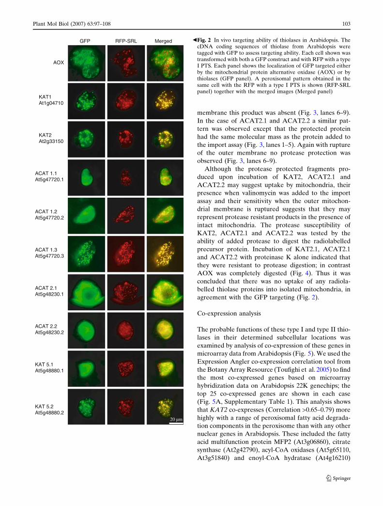
Targeting of type I and II thiolases in vivo
To localize thiolases in vivo, GFP and RFP fusions
were employed. GFP and RFP have been used exten-
sively to study protein targeting to mitochondria,
peroxisomes and chloroplasts (Heazlewood et al.
2005). To demonstrate specific mitochondrial and
peroxisomal targeting in vivo and our ability to
distinguish the two, an AOX–GFP construct (Lee and
Whelan 2004) and an RFP–PTS1 construct (Pracha-
roenwattana et al. 2005), were employed. The two
gene constructs were co-delivered into Arabidopsis
suspension culture cells using a biolistic gene gun
(Thirkettle-Watts et al. 2003). After 48 h individual
cells expressing both GFP and RFP fluorescence were
imaged. The results show that GFP and RFP were
targeted to discrete organelles consistent with specific
targeting to mitochondria and peroxisomes respec-
tively (Fig. 2).
To examine thiolase targeting we made translational
fusions with GFP at the C-terminus since peroxisomal
(PTS2) and mitochondrial targeting sequences are both
N-terminal. Thiolase cDNAs encoding all nine pro-
teins were linked to the GFP coding region and cloned
downstream of the CaMV 35S promoter. They were
Fig. 1 Classification of thiolase (KAT and ACAT) genes andgene structure in Arabidopsis. (A) A phylogenetic tree wasgenerated using the neighbour joining method, using ClustalW ofthiolase proteins from a variety of organisms. KAT = 3-ketoacyl-CoA thiolases, ACAT = acetoacetly-CoA thiolases. (B) Genestructure and predicted proteins encoded by thiolase genes. Thedifferences in the proteins encoded by each locus are indicated inbold where evidence for more than one cDNA exists. The openwhite boxes indicate exons
c
100 Plant Mol Biol (2007) 63:97–108
123
MEKATERQRI LLRHLQPSSS SDASLSASAC LSKDSAAYQY
MEKAIERQRV LLEHLRPSSS SSHNYEASLS ASACLAGDSA
MAPPVSDDSL QPRDVCVVGV ARTPIGDFLG SLSSLTATRL
MNVDESDVCI VGVARTPMGG FLGSLSSLPA TKLGSLAIAA
MAHTSESVNP RDVCIVGVAR TPMGGFLGSL SSLPATKLGS
KKGKYGVASI CNGGGGASAL VLEFMSEKTI GYSAL
MERAMERQKI LLRHLNPVSS SNSSLKHEPS LLSPVNCVSE
MAAFGDDIVI VAAYRTAICK ARRGGFKDTL PDDLLASVLK
KAT1At1g04710
KAT2At2g33150
At5g47720.1
At5g47720.2
At5g48230.1
At5g48230.2
KAT5.1 At5g48880.1
KAT5.2At5g48880.2
ACAT1.1
ACAT 1.2
ACAT 2.2
ACAT 2.1
ACAT 1.3
At5g47720.3
MYLSFDPAVM ATYSSVPVCA DVCVVGVART PIGDFLGSLS
A
B
Plant Mol Biol (2007) 63:97–108 101
123
each delivered into Arabidopsis suspension culture
cells together with the RFP–PTS1 construct. After 48 h
individual cells expressing both GFP and RFP fluo-
rescence were imaged, and the images merged. The
results show that KAT1, KAT2 and KAT5.2 and
ACAT 1.3 were targeted to peroxisomes, as indicated
by coincidence of GFP and RFP images (Fig. 2). In
contrast, ACAT1.1, ACAT1.2, ACAT2.1, ACAT2.2
and KAT5.1 show diffuse fluorescence throughout the
cell indicating that no specific targeting to any orga-
nelle has occurred, suggesting a cytosolic localization.
With peroxisomal targeting of KAT1, KAT2, KAT5.2
and ACAT 1.3, although it was apparent that the
patterns of GFP and RFP were essentially identical,
the higher intensity of the former means that when
merged the green fluorescence was dominant in some
cells.
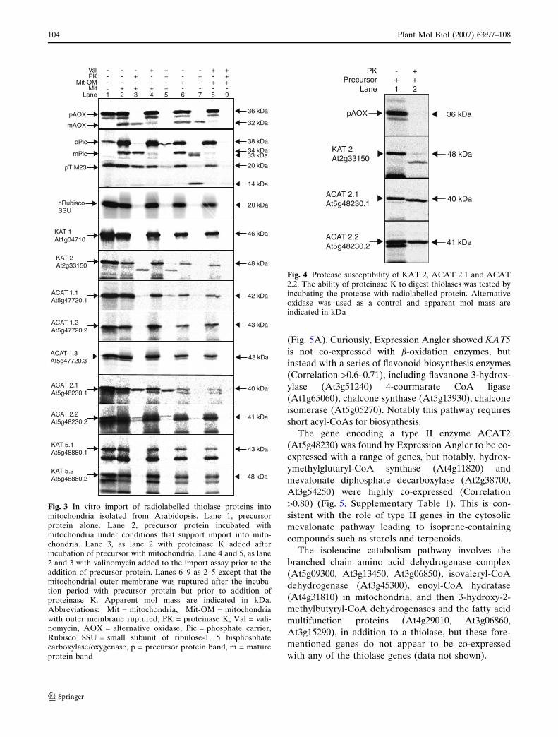
Protein import into isolated mitochondria
None of the thiolases were apparently targeted to
mitochondria in vivo. However, it is possible that up-
take was prevented by the GFP fusion, or that mito-
chondria normally take up less thiolase than
peroxisomes, such that GFP fluorescence from mito-
chondria did not reach an intensity to be detected. To
test these possibilities we examined the ability of iso-
lated mitochondria to take up all nine thiolases. Each
protein was synthesized in a rabbit reticulocyte lysate
translation system in the presence of radiolabelled
methionine, and then tested for import into mito-
chondria isolated from Arabidopsis cell cultures. As a
control the import and processing of AOX and Rubisco
SSU were examined, the former as a positive control
for import and the latter as a control to demonstrate
the specificity of import into isolated mitochondria
(Chew et al. 2003; Chew and Whelan 2004). In this
case the AOX precursor protein (36 kDa) was
imported and cleaved to a mature protein (32 kDa) as
previously demonstrated (Fig. 3, lanes 1 and 2)
(Whelan et al. 1995). Addition of protease resulted in
the 32-kDa mature form being resistant to protease
digestion indicating uptake by mitochondria. This
resistance was abolished by the addition of valinomy-
cin that dissipates the inner membrane potential
(Fig. 3, lanes 4 and 5) (Tanudji et al. 2001). The
phosphate translocator from maize was used as an
additional control, after uptake into mitochondria and
rupture of the outer membrane protease digestion
results in a small cleaved protected fragment of
33 kDa, indicating that the added protease has access
to inside the outer membrane (Bathgate et al. 1989;
Murcha et al. 2004, 2005; Winning et al. 1992). In
contrast to the mitochondrial controls, Rubisco SSU
was not protected from protease digestion indicating it
was not imported into mitochondria (Fig. 3).
When the nine thiolase proteins were tested for
mitochondrial uptake two distinct patterns were ob-
served, KAT1, ACAT 1.1, ACAT 1.2, ACAT 1.3,
KAT5.1 and KAT5.2 did not yield any protease pro-
tected products upon incubation with mitochondria
and thus were deemed not to be imported (Fig. 3).
KAT2, ACAT2.1 and ACAT2.2 yielded resistant
products upon incubation with mitochondria. Although
KAT2 was not proteolytically cleaved by mitochon-
dria, a protease resistant product with a lower mol
mass was obtained when mitochondria were treated
with Proteinase K (Fig. 3, lanes 1–3). Notably this was
also generated in the presence of valinomycin (Fig. 3,
lanes 4–5). However, upon rupture of the outer
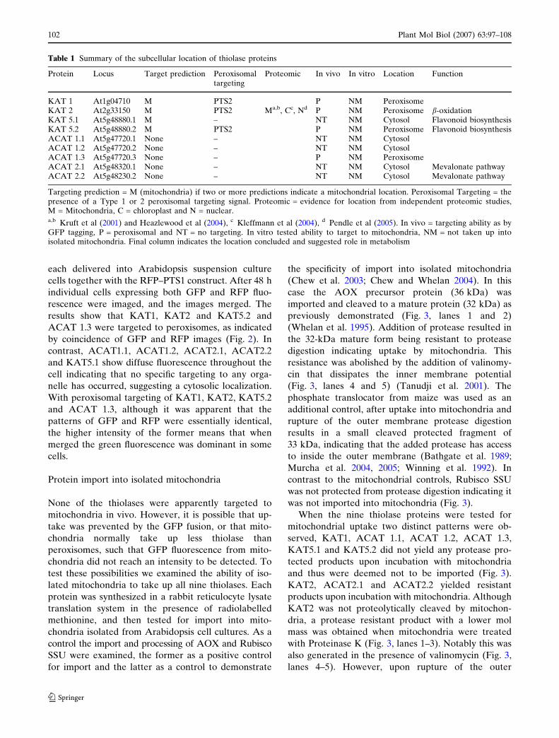
Table 1 Summary of the subcellular location of thiolase proteins
Protein Locus Target prediction Peroxisomaltargeting
Proteomic In vivo In vitro Location Function
KAT 1 At1g04710 M PTS2 P NM PeroxisomeKAT 2 At2g33150 M PTS2 Ma,b, Cc, Nd P NM Peroxisome b-oxidationKAT 5.1 At5g48880.1 M – NT NM Cytosol Flavonoid biosynthesisKAT 5.2 At5g48880.2 M PTS2 P NM Peroxisome Flavonoid biosynthesisACAT 1.1 At5g47720.1 None – NT NM CytosolACAT 1.2 At5g47720.2 None – NT NM CytosolACAT 1.3 At5g47720.3 None – P NM PeroxisomeACAT 2.1 At5g48320.1 None – NT NM Cytosol Mevalonate pathwayACAT 2.2 At5g48230.2 None – NT NM Cytosol Mevalonate pathway
Targeting prediction = M (mitochondria) if two or more predictions indicate a mitochondrial location. Peroxisomal Targeting = thepresence of a Type 1 or 2 peroxisomal targeting signal. Proteomic = evidence for location from independent proteomic studies,M = Mitochondria, C = chloroplast and N = nuclear.a,b Kruft et al (2001) and Heazlewood et al (2004), c Kleffmann et al (2004), d Pendle et al (2005). In vivo = targeting ability as byGFP tagging, P = peroxisomal and NT = no targeting. In vitro tested ability to target to mitochondria, NM = not taken up intoisolated mitochondria. Final column indicates the location concluded and suggested role in metabolism
102 Plant Mol Biol (2007) 63:97–108
123
membrane this product was absent (Fig. 3, lanes 6–9).
In the case of ACAT2.1 and ACAT2.2 a similar pat-
tern was observed except that the protected protein
had the same molecular mass as the protein added to
the import assay (Fig. 3, lanes 1–5). Again with rupture
of the outer membrane no protease protection was
observed (Fig. 3, lanes 6–9).
Although the protease protected fragments pro-
duced upon incubation of KAT2, ACAT2.1 and
ACAT2.2 may suggest uptake by mitochondria, their
presence when valinomycin was added to the import
assay and their sensitivity when the outer mitochon-
drial membrane is ruptured suggests that they may
represent protease resistant products in the presence of
intact mitochondria. The protease susceptibility of
KAT2, ACAT2.1 and ACAT2.2 was tested by the
ability of added protease to digest the radiolabelled
precursor protein. Incubation of KAT2.1, ACAT2.1
and ACAT2.2 with proteinase K alone indicated that
they were resistant to protease digestion; in contrast
AOX was completely digested (Fig. 4). Thus it was
concluded that there was no uptake of any radiola-
belled thiolase proteins into isolated mitochondria, in
agreement with the GFP targeting (Fig. 2).
Co-expression analysis
The probable functions of these type I and type II thio-
lases in their determined subcellular locations was
examined by analysis of co-expression of these genes in
microarray data from Arabidopsis (Fig. 5). We used the
Expression Angler co-expression correlation tool from
the Botany Array Resource (Toufighi et al. 2005) to find
the most co-expressed genes based on microarray
hybridization data on Arabidopsis 22K genechips; the
top 25 co-expressed genes are shown in each case
(Fig. 5A, Supplementary Table 1). This analysis shows
that KAT2 co-expresses (Correlation >0.65–0.79) more
highly with a range of peroxisomal fatty acid degrada-
tion components in the peroxisome than with any other
nuclear genes in Arabidopsis. These included the fatty
acid multifunction protein MFP2 (At3g06860), citrate
synthase (At2g42790), acyl-CoA oxidases (At5g65110,
At3g51840) and enoyl-CoA hydratase (At4g16210)
Fig. 2 In vivo targeting ability of thiolases in Arabidopsis. ThecDNA coding sequences of thiolase from Arabidopsis weretagged with GFP to assess targeting ability. Each cell shown wastransformed with both a GFP construct and with RFP with a typeI PTS. Each panel shows the localization of GFP targeted eitherby the mitochondrial protein alternative oxidase (AOX) or bythiolases (GFP panel). A peroxisomal pattern obtained in thesame cell with the RFP with a type I PTS is shown (RFP-SRLpanel) together with the merged images (Merged panel)
bGFP RFP-SRL Merged
AOX
KAT1At1g04710
KAT2At2g33150
ACAT 1.1At5g47720.1
ACAT 1.2At5g47720.2
ACAT 2.1At5g48230.1
ACAT 2.2At5g48230.2
KAT 5.1At5g48880.1
KAT 5.2At5g48880.2
ACAT 1.3At5g47720.3
20 µm
Plant Mol Biol (2007) 63:97–108 103
123
(Fig. 5A). Curiously, Expression Angler showed KAT5
is not co-expressed with b-oxidation enzymes, but
instead with a series of flavonoid biosynthesis enzymes
(Correlation >0.6–0.71), including flavanone 3-hydrox-
ylase (At3g51240) 4-courmarate CoA ligase
(At1g65060), chalcone synthase (At5g13930), chalcone
isomerase (At5g05270). Notably this pathway requires
short acyl-CoAs for biosynthesis.
The gene encoding a type II enzyme ACAT2
(At5g48230) was found by Expression Angler to be co-
expressed with a range of genes, but notably, hydrox-
ymethylglutaryl-CoA synthase (At4g11820) and
mevalonate diphosphate decarboxylase (At2g38700,
At3g54250) were highly co-expressed (Correlation
>0.80) (Fig. 5, Supplementary Table 1). This is con-
sistent with the role of type II genes in the cytosolic
mevalonate pathway leading to isoprene-containing
compounds such as sterols and terpenoids.
The isoleucine catabolism pathway involves the
branched chain amino acid dehydrogenase complex
(At5g09300, At3g13450, At3g06850), isovaleryl-CoA
dehydrogenase (At3g45300), enoyl-CoA hydratase
(At4g31810) in mitochondria, and then 3-hydroxy-2-
methylbutyryl-CoA dehydrogenases and the fatty acid
multifunction proteins (At4g29010, At3g06860,
At3g15290), in addition to a thiolase, but these fore-
mentioned genes do not appear to be co-expressed
with any of the thiolase genes (data not shown).
pAOX
KAT 2At2g33150
ACAT 2.1At5g48230.1
ACAT 2.2At5g48230.2
36 kDa
48 kDa
40 kDa
41 kDa
PKPrecursor
Lane 1 2+ +
+-
Fig. 4 Protease susceptibility of KAT 2, ACAT 2.1 and ACAT2.2. The ability of proteinase K to digest thiolases was tested byincubating the protease with radiolabelled protein. Alternativeoxidase was used as a control and apparent mol mass areindicated in kDa
ValPK
Mit-OMMit
Lane-
+-
1 2 3 4 5 6 7 8 9
pAOX
pPic
KAT 1 At1g04710
pTIM23
pRubisco SSU
mAOX
mPic
KAT 2At2g33150
ACAT 1.1At5g47720.1
ACAT 1.2At5g47720.2
ACAT 2.1At5g48230.1
ACAT 2.2At5g48230.2
KAT 5.1At5g48880.1
KAT 5.2At5g48880.2
46 kDa
48 kDa
42 kDa
43 kDa
40 kDa
41 kDa
43 kDa
48 kDa
36 kDa
32 kDa
20 kDa
14 kDa
38 kDa34 kDa33 kDa
20 kDa
---
-- -
-- - - - -- -- - -
-
+ + + ++ + +
+ + + ++ + + +
ACAT 1.3At5g47720.3 43 kDa
Fig. 3 In vitro import of radiolabelled thiolase proteins intomitochondria isolated from Arabidopsis. Lane 1, precursorprotein alone. Lane 2, precursor protein incubated withmitochondria under conditions that support import into mito-chondria. Lane 3, as lane 2 with proteinase K added afterincubation of precursor with mitochondria. Lane 4 and 5, as lane2 and 3 with valinomycin added to the import assay prior to theaddition of precursor protein. Lanes 6–9 as 2–5 except that themitochondrial outer membrane was ruptured after the incuba-tion period with precursor protein but prior to addition ofproteinase K. Apparent mol mass are indicated in kDa.Abbreviations: Mit = mitochondria, Mit-OM = mitochondriawith outer membrane ruptured, PK = proteinase K, Val = vali-nomycin, AOX = alternative oxidase, Pic = phosphate carrier,Rubisco SSU = small subunit of ribulose-1, 5 bisphosphatecarboxylase/oxygenase, p = precursor protein band, m = matureprotein band
104 Plant Mol Biol (2007) 63:97–108
123
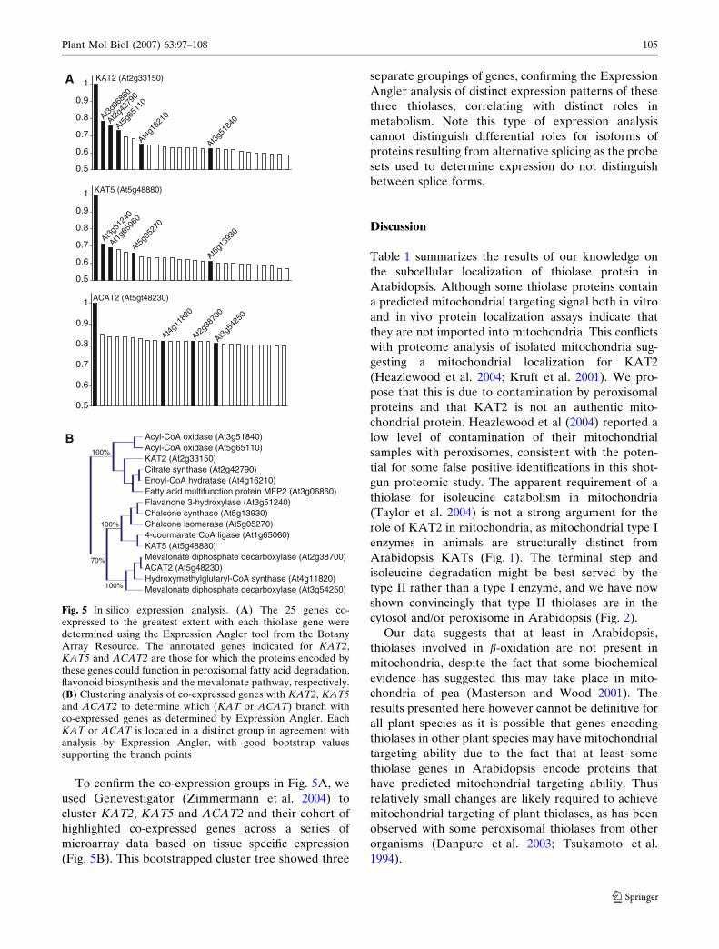
To confirm the co-expression groups in Fig. 5A, we
used Genevestigator (Zimmermann et al. 2004) to
cluster KAT2, KAT5 and ACAT2 and their cohort of
highlighted co-expressed genes across a series of
microarray data based on tissue specific expression
(Fig. 5B). This bootstrapped cluster tree showed three
separate groupings of genes, confirming the Expression
Angler analysis of distinct expression patterns of these
three thiolases, correlating with distinct roles in
metabolism. Note this type of expression analysis
cannot distinguish differential roles for isoforms of
proteins resulting from alternative splicing as the probe
sets used to determine expression do not distinguish
between splice forms.
Discussion
Table 1 summarizes the results of our knowledge on
the subcellular localization of thiolase protein in
Arabidopsis. Although some thiolase proteins contain
a predicted mitochondrial targeting signal both in vitro
and in vivo protein localization assays indicate that
they are not imported into mitochondria. This conflicts
with proteome analysis of isolated mitochondria sug-
gesting a mitochondrial localization for KAT2
(Heazlewood et al. 2004; Kruft et al. 2001). We pro-
pose that this is due to contamination by peroxisomal
proteins and that KAT2 is not an authentic mito-
chondrial protein. Heazlewood et al (2004) reported a
low level of contamination of their mitochondrial
samples with peroxisomes, consistent with the poten-
tial for some false positive identifications in this shot-
gun proteomic study. The apparent requirement of a
thiolase for isoleucine catabolism in mitochondria
(Taylor et al. 2004) is not a strong argument for the
role of KAT2 in mitochondria, as mitochondrial type I
enzymes in animals are structurally distinct from
Arabidopsis KATs (Fig. 1). The terminal step and
isoleucine degradation might be best served by the
type II rather than a type I enzyme, and we have now
shown convincingly that type II thiolases are in the
cytosol and/or peroxisome in Arabidopsis (Fig. 2).
Our data suggests that at least in Arabidopsis,
thiolases involved in b-oxidation are not present in
mitochondria, despite the fact that some biochemical
evidence has suggested this may take place in mito-
chondria of pea (Masterson and Wood 2001). The
results presented here however cannot be definitive for
all plant species as it is possible that genes encoding
thiolases in other plant species may have mitochondrial
targeting ability due to the fact that at least some
thiolase genes in Arabidopsis encode proteins that
have predicted mitochondrial targeting ability. Thus
relatively small changes are likely required to achieve
mitochondrial targeting of plant thiolases, as has been
observed with some peroxisomal thiolases from other
organisms (Danpure et al. 2003; Tsukamoto et al.
1994).
0.5
0.6
0.7
0.8
0.9
1
At3g0
6860
At2g4
2790
At5g6
5110
At4g1
6210
At3g5
1840
0.5
0.6
0.7
0.8
0.9
1
At3g5
1240
At5g0
5270
At5g1
3930
KAT5 (At5g48880)
0.5
0.6
0.7
0.8
0.9
1
At4g1
1820
At2g3
8700
At3g5
4250
Acyl-CoA oxidase (At3g51840)Acyl-CoA oxidase (At5g65110)KAT2 (At2g33150)Citrate synthase (At2g42790)Enoyl-CoA hydratase (At4g16210)Fatty acid multifunction protein MFP2 (At3g06860)Flavanone 3-hydroxylase (At3g51240)Chalcone synthase (At5g13930)Chalcone isomerase (At5g05270)4-courmarate CoA ligase (At1g65060)KAT5 (At5g48880)Mevalonate diphosphate decarboxylase (At2g38700)ACAT2 (At5g48230)Hydroxymethylglutaryl-CoA synthase (At4g11820)Mevalonate diphosphate decarboxylase (At3g54250)
100%
100%
KAT2 (At2g33150)
At1g6
5060
ACAT2 (At5gt48230)
A
B
70%
100%
Fig. 5 In silico expression analysis. (A) The 25 genes co-expressed to the greatest extent with each thiolase gene weredetermined using the Expression Angler tool from the BotanyArray Resource. The annotated genes indicated for KAT2,KAT5 and ACAT2 are those for which the proteins encoded bythese genes could function in peroxisomal fatty acid degradation,flavonoid biosynthesis and the mevalonate pathway, respectively.(B) Clustering analysis of co-expressed genes with KAT2, KAT5and ACAT2 to determine which (KAT or ACAT) branch withco-expressed genes as determined by Expression Angler. EachKAT or ACAT is located in a distinct group in agreement withanalysis by Expression Angler, with good bootstrap valuessupporting the branch points
Plant Mol Biol (2007) 63:97–108 105
123
The data on enzymes required for isoleucine deg-
radation from 2-methyl-3-hydroxybutyryl-CoA
through to propionyl-CoA increasingly suggests that
this part of the biochemical pathway is a non-mito-
chondrial activity. Of the three 3-hydroxy-2-methyl-
butyryl-CoA dehydrogenases in Arabidopsis
(At4g29010, At3g06860, At3g15290), At3g06860 has
been located to peroxisomes by three separate reports
using GFP tagging (Cutler et al. 2000; Koh et al. 2005;
Tian et al. 2004) and At3g15290 has been located to
chloroplasts by mass spectrometry (Kleffmann et al.
2004). The type II ACAT thiolases are all non-mito-
chondrial (Fig. 2), being present in either the cytosol
or peroxisome from our own data. Transport of
2-methyl-3-hydroxybutyryl-CoA out of mitochondria
has not been investigated, but the substrate specificity
of an array of known mitochondrial carriers from the
Mitochondrial Carrier Protein (MCP) family, the
Preprotein and Amino acid Transporter (PRAT)
family and ATP Binding Cassette (ABC) transporters
remain to be studied in Arabidopsis (Pohlmeyer et al.
1997; Rassow et al. 1999; Brugiere et al. 2004; Picault
et al. 2004). The distribution of pathways of amino
acid biosynthesis and metabolism between organelles
and the cytosol is relatively common in plants, but in
the case of isoleucine metabolism, although the met-
abolic enzymes involved are now relatively clear, the
transport activities that facilitate this pathway be-
tween mitochondria, the cytosol and the peroxisome
remain to be elucidated.
Co-expression analysis of transcript data can be a
powerful tool to confirm other data or provide leads
for further analysis. In this case, the co-expression
results for KAT2 are consistent with all our
experimental data. This gene encodes a peroxisomal
thiolase and is co-expressed with other peroxisomal
proteins involved in the same process, namely
b-oxidation of fatty acids. For ACAT2 the subcellu-
lar location, enzyme class and co-expression also
coincide to suggest a role in mevalonate biosynthesis
leading to isoprenes. The KAT5 co-expression result
was a surprise as this protein was suspected to be
involved in b-oxidation based on its enzyme class.
However, the different location of KAT5.1 and
KAT5.2 (Table 1), the fact that KAT5 does not
maintain b-oxidation in seedlings of the KAT
knockout but can partially complement for the lack
of KAT2 when driven by 35S expression (Germain
et al 2001), and the co-expression link with flavonoid
biosynthesis rather than b-oxidation genes (Fig. 5),
suggests that while KAT5 encodes a thiolase, it has a
distinct role to KAT2 in acyl-CoA metabolism in
plants.
Acknowledgements This work was funded through grants fromthe Australian Research Council (ARC) Centre of ExcellenceProgramme to JW, SS and AHM. AHM is funded as an ARCQueen Elizabeth II Fellow and SS as an ARC Federation Fellow.
References
Baker A, Sparkes IA (2005) Peroxisome protein import: someanswers, more questions. Curr Opin Plant Biol 8:640–647
Bannai H, Tamada Y, Maruyama O, Nakai K, Miyano S (2002)Extensive feature detection of N-terminal protein sortingsignals. Bioinformatics 18:298–305
Bathgate B, Baker A, Leaver CJ (1989) Two genes encode theadenine nucleotide translocator of maize mitochondria.Isolation, characterisation and expression of the structuralgenes. Eur J Biochem 183:303–310
Brugiere S, Kowalski S, Ferro M, Seigneurin-Berny D, Miras S,Salvi D, Ravanel S, d’Herin P, Garin J, Bourguignon J,Joyard J, Rolland N (2004) The hydrophobic proteome ofmitochondrial membranes from Arabidopsis cell suspen-sions. Phytochemistry 65:1693–1707
Campanella JJ, Bitincka L, Smalley J (2003) MatGAT: anapplication that generates similarity/identity matrices usingprotein or DNA sequences. BMC Bioinform 4:29
Chew O, Whelan J (2004) Just read the message: a model forsorting of proteins between mitochondria and chloroplasts.Trends Plant Sci 9:318–319
Chew O, Rudhe C, Glaser E, Whelan J (2003) Characterizationof the targeting signal of dual-targeted pea glutathionereductase. Plant Mol Biol 53:341–356
Claros MG, Vincens P (1996) Computational method to predictmitochondrially imported proteins and their targetingsequences. Eur J Biochem 241:779–786
Cutler SR, Ehrhardt DW, Griffitts JS, Somerville CR (2000)Random GFP::cDNA fusions enable visualization of sub-cellular structures in cells of Arabidopsis at a high fre-quency. Proc Natl Acad Sci USA 97:3718–3723
Danpure CJ, Lumb MJ, Birdsey GM, Zhang X (2003)Alanine:glyoxylate aminotransferase peroxisome-to-mito-chondrion mistargeting in human hereditary kidney stonedisease. Biochim Biophys Acta 1647:70–75
Derrick S, Large PJ (1993) Activities of the enzymes of theEhrlich pathway and formation of branched-chain alcoholsin Saccharomyces cerevisiae and Candida utilis grown incontinuous culture on valine or ammonium as sole nitrogensource. J Gen Microbiol 139:2783–2792
Eisen MB, Spellman PT, Brown PO, Botstein D (1998) Clusteranalysis and display of genome-wide expression patterns.Proc Natl Acad Sci USA 95:14863–14868
Emanuelsson O, Nielsen H, Brunak S, von Heijne G (2000)Predicting subcellular localization of proteins based on theirN-terminal amino acid sequence. J Mol Biol 300:1005–1016
Emanuelsson O, Elofsson A, von Heijne G, Cristobal S (2003) Insilico prediction of the peroxisomal proteome in fungi,plants and animals. J Mol Biol 330:443–456
Footitt S, Slocombe SP, Larner V, Kurup S, Wu Y, Larson T,Graham I, Baker A, Holdsworth M (2002) Control of ger-mination and lipid mobilization by COMATOSE, theArabidopsis homologue of human ALDP. EMBO J21:2912–2922
Fujiki Y, Sato T, Ito M, Watanabe A (2000) Isolation andcharacterization of cDNA clones for the e1beta and E2subunits of the branched-chain alpha-ketoacid dehydroge-nase complex in Arabidopsis. J Biol Chem 275:6007–6013
106 Plant Mol Biol (2007) 63:97–108
123
Fukao T, Scriver CR, Kondo N (2001) The clinical phenotypeand outcome of mitochondrial acetoacetyl-CoA thiolasedeficiency (b-ketothiolase or T2 deficiency) in 26 enzymat-ically proved and mutation-defined patients. Mol GenetMetab 72:109–114
Germain V, Rylott EL, Larson TR, Sherson SM, Bechtold N,Carde JP, Bryce JH, Graham IA, Smith SM (2001)Requirement for 3-ketoacyl-CoA thiolase-2 in peroxisomedevelopment, fatty acid b-oxidation and breakdown of tri-acylglycerol in lipid bodies of Arabidopsis seedlings. Plant J28:1–12
Graham IA, Eastmond PJ (2002) Pathways of straight andbranched chain fatty acid catabolism in higher plants. ProgLipid Res 41:156–181
Heazlewood JL, Tonti-Filippini JS, Gout AM, Day DA, WhelanJ, Millar AH (2004) Experimental analysis of the Arabid-opsis mitochondrial proteome highlights signaling andregulatory components, provides assessment of targetingprediction programs, and indicates plant-specific mitochon-drial proteins. Plant Cell 16:241–256
Heazlewood JL, Tonti-Filippini J, Verboom RE, Millar AH(2005) Combining experimental and predicted datasets fordetermination of the subcellular location of proteins inArabidopsis. Plant Physiol 139:598–609
Hooks MA (2002) Molecular biology, enzymology, and physi-ology of ß-oxidation. In: Baker A, Graham I (eds) Plantperoxisomes. Kluwer Academic Publishers, London, pp 19–55
Hua S, Sun Z (2001) Support vector machine approach forprotein subcellular localization prediction. Bioinformatics17:721–728
Johnson TL, Olsen LJ (2003) Import of the peroxisomaltargeting signal type 2 protein 3-ketoacyl-coenzyme athiolase into glyoxysomes. Plant Physiol 133:1991–1999
Kleffmann T, Russenberger D, von Zychlinski A, ChristopherW, Sjolander K, Gruissem W, Baginsky S (2004) TheArabidopsis thaliana chloroplast proteome reveals pathwayabundance and novel protein functions. Curr Biol 14:354–362
Koh S, Andre A, Edwards H, Ehrhardt D, Somerville S (2005)Arabidopsis thaliana subcellular responses to compatibleErysiphe cichoracearum infections. Plant J 44:516–529
Kruft V, Eubel H, Jansch L, Werhahn W, Braun HP (2001)Proteomic approach to identify novel mitochondrial pro-teins in Arabidopsis. Plant Physiol 127:1694–1710
Kumar M, Verma R, Raghava GP (2006) Prediction of mito-chondrial proteins using support vector machine and hiddenmarkov model. J Biol Chem 281:5357–5363
Lange PR, Eastmond PJ, Madagan K, Graham IA (2004) AnArabidopsis mutant disrupted in valine catabolism is alsocompromised in peroxisomal fatty acid b-oxidation. FEBSLett 571:147–153
Lee MN, Whelan J (2004) Identification of signals required forimport of the soybean F(A)d subunit of ATP synthase intomitochondria. Plant Mol Biol 54:193–203
Lister R, Chew O, Lee MN, Heazlewood JL, Clifton R, ParkerKL, Millar AH, Whelan J (2004) A transcriptomic andproteomic characterization of the Arabidopsis mitochon-drial protein import apparatus and its response to mito-chondrial dysfunction. Plant Physiol 134:777–789
Masterson C, Wood C (2001) Mitochondrial and peroxisomalb-oxidation capacities of organs from a non-oilseed plant.Proc Biol Sci 268:1949–1953
Mockler TC, Chan S, Sundaresan A, Chen H, Jacobsen SE,Ecker JR (2005) Applications of DNA tiling arrays forwhole-genome analysis. Genomics 85:1–15
Murcha MW, Elhafez D, Millar AH, Whelan J (2004) TheN-terminal extension of plant mitochondrial carrier proteinsis removed by two-step processing: the first cleavage is by themitochondrial processing peptidase. J Mol Biol 344:443–454
Murcha MW, Millar AH, Whelan J (2005) The N-terminalcleavable extension of plant carrier proteins is responsiblefor efficient insertion into the inner mitochondrial mem-brane. J Mol Biol 351:16–25
Pendle AF, Clark GP, Boon R, Lewandowska D, Lam YW,Andersen J, Mann M, Lamond AI, Brown JW, Shaw PJ(2005) Proteomic analysis of the Arabidopsis nucleolussuggests novel nucleolar functions. Mol Biol Cell 16:260–269
Pereto J, Lopez-Garcia P, Moreira D (2005) Phylogenetic anal-ysis of eukaryotic thiolases suggests multiple proteobacterialorigins. J Mol Evol 61:65–74
Picault N, Hodges M, Palmieri L, Palmieri F (2004) The growingfamily of mitochondrial carriers in Arabidopsis. TrendsPlant Sci 9:138–146
Pohlmeyer K, Soll J, Steinkamp T, Hinnah S, Wagner R (1997)Isolation and characterization of an amino acid-selectivechannel protein present in the chloroplastic outer envelopemembrane. Proc Natl Acad Sci USA 94:9504–9509
Pracharoenwattana I, Cornah JE, Smith SM (2005) Arabidopsisperoxisomal citrate synthase is required for fatty acidrespiration and seed germination. Plant Cell 17:2037–2048
Rassow J, Dekker PJ, van Wilpe S, Meijer M, Soll J (1999) Thepreprotein translocase of the mitochondrial inner mem-brane: function and evolution. J Mol Biol 286:105–120
Saeed AI, Sharov V, White J, Li J, Liang W, Bhagabati N,Braisted J, Klapa M, Currier T, Thiagarajan M, Sturn A,Snuffin M, Rezantsev A, Popov D, Ryltsov A, KostukovichE, Borisovsky I, Liu Z, Vinsavich A, Trush V, QuackenbushJ (2003) TM4: a free, open-source system for microarraydata management and analysis. Biotechniques 34:374–378
Small I, Peeters N, Legeai F, Lurin C (2004) Predotar: a tool forrapidly screening proteomes for N-terminal targetingsequences. Proteomics 4:1581–1590
Tanudji M, Dessi P, Murcha M, Whelan J (2001) Protein importinto plant mitochondria: precursor proteins differ in ATPand membrane potential requirements. Plant Mol Biol45:317–325
Taylor NL, Heazlewood JL, Day DA, Millar AH (2004) Lipoicacid-dependent oxidative catabolism of alpha-keto acids inmitochondria provides evidence for branched-chain aminoacid catabolism in Arabidopsis. Plant Physiol 134:838–848
Thirkettle-Watts D, McCabe TC, Clifton R, Moore C, FinneganPM, Day DA, Whelan J (2003) Analysis of the alternativeoxidase promoters from soybean. Plant Physiol 133:1158–1169
Thompson JD, Higgins DG, Gibson TJ (1994) CLUSTAL W:improving the sensitivity of progressive multiple sequencealignment through sequence weighting, position-specific gappenalties and weight matrix choice. Nucleic Acids Res22:4673–4680
Thompson JD, Gibson TJ, Plewniak F, Jeanmougin F, HigginsDG (1997) The CLUSTAL_X windows interface: flexiblestrategies for multiple sequence alignment aided by qualityanalysis tools. Nucleic Acids Res 25:4876–4882
Tian GW, Mohanty A, Chary SN, Li S, Paap B, Drakakaki G,Kopec CD, Li J, Ehrhardt D, Jackson D, Rhee SY, RaikhelNV, Citovsky V (2004) High-throughput fluorescent taggingof full-length Arabidopsis gene products in planta. PlantPhysiol 135:25–38
Toufighi K, Brady SM, Austin R, Ly E, Provart NJ (2005) Thebotany array resource: e-Northerns, expression angling, andpromoter analyses. Plant J 43:153–163
Plant Mol Biol (2007) 63:97–108 107
123
Tsukamoto T, Hata S, Yokota S, Miura S, Fujiki Y, Hijikata M,Miyazawa S, Hashimoto T, Osumi T (1994) Characteriza-tion of the signal peptide at the amino terminus of the ratperoxisomal 3-ketoacyl-CoA thiolase precursor. J BiolChem 269:6001–6010
van Roermund CW, Waterham HR, Ijlst L, Wanders RJ (2003)Fatty acid metabolism in Saccharomyces cerevisiae. Cell MolLife Sci 60:1838–1851
Whelan J, Hugosson M, Glaser E, Day DA (1995) Studies on theimport and processing of the alternative oxidase precursorby isolated soybean mitochondria. Plant Mol Biol 27:769–778
Winning BM, Sarah CJ, Purdue PE, Day CD, Leaver CJ (1992)The adenine nucleotide translocator of higher plants issynthesized as a large precursor that is processed uponimport into mitochondria. Plant J 2:763–773
Yamada K, Lim J, Dale JM, Chen H, Shinn P, Palm CJ,Southwick AM, Wu HC, Kim C, Nguyen M, Pham P, CheukR, Karlin-Newmann G, Liu SX, Lam B, Sakano H, Wu T,
Yu G, Miranda M, Quach HL, Tripp M, Chang CH, LeeJM, Toriumi M, Chan MM, Tang CC, Onodera CS, DengJM, Akiyama K, Ansari Y, Arakawa T, Banh J, Banno F,Bowser L, Brooks S, Carninci P, Chao Q, Choy N, Enju A,Goldsmith AD, Gurjal M, Hansen NF, HayashizakiY, Johnson-Hopson C, Hsuan VW, Iida K, Karnes M, KhanS, Koesema E, Ishida J, Jiang PX, Jones T, Kawai J, KamiyaA, Meyers C, Nakajima M, Narusaka M, Seki M, Sakurai T,Satou M, Tamse R, Vaysberg M, Wallender EK, Wong C,Yamamura Y, Yuan S, Shinozaki K, Davis RW, TheologisA, Ecker JR (2003) Empirical analysis of transcriptionalactivity in the Arabidopsis genome. Science 302:842–846
Zimmermann P, Hirsch-Hoffmann M, Hennig L, Gruissem W(2004) GENEVESTIGATOR. Arabidopsis microarraydatabase and analysis toolbox. Plant Physiol 136:2621–2632
108 Plant Mol Biol (2007) 63:97–108
123
Related Documents


![Photoaffinity labeling of acyl-CoA oxidase with 12-azidooleoyl-CoA and 12-[(4-azidosalicyl)amino]dodecanoyl-CoA](https://static.cupdf.com/doc/110x72/63133963b033aaa8b20ffcc4/photoaffinity-labeling-of-acyl-coa-oxidase-with-12-azidooleoyl-coa-and-12-4-azidosalicylaminododecanoyl-coa.jpg)