We would like to take this opportunity to thank these organisations for their kind support and sponsorship of this event Sponsorship was for a stand at the event and the companies had no influence on the content of the symposium

Welcome message from author
This document is posted to help you gain knowledge. Please leave a comment to let me know what you think about it! Share it to your friends and learn new things together.
Transcript

We would like to take this opportunity to thank theseorganisations for their kind support
and sponsorship of this event
Sponsorship was for a stand at the event and the companieshad no influence on the content of the symposium
NOTTINGHAM
EYE SYMPOSIUMand Research Meeting
30th January 2015
Programme
and Abstracts
PROGRAMME CONTENTS
Programme Summary 1
Clinical and Translational Research Presentations, Part 1Abstracts
22-7
Optometry Guest Speaker 8
Clinical and Translational Research Presentations, Part 2Abstracts
89-13
Poster PresentationsAbstracts
1314-19
19th Norman Galloway LectureProfessor Bhupendra PatelThe History of the Norman Galloway LecturePrevious Norman Galloway Lectures
202020-2121-22
Nottingham Eye Symposium:‘Looking inside and outside the box’: Lids and orbitIn the PipelineSpeaker Profiles and Abstracts
222222-24
Previous Prize Winners 25-27
Honorary Delegates 28
Details of Next Meeting 29
1
PROGRAMME SUMMARY
8.30am: Registration, Coffee and Pastries
9.00am: Chairman’s welcome and opening remarks
9.10am: Clinical and Translational Research PresentationsChair: Professor Harminder Dua
10.30am: Coffee, Trade Stand and Poster Viewing
11.00am: Optometry Guest Lecture ‘Cataract surgery changes to falls rates’Professor David Elliott, Bradford University
11.25am: Clinical and Translational Research Presentations continuedChair: Mr Winfried Amoaku and Professor Martin Rubinstein
12.25pm: Hot Buffet Lunch, Trade Stand and Poster Viewing1pm: Poster Session in Trade Hall
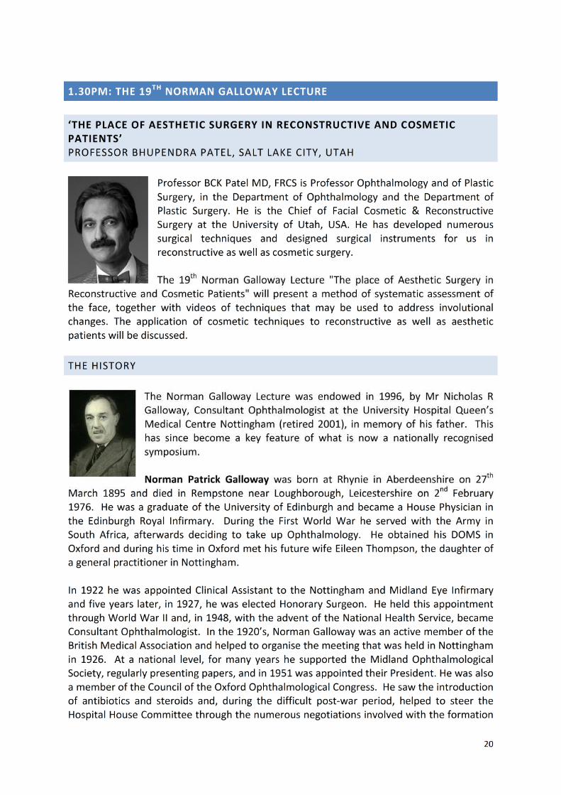
1.30pm: 19th Norman Galloway Lecture:‘The place of Aesthetic Surgery in Reconstructive and Cosmetic Patients’
Professor Bhupendra Patel, Salt Lake City, Utah
Symposium ‘Looking inside and outside the box’: Lids and orbitChair: Katya Tambe
2.30pm: To fill or not to fill, non-aesthetic uses of fillersRaman Malhotra, Queen Victoria Hospital, East Grinstead
2.55pm: In the Pipeline: Sponsor Presentations(Grafton Optical, Instinctive, NuVision, Scope, Spectrum, Topcon, Zeiss)
3.30pm: Coffee, Trade Stand and Poster Viewing
4.00pm: Jack in the box - orbital surprisesGeoff Rose, Moorfields Eye Hospital
4.25pm: The trinket box - paediatric socketsFrancesco Quaranta-Lioni, Rome, Italy
4.50pm: The box wrapped up - eyelid malpositionsMaria Amesty, Moorfields Eye Hospital
5.20pm: Research and Poster Prize Presentations
5.25pm: Chairman’s concluding remarks and close of meeting
2
9.10AM: CLINICAL AND TRANSLATIONAL RESEARCH PRESENTATIONS, PART 1
Chairs: Professor Harminder Dua
9.10am: Anaesthetic corneas with intact corneal nervesMouhamed Al-Aqaba, University of Nottingham
9.20am: The dynamics of big bubble formation after intrastromal air injection in humancorneasMohamed Elalfy, University of Nottingham
9.30am: Corneal stromal cells: A potential cell source for ocular surface regenerationLaura Sidney, University of Nottingham
9.40am: Surgical trabeculectomy training – are we safe at supervising?Andrew Walkden
9.50am: Dynamic muscle transfer in facial nerve palsyAruna Dharmasena, Manchester Royal Eye Hospital
10.00am: A standardised, off the shelf substrate for enhanced tissue engineeringMatthew Branch, University of Nottingham
10.10am: Safety and efficacy of intravitreal Ocriplasmin injection for the treatment ofvitreomacular traction (VMT) and macular holeRohit Saxena, Nottingham University Hospitals NHS Trust
10.20am: Dynamic digital subtraction dacryocystography for paediatric epiphoraRuth Chen, Nottingham University Hospitals NHS Trust
ABSTRACTS
9.10pm: Anaesthetic Corneas with Intact Corneal Nerves
Mouhamed Al-Aqaba and Virinder Dhillon, Mohamed Elalfy, Harminder S. DuaAcademic Ophthalmology, Division of Clinical Neuroscience, University of Nottingham, UK
Purpose: To report two cases presented with interesting and unexpected findings of absentcorneal sensation and intact sub-basal nerve plexus.Methods: Two patients presented with persistent uniocular dry eye symptoms. Full historyand clinical examination was obtained from each patient. The corneal sensation wasmeasured using Cochet-Bonnet Esthesiometer. In vivo confocal microscopy (IVCM) wasperformed to evaluate corneal innervation.Results: A 57-year-old woman with history of left trigeminal neuralgia for several years hadunsuccessful left alcohol trigeminal root injection followed by left microvasculardecompression. Postoperatively she developed persistent dry eye symptoms. Her leftcornea showed superficial punctuate epithelial erosions (SPEE). Corneal sensation was 60(normal) in her right eye and 0 (absent) in her left eye. IVCM revealed normal stromal andsub-basal nerves with equal density on both sides. A 46-year-old man presented with a 2-week history of sudden onset left blurred vision and left-sided facial numbness affectingprimarily the V-1 and V-2 areas. This was preceded by an episode of diarrhoea. MRI of hishead and trigeminal tract did not reveal any abnormality. The left cornea showed SPEE and
3
the corneal sensation was absent. IVCM showed normal appearances of his left stromal andsub-basal nerves with equal density on both sides.Conclusions: Our observations highlight two new aspects of corneal innervation andsensation in disease process. First, there is a lack of correlation between corneal sensationand sub-basal nerve plexus. Second, it likely that damage to the nerve fibres proximal totrigeminal primary sensory neurons results in impaired corneal sensation but intact cornealnerves and their trophic function. This could explain the absence of severe neurotrophickeratitis in our cases.
9.20am: The Dynamics of Big Bubble Formation after Intrastromal Air Injection in HumanCorneas
M Elalfy, L Faraj, DG Said, T Katamish, HS DuaAcademic Ophthalmology, Division of Clinical Neuroscience, University of Nottingham, UK
Purpose: Dua’s layer (DL) has considerable relevance to Deep anterior lamellar keratoplasty(DALK) procedure and the hitherto clinically observed but unexplained formation of Types 1,2 and mixed Big Bubble (BB). In this study we explored the dynamics of the formation ofdifferent types of BB.Methods: 50 human sclerocorneal discs were injected with air under balanced salt solutionand recorded. Videos were studied for leakage of air and formation of BB. Height anddiameter of BB were measured. Specimens were subjected to electron microscopy andimmunohistology for collagens & matricellular proteins. Age ranged from 3 weeks – 80years.Results: Air injected in the cornea preferentially reached the limbus and movedcircumferentially in a clockwise & anticlockwise direction as bands of 2-3 mm till they met,irrespective of direction of needle tip. The air then migrated centripetally to fill the stroma.Air leaked from the trabecular meshwork (TM) area at one or more points. Small bubblesformed in the centre and coalesced into a Type-1 BB. This rapidly expanded to attain aheight of 5 mm and a diameter of ≤ 9 mm. The anterior stomal wall of Type-1 BB showed multiple 'holes' through which air leaked to lift DL. DL was impervious to air. Type-2 BBalways started at the periphery. Distinct pores were seen in the peripheral stroma of DL.Most of these were located distal to attachment of Descemet’s membrane and explainedthe leakage of air from TM. Some were located centrally to the attachment and explainedformation of Type-2BB. More than 80% of BB were Type-1. Immunohistology did not offeran explanation for DL being impervious to air.Conclusion: DL is a distinct part of the surgical anatomy of the cornea. Identification ofpores in DL periphery is novel and explains the formation of a Type-2BB and the clinicalobservation of appearance of air in anterior chamber during DALK. Leakage is not throughthe TM.
4
9.30am: Corneal Stromal Cells: A Potential Cell Source for Ocular Surface Regeneration
Sidney, L. E., Dunphy S. E., Branch, M. J., Dua H. S. and Hopkinson, A.Academic Ophthalmology, Division of Clinical Neuroscience, University of Nottingham, UK
Purpose: Keratocytes of the corneal stroma are quiescent, dendritic cells, which maintainthe extracellular matrix. It is believed that there is a subpopulation of keratocytes that act asmultipotent stem/progenitor cells in the limbal region. To utilise these cells in regenerativetherapies, it is important to understand the effect of in vitro culture environment and theprocesses of dedifferentiation to a keratocyte phenotype. These cells may also play a role inrejuvenation of other layers of the cornea, such as the epithelium.Methods: Primary human corneal stromal cells (hCSC) were extracted from corneal rims.Optimum culture media was determined from a selection available, and the effect ofpassage on cell phenotype was assessed. The potential for differentiation back into astromal keratocyte phenotype was investigated by culture of hCSC on a 3D polymericmicrofibre carrier. Potential transdifferentiation of hCSC into a corneal epithelial lineagewas investigated using a specialised differentiation medium.Results: Phenotype of hCSC is strongly affected by extraction, passage and culture mediaformulation. Under optimum conditions, hCSC show potential as a multipotent stem cell,expressing indicative markers and differentiating to mesenchymal lineages. hCSC culturedon microfibre scaffolds demonstrate a morphology and phenotype more similar to that of akeratocyte (CD34 and ALDH positive). CD34+ cells isolated from hCSC at low passagedemonstrate a higher stem cell potential than the unsorted cells, with evidence oftransdifferentiation to corneal epithelial cell morphology and high expression of key cornealepithelial markers.Conclusions: Corneal stromal cells demonstrate stem cell potential, independent of the rolethey are traditionally associated with in the cornea. This potential depends upon in vitroculture environment and isolation of certain subpopulations. In future, these cells may havepotential in the regeneration of the ocular surface in cases of disease or trauma.
9.40am: Surgical Trabeculectomy Training – Are We Safe At Supervising?
1Walkden, A., 1Senior, M., 1Naylor, S., 1Lee, H., 2Anand, N., 2Turner, S., 2Ivanova, K.,1Bhargava, A., 2McNaught, A.
1Royal Preston Hospital, North West Deanery, 2Royal Gloucester Hospital
Purpose: Surgical exposure for trainees is limited due to service provision demands, theEuropean working time directive and subspecialisation of glaucoma surgery. Limitedknowledge exists on the outcomes of supervised glaucoma surgery. The aim is to determinethe safety of supervised trabeculectomy surgery performed by trainee ophthalmologists.Methods: Retrospective case note review of all eyes (n=231) that underwenttrabeculectomy surgery with MMC by consultant and trainee surgeons between March2011 and November 2013 across 3 UK centres. All eyes have 1-year follow-up. Datacollection includes pre-operative IOP, IOP at 1 year, and Snellen visual acuities. Failure ratesand surgical complications were recorded. Two-tailed p-values were obtained using Fisher’sexact test to ascertain statistical significance between groups.
5
Results: 140 (60.6%) cases were performed by consultant ophthalmologists (mean age=67;range 44-89 years). Trainees performed: 93 (40.3%) cases mean age= 70; 37-89 rangeyears). No statistical significance was observed between consultant and trainee eyesachieving IOP <21mmHg and <16mmHg (p=0.31 and 0.75 respectively). No statisticalsignificance was observed between the two groups in terms of Snellen acuity loss (p=0.37).No statistical significance was seen between consultant failure rate (n=20) and supervisedtrainee failure rate (n=27) (p=0.38) or complication rate (p=0.45).Conclusion: Supervised trainee cases did not show higher complication rates thanconsultant cases. These findings help guide informed consent if a trainee is to performsurgery. The findings may encourage trainee participation in more glaucoma surgerytherefore increasing experience and enhancing training.
9.50am: Dynamic Muscle Transfer in Facial Nerve Palsy
Saghir Ahmed Sadiq, Aruna DharmasenaManchester Royal Eye Hospital
Purpose: To describe the results of dynamic muscle transfer with an orbicularis oculi muscleflap from the contralateral side to the paralysed side in patients with House-Brackmangrade 6 facial nerve palsy.Methods: A case series of 6 patients who underwent dynamic muscle transfer with a flap ofhealthy orbicularis oculi muscle fibres from the contralateral side into the paralysedorbicularis oculi muscle. All patients had a House-Brackman grade 6 facial nerve palsy. Allthe subjects had previous multiple surgical procedures to improve the eyelid function. Inspite of this they were all symptomatic in terms of corneal exposure before orbicularismuscle transfer. All patients had post-operative follow up in excess of 6 months after theprocedure.Results: All patients improved symptomatically and had clinically reduced lagophthalmospost-operatively. Five patients who had an absent blink reflex showed a significantimprovement in their blink reflex post-operatively. There were no complications at thedonor site.Conclusion: All patients showed a significant improvement of their symptoms and theirlagophthalmos reduced post-operatively. Most importantly, the blink occurred involuntarilyat the same time as the blink on the normal side. The authors propose that a dynamicmuscle transfer using the contralateral orbicularis muscle may be considered to improve lidclosure and blink reflex to improve corneal exposure in patients with grade 6 facial palsywho have not benefited from conventional surgical procedures.
10.00am: A standardised, off the shelf substrate for enhanced tissue engineering
Branch, M.J., Hopkinson, AUniversity of Nottingham, NuVision
Purpose: Amniotic membrane (AM) is a popular ophthalmic tissue engineering adjunct. Wehave previously developed a highly effective thermolysin-based denuding technique thatpreserves basement membrane integrity. This technique combined with dry preservation ofAM produces a simple easy to use substrate for research.
6
Methods: Dry preserved thermolysin denuded AM (DAM), and FAM denuded withethylenediaminetetraacetic acid (EDTA), and dispase -based methodologies were prepared.Denuding efficiencies were compared using electron microscopy. The effect of denuding onAM molecular composition was investigated and characterised using proteomics. Thepropensity of DAM to support stem cells was explored using fluorescentimmunohistochemistry for defined markers.Results: Electron microscopy demonstrated thermolysin denuding efficiency wascomparable in FAM and DAM. Proteomic analyses showed effective removal of epithelialcell proteins in DAM, but not EDTA–based denuding techniques. Whilst similar enzymaticactivity to thermolysin, mechanical scraping reduces the efficacy of dispase denuding.Collagens IV, VI, periostin, βig-h3 and VLA-6 are targets of thermolysin activity. DAM maintains stem cell characteristics and is most effective in preventing differentiation.Conclusions: Conventional EDTA and dispase procedures for preparing AM for tissueengineering are ineffective at removing cells whilst preserving the basement membrane.Combining our novel thermolysin denuding and dry preservation techniques improves theoverall quality of AM for tissue-engineering and other research and clinical basedapplications.
10.10am: Safety and Efficacy of Intravitreal Ocriplasmin Injection for the Treatment ofVitreomacular Traction (VMT) and Macular Hole
Saxena R, Kumudhan DNottingham University Hospitals NHS Trust
Purpose: To evaluate the results of intravitreal ocriplasmin injection for the management ofvitreomacular traction and macular hole.Methods: This was a prospective audit from March’2014 to September’2014. The diagnosisof macular hole and vitreomacular traction was confirmed with spectral domain opticalcoherence topography in all patients. Patients’ eligibility for treatment of vitreomaculartraction was based on NICE guidance. All eligible patients were administered 125μg intravitreal Ocriplasmin. Mean follow up was after 6.5 weeks (Range 5-10 weeks). Theresults were compared with studies published in peer reviewed journals.Results: 8 patients (3 males & 5 females) with mean age of 73.37 years (Range 49-84 years)were treated. Vitreomacular adhesion was relieved in 5/8 (62.50%) patients. Total posteriorvitreous detachment was induced in 1/8 (12.50%) patient. Non-surgical closure of macularhole was achieved in 3/8 (37.50%) patients. Transient ocular adverse effect, like floaterswere reported in 2/8 (25%) patients. There were no severe ocular or systemic adverseeffects.Conclusions: The results compared favourably with the studies published in peer reviewedjournals. Intravitreal ocriplasmin injection relieves vitreomacular adhesion with minimalocular adverse effects.
7
10.20am: Dynamic Digital Subtraction Dacryocystography for Paediatric Epiphora
Ruth Chen, Maria Amesty, Julia Baxter, Shery Thomas, Timothy Taylor, LorraineAbercrombie, Katya Tambe
Nottingham University Hospitals NHS Trust
Purpose: To study the utility of dynamic digital subtraction dacryocystography (DSDCG)technique in paediatric epiphora.Method: Retrospective study of 45 lacrimal drainage systems (LDS) of 32 children, between2 and 12 years of age were studied. The presenting symptoms were persistent epiphoradespite previous syringing and probing, presence of fistulae, dacryocystitis, or older childrenpresenting for the first time to the clinic with epiphora. DSDCG was performed in all casesunder general anaesthesia, using a 27G Rabinov catheter and N300 contrast medium.Intubation of the lacrimal system was carried out using a self-retaining monocanalicularMonoka silicone stent via the upper punctum (FCI, Issy-les-Moulineaux Cedex®, France).Results: DSDCG identified the site of obstruction in 96% of our patients. In 25 LDS (45%) theobstruction was at the distal nasolacrimal duct, 9 LDS (20%) at the punctual/canalicularducts, and 9 LDS (20%) at the proximal nasolacrimal duct. 2 LDS (5%) were patent. Apartfrom these findings, 5 fistulae were also identified. These results guided managementaccordingly with intubation of the nasolacrimal duct, canaliculus, canalicular trephination,dacryocystorhinostomy and fistulectomy.Conclusions: Intraoperative DSDCG is useful to identify the exact location of the lacrimaldrainage system obstruction in cases of failed probing or delayed referral for paediatricepiphora. It demonstrates the anatomy very well, and shows the calibre of the differentstructures. DSDCG also provides additional information, as shown in the cases that requiredfistulectomy. It demonstrated complete closure of the fistula tract postoperatively, which isuseful information for the operating surgeon.
8
11.00AM: OPTOMETRY GUEST SPEAKER
PROFESSOR DAVID ELLIOTT, BRADFORD SCHOOL OF OPTOMETRY AND VISIONSCIENCES, BRADFORD UNIVERSITY ‘CATARACT SURGERY CHANGES TO FALLSRATES’
David Elliott is the Professor of Clinical Vision Science at the BradfordSchool of Optometry & Vision Science. He is the Editor-in-Chief ofOphthalmic & Physiological Optics (JIF 2.66) and the editor of ClinicalProcedures in Primary Eye Care, now in its 4th edition, in addition tochapters in several other standard textbooks (e.g., Borish’s ClinicalRefraction; The Oxford Textbook of Ophthalmology) and over 100 peerreviewed research papers in leading international journals of optometry,ophthalmology and gait.
His talk ‘Cataract Surgery Changes To Falls Rates’ will provide a review of the link betweenvision and falls in the elderly population will be presented with particular emphasis on fallsrate changes due to cataract surgery, new spectacles and multifocals. Preliminary analysis ofnew data will be presented which may help explain why falls rates are not reduced as muchas expected by cataract surgery.
11.25AM: CLINICAL AND TRANSLATIONAL RESEARCH PRESENTATIONS, PART 2
Chairs: Mr Winfried Amoaku and Professor Martin Rubinstein
11.25am: In-vitro Anti-Angiogenic Effects of Cryo-preserved Amniotic MembraneLana Faraj, University of Nottingham
11.35am: Investigating the use of dextran in corneal decellularisationSamantha Wilson, University Of Nottingham
11.45am: Levator Resection for Congenital PtosisRuth Chen, Nottingham University Hospitals NHS Trust
11.55am: A randomized controlled single centre open-label pilot trial in chronic anteriorblephatitis by external use of Chinese herbal medicineWenqing Li, St James Hospital Leeds
12.05am: The contribution of vision to balance during smartphone useParnika Sharma, University of Leicester
12.15am: The introduction of an ophthalmic clinical officer in a rural Ugandan hospital: areflection Eleanor Crossley, University of Birmingham
9
ABSTRACTS
11.25am: In-vitro Anti-Angiogenic Effects of Cryo-preserved Amniotic Membrane
1Lana Faraj, 1Elizabeth Stewart, 2Réka Albert, 1Claire Allen, 1Harminder Dua, 1WinfriedAmoaku
1University of Nottingham, Academic Ophthalmology, Division of Clinical Neuroscience2Department of Ophthalmology, University of Szeged, Szeged, Hungary
Background: Amniotic membrane (AM) has been used as a biological substrate inophthalmology and other fields for several years. It has a valuable role in ocular surfaceburns and non-healing ulcers. It is known that AM is non-immunogenic and has anti-inflammatory and anti-fibrotic properties and supports epithelial cell proliferation andmigration. These are supposedly mediated through a host of growth factors and cytokines.Despite its clinically accepted role in reducing corneal vascularisation when applied on theeye, there have been contradicting reports on the effect of AM on angiogenesis.Methods: The effect of soluble factors released from clinically prepared cryo-preserved AMon cultured human umbilical vein endothelial cell (HUVEC) proliferation was quantified andangiogenesis assessed on matrigel. Soluble anti-angiogenic factors were identified usingSearchlight protein arrays.Results: AM conditioned medium (AMCM) was found to reduce HUVEC proliferation.Angiogenesis was reduced by direct application of AM, a gradient effect was observed,where HUVEC closer to the AM failed completely in forming any tubules. In addition, HUVECfailed to survive directly on the AM. Analysis of the soluble factors released by the amnionincluded high levels of anti-angiogenic factors, thrombospondin and tissue inhibitors ofmetalloproteinase (TIMP) 1 and 2.Conclusion: AM in vitro has anti-angiogenic properties, validating some of the effectsobserved clinically.
11.35am: Investigating the Use of Dextran in Corneal Decellularisation
Wilson, S.L.1, Lynch, A.2, Ahearne, M.2,3
1Academic Ophthalmology, Division of Clinical Neuroscience, University of Nottingham,2Trinity Centre for Bioengineering, Trinity Biomedical Sciences Institute, Dublin
3Department of Mechanical and Manufacturing Engineering, School of Engineering, Dublin
Purpose: Dextran is routinely used as a de-swelling agent during organ culture and forpreservation of corneal tissue. 5% (w/v) dextran is commonly included in cornealdecellularisation protocols to prevent swelling, since it acts to draw fluid out of the cornea,due to its high affinity for water. To date, there is no standardised and reproducible cornealdecellularisation technique suitable for clinical translation. Since small changes toprocedures may have large impacts on the decellularisation efficacy, the aim of this studywas to systematically investigate the use of dextran in the decellularisation of adult porcinecorneas.Methods: Five groups of 8 mm adult porcine corneal buttons were investigated: (i) nativecornea; (ii) freeze-thaw treatment using ultra-pure water; (iii) detergent treatment using0.5% (w/v) sodium dodecyl sulphate and 1 % (w/v) Triton X-100; (iv) detergent/dextrantreatment, as detergent treatment, with the addition of 5 % dextran used throughout the

10
procedure; (v) detergent/dextran-wash treatment; as detergent treatment, followed by anadditional washing step in 5% dextran at the end of the procedure. Tissue transparency,thickness and mass pre/post decellularisation was measured. Removal of detectable cellularand immune reactive material was evidenced by histological and quantitative assays.Retention of corneal architecture and intrinsic biological cues (glycosaminoglycans) wereassessed via histological, immunofluorescence, electron microscopy and biochemicalanalysis.Results: Irrespective of the procedure used, all decellularised tissues experienced asignificant reduction in transparency (more so when dextran was used in the procedure),mass, and residual DNA compared to native tissue, with dextran having no apparent effecton mass and residual DNA content. The use of dextran did however preserve/restorenormal tissue thickness and maintained higher levels of glycosaminoglycans postdecellularisation. Dextran use appeared to have a positive effect with regards tomaintaining/restoring the collagen tissue microstructure. However, electron microscopystudies further revealed that the use of dextran throughout the whole decellularisationprotocol (detergent/dextran treatment) was vital for maintenance of the nanoscaleultrastructural organisation, and that use of dextran at the end of the decellularisationprotocol (detergent/dextran-wash) did not sufficiently preserve/restore the nanoscaleultrastructure of the corneal tissue.Conclusions: This study highlights the importance of performing systematic, in-depthstudies with extensive characterisation when devising appropriate decellularisationprocedures for clinical translation. Seemingly small changes to procedures can have hugeimpacts on the resulting acellular scaffold which will ultimately affect the efficacy andbiocompatibility upon recellularization either in vivo or in vitro. Future work will investigatethe effect dextran use during decellularisation has on cell infiltration, behaviour andremodelling following recellularization.
11.45am: Levator Resection for Congenital Ptosis
Ruth Chen, Maria Amesty, Katya TambeNottingham University Hospitals NHS Trust
Purpose: To evaluate the outcomes of anterior and posterior approach of levator resectionfor correction of congenital ptosisStudy design: Retrospective review looking at a consecutive series of 32 patients, who hadlevator resection surgery, over a 5-year period in a tertiary unit.Methods: Congenital ptosis with levator function > 4mm were managed with levatorpalpebrael superioris resection. The anterior approach involves a skin crease incision,dissecting the levator, and resecting it to the tarsus. The posterior approach exposes theposterior surface of the levator muscle via a transconjunctival approach. This preserves theseptum and leaves no visible scar.These techniques were performed for congenital ptosis under general anaesthesia by thesame surgeon. Data was collected on 39 eyes of 34 patients (age range 0 – 18). Outcomemeasures included measurements of preoperative and postoperative palpebral aperture,margin reflex distance (MRD1), symmetry of eyelid height, contour, complications and redooperations.
11
Results: Mean age was 9.1 years (2 years 5 months to 18 years 3 months). Mean levatorfunction was 9.8mm. Mean difference between the preoperative and postoperative MRD1was 2.5mm. There were 6 undercorrected cases, 4 with unsatisfactory eyelid contour, and 1with a suture granuloma. Success rate considering the criteria of postoperative MRD1 >=2mm and =<4.5mm was 85%, and postoperative palpebral aperture symmetry within 1mmof difference was 82%. Mean follow-up period was 8 months.Conclusion: Anterior and posterior approach levator resection has a good success rate forcorrecting congenital ptosis with moderate and good levator function.
11.55am: A Randomized Controlled Single Centre Open-Label Pilot Trial in ChronicAnterior Blephatitis by External Use of Chinese Herbal Medicine
Wenqing Li, Bernard ChangUniversity of Leeds Teaching St James Hospital Ophthalmology Department
Background: Blephatitis is inflammatory disease associated with itchiness, redness andcrusting of the margin of eyelids. It can lead to permanent alterations to the eyelid marginor vision loss from superficial keratopathy, corneal neovascularization, and ulceration. Mostimportantly, frequently cause significant ocular symptoms such as burning sensation,irritation, tearing, and red eye as well as visual problems such as photophobia and blurredvision. Blephatitis leads to high morbidity and accounts for 5% of all ophthalmologicalproblems presenting in primary care and about 2-5% of GP consultations. Current therapiesfor blepharitis are ‘good eyelid hygiene’, warm compresses, and antibiotics (external:chloramphenicol eye ointment, fucidic acid eye drops or oral antibiotics). However, it isdifficult to clinically prescribe targeted antibiotics. Considerable positive evidence from over39 clinical trials including two reviews on the treatment of Blepharitis using Chinese herbalmedicine (CHM) or integrated medicine has been described in detail. However, until nowthere has been no rigorous research into the clinical effectiveness of CHM for Blepharitis inthe UK, hence the proposed research on treating Blepharitis using CHM. This may yield aneffective result with possible long-term lower costs and risk.Aims: To evaluate the safety and efficacy of CHM on signs and symptoms of blepharitis.Study design: This study is an open-label, single centre pilot RCT. The target population inthis trial will be from the UK. The 60 eligible Blepharitis participants who meet the inclusionand exclusion criteria will be randomly located to an intervention group (good eyelidhygiene + CHM) and a control group (usual good eyelid hygiene + warm compresses) afterconsenting. Total treatment duration was 4 weeks. Intervention: the CHM treatment isbased on the Pharmacopoeia of People’s Republic of China and the principles of ChineseMedicine, prescribed by Consultant Wenqing Li.Outcome measures: Primary outcome: Data on the scale of signs and symptoms ofBletharitis. Secondary outcome: Standard ocular safety assessments (visual acuity,intraocular pressure and blood tests) photos of eye margin and patient’s quality of life (SF-36 questionnaire), Follow up questionnaires (3months, 6 months). Ethics approval will beobtained from the NHS local Research Ethic Committee. Participants will be recruited andtreated at the ophthalmology department of Leeds St James’s university teaching hospital inthe UK. Data Analysis: Data will be analysed using SPSS 21. Analysis will be by intention-to-treat.
12
12.05am: The Contribution of Vision to Balance during Smartphone Use
Parnika N. Sharma, Zhanhan Tu, Irene Gottlob, Frank A. ProudlockUniversity of Leicester
Purpose: Smartphones are the most popular mobile device with over 1.75 billion usersworldwide. Studies demonstrate that smartphone use leads to postural instability duringstanding and walking although the causes are unclear. We use computerised dynamicposturography (CDP) to explore the role of vision in balance during smartphone use alsocomparing the effects of attentional load and biomechanics.Methods: Postural stability was measured using CDP in 20 healthy adults standing inside astatic full field patterned visual surround. The degree of instability during phone use wascompared to baseline values of normal vision and no vision (eyes closed). To explore theeffect of: (i) vision: a fixation target was viewed on the phone whilst handheld compared tothe same task with the phone fixed in near space; (ii) attentional load: passive reading wascompared to counting 4 letter words; (iii) biomechanics: the held phone was switched offand the surround attended to. Each task was performed with fixed base (FB) and undersway referenced (SR) conditions, which reduces somatosensory inputs.Results: Postural stability during reading and texting was equivalent to reducing thecontribution of vision to balance by 81% and 73% during the FB task and 54% and 59%during SR conditions, respectively (comparison to baseline of normal vision, p<0.0001).Viewing a fixation target on the held phone led to a similar reduction in stability as duringreading and texting (p<0.0001). Changing attentional and biomechanical load did notsignificantly change stability (p>0.95, p>0.25).Conclusion: Reading and texting leads to a significant reduction in postural stability. This isdue to visual attention being paid to an object fixed to a body-centred rather than an earth-centred frame of reference. In comparison, attentional load and biomechanical factors donot significantly affect stability. These findings may help us in the prevention of accidentscaused by smartphone use.
12.15am: The Introduction of an Ophthalmic Clinical Officer in a Rural Ugandan Hospital:A Reflection
E Crossley, L RobertsUniversity of Birmingham
Purpose: More than 90% of the world’s blind population live in developing countries, withmost in poor rural communities. Several authors have identified a need for simple ruraleyecare to reduce preventable blindness, alongside eye health education. This reflectivestudy aims to use the Kolb learning cycle to formally reflect on experiences to makerecommendations for ophthalmic clinical officers’ roles and future introduction, which maybe of value to similar nations establishing ophthalmic clinical officer services.Methods: The author undertook a four week placement based at a rural hospital in Uganda,Kasangati Health Centre IV, which introduced an ophthalmic clinical officer in October 2013,with further experiences at two urban referral ophthalmology departments. Immersion inophthalmic care provision in rural Uganda enabled a detailed understanding of the patientpathway including barriers and facilitators to eyecare delivery.
13
Results: Three broad themes emerged from critical reflection which related to challenges toaccessing eyecare (e.g. poor community knowledge, traditional therapy use, local drugstores), inadequacies in care provided and challenges faced by the care provider. These arediscussed in turn using personal experiences and reflections to give context and validationto themes.Conclusion: The author recommends the introduction of community-based education andposters highlighting early signs necessitating urgent eye clinic attendance, and sensitivelyaddressing traditional medicine beliefs. Increased ophthalmic health education on radio andtelevision would also help to minimise preventable blindness, in addition to continuedefforts to introduce a school screening programme and increasing the frequency of the ruraleye clinic.
POSTER PRESENTATIONS 1PM
1 Corneal stromal cells: A potential cell source for ocular surface regenerationLaura Sidney, University of Nottingham
2 Evaluation of Preclinical Medical Students’ Experience of the Hospital Visit Program atUniversity Of NottinghamBina Kulkarni, Nottingham University Hospitals NHS Trust
3 Metastatic Neuroendocrine Carcinoid Tumour Presenting with an Orbital Mass: A CaseReportRupa Patel, Blackpool Victoria Hospital
4 Corneal Collagen Cross-Linking in Ultrathin Keratoconic CorneasAmreen Qureshi, North West Deanery
5 Tacrolimus: A novel steroid sparing treatment for resistantblepharokeratoconjunctivitisHanif Suleman, Midlands and Counties Eye Infirmary, Wolverhampton
6 Investigating the Use of Dextran in Corneal DecellularisationSamantha Wilson, Academic Ophthalmology, University of Nottingham
7 LimboTox™: An Animal-Free Engineered Solution For Industrial And Pre-clinicalToxicity TestingSamantha Wilson, Academic Ophthalmology, University of Nottingham
8 Developing a Visual Grading System for Drains Used in Orbital SurgeryRuth jones, Royal Hallamshire Hospital
9 Expression of Toll-Like Receptors in Human Retinal and Choroidal Vascular EndothelialCellsElizabeth A Stewart, Academic Ophthalmology, University of Nottingham
14
POSTER PRESENTATION ABSTRACTS
The Poster Exhibition is located in the Main Conference Hall from 1pm
1: Corneal Stromal Cells: A Potential Cell Source For Ocular Surface Regeneration
Sidney, L. E., Dunphy S. E., Branch, M. J., Dua H. S. and Hopkinson, A.Academic Ophthalmology, Division of Clinical Neuroscience, University of Nottingham, UK
Purpose: Keratocytes of the corneal stroma are quiescent, dendritic cells, which maintainthe extracellular matrix. It is believed that there is a subpopulation of keratocytes that act asmultipotent stem/progenitor cells in the limbal region. To utilise these cells in regenerativetherapies, it is important to understand the effect of in vitro culture environment and theprocesses of dedifferentiation to a keratocyte phenotype. These cells may also play a role inrejuvenation of other layers of the cornea, such as the epithelium.Methods: Primary human corneal stromal cells (hCSC) were extracted from corneal rims.Optimum culture media was determined from a selection available, and the effect ofpassage on cell phenotype was assessed. The potential for differentiation back into astromal keratocyte phenotype was investigated by culture of hCSC on a 3D polymericmicrofibre carrier. Potential transdifferentiation of hCSC into a corneal epithelial lineagewas investigated using a specialised differentiation medium.Results: Phenotype of hCSC is strongly affected by extraction, passage and culture mediaformulation. Under optimum conditions, hCSC show potential as a multipotent stem cell,expressing indicative markers and differentiating to mesenchymal lineages. hCSC culturedon microfibre scaffolds demonstrate a morphology and phenotype more similar to that of akeratocyte (CD34 and ALDH positive). CD34+ cells isolated from hCSC at low passagedemonstrate a higher stem cell potential than the unsorted cells, with evidence oftransdifferentiation to corneal epithelial cell morphology and high expression of key cornealepithelial markers.Conclusions: Corneal stromal cells demonstrate stem cell potential, independent of the rolethey are traditionally associated with in the cornea. This potential depends upon in vitroculture environment and isolation of certain subpopulations. In future, these cells may havepotential in the regeneration of the ocular surface in cases of disease or trauma.
2: Evaluation of Preclinical Medical Students’ Experience of the Hospital Visit Program atUniversity Of Nottingham
Dr Bina Kulkarni, Dr Mohsen Tavakol, Prof Harminder Dua, Prof Reg DennickNottingham University Hospitals NHS Trust
Purpose: To address the GMC recommendations, set in “Tomorrows Doctors” guidance forachievement of professional awareness at an early stage of medical education basicsciences teaching was integrated with clinical practice, by attaching the medical students tothe hospital specialty teams and general practices in the community as regular timetabled“hospital visits” from beginning of their medical education.The purpose of this study is to evaluate the preclinical (1st and 2nd years) medical students’experience of the hospital based clinical teaching program between 2010-2012 at University
15
of Nottingham based on their feedback forms and to reassess their experience followingsuitable modifications to the hospital visit program.Methods: This study is based on quasi-experimental design in which comparisons of pre-testfeedbacks was made with post-test feedbacks following suitable modifications with 330feedbacks in each group during 2010-2012. Quantitative based questions in the feedbackwere statistically analysis using independent T-Test and free text questions werequalitatively analysed and grouped into themes.Results: Data analyses have shown significant difference (p value 0.000) between the preand post-test groups. The main themes identified were number of the patients examined,organization of the visit, patient selection, introduction talk, briefing and debriefing beforeand after the visit. These themes are being analysed.Conclusion: The structure of the hospital visit program is influenced by the availableinfrastructure, flexibility of access and delivery of clinical teaching. Potential benefits to thestudents and the staff was building up of professional attitudes and encouragesindependent learning.
3: Metastatic Neuroendocrine Carcinoid Tumour Presenting with an Orbital Mass: A CaseReport
Dr Rupa Patel, Mr Vikesh Patel.Blackpool Victoria Hospital
Purpose: To report a rare case of metastatic orbital paraganglioma and its management.Methods: A 58year old man attended with vertical diplopia and proptosis of the left eye.With a known history of paraganglioma an orbital mass metastatic in origin was suspected.Initial treatment with radiotherapy failed and thus surgical resection with lateral orbitotomywas conducted due to worsening visual function.Results: Histology confirmed the diagnosis of paraganglioma. The patient recovered fullvisual function but unfortunately passed away due to further metastatic disease 3 monthslater.Conclusions: We report a rare case of orbital paraganglioma and outline the presentation,surgical management and controversies in management.
4: Corneal Collagen Cross-Linking in Ultrathin Keratoconic Corneas
Qureshi A1, Rahman I2
1 Ophthalmic specialist trainee North West Deanery 2 Consultant Ophthalmologist, BlackpoolVictoria Hospital
Purpose: To evaluate the benefits of corneal collagen cross-linking (CXL) in patients withsevere progressive keratoconus and ultrathin corneas.Methods: Ten eyes of eight patients undergoing accelerated corneal cross-linking fortreatment of severe keratoconus with ultrathin corneas at the Blackpool Victoria Hospitalbetween July 2011 and September 2012 were identified and analyzed retrospectively. AllCXL-treated eyes had a pre-operative corneal thickness less than 400µm at the thinnestpoint, as measured by Pentacam, prior to epithelial removal. Best corrected visual acuity,
16
refractions and corneal topography pretreatment and at least 12 month post treatment arepresented.Results: The mean age at cross-linking was 21.6 years (range: 17-29) and the mean followup was 22 months (range: 12-32 months). 1 eye of one patient was lost to follow up andwas excluded from the analysis. No intraoperative complications were noted. Re-epithelialization was complete within 1 week for all patients. Clinical examination at 1 weekand 6 and 12 month review did not reveal a visible stromal demarcation line, haze, lensopacification or retinal damage. An observed improvement in the average K Max value at12 months was -1.7D of flattening, (range: -9D to +5.5D). The mean BCVA improved to 6/9or better in all patients. Additionally, patients commented on contact lens fitting beingmore comfortable with improved visual clarity. At final review, no complications as a resultof the cross-linking were noted.Conclusions: Our results show the potential benefits of cross-linking in ultrathin corneasincluding delaying corneal transplantation outweighed the potential risks.
5: Tacrolimus: A Novel Steroid Sparing Treatment For ResistantBlepharokeratoconjunctivitis
H Suleman, W Fusi-Rubiano, , M Awad, B Manoj, J GandhewarMidlands and Counties Eye Infirmary, Wolverhampton, UK
Aims: Prolonged courses of topical steroids can often lead to significant adverse effects. Wewanted to establish the potential role of tacrolimus 0.03% eye ointment, as a steroidsparing agent, in the treatment of resistant symptomatic blepharokeratoconjunctivitis(BKC).Methods: Retrospective case series of all patients with BKC prescribed tacrolimus 0.03%ointment from 2013 onwards. All patients/guardians had given verbal consent to trytacrolimus ointment as it is unlicensed for ophthalmic use. Case notes were reviewed todetermine patient demographics, duration of BKC, previous and concurrent treatmentregimens, tacrolimus therapy and visual acuity before and after therapy. Conjunctival andcorneal findings on slit lamp examination, response to therapy and change in strengthand/or dosage frequency of topical steroids were also evaluated.Results: Fourteen patients were commenced on tacrolimus 0.03% ointment for resistantsymptomatic BKC. Ages ranged from 6 to 45 years. Duration of BKC varied from 3 months to13 years. Duration of treatment ranged from 4 weeks to 10 months. Visual Acuity, cornealsigns and symptoms improved in most of the patients. No adverse effects were noted. Thestrength and/or dosage frequency of topical steroids was reduced in 11 out of 14 patients.Conclusions: Tacrolimus 0.03% ointment is unlicensed for ophthalmic use but it is a usefultreatment option in reducing the requirement for topical steroids in resistant BKC. Wefound that it improved symptoms, visual acuity and ocular surface signs and was also welltolerated. Further prospective study is warranted.
17
6: Investigating the Use of Dextran in Corneal Decellularisation
Wilson, S.L.1, Lynch, A.2, Ahearne, M.2,3
1Academic Ophthalmology, Division of Clinical Neuroscience, University of Nottingham,2Trinity Centre for Bioengineering, Trinity Biomedical Sciences Institute, Dublin
3Department of Mechanical and Manufacturing Engineering, School of Engineering, Dublin
Purpose: Dextran is routinely used as a de-swelling agent during organ culture and forpreservation of corneal tissue. 5% (w/v) dextran is commonly included in cornealdecellularisation protocols to prevent swelling, since it acts to draw fluid out of the cornea,due to its high affinity for water. To date, there is no standardised and reproducible cornealdecellularisation technique suitable for clinical translation. Since small changes toprocedures may have large impacts on the decellularisation efficacy, the aim of this studywas to systematically investigate the use of dextran in the decellularisation of adult porcinecorneas.Methods: Five groups of 8 mm adult porcine corneal buttons were investigated: (i) nativecornea; (ii) freeze-thaw treatment using ultra-pure water; (iii) detergent treatment using0.5% (w/v) sodium dodecyl sulphate and 1 % (w/v) Triton X-100; (iv) detergent/dextrantreatment, as detergent treatment, with the addition of 5 % dextran used throughout theprocedure; (v) detergent/dextran-wash treatment; as detergent treatment, followed by anadditional washing step in 5% dextran at the end of the procedure. Tissue transparency,thickness and mass pre/post decellularisation was measured. Removal of detectable cellularand immune reactive material was evidenced by histological and quantitative assays.Retention of corneal architecture and intrinsic biological cues (glycosaminoglycans) wereassessed via histological, immunofluorescence, electron microscopy and biochemicalanalysis.Results: Irrespective of the procedure used, all decellularised tissues experienced asignificant reduction in transparency (more so when dextran was used in the procedure),mass, and residual DNA compared to native tissue, with dextran having no apparent effecton mass and residual DNA content. The use of dextran did however preserve/restorenormal tissue thickness and maintained higher levels of glycosaminoglycans postdecellularisation. Dextran use appeared to have a positive effect with regards tomaintaining/restoring the collagen tissue microstructure. However, electron microscopystudies further revealed that the use of dextran throughout the whole decellularisationprotocol (detergent/dextran treatment) was vital for maintenance of the nanoscaleultrastructural organisation, and that use of dextran at the end of the decellularisationprotocol (detergent/dextran-wash) did not sufficiently preserve/restore the nanoscaleultrastructure of the corneal tissue.Conclusions: This study highlights the importance of performing systematic, in-depthstudies with extensive characterisation when devising appropriate decellularisationprocedures for clinical translation. Seemingly small changes to procedures can have hugeimpacts on the resulting acellular scaffold which will ultimately affect the efficacy andbiocompatibility upon recellularization either in vivo or in vitro. Future work will investigatethe effect dextran use during decellularisation has on cell infiltration, behaviour andremodelling following recellularization.
18
7: LimboTox™: An Animal-Free Engineered Solution for Industrial and Pre-clinical ToxicityTesting
Wilson, S.L.Academic Ophthalmology, Division of Clinical Neuroscience, University of Nottingham
Purpose: Changing European directives and a global drive to limit the use of animals fortoxicology testing has created a crucial need for reliable in vitro corneal alternatives. Todate, due to the complexity of the tissue, there is no such realistic and validated substitute,thus creating a unique commercial and economic opportunity.The purpose of this proof-of-concept study was to investigate the feasibility of bringingtogether corneal stromal cells with a porous collagen scaffold, Ologen™ collagen matrix tocreate a 3D corneal biomimetic tissue.Methods: Corneal stromal cells were seeded throughout the Ologen™ collagen matrix. Theconstructs were cultured for up to 28 days in serum-containing media.Cell viability/proliferation studies were used to demonstrate Ologen™ collagen matrixbiocompatibility. Biochemical analysis was used to demonstrate the cellular secretion ofglycosaminoglycans following prolonged culture periods. Histological staining was used todemonstrate cell infiltration and matrix remodelling.Results: It was demonstrated that Ologen™ collagen matrix supports corneal cellproliferation for up to 28 days culture. Glycosaminoglycan content increased with increasingculture periods. The corneal stromal cells are able infiltrate throughout the thickness of theOlogen™ collagen matrix, remodel and contract the surrounding substrate.Conclusions: The initial proof-of-concept studies have yielded promising results. Oncecomplete, LimboTox™ will provide a reliable, animal-free biomimetic cornea, available fortoxicology testing purposes, for use by toxicology testing facilities and academic researchinstitutions. It is expected to be capable of providing superior, quantifiable data, incomparison to existing in vivo and ex vivo animal testing protocols. Limbotox™ will bedeveloped as a standardised, in vitro manufactured, multicellular, cornea-mimetic collagenconstruct. The gross morphology and histology will be similar to that of a complete naturalcornea, resulting in similar gene expression wound healing responses when compared to invivo human responses.
8: Developing a Visual Grading System for Drains Used In Orbital Surgery
Ruth Jones, Kareem Mahgoub, Sachin SalviRoyal Hallamshire Hospital
Introduction: Drains used in orbital surgery work through a negative pressure bellowssystem, currently the amount of fluid collected is monitored via “guesstimating” thequantity in the bellows. Priming the bellows prior to insertion distorts the drain makingblood loss interpretation even harder.The Mini Vac is the most commonly used Orbital drain and currently grading systems do notexist to aid in more accurate fluid loss assessments.Purpose: To inform and correlate volume perceived in the bellows to actual quantity of fluiddrained.
19
Methods: Maintaining the negative pressure system increasing known quantities of bloodallowed into the drain. Pictures and distance measurements between drain corrugationswere taken at 1ml and each 5ml increments thereafter. A visual grading table of knownvolumes was then created and presented to Ophthalmology Consultants and Trainees withvaried experience in using these drains. Participates were asked to grade volumes of bloodin the drain both pre and post presentation.
Results: Blood loss of less than 1ml will not make its way into the bellows and the drainslimit is 38.9mls, just under the manufactures defined 40ml capacity. Average distanceexpansion between drain corrugations was 3.13 mm per 5ml volume increase. Estimationof drain volumes were calculated as average percentage difference from actual volumes.Preceding average percentage difference of 40% and following visual grading table 1.8%.Conclusions: Estimating fluid loss is a difficult task but a comparative visual grading tabledoes seem to help in more accurate volume estimations.
9: Expression of Toll-Like Receptors in Human Retinal and Choroidal Vascular EndothelialCells
Elizabeth A Stewart, Ruoxin Wei, Matthew J Branch, Laura E Sidney and Winfried M AmoakuAcademic Ophthalmology, Division of Clinical Neuroscience, University of Nottingham
Purpose: Toll-like receptors (TLRs) are a family of proteins that initiate the innate immuneresponse in reaction to invading microbes. Studies confirm the expression of TLRs in avariety of ocular tissues and cells, and it has also been suggested that selected TLRs may beassociated with geographic atrophy and neovascularisation in age-related maculardegeneration, diabetic retinopathy and other vascular and inflammatory diseases of theocular posterior segment. However, TLR expression and localisation in the retinal andchoroidal vasculature has not been defined. A better understanding of differential TLRexpression in the choroid and retina, particularly in endothelial cells would improve ourknowledge of vascular and inflammatory diseases in the posterior segment of the eye.Methods: In this study the gene (mRNA) expression of TLRs 1-10 was investigated using RT-PCR and comparative qPCR and the protein expression and localisation of those whichappeared most abundant (3, 4, 6 and 9) were examined using western blotting, flowcytometry and immunofluorescent staining. PCR showed gene expression of TLR1-6 and 9 inhuman choroidal endothelial cells (hCEC) and TLR2-6, 9 and 10 in human retinal endothelialcells (hREC).Results: Western blotting detected TLR3, 4 and 9 proteins in both hCEC and hREC withhigher levels in hCEC, whilst TLR6 protein was not detectable in either EC type. All fourabundant TLRs (3, 4, 6 and 9) were found to be expressed on the cell surface andintracellularly, TLR6 expression was detectable but low. The expression and localisation ofTLR3, 4 and 9 were confirmed by immunofluorescent staining in endothelial cells and wholetissue sections and the functionality tested by expression of IL-6 (ELISA) and ICAM, VCAM(qPCR) in response to stimulation with TLR ligands.Conclusions: This study has, for the first time, identified the differential expression andlocalisation of TLRs in intraocular endothelial cells. This profiling will help inform ourunderstanding of different retinal and choroidal vascular diseases, as well as thedevelopment of future treatments for intraocular vascular diseases.
21
of the National Health Service. He was also instrumental in gaining funding for the EyeHospital extension to the wards and outpatient department. From 1950 to 1951 he wasPresident of the Nottingham Medico-Chirurgical Society.
During his working life, Norman Galloway saw and helped to implement great changes inthe practice of Ophthalmology in Nottingham. The old outpatient system where the doctorstood by a desk facing a queue of patients was replaced by consulting rooms and thebuilding of the new extension allowed the introduction of special clinics. Nottingham hadan Ophthalmic Nursing School before the war and at an early stage had an OrthopticDepartment. Norman Galloway retired from the hospital in March 1959 after 34 years ofservice. His patients remember him as a kindly man who preferred one-to-onerelationships. He tended to avoid public speaking whenever possible.
Nicholas R Galloway
PREVIOUS NORMAN GALLOWAY LECTURES
2014: Professor Marie-José Tassignon, University Hospital Antwerp, Belgium.Prerequisites for complex optics IOL implantation
2013: Professor José Alvaro Pereira Gomes, São Paulo, Brazil. New Perspectives for thetreatment of Ocular Surface Disease
2012: Professor Irene Gottlob, University of Leicester. What is moving in Nystagmus?
2011: Professor F Kruse, Erlangen, Germany. Descemet Membrane EndothelialKeratoplasty, the Thinner, the Better
2010: Professor D Wong, University of Liverpool and Hong Kong. East and West
2009: Prof IG Rennie, Sheffield. The Good, the Bad and the Ugly: The Metastatic Potential ofUveal Melanoma
2008: Professor A Fielder, London. Paediatric Ophthalmology – Where Next?
2007: Professor J-J De Laey, Ghent, Belgium. Paraneoplastic Retinopathies
2006: Mr JKG Dart, Moorfields Eye Hospital, London. When Topical Steroids Fail: ManagingSevere Anterior Segment Inflammation
2005: Professor D Azar, Massachusetts Eye Infirmary, Harvard University, Boston, USA.Wavefront-guided Keratrorefractive Surgery: Advantages and limitations
2004: Professor R Hitchings, Moorfields Eye Hospital, London. Normal Tension Glaucoma
2003: Professor CNJ McGhee, University of Auckland, NZ. Exploring the Topographic andInner World of the Cornea to the Horizon of the Iris Plane: Contemporary Imaging of theAnterior Segment of the Eye
2002: Professor AC Bird, Institute of Ophthalmology, University College London. Prospectsof Treating Inherited Retinal Diseases
2001: Professor JV Forrester, University of Aberdeen. Classification and Treatment ofPosterior Uveitis
22
2000: Professor PR Laibson, Wills Eye Hospital, Philadelphia, USA. Herpes Simplex ViralKeratitis: What HEDS (Herpetic Eye Disease Studies) has taught us
1999: Mr JRO Collin, Moorfields Eye Hospital, London. Management of Traumatic Ptosis
1998: Professor LA Donoso, Wills Eye Hospital, Philadelphia, USA. Stargardt’s MacularDegeneration
1997: Professor DB Archer, Queen’s University, Belfast. Diabetic Retinopathy – a TolerableDisease
2.30PM: 19TH NOTTINGHAM EYE SYMPOSIUM
‘LOOKING INSIDE AND OUTSIDE THE BOX’: LIDS AND ORBIT
Chair: Katya Tambe
This year the symposium has the theme ‘Looking inside and outside the box’: Lids and orbit.
2.30pm: To fill or not to fill, non-aesthetic uses of fillers Raman Malhotra, Queen VictoriaHospital, East Grinstead
Raman Malhotra graduated in medicine and surgery from the Universityof Bristol, completed his basic ophthalmic surgical training at theWestern Eye Hospital in London and went on to higher surgical trainingin Oxford. He became a Fellow in oculoplastic, orbital and lacrimalsurgery at the Royal Adelaide Hospital and Women and Children'sHospital in Adelaide, Australia, in 2002. In 2003, he returned to the UKand was appointed Consultant Ophthalmic and Oculoplastic surgeon atthe Queen Victoria Hospital in East Grinstead.
He is section editor of The British Journal of Ophthalmology and an editorial board memberof Orbit and Clinical Experimental Ophthalmology.He is a member of the British Oculoplastic Surgeons Society (BOPSS), European Society ofOphthalmic, Plastic and Reconstructive Surgeons (ESOPRS) and American Society ofOphthalmic, Plastic and Reconstructive Surgeons (ASOPRS).
During his talk "To fill or not to fill: non-aesthetic uses of fillers" he shall be highlighting howaesthetic fillers have transformed certain outcomes in ophthalmology and reduced the needfor oculoplastic surgery. He will use examples from his personal experience, developing theuse of these fillers in oculoplastics over the last 10 years.
2.55PM: IN THE PIPELINE
Updates on the most exciting new products coming to the market from some of our faithfulsponsors
23
2.30PM: 19TH NOTTINGHAM EYE SYMPOSIUM CONTINUED
‘LOOKING INSIDE AND OUTSIDE THE BOX’: LIDS AND ORBIT
4.00pm: Jack in the box - orbital surprises Geoff Rose, Moorfields Eye Hospital
Geoffrey Rose graduated as Bachelor of Science in Pharmacology, withfirst-class honours, in 1976. He qualified in Medicine at King’s CollegeHospital, London, in 1979 and subsequently gained experience in InternalMedicine with award of Membership of the Royal College of Physicians in1982. Postgraduate ophthalmic training was undertaken at King’sCollege Hospital, St Thomas Hospital and Moorfields Eye Hospital, withaward of Fellowship of the Royal College of Surgeons in 1985 and theRoyal College of Ophthalmologists at its foundation in 1988. In 1990 the
University of London granted an MS doctorate for his research on corneal endothelialchanges after cataract surgery and, in 2004, awarded him a Doctor of Science inOphthalmology and Ophthalmic Surgery.Appointed as consultant surgeon to Moorfields Eye Hospital in 1990, Geoffrey Rose hasserved as Director of the Adnexal Service, and is currently the senior consultant specialisingin orbital and lacrimal diseases. The unique clinical case-mix at Moorfields has providedopportunity for widespread research interests and publication of over 200 papers and 30chapters on adnexal disease. He lectures widely at national and international level, haspresented various named and guest lectures. In 2004, he was awarded the Lester JonesAnatomy Award of the American Society of Ophthalmic Plastic and Reconstructive Surgery(of which he is an honorary fellow). Professor Rose is a Senior Research Fellow of theBiomedical Research Centre of the Institute of Ophthalmology with University College,London.Professor Rose is President Elect for the European Society of Oculoplastic andReconstructive Surgeons, and for 8 years served as the British Council member of this group.He is also a Past-President of the British Oculo-Plastic Surgical Society.
The topic of his talk is orbital surprises, the majority of orbital disease is relatively "bland"and routine, and the correct diagnosis can be elicited with a good history and examination(backed up by appropriate imaging). However, there are still some rare orbital conditionsthat present as "wolves in sheep's clothing" and it is for these conditions that the clinicianneeds to be extra vigilant.
4.25pm: The trinket box - paediatric sockets Francesco Quaranta-Lioni, Rome, Italy
Dr Quaranta-Leoni attended Medical School at Catholic University ofRome - A. Gemelli Hospital, graduating with first-class honours in 1988.His postgraduate ophthalmic training was undertaken at the Departmentof Ophthalmology of Catholic University of Rome. He was awarded theDiploma of Ophthalmology with first-class honours in 1992 andundertook further training in Ophthalmology with a Scholarship inStrabismus Surgery under the care of late Professor Bruno Bagolini in1992-1994. In 1995-1997 he attended a two year Clinical Fellowship in
24
Oculoplastic, Lacrimal and Orbital Surgery at Moorfields Eye Hospital – University of Londonunder the care of John Wright, Geoffrey Rose and Richard Collin and specialized in orbital,lacrimal and oculoplastic diseases and their surgery. He is the Director of the Adnexal andOrbital Service in the Dept. of Ophthalmology of Villa Tiberia Hospital in Rome and Professorof Adnexal and Orbital Surgery at University of Rome – Campus Biomedico, and has acommitment to teaching, actively participating in residents training in adnexal disease. Helectures at national and international level and is Full Member of the European Society ofOphthalmic Plastic and Reconstructive Surgery (ESOPRS), International Associate Member ofthe American Society of Ophthalmic Plastic and Reconstructive Surgery (ASOPRS), ItalianSociety of Ophthalmology (SOI), Italian Oculoplastic Surgery Society (SICOP) and AmericanAcademy of Ophthalmology (AAO). For four years (2005-2009), he served as the ItalianCouncil Member of the ESOPRS and is Past-Secretary of the Italian Oculoplastic SurgerySociety (SICOP). In 2014 he has been chosen to receive the ASOPRS Merril Reeh PathologyAward for his work on: Management of Porous orbital Implants Requiring Explantation: AClinical and Histopathological Study. OPRS, March/April 2014.The management of thepediatric anophthalmic socket is challenging, because both socket and facial developmentare strictly dependent on orbital growth and there is not still a standard opinion inmanaging the anophthalmic socket in children.
The topic of his talk is paediatric sockets, in children affected by clinical congenitalanophthalmia the aim of treatment is expansion of the lids, of the soft tissues of the socketand of the bony orbit: socket expansion, with either self-inflating expanders or custom-made conformers must be done before implantation, and dermis-fat graft positioningshould follow an adequate lid and socket expansion. In these patients a dermis-fat graft mayexert adequate orbital pressure, continues to expand the lids and the soft tissues of thesocket, and may help orbital development.
4.50pm: The box wrapped up - eyelid malpositions Maria Amesty, Moorfields Eye Hospital
Maria Amesty studied Medicine at Universidad Complutense de Madridand then at San Carlos University Hospital. She specialised inOphthalmology at Puerta de Hierro University Hospital in Madrid. Afterthat period in Madrid, she started her Oculoplastic training as a fellow inOphthalmic, Plastic and Reconstructive surgery at Nottingham UniversityHospitals NHS Foundation Trust. She is currently continuing her surgicaltraining in the Adnexal Service at Moorfields Eye Hospital in London. Herresearch into keratopigmentation using micronised mineral pigments led
to her doctorate project, which is about to be awarded by the Universidad Autónoma deMadrid.
Her talk, entitled ‘Eyelid malpositions’ will give a review of the causes and surgicalapproaches to correct eyelid malpositions that clinicians encounter most often. Symptomscan range from none to tearing or severe pain. Patients can develop vision-threateningsequellae. Prompt evaluation and management will protect the eye and avoid complicationsdifficult to treat.
25
PREVIOUS PRIZE WINNERS
NOTTINGHAM RESEARCH PRIZE
A rolling trophy and an individual shield awarded to the best presentation in the clinicalresearch category considered by a panel of judges on the day.
2014: MS Elalfy, University of Nottingham. Collagen Cross-linking with PhotoactivatedRiboflavin for the Treatment of Advanced Infectious Keratitis with Corneal Melting
2013: H Lee, University of Leicester. Characteristics of infantile nystagmus using hand-heldultra-high resolution spectral domain optical coherence tomography in infants and smallchildren
2012: G Maconachie, University of Leicester. Effect of Compliance to Glasses Wear onOutcome of Visual Acuity after Refractive Adaptation
2011: MG Thomas, University of Leicester. High Resolution in-vivo Imaging inAchromaptosia
2010: M Al-Aqaba, University of Nottingham. Architecture and Distribution of HumanCorneal Nerves
2009: MG Thomas, University of Leicester. Voluntary Modulation of Involuntary EyeMovements during Reading
2008: A Bhan-Bhargava, University of Nottingham. Glaucoma in an Elderly CaucasianPopulation (The Bridlington Eye Assessment Project)
2007: A Shwe-Tin. Digital Infrared Pupillometry for Comparing Cocaine with ApraclonidineTesting when Investigating Horner’s Syndrome
2006: MJ Hawker. Linear Regression Modelling of Rim Area to Discriminate Between Normaland Glaucomatous Optic Nerve Heads: The Bridlington Eye Assessment Project
2005: M Awan, University of Leicester. Can Patching be Improved in Amblyopia Treatment?
2004: VS Maharajan, University of Nottingham. Amniotic Membrane Transplantation forOcular Surface Reconstruction: A Seven Year Retrospective Analysis
2003: M Awan, University of Leicester. Effect and Compliance of Strabismic AmblyopiaMonitored with the Occlusion Dose Monitor
2002: D Squirrell. A Prospective, Case Controlled Study of the Natural History of DiabeticRetinopathy and Maculopathy after Uncomplicated Phacoemulsification Cataract Surgery inPatients with Type 2 Diabetes
2001: J Morgan, University of Nottingham. The Detection of T-Cell Activation by RetinalAutoantigen in Uveitis Patients using Cytokine Flow Cytometry
2000: C Weir. Spatial Localisation in Esotropia - is Extra ocular Muscle ProprioceptionInvolved?
1999: P Hossain. A Method to Visualise Leukocytes in the Retinal and Choroidal Circulation invivo
26
1998: C M Sloper, University of Nottingham. Tacrolimus in High-Risk Corneal and LimbalTransplants
1997: A R Sarhan, University of Nottingham. Rapid Suture Management of Post-Keratoplasty Astigmatism
DAVID MEYER RESEARCH PRIZE
A rolling plaque and an individual shield awarded to the best presentation in the basicscience research category considered by a panel of judges on the day.
2014: M Al-Aqaba, University of Nottingham. Ultrastructural Characterisation of a NewlyDiscovered Neuronal Structure in Human Corneas
2013: J Rose, University Of Nottingham. Evaluation of Electrospun Gelatin/Polycaprolactone as a Material Suitable For Use in Corneal Regeneration
2012: K Hashmani, University of Nottingham. Corneal Stromal Stem Cells – A MesenchymalEpithelial Transition
2011: P Dhillon, University of Nottingham. Characterisation of Corneal Stromal Cells as aNovel Mesenchymal Stem Cell Source
2010: MG Thomas, University of Leicester. High Resolution Spatial and Temporal ExpressionProfile of FRMD7 in Neuronal Tissue Provides Clues for Pathogenesis and Treatment
2009: I Mohammed, University of Nottingham. Interleukin-1 Beta induced RNase-7Expression requires MAPK but not NF-kB Signalling
2008: EA Stewart, University of Nottingham. Human Choroidal Endothelial Cell GrowthFactor Signalling in Age-Related Macular Degeneration
2007: S Thomas, University of Leicester. Mutations in FRMD7, a Novel Gene, Cause X-linkedCongenital Idiopathic Nystagmus
2006: A Hopkinson, University of Nottingham. Amniotic Membrane for Ocular SurfaceReconstruction: Donor Variations and Handling affect Membrane Constituents
2005: KH Weed. In vivo Confocal Microscopy: Corneal Changes Following RetinalDetachment Surgery with Intra-ocular Silicone Oil
2004: A Browning, University of Nottingham. The Isolation and Characterisation of AdultHuman Sub-macular Inner Choroidal Endothelial Cells
2003: RD Hamilton, University of Nottingham. Characterisation of an In vitro Model forStudies into Age Related Macular Degeneration
NOTTINGHAM POSTER PRIZE
An individual shield awarded to the best poster presentation considered by a panel of judgeson the day.
2014: A Yeung, University of Nottingham. Fibrin Glue in Corneal Epithelial Cell Migration
27
2013: SL Wilson, Keele University. Characterisation of Cultured Stromal Cells: In vitroRestoration of the Keratocyte Phenotype using Co-culture Approaches
2012: MJ Branch, University of Nottingham. Lymphocyte Proliferation Assay forOphthalmology based Tissue Engineering
2011: U Fares, University of Nottingham. Correlation of Central and Peripheral CornealThickness in Healthy Corneas
2010: I Mohammed, University of Nottingham. Human Defensin 9, a ‘Functional’ HostDefence Protein
2009: AM Otri, University of Nottingham. Expression Pattern of Anti-microbial peptides(AMPs) in Acanthamoeba Keratitis
2008: M Mathew, University of Nottingham. Malignancies after Tacrolimus Therapy in theManagement of Ocular Inflammatory Disease
2007: JJ Gicquel, Poitiers, France. A 24-month Follow-up of Severe Ocular Burns withImpression Cytology
2006: P Ji. Retinal Features in Children with Down’s Syndrome
2005: H Kolli. Intravitreal Triamcinolone Acetonide in the Management of Refractory Uveitis
2004: I Choudhari, University of Leicester. National Survey of Management of AcquiredNystagmus
2003: P Tesha, University of Leicester. Interactive Teaching in Ophthalmology
2002: D Thomas. The Taut Thickened Posterior Hyaloid (TTPH)
2001: R Amankwah, University of Nottingham. Hyaluronic Acid Promotes the Migration ofCorneal Epithelial Cells In vitro
2000: IA El-Ghrably, University of Nottingham. Quantitative Assessment of Cytokine mRNAand Secreted Protein in Proliferative Vitreoretinopathy
1999: A Pearson. Does Ethnic Origin Influence the Incidence or Severity of Keratoconus?
1998: R Ahmed, University of Nottingham. Modified Sheridan Gardiner Vision Test withSemi-transparent Card
1997: D Raj, University Hospitals Nottingham. Stem Cell Deficiency of the CorneosleralLimbus: a New Approach to Surgical Management
28
HONORARY DELEGATES
Nomination of delegates as “Honorary delegates” of the Symposium was considered for thefirst time in 2006. This was to recognise individuals who had supported the meeting andcontributed to it over the years. These delegates have the privilege of full participation andattendance in the meeting as guests of the Symposium.
Mr Nicholas R Galloway, Nottingham (2006)
Professor Larry Donoso, Wills Eye Hospital, Philadelphia (2010)
Mr A A Zaidi, Rotherham, UK (2011)
Professor Martin Rubinstein, UK (2012)
29
NEXT MEETING
The 20th Nottingham Eye Symposium and Research Meetingfeaturing the Norman Galloway Lecture will be held on
Friday, 29th January 2016
Research trainee abstract presentations (oral and poster) and prizes
Guest presentation by a leading optometrists
A symposium with talks from prestigious ophthalmologists
The Norman Galloway Lecture
Excellent conference facilities including free parking
Tea, coffee and a hot buffet lunch provided
All for a very reasonable registration fee!
Contact the NES Meeting Co-ordinator: [email protected] to receive details on thenext meeting and check out the website for details of previous and next year’s meetings.http://www.nottingham.ac.uk/conference/fac-mhs/medicine/nottingham-eye-symposium-and-research-meeting/index.aspx
For the latest news on the Eye SymposiumFollow us on twitter: twitter.com/eyesymposiumLike us on facebook: www.facebook.com/eyesymposiumand join us on linkedin: www.linkedin.com/groups/Nottingham-Eye-Symposium-3806753/about?trk=anet_ug_grppro
Original artwork supplied by Samantha L Wilson
Related Documents