Molecular mechanisms underlying presynaptic plasticity: characterization of the RIM1α and SV2A interactome Dissertation zur Erlangung des Doktorgrades (Dr. rer. nat.) der Mathematisch-Naturwissenschaftlichen Fakultät der Rheinischen Friedrich-Wilhelms-Universität Bonn vorgelegt von Ana-Maria Oprişoreanu aus Târgovişte, Rumänien Bonn 2014

Welcome message from author
This document is posted to help you gain knowledge. Please leave a comment to let me know what you think about it! Share it to your friends and learn new things together.
Transcript
Molecular mechanisms underlying presynaptic plasticity: characterization of the RIM1α and
SV2A interactome
Dissertation
zur
Erlangung des Doktorgrades (Dr. rer. nat.)
der
Mathematisch-Naturwissenschaftlichen Fakultät
der
Rheinischen Friedrich-Wilhelms-Universität Bonn
vorgelegt von
Ana-Maria Oprişoreanu
aus
Târgovişte, Rumänien
Bonn 2014
Angefertigt mit Genehmigung der Mathematisch-Naturwissenschaftlichen Fakultät der Rheinischen Friedrich-Wilhelms-Universität Bonn
1. Gutachter Prof. Dr. Susanne Schoch
2. Gutachter Prof. Dr. Albert Haas
Tag der Promotion: 13.01.2015
Erscheinungsjahr: 2015
Diese Dissertation ist auf dem Hochschulschriftenserver der ULB Bonn unter http://hss.ulb.uni-bonn.de/diss_online electronisch publiziert.
Erklärung
Diese Dissertation wurde im Sinne von § 4 der Promotionsordnung vom 17.06.2011 am
Institut für Neuropathologie und Klinik für Epileptologie der Universität Bonn unter der
Leitung von Frau Prof. Dr. Susanne Schoch angefertigt.
Hiermit versichere ich, dass ich die vorliegende Arbeit selbständig angefertigt habe und keine
weiteren als die angegebenen Hilfsmittel und Quelle verwendet habe, die gemäß § 6 der
Promotionsordnung kenntlich gemacht sind.
Bonn, den
Ana-Maria Oprişoreanu
Table of contents
IV
1.Introduction .......................................................................................................................... 1
1.1 The synapse ....................................................................................................................... 1 1.2 Cytometrix at the active zone (CAZ) ................................................................................ 1
1.2.1 Active Zone Ultrastructure ....................................................................................... 1 1.2.2 Active Zone composition ......................................................................................... 3
1.3 The synaptic vesicle cycle ................................................................................................. 4 1.4 Synaptic plasticity .............................................................................................................. 5
1.4.1 Presynaptic dormancy .............................................................................................. 6 1.4.2 Molecular mechanisms involved in presynaptic LTP .............................................. 7
1.5 Two major players in synaptic plasticity ........................................................................... 7 1.5.1 RIMs ......................................................................................................................... 8
1.5.1.1 RIM gene structure ...................................................................................... 8 1.5.1.2 RIM protein structure and binding partners................................................. 9 1.5.1.3 RIM function ............................................................................................. 11
1.5.1.3.1 RIM in invertebrates (C.elegans and D.melanogaster) ................ 11 1.5.1.3.2 RIM in vertebrates (M.musculus) .................................................. 12
1.5.1.3.2.1 RIM1α knock-out mice ..................................................... 12 1.5.1.3.2.2 RIM1αβ double knock-out mice ....................................... 13 1.5.1.3.2.3 RIM2α knock-out mice ..................................................... 13 1.5.1.3.2.4 RIM1α/RIM2α double knock-out mice ............................ 13 1.5.1.3.2.5 RIM conditional knockout mice ....................................... 14
1.5.2 Synaptic vesicle protein 2A (SV2A) ...................................................................... 15 1.5.2.1 SV2A function ........................................................................................... 15 1.5.2.2 SV2A knock-out mice ............................................................................... 16
1.6 Aim of the study ............................................................................................................... 17 2. Materials .............................................................................................................................. 18
2.1 Equipment ....................................................................................................................... 18 2.2 Chemicals ......................................................................................................................... 19 2.3 Cell culture media ............................................................................................................ 20 2.4 Kits .................................................................................................................................. 20 2.5 Enzymes .......................................................................................................................... 20 2.6 Inhibitors ......................................................................................................................... 20 2.7 Diverse materials ............................................................................................................. 20 2.8 Cloning primers ................................................................................................................ 21 2.9 Sequencing primers .......................................................................................................... 22 2.10 Site-directed mutagenesis ............................................................................................... 22 2.11 Oligonucleotides used for HA-tag cloning ................................................................... 22 2.12 Oligonucleotides used for shRNA cloning ................................................................... 22 2.13 Generated constructs ...................................................................................................... 23 2.14 Plasmids obtained from other sources and used in this thesis ....................................... 23 2.15 Primary and secondary antibodies ................................................................................. 24
3. Methods ............................................................................................................................... 25 3.1 Molecular Biology ........................................................................................................... 25
Table of contents
V
3.1.1 RNA extraction and cDNA synthesis .................................................................... 25 3.1.2 Polymerase chain reaction (PCR) .......................................................................... 25 3.1.3 Site directed mutagenesis ....................................................................................... 25 3.1.4 Sequencing ............................................................................................................. 26 3.1.5 Cloning technique .................................................................................................. 26
3.1.5.1 Oligonucleotides cloning .......................................................................... 26 3.2 Cell Culture ..................................................................................................................... 26
3.2.1 HEK (AAV) 293T cell culture ............................................................................... 26 3.2.2 HEK (AAV) 293T transfection methods .............................................................. 27
3.2.2.1 Ca2+ -phosphate method ............................................................................. 27 3.2.2.2 Lipofectamine method ............................................................................... 27
3.2.3 Neuronal primary cell culture ................................................................................ 27 3.2.3.1 Generation of primary cell culture ............................................................. 27 3.2.3.2 Transfection of neurons ............................................................................. 28 3.2.3.3 Infection of neurons ................................................................................... 28
3.3 Virus Production .............................................................................................................. 28 3.3.1 rAAV serotype 1/2 and 8 production (Ca2+-phosphate method) ........................... 28 3.3.2 rAAV serotype 8 purification ............................................................................... 29 3.3.3 P0-P3 animal injection ........................................................................................... 29
3.4 Biochemistry ................................................................................................................... 30 3.4.1 Preparation of crude synaptosomes ....................................................................... 30 3.4.2 Protein-protein interaction assays .......................................................................... 30
3.4.2.1 Protein induction and purification from BL21 bacteria ............................. 30 3.4.2.2 GST-pull down assay ................................................................................. 31 3.4.2.3 Co-immunoprecipitation (co-IP) ............................................................... 31 3.4.2.4 Immunoprecipitation (IP) .......................................................................... 31
3.4.3 Protein concentration determination ...................................................................... 32 3.4.4 Western Blotting (WB) .......................................................................................... 32
3.5 Identification of novel binding partners by tandem-affinity purification (TAP) ............. 32 3.5.1 Protein cross-linking .............................................................................................. 32 3.5.2 Strep/FLAG tandem affinity purification ............................................................. 33 3.5.3 Protein purification from HEK293T cells .............................................................. 34 3.5.4 Binding assays between the different regions of RIM1α and crude synaptosomes ......................................................................................................................................... 34 3.5.5 Sample preparation for mass spectrometer analysis ............................................. 34
3.6 Immunochemical methods ............................................................................................... 36 3.6.1 Pre-treatment of primary neurons with various inhibitors ..................................... 36 3.6.2 Immunofluorescence (IF) ....................................................................................... 36 3.6.3 Immunohistochemistry (IHC) ............................................................................... 36
3.7 Imaging ............................................................................................................................ 37 3.8 Quantifications and statistical analysis ........................................................................... 37
3.8.1 Image quantification .............................................................................................. 37 3.8.2 WB quantification .................................................................................................. 37
3.9 Programmes and URLs .................................................................................................... 37
Table of contents
VI
4. Results .................................................................................................................................. 38 4.1 Impact of phosphorylation status on the properties of RIM1α ....................................... 38
4.1.1 Distribution of RIM1α in synaptic boutons is altered by hyperphosphorylation events .............................................................................................................................. 38 4.1.2 Identification of novel phosphorylation-dependent RIM1α binding proteins ....... 40
4.1.2.1 Identification of protein complexes associated with the C-terminal region of RIM1α ............................................................................................................... 41 4.1.2.2 Analysis of protein complexes associated with the N-terminal region of RIM1α .................................................................................................................... 44 4.1.2.3 Analysis of the protein complexes co-purified with the overexpressed C-terminal region of RIM1α in primary cultured neurons ....................................... 45
4.1.3 Validation of the newly identified RIM1α binding proteins ................................. 48 4.1.3.1 Unc-51-like kinase (ULK) ......................................................................... 48
4.1.3.1.1 ULK proteins bind RIM1α ............................................................ 48 4.1.3.1.2 The ULK-kinase domain mediates binding to RIM1α .................. 49 4.1.3.1.3 ULK1/2 partially co-localize with endogenous RIM1/2 at synapses ...................................................................................................................... 50 4.1.3.1.4 Generation of a short-hairpin RNA against ULK2 ....................... 54
4.1.3.2 Serine-arginine protein kinase 2 (SRPK2) ................................................ 55 4.1.3.2.1 SRPK2 targets RIM1α .................................................................. 56 4.1.3.2.2 Non-kinase core regions do not mediate direct binding to RIM1α ...................................................................................................................... 60 4.1.3.2.3 The effect of SRPIN340 inhibitor on the SRPK2 co-localization with endogenous RIM1α ............................................................................. 62
4.1.3.3 Vesicle-associated membrane protein (VAMP) associated-protein A/B (VAPA/VAPB) ..................................................................................................... 63
4.1.3.3.1 VAPA/VAPB binds RIM1α .......................................................... 63 4.1.3.3.2 Kinase inhibition strengthens the VAPA-RIM1α interaction ...... 65 4.1.3.3.3 The T812/814A point mutations in the RIM1α C2A-domain impair binding to VAPA .......................................................................................... 66 4.1.3.3.4 VAP proteins bind RIM1α in co-IP assays ................................... 66 4.1.3.3.5 Co-localisation of VAP proteins with endogenous RIM1/2 in neuronal cell culture ..................................................................................... 67
4.1.3.4 Copine VI .................................................................................................. 71 4.1.3.4.1 Copine VI binds RIM1α ................................................................ 71 4.1.3.4.2 The Copine VI-RIM1α interaction is calcium dependent ............. 72 4.1.3.4.3 Copine VI and RIM1/2 co-localized at a subset of synapses ....... 72
4.2 SV2A ................................................................................................................................ 73 4.2.1 Generation and characterisation of the TAP-tagged SV2A constructs ................. 73 4.2.2 Optimization of SV2A protein purification from primary rat cortical neurons ..... 75
4.2.2.1 One-step purification yields good recovery of TAP-tagged SV2A ........... 75 4.2.2.2 Two-step purification of fusion proteins leads to a decrease in elusion efficiency ............................................................................................................... 76
4.2.3 SV2A overexpression and affinity purification from mouse brain ........................ 78
Table of contents
VII
4.2.3.1 Analysis of mouse brain transduced with rAAV-SV2A-GFP indicates high levels of expression of recombinant protein .......................................................... 79 4.2.3.2 N- and C-tagged SV2A affinity purification from transduced mouse brain ............................................................................................................................... 80
4.2.3.2.1 Analysis of single-step purification method ................................. 80 4.2.3.2.2 Two-step purification procedure ................................................... 82
4.2.4 Analysis of protein complexes co-immunprecipitated with overexpressed SV2A in primary neuronal cell culture ......................................................................................... 83
4.2.4.1 Enrichment of bound protein complexes to SV2A by using cross-linkers and primary neurons from hetero- and homozygous SV2A mice ......................... 83 4.2.4.2 Identification of novel potential binding partners for SV2A by mass-spectrometry .......................................................................................................... 85
5. Discussion ............................................................................................................................ 86 5.1 Hyperphosphorylation alters the distribution of the presynaptic protein RIM1α at synapses .................................................................................................................................. 86 5.2 Identification of novel phosphorylation-dependent RIM1α binding proteins ................. 89
5.2.1 Two novel potential kinases associate with RIM1α protein ................................. 90 5.2.1.1 Unc-51-like kinase (ULK) binds the C2-domains of RIM1α .................... 91 5.2.1.2 Serine Arginine protein kinase 2 (SRPK2) targets specifically the C2A-domain of RIM1α .................................................................................................. 93
5.2.2 VAPA/B proteins bind specifically the C2A-domain of RIM1α .......................... 96 5.2.3 Copine VI binds RIM1α in a calcium-dependent manner .................................... 98
5.3 Identification of novel SV2A binding partners: new experimental approaches ............. 99 6. Outlook .............................................................................................................................. 102 7. Summary ........................................................................................................................... 103 8. Appendix ........................................................................................................................... 105 9. Abbreviations .................................................................................................................... 113 10. References........................................................................................................................ 115 11. Acknowledgments .......................................................................................................... 128
Chapter 1. Introduction
1
1. Introduction
1.1 The synapse
Already in 1897 Foster and Sherrington introduced the term synapse (from Greek synapsis
"conjunction", from synaptein "to clasp", from syn- "together" and haptein "to fasten)1
(WESTFALL et al., 1996). By 1962 the first nervous system, though a simple one, in Phylum
Cnidaria (corals, anemones, and jellyfish) was defined by Horridge and Mackay. After
Santiago Ramón y Cajal, the founder of modern neuroscience (LLINÁS, 2003), many scientists
dedicated themselves in understanding the structure and function of synapses. In 1954 Palade
and Palay described for the first time the structure of a vertebrate synapse using electron
microscopy (EM). Since that time our understanding of synapse architecture has deepened,
facilitated also by enhanced imaging techniques.
The synapse is an asymmetrical structure composed of a presynaptic terminal, a
synaptic cleft and a postsynaptic terminal. The presynaptic terminal is important in regulating
synaptic vesicle docking, priming, fusion and neurotransmitter release into the cleft, where the
neurotransmitter molecules bind to the postsynaptic terminal’s receptors. In the postsynaptic
terminal the chemical signal is converted into an electrical one and further propagated within
the neuron. Several steps of synaptic vesicle (SV) fusion take place at a specialized structure
in the presynaptic terminal, which contains an electron-dense cytoskeletal matrix, known as
cytometrix at the active zone (CAZ) (review: SCHOCH and GUNDELFINGER, 2006; review: SÜDHOF,
2012).
1.2 Cytometrix at the active zone (CAZ)
1.2.1 Active Zone Ultrastructure
In a simplistic model the active zone consists of a proximal zone close to the plasma
membrane, where the docking of synaptic vesicles (SV) takes place and a more distal zone
where vesicles are tethered. Over the decades electron microscopy and tomography (EM)
techniques have revealed the existence of an electron-dense structure expanding into the
cytoplasma. These observed dense projections differ considerably between species (review:
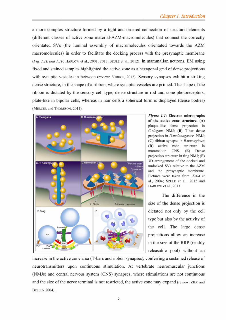
ZHAI and BELLEN, 2004). At the neuromuscular junction (NMJ) of C.elegans the dense projection
has been described as a plaque surrounded within 100nm by a subpopulation of vesicles (Fig.
1.1A; WEIMER et al., 2006); while in D.melanogaster, the dense structure takes the shape of a
pedestal and a platform (T-bars) enclosed by synaptic vesicles and closely associated with
calcium channels (Fig. 1.1B; PROKOP and MEINERTZHAGEN, 2006). In vertebrates (frog), the NMJ has
1 ONLINE ETYMOLOGY DICTIONARY: www.etymonline.com
a mo
(diffe
orien
macr
(Fig.
fixed
with
dense
ribbo
plate
(MER
incre
neuro
(NM
and t
BELLE
ore complex
ferent classe
ntated SVs
romolecules
1.1E and 1.1F
d and staine
synaptic v
e structure,
on is dictate
e-like in bip
RCER and THOR
ease in the a
otransmitter
MJs) and cen
the size of t
EN,2004).
x structure
es of active
(the lumin
s) in order
F; HARLOW e
d samples h
vesicles in b
in the shap
ed by the s
polar cells,
RESON, 2011)
active zone
rs upon c
ntral nervou
the nerve ter
formed by
e zone mate
nal assemb
to facilitate
et al., 2001, 20
highlighted
between (rev
pe of a ribbo
sensory cell
whereas in
.
area (T-bars
continuous
us system (
rminal is no
2
y a tight and
erial-AZM-
bly of mac
e the docki
013; SZULE et
the active z
view: SÜDHOF
on, where sy
l type; dens
hair cells a
s and ribbon
stimulation
(CNS) syna
ot restricted
d ordered c
-macromole
cromolecule
ing process
t al., 2012). I
zone as a he
F, 2012). Sen
ynaptic vesi
se structure
a spherical
n synapses)
n. At vert
apses, where
d, the active
Ch
connection
ecules) that
es orientate
s with the p
n mammali
exagonal gri
nsory synap
icles are pri
in rod and
form is dis
Figure 1.of the acplaque-likC.elegans projection(C) ribbon(D) actimammaliaprojection3D arrangundocked and the Pictures wal., 2004;HARLOW e
T
size of t
dictated
type but
the cell
projectio
in the siz
releasabl
), conferring
ebrate neu
e stimulatio
zone may e
hapter 1. In
of structura
connect th
ed towards
presynaptic
ian neurons
id of dense
pses exhibi
imed. The s
d cone phot
splayed (den
.1: Electron ctive zone stke dense ps NMJ; (B) n in D.melanon synapse in Rive zone an CNS. n structure in fgement of the
SVs relativepresynaptic
were taken fr; SZULE et aet al., 2013.
The differe
the dense p
not only b
t also by the
l. The la
ons allow a
ze of the RR
le pool) w
g a sustaine
uromuscular
ons are not
expand (revi
ntroduction
al elements
he correctly
the AZM
membrane
s, EM using
projections
it a striking
shape of the
toreceptors,
nse bodies)
micrographstructure. (A)projection in
T-bar denseogaster NMJ;R.norvegicus;structure in(E) Dense
frog NMJ; (F)e docked ande to the AZM
membrane.rom: ZHAI etal., 2012 and
nce in the
rojection is
by the cell
e activity of
arge dense
an increase
RP (readily
without an
d release of
r junctions
continuous
iew: ZHAI and
n
s
y
M
e
g
s
g
e
,
)
s ) n e ; ; n e ) d
M . t d
e
s
l
f
e
e
y
n
f
s
s
d
techn
the A
treatm
is los
false
samp
dense
betw
FERNA
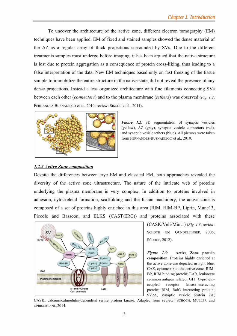
1.2.2
Desp
diver
unde
adhe
comp
Picco
CASKOPRIS
To unco
niques have
AZ as a re
ments samp
st due to pr
interpretati
ple to immo
e projection
ween each ot
ANDEZ-BUSNA
2 Active Zon
pite the diff
rsity of the
erlying the
sion, cytos
posed of a
olo and B
K, calcium/caOREANU,2014
over the arc
e been appli
egular array
ples must un
rotein aggre
ion of the d
obilize the e
ns. Instead
ther (connec
ADIEGO et al.,
ne composit
ferences be
e active zo
plasma m
keletal form
set of prote
Bassoon, an
almodulin-dep4.
chitecture o
ied. EM of f
y of thick
ndergo befo
egation as a
data. New E
ntire structu
a less orga
ctors) and t
2010; review
tion
etween cryo
one ultrastr
embrane is
mation, sca
eins highly
nd ELKS
pendent serine
3
f the active
fixed and st
projections
ore imaging
a consequen
EM techniq
ure in the na
anized arch
to the plasm
w: SIKSOU et al
Figu(yelland sfrom
o-EM and c
ructure. Th
s very com
affolding an
enriched in
(CAST/ER
e protein kin
e zone, diff
tained samp
s surrounde
g, it has bee
nce of prote
ques based o
ative state,
itecture wit
ma membran
l., 2011).
ure 1.2: 3Dlow), AZ (grsynaptic vesic
m FERNANDEZ-
classical EM
he nature o
mplex. In a
nd the fusio
n this area
RC)) and
(C
SC
SÜ
FicothCABPcocoprSV
nase. Adapted
Ch
ferent electr
ples showed
ed by SVs.
en argued th
ein cross-lik
only on fast
did not reve
th fine filam
ne (tethers)
D segmentatioay), synaptic
cle tethers (blu-BUSNADIEGO
M, both app
of the intri
addition to
on machine
(RIM, RIM
proteins a
CASK/Veli/
CHOCH and
ÜDHOF, 2012)
igure 1.3: omposition. Pe active zone AZ, cytometriP, RIM bindinommon antigeoupled recerotein; RIM, V2A, synaptd from review
hapter 1. In
ron tomogr
d the dense
Due to th
hat the nativ
king, thus l
t freezing o
eal the pres
ments conn
was observ
on of synap vesicle connue). All picturO et al., 2010.
pproaches re
icate web
proteins i
ery, the acti
M-BP, Liprin
associated
/Mint1) (Fig
GUNDELFIN
).
Active ZProteins highle are depicted rix at the activng protein; LAen related; GIeptor kinaRab3 interac
tic vesicle w: SCHOCH,
ntroduction
raphy (EM)
material of
he different
ve structure
eading to a
of the tissue
ence of any
necting SVs
ved (Fig. 1.2;
ptic vesiclesnectors (red),res were taken
evealed the
of proteins
involved in
ive zone is
n, Munc13,
with these
g. 1.3; review:
NGER, 2006;
one proteinly enriched atin light blue.
ve zone; RIM-AR, leukocyteIT, G-protein-ase-interactingcting protein;protein 2A;MÜLLER und
n
)
f
t
e
a
e
y
s
;
s , n
e
s
n
s
,
e
:
;
n t . -e -g ; ; d
neuro
order
and
plast
perfo
2012)
1.3 T
The
neuro
termi
The
repea
neuro
review
sever
medi
sensi
and o
2013)
comp
assis
The acti
otransmitter
r to facilitat
postsynapti
ticity. As a
ormed with
).
The synapti
active zo
otransmitter
inal, the vo
transient C
ated cycles
otransmitter
ws: SÜDHOF, 2
After the
ral changes
iated on on
itive factor
on the othe
). Originall
peting with
t the trans
ive zone, d
rs release: (
te fast synch
ic compartm
a result, all
the requisit
ic vesicle cy
one is the
r release t
ltage gated
Ca2+ elevatio
of exocytos
rs followed
2004, 2013).
e synaptic v
s until they
ne side by th
attachment
er side by M
y Munc18-
core-compl
sition to an
due to its c
(1) SV dock
hronous exc
ments; (4)
these func
te speed an
ycle
e specializ
akes place
calcium ch
on stimulate
sis-endocyto
by docking
vesicles are
y become f
he formatio
t protein rec
Munc18, Mu
-1 was rep
lex formatio
n ‘open’ co
4
complex or
king and pr
citation/rele
mediation
ctions guara
nd plasticity
zed structu
e. When an
hannels open
es SV exoc
osis. Durin
g, priming,
filled with
fusion-comp
on of the SN
ceptor) con
unc13, RIM
ported to b
on (DULUBOV
Ffildoremcorefr
S
ac
do
onformation
rganization,
riming; (2)
ease couplin
of both s
antee that p
needed for
ure in the
n action po
n followed b
cytosis. In t
ng exocytos
fusion and
neurotransm
petent (prim
NARE com
sisting of sy
M and Syna
bind syntax
VA et al., 1999
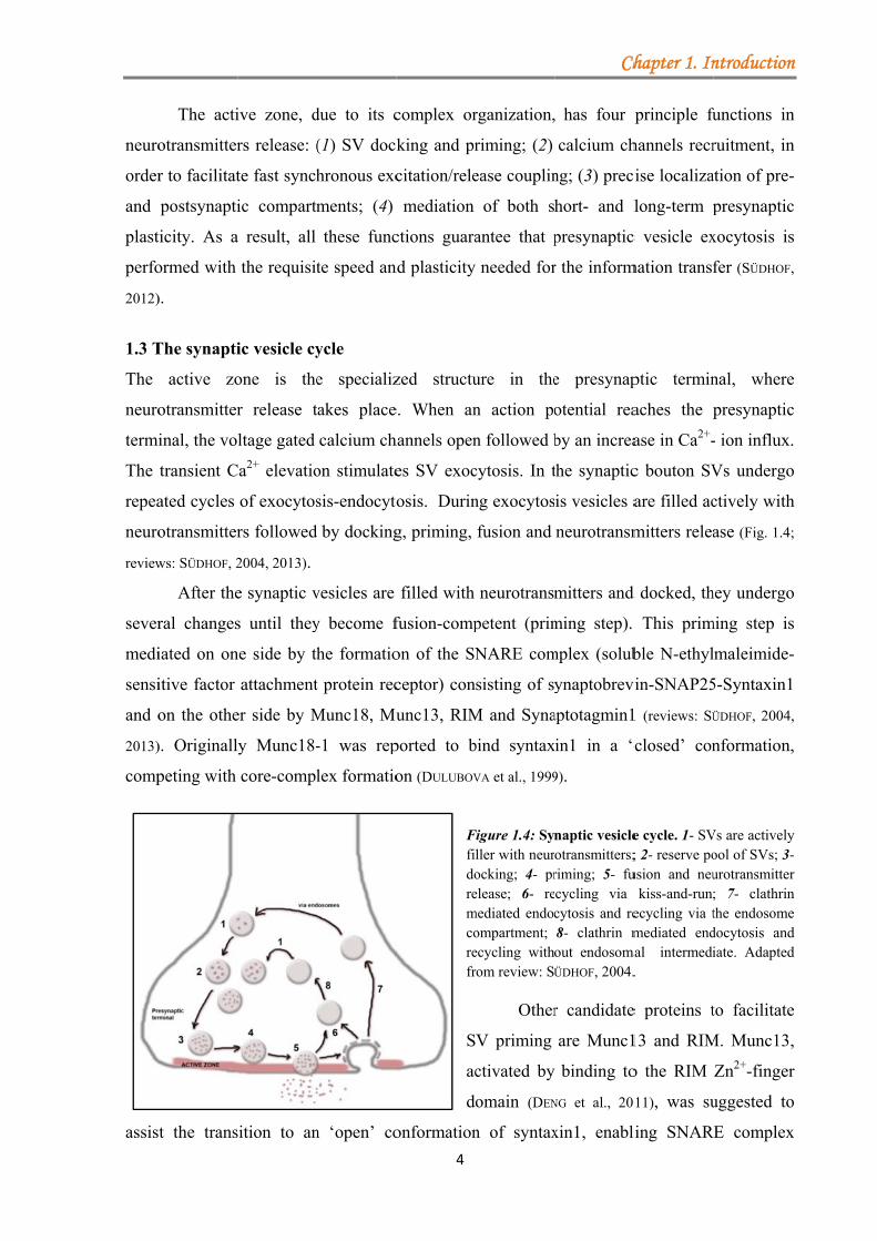
Figure 1.4: Syller with neurocking; 4- prelease; 6- re
mediated endocompartment; ecycling withrom review: SÜ
Other
V priming
ctivated by
omain (DEN
n of syntax
Ch
has four p
calcium ch
ng; (3) prec
hort- and l
presynaptic
r the inform
e presynap
otential rea
by an increa
the synaptic
is vesicles a
neurotransm
mitters and
ming step).
mplex (solub
ynaptobrevi
aptotagmin1
in1 in a ‘c
9).
naptic vesicleotransmitters;riming; 5- fucycling via cytosis and re8- clathrin mout endosomaÜDHOF, 2004.
r candidate
are Munc1
binding to
NG et al., 20
xin1, enabli
hapter 1. In
principle fu
hannels recr
cise localiza
long-term p
c vesicle ex
mation transf
ptic termin
aches the p
ase in Ca2+-
c bouton SV
are filled ac
mitters relea
d docked, th
This prim
ble N-ethyl
vin-SNAP25
1 (reviews: SÜ
closed’ con
e cycle. 1- SV; 2- reserve pousion and neu
kiss-and-run;ecycling via tmediated end
mal intermedi.
e proteins t
13 and RIM
o the RIM
11), was su
ling SNAR
ntroduction
functions in
ruitment, in
ation of pre-
presynaptic
xocytosis is
fer (SÜDHOF,
nal, where
presynaptic
- ion influx.
Vs undergo
ctively with
ase (Fig. 1.4;
hey undergo
ming step is
lmaleimide-
5-Syntaxin1
ÜDHOF, 2004,
nformation,
Vs are activelyool of SVs; 3-urotransmitter; 7- clathrinthe endosomedocytosis andiate. Adapted
to facilitate
M. Munc13,
Zn2+-finger
uggested to
RE complex
n
n
n
-
c
s
,
e
c
.
o
h
;
o
s
-
,
,
y - r n e d d
e
,
r
o
x
Chapter 1. Introduction
5
assembly and thereby the priming step (BETZ et al., 1997; review: RIZO and SÜDHOF, 2002; STEVENS et
al., 2005; review: SÜDHOF, 2013).
After the action potential reaches the presynaptic terminal, voltage gated calcium
channels open and the calcium concentration builds up in a microdomain near the priming
complex. The calcium sensor synaptotagmin1 (present on the synaptic vesicle) together with
the SNARE complex further enables membrane fusion (DAI et al., 2008; CHOI et al., 2010; VRLJIC et
al., 2010) with the formation of the pore to release the neurotransmitters into the synaptic cleft.
Following neurotransmitter release SVs are recycled via different routes, like kiss-and-run
(vesicles undock and recycle locally), clathrin mediated endocytosis (vesicles are reacidified
and refilled directly or by passing via the endosome compartment) (review: SÜDHOF, 2004) or via
bulk endocytosis. Activity-dependent bulk endocytosis (ADBE) is the dominant retrieval
pathway after an elevated stimulation activity (CHEUNG and COUSIN, 2013).
In accordance with the network’s needs, the amount of SVs ready to release
neurotransmitter may very as well. SV recycling is tightly regulated by the action of different
proteins, resident at the AZ. Therefore, fluctuations in the activity of synapses could be
mediated by the actions of various AZ proteins, as well as by the SVs cycle. These changes
represent the fundament of presynaptic plasticity.
1.4 Synaptic plasticity
The concept of synaptic plasticity, which was for the first time formulated by Hebb in 1949,
refers to the capacity of synapses to react accordingly to the network’s needs either be
weakening (depression) or strengthening (potentiation) its activity. These types of changes
may well extend over short periods (short-term plasticity) or long periods of time (long-term
plasticity). The Hebbian theory is used to describe these synaptic changes as being associative
and rapidly induced, shortly explained as a positive feedback process (HEBB., 1949). For
example, upon LTP induction, synapses become more excitable and the entire network
activity would increase leading to a runaway potentiation. To prevent such extremes, the
homeostatic process, which hinders the network to reach high levels of activity and preserve
the stored information, has an important role (review: POZO and GODA, 2010).
In the active state or basal conditions synaptic transmission is mediated by the release
of neurotransmitters from presynaptic terminals into the synaptic cleft, followed by the
activation of different receptors on the postsynaptic terminal. Under increased network
activity presynaptic neurons decrease their release probability (LTD-long-term depression),
while the postsynaptic cells decrease the number of their receptors. To offset reduced network
Chapter 1. Introduction
6
activity, presynaptic neurons enhance the recycling, the number of docked vesicle and the
release probability (LTP-long-term potentiation) (review: POZO and GODA, 2010; CASTILLO, 2012).
There are multiple parallel mechanisms responsible for controlling pre- and postsynaptic
homeostasis, and consequently affecting synapse activity. The molecular mechanisms that
govern the negative feedback (homeostatic plasticity) rely on the efficiency of different
intracellular signalling cascades to detect and to respond accordingly to changes in the
network. These fine-tuned mechanisms include: gene expression induction, protein synthesis
and degradation. Besides the two major mechanisms: transcription and translation, post-
translational modifications have emerged as an important factor in controlling plasticity
(review: POZO and GODA., 2010). Several post-translational modifications have been suggested to
modulate the function of various pre- and postsynaptic proteins, like: palmitoylation (review:
EL-HUSSEINI and BREDT 2002), myristilation and prenylation (KUTZLEB et al., 1998; O’CALLAGHAN et al.,
2003), SUMOylation (Small Ubiquitin-like Modifier) (GIRACH et al., 2013) and phosphorylation
(review: BARRIA, 2001).
1.4.1 Presynaptic dormancy
Presynaptic dormancy is induced as a response to a prolonged strong depolarization or
increased action potential firing. Dormant synapses display a decrease in neurotransmitter
release. The molecular mechanism is based on the inhibitory action of G proteins on adenylyl
cyclase (AC), which causes a decrease in the level of cAMP and thereby directly affects the
activity of protein kinase A (PKA) (Fig. 1.5). Therefore, presynaptic proteins are less
phosphorylated and become susceptible to degradation through the proteasome (review:
CRAWFORD and MENNERICK, 2012). The protein levels of RIM1α and Munc13-1 were shown to be
decreased upon induction of presynaptic dormancy through the action of the ubiquitin-
proteasome system, while an overexpression of RIM1α in cultured neurons prevented the
induction of silencing (JIANG et al., 2010). Recently two other presynaptic proteins, Piccolo and
Bassoon were identified as negative regulators of the E3 ligase Siah1. In the DKO neurons the
rate of presynaptic protein degradation was increased, leading to the observation that these
two proteins are important regulators of the protein ubiquitination in the presynaptic terminal,
therefore maintaining synapse integrity (WAITES et al., 2013).aaaaaaaaaaaaaaaaaaaaaaaaaaaaaaaaaaaaaa
1.4.2
At th
in Ca
presy
an in
RIM
impo
2002;
signi
Addi
kinas
CASTI
LTD
1.5 T
Even
eluci
were
leadi
one r
descr
funct
trans
2 Molecular
he mossy fib
a2+ influx v
ynaptic term
ncrease in cA
M1α, a highly
ortant in se
review: CAS
ificant func
itionally, P
se, PKC wa
ILLO, 2012).
D are still no
Two major
n though t
idated, seve
e suggested
ing to neuro
resident of
ribed to be
tion could b
smission.
r mechanism
bres (MF) p
via voltage
minal will ac
AMP and a
y enriched p
everal cAM
STILLO, 2012
ction in pr
KA may r
as reported t
Overall, th
ot fully eluci
players in
he exact m
eral cellula
to modulat
otransmitter
the AZ (R
actively inv
be altered,
ms involved
presynaptic
gated calci
ctivate the C
activation of
protein in th
MP/PKA-dep
) its direct
resynaptic
egulate LT
to be respon
he precise m
idated.
synaptic p
mechanisms
r processes
e this event
rs release, it
RIM1α), the
volved in ta
via posttran
7
d in presyna
long-term
ium channe
Ca2+ /calmo
f PKA (revie
he active zo
pendent form
phosphory
LTP (KAES
TP independ
nsible for L
molecular m
lasticity
s governing
s, like SV
t. Since SV
t’s imperiou
e other a co
aking part i
nslational m
FInthAisdacophsuprMMM
aptic LTP
potentiation
els (VGCG)
odulin depen
ew: CASTILLO
one. Althoug
ms of pres
ylation by P
SER et al.,
dently of R
LTP in MF-
mechanism
g the pres
priming or
priming is
us to be un
omponent o
in the regul
modification
Ch
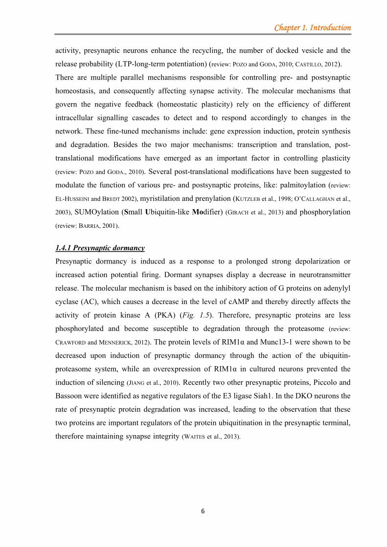
Figure 1.5: ncreased actiohe inhibitory
Adenylyl cyclas impaired anark blue) actonsequence hosphorylatedusceptible toroteasome.
Munc13 proteinModel adapteMENNERICK, 20
n (LTP) is i
). The incre
ndent-adeny
O, 2012). One
gh RIM1α h
ynaptic pla
PKA could
2008a; YANG
RIM1α pho
dentate gyr
governing
synaptic pla
r posttransl
one of the
der a tight
of synaptic
ation of pri
ns, triggerin
hapter 1. In
Presynapticon potential fiy G protein ase (AC, ligh
nd protein kintivity is decr
RIM1α, d (red mao degradatioTogether w
in (green) is aed after CRA
012.
induced by
ease in calc
ylyl cyclase
e of the PKA
has been rep
asticity (CAS
not be lin
G and CALA
osphorylatio
rus basket c
presynapti
asticity are
lational mo
most impor
control. Tw
vesicle (SV
iming. Mor
ng changes
ntroduction
dormancy.iring activates
(Gi, pink),t blue) action
nase A (PKA,reased. As a
is lessarkings) andon, via thewith RIM1,also degraded.AWFORD and
an increase
cium in the
e, leading to
A targets is
ported to be
STILLO et al.,
nked to any
AKOS, 2010).
on. Another
cells (review:
ic LTP and
e not fully
odifications,
rtant events
wo proteins,
V2A), were
eover, their
in synaptic
n
. s ,
n , a s d e , .
d
e
e
o
s
e
,
y
.
r
:
d
y
,
s
,
e
r
c
1.5.1
1.5.1
The
RIM
distin
(α, β
RIM
lacks ZF. Aα-isof
altern
site A
C2B
and 3
separ
block
RIM
prote
(28 i
for e
and C
rich-
1 RIMs
1.1 RIM gen
RIM protei
M3 and RIM4
nct promote
β, γ) (WANG a
M3γ and RIM
the Rab3 binAll γ-isoforms forms.
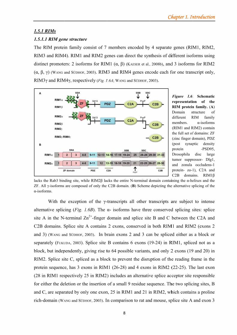
With the
native splic
A in the N
domains. S
3) (WANG an
rately (FUKU
k, but indep
M2. Splice si
ein sequenc
in RIM1 res
ither the de
C, are separ
domain (WA
ne structure
in family c
4). RIM1 an
ers: 2 isofor
and SÜDHOF,
M4γ, respect
nding site, whare compose
e exception
cing (Fig. 1
-terminal Z
Splice site A
nd SÜDHOF, 2
UDA, 2003).
pendently, g
ite C, splice
e, has 3 ex
spectively 2
eletion or th
rated by onl
ANG and SÜDH
e
onsist of 7
nd RIM2 ge
rms for RIM
2003). RIM
tively (Fig. 1
ile RIM2β lacd of only the
n of the γ-
1.6B). The α
Zn2+-finger d
A contains
2003). In br
Splice site
giving rise t
ed as a bloc
ons in RIM
25 in RIM2)
he insertion
ly one exon
HOF, 2003). In
8
members e
enes can dir
M1 (α, β) (K
M3 and RIM
1.6A; WANG a
cks the entireC2B domain
-transcripts
α- isoforms
domain and
2 exons, c
rain exons
B contains
to 64 possib
ck to preven
M1 (26-28) a
) includes a
of a small 9
n, 25 in RIM
n compariso
encoded by
rect the syn
KAESER et al.,
M4 genes enc
and SÜDHOF, 2
e N-terminal d. (B) Scheme
all other t
s have three
d splice site
onserved in
2 and 3 ca
6 exons (1
ble variants
nt the disru
and 4 exons
an alternativ
9 residue se
M1 and 21 in
on to rat an
Ch
4 separate
nthesis of di
, 2008b), and
code each f
2003).
domain contaidepicting the
ranscripts a
e conserved
e B and C
n both RIM
an be splice
19-24) in R
, and only 2
uption of the
s in RIM2 (
ve splice ac
equence. Th
n RIM2, wh
d mouse, sp
hapter 1. In
e genes (RIM
ifferent isof
d 3 isoform
for one tran
Figure 1.6:representatioRIM proteinDomain stdifferent Rmembers. (RIM1 and Rthe full set of(zinc finger d(post synapprotein Drosophila tumor supprand zonula protein- zo-1C2B doma
ining the α-hee alternative s
are subject
d splicing s
between th
M1 and RIM
ed either as
RIM1, splic
2 exons (19
e reading fr
(22-25). Th
cceptor site
he two splic
hich contain
plice site A
ntroduction
M1, RIM2,
forms using
ms for RIM2
nscript only,
: Schematicon of then family. (A)tructure of
RIM familyα-isoforms
RIM2) containf domains: ZFdomain), PDZptic density
-PSD95,disc large
ressor- Dlg1,occludens-1
1), C2A andins. RIM1βelixes and thesplicing of the
to intense
sites: splice
he C2A and
M2 (exons 2
a block or
ed not as a
9 and 20) in
frame in the
he last exon
responsible
cing sites, B
ns a proline
and exon 3
n
,
g
2
,
c e ) f y s n F Z y , e ,
d β e e
e
e
d
2
r
a
n
e
n
e
B
e
3
are m
brain
may
2003)
respo
MÜLL
1.5.1
RIM
doma
1997,
(KAES
isofo
(Fig.1
doma
termi
for o
bind
comp
Mun
Mun
prim
Mun
missing from
n specific. A
be respons
). In invert
onsible for t
LER et al., 201
1.2 RIM pro
M1α and RIM
ain, a centr
2000). RIM
SER et al., 20
orms contain
1. 6A; WANG e
The N-te
ain, is impo
inal α1 heli
other Rab m
RIM2α) (F
posed of th
nc13-1 and u
nc13 exists
ming. The in
nc13 homod
m the human
Another inte
ible for the
tebrates (C
the synthesi
2).
otein structu
M2α protein
al PDZ dom
M1β lacks th
008b), RIM2
n only the l
et al., 2000; W
erminal dom
ortant for th
ix of RIM1α
molecules (R
Fig.1.7; SUN
he zinc dom
ubMunc13-
in an inac
nteraction o
dimerization
n transcript,
eresting fea
generation
C.elegans, D
is of the RI
ure and bin
ns contain t
main, and tw
e α1-helix i
2β lacks th
last C-termi
WANG and SÜD
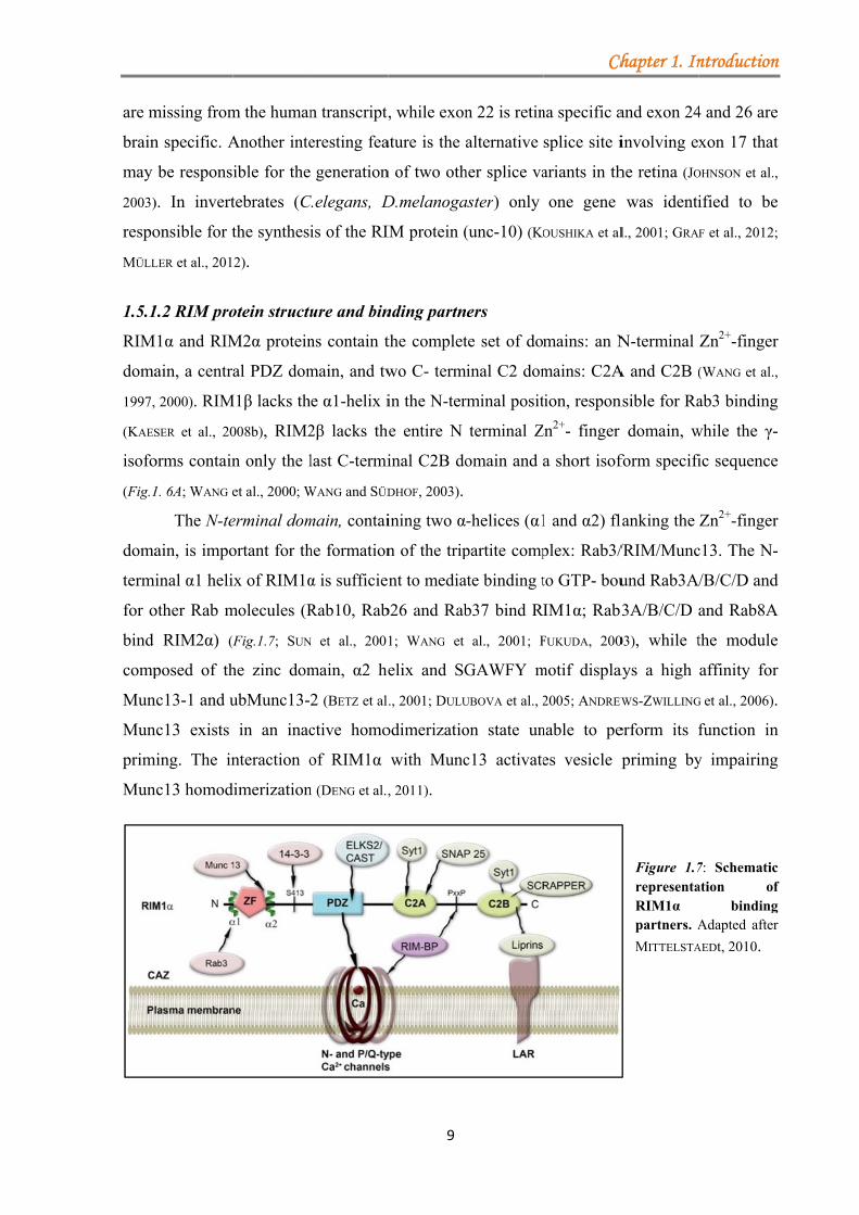
main, contai
he formation
α is sufficien
Rab10, Rab
N et al., 2001
main, α2 he
2 (BETZ et al
ctive homo
of RIM1α
n (DENG et al.,
9
, while exon
ature is the
n of two oth
D.melanoga
IM protein (
nding partn
the complet
wo C- term
in the N-ter
he entire N
inal C2B do
DHOF, 2003).
ining two α
n of the trip
nt to media
b26 and Rab
1; WANG et
elix and SG
., 2001; DULU
odimerizatio
with Munc
, 2011).
n 22 is retin
alternative
her splice va
aster) only
(unc-10) (KO
ers
te set of do
minal C2 dom
rminal posit
terminal Z
omain and
-helices (α1
partite comp
ate binding t
b37 bind R
al., 2001; F
GAWFY m
UBOVA et al., 2
on state un
c13 activate
Ch
na specific a
splice site i
ariants in th
one gene
OUSHIKA et al
mains: an N
mains: C2A
tion, respon
Zn2+- finger
a short isof
1 and α2) fl
plex: Rab3/
to GTP- bou
IM1α; Rab3
FUKUDA, 200
motif displa
2005; ANDREW
nable to pe
es vesicle p
hapter 1. In
and exon 24
involving ex
he retina (JO
was identi
l., 2001; GRA
N-terminal
A and C2B (
nsible for Ra
domain, w
form specifi
lanking the
/RIM/Munc
und Rab3A
3A/B/C/D
03), while t
ays a high
WS-ZWILLING
erform its f
priming by
Figure 1.7representaRIM1α partners. A
MITTELSTA
ntroduction
4 and 26 are
xon 17 that
OHNSON et al.,
ified to be
F et al., 2012;
Zn2+-finger
(WANG et al.,
ab3 binding
while the γ-
ic sequence
Zn2+-finger
c13. The N-
/B/C/D and
and Rab8A
the module
affinity for
G et al., 2006).
function in
y impairing
7: Schematication of
bindingAdapted after
AEDt, 2010.
n
e
t
,
e
;
r
,
g
-
e
r
-
d
A
e
r
.
n
g
c f g r
Chapter 1. Introduction
10
The sequence between the zinc and PDZ domains contains several amino acid residues
that have been suggested to be important in modulating RIM’s function (Fig. 1.7). Serine 413
was identified as a phospho-switch that triggers presynaptic LTP in cultured cerebellar
granular and Purkinje cell neurons, upon phosphorylation by PKA (LONART et al., 2003). These
findings however were not confirmed by studies in knockin mice, bearing the S413A
mutation. The phosphorylation of serine 413, although important in binding 14-3-3 proteins,
displayed no significant role in presynaptic plasticity or in learning and memory (KAESER et al.,
2008a; YANG and CALAKOS, 2010). Other phosphoserines (Ser241 and Ser287 in RIM1α, and
Ser335 in RIM2α) were also associated with binding to 14-3-3 proteins, when phosphorylated
by the Ca2+/calmodulin dependent kinase II (CaMKII). The ability of RIM to bind 14-3-3
proteins does apparently not impair the binding between RIM-Munc13 and RIM-Rab3A (SUN
et al., 2003). The same linker region between zinc finger and PDZ domain may also act as a
substrate for ERK2 kinase, which phosphorylates Ser447, a residue linked to the enhancement
of glutamatergic transmission in hippocampal CA1 after stimulation with BDNF (SIMSEK-
DURAN and LONART, 2008).
The central PDZ domain that interacts with the ELKS2/CAST protein (OHTSUKA et al.,
2002; WANG et al., 2002), plays an important role in RIM1’s distribution in cultured neurons; the
truncated form lacking this domain being diffusely localized (Fig. 1.7; OHTSUKA et al., 2002).
CAST binds directly not only RIM1, but also Bassoon and Piccolo, and the entire ternary
complex RIM1-CAST-Bassoon is involved in controlling neurotransmitter release (TAKAO-
RIKITSU et al., 2004). Two reports from 2011 attribute to RIM1/2 a key role in controlling not
only the number of docked vesicles but also the distribution and/or density of calcium
channels at the active zone (HAN et al., 2011; KAESER et al., 2011). By generating RIM1/2 floxed
mouse lines, in which all RIM isoforms containing a PDZ domain can be deleted by cre-
recombinase in vitro, it was shown that the PDZ domain alone was required for the proper
localization of N- and P/Q type calcium channels (KAESER et al., 2011).
The α- and β-RIMs contain two C-terminal domains: C2A and C2B that are separated
by a proline-rich domain and two splice sites (B and C) (Fig. 1.7; WANG and SÜDHOF, 2003). Both
domains do not contain the consensus calcium binding sites present in synaptotagmin’s C2-
domains (WANG et al., 2000; DAI et al., 2005). The C2A domain was shown to have affinity in a
calcium dependent manner for SNAP25 and Synaptotagmin1 (COPPOLA et al., 2001), even though
NMR studies suggested that there was little binding between these proteins (DAI et al., 2005).
Very intriguing is a point mutation in human RIM1 (R844H) that was identified in a patient
with autosomal dominant cone-rod dystrophy-CORD7, characterized by impaired vision due
Chapter 1. Introduction
11
to the reduction in the cone and rod sensitivity (JOHNSON et al., 2003; MICHAELIDES et al., 2005). The
C2B domain has been shown to interact with several proteins that may have an impact on
RIM1α function at the active zone, among them Synaptotagmin1, identified to bind with high
affinity to the C2B domain in biochemical assays (COPPOLA et al., 2001; SCHOCH et al., 2002),
results not reproduced by NMR studies (GUAN et al., 2007). Other proteins that bind the C2B
domain are: liprins-α (SCHOCH et al., 2002); the E3 ubiquitin ligase SCRAPPER (YAO et al., 2007)
that controls RIM1 turn-over, facilitating ubiquitination and degradation; SAD kinase (INOUE et
al., 2006); and the β4 subunit of voltage gated calcium channels (COPPOLA et al., 2001; KIYONAKA et
al., 2007). In addition the interaction between RIM1 and the α1 subunit of the N-type calcium
channel is regulated by cyclin-dependent kinase 5 (Cdk5), which enhances channel opening
and facilitates neurotransmitters release (SU et al., 2012).
SUMOylation was recently reported by the group of Hanley to act as a molecular
switch for RIM1α. SUMOylated RIM1α confers affinity for Cav2.1, therefore promoting
calcium channel clustering and synchronous synaptic vesicle release, while non-SUMOylated
form is responsible only for vesicle priming and docking (GIRACH et al., 2013).
Other proteins that couple RIM1/2 to calcium channels are RIM-BPs. On one hand
RIM-BP binds the proline-rich domain of RIM1/2 (WANG et al., 2000) and on the other hand
calcium channels, bringing these proteins in close proximity at the active zone (HIBINO et al.,
2002).
1.5.1.3 RIM function
1.5.1.3.1 RIM in invertebrates (C.elegans and D.melanogaster)
Analysis of RIM protein function in C.elegans demonstrated that UNC-10 has a major role in
coordinating vesicle docking and priming by regulating UNC-13 activity. It has been
hypothesised that UNC-10/RIM may signal syntaxin, via UNC-13, to change its conformation
from a closed to an open state. UNC-10 mutants exhibit a decrease in vesicle fusion at release
sites, an effect suppressed by the expression of the open form of syntaxin (KOUSHIKA et al.,
2001). Furthermore, disruption of the unc-10 gene triggers a depletion of docked synaptic
vesicles since the normal connections between SVs and dense projection filaments are
impaired (STIGLOHER et al., 2011).
D.melanogaster RIM mutants show decreased evoked synaptic transmission as a
consequence of the reduction in the size of the RRP of SVs and altered Ca2+-channels
clustering together with a decreased calcium influx. Mutants present a normal cellular
morphology with no major changes in active zone architecture (GRAF et al., 2012; MÜLLER et al.,
2012).
Chapter 1. Introduction
12
1.5.1.3.2 RIM in vertebrates (M.musculus)
In the recent years several reports have been published, providing new data about the possible
role of RIMs at the active zone. Different mouse models have been generated, knocking out
either one or more isoforms, in order to gain new insights into how different variants of RIMs
influence neurotransmitter release and presynaptic plasticity as well as to understand ability of
the various isoforms to compensate for each other.
1.5.1.3.2.1 RIM1α knock-out mice
The first model generated targeted the most abundant isoform in the brain, RIM1α (SCHOCH et
al., 2002). Homozygous mice were viable and fertile, with no evident structural abnormalities
or changes in brain architecture. Overall, active zone architecture was comparable to WT
littermates. Among the AZ proteins, Munc13-1 showed a major decrease of 60% in KOs,
while several postsynaptic density proteins (SynGAP, PSD95, SHANK) exhibited a moderate
increase, suggesting a role for RIM1α in synaptic remodelling (SCHOCH et al., 2002).
Electrophysiological recordings revealed that RIM1α knockout caused a decrease in the size
of the RRP, with no effect on synaptic vesicle recycling. These data together with findings
from D.melanogaster and C.elegans suggest a role for RIM1α in vesicle maturation, from
priming to calcium triggered fusion (KOUSHIKA et al., 2001; SCHOCH et al., 2002; CALAKOS et al., 2004;
MÜLLER et al., 2012). Additionally, the RIM1α protein seems to be involved both in short-term
plasticity as well as in presynaptic long-term potentiation (LTP) (review: MITTELSTAEDT et al.,
2010).
Cryo-electron tomography revealed a series of changes in the AZ with regard to
vesicle tethering and vesicle concentration in synaptosomes from RIM1α KO mice (40%
reduction in proximal vesicles compared to control) that may account for the decrease in the
size of the RRP. Blocking proteasome activity with MG132, the KO phenotype was rescued
and the treated KO synaptosomes became indistinguishable from WT synaptosomes,
displaying an increase in the number of vesicles at the AZ. This recent study highlights the
importance of the ubiquitin-proteasome system (UPS) in the turn-over of RIM proteins,
emerging as a key factor in controlling presynaptic plasticity (FERNANDEZ-BUSNADIEGO et al.,
2013).
Besides deficits in synaptic transmission, KO mice display impaired learning and
memory (POWELL et al., 2004), schizophrenia-like behaviour (BLUNDELL et al., 2010), and a higher
susceptibility to develop spontaneous seizures after status epilepticus (PITSCH et al., 2012).
Chapter 1. Introduction
13
1.5.1.3.2.2 RIM1αβ double knock-out mice
Mutant mice lacking both RIM1 isoforms, α and β, display a more severe impairment in
synaptic transmission and significant changes in the solubility of different active zone
proteins. Both isoforms are expressed in a similar pattern in the brain, with a slight increase of
RIM1β levels in the brainstem. During development RIM1β is highly expressed in the early
postnatal phase in this region, which may account for the lethality of the DKO mice.
Interestingly, in RIM1α KO mice the level of RIM1β is increased 2 fold, indicating a
compensatory effect. Among the presynaptic proteins, ELKS1/2, RIM-BP2 and the remaining
Munc13-1 (reduced to 30% in these mutant mice), showed a higher dissociation rate from the
insoluble protein matrix, supporting the notion of RIMs acting as scaffolding proteins for
various AZ proteins. Synaptic transmission is severely impaired in the DKO mice with the
observation that presynaptic long-term plasticity is not aggravated by this double deletion
compared to RIM1 KO. Therefore, it has been suggested that RIM1α mediates both long-term
plasticity via Rab3 as well as short-term plasticity via Munc13, while RIM1β (since it lacks
the binding motif for Rab3) is involved only in short-term plasticity (KAESER et al., 2008b).
1.5.1.3.2.3 RIM2α knock-out mice
Since RIM1α and RIM2α, which is much less abundant, display high homology, it was
expected that the knockout of RIM2α might partially resemble the phenotype of the RIM1α
KO. However, deletion of the RIM2α gene did not trigger any change in release probability
compared to the impairment in synaptic transmission and facilitation observed in the RIM1α
KO mice (CASTILLO et al., 2002; SCHOCH et al., 2002, 2006). RIM2α KO mice were viable and fertile,
and displayed normal brain morphology (SCHOCH et al., 2006).
1.5.1.3.2.4 RIM1α/RIM2α double knock-out mice
Deletion of both α isoforms (RIM1 and RIM2) turned out to be lethal, RIM1α/2α DKO mice
die immediately at birth, not due to changes in brain development but due to breathing
problems. No obvious alterations in brain morphology were detected by conventional EM.
Protein composition analysis revealed no additional decrease in the level of Munc13-1
compared to RIM1α KO mice. Nonetheless, immunostaining analysis of the whole-mount
diaphragm muscle at E18.5 revealed an increased innervation or expansion of innervation
with no major changes in the ultrastructure of the NMJ in the DKO mice. These changes were
accompanied by impairment in synaptic transmission. Spontaneous or Ca2+-dependent
exocytosis was not abolished, only evoked synaptic transmission (Ca2+- triggering exocytosis)
was strongly impaired in these mutants (SCHOCH et al., 2006).

Chapter 1. Introduction
14
1.5.1.3.2.5 RIM conditional knockout mice
As both RIM1α/RIM1β (KAESER et al., 2008b) and RIM1α/RIM2α (SCHOCH et al., 2006) DKO mice
were lethal, conditional knockouts (floxed mouse lines) were generated to further study the
consequences of a deficiency of all RIMs isoforms. Deletion of both RIM genes in vitro
supported the role of RIMs in controlling vesicle priming and neurotransmitter release (KAESER
et al., 2012). Furthermore, RIMs were shown to be responsible for proper tethering of the Ca2+
channels via the PDZ domain (HAN et al., 2011; KAESER et al., 2011).
Single deletions (RIM1αβ or RIM2αβγ) altered SV priming, while double deletion
(RIM1αβ/RIM2αβγ) impaired not only the priming but also the calcium responsiveness and
synchronization of release. In HEK293T cells and in RIM1/2 double deficient neurons,
RIM2γ wasn’t able to rescue the phenotype, suggesting that the C2 domain alone neither
contributes to calcium channel activity modulation nor plays an important role in the synaptic
function of RIM proteins (KAESER et al., 2012).
Taken together, RIM1α plays an important role in synaptic vesicle priming, and in
both presynaptic short-term and long-term plasticity. Moreover, the level of RIM1α seems to
be correlated with the synaptic activity.
1.5.2
1.5.2
Syna
integ
(SV2
have
distri
the s
SV2A
C2B
facili
presu
(PYLE
chan
was i
the t
vesic
regio
howe
(TMR
local
foldi
NAD
level
2 Synaptic v
2.1 SV2A fu
aptic vesicl
grated in th
2A, SV2B a
a more ub
ibution. Ho
same neuro
A, but not S
domain o
itated by p
umably the
E et al., 2000),
nges in synap
identified to
trafficking o
cles (YAO et
on appears n
ever. Additi
R1) and in
lization or
ng (CHANG a
Interestin
D), pointing
ls (YAO and B
vesicle prote
unction
le proteins
he synaptic
and SV2C),
biquitous ex
owever, both
ons and eve
SV2B, was
of synaptota
phosphoryla
affinity of
, and inhibit
ptotagmin1
o be require
of Synaptot
al., 2010). C
not to play,
ionally, mu
n the glyco
abolish com
and SÜDHOF, 2
ngly, SV2 p
g to the fact
BAJJALIEH, 20
ein 2A (SV2
(SV2s) ar
c vesicles m
expressed
xpression t
h isoforms,
en on the s
reported to
agmin1 (SC
ation event
the N-term
ted by high
, determinin
ed for bindin
tagmin1 an
Chang and S
if any, a m
tations of d
osylation si
mpletely S
2009; NOWAC
proteins bin
that these p
008).aaaaaaaaa
15
V2A)
are a class
membrane
only in neu
throughout
SV2A and
ame synapt
o interact di
CHIVELL et a
ts of differ
minal domain
h concentrat
ng its dissoc
ng to differe
nd affecting
Südhof (2009
major role in
different am
ite were re
V2A funct
CK et al., 2010
nd with hig
proteins’ ac
aaaaaaaaaaaaa
of transm
(Fig.1.8), w
urons and en
the brain,
d B, can be
tic vesicles
rectly throu
l., 1996). T
rent serine
n toward th
ion of calci
ciation from
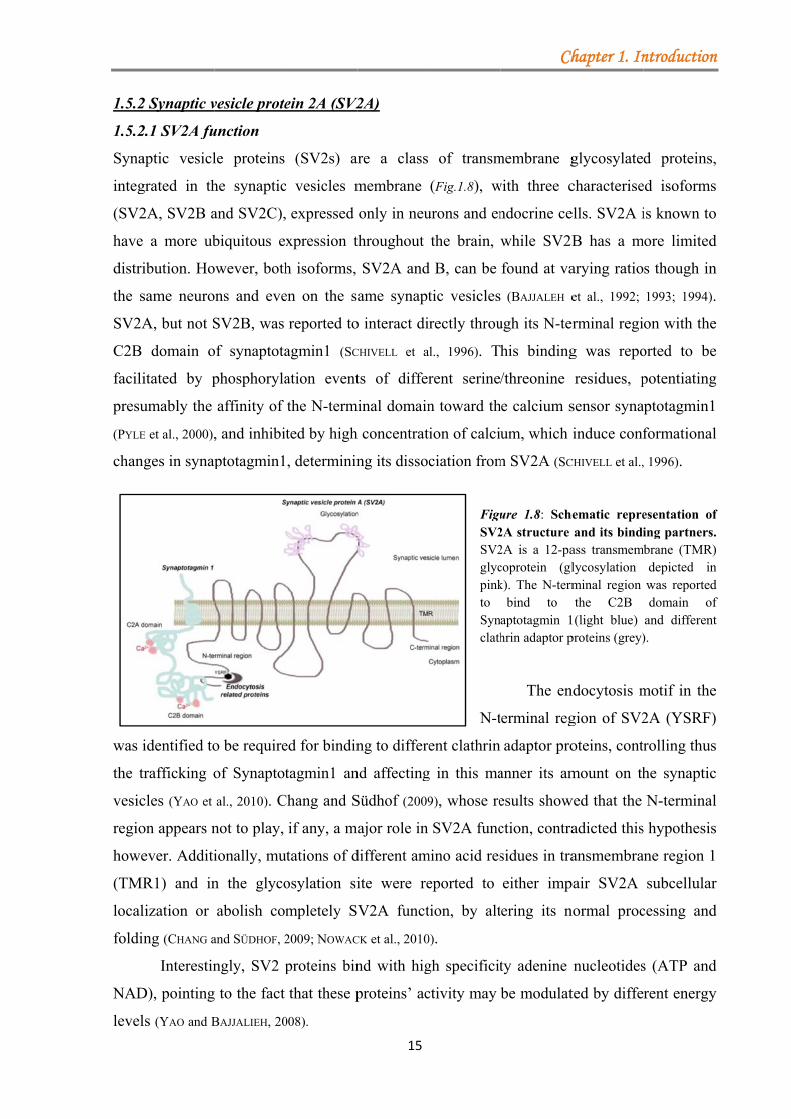
FiguSV2SV2glycpinkto Synclath
N-t
ent clathrin
g in this ma
9), whose re
SV2A func
mino acid res
eported to
tion, by alt
0).
gh specificit
ctivity may
aaaaaaaaaaaaa
Ch
membrane g
with three c
ndocrine ce
while SV2B
found at va
(BAJJALEH e
ugh its N-ter
his binding
/threonine
he calcium s
um, which
m SV2A (SC
ure 1.8: Sche2A structure 2A is a 12-pacoprotein (glk). The N-term
bind to aptotagmin 1hrin adaptor p
The en
terminal reg
adaptor pro
anner its am
esults show
ction, contra
sidues in tra
either imp
tering its n
ty adenine
be modulat
aaaaaaaaaaaaa
hapter 1. In
glycosylate
characterise
ells. SV2A i
B has a m
arying ratio
et al., 1992;
erminal regi
g was repo
residues, p
sensor syna
induce conf
CHIVELL et al.,
ematic repreand its bindi
ass transmemlycosylation minal region the C2B
1(light blue) proteins (grey)
ndocytosis m
gion of SV2
oteins, cont
mount on th
wed that the
adicted this
ansmembra
pair SV2A
normal proc
nucleotides
ted by diffe
aaaaaaaaaaaaa
ntroduction
d proteins,
ed isoforms
is known to
more limited
s though in
1993; 1994).
on with the
orted to be
potentiating
aptotagmin1
formational
, 1996).
esentation ofing partners.
mbrane (TMR)depicted inwas reporteddomain of
and different).
motif in the
2A (YSRF)
rolling thus
he synaptic
N-terminal
s hypothesis
ane region 1
subcellular
cessing and
s (ATP and
rent energy
aaaaaaaaaaaaa
n
,
s
o
d
n
.
e
e
g
l
f . ) n d f t
e
)
s
c
l
s
r
d
d
y
a
Chapter 1. Introduction
16
1.5.2.2 SV2A knock-out mice
In spite of all the data collected until now the exact function of SV2A still remains enigmatic.
To gain further insights into SV2A function, SV2A deficient mice were generated (CROWDER et
al., 1999; JANZ et al., 1999). Albeit SV2A KO littermates appeared normal at birth, mice
experienced severe seizures and died about three weeks after birth. No obvious alterations of
synaptic density or morphology in the brain of SV2A KO mice were observed (CROWDER et al.,
1999; JANZ et al., 1999). Therefore, SV2A seems not to be required in embryonic development
but rather its presence is essential for survival afterwards. Electrophysiological studies further
revealed that inhibitory (CROWDER et al., 1999; CHANG and SÜDHOF, 2009) as well as excitatory
(CUSTER et al., 2006) neurotransmission in these mice were impaired. A similar impairment was
also detected in adrenal chromaffin cells from SV2A KO mice, where the exocytotic burst
defining the size of the readily releasable pool (RRP) was observed to be decreased with no
evident alterations in the calcium level (XU and BAJJALIEH, 2001). A role in priming after vesicle
tethering was suggested by Custer et al. (2006), who observed a similar decrease in RRP in the
SV2A deficient mice’s brain, with no oscillation in calcium level.
However, earlier studies using SV2A/SV2B double knockout mice with a phenotype
resembling SV2A KO, proposed a role in regulating the calcium level during repetitive
stimulation trains rather than priming (JANZ et al., 1999). The described decrease in the RRP size
(CUSTER et al., 2006) was not reproduced by Chang (CHANG and SÜDHOF, 2009). A further
observation that the protein components of SNARE complexe were reduced in SV2A KO
mice supported the hypotheses that SV2A may have a role in the fusion mechanism (XU and
BAJJALIEH, 2001).
Taken together, the collected data suggest a role of SV2A in SV priming. Moreover,
SV2A act as a receptor for the anti-epileptic drug Keppra. It has been suggested that Keppra
may inhibit inappropriate interactions to occur when SV2A is overexpressed in neuronal cell
cultures. Neurons with elevated amount of overexpressed SV2A display similar impairments
in synaptic transmission as neurons from SV2A KO mice (NOWACK et al., 2011). It seems that
the protein amount plays an important role in maintaining the neuronal function as well. The
molecular mechanism of action of Keppra on SV2A is not fully elucidated.
Chapter 1. Introduction
17
Aim of the study
One of the most important properties of the synapse is the capacity to remodel itself in
response to ongoing activity in its environment. The synapse’s ability to weaken or strengthen
over time in response to different stimuli is called synaptic plasticity. One way of inducing
changes in presynaptic plasticity is to modulate SV priming and the protein machinery
involved in this process, like RIM1α and SV2A proteins. Over the years it has been proposed
that regulated phosphorylation/dephosphorylation events may play a role in plasticity-
induced remodelling of established and the assembly of novel active zones. To date the
molecular mechanism governing these changes are not understood in detail.
In this thesis two goals will be pursued:
1. Examine how phosphorylation events affect RIM1α binding affinities
RIM1α, a scaffold multi-domain protein residing in the active zone (AZ), has been shown to
be involved in synaptic vesicle priming and in different forms of presynaptic plasticity. Both
synaptic abundance and function have been suggested to be regulated by posttranslational
modifications. However, the precise mechanisms involved in controlling RIM1α protein
levels and function in the presynaptic terminal are not yet resolved.
To understand the impact that RIM1α phosphorylation has on active zone
reorganisation and presynaptic function, we aim at identifying novel phospho-dependent
binding partners. Combining various stimulation protocols, in order to block or enhance
kinases activity, different protein complexes binding to RIM1α will be identified by mass-
spectrometry. Next, these newly identified binding partners will be verified by protein-protein
interaction assays, and the functional role of these new proteins addressed in neuronal cell
culture.
2. Identify novel binding partners for SV2A
The last part of this study will be focused on the synaptic vesicle protein 2A (SV2A) protein,
whose involvement in vesicle priming or in controlling calcium levels is not yet elucidated.
Although SV2A is targeted by the antiepileptic drugs Keppra that only acts in case of strong
pathophysiological activity, its mode of action is still unresolved.
Therefore, to gain insight into the SV2A function identification of novel binding
partners will be addressed. Different affinity methods in combination with rAAV injections in
mice will be applied in order to find the best approach possible to purify the entire complex of
proteins under native conditions, followed by mass-spectrometry.
The results of this study will provide new insight into the molecular mechanisms by
which the functional properties of the presynaptic release machinery might be modulated.
Chapter 2. Materials
18
2. Materials
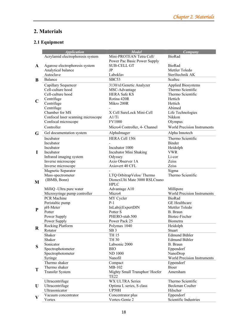
2.1 Equipment
Application Model Company
A
Acrylamid electrophoresis system Mini-PROTEAN Tetra Cell/ Power Pac Basic Power Supply
BioRad
Agarose electrophoresis system SUB-CELL GT BioRad Analytical balance JP Mettler Toledo Autoclave Laboklav Steriltechnik AK
B Balance SBC53 Scaltec
C
Capillary Sequencer 3130/xl/Genetic Analyzer Applied Biosystems Cell-culture hood MSC-Advantage Thermo Scientific Cell-culture hood HERA Safe KS Thermo Scientific Centrifuge Rotina 420R Hettich Centrifuge Mikro 200R Hettich Centrifuge - Abimed Chamber for MS X Cell SureLock Mini-Cell Life Technologies Confocal laser scanning microscope A1/Ti Nikkon
Confocal microscope FV1000 Olympus
Controller Micro4 Controller, 4- Channel World Precision Instruments
G Gel documentation system AlphaImager Alpha Innotech
I
Incubator HERA Cell 150i Thermo Scientific Incubator - Binder Incubator Incubator 1000 Heidolph Incubator Incubator Mini Shaking VWR Infrared imaging system Odyssey Li-cor Inverse microscope Axio Observer 1A Zeiss Inverse microscope Axiovert 40 CFL Zeiss
M
Magnetic Separator - Sigma Mass-spectrometer (IBMB, Bonn)
LTQ OrbitrapVelos/ Thermo DionexUlti Mate 3000 RSLCnano HPLC
Thermo Scientific
MilliQ -Ultra pure water Advantage A10 Millipore Microsyringe pump controller Micro4 World Precision Instruments
P
PCR Machine MY Cycler BioRad Peristaltic pump P-1 GE Healthcare pH-Meter InLab@ExpertDIN Mettler Toledo Potter Potter S B. Braun Power Supply PHERO-stab.500 Biotec-Fischer Power Supply Power Pack 25 Biometra
R Rocking Platform Polymax 1040 Heidolph Rotator SB 3 Stuart
S
Shaker TH 15 Edmund Bühler Shaker TH 30 Edmund Bühler Sonicator Labsonic 2000 B. Braun Spectrophotometer BIO Eppendorf Spectrophotometer ND 1000 NanoDrop Syringe Nanofil World Precision Instruments
T
Thermo shaker Compact Eppendorf Thermo shaker MB-102 Bioer Transfer System Mighty Small Transphor/ Hoefer
TE22 Amersham
U Ultracentrifuge WX ULTRA Series Thermo Scientific Ultracentrifuge Optima L series, S class Beckman Coulter Ultrasonicator UP50H Hilscher
V Vacuum concentrator Concentrator plus Eppendorf Vortex Vortex-Genie 2 Scientific Industries
Chapter 2. Materials
19
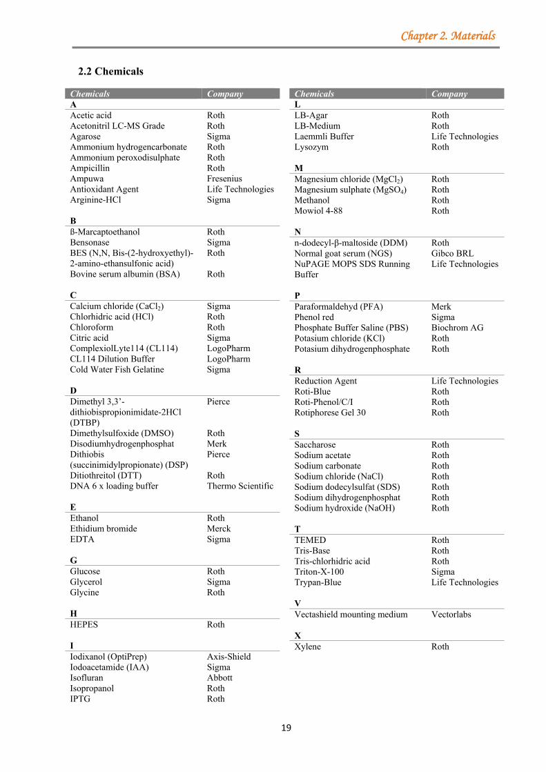
2.2 Chemicals
Chemicals Company A Acetic acid Roth Acetonitril LC-MS Grade Roth Agarose Sigma Ammonium hydrogencarbonate Roth Ammonium peroxodisulphate Roth Ampicillin Roth Ampuwa Fresenius Antioxidant Agent Life Technologies Arginine-HCl Sigma B ß-Marcaptoethanol Roth Bensonase Sigma BES (N,N, Bis-(2-hydroxyethyl)-2-amino-ethansulfonic acid)
Roth
Bovine serum albumin (BSA) Roth C Calcium chloride (CaCl2) Sigma Chlorhidric acid (HCl) Roth Chloroform Roth Citric acid Sigma ComplexiolLyte114 (CL114) LogoPharm CL114 Dilution Buffer LogoPharm Cold Water Fish Gelatine Sigma D Dimethyl 3,3’-dithiobispropionimidate-2HCl (DTBP)
Pierce
Dimethylsulfoxide (DMSO) Roth Disodiumhydrogenphosphat Merk Dithiobis (succinimidylpropionate) (DSP)
Pierce
Ditiothreitol (DTT) Roth DNA 6 x loading buffer Thermo Scientific E Ethanol Roth Ethidium bromide Merck EDTA Sigma G Glucose Roth Glycerol Sigma Glycine Roth H HEPES Roth I Iodixanol (OptiPrep) Axis-Shield Iodoacetamide (IAA) Sigma Isofluran Abbott Isopropanol Roth IPTG Roth
Chemicals Company L LB-Agar Roth LB-Medium Roth Laemmli Buffer Life Technologies Lysozym Roth M Magnesium chloride (MgCl2) Roth Magnesium sulphate (MgSO4) Roth Methanol Roth Mowiol 4-88 Roth N n-dodecyl-β-maltoside (DDM) Roth Normal goat serum (NGS) Gibco BRL NuPAGE MOPS SDS Running Buffer
Life Technologies
P Paraformaldehyd (PFA) Merk Phenol red Sigma Phosphate Buffer Saline (PBS) Biochrom AG Potasium chloride (KCl) Roth Potasium dihydrogenphosphate Roth R Reduction Agent Life Technologies Roti-Blue Roth Roti-Phenol/C/I Roth Rotiphorese Gel 30 Roth S Saccharose Roth Sodium acetate Roth Sodium carbonate Roth Sodium chloride (NaCl) Roth Sodium dodecylsulfat (SDS) Roth Sodium dihydrogenphosphat Roth Sodium hydroxide (NaOH) Roth T TEMED Roth Tris-Base Roth Tris-chlorhidric acid Roth Triton-X-100 Sigma Trypan-Blue Life Technologies V Vectashield mounting medium Vectorlabs X Xylene Roth
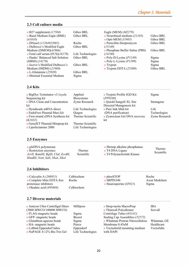
Chapter 2. Materials
20
2.3 Cell culture media
» B27 supplement (17504) Gibco BRL » Basal Medium Eagle (BME) (41010)
Gibco BRL
» DNaseI (11284932001) Roche » Dulbecco’s Modified Eagle Medium (DMEM)(41966)
Gibco BRL
» Fetal calf serum (FCS)(16170) Life Technologies » Hanks’ Balanced Salt Solution (HBSS) (14170)
Gibco BRL
» Iscove’s Modified Dulbecco’s Medium (IMDM) (21980)
Gibco BRL
» L-Glutamine (25030) Gibco BRL » Minimal Essential Medium Sigma
Eagle (MEM) (M2279) » Neurobasal medium (21103) Gibco BRL » Opti-MEM (31985) Gibco BRL » Penicillin-Streptomycin (15140)
Gibco BRL
» Phosphate Buffer Saline (PBS) (14190)
Gibco BRL
» Poly-D-Lysine (P1149) Sigma » Poly-L-Lysine (P1399) Sigma » Trypsin Sigma » Trypsin EDTA (25300) Gibco BRL
2.4 Kits
» BigDye Terminator v3.1cycle Sequencing kit
Applied Biosystems
» DNA Clean and Concentration kit
Zymo Research
» Dynabeads mRNA direct Life Technologies » EndoFree Plasmid Maxi kit Qiagen » First-strand cDNA Synthesis kit (K1632)
Thermo Scientific
» GeneJET Plasmid Miniprep kit Thermo Scientific » Lipofectamine 2000 Life Technologies
» Trypsin Profile IGD Kit (PP0100)
Sigma
» QuickChangeII XL Site Directed Mutagenesis kit
Stratagene
» Pure link Midi kit (DNA purification)
Life Technologies
» Zymoclean Gel DNA recovery kit
Zymo Research
2.5 Enzymes
» pfuDNA polymerase Thermo
Scientific » Restriction enzymes: AvrII, BamHI, BglII, ClaI, EcoRI, HindIII, NotI, SalI, XbaI, XhoI
» Shrimp alkaline phosphatase Thermo
Scientific » T4 DNA Ligase » T4 Polynucleotide Kinase
2.6 Inhibitors
» Calyculin A (208851) Calbiochem » Complete Mini EDTA free proteinase inhibitors
Roche
» Okadaic acid (495604) Calbiochem
» phosSTOP Roche » SRPIN340 Axon Medchem » Staurosporine (S5921) Sigma
2.7 Diverse materials
» Amicon Ultra Centrifigal filters (3000 MWCO/100000 MWCO)
Millipore
» FLAG-magnetic beads Sigma » GFP- magnetic beads Biozol » Glutathion-agarose beads Sigma » HA- magnetic beads Pierce » LoBind Eppendorf tubes Eppendorf » NuPAGE 4-12% Bis-Tris Gel Life Technologies
» Strep-tactin MacroPrep IBA » Thinwall Polyallomer Centrifuge Tubes (03141)/ Sealing Cap Assemblies (52572)
Sorvall
» Whatman Protran Nitrocellulose Membrane 0.45uM
Whatman, GE Healthcare
» Vectashield mounting medium with DAPI
Vectorlabs
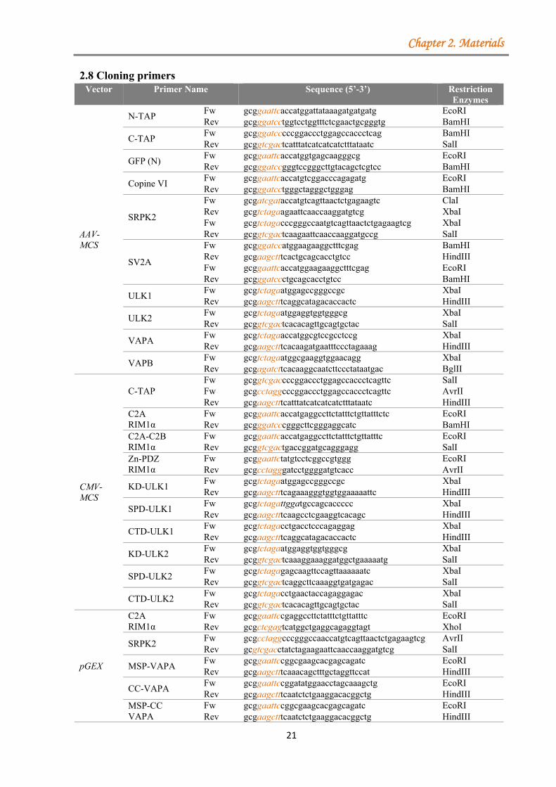
Chapter 2. Materials
21
2.8 Cloning primers Vector Primer Name Sequence (5’-3’) Restriction
Enzymes
AAV-MCS
N-TAP Fw gcggaattcaccatggattataaagatgatgatg EcoRI Rev gcgggatcctggtcctggtttctcgaactgcgggtg BamHI
C-TAP Fw gcgggatcccccggaccctggagccaccctcag BamHI Rev gcggtcgactcatttatcatcatcatctttataatc SalI
GFP (N) Fw gcggaattcaccatggtgagcaagggcg EcoRI Rev gcgggatccgggtccgggcttgtacagctcgtcc BamHI
Copine VI Fw gcggaattcaccatgtcggacccagagatg EcoRI Rev gcgggatcctgggctagggctgggag BamHI
SRPK2
Fw gcgatcgataccatgtcagttaactctgagaagtc ClaI Rev gcgtctagaagaattcaaccaaggatgtcg XbaI Fw gcgtctagacccgggccaatgtcagttaactctgagaagtcg XbaI Rev gcggtcgactcaagaattcaaccaaggatgccg SalI
SV2A
Fw gcgggatccatggaagaaggctttcgag BamHI Rev gcgaagctttcactgcagcacctgtcc HindIII Fw gcggaattcaccatggaagaaggctttcgag EcoRI Rev gcgggatccctgcagcacctgtcc BamHI
ULK1 Fw gcgtctagaatggagccgggccgc XbaI Rev gcgaagctttcaggcatagacaccactc HindIII
ULK2 Fw gcgtctagaatggaggtggtgggcg XbaI Rev gcggtcgactcacacagttgcagtgctac SalI
VAPA Fw gcgtctagaaccatggcgtccgcctccg XbaI Rev gcgaagctttcacaagatgaatttccctagaaag HindIII
VAPB Fw gcgtctagaatggcgaaggtggaacagg XbaI Rev gcgagatcttcacaaggcaatcttccctataatgac BglII
CMV-MCS
C-TAP Fw gcggtcgaccccggaccctggagccaccctcagttc SalI Fw gcgcctaggcccggaccctggagccaccctcagttc AvrII Rev gcgaagctttcatttatcatcatcatctttataatc HindIII
C2A RIM1α
Fw gcggaattcaccatgaggccttctatttctgttatttctc EcoRI Rev gcgggatcccgggcttcgggaggcatc BamHI
C2A-C2B RIM1α
Fw gcggaattcaccatgaggccttctatttctgttatttc EcoRI Rev gcggtcgactgaccggatgcagggagg SalI
Zn-PDZ RIM1α
Fw gcggaattctatgtcctcggccgtggg EcoRI Rev gcgcctagggatcctggggatgtcacc AvrII
KD-ULK1 Fw gcgtctagaatggagccgggccgc XbaI Rev gcgaagctttcagaaagggtggtggaaaaattc HindIII
SPD-ULK1 Fw gcgtctagattggatgccagcaccccc XbaI Rev gcgaagctttcaagcctcgaaggtcacagc HindIII
CTD-ULK1 Fw gcgtctagacctgacctcccagaggag XbaI Rev gcgaagctttcaggcatagacaccactc HindIII
KD-ULK2 Fw gcgtctagaatggaggtggtgggcg XbaI Rev gcggtcgactcaaaggaaaggatggctgaaaaatg SalI
SPD-ULK2 Fw gcgtctagagagcaagttccagttaaaaaatc XbaI Rev gcggtcgactcaggcttcaaaggtgatgagac SalI
CTD-ULK2 Fw gcgtctagacctgaactaccagaggagac XbaI Rev gcggtcgactcacacagttgcagtgctac SalI
pGEX
C2A RIM1α
Fw gcggaattccgaggccttctatttctgttatttc EcoRI Rev gcgctcgagtcatggctgaggcagaggtagt XhoI
SRPK2 Fw gcgcctaggcccgggccaaccatgtcagttaactctgagaagtcg AvrII Rev gcgtcgacctatctagaagaattcaaccaaggatgtcg SalI
MSP-VAPA Fw gcggaattccggcgaagcacgagcagatc EcoRI Rev gcgaagctttcaaacagctttgctaggttccat HindIII
CC-VAPA Fw gcggaattccggatatggaacctagcaaagctg EcoRI Rev gcgaagctttcaatctctgaaggacacggctg HindIII
MSP-CC VAPA
Fw gcggaattccggcgaagcacgagcagatc EcoRI Rev gcgaagctttcaatctctgaaggacacggctg HindIII
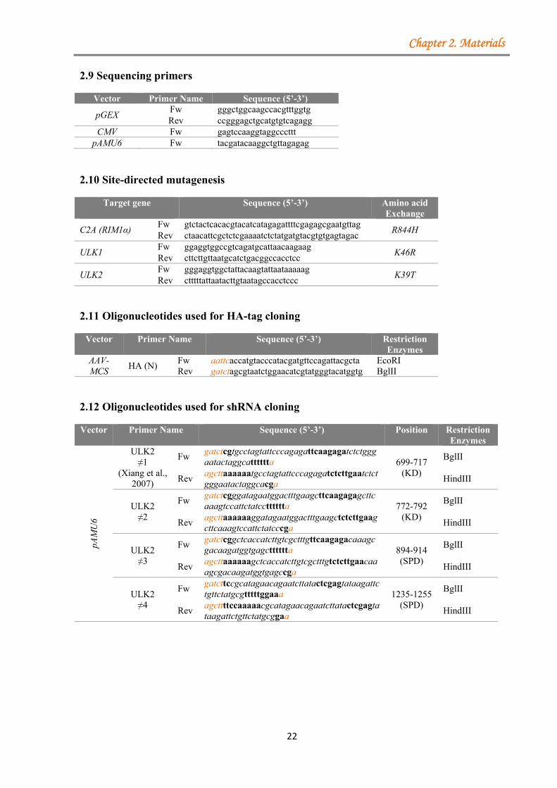
Chapter 2. Materials
22
2.9 Sequencing primers
Vector Primer Name Sequence (5’-3’)
pGEX Fw gggctggcaagccacgtttggtg Rev ccgggagctgcatgtgtcagagg
CMV Fw gagtccaaggtaggcccttt pAMU6 Fw tacgatacaaggctgttagagag
2.10 Site-directed mutagenesis
Target gene
Sequence (5’-3’) Amino acid Exchange
C2A (RIM1α) Fw gtctactcacacgtacatcatagagattttcgagagcgaatgttag
R844H Rev ctaacattcgctctcgaaaatctctatgatgtacgtgtgagtagac
ULK1 Fw ggaggtggccgtcagatgcattaacaagaag
K46R Rev cttcttgttaatgcatctgacggccacctcc
ULK2 Fw gggaggtggctattacaagtattaataaaaag
K39T Rev ctttttattaatacttgtaatagccacctccc
2.11 Oligonucleotides used for HA-tag cloning
Vector Primer Name Sequence (5’-3’) Restriction Enzymes
AAV-MCS
HA (N) Fw aattcaccatgtacccatacgatgttccagattacgcta EcoRI Rev gatctagcgtaatctggaacatcgtatgggtacatggtg BglII
2.12 Oligonucleotides used for shRNA cloning
Vector Primer Name Sequence (5’-3’) Position Restriction Enzymes
pAM
U6
ULK2 ≠1
(Xiang et al., 2007)
Fw gatctcgtgcctagtattcccagagattcaagagatctctgggaatactaggcatttttta 699-717
(KD)
BglII
Rev agcttaaaaaatgcctagtattcccagagatctcttgaatctctgggaatactaggcacga
HindIII
ULK2 ≠2
Fw gatctcgggatagaatggactttgaagcttcaagagagcttcaaagtccattctatcctttttta 772-792
(KD)
BglII
Rev agcttaaaaaaggatagaatggactttgaagctctcttgaagcttcaaagtccattctatcccga
HindIII
ULK2 ≠3
Fw gatctcggctcaccatcttgtcgctttgttcaagagacaaagcgacaagatggtgagctttttta 894-914
(SPD)
BglII
Rev agcttaaaaaagctcaccatcttgtcgctttgtctcttgaacaaagcgacaagatggtgagccga
HindIII
ULK2 ≠4
Fw gatcttccgcatagaacagaatcttatactcgagtataagattctgttctatgcgtttttggaaa 1235-1255
(SPD)
BglII
Rev agcttttccaaaaacgcatagaacagaatcttatactcgagtataagattctgttctatgcggaa
HindIII
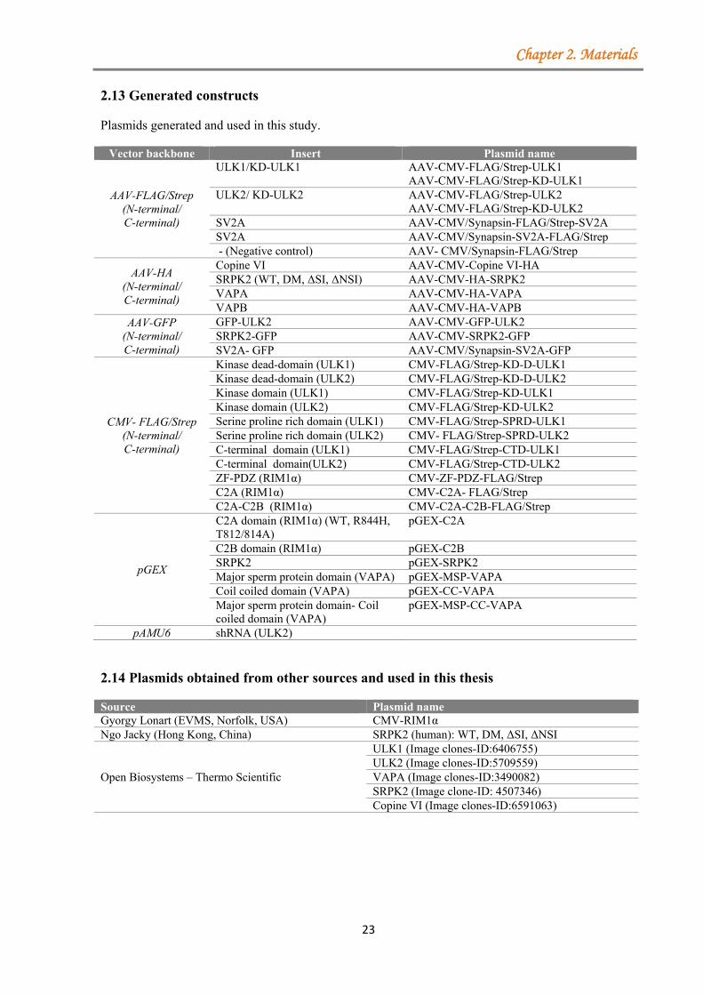
Chapter 2. Materials
23
2.13 Generated constructs
Plasmids generated and used in this study.
Vector backbone Insert Plasmid name
AAV-FLAG/Strep (N-terminal/ C-terminal)
ULK1/KD-ULK1 AAV-CMV-FLAG/Strep-ULK1 AAV-CMV-FLAG/Strep-KD-ULK1
ULK2/ KD-ULK2 AAV-CMV-FLAG/Strep-ULK2 AAV-CMV-FLAG/Strep-KD-ULK2
SV2A AAV-CMV/Synapsin-FLAG/Strep-SV2A SV2A AAV-CMV/Synapsin-SV2A-FLAG/Strep - (Negative control) AAV- CMV/Synapsin-FLAG/Strep
AAV-HA (N-terminal/ C-terminal)
Copine VI AAV-CMV-Copine VI-HA SRPK2 (WT, DM, ΔSI, ΔNSI) AAV-CMV-HA-SRPK2 VAPA AAV-CMV-HA-VAPA VAPB AAV-CMV-HA-VAPB
AAV-GFP (N-terminal/ C-terminal)
GFP-ULK2 AAV-CMV-GFP-ULK2 SRPK2-GFP AAV-CMV-SRPK2-GFP SV2A- GFP AAV-CMV/Synapsin-SV2A-GFP
CMV- FLAG/Strep (N-terminal/ C-terminal)
Kinase dead-domain (ULK1) CMV-FLAG/Strep-KD-D-ULK1 Kinase dead-domain (ULK2) CMV-FLAG/Strep-KD-D-ULK2 Kinase domain (ULK1) CMV-FLAG/Strep-KD-ULK1 Kinase domain (ULK2) CMV-FLAG/Strep-KD-ULK2 Serine proline rich domain (ULK1) CMV-FLAG/Strep-SPRD-ULK1 Serine proline rich domain (ULK2) CMV- FLAG/Strep-SPRD-ULK2 C-terminal domain (ULK1) CMV-FLAG/Strep-CTD-ULK1 C-terminal domain(ULK2) CMV-FLAG/Strep-CTD-ULK2 ZF-PDZ (RIM1α) CMV-ZF-PDZ-FLAG/Strep C2A (RIM1α) CMV-C2A- FLAG/Strep C2A-C2B (RIM1α) CMV-C2A-C2B-FLAG/Strep
pGEX
C2A domain (RIM1α) (WT, R844H, T812/814A)
pGEX-C2A
C2B domain (RIM1α) pGEX-C2B SRPK2 pGEX-SRPK2 Major sperm protein domain (VAPA) pGEX-MSP-VAPA Coil coiled domain (VAPA) pGEX-CC-VAPA Major sperm protein domain- Coil coiled domain (VAPA)
pGEX-MSP-CC-VAPA
pAMU6 shRNA (ULK2)
2.14 Plasmids obtained from other sources and used in this thesis
Source Plasmid nameGyorgy Lonart (EVMS, Norfolk, USA) CMV-RIM1α Ngo Jacky (Hong Kong, China) SRPK2 (human): WT, DM, ΔSI, ΔNSI
Open Biosystems – Thermo Scientific
ULK1 (Image clones-ID:6406755) ULK2 (Image clones-ID:5709559) VAPA (Image clones-ID:3490082) SRPK2 (Image clone-ID: 4507346) Copine VI (Image clones-ID:6591063)
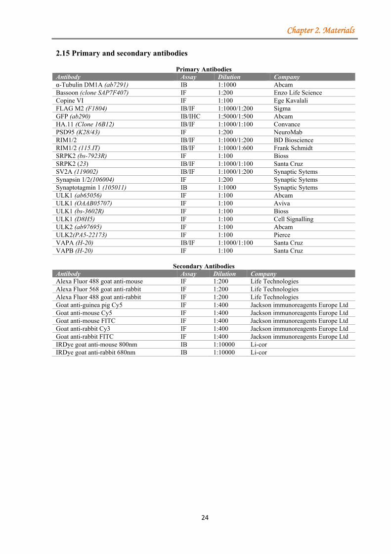
Chapter 2. Materials
24
2.15 Primary and secondary antibodies
Primary Antibodies Antibody Assay Dilution Company α-Tubulin DM1A (ab7291) IB 1:1000 Abcam Bassoon (clone SAP7F407) IF 1:200 Enzo Life Science Copine VI IF 1:100 Ege Kavalali FLAG M2 (F1804) IB/IF 1:1000/1:200 Sigma GFP (ab290) IB/IHC 1:5000/1:500 Abcam HA.11 (Clone 16B12) IB/IF 1:1000/1:100 Convance PSD95 (K28/43) IF 1:200 NeuroMab RIM1/2 IB/IF 1:1000/1:200 BD Bioscience RIM1/2 (115.IT) IB/IF 1:1000/1:600 Frank Schmidt SRPK2 (bs-7923R) IF 1:100 Bioss SRPK2 (23) IB/IF 1:1000/1:100 Santa Cruz SV2A (119002) IB/IF 1:1000/1:200 Synaptic Sytems Synapsin 1/2(106004) IF 1:200 Synaptic Sytems Synaptotagmin 1 (105011) IB 1:1000 Synaptic Sytems ULK1 (ab65056) IF 1:100 Abcam ULK1 (OAAB05707) IF 1:100 Aviva ULK1 (bs-3602R) IF 1:100 Bioss ULK1 (D8H5) IF 1:100 Cell Signalling ULK2 (ab97695) IF 1:100 Abcam ULK2(PA5-22173) IF 1:100 Pierce VAPA (H-20) IB/IF 1:1000/1:100 Santa Cruz VAPB (H-20) IF 1:100 Santa Cruz
Secondary Antibodies Antibody Assay Dilution Company Alexa Fluor 488 goat anti-mouse IF 1:200 Life Technologies Alexa Fluor 568 goat anti-rabbit IF 1:200 Life Technologies Alexa Fluor 488 goat anti-rabbit IF 1:200 Life Technologies Goat anti-guinea pig Cy5 IF 1:400 Jackson immunoreagents Europe Ltd Goat anti-mouse Cy5 IF 1:400 Jackson immunoreagents Europe Ltd Goat anti-mouse FITC IF 1:400 Jackson immunoreagents Europe Ltd Goat anti-rabbit Cy3 IF 1:400 Jackson immunoreagents Europe Ltd Goat anti-rabbit FITC IF 1:400 Jackson immunoreagents Europe Ltd IRDye goat anti-mouse 800nm IB 1:10000 Li-cor IRDye goat anti-rabbit 680nm IB 1:10000 Li-cor
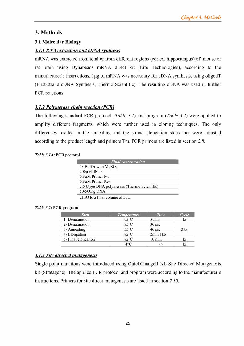
Chapter 3. Methods
25
3. Methods
3.1 Molecular Biology
3.1.1 RNA extraction and cDNA synthesis
mRNA was extracted from total or from different regions (cortex, hippocampus) of mouse or
rat brain using Dynabeads mRNA direct kit (Life Technologies), according to the
manufacturer’s instructions. 1μg of mRNA was necessary for cDNA synthesis, using oligodT
(First-strand cDNA Synthesis, Thermo Scientific). The resulting cDNA was used in further
PCR reactions.
3.1.2 Polymerase chain reaction (PCR)
The following standard PCR protocol (Table 3.1) and program (Table 3.2) were applied to
amplify different fragments, which were further used in cloning techniques. The only
differences resided in the annealing and the strand elongation steps that were adjusted
according to the product length and primers Tm. PCR primers are listed in section 2.8.
Table 3.1A: PCR protocol
Final concentration 1x Buffer with MgSO4 200µM dNTP 0.3µM Primer Fw 0.3µM Primer Rev 2.5 U pfu DNA polymerase (Thermo Scientific) 50-500ng DNA
dH2O to a final volume of 50μl Table 3.2: PCR program
Step Temperature Time Cycle 1- Denaturation 95°C 5 min 1x 2- Denaturation 95°C 30 sec
35x 3- Annealing 55°C 40 sec 4- Elongation 72°C 2min/1kb 5- Final elongation 72°C 10 min 1x 4°C ∞ 1x
3.1.3 Site directed mutagenesis
Single point mutations were introduced using QuickChangeII XL Site Directed Mutagenesis
kit (Stratagene). The applied PCR protocol and program were according to the manufacturer’s
instructions. Primers for site direct mutagenesis are listed in section 2.10.
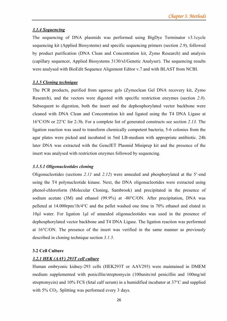
Chapter 3. Methods
26
3.1.4 Sequencing
The sequencing of DNA plasmids was performed using BigDye Terminator v3.1cycle
sequencing kit (Applied Biosystems) and specific sequencing primers (section 2.9), followed
by product purification (DNA Clean and Concentration kit, Zymo Research) and analysis
(capillary sequencer, Applied Biosystems 3130/xl/Genetic Analyser). The sequencing results
were analysed with BioEdit Sequence Alignment Editor v.7 and with BLAST from NCBI.
3.1.5 Cloning technique
The PCR products, purified from agarose gels (Zymoclean Gel DNA recovery kit, Zymo
Research), and the vectors were digested with specific restriction enzymes (section 2.8).
Subsequent to digestion, both the insert and the dephosphorylated vector backbone were
cleaned with DNA Clean and Concentration kit and ligated using the T4 DNA Ligase at
16°C/ON or 22°C for 2-3h. For a complete list of generated constructs see section 2.13. The
ligation reaction was used to transform chemically competent bacteria; 5-6 colonies from the
agar plates were picked and incubated in 5ml LB-medium with appropriate antibiotic. 24h
later DNA was extracted with the GeneJET Plasmid Miniprep kit and the presence of the
insert was analysed with restriction enzymes followed by sequencing.
3.1.5.1 Oligonucleotides cloning
Oligonucleotides (sections 2.11 and 2.12) were annealed and phosphorylated at the 5’-end
using the T4 polynucleotide kinase. Next, the DNA oligonucleotides were extracted using
phenol-chloroform (Molecular Cloning, Sambrook) and precipitated in the presence of
sodium acetate (3M) and ethanol (99.9%) at -80°C/ON. After precipitation, DNA was
pelleted at 14.000rpm/1h/4°C and the pellet washed one time in 70% ethanol and eluted in
10μl water. For ligation 1μl of annealed oligonucleotides was used in the presence of
dephosphorylated vector backbone and T4 DNA Ligase. The ligation reaction was performed
at 16°C/ON. The presence of the insert was verified in the same manner as previously
described in cloning technique section 3.1.5.
3.2 Cell Culture
3.2.1 HEK (AAV) 293T cell culture
Human embryonic kidney-293 cells (HEK293T or AAV293) were maintained in DMEM
medium supplemented with penicillin/streptomycin (100units/ml penicillin and 100mg/ml
streptomycin) and 10% FCS (fetal calf serum) in a humidified incubator at 37°C and supplied
with 5% CO2. Splitting was performed every 3 days.aaaaaaaaaaaaaaaaaaaaaaaaaaaaaaaaaaaaa
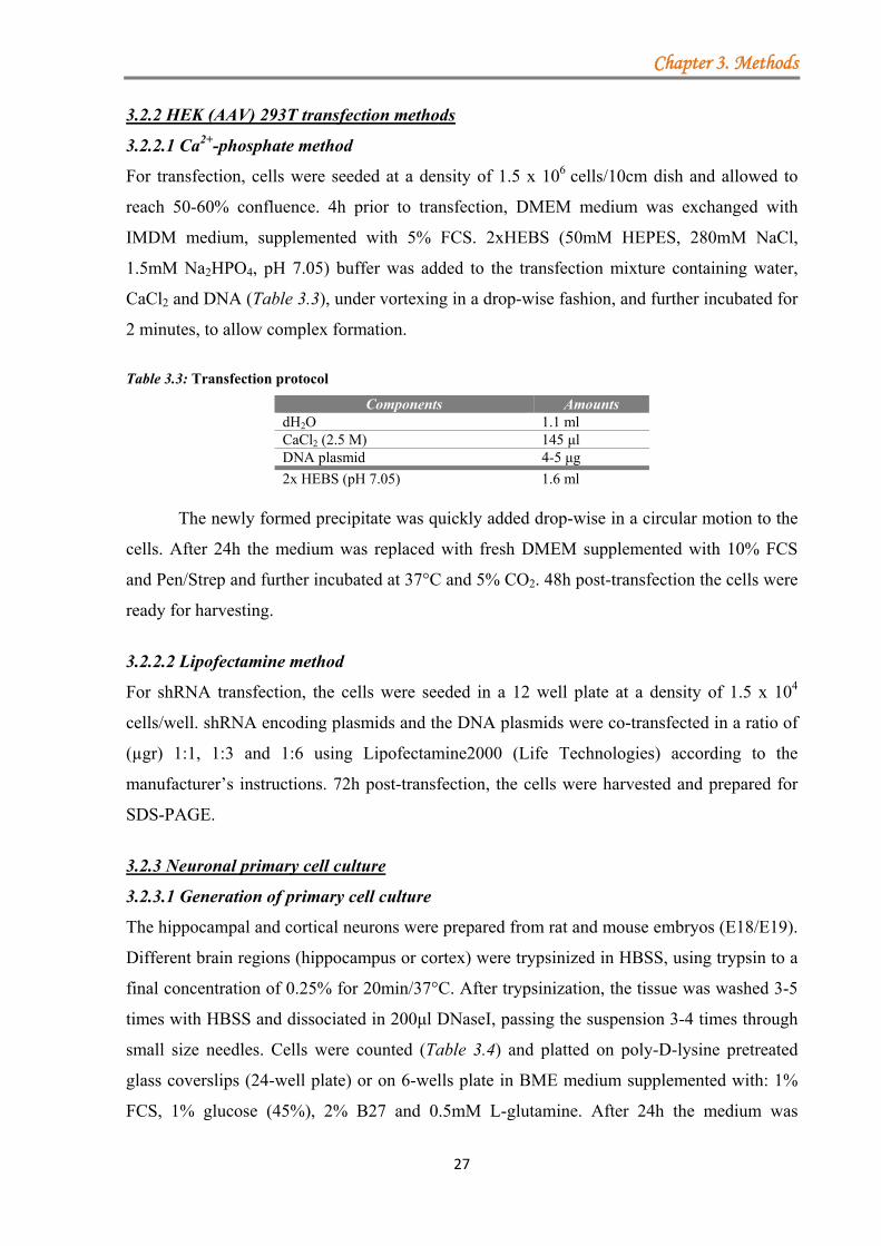
Chapter 3. Methods
27
3.2.2 HEK (AAV) 293T transfection methods
3.2.2.1 Ca2+-phosphate method
For transfection, cells were seeded at a density of 1.5 x 10⁶ cells/10cm dish and allowed to
reach 50-60% confluence. 4h prior to transfection, DMEM medium was exchanged with
IMDM medium, supplemented with 5% FCS. 2xHEBS (50mM HEPES, 280mM NaCl,
1.5mM Na2HPO4, pH 7.05) buffer was added to the transfection mixture containing water,
CaCl2 and DNA (Table 3.3), under vortexing in a drop-wise fashion, and further incubated for
2 minutes, to allow complex formation.
Table 3.3: Transfection protocol
Components Amounts dH2O 1.1 ml CaCl2 (2.5 M) 145 μl DNA plasmid 4-5 μg
2x HEBS (pH 7.05) 1.6 ml
The newly formed precipitate was quickly added drop-wise in a circular motion to the
cells. After 24h the medium was replaced with fresh DMEM supplemented with 10% FCS
and Pen/Strep and further incubated at 37°C and 5% CO2. 48h post-transfection the cells were
ready for harvesting.
3.2.2.2 Lipofectamine method
For shRNA transfection, the cells were seeded in a 12 well plate at a density of 1.5 x 10⁴
cells/well. shRNA encoding plasmids and the DNA plasmids were co-transfected in a ratio of
(µgr) 1:1, 1:3 and 1:6 using Lipofectamine2000 (Life Technologies) according to the
manufacturer’s instructions. 72h post-transfection, the cells were harvested and prepared for
SDS-PAGE.
3.2.3 Neuronal primary cell culture
3.2.3.1 Generation of primary cell culture
The hippocampal and cortical neurons were prepared from rat and mouse embryos (E18/E19).
Different brain regions (hippocampus or cortex) were trypsinized in HBSS, using trypsin to a
final concentration of 0.25% for 20min/37°C. After trypsinization, the tissue was washed 3-5
times with HBSS and dissociated in 200μl DNaseI, passing the suspension 3-4 times through
small size needles. Cells were counted (Table 3.4) and platted on poly-D-lysine pretreated
glass coverslips (24-well plate) or on 6-wells plate in BME medium supplemented with: 1%
FCS, 1% glucose (45%), 2% B27 and 0.5mM L-glutamine. After 24h the medium was
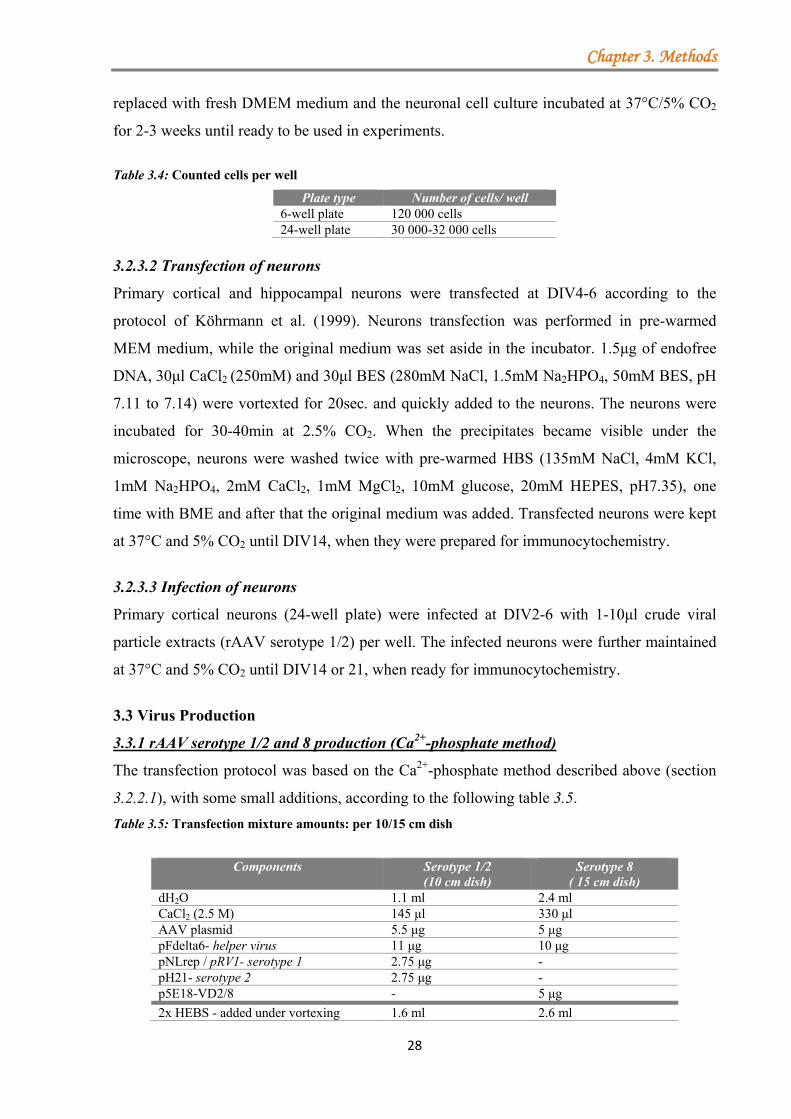
Chapter 3. Methods
28
replaced with fresh DMEM medium and the neuronal cell culture incubated at 37°C/5% CO2
for 2-3 weeks until ready to be used in experiments.
Table 3.4: Counted cells per well
Plate type Number of cells/ well 6-well plate 120 000 cells 24-well plate 30 000-32 000 cells
3.2.3.2 Transfection of neurons
Primary cortical and hippocampal neurons were transfected at DIV4-6 according to the
protocol of Köhrmann et al. (1999). Neurons transfection was performed in pre-warmed
MEM medium, while the original medium was set aside in the incubator. 1.5μg of endofree
DNA, 30μl CaCl2 (250mM) and 30μl BES (280mM NaCl, 1.5mM Na2HPO4, 50mM BES, pH
7.11 to 7.14) were vortexted for 20sec. and quickly added to the neurons. The neurons were
incubated for 30-40min at 2.5% CO2. When the precipitates became visible under the
microscope, neurons were washed twice with pre-warmed HBS (135mM NaCl, 4mM KCl,
1mM Na2HPO4, 2mM CaCl2, 1mM MgCl2, 10mM glucose, 20mM HEPES, pH7.35), one
time with BME and after that the original medium was added. Transfected neurons were kept
at 37°C and 5% CO2 until DIV14, when they were prepared for immunocytochemistry.
3.2.3.3 Infection of neurons
Primary cortical neurons (24-well plate) were infected at DIV2-6 with 1-10μl crude viral
particle extracts (rAAV serotype 1/2) per well. The infected neurons were further maintained
at 37°C and 5% CO2 until DIV14 or 21, when ready for immunocytochemistry.
3.3 Virus Production
3.3.1 rAAV serotype 1/2 and 8 production (Ca2+-phosphate method)
The transfection protocol was based on the Ca2+-phosphate method described above (section
3.2.2.1), with some small additions, according to the following table 3.5.
Table 3.5: Transfection mixture amounts: per 10/15 cm dish
Components Serotype 1/2 (10 cm dish)
Serotype 8 ( 15 cm dish)
dH2O 1.1 ml 2.4 ml CaCl2 (2.5 M) 145 μl 330 μl AAV plasmid 5.5 μg 5 μg pFdelta6- helper virus 11 μg 10 μg pNLrep / pRV1- serotype 1 2.75 μg - pH21- serotype 2 2.75 μg - p5E18-VD2/8 - 5 μg
2x HEBS - added under vortexing 1.6 ml 2.6 ml
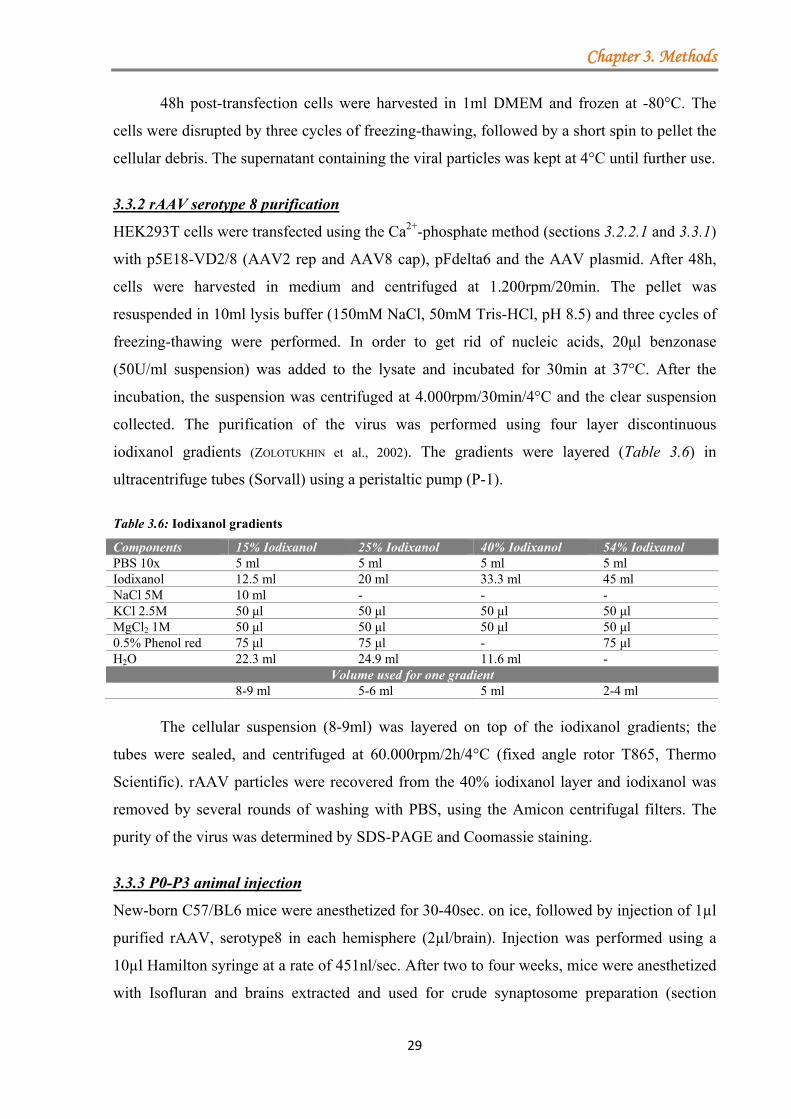
Chapter 3. Methods
29
48h post-transfection cells were harvested in 1ml DMEM and frozen at -80°C. The
cells were disrupted by three cycles of freezing-thawing, followed by a short spin to pellet the
cellular debris. The supernatant containing the viral particles was kept at 4°C until further use.
3.3.2 rAAV serotype 8 purification
HEK293T cells were transfected using the Ca2+-phosphate method (sections 3.2.2.1 and 3.3.1)
with p5E18-VD2/8 (AAV2 rep and AAV8 cap), pFdelta6 and the AAV plasmid. After 48h,
cells were harvested in medium and centrifuged at 1.200rpm/20min. The pellet was
resuspended in 10ml lysis buffer (150mM NaCl, 50mM Tris-HCl, pH 8.5) and three cycles of
freezing-thawing were performed. In order to get rid of nucleic acids, 20μl benzonase
(50U/ml suspension) was added to the lysate and incubated for 30min at 37°C. After the
incubation, the suspension was centrifuged at 4.000rpm/30min/4°C and the clear suspension
collected. The purification of the virus was performed using four layer discontinuous
iodixanol gradients (ZOLOTUKHIN et al., 2002). The gradients were layered (Table 3.6) in
ultracentrifuge tubes (Sorvall) using a peristaltic pump (P-1).
Table 3.6: Iodixanol gradients
Components 15% Iodixanol 25% Iodixanol 40% Iodixanol 54% Iodixanol PBS 10x 5 ml 5 ml 5 ml 5 ml Iodixanol 12.5 ml 20 ml 33.3 ml 45 ml NaCl 5M 10 ml - - - KCl 2.5M 50 μl 50 μl 50 μl 50 μl MgCl2 1M 50 μl 50 μl 50 μl 50 μl 0.5% Phenol red 75 μl 75 μl - 75 μl H2O 22.3 ml 24.9 ml 11.6 ml -
Volume used for one gradient 8-9 ml 5-6 ml 5 ml 2-4 ml
The cellular suspension (8-9ml) was layered on top of the iodixanol gradients; the
tubes were sealed, and centrifuged at 60.000rpm/2h/4°C (fixed angle rotor T865, Thermo
Scientific). rAAV particles were recovered from the 40% iodixanol layer and iodixanol was
removed by several rounds of washing with PBS, using the Amicon centrifugal filters. The
purity of the virus was determined by SDS-PAGE and Coomassie staining.
3.3.3 P0-P3 animal injection
New-born C57/BL6 mice were anesthetized for 30-40sec. on ice, followed by injection of 1µl
purified rAAV, serotype8 in each hemisphere (2µl/brain). Injection was performed using a
10µl Hamilton syringe at a rate of 451nl/sec. After two to four weeks, mice were anesthetized
with Isofluran and brains extracted and used for crude synaptosome preparation (section
Chapter 3. Methods
30
3.4.1). All experiments were performed in agreement with the regulations of the University
of Bonn Medical Centre Animal Care Committee.
3.4 Biochemistry
3.4.1 Preparation of crude synaptosomes
The preparation was performed at 4°C using detergent free equipment and buffers
supplemented with proteinase inhibitors (Roche). C57/BL6 mice (6-8 weeks old) were
euthanized with Isoflorane, decapitated and the brains extracted. The cerebellum together with
the most of the white matter was removed. Both hemisphere were homogenized in ice-cold
homogenization buffer (0.32M sucrose, 50mM EDTA, 2mM HEPES, pH 7.4), supplemented
with proteinase inhibitor (Roche) in a Teflon-glass homogenizer (7strokes, 900rpm), followed
by centrifugation at 3.000g/15min/4°C. The pellet (P1-nuclear fraction) was removed and the
supernatant (S1-crude synaptosomal fraction) transferred into 2ml eppendorf tubes and
centrifuged at 14.000rpm/25min/4°C. The synaptosomal cytosol fraction (S2) was discarded
and the pellet (P2-crude synaptosomes) resuspended in either 500μl lysis buffer (CL114
detergent, Logopharm) or 100μl equilibration Krebs-Henseleit-HEPES buffer (118mM NaCl,
3.5mM KCl, 1.25mM CaCl2, 1.2 mM MgSO4, 1.2mM KH2PO4, 25mM NaHCO3, 11.5mM
glucose and 5mM HEPES-NaOH, pH 7.4).
In the case of SV2A protein purification, P0-P3 mice were injected with purified
rAAV (serotype 8) and after several weeks crude synaptosomes were prepared as described
above. The P2 fraction was resuspended in lysis buffer (50mM Tris-HCl, 150mM NaCl, pH
7.5) supplemented with proteinase, phosphatase inhibitors and 3.9mM n-dodecyl-β-maltoside
(DDM, specific detergent for SV2A) as previously described by Lambeng et al. (2006). After
1h incubation at 4°C the solution was clarified by centrifugation and the supernatant used for
immunoprecipitation (section 3.4.2.4) or TAP purification (section 3.5.2).
3.4.2 Protein-protein interaction assays
3.4.2.1 Protein induction and purification from BL21 bacteria
pGEX plasmids encoding for the protein of interest were retransformed in Escherichia coli
BL21 (DE3). At an optic density (OD) of 0.6-0.8 of the bacterial culture, the expression of the
GST-fusion proteins was induced by addition of IPTG (1mM) for 3-4h, under constant
shaking at 37°C. After a centrifugation step at 4.500rpm/30min/4°C the bacterial pellet was
resuspended in PBS supplemented with proteinase inhibitor (Roche) and lysozym (1mg/ml)
and lysed on ice for 20min, followed by sonication and centrifugation at 4.500rpm/1h/4°C. To
capture the protein of interest the clear supernatant was incubated for 1h with prewashed
Chapter 3. Methods
31
Glutathion-agarose beads. Beads were extensively washed and resuspended in 1ml of PBS
supplemented with proteinase inhibitors to reach 50% slurry, further used in GST-pull down
assay (section 3.4.2.2). In order to check the efficiency of protein induction a small aliquot
was analyzed by Coomassie staining.
3.4.2.2 GST-pull down assay
To analyse the binding of different GST-fusion proteins to native or overexpressed proteins,
GST-pull down assays were performed with crude synaptosomes (section 3.4.1), primary
cortical neurons and with transfected HEK293T cells by the Ca2+-phosphate method (section
3.2.2.1). HEK293T cells were lysed for 1h in ice-cold lysis buffer (50mM HEPES pH 7.4,
150mM NaCl, 1% Triton X-100, Complete Protease Inhibitor Cocktail Tablets), spun at
14.000rpm/10 min/4°C and the resulting clear supernatant was incubated for 1-2h with GST
and GST-fusion proteins. Beads were washed five times with PBS-0.5% Triton X-100
washing buffer, boiled at 95°C/5min in Laemmli buffer with β-ME and resolved in SDS-
PAGE gel. GST and GST-fusion proteins were incubated also with lysed crude synaptosomes
or lysed cortical neurons for 2-4h. Bound protein complexes were washed with either CL-114
dilution buffer or PBS-0.5% Triton X-100, boiled in Laemmli buffer with β-ME at 95°C/5
min and resolved in SDS-PAGE gel.
3.4.2.3 Co-immunoprecipitation (co-IP)
HEK293T cells were co-transfected with DNA plasmids containing the sequence encoding for
the proteins of interest using the Ca2+-phosphate method as described in section 3.2.2.1. 48h
post-transfection the medium was discarded and the cells harvested in ice-cold lysis buffer
(50mM HEPES pH 7.4, 150mM NaCl, 1% Triton X-100, Complete Protease Inhibitor
Cocktail Tablets) and lysed on ice for 1h, followed by a short centrifugation step at
14.000rpm/10min/4°C. The supernatant was incubated at 4°C with pre-washed (in PBS)
magnetic beads for 1h (FLAG M2 beads) or for 2-3h (HA- or GFP-magnetic beads) on a
rotator. After the incubation time, the beads containing the protein complexes were washed
with PBS-0.5% Triton X-100 buffer. The beads were boiled in Laemmli buffer with β-ME at
95°C/5 min and proteins resolved in SDS-PAGE gel (8% or 10%).
3.4.2.4 Immunoprecipitation (IP)
The P0-3 C57/BL6 mice were injected into the hemisphere with 2µl of purified virus (rAAV-
SV2A-GFP, serotype 8) and two weeks after the injection crude synaptosomes were prepared
(section 3.4.1). After the P2 fraction was solubilised, the clear supernatant was mixed for 2-3h
Chapter 3. Methods
32
with anti-GFP antibodies (abcam 290). The antibodies were collected overnight by incubation
with protein A/G-agarose beads (Santa Cruz). The A/G-agarose beads were collected by
centrifugation at low speed (2.000 rpm/5 min), washed five times in PBS and boiled in
Laemmli buffer with β-ME at 95°C/5 min.
3.4.3 Protein concentration determination
The protein concentration was determined by measuring the optical density at 260nm using
NanoDrop. For shRNA testing, the protein concentration of all samples was adjusted to the
lowest one, before analysing by WB. In binding assays the protein concentration of the crude
synaptosomes was adjusted to 3-5mg/ml/reaction.
3.4.4 Western Blotting (WB)
Prior to WB, samples were resolved in 8% or 10% SDS-polyacrylamide gel electrophoresis
(SDS-PAGE) and separated by size. The separated proteins were transferred to nitrocellulose
membrane, followed by protein detection. The membranes were blocked with 3% cold water
fish gelatin in PBS for 1h, incubated with primary antibodies in blocking buffer for 2h,
washed three times/15min with PBS-0.1% Tween 20 and further incubated with secondary
antibodies (IRDye-1:10000, section 2.15) for 40min. The detection was achieved with an
infrared imaging system (Odyssey, Li-cor).
3.5 Identification of novel binding partners by tandem-affinity purification (TAP)
3.5.1 Protein cross-linking
Cultured cortical neurons (DIV2) obtained from wild-type (WT), SV2A +/- and SV2A -/-
mice were infected with crude viral particles, serotype 1/2, expressing N- or C-TAP-tagged
SV2A. At DIV14 neurons were washed one time with PBS and proteins were cross-linked
using three different cross-linkers (Table 3.7): 1% formaldehyde (VASILESCU et al., 2004;
KLOCKENBUSCH and KAST, 2010), 5mM DSP (dithiobis(succinimidylpropionate), Pierce), 5mM
DTBP (dimethyl 3,3’-dithiobispropionimidate-2HCl, Pierce). The protein cross-linking was
performed at room temperature or at 37°C/30-60 min, followed by quenching the reaction
with 50mM Tris, pH 7.5 or 2.5mM glycine for 5-15min. Cells were lysed either in HEPES
buffer (50mM HEPES, 150mM NaCl, pH 7.4) or in Tris-HCl buffer (50mM Tris-HCl,
150mM NaCl, pH 7.4) supplemented with proteinase, phosphatase inhibitors and 3.9mM n-
dodecyl-β-maltoside (DDM) for 1h/4°C. The clear supernatant was subject to either one-step
or tandem purification as described below (section 3.5.2), with only one difference: the
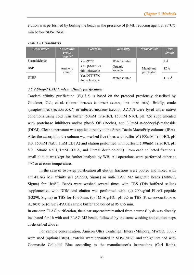
Chapter 3. Methods
33
elution was performed by boiling the beads in the presence of β-ME reducing agent at 95°C/5
min before SDS-PAGE.
Table 3.7: Cross-linkers
Cross-linker Functional group
targeted
Cleavable Solubility Permeability Arm length
Formaldehyde
Amine to amine
Yes /95°C Water soluble
Membrane permeable
2 Å
DSP Yes/ β-ME/95°C
thiol-cleavable Organic solvents
12 Å
DTBP Yes/DTT/37°C
thiol-cleavable Water soluble 11.9 Å
3.5.2 Strep/FLAG tandem affinity purification
Tandem affinity purification (Fig.3.1) is based on the protocol previously described by
Glockner, C.J., et al. (Current Protocols in Protein Science, Unit 19.20, 2009). Briefly, crude
synaptosomes (section 3.4.1) or infected neurons (section 3.2.3.3) were lysed under native
conditions using cold lysis buffer (50mM Tris-HCl, 150mM NaCl, pH 7.5) supplemented
with proteinase inhibitors and/or phosSTOP (Roche), and 3.9mM n-dodecyl-β-maltoside
(DDM). Clear supernatant was applied directly to the Strep-Tactin MacroPrep columns (IBA).
After the adsorption, the column was washed five times with buffer W (100mM Tris-HCl, pH
8.0, 150mM NaCl, 1mM EDTA) and elution performed with buffer E (100mM Tris-HCl, pH
8.0, 150mM NaCl, 1mM EDTA, and 2.5mM desthiobiotin). From each collected fraction a
small aliquot was kept for further analysis by WB. All operations were performed either at
4°C or at room temperature.
In the case of two-step purification all elution fractions were pooled and mixed with
anti-FLAG M2 affinity gel (A2220, Sigma) or anti-FLAG M2 magnetic beads (M8823,
Sigma) for 1h/4°C. Beads were washed several times with TBS (Tris buffered saline)
supplemented with DDM and elution was performed with: (a) 200µg/ml FLAG peptide
(F3290, Sigma) in TBS for 10-30min; (b) 1M Arg-HCl pH 3.5 in TBS (FUTATSUMORI-SUGAI et
al., 2009) or (c) SDS-PAGE sample buffer and boiled at 95°C/5 min.
In one-step FLAG purification, the clear supernatant resulted from neurons’ lysis was directly
incubated for 1h with anti-FLAG M2 beads, followed by the same washing and elution steps
as described above.
For sample concentration, Amicon Ultra Centrifigal filters (Milipore, MWCO, 3000)
were used (optional step). Proteins were separated in SDS-PAGE and the gel stained with
Coomassie Colloidal Blue according to the manufacturer’s instructions (Carl Roth).
Chapter 3. Methods
34
3.5.3 Protein purification from HEK293T cells
HEK293T cells transfected with the FLAG/Strep-tagged truncated form of RIM1α (ZF-PDZ
and C2A-C2B domains) were lysed for 1h in ice-cold lysis buffer (50mM HEPES, pH 7.4,
150mM NaCl, 1% Triton X-100, Complete Protease Inhibitor Cocktail Tablets), centrifuged
at 14.000rpm/10min/4°C and the resulted supernatant was incubated for 1h with pre-washed
FLAG-M2 magnetic beads. The beads were washed five times with lysis buffer and incubated
with lysed crude synaptosomes.
3.5.4 Binding assays between the different regions of RIM1α and crude synaptosomes
Crude synaptosomes prepared as previously described (section 3.4.1) were pre-equilibrated
for 10min/37°C in Krebs-Henseleit-HEPES buffer (118mM NaCl, 3.5mM KCl, 1.25mM
CaCl2, 1.2mM MgSO4, 1.2mM KH2PO4, 25mM NaHCO3, 11.5mM glucose and 5mM
HEPES-NaOH, pH 7.4) and studied under three treatment conditions for 15min/37°C:
methanol control, Staurosporine (1μM) and 1xphosSTOP (Roche). After kinase or
phosphatase inhibition, crude synaptosomes were lysed 1h/4°C in CL114 detergent (in the
presence of the corresponding inhibitors), spun at 14.000rpm/20min/4°C and the supernatant
incubated for 3-4h with purified ZF-PDZ and C2A-C2B domains of RIM1α, coupled to
FLAG-magnetic beads. Subsequent to incubation, the magnetic beads were washed five times
in CL114 dilution buffer and proteins denaturated at 95°C in 1x loading buffer (Life
Technologies). Samples were separate by size in pre-cast NuPAGE4-12% Bis-Tris Gel,
according to the manufacturer’s instruction (Life Technologies) and stained with Coomassie
Colloidal Blue (CCB).
3.5.5 Sample preparation for mass spectrometer analysis
Bands were excised with a cleaned scalpel, placed in 0.5ml LoBind Eppendorf tubes and
destained using destaining solution, containing 200mM ammonium hydrogencarbonate and
40% acetonitrile (according to Sigma Protocol IGD profile kit). Gel pieces were dried using
the vacuum concentrator for 30-40min/RT. For the maximum recovery of proteins, disulfuric
bridges were reduced with 20mM DL-Dithiothreitol (DTT) in 100mM ammonium
hydrogencarbonate for 30min/55°C and, the generated thiol groups were alkylated in the
presence of 40mM iodoacetamide (IAA) in 100mM ammonium hydrogencarbonate for
30min/RT/in the dark (aminocarboxymethylation). The solution was removed and the gel
pieces were incubated 5min with 100mM ammonium hydrogencarbonate, further dehydrated
two times (5min each) with 50% acetonitrile and one time (5min) with 100% acetonitrile, and
completely dried in the vacuum concentrator. In the tryptic digestion step, performed
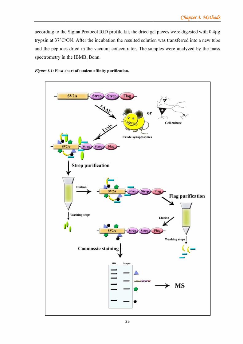
accor
tryps
and t
spect
Figur
rding to the
sin at 37°C/
the peptide
trometry in
re 3.1: Flow c
e Sigma Pro
/ON. After t
s dried in t
the IBMB,
chart of tande
otocol IGD p
the incubati
he vacuum
Bonn.
em affinity pu
35
profile kit, t
ion the resu
concentrat
urification.
the dried ge
ulted solutio
tor. The sam
el pieces we
on was trans
mples were
Chapter
ere digested
sferred into
analyzed b
3. Methods
d with 0.4μg
a new tube
by the mass
s
g
e
s
Chapter 3. Methods
36
3.6 Immunochemical methods
3.6.1 Pre-treatment of primary neurons with various inhibitors
Rat primary cortical neurons were stimulated at DIV19-21 with different phosphatase
inhibitors and at DIV12-14 with a specific inhibitor for SRPKs (table 3.8). As control the
equivalent amount of methanol was used for Calyculin A and okadaic acid, DMSO for
SRPIN340, while for phosSTOP, PBS was added to control cells (1 tablet of phosSTOP in
1ml PBS to obtain a concentration of 10x).
Table 3.8: Phosphatase and kinase inhibitors
Inhibitor Target Concentration Inhibition time
Phosphatase Inhibitors
Calyculin A PP1, PP2A 2nM 30 min Okadaic acid PP1, PP2A 10nM 60 min phosSTOP all 0.1x 60 min
SRPK inhibitor
SRPIN340 SRPK1 SRPK2
10µM 12-16h
After the incubation time, neurons were washed 1x with PBS and fixed in 4%
paraformaldehyde for 5min, followed by immunofluorescence (section 3.6.2).
3.6.2 Immunofluorescence (IF)
Transfected or infected neuronal cultures were fixed for 5min in 4% paraformaldehyde and
4% glucose in PBS, permeabilised with 0.3% Triton X-100 and blocked for 1h/RT in
blocking solution (10% BSA, 1% NGS, 0.1 Triton X-100 in PBS). Neurons were incubated
with primary antibodies at 4°C/ON. Following the incubation, cells were washed three times
with PBS and incubated with the secondary antibodies (section 2.15) for 40min/RT/in dark.
Subsequently to PBS washing, cover-slips were mounted in Mowiol (Sigma) and let to dry
O.N.
3.6.3 Immunohistochemistry (IHC)
Brains were fixed in 4% paraformaldehyde and embedded in paraffin blocks. 4µm brain slices
were deparaffinised in xylene and rehydrated in a series of ethanol baths (100%, 95% 70%
and 50% in PBS) for 2min each. Heat mediated antigen retrieval was performed in citric
buffer (10mM citric acid, pH 6.0), microwaved for 10min and cooled for 30min/RT. Slices
were blocked in PBS with normal goat serum (1:100 to 1:200) and 10% fetal calf serum for
2h in a humidified chamber, followed by incubation with anti-GFP (ab290, 1:100), anti-
FLAG (1:100, Sigma) and anti-SV2A (1:200, 119002 SySy) primary antibodies overnight.
After several washings with PBS, secondary antibodies goat anti-mouse FITC and goat anti-
rabbit Cy3 (Jackson ImmunoResearch) were applied at a concentration of 1:400 and
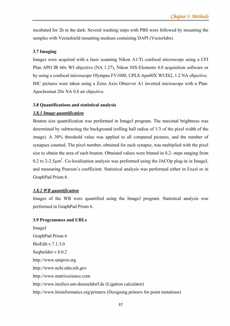
Chapter 3. Methods
37
incubated for 2h in the dark. Several washing steps with PBS were followed by mounting the
samples with Vectashield mounting medium containing DAPI (Vectorlabs).
3.7 Imaging
Images were acquired with a laser scanning Nikon A1/Ti confocal microscope using a CFI
Plan APO IR 60x WI objective (NA 1.27), Nikon NIS-Elements 4.0 acquisition software or
by using a confocal microscope Olympus FV1000, UPLS Apo60X WUIS2, 1.2 NA objective.
IHC pictures were taken using a Zeiss Axio Observer A1 inverted microscope with a Plan-
Apochromat 20x NA 0.8 air objective.
3.8 Quantifications and statistical analysis
3.8.1 Image quantification
Bouton size quantification was performed in ImageJ program. The maximal brightness was
determined by subtracting the background (rolling ball radius of 1/3 of the pixel width of the
image). A 30% threshold value was applied to all compared pictures, and the number of
synapses counted. The pixel number, obtained for each synapse, was multiplied with the pixel
size to obtain the area of each bouton. Obtained values were binned in 0.2- steps ranging from
0.2 to 2-2.5µm2. Co-localization analysis was performed using the JACOp plug-in in ImageJ,
and measuring Pearson’s coefficient. Statistical analysis was performed either in Excel or in
GraphPad Prism 6.
3.8.2 WB quantification
Images of the WB were quantified using the ImageJ program. Statistical analysis was
performed in GraphPad Prism 6.
3.9 Programmes and URLs
ImageJ
GraphPad Prism 6
BioEdit v.7.1.3.0
Seqbuilder v.8.0.2
http://www.uniprot.org
http://www.ncbi.nlm.nih.gov
http://www.matrixscience.com
http://www.insilico.uni-duesseldorf.de (Ligation calculator)
http://www.bioinformatics.org/primerx (Designing primers for point mutations)
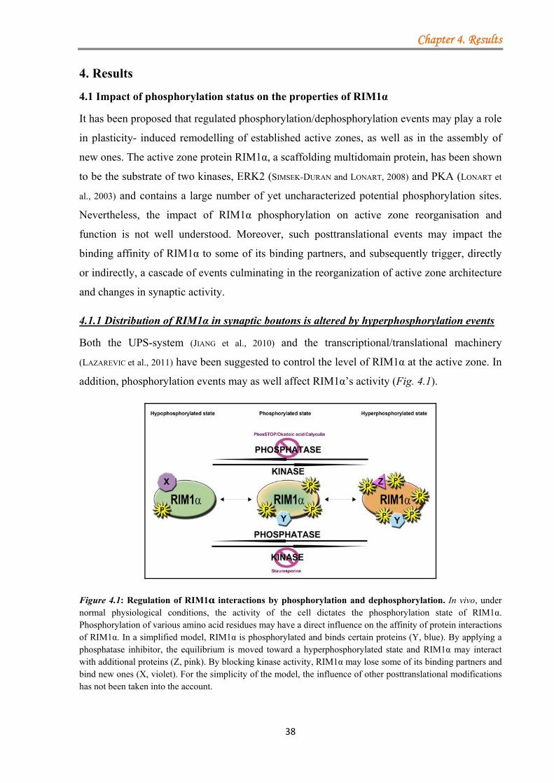
Chapter 4. Results
38
4. Results
4.1 Impact of phosphorylation status on the properties of RIM1α
It has been proposed that regulated phosphorylation/dephosphorylation events may play a role
in plasticity- induced remodelling of established active zones, as well as in the assembly of
new ones. The active zone protein RIM1α, a scaffolding multidomain protein, has been shown
to be the substrate of two kinases, ERK2 (SIMSEK-DURAN and LONART, 2008) and PKA (LONART et
al., 2003) and contains a large number of yet uncharacterized potential phosphorylation sites.
Nevertheless, the impact of RIM1α phosphorylation on active zone reorganisation and
function is not well understood. Moreover, such posttranslational events may impact the
binding affinity of RIM1α to some of its binding partners, and subsequently trigger, directly
or indirectly, a cascade of events culminating in the reorganization of active zone architecture
and changes in synaptic activity.
4.1.1 Distribution of RIM1α in synaptic boutons is altered by hyperphosphorylation events
Both the UPS-system (JIANG et al., 2010) and the transcriptional/translational machinery
(LAZAREVIC et al., 2011) have been suggested to control the level of RIM1α at the active zone. In
addition, phosphorylation events may as well affect RIM1α’s activity (Fig. 4.1).
Figure 4.1: Regulation of RIM1α interactions by phosphorylation and dephosphorylation. In vivo, under normal physiological conditions, the activity of the cell dictates the phosphorylation state of RIM1α. Phosphorylation of various amino acid residues may have a direct influence on the affinity of protein interactions of RIM1α. In a simplified model, RIM1α is phosphorylated and binds certain proteins (Y, blue). By applying a phosphatase inhibitor, the equilibrium is moved toward a hyperphosphorylated state and RIM1α may interact with additional proteins (Z, pink). By blocking kinase activity, RIM1α may lose some of its binding partners and bind new ones (X, violet). For the simplicity of the model, the influence of other posttranslational modifications has not been taken into the account.

level
neuro
phos
distri
for d
follo
Initia
acid3
was q
a non
obser
30mi
versu
the a
p=0.0
The awere measustuden
PP2A
cortic
2 IC50 (P3 IC50 (P
Thus, to
ls and distr
onal cell cu
phorylation
ibution of e
different per
wed by par
ally, two ph3 10nM for
quantified u
n-significan
rved compa
in. (Fig. 4.2
us negative
area size w
016).
area of the boexcluded from
urements wernt (p*˂0.05).
Since the
A, a more
cal neurons
PP1): 2nM; IC50
PP1): 10-15nM;
gain insigh
ribution, th
ulture. To th
n status was
endogenous
riods of tim
aformaldeh
hosphatase
60min. Aft
using the Im
nt increase
ared to the
2A), induce
control (0.2
was observe
outons was binm the measure compared tScale bar: 20µ
e spectrum
general ph
s at DIV21
(PP2A): 0.5-1.0nIC50 (PP2A): 0.1
ht into the m
he effect o
his end diff
s shifted ar
s RIM1α in
me with var
hyde fixation
inhibitors w
ter the incub
mageJ softw
in the num
e methanol
ed an increa
2-0.4µm2, p
ed for bigg
nned in 0.2-strements in ordo the methanoµm (overview
of inhibitio
hosphatase
1 with 0.1x
nM 1nM
39
molecular m
of phosphat
ferent phosp
rtificially to
n boutons, r
rious concen
n and immu
were tested
bation, the b
ware. In the
mber of RIM
control. Ac
ase in the nu
p=0.011). In
ger boutons
tep groups rander to diminisol control and
w) and 10µm (
on of okadai
inhibitor,
x phosSTO
mechanisms
tase inhibit
phatase blo
oward hyper
rat primary
ntrations of
unofluoresce
d: Calyculin
bouton area
case of oka
M1/2 labelle
ccordingly,
umber of sm
nterestingly,
s, above 0
nging from 0-sh staining ard statistical aninsets). N, num
ic acid and
phosSTOP,
OP for up t
that dynam
ors on RIM
ockers were
rphosphory
cortical neu
f phosphatse
ence agains
n A2 2nM fo
a labelled by
adaic acid a
ed boutons w
treatment
mall bouton
, in both cas
.6µm2 (Cal
Figure PP1/PPAbouton endogenocortical n10nM okaand with for 30minendogenoagainst enpresence each apindependeperformedtaken uconfocal m
-0.2 µm2 to 2rtefacts. Withinalysis performber of indep
CalyculinA
, was teste
to 60min r
Chapte
mically regul
M1α was a
applied an
ylation. To a
urons were
e inhibitors
st endogeno
for 30min a
by endogeno
application (
with smalle
with Calyc
ns labelled
ses a small
lyulin A, 0
4.2: The A2 inhibitor
area laous RIM1/2.neurons were adaic acid (A/2nM Calycu
n, followed bous RIM1/2.ndogenous Rof different inpplied inhibent experimd (N=3). All using a lmicroscope (N
2 µm2. Largerin the same srmed using Stpendent experi
A is limited
ed. Stimula
reproduced
r 4. Results
late RIM1α
assessed in
nd RIM1α’s
analyze the
e stimulated
s at DIV21,
us RIM1/2.
and okadaic
ous RIM1/2
(Fig. 4.2B),
er area was
culin A for
by RIM1/2
decrease in
0.6-0.8µm2,
effect ofrs on theabelled by Primary rattreated with
/C) for 60minulin A. (B/D)by IF against. (A/B) IF
RIM1/2 in thenhibitors. Forbitor, three
ments werepictures wereaser-scanningNikon A1/Ti).r bouton sizessize group thetudent’s t-testiments.
to PP1 and
ation of rat
the effects
s
α
n
s
e
d
,
.
c
2
,
s
r
2
n
,
f e y t h n ) t
F e r e e e g . s e t
d
t
s
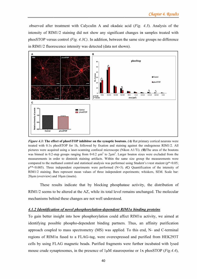
obse
inten
phos
in RI
Figurtreatepicturwas bmeasucompp**˂0RIM120µm
RIM
mech
4.1.2
To g
ident
appro
regio
cells
mous
erved after
nsity of RIM
STOP versu
IM1/2 fluor
re 4.3: The efd with 0.1x pres were acqubinned in 0.2-urements in oared to the me0.005). Three1/2 staining. B
m (overview) a
These re
M1/2 seems t
hanisms beh
2 Identificat
gain better i
tifying pos
oach couple
ons of RIM
by using F
se crude syn
treatment
M1/2 stainin
us control (F
rescence int
ffect of phosSphosSTOP fo
uired using a l-step groups rorder to dimiethanol contro
e independentBars represenand 10µm (ins
esults indic
to be altered
hind these c
tion of nove
insight into
ssible phosp
ed to mass
M1α fused to
FLAG magn
naptosomes
with Calyc
ng did not
(Fig. 4.3C).
ensity was d
STOP inhibitoor 1h, followelaser-scanningranging from inish stainingol and statistict experimentsnt mean valueets).
cate that b
d at the AZ,
changes are
el phosphor
o how phos
pho-depend
spectromet
o a FLAG-
netic beads
s, in the pre
40
culin A an
show any
In addition
detected (da
or on the syned by fixationg confocal mi0-0.2 µm2 to
g artifacts. Wcal analysis ws were perfores of three in
by blocking
, while its to
not well un
rylation-dep
sphorylation
dent bindin
try (MS) w
-tag, were o
s. Purified f
esence of 1μ
d okadaic
significant
n, between th
ata not show
naptic boutonn and staininicroscope (Niko 2µm2. Large
Within the samwas performedrmed (N=3). ndependent ex
g phosphat
otal level re
nderstood.
pendent RIM
n could affe
ng partners
was applied.
overexpress
fragments w
μM staurosp
acid (Fig.
changes in
he same siz
wn).
s. (A) Rat primg against thekon A1/Ti). (Ber bouton sizeme size groupd using Studen
(C) Quantifixperiments; w
ase activity
emains unch
IM1α bindin
ect RIM1α
. Thus, an
. To this en
sed and pur
were further
porine or 1x
Chapte
4.3). Anal
n samples tr
ze groups no
mary cortical e endogenous (B)The area oes were exclu
up the measurnt’s t-test studication of thewhiskers, SEM
y, the dist
hanged. The
ng proteins
activity, w
n affinity p
nd, N- and
rified from
r incubated
x phosSTOP
r 4. Results
ysis of the
reated with
o difference
neurons wereRIM1/2. All
of the boutonsuded from therements were
dent (p*˂0.05;e intensity ofM. Scale bar:
ribution of
e molecular
we aimed at
purification
C-terminal
HEK293T
with lysed
P (Fig.4.4),
s
e
h
e
e l s e e ; f :
f
r
t
n
l
T
d
,
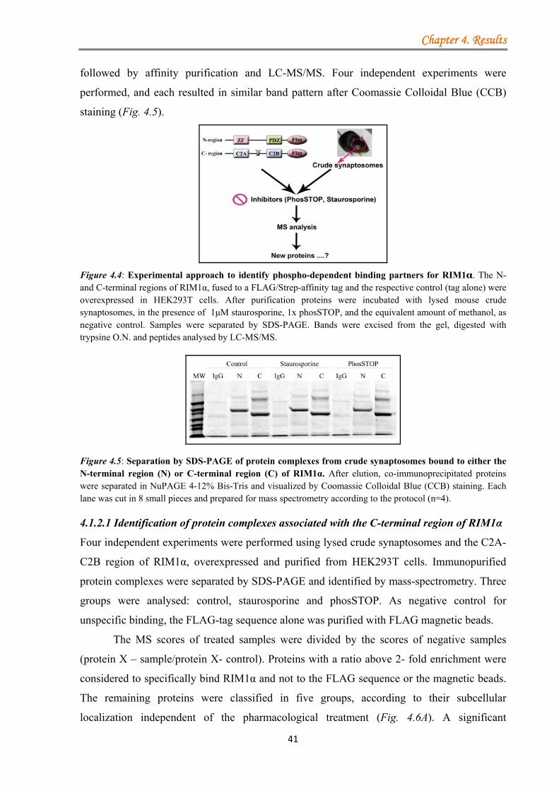
Chapter 4. Results
41
followed by affinity purification and LC-MS/MS. Four independent experiments were
performed, and each resulted in similar band pattern after Coomassie Colloidal Blue (CCB)
staining (Fig. 4.5).
Figure 4.4: Experimental approach to identify phospho-dependent binding partners for RIM1α. The N- and C-terminal regions of RIM1α, fused to a FLAG/Strep-affinity tag and the respective control (tag alone) were overexpressed in HEK293T cells. After purification proteins were incubated with lysed mouse crude synaptosomes, in the presence of 1μM staurosporine, 1x phosSTOP, and the equivalent amount of methanol, as negative control. Samples were separated by SDS-PAGE. Bands were excised from the gel, digested with trypsine O.N. and peptides analysed by LC-MS/MS.
Figure 4.5: Separation by SDS-PAGE of protein complexes from crude synaptosomes bound to either the N-terminal region (N) or C-terminal region (C) of RIM1α. After elution, co-immunoprecipitated proteins were separated in NuPAGE 4-12% Bis-Tris and visualized by Coomassie Colloidal Blue (CCB) staining. Each lane was cut in 8 small pieces and prepared for mass spectrometry according to the protocol (n=4). 4.1.2.1 Identification of protein complexes associated with the C-terminal region of RIM1α
Four independent experiments were performed using lysed crude synaptosomes and the C2A-
C2B region of RIM1α, overexpressed and purified from HEK293T cells. Immunopurified
protein complexes were separated by SDS-PAGE and identified by mass-spectrometry. Three
groups were analysed: control, staurosporine and phosSTOP. As negative control for
unspecific binding, the FLAG-tag sequence alone was purified with FLAG magnetic beads.
The MS scores of treated samples were divided by the scores of negative samples
(protein X – sample/protein X- control). Proteins with a ratio above 2- fold enrichment were
considered to specifically bind RIM1α and not to the FLAG sequence or the magnetic beads.
The remaining proteins were classified in five groups, according to their subcellular
localization independent of the pharmacological treatment (Fig. 4.6A). A significant
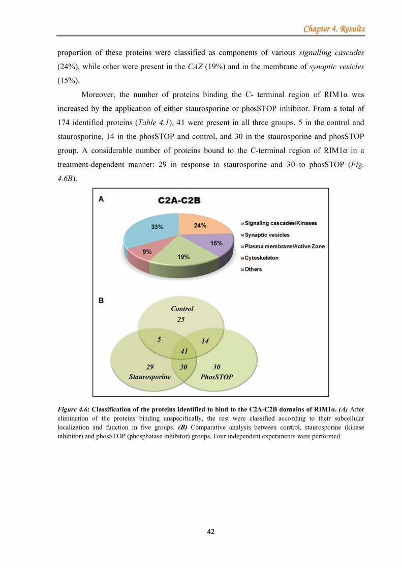
propo
(24%
(15%
incre
174 i
staur
group
treatm
4.6B
Figureliminlocalizinhibi
ortion of th
%), while ot
%).
Moreove
eased by the
identified p
rosporine, 1
p. A consid
ment-depen
).
re 4.6: Classination of thezation and fuitor) and phos
hese protein
ther were pr
er, the num
e applicatio
proteins (Ta
14 in the ph
derable num
ndent mann
fication of the proteins binunction in fivSTOP (phosp
ns were cla
resent in the
mber of pro
on of either
able 4.1), 41
hosSTOP an
mber of pro
ner: 29 in r
he proteins idnding unspecve groups. (Bhatase inhibit
42
assified as
e CAZ (19%
oteins bindi
r staurospor
1 were pres
nd control,
oteins boun
response to
dentified to bcifically, the B) Comparatitor) groups. Fo
component
%) and in th
ing the C-
rine or phos
sent in all t
and 30 in
nd to the C
o staurospo
bind to the C2rest were clive analysis our independe
ts of variou
he membran
terminal r
sSTOP inhi
hree groups
the staurosp
C-terminal r
rine and 3
2A-C2B domassified accobetween cont
ent experiment
Chapte
us signallin
ne of synap
region of R
ibitor. From
s, 5 in the
porine and
region of R
0 to phosS
mains of RIMording to theitrol, staurosp
nts were perfor
r 4. Results
ng cascades
ptic vesicles
RIM1α was
m a total of
control and
phosSTOP
RIM1α in a
STOP (Fig.
M1α. (A) Afterir subcellular
porine (kinasermed.
s
s
s
s
f
d
P
a
r r e
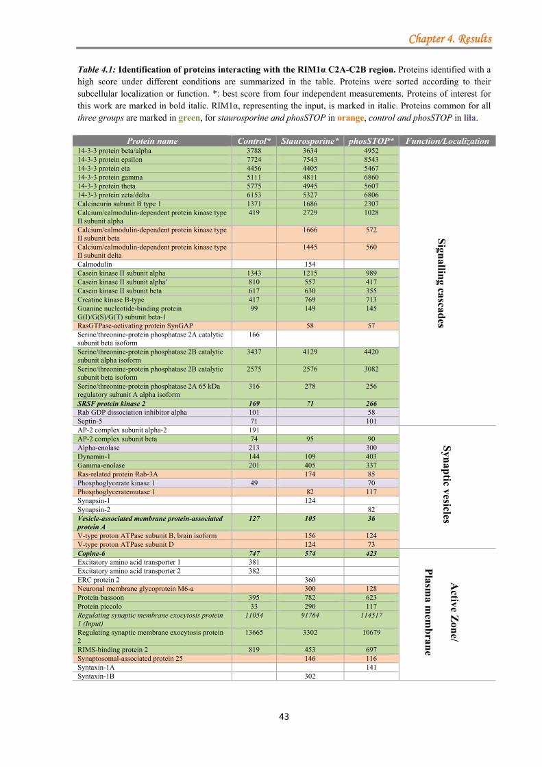
Chapter 4. Results
43
Table 4.1: Identification of proteins interacting with the RIM1α C2A-C2B region. Proteins identified with a high score under different conditions are summarized in the table. Proteins were sorted according to their subcellular localization or function. *: best score from four independent measurements. Proteins of interest for this work are marked in bold italic. RIM1α, representing the input, is marked in italic. Proteins common for all three groups are marked in green, for staurosporine and phosSTOP in orange, control and phosSTOP in lila.
Protein name Control* Staurosporine* phosSTOP* Function/Localization 14-3-3 protein beta/alpha 3788 3634 4952
Sign
alling cascad
es
14-3-3 protein epsilon 7724 7543 8543 14-3-3 protein eta 4456 4405 5467 14-3-3 protein gamma 5111 4811 6860 14-3-3 protein theta 5775 4945 5607 14-3-3 protein zeta/delta 6153 5327 6806 Calcineurin subunit B type 1 1371 1686 2307 Calcium/calmodulin-dependent protein kinase type II subunit alpha
419 2729 1028
Calcium/calmodulin-dependent protein kinase type II subunit beta
1666 572
Calcium/calmodulin-dependent protein kinase type II subunit delta
1445 560
Calmodulin 154 Casein kinase II subunit alpha 1343 1215 989 Casein kinase II subunit alpha' 810 557 417 Casein kinase II subunit beta 617 630 355 Creatine kinase B-type 417 769 713 Guanine nucleotide-binding protein G(I)/G(S)/G(T) subunit beta-1
99 149 145
RasGTPase-activating protein SynGAP 58 57 Serine/threonine-protein phosphatase 2A catalytic subunit beta isoform
166
Serine/threonine-protein phosphatase 2B catalytic subunit alpha isoform
3437 4129 4420
Serine/threonine-protein phosphatase 2B catalytic subunit beta isoform
2575 2576 3082
Serine/threonine-protein phosphatase 2A 65 kDa regulatory subunit A alpha isoform
316 278 256
SRSF protein kinase 2 169 71 266 Rab GDP dissociation inhibitor alpha 101 58 Septin-5 71 101 AP-2 complex subunit alpha-2 191
Syn
aptic vesicles
AP-2 complex subunit beta 74 95 90 Alpha-enolase 213 300 Dynamin-1 144 109 403 Gamma-enolase 201 405 337 Ras-related protein Rab-3A 174 85 Phosphoglycerate kinase 1 49 70 Phosphoglyceratemutase 1 82 117 Synapsin-1 124 Synapsin-2 82 Vesicle-associated membrane protein-associated protein A
127 105 36
V-type proton ATPase subunit B, brain isoform 156 124 V-type proton ATPase subunit D 124 73 Copine-6 747 574 423
A
ctive Zon
e/
Plasm
a mem
bran
e
Excitatory amino acid transporter 1 381 Excitatory amino acid transporter 2 382 ERC protein 2 360 Neuronal membrane glycoprotein M6-a 300 128 Protein bassoon 395 782 623 Protein piccolo 33 290 117 Regulating synaptic membrane exocytosis protein 1 (Input)
11054 91764 114517
Regulating synaptic membrane exocytosis protein 2
13665 3302 10679
RIMS-binding protein 2 819 453 697 Synaptosomal-associated protein 25 146 116 Syntaxin-1A 141 Syntaxin-1B 302
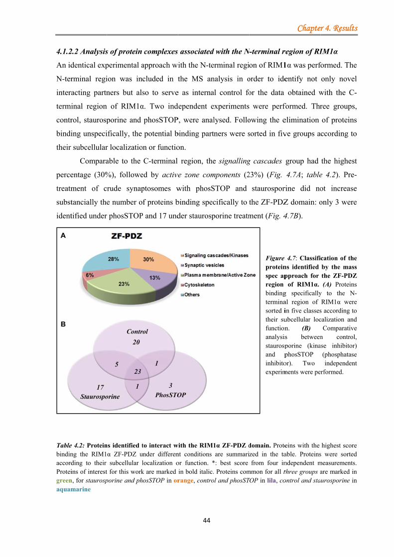
4.1.2
An id
N-ter
intera
termi
contr
bindi
their
perce
treatm
subst
ident
TablebindinaccordProteigreenaquam
2.2 Analysis
dentical exp
rminal regi
acting partn
inal region
rol, staurosp
ing unspeci
subcellular
Compara
entage (30%
ment of c
tancially the
tified under
e 4.2: Proteinng the RIM1ding to their ins of interest
n, for staurospmarine
s of protein
perimental a
ion was in
ners but als
n of RIM1α
porine and
ifically, the
r localizatio
able to the
%), followe
rude synap
e number o
r phosSTOP
ns identified tα ZF-PDZ usubcellular l
t for this workporine and ph
complexes
approach wi
cluded in t
so to serve
α. Two ind
phosSTOP
potential b
on or functio
C-terminal
ed by active
ptosomes w
f proteins b
P and 17 und
to interact wunder differenlocalization ok are marked hosSTOP in or
44
associated
ith the N-te
the MS an
e as interna
dependent e
P, were anal
binding part
on.
region, the
e zone com
with phosS
binding spec
der staurosp
with the RIM1nt conditions or function. *in bold italic.range, contro
d with the N
erminal regi
nalysis in o
al control fo
experiments
lysed. Follo
tners were s
e signalling
mponents (2
STOP and
cifically to
porine treatm
1α ZF-PDZ dare summari
*: best score . Proteins comol and phosST
N-terminal r
on of RIM1
order to ide
or the data
were perf
owing the e
sorted in fiv
g cascades
23%) (Fig.
staurospor
the ZF-PDZ
ment (Fig. 4
Figure proteinspec apregion bindingterminasorted itheir sufunctionanalysisstaurospand pinhibitoexperim
domain. Protezed in the tafrom four in
mmon for all tTOP in lila, co
Chapte
region of RI
1α was perf
entify not
obtained w
formed. Thr
elimination
ve groups a
group had
4.7A; table
rine did no
Z domain: o
4.7B).
4.7: Classifins identified pproach for
of RIM1α. g specificallyal region of in five classesubcellular locn. (B) s betweenporine (kinaphosSTOP or). Two ments were pe
eins with the able. Proteinsndependent mthree groups aontrol and sta
r 4. Results
IM1α
formed. The
only novel
with the C-
ree groups,
of proteins
ccording to
the highest
e 4.2). Pre-
ot increase
only 3 were
cation of theby the massthe ZF-PDZ(A) Proteins
y to the N-RIM1α were
s according tocalization and
Comparativen control,
ase inhibitor)(phosphataseindependent
erformed.
highest scores were sortedmeasurements.are marked inurosporine in
s
e
l
-
,
s
o
t
-
e
e
e s
Z s - e o d e , ) e t
e d . n n
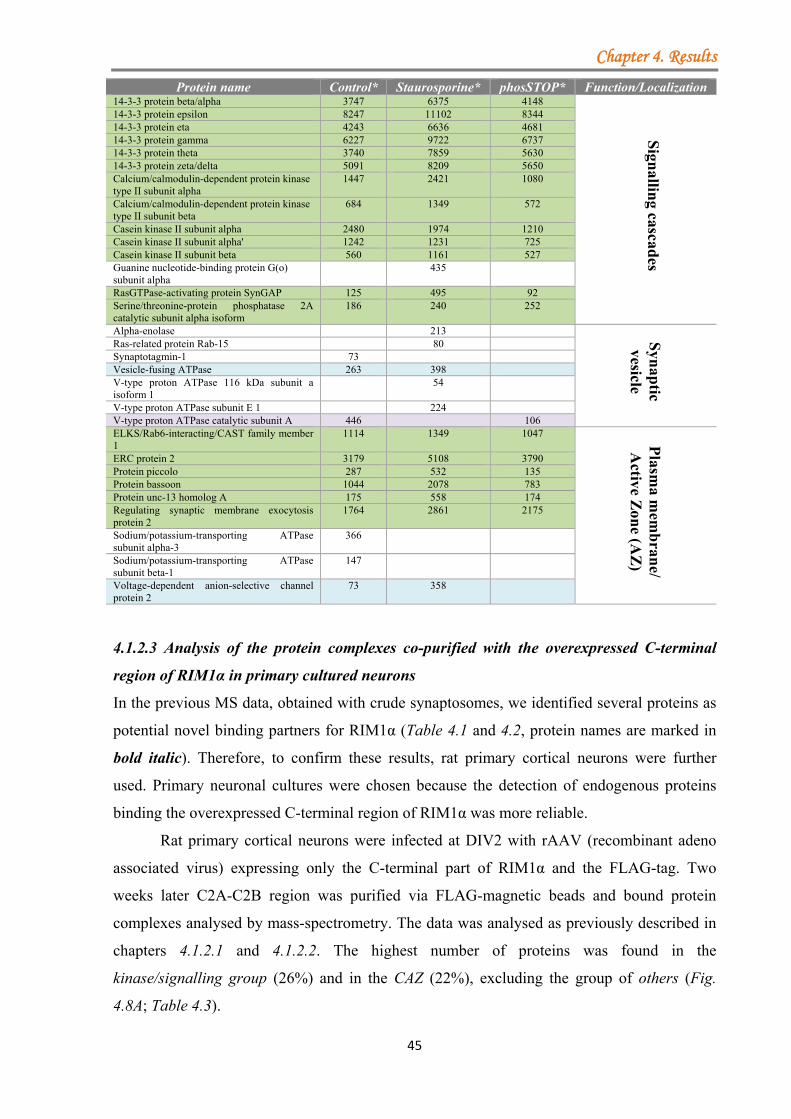
Chapter 4. Results
45
4.1.2.3 Analysis of the protein complexes co-purified with the overexpressed C-terminal
region of RIM1α in primary cultured neurons
In the previous MS data, obtained with crude synaptosomes, we identified several proteins as
potential novel binding partners for RIM1α (Table 4.1 and 4.2, protein names are marked in
bold italic). Therefore, to confirm these results, rat primary cortical neurons were further
used. Primary neuronal cultures were chosen because the detection of endogenous proteins
binding the overexpressed C-terminal region of RIM1α was more reliable.
Rat primary cortical neurons were infected at DIV2 with rAAV (recombinant adeno
associated virus) expressing only the C-terminal part of RIM1α and the FLAG-tag. Two
weeks later C2A-C2B region was purified via FLAG-magnetic beads and bound protein
complexes analysed by mass-spectrometry. The data was analysed as previously described in
chapters 4.1.2.1 and 4.1.2.2. The highest number of proteins was found in the
kinase/signalling group (26%) and in the CAZ (22%), excluding the group of others (Fig.
4.8A; Table 4.3).
Protein name Control* Staurosporine* phosSTOP* Function/Localization 14-3-3 protein beta/alpha 3747 6375 4148
Sign
alling cascad
es
14-3-3 protein epsilon 8247 11102 8344 14-3-3 protein eta 4243 6636 4681 14-3-3 protein gamma 6227 9722 6737 14-3-3 protein theta 3740 7859 5630 14-3-3 protein zeta/delta 5091 8209 5650 Calcium/calmodulin-dependent protein kinase type II subunit alpha
1447 2421 1080
Calcium/calmodulin-dependent protein kinase type II subunit beta
684 1349 572
Casein kinase II subunit alpha 2480 1974 1210 Casein kinase II subunit alpha' 1242 1231 725 Casein kinase II subunit beta 560 1161 527 Guanine nucleotide-binding protein G(o) subunit alpha
435
RasGTPase-activating protein SynGAP 125 495 92 Serine/threonine-protein phosphatase 2A catalytic subunit alpha isoform
186 240 252
Alpha-enolase 213 Syn
aptic
vesicle
Ras-related protein Rab-15 80 Synaptotagmin-1 73 Vesicle-fusing ATPase 263 398 V-type proton ATPase 116 kDa subunit a isoform 1
54
V-type proton ATPase subunit E 1 224 V-type proton ATPase catalytic subunit A 446 106 ELKS/Rab6-interacting/CAST family member 1
1114 1349 1047
Plasm
a mem
bran
e/ A
ctive Zon
e (AZ
)
ERC protein 2 3179 5108 3790 Protein piccolo 287 532 135 Protein bassoon 1044 2078 783 Protein unc-13 homolog A 175 558 174 Regulating synaptic membrane exocytosis protein 2
1764 2861 2175
Sodium/potassium-transporting ATPase subunit alpha-3
366
Sodium/potassium-transporting ATPase subunit beta-1
147
Voltage-dependent anion-selective channel protein 2
73 358

prim
More
to ha
4.8B
also
(P/Q
pharm
as in
FigurAfter localizC2B rwhile distribPM-Aindep
A compa
mary neuron
eover, prote
ave the high
).
Addition
identified in
- and N- typ
Taken t
macologica
dependent n
re 4.8: Novel bunspecific bozation. (B) Cregion. Data d
in blue foubution of the
AZ, plasma mendent experi
arative anal
s, for the R
eins belongi
hest probab
nally, RIM1
n the measu
pe) could no
together, af
al treatment
novel protei
binding protound proteins omparative andepicted in orur independengroups is dep
membrane andiments.
lysis betwe
RIM1α C2-d
ing to the ki
bility of bin
1α’s known
urements (T
ot be detect
ffinity puri
s resulted in
in interactio
ein for the RIwere excludenalysis of therange represennt measureme
picted in perced active zone
46
een percenta
domains, re
inase/signa
nding, direct
binding pa
Table 4.3). H
ted using th
ification co
n the identi
ons for RIM
RIM1α C2A-Cd, the remaini
e distribution nts the co-IP eents with cruentages. SC, se group; C,
ages acquir
evealed a s
lling group
tly or indire
artners such
However, th
ese method
oupled to
ification of
M1α.
C2B domain ping ones wereof different g
experiment peude synaptossignalling casccytoskeletal
ed with cru
imilar distr
and the act
ectly, the C
h as, liprins-
he voltage-g
s and reage
mass-spectr
f both phosp
purified frome classified accgroups obtainerformed with
somes are shcade group; Sgroup; O, ot
Chapte
ude synapto
ribution of
tive zone gr
C-terminal r
-α and RIM
gated calciu
ents.
rometry an
pho-depend
m rat cortical cording to theed using the rat cortical ne
hown (N=4). SV, synaptic vther group; N
r 4. Results
osomes and
the groups.
roup tended
region (Fig.
M-BPs were
um channels
nalysis and
dent as well
neurons. (A)eir subcellularRIM1α C2A-eurons (N=1),The relative
vesicle group;N, number of
s
d
.
d
e
s
d
l
) r -, e ; f
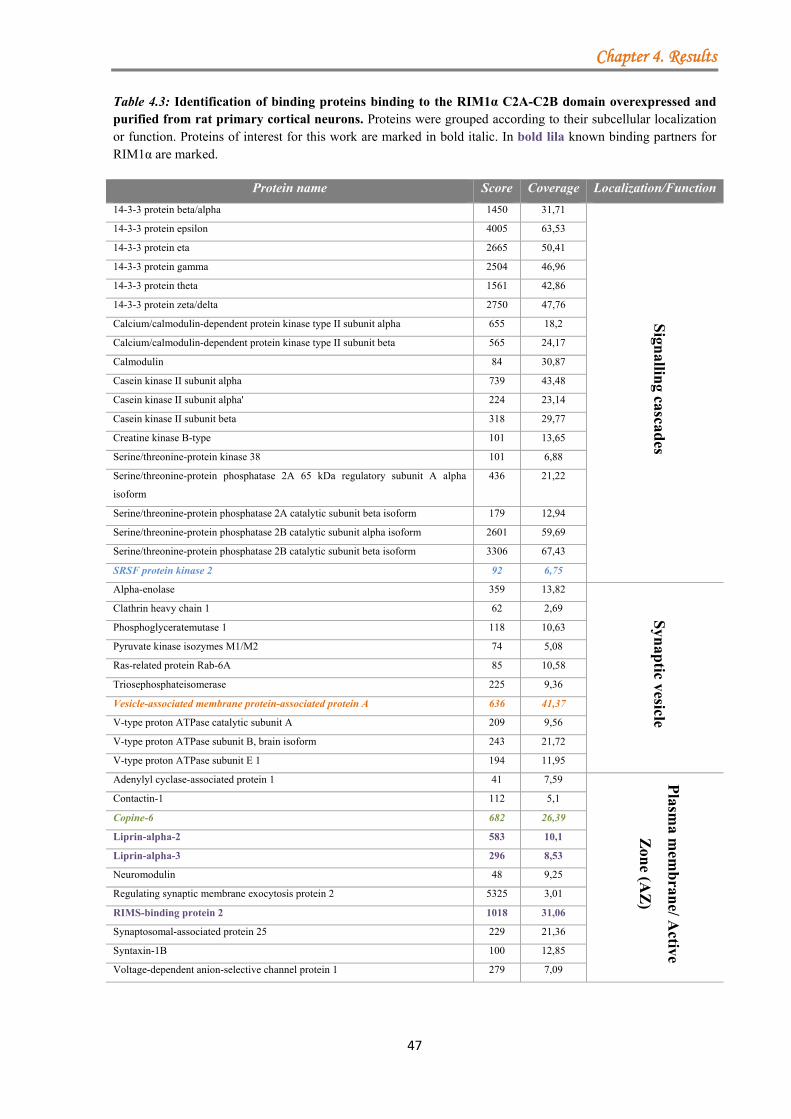
Chapter 4. Results
47
Table 4.3: Identification of binding proteins binding to the RIM1α C2A-C2B domain overexpressed and purified from rat primary cortical neurons. Proteins were grouped according to their subcellular localization or function. Proteins of interest for this work are marked in bold italic. In bold lila known binding partners for RIM1α are marked.
Protein name Score Coverage Localization/Function
14-3-3 protein beta/alpha 1450 31,71
Sign
alling cascad
es
14-3-3 protein epsilon 4005 63,53
14-3-3 protein eta 2665 50,41
14-3-3 protein gamma 2504 46,96
14-3-3 protein theta 1561 42,86
14-3-3 protein zeta/delta 2750 47,76
Calcium/calmodulin-dependent protein kinase type II subunit alpha 655 18,2
Calcium/calmodulin-dependent protein kinase type II subunit beta 565 24,17
Calmodulin 84 30,87
Casein kinase II subunit alpha 739 43,48
Casein kinase II subunit alpha' 224 23,14
Casein kinase II subunit beta 318 29,77
Creatine kinase B-type 101 13,65
Serine/threonine-protein kinase 38 101 6,88
Serine/threonine-protein phosphatase 2A 65 kDa regulatory subunit A alpha
isoform
436 21,22
Serine/threonine-protein phosphatase 2A catalytic subunit beta isoform 179 12,94
Serine/threonine-protein phosphatase 2B catalytic subunit alpha isoform 2601 59,69
Serine/threonine-protein phosphatase 2B catalytic subunit beta isoform 3306 67,43
SRSF protein kinase 2 92 6,75
Alpha-enolase 359 13,82
Syn
aptic vesicle
Clathrin heavy chain 1 62 2,69
Phosphoglyceratemutase 1 118 10,63
Pyruvate kinase isozymes M1/M2 74 5,08
Ras-related protein Rab-6A 85 10,58
Triosephosphateisomerase 225 9,36
Vesicle-associated membrane protein-associated protein A 636 41,37
V-type proton ATPase catalytic subunit A 209 9,56
V-type proton ATPase subunit B, brain isoform 243 21,72
V-type proton ATPase subunit E 1 194 11,95
Adenylyl cyclase-associated protein 1 41 7,59 Plasm
a mem
bran
e/ Active
Zon
e (AZ
)
Contactin-1 112 5,1
Copine-6 682 26,39
Liprin-alpha-2 583 10,1
Liprin-alpha-3 296 8,53
Neuromodulin 48 9,25
Regulating synaptic membrane exocytosis protein 2 5325 3,01
RIMS-binding protein 2 1018 31,06
Synaptosomal-associated protein 25 229 21,36
Syntaxin-1B 100 12,85
Voltage-dependent anion-selective channel protein 1 279 7,09
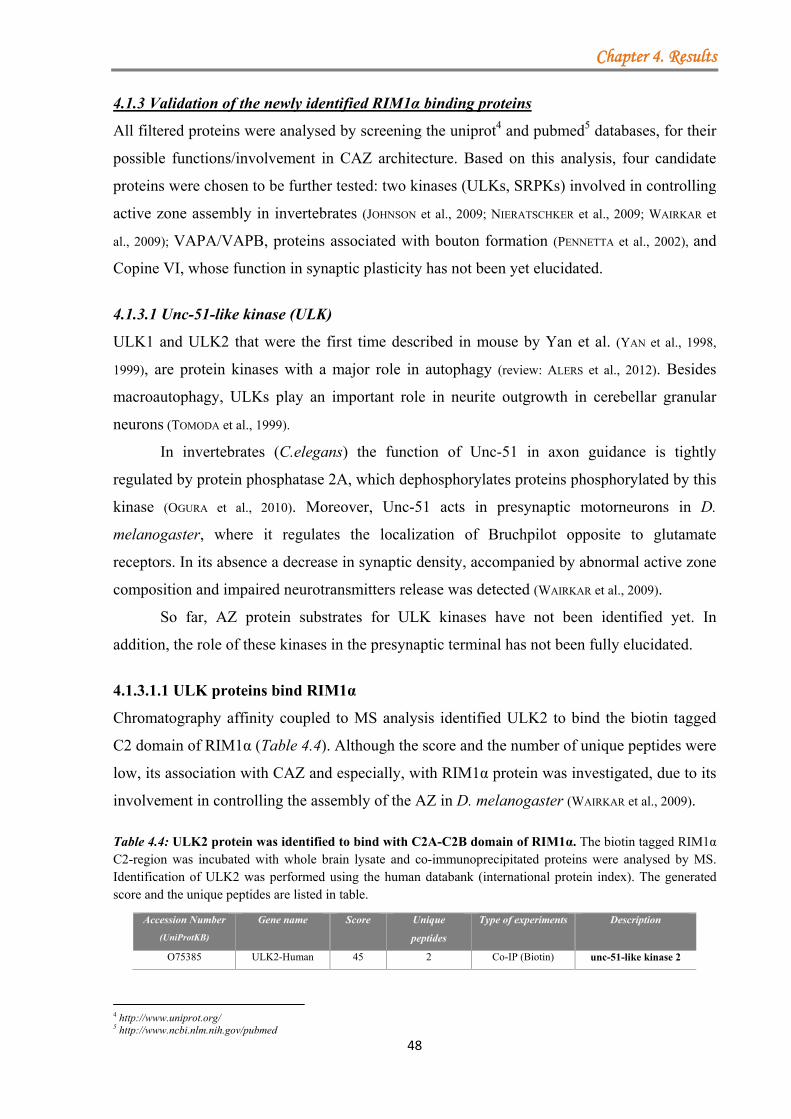
Chapter 4. Results
48
4.1.3 Validation of the newly identified RIM1α binding proteins
All filtered proteins were analysed by screening the uniprot4 and pubmed5 databases, for their
possible functions/involvement in CAZ architecture. Based on this analysis, four candidate
proteins were chosen to be further tested: two kinases (ULKs, SRPKs) involved in controlling
active zone assembly in invertebrates (JOHNSON et al., 2009; NIERATSCHKER et al., 2009; WAIRKAR et
al., 2009); VAPA/VAPB, proteins associated with bouton formation (PENNETTA et al., 2002), and
Copine VI, whose function in synaptic plasticity has not been yet elucidated.
4.1.3.1 Unc-51-like kinase (ULK)
ULK1 and ULK2 that were the first time described in mouse by Yan et al. (YAN et al., 1998,
1999), are protein kinases with a major role in autophagy (review: ALERS et al., 2012). Besides
macroautophagy, ULKs play an important role in neurite outgrowth in cerebellar granular
neurons (TOMODA et al., 1999).
In invertebrates (C.elegans) the function of Unc-51 in axon guidance is tightly
regulated by protein phosphatase 2A, which dephosphorylates proteins phosphorylated by this
kinase (OGURA et al., 2010). Moreover, Unc-51 acts in presynaptic motorneurons in D.
melanogaster, where it regulates the localization of Bruchpilot opposite to glutamate
receptors. In its absence a decrease in synaptic density, accompanied by abnormal active zone
composition and impaired neurotransmitters release was detected (WAIRKAR et al., 2009).
So far, AZ protein substrates for ULK kinases have not been identified yet. In
addition, the role of these kinases in the presynaptic terminal has not been fully elucidated.
4.1.3.1.1 ULK proteins bind RIM1α
Chromatography affinity coupled to MS analysis identified ULK2 to bind the biotin tagged
C2 domain of RIM1α (Table 4.4). Although the score and the number of unique peptides were
low, its association with CAZ and especially, with RIM1α protein was investigated, due to its
involvement in controlling the assembly of the AZ in D. melanogaster (WAIRKAR et al., 2009).
Table 4.4: ULK2 protein was identified to bind with C2A-C2B domain of RIM1α. The biotin tagged RIM1α C2-region was incubated with whole brain lysate and co-immunoprecipitated proteins were analysed by MS. Identification of ULK2 was performed using the human databank (international protein index). The generated score and the unique peptides are listed in table.
4 http://www.uniprot.org/ 5 http://www.ncbi.nlm.nih.gov/pubmed
Accession Number
(UniProtKB)
Gene name Score Unique
peptides
Type of experiments Description
O75385 ULK2-Human 45 2 Co-IP (Biotin) unc-51-like kinase 2
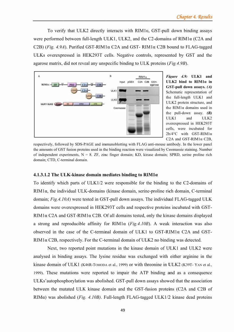
were
C2B)
ULK
agaro
respecthe amof inddomai 4.1.3
To id
RIM
doma
doma
RIM
a str
obser
RIM
analy
kinas
1999)
ULK
betw
RIM
To verif
e performed
) (Fig. 4.9A
Ks overexpr
ose matrix,
ctively, followmounts of GSTdependent expin; CTD, C-te
3.1.2 The U
dentify whi
M1α, the indi
ain; Fig.4.1
ains were o
M1α C2A and
rong and re
rved in the
M1α C2B, res
Next, tw
ysed in bin
se domain o
). These m
Ks’autophos
ween the mu
Mα) was abo
fy that ULK
d between fu
A). Purified
ressed in H
did not reve
wed by SDS-PT fusion proteperiments, N erminal domai
ULK-kinase
ich parts of
ividual ULK
10A) were te
overexpresse
d GST-RIM
eproducible
e case of th
spectively.
wo reported
nding assay
of ULK1 (K
mutations we
sphorylation
utated ULK
olished (Fig
K2 directly
ull-length U
GST-RIM1
HEK293T c
eal any unsp
PAGE and imeins used in th
= 8. ZF, zinin.
e domain m
f ULK1/2 w
K-domains
ested in GS
ed in HEK2
M1α C2B. O
e affinity fo
he C-termin
For the C-te
point muta
s. The lysin
K46R-TOMODA
ere reporte
n was abolis
K kinase do
g. 4.10B). F
49
interacts w
ULK1, ULK
1α C2A and
cells. Negat
pecific bind
mmunoblottinghe binding reanc finger dom
mediates bin
were respon
(kinase dom
T-pull dow
293T cells a
Of all domai
for RIM1α
nal domain
erminal dom
ations in th
ne residue
A et al., 1999)
ed to impai
shed. GST-p
omain and
Full-length F
with RIM1α
K2, and the
d GST- RIM
tive contro
ding to ULK
g with FLAG action were vismain; KD, kin
nding to RI
nsible for th
main, serine
wn assays. T
and respect
ins tested, o
(Fig.4.10B
n of ULK1
main of ULK
he kinase do
was excha
) or with thr
ir the ATP
pull down a
the GST-fu
FLAG-tagg
α, GST-pull
C2-domain
M1α C2B b
ls, represen
K proteins (F
FiULGSctheULthetheULovce2hC2
anti-mouse ansualized by Co
nase domain;
IM1α
he binding
e-proline ric
he individu
ive proteins
only the kin
B). A weak
to GST-RI
K2 no bindi
omain of U
anged with
reonine in U
P binding a
assays show
usion protei
ged ULK1/2
Chapte
l down bind
ns of RIM1α
bound to FL
nted by GS
Fig.4.9B).
igure 4.9: LK2 bind to
GST-pull dowchematic repr
he full-lengthLK2 protein
he RIM1α domhe pull-downLK1 andverexpressed ells, were inh/4°C with 2A and GST-ntibody. In thoomassie stainSPRD, serine
to the C2-
ch domain,
ual FLAG-ta
s incubated
nase domain
k interaction
IM1α C2A
ing was det
ULK1 and U
either argi
ULK2 (K39T
and as a co
wed that the
ins (C2A a
2 kinase de
r 4. Results
ding assays
α (C2A and
LAG-tagged
ST and the
ULK1 ando RIM1α inn assays. (A)resentation of
h ULK1 andstructure, andmains used inn assay. (B)d ULK2in HEK293Tncubated forGST-RIM1α
-RIM1α C2B,e lower panelning. Numbere proline rich
domains of
C-terminal
agged ULK
with GST-
ns displayed
n was also
A and GST-
tected.
ULK2 were
nine in the
T- YAN et al.,
onsequence
association
and C2B of
ead proteins
s
s
d
d
e
d n ) f d d n ) 2 T r α , l r h
f
l
K
-
d
o
-
e
e
,
e
n
f
s
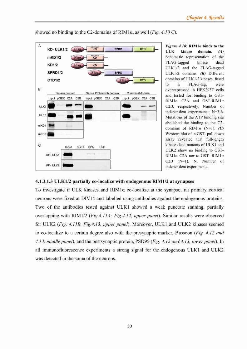
show
4.1.3
To in
neuro
Two
overl
for U
to co
4.13,
all im
was d
wed no bind
3.1.3 ULK1
nvestigate i
ons were fi
of the an
lapping wit
ULK2 (Fig.
o-localize to
, middle pan
mmunofluo
detected in
ing to the C
/2 partially
if ULK kin
ixed at DIV
ntibodies te
th RIM1/2 (
4.11B, Fig
o a certain
nel), and the
rescence ex
the soma of
C2-domains
y co-localiz
nases and R
V14 and lab
sted agains
(Fig.4.11A;
g.4.13, uppe
degree also
e postsynap
xperiments
f the neuron
50
of RIM1α,
ze with end
RIM1α co-l
elled using
st ULK1 s
; Fig.4.12,
er panel). M
o with the p
ptic protein,
a strong si
ns.
as well (Fig
ogenous RI
localize at
antibodies
howed a w
upper pane
Moreover, U
presynaptic
PSD95 (Fi
ignal for th
g. 4.10 C).
Figure 4ULK SchematFLAG-tULK1/2ULK1/2domainsto aoverexpand testRIM1α C2B, rindepenMutationabolishedomainsWesternassay rkinase dULK2 sRIM1α C2B (indepen
IM1/2 at sy
the synapse
against the
weak punct
el). Similar
ULK1 and U
marker, B
ig. 4.12 and
he endogeno
Chapte
4.10: RIM1αkinase do
atic representtagged kin2 and the F2 domains. (s of ULK1/2 ka FLAG-tpressed in HEsted for bind
C2A and respectively.
ndent experimons of the ATPed the bindins of RIM1αn blot of a GSrevealed thadead mutants show no bindC2A nor to
(N=1). N, ndent experime
ynapses
e, rat prima
e endogenou
tate stainin
results wer
ULK2 kina
assoon (Fig
d 4.13, lowe
ous ULK1
r 4. Results
α binds to theomain. (A)tation of thenase deadFLAG-tagged(B) Differentkinases, fusedtag, wereEK293T cellsding to GST-
GST-RIM1αNumber of
ments, N=3-6.P binding site
ng to the C2-α (N=1). (C)ST- pull downat full-lengthof ULK1 andding to GST-GST- RIM1αNumber of
ents.
ary cortical
us proteins.
g, partially
re observed
ases seemed
g. 4.12 and
er panel). In
and ULK2
s
e ) e d d t d e s -α f . e -) n h d - α f
l
.
y
d
d
d
n
2
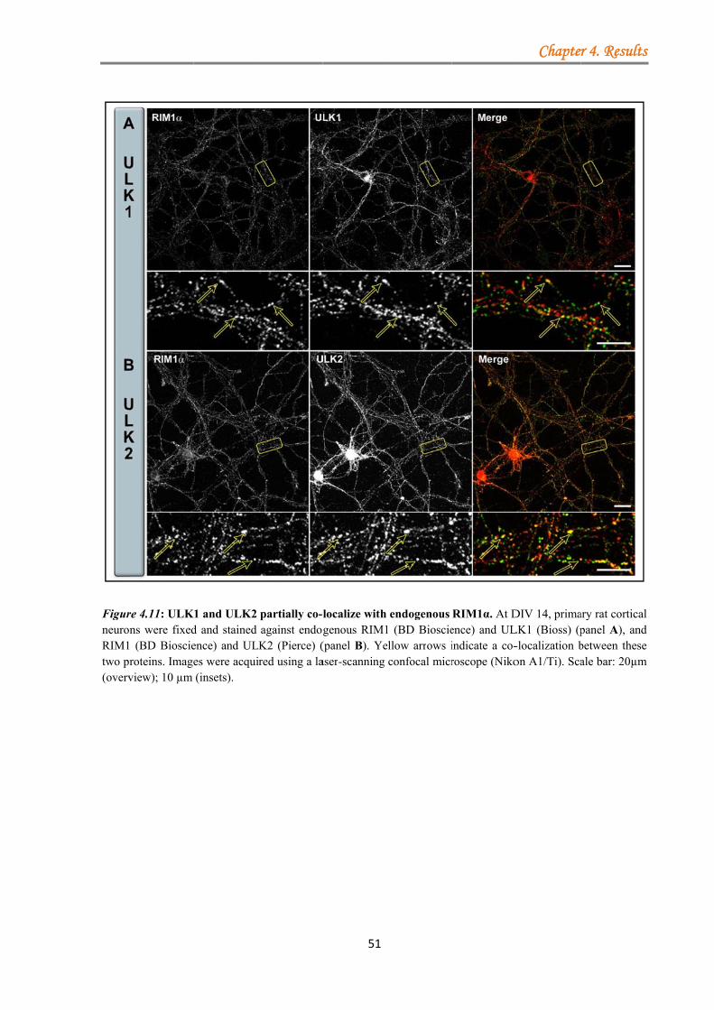
FigurneuroRIM1two p(overv
re 4.11: ULKons were fixed1 (BD Bioscieroteins. Imageview); 10 µm
K1 and ULK2 d and stained ence) and ULes were acqui(insets).
partially co-lagainst endog
LK2 (Pierce) (ired using a la
51
localize with genous RIM1(panel B). Yeaser-scanning
endogenous 1 (BD Bioscieellow arrows iconfocal micr
RIM1α. At Dence) and ULindicate a co-roscope (Niko
Chapte
DIV 14, primaLK1 (Bioss) (p-localization bon A1/Ti). Sca
r 4. Results
ary rat corticalpanel A), andbetween theseale bar: 20µm
s
l d e
m
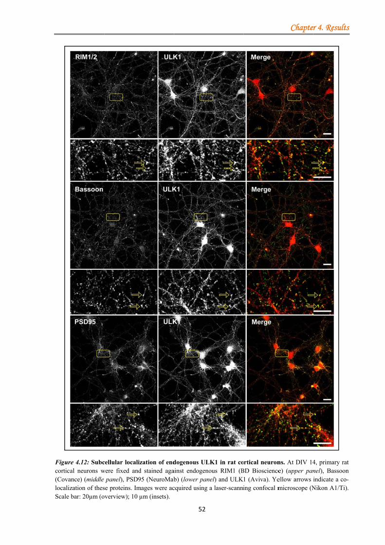
Figurcortic(CovalocalizScale
re 4.12: Subccal neurons wance) (middle zation of thesbar: 20µm (o
cellular localiwere fixed and
panel), PSD9se proteins. Imoverview); 10
ization of endd stained aga95 (NeuroMab
mages were acµm (insets).
52
dogenous ULainst endogenob) (lower pancquired using
LK1 in rat coous RIM1 (B
nel) and ULKa laser-scanni
rtical neuronBD Bioscience1 (Aviva). Yeing confocal m
Chapte
ns. At DIV 14e) (upper panellow arrows microscope (N
r 4. Results
4, primary ratnel), Bassoonindicate a co-Nikon A1/Ti).
s
t n -.
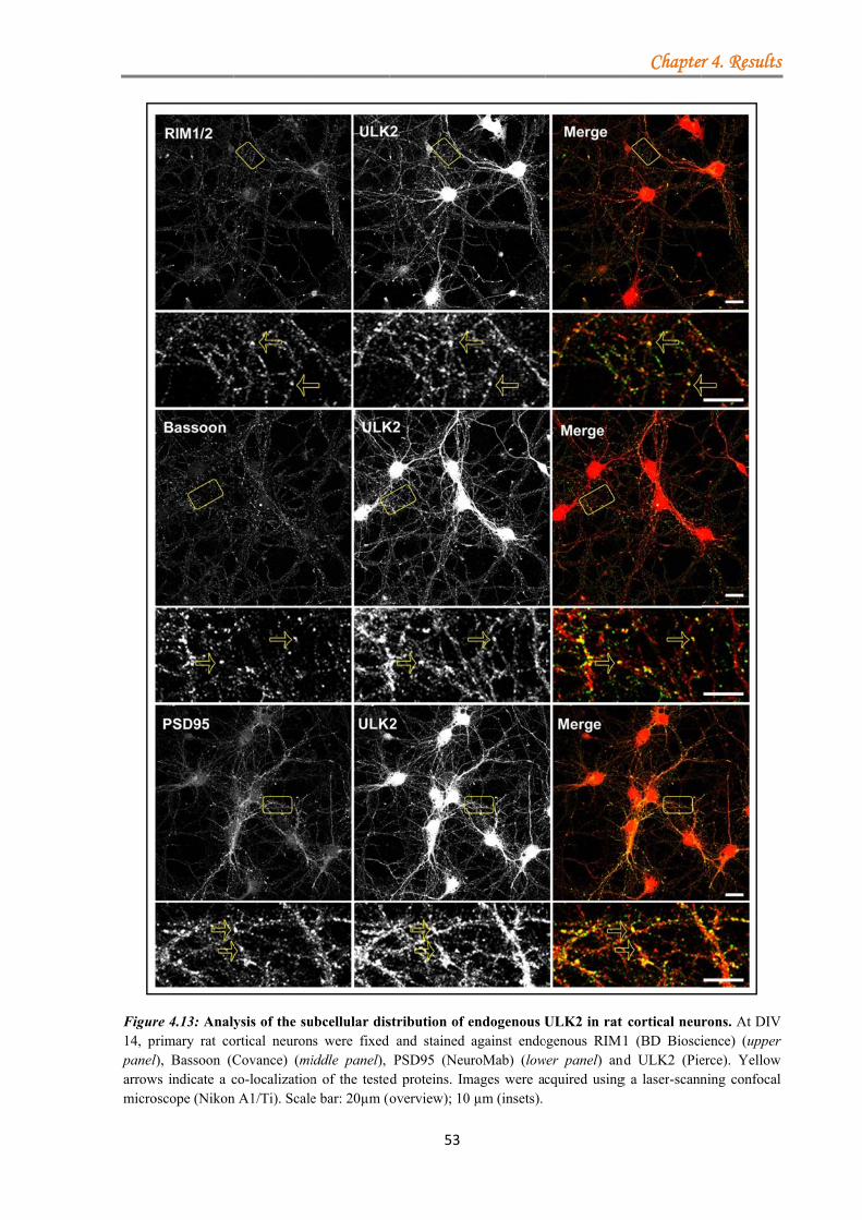
Figur14, prpanel)arrowmicro
re 4.13: Analyrimary rat co
l), Bassoon (Cws indicate a coscope (Nikon
ysis of the suortical neuronCovance) (mico-localizationn A1/Ti). Scale
ubcellular disns were fixed iddle panel), n of the testede bar: 20µm (o
53
stribution of and stained PSD95 (Neu
d proteins. Imoverview); 10
endogenous Uagainst endo
uroMab) (lowmages were ac0 µm (insets).
ULK2 in rat ogenous RIM1wer panel) and
cquired using
Chapte
cortical neu1 (BD Biosci
nd ULK2 (Pieg a laser-scann
r 4. Results
rons. At DIVience) (uppererce). Yellowning confocal
s
V r
w l
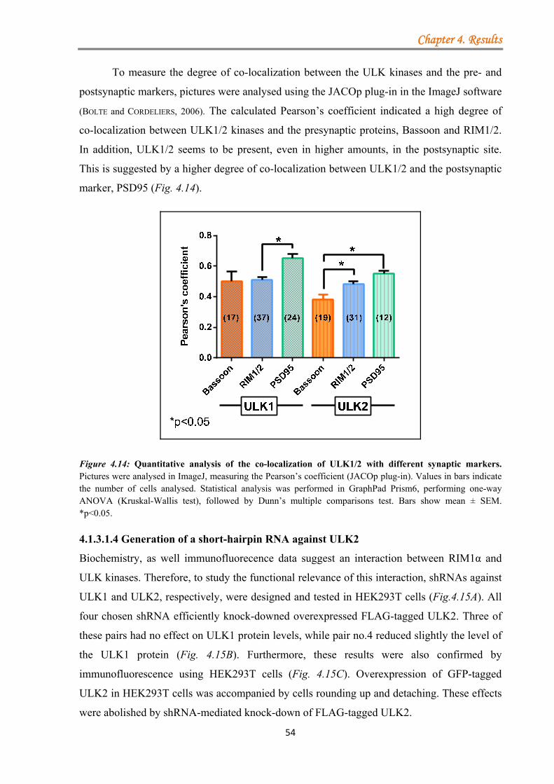
Chapter 4. Results
54
To measure the degree of co-localization between the ULK kinases and the pre- and
postsynaptic markers, pictures were analysed using the JACOp plug-in in the ImageJ software
(BOLTE and CORDELIERS, 2006). The calculated Pearson’s coefficient indicated a high degree of
co-localization between ULK1/2 kinases and the presynaptic proteins, Bassoon and RIM1/2.
In addition, ULK1/2 seems to be present, even in higher amounts, in the postsynaptic site.
This is suggested by a higher degree of co-localization between ULK1/2 and the postsynaptic
marker, PSD95 (Fig. 4.14).
Figure 4.14: Quantitative analysis of the co-localization of ULK1/2 with different synaptic markers. Pictures were analysed in ImageJ, measuring the Pearson’s coefficient (JACOp plug-in). Values in bars indicate the number of cells analysed. Statistical analysis was performed in GraphPad Prism6, performing one-way ANOVA (Kruskal-Wallis test), followed by Dunn’s multiple comparisons test. Bars show mean ± SEM. *p<0.05.
4.1.3.1.4 Generation of a short-hairpin RNA against ULK2
Biochemistry, as well immunofluorecence data suggest an interaction between RIM1α and
ULK kinases. Therefore, to study the functional relevance of this interaction, shRNAs against
ULK1 and ULK2, respectively, were designed and tested in HEK293T cells (Fig.4.15A). All
four chosen shRNA efficiently knock-downed overexpressed FLAG-tagged ULK2. Three of
these pairs had no effect on ULK1 protein levels, while pair no.4 reduced slightly the level of
the ULK1 protein (Fig. 4.15B). Furthermore, these results were also confirmed by
immunofluorescence using HEK293T cells (Fig. 4.15C). Overexpression of GFP-tagged
ULK2 in HEK293T cells was accompanied by cells rounding up and detaching. These effects
were abolished by shRNA-mediated knock-down of FLAG-tagged ULK2.
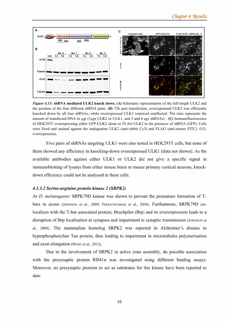
Figurthe poknockamounof HEwere overex
them
avail
immu
down
4.1.3
At D
bars
local
disru
al., 2
hype
and a
with
More
date.
re 4.15: shRNosition of the ked down by nt of transfect
EK293T overefixed and staxpression.
Five pair
m showed an
lable antibo
unoblotting
n efficiency
3.2 Serine-a
D. melanoga
in axons (
lizes with th
uption of Br
2009). The
erphosphory
axon elonga
Due to t
the presy
eover, no p
NA mediated four differenall four shRNted DNA in µexpressing eitained against
rs of shRNA
ny efficiency
odies again
g of lysates f
y could not b
arginine pro
aster SRPK
(JOHNSON et
he T-bar ass
rp localizati
mammalia
ylate Tau pr
ation (HONG
the involvem
ynaptic pro
presynaptic
ULK2 knocknt shRNA pairNAs, while ovµgr (1µgr ULKther GFP-ULKthe endogeno
As targeting
y in knocki
nst either U
from either
be analysed
otein kinase
K79D kinase
al., 2009; N
sociated pro
ion at synap
an homolog
rotein, thus
et al., 2012).
ment of SR
otein RIM1
proteins to
55
k down. (A) Srs. (B) 72h poverexpressed K2 or ULK1, K2 alone or Fous ULK2 (a
g ULK1 we
ing-down ov
ULK1 or
mouse brai
d in these ce
e 2 (SRPK2
e was show
NIERATSCHKE
otein, Bruch
pses and imp
g SRPK2
leading to
RPK2 in ac
1α was inv
act as subs
Schematic repost transfectioULK1 remainand 3 and 6 µ
FLAG-ULK2 anti-rabbit Cy3
ere also teste
verexpresse
ULK2 did
in or mouse
ells.
2)
wn to preven
ER et al., 200
hpilot (Brp)
pairment in
was repor
impairmen
ctive zone a
vestigated
strates for t
presentation ofon, overexpresned unaffectedµgr shRNAs) in the presenc3) and FLAG
ed in HEK2
ed ULK1 (d
d not give
e primary co
nt the prem
09). Further
) and its ove
n synaptic tr
rted in Al
nt in microt
assembly, it
using diffe
this kinase
Chapte
f the full-lengssed ULK2 w
ed. The ratio r. (C) Immuno
nce of shRNAG (anti-mouse
293T cells, b
data not show
a specific
ortical neuro
mature form
rmore, SRP
erexpression
ransmission
lzheimer’s
tubules poly
ts possible
ferent bindi
have been
r 4. Results
gth ULK2 andwas efficientlyrepresents theofluorescence
A (GFP). Cellse FITC). O.E,
but none of
wn). As the
c signal in
ons, knock-
mation of T-
PK79D co-
n leads to a
n (JOHNSON et
disease to
ymerisation
association
ing assays.
reported to
s
d y e e s ,
f
e
n
-
-
-
a
t
o
n
n
.
o
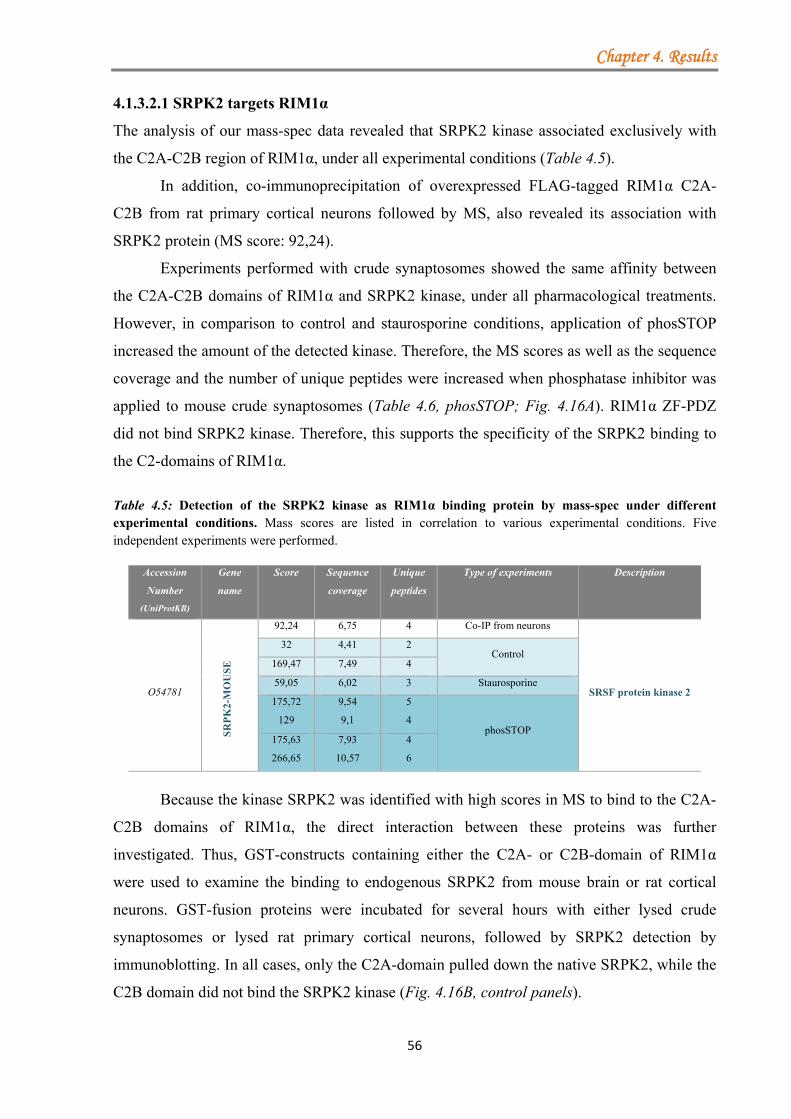
Chapter 4. Results
56
4.1.3.2.1 SRPK2 targets RIM1α
The analysis of our mass-spec data revealed that SRPK2 kinase associated exclusively with
the C2A-C2B region of RIM1α, under all experimental conditions (Table 4.5).
In addition, co-immunoprecipitation of overexpressed FLAG-tagged RIM1α C2A-
C2B from rat primary cortical neurons followed by MS, also revealed its association with
SRPK2 protein (MS score: 92,24).
Experiments performed with crude synaptosomes showed the same affinity between
the C2A-C2B domains of RIM1α and SRPK2 kinase, under all pharmacological treatments.
However, in comparison to control and staurosporine conditions, application of phosSTOP
increased the amount of the detected kinase. Therefore, the MS scores as well as the sequence
coverage and the number of unique peptides were increased when phosphatase inhibitor was
applied to mouse crude synaptosomes (Table 4.6, phosSTOP; Fig. 4.16A). RIM1α ZF-PDZ
did not bind SRPK2 kinase. Therefore, this supports the specificity of the SRPK2 binding to
the C2-domains of RIM1α.
Table 4.5: Detection of the SRPK2 kinase as RIM1α binding protein by mass-spec under different experimental conditions. Mass scores are listed in correlation to various experimental conditions. Five independent experiments were performed.
Accession
Number
(UniProtKB)
Gene
name
Score Sequence
coverage
Unique
peptides
Type of experiments Description
O54781
SR
PK
2-M
OU
SE
92,24 6,75 4 Co-IP from neurons
SRSF protein kinase 2
32 4,41 2 Control
169,47 7,49 4
59,05 6,02 3 Staurosporine
175,72
129
9,54
9,1
5
4 phosSTOP
175,63
266,65
7,93
10,57
4
6
Because the kinase SRPK2 was identified with high scores in MS to bind to the C2A-
C2B domains of RIM1α, the direct interaction between these proteins was further
investigated. Thus, GST-constructs containing either the C2A- or C2B-domain of RIM1α
were used to examine the binding to endogenous SRPK2 from mouse brain or rat cortical
neurons. GST-fusion proteins were incubated for several hours with either lysed crude
synaptosomes or lysed rat primary cortical neurons, followed by SRPK2 detection by
immunoblotting. In all cases, only the C2A-domain pulled down the native SRPK2, while the
C2B domain did not bind the SRPK2 kinase (Fig. 4.16B, control panels).
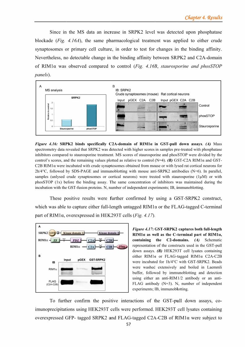
block
synap
Neve
of R
pane
FigurspectrinhibicontroC2B R2h/4°CsamplphosSincub
whic
part o
imun
overe
Since in
kade (Fig.
ptosomes o
ertheless, no
RIM1α was
els).
re 4.16: SRPrometry data ritors compareol’s scores, anRIM1α were iC, followed bles (unlysed STOP (1x) beation with the
These p
ch was able
of RIM1α, o
To furth
noprecipitat
expressed G
n the MS d
4.16A), th
or primary
o detectable
observed
PK2 binds srevealed that Sd to staurospond the remainiincubated withby SDS-PAGcrude synaptefore the binde GST-fusion p
ositive resu
to capture e
overexpress
her confirm
ions using H
GFP- tagged
data an incr
he same ph
cell culture
e change in
compared t
pecifically CSRPK2 was dorine treatmening values ploh crude synap
GE and immunosomes or coding assay. Tproteins. N, n
ults were f
either full-l
sed in HEK
m the posit
HEK293T c
d SRPK2 a57
rease in SR
harmacologi
e, in order
n the bindin
to control
C2A-domain detected with hnt. MS scoresotted as relativptosomes obtainoblotting wiortical neuronThe same connumber of inde
further con
length untag
K293T cells
FRcrdewwbuFe
tive interac
cells were p
and FLAG-t
RPK2 level
ical treatme
to test for
g affinity b
(Fig. 4.16B
of RIM1α ihigher scores of staurosporve to control ined from moith mouse antns) were treancentration ofependent expe
nfirmed by
gged RIM1α
(Fig. 4.17).
Figure 4.17: GRIM1α as wcontaining representationdown assays.either RIM1αwere incubatewere washedbuffer, followusing either FLAG antiboexperiments; I
ctions of t
performed.
tagged C2A
was detec
ent was ap
changes in
between SRP
B, staurosp
in GST-pull in samples prrine and phos(N=4). (B) Guse or with lyti-SRPK2 antiated with stauf inhibitors weriments; IB, i
using a G
α or the FLA
GST-SRPK2ell as the C-the C2-do
n of the constr(B) HEK293
α or FLAG-ted for 1h/4°Cd extensively wed by immuan anti-RIM
ody (N=3). NIB, immunobl
the GST-pu
HEK293T
A-C2B of R
Chapte
cted upon p
pplied to e
n the bindin
RPK2 and C
porine and
down assayre-treated withsSTOP were d
GST-C2A RIMysed rat corticaibodies (N=6urosporine (1was maintaineimmunoblottin
GST-SRPK2
LAG-tagged
2 captures bot-terminal paromains. (A)ructs used in 3T cell lysattagged RIM1
C with GST-Sand boiled
unoblotting aM1/2 antibodyN, number oflotting.
ull down a
cell lysates
RIM1α were
r 4. Results
phosphatase
ither crude
ng affinity.
2A-domain
phosSTOP
ys. (A) Massh phosphatasedivided by the
M1α and GST-al neurons for). In parallel,µM) or with
ed during theng.
2 construct,
C-terminal
th full-lengthrt of RIM1α,) Schematicthe GST-pulles containing1α C2A-C2BRPK2. Beadsin Laemmli
and detectiony or an anti-f independent
assays, co-
s containing
e subject to
s
e
e
.
n
P
s e e -r ,
h e
,
l
h , c l g B s i n -t
-
g
o
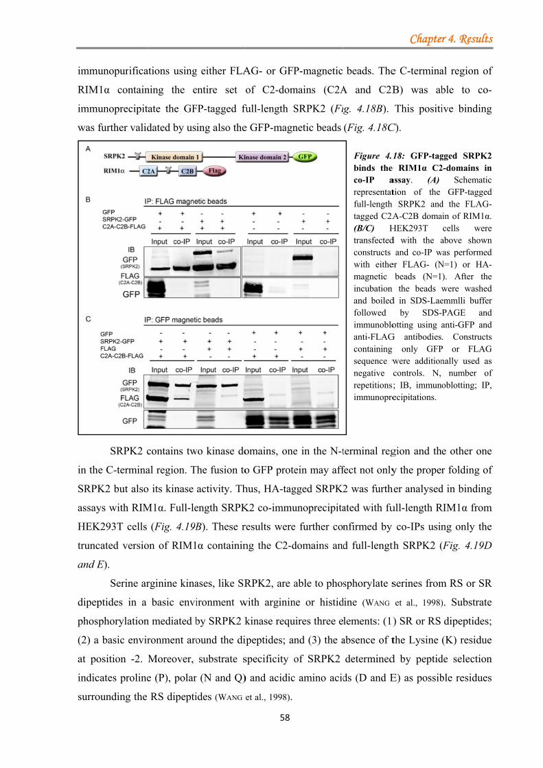
immu
RIM
immu
was f
in th
SRPK
assay
HEK
trunc
and E
dipep
phos
(2) a
at po
indic
surro
unopurifica
M1α contain
unoprecipit
further valid
SRPK2 c
e C-termina
K2 but also
ys with RIM
K293T cells
cated versio
E).
Serine ar
ptides in a
phorylation
a basic envir
osition -2.
cates proline
ounding the
ations using
ning the e
tate the GF
dated by us
contains tw
al region. T
o its kinase
M1α. Full-le
(Fig. 4.19B
on of RIM1
rginine kina
basic envi
n mediated b
ronment aro
Moreover,
e (P), polar
RS dipepti
g either FLA
entire set o
FP-tagged fu
ing also the
wo kinase do
The fusion to
activity. Th
ength SRPK
B). These r
α containin
ases, like SR
ironment w
by SRPK2 k
ound the di
substrate s
r (N and Q)
ides (WANG
58
AG- or GFP
of C2-dom
full-length S
e GFP-magn
omains, one
o GFP prote
hus, HA-tag
K2 co-immu
results were
ng the C2-d
RPK2, are a
with arginin
kinase requ
ipeptides; an
specificity o
) and acidic
et al., 1998).a
P-magnetic
mains (C2A
SRPK2 (Fi
netic beads
e in the N-t
ein may aff
gged SRPK2
unoprecipita
e further con
domains and
able to phos
ne or histid
uires three e
nd (3) the a
of SRPK2
c amino acid
aaaaaaaaaaa
beads. The
A and C2B
g. 4.18B).
(Fig. 4.18C
Figure 4.1binds the co-IP arepresentatfull-length tagged C2A(B/C) Htransfectedconstructs with eithemagnetic incubation and boiledfollowed immunobloanti-FLAGcontaining sequence wnegative crepetitionsimmunopre
terminal reg
fect not only
2 was furth
ated with fu
nfirmed by
d full-lengt
sphorylate s
dine (WANG
lements: (1)
absence of t
determined
ds (D and E
aaaaaaaaaaa
Chapte
e C-termina
B) was ab
This positi
C).
18: GFP-tage RIM1α C2assay. (A) tion of the
h SRPK2 andA-C2B doma
HEK293T d with the a
and co-IP wer FLAG- (N
beads (N=1n the beads wd in SDS-Lae
by SDS-otting using a
G antibodiesg only GFPwere additioncontrols. N, ; IB, immunecipitations.
gion and th
y the proper
her analysed
ull-length R
co-IPs usin
th SRPK2 (
serines from
et al., 1998)
) SR or RS
the Lysine
d by peptid
E) as possib
aaaaaaaaaaa
r 4. Results
al region of
ble to co-
ive binding
gged SRPK22-domains in
SchematicGFP-tagged
d the FLAG-ain of RIM1α.
cells wereabove shown
was performedN=1) or HA-). After thewere washedemmlli bufferPAGE andanti-GFP and. Constructs
P or FLAGnally used as
number ofoblotting; IP,
e other one
r folding of
d in binding
RIM1α from
ng only the
(Fig. 4.19D
m RS or SR
). Substrate
dipeptides;
(K) residue
de selection
ble residues
aaaaaaaaaaa
s
f
-
g
2 n c d -. e n d - e d r d d s
G s f ,
e
f
g
m
e
D
R
e
;
e
n
s
a
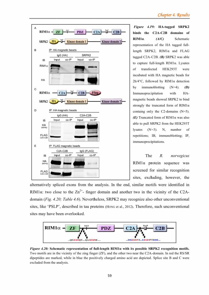
altern
RIM
doma
sites,
sites
FigurTwo mdipeptexclud
natively spl
M1α: two clo
ain (Fig. 4.2
, like ‘PSLP
may have b
re 4.20: Schemotifs are in ttides are markded from the a
liced exons
ose to the Z
20; Table 4
P’, describe
been overlo
matic represthe vicinity ofked, while in analysis.
s from the
Zn2+- finger
.6). Neverth
ed in tau pro
oked.
sentation of ff the zing fingblue the posi
59
analysis. In
r domain a
heless, SRP
oteins (HONG
full-length RIger (ZF), and titively charge
n the end,
and another
PK2 may rec
G et al., 2012)
IM1α with itthe other two ed amino acid
Figure 4.1
binds the
RIM1α.
representation
length SRPK
tagged C2A-
to capture fu
of transfec
incubated wi
2h/4°C, follo
by immun
Immunopreci
magnetic bea
strongly the
containg onl
(E) Truncate
able to pull S
lysates (N
repetitions;
immunopreci
The
RIM1α pr
screened fo
sites, excl
similar mot
two in the
cognize also
). Therefore
ts possible SRnear the C2A
d are depicted.
Chapte
19: HA-tagg
C2A-C2B
(A/C)
n of the HA
K2, RIM1α
-C2B. (B) SRP
ull-length RIM
cted HEK2
ith HA magne
owed by RIM
noblotting (
ipitation w
ads showed SR
truncated for
ly the C2-dom
ed form of RIM
SRPK2 from t
N=3). N,
IB, immuno
ipitations.
e R.
rotein sequ
for similar
luding, how
tifs were id
e vicinity o
o other unco
e, such unco
RPK2 recognA-domain. In r
. Splice site B
r 4. Results
ged SRPK2
domains of
Schematic
A tagged full-
and FLAG
PK2 was able
M1α. Lysates
293T were
etic beads for
M1α detection
(N=4). (D)
with HA-
RPK2 to bind
rm of RIM1α
mains (N=5).
M1α was also
the HEK293T
number of
oblotting; IP,
norvegicus
uence was
recognition
wever, the
dentified in
f the C2A-
onventional
onventional
nition motifs.red the RS/SRB and C were
s
2
f
c
-
G
e
s
e
r
n
)
-
d
α
.
o
T
f
,
s
s
n
e
n
-
l
l
. R e
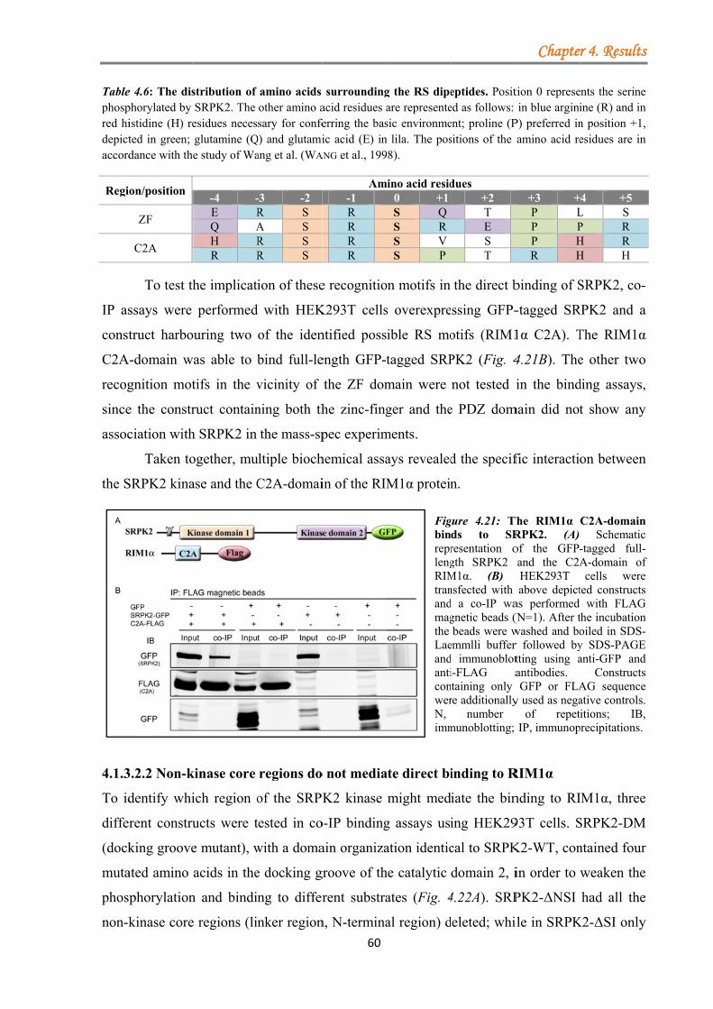
Tablephospred hidepictaccord
Regi
IP as
const
C2A
recog
since
assoc
the S
4.1.3
To id
diffe
(dock
muta
phos
non-k
e 4.6: The disphorylated by istidine (H) reted in green; dance with the
on/position
ZF
C2A
To test th
ssays were
truct harbo
A-domain w
gnition mot
e the constr
ciation with
Taken to
SRPK2 kina
3.2.2 Non-k
dentify whi
rent constru
king groove
ated amino
phorylation
kinase core
stribution of SRPK2. The esidues necesglutamine (Qe study of Wa
-4 -E RQ AH RR R
he implicati
performed
uring two
as able to b
tifs in the v
ruct contain
h SRPK2 in
ogether, mu
ase and the C
kinase core
ich region o
ucts were t
e mutant), w
acids in the
n and bindin
e regions (lin
amino acids other amino asary for confe
Q) and glutamiang et al. (WA
-3 -2 R S A S R S R S
ion of these
with HEK
of the iden
bind full-le
vicinity of
ning both th
the mass-sp
ultiple bioch
C2A-domai
regions do
of the SRPK
ested in co
with a doma
e docking g
ng to differ
nker region
60
surroundingacid residues aferring the basic acid (E) in
ANG et al., 199
Am-1 R R R R
e recognitio
K293T cells
ntified poss
ength GFP-t
the ZF dom
he zinc-fing
pec experim
hemical assa
in of the RIM
o not media
K2 kinase
o-IP binding
ain organiza
groove of th
rent substra
n, N-termina
g the RS dipeare representesic environmen lila. The pos98).
mino acid resid0 +1S QS RS VS P
on motifs in
s overexpre
ible RS mo
tagged SRP
main were
ger and the
ments.
ays revealed
M1α protein
FigbinreprlengRIMtranandmagthe LaeandanticonwerN, imm
ate direct bi
might med
g assays usi
ation identic
he catalytic
ates (Fig. 4
al region) d
eptides. Posited as follows: ent; proline (Psitions of the
dues +2 T E S T
the direct b
ssing GFP-
otifs (RIM1
PK2 (Fig. 4
not tested i
e PDZ dom
d the specif
n.
gure 4.21: Tds to SRresentation ogth SRPK2 M1α. (B)nsfected with d a co-IP wagnetic beads (beads were w
emmlli bufferd immunobloti-FLAG a
ntaining only re additionally
number munoblotting;
inding to R
iate the bin
ing HEK29
cal to SRPK
domain 2, i
4.22A). SRP
deleted; whi
Chapte
tion 0 represein blue argini
P) preferred inamino acid re
+3 +P LP PP HR H
binding of S
-tagged SR
1α C2A). T
4.21B). The
in the bind
main did not
fic interacti
The RIM1α RPK2. (A) of the GFP-and the C2AHEK293T above depict
as performed (N=1). After twashed and bor followed bytting using aantibodies. GFP or FLA
y used as negaof repet
IP, immunop
RIM1α
nding to RI
93T cells. S
K2-WT, con
in order to
PK2-ΔNSI
ile in SRPK
r 4. Results
ents the serineine (R) and inn position +1,esidues are in
+4 +5 L S P R H R H H
SRPK2, co-
RPK2 and a
The RIM1α
e other two
ding assays,
t show any
on between
C2A-domainSchematic
-tagged full-A-domain ofcells were
ted constructswith FLAG
the incubationoiled in SDS-y SDS-PAGEanti-GFP and
ConstructsAG sequenceative controls.itions; IB,
precipitations.
IM1α, three
SRPK2-DM
ntained four
weaken the
had all the
K2-ΔSI only
s
e n ,
n
-
a
α
o
,
y
n
n c -f e s
G n -
E d s e . ,
e
M
r
e
e
y
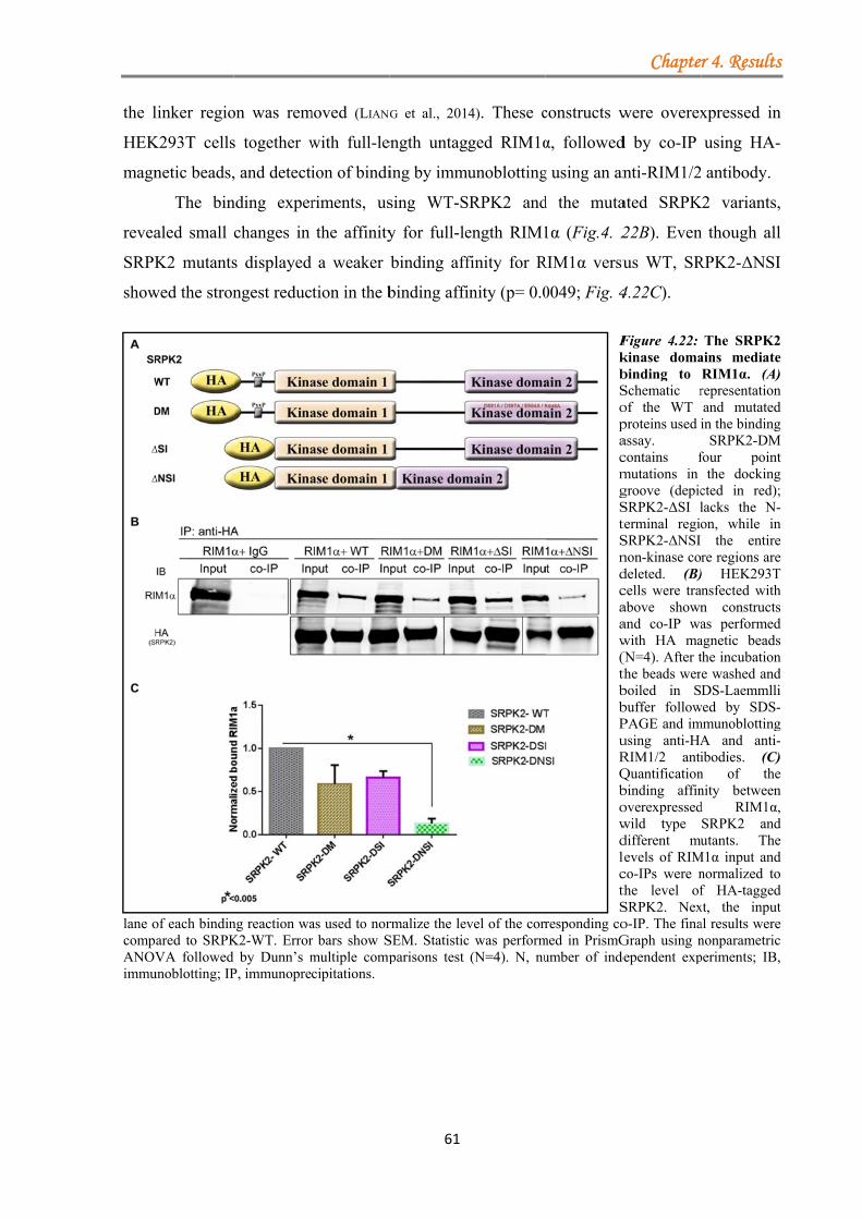
the l
HEK
magn
revea
SRPK
show
lane ocompANOVimmu
linker regio
K293T cells
netic beads,
The bin
aled small
K2 mutants
wed the stron
of each bindinared to SRPKVA followed
unoblotting; IP
on was rem
s together w
, and detecti
nding exper
changes in
s displayed
ngest reduc
ng reaction waK2-WT. Error
by Dunn’s mP, immunopre
moved (LIANG
with full-le
ion of bindi
riments, us
the affinity
a weaker
tion in the b
as used to norr bars show Smultiple compcipitations.
61
G et al., 201
ength untag
ing by imm
sing WT-S
y for full-le
binding aff
binding affi
rmalize the levSEM. Statisticparisons test
4). These c
ged RIM1α
unoblotting
SRPK2 and
ength RIM1
finity for R
inity (p= 0.0
vel of the corrc was perform(N=4). N, nu
constructs w
α, followed
g using an an
d the muta
1α (Fig.4. 2
RIM1α versu
0049; Fig. 4
FkbSopacmgStSndcaaw(thbbPuRQbowdlcthS
responding comed in PrismGumber of inde
Chapte
were overex
d by co-IP
nti-RIM1/2
ated SRPK
22B). Even
us WT, SR
4.22C).
Figure 4.22: kinase domabinding to Schematic rof the WT proteins used iassay. contains fmutations in groove (depicSRPK2-ΔSI lterminal regioSRPK2-ΔNSI non-kinase codeleted. (B)cells were traabove shownand co-IP wawith HA ma(N=4). After tthe beads werboiled in Sbuffer followPAGE and imusing anti-HARIM1/2 antiQuantificationbinding affinoverexpressedwild type different mulevels of RIMco-IPs were nthe level ofSRPK2. Nexo-IP. The finaGraph using ndependent exp
r 4. Results
xpressed in
using HA-
antibody.
K2 variants,
n though all
RPK2-ΔNSI
The SRPK2ains mediateRIM1α. (A)representationand mutatedin the binding
SRPK2-DMfour point
the dockingcted in red);lacks the N-on, while in
the entirere regions are) HEK293Tnsfected withn constructsas performedagnetic beadsthe incubatione washed andDS-Laemmlli
wed by SDS-mmunoblotting
A and anti-ibodies. (C)n of thenity betweend RIM1α,SRPK2 andutants. The
M1α input andnormalized tof HA-taggedxt, the inputal results werenonparametricperiments; IB,
s
n
-
,
l
I
2 e ) n d g
M t g ; -n e e T h s d s n d i -g -) e n ,
d e d o d t e c ,
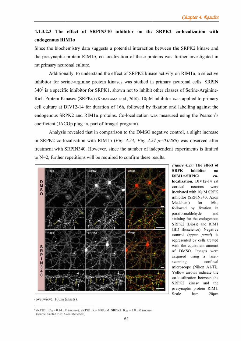
4.1.3
endo
Since
the p
rat pr
inhib
3406
Rich
cell c
endo
coeff
in SR
treatm
to N=
(overw
6SRPK (sourc
3.2.3 The
ogenous RI
e the bioch
presynaptic
rimary neur
Addition
bitor for ser
is a specifi
h Protein Ki
culture at D
ogenous SR
ficient (JAC
Analysis
RPK2 co-lo
ment with S
=2, further r
wiev); 10μm (
K1: IC50 = 0.14 µMce: Santa Cruz; A
effect of
M1α
hemistry dat
protein RIM
ronal cultur
nally, to und
rine-arginin
ic inhibitor
inases (SRP
DIV12-14 f
RPK2 and R
COp plug-in
s revealed th
ocalisation
SRPIN340.
repetitions w
(insets).
M (mouse); SRPK
Axon Medchem)
SRPIN34
ta suggests
M1α, co-lo
e.
derstand the
ne protein k
for SRPK1
PKs) (KARAK
for duration
RIM1α prote
n, part of Im
hat in comp
with RIM1
However, s
will be requ
K1: Ki= 0.89 µM
62
40 inhibito
a potential
ocalization o
e effect of S
kinases was
1, shown no
KAMA et al., 2
n of 16h, fo
eins. Co-loc
mageJ progra
parison to th
1α (Fig. 4.2
since the nu
uired to con
M; SRPK2: IC50 =
or on the
l interaction
of these pro
SRPK2 kina
s studied in
ot to inhibit
2010). 10μM
ollowed by
calization w
am).
he DMSO n
23; Fig. 4.2
umber of in
nfirm these r
= 1.8 µM (mouse)
SRPK2
n between t
oteins was f
ase activity
n primary n
other class
M inhibitor w
fixation and
was measure
negative co
24 p=0.028
ndependent
results.
FSRRlocoininMfopastSR(BcorewofacscmYcoSRprS
Chapte
co-localiza
the SRPK2
further inve
on RIM1α,
neuronal ce
ses of Serine
was applied
d labelling
ed using the
ontrol, a slig
86) was obs
experiment
Figure 4.23: TSRPK inhRIM1α-SRPKocalization. Dortical neuncubated withnhibitor (SRP
Medchem) ollowed by araformaldehytaining for theRPK2 (BiossBD Biosciencontrol (uppeepresented by
with the equivf DMSO. Icquired usincanning
microscope (NYellow arrowso-localizationRPK2 kinasresynaptic prcale bar
r 4. Results
ation with
kinase and
estigated in
, a selective
ells. SRPIN
e-Arginine-
d to primary
against the
e Pearson’s
ght increase
served after
ts is limited
The effect ofhibitor onK2 co-DIV12-14 raturons wereh 10μM SRPKPIN340, Axon
for 16h.,fixation in
yde ande endogenouss) and RIM1ce). Negativeer panel) isy cells treatedvalent amountImages wereng a laser-
confocalNikon A1/Ti).s indicate the
n between these and therotein RIM1.r: 20μm
s
h
d
n
e
N
-
y
e
s
e
r
d
f n -t e
K n ,
n d s
e s d t e -l . e e e .
m
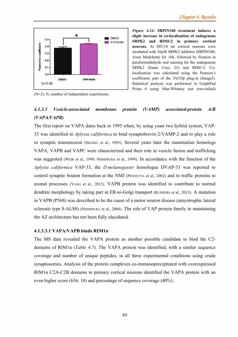
Chapter 4. Results
63
Figure 4.24: SRPIN340 treatment induces a slight increase in co-localization of endogenous SRPK2 and RIM1/2 in primary cortical neurons. At DIV14 rat cortical neurons were incubated with 10μM SRPK2 inhibitor (SRPIN340, Axon Medchem) for 16h, followed by fixation in paraformaldehyde and staining for the endogenous SRPK2 (Santa Cruz, 23) and RIM1/2. Co-localization was calculated using the Pearson’s coefficient, part of the JACOp plug-in (ImageJ). Statistical analysis was performed in GraphPad Prism 6 using Man-Whitney test (two-tailed)
(N=2). N, number of independent experiments.
4.1.3.3 Vesicle-associated membrane protein (VAMP) associated-protein A/B
(VAPA/VAPB)
The first report on VAPA dates back to 1995 when, by using yeast two hybrid system, VAP-
33 was identified in Aplysia californica to bind synaptobrevin-2/VAMP-2 and to play a role
in synaptic transmission (SKEHEL et al., 1995). Several years later the mammalian homologs
VAPA, VAPB and VAPC were characterized and their role in vesicle fusion and trafficking
was suggested (WEIR et al., 1998; NISHIMURA et al., 1999). In accordance with the function of the
Aplysia californica VAP-33, the D.melanogaster homologue DVAP-33 was reported to
control synaptic bouton formation at the NMJ (PENNETTA et al., 2002) and to traffic proteins to
axonal processes (YANG et al., 2012). VAPB protein was identified to contribute to normal
dendrite morphology by taking part in ER-to-Golgi transport (KUIJPERS et al., 2013). A mutation
in VAPB (P56S) was described to be the cause of a motor neuron disease (amyotrophic lateral
sclerosis type 8-ALS8) (NISHIMURA et al., 2004). The role of VAP protein family in maintaining
the AZ architecture has not been fully elucidated.
4.1.3.3.1 VAPA/VAPB binds RIM1α
The MS data revealed the VAPA protein as another possible candidate to bind the C2-
domains of RIM1α (Table 4.7). The VAPA protein was identified, with a similar sequence
coverage and number of unique peptides, in all three experimental conditions using crude
synaptosomes. Analysis of the protein complexes co-immunoprecipitated with overexpressed
RIM1α C2A-C2B domains in primary cortical neurons identified the VAPA protein with an
even higher score (636, 10) and percentage of sequence coverage (40%).
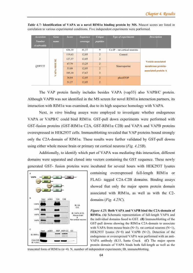
Tablecorrel
A
(
Altho
intera
VAP
GST
overe
only
using
doma
gene
trunca
e 4.7: Identifilation to vario
Accession
Number
(UniProtKB)
Q9WV55
The VA
ough VAPB
action with
Next, in
PA or VAP
-fusion pro
expressed in
the C2A-d
g either who
Addition
ains were s
rated GST-
ated form of R
fication of VAous experimen
Gene
name
Sco
VA
PA
-MO
US
E
636
119
127
47,
53,
105
36,
31,
AP protein
B was not id
RIM1α wa
n vitro bind
PB/C could
oteins (GST
n HEK293T
domain of R
ole mouse b
nally, to ide
separated an
- fusion pr
RIM1α (n=4).
APA as a nontal conditions
ore Sequen
coverag
6,10 41,37
9,63
7,37
12,05
12,05
,79 13,25
,88
5,10
12,05
17,67
,84
,27
12,05
12,05
family inc
dentified in
as examined
ding assays
bind RIM1
-RIM1α C2
T cells. Imm
RIM1α. Th
brain or prim
ntify which
nd cloned in
roteins were
N, number of
64
ovel RIM1α bs. Five indepen
nce
ge
Unique
peptides
7 9
5
5
2
2
5 2
5
7
2
3
5
5
2
2
cludes besid
the MS scr
d, due to its
s were em
1α. GST-pu
2A, GST-R
munoblottin
hese results
mary rat cor
h part of VA
nto vectors
e incubated
containin
FLAG- t
showed t
associated
domains
Figure 4.25RIM1α. (Athe individuGST-pull dwith VAPAHEK293T endogenouVAPA antprotein dom
f independent
binding protendent experim
Type of e
Co-IP – rat c
Co
Stauro
phos
des VAPA
reen for nov
high sequen
mployed to
ull down ex
RIM1α C2B)
ng revealed t
were furth
rtical neuron
APA was m
containing
d for sever
ng overexp
tagged C2A
that only th
d with RIM
(Fig. 4.25C
5: Both VAPA) Schematic ual domains fdowns showinA from mouse
lysates (N=8s or overexpretibody (K15, main of VAPexperiments;
ein by MS. Mments were per
experiments
cortical neurons
ontrol
osporine
sSTOP
(vap33) a
vel RIM1α i
nce homolo
investigate
xperiments
) and VAPA
that VAP pr
her validated
ns (Fig. 4.2
mediating thi
the GST s
al hours w
pressed fu
A-C2B dom
he major s
M1α, as w
C).
A and VAPBrepresentation
fused to GST. ng the RIM1α
brain (N=3), 8) and VAPBesed VAPA w
Santa Cruz)A binds bothIB, immunob
Chapte
Mascot scoresrformed.
Descrip
Vesicle-ass
membrane
associated p
also VAPB
interaction p
ogy with VA
whether e
were perfo
A and VAP
proteins bou
d by GST-
5B).
is interactio
sequence. T
with HEK29
ull-length R
mains. Bind
sperm prote
well as wit
B bind the C2n of full-lengt(B) Immunobα C2A-domain
rat cortical neB (N=2). Detwas performed). (C) The h full-length ablotting.
r 4. Results
s are listed in
ption
sociated
protein-
protein A
/C protein.
partners, its
APA.
endogenous
ormed with
PB proteins
und strongly
pull downs
on, different
These newly
93T lysates
RIM1α or
ding assays
ein domain
th the C2-
2A-domain ofth VAPA andblotting of then to associateeurons (N=1),tection of thed with an anti-
major spermas well as the
s
n
.
s
s
h
s
y
s
t
y
s
r
s
n
-
f d e e , e -
m e
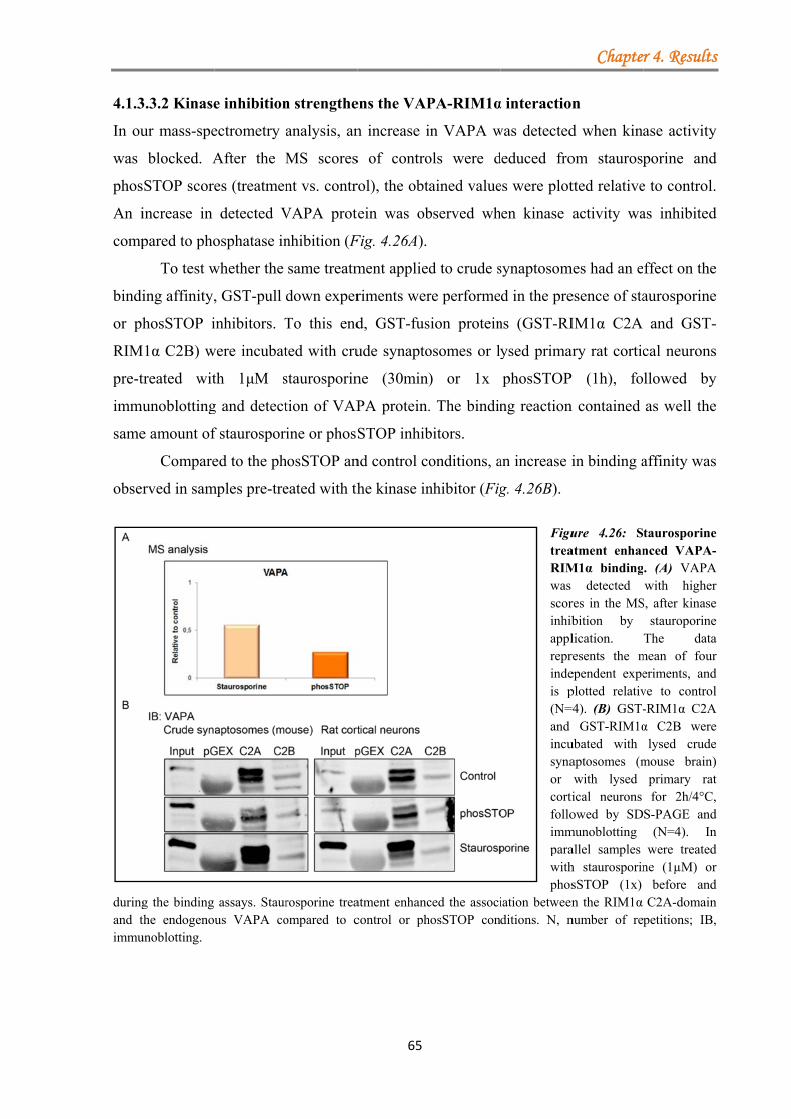
4.1.3
In ou
was
phos
An i
comp
bindi
or ph
RIM
pre-t
immu
same
obser
duringand timmu
3.3.2 Kinase
ur mass-spe
blocked. A
STOP score
increase in
pared to pho
To test w
ing affinity,
hosSTOP i
M1α C2B) w
treated wit
unoblotting
e amount of
Compare
rved in sam
g the binding the endogenounoblotting.
e inhibition
ectrometry
After the M
es (treatmen
detected V
osphatase in
whether the
, GST-pull
inhibitors. T
were incubat
th 1μM s
g and detect
f staurospor
ed to the ph
mples pre-tre
assays. Staurus VAPA co
n strengthe
analysis, an
MS scores
nt vs. contr
VAPA prot
nhibition (F
same treatm
down exper
To this end
ted with cru
staurosporin
tion of VAP
ine or phosS
hosSTOP an
eated with th
rosporine treaompared to c
65
ens the VAP
n increase i
s of contro
rol), the obt
ein was ob
Fig. 4.26A).
ment applied
riments wer
d, GST-fus
ude synapto
ne (30min)
PA protein.
STOP inhib
nd control co
he kinase in
atment enhanccontrol or ph
PA-RIM1α
in VAPA w
ols were d
ained value
bserved wh
d to crude s
re performe
sion protein
osomes or l
) or 1x
. The bindin
bitors.
onditions, a
nhibitor (Fig
ced the associhosSTOP con
α interaction
was detected
deduced fro
es were plot
hen kinase
synaptosom
d in the pre
ns (GST-RI
ysed primar
phosSTOP
ng reaction
an increase i
g. 4.26B).
FigutreaRIMwas scorinhibapplreprindeis p(N=4and incusynaor cortifolloimmparawithphos
iation betweenditions. N, n
Chapte
n
d when kina
om staurosp
tted relative
activity wa
mes had an e
esence of sta
IM1α C2A
ary rat cortic
P (1h), fo
n contained
in binding a
ure 4.26: Satment enhanM1α binding
detected res in the MSbition by lication. resents the mependent expeplotted relativ=4). (B) GST-
GST-RIM1αubated with aptosomes (mwith lysed ical neurons owed by SD
munoblotting allel samples h staurosporinsSTOP (1x) n the RIM1α
number of re
r 4. Results
ase activity
porine and
e to control.
as inhibited
ffect on the
aurosporine
and GST-
cal neurons
llowed by
as well the
affinity was
taurosporinenced VAPA-. (A) VAPAwith higher
S, after kinasestauroporine
The datamean of foureriments, andve to control-RIM1α C2Aα C2B were
lysed crudemouse brain)
primary ratfor 2h/4°C,
S-PAGE and(N=4). In
were treatedne (1µM) or
before andC2A-domain
petitions; IB,
s
y
d
.
d
e
e
-
s
y
e
s
e -
A r e e a r d l
A e e ) t , d n d r d n ,
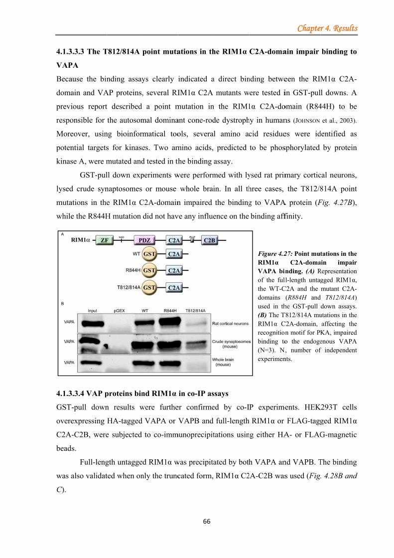
4.1.3
VAP
Beca
doma
previ
respo
More
poten
kinas
lysed
muta
while
4.1.3
GST
overe
C2A
bead
was a
C).
3.3.3 The T
PA
ause the bin
ain and VA
ious report
onsible for
eover, usin
ntial targets
se A, were m
GST-pul
d crude syn
ations in the
e the R844H
3.3.4 VAP p
-pull down
expressing
A-C2B, were
ds.
Full-leng
also validat
T812/814A
nding assay
AP proteins,
t described
the autosom
ng bioinfor
s for kinase
mutated and
ll down exp
naptosomes
e RIM1α C
H mutation
proteins bin
n results w
HA-tagged
e subjected
gth untagge
ted when on
point muta
ys clearly i
, several RI
a point m
mal domina
matical too
es. Two am
d tested in th
periments w
or mouse
C2A-domain
did not hav
nd RIM1α
were furthe
VAPA or V
to co-imm
d RIM1α w
nly the trunc
66
ations in th
indicated a
IM1α C2A
mutation in
ant cone-rod
ols, severa
mino acids,
he binding
were perform
whole brai
n impaired
ve any influe
in co-IP as
er confirme
VAPB and
munoprecipit
was precipita
cated form,
he RIM1α
a direct bin
mutants w
n the RIM1
de dystroph
l amino ac
predicted
assay.
med with ly
in. In all th
the binding
ence on the
ssays
ed by co-I
full-length
tations usin
ated by both
RIM1α C2
C2A-doma
nding betwe
ere tested i
1α C2A-do
hy in human
cid residue
to be phos
ysed rat prim
hree cases,
g to VAPA
binding aff
Figure 4.2RIM1α VAPA binof the fulthe WT-Cdomains used in th(B) The TRIM1α Crecognitionbinding to(N=3). Nexperimen
P experime
RIM1α or
ng either HA
h VAPA an
2A-C2B was
Chapte
ain impair
een the RIM
in GST-pul
omain (R84
ns (JOHNSON
es were id
sphorylated
mary cortic
the T812/8
A protein (F
finity.
27: Point mutC2A-doma
inding. (A) Rll-length untagC2A and the
(R884H andhe GST-pull
T812/814A muC2A-domain, on motif for PKo the endoge
N, number ofnts.
ents. HEK
FLAG-tagg
A- or FLAG
nd VAPB. T
s used (Fig.
r 4. Results
binding to
M1α C2A-
l downs. A
44H) to be
et al., 2003).
dentified as
by protein
cal neurons,
814A point
Fig. 4.27B),
tations in theain impairRepresentationgged RIM1α,mutant C2A-
d T812/814A)down assays.
utations in theaffecting theKA, impairedenous VAPAf independent
K293T cells
ged RIM1α
G-magnetic
The binding
. 4.28B and
s
o
-
A
e
.
s
n
,
t
,
e r n , -) . e e d A t
s
α
c
g
d
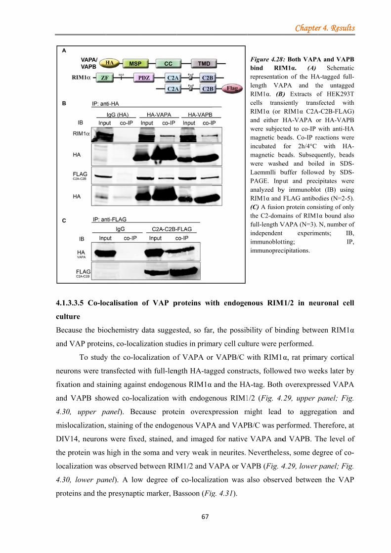
4.1.3
cultu
Beca
and V
neuro
fixati
and V
4.30,
mislo
DIV
the p
local
4.30,
prote
3.3.5 Co-lo
ure
ause the bio
VAP protein
To study
ons were tr
ion and stai
VAPB show
, upper p
ocalization,
14, neurons
protein was
lization was
, lower pan
eins and the
calisation
ochemistry
ns, co-local
y the co-loc
ansfected w
ining agains
wed co-loc
anel). Bec
staining of
s were fixed
high in the
s observed b
nel). A low
e presynapti
of VAP p
data sugges
lization stud
calization o
with full-len
st endogeno
calization w
cause prote
f the endoge
d, stained, a
soma and v
between RIM
w degree of
c marker, B
67
proteins wi
sted, so far
dies in prim
f VAPA or
ngth HA-tag
ous RIM1α
with endoge
ein overexp
enous VAPA
and imaged
very weak i
M1/2 and V
f co-localiz
Bassoon (Fig
ith endoge
r, the possib
mary cell cult
r VAPB/C w
gged constru
and the HA
enous RIM1
pression m
A and VAP
d for native
in neurites.
VAPA or VA
zation was
g. 4.31).
Figure 4.28bind RIMrepresentatiolength VARIM1α. (B)cells transRIM1α (or and either Hwere subjectmagnetic beincubated magnetic bewere washeLaemmlli bPAGE. Inpuanalyzed byRIM1α and (C) A fusionthe C2-domafull-length Vindependent immunoblottimmunoprec
nous RIM
bility of bin
ture were pe
with RIM1
ucts, follow
A-tag. Both
1/2 (Fig. 4.
might lead
B/C was pe
VAPA and
Neverthele
APB (Fig. 4
also observ
Chapte
8: Both VAPAM1α. (A) on of the HA
APA and thB) Extracts osiently transRIM1α C2AHA-VAPA o
cted to co-IP eads. Co-IP refor 2h/4°C eads. Subsequed and boilbuffer followut and precy immunobloFLAG antibo
n protein consains of RIM1
VAPA (N=3). t experimtting; cipitations.
M1/2 in neu
nding betwe
erformed.
α, rat prima
wed two wee
h overexpres
29, upper p
to aggreg
erformed. T
d VAPB. T
ess, some de
4.29, lower
ved betwee
r 4. Results
A and VAPBSchematic
A-tagged full-he untaggedof HEK293Tsfected with
A-C2B-FLAG)or HA-VAPBwith anti-HAeactions were
with HA-uently, beadsled in SDS-
wed by SDS-ipitates were
ot (IB) usingodies (N=2-5).sisting of only1α bound alsoN, number of
ments; IB,IP,
uronal cell
een RIM1α
ary cortical
eks later by
ssed VAPA
panel; Fig.
gation and
herefore, at
The level of
egree of co-
panel; Fig.
n the VAP
s
B c -d T h )
B A e -s --e g . y o f , ,
l
α
l
y
A
d
t
f
-
P
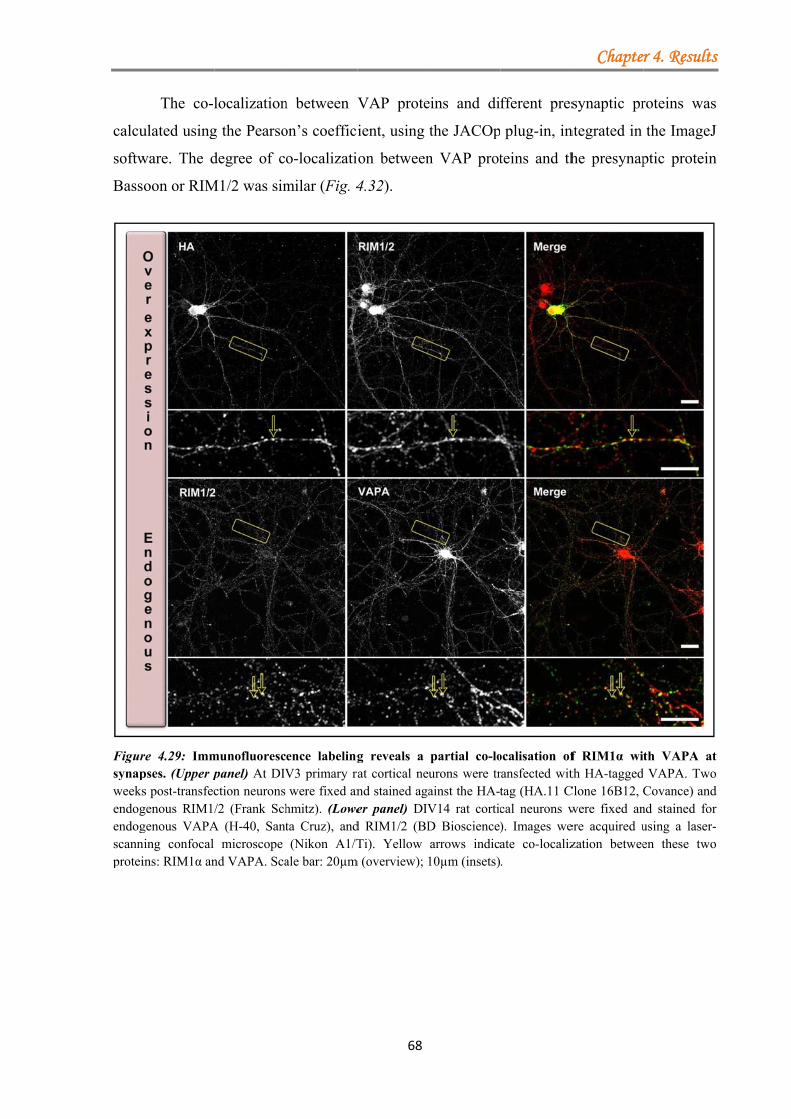
calcu
softw
Bass
Figursynapweeksendogendogscannprotei
The co-l
ulated using
ware. The d
oon or RIM
re 4.29: Immpses. (Upper ps post-transfecgenous RIM1/genous VAPAning confocal ins: RIM1α an
localization
g the Pearso
degree of c
M1/2 was sim
munofluorescpanel) At DIVction neurons /2 (Frank Sch
A (H-40, Santmicroscope
nd VAPA. Sca
n between V
on’s coeffici
o-localizati
milar (Fig. 4
ence labelingV3 primary rawere fixed an
hmitz). (Loweta Cruz), and(Nikon A1/T
ale bar: 20µm
68
VAP prote
ient, using
on between
4.32).
g reveals a at cortical neund stained agaer panel) DIV
d RIM1/2 (BDTi). Yellow
m (overview); 1
eins and di
the JACOp
n VAP prot
partial co-lourons were trainst the HA-tV14 rat corticD Bioscience)arrows indica10µm (insets)
fferent pres
plug-in, in
teins and th
ocalisation ofransfected wittag (HA.11 Ccal neurons w). Images weate co-localiz.
Chapte
synaptic pr
ntegrated in
he presynap
f RIM1α with HA-tagged
Clone 16B12, Cwere fixed anere acquired uzation betwee
r 4. Results
roteins was
the ImageJ
ptic protein
th VAPA atVAPA. Two
Covance) andnd stained forusing a laser-en these two
s
s
J
n
t o d r -o
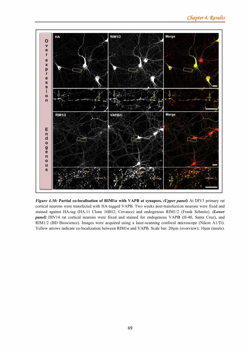
Figurcorticstainepanel)RIM1Yellow
re 4.30: Partical neurons weed against HAl) DIV14 rat 1/2 (BD Bioscw arrows indi
ial co-localisaere transfectedA-tag (HA.11
cortical neurcience). Imagicate co-locali
ation of RIMd with HA-tag Clone 16B1
rons were fixges were acquization betwee
69
M1α with VAPgged VAPB. 12, Covance) xed and stainuired using a en RIM1α and
PB at synapsTwo weeks pand endogen
ed for endoglaser-scannin
d VAPB. Sca
ses. (Upper papost-transfectionous RIM1/2
genous VAPBng confocal mle bar: 20µm
Chapte
anel) At DIVion neurons w (Frank Schm
B (H-40, Santmicroscope (N(overview); 1
r 4. Results
V3 primary ratwere fixed andmitz). (Lowerta Cruz), and
Nikon A1/Ti).10µm (insets).
s
t d r d . .
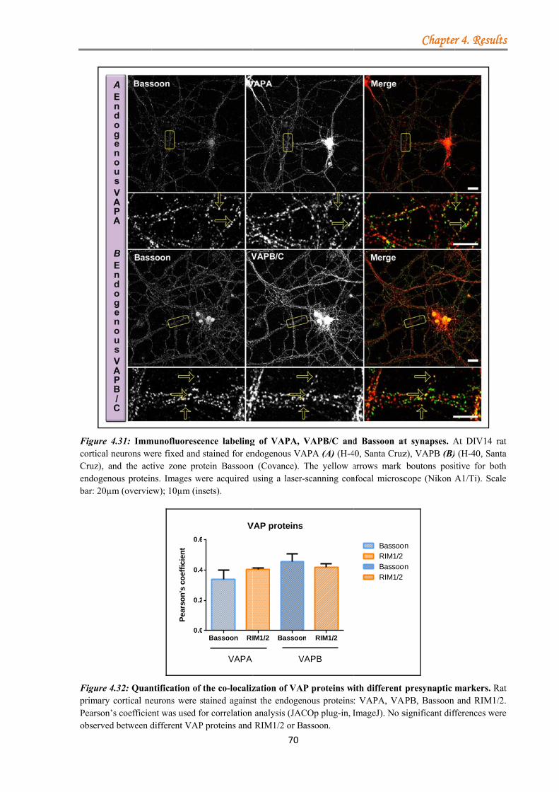
FigurcorticCruz)endogbar: 2
FigurprimaPearsoobserv
re 4.31: Immcal neurons we, and the act
genous protein20µm (overvie
re 4.32: Quanary cortical neon’s coefficieved between d
munofluoresceere fixed and tive zone prons. Images wew); 10µm (in
0.0
0.2
0.4
0.6
Pea
rso
n's
co
effi
cien
t
ntification of eurons were snt was used fodifferent VAP
ence labelingstained for en
otein Bassoonere acquired u
nsets).
Bassoon RI0
2
4
6
V
VAPA
the co-localizstained againsor correlation
P proteins and
70
g of VAPA, ndogenous VAn (Covance). using a laser-
IM1/2 Bassoo
VAP proteins
V
zation of VAst the endogenanalysis (JACRIM1/2 or B
VAPB/C andAPA (A) (H-4The yellow a-scanning con
n RIM1/2
VAPB
P proteins wnous proteinsCOp plug-in, Iassoon.
d Bassoon at40, Santa Cruzarrows mark nfocal microsc
BassoonRIM1/2BassoonRIM1/2
ith different : VAPA, VAImageJ). No s
Chapte
t synapses. Az), VAPB (B)boutons posi
cope (Nikon
on
on
presynaptic
APB, Bassoon significant dif
r 4. Results
At DIV14 rat (H-40, Santaitive for bothA1/Ti). Scale
markers. Ratand RIM1/2.
fferences were
s
t a h e
t . e
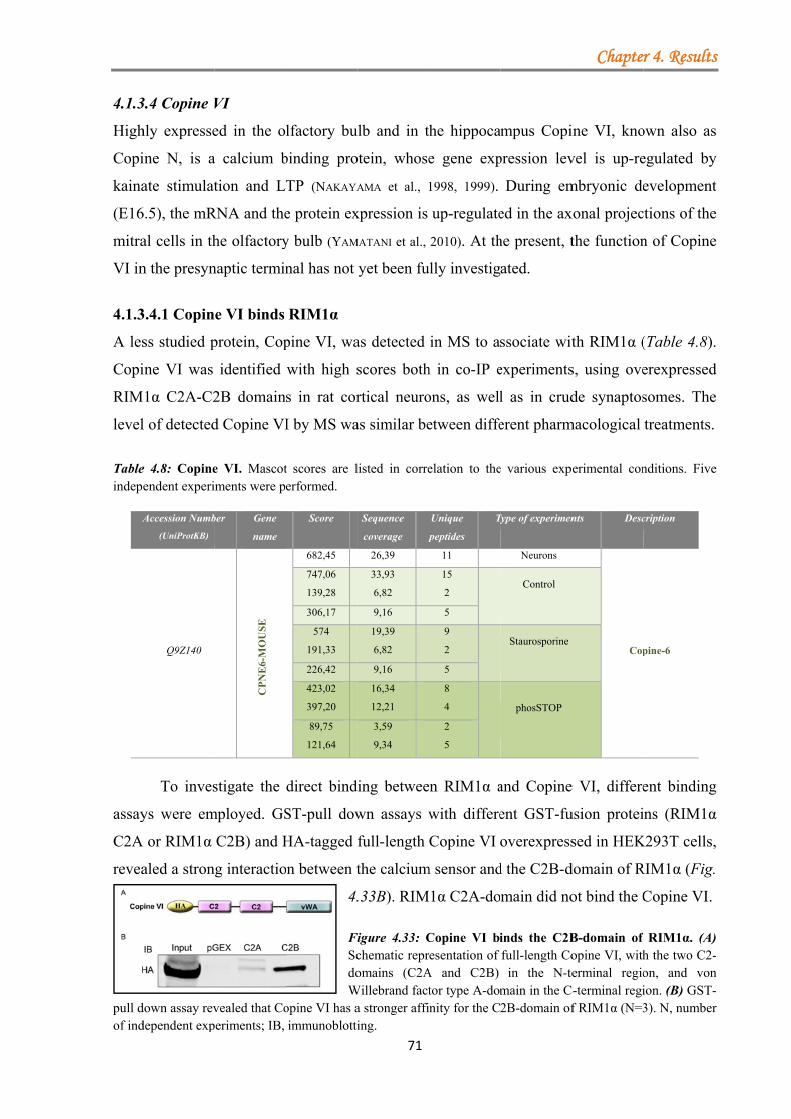
4.1.3
High
Copi
kaina
(E16
mitra
VI in
4.1.3
A les
Copi
RIM
level
Tableindep
A
assay
C2A
revea
pull dof ind
3.4 Copine V
hly expresse
ine N, is a
ate stimulat
6.5), the mR
al cells in th
n the presyn
3.4.1 Copin
ss studied p
ine VI was
M1α C2A-C2
l of detected
e 4.8: Copineendent experi
Accession Numb
(UniProtKB)
Q9Z140
To inves
ys were em
A or RIM1α
aled a stron
down assay revdependent exp
VI
ed in the o
a calcium b
tion and L
RNA and the
he olfactory
naptic termin
e VI binds
protein, Cop
identified
2B domain
d Copine VI
e VI. Mascotiments were p
ber Gene
name
CP
NE
6-M
OU
SE
stigate the
mployed. GS
C2B) and H
ng interactio
vealed that Coperiments; IB,
lfactory bu
binding pro
TP (NAKAYA
e protein ex
y bulb (YAM
nal has not
RIM1α
pine VI, wa
with high s
s in rat cor
I by MS wa
t scores are lerformed.
Score
682,45
747,06
139,28
306,17
574
191,33
226,42
423,02
397,20
89,75
121,64
direct bind
ST-pull dow
HA-tagged f
on between t
4.
FiScdoW
opine VI has aimmunoblotti
71
ulb and in t
otein, whose
AMA et al., 1
xpression is
MATANI et al.,
yet been fu
as detected
scores both
rtical neuro
as similar be
listed in corre
Sequence
coverage p
26,39
33,93
6,82
9,16
19,39
6,82
9,16
16,34
12,21
3,59
9,34
ding betwee
wn assays w
full-length
the calcium
.33B). RIM
Figure 4.33: Cchematic repromains (C2A
Willebrand facta stronger affiing.
he hippoca
e gene exp
1998, 1999).
up-regulate
2010). At th
ully investig
in MS to a
h in co-IP e
ons, as well
etween diffe
elation to the
Unique
peptides
T
11
15
2
5
9
2
5
8
4
2
5
en RIM1α a
with differe
Copine VI
m sensor and
M1α C2A-do
Copine VI biresentation of A and C2B) tor type A-domnity for the C
mpus Copi
pression lev
During em
ed in the ax
he present, t
ated.
ssociate wit
experiments
l as in crud
erent pharm
e various expe
Type of experimen
Neurons
Control
Staurosporine
phosSTOP
and Copine
ent GST-fu
overexpress
d the C2B-d
main did no
inds the C2Bfull-length Coin the N-te
main in the C2B-domain of
Chapte
ine VI, kno
vel is up-re
mbryonic de
xonal projec
the function
th RIM1α (
s, using ove
de synaptos
macological
erimental con
nts Desc
Cop
e VI, differe
usion protei
sed in HEK
domain of R
ot bind the C
B-domain of opine VI, witherminal regio
C-terminal regif RIM1α (N=3
r 4. Results
own also as
egulated by
evelopment
ctions of the
n of Copine
(Table 4.8).
erexpressed
somes. The
treatments.
nditions. Five
cription
pine-6
ent binding
ns (RIM1α
K293T cells,
RIM1α (Fig.
Copine VI.
RIM1α. (A)h the two C2-on, and vonion. (B) GST-3). N, number
s
s
y
t
e
e
.
d
e
e
g
α
,
) -n -r

4.1.3Give
al., 1
immu
agen
impa
comp
affin
4.1.3
Beca
local
were
4.35)
arrowmicro
3.4.2 The Cen that prev
1998, 1999),
unoprecipit
nt, EGTA.
In the pr
aired (Fig.4
pared to un
nity for the H
3.4.3 Copin
ause of the
lization of th
e fixed at DI
).
ws mark the poscope (Nikon
opine VI-Rious studies
the requi
tations were
resence of
.34B). The
ntreated co
HA-tagged C
e VI and R
direct bind
hese two pr
IV14 and th
positive bouton A1/Ti). Scale
RIM1α intes have show
irement of
e carried ou
EGTA the
addition of
ontrol. The
Copine VI (
RIM1/2 co-l
ding betwee
roteins was
he endogeno
ons for both e bar: 20µm (o
72
eraction is cwn copines
f calcium
ut in the ab
binding be
f calcium di
truncated
(Fig. 4.34C
FigdeptaggOveHA(1.2inteFLAN, imm
localized at
en RIM1α a
studied in p
ous RIM1/2
proteins. Imaoverview); 10
calcium depto be calciu
in this bin
bsence or p
etween RIM
id not have
form of R
C).
gure 4.34: Cpendent. (A/Cged full-lengerexpressed p
A-magnetic be2mM) or Eeraction betwAG-tagged RI
number munoblotting;
t a subset o
and the cal
primary rat
2 and Copin
ages were ac0µm (insets).
pendent um-binding
nding was
presence of
M1α and Co
any effect o
RIM1α (C2
opine VI-RIC) Schematic gth Copine Vproteins wereeads in the pGTA (2mM)een Copine VIM1α C2A-C2of indepen IP, immunop
of synapses
lcium senso
cortical neu
ne VI protei
quired using
Chapte
proteins (N
assessed.
the calcium
opine VI w
on the bind
2A-C2B) al
IM1α bindinrepresentatio
VI and the e subject to presence of e). EGTA w
VI and RIM12B binds Cop
ndent experprecipitations.
or, Copine
urons. Prima
ins were lab
Figure Immunolabellingand Cosynapsesrat cortwere fixefor proteins: BiosciencCopine Kavalali)
a laser-scann
r 4. Results
NAKAYAMA et
Thus, co-
m chelating
was strongly
ding affinity
lso showed
g is calciumn of the HA-RIM1α. (B)co-IPs usingither calcium
weakened theα (N=4). (D)ine VI (N=3).iments; IB,
VI, the co-
ary cultures
belled (Fig.
4.35:ofluorescenceg of RIM1/2opine VI ats. At DIV14ical neuronsed and stained
endogenousRIM1 (BD
ce) andVI (Ege
). The yellowning confocal
s
t
-
g
y
y
d
m -) g
m e ) . ,
-
s
: e 2 t 4 s d s
D d e
w l
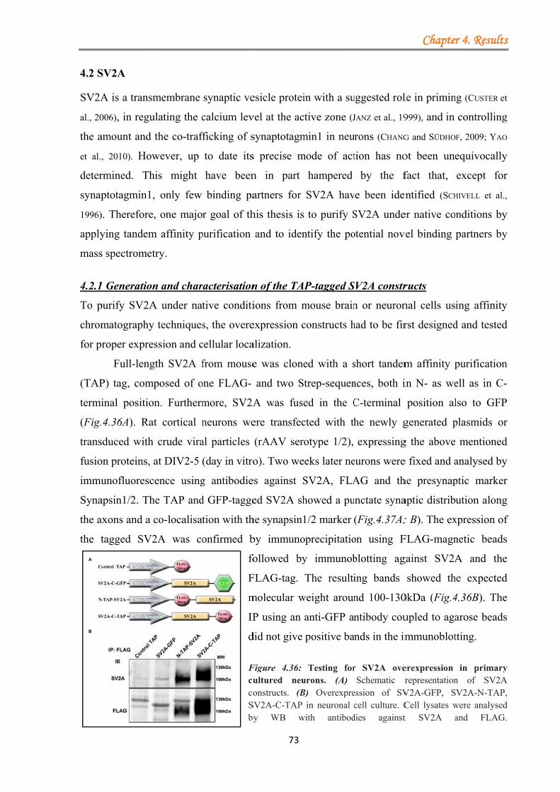
4.2 S
SV2A
al., 20
the a
et al.
deter
synap
1996)
apply
mass
4.2.1
To p
chrom
for p
(TAP
termi
(Fig.
trans
fusio
immu
Syna
the a
the t
SV2A
A is a trans
006), in regu
amount and
, 2010). How
rmined. Th
ptotagmin1
). Therefore
ying tandem
s spectrome
1 Generatio
purify SV2A
matography
proper expre
Full-leng
P) tag, com
inal positio
4.36A). Ra
sduced with
on proteins,
unofluoresc
apsin1/2. Th
axons and a
tagged SV
smembrane
ulating the c
the co-traf
wever, up
his might
, only few
e, one majo
m affinity p
try.
n and char
A under nat
y techniques
ession and c
gth SV2A f
mposed of o
on. Furtherm
at cortical n
h crude vira
at DIV2-5
cence using
he TAP and
co-localisat
2A was co
synaptic ve
calcium lev
fficking of s
to date its
have been
binding pa
r goal of th
purification
racterisation
tive conditi
s, the overe
cellular loca
from mouse
one FLAG-
more, SV2A
neurons we
al particles
(day in vitro
g antibodie
d GFP-tagge
tion with th
onfirmed b
fo
F
m
IP
d
FcucoSby
73
esicle protei
vel at the ac
synaptotagm
s precise m
n in part
artners for
his thesis is
and to iden
n of the TA
ions from m
expression c
alization.
e was clone
and two S
A was fuse
ere transfec
(rAAV ser
o). Two we
es against S
ed SV2A sh
he synapsin1
by immuno
followed by
FLAG-tag.
molecular w
P using an
did not give
Figure 4.36: ultured neuonstructs. (B
SV2A-C-TAP y WB w
in with a su
ctive zone (J
min1 in neu
mode of act
hampered
SV2A hav
to purify S
ntify the po
P-tagged S
mouse brain
constructs h
ed with a s
trep-sequen
ed in the C
cted with th
otype 1/2),
eeks later ne
SV2A, FLA
howed a pu
1/2 marker
oprecipitatio
y immunob
The resulti
weight aroun
anti-GFP an
positive ba
Testing forurons. (A) B) Overexpre
in neuronal cwith antibod
uggested rol
JANZ et al., 19
rons (CHANG
tion has no
by the f
ve been iden
SV2A unde
otential nov
SV2A constr
n or neuron
had to be fir
short tandem
nces, both i
C-terminal
he newly g
expressing
eurons were
AG and th
unctate syna
(Fig.4.37A;
on using F
blotting ag
ing bands
nd 100-130
ntibody cou
nds in the im
r SV2A oveSchematic r
ession of SVcell culture. C
dies against
Chapte
le in primin
999), and in
G and SÜDHO
ot been une
fact that,
entified (SCH
er native co
vel binding
ructs
nal cells us
rst designed
m affinity p
in N- as we
position al
generated p
g the above
e fixed and a
he presynap
aptic distrib
; B). The ex
FLAG-magn
gainst SV2A
showed th
0kDa (Fig.4
upled to aga
mmunoblot
erexpression representation
V2A-GFP, SCell lysates wt SV2A a
r 4. Results
ng (CUSTER et
controlling
F, 2009; YAO
equivocally
except for
HIVELL et al.,
nditions by
partners by
ing affinity
d and tested
purification
ell as in C-
lso to GFP
plasmids or
mentioned
analysed by
ptic marker
ution along
xpression of
netic beads
A and the
he expected
4.36B). The
arose beads
tting.
in primaryn of SV2AV2A-N-TAP,
were analysedand FLAG.
s
t
g
O
y
r
,
y
y
y
d
n
-
P
r
d
y
r
g
f
s
e
d
e
s
y A ,
d .
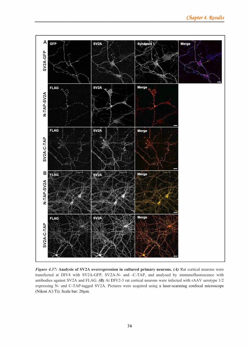
Figurtransfantiboexpre(Niko
re 4.37: Analfected at DIVodies against ssing N- and
on A1/Ti). Sca
lysis of SV2AV4 with SV2SV2A and FLC-TAP-tagg
ale bar: 20µm.
A overexpress2A-GFP, SV2LAG. (B) At Ded SV2A. Pic.
74
sion in cultur2A-N- and -CDIV2-3 rat coctures were a
red primary C-TAP, and ortical neuronsacquired using
neurons. (A)analysed by s were infecteg a laser-scan
Chapte
) Rat cortical immunofluor
ed with rAAVnning confoca
r 4. Results
neurons wererescence withV serotype 1/2al microscope
s
e h 2 e
Chapter 4. Results
75
4.2.2 Optimization of SV2A protein purification from primary rat cortical neurons
To test and optimze the affinity purification of SV2A, primary rat cortical neurons were
transduced with viral particles (rAAV serotype 1/2) expressing SV2A with the TAP-tag either
in the N- or C-terminal position. Two weeks after the transduction the cells were harvested,
lysed in cold ice lysis buffer containing 3.9mM DDM (n-Dodecyl-ß-maltoside) and subjected
to either a one- or two-step purification procedure.
4.2.2.1 One-step purification yields good recovery of TAP-tagged SV2A
Strep purification was based on the ability of the Strep-tactin matrices (streptavidin) to bind
with high selectively to the strep sequence of the TAP-tag, allowing the purification of SV2A
in one single step and in a column modus. To determine the purification efficiency, a small
aliquot was kept after each purification step for further analysis by WB. The Strep purification
alone showed a good recovery of the recombinant protein, since little material was lost during
the procedure and the entire material was recovered after the 3rd and 4th elution steps. The
disadvantage of this procedure was the limited binding capacity of the columns (50 to
100nmol/ml recombinant proteins), which were therefore unable to capture the entire applied
tagged-SV2A protein. The bands present in the flow fraction and the first two washing steps
may indicate that the column was over-saturated with the fusion protein (Fig.4.38B).
The capacity of the monoclonal M2-FLAG antibodies, covalently attached to the
agarose beads, to recognise the octapeptide (DVKDDDDK) present in the TAP-tag allowed
for the protein purification using the FLAG-tag (Batch modus). As in the case of the Strep
purification, the column binding capacity was the limiting factor in capturing the whole
amount of recombinant protein. However, the elution step performed with competitive FLAG
peptides showed a sufficiently high recovery of the targeted protein (Fig.4.38C).
Since the purpose of these procedures was to purify proteins maintaining their native
configuration, it was mandatory to control for SV2A structural integrity. For this purpose
Synaptotagmin1 (Syt1) was chosen because it is one of the proteins known to bind to SV2A
(SCHIVELL et al., 1996; PYLE et al., 2000). The immunoblotting experiments showed that a
significant amount of Synaptotagmin1 was co-eluted in the Strep purification and to a lesser
degree in the FLAG purification procedure, indicating that the recovered SV2A protein
retained to a certain degree its native structure (Fig.4.38).

4.2.2
In o
minim
follo
purif
purif
steps
colle
FLA
poor
of re
rema
purif
recov
notic
prote
estim
2.2 Two-step
rder to pu
mize the lar
wed by th
fication.
In the fi
fication) mo
s, even tho
ected fractio
AG M2 affin
. There wer
ecombinant
ained bound
The sec
fication, wh
vered from
ced after th
ein of intere
mated by its
p purificati
urify SV2A
rge amount
he FLAG-ta
first approa
ost of the re
ough some
ons were th
nity gel). Th
re no detecta
protein du
d to the bead
ond appro
hich has pr
the M2 aff
he subseque
est was achi
s capacity t
on of fusion
A under nat
s of contam
ag purificat
ach (strep-t
combinant p
material se
an pooled a
he recovery
able bands i
uring the in
ds or was ra
ach started
roven to ha
finity gel wa
ent steps of
ieved in the
o bind syna
76
n proteins l
tive condit
minants, two
tion and b)
tag column
protein bou
eemed to r
and subject
y rate after
in the unbo
ncubation.
apidly degra
d with the
ave a better
as sufficien
f strep puri
e end (Fig.4
aptotagmin
leads to a d
tions retain
o approache
) the FLAG
n purificatio
und to the m
remain on
ed to the F
the elution
und fraction
This sugge
aded (Fig.4.
e FLAG pu
r efficiency
ntly high an
ification. A
4.39C). The
1 (determin
Figure 4.38between FLpurificationneurons.representatio(B) Columntactin matriusing anti-FThe efficiepurificationseach acquPurified SVbind Syindicating washing steunbound fra
decrease in e
ing its bin
es were com
G-tag follo
on followed
matrix was re
the strepta
LAG purifi
n with FLA
n, pointing t
ests that th
39B).
urification
y. The amo
nd no detect
As a result,
structural i
ned by WB)
Chapte
8: ComparaLAG- and Stn from ra
(A) on of the Nn purificationix. (C) BatchFLAG M2 agency of bothns was verifieduired fractioV2A maintainsynaptotagminstructural in
ep; ES, elutiaction.
elusion effi
nding partn
mpared: a) th
owed by th
d by FLAG
ecovered in
avidin matr
ication proc
AG peptides
to an insign
he total pro
followed b
ount of SV
table materi
a higher y
integrity of
). Since not
r 4. Results
tive analysistrep-one-stepat primary
SchematicN-TAP-SV2A.n using Strep-h purificationgarose beads.h single stepd by analysingn by WB.s its ability to
n1 (Syt1),ntegrity. WS,on step; UF,
iciency
ners and to
he strep-tag
he strep-tag
G-tag batch
n the elution
rix. All six
cedure (anti
was rather
nificant loss
otein either
by a strep
V2A protein
ial loss was
yield of the
f SV2A was
t the whole
s
s p y c . -n .
p g .
o , , ,
o
g
g
h
n
x
i
r
s
r
p
n
s
e
s
e
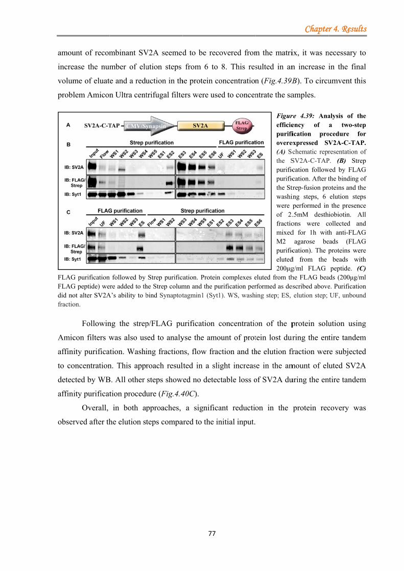
amou
incre
volum
probl
FLAGFLAGdid nofractio
Amic
affin
to co
detec
affin
obser
unt of recom
ease the num
me of eluat
lem Amicon
G purificationG peptide) weot alter SV2Aon.
Followin
con filters w
nity purifica
oncentration
cted by WB
nity purificat
Overall,
rved after th
mbinant SV
mber of elu
e and a redu
n Ultra cent
n followed by ere added to thA’s ability to b
ng the strep
was also us
ation. Washi
n. This appr
B. All other
tion proced
in both a
he elution s
V2A seemed
ution steps
uction in th
trifugal filte
Strep purifiche Strep columbind Synaptot
p/FLAG pu
sed to analy
ing fraction
roach resul
steps show
ure (Fig.4.4
pproaches,
teps compa
77
d to be rec
from 6 to
he protein co
ers were use
cation. Proteinmn and the putagmin1 (Syt1
urification c
yse the amo
ns, flow frac
lted in a sli
wed no detec
40C).
a significa
ared to the in
overed from
8. This res
oncentration
ed to concen
n complexes eurification per1). WS, wash
concentratio
ount of prot
ction and th
ight increas
ctable loss o
ant reductio
nitial input.
m the matri
sulted in an
n (Fig.4.39B
ntrate the sa
Figurefficiepurifiovere(A) Scthe Spurificpurificthe Stwashiwere of 2.fractiomixedM2 purificeluted200μg
eluted from thrformed as deing step; ES,
on of the p
tein lost dur
he elution fr
se in the am
of SV2A du
on in the
Chapte
ix, it was n
n increase i
B). To circu
amples.
re 4.39: Anaency of ication pro
expressed SVchematic reprSV2A-C-TAPcation followcation. After ttrep-fusion proing steps, 6 performed in.5mM desthons were cd for 1h wit
agarose becation). The p
d from the g/ml FLAG he FLAG beaescribed above
elution step;
protein solu
uring the ent
fraction wer
mount of el
uring the en
protein rec
r 4. Results
necessary to
in the final
umvent this
alysis of thea two-step
ocedure forV2A-C-TAP.resentation of
P. (B) Strepwed by FLAG
the binding ofoteins and theelution steps
n the presenceiobiotin. Allollected andh anti-FLAG
eads (FLAGproteins were
beads withpeptide. (C)
ads (200μg/mle. PurificationUF, unbound
ution using
tire tandem
re subjected
uted SV2A
ntire tandem
covery was
s
o
l
s
e p r . f p G f e s e l d G G e h ) l n d
g
m
d
A
m
s
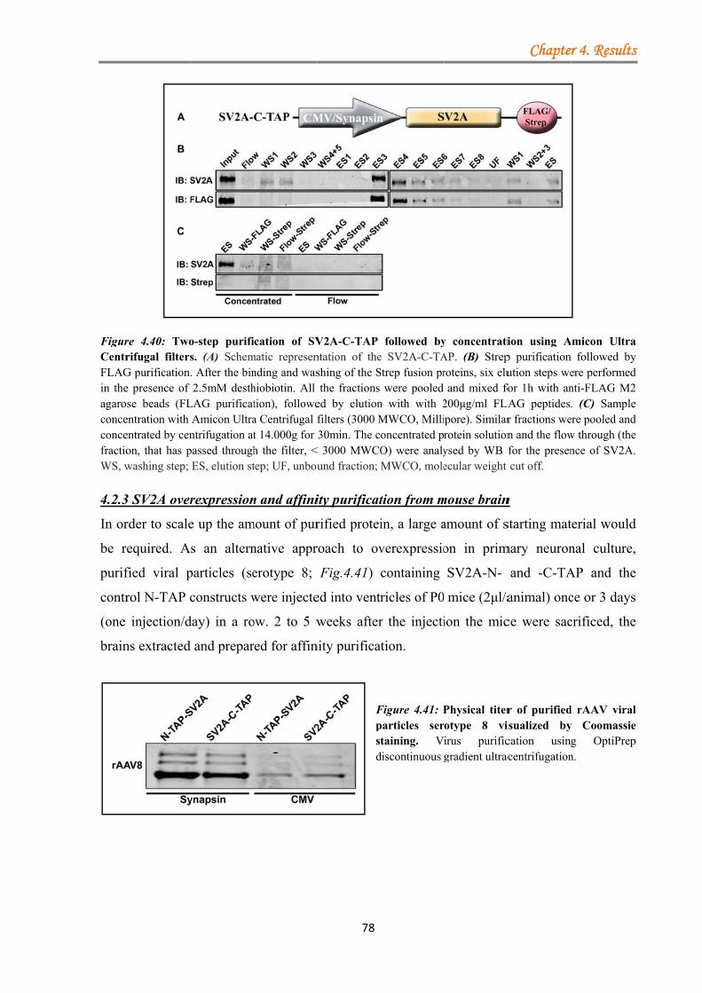
FigurCentrFLAGin theagaroconceconcefractioWS, w
4.2.3
In or
be re
purif
contr
(one
brain
re 4.40: Tworifugal filtersG purificatione presence of se beads (FL
entration with entrated by cenon, that has pwashing step;
3 SV2A over
rder to scale
equired. A
fied viral p
rol N-TAP c
injection/d
ns extracted
o-step purifis. (A) Schem. After the bin2.5mM desth
LAG purificatAmicon Ultrantrifugation atpassed throughES, elution st
rexpression
e up the am
s an altern
particles (se
constructs w
day) in a ro
d and prepar
cation of SVmatic represennding and washiobiotin. All tion), followea Centrifugal ft 14.000g for h the filter, <tep; UF, unbou
n and affini
mount of pur
native appr
erotype 8;
were injecte
ow. 2 to 5 w
red for affin
78
V2A-C-TAP ntation of theshing of the Sthe fractions
ed by elutionfilters (3000 M30min. The c
< 3000 MWCOund fraction;
ity purificat
rified protei
roach to ov
Fig.4.41)
ed into ventr
weeks after
nity purifica
Fpstd
followed bye SV2A-C-TAStrep fusion pr
were pooled n with with 2MWCO, Millioncentrated pO) were analyMWCO, mole
tion from m
in, a large a
verexpressio
containing
ricles of P0
r the injecti
tion.
Figure 4.41: Pparticles sertaining. V
discontinuous
y concentratiAP. (B) Streproteins, six eluand mixed fo
200μg/ml FLAipore). Similarrotein solutionysed by WB fecular weight
mouse brain
amount of s
on in prim
SV2A-N-
mice (2μl/a
ion the mic
Physical titerotype 8 viirus purificgradient ultrac
Chapte
ion using Ap purificationution steps wefor 1h with anLAG peptides
r fractions wen and the flowfor the presencut off.
n
starting mat
mary neuron
and -C-TA
animal) onc
ce were sac
r of purifiedisualized bycation usingacentrifugation
r 4. Results
Amicon Ultrafollowed by
ere performednti-FLAG M2. (C) Sample
ere pooled andw through (thence of SV2A.
terial would
nal culture,
AP and the
ce or 3 days
crificed, the
d rAAV viraly Coomassieg OptiPrepn.
s
a y d 2 e d e .
d
,
e
s
e
l e p
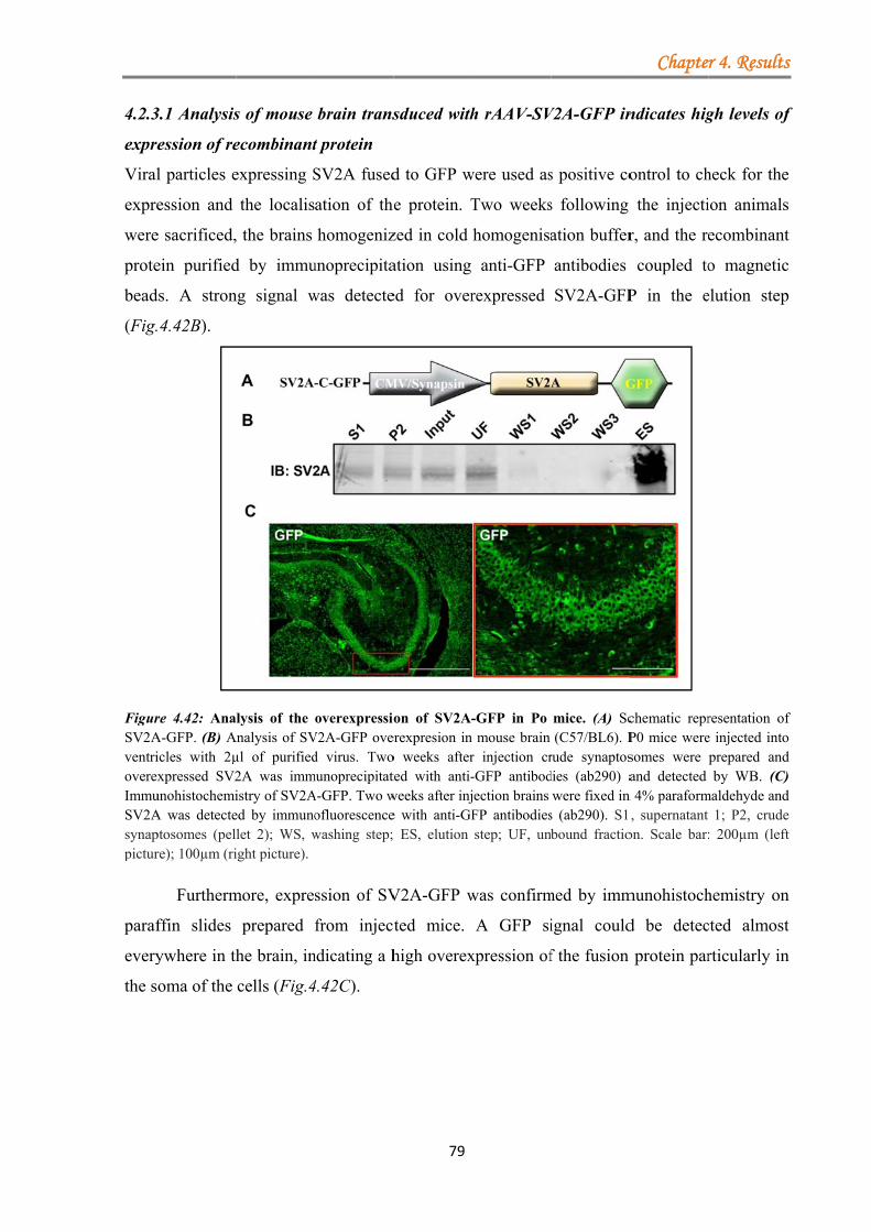
4.2.3
expre
Viral
expre
were
prote
bead
(Fig.
FigurSV2AventrioverexImmuSV2Asynappictur
paraf
every
the s
3.1 Analysis
ession of re
l particles e
ession and
e sacrificed,
ein purified
ds. A stron
4.42B).
re 4.42: AnaA-GFP. (B) Aicles with 2µxpressed SV2
unohistochemiA was detecteptosomes (pelre); 100µm (ri
Furtherm
ffin slides
ywhere in th
oma of the
s of mouse
ecombinant
expressing S
the localis
the brains
d by immu
ng signal w
lysis of the Analysis of SVµl of purified2A was immistry of SV2Aed by immunolet 2); WS, wight picture).
more, expre
prepared
he brain, in
cells (Fig.4
brain trans
t protein
SV2A fused
ation of the
homogeniz
unoprecipita
was detecte
overexpressioV2A-GFP overd virus. Two
munoprecipitatA-GFP. Two wofluorescencewashing step;
ssion of SV
from inject
ndicating a h
4.42C).
79
sduced with
d to GFP w
e protein. T
zed in cold
ation using
ed for over
on of SV2A-
erexpresion in o weeks afterted with anti-weeks after inje with anti-GF ES, elution
V2A-GFP w
ted mice.
high overex
h rAAV-SV
were used as
Two weeks
homogenisa
anti-GFP
rexpressed
-GFP in Po mouse brain
r injection cr-GFP antibodection brains FP antibodiesstep; UF, un
was confirm
A GFP si
xpression of
V2A-GFP in
s positive co
s following
ation buffer
antibodies
SV2A-GFP
mice. (A) Sc(C57/BL6). P
rude synaptosdies (ab290) a
were fixed in s (ab290). S1bound fractio
med by imm
ignal could
f the fusion
Chapte
ndicates hig
ontrol to ch
the injecti
r, and the re
coupled to
P in the e
chematic reprP0 mice weresomes were and detected
n 4% paraform, supernatant
on. Scale bar:
munohistoch
d be detec
protein par
r 4. Results
gh levels of
heck for the
ion animals
ecombinant
o magnetic
elution step
resentation ofe injected intoprepared andby WB. (C)
maldehyde and1; P2, crude
: 200µm (left
hemistry on
cted almost
rticularly in
s
f
e
s
t
c
p
f o d ) d e t
n
t
n
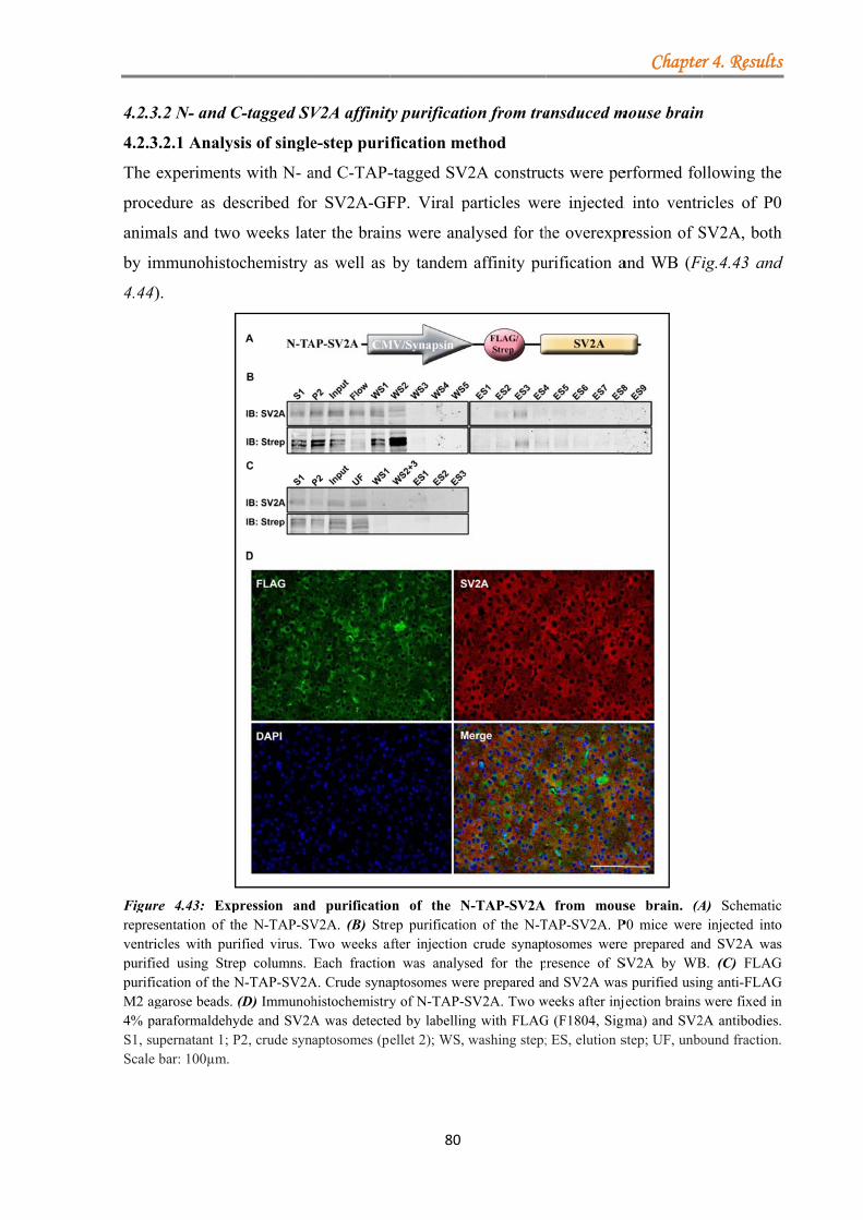
4.2.3
4.2.3
The
proce
anim
by im
4.44)
FigurrepresventripurifipurifiM2 ag4% paS1, suScale
3.2 N- and C
3.2.1 Analy
experiment
edure as de
mals and two
mmunohisto
).
re 4.43: Expsentation of thicles with pured using Strecation of the Ngarose beads. araformaldehyupernatant 1; Pbar: 100µm.
C-tagged SV
sis of single
ts with N- a
escribed for
o weeks lat
ochemistry
pression andhe N-TAP-SVrified virus. Tep columns. N-TAP-SV2A(D) Immunoh
yde and SV2AP2, crude syn
V2A affinity
e-step purif
and C-TAP-
r SV2A-GF
ter the brain
as well as
d purificatioV2A. (B) StreTwo weeks aEach fraction
A. Crude synahistochemistryA was detecteaptosomes (pe
80
ty purificati
fication me
-tagged SV
FP. Viral p
ns were ana
by tandem
on of the Nep purificatio
after injection n was analysaptosomes wey of N-TAP-Sed by labellinellet 2); WS,
ion from tra
ethod
2A constru
particles we
alysed for th
affinity pu
N-TAP-SV2A on of the N-T
crude synaptsed for the pre prepared an
SV2A. Two wng with FLAGwashing step;
ansduced m
cts were pe
ere injected
he overexpr
urification a
from mousTAP-SV2A. Ptosomes wereresence of Snd SV2A was
weeks after injG (F1804, Sigm; ES, elution s
Chapte
mouse brain
erformed fo
d into ventr
ression of S
and WB (F
se brain. (AP0 mice were e prepared anSV2A by WBs purified usinjection brains
gma) and SV2step; UF, unbo
r 4. Results
n
llowing the
ricles of P0
SV2A, both
ig.4.43 and
A) Schematicinjected into
nd SV2A wasB. (C) FLAGng anti-FLAGwere fixed in
2A antibodies.ound fraction.
s
e
0
h
d
c o s
G G n . .

FigurStrep synapCrudeSV2ASigmaelutio
of th
perfo
furth
of th
three
proba
were
re 4.44: Purifpurification
ptosomes prepe synaptosomeA-C-TAP. Mica) and SV2A
on step; UF, un
From eac
he detergen
ormed: Stre
hermore, a l
he Strep pur
e (Fig.4.43B
ably due to
e performed
fication and of the SV2A
pared. SV2A wes were prepace brains wer
A antibodies. Snbound fractio
ch injected
nt DDM (
ep and/or F
arge percen
ification, w
B and 4.44B
o the low t
d in order t
analysis of SA-C-TAP by. was purified uared and SV2Are fixed in 4%S1, supernataon. Scale bar:
brain, crude
(3.9mM). T
FLAG purif
ntage of reco
while the rem
B). The FL
itre of the
to avoid SV
81
SV2A-C-TAPTwo weeks
using Strep coA was purified% paraformaldant 1; P2, cru
100µm.
e synaptoso
Two differ
fication. Th
ombinant p
maining pro
LAG purific
injected vi
V2A remai
P. (A) Schemaafter injectio
olumns. (C) Fd and analysedehyde and S
ude synaptoso
omes were p
rent types
he expressi
rotein was
otein was po
cation itself
irus. Moreo
ining bound
atic representaon P0 mice wFLAG purificad by WB. (D)
SV2A detectedmes (pellet 2
prepared and
of one-ste
on level of
lost in the f
oorly recove
f did not giv
over, supple
d to the an
Chapte
ation of SV2Awere sacrificeation of the S) Immunohistd by using FL2); WS, wash
d lysed in th
ep purifica
f SV2A wa
first two wa
ered in the e
ve satisfact
ementary el
nti-FLAG M
r 4. Results
A-C-TAP. (B)ed and crude
SV2A-C-TAP.ochemistry ofLAG (F1804,
hing step; ES,
he presence
ations were
as low and
ashing steps
elution step
tory results,
lution steps
M2 agarose
s
) e . f , ,
e
e
d
s
p
,
s
e
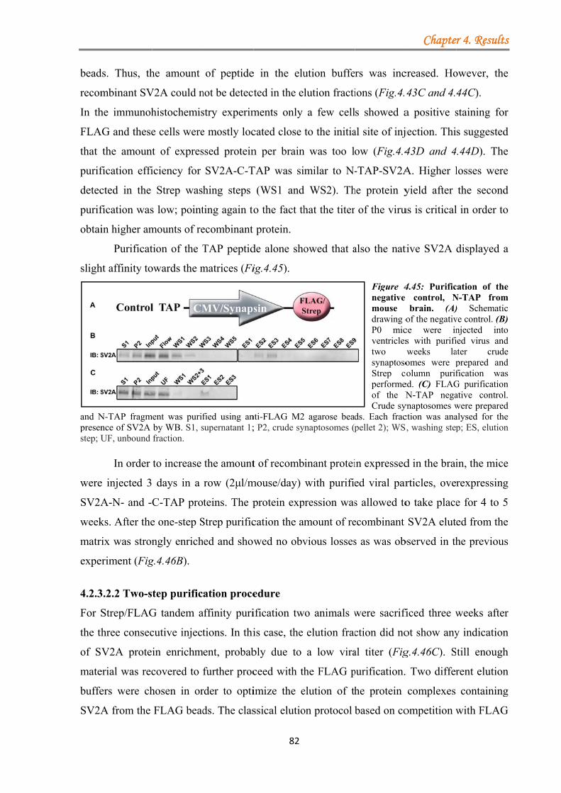
bead
recom
In th
FLA
that
purif
detec
purif
obtai
sligh
and Npresenstep; U
were
SV2A
week
matri
expe
4.2.3
For S
the th
of SV
mate
buffe
SV2A
ds. Thus, th
mbinant SV
he immunoh
AG and these
the amount
fication effi
cted in the
fication was
in higher am
Purificat
ht affinity to
N-TAP fragmnce of SV2A UF, unbound
In order
e injected 3
A-N- and -
ks. After the
ix was stron
riment (Fig
3.2.2 Two-s
Strep/FLAG
hree consec
V2A prote
erial was rec
ers were ch
A from the
he amount
V2A could n
histochemis
e cells were
t of expres
iciency for
Strep was
s low; point
mounts of re
tion of the T
owards the m
ment was purifby WB. S1, sfraction.
to increase
days in a
C-TAP pro
e one-step S
ngly enrich
g.4.46B).
tep purific
G tandem a
cutive inject
in enrichm
covered to
hosen in or
FLAG bead
of peptide
not be detect
stry experim
e mostly loc
ssed protein
SV2A-C-T
hing steps
ting again to
ecombinant
TAP peptid
matrices (Fi
fied using ansupernatant 1;
the amount
row (2μl/m
teins. The p
Strep purific
hed and show
ation proce
affinity puri
tions. In thi
ment, probab
further proc
rder to opti
ds. The clas
82
e in the elu
ted in the el
ments only
cated close
n per brain
TAP was si
(WS1 and
o the fact th
protein.
de alone sho
ig.4.45).
nti-FLAG M2; P2, crude sy
t of recomb
mouse/day)
protein exp
cation the am
wed no obv
edure
ification tw
is case, the
bly due to
ceed with th
imize the e
ssical elutio
ution buffe
lution fracti
a few cell
to the initia
n was too l
milar to N-
WS2). Th
hat the titer
owed that a
agarose beadynaptosomes (p
inant protei
with purifie
pression was
mount of re
vious losses
wo animals w
elution frac
a low vira
he FLAG p
elution of th
on protocol
rs was inc
ions (Fig.4.
s showed a
al site of inj
ow (Fig.4.4
-TAP-SV2A
he protein y
of the viru
also the nat
Figure 4negative mouse drawing oP0 micventriclestwo synaptosoStrep cperformeof the NCrude sy
ds. Each fracpellet 2); WS
in expressed
ed viral par
s allowed to
ecombinant
s as was ob
were sacrifi
ction did no
al titer (Fig
purification.
he protein
based on co
Chapte
creased. Ho
43C and 4.
a positive s
jection. Thi
43D and 4
A. Higher l
yield after
us is critical
tive SV2A
4.45: Purific control, Nbrain. (A)
of the negativce were ins with purifiweeks laomes were p
column purifed. (C) FLAGN-TAP nega
ynaptosomes wction was anaS, washing step
d in the brai
rticles, ove
o take place
SV2A elut
bserved in th
ficed three w
ot show any
g.4.46C). S
. Two differ
complexes
ompetition w
r 4. Results
owever, the
44C).
staining for
s suggested
4.44D). The
losses were
the second
l in order to
displayed a
cation of theN-TAP from) Schematicve control. (B)njected intoied virus andater crudeprepared andfication wasG purificationative control.were preparedalysed for thep; ES, elution
in, the mice
rexpressing
e for 4 to 5
ed from the
he previous
weeks after
y indication
till enough
rent elution
containing
with FLAG
s
e
r
d
e
e
d
o
a
e m c ) o d e d s n .
d e n
e
g
5
e
s
r
n
h
n
g
G
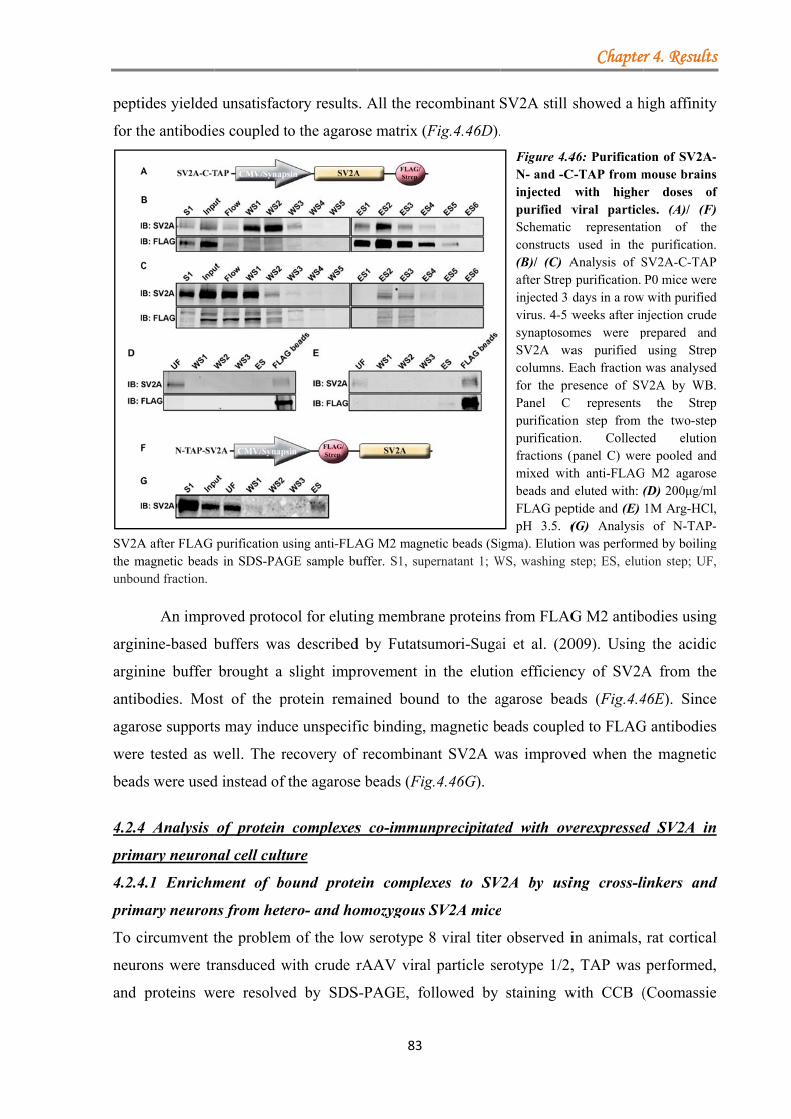
pepti
for th
SV2Athe munbou
argin
argin
antib
agaro
were
bead
4.2.4
prim
4.2.4
prim
To c
neuro
and
ides yielded
he antibodie
A after FLAG magnetic beadsund fraction.
An impr
nine-based b
nine buffer
bodies. Mos
ose support
e tested as w
ds were used
4 Analysis
mary neuron
4.1 Enrichm
mary neuron
ircumvent t
ons were tr
proteins w
d unsatisfac
es coupled t
purification us in SDS-PAG
oved protoc
buffers was
brought a
st of the p
s may induc
well. The r
d instead of
of protein
nal cell cult
ment of bo
ns from hete
the problem
ransduced w
were resolve
tory results
to the agaro
using anti-FLAGE sample bu
col for eluti
s described
slight impr
protein rema
ce unspecifi
recovery of
the agarose
complexes
ure
ound prote
ero- and ho
m of the low
with crude r
ed by SDS
83
s. All the rec
ose matrix (F
AG M2 magnuffer. S1, sup
ing membra
d by Futatsu
rovement in
ained boun
fic binding,
f recombina
e beads (Fig
s co-immun
ein comple
omozygous S
w serotype
rAAV viral
S-PAGE, fo
combinant S
Fig.4.46D).
netic beads (Sipernatant 1; W
ane proteins
umori-Suga
n the elutio
nd to the a
magnetic b
ant SV2A w
g.4.46G).
nprecipitate
exes to SV
SV2A mice
8 viral titer
l particle se
ollowed by
SV2A still
Figure 4.4N- and -Cinjected purified vSchematic constructs (B)/ (C) Aafter Strep injected 3 dvirus. 4-5 wsynaptosomSV2A wacolumns. Efor the prPanel C purificationpurificationfractions (pmixed witbeads and FLAG peppH 3.5. (
igma). ElutionWS, washing s
from FLAG
ai et al. (20
on efficienc
agarose bea
eads couple
was improve
ed with ov
V2A by usi
e
r observed i
erotype 1/2,
staining w
Chapte
showed a h
46: PurificatiC-TAP from m
with higheviral particl representatused in the
Analysis of Sp purification. days in a rowweeks after inmes were pas purified Each fraction resence of SV
represents n step from n. Collect
(panel C) werth anti-FLAGeluted with:
ptide and (E) (G) Analysisn was performstep; ES, elut
G M2 antib
009). Using
cy of SV2A
ads (Fig.4.4
ed to FLAG
ed when th
verexpressed
ing cross-li
in animals,
, TAP was
with CCB (
r 4. Results
high affinity
ion of SV2A-mouse brainsr doses ofles. (A)/ (F)tion of thee purification.SV2A-C-TAPP0 mice were
w with purifiednjection crudeprepared and
using Strepwas analysed
V2A by WB.the Strep
the two-stepted elutionre pooled and
G M2 agarose(D) 200μg/ml1M Arg-HCl,
s of N-TAP-med by boilingtion step; UF,
bodies using
g the acidic
A from the
46E). Since
G antibodies
he magnetic
d SV2A in
inkers and
rat cortical
performed,
(Coomassie
s
y
-s f ) e .
P e d e d p d .
p p n d e l , -g ,
g
c
e
e
s
c
n
d
l
,
e
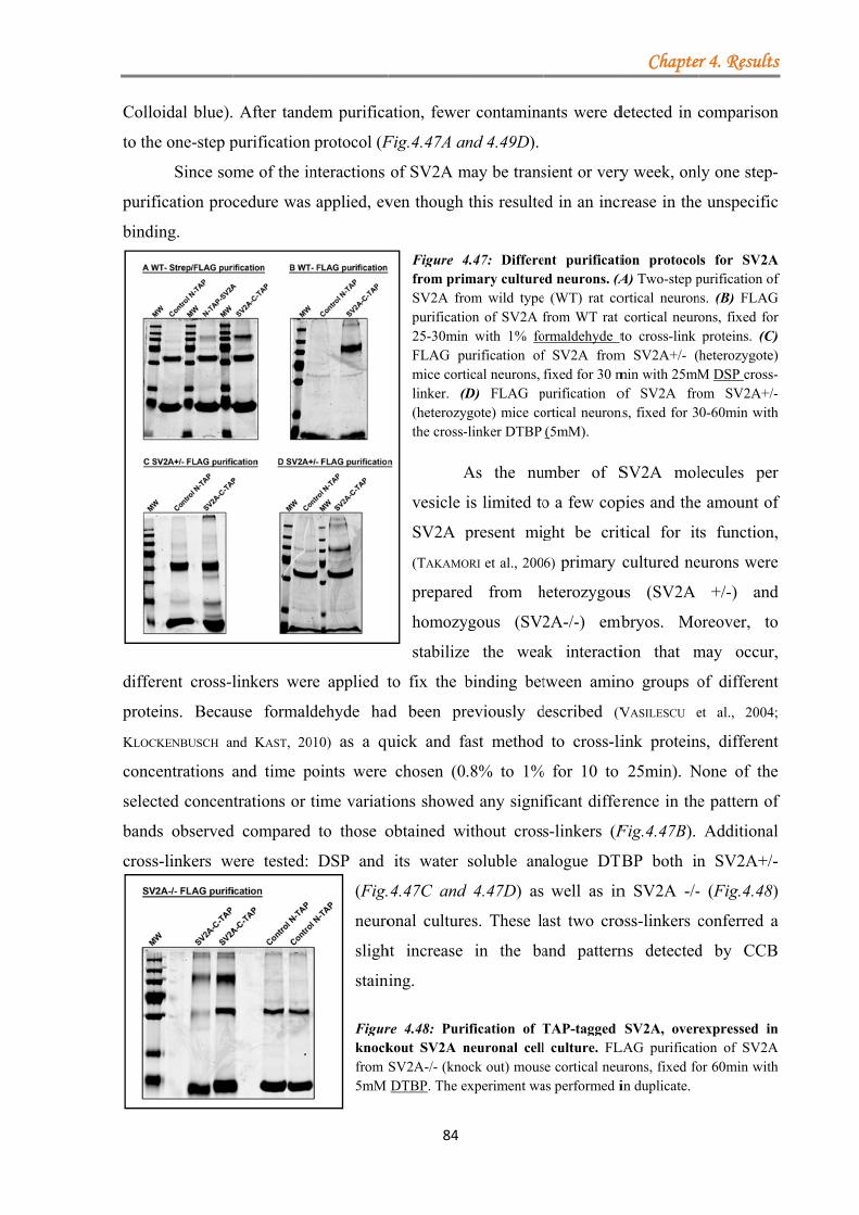
Collo
to the
purif
bindi
diffe
prote
KLOC
conc
selec
band
cross
oidal blue).
e one-step p
Since so
fication proc
ing.
rent cross-l
eins. Becau
CKENBUSCH an
entrations a
cted concen
ds observed
s-linkers w
After tand
purification
me of the in
cedure was
linkers wer
use formald
nd KAST, 20
and time p
ntrations or
d compared
were tested:
dem purifica
protocol (F
nteractions
applied, ev
re applied t
dehyde had
010) as a qu
points were
time variati
to those o
DSP and
(Fig.
neuro
sligh
stain
Figurknockfrom 5mM
84
ation, fewer
Fig.4.47A an
of SV2A m
ven though
Figure 4from primSV2A fropurificatio25-30minFLAG pumice cortlinker. (D(heterozythe cross-
A
vesicle
SV2A p
(TAKAMO
prepared
homozy
stabilize
to fix the b
d been pr
uick and fa
chosen (0
ions showe
obtained w
its water
4.47C and
onal culture
ht increase
ning.
re 4.48: Purikout SV2A nSV2A-/- (knoDTBP. The e
r contamina
nd 4.49D).
may be trans
this resulte
4.47: Differemary cultureom wild typeon of SV2A f
n with 1% fourification oftical neurons, (D) FLAG pygote) mice co-linker DTBP
As the nu
is limited to
present mi
ORI et al., 200
d from h
ygous (SV2
e the wea
binding bet
reviously d
fast method
.8% to 1%
d any signi
ithout cros
soluble ana
d 4.47D) as
es. These la
in the ba
ification of Tneuronal cellock out) mousexperiment wa
ants were d
sient or very
ed in an incr
ent purificatied neurons. (Ae (WT) rat cofrom WT rat
ormaldehyde tf SV2A fromfixed for 30 m
purification oortical neuron(5mM).
umber of S
o a few cop
ght be crit
6) primary
heterozygou
2A-/-) emb
ak interacti
tween amin
described (V
to cross-li
% for 10 to
ficant differ
s-linkers (F
alogue DTB
well as in
ast two cro
and pattern
TAP-tagged l culture. FLse cortical neuas performed i
Chapte
detected in c
y week, onl
rease in the
ion protocolA) Two-step portical neuroncortical neuroto cross-link
m SV2A+/- (min with 25mMof SV2A frons, fixed for 3
SV2A mol
pies and the
tical for it
cultured ne
us (SV2A
mbryos. Mo
ion that m
no groups o
VASILESCU e
ink protein
25min). N
erence in the
Fig.4.47B).
TBP both in
n SV2A -/-
oss-linkers c
ns detected
SV2A, overLAG purificaturons, fixed foin duplicate.
r 4. Results
comparison
ly one step-
e unspecific
ls for SV2Apurification ofns. (B) FLAGons, fixed forproteins. (C)heterozygote)M DSP cross-om SV2A+/-0-60min with
lecules per
e amount of
ts function,
eurons were
+/-) and
oreover, to
may occur,
of different
et al., 2004;
ns, different
None of the
e pattern of
Additional
n SV2A+/-
- (Fig.4.48)
conferred a
d by CCB
expressed inion of SV2Aor 60min with
s
n
-
c
A f
G r ) ) -- h
r
f
,
e
d
o
,
t
;
t
e
f
l
-
)
a
B
n A h
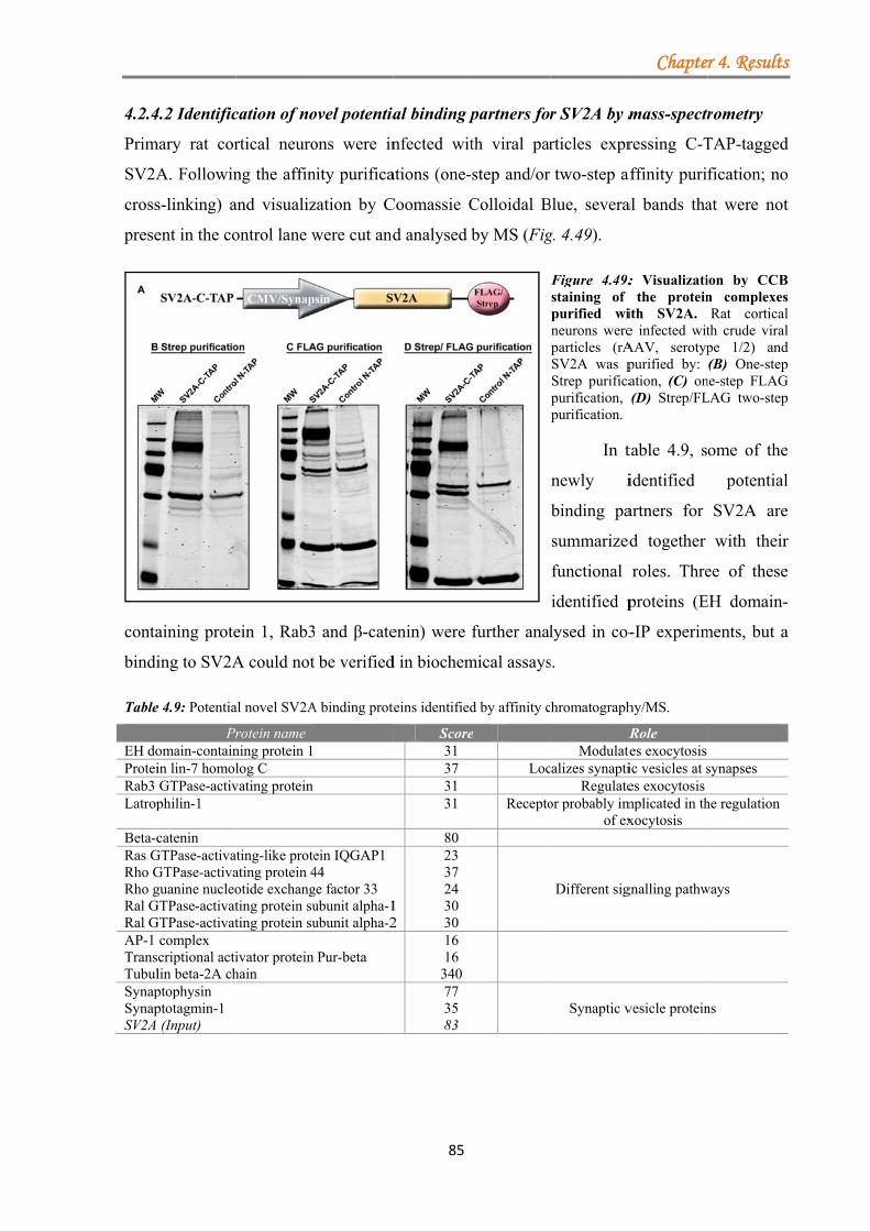
4.2.4
Prim
SV2A
cross
prese
conta
bindi
Table
EH doProteiRab3 Latrop
Beta-cRas GRho GRho gRal GRal GAP-1 TransTubulSynapSynapSV2A
4.2 Identific
mary rat cor
A. Followin
s-linking) a
ent in the co
aining prote
ing to SV2A
e 4.9: Potentia
Promain-containin lin-7 homolGTPase-activphilin-1
catenin GTPase-activaGTPase-activaguanine nucleo
GTPase-activatGTPase-activat
complex scriptional actilin beta-2A chptophysin ptotagmin-1 (Input)
cation of no
rtical neuro
ng the affin
and visualiz
ontrol lane w
ein 1, Rab3
A could not
al novel SV2A
rotein name ning protein 1 log C
vating protein
ating-like proteating protein 4otide exchangting protein suting protein su
ivator protein hain
ovel potentia
ons were in
nity purifica
zation by C
were cut and
3 and β-cate
be verified
A binding prot
ein IQGAP1 44 ge factor 33 ubunit alpha-1ubunit alpha-2
Pur-beta
85
al binding p
nfected wit
ations (one-
Coomassie C
d analysed b
enin) were
d in biochem
teins identified
Score31 37 31 31
80
1 2
23 37 24 30 30 16 16 340 77 35 83
partners for
th viral par
step and/or
Colloidal B
by MS (Fig
further anal
mical assays
d by affinity c
e
Loc
Recepto
r SV2A by m
rticles expr
two-step a
Blue, severa
g. 4.49).
Figure 4.49:staining of purified wineurons wereparticles (rASV2A was pStrep purificapurification, purification.
In t
newly i
binding pa
summarized
functional
identified p
lysed in co-
s.
hromatograph
RModulate
calizes synaptiRegulate
or probably imof ex
Different sig
Synaptic v
Chapte
mass-spectr
ressing C-T
affinity puri
al bands tha
: Visualizatithe protein
ith SV2A. e infected witAAV, serotyppurified by: ation, (C) on(D) Strep/FL
table 4.9, s
identified
artners for
d together
roles. Thre
proteins (E
-IP experim
hy/MS.
Role tes exocytosis ic vesicles at ses exocytosis
mplicated in thxocytosis
gnalling pathw
vesicle protein
r 4. Results
rometry
TAP-tagged
fication; no
at were not
ion by CCBn complexesRat cortical
th crude viralpe 1/2) and(B) One-step
ne-step FLAGLAG two-step
ome of the
potential
SV2A are
with their
ee of these
EH domain-
ments, but a
synapses
he regulation
ways
ns
s
d
o
t
B s l l d p G p
e
l
e
r
e
-
a
Chapter 5. Discussion
86
5. Discussion
Synaptic plasticity denotes the ability of neurons to respond to the ongoing network activity,
by weakening or strengthening their activity. These adaptive changes can take place both at
the pre- and postsynaptic terminals. Whereas the molecular mechanisms underlying
postsynaptic plasticity have been studied in great detail, the cellular events mediating
presynaptic plasticity are not as well understood. It has been hypothesized that presynaptic
plasticity may involve the posttranslational modification of proteins. One of these
modifications is the phosphorylation of proteins, which plays a role in the plasticity-induced
remodelling of established AZs or in the assembly of novel active zones.
Two presynaptic proteins, the AZ protein RIM1α and the SV protein SV2A have been
shown to be important in presynaptic plasticity, by modulating the function and properties of
the presynaptic release machinery, e.g. SV priming. However, their precise mode of action is
still unresolved. Thus, to gain a better understanding the following goals were pursued in this
thesis:
(5.1) The effect of hyperphosphorylation on RIM1α at synapses.
(5.2) The identification of novel potential phospho-dependent binding partners for RIM1α.
(5.3) To gain insight into the enigmatic function of SV2A, we aimed to purify and analyse
novel SV2A binding partners.
5.1 Hyperphosphorylation alters the distribution of the presynaptic protein RIM1α at
synapses
Presynaptic plasticity involves the structural remodelling of the CAZ, which in turn engages
dynamic changes in protein turnover and protein interactions. At the D.melanogaster NMJs
an increase in the amount of the active zone protein Bruchpilot (Brp) and an enlargement of
the presynaptic cytometrix structure was detected after the rapid induction of presynaptic
strengthening. Moreover, the fast recruitment of Brp to the AZ led to an increase in the
number of SVs and calcium channels at synapses (WEYHERSMÜLLER et al., 2011). The structural
remodelling of the CAZ on a timescale of minutes was further supported by studies in
hippocampal neurons using a fluorescently labelled Bassoon. The local redistribution of the
AZ component led to rapid changes in the size of the AZ, which correlated with modifications
in the RRP and the release probability (MATZ et al., 2010). Prolonged silencing of excitatory
neurons caused a down-regulation of cellular expression levels of distinct presynaptic
proteins, including RIM1α (LAZAREVIC et al., 2011). Interestingly, the decrease in the amount of
RIM1α was not caused by UPS-dependent degradation but rather dependent on other
Chapter 5. Discussion
87
mechanisms, like the regulation of transcription and/or translation. Whereas the number of
synapses labelled by RIM1α was decreased after silencing, the remaining positive puncta
showed an increase in the level of RIM1α. This suggested a redistribution of RIM1α after
prolonged activity deprivation (LAZAREVIC et al., 2011).
Even though the precise molecular mechanisms controlling synaptic adaptation are not
well understood, protein phosphorylation has been postulated to contribute to the underlying
synapse remodelling and plasticity. For example, ERK kinase, which can integrate a variety
of signals, exerts many downstream effects, like, dendritic spine stabilization, modulation of
ion channels or receptor insertion (review: SWEATT, 2004). Cdk5 was shown to be involved in
LTP and LTD, acting as a homeostatic regulator of synaptic plasticity (review: SHAH and LAHIRI,
2014). One of the central enzymes actively involved in presynaptic plasticity is PKA, e.g.
essential for mossy fiber (MF)-LTP (review: CASTILLO, 2012).
It has been shown that the presynaptic protein RIM1α acts as substrate for several
kinases: PKA (LONART et al., 2003), ERK2 (SIMSEK-DURAN and LONART, 2008), CaMKII (SUN et al.,
2003) and SAD-B (INOUE et al., 2006). Whereas the phosphorylation of the amino acid residue
S413in RIM1α by PKA was required for the induction of presynaptic LTP in cultured
cerebellar granular and Purkinje cell neurons (LONART et al., 2003), studies in mice expressing
mutations in this position were not able to confirm this (KAESER et al., 2008a; YANG and CALAKOS,
2010). Although RIM1α has been shown to be phosphorylated by a variety of kinases, the
functional implications of phosphorylation/dephosphorylation of RIM proteins are not well
understood. However, such posttranslational modifications regulated by synaptic activity,
may directly impact RIM1α’s properties, like its binding affinity to other proteins or its
stability.
To test if the increased phosphorylation status of RIM1α affects its level or its
association with the AZ, neuronal cell cultures were incubated with various phosphatase
inhibitors. At different time intervals the neuronal cultures were fixed, endogenous RIM1/2
was labelled and the area of the bouton marked by RIM1α determined. Our results show, that
by shifting the equilibrium to a hyperphosphorylated state, RIM1α distribution at synapses is
altered. Application of phosSTOP led to a significant increase in the number of boutons with
smaller RIM1α marked areas in comparison to control conditions. By blocking only the PP1
and PP2A phosphatases using either 10nM okadaic acid or 2nM Calyculin A, a non-
significant increase in the number of RIM1/2 labelled boutons with smaller sizes compared to
DMSO control conditions was detected.
Chapter 5. Discussion
88
High level of protein phosphorylation may affect RIM1α turn-over by either
increasing or decreasing its stability. Computer-based simulations have described proteins to
become more degradable if more sites are phosphorylated (multi-site phosphorylation). Such
phosphorylation-dependent degradation processes are important for proteins involved in
regulating the cell cycle (VAREDI et al., 2010). Because RIM1α is a protein with multiple
phosphorylation sites, high kinase activity might trigger a degradation process. However, the
total intensity of RIM1/2 labelling was not changed after phosSTOP treatment compared to
the control condition. Therefore, these results do not suggest that a degradation mechanism
might be induced by phosphorylation of RIM1α. On the other hand, phosphorylation may as
well protect proteins from degradation, by blocking for example either the binding of E3
ubiquitin ligase to the substrate or by impairing its interaction with E2 ubiquitin-conjugation
enzymes. It was hypothesised that RIM1α phosphorylation by PKA, may provide resistance to
the degradation by the proteasome (CROWFORD and MENNERICK, 2012). However, to date there is
no experimental evidence supporting this hypothesis.
In addition to protein stability, phosphorylation events may impact the association of
RIM1α with the CAZ. It has been shown that certain phosphorylation events of CAZ
components induce protein solubilisation by interfering with their intermolecular interactions.
Binding of the 14-3-3 adapter protein to phosphorylated Bassoon (S2845) decreased the
attachment of Bassoon to the AZ (SCHRÖDER et al., 2013). Moreover, treatment of neuronal
cultures with the phosphatase inhibitor okadaic acid inhibitor further induced solubilisation
and diffusion of Bassoon, CAST and RIM proteins (SCHRÖDER et al., 2013). Accordingly, our
experiments may indicate that hyperphosphorylated RIM1/2 protein becomes more soluble
and diffuses away from the AZ and the bouton.
Prolonged exposure to okadaic acid has been shown to have cytotoxic effects on a
variety of cells and to mediate neurodegenerative mechanisms by blocking PP2A phosphatase
activity. The impairment of PP2A activity stimulates a large group of kinases, like ERK2,
CaMKII, PKA (review: KAMAT et al., 2013). In our experiments, 1h stimulation did not induce
neurotoxicity and moreover, the observed changes in the distribution of RIM1α may be a
direct consequence of the interplay between multiple kinases that act at synapses.
However, in interpreting the functional relevance of our observation, it has to be
considered that hyperphosphorylation of proteins induced by various pharmacological
treatments is not physiological, but rather pushes the system to one side of the equilibrium.
Secondly, our approach, immunocytochemistry, does not allow capturing the dynamic nature
of phosphorylation events. In summary, our analysis revealed that the distribution of the
Chapter 5. Discussion
89
RIM1/2 protein is influenced by kinase activity and that hyperphosphorylation may affect the
association of RIM1α with the CAZ.
5.2 Identification of novel phosphorylation-dependent RIM1α binding proteins
Over the years proteomics has emerged as an important tool to identify and characterize
components of the pre- and postsynaptic terminals. Over 1000 proteins were identified in
synaptosomes from whole mouse brain, representing the first inventory of the synaptic
proteome (SCHRIMPF et al., 2005). Nevertheless the interactome of presynaptic proteins is not yet
fully characterized, especially, when considering posttranslational modifications or transitory
protein interactions.
To identify novel potential binding partners for the N- and C-terminal region of
RIM1α we performed affinity purifications and mass spectrometry (MS). By using this
approach more than 100 proteins were identified to associate with either the RIM1α ZF-PDZ
or the RIM1α C2A-C2B domains. These proteins were classified in five categories: signaling
cascades/kinases (SC), synaptic vesicles (SV), plasma membrane/active zone (AZ),
cytoskeleton (C) and others (O). Of these, the proteins of the signalling cascades/kinases and
plasma membrane/active zone group accounted for nearly half of the total identified proteins.
Another large class was the others group, which included mitochondrial proteins or proteins
whose localizations and functions were not well known.
In a recent study, the immunopurification and MS analysis of the presynaptic AZ
resulted in the identification of 485 proteins, data that suggests the AZ to be enriched in
proteins involved not only in neurotransmitter release but also in other cellular activities
(WEINGARTEN et al., 2014). On the other hand, a comparison of the proteome of glutamatergic and
GABA-ergic synapses showed no major differences between proteins involved in SV docking
and release. Besides SV and AZ proteins, ion channels and transporters were also identified
(BOYKEN et al., 2013). In accordance with Weingarten et al., we detected an overlap between our
identified proteins and the purified AZ-proteins from their study. Similar proteins were
presented in the SV group (vATPase subunit A, C1 and E1; synapsin-1, Rab-3A,
phosphoglycerate kinase 1, peroxiredoxin-6, alpha-enolase, pyruvate kinase isozymes
M1/M2, malate dehydrogenase, AP-2 subunit alpha-2, dynamin-1-like protein, clathrin light
chain A and B); the AZ group (GLAST-1, synatxin-1B, EAAT2, SNAP-25, Thy-1, neuronal
membrane glycoprotein M6-a, paralemmin-1, copine, thioredoxin-related transmembrane
protein); the SC group (heat shock proteins, G proteins, 14-3-3 proteins, septin-5,
calcineurin); the C group (tubulins, actin, septin-11, septin-6, cofilin-1, profilin-1).
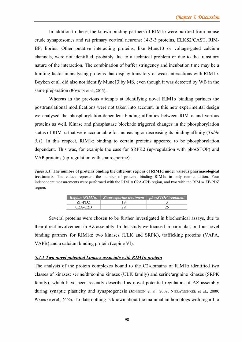
Chapter 5. Discussion
90
In addition to these, the known binding partners of RIM1α were purified from mouse
crude synaptosomes and rat primary cortical neurons: 14-3-3 proteins, ELKS2/CAST, RIM-
BP, liprins. Other putative interacting proteins, like Munc13 or voltage-gated calcium
channels, were not identified, probably due to a technical problem or due to the transitory
nature of the interaction. The combination of buffer stringency and incubation time may be a
limiting factor in analysing proteins that display transitory or weak interactions with RIM1α.
Boyken et al. did also not identify Munc13 by MS, even though it was detected by WB in the
same preparation (BOYKEN et al., 2013).
Whereas in the previous attempts at identifying novel RIM1α binding partners the
posttranslational modifications were not taken into account, in this new experimental design
we analysed the phosphorylation-dependent binding affinities between RIM1α and various
proteins as well. Kinase and phosphatase blockade triggered changes in the phosphorylation
status of RIM1α that were accountable for increasing or decreasing its binding affinity (Table
5.1). In this respect, RIM1α binding to certain proteins appeared to be phosphorylation
dependent. This was, for example the case for SRPK2 (up-regulation with phosSTOP) and
VAP proteins (up-regulation with staurosporine).
Table 5.1: The number of proteins binding the different regions of RIM1α under various pharmacological treatments. The values represent the number of proteins binding RIM1α in only one condition. Four independent measurements were performed with the RIM1α C2A-C2B region, and two with the RIM1α ZF-PDZ region.
Region (RIM1α) Staurosporine treatment phosSTOP treatment ZF-PDZ 18 3
C2A-C2B 29 25 Several proteins were chosen to be further investigated in biochemical assays, due to
their direct involvement in AZ assembly. In this study we focused in particular, on four novel
binding partners for RIM1α: two kinases (ULK and SRPK), trafficking proteins (VAPA,
VAPB) and a calcium binding protein (copine VI).
5.2.1 Two novel potential kinases associate with RIM1α protein
The analysis of the protein complexes bound to the C2-domains of RIM1α identified two
classes of kinases: serine/threonine kinases (ULK family) and serine/arginine kinases (SRPK
family), which have been recently described as novel potential regulators of AZ assembly
during synaptic plasticity and synaptogenesis (JOHNSON et al., 2009; NIERATSCHKER et al., 2009;
WAIRKAR et al., 2009). To date nothing is known about the mammalian homologs with regard to
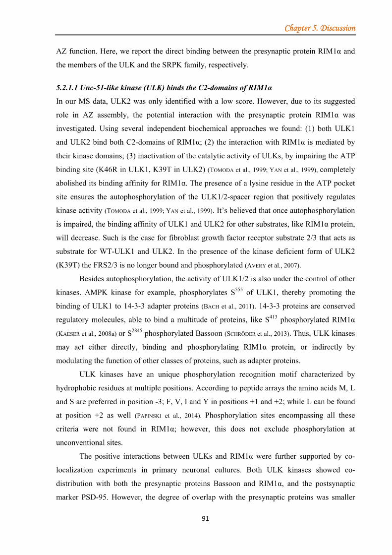
Chapter 5. Discussion
91
AZ function. Here, we report the direct binding between the presynaptic protein RIM1α and
the members of the ULK and the SRPK family, respectively.
5.2.1.1 Unc-51-like kinase (ULK) binds the C2-domains of RIM1α
In our MS data, ULK2 was only identified with a low score. However, due to its suggested
role in AZ assembly, the potential interaction with the presynaptic protein RIM1α was
investigated. Using several independent biochemical approaches we found: (1) both ULK1
and ULK2 bind both C2-domains of RIM1α; (2) the interaction with RIM1α is mediated by
their kinase domains; (3) inactivation of the catalytic activity of ULKs, by impairing the ATP
binding site (K46R in ULK1, K39T in ULK2) (TOMODA et al., 1999; YAN et al., 1999), completely
abolished its binding affinity for RIM1α. The presence of a lysine residue in the ATP pocket
site ensures the autophosphorylation of the ULK1/2-spacer region that positively regulates
kinase activity (TOMODA et al., 1999; YAN et al., 1999). It’s believed that once autophosphorylation
is impaired, the binding affinity of ULK1 and ULK2 for other substrates, like RIM1α protein,
will decrease. Such is the case for fibroblast growth factor receptor substrate 2/3 that acts as
substrate for WT-ULK1 and ULK2. In the presence of the kinase deficient form of ULK2
(K39T) the FRS2/3 is no longer bound and phosphorylated (AVERY et al., 2007).
Besides autophosphorylation, the activity of ULK1/2 is also under the control of other
kinases. AMPK kinase for example, phosphorylates S555 of ULK1, thereby promoting the
binding of ULK1 to 14-3-3 adapter proteins (BACH et al., 2011). 14-3-3 proteins are conserved
regulatory molecules, able to bind a multitude of proteins, like S413 phosphorylated RIM1α
(KAESER et al., 2008a) or S2845 phosphorylated Bassoon (SCHRÖDER et al., 2013). Thus, ULK kinases
may act either directly, binding and phosphorylating RIM1α protein, or indirectly by
modulating the function of other classes of proteins, such as adapter proteins.
ULK kinases have an unique phosphorylation recognition motif characterized by
hydrophobic residues at multiple positions. According to peptide arrays the amino acids M, L
and S are preferred in position -3; F, V, I and Y in positions +1 and +2; while L can be found
at position +2 as well (PAPINSKI et al., 2014). Phosphorylation sites encompassing all these
criteria were not found in RIM1α; however, this does not exclude phosphorylation at
unconventional sites.
The positive interactions between ULKs and RIM1α were further supported by co-
localization experiments in primary neuronal cultures. Both ULK kinases showed co-
distribution with both the presynaptic proteins Bassoon and RIM1α, and the postsynaptic
marker PSD-95. However, the degree of overlap with the presynaptic proteins was smaller
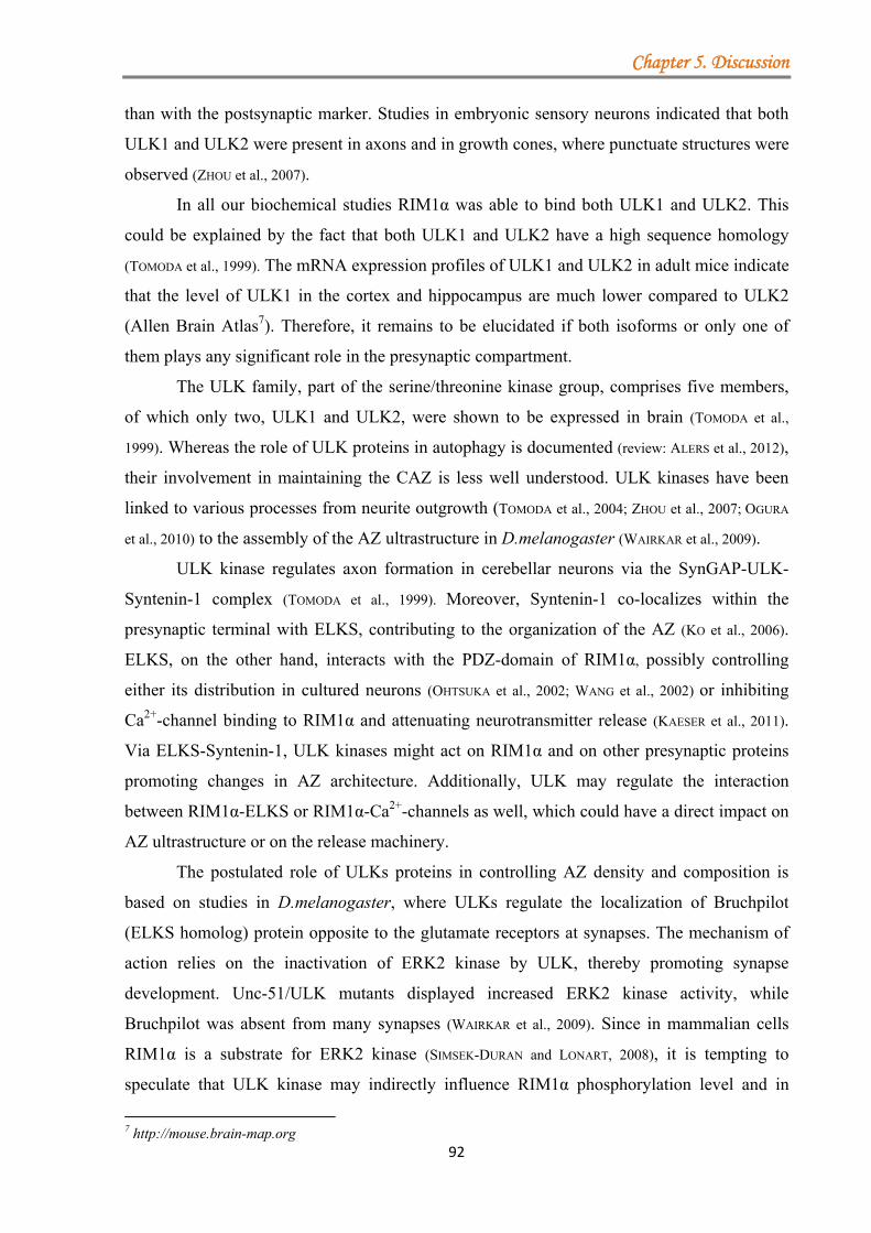
Chapter 5. Discussion
92
than with the postsynaptic marker. Studies in embryonic sensory neurons indicated that both
ULK1 and ULK2 were present in axons and in growth cones, where punctuate structures were
observed (ZHOU et al., 2007).
In all our biochemical studies RIM1α was able to bind both ULK1 and ULK2. This
could be explained by the fact that both ULK1 and ULK2 have a high sequence homology
(TOMODA et al., 1999). The mRNA expression profiles of ULK1 and ULK2 in adult mice indicate
that the level of ULK1 in the cortex and hippocampus are much lower compared to ULK2
(Allen Brain Atlas7). Therefore, it remains to be elucidated if both isoforms or only one of
them plays any significant role in the presynaptic compartment.
The ULK family, part of the serine/threonine kinase group, comprises five members,
of which only two, ULK1 and ULK2, were shown to be expressed in brain (TOMODA et al.,
1999). Whereas the role of ULK proteins in autophagy is documented (review: ALERS et al., 2012),
their involvement in maintaining the CAZ is less well understood. ULK kinases have been
linked to various processes from neurite outgrowth (TOMODA et al., 2004; ZHOU et al., 2007; OGURA
et al., 2010) to the assembly of the AZ ultrastructure in D.melanogaster (WAIRKAR et al., 2009).
ULK kinase regulates axon formation in cerebellar neurons via the SynGAP-ULK-
Syntenin-1 complex (TOMODA et al., 1999). Moreover, Syntenin-1 co-localizes within the
presynaptic terminal with ELKS, contributing to the organization of the AZ (KO et al., 2006).
ELKS, on the other hand, interacts with the PDZ-domain of RIM1α, possibly controlling
either its distribution in cultured neurons (OHTSUKA et al., 2002; WANG et al., 2002) or inhibiting
Ca2+-channel binding to RIM1α and attenuating neurotransmitter release (KAESER et al., 2011).
Via ELKS-Syntenin-1, ULK kinases might act on RIM1α and on other presynaptic proteins
promoting changes in AZ architecture. Additionally, ULK may regulate the interaction
between RIM1α-ELKS or RIM1α-Ca2+-channels as well, which could have a direct impact on
AZ ultrastructure or on the release machinery.
The postulated role of ULKs proteins in controlling AZ density and composition is
based on studies in D.melanogaster, where ULKs regulate the localization of Bruchpilot
(ELKS homolog) protein opposite to the glutamate receptors at synapses. The mechanism of
action relies on the inactivation of ERK2 kinase by ULK, thereby promoting synapse
development. Unc-51/ULK mutants displayed increased ERK2 kinase activity, while
Bruchpilot was absent from many synapses (WAIRKAR et al., 2009). Since in mammalian cells
RIM1α is a substrate for ERK2 kinase (SIMSEK-DURAN and LONART, 2008), it is tempting to
speculate that ULK kinase may indirectly influence RIM1α phosphorylation level and in
7 http://mouse.brain-map.org
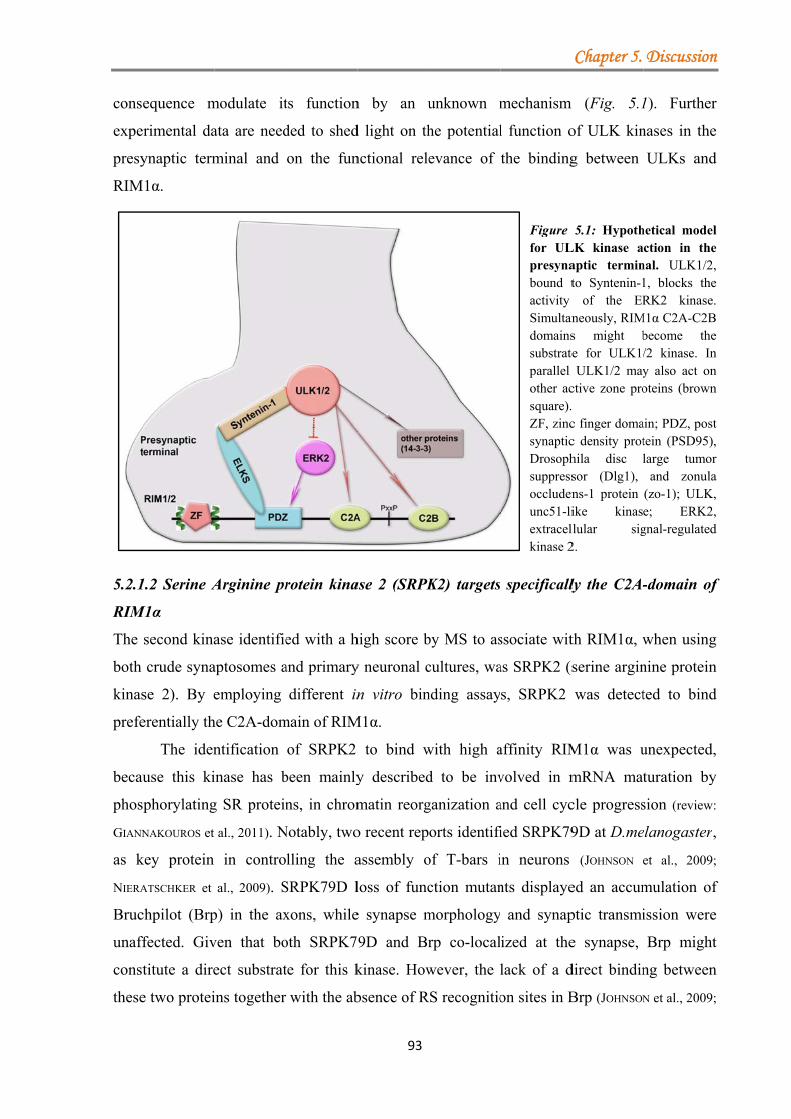
conse
expe
presy
RIM
5.2.1
RIM
The
both
kinas
prefe
becau
phos
GIANN
as k
NIERA
Bruc
unaff
const
these
equence m
rimental da
ynaptic term
M1α.
1.2 Serine A
M1α
second kina
crude syna
se 2). By e
erentially th
The iden
use this ki
phorylating
NAKOUROS et
key protein
ATSCHKER et
chpilot (Brp
ffected. Giv
titute a dire
e two protei
modulate it
ata are need
minal and
Arginine pr
ase identifie
aptosomes a
employing
he C2A-dom
ntification
inase has b
g SR protein
al., 2011). N
in contro
al., 2009). S
p) in the ax
ven that bo
ect substrat
ins together
ts function
ded to shed
on the fun
rotein kina
ed with a h
and primary
different in
main of RIM
of SRPK2
been mainly
ns, in chrom
Notably, two
lling the a
SRPK79D l
xons, while
oth SRPK7
te for this k
with the ab
93
n by an u
d light on th
nctional rele
ase 2 (SRPK
high score b
y neuronal c
n vitro bin
M1α.
to bind w
y described
matin reorg
o recent rep
assembly o
loss of func
e synapse m
9D and Br
kinase. How
bsence of R
unknown m
he potential
evance of
K2) targets
by MS to as
cultures, wa
nding assay
with high a
d to be inv
ganization a
orts identifi
of T-bars i
ction mutan
morphology
rp co-local
wever, the
RS recognitio
mechanism
l function o
the binding
Figure for ULpresynabound tactivity Simultandomainssubstrateparallel other acsquare).ZF, zincsynapticDrosophsuppressoccludenunc51-liextracelkinase 2
specifically
ssociate wit
as SRPK2 (s
s, SRPK2
affinity RIM
volved in m
and cell cyc
ied SRPK79
in neurons
nts displaye
and synap
ized at the
lack of a d
on sites in B
Chapter 5.
m (Fig. 5.1
of ULK kin
g between
5.1: HypothLK kinase aaptic terminto Syntenin-1
of the ERneously, RIMs might bte for ULK1/
ULK1/2 mayctive zone pro. c finger domac density prothila disc lsor (Dlg1), ns-1 protein ike kinasllular sig2.
ly the C2A-
th RIM1α, w
serine argin
was detect
M1α was u
mRNA mat
cle progress
9D at D.me
(JOHNSON e
ed an accum
ptic transmi
e synapse,
direct bindin
Brp (JOHNSON
Discussion
1). Further
nases in the
ULKs and
hetical modelction in the
nal. ULK1/2,1, blocks theRK2 kinase.
M1α C2A-C2Bbecome the/2 kinase. Iny also act onoteins (brown
ain; PDZ, posttein (PSD95),large tumor
and zonula(zo-1); ULK,e; ERK2,gnal-regulated
-domain of
when using
nine protein
ted to bind
unexpected,
turation by
sion (review:
elanogaster,
et al., 2009;
mulation of
ission were
Brp might
ng between
N et al., 2009;
n
r
e
d
l e , e .
B e n n n
t , r a , ,
d
f
g
n
d
,
y
:
,
;
f
e
t
n
;
Chapter 5. Discussion
94
NIERATSCHKER et al., 2009) may indicate that other presynaptic proteins could act as mediators
between SRPK79D and Brp.
One such protein could be RIM1α. The following data favours the idea that RIM1α
could act as a possible substrate for SRPK2: (1) the high affinity of SRPK2 for the C2A-
domain of RIM1α; (2) bioinformatical identification of RS dipeptides in the RIM1α sequence.
Thus, SRPK2 could directly associate with RIM1α and phosphorylate it. Besides the RS
dipeptides, SRPK2 may also phosphorylate unconventional sites in RIM1α, like the one
previously described in Tau proteins-‘PSLP’ (HONG et al., 2012). Analysis of the RIM1α binding
in the presence of a docking grove mutant (SRPK2-DM) revealed only a slight decrease in the
RIM1α binding affinity compared to SRPK2-WT control. Moreover, deletion of both, the N-
terminal region, important for the kinase activity, and the linker region, triggered as well a
decrease in RIM1α binding affinity. Because in the presence of these truncated proteins, the
binding to RIM1α was not completely abolished, the only regions from SRPK2 that could
directly mediate these affinities are the catalytic domains. In consequence it remains to be
elucidated, which kinase domain is directly involved in this interaction.
Up to date, only two proteins, SNAP25 and Syt1, were identified to bind the C2A-
domain of RIM1α (COPPOLA et al., 2001). However, NMR studies were not able to confirm these
findings (DAI et al., 2005). Identification of SRPK2 may represent the first specific binding
partner for the RIM1α C2A-domain. The exact role of the C2A-domain in RIM proteins has
not been fully elucidated. One point mutation in the C2A-domain of RIM1α (R844H) was
linked to the autosomal dominant cone-rod dystrophy-CORD7, characterized by impaired
vision due to the reduction in the cone and rod sensitivity (JOHNSON et al., 2003; MICHAELIDES et al.,
2005). Individuals with such mutations display enhanced cognitive functions in at least the
verbal and executive domains (SISODIYA et al., 2007).
IF studies support the co-localization of SRPK2 and RIM1α in the presynaptic
compartment, despite the fact that the detected level of SRPK2 at synapses was significantly
lower than at the soma. However, this is in agreement with the previous report of Nieratschker
et al. (2009), where they could show that the expression level of SRPK79D was low and not
well detected by the antisera raised against this protein. According to the Allen Brain Atlas,
the mRNA expression level of SRPK2 in adult mouse brains seems to be low in the cortex
compared to hippocampus. Because all our immunohistochemistry was performed with
cortical neurons, a further investigation of the co-localization of SRPK2 and RIM1α in
hippocampal neurons should be conducted as well.
Chapter 5. Discussion
95
Whereas studies in D.melanogaster suggested an important role for SRPK2 in
preventing the ectopic formation of AZs within the axons (JOHNSON et al., 2009; NIERATSCHKER et
al., 2009), the role of SRPKs in mammalian neurons, in particular in AZ formation, has not
been fully addressed. The molecular mechanism by which SRPK79D prevents the unspecific
accumulation of Brp at unconventional sites is not understood. However, it could be
hypothesised that in mammalian cells SRPK2 might act in a similar way, regulating the
assembly of AZ as well. Phosphorylation of RIM1α by SRPK2 may protect RIM1α against an
unspecific accumulation/aggregation in different parts of the cell preventing in this way a
premature assembly of the AZ. Once RIM1α reaches the correct destination (synaptic
bouton), phosphatases could remove some of the phosphate groups promoting protein-protein
interactions to occur. Additionally, certain functions of presynaptic proteins may be directly
regulated by SRPK2 kinase activity. In this respect, the amount of SRPK2 present in the
presynaptic terminal may be critical. Intriguingly, our IF data revealed that by blocking the
activity of SRPK1 and SRPK2, a slight increase in the co-localization of SRPK2 with RIM1α
was detected in the boutons. Because no sufficient data is available regarding the
irreversibility of the inhibitor SRPIN340, we cannot conclude whether this presynaptic
accumulation of SRPK2 represents active or inactive kinase.
Besides affecting the functions of presynaptic proteins, SRPK2 might promote
changes in AZ architecture, by targeting the cytoskeleton as well. It has been reported that
SRPK2 binds and phosphorylates Tau proteins (tau proteins stabilize the microtubules)
impairing tau-dependent microtubule polymerization and neurite outgrowth (HONG et al., 2012).
Hong et al. (2012) also showed that the knockdown of SRPK2 in the hippocampus of the
Alzheimer’s disease mouse model (APP/PS1) impact presynaptic functions. The amplitude of
pair pulse facilitation (PPF), an indicator of presynaptic activity, was elevated in APP/PS1
mice, in which SRPK2 levels were decreased by injecting a lentivirus expressing a specific
shRNA against this kinase (LV-shSRPK2), compared to WT. Unfortunately no
electrophysiological data comparing the WT versus WT LV-shSRPK2 mice was presented.
An increased in PPF was also measured in RIM1α KO mice, consistent with a reduced release
probability (SCHOCH et al., 2002; PITSCH et al., 2012). Thus, it can be hypothesized that SRPK2 by
phosphorylating various substrates may impact proper synaptic transmission. In
D.melanogaster the deletion of SRPK79D does not induce any significant changes in the
synaptic transmission at the NMJs (NIERATSCHKER et al., 2009). However, an overexpression of
this kinase impaired synaptic transmission, probably by disrupting either the assembly of T-
bars or the AZ organization (JOHNSON et al., 2009). Due to multiple effects this kinase might
Chapter 5. Discussion
96
have on various presynaptic proteins, further experimental data are necessary to explain
whether SRPK2 acts as a negative regulator of AZ assembly, similar to D.melanogaster, or
has a more subtle role in controlling diverse aspects in synaptic transmission.
5.2.2 VAPA/B proteins bind specifically the C2A-domain of RIM1α
The protein VAPA was identified with relative high scores in our MS analysis with both
crude synaptosomes and neuronal cultures. In addition, the treatment of mouse crude
synaptosomes with a kinase inhibitor (staurosporine) increased the level of VAPA detected by
MS compared to the phosSTOP or control samples. These results corroborated with GST-pull
down assays, in which kinase blockade increased not only the endogenous level of VAPA but
also the binding affinity to the C2A-domain of RIM1α. Thus, a global kinase inhibition seems
to favour the binding between RIM1α and VAPA.
The VAP protein family includes two highly homologous members: VAPA and
VAPB/C (NISHIMURA et al., 1999). Both VAPA and VABP were identified to bind exclusively
the C2A-domain of RIM1α in various biochemical assays, potencially due to their high degree
of sequence homology. However, in our MS data we only detected VAPA, even though
VAPB is expressed more abundantly in the brain (Allan Brain Atlas), indicating a preferential
binding of VAPA to RIM1α.
Interestingly, the mutation of two threonine residues (T812/814A) in the RIM1α C2A-
domain completely abolished the binding of RIM1α to VAPA. Bioinformatics predicts these
threonines to be part of a PKA recognition motif. Either these threonine residues are
necessary to mediate the direct binding of the RIM1α C2A-domain to VAPA or the
phosphorylation status of these amino acid residues could impact the binding.
Taken together, this study shows that: (1) VAP proteins, and especially VAPA, bind
specifically the RIM1α C2A-domain; (2) this association seems to be mediated by the
threonine residues in the RIM1α C2A-domain.
Even though, Teuling et al. reported that VAPB did not co-distribute with presynaptic
proteins, we observed that in rat cortical neurons these proteins were present in the same
presynaptic compartment. The highest VAPA/B signal was detected, as previously published,
in the soma, where the ER compartment is located (TEULING et al., 2007). However, IF analysis
revealed that both VAP proteins showed a weak co-localization with endogenous RIM1α and
Bassoon at the synapse. Because VAPs are actively involved in trafficking, their steady-state
levels at synapses might be low.
Initially, VAP proteins were associated with plasma membrane fusion events in
neuronal cells, via their interaction with Synaptobrevin/VAMP-2 protein (SKEHEL et al., 1995;
Chapter 5. Discussion
97
WEIR et al., 1998). However, later studies have shown that VAPA was not directly involved in
SVs exocytosis but rather involved in regulating the organization of the ER (AMARILIO et al.,
2005) and in ER-Golgi trafficking (KUIJPERS et al., 2013). Moreover, it was hypothesised that
VAPA might play a role in trafficking or chaperoning vesicular components such as VAMP-2
through the ER to the presynaptic compartment (SKEHEL et al., 2000). The hypothesis of VAP
proteins being actively involved in protein trafficking is supported by the fact that the
D.melanogaster homolog, DVAP-33A was identified to selectively transport proteins to
axonal processes (YANG et al., 2012). A reduction in the presynaptic DVAP-33A induced
structural changes, like the disruption of synaptic microtubules and the accumulation of
clusters of proteins and SVs along the axons (FORREST et al., 2013). In mouse cortical neurons
VAPA is transported, through its interaction with protruding via KIF5, from the soma to
neurites (MATSUZAKI et al., 2011). All these data could suggest a potential role of VAP proteins in
assisting the selective transport of various axonal proteins, such as RIM1α, to either nascent
or mature AZs. Therefore, we may speculate that besides VAMP-2, RIM1α could also be
trafficked to the synaptic bouton via VAP proteins, a transport dependent on the
phosphorylation status of both proteins. Future experiments will try to decipher the role of
phosphorylation events in controlling the traffic of RIM1α.
Moreover, studies on DVAP-33A provide evidence of its importance in bouton
formation by mediating the interaction between microtubules and presynaptic membranes
(PENNETTA et al., 2002). DVAP-33 overexpression in presynaptic terminals induced an increase
in the number of boutons that displayed a significantly reduced size and contained fewer
vesicles (PENNETTA et al., 2002; CHAI et al., 2008). With respect to the total number of AZs Chai et
al. (2008) did not observe any significant changes, while Ratnaparkhi et al. (2008) observed a
decrease in the number of AZs. The study of Ratnaparkhi et al. suggests that VAP proteins
might control the structural remodelling of AZs. However, up to date there not sufficient data
connecting VAP proteins and AZ assembly in mammalian neurons.
Besides the suggested role in protein transport and bouton formation, VAP proteins
could be actively involved in AZ protein sorting in ER-Golgi as well (Fig. 5.2). Active zone
proteins are processed in the soma of neurons in different ways. While Piccolo, Bassoon and
ELKS share a common Golgi-derived transport vesicle, Munc13 exits the soma on a distinct
Golgi-derived vesicle. Surprisingly, RIM1α associates with transport vesicles in a post-Golgi
compartment. However, to date the molecular mechanism governing this sorting is not well
understood (MAAS et al., 2012). Thus, an intriguing question is the potential involvement of VAP
proteins in sorting RIM1α in the ER-Golgi compartment. VAPA was shown to be involved
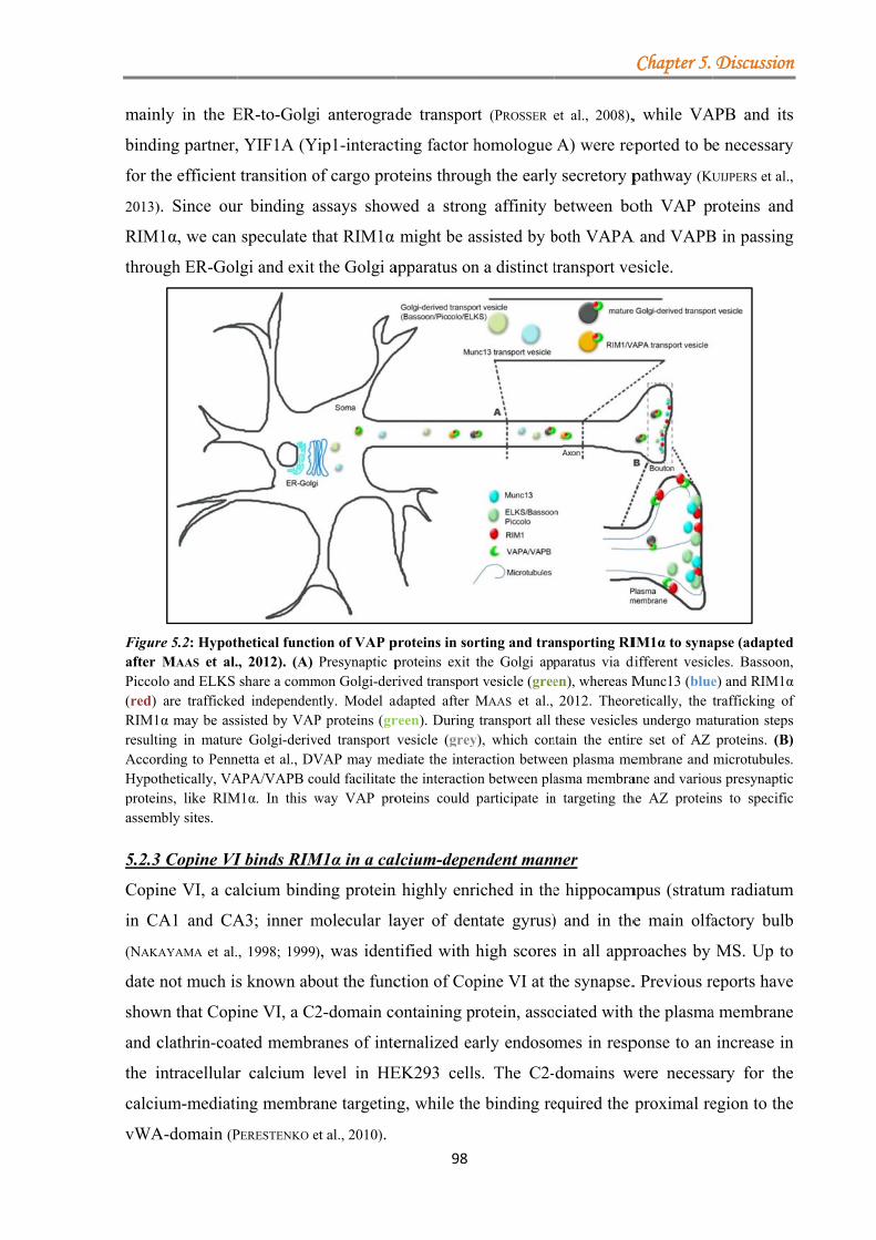
main
bindi
for th
2013)
RIM
throu
Figurafter Picco(red) RIM1resultAccorHypotproteiassem
5.2.3
Copi
in C
(NAKA
date
show
and c
the i
calci
vWA
nly in the E
ing partner,
he efficient
). Since our
M1α, we can
ugh ER-Gol
re 5.2: HypotMAAS et al.
lo and ELKS are trafficke
1α may be assing in maturerding to Pennthetically, VAins, like RIM
mbly sites.
3 Copine VI
ine VI, a ca
A1 and CA
AYAMA et al.
not much is
wn that Copi
clathrin-coa
intracellular
um-mediati
A-domain (P
ER-to-Golgi
, YIF1A (Y
transition o
r binding a
speculate t
lgi and exit
hetical functi., 2012). (A) share a commd independensisted by VAPe Golgi-deriv
netta et al., DVAPA/VAPB co
M1α. In this w
I binds RIM
alcium bind
A3; inner m
, 1998; 1999)
s known ab
ine VI, a C2
ated membr
r calcium l
ing membra
PERESTENKO e
i anterograd
Yip1-interact
of cargo pro
assays show
hat RIM1α
the Golgi a
ion of VAP pPresynaptic p
mon Golgi-derntly. Model aP proteins (grved transport VAP may medould facilitate way VAP pro
M1α in a cal
ding protein
molecular la
), was ident
out the func
2-domain co
ranes of inte
level in HE
ane targetin
et al., 2010). 98
de transpor
ting factor h
oteins throug
wed a stron
might be a
apparatus on
proteins in sorproteins exit rived transportadapted after reen). During vesicle (greydiate the inter the interactiooteins could
lcium-depe
n highly enr
ayer of den
tified with
ction of Cop
ontaining p
ernalized ea
EK293 cells
ng, while the
rt (PROSSER
homologue
gh the early
ng affinity
assisted by b
n a distinct t
rting and trathe Golgi appt vesicle (greeMAAS et al., transport all
y), which conraction betwe
on between plaparticipate in
endent mann
riched in the
ntate gyrus)
high scores
pine VI at th
rotein, asso
arly endoso
s. The C2-
e binding re
et al., 2008),
A) were rep
y secretory p
between bo
both VAPA
transport ve
nsporting RIparatus via den), whereas M2012. Theor
these vesiclesntain the entiren plasma measma membran targeting th
ner
e hippocam
) and in the
s in all appr
he synapse.
ciated with
mes in resp
domains w
equired the
Chapter 5.
, while VA
eported to b
pathway (KU
oth VAP pr
A and VAPB
esicle.
IM1α to synadifferent vesicMunc13 (blueretically, the s undergo mare set of AZ embrane and ane and variouhe AZ protein
mpus (stratum
e main olfa
roaches by
. Previous r
h the plasma
ponse to an
were necess
proximal re
Discussion
APB and its
e necessary
UIJPERS et al.,
roteins and
B in passing
apse (adaptedcles. Bassoon,e) and RIM1αtrafficking of
aturation stepsproteins. (B)microtubules.
us presynapticns to specific
m radiatum
actory bulb
MS. Up to
eports have
a membrane
increase in
ary for the
egion to the
n
s
y
,
d
g
d , α f s ) . c c
m
b
o
e
e
n
e
e
Chapter 5. Discussion
99
Our biochemistry data suggests a strong preference of Copine VI for the C2B-domain
of RIM1α, while binding to full-length RIM1α is Ca2+-dependent. Since RIM1α C2-domains
lack the amino acids necessary to bind calcium ions (COPPOLA et al., 2001; GUAN et al., 2007), only
the C2-domains of Copine VI are able to bind these divalent ions.
Because these C2-domains can form homo- or heterodimers, the MS data was
analysed for increased occurrence of C2-domain containing proteins, such as Synaptotagmin1
or other copines. Such proteins, associating with the C-terminal part of RIM1α, were scarcely
present in our MS data. Moreover, other copines were not detected in the MS. Together these
observations support the specificity of the interaction.
Previous reports have suggested that Copine VI is enriched in the postsynaptic
terminal (Alexander Kriz, 2010; urn: urn:nbn:ch:bel-bau-diss89692). However, our IF of endogenous
Copine VI in primary cultured neurons also suggests a presynaptic localization, where it
partially co-localizes with the presynaptic protein RIM1α. Therefore, Copine VI might be
present on both pre- and postsynaptic terminals, mediating different cellular processes.
Additionally, the study of Nakayama showed that Copine VI could be present in low amounts
in some parts of axons as well (NAKAYAMA et al., 1999). Unpublished data of our collaborators
attribute to Copine VI a presynaptic function as well, e.g. the regulation of SV fusion.
In summary, identification of Copine VI, as a potential binding partner for RIM1α,
may point to a possible link between the function of RIM1α and the calcium-dependent events
in the presynaptic terminal. Future experiments will be performed in order to examine the
relevance of this interaction in the presynaptic terminal.
5.3 Identification of novel SV2A binding partners: new experimental approaches
Because of the still enigmatic function of the synaptic vesicle protein 2A (SV2A) in synaptic
transmission and neuronal network plasticity, identification of novel potential binding
partners for SV2A could provide insights into its role at synapses.
SV2A is 12-pass transmembrane protein with a high degree of posttranslational
modifications that include a highly glycosylated intravesicular loop and a N-terminus
containing at least ten phosphorylation sites (review: MENDOZA-TORREBLANCA et al., 2013). To date
only a limited number of binding partners for SV2A have been identified, such as
Synaptotagmin1 and clathrin adaptor proteins (review: MENDOZA-TORREBLANCA et al., 2013; YAO et
al., 2010), whose functional relevance for the function of SV2A are still unclear.
Since the previous strategies to purify and identify novel SV2A binding partners were
based mainly on GST pull-down or yeast two-hybrid assays using only parts of SV2A and did
Chapter 5. Discussion
100
not take into the account the posttranslational modifications (SCHIVELL et al., 1996; YAO et al.,
2010), we attempted to analyse the binding partners of overexpressed SV2A using a tandem
affinity purification method. By applying one- or two-step purifications we aimed at the
identification of specific SV2A binding partners by maintaining at the same time the native
SV2A protein conformation. Therefore, a FLAG/Strep sequence was tagged either in the N-
or C-terminus of SV2A in order to detect any differences in SV2A binding affinities.
Different approaches were tested in order to co-purify SV2A and its potential binding partners
both from cortical neurons as well as from transduced mouse brain.
Purification of overexpressed SV2A from brain by employing the tandem-affinity
methodology proved to be inconsistent between different trials. The major problems that we
encountered were: (1) the expression level of the recombinant protein did not give a good
yield after purification because either the viral titer was not high enough or the virus did not
express high levels of protein; (2) the purification methodology used to co-purify the tagged-
SV2A had an unsatisfactory efficiency. During different approaches of purification, in most
of the cases, the recombinant protein was lost, especially when the Strep column was used,
either because the column was not able to retain the applied material (technical problem) or
the Strep sequence in the tag was masked by various protein contaminants. In the case of
FLAG-beads, most of the captured SV2A could not be eluted from beads. In contrary to the
poor yield of SV2A after purification from brain, sufficiently high amounts were obtained
when mouse primary cultured neurons were used as starting material.
It has been demonstrated that the number of SV2A molecules per SV is critical for its
function (TAKAMORI et al., 2006; NOWACK et al., 2011). Therefore, to avoid high levels of
overexpressed SV2A in addition to the endogenous one, neuronal primary cultures were
prepared from SV2A+/- or SV2A -/- mice. Moreover, to fix transient and/or weak interactions
between SV2A and other proteins, a series of cross-linkers were used. However, neither the
cross-linkers nor the primary cultures prepared from KO treatment produced a visible
enrichment in the co-purified proteins. Because we could not see an improvement, wild-type
neuronal cultures were further used for the final experiment. Compared to heterozygous or
knock-out neurons, WT neurons allowed for a higher amount of starting material. Preparation
of neuronal cultures from heterozygous or knock-out mice was limited to the number of
embryos available at a certain time-point, since KO SV2A mice cannot be breed (CROWDER et
al., 1999).
The analysis of protein complexes co-precipitated with SV2A by MS revealed several
proteins previosly reported to associate with the N-terminal region of SV2A (YAO et al., 2010),

Chapter 5. Discussion
101
like: AP-1 complex, Transcriptional activator protein Pur-beta, Tubulin beta-2A chain, Syt1.
Additionally, other proteins were identified by MS, e.g. Latrophilin, EHD1; however, none of
these potential SV2A binding partners could be verified in binding assays.
Overall, the attempt to identify novel potential interacting partners for SV2A has been
unsuccessful. This might be due to the fact that interactions require the native structure of the
12-transmembrane protein, a structure that in turn could have hampered the purifications of
the SV2A proteome. Even though the detergent, used to solubilize SV2A under native
conditions, was reported to maintain SV2A’s binding properties to Syt1 and Keppra (anti-
epileptic drug) (LAMBENG et al., 2006), it might not have preserved the native structure and
therefore interactions might have been lost. According to various protein-protein databases8,
also for other multi-pass membrane proteins, e.g. synaptophysin or SCAMP, fewer binding
partners are known.
Over the years different functions have been attributed to SV2A, like: neurotransmitter
transport, gel matrix (the sugar moieties attached to the intravesicular loop may hold and
release neurotransmitters) or modulator of exocytosis (review: MENDOZA-TORREBLANCA et al., 2013).
Experimental data suggest that SV2A mediates SV fusion by regulating the action of Syt1.
The N-terminal region of SV2A binds and keeps Syt1 inactive until the level of intracellular
calcium rises. Once the calcium level is high the binding is disrupted and Syt1 interacts with
the SNARE complex facilitating SV fusion. Additionally, during endocytosis SV2A binds
Syt1 and prevents its diffusion. The recycling after SV fusion is mediated by clathrin adaptor
proteins that bind both Syt1 and SV2A (review: MENDOZA-TORREBLANCA et al., 2013). The fact that
these proteins, Syt1 and various adaptor proteins have been identified by our MS may suggest
that SV2A, in order to fulfil its function, does not require a large number of interacting
proteins.
Because our attempt to purify and resolve the SV2A proteome was not successful,
other novel approaches are needed to be developed, e.g. generation of a knock-in mouse
model expressing a tagged SV2A. Such a model would give the opportunity to study more
closely not only the potential binding partners of SV2A but also the function of this protein.
8 http://www.uniprot.org/uniprot/P07825 (synaptophysin, R.norvegicus) http://www.uniprot.org/uniprot/P56603 (SCAMP, R.norvegicus)
Chapter 6. Outlook
102
6. Outlook
Taken together, the results of this thesis identified four novel RIM1α binding partners: two
kinases, a calcium binding protein and proteins involved in trafficking and bouton formation.
The next step will be to investigate the functional relevance of these new RIM1α binding
proteins. Therefore, the following three major questions will need to be addressed in future
experiments:
1. The role of SRPK2 and ULK1/2 in maintaining or/and controlling the AZ architecture and
composition. In this regard, the role of these novel kinases in the presynaptic terminal should
be investigated by using a specific SRPK2 inhibitor (SRPIN340) as well as shRNA-mediated
knock-down in neuronal cultures. In addition, it will be necessary to examine the possible
contribution of these kinases in controlling the release machinery, for example by measuring
the SV exo- and endocytosis using the fluorescent FM-dyes. Moreover, electrophysiological
recordings of neuronal cultures treated with either shRNA or SRPIN340 would be useful to
reveal any changes that may occur during synaptic transmission when kinase function is
blocked. Additionally, it will be necessary to examine whether RIM1α is a direct substrate of
these kinases.
2. The role of VAP proteins in modulating the function or trafficking of RIM1α. It should be
tested if VAP proteins play a role in trafficking presynaptic proteins to the boutons, in
particular RIM1α. Analysing if the absence of VAPA and/or VAPB results in an impairment
of RIM1α trafficking, could be tested by confocal live-imaging. Next, it should be examined
the functional relevance of C2A-domain phosphorylation in respect to the function of VAPA.
To evaluate whether VAP proteins may constitute a link between the core release machinery
and RIM1α, electrophysiological recordings of neuronal cultures overexpressing WT or
T812/814A RIM1α (point mutations in the C2A-domain that abolished the binding to VAPA)
could be performed.
3. The role of the interaction between the calcium-binding protein, Copine VI and RIM1α in
presynaptic plasticity. To address the function of Copine VI, shRNAs targeting specifically
Copine VI will be used to investigate whether this protein has any major impact on the
function of RIM1α and on synaptic transmission.
Because the amount of SV2A seems to be critical for its function, a knock-in mouse
model could be generated by inserting a tag in SV2A locus. Expression of this new tagged
protein could be analysed in the whole brain. Moreover, the tagged SV2A could be purified
from either whole brain or specific brain area in order to identify novel interacting partners.
Chapter 7. Summary
103
7. Summary
Synaptic plasticity encompasses various cellular mechanisms, which confer synapses the
ability to react and adapt to ongoing changes in network activity. Some of the suggested
mechanisms include remodelling and/or assembly of active zones (AZ), and modulation of
neurotransmitter release. At the molecular level posttranslational modifications of proteins,
e.g. phosphorylation, have been reported to be associated with these events. Two components
of the release machinery, RIM1α and synaptic vesicle protein 2A (SV2A) were shown to be
actively involved in presynaptic plasticity. However, the impact of posttranslational
modifications, like phosphorylation, on the function of these proteins is not well understood.
Therefore, the goals of this thesis were:
(1) To examine the impact of phosphorylation on the binding properties of RIM1α;
(2) To identify and analyse novel binding partners for SV2A.
We found that the distribution of RIM1α at synapses is altered after globally
increasing the level of phosphorylation, while its total level remained unchanged, suggesting
that the association of RIM1α with the CAZ is controlled by its phosphorylation status.
Affinity purification and MS revealed that alterations in the phosphorylation status of RIM1α
affected its affinity to specific binding partners. Out of the identified proteins, four candidates
with a potential functional link were chosen to be further analysed in binding assays: two
kinases (unc-51-like kinase 1/2, serine arginine protein kinase 2), one calcium-binding protein
(Copine VI), and proteins involved in trafficking (vesicle-associated membrane protein
(VAMP) associated-protein A/B). Interestingly, RIM1α may represent the first AZ substrate
for ULKs and SRPK2, which in D.melanogaster have already been linked to the assembly of
AZs. This may support the hypothesis that both ULKs and SRPK2 could be actively involved
in controlling not only RIM1α’s function but also its association with the CAZ. VAP proteins,
by specifically binding the C2A-domain of RIM1α, may contribute to control the trafficking
of RIM1α to the synapse. Copine VI may regulate the function of RIM1α in a calcium-
dependent manner. Further analysis will reveal if these novel interactions may have any
functional relevance for the function of RIM1α. In summary, we identified novel RIM1α
binding partners, of which some interact with RIM1α in a phosphorylation-dependent manner.
Further studies will have to examine if these are involved in mediating the association of
RIM1α with the CAZ or its role in plasticity.
The last part of the study was dedicated to another presynaptic protein, SV2A. To date
the role played by SV2A in SV priming is not fully elucidated. Therefore, to gain insight into
the enigmatic function of SV2A identification of novel binding partners was pursued.
Chapter 7. Summary
104
Different affinity purification strategies coupled to MS were performed in order to identify the
SV2A proteome. However, none of these approaches resulted in the identification of novel
interacting proteins, which could be further verified in biochemical assays.
Taken together, the findings of this thesis may form the basis for further functional
studies in order to decipher the molecular mechanisms underlying the function of RIM1α and
in consequence, the role of RIM1α in presynaptic plasticity.
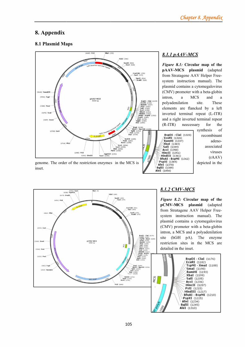
8. A
8.1 P
genominset.
Appendix
Plasmid Ma
me. The order
aps
r of the restricction enzymes
105
s in the MCSS is
8.1.1 pA
Figure 8pAAV-Mfrom Strasystem iplasmid c(CMV) pintron, polyadenelements inverted and a rig(R-ITR)
8.1.2 CM
Figure 8.pCMV-Mfrom Stratsystem inplasmid co(CMV) printron, a Msite (hGrestriction detailed in
Chapter 8
AAV-MCS
8.1: CircularMCS plasmatagene AAVinstruction mcontains a cytpromoter with
a MCS nilation si
are flancketerminal rep
ght inverted teneecessary
sy
de
MV-MCS
.2: Circular MCS plasmi
tagene AAV nstruction montains a cytromoter with MCS and a po
GH pA). Tn sites in thn the inset.
8. Appendix
r map of themid (adapted
Helper Free-manual). Thetomegalovirusa beta-globin
and aite. Theseed by a leftpeat (L-ITR)erminal repeaty for theynthesis of
recombinantadeno-
associatedviruses
(rAAV)epicted in the
map of theid (adaptedHelper Free-
manual). Thetomegalovirusa beta-globin
olyadenilationThe enzymehe MCS are
x
e d -e s n a e t ) t e f t -d s ) e
e d -e s n n e e
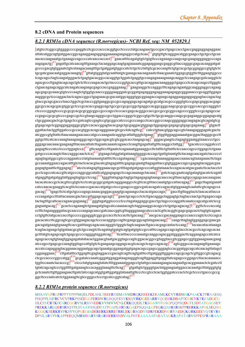
Chapter 8. Appendix
106
8.2 cDNA and Protein sequences
8.2.1 RIM1α cDNA sequence (R.norvegicus)- NCBI Ref. seq: NM_052829.1
[atgtcctcggccgtggggccccgaggtcctcgcccacccacggtgcctccccctatgcaagaactgcccgacctgagccacctgaccgaggaggagaggaacattatcatggcagtgatggaccggcagaaggaagaggaggaaaaagaagaggccatgctcaa]E1gtgtgttgtcagggacatggcgaagcctgctgcctgcaaaacaccaagaaatgctgaaagccagccccatcaaccaccact]E2gaacattttcagatgtgtctgtgttcccagaaagccaagcagcgaagagggaggcccagaaagagactg]E3gagattgcatcaacagtttgaaagctacaaggagcaagtgagaaaaatcggagaggaagcgaggcgttaccagggcgagcacaaggatgatgccccgacgtgtggaatctgtcataagacaaagtttgctgatggatgtggccatctctgctcctattgtcgcaccaagttctgtgcacgctgcggaggccgtgtgtctctgcgatcgaacaatgaggacaaagtg]E4gttatgtgggtatgcaatttatgtcgaaagcaacaagaaatcttaacgaaatctggagcgtggttctttggaagtggccctcagcagcctagtcaagatgggactctgagtgacacggccacaggtgctggatctgaggtgccaagagaaaagaaagcaaggctccaagagcgatcaaggtctcagacgcccttgagtacagcagctgtctcttcccaagacactgctacccccggtgcaccgttgcacaggaacaaaggggctgagccctcacagcaagccttgggtcctgaacagaagcaggcatcaagatcaagaagcgagccaccgagggaaag]E5gaagaaggctccagggctttcagagcagaatggcaagggaggccagaagagcgagcgcaaacgtgtccccaagtctgtggtgcaacccggggaagggatcgcggatgagagggagaggaaagagaggcgggaaacccgcaggttggagaaagggcgctcccaggactactcagaccggcctgagaaacgcgacaatggcagggtggcggaagaccagaagcagaggaaggaggaggagtaccagactaggtaccgcagcgaccctaacctggctcgctacccggtgaaggcgccgccagaggagcagcagatgcgcatgcacgcccgggtgtcccgagcgaggcacgagcggcgccacagcgacgtggcgctcccgcacaccgaggcagctgccgccgcgccggctgaggccacggcgggcaagcgcgcgccggccaccgccagggtctctcccccggagtccccgcgcgcacgcgcggcggccgcccagcctcccaccgagcacgggccaccgccgccgcggccagccccgggtcccgcagagccacccgagccgcgcgtccccgagccgctccgtaagcagggccgcctggacccgggctcggccgtgcttctgcgcaaggccaagcgcgagaaggcggagagcatgctgcggaacgactcgctgagctccgatcagtccgagtccgtgcggccatccccgcccaagcctcaccggcccaagcggggaggcaagagacgtcagatgtcggtgagcagctcggaggaggagggcgtgtccacaccggagtacacgagctgcgaggacgtggagctggagagcgagagcgtgagcgagaaag]E6gtgacttggattactactggttggatcccgccacgtggcacagcagggaaacgtcgcctatcagttcg]E7catcctgtaacgtggcagccgtctaaagagggagatcgactaatcggccgtgttattcttaacaaaagaacaaccatgcccaaagaatcaggtgcattattgggtctgaag]E8gtggttggaggaaaaatgacggacttagggcgccttggtgctttcatcaccaaagtaaagaagggcagcctggcagacgtcgtcggacacctaagagcag]E9gggacgaagtcctagagtggaatggtaaacccctgccgggagcaacaaacgaagaagtttacaacattatcttagaatcaaaatcagaacctcaagttgagattattgtttcaaggcctattgg]E10tgacatccccaggatccctgagagttcccatcctcccctggagtcca]E11gttcaagttcctttgaatctcagaaaatggaaaggccttctatttctgttatttctccaaccagccctggagctctgaaagatgccccacaagtcttaccagggcaactctca]E12gtgaagctatggtatgataaagtggggcaccagctgattgtaaatgttctacaagcaacagatctaccccctagagtagatggccgtcccaggaatccctatgtaaaaatgtattttcttccagatagaag]E13cgacaaaagtaaaaggagaaccaaaacagtaaagaaacttctagagccaaaatggaaccagacatttgtctactcacacgtacatcgtagagattttcgagagcgaatgttagagattaccgtgtgggaccagccgagagtacaggacgaagagagtgaatttcttggagag]E14atcctcatagagttggaaacagcgcttttagatgatgagccccattggtataaactccagacacatgacgaatcttcactacctctgcctcagccatcaccgttcatgcccaggcggcatattcatggagagagctccagcaaaaagctacaaa]E15gatctcagcgaatcagtgatagtgacatctcagattatgaggttgatgatggtattggagtagtgcctccag]E16tgggttatagagctagtgctagagagagtaaagcaaccacgttaacagtgccagagcaacaaagaactacacatcaccgctcacgttccgtgtctcctcatcgcggcgatgatcagggaaggcctcgttcacgtttaccaaatgtgccattacagag]E17gagcttagatgaaattcatccaacacgaaggtcacgttctccaacccgacaccatgatgcctcccgaagcccggccgatcacagatccagacatgtggaaagtcaatattcgtcagagccagacag]E18tgagcttctcatgctgcccagagcaaaacgaggacgaagtgcagaaagcctacacatgaccaga]E19gaccttgttaggtactctaacacattaccacccaagatgcctttattacaaaacgactaccgttggagcagcagt]E20gaactgcagccctctcttgacagggctaggagtgctagtaccaactgcttgagaccagatactagtttgcattcaccagaacgagaaag]E21gggtagatggtccccctccctagataggaggcgacctgctagccccaggattcaaatccagcatgcatctccggagaatgacag]E22gcactccagaaagtctgaaagatgtagcatccaaaaacagtctaggaaaggcacagcctctgatgcagacag]E23ggttctcccaccatgcctttctagaaggggatacgcaaccccaagagcaaccgatcaaccggtcgttaggggaaagcatcccactcgttcacggtcgagcgagcactctagtgtcagaaccctgtgttctatgcaccaccttgcccccggagggtcggcgccaccttctccacttctgacaag]E24aacgcaccgacaaggaagcccaacccagtctcctccagcagacacatccttcggcagtcgccgtggaagacagctcccacaggtgccagttcgaagcggcagtatagaacaag]E25caagcttagtagtggaggagcgaacgagacagatgaaagtgaaagttcaccgatttaagcagacaacagggtctgggtctagtcaagaacttgaccacgagcaatactccaag]E26tacaacatacataaagatcagtacagaagctgtgataacgcgtctgccaagtcttcagatagtgatgtcagtgatgtgtccgccatttccagagccagcagtacctcacgcctcagcagcacaagctttatgtcagagcagtctgagcgccccaggggtaggatcag]E27ttcatttacccccaaaatgcaaggcagacggatggggacttcaggaagagccatcatcaagagcaccagtgtaagtggagagatatatacactggaacgtaatgacggtagccagtcggacacggccgtaggtaccgtcggagccggtggaaagaaacgaagatccagcctgagcgccaaagtggtagccattgtgtctcgaagaagcaggagcacgtcacagctcagccagacag]E28agtcgggccacaagaagttgaaaagcaccatccagaggagtacggaaacaggaatggcagctgaaatgcggaagatggtgagacagccgagccgggagtccacggatggcagcatcaacagttatagctcggaaggaaa]E29cttgatatttcctggagttcgagtaggacccgacagtcagttcagtgatttccttgatgggttgggaccagcgcagctcgttggccgtcagacgctcgccaccccggccatgg]E30gcgatatccaaatcgggatggaggataagaagggtcagttggaggttgaggttatcagagcccggagccttacacaaaaacctggttccaaatctacacccg]E31ctccctatgtgaaagtatatcttttggaaaatggagcctgtattgccaaaaagaagacaagaattgcacggaaaactctcgatcctttgtatcagcagtccctggtttttgatgaaagtccacagggtaaagttcttcag]E32gtgattgtctggggtgactatggaagaatggaccacaaatgctttatgggtgtggctcaaatcttgttggaagaacttgatctatccagcatggtgattggatggtataaattgttccctccgtcctcactggtggatcccactctcgctcccctgacccgccgggcttcccaatcatctctggaaagttcgtccgggcctccctgcatccggtcatag]E33 8.2.2 RIM1α protein sequence (R.norvegicus)
MSSAVGPRGPRPPTVPPPMQELPDLSHLTEEERNIIMAVMDRQKEEEEKEEAMLKCVVRDMAKPAACKTPRNAESQPHQPPLNIFRCVCVPRKPSSEEGGPERDWRLHQQFESYKEQVRKIGEEARRYQGEHKDDAPTCGICHKTKFADGCGHLCSYCRTKFCARCGGRVSLRSNNEDKVVMWVCNLCRKQQEILTKSGAWFFGSGPQQPSQDGTLSDTATGAGSEVPREKKARLQERSRSQTPLSTAAVSSQDTATPGAPLHRNKGAEPSQQALGPEQKQASRSRSEPPRERKKAPGLSEQNGKGGQKSERKRVPKSVVQPGEGIADERERKERRETRRLEKGRSQDYSDRPEKRDNGRVAEDQKQRKEEEYQTRYRSDPNLARYPVKAPPEEQQMRMHARVSRARHERRHSDVALPHTEAAAAAPAEATAGKRAPATARVSPPESPRARAAA
Chapter 8. Appendix
107
AQPPTEHGPPPPRPAPGPAEPPEPRVPEPLRKQGRLDPGSAVLLRKAKREKAESMLRNDSLSSDQSESVRPSPPKPHRPKRGGKRRQMSVSSSEEEGVSTPEYTSCEDVELESESVSEKGDLDYYWLDPATWHSRETSPISSHPVTWQPSKEGDRLIGRVILNKRTTMPKESGALLGLKVVGGKMTDLGRLGAFITKVKKGSLADVVGHLRAGDEVLEWNGKPLPGATNEEVYNIILESKSEPQVEIIVSRPIGDIPRIPESSHPPLESSSSSFESQKMERPSISVISPTSPGALKDAPQVLPGQLSVKLWYDKVGHQLIVNVLQATDLPPRVDGRPRNPYVKMYFLPDRSDKSKRRTKTVKKLLEPKWNQTFVYSHVHRRDFRERMLEITVWDQPRVQDEESEFLGEILIELETALLDDEPHWYKLQTHDESSLPLPQPSPFMPRRHIHGESSSKKLQRSQRISDSDISDYEVDDGIGVVPPVGYRASARESKATTLTVPEQQRTTHHRSRSVSPHRGDDQGRPRSRLPNVPLQRSLDEIHPTRRSRSPTRHHDASRSPADHRSRHVESQYSSEPDSELLMLPRAKRGRSAESLHMTRDLVRYSNTLPPKMPLLQNDYRWSSSELQPSLDRARSASTNCLRPDTSLHSPERERGRWSPSLDRRRPASPRIQIQHASPENDRHSRKSERCSIQKQSRKGTASDADRVLPPCLSRRGYATPRATDQPVVRGKHPTRSRSSEHSSVRTLCSMHHLAPGGSAPPSPLLTRTHRQGSPTQSPPADTSFGSRRGRQLPQVPVRSGSIEQASLVVEERTRQMKVKVHRFKQTTGSGSSQELDHEQYSKYNIHKDQYRSCDNASAKSSDSDVSDVSAISRASSTSRLSSTSFMSEQSERPRGRISSFTPKMQGRRMGTSGRAIIKSTSVSGEIYTLERNDGSQSDTAVGTVGAGGKKRRSSLSAKVVAIVSRRSRSTSQLSQTESGHKKLKSTIQRSTETGMAAEMRKMVRQPSRESTDGSINSYSSEGNLIFPGVRVGPDSQFSDFLDGLGPAQLVGRQTLATPAMGDIQIGMEDKKGQLEVEVIRARSLTQKPGSKSTPAPYVKVYLLENGACIAKKKTRIARKTLDPLYQQSLVFDESPQGKVLQVIVWGDYGRMDHKCFMGVAQILLEELDLSSMVIGWYKLFPPSSLVDPTLAPLTRRASQSSLESSSGPPCIRS*
8.2.3 ULK1 cDNA and Protein sequence (M.musculus)- Clone ID:6406755/GenBank: BC059835 ATGGAGCCGGGCCGCGGCGGCGTCGAGACCGTGGGCAAGTTCGAGTTCTCTCGCAAGGACCTGATTGGACACGGCGCCTT M E P G R G G V E T V G K F E F S R K D L I G H G A F CGCGGTGGTCTTCAAGGGTCGACACCGCGAGAAGCACGACCTGGAGGTGGCCGTCAAATGCATTAACAAGAAGAACCTTG A V V F K G R H R E K H D L E V A V K C I N K K N L CCAAGTCCCAAACACTGCTGGGAAAGGAAATCAAAATCCTGAAGGAACTAAAGCACGAAAACATCGTGGCGCTGTATGAC A K S Q T L L G K E I K I L K E L K H E N I V A L Y D TTCCAGGAAATGGCTAATTCTGTCTACCTGGTCATGGAGTATTGTAATGGTGGAGACCTGGCTGACTACCTGCACACTAT F Q E M A N S V Y L V M E Y C N G G D L A D Y L H T M GCGCACACTGAGTGAAGACACTGTCAGGCTTTTCCTACAGCAGATCGCTGGCGCCATGCGGCTGCTGCACAGCAAGGGCA R T L S E D T V R L F L Q Q I A G A M R L L H S K G TCATCCACCGGGACCTGAAGCCCCAAAACATCCTGCTGTCCAACCCTGGGGGCCGCCGGGCCAACCCCAGCAACATCCGA I I H R D L K P Q N I L L S N P G G R R A N P S N I R GTCAAGATTGCTGACTTTGGATTCGCTCGGTACCTCCAGAGCAACATGATGGCGGCCACACTCTGTGGTTCTCCTATGTA V K I A D F G F A R Y L Q S N M M A A T L C G S P M Y CATGGCTCCTGAGGTCATTATGTCCCAGCACTACGATGGAAAGGCTGACCTGTGGAGCATTGGCACCATTGTCTACCAGT M A P E V I M S Q H Y D G K A D L W S I G T I V Y Q GTCTGACAGGGAAGGCCCCTTTTCAGGCCAGCAGCCCTCAGGATTTGCGCCTGTTTTATGAGAAGAACAAGACACTAGTT C L T G K A P F Q A S S P Q D L R L F Y E K N K T L V CCTGCCATCCCCCGGGAGACATCAGCTCCCCTGCGGCAGCTGCTCCTGGCTCTGTTGCAGCGGAACCACAAGGACCGCAT P A I P R E T S A P L R Q L L L A L L Q R N H K D R M GGACTTTGATGAATTTTTCCACCACCCTTTCTTGGATGCCAGCACCCCCATCAAGAAATCCCCACCTGTGCCTGTGCCCT D F D E F F H H P F L D A S T P I K K S P P V P V P CATATCCAAGCTCAGGGTCTGGCAGCAGCTCCAGCAGCAGCTCTGCCTCCCACCTGGCCTCTCCACCGTCCCTGGGGGAG S Y P S S G S G S S S S S S S A S H L A S P P S L G E ATGCCACAGCTACAGAAGACCCTTACCTCCCCAGCCGATGCTGCTGGCTTTCTTCAGGGCTCCCGGGACTCTGGTGGCAG M P Q L Q K T L T S P A D A A G F L Q G S R D S G G S CAGCAAAGACTCCTGTGACACAGATGACTTTGTCATGGTCCCAGCCCAGTTTCCAGGTGATCTAGTTGCTGAGGCAGCCA S K D S C D T D D F V M V P A Q F P G D L V A E A A GTGCCAAGCCCCCACCTGATAGCCTGCTGTGTAGTGGGAGCTCATTGGTGGCCTCTGCTGGCCTAGAGAGCCACGGCCGT S A K P P P D S L L C S G S S L V A S A G L E S H G R ACCCCCTCTCCCTCTCCGACCTGCAGCAGCTCTCCCAGCCCCTCTGGCCGGCCTGGCCCCTTCTCCAGCAACAGGTACGG T P S P S P T C S S S P S P S G R P G P F S S N R Y G TGCCTCGGTCCCCATTCCTGTCCCCACTCAGGTGCACAATTACCAGCGCATCGAGCAAAACCTGCAATCGCCCACTCAAC A S V P I P V P T Q V H N Y Q R I E Q N L Q S P T Q AGCAGACAGCCAGGTCCTCTGCCATCCGAAGGTCAGGGAGCACCAGCCCCCTGGGCTTTGGCCGGGCCAGCCCATCACCC Q Q T A R S S A I R R S G S T S P L G F G R A S P S P CCCTCCCACACCGATGGAGCCATGCTGGCCAGGAAGCTGTCACTTGGAGGTGGCCGTCCCTACACACCTTCTCCCCAAGT P S H T D G A M L A R K L S L G G G R P Y T P S P Q V GGGAACCATCCCAGAGCGACCCAGCTGGAGCAGAGTGCCCTCCCCACAAGGAGCTGATGTGCGGGTTGGCAGGTCACCAC G T I P E R P S W S R V P S P Q G A D V R V G R S P GACCCGGTTCCTCTGTGCCTGAGCACTCTCCAAGAACCACTGGGCTGGGCTGCCGCCTGCACAGTGCCCCTAACCTGTCC R P G S S V P E H S P R T T G L G C R L H S A P N L S GACTTCCATGTTGTGCGTCCCAAGCTGCCTAAGCCCCCAACAGACCCACTGGGAGCCACCTTTAGCCCACCCCAGACCAG D F H V V R P K L P K P P T D P L G A T F S P P Q T S CGCACCCCAGCCATGCCCAGGGCTACAGTCTTGCCGGCCACTGCGTGGCTCACCTAAGCTGCCTGACTTCCTACAGCGGA A P Q P C P G L Q S C R P L R G S P K L P D F L Q R GTCCCCTACCCCCCATCCTAGGCTCTCCTACCAAGGCCGGGCCCTCCTTTGACTTCCCCAAAACCCCCAGCTCTCAGAAT S P L P P I L G S P T K A G P S F D F P K T P S S Q N
Chapter 8. Appendix
108
TTGCTGACCCTGTTGGCTAGGCAGGGGGTAGTAATGACACCACCTCGGAACCGTACACTGCCTGACCTCTCCGAGGCCAG L L T L L A R Q G V V M T P P R N R T L P D L S E A S TCCTTTCCATGGCCAGCAGCTGGGCTCTGGCCTTCGGCCCGCTGAAGACACCCGGGGTCCCTTTGGACGGTCCTTCAGCA P F H G Q Q L G S G L R P A E D T R G P F G R S F S CCAGCCGCATTACGGACCTGCTGCTTAAGGCTGCATTTGGGACTCAGGCCTCTGACTCAGGCAGCACAGACAGCCTACAG T S R I T D L L L K A A F G T Q A S D S G S T D S L Q GAGAAACCTATGGAGATTGCTCCCTCTGCTGGCTTTGGAGGGACTCTGCATCCAGGAGCTCGTGGTGGAGGGGCCAGCAG E K P M E I A P S A G F G G T L H P G A R G G G A S S CCCAGCACCTGTGGTATTTACTGTAGGCTCCCCACCCAGTGGTGCCACCCCACCCCAGAGTACCCGTACCAGAATGTTCT P A P V V F T V G S P P S G A T P P Q S T R T R M F CAGTGGGCTCTTCCAGCTCCCTGGGCTCTACTGGCTCCTCCTCTGCCCGCCACTTAGTGCCTGGGGCCTGTGGAGAGGCC S V G S S S S L G S T G S S S A R H L V P G A C G E A CCGGAGCTTTCTGCCCCAGGCCACTGCTGTAGCCTTGCTGACCCCCTTGCTGCCAACTTGGAGGGGGCTGTGACCTTCGA P E L S A P G H C C S L A D P L A A N L E G A V T F E GGCTCCTGACCTCCCAGAGGAGACCCTCATGGAGCAAGAGCACACGGAAACCCTACACAGTCTGCGCTTCACACTAGCGT A P D L P E E T L M E Q E H T E T L H S L R F T L A TTGCACAGCAAGTTCTGGAGATTGCAGCCCTGAAGGGAAGTGCCAGTGAGGCCGCCGGTGGCCCTGAGTACCAGCTCCAG F A Q Q V L E I A A L K G S A S E A A G G P E Y Q L Q GAAAGTGTGGTGGCTGACCAGATCAGTCAGTTGAGCCGAGAGTGGGGCTTTGCAGAGCAACTGGTTCTGTACTTGAAGGT E S V V A D Q I S Q L S R E W G F A E Q L V L Y L K V GGCTGAGCTGCTGTCCTCAGGCCTACAGACTGCCATTGACCAGATTCGAGCTGGCAAACTCTGCCTTTCATCTACTGTGA A E L L S S G L Q T A I D Q I R A G K L C L S S T V AGCAGGTGGTACGCAGACTAAATGAGCTGTACAAGGCCAGCGTGGTATCCTGCCAGGGCCTCAGCTTGCGACTTCAGCGC K Q V V R R L N E L Y K A S V V S C Q G L S L R L Q R TTCTTTCTGGACAAACAACGGCTGCTGGACGGGATCCATGGTGTCACTGCAGAGCGGCTCATCCTCAGCCATGCTGTGCA F F L D K Q R L L D G I H G V T A E R L I L S H A V Q AATGGTACAATCAGCTGCCCTTGATGAGATGTTCCAGCACCGAGAGGGCTGTGTACCGAGATATCACAAAGCCCTGCTAT M V Q S A A L D E M F Q H R E G C V P R Y H K A L L TGCTGGAGGGGTTGCAGCACACTCTCACGGACCAGGCAGACATTGAGAACATTGCCAAATGCAAGCTGTGCATTGAGAGG L L E G L Q H T L T D Q A D I E N I A K C K L C I E R AGACTCTCGGCCCTGCTGAGTGGTGTCTATGCCTGA R L S A L L S G V Y A *
8.2.4 ULK2 cDNA and Protein sequence (M.musculus)- Clone ID:5709559/GenBank: BC046778 ATGGAGGTGGTGGGCGACTTCGAGTACTGCAAGCGGGACCTCGTGGGACACGGGGCCTTCGCTGTGGTCTTCCGGGGGCG M E V V G D F E Y C K R D L V G H G A F A V V F R G R GCACCGCCAGAAAACTGATTGGGAGGTGGCTATTAAAAGTATTAATAAAAAGAACTTGTCAAAATCACAAATTCTGCTTG H R Q K T D W E V A I K S I N K K N L S K S Q I L L GAAAGGAAATAAAAATCTTAAAGGAGCTTCAGCATGAAAACATCGTAGCGCTCTATGATGTTCAGGAATTGCCCAACTCT G K E I K I L K E L Q H E N I V A L Y D V Q E L P N S GTCTTTCTGGTGATGGAGTATTGCAATGGTGGAGACCTGGCAGATTATTTGCAAGCTAAAGGAACTCTGAGTGAAGATAC V F L V M E Y C N G G D L A D Y L Q A K G T L S E D T TATCAGAGTGTTTCTCCATCAGATTGCGGCAGCCATGCGAATCCTGCACAGCAAAGGGATAATCCACAGGGATCTCAAAC I R V F L H Q I A A A M R I L H S K G I I H R D L K CACAGAATATCCTGTTGTCTTATGCCAATCGAAGGAAGTCGAATGTCAGTGGTATTCGTATTAAAATAGCTGATTTTGGT P Q N I L L S Y A N R R K S N V S G I R I K I A D F G TTCGCACGGTACCTACATAGTAACACAATGGCAGCGACACTGTGTGGATCCCCAATGTACATGGCTCCCGAGGTTATTAT F A R Y L H S N T M A A T L C G S P M Y M A P E V I M GTCTCAACATTATGATGCTAAGGCAGATTTATGGAGCATAGGAACAGTGATCTATCAATGCCTAGTTGGAAAACCACCTT S Q H Y D A K A D L W S I G T V I Y Q C L V G K P P TTCAGGCTAATAGTCCTCAGGACCTAAGGATGTTTTATGAAAAAAACAGGAGCTTAATGCCTAGTATTCCCAGAGAAACA F Q A N S P Q D L R M F Y E K N R S L M P S I P R E T TCACCTTACTTGGCTAATCTCCTTTTGGGTTTGCTTCAGAGAAATCAAAAGGATAGAATGGACTTTGAAGCATTTTTCAG S P Y L A N L L L G L L Q R N Q K D R M D F E A F F S CCATCCTTTCCTTGAGCAAGTTCCAGTTAAAAAATCTTGCCCAGTCCCAGTGCCTGTGTATTCTGGCCCTGTCCCTGGAA H P F L E Q V P V K K S C P V P V P V Y S G P V P G GCTCCTGCAGCAGCTCACCATCTTGTCGCTTTGCTTCTCCACCATCCCTTCCAGATATGCAGCATATTCAGGAAGAAAAC S S C S S S P S C R F A S P P S L P D M Q H I Q E E N TTATCCTCCCCACCGTTGGGTCCTCCCAACTATCTACAGGTGTCCAAAGACTCTGCGAGTAATAGTAGCAAGAACTCTTC L S S P P L G P P N Y L Q V S K D S A S N S S K N S S TTGTGACACGGATGACTTTGTTTTGGTTCCACACAACATCTCGTCAGACCACTCATATGACATGCCAATGGGGACTACGG C D T D D F V L V P H N I S S D H S Y D M P M G T T CCAGACGTGCTTCAAATGAATTCTTTATGTGTGGAGGGCAGTGTCAACCTACTGTGTCACCTCACAGCGAAACAGCCCCA A R R A S N E F F M C G G Q C Q P T V S P H S E T A P ATTCCAGTTCCTACTCAAGTAAGGAATTATCAGCGCATAGAACAGAATCTTATATCCACTGCCAGCTCTGGCACAAACCC I P V P T Q V R N Y Q R I E Q N L I S T A S S G T N P
Chapter 8. Appendix
109
ACATGGTTCTCCAAGATCTGCAGTAGTACGAAGGTCTAATACCAGCCCCATGGGCTTCCTCCGGGTTGGGTCCTGCTCCC H G S P R S A V V R R S N T S P M G F L R V G S C S CTGTACCAGGAGACACAGTGCAGACAGGAGGACGAAGACTCTCTACTGGCTCTTCCAGGCCTTACTCACCATCCCCTTTG P V P G D T V Q T G G R R L S T G S S R P Y S P S P L GTTGGTACCATTCCTGAACAGTTTAGTCAGTGCTGCTGTGGACATCCTCAGGGCCATGAAGCCAGGAGTAGGCACTCCTC V G T I P E Q F S Q C C C G H P Q G H E A R S R H S S AGGTTCTCCAGTGCCACAGACCCAGGCACCACAGTCACTCTTACTGGGTGCTAGACTGCAGAGTGCACCCACCCTCACCG G S P V P Q T Q A P Q S L L L G A R L Q S A P T L T ATATCTATCAGAACAAGCAGAAGCTCAGAAAGCAGCACTCTGACCCTGTGTGTCCGTCCCATGCTGGAGCTGGGTATAGT D I Y Q N K Q K L R K Q H S D P V C P S H A G A G Y S TACTCACCTCAGCCTAGTCGGCCTGGCAGCCTTGGGACCTCTCCCACCAAGCACACGGGGTCCTCTCCACGGAATTCTGA Y S P Q P S R P G S L G T S P T K H T G S S P R N S D CTGGTTCTTTAAAACTCCTTTACCAACAATCATTGGCTCTCCTACTAAGACTACAGCTCCTTTCAAAATCCCTAAAACAC W F F K T P L P T I I G S P T K T T A P F K I P K T AAGCATCTTCTAACCTGTTAGCCTTGGTTACTCGTCATGGGCCTGCTGAAAGCCAGTCCAAAGATGGGAATGACCCTCGT Q A S S N L L A L V T R H G P A E S Q S K D G N D P R GAGTGTTCCCACTGCCTCTCAGTACAAGGAAGCGAGAGGCATCGATCTGAGCAGCAGCAGAGCAAGGCAGTGTTTGGCAG E C S H C L S V Q G S E R H R S E Q Q Q S K A V F G R ATCTGTCAGTACTGGGAAGTTATCAGAACAACAAGTAAAGGCACCTTTAGGTGGACACCAGGGCAGCACGGATAGTTTAA S V S T G K L S E Q Q V K A P L G G H Q G S T D S L ACACAGAACGACCAATGGATGTAGCTCCTGCAGGAGCCTGTGGTGTTATGCTGGCATTGCCAGCAGGAACAGCAGCAAGC N T E R P M D V A P A G A C G V M L A L P A G T A A S GCCAGAGCTGTCCTCTTCACCGTGGGGTCTCCTCCACACAGTGCCACAGCCCCCACTTGTACTCATATGGTCCTTCGAAC A R A V L F T V G S P P H S A T A P T C T H M V L R T AAGAACCACCTCAGTGGGGTCCAGCAGCTCAGGAGGTTCCTTGTGTTCTGCAAGTGGCCGAGTATGTGTGGGCTCCCCTC R T T S V G S S S S G G S L C S A S G R V C V G S P CTGGACCAGGGTTGGGCTCTTCCCCACCAGGAGCAGAGGGAGCTCCCAGCCTAAGATACGTGCCTTATGGTGCTTCACCA P G P G L G S S P P G A E G A P S L R Y V P Y G A S P CCCAGCCTAGAGGGTCTCATCACCTTTGAAGCCCCTGAACTACCAGAGGAGACACTGATGGAGCGAGAGCACACAGACAC P S L E G L I T F E A P E L P E E T L M E R E H T D T CTTACGCCATCTGAACATGATGTTAATGTTTACTGAGTGTGTGCTGGACCTGACGGCAGTGAGGGGTGGGAACCCTGAGC L R H L N M M L M F T E C V L D L T A V R G G N P E TGTGCACATCTGCTGTGTCCTTGTACCAGATTCAGGAGAGTGTAGTTGTGGACCAGATCAGCCAGCTAAGCAAAGATTGG L C T S A V S L Y Q I Q E S V V V D Q I S Q L S K D W GGGCGGGTGGAGCAGCTGGTGTTGTACATGAAGGCAGCACAGCTGCTGGCGGCTTCCCTGCATCTCGCCAAAGCTCAGGT G R V E Q L V L Y M K A A Q L L A A S L H L A K A Q V CAAGTCTGGGAAGCTGAGCCCATCCATGGCTGTGAAACAAGTTGTTAAAAATCTGAATGAAAGATACAAATTCTGCATCA K S G K L S P S M A V K Q V V K N L N E R Y K F C I CCATGTGCAAGAAACTTACAGAAAAGCTGAATCGCTTCTTCTCCGATAAACAGAGATTTATTGATGAAATCAACAGTGTG T M C K K L T E K L N R F F S D K Q R F I D E I N S V ACTGCAGAGAAACTCATCTATAATTGTGCTGTGGAAATGGTTCAATCTGCAGCCCTGGATGAGATGTTTCAGCAGACTGA T A E K L I Y N C A V E M V Q S A A L D E M F Q Q T E AGACATCGTTTATCGCTACCACAAGGCAGCCCTTCTTTTGGAAGGCTTAAGTAAGATCCTGCAGGACCCTACAGATGTTG D I V Y R Y H K A A L L L E G L S K I L Q D P T D V AAAATGTGCATAAGTATAAATGTAGTATTGAAAGAAGATTGTCAGCACTCTGCTGTAGCACTGCAACTGTGTGA E N V H K Y K C S I E R R L S A L C C S T A T V *
Kinase domain of ULK1/2 is depicted in blue, serine proline rich domain in black and C-terminal domain in orange. 8.2.5 VAPA cDNA sequence (M.musculus)- Clone ID:3490082/GenBank:BC003866 ATGGCGTCCGCCTCCGGGGCCATGGCGAAGCACGAGCAGATCCTGgTCCTCGACCCTCCTTCAGACCTCAAATTCAAAGGCCCCTTCACAGATGTAGTCACTACAAATCTTAAATTGCAAAATCCATCGGATAGAAAAGTGTGTTTCAAAGTGAAGACTACAGCACCTCGCCGGTACTGTGTGCGGCCCAACAGTGGGATTATTGACCCGGGGTCAATTGTGACTGTTTCAGTAATGCTGCAACCCTTTGATTATGATCCGAATGAAAAGAGTAAACATAAGTTCATGGTACAGACAATTTTTGCTCCACCAAACATTTCAGATATGGAAGCTGTGTGGAAAGAAGCAAAACCTGATGAATTAATGGATTCTAAATTGAGATGTGTGTTTGAAATGCCGAATGAAAATGATAAGCTGAATGATATGGAACCTAGCAAAGCTGTTCCACTGAATGCATCCAAACAAGACGGACCCCTGCCAAAACCAcACAGTGTTTCACTCaATGATACGGAAACAAGGAAACTGATGGAAGAGTGCAAGCGACTCCAGGGAGAAATGATGAAGCTCTCAGAAGAAAACCGACACCTGAGAGATGAAGGCCTAAGGCTCAGAAAGGTAGCACATTCGGATAAACCTGGATCCACCTCAGCCGTGTCCTTCAGAGATAATGTCACCAGTCCTCTTCCTTCTCTTCTGGTTGTAATTGCAGCCATTTTCATTGGATTCTTTCTAGGGAAATTCATCTTG

Chapter 8. Appendix
110
8.2.6 VAPA Protein sequence (M.musculus) MASASGAMAKHEQILVLDPPSDLKFKGPFTDVVTTNLKLQNPSDRKVCFKVKTTAPRRYCVRPNSGIIDPGSIVTVSVMLQPFDYDPNEKSKHKFMVQTIFAPPNISDMEAVWKEAKPDELMDSKLRCVFEMPNENDKLNDMEPSKAVPLNASKQDGPLPKPHSVSLNDTETRKLMEECKRLQGEMMKLSEENRHLRDEGLRLRKVAHSDKPGSTSAVSFRDNVTSPLPSLLVVIAAIFIGFFLGKFIL
8.2.7 VAPB cDNA sequence (M.musculus)- NCBI Ref. seq: NM_019806.5 ATGGCGAAGGTGGAACAGGTCCTGAGCCTCGAGCCACAACACGAGCTCAAGTTCCGAGGTCCCTTCACTGATGTTGTCACCACCAACCTAAAGCTTGGCAACCCAACAGACCGAAATGTGTGTTTTAAAGTGAAGACCACAGCACCTCGCAGGTACTGCGTGCGGCCCAACAGTGGGGTCATTGATGCCGGGGCCTCTCTCAATGTGTCTGTGATGTTACAGCCTTTCGATTATGATCCCAATGAGAAAAGTAAACACAAGTTTATGGTTCAGTCTATGTTTGCTCCGCCTGACACTTCTGATATGGAGGCAGTATGGAAGGAGGCAAAACCGGAAGACCTTATGGATTCAAAACTTAGATGTGTGTTTGAATTGCCAGCAGAAAATGCTAAACCACATGATGTAGAAATAAATAAAATCATACCTACGAGTGCATCCAAGACAGAAGCACCGGCAGCCGCAAAGTCCCTGACATCGCCCCTCGATGACACAGAAGTAAAGAAGGTGATGGAAGAGTGCAGGCGGCTGCAGGGGGAGGTGCAGAGGCTTCGGGAGGAGAGCAGGCAGCTCAAGGAAGAAGACGGACTTCGGGTGAGGAAGGCGATGCCGAGCAACAGCCCCGTGGCGGCTCTGGCGGCCACTGGGAAGGAGGAGGGCCTGAGCGCCCGGCTGCTGGCCCTGGTGGTTCTGTTCTTTATCGTTGGTGTCATTATAGGGAAGATTGCCTTG
8.2.8 VAPB Protein sequence (M.musculus) MAKVEQVLSLEPQHELKFRGPFTDVVTTNLKLGNPTDRNVCFKVKTTAPRRYCVRPNSGVIDAGASLNVSVMLQPFDYDPNEKSKHKFMVQSMFAPPDTSDMEAVWKEAKPEDLMDSKLRCVFELPAENAKPHDVEINKIIPTSASKTEAPAAAKSLTSPLDDTEVKKVMEECRRLQGEVQRLREESRQLKEEDGLRVRKAMPSNSPVAALAATGKEEGLSARLLALVVLFFIVGVIIGKIAL
8.2.9 Copine VI cDNA sequence (M.musculus)- Clone ID:6591063/GenBank:BC050766 atgtcggacccagagatgggatgggtgcctgagcccccggccatgaccctgggagcctctcgagtggagctgcgggtgtcctgccatggcctgctggaccgagacacgctcacaaagccgcatccatgtgtgctgctcaaactctactcggatgagcagtgggtggaggtggaacgcacggaggtgcttcgctcctgttcaagcccagtcttctcccgggtgctggccattgaatacttttttgaagagaagcagcctttgcagttccacgtgttcgatgccgaggatggagccaccagccccagcagcgacaccttcctcgggtctacggagtgcaccttgggccagattgtgtcacaaaccaaggtcactaagccattactgctgaagaatgggaagacggcgggcaagtctactatcacgattgtggctgaggaggtatcaggtaccaacgactatgtgcaacttaccttcagagcccacaagctggataacaaggatctgttcagcaagtctgaccccttcatggagatttataagaccaatggagaccagagtgaccaactggtctggaggactgaggtggtgaaaaacaacctgaaccccagctgggagccattccgcctgtccttgcattctttgtgcagctgtgacatccacaggccgctcaagttcctggtatatgactatgactccagtggaaagcacgacttcatcggcgagttcaccagcacattccaggaaatgcaagaggggaccgcaaaccctgggcaggagatgcagtgggactgtatcaaccccaagtaccgagacaagaagaagaattacaagagctcagggacagtcgtgctggcccagtgcaccgtggaaaaagtccacaccttcctggattatatcatgggtggctgccagatcagcttcacggtggctatcgacttcactgcctccaatggggacccaaggagcagccagtctctgcactgcctcagcccccgacagcccaaccactacctgcaggccttgcgcacagtgggcggtatctgccaggactatgacagtgataagcggttcccagcttttggcttcggagctcgaatccccccaaactttgaggtctcccatgactttgctatcaactttgacccagaaaatcctgaatgtgaagagatctcaggggtcatagcctcctaccgccgctgcttgcctcagatccagctctatggtcctaccaacgtggcccctatcatcaaccgtgtggctgaaccagcccagcgagagcagagcaccggccaagccacgaagtattcagtactgctggtgctcactgacggtgtggtgagtgatatggcagagacccgaacagccattgtgcgagcctcccgcctgcccatgtcaatcatcatcgtgggtgtgggcaacgctgacttctctgacatgaggctactggacggagatgatggtcccctgcgttgcccaaagggggtacctgcagcccgtgacattgtccagtttgtgcccttcagggacttcaaggacgctgccccctctgcactagctaagtgtgtcctggcagaggtgccacggcaggtggtagagtactatgccagccaaggtatcagtcccggggctcccaggccctccacgccagctatgactcccagccctagccca 8.2.10 Copine VI Protein sequence (M.musculus) MSDPEMGWVPEPPAMTLGASRVELRVSCHGLLDRDTLTKPHPCVLLKLYSDEQWVEVERTEVLRSCSSPVFSRVLAIEYFFEEKQPLQFHVFDAEDGATSPSSDTFLGSTECTLGQIVSQTKVTKPLLLKNGKTAGKSTITIVAEEVSGTNDYVQLTFRAHKLDNKDLFSKSDPFMEIYKTNGDQSDQLVWRTEVVKNNLNPSWEPFRLSLHSLCSCDIHRPLKFLVYDYDSSGKHDFIGEFTSTFQEMQEGTANPGQEMQWDCINPKYRDKKKNYKSSGTVVLAQCTVEKVHTFLDYIMGGCQISFTVAIDFTASNGDPRSSQSLHCLSPRQPNHYLQALRTVGGICQDYDSDKRFPAFGFGARIPPNFEVSHDFAINFDPENPECEEISGVIASYRRCLPQIQLYGPTNVAPIINRVAEPAQREQSTGQATKYSVLLVLTDGVVSDMAETRTAIVRASRLPMSIIIVGVGNADFSDMRLLDGDDGPLRCPKGVPAARDIVQFVPFRDFKDAAPSALAKCVLAEVPRQVVEYYASQGISPGAPRPSTPAMTPSPSP

Chapter 8. Appendix
111
8.2.11 SRPK2 cDNA sequence (M.musculus)-Clone ID: 4507346/GenBank:BC020178 ATGTCAGTTAACTCTGAGAAGTCGTCCTCTTCAGAAAGGCCGGAGCCTCAACAGAAAGCTCCTTTAGTTCCTCCTCCTCCACCACCACCACCGCCACCACCACTGCCAGACCCCGCACCCCCAGAGCCAGAGGAGGAGATTCTGGGGTCAGATGATGAGGAGCAGGAGGACCCCGCAGATTACTGCAAAGGTGGCTATCATCCAGTGAAAATTGGAGATCTCTTCAATGGTCGATATCATGTCATTAGAAAGCTAGGATGGGGGCACTTTTCTACTGTATGGCTGTGCTGGGATATGCAAGGGAAAAGATTTGTTGCAATGAAAGTTGTAAAAAGTGCCCAGCATTATACAGAGACAGCCTTGGATGAAATTAAACTACTCAAATGCGTTCGAGAAAGTGACCCCAGTGACCCAAACAAAGACATGGTAGTTCAGCTAATTGATGACTTCAAGATCTCAGGCATGAATGGGATACATGTCTGCATGGTCTTTGAAGTACTTGGTCACCATCTCCTCAAATGGATCATCAAATCCAACTATCAAGGCCTCCCAGTACGTTGTGTGAAGAGTATCATTCGACAGGTCCTTCAAGGGTTAGATTATCTACACAGTAAGTGCAAGATAATTCACACCGACATAAAGCCGGAAAACATCTTGATGTGTGTGGATGACGCTTACGTGAGAAGAATGGCAGCCGAAGCCACGGAGTGGCAGAAAGCAGGTGCTCCTCCTCCCTCTGGGTCTGCAGTGAGTACGGCTCCACAGCAAAAACCTATAGGAAAAATATCTAAAAACAAAAAGAAAAAGCTGAAAAAGAAACAGAAGAGACAGGCTGAGTTGCTGGAGAAACGCCTACAGGAGATTGAGGAATTGGAGCGAGAAGCCGAAAGGAAAATCCTAGAGGAGAACATCACCTCTGCAGAAGCTTCCGGGGAGCAGCAGGATGGAGAGTACCAGCCGGAGGTGACACTGAAAGCAGCCGACTTAGAGGACACAACTGAGGAAGAGACAGCAAAGGATAATGGTGAAGTTGAAGACCAGGAAGAGAAAGAAGATGCAGAGAAGGAGAACGCGGAGAAGGATGAAGATGATGTTGAACAGGAACTTGCAAACTTAGACCCTACCTGGGTGGAGTCCCCGAAAGCCAATGGCCATATTGAAAATGGCCCGTTCTCACTGGAGCAGCAGCTGGAGGATGAAGAGGACGATGAAGATGACTGTGCAAATCCCGAGGAGTATAACCTCGATGAGCCAAATGCAGAGAGTGATTACACGTATAGCAGCTCCTATGAACAATTCAATGGTGAATTGCCAAATGGACAACATAAGACTTCAGAGTTTCCCACACCGTTGTTTTCTGGGCCCTTAGAACCTGTGGCCTGTGGCTCTGTGATTTCAGAGGGATCGCCACTTACCGAGCAGGAGGAAAGCAGTCCCTCCCATGACAGAAGCAGGACAGTTTCAGCCTCTAGTACTGGAGATTTGCCAAAAACAAAAACCCGGGCGGCTGACCTGTTGGTGAACCCTCTGGATCCACGGAATGCAGATAAAATTAGAGTAAAAATTGCTGACCTGGGAAATGCTTGTTGGGTGCATAAACATTTCACAGAGGATATCCAGACACGTCAGTATAGGTCCATAGAGGTTTTAATAGGAGCAGGCTACAGCACACCTGCAGACATTTGGAGTACAGCTTGCATGGCATTTGAGCTCGCCACAGGAGACTATTTGTTCGAACCGCATTCTGGGGAAGACTATTCCAGAGATGAAGACCACATAGCCCACATCATAGAGCTGCTAGGCAGTATCCCAAGGCACTTTGCTCTGTCTGGAAAATATTCTCGGGAATTCTTCAATCGCAGAGGAGAACTGCGGCACATCACCAAGCTGAAGCCCTGGAGCCTCTTTGATGTACTTGTGGAAAAGTATGGCTGGCCCCATGAAGATGCTGCACAATTTACAGATTTCCTGATCCCAATGTTAGAGATGGTTCCAGAAAAACGAGCCTCAGCTGGCGAATGCCTTCGACATCCTTGGTTGAATTCT
8.2.12 SRPK2 Protein sequence (M.musculus) MSVNSEKSSSSERPEPQQKAPLVPPPPPPPPPPPLPDPAPPEPEEEILGSDDEEQEDPADYCKGGYHPVKIGDLFNGRYHVIRKLGWGHFSTVWLCWDMQGKRFVAMKVVKSAQHYTETALDEIKLLKCVRESDPSDPNKDMVVQLIDDFKISGMNGIHVCMVFEVLGHHLLKWIIKSNYQGLPVRCVKSIIRQVLQGLDYLHSKCKIIHTDIKPENILMCVDDAYVRRMAAEATEWQKAGAPPPSGSAVSTAPQQKPIGKISKNKKKKLKKKQKRQAELLEKRLQEIEELEREAERKILEENITSAEASGEQQDGEYQPEVTLKAADLEDTTEEETAKDNGEVEDQEEKEDAEKENAEKDEDDVEQELANLDPTWVESPKANGHIENGPFSLEQQLEDEEDDEDDCANPEEYNLDEPNAESDYTYSSSYEQFNGELPNGQHKTSEFPTPLFSGPLEPVACGSVISEGSPLTEQEESSPSHDRSRTVSASSTGDLPKTKTRAADLLVNPLDPRNADKIRVKIADLGNACWVHKHFTEDIQTRQYRSIEVLIGAGYSTPADIWSTACMAFELATGDYLFEPHSGEDYSRDEDHIAHIIELLGSIPRHFALSGKYSREFFNRRGELRHITKLKPWSLFDVLVEKYGWPHEDAAQFTDFLIPMLEMVPEKRASAGECLRHPWLNS
8.2.13 SV2A (R.norvegicus)-NCBI Ref. seq: NM_057210.2 atggaagaaggctttcgagaccgagcagcgttcatccgtggggccaaagacattgccaaggaagttaagaagcacgcggccaagaaggtggtgaagggtctcgacagagtccaggatgaatattcccgaaggtcctactcccgctttgaggaggaggaggatgatgatgacttccctgcccctgctgacggctattaccgcggagaaggggcccaggatgaggaggaaggtggcgcttccagtgatgccactgagggccacgatgaggatgatgagatctacgagggagaatatcagggcatcccccgggcagagtctgggggcaaaggcgaacggatggcagatggggcacccctggctggagtgagagggggcttaagtgatggggagggtccccctgggggtcgcggggaggcgcagcggcgtaaagatcgggaagaattggctcagcagtatgagaccatcctccgggagtgcggccatggtcgcttccagtggacactctacttcgtgctgggtctggcgctgatggccgatggtgtagaggtctttgtggtgggctttgtgctgcccagtgctgagaaagatatgtgcctgtcggactccaacaaaggcatgctaggcctcattgtgtacctgggcatgatggtgggggccttcctctggggaggcctggctgatcggctgggtcggagacagtgtctgctcatctcgctctcagtcaacagcgtcttcgctttcttctcatccttcgtccagggttatggcaccttccttttctgccgcctcctttctggggttgggattggtggttccatccccattgtcttctcctatttttcggagtttctggcccaggagaaacgtggggagcatttgagctggctctgtatgttctggatgattggtggcgtgtatgcagctgcaatggcctgggccatcatcccccactatgggtggagtttccagatgggctctgcttaccagttccacagctggagggtctttgtcctcgtctgtgcctttccctctgtgtttgccatcggggctctgactacgcagccggagagtccccgcttcttcttagagaatgggaagcacgatgaggcctggatggtgctgaagcaggttcatgacaccaacatgcgagccaagggccatcctgagcgagtcttctcagtaacccacattaaaacgattcatcaggaggatgaattgattgagatccagtcagacacaggaacctggtaccagcgctggggagtgcgggctttgagcctggggggtcaggtttgggggaacttcctctcctgcttcagtccagagtaccggcgcatcactctgatgatgatgggggtatggttcaccatgtccttcagctactacggtttgactgtctggtttcccgacatgatccgccatctccaggctgtggactatgcagcccgaaccaaagtgttcccaggggagcgcgtggagcacgtgacatttaacttcacactggagaatcagatccaccgagggggacagtacttcaatgacaagttcatcgggctgcgtctgaagtcagtgtcctttgaggattccctgtttgaggaatgttactttgaagatgtcacatccagcaacacattcttccgcaactgcacattcatcaacaccgtgttctacaacacggacctgtttgagtacaagttcgtgaacagccgcctggtgaacagcacattcctgcacaataaggaaggttgcccactagatgtgacagggacgggcgaaggtgcctacatggtgtactttgtcagcttcttggggacactgg
Chapter 8. Appendix
112
ctgtgctccctggaaatattgtgtctgctctgctcatggacaagattggcaggctcagaatgcttgctggttccagtgtgttgtcctgtgtttcctgcttcttcctgtcttttgggaacagtgagtcagccatgatcgctctgctctgcctttttgggggagtcagtattgcatcctggaacgcgctggacgtgctgactgttgaactctacccttccgacaagaggacgacggccttcggcttcctgaatgccctgtgtaagctggcagctgtactgggcatcagcatcttcacgtcctttgtgggaatcaccaaggccgctcccatcctcttcgcctcagctgcgcttgcccttggtagctctctggctctgaagctgcctgagacccggggacaggtgctgcag 8.2.14 SV2A Protein sequence (R. norvegicus) MEEGFRDRAAFIRGAKDIAKEVKKHAAKKVVKGLDRVQDEYSRRSYSRFEEEEDDDDFPAPADGYYRGEGAQDEEEGGASSDATEGHDEDDEIYEGEYQGIPRAESGGKGERMADGAPLAGVRGGLSDGEGPPGGRGEAQRRKDREELAQQYETILRECGHGRFQWTLYFVLGLALMADGVEVFVVGFVLPSAEKDMCLSDSNKGMLGLIVYLGMMVGAFLWGGLADRLGRRQCLLISLSVNSVFAFFSSFVQGYGTFLFCRLLSGVGIGGSIPIVFSYFSEFLAQEKRGEHLSWLCMFWMIGGVYAAAMAWAIIPHYGWSFQMGSAYQFHSWRVFVLVCAFPSVFAIGALTTQPESPRFFLENGKHDEAWMVLKQVHDTNMRAKGHPERVFSVTHIKTIHQEDELIEIQSDTGTWYQRWGVRALSLGGQVWGNFLSCFSPEYRRITLMMMGVWFTMSFSYYGLTVWFPDMIRHLQAVDYAARTKVFPGERVEHVTFNFTLENQIHRGGQYFNDKFIGLRLKSVSFEDSLFEECYFEDVTSSNTFFRNCTFINTVFYNTDLFEYKFVNSRLVNSTFLHNKEGCPLDVTGTGEGAYMVYFVSFLGTLAVLPGNIVSALLMDKIGRLRMLAGSSVLSCVSCFFLSFGNSESAMIALLCLFGGVSIASWNALDVLTVELYPSDKRTTAFGFLNALCKLAAVLGISIFTSFVGITKAAPILFASAALALGSSLALKLPETRGQVLQ
Chapter 9. Abbreviations
113
9. Abbreviations
A EM Electron microscopy AAV Adeno associated virus ER Endoplasmic reticulum AC Adenylyl cyclase ERK2 Extracellular signal-regulated kinase2 ADBE Activity-dependent bulk endocytosis ES Elution steps APS Ammonium peroxodisulphate F ATP Adenosine tri-phosphate FCS Fetal calf serum AZ
Active Zone
FM Name after Fei Mao, who synthetized for the first time these dyes
B Fw Forward BDNF Brain-derived neurotrophic factor G BES N,N, Bis-(2-hydroxyethyl)-2-amino-
ethansulfonic acid G GFP
Gravitational Green fluorescent protein
BME Basal Medium Eagle GPCR G-protein coupled receptor Brp Bruchpilot GSH Glutathione BSA Bovine serum albumin GST Glutathione S-transferase β-ME Beta mercaptoethanol GTP Guanosintriphosphate C H Ca2+ Calcium ions h Hour CaCl2
CaMKII Calcium chloride Ca2+/calmodulin-dependent protein kinase II
HA HBS HBSS
Human influenza hemaglutinin HEPES buffered saline Hanks buffered salt solution
cAMP CASK CAZ CC
Cyclic adenosine-3,5-monophosphate Calcium/calmodulin-dependent serine protein kinase Cytomatrix at the active zone Coiled-coil domain
HEK 293T HEPES
Human embryonic kidney cell line 293T 2-[4-(2-hydroxyethyl)piperazin-1-yl]ethanesulfonic acid
CCB Coomassie Colloidal Blue I cDNA Complementary DNA IAA Iodamidacetate Cdk5 Cyclin-dependent kinase 5 IB Immunoblotting CMV Cytomegalovirus IC50 Half inhibitory concentration CNS Central nervous system IF Immunofluorescence CO2 Carbon dioxide IgG Immunoglobulin G Co-IP Co-immunoprecipitation IHC Immunohistochemistry CTD C-terminal domain IMDM Iscove’s Modified Dulbecco’s Medium C-terminus
Carboxyl terminus IP IPTG
Immunoprecipitation Isopropyl-β-D-thiogalactoside
D IRDye InfraRed Dye DAPI 4’,6-diamidin-2-phenylindol K DDM n-dodecyl-β-maltoside kb Kilo base dH2O Distillate water KCl Potassium chloride DIV Day in vitro KD Kinase domain DMEM Dulbecco's Modified Eagle's Medium kDa Kilo Dalton DMSO Dimethyl sulfoxide KH2PO4 Potasium dihydrogenphosphate DNA Deoxyribonucleic acid KO Knock-out dNTP Deoxyribonucleotide triphosphate L DSP Dithiobis (succinimidylpropionate) LAR Leukocyte common antigen related DTBP Dimethyl 3,3’-dithiobispropionimidate-
2HCl LB-medium
Luria broth medium
DTT Ditiothreitol LC-MS Liquid chromatography- mass spectrometry E LTD Long-term depression E18,5 Embryo at day 18.5 after fertilization LTP Long-term potentiation EDTA Ethylenediaminetetraacetic acid ELKS Name stems from the proteins high content
in glutamate (E), leucine (L), lysine (K), and serine (S)
Chapter 9. Abbreviations
114
M RIM-BPs RIM-binding proteins M Molar RNA Ribonucleic acid MCS Multiple cloning site rpm Rotation per minute MEM Minimal essential medium RRP Readily releasable pool MF Mossy fibres RS Arginine/serine dipeptides mg Miligram RT Room temperature MgCl2 Magnesium chloride RT-PCR Reverse transcription PCR MgSO4 Magnesium sulphate S min Minute S1 Supernatant 1 ml Millilitre sec Seconds mM Milimolar SEM Standard error of the mean mRNA Messenger RNA shRNA Short hairpin RNA MS Mass spectrometry SDS Sodium dodecyl sulfate MSP Major sperm protein domain SNAP25 Synaptosomal-associated protein 25 MW MWCO
Molecular weight Molecular weight cut off
SNARE
Soluble N-ethylmaleimide-sensitive factor (NSF) attachment protein receptors
Μg Microgram SPRD Serine proline rich domain μl Microliter SRPK Serine arginine protein kinase µm Micromolar SS Splice site N SV Synaptic vesicle NaCl Sodium chloride SV2A Synaptic vesicle protein 2 A NAD Nicotinamide adenine dinucleotide Syt1 Synaptotagmin1 NaHCO3 Sodium carbonate T Na2HPO4 Disodiumhydrogenphosphat TAP Tandem affinity purification NaOH Sodium hydroxide TBS Tris buffered saline NCBI National Center for Biotechnology
Information TEMED Tm
N,N,N',N'-Tetramethylethylendiamin Melting temperature
ng Nanogram TMD Transmembrane domain NGS Normal goat serum TMR Transmembrane region nm Nanometer U nM Nanomolar U Units NMJ Neuromuscular junction ULK Unc51-like kinase N-terminus
Amino-terminus UF UNC
Unbound fraction Uncoordinated
O UPS Ubiquitin-proteasome system OD Optical density V OE ON
Over expression Over night
VAPA/VAPB
Vesicle-associated membrane protein (VAMP) associated protein A/B
P VAMP-2 Vesicle associated membrane protein 2 P2 Pellet 2- crude synaptosomes VGCG Voltage gated calcium channels PAGE Polyacrylamide gel electrophoresis vWA Von Willebrand factor type A PBS Phosphate buffered saline W PCR Polymerase chain reaction WB Western blot PDZ Post synaptic density; Drosophila disc
large tumour suppressor; zonula occludens-1 protein
WS WT
Washing steps Wild-type
Pen/Strep Penicillin/Streptomycin Z PFA Paraformaldehyde ZF Zinc finger domain PKA Protein kinase A PKC Protein kinase C PP1 Protein phosphatase 1 PP2A Protein phosphatase 2A R Rev Reverse RIM Rab3 interacting molecule
Chapter 10. References
115
10. References
Alers, S., Löffler, A. S., Wesselborg, S., and Stork, B. (2012). The incredible ULKs. Cell Communication and Signaling : CCS, 10(1), 7.
Amarilio, R., Ramachandran, S., Sabanay, H., and Lev, S. (2005). Differential regulation of endoplasmic reticulum structure through VAP-Nir protein interaction. The Journal of Biological Chemistry, 280(7), 5934–44.
Andrews-Zwilling, Y. S., Kawabe, H., Reim, K., Varoqueaux, F., and Brose, N. (2006). Binding to Rab3A-interacting molecule RIM regulates the presynaptic recruitment of Munc13-1 and ubMunc13-2. The Journal of biological chemistry, 281(28), 19720–19731.
Avery, A. W., Figueroa, C., and Vojtek, A. B. (2007). UNC-51-like kinase regulation of fibroblast growth factor receptor substrate 2/3. Cellular Signalling, 19(1), 177–84.
Bach, M., Larance, M., James, D. E., and Ramm, G. (2011). The serine/threonine kinase ULK1 is a target of multiple phosphorylation events. The Biochemical Journal, 440(2), 283–291.
Bajjalieh, S. M., Peterson, K., Shinghal, R., and Scheller, R. H. (1992). Synaptic Vesicle Protein Homologous. Science, 257(8), 1271–1273.
Bajjalieh, S. M., Peterson, K., Linial, M., and Scheller, R. H. (1993). Brain contains two forms of synaptic vesicle protein 2. PNAS, 90(6), 2150–2154.
Bajjalieh, S. M., Frantz, G. D., Weimann, J. M., McConnell, S. K., and Scheller, R. H. (1994). Differential expression of synaptic vesicle protein 2 (SV2) isoforms. The Journal of neuroscience, 14(9), 5223–5235.
Barria, A., Derkach, V., Soderling, T.R., and (2001). Protein Phosphorylation and Long-term Synaptic Plasticity. Encyclopedia of Life Sciences, 1–7.
Betz, A., Okamoto, M., Benseler, F., Brose, N., (1997). Direct Interaction of the Rat unc-13 Homologue Munc13-1 with the N Terminus of Syntaxin. Journal of Biological Chemistry, 272(4), 2520–2526.
Betz, A., Thakur, P., Junge, H. J., Ashery, U., Rhee, J. S., Scheuss, V., Rosenmund, C., Rettig, J., and Brose, N. (2001). Functional interaction of the active zone proteins Munc13-1 and RIM1 in synaptic vesicle priming. Neuron, 30(1), 183–196.
Bolte, S., and Cordeliers, F.P. (2006). A guided tour into subcellular colocalization analysis in light microscopy. J Microsc., 224, 213–232.
Blundell, J., Kaeser, P. S., Südhof, T. C., and Powell, C. M. (2010). RIM1alpha and interacting proteins involved in presynaptic plasticity mediate prepulse inhibition and additional behaviors linked to schizophrenia. The Journal of neuroscience, 30(15), 5326–5333.
Chapter 10. References
116
Boyken, J., Grønborg, M., Riedel, D., Urlaub, H., Jahn, R., and Chua, J. J. E. (2013). Molecular profiling of synaptic vesicle docking sites reveals novel proteins but few differences between glutamatergic and GABAergic synapses. Neuron, 78(2), 285–297.
Calakos, N., Schoch, S., Südhof, T. C., and Malenka, R. C. (2004). Multiple roles for the active zone protein RIM1alpha in late stages of neurotransmitter release. Neuron, 42(6), 889–896.
Castillo,P. E., Schoch, S., Schmitz, F., Südhof, T. C., and Malenka, R. C. (2002). RIM1α is required for presynaptic long-term potentiation. Nature, 415(6869), 327–330.
Castillo, P. E. (2012). Presynaptic LTP and LTD of excitatory and inhibitory synapses. Cold Spring Harbor Perspectives in Biology, 4(2).
Chai, A., Withers, J., Koh, Y. H., Parry, K., Bao, H., Zhang, B., Budnik, V., and Pennetta, G. (2008). hVAPB, the causative gene of a heterogeneous group of motor neuron diseases in humans, is functionally interchangeable with its Drosophila homologue DVAP-33A at the neuromuscular junction. Human Molecular Genetics, 17(2), 266–280.
Chang, W.-P., and Südhof, T. C. (2009). SV2 renders primed synaptic vesicles competent for Ca2+ -induced exocytosis. The Journal of neuroscience, 29(4), 883–897.
Cheung, G., and Cousin, M. a. (2013). Synaptic vesicle generation from activity-dependent bulk endosomes requires calcium and calcineurin. The Journal of Neuroscience, 33(8), 3370–3379.
Choi, U. B., Strop, P., Vrljic, M., Chu, S., Brunger, A. T., and Weninger, K. R. (2010). Single-molecule FRET-derived model of the synaptotagmin 1-SNARE fusion complex. Nature structural & molecular biology, 17(3), 318–324.
Coppola, T., Magnin-Luthi, S., Perret-Menoud, V., Gattesco, S., Schiavo, G., and Regazzi, R. (2001). Direct interaction of the Rab3 effector RIM with Ca2+ channels, SNAP-25, and synaptotagmin. The Journal of Biological Chemistry, 276(35), 32756–32762.
Crawford, D. C., and Mennerick, S. (2012). Presynaptically silent synapses: dormancy and awakening of presynaptic vesicle release. The Neuroscientist : a review journal bringing neurobiology, neurology and psychiatry, 18(3), 216–223.
Crowder, K. M., Gunther, J. M., Jones, T. a, Hale, B. D., Zhang, H. Z., Peterson, M. R., Scheller, R. H., Chavkin, C., and Bajjalieh, S. M. (1999). Abnormal neurotransmission in mice lacking synaptic vesicle protein 2A (SV2A). PNAS, 96(26), 15268–15273.
Custer, K. L., Austin, N. S., Sullivan, J. M., and Bajjalieh, S. M. (2006). Synaptic vesicle protein 2 enhances release probability at quiescent synapses. The Journal of neuroscience, 26(4), 1303–1313.
Dai, H., Tomchick, D. R., García, J., Südhof, T. C., Machius, M., and Rizo, J. (2005). Crystal structure of the RIM2 C2A-domain at 1.4 A resolution. Biochemistry, 44(41), 13533–13542.
Chapter 10. References
117
Dai, H., Shen, N., Araç, D., and Rizo, J. (2008). A Quaternary SNARE-Synaptotagmin-Ca2+ -Phospholipid Complex in Neurotransmitter Release. J Mol Bio,367(3), 848–863.
Deng, L., Kaeser, P. S., Xu, W., and Südhof, T. C. (2011). RIM proteins activate vesicle priming by reversing autoinhibitory homodimerization of Munc13. Neuron, 69(2), 317–331.
Dulubova, I., Sugita, S., Hill, S., Hosaka, M., Fernandez, I., and Südhof, T. C. (1999). A conformational switch in syntaxin during exocytosis : role of munc18. The EMBO Journal, 18(16), 4372–4382.
Dulubova, I., Lou, X., Lu, J., Huryeva, I., Alam, A., Schneggenburger, R., Südhof, T. C., and Rizo, J. (2005). A Munc13/RIM/Rab3 tripartite complex: from priming to plasticity? The EMBO journal, 24(16), 2839–2850.
el-Husseini, A. el-D., and Bredt, D. S. (2002). Protein palmitoylation: a regulator of neuronal development and function. Nature Reviews. Neuroscience, 3(10), 791–802.
Fernández-Busnadiego, R., Zuber, B., Maurer, U. E., Cyrklaff, M., Baumeister, W., and Lucic, V. (2010). Quantitative analysis of the native presynaptic cytomatrix by cryoelectron tomography. The Journal of cell biology, 188(1), 145–156.
Fernández-Busnadiego, R., Asano, S., Oprisoreanu, A.-M., Sakata, E., Doengi, M., Kochovski, Z., Zürner, M., Stein, V., Schoch, S., Baumeister, W., and Lucic, V. (2013). Cryo-electron tomography reveals a critical role of RIM1α in synaptic vesicle tethering. The Journal of cell biology, 201(5), 725–740.
Forrest, S., Chai, A., Sanhueza, M., Marescotti, M., Parry, K., Georgiev, A., Sahota, V., Mendez- Castro, R., and Pennetta, G. (2013). Increased levels of phosphoinositides cause neurodegeneration in a Drosophila model of amyotrophic lateral sclerosis. Human Molecular Genetics, 22(13), 2689–704.
Fukuda, M. (2003). Distinct Rab binding specificity of Rim1, Rim2, rabphilin, and Noc2. Identification of a critical determinant of Rab3A/Rab27A recognition by Rim2. The Journal of biological chemistry, 278(17), 15373–15380.
Futatsumori-Sugai, M., Abe, R., Watanabe, M., Kudou, M., Yamamoto, T., Ejima, D., Arakawa, T., and Tsumoto, K. (2009). Utilization of Arg-elution method for FLAG-tag based chromatography. Protein expression and purification, 67(2), 148–155.
Giannakouros, T., Nikolakaki, E., Mylonis, I., and Georgatsou, E. (2011). Serine-arginine protein kinases: a small protein kinase family with a large cellular presence. The FEBS journal, 278(4), 570–586.
Girach, F., Craig, T. J., Rocca, D. L., and Henley, J. M. (2013). RIM1α SUMOylation Is Required for Fast Synaptic Vesicle Exocytosis. Cell Reports, 1–8.
Glockner, C.J., Boldt, K., and Ueffing, M. (2009). UNIT 19.20 Strep/FLAG Tandem Affinity Purification (SF-TAP) to Sudy Protein Interactions. Current Protocols in Protein Science.
Chapter 10. References
118
Graf, E. R., Valakh, V., Wright, C. M., Wu, C., Liu, Z., Zhang, Y. Q., and DiAntonio, A. (2012). RIM promotes calcium channel accumulation at active zones of the Drosophila neuromuscular junction. The Journal of Neuroscience, 32(47), 16586–16596.
Guan, R., Dai, H., Tomchick, D. R., Dulubova, I., Machius, M., Südhof, T. C. and Rizo, J. (2007). Crystal Structure of the RIM1α C2B Domain at 1.7 Å Resolution. Biochemistry, (46), 8988–8998.
Han, Y., Kaeser, P. S., Südhof, T. C., and Schneggenburger, R. (2011). RIM determines Ca2+ channel density and vesicle docking at the presynaptic active zone. Neuron, 69(2), 304–316.
Harlow, M. L., Ress, D., Stoschek, a, Marshall, R. M., and McMahan, U. J. (2001). The architecture of active zone material at the frog’s neuromuscular junction. Nature, 409(6819), 479–484.
Harlow, M. L., Szule, J. a, Xu, J., Jung, J. H., Marshall, R. M., and McMahan, U. J. (2013). Alignment of synaptic vesicle macromolecules with the macromolecules in active zone material that direct vesicle docking. PloS One, 8(7), e69410.
Hebb D. O (1949) The Organization of Behavior, New York : Wiley , Introduction and Chapter 4, "The first stage of perception : growth of the assembly," pp. xi - xix , 60–78.
Hibino, H., Pironkova, R., Onwumere, O., Vologodskaia, M., Hudspeth, A. J., and Lesage, F. (2002). RIM - binding proteins ( RBPs ) couple Rab3 - interacting molecules ( RIMs ) to voltage - gated Ca 2 + channels. Neuron, 34(3), 411–423.
Hong, Y., Chan, C. B., Kwon, I.-S., Li, X., Song, M., Lee, H.-P., Liu, X., Sompol, P., Jin, P., Lee, H-gon., Yu, S. P., and Ye, K. (2012). SRPK2 phosphorylates tau and mediates the cognitive defects in Alzheimer’s disease. The Journal of neuroscience, 32(48), 17262–17272.
Horridge, B. G. A., and Mackay, B. (1962). Naked axons and symmetrical synapses in coelenterates. Quarterly Journal of Microscopical Science, 103, 531–41.
Inoue, E., Mochida, S., Takagi, H., Higa, S., Deguchi-Tawarada, M., Takao-Rikitsu, E., Inoue, M., Yao, I., Takeuchi, K., Kitajima, I., Setou, M., Ohtsuka, T., and Takai, I. (2006). SAD: a presynaptic kinase associated with synaptic vesicles and the active zone cytomatrix that regulates neurotransmitter release. Neuron, 50(2), 261–275.
Janz, R., Goda, Y., Geppert, M., Missler, M., and Südhof, T. C. (1999). SV2A and SV2B Function as Redundant Ca 2+ Regulators in Neurotransmitter Release. Neuron 24, 1003–1016.
Jiang, X., Litkowski, P. E., Taylor, A. a, Lin, Y., Snider, B. J., and Moulder, K. L. (2010). A role for the ubiquitin-proteasome system in activity-dependent presynaptic silencing. The Journal of neuroscience, 30(5), 1798–1809.
Johnson, E. L., Fetter, R. D., and Davis, G. W. (2009). Negative regulation of active zone assembly by a newly identified SR protein kinase. PLoS biology, 7(9), e1000193.
Chapter 10. References
119
Johnson, S., Halford, S., Morris, A. G., Patel, R. J., Wilkie, S. E., Hardcastle, A. J., Moore, A. T., Zhang, K., and Hunt, D. M. (2003). Genomic organisation and alternative splicing of human RIM1, a gene implicated in autosomal dominant cone-rod dystrophy (CORD7)☆. Genomics, 81(3), 304–314.
Kaeser, P. S., Kwon, H.-B., Blundell, J., Chevaleyre, V., Morishita, W., Malenka, R. C., Powell, C. M., Castillo, P. E., and Südhof, T. C. (2008a). RIM1alpha phosphorylation at serine-413 by protein kinase A is not required for presynaptic long-term plasticity or learning. PNAS, 105(38), 14680–14685.
Kaeser, P. S., Kwon, H.-B., Chiu, C. Q., Deng, L., Castillo, P. E., and Südhof, T. C. (2008b). RIM1alpha and RIM1beta are synthesized from distinct promoters of the RIM1 gene to mediate differential but overlapping synaptic functions. The Journal of neuroscience, 28(50), 13435–13447.
Kaeser, P. S., Deng, L., Wang, Y., Dulubova, I., Liu, X., Rizo, J., and Südhof, T. C. (2011). RIM Proteins Tether Ca 2 + Channels to Presynaptic Active Zones via a Direct PDZ-Domain Interaction. Cell, 144, 282-295.
Kaeser, P. S., Deng, L., Fan, M., and Südhof, T. C. (2012). RIM genes differentially contribute to organizing presynaptic release sites. PNAS, 109(29), 11830–11835.
Kamat, P. K., Rai, S., and Nath, C. (2013). Okadaic acid induced neurotoxicity: an emerging tool to study Alzheimer’s disease pathology. Neurotoxicology, 37, 163–172.
Karakama, Y., Sakamoto, N., Itsui, Y., Nakagawa, M., Tasaka-Fujita, M., Nishimura-Sakurai, Y., Kakinuma, S., Oooka, M., Azuma, S., Tsuchiya, K., Onogi, H., Hagiwara, M., and Watanabe, M. (2010). Inhibition of hepatitis C virus replication by a specific inhibitor of serine-arginine-rich protein kinase. Antimicrobial Agents and Chemotherapy, 54(8), 3179–3186.
Kiyonaka, S., Wakamori, M., Miki, T., Uriu, Y., Nonaka, M., Bito, H., Beedle, A. M., Mori, E., Hara, Y., Waard, D. M., Kanagawa, M., Itakura, M., Takahashi, M., Campbell, P. K., and Mori, Y. (2007). RIM1 confers sustained activity and neurotransmitter vesicle anchoring to presynaptic Ca2+ channels. Nature neuroscience, 10(6), 691–701.
Klockenbusch, C., and Kast, J. (2010). Optimization of formaldehyde cross-linking for protein interaction analysis of non-tagged integrin beta1. Journal of Biomedicine and Biotechnology, 2010, 927585.
Ko, J., Yoon, C., Piccoli, G., Chung, H. S., Kim, K., Lee, J.-R., Lee, H. W., Kim, H., Sala, C., and Kim, E. (2006). Organization of the presynaptic active zone by ERC2/CAST1-dependent clustering of the tandem PDZ protein syntenin-1. The Journal of neuroscience, 26(3), 963–970.
Köhrmann, M., Haubensak, W., Hemraj, I., Kaether, C., Leßmann, V. J., and Kiebler, M. A. (1999). Rapid Communication Fast, Convenient , and Effective Method to Transiently Transfect Primary Hippocampal Neurons. Journal of neuroscience research, 58, 831–835.
Chapter 10. References
120
Koushika, S. P., Richmond, J. E., Hadwiger, G., Weimer, R. M., Jorgensen, E. M., and Nonet, M. L. (2001). A post-docking role for active zone protein Rim. Nature neuroscience, 4(10), 997–1005.
Kriz, A. (2010). Copine 6, a novel calcium sensor translating synaptic activity into spine plasticity. PhD Thesis, University of Basel, Faculty of Science. http://edoc.unibas.ch/diss/DissB_8969; urn: urn:nbn:ch:bel-bau-diss89692.
Kuijpers, M., Yu, K. Lou, Teuling, E., Akhmanova, A., Jaarsma, D., and Hoogenraad, C. C. (2013). The ALS8 protein VAPB interacts with the ER-Golgi recycling protein YIF1A and regulates membrane delivery into dendrites. The EMBO journal, 32(14), 2056–2072.
Kutzleb, C., Sanders, G., Yamamoto, R., Wang, X., Lichte, B., Petrasch-Parwez, E., and Kilimann, M. W. (1998). Paralemmin, a prenyl-palmitoyl-anchored phosphoprotein abundant in neurons and implicated in plasma membrane dynamics and cell process formation. The Journal of cell biology, 143(3), 795–813.
Lambeng, N., Grossmann, M., Chatelain, P., and Fuks, B. (2006). Solubilization and immunopurification of rat brain synaptic vesicle protein 2A with maintained binding properties. Neuroscience letters, 398(1-2), 107–112.
Lazarevic, V., Schöne, C., Heine, M., Gundelfinger, E. D., and Fejtova, A. (2011). Extensive remodeling of the presynaptic cytomatrix upon homeostatic adaptation to network activity silencing. The Journal of Neuroscience, 31(28), 10189–10200.
Liang, N., Zeng, C., Tao, K. P., Sou, W. H., Hsia, H. P., Qu, D., Lau, S. N., and Ngo, K. (2014). Primary structural features of SR-like protein acinusS govern the phosphorylation mechanism by SRPK2. Biochemical Journal, 459(1), 181–191.
Llinás, R. R. (2003). The contribution of Santiago Ramón y Cajal to functional neuroscience. Nature Reviews. Neuroscience, 4(1), 77–80.
Lonart, G., Schoch, S., Kaeser, P. S., Larkin, C. J., Südhof, T. C., and Linden, D. J. (2003). Phosphorylation of RIM1alpha by PKA triggers presynaptic long-term potentiation at cerebellar parallel fiber synapses. Cell, 115(1), 49–60.
Maas, C., Torres, V. I., Altrock, W. D., Leal-Ortiz, S., Wagh, D., Terry-Lorenzo, R. T., Fejtova, A., Gundelfinger, E. D., Noam, E. Z., and Garner, C. C. (2012). Formation of Golgi-Derived Active Zone Precursor Vesicles. Journal of Neuroscience, 32(32), 11095–11108.
Matz, J., Gilyan, A., Kolar, A., Mccarvill, T., and Krueger, S. R. (2010). Rapid structural alterations of the active zone lead to sustained changes in neurotransmitter release. PNAS, 107(19), 8836–8841.
Matsuzaki, F., Shirane, M., Matsumoto, M., and Nakayama, K. I. (2011). Protrudin serves as an adaptor molecule that connects KIF5 and its cargoes in vesicular transport during process formation. Molecular Biology of the Cell, 22(23), 4602–4620.
Chapter 10. References
121
Mendoza-Torreblanca, J. G., Vanoye-Carlo, A., Phillips-Farfán, B. V., Carmona-Aparicio, L., and Gómez-Lira, G. (2013). Synaptic vesicle protein 2A: basic facts and role in synaptic function. The European Journal of Neuroscience, 38(11), 3529–3539.
Mercer, A.J., and Thoreson, W. B. (2011). The dynamic architecture of photoreceptor ribbon synapses: Cytoskeletal, extracellular matrix, and intramembrane proteins. Vis Neuroscience, 28 (6), 453–471.
Michaelides, M., Holder, G. E., Hunt, D. M., Fitzke, F. W., Bird, a C., and Moore, a T. (2005). A detailed study of the phenotype of an autosomal dominant cone-rod dystrophy (CORD7) associated with mutation in the gene for RIM1. The British journal of ophthalmology, 89(2), 198–206.
Mittelstaedt, T., Alvaréz-Baron, E., and Schoch, S. (2010). RIM proteins and their role in synapse function. Biological Chemistry, 391(6), 599–606.
Müller, M., Liu, K. S. Y., Sigrist, S. J., and Davis, G. W. (2012). RIM Controls Homeostatic Plasticity through Modulation of the Readily-Releasable Vesicle Pool. Journal of Neuroscience, 32(47), 16574–16585.
Nakayama, T., Yaoi, T., and Kuwajima, G. (1999). Localization and subcellular distribution of N-copine in mouse brain. Journal of neurochemistry, 72(1), 373–379.
Nakayama, T., Yaoi, T., Yasui, M., and Kuwajima, G. (1998). N-copine: a novel two C2-domain-containing protein with neuronal activity-regulated expression. FEBS letters, 428(1-2), 80–84.
Nieratschker, V., Schubert, A., Jauch, M., Bock, N., Bucher, D., Dippacher, S., Krohne, G., Asan, E., Buchner, S., and Buchner, E. (2009). Bruchpilot in ribbon-like axonal agglomerates, behavioral defects, and early death in SRPK79D kinase mutants of Drosophila. PLoS genetics, 5(10), e1000700.
Nishimura, A. L., Mitne-Neto, M., Silva, H. C. a, Richieri-Costa, A., Middleton, S., Cascio, D., Kok, F., Oliveira, J. R. M., Gilingwater, T., Webb, J., Skehel, P., and Zatz, M. (2004). A mutation in the vesicle-trafficking protein VAPB causes late-onset spinal muscular atrophy and amyotrophic lateral sclerosis. American journal of human genetics, 75(5), 822–831.
Nishimura, Y., Hayashi, M., Inada, H., and Tanaka, T. (1999). Molecular cloning and characterization of mammalian homologues of vesicle-associated membrane protein-associated (VAMP-associated) proteins. Biochemical and biophysical research communications, 254(1), 21–26.
Nowack, A., Yao, J., Custer, K. L., and Bajjalieh, S. M. (2010). SV2 regulates neurotransmitter release via multiple mechanisms. American journal of physiology. Cell physiology, 299(5), C960–967.
Nowack, A., Malarkey, E. B., Yao, J., Bleckert, A., Hill, J., and Bajjalieh, S. M. (2011). Levetiracetam reverses synaptic deficits produced by overexpression of SV2A. PloS One,6(12),e29560.
Chapter 10. References
122
O’Callaghan, D. W., Hasdemir, B., Leighton, M., and Burgoyne, R. D. (2003). Residues within the myristoylation motif determine intracellular targeting of the neuronal Ca2+ sensor protein KChIP1 to post-ER transport vesicles and traffic of Kv4 K+ channels. Journal of Cell Science, 116(23), 4833–4845.
Ogura, K., Okada, T., Mitani, S., Gengyo-Ando, K., Baillie, D. L., Kohara, Y., and Goshima, Y. (2010). Protein phosphatase 2A cooperates with the autophagy-related kinase UNC-51 to regulate axon guidance in Caenorhabditis elegans. Development (Cambridge, England), 137(10), 1657–1667.
Ohtsuka, T., Takao-Rikitsu, E., Inoue, E., Inoue, M., Takeuchi, M., Matsubara, K., Deguchi-Tawarada, M., Satoh, K., Morimoto, K., Nakanishi, H., and Takai, Y. (2002). Cast: a novel protein of the cytomatrix at the active zone of synapses that forms a ternary complex with RIM1 and munc13-1. The Journal of cell biology, 158(3), 577–590.
Palade, G. E. and Palay, S. L. (1954). Electron microscope observations of interneuronal and neuromuscular synapses. Anatomical Record 118, 335–336.
Papinski, D., Schuschnig, M., Reiter, W., Wilhelm, L., Barnes, C. a, Maiolica, A., Hansmann, I., Pfaffenwimmer, T., Kijanska, M., Stoffel, I., Lee, S. S., Brezovich, A., Lou, J. H., Turk, B. E., Aebersold, R., Ammerer, G., Peter, M., and Kraft, C. (2014). Early steps in autophagy depend on direct phosphorylation of Atg9 by the Atg1 kinase. Molecular Cell, 53(3), 471–483.
Pennetta, G., Hiesinger, P. R., Fabian-Fine, R., Meinertzhagen, I. a, and Bellen, H. J. (2002). Drosophila VAP-33A directs bouton formation at neuromuscular junctions in a dosage-dependent manner. Neuron, 35(2), 291–306.
Perestenko, P. V, Pooler, A. M., Noorbakhshnia, M., Gray, A., Bauccio, C., and Jeffrey McIlhinney, R. A. (2010). Copines-1, -2, -3, -6 and -7 show different calcium-dependent intracellular membrane translocation and targeting. The FEBS Journal, 277(24), 5174–89.
Pitsch, J., Opitz, T., Borm, V., Woitecki, A., Staniek, M., Beck, H., Becker, A. J., and Schoch, S. (2012). The presynaptic active zone protein RIM1α controls epileptogenesis following status epilepticus. The Journal of neuroscience, 32(36), 12384–12395.
Powell, C. M., Schoch, S., Monteggia, L., Barrot, M., Matos, M. F., Feldmann, N., Südhof, T. C., and Nestler, E. J. (2004). The presynaptic active zone protein RIM1alpha is critical for normal learning and memory. Neuron, 42(1), 143–153.
Pozo, K., and Goda, Y. (2010). Unraveling mechanisms of homeostatic synaptic plasticity. Neuron, 66(3), 337–351.
Prokop, A., and Meinertzhagen, I. a. (2006). Development and structure of synaptic contacts in Drosophila. Seminars in cell and developmental biology, 17(1), 20–30.
Prosser, D. C., Tran, D., Gougeon, P.-Y., Verly, C., and Ngsee, J. K. (2008). FFAT rescues VAPA-mediated inhibition of ER-to-Golgi transport and VAPB-mediated ER aggregation. Journal of Cell Science, 121(Pt 18), 3052–3061.aaaaaaaaaaaaaaaaaaaaaaaaa
Chapter 10. References
123
Pyle, R. a, Schivell, a E., Hidaka, H., and Bajjalieh, S. M. (2000). Phosphorylation of synaptic vesicle protein 2 modulates binding to synaptotagmin. The Journal of biological chemistry, 275(22), 17195–17200.
Ratnaparkhi, A., Lawless, G. M., Schweizer, F. E., Golshani, P., and Jackson, G. R. (2008). A Drosophila model of ALS: human ALS-associated mutation in VAP33A suggests a dominant negative mechanism. PloS One, 3(6), e2334.
Rizo, J., and Südhof, T. C. (2002). Snares and Munc18 in synaptic vesicle fusion. Nature reviews. Neuroscience, 3(8), 641–653.
Schivell, a E., Batchelor, R. H., and Bajjalieh, S. M. (1996). Isoform-specific, calcium-regulated interaction of the synaptic vesicle proteins SV2 and synaptotagmin. The Journal of biological chemistry, 271(44), 27770–27775.
Sambrook, J. and Russell, D. W. (2001). Molecular Cloning. A Laboratory Manual. 3rd Edition. Cold Sprin Harbour Laboratory press.
Schoch, S., Castillo, P. E., Jo, T., Mukherjee, K., Geppert, M., Wang, Y., Schmitz, F., Malenka, R. C., and Südhof, T. C. (2002). RIM1alpha forms a protein scaffold for regulating neurotransmitter release at the active zone. Nature, 415(6869), 321–326.
Schoch, S., and Gundelfinger, E. D. (2006). Molecular organization of the presynaptic active zone. Cell Tissue Research, 326 (2), 379–391.
Schoch, S., Mittelstaedt, T., Kaeser, P. S., Padgett, D., Feldmann, N., Chevaleyre, V., Castillo, P. E., Hammer, R. E., Han, W., Schmitz, F., Lin, W., and Südhof, T. C. (2006). Redundant functions of RIM1alpha and RIM2alpha in Ca (2+)-triggered neurotransmitter release. The EMBO journal, 25(24), 5852–5863.
Schoch, S., Müller, A. J., and Oprisoreanu A. M., (2014). Liprins, ELKS, and RIM-BP Proteins. (In press). Reference Module in Biomedical Sciences. Elsevier
Schrimpf, S. P., Meskenaite, V., Brunner, E., Rutishauser, D., Walther, P., Eng, J., Aebersold, R., and Sonderegger, P. (2005). Proteomic analysis of synaptosomes using isotope-coded affinity tags and mass spectrometry. Proteomics, 5(10), 2531–2541.
Schröder, M. S., Stellmacher, A., Romorini, S., Marini, C., Montenegro-Venegas, C., Altrock, W. D., Gundelfinger, E. D., and Fejtova, A. (2013). Regulation of presynaptic anchoring of the scaffold protein bassoon by phosphorylation-dependent interaction with 14-3-3 adaptor proteins. PloS One, 8(3), e58814.
Shah, K., and Lahiri, D. K. (2014). Cdk5 activity in the brain - multiple paths of regulation. Journal of Cell Science, 127(Pt 11), 2391–400.
Siksou, L., Triller, A., and Marty, S. (2011). Ultrastructural organization of presynaptic terminals. Current Opinion in Neurobiology, 21(2), 261–268.
Simsek-Duran, F. and Lonart, G. (2008). The role of RIM1alpha in BDNF-enhanced glutamate release. Neuropharmacology, 55(1), 27–34.
Chapter 10. References
124
Sisodiya, S. M., Thompson, P. J., Need, A., Harris, S. E., Weale, M. E., Wilkie, S. E., Michaelides, M., Free, S. L., Walley, N., Gumbs, C., Gerrelli, D., Ruddle, P., Whalley, L. J., Starr, J. M., Hunt, D. M., Goldstein, D. B., Deary, I. J., and Moore, A. T. (2007). Genetic enhancement of cognition in a kindred with cone-rod dystrophy due to RIMS1 mutation. Journal of Medical Genetics, 44(6), 373–380.
Skehel, P. a, Armitage, B. a, Bartsch, D., Hu, Y., Kaang, B. K., Siegelbaum, S. a, Kandel, E. R. and Martin, K. C. (1995). Proteins functioning in synaptic transmission at the sensory to motor synapse of Aplysia. Neuropharmacology, 34(11), 1379–1385.
Skehel, P. a, Fabian-Fine, R., and Kandel, E. R. (2000). Mouse VAP33 is associated with the endoplasmic reticulum and microtubules. PNAS, 97(3), 1101–1106.
Stevens, D. R., Wu, Z., Matti, U., Junge, H. J., Schirra, C., Becherer, U., Wojcik, S. M., Brose, N., and Rettig, J. (2005). Report Identification of the Minimal Protein Domain Required for Priming Activity of Munc13-1, 15, 2243–2248.
Stigloher, C., Zhan, H., Zhen, M., Richmond, J., and Bessereau, J.-L. (2011). The presynaptic dense projection of the Caenorhabditis elegans cholinergic neuromuscular junction localizes synaptic vesicles at the active zone through SYD-2/liprin and UNC-10/RIM-dependent interactions. The Journal of neuroscience, 31(12), 4388–4396.
Su, S. C., Seo, J., Pan, J. Q., Samuels, B. A., Rudenko, A., Ericsson, M., Neve, R. L., Yue, D. T., and Tsai, L.-H. (2012). Regulation of N-type voltage-gated calcium channels and presynaptic function by cyclin-dependent kinase 5. Neuron, 75(4), 675–687.
Südhof, T. C. (2004). The synaptic vesicle cycle. Annual Review of Neuroscience, 27, 509–47.
Südhof, T. C. (2012). The presynaptic active zone. Neuron, 75(1), 11–25.
Südhof, T. C. (2013). Neurotransmitter release: the last millisecond in the life of a synaptic vesicle. Neuron, 80(3), 675–690.
Sun, L., Bittner, M. a, and Holz, R. W. (2001). Rab3a binding and secretion-enhancing domains in Rim1 are separate and unique. Studies in adrenal chromaffin cells. The Journal of biological chemistry, 276(16), 12911–12917.
Sun, L., Bittner, M. a, and Holz, R. W. (2003). Rim, a component of the presynaptic active zone and modulator of exocytosis, binds 14-3-3 through its N terminus. The Journal of biological chemistry, 278(40), 38301–38309.
Sweatt, J. D. (2004). Mitogen-activated protein kinases in synaptic plasticity and memory. Current Opinion in Neurobiology, 14(3), 311–317.
Szule, J. a, Harlow, M. L., Jung, J. H., De-Miguel, F. F., Marshall, R. M., and McMahan, U. J. (2012). Regulation of synaptic vesicle docking by different classes of macromolecules in active zone material. PloS One, 7(3), e33333.
Takamori, S., Holt, M., Stenius, K., Lemke, E. a, Grønborg, M., Riedel, D., Urlaub, H., Schenck, S., Brügger, B., Ringler, P., Müller, S.A. Rammner, B., Gräter, F., Hub, J.S., De
Chapter 10. References
125
Groot, B. L., Mieskes, G., Moriyama, Y., Klingauf, J., Grubmüller, H., Heuser, J., Wieland, F., and Jahn, R. (2006). Molecular anatomy of a trafficking organelle. Cell, 127(4),831–846.
Takao-Rikitsu, E., Mochida, S., Inoue, E., Deguchi-Tawarada, M., Inoue, M., Ohtsuka, T., and Takai, Y. (2004). Physical and functional interaction of the active zone proteins, CAST, RIM1, and Bassoon, in neurotransmitter release. The Journal of cell biology, 164(2), 301–311.
Teuling, E., Ahmed, S., Haasdijk, E., Demmers, J., Steinmetz, M. O., Akhmanova, A., Jaarsma, D., and Hoogenraad, C. C. (2007). Motor neuron disease-associated mutant vesicle-associated membrane protein-associated protein (VAP) B recruits wild-type VAPs into endoplasmic reticulum-derived tubular aggregates. The Journal of Neuroscience, 27(36), 9801–15.
Tomoda, T., Bhatt, R. S., Kuroyanagi, H., Shirasawa, T., and Hatten, M. E. (1999). A mouse serine/threonine kinase homologous to C. elegans UNC51 functions in parallel fiber formation of cerebellar granule neurons. Neuron, 24(4), 833–846.
Tomoda, T., Kim, J. H., Zhan, C., and Hatten, M. E. (2004). Role of Unc51.1 and its binding partners in CNS axon outgrowth. Genes Dev, 18, 541–558.
Varedi K, S. M., Ventura, A. C., Merajver, S. D., and Lin, X. N. (2010). Multisite Phosphorylation Provides an Effective and Flexible Mechanism for Switch-Like Protein Degradation. PloS One, 5(12), e14029.
Vasilescu, J., Guo, X., and Kast, J. (2004). Identificationof protein-protein interactions using in vivo cross-linking and massspectrometry. Proteomics, 4(12), 3845–3854.
Vrljic, M., Strop, P., Ernst, J. a, Sutton, R. B., Chu, S., and Brunger, A. T. (2010). Molecular mechanism of the synaptotagmin-SNARE interaction in Ca2+-triggered vesicle fusion. Nature structural and molecular biology, 17(3), 325–331.
Wairkar, Y. P., Toda, H., Mochizuki, H., Furukubo-Tokunaga, K., Tomoda, T., and Diantonio, A. (2009). Unc-51 controls active zone density and protein composition by downregulating ERK signaling. The Journal of Neuroscience, 29(2), 517–528.
Waites, C. L., Leal-Ortiz, S. a, Okerlund, N., Dalke, H., Fejtova, A., Altrock, W. D., Gundelfinger, E.D., and Garner, C. C. (2013). Bassoon and Piccolo maintain synapse integrity by regulating protein ubiquitination and degradation. The EMBO journal, 32(7), 954–969.
Wang, Y., Okamoto, M., Schmitz, F., Hofmann, K., and Südhof, T. C. (1997). Rim is a putative Rab3 effector in regulating synaptic-vesicle fusion. Nature, 388(6642), 593–598.
Wang, H. Y., Lin, W., Dyck, J. a, Yeakley, J. M., Songyang, Z., Cantley, L. C., and Fu, X. D. (1998). SRPK2: a differentially expressed SR protein-specific kinase involved in mediating the interaction and localization of pre-mRNA splicing factors in mammalian cells. The Journal of cell biology, 140(4), 737–750.
Chapter 10. References
126
Wang, Y., Sugita, S., and Südhof, T. C. (2000). The RIM/NIM family of neuronal C2 domain proteins. Interactions with Rab3 and a new class of Src homology 3 domain proteins. The Journal of biological chemistry, 275(26), 20033–20044.
Wang, X., Hu, B., Zimmermann, B., and Kilimann, M. W. (2001). Rim1 and rabphilin-3 bind Rab3-GTP by composite determinants partially related through N-terminal alpha -helix motifs. The Journal of biological chemistry, 276(35), 32480–32488.
Wang, Y., Liu, X., Biederer, T., and Südhof, T. C. (2002). A family of RIM-binding proteins regulated by alternative splicing: Implications for the genesis of synaptic active zones. PNAS, 99(22), 14464–14469.
Wang, Y., and Südhof, T. C. (2003). Genomic definition of RIM proteins: evolutionary amplification of a family of synaptic regulatory proteins. Genomics, 81(2), 126–137.
Weimer, R. M., Gracheva, E. O., Meyrignac, O., Miller, K. G., Richmond, J. E., and Bessereau, J.-L. (2006). UNC-13 and UNC-10/rim localize synaptic vesicles to specific membrane domains. The Journal of neuroscience, 26(31), 8040–8047.
Weingarten, J., Laßek, M., Mueller, B. F., Rohmer, M., Lunger, I., Baeumlisberger, D., Dudek, S., Gogesch, P., Karas, M., and Volknandt, W. (2014). The proteome of the presynaptic active zone from mouse brain. Molecular and Cellular Neurosciences, 59, 106–118.
Weir, M. L., Klip, A., and Trimble, W. S. (1998). Identification of a human homologue of the vesicle-associated membrane protein (VAMP)-associated protein of 33 kDa (VAP-33): a broadly expressed protein that binds to VAMP. Biochem. J., 333, 247–251.
Westfall, I. a. (1996). Ultrastructure of synapses in the first-evolved nervous systems. Journal of neurocytology, 25(12), 735–746.
Weyhersmüller, A., Hallermann, S., Wagner, N., and Eilers, J. (2011). Rapid active zone remodeling during synaptic plasticity. The Journal of Neuroscience, 31(16), 6041–52.
Xu, T., and Bajjalieh, S. M. (2001). SV2 modulates the size of the readily releasable pool of secretory vesicles. Nature cell biology, 3(8), 691–698.
Yamatani, H., Kawasaki, T., Mita, S., Inagaki, N., and Hirata, T. (2010). Proteomics analysis of the temporal changes in axonal proteins during maturation. Developmental Neurobiology, 70(7), 523–537.
Yan, J., Kuroyanagi, H., Kuroiwa, a, Matsuda, Y., Tokumitsu, H., Tomoda, T., Shirasawa, T., and Muramatsu, M. (1998). Identification of mouse ULK1, a novel protein kinase structurally related to C. elegans UNC-51. Biochemical and biophysical research communications, 246(1), 222–227.
Yan, J., Kuroyanagi, H., Tomemori, T., Okazaki, N., Asato, K., Matsuda, Y., Suzuki, Y., Ohshima, Y., Mitani, S., Masuho, Y., Shirasawa, T., and Muramatsu, M. (1999). Mouse ULK2, a novel member of the UNC-51-like protein kinases: unique features of functional domains. Oncogene, 18(43), 5850–5859.
Chapter 10. References
127
Yang, Y., and Calakos, N. (2010). Acute in vivo genetic rescue demonstrates that phosphorylation of RIM1alpha serine 413 is not required for mossy fiber long-term potentiation. The Journal of Neuroscience, 30(7), 2542–2546.
Yang, Z., Huh, S. U., Drennan, J. M., Kathuria, H., Martinez, J. S., Tsuda, H., Hall, M. C., and Clemens, J. C. (2012). Drosophila Vap-33 Is Required for Axonal Localization of Dscam Isoforms. Journal of Neuroscience, 32(48), 17241–17250.
Yao, I., Takagi, H., Ageta, H., Kahyo, T., Sato, S., Hatanaka, K., Fukuda, Y., Chiba, T., Morone, N., Yuasa, S., Inokuchi, K., Ohtsuka, T., MacGregor, G. R., Tanaka, K., and Setou, M. (2007). SCRAPPER-dependent ubiquitination of active zone protein RIM1 regulates synaptic vesicle release. Cell, 130(5), 943–957.
Yao, J., and Bajjalieh, S. M. (2008). Synaptic vesicle protein 2 binds adenine nucleotides. The Journal of biological chemistry, 283(30), 20628–20634.
Yao, J., Nowack, A., Kensel-Hammes, P., Gardner, R. G., and Bajjalieh, S. M. (2010). Cotrafficking of SV2 and synaptotagmin at the synapse. The Journal of neuroscience, 30(16), 5569–5578.
Zhai, R. G., and Bellen, H. J. (2004). The architecture of the active zone in the presynaptic nerve terminal. Physiology, 19, 262–270.
Zhou, X., Babu, J. R., Silva, S., Shu, Q., Graef, I. A., Oliver, T., Tomoda, T., Tani, T., Wooten, M. W., and Wang, F. (2007). Processes regulate filopodia extension and branching of sensory axons. PNAS, 104(14), 5842–5847.
Zolotukhin, S., Potter, M., Zolotukhin, I., Sakai, Y., Loiler, S., Fraites, T. J., Chiodo, V. a, Phillipsberg, T., Muzyczka, N., Hauswirth, W. W., Flotte, T. R., Byrne, B. J., and Snyder, R. O. (2002). Production and purification of serotype 1, 2, and 5 recombinant adeno-associated viral vectors. Methods, 28(2), 158–167.

128
11. Acknowledgments
Today, finding myself at the end of my PhD studies and looking back, I cannot stop thinking
that this work would not have been possible without a lot of goodwill and support from many
other persons, on whom I could rely on for advice and assistance.
To my PhD supervisor and my boss, Prof. Dr. Susanne Schoch, I would like to convey
my deepest gratitude for believing in me, and giving me the chance to complete my studies in
her lab. The intellectual and professional support, as well as the freedom I was granted in
performing the experimental part, since I was often given the opportunity to rethink some of
the experiments in my own way, helped me to unfold myself into the more confident person I
am today. In times of distress and crisis of faith, when nothing seemed to fall into place, she
always found the time to encourage me and give me new directions to follow. The huge
optimism she is gifted with is something I could always link to and for me at least, she is the
best of the mentors.
Deep gratitude goes to Prof. Dr. Albert Haas for taking the necessary time to read and
supervise my PhD thesis.
To Prof. Dr. Walter Witke, who kindly accepted to be part of my thesis committee I
would like to extend my sincere thanks and appreciation.
Prof. Alf Lamprecht I would like to thank for his willingness to be part of my thesis
committee.
I am awfully thankful to Sabine Opitz for the huge work she has done and the patience
she had to prepare hundreds of plates of neuronal cultures, especially from SV2A KO mice.
Many thanks go to, Vanessa Schmitt, Lioba Dammer and Daniela Frangenberg, who
performed exceptionally in their duties making the life in the lab very pleasant, as well as to
the former Master students Sarah Lenz, Katharina Schulenburg, Alexander Müller and Andrea
Franz for their help within these projects.
The entire collective Schoch/Becker I would like to thank also for their help and constructive discussions over the past years, for they showed me what teamwork really means.
Related Documents