Annals of Otology. Rhinology & Laryngology 121(9):609-614. © 2012 Annals Publishing Company. All rights reserved. Middle Ear Effusion and Eungi Atsushi Murakami, MD; Takesi Tutumi, MD; Kensuke Watanabe, MD Objectives: Bacteria and viruses are rarely isolated from the middle ear fluid in cases of otitis media with effusion (OME). However, since endotoxins are often detected in such effusions, it is suspected that patients with OME have a previous history of gram-negative infection. Recently, fungi have drawn attention as microorganisms that cause chronic smusitis. We investigated the involvement of fungi in the formation of middle ear effusions of patients with OME and eosmophilic otitis media, in which patients have viscous middle ear effusions and a history of adult bronchial asthma in- dicating definite involvement of eosinophils. Methods: Middle ear effusions and nasal secretions were collected from patients with eosinophilic otitis media (7 pa- tients) or OME (12 patients), and smears were prepared for methenamine silver staining. The remaining specimens were embedded in Epon and stained with toluidine blue for observation under a light microscope, and ultrathin sections were prepared for examination under an electron microscope. Results: Fungal hyphae were detected in the middle ear fluid in all of the patients with eosinophilic otitis media or OME. Charcot-Leyden crystals (CLCs) were observed in 6 of the 7 patients with eosinophilic otitis media. In regard to the flnd- mgs in the nasal secretions, fungal hyphae were also detected in the nasal secretions of all patients, whereas CLCs were detected in only 1 patient with eosinophilic otitis media. Conclusions: It was clarifled by use of the methenamine silver staining method that fungi were present in the middle ear fluid in 100% of the studied cases of eosinophilic otitis media or OME. Whether fungi are also present in the middle ear cavity of normal persons is unknown, but the possibility that they may contribute as a cause of both diseases cannot be excluded. Particularly in eosinophilic otitis media, the observation of numerous CLCs in the middle ear fluid suggests that many eosinophils have degenerated. The eosinophil granule proteins released from the degenerated eosinophils can cause epithelial injury of the middle ear. The possibility that fungi induce the eosinophils in the middle ear also cannot be excluded. Key Words: Charcot-Leyden crystal, eosinophilic otitis media, fungal hypha, middle ear effusion, otitis media with ef- fusion. INTRODUCTION The middle ear cavity and paranasal sinuses are quite similar, in that they both open directly into the upper respiratory tract. A similar spectrum of patho- genic bacteria causing infection in these cavities has also been reported.'-^ The bacteria include Strepto- coccus pneumoniae and Haemophilus influenzae, and Moraxella catarrhalis has also recently begun to garner attention in this respect. These pathogenic bacteria are believed to first colonize the nasophar- ynx and infect the middle ear cavity via the eusta- chian tube and thereby cause acute otitis media; then they infect the paranasal sinuses via the ostia of the sinuses, causing acute sinusitis. The nasopharynx is thus considered a source of infection of the upper respiratory tract.3 In 1999, Ponikau et aH reported that fungi were isolated in the nasal secretions of nearly all healthy subjects, as well as patients with chronic sinusitis. They suggested that the cause of chronic sinusitis in all cases is induction by these fungi of local migration of eosinophils, resulting in eosinophilic inflammation. Since, as noted above, the same pathogenic bacteria cause infection of the middle ear cavity and the paranasal sinuses, both of which have openings into the upper respiratory tract and similar histology, the fungi that are isolated in the nasal secretions in 100% of cases are also high- ly likely to be present in the middle ear cavity. The presence of fungal DNA in middle ear effusions has been demonstrated by polymerase chain reaction (PCR).5 If fungi are present in middle ear effusions, they may play important roles as pathogenic micro- organisms in the middle ear. We investigated the frequency of isolation of fungi in middle ear effu- sions collected from patients with otitis media with effusion (OME) and patients with eosinophilic otitis media exhibiting viscous middle ear effusions and a history of adult bronchial asthma, which indicated definite involvement of eosinophils. SUBJECTS AND METHODS Middle ear effusions, which were observed on From the Department of Otorhinolaryngology, Dokkyo Medical University Koshigaya Hospital, Koshigaya, Japan. Correspondence: Kensuke Watanabe, MD, 2-14-5 Nishihara Shibuya-ku, Tokyo, Japan. 609

Welcome message from author
This document is posted to help you gain knowledge. Please leave a comment to let me know what you think about it! Share it to your friends and learn new things together.
Transcript

Annals of Otology. Rhinology & Laryngology 121(9):609-614.© 2012 Annals Publishing Company. All rights reserved.
Middle Ear Effusion and Eungi
Atsushi Murakami, MD; Takesi Tutumi, MD; Kensuke Watanabe, MD
Objectives: Bacteria and viruses are rarely isolated from the middle ear fluid in cases of otitis media with effusion(OME). However, since endotoxins are often detected in such effusions, it is suspected that patients with OME have aprevious history of gram-negative infection. Recently, fungi have drawn attention as microorganisms that cause chronicsmusitis. We investigated the involvement of fungi in the formation of middle ear effusions of patients with OME andeosmophilic otitis media, in which patients have viscous middle ear effusions and a history of adult bronchial asthma in-dicating definite involvement of eosinophils.
Methods: Middle ear effusions and nasal secretions were collected from patients with eosinophilic otitis media (7 pa-tients) or OME (12 patients), and smears were prepared for methenamine silver staining. The remaining specimens wereembedded in Epon and stained with toluidine blue for observation under a light microscope, and ultrathin sections wereprepared for examination under an electron microscope.
Results: Fungal hyphae were detected in the middle ear fluid in all of the patients with eosinophilic otitis media or OME.Charcot-Leyden crystals (CLCs) were observed in 6 of the 7 patients with eosinophilic otitis media. In regard to the flnd-mgs in the nasal secretions, fungal hyphae were also detected in the nasal secretions of all patients, whereas CLCs weredetected in only 1 patient with eosinophilic otitis media.
Conclusions: It was clarifled by use of the methenamine silver staining method that fungi were present in the middle earfluid in 100% of the studied cases of eosinophilic otitis media or OME. Whether fungi are also present in the middle earcavity of normal persons is unknown, but the possibility that they may contribute as a cause of both diseases cannot beexcluded. Particularly in eosinophilic otitis media, the observation of numerous CLCs in the middle ear fluid suggeststhat many eosinophils have degenerated. The eosinophil granule proteins released from the degenerated eosinophils cancause epithelial injury of the middle ear. The possibility that fungi induce the eosinophils in the middle ear also cannotbe excluded.
Key Words: Charcot-Leyden crystal, eosinophilic otitis media, fungal hypha, middle ear effusion, otitis media with ef-fusion.
INTRODUCTION
The middle ear cavity and paranasal sinuses arequite similar, in that they both open directly into theupper respiratory tract. A similar spectrum of patho-genic bacteria causing infection in these cavities hasalso been reported.'-^ The bacteria include Strepto-coccus pneumoniae and Haemophilus influenzae,and Moraxella catarrhalis has also recently begunto garner attention in this respect. These pathogenicbacteria are believed to first colonize the nasophar-ynx and infect the middle ear cavity via the eusta-chian tube and thereby cause acute otitis media; thenthey infect the paranasal sinuses via the ostia of thesinuses, causing acute sinusitis. The nasopharynx isthus considered a source of infection of the upperrespiratory tract.3 In 1999, Ponikau et aH reportedthat fungi were isolated in the nasal secretions ofnearly all healthy subjects, as well as patients withchronic sinusitis. They suggested that the cause ofchronic sinusitis in all cases is induction by thesefungi of local migration of eosinophils, resulting in
eosinophilic inflammation. Since, as noted above,the same pathogenic bacteria cause infection of themiddle ear cavity and the paranasal sinuses, both ofwhich have openings into the upper respiratory tractand similar histology, the fungi that are isolated inthe nasal secretions in 100% of cases are also high-ly likely to be present in the middle ear cavity. Thepresence of fungal DNA in middle ear effusions hasbeen demonstrated by polymerase chain reaction(PCR).5 If fungi are present in middle ear effusions,they may play important roles as pathogenic micro-organisms in the middle ear. We investigated thefrequency of isolation of fungi in middle ear effu-sions collected from patients with otitis media witheffusion (OME) and patients with eosinophilic otitismedia exhibiting viscous middle ear effusions and ahistory of adult bronchial asthma, which indicateddefinite involvement of eosinophils.
SUBJECTS AND METHODS
Middle ear effusions, which were observed onFrom the Department of Otorhinolaryngology, Dokkyo Medical University Koshigaya Hospital, Koshigaya, Japan.Correspondence: Kensuke Watanabe, MD, 2-14-5 Nishihara Shibuya-ku, Tokyo, Japan.
609

610 Murakami et al. Fungi in Middle Far
both sides in every patient, and nasal secretionswere collected from patients with eosinophilic oti-tis media and patients with OME. The diagnosis ofeosinophilic otitis media was made by medical his-tory,^ and the presence of many eosinophils in themiddle ear effusion was determined by microscopicexamination of specimens stained with Eosino StainTORIL Seven patients (1 man and 6 women; meanage, 60.4 years) had eosinophilic otitis media; all pa-tients had a past history of asthma for which system-ic high doses of steroids had been given, althoughnone had received any steroid during the 6 monthsprior to the sample collection. Four of the 7 patientshad aspirin-induced asthma. There was only 1 pa-tient who was positive for antibodies to Aspergillusby radioallergosorbent test (RAST) among the 7 pa-tients. Among the 12 patients with OME, there were7 (4 male and 3 female; mean age, 51 years) withserous effusions and 5 (4 male and 1 female; meanage, 15 years) with mucous effusions. The middleear effusions were bilateral in all of the OME pa-tients. None of the patients with eosinophilic otitismedia or OME had tympanic membrane perforationat the time of collection of the effusion fluid sam-ples. The external ear canal was washed 3 times with
2ü¡im
distilled water before collection of the effusion flu-id samples. The effusion fluid was obtained by suc-tion after myringotomy. Samples of nasal secretionswere also obtained by suction. Portions of the col-lected middle ear effusion and nasal secretion speci-mens were directly smeared on glass slides, and theremaining portions were fixed in 2% glutaraldehyde(0.1 mol/L cacodylate buffer) for 1 hour. The lattersamples were washed 3 times with 0.1 mol/L caco-dylate buffer, fixed in osmic acid for 40 minutes, andthen embedded in Epon 812. Sections with an areaof 1 mm2 and a thickness of 1 |im were preparedand placed on glass slides for toluidine blue stain-ing. After staining, 5 random sections were selected,and Charcot-Leyden crystals (CLCs) within an areaof 1 mm2 were counted. The Epon-embedded blockswere also sliced into ultrathin sections and stuck ona mesh grid with 117-|i,m-square holes and exam-ined by electron microscopy. The eosinophil num-bers in 5 randomly selected holes were counted, andthe ultrafine structures of the CLCs were observed.
The specimens directly smeared on glass slideswere stained with methenamine silver for 1 hourand further stained with safranine O for 5 minutes;
•M
Fig 1. Smears of middle ear effusions. Fungi tum black when stained with methenamine silver.
Murakami et al. Fungi in Middle Ear 611
PATIENTS WITH EOSINOPHILIC OTITIS MEDIA
Group Patient Age (y)
A 12
345
B 67
70526757735252
Sex
MFFFFFF
Asthma
+-̂
+*-f-
+*+*+*
History ofRAST Steroid Use
- -h
4-
-1-
+t +
In group A, symptoms were not controlled by transtympanic ventilationtion tube.RAST — radioallergosorbent test; CLCs —
*Aspirin-induced.
tFor Aspergillus.
Charcot-Leyden crystals.
EosinophilsPer 1 Mesh Hole
25.718.713.73.3
12.34.5
29.7
Middle Ear Effusions
Hyphae
+
+
+
+
++
+
tube. In group B, symptoms were
CLCs
-
0.24/mm20.25/mm20.86/mm21.65/mm23.71/mm2
5.43/mm2
Nasal Secretions
Hyphae CLCs
-¥ -¥
+
+ -+ -
-H+ -_| |_
_ j _
controlled by transtympanic ventila-
each specimen was observed under 400x magnifica-tion in 5 fields of view, and specimens with fungiin at least 1 of the 5 fields of view were graded as+, whereas those with fungi in 2 or more fields ofview were graded as -i-i-. No examinations by fun-gal culture were performed. Although the rate ofdetection by fungal culture appears to vary greatlyamong test facilities, the rate by the methods rou-tinely used in Japan is generally very low. Ponikauet al'̂ stated that the rate of detection of fungi is lowunless special culture methods are used. At our fa-cility, the rate of detection of fungi is low becauseroutine methods are used for detection. Therefore,in this study, we adopted a method in which smearswere actually stained and microscopically observedto detect fungi.
RESULTS
Fosinophilic Otitis Media. Several kinds of fun-gal hyphae were observed in the smear preparations(Fig 1) of the middle ear effusion fluids in all 7 pa-tients (see Table); 2 patients received grades of -i-i-,and 5 patients grades of -i-. In Epon-embedded sec-
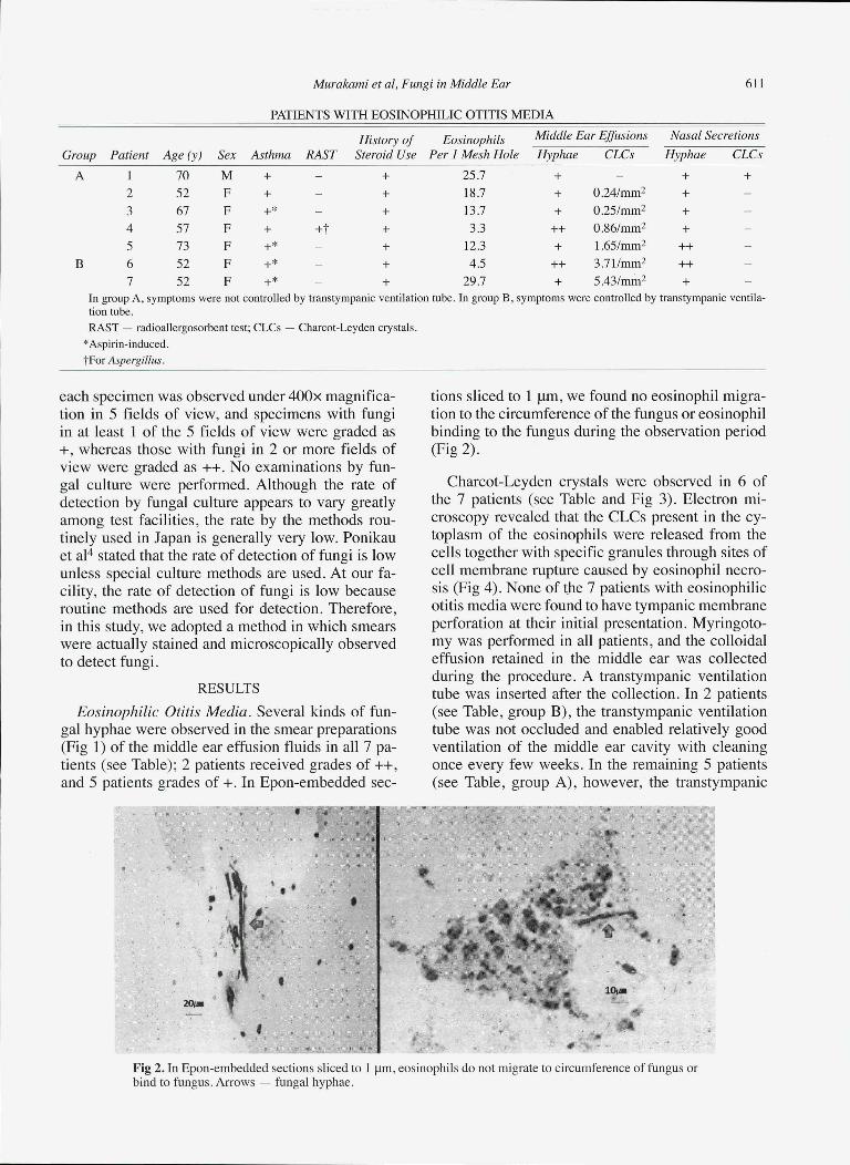
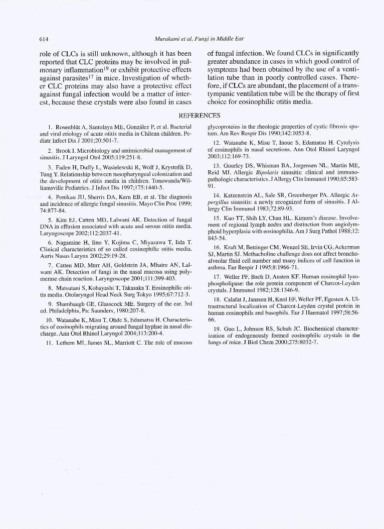
tions sliced to 1 |a,m, we found no eosinophil migra-tion to the circumference of the fungus or eosinophilbinding to the fungus during the observation period(Fig 2).
Charcot-Leyden crystals were observed in 6 ofthe 7 patients (see Table and Fig 3). Electron mi-croscopy revealed that the CLCs present in the cy-toplasm of the eosinophils were released from thecells together with specific granules through sites ofcell membrane rupture caused by eosinophil necro-sis (Fig 4). None of the 7 patients with eosinophilicotitis media were found to have tympanic membraneperforation at their initial presentation. Myringoto-my was performed in all patients, and the colloidaleffusion retained in the middle ear was collectedduring the procedure. A transtympanic ventilationtube was inserted after the collection. In 2 patients(see Table, group B), the transtympanic ventilationtube was not occluded and enabled relatively goodventilation of the middle ear cavity with cleaningonce every few weeks. In the remaining 5 patients(see Table, group A), however, the transtympanic
%
10MB
m t
Fig 2. In Epon-embedded sections sliced to 1 |im, eosinophils do not migrate to circumference of fungus orbind to fungus. Arrows — fungal hyphae.

612 Murakami et al. Fungi in Middle Ear
• f - • • >
Fig 3. Charcot-Leyden crystals (arrows) in middle ear ef-fusions.
ventilation tube became occluded immediately, andthe symptoms could not be controlled by the trans-tympanic ventilation tube. The number of CLCs perunit area was 0.6/mm2 in the cases that were poor-ly controlled despite insertion of a ventilation tube,and 4.57/mm2 in those that were adequately con-trolled after insertion of a ventilation tube. A statis-tically significant difference was found between thetwo groups (Student's r-test, p < 0.01) although thestatistical processing may have been improper, be-cause the number of patients was low. No significantcorrelation could be recognized between the numberof eosinophils and the number of CLCs, or betweenthe number of eosinophils and the number of hy-phae in the middle ear effusion (Pearson's correla-tion coefficient). There was no deterioration of hear-ing during the clinical course in any of the patients.The rate of detection of fungal hyphae was 100% inthe nasal smears; however, CLCs were observed inthe nasal secretions of only 1 of the 7 patients (seeTable).
OME. The 7 patients with serous effusions were18 to 75 years of age, and included no children. Oneof the 5 patients with mucous effusions was 51 yearsof age, whereas the other 4 were children 6 years ofage or younger. The rate of detection of fungal hy-phae in methenamine silver-stained smear prepara-tions was 100% for both serous and mucous effu-sions. The types of detected fungal hyphae were al-most the same as those in patients with eosinophilicotitis media. However, no CLCs were detected inany of the patients. Observation of methenamine sil-ver-stained nasal smear preparations yielded a rateof detection of fungal hyphae of 100%. Neither eo-sinophils nor CLCs were observed in the nasal se-cretions of patients with OME.
DISCUSSION
Culture of the middle ear effusions of patients with
Fig 4. Charcot-Leyden crystals within disrupted eosino-phil. Most eosinophils in effusions of patients with eo-sinophilic otitis media have become necrotic. Hexagonalcolumnar ciystal is on verge of being released from dis-rupted eosinophil.
OME rarely leads to isolation of bacteria. However,as endotoxins are often detected in the effusions,previous infection with gram-negative H infiuen-zae is suspected. On the other hand, the possibilityof infection with pathogenic microorganisms otherthan bacteria cannot be ruled out. Eungi have drawnattention as microorganisms that can cause chron-ic sinusitis. Kim et al-'' reported that the presence offungi in middle ear effusions could not be demon-strated by culture, whereas fungal DNA was detect-ed in 34% of the effusions by PCR. The isolationof fungi by PCR in the nasal secretions of patientswith chronic sinusitis and normal subjects has alsobeen reported.^ In that study, fungal DNA was de-tected in the nasal secretions of an estimated 42%of patients with sinusitis and 40% of normal sub-jects. Because fungi were isolated in the nasal se-cretions of nearly all patients with chronic sinusitisand normal subjects, according to Ponikau et al,"* thefinding of a 34% rate of detection of fungal DNA inthe middle ear effusions by PCR suggests that fungimight have been present in almost 100% of middleear effusions, as well, if the method described byPonikau et al^ was used. This corresponds to ourisolation of fungi at a rate of 100% by histologiemethods. The middle ear cavities in which the fungiwere isolated in this study were pathologic cavitieswith effusions. It is unknown whether fungi can beisolated in normal middle ears without middle earlesions. In normal middle ears, sampling of materi-als for examination is difficult because of the lackof fluid accumulation. If the precipitates of the so-lution used to wash the middle ear were examined,even though the myringotomy and irrigation of themiddle ear cavity in healthy persons may not be per-mitted on ethical grounds, fungi residing in normalmiddle ears might be isolated. However, since fun-

Murakami et al. Fungi in Middle Ear 613
gal hyphae were detected in 100% of the effusionsof typical patients with OME, the possibility of thefungi being the cause of OME cannot be exclud-ed. Passage of fungi into the middle ear may occurthrough the eustachian tube, in view of the fact thatall collected middle ear effusions were from patientswith no tympanic membrane perforation. The pos-sibility of contamination at the time of collection ofthe samples is slight, because the myringotomy wasperformed carefully, with aseptic precautions tak-en after washing the external ear canal. The fungimight have been carried via air or mucus flow fromthe nasopharynx into the middle ear cavity, owing tothe relationship between the middle ear pressure andatmospheric pressure. The finding of fungi in the na-sal cavity in all cases suggests that they are probablyalso frequently present in the nasopharynx.
Eosinophilic otitis media was first proposed asa new disease entity in 1995 by Matsutani et al.^Shambaugh and Glasscock'̂ described middle earinflammation associated with allergic disease as "al-lergic otitis media" in their textbook, documentingthe presence of an extremely viscous mucoid effu-sion in the middle ear containing eosinophils. How-ever, Matsutani et al*̂ called this condition "eosino-philic otitis media," and not "allergic otitis media,"because almost all of their patients had nonatopicasthma. Nagamine et al^ advocated as diagnostic cri-teria for eosinophilic otitis media 1) the presence ofyellow and extremely viscous middle ear effusion,predominantly containing eosinophils; and 2) pre-cedent and associated adult bronchial asthma. In ourstudy, 6 of the 7 cases of eosinophilic otitis mediawere RAST-negative. Adult asthma patients in crisisare usually RAST-negative. Therefore, it is suggest-ed that the eosinophilic otitis media in our study wasa nonallergic disease, as pointed out by Matsutani etal.^ It is considered that steroid hormones did not af-fect the results of the RAST in this study, as they hadnot been administered during at least 6 months priorto the collection of the middle ear effusion fluids.
Ponikau et al̂ wrote that fungi in nasal secretionsinduce the migration of eosinophils in the secre-tions, resulting in eosinophilic inflammation of theparanasal sinuses, and they proposed to call this dis-ease entity "eosinophilic fungal rhinosinusitis." Ifthis were correct, fungi passing into the middle earcavity would cause eosinophilic inflammation in themiddle ear. However, eosinophils do not migrate intomiddle ear effusions in typical patients with OME,despite the presence of fungi. The suggestion byPonikau et aH that fungi induce migration of eosino-phils is attractive, but it cannot explain why the fun-gi in OME do not induce migration of eosinophils.
As eosinophilic otitis media is a very rare disease ascompared to OME, the induction of eosinophils byfungi must be rare. Additional evidence is requiredto support the hypothesis that fungi may play rolesas microorganisms in the development of OME oreosinophilic otitis media. In this study, we could notobserve, in either OME or eosinophilic otitis media,eosinophils or neutrophils migrating to the circum-ference of the fungi and binding to them, as has beenreported in nasal secretions.^^ Whether fungi havepathogenicity or cause immunologie reactivity inthe middle ear has not yet been definitively deter-mined. Just as it is known that resident bacteria maysuddenly begin to exhibit pathogenicity, it is pos-sible that resident fungi may also suddenly begin toexhibit pathogenicity or cause immune reactivity.
The characteristics of eosinophilic otitis mediainclude viscous middle ear effusion that predomi-nantly contains degenerated eosinophils. The keyfactor in imparting the viscosity may be the degen-erated eosinophils, which are not usually observedin OME. It has been reported that the viscoelasticityof the purulent secretions in cystic fibrosis is primar-ily due to the high concentrations of DNA' ' and ac-tin released from the large number of necrotic neu-trophils accumulated in the airways. Therefore, it ispossible that viscous ear effusion is caused by thereleased DNA from degenerated eosinophils. How-ever, the nasal mucus does not exhibit high viscos-ity in patients with nasal house dust allergy, in spiteof the large number of disrupted eosinophils that areobserved.12 It may be difficult to definitively con-clude that the viscous ear effusion may be causedonly by DNA from degenerated eosinophils. Furtherresearch is needed to clarify the role of degeneratingeosinophils in the viscoelastic characteristics of mu-cus in cases of eosinophilic otitis media.
Charcot-Leyden crystals are hexagonal columnarcrystals with pointed edges that are frequently ob-served at sites of eosinophilic inflammation. Therehave been reports of detection of these crystals incases of allergic fungal sinusitis,'^-'^ Kimura's dis-ease,i5 and asthma.'^ Charcot-Leyden crystals areproduced from proteins with a molecular weightof 17.4 kd that have lysophospholipase activity.''^Charcot-Leyden crystal proteins are present in largeamounts in nuclear euchromatin and cytoplasm.Charcot-Leyden crystals are believed to crystallizegradually in the cytoplasm of eosinophils.'^ In thisstudy, CLCs being released directly from disruptedeosinophils could be observed by electron micros-copy. It can therefore be supported that the CLCsobserved in middle ear effusions were released di-rectly from disrupted eosinophils. The pathologic
614 Murakami et al. Fungi in Middle Far
role of CLCs is still unknown, although it has beenreported that CLC proteins may be involved in pul-monary inflammation'^ or exhibit protective effectsagainst parasites'"' in mice. Investigation of wheth-er CLC proteins may also have a protective effectagainst fungal infection would be a matter of inter-est, because these crystals were also found in cases
of fungal infection. We found CLCs in significantlygreater abundance in cases in which good control ofsymptoms had been obtained by the use of a venti-lation tube than in poorly controlled cases. There-fore, if CLCs are abundant, the placement of a trans-tympanic ventilation tube will be the therapy of firstchoice for eosinophilic otitis media.
REFERENCES
1. Rosenbliit A, Santolaya MF, González P, et al. Bacterialand viral etiology of acute otitis media in Chilean children. Pc-diatr Infect Dis J 2001;20:501-7.
2. Brook I. Microbiology and antimicrobial management ofsinusitis. J Laryngol Otol 2005;119:251-8.
3. Faden H, Duffy L, Wasielewski R, Wolf J, Krystofik D,Tung Y. Relationship between nasopharyngeal colonization andthe development of otitis media in children. Tonawanda/Wil-liamsville Pediatrics. J Infect Dis 1997;175:1440-5.
4. Ponikau JU, Shenis DA, Kern FB, et al. The diagnosisand incidence of allergic fungal sinusitis. Mayo Clin Proc 1999;74:877-84.
5. Kim EJ, Cattcn MD, Lalwani AK. Detection of fungalDNA in effusion associated with acute and serous otitis media.Laryngoscope 2002;l 12:2037-41.
6. Nagaminc H, lino Y, Kojima C, Miyazawa T, Iida T.Clinical characteristics of so called eosinophilic otitis media.Auris Nasus Larynx 2002;29:19-28.
7. Catten MD, Murr AH, Goldstein JA, Mhatre AN, Lal-wani AK. Detection of fungi in the nasal mucosa using poly-merase chain reaction. Laryngoscope 2001;lll:399-403.
8. Matsutani S, Kobayashi T, Takasaka T. Eosinophilic oti-tis media. Otolaryngol Head Neck Surg Tokyo 1995;67:712-3.
9. Shambaugh GE, Glasscock ME. Surgery of the ear. 3rded. Philadelphia, Pa: Saunders, 1980:207-8.
10. Watanabe K, Misu T, Ohde S, Edamatsu H. Characteris-tics of eosinophils migrating around fungal hyphae in nasal dis-charge. Ann Otol Rhinol Laryngol 2004;l 13:200-4.
11. Lethem MI, James SL, Marriott C. The role of mucous
glycoproteins in the rhéologie properties of cystic fibrosis spu-tum. Am Rev Rcspir Dis 1990; 142:1053-8.
12. Watanabe K, Misu T, Inoue S, Fdamatsu H. Cytolysisof eosinophils in nasal secretions. Ann Otol Rhinol Laryngol2003;112:169-73.
13. Gourley DS, Whisman BA, Jorgensen NL, Martin ME,Reid MJ. Allergic Bipolaris sinusitis: clinical and immuno-pathologic characteristics. J Allergy Clin Immunol 1990;85:583-91.
14. Katzenstein AL, Sale SR, Greenbergcr PA. Allergic As-pergillus sinusitis: a newly recognized form of sinusitis. J Al-lergy Clin Immunol 1983;72:89-93.
15. Kuo TT, Shih LY, Chan HL. Kimura's disease. Involve-ment of regional lymph nodes and distinction from angiolym-phoid hyperplasia with eosinophilia. Am J Surg Pathol 1988; 12:843-54.
16. Kraft M, Bettinger CM, Wenzel SE, Irvin CG, AckcrmanSJ, Martin SJ. Methacholine challenge does not affect broncho-alveolar fluid cell number and many indices of cell function inasthma. Eur RespirJ 1995;8:1966-71.
17. Weiler PF, Bach D, Austen KF. Human eosinophil lyso-phospholipase: the role protein component of Charcot-Leydencrystals. J Immunol 1982;128:1346-9.
18. Calafat J, Janssen H, Knol EF, Weiler PF, Egcsten A. Ul-trastructural localization of Charcot-Leyden crystal protein inhuman eosinophils and basophils. Eur J Hacmatol 1997;58:56-66.
19. Guo L, Johnson RS, Schuh JC. Biochemical character-ization of endogenously formed eosinophilic crystals in thelungs of mice. J Biol Chem 2000;275:8032-7.

Copyright of Annals of Otology, Rhinology & Laryngology is the property of Annals Publishing Company and
its content may not be copied or emailed to multiple sites or posted to a listserv without the copyright holder's
express written permission. However, users may print, download, or email articles for individual use.
Related Documents