Microcirculation, 12: 5–15, 2005 Copyright c 2005 Taylor & Francis Inc. ISSN: 1073-9688 print / 1549-8719 online DOI: 10.1080/10739680590894966 Microvascular Rheology and Hemodynamics HERBERT H. LIPOWSKY Department of Bioengineering, The Pennsylvania State University, University Park, Pennsylvania, USA ABSTRACT The goal of elucidating the biophysical and physiological basis of pressure–flow relations in the mi- crocirculation has been a recurring theme since the first observations of capillary blood flow in living tissues. At the birth of the Microcirculatory Society, seminal observations on the heterogeneous distri- bution of blood cells in the microvasculature and the rheological properties of blood in small bore tubes raised many questions on the viscous properties of blood flow in the microcirculation that captured the attention of the Society’s membership. It is now recognized that blood viscosity in small bore tubes may fall dramatically as shear rates are increased, and increase dramatically with elevations in hema- tocrit. These relationships are strongly affected by blood cell deformability and concentration, red cell aggregation, and white cell interactions with the red cells and endothelium. Increasing strength of red cell aggregation may result in sequestration of clumps of red cells with either reductions or increases in microvascular hematocrit dependent upon network topography. During red cell aggregation, resis- tance to flow may thus decrease with hematocrit reduction or increase due to redistribution of red cells. Blood cell adhesion to the microvessel wall may initiate flow reductions, as, for example, in the case of red cell adhesion to the endothelium in sickle cell disease, or leukocyte adhesion in inflammation. The endothelial glycocalyx has been shown to result from a balance of the biosynthesis of new glycans, and the enzymatic or shear-dependent alterations in its composition. Flow-dependent reductions in the endothelial surface layer may thus affect the resistance to flow and/or the adhesion of red cells and/or leukocytes to the endothelium. Thus, future studies aimed at the molecular rheology of the endothelial surface layer may provide new insights into determinants of the resistance to flow. Microcirculation (2005) 12, 5–15. doi:10.1080/10739680590894966 KEY WORDS: blood viscosity, flow, intravascular pressure, rheology, shear rates, wall shear stress INTRODUCTION At the birth of the Microcirculatory Society in 1954, relatively little was known of the rheological behavior of blood in the microcirculation. The seminal stud- ies of Poiseuille (54), Landis (39), F˚ ahraeus (20), Vejlens (68), and Krogh (38) provided a conceptual framework for understanding the basis for the resis- tance to flow. Although best known for his experi- mental studies of the flow of fluids through tubes, Poiseuille’s earlier observations on the separation of cells and plasma in arterioles and venules led to the discovery of “plasma skimming’’ and the need for a greater understanding of the mechanics of blood flow (67). The pioneering intravital studies of Landis, Supported in part by NIH research grant R01 HL-39286. Address correspondence to Herbert H. Lipowsky, PhD, Depart- ment of Bioengineering, Penn State University, 205 Hallowell Bldg., University Park, PA 16802, USA. E-mail: hhlbio@engr. psu.edu Received 7 September 2004; accepted 28 September 2004. using a forerunner of the modern servo-null tech- nique to measure capillary pressure, attempted to ex- plore the applicability of Poiseuille’s law to describe microvascular resistance. F˚ ahraeus’ discovery of re- ductions in tube hematocrit as blood flows through small bore tubes, and subsequent studies on the at- tendant reduction in blood viscosity (21), defined the approach to elucidating the rheological basis of mi- crovascular blood flow. The comprehensive experi- mental studies on leukocyte behavior in the microvas- culature by Vejlens delineated many features of their distribution and sequestration in the microcirculation that affect blood flow. The pioneering observations by Krogh delineated many facets of flow distribution through networks of capillaries and sequestration of red cells under normal and pathological conditions. However, it would take the subsequent five decades to develop a comprehensive understanding of the role for the intrinsic properties of blood and microvascu- lar topography as determinants of the resistance to flow. Due to the advent of new quantitative methods for intravital microscopy, it is now well understood that in addition to blood cell concentration, red cell

Welcome message from author
This document is posted to help you gain knowledge. Please leave a comment to let me know what you think about it! Share it to your friends and learn new things together.
Transcript
Microcirculation, 12: 5–15, 2005Copyright c© 2005 Taylor & Francis Inc.ISSN: 1073-9688 print / 1549-8719 onlineDOI: 10.1080/10739680590894966
Microvascular Rheology and HemodynamicsHERBERT H. LIPOWSKY
Department of Bioengineering, The Pennsylvania State University, University Park,Pennsylvania, USA
ABSTRACT
The goal of elucidating the biophysical and physiological basis of pressure–flow relations in the mi-crocirculation has been a recurring theme since the first observations of capillary blood flow in livingtissues. At the birth of the Microcirculatory Society, seminal observations on the heterogeneous distri-bution of blood cells in the microvasculature and the rheological properties of blood in small bore tubesraised many questions on the viscous properties of blood flow in the microcirculation that capturedthe attention of the Society’s membership. It is now recognized that blood viscosity in small bore tubesmay fall dramatically as shear rates are increased, and increase dramatically with elevations in hema-tocrit. These relationships are strongly affected by blood cell deformability and concentration, red cellaggregation, and white cell interactions with the red cells and endothelium. Increasing strength of redcell aggregation may result in sequestration of clumps of red cells with either reductions or increasesin microvascular hematocrit dependent upon network topography. During red cell aggregation, resis-tance to flow may thus decrease with hematocrit reduction or increase due to redistribution of red cells.Blood cell adhesion to the microvessel wall may initiate flow reductions, as, for example, in the caseof red cell adhesion to the endothelium in sickle cell disease, or leukocyte adhesion in inflammation.The endothelial glycocalyx has been shown to result from a balance of the biosynthesis of new glycans,and the enzymatic or shear-dependent alterations in its composition. Flow-dependent reductions inthe endothelial surface layer may thus affect the resistance to flow and/or the adhesion of red cellsand/or leukocytes to the endothelium. Thus, future studies aimed at the molecular rheology of theendothelial surface layer may provide new insights into determinants of the resistance to flow.Microcirculation (2005) 12, 5–15. doi:10.1080/10739680590894966
KEY WORDS: blood viscosity, flow, intravascular pressure, rheology, shear rates, wall shear stress
INTRODUCTION
At the birth of the Microcirculatory Society in 1954,relatively little was known of the rheological behaviorof blood in the microcirculation. The seminal stud-ies of Poiseuille (54), Landis (39), Fahraeus (20),Vejlens (68), and Krogh (38) provided a conceptualframework for understanding the basis for the resis-tance to flow. Although best known for his experi-mental studies of the flow of fluids through tubes,Poiseuille’s earlier observations on the separation ofcells and plasma in arterioles and venules led to thediscovery of “plasma skimming’’ and the need fora greater understanding of the mechanics of bloodflow (67). The pioneering intravital studies of Landis,
Supported in part by NIH research grant R01 HL-39286.Address correspondence to Herbert H. Lipowsky, PhD, Depart-ment of Bioengineering, Penn State University, 205 HallowellBldg., University Park, PA 16802, USA. E-mail: [email protected] 7 September 2004; accepted 28 September 2004.
using a forerunner of the modern servo-null tech-nique to measure capillary pressure, attempted to ex-plore the applicability of Poiseuille’s law to describemicrovascular resistance. Fahraeus’ discovery of re-ductions in tube hematocrit as blood flows throughsmall bore tubes, and subsequent studies on the at-tendant reduction in blood viscosity (21), defined theapproach to elucidating the rheological basis of mi-crovascular blood flow. The comprehensive experi-mental studies on leukocyte behavior in the microvas-culature by Vejlens delineated many features of theirdistribution and sequestration in the microcirculationthat affect blood flow. The pioneering observationsby Krogh delineated many facets of flow distributionthrough networks of capillaries and sequestration ofred cells under normal and pathological conditions.
However, it would take the subsequent five decadesto develop a comprehensive understanding of the rolefor the intrinsic properties of blood and microvascu-lar topography as determinants of the resistance toflow. Due to the advent of new quantitative methodsfor intravital microscopy, it is now well understoodthat in addition to blood cell concentration, red cell
Microvascular rheology and hemodynamics6 HH Lipowsky
deformability and aggregation and white blood celldeformability and adhesion to the endothelium arethe principal intrinsic factors that affect resistanceto flow. The extent to which they affect resistance isdetermined by topographical branching patterns andmicrovessel diameters. During the last five decades,numerous contributions by members of the Micro-circulatory Society have explored the details of theseinteractions in health and disease. A brief overviewof some of these studies that have set the stage forfuture studies of interactions between blood rheol-ogy and microvascular function is presented in thefollowing.
THE IN VITRO FRAMEWORK
Acquisition of the viscosity of blood by bulk viscom-etry has emphasized the importance of shear rate,hematocrit, and red cell aggregation and deformabil-ity as it pertains to flow in large blood vessels (11).With the use of tube, Couette, and cone-plate vis-cometers, under the assumption that blood is a ho-mogeneous fluid with an intrinsic viscosity, in vitrostudies have revealed that blood viscosity falls about75% as shear rates (γ ) rise from on the order of 0.1to 1000 sec−1. A comparison of this “shear thinning’’of blood in the presence and absence of aggregatingagents suggests that about 75% of the decrease is a re-sult of the disruption of red cell aggregates, and 25%is due to red cell deformation in response to increasedshear stresses. At a given shear rate, blood viscos-ity rises exponentially with increasing red blood cell(RBC) concentration (hematocrit) to a degree depen-dent on prevailing γ . The viscosity of the suspendingmedium (plasma) has been shown to be invariantwith γ (Newtonian) and is dependent mainly on pro-tein content and temperature.
Within the circulation, in large diameter vessels rep-resentative of the macrocirculation (i.e., >100 µm),blood may be treated as a homogeneous continuumwith intrinsic properties characterized by an “appar-ent viscosity.’’ The term apparent viscosity is usedsince viscosity of a homogeneous fluid (e.g., water,molasses) is a material property that may be depen-dent on shear rate and temperature and is invariantwith the size of the vessel through which it flows. Invivo, ever diminishing length scales and the particu-late nature of blood affect this relationship as bloodcourses its way through successive divisions of thecirculatory tree. The term “effective viscosity’’ (η)is often used to represent the value of viscosity thatsatisfies Poiseuille’s law, since it does not explicitly
reflect the shear dependency of viscosity along thetube radius.
According to Poiseuille’s law, flow (Q) and pressuredrop (�P) are related by
Q = π
128D4
ηl�P (1)
where D is luminal diameter and l is vessel length.Hence, given measurements of Q and �P, one maycalculate η for a microvessel of specified length anddiameter. The dominance of noncontinuum effects inthe smallest microvessels (approaching red cell di-ameter) results in an effective blood viscosity that isstrongly dependent on microvessel diameter and adeparture from this relationship.
PRESSURE AND FLOW RELATIONS IN THEMICROVASCULAR NETWORK
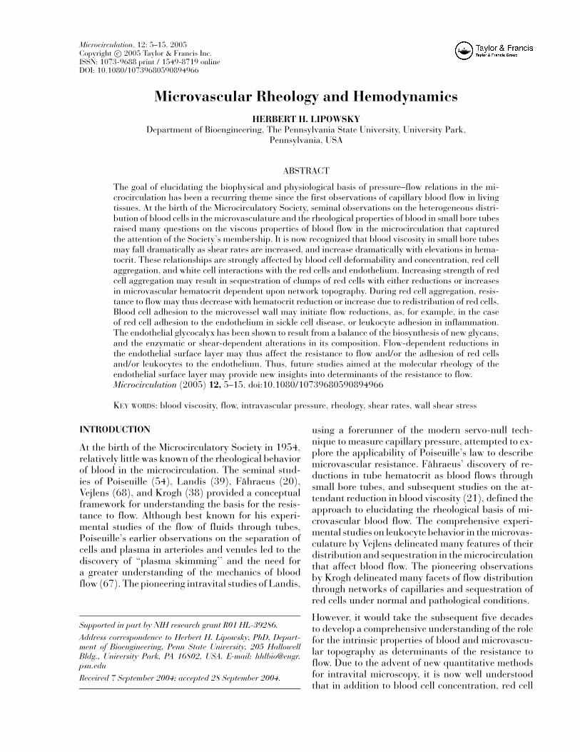
Direct measurements of pressures and flows in ex-teriorized tissues have provided a wealth of dataon microvascular hemodynamics throughout succes-sive microvascular divisions (77). Relating these datato the architecture of the microvascular networkhas presented a challenging problem. The disparatetopography of arterioles, capillaries, and venulesamong numerous tissues (e.g., mesentery, omentum,intestine, striated muscle) has prompted a searchfor methods to discern commonalities in structureand function among various tissues. As typified inFigure 1 for the mesenteric circulation (cat) (75), thedistribution of intravascular pressures (determinedby the servo-null method (33; 73)) and red cell ve-locity (two-slit method (71)) is presented using vessellumen width (assumed equal to internal diameter) asan index of position within the hierarchy of microves-sels. The increasingly precipitous decline in arterialpressure with diminishing diameters reflects a steadyrise in resistance to flow for the entire throughput ofthe network. It is evident that the resistance to flowwithin each major architectural division (arterioles,precapillaries, capillaries, etc.) attains a maximumin the precapillary vessels; in contrast to the expecta-tion that the maximum resistance (R ) occurs in thesmallest vessels, based upon Poiseuille’s law, whereR = �P/Q = (128/π)(η�/D4). The steady declinein red cell velocity in the arteriolar network, and itssubsequent rise in venular segments, represent con-servation of the total throughput of the network asthe number of vessels varies through sequential seg-ments. These trends have been shown to be indicativeof the unique branching patterns of many tissues, as
Microvascular rheology and hemodynamicsHH Lipowsky 7
Figure 1. Arteriovenous distribution of intravascularpressure and red cell velocity from arterioles to venulesin mesentery (cat) obtained in the laboratory of BenjaminW. Zweifach (75) (with permission). Microvessel diameter(abscissa) is taken as an index of position within the net-work. Pressure falls in accord with the resistance to flowthrough successive divisions, which is dependent on net-work topography and the viscous properties of blood.
described by the late Benjamin W. Zweifach, who in along and distinguished career pioneered many studiesof hemodynamics in the microvasculature, nurturedthe development of the methodology to perform suchstudies, and stimulated and inspired many of his stu-dents to continue in his footsteps.
Whereas such spatial distributions of hemodynamicparameters loosely correlate with form and func-tion of the microvascular network, their ability toreveal insights into the relationship between thetotal throughput of the network and demands ofthe parenchymal tissue is somewhat limited. Otherschemes to relate hemodynamics to function have re-volved around centripetal and centrifugal order ofbranching schemes (72), the application of Strahler’sand Horton’s laws that describe the topography ofriver patterns (23), a combination of the two schemes(16), expressing hemodynamics in terms of gener-ations of branching (40), fractal representation oftopography and flows (26), or spatial patterns ofbundles of microvessels (66). Dynamic studies of thedispersion of indicators throughout the microvascu-lar network (50) have highlighted the difficulty in
delineating the order of perfusion of divisions of agiven network. The appearance of indicators (labeledplasma or red cells) within the venular network oftenprecedes that in nearby arterioles, thus suggesting amultiplicity of pathways from inlet to outlet in mostnetworks. It is evident that the microvascular net-work cannot be simply expressed as an ensemble ofpurely serial and parallel elements (24). The multi-plicity of pathways to the true capillaries may resultin a functional capillary density that is flow depen-dent. It has been shown that flow reductions withinan individual capillary may have a minimal effect onarteriolar flow (51). Computer simulations of flowthrough skeletal muscle reveal an invariance of resis-tance to flow through the network until greater than30% of all capillaries are occluded (32).
WALL SHEAR STRESS
Hydrodynamic shear stresses acting on the endothe-lium are now recognized as important determinantsof mechanotransduction and the release of vasoactivesubstances (6). Recent studies have demonstratedthat the coupling of blood rheological propertiesand vasoregulatory behavior may dramatically affectcapillary perfusion. In studies of blood substitutes, ithas been shown that an elevated viscosity elicits avasodilatory response due to increased shear stressesthat enhances flow within capillaries (10).
In vitro studies with glass tubes by Barbee andCokelet (3) paved the way for a realistic appraisalof shear rates and stresses throughout the microvas-cular tree. Using glass tubes as small as 29 µm, thesestudies demonstrated that shear stress in tubes of mi-crovessel dimensions could be described in terms ofshear rates (regardless of tube size) provided thatthe tube hematocrit was correctly specified. Thus,given the correct tube hematocrit, the relationshipbetween blood viscosity and shear rates in microves-sels could be uniquely specified for tubes represen-tative of arterioles and venules. In vivo estimates ofeffective viscosity from upstream to downstreampressure drop (�P) and flow in small arterioleswere in general agreement with these studies (46).Whereas the in vivo measurement of upstream todownstream pressure drops in single unbranched mi-crovessels is technically challenging, the value of theinformation may far exceed the inherent errors anduncertainties. Values of wall shear stress (τWALL) havebeen obtained by direct measurement of �P and ves-sel length l and diameter D (44). Assuming a ves-sel of uniform diameter and circular cross section,
Microvascular rheology and hemodynamics8 HH Lipowsky
application of the principle of static equilibrium dic-tates that τWALL = �PD/4l. Thus, with these as-sumptions, τWALL was found to average 47 dyn/cm2
in arterioles and 29 dyn/cm2 in venules of the mesen-tery (cat) in the normal flow state (44).
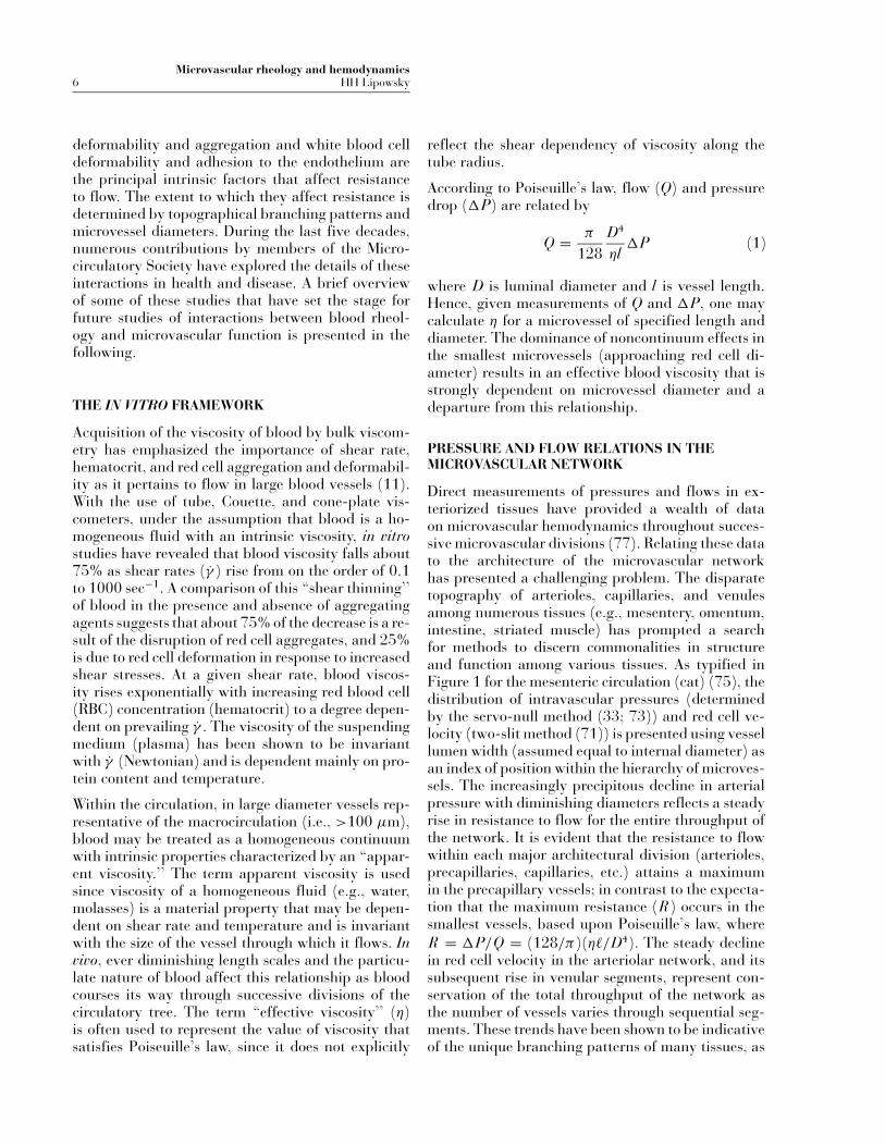
The foundation laid down by in vitro rheologi-cal studies facilitated estimation of shear rates andstresses throughout the microvasculature based ondirect measurements of red cell velocity and mi-crovessel geometry in tissues such as mesentery (76),cremaster muscle (31), and spinotrapezius muscle(77), as illustrated in Figure 2. These distributionsof shear rates γ were calculated from measurementsof red cell velocity (VRBC, two-slit method) using em-pirical correlations between volumetric flow and VRBCobtained in glass tubes (2). Using an effective value ofblood viscosity obtained in mesentery, shear stressesτWALL were estimated from the Newtonian relation-ship τWALL = ηγ , assuming a value of η = 3.5 cP(44). These data reveal shear stresses in arteriolesranging from 25–60 dyn/cm2, which fall dramati-cally to 10–30 dyn/cm2 in postcapillary venules, inthe normal flow state (43).
RESISTANCE
The resistance R to flow arises from the effective vis-cosity η of blood and vascular hindrance z, such thatR = ηz . Using Poiseuille’s law as a model, the re-sistance per unit length (R/L) of microvessel at agiven η should be inversely proportional to D4. Di-rect measurements of upstream to downstream pres-sure drop in single unbranched microvessels usingthe dual servo-null technique (46) and flow (two-
Figure 2. Arteriovenous distribution of wall shear rate(γ , based on measured VRBC and diameter), and wall shearstress, τWALL, calculated from the product of γ η assumingthat η = 3.5 cP for 3 representative microvascular net-works (from (43), with permission).
slit technique (2,47,71)) permitted calculation of R/L(from R = �P/Q and vessel length) to reveal a four-decade increase in R/L (44) as blood traverses thearteriolar network in mesentery (cat). Remarkably, apower law regression against D revealed an exponentof 4.0 on the arteriolar side and 3.9 on the venousside of the capillary network. Thus, in the normalflow state, the fourth-power dependency on diame-ter is the dominant determinant of resistance to flow.Although the conditions for which Poiseuille’s law ap-plies (uniform fluid with constant viscosity) are notreadily applicable to the microcirculation, the broadrange of diameters encountered as blood traverses thenetwork from arteriole to capillary results in a domi-nance of the fourth power of diameter. This behavioris of great importance since small changes in vasculardiameter may result in a fourfold greater change inresistance within individual microvessels, thus affect-ing the distribution of flow within the microvascularnetwork.
APPARENT VISCOSITY
Using Poiseuille’s law as a constitutive equation forblood flow in microvessels, the apparent viscosity η
of blood may be computed from measured pressuredrops and flows. The measurement of upstream todownstream pressure drops poses many challenges.Cumulative errors in the calculation of η may amountto 46% (46). The major determinants of in vivo ap-parent viscosity are shear rates and tube hematocrit.At the low level of microvascular hematocrits foundin most tissues in the normal flow state, the apparentviscosity varies linearly with microvessel hematocrit(HMICRO) (45). These studies have revealed averagearteriolar values of about 3.6 cP and average venularvalues of about 5.2 cP in the mesentery of the cat.The greater viscosity in venules was attributed to therelatively lower shear rates, leukocyte adhesion to thewalls of venules that may obstruct the lumen and givea falsely elevated viscosity, and greater departuresfrom a circular cross section. As flow rates are de-creased, such as in shock, hemorrhage, and other cir-culatory disorders, flow within individual microves-sels falls nonlinearly with pressure drop, suggestingincreases in the apparent viscosity of blood (44). De-termination of the variation of effective viscosity withshear rate and diameter by indirect analysis, usingcomputations of pressures and flows throughout themesentery (rat) to match measured red cell fluxes andboundary pressures, have resulted in a self-consistentappraisal of effective viscosity that falls betweenin vitro tube studies and direct in situ measurements
Microvascular rheology and hemodynamicsHH Lipowsky 9
(57), although considerable errors may be presentdue to inaccuracies in accounting for the true geom-etry of the vessel cross section in the measurements(14).
MICROVASCULAR HEMATOCRIT
The pioneering studies of Fahraeus (20) on bloodflow in small-bore glass tubes revealed that twohematocrits are necessary to describe blood withinthe microcirculation: The tube, or microvessel hema-tocrit (HMICRO), represents the red cell fraction resi-dent in the tube at any instant of time, and the dis-charge hematocrit (HDISCH) is the red cell fraction ofthe effluent of a tube, collected in a hypothetical mix-ing cup. The disparity between tube and dischargehematocrit arises with reductions in tube diameterbecause the mean velocity of red cells increases rela-tive to mean plasma velocity, hence fewer cells mustbe resident within the tube to ensure that the dis-charge hematocrit equals the feed hematocrit. Tubehematocrit falls as diameter is decreased until a min-imum diameter is reached, below which deformationof RBCs lead to their sequestration (1). Poiseuille(67) observed the skimming of plasma from sidebranches in small blood vessels. These two processeslead to a decrease in microvessel hematocrit as bloodcourses its way from arterioles to capillaries and asubsequent rise in postcapillary venules. Studies byBarbee and Cokelet (4) and others on the effect oftube diameter and Fenton, Carr, and Cokelet (22)on plasma skimming at bifurcations have provideda basis for understanding in vivo observations.In vivo measurements of HMICRO in networks suchas the cheek pouch (61), cremaster muscle (36) andmesentery (45) have suggested that these effects aredependent on network topography and local flowrates. It is thought that the heterogeneity in red cellvelocity and microvessel hematocrit within any givenmicrovascular division may contribute to about 20%of the total reduction in capillary hematocrit (55).
It has been suggested that HMICRO is relatively uni-form across the lumen (radial direction) of an ar-teriole or venule, compared with the heterogeneityassociated with successive branchings. However, thepresence of a thin annulus of plasma surroundingthe core of RBCs within a microvessel facilitates anuneven distribution of RBCs at arteriolar branchings(12). The proportion of RBCs from the core and cell-free plasma layer that is captured by an arteriolarbranch at a bifurcation is dependent upon the rel-ative magnitudes of total volumetric flow from par-
ent to daughter branch at a bifurcation. At the finalramifications of the arteriolar network, red cell en-try into capillaries is dependent on the presence of asufficient pressure gradient that can sustain red celldeformations at the capillary entrance, hence the cap-illary branch with the fastest stream (greatest pres-sure gradient) captures the majority of red cells. Ifone averages daughter to parent branch ratios of redcell flux and their respective bulk flow ratios overmany bifurcations, these effects appear to be greatlyattenuated and RBC flux and bulk flow rates follow1:1 within a small deviation (37). However, for anindividual bifurcation, the relationships between cellflux and flow are strongly nonlinear (sigmoidal) (62).The forces that govern phase separation (cells fromplasma) of blood have been described in terms ofthe hydrodynamic forces (25,62,74). Radial forcesarise from the summated effects of shear stress andthe pressure distribution on individual cells that di-rect cells into the daughter branch with the greaterflow rate. Similar dynamics have been revealed forthe case of WBCs negotiating bifurcations in thenetwork (41).
Low values of HMICRO relative to systemic hemat-ocrit (HSYS) suggest that oxygen transport to tissuemay be compromised by diminished oxygen-carryingcapacity. Direct intubation of microvessels with mi-cropipettes to aspirate the red cell flux has suggestedthat the discharge hematocrit (HDISCH) agrees wellwith HSYS (17) for microvessels ranging in diameterfrom 6 to 98 µm. Calculations of an average tissuehematocrit, based upon the relative transit times ofplasma and red cells measured within the microvas-cular network have revealed a close agreement withHSYS (52).
BLOOD CELL DEFORMABILITY
With diminishing vessel diameter, the particulate na-ture of blood dominates the resistance to flow. In vitrosimulation of capillary flows using polycarbonatesieves with 5 µm pores (Nuclepore filters) was pi-oneered by Gregersen et al. (27) and emphasized thedominant contribution of red cell deformability toperfusion of capillaries. Through such experimen-tal simulations it is now recognized that the initialdeformation that red blood cells (RBCs) and whiteblood cells (WBCs) incur upon entry to a capillarycontributes significantly to the pressure drop acrossindividual capillaries (42,62,64). Thus, the contri-bution of the intrinsic properties of blood to hemo-dynamic resistance changes markedly throughout the
Microvascular rheology and hemodynamics10 HH Lipowsky
succession of vessels from arterioles to capillaries asdiameter varies between divisions.
Blood cell deformability affects the entrance of bloodcells into capillaries. RBCs with reduced deforma-bility in pathological disorders (e.g., sickle cell dis-ease) may be sequestered at the capillary entrance.Stiffening of the red cell membrane or elevationsin hemoglobin viscosity may impede RBC transitthrough capillaries (28,34,52). The normally stifferWBCs traverse the capillary network through largerthoroughfare channels (19) and WBCs may becometrapped at the capillary entrance or incur a prolongedtransit time following their stiffening with activationduring inflammation (5, 29).
BLOOD CELL AGGREGATION
The effect of red cell aggregation (RCA) on the in vivoresistance to flow has been fraught with controversy.In vitro studies of RCA in vertically positioned, small-bore glass tubes (13,59,60) revealed that apparentviscosity decreases with increasing degrees of RCA.However, in vivo measurements in the low flow statesuggest a dramatic rise in apparent viscosity withreductions in γ based on resistance measurementswithin individual venules (44) or isolated regions ofskeletal muscle (9). Studies of aggregate formationin postcapillary venules in response to high molecu-lar weight dextrans (500 kDa) reveal a blunting ofthe velocity profile that may result in greater energydissipation and hence increased resistance (7). Stud-ies of the radial dispersion of RBCs during dextran-induced aggregation suggest that the succession ofbranches in the venous circulation attenuates axialmigration of RBCs (8), thus inhibiting the develop-ment of an annular plasma layer as occurs in therelatively longer unbranched tubes in vitro.
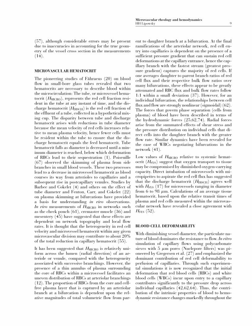
It is apparent that the effects of RCA vary betweenregional networks as well as within a discrete ensem-ble of microvessels perfused by the same source. Toillustrate, shown in Figure 3 is the variation of re-sistance between a paired arteriole and venule thatserve the single inflow/outflow modular network ofmicrovessels peculiar to the rabbit omentum. Arte-riovenous resistance (RAV) was computed from theratio of arteriolar–venular pressure drop (dual servo-null method, (46)) to the volumetric flow measuredin the feeding arteriole (two-slit method, (47)). Withinfusion of successively greater levels of high molec-ular weight dextran (500 kDa; D × 500) RAV firstrises slightly, as plasma viscosity increases, and thenfalls precipitously as [D × 500] exceeds 1 g%. These
Figure 3. Arteriole to venule resistance obtained in a sin-gle inlet/outlet module of the rabbit omentum (inset) forvarious plasma concentrations of high molecular weightdextran (500 kDa, D × 500). The resistance was com-puted from R = �P/Q, where Q was obtained from VRBCin the feeding arteriole. With increasing [D × 500] R ini-tially rises as plasma viscosity becomes elevated and thenfalls dramatically as red cell aggregation causes a markedreduction in systemic hematocrit as RBCs are sequesteredin other organs. Arteriolar microvessel hematocrit fell from26 to 13% as HSYS fell from 33 to 20% as [D × 500] rose to2.6 g%. These trends suggest that the response to extremered cell aggregation may be quite variable among differentnetworks, with some showing severe increases in red cellentrapment, and others showing severe hemodilution.
trends arise because of red cell sequestration in otherorgans with increasing RCA that leads to a dramaticdecrease in HSYS and HMICRO in the arteriole. In thisparticular case, arteriolar HMICRO fell from 25.6 to13.1% as HSYS fell from 32.5 to 20.0% in responseto [D × 500] increasing from 0 to 2.6 g%.
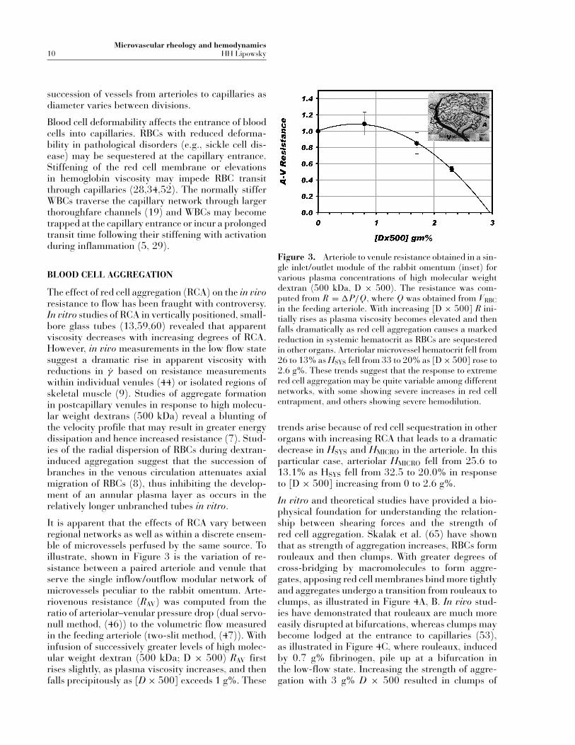
In vitro and theoretical studies have provided a bio-physical foundation for understanding the relation-ship between shearing forces and the strength ofred cell aggregation. Skalak et al. (65) have shownthat as strength of aggregation increases, RBCs formrouleaux and then clumps. With greater degrees ofcross-bridging by macromolecules to form aggre-gates, apposing red cell membranes bind more tightlyand aggregates undergo a transition from rouleaux toclumps, as illustrated in Figure 4A, B. In vivo stud-ies have demonstrated that rouleaux are much moreeasily disrupted at bifurcations, whereas clumps maybecome lodged at the entrance to capillaries (53),as illustrated in Figure 4C, where rouleaux, inducedby 0.7 g% fibrinogen, pile up at a bifurcation inthe low-flow state. Increasing the strength of aggre-gation with 3 g% D × 500 resulted in clumps of
Microvascular rheology and hemodynamicsHH Lipowsky 11
Figure 4. As the strength of red cell aggregation increases, aggregates undergo a transition from rouleaux (A) to clumps(B) (from (65), with permission). The weaker rouleaux that tend to pile up at branch points in the arteriolar network(C) in the low flow state are easily disrupted compared to the stronger clumps (D) that become lodged at the entranceto capillaries and resist disruption upon recovery from a low flow state (from (53), with permission).
RBCs that frequently became lodged at the capillaryentrance and resisted disruption by hydrodynamicforces.
BLOOD CELL–ENDOTHELIUM INTERACTIONS
The adhesive interactions of RBCs and WBCs withthe endothelium in, for example, sickle cell diseaseand inflammation, respectively, have prompted rhe-ological studies of the effect of blood cell adhesion onflow resistance, the molecular basis for adhesion andthe effect of hydrodynamic forces on the dynamics ofadhesion. In experimental models of inflammation, ithas been shown that as few as 12 WBCs adhering per100 µm of venule length may double the resistance toflow in postcapillary venules (30). Significant retar-dation of flow may occur when sickle red cells adhereto the microvessel wall and obstruct the lumen (34).These events may initiate a further decline in flow
leading to the sequence of red cell deoxygenation,hemoglobin polymerization and further cell seques-tration (35).
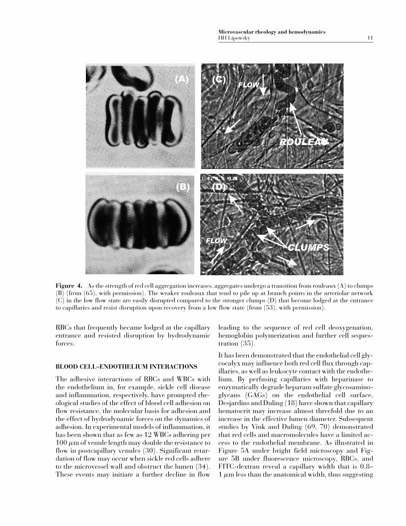
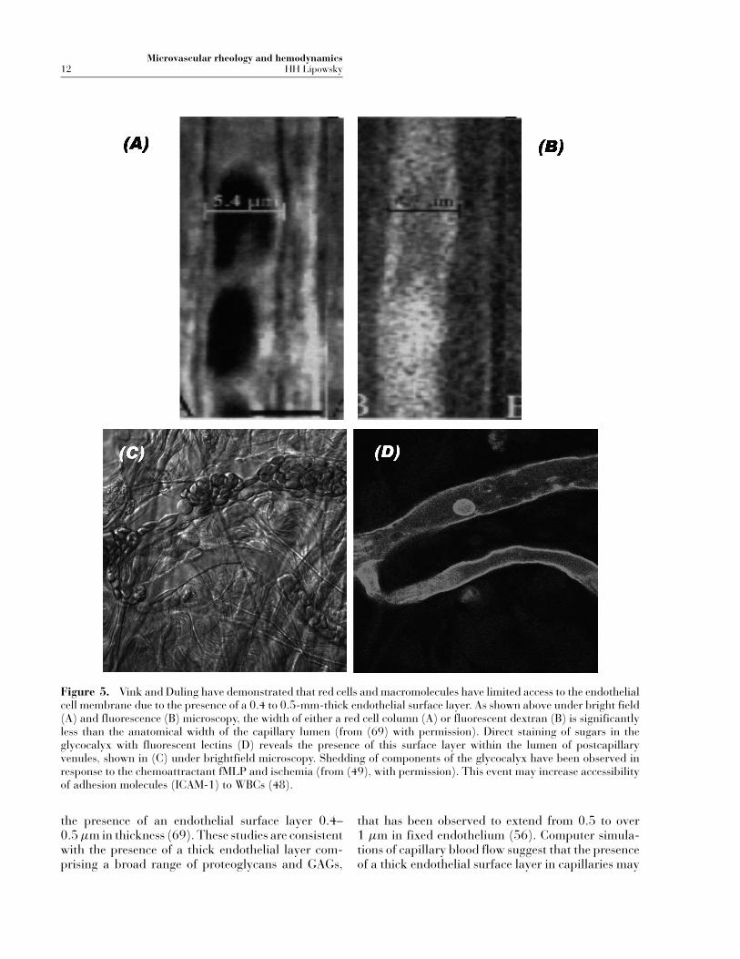
It has been demonstrated that the endothelial cell gly-cocalyx may influence both red cell flux through cap-illaries, as well as leukocyte contact with the endothe-lium. By perfusing capillaries with heparinase toenzymatically degrade heparam sulfate glycosamino-glycans (GAGs) on the endothelial cell surface,Desjardins and Duling (18) have shown that capillaryhematocrit may increase almost threefold due to anincrease in the effective lumen diameter. Subsequentstudies by Vink and Duling (69, 70) demonstratedthat red cells and macromolecules have a limited ac-cess to the endothelial membrane. As illustrated inFigure 5A under bright field microscopy and Fig-ure 5B under fluorescence microscopy, RBCs, andFITC-dextran reveal a capillary width that is 0.8–1 µm less than the anatomical width, thus suggesting
Microvascular rheology and hemodynamics12 HH Lipowsky
Figure 5. Vink and Duling have demonstrated that red cells and macromolecules have limited access to the endothelialcell membrane due to the presence of a 0.4 to 0.5-mm-thick endothelial surface layer. As shown above under bright field(A) and fluorescence (B) microscopy, the width of either a red cell column (A) or fluorescent dextran (B) is significantlyless than the anatomical width of the capillary lumen (from (69) with permission). Direct staining of sugars in theglycocalyx with fluorescent lectins (D) reveals the presence of this surface layer within the lumen of postcapillaryvenules, shown in (C) under brightfield microscopy. Shedding of components of the glycocalyx have been observed inresponse to the chemoattractant fMLP and ischemia (from (49), with permission). This event may increase accessibilityof adhesion molecules (ICAM-1) to WBCs (48).
the presence of an endothelial surface layer 0.4–0.5 µm in thickness (69). These studies are consistentwith the presence of a thick endothelial layer com-prising a broad range of proteoglycans and GAGs,
that has been observed to extend from 0.5 to over1 µm in fixed endothelium (56). Computer simula-tions of capillary blood flow suggest that the presenceof a thick endothelial surface layer in capillaries may
Microvascular rheology and hemodynamicsHH Lipowsky 13
significantly affect red cell flux, and that its deforma-tion with fluid shear stresses may enhance the abilityof red cells to traverse nonuniform capillaries (63).
Enzymatic degradation of the glycocalyx by infu-sion of heparinase in mesenteric arterioles has re-sulted in a decrease in regional resistance up to 21%(58). It has been demonstrated that removal of hep-aran sulfate GAGs by perfusion of mesenteric post-capillary venules with heparinase results in increasedWBC-EC adhesion (48). Similar findings have beennoted in cremaster muscle in response to infusion ofoxidized LDL (15). Superfusion of mesenteric tis-sue with the peptide fMLP (f-met-leu-phe) resultsin a rapid increase in the adhesion of inert micro-spheres labeled with antibody for ICAM-1(48), thussuggesting that elements of the endothelial surfacelayer were being shed to result in greater exposure ofICAM-1 that was buried in the glycocalyx. In supportof this hypothesis, studies were conducted to quantifythe adhesion of fluorescent microspheres coated withlectins or FITC-conjugated lectins, specific for glu-cosaminoglycans or galactosaminoglycans (49). Il-lustrated in Figure 5C (bright-field) and 5D (fluo-rescence) is the accumulation of FITC labeled lectinspecific for galactosaminoglycans (49). These studiesrevealed a time-dependent reduction in lectin labelfollowing superfusion with fMLP. A similar reductionwas observed during reperfusion following a 60-minperiod of normoxic ischemia. These responses weresuggestive of glycan shedding and could be inhibitedby G-protein inhibition with pertussis toxin. Hence itappears that the thickness of the endothelial surfacelayer may reflect, at any time, a balance of continuedbiosynthesis of glycans and their shear-dependentremoval.
CONCLUSIONS
Rheological studies of blood flow in the microcircula-tion by members of the Microcirculatory Society haveprovided a wealth of information on factors that af-fect control and regulation of the terminal vascularnetwork and the delivery of oxygen to tissue. Someof these observations include (1) the overwhelm-ing dominance of D4 as a determinant of resistanceto flow within individual microvessels, (2) that mi-crovessel hematocrit is a principal determinant of ef-fective viscosity and oxygen transport to tissue and isstrongly dependent on network topography, (3) thatmechanical properties of RBCs and WBCs may influ-ence perfusion and the convective transport of oxy-gen to tissue, (4) that adhesive interactions between
blood cells and the endothelium are important deter-minants of the resistance to flow and recovery froman ischemic flow state, and (5) that the molecularinteractions of blood cells with one another (red cellaggregation) and with the endothelial glycocalyx maygovern the function of inflammatory cells and reper-fusion following ischemia. It should be readily appar-ent that studies of the rheological behavior of bloodflow within the microvasculature necessitate an un-derstanding of the interaction between flow distri-bution throughout the network and blood viscousbehavior within individual microvessels. This goalappears to be a challenging task, but one with poten-tially great benefits toward unraveling the mysteriesof the microcirculation.
REFERENCES
1. Albrecht KH, Gaehtgens P, Pries A, Heuser M. (1979).The Fahraeus effect in narrow capillaries (i.d. 3.3 to11.0 micron). Microvasc Res 18:33–47.
2. Baker M, Wayland H. (1974). On-line volume flowrate and velocity profile measurement for blood in mi-crovessels. Microvasc Res 7:131–143.
3. Barbee JH, Cokelet GR. (1971). Prediction of bloodflow in tubes with diameters as small as 29 microns.Microvasc Res 3:17–21.
4. Barbee JH, Cokelet GR. (1971). The Fahraeus effect.Microvasc Res 3:6–16.
5. Bathe M, Shirai A, Doerschuk CM, Kamm RD. (2002).Neutrophil transit times through pulmonary capil-laries: the effects of capillary geometry and fMLP-stimulation. Biophys J 83:1917–1933.
6. Bevan JA, Kaley G, Rubanyi GM. (1995). Flow-Dependent Regulation of Vascular Function. NewYork: Oxford University Press.
7. Bishop JJ, Nance PR, Popel AS, Intaglietta M, JohnsonPC. (2001) Effect of erythrocyte aggregation on veloc-ity profiles in venules. Am J Physiol Heart Circ Physiol280:H222–H236.
8. Bishop JJ, Popel AS, Intaglietta M, Johnson PC.(2001). Effects of erythrocyte aggregation and venousnetwork geometry on red blood cell axial migration.Am J Physiol Heart Circ Physiol 281:H939–H950.
9. Cabel M, Meiselman HJ, Popel AS, Johnson PC.(1997). Contribution of red blood cell aggregation tovenous vascular resistance in skeletal muscle. Am JPhysiol 272:H1020–H1032.
10. Cabrales P, Tsai AG, Intaglietta M. (2004). Microvas-cular pressure and functional capillary density in ex-treme hemodilution with low- and high-viscosity dex-tran and a low-viscosity Hb-based O2 carrier.Am JPhysiol Heart Circ Physiol 287:H363–H373.
11. Chien S, Usami S, Dellenback J, Magazinovic V.(1972) Blood rheology after hemorrhage and endo-toxin. Adv Exp Med Biol 33:75–93.
Microvascular rheology and hemodynamics14 HH Lipowsky
12. Cokelet GR. (1999) Poiseuille Award Lecture: visco-metric, in vitro and in vivo blood viscosity relation-ships: how are they related? Biorheology 36:343–358.
13. Cokelet GR, Goldsmith HL. (1991) Decreased hydro-dynamic resistance in the two-phase flow of bloodthrough small vertical tubes at low flow rates. CircRes 68:1–17.
14. Cokelet GR, Pries AR, Kiani MF. (1998). Observa-tions on the accuracy of photometric techniques usedto measure some in vivo microvascular blood flow pa-rameters. Microcirculation 5:61–70.
15. Constantinescu AA, Vink H, Spaan JA. (2003). En-dothelial cell glycocalyx modulates immobilizationof leukocytes at the endothelial surface. ArteriosclerThromb Vasc Biol 23:1541–1547.
16. Davis MJ, Ferrer PN, Gore RW. (1986). Vascularanatomy and hydrostatic pressure profile in the ham-ster cheek pouch. Am J Physiol 250:H291–H303.
17. Desjardins C, Duling BR. (1987). Microvessel hemat-ocrit: measurement and implications for capillary oxy-gen transport. Am J Physiol 252:H494–H503.
18. Desjardins C, Duling BR. (1990). Heparinase treat-ment suggests a role for the endothelial cell glycoca-lyx in regulation of capillary hematocrit. Am J Physiol258:H647–H654.
19. Eppihimer MJ, Lipowsky HH. (1994). Leukocyte se-questration in the microvasculature in normal and lowflow states. Am J Physiol 267:H1122–H1134.
20. Fahraeus R. (1929). The suspension stability of blood.Physiol Rev 9:241–274.
21. Fahraeus R, Lindqvist T. (1931). The viscosity of theblood in narrow capillary tubes. Am J Physiol 96:562–568.
22. Fenton BM, Carr RT, Cokelet GR. (1985). Nonuni-form red cell distribution in 20 to 100 micrometersbifurcations. Microvasc Res 29:103–126.
23. Fenton BM, Zweifach BW. (1981). Microcirculatorymodel relating geometrical variation to changes inpressure and flow rate. Ann Biomed Eng 9:303–331.
24. Frasher WG Jr, Wayland H. (1972). A repeating mod-ular organization of the microcirculation of cat mesen-tery. Microvasc Res 4:62–76.
25. Fung YC. (1973). Stochastic flow in capillary bloodvessels. Microvasc Res 5:34–48.
26. Glenny RW, Robertson HT, Yamashiro S,Bassingthwaighte JB. (1991). Applications of fractalanalysis to physiology. J Appl Physiol 70:2351–2367.
27. Gregersen MI, Bryant CA, Hammerle WE, UsamiS, Chien S. (1967). Flow characteristics of humanerythrocytes through polycarbonate sieves. Science157:825–827.
28. Hakim TS. (1994). Effect of erythrocyte heat treat-ment on pulmonary vascular resistance. Microvasc Res48:13–25.
29. Harris AG, Skalak TC. (1996). Effects of leuko-cyte capillary plugging in skeletal muscle ischemia–reperfusion injury. Am J Physiol 271:H2653–H2660.
30. House SD, Lipowsky HH. (1987) Leukocyte-endothelium adhesion: microhemodynamics inmesentery of the cat. Microvasc Res 34:363–379.
31. House SD, Lipowsky HH. (1987). Microvascularhematocrit and red cell flux in rat cremaster muscle.Am J Physiol 252:H211–H222.
32. Hudetz AG. (1993). Percolation phenomenon: the ef-fect of capillary network rarefaction. Microvasc Res45:1–10.
33. Intaglietta M, Pawula RF, Tompkins WR. (1970).Pressure measurements in the mammalian microvas-culature. Microvasc Res 2:212–220.
34. Kaul DK, Fabry ME. (2004). In vivo studies of sicklered blood cells. Microcirculation 11:153–165.
35. Kaul DK, Fabry ME, Costantini F, Rubin EM, NagelRL. (1995). In vivo demonstration of red cell–endothelial interaction, sickling and altered microvas-cular response to oxygen in the sickle transgenicmouse. J Clin Invest 96:2845–2853.
36. Klitzman B, Duling BR. (1979). Microvascular hema-tocrit and red cell flow in resting and contracting stri-ated muscle. Am J Physiol 237:H481–H490.
37. Klitzman B, Johnson PC. (1982). Capillary networkgeometry and red cell distribution in hamster cremas-ter muscle. Am J Physiol 242:H211–H219.
38. Krogh A. (1922). The Anatomy and Physiology of theCapillaries. New Haven, CT: Yale University Press.
39. Landis EM. (1933). Poiseuille’s law and the capillarycirculation. Am J Physiol 103:432–443.
40. Ley K, Pries AR, Gaehtgens P. (1986). Topologicalstructure of rat mesenteric microvessel networks. Mi-crovasc Res 32:315–332.
41. Ley K, Pries AR, Gaehtgens P. (1988). Preferentialdistribution of leukocytes in rat mesentery microvesselnetworks. Pflugers Arch 412:93–100.
42. Lingard PS. (1974). Capillary pore rheology of ery-throcytes, 1: hydroelastic behaviour of human ery-throcytes. Microvasc Res 8:53–63.
43. Lipowsky HH. (1995). Shear stress in the circulation.In: Flow-Dependent Regualation of Vascular Function(JA Bevan, G Kaley, GM Rubanyi, Eds.) New York,NY: Oxford University Press.
44. Lipowsky HH, Kovalcheck S, Zweifach BW. (1978).The distribution of blood rheological parameters in themicrovasculature of cat mesentery.Circ Res 43:738–749.
45. Lipowsky HH, Usami S, Chien S. (1980). In vivomeasurements of ”apparent viscosity” and microves-sel hematocrit in the mesentery of the cat. MicrovascRes 19:297–319.
46. Lipowsky HH, Zweifach BW. (1977). Methods forthe simultaneous measurement of pressure differen-tials and flow in single unbranched vessels of the mi-crocirculation for rheological studies. Microvasc Res14:345–361.
47. Lipowsky HH, Zweifach BW. (1978). Applicationof the ”two-slit” photometric technique to the
Microvascular rheology and hemodynamicsHH Lipowsky 15
measurement of microvascular volumetric flow rates.Microvasc Res 15:93–101.
48. Mulivor AW, Lipowsky HH. (2002). Role of glycocalyxin leukocyte–endothelial cell adhesion. Am J PhysiolHeart Circ Physiol 283:H1282–H1291.
49. Mulivor AW, Lipowsky HH. (2004). Inflammation-and ischemia-induced shedding of venular glycocalyx.Am J Physiol Heart Circ Physiol 286:H1672–H1680.
50. Nellis SH, Lee JS. (1974). Dispersion of indicator mea-sured from microvessels of cat mesentery. Circ Res35:580–591.
51. Nellis SH, Zweifach BW. (1977). A method for deter-mining segmental resistances in the microcirculationfrom pressure–flow measurements. Circ Res 40:546–556.
52. Parthasarathi K, Lipowsky HH. (1999). Capillary re-cruitment in response to tissue hypoxia and its depen-dence on red blood cell deformability. Am J Physiol277:H2145–H2157.
53. Pearson MJ, Lipowsky HH. (2004). Effect of fibrino-gen on leukocyte margination and adhesion in post-capillary venules. Microcirculation 11:295–306.
54. Poiseuille JLM. (1840). Rechershes experimentalessur le mouvement des liquides dans les tubes de trespetits diametres. CR Acad Sci 11:961–967.
55. Pries AR, Ley K, Gaehtgens P. (1986). Generalizationof the Fahraeus principle for microvessel networks.Am J Physiol 251:H1324–H1332.
56. Pries AR, Secomb TW, Gaehtgens P. (2000). The en-dothelial surface layer.Pflugers Arch 440: 653–666.
57. Pries AR, Secomb TW, Gessner T, Sperandio MB, GrossJF, Gaehtgens P. (1994). Resistance to blood flow inmicrovessels in vivo. Circ Res 75:904–915.
58. Pries AR, Secomb TW, Jacobs H, Sperandio M,Osterloh K, Gaehtgens P. (1997). Microvascular bloodflow resistance: role of endothelial surface layer. Am JPhysiol 273:H2272–H2279.
59. Reinke W, Gaehtgens P, Johnson PC. (1987). Bloodviscosity in small tubes: effect of shear rate, aggre-gation, and sedimentation. Am J Physiol 253:H540–H547.
60. Reinke W, Johnson PC, Gaehtgens P. (1986). Effect ofshear rate variation on apparent viscosity of humanblood in tubes of 29 to 94 microns diameter. Circ Res59:124–132.
61. Sarelius IH, Duling BR. (1982). Direct measurementof microvessel hematocrit, red cell flux, velocity, andtransit time. Am J Physiol 243:H1018–H1026.
62. Schmid-Schonbein GW, Skalak R, Usami S, Chien S.(1980). Cell distribution in capillary networks. Mi-crovasc Res 19:18–44.
63. Secomb TW, Hsu R, Pries AR. (2002). Blood flow andred blood cell deformation in nonuniform capillaries:effects of the endothelial surface layer. Microcircula-tion 9:189–196.
64. Skalak R, Soslowsky L, Schmalzer E, Impelluso T,Chien S. (1987). Theory of filtration of mixed bloodsuspensions. Biorheology 24:35–52.
65. Skalak R, Zarda PR, Jan KM, Chien S. (1981). Me-chanics of Rouleau formation. Biophys J 35:771–781.
66. Skalak TC, Schmid-Schonbein GW. (1986). The mi-crovasculature in skeletal muscle, IV: a model of thecapillary network. Microvasc Res 32:333–347.
67. Sutera SP, Skalak R. (1993). The history ofPoiseuille’s law. Annu Rev Fluid Mech 25:1–20.
68. Vejlens G. (1938). The distribution of leukocytes in thevascular system. Acta Pathol Microbiol Scand Suppl33:3–239.
69. Vink H, Duling BR. (1996). Identification of distinctluminal domains for macromolecules, erythrocytes,and leukocytes within mammalian capillaries. Circ Res79:581–589.
70. Vink H, Duling BR. (2000). Capillary endothelialsurface layer selectively reduces plasma solute dis-tribution volume. Am J Physiol Heart Circ Physiol278:H285–H289.
71. Wayland H, Johnson PC. (1967). Erythrocyte velocitymeasurement in microvessels by a correlation method.Bibl Anat 9:160–163.
72. Wiedeman MP. (1984). Architecture. In: Handbook ofPhysiology, Section 2: The Cardiovascular System, VolIV: Microcirculation (EM Renkin, CC Michel, Eds.)Bethesd, MD: American Physiological Society, 11–40.
73. Wiederhielm CA, Woodbury JW, Kirk S, Rushmer RF.(1964). Pulsatile pressures in the microcirculation offrog’s mesentery. Am J Physiol 207:173–176.
74. Yen RT, Fung YC. (1978). Effect of velocity of distri-bution on red cell distribution in capillary blood ves-sels. JR Coll Gen Pract Occas Pap 235:H251–H257.
75. Zweifach BW. (1974). Quantitative studies of micro-circulatory structure and function, I: analysis of pres-sure distribution in the terminal vascular bed in catmesentery. Circ Res 34:843–857.
76. Zweifach BW, Lipowsky HH. (1977). Quantitativestudies of microcirculatory structure and function, III:microvascular hemodynamics of cat mesentery andrabbit omentum. Circ Res 41:380–390.
77. Zweifach BW, Lipowsky HH. (1984). Pressure–flowrelations in blood and lymph microcirculation. In:Handbook of Physiology—The Cardiovascular Sys-tem IV (EM Renkin, CC Michel, Eds.) Bethesda, MD:American Physiological Society, 251–307.
Related Documents