JIST JOURNAL OF IMAGING SCIENCE AND TECHNOLOGY imaging.org VOL. 54, NO. 4 | JULY/AUGUST 2010 Reprinted From:

Welcome message from author
This document is posted to help you gain knowledge. Please leave a comment to let me know what you think about it! Share it to your friends and learn new things together.
Transcript
JISTJOURNAL OF
IMAGING SCIENCE AND TECHNOLOGY
imaging.org
VOL.
54,N
O.4
|JU
LY/A
UG
UST
2010
Reprinted From:
AcioditodctmdfiufT�
ITsss
†
Ro
1
Journal of Imaging Science and Technology® 54(4): 040201–040201-8, 2010.© Society for Imaging Science and Technology 2010
J
Microscopic and Macroscopic Characteristics of theShroud of Turin Image Superficiality
G. FantiDepartment of Mechanical Engineering, University of Padua, Via Venezia 1, 35137 Padova, Italy
E-mail: [email protected]
J. A. BotellaUniversität Regensburg, Lehrstuhl für Entwicklungsbiologie, Universitätstrasse 31, D-93040, Regensburg,
Germany
P. Di LazzaroENEA, Department of Physical Technologies and New Materials, Frascati Research Center, C.P. 65, 00044
Frascati, Italy
T. Heimburger62, rue Danielle Casanova, 93200 Sait Denis, France
R. SchneiderBridgewater College, 402 E. College St. McK-231, Bridgewater, VA 22812
N. Svensson
Kalvemosevej 4, DK-4930 Maribo, Denmarkmtab
C2c
gtpmlrwbb
(fesimitf
bstract. The “superficiality” of the Turin Shroud body image is aharacteristic frequently described in scientific papers but too oftenn vague terms. Originating from a discussion among the membersf the Shroud Science Group†, this paper was compiled thoroughlyescribing the unique characteristics of the body image superficial-
ty. This concept of superficiality is here described at the fabric,hread and fiber levels. At the fabric level, we show the importancef the geometry of the fabric. At the thread level, the very specificistribution of the color is emphasized. Finally, at the fiber level, weonfirm that the color is a chemically altered layer about 200 nmhick found at the surface of the colored fibers (the inner part re-ains uncolored). We suggest that the chemical alteration that pro-uced the discoloration is related to the primary cell wall of the linenber. The description of image superficiality here reported will beseful for the formulation of future hypotheses about the body imageormation process. © 2010 Society for Imaging Science andechnology.DOI: 10.2352/J.ImagingSci.Technol.2010.54.4.040201�
NTRODUCTIONhe Turin Shroud (TS) is a 4.4 m long and 1.1 m wide linen
heet that appears to have enveloped the corpse of acourged, thorn-crowned, and crucified man who wastabbed in the side with a lance.1,2 There are also many
http://shroud.wikispaces.com/
eceived Nov. 10, 2009; accepted for publication Mar. 15, 2010; publishednline Jun. 30, 2010.
062-3701/2010/54�4�/040201/8/$20.00.
. Imaging Sci. Technol. 040201-
arks caused by blood, fire, water, and folding on the clothhat partially obscure the double, front and back, body im-ge. The wounds are of great interest to forensic pathologistsecause they are difficult to reproduce artificially.
Many believe the TS is the burial cloth in which Jesushrist was enveloped and placed in a Palestine tomb about000 years ago. It is both the most important and mostontroversial relic of Christianity.
Scientific interest in the TS developed after Pia photo-raphed it in 1898 and observed that the negative image ofhe TS looked like a photographic positive. In 1931, G. Enriehotographed the TS at high resolution using orthochro-atic plates. In these photographs, the TS body image again
ooked like a photographic negative. The luminance levelsevealed a three-dimensional (3D) image of a human bodyhen interpreted as mapping cloth to body distance.3 Theloodstains originated from human blood left by directody/cloth contact.
A scientific analysis of the TS in 1978 by the STURP1,2
Shroud of TUrin Research Project) yielded no explanationsor the body image formation on the TS. The body image isxtremely superficial. In some areas of the frontal image,uch as those of the face and perhaps the hands, the TS bodymage is superficial on both sides.4 Many more recent papers
ake reference to the superficial nature of the TS bodymage,5–8 however only a few researchers have described it inhe past.1,9 No definitive description of “superficiality” isound in the literature. A discussion which began in the SSG
Jul.-Aug. 20101
(ti
rccStfirtsocd
iocicsfi
PTttibfstroaatc
imFtTamps
Fanti et al. : Microscopic and macroscopic characteristics of the Shroud of Turin image superficiality
J
Shroud Science Group), led to the preparation of an articleo clarify the meaning of the superficiality of the TS bodymage.10
The term “superficial” describes something that onlyesides at the external surface of an object. In the presentase there is a visible modification of the polysaccharidesonstituting the linen fibers only on the surface of the TS.uperficiality applies somewhat differently at the fabric,hread, and fiber levels. At the fiber level the image is super-cial in the sense that the color alteration of the fiber isestricted to chemical changes in the approximately 200 nmhick external cell layer. At the thread level the coloration isuperficial in the sense that the it extends only to depths of 2r 3 fibers into the thread. At the fabric level these superficialolorations at the thread and fiber levels cumulatively pro-uce the phenomenon called “the image.”
In agreement with STURP,1 the yellowish color of themage results from some kind of dehydration and oxidationf the fibers. Many different processes, including the redoxellulose/hemicellulose11 process could be involved, promot-ng the final formation of chromophores made of carbon-arbon double bonds CvC. This ultimately leads to a thinurface layer that absorbs at ca. 500 nm producing the imagebers’ yellowish color.
REVIOUS DESCRIPTIONS OF SUPERFICIALITYhe most comprehensive description of the characteristics of
he body image of the TS is provided in two papers1,9 writ-en by the STURP team after the direct analyses performedn 1978, and in a third paper2 which added further dataased on hundreds of hours of study of image fibers taken
rom Rogers’ Mylar™ pressure sensitive adhesive tapeamples. These three papers present a detailed description ofhe results obtained from hundreds of tests performed di-ectly on the TS and on samples taken from it. The adhesivef the pressure sensitive adhesive tapes used by STURP waspure hydrocarbon that did not contain any liquid fraction
nd the inert adhesive enabled many types of chemical testso be made directly on the tape’s surface12. They were spe-ially made by the Dupont Corporation for this purpose.
In summary the results are:
(1) The Shroud threads are composed of linen fibers of10–20 �m diameter9;
(2) The image is caused by a discontinuoustranslucent-yellow discoloration of these fibers.9
(3) The image fibers reside only on the uppermost por-tions of the threads1,9 and the image goes one fiberor two deep into the thread.2
(4) Image fibers are adjacent to unyellowed fibers.1
(5) The darker portions of the image are not due to avariation of the degree of the yellowing of the fiber,but rather to the presence of more yellowed fibrilsper unit area1; the image has a half-tone quality9;the body image is an areal density image2; the hueof the discolored fibers is the same in light and darkdensity areas.9
. Imaging Sci. Technol. 040201-
(6) The front and rear images of the body show almostthe same distribution of fiber coloration.9
(7) The yellowed fibrils (fibers) are not yellowed con-tinuously over their entire length1; the colorationdoes not appear under the crossing threads of theweave or penetrate the cloth.1,2
(8) In contrast to the blood area, there is no evidenceof cementation between fibers or capillary flow ofliquids in image fibers9; there is no cementation.2
(9) Fibers from the image area have a “frosty” appear-ance, that is their surfaces show a more diffuse lightreflectance than do the nonimage areas.9
(10) At magnifications up to 100�, there is no evi-dence of any coating of paint medium1; the imagedoes not look like a painting9; the dislocations orslip planes13,14 are clear and sharp with no evidentmeniscus marks (separation lines typical of fluidscharacterized by different refractive indexes suchas air and water or oil)9; the joints are clearly andsharply defined with no evidence of a coating.1
(11) Image-area tapes (pressure sensitive adhesive tapesused by STURP team to sample the TS) “lifted”more easily than non-image tapes suggesting thatthe topmost fibers in the image area were some-how weakened2,9; the linen fibers seen on thebody-image tapes are shorter and more fracturedthan are those from nonimage areas.1
(12) The color could not be bleached by strong oxi-dants (e.g., hydrogen peroxide) or by treatmentwith standard addition reagents such as iodine inthe Hanus, and Wijs methods; however strong re-ductants as diimide and hydrazine could bleachthe color of image fibers.9
(13) The fibers indicate a difference in the degree ofoxidation and dehydration between image andnonimage fibers9; organic functional groups thatare characteristic of dehydratively oxidized, de-graded cellulose have been found1; further, suchgroups have also been found in the “ghost” pat-terns on the pressure sensitive adhesive tape afterthe fibers have been removed1; under phase con-trast microscopy, image fibers show “corroded”surfaces as would be expected for an oxidativelydegraded cellulosic material.1
(14) No image is found formed under the bloodstains.9
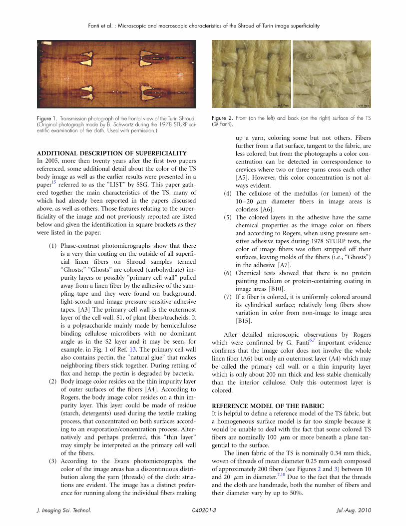
Further evidence for the superficiality of the TS bodymage is demonstrated by the transmission photograph
ade by the STURP photographer B. Schwortz shown inigure 1. Rather than radiation reflected from the surface ofhe cloth, the photo depicts the radiation transmitted by theS through the water stains, scorches, blood, and body im-ge. In the transmission photograph those marks which per-eated the TS remain evident, but the body image disap-
ears almost completely demonstrating its extremeuperficiality.
Jul.-Aug. 20102
AIrbpewafibw
wclbwtc
RIawfig
woaa
F�e
F�
Fanti et al. : Microscopic and macroscopic characteristics of the Shroud of Turin image superficiality
J
DDITIONAL DESCRIPTION OF SUPERFICIALITYn 2005, more then twenty years after the first two paperseferenced, some additional detail about the color of the TSody image as well as the earlier results were presented in aaper15 referred to as the “LIST” by SSG. This paper gath-red together the main characteristics of the TS, many ofhich had already been reported in the papers discussed
bove, as well as others. Those features relating to the super-ciality of the image and not previously reported are listedelow and given the identification in square brackets as theyere listed in the paper:
(1) Phase-contrast photomicrographs show that thereis a very thin coating on the outside of all superfi-cial linen fibers on Shroud samples termed“Ghosts;” “Ghosts” are colored (carbohydrate) im-purity layers or possibly “primary cell wall” pulledaway from a linen fiber by the adhesive of the sam-pling tape and they were found on background,light-scorch and image pressure sensitive adhesivetapes. [A3] The primary cell wall is the outermostlayer of the cell wall, S1, of plant fibers/tracheids. Itis a polysaccharide mainly made by hemicellulosebinding cellulose microfibers with no dominantangle as in the S2 layer and it may be seen, forexample, in Fig. 1 of Ref. 13. The primary cell wallalso contains pectin, the “natural glue” that makesneighboring fibers stick together. During retting offlax and hemp, the pectin is degraded by bacteria.
(2) Body image color resides on the thin impurity layerof outer surfaces of the fibers [A4]. According toRogers, the body image color resides on a thin im-purity layer. This layer could be made of residue(starch, detergents) used during the textile makingprocess, that concentrated on both surfaces accord-ing to an evaporation/concentration process. Alter-natively and perhaps preferred, this “thin layer”may simply be interpreted as the primary cell wallof the fibers.
(3) According to the Evans photomicrographs, thecolor of the image areas has a discontinuous distri-bution along the yarn (threads) of the cloth: stria-tions are evident. The image has a distinct prefer-
igure 1. Transmission photograph of the frontal view of the Turin Shroud.Original photograph made by B. Schwortz during the 1978 STURP sci-ntific examination of the cloth. Used with permission.�
ence for running along the individual fibers making t
. Imaging Sci. Technol. 040201-
up a yarn, coloring some but not others. Fibersfurther from a flat surface, tangent to the fabric, areless colored, but from the photographs a color con-centration can be detected in correspondence tocrevices where two or three yarns cross each other[A5]. However, this color concentration is not al-ways evident.
(4) The cellulose of the medullas (or lumen) of the10–20 �m diameter fibers in image areas iscolorless [A6].
(5) The colored layers in the adhesive have the samechemical properties as the image color on fibersand according to Rogers, when using pressure sen-sitive adhesive tapes during 1978 STURP tests, thecolor of image fibers was often stripped off theirsurfaces, leaving molds of the fibers (i.e., “Ghosts”)in the adhesive [A7].
(6) Chemical tests showed that there is no proteinpainting medium or protein-containing coating inimage areas [B10].
(7) If a fiber is colored, it is uniformly colored aroundits cylindrical surface; relatively long fibers showvariation in color from non-image to image area[B15].
After detailed microscopic observations by Rogershich were confirmed by G. Fanti6,7 important evidence
onfirms that the image color does not involve the wholeinen fiber (A6) but only an outermost layer (A4) which maye called the primary cell wall, or a thin impurity layerhich is only about 200 nm thick and less stable chemically
han the interior cellulose. Only this outermost layer isolored.
EFERENCE MODEL OF THE FABRICt is helpful to define a reference model of the TS fabric, but
homogeneous surface model is far too simple because itould be unable to deal with the fact that some colored TSbers are nominally 100 �m or more beneath a plane tan-ential to the surface.
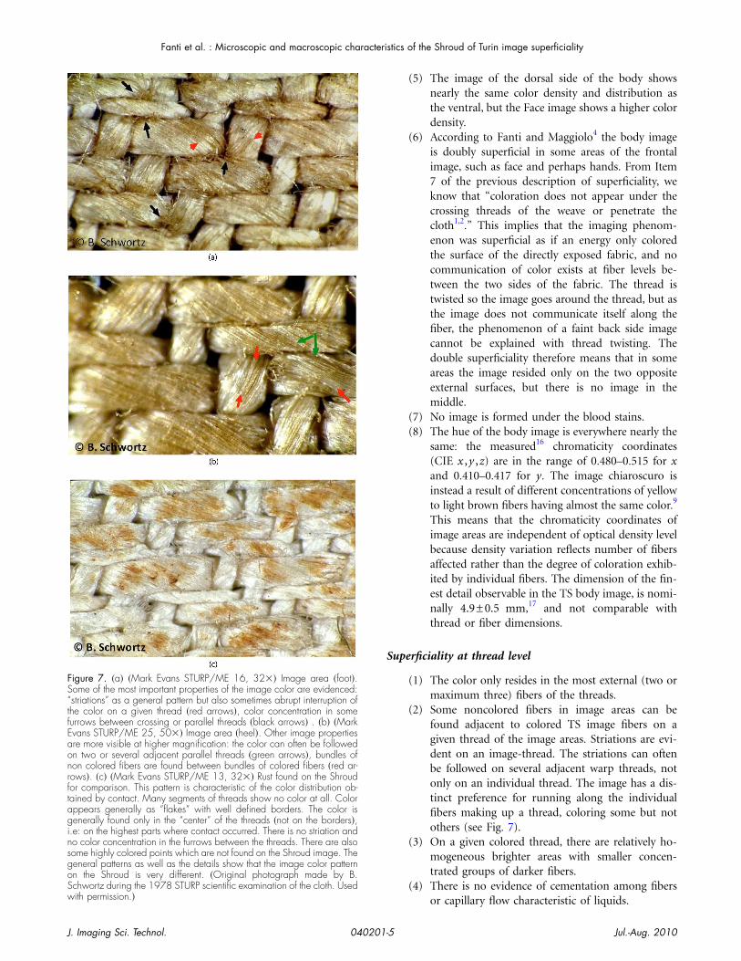
The linen fabric of the TS is nominally 0.34 mm thick,oven of threads of mean diameter 0.25 mm each composedf approximately 200 fibers (see Figures 2 and 3) between 10nd 20 �m in diameter.7,10 Due to the fact that the threadsnd the cloth are handmade, both the number of fibers and
igure 2. Front �on the left� and back �on the right� surface of the TS© Fanti�.
heir diameter vary by up to 50%.
Jul.-Aug. 20103

luowg(tai
aFtn(y
SUffiat
S
Ftos7t
Fw
Ff
Fsitc
Fanti et al. : Microscopic and macroscopic characteristics of the Shroud of Turin image superficiality
J
The fabric is composed of warp and weft threads. If weook at the TS laid out flat with the body image (frontal) sidep, we see a greater percentage of the warp threads goingver weft threads (see Figure 4). Figure 5 shows the 3–1 twilleave (herringbone pattern) of the linen threads. From aeometrical point of view, the body image surface of the TSfrontal), consists of 75% warp threads and 25% wefthreads; the warp percentage, due to the 3–1 geometry gives�3/ �3+1��=75% result. For this reason the frontal surface
s also called “warp side.”A macro-model of a TS linen thread may be thought of
s analogous to a bundle of drinking straws as shown inigure 6. The image fibers are represented by the red strawshat are side-by-side with the yellow ones representingonimage fibers. If the thin external layer of a colored strawrepresenting an image fiber) is pulled away, the non-colored
igure 4. Scheme �on the left� and corresponding photo �on the right� ofhe front surface of a TS fabric model: vertical threads �clearer� are thosef warp, horizontal ones �darker� are those of weft. On the TS frontalurface about only the 25% of weft threads are visible, being about the5% of warp threads; these are higher than the weft ones with respect to
he fabric plane.
igure 5. Two cross sections of Vercelli’s TS facsimile in which warp andeft treads are evidenced �© Fanti�.
igure 3. Front �on the left� and back �on the right� surface of a TSacsimile from a Vercelli’s sample �© Fanti�.
ellow inner part (cellulose) is exposed.
. Imaging Sci. Technol. 040201-
UPERFICIALITY OF THE TS BODY IMAGEsing previous reports of image superficiality and the TS
abric model introduced above, the description of the super-ciality of the body image can be improved. The superfici-lity of the body image can be described at the fabric level,he thread level and the fiber level.
uperficiality at the fabric level
(1) The color only resides on the external surface of theTS. According to the fabric model described earlier,that surface is not flat. In any given region of thebody image, there are more colored warp threadsthan adjacent weft threads: the image is mainly car-ried by the warp threads. However, some weftthreads are also colored. The color does not pen-etrate the whole cloth in any image area. FromFig. 1 where no body image is detectable, we caninfer that the contribution of colored body imagefibers in the transmitted light image is lower thanthe visibility threshold therefore, less than about1% of the recorded radiation.
(2) The superficial color is not due to any pigmentsince no pigment particles can be seen either mac-roscopically or microscopically nor are there anyexternal substances or evidence of media scorchingin image areas. The color is only due to a chemicalreaction (dehydration and oxidation).
(3) Where one of the image-threads crosses over an-other, the yellow coloration of the fibers is ofteninterrupted on the lower thread.
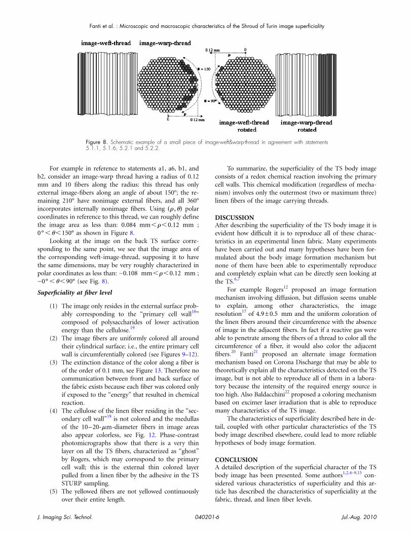
(4) A color concentration can be detected in corre-spondence to furrows where two or three yarnscross each other, or between two colored parallelyarns. This color concentration becomes more evi-dent after a contrast enhancement; see the photosin Figures 7(a) and 7(b). For comparison Figure7(c) shows the difference of a contact image caused
igure 6. Macromodel of a TS thread consisting in a bundle of drinkingtraws; above left, model of a nonimage thread; below left, model of anmage thread in which the red straws �of image� are put side-by-side withhe yellow ones �nonimage�. On the right a color straw �image fiber� isolored only on its surface �© Fanti�.
by rust.
Jul.-Aug. 20104
S
FS“tfEaonrftaginsgoSwith permission.�
Fanti et al. : Microscopic and macroscopic characteristics of the Shroud of Turin image superficiality
J. Imaging Sci. Technol. 040201-
(5) The image of the dorsal side of the body showsnearly the same color density and distribution asthe ventral, but the Face image shows a higher colordensity.
(6) According to Fanti and Maggiolo4 the body imageis doubly superficial in some areas of the frontalimage, such as face and perhaps hands. From Item7 of the previous description of superficiality, weknow that “coloration does not appear under thecrossing threads of the weave or penetrate thecloth1,2.” This implies that the imaging phenom-enon was superficial as if an energy only coloredthe surface of the directly exposed fabric, and nocommunication of color exists at fiber levels be-tween the two sides of the fabric. The thread istwisted so the image goes around the thread, but asthe image does not communicate itself along thefiber, the phenomenon of a faint back side imagecannot be explained with thread twisting. Thedouble superficiality therefore means that in someareas the image resided only on the two oppositeexternal surfaces, but there is no image in themiddle.
(7) No image is formed under the blood stains.(8) The hue of the body image is everywhere nearly the
same: the measured16 chromaticity coordinates(CIE x ,y ,z) are in the range of 0.480–0.515 for xand 0.410–0.417 for y. The image chiaroscuro isinstead a result of different concentrations of yellowto light brown fibers having almost the same color.9
This means that the chromaticity coordinates ofimage areas are independent of optical density levelbecause density variation reflects number of fibersaffected rather than the degree of coloration exhib-ited by individual fibers. The dimension of the fin-est detail observable in the TS body image, is nomi-nally 4.9±0.5 mm,17 and not comparable withthread or fiber dimensions.
uperficiality at thread level
(1) The color only resides in the most external (two ormaximum three) fibers of the threads.
(2) Some noncolored fibers in image areas can befound adjacent to colored TS image fibers on agiven thread of the image areas. Striations are evi-dent on an image-thread. The striations can oftenbe followed on several adjacent warp threads, notonly on an individual thread. The image has a dis-tinct preference for running along the individualfibers making up a thread, coloring some but notothers (see Fig. 7).
(3) On a given colored thread, there are relatively ho-mogeneous brighter areas with smaller concen-trated groups of darker fibers.
(4) There is no evidence of cementation among fibers
igure 7. �a� �Mark Evans STURP/ME 16, 32�� Image area �foot�.ome of the most important properties of the image color are evidenced:striations” as a general pattern but also sometimes abrupt interruption ofhe color on a given thread �red arrows�, color concentration in someurrows between crossing or parallel threads �black arrows� . �b� �Markvans STURP/ME 25, 50�� Image area �heel�. Other image propertiesre more visible at higher magnification: the color can often be followedn two or several adjacent parallel threads �green arrows�, bundles ofon colored fibers are found between bundles of colored fibers �red ar-ows�. �c� �Mark Evans STURP/ME 13, 32�� Rust found on the Shroudor comparison. This pattern is characteristic of the color distribution ob-ained by contact. Many segments of threads show no color at all. Colorppears generally as “flakes” with well defined borders. The color isenerally found only in the “center” of the threads �not on the borders�,
.e: on the highest parts where contact occurred. There is no striation ando color concentration in the furrows between the threads. There are alsoome highly colored points which are not found on the Shroud image. Theeneral patterns as well as the details show that the image color patternn the Shroud is very different. �Original photograph made by B.chwortz during the 1978 STURP scientific examination of the cloth. Used
or capillary flow characteristic of liquids.
Jul.-Aug. 20105
bmemict0
sttp−
S
ccnl
DAethmnat
mtrtoacfimtittbm
tbh
CAbst
Fanti et al. : Microscopic and macroscopic characteristics of the Shroud of Turin image superficiality
J
For example in reference to statements a1, a6, b1, and2, consider an image-warp thread having a radius of 0.12m and 10 fibers along the radius: this thread has only
xternal image-fibers along an angle of about 150°; the re-aining 210° have nonimage external fibers, and all 360°
ncorporates internally nonimage fibers. Using �� ,�� polaroordinates in reference to this thread, we can roughly definehe image area as less than: 0.084 mm���0.12 mm ;° ���150° as shown in Figure 8.
Looking at the image on the back TS surface corre-ponding to the same point, we see that the image area ofhe corresponding weft-image-thread, supposing it to havehe same dimensions, may be very roughly characterized inolar coordinates as less than: −0.108 mm���0.12 mm ;0° ���90° (see Fig. 8).
uperficiality at fiber level
(1) The image only resides in the external surface prob-ably corresponding to the “primary cell wall18”composed of polysaccharides of lower activationenergy than the cellulose.19
(2) The image fibers are uniformly colored all aroundtheir cylindrical surface; i.e., the entire primary cellwall is circumferentially colored (see Figures 9–12).
(3) The extinction distance of the color along a fiber isof the order of 0.1 mm, see Figure 13. Therefore nocommunication between front and back surface ofthe fabric exists because each fiber was colored onlyif exposed to the “energy” that resulted in chemicalreaction.
(4) The cellulose of the linen fiber residing in the “sec-ondary cell wall”19 is not colored and the medullasof the 10–20-�m-diameter fibers in image areasalso appear colorless, see Fig. 12. Phase-contrastphotomicrographs show that there is a very thinlayer on all the TS fibers, characterized as “ghost”by Rogers, which may correspond to the primarycell wall; this is the external thin colored layerpulled from a linen fiber by the adhesive in the TSSTURP sampling.
(5) The yellowed fibers are not yellowed continuously
Figure 8. Schematic example of a small piece o5.1.1, 5.1.6, 5.2.1 and 5.2.2.
over their entire length. f
. Imaging Sci. Technol. 040201-
To summarize, the superficiality of the TS body imageonsists of a redox chemical reaction involving the primaryell walls. This chemical modification (regardless of mecha-ism) involves only the outermost (two or maximum three)
inen fibers of the image carrying threads.
ISCUSSIONfter describing the superficiality of the TS body image it is
vident how difficult it is to reproduce all of these charac-eristics in an experimental linen fabric. Many experimentsave been carried out and many hypotheses have been for-ulated about the body image formation mechanism but
one of them have been able to experimentally reproducend completely explain what can be directly seen looking athe TS.6,7
For example Rogers12 proposed an image formationechanism involving diffusion, but diffusion seems unable
o explain, among other characteristics, the imageesolution17 of 4.9±0.5 mm and the uniform coloration ofhe linen fibers around their circumference with the absencef image in the adjacent fibers. In fact if a reactive gas wereble to penetrate among the fibers of a thread to color all theircumference of a fiber, it would also color the adjacentbers.20 Fanti21 proposed an alternate image formationechanism based on Corona Discharge that may be able to
heoretically explain all the characteristics detected on the TSmage, but is not able to reproduce all of them in a labora-ory because the intensity of the required energy source isoo high. Also Baldacchini22 proposed a coloring mechanismased on excimer laser irradiation that is able to reproduceany characteristics of the TS image.
The characteristics of superficiality described here in de-ail, coupled with other particular characteristics of the TSody image described elsewhere, could lead to more reliableypotheses of body image formation.
ONCLUSIONdetailed description of the superficial character of the TS
ody image has been presented. Some authors1,2,4–9,15 con-idered various characteristics of superficiality and this ar-icle has described the characteristics of superficiality at the
e-weft&warp-thread in agreement with statements
f imagabric, thread, and linen fiber levels.
Jul.-Aug. 20106
bltrowromlcbfiTopsc
Fiatfii�afvct3
Fldecon the left shows the colorless inner cellulose �© Fanti�.
Ftc
Fmvitnc
Fib
Fanti et al. : Microscopic and macroscopic characteristics of the Shroud of Turin image superficiality
J
The findings encompass the superficiality of the TSody image as studied by STURP1,2,9 up to a general fiber
evel revealing that the image is caused by a chemical reac-ion involving the cellulose of the linen fibers. The reactionesults in a discontinuous, translucent yellow discolorationf the fibers limited to the topmost surface of the threadsith an extinction of the order of 0.1 mm. The mechanical
esistance to tensile stress of image fibers is lower than thatf nonimage fibers. Image fibers show no evidence of ce-entation between fibers or the presence of capillary flow of
iquids, and no pigment particles are detectable. The yellowoloration of the fibers is interrupted as the thread goeseneath crossing threads in the weave pattern: the coloredbers are not yellowed continuously over their entire length.he darker portions of the image are not due to a variationf the degree of the yellowing of the fibers, but rather to theresence of more yellowed fibers per unit area. Only verytrong reductants (diimide and hydrazine) can bleach the
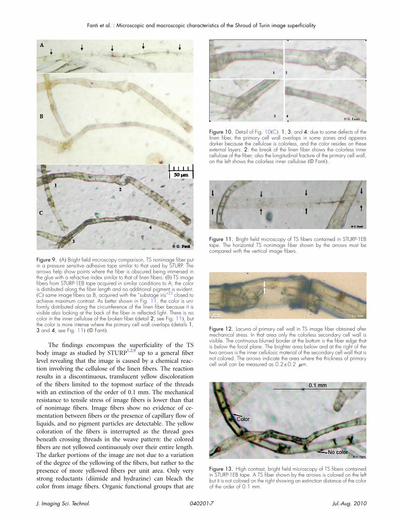
igure 9. �A� Bright field microscopy comparison, TS nonimage fiber putn a pressure sensitive adhesive tape similar to that used by STURP. Therrows help show points where the fiber is obscured being immersed in
he glue with a refractive index similar to that of linen fibers. �B� TS imagebers from STURP-1EB tape acquired in similar conditions to A; the colors distributed along the fiber length and no additional pigment is evident.C� same image fibers as B, acquired with the “substage iris”23 closed tochieve maximum contrast. As better shown in Fig. 11, the color is uni-
ormly distributed along the circumference of the linen fiber because it isisible also looking at the back of the fiber in reflected light. There is noolor in the inner cellulose of the broken fiber �detail 2, see Fig. 11�, buthe color is more intense where the primary cell wall overlaps �details 1,
and 4, see Fig. 11� �© Fanti�.
olor from image fibers. Organic functional groups that are o
. Imaging Sci. Technol. 040201-
igure 10. Detail of Fig. 10�C�. 1, 3, and 4: due to some defects of theinen fiber, the primary cell wall overlaps in some zones and appearsarker because the cellulose is colorless, and the color resides on thesexternal layers. 2: the break of the linen fiber shows the colorless innerellulose of the fiber; also the longitudinal fracture of the primary cell wall,
igure 11. Bright field microscopy of TS fibers contained in STURP-1EBape. The horizontal TS nonimage fiber shown by the arrows must beompared with the vertical image fibers.
igure 12. Lacuna of primary cell wall in TS image fiber obtained afterechanical stress. In that area only the colorless secondary cell wall isisible. The continuous blurred border at the bottom is the fiber edge thats below the focal plane. The brighter area below and at the right of thewo arrows is the inner cellulosic material of the secondary cell wall that isot colored. The arrows indicate the area where the thickness of primaryell wall can be measured as 0.2±0.2 �m.
igure 13. High contrast, bright field microscopy of TS fibers containedn STURP-1EB tape. A TS fiber shown by the arrows is colored on the leftut it is not colored on the right showing an extinction distance of the color
f the order of 0.1 mm.Jul.-Aug. 20107
cht
sStccift
comoNntumpC
AT(tPhpMtfiipg
R
Fanti et al. : Microscopic and macroscopic characteristics of the Shroud of Turin image superficiality
J
haracteristic of dehydratively oxidized, degraded celluloseave been found in image fibers, therefore the image seems
o result from some cellulose degradation effect.In succeeding years the TS body image was also closely
tudied at the subfiber component level6,7,12 and to theTURP studies can be added that the redox chemical reac-ion causing the image color only developed in the primaryell walls of the linen fibers (0.2 �m thick) a layer chemi-ally less stable than the inner secondary cell wall. The bodymage is visible on both the sides of the fabric at least in theace region,4 but no image is detectable on the linen fibers inhe middle of the cloth.
In summary, the superficiality of the TS body imageonsists of a redox chemical reaction probably involving theuter primary cell walls of the linen fibers; this chemicalodification (regardless of mechanism) involves only the
utmost (two or maximum three) linen fibers of a thread.evertheless the linen fibers are chemically altered by a phe-omenon not yet understood. For this reason the descrip-
ion of the image superficiality here reported will also beseful to future studies on the body image formationechanism seeking to explain who or what was able to im-
rint such a peculiar image on the most important relic ofhristianity.
CKNOWLEDGMENTShe authors are members of the Shroud Science Group
SSG). They would like to acknowledge the forum providedhere as helping shape the concepts explored in this paper.articular thanks to SSG Member L. G. Thygesen who, wither deep expertise in linen fibers, helped the authors in theaper compilation. Particular thanks also to SSG Member. Alonso who, with his constructive discussions, pushed
he compilation of the present article and helped the authorsnd many of the facts about the superficiality of the TS body
mage. Thanks also to B. Schwortz who gave permission toublish without charge the STURP-M. Evans photomicro-raphs.
EFERENCES1 E. J. Jumper, Archaeological Chemistry III, ACS Advances in Chemistry
(American Chemical Society, Washington, D.C., 1984), Vol. 205, pp.447–476.
2 A. Adler, A Shroud Spectrum Int. Special Issue (Effatà Editrice, Torino,Italy, 2002).
3 J. P. Jackson, E. J. Jumper, and W. R. Ercoline, “Correlation of imageintensity on the Turin Shroud with the 3-D structure of a human bodyshape”, Appl. Opt. 23, 2244–2270 (1984).
4 G. Fanti and R. Maggiolo, “The double superficiality of the frontal imageof the Turin Shroud”, J. Opt. A, Pure Appl. Opt. 6, 491–503 (2004).
5 G. Fanti and M. Moroni, “Comparison of luminance between face of
. Imaging Sci. Technol. 040201-
Turin Shroud Man and experimental results”, J. Imaging Sci. Technol.46, 142–154 (2002).
6 G. Fanti and R. Basso, The Turin Shroud, Optical Research in the Past,Present and Future (Nova Science Publisher Inc., New York, 2007).
7 G. Fanti, La Sindone, una sfida alla Scienza Moderna (Edizione Aracne,Roma, Italy, 2008).
8 M. Antonacci, The Resurrection of the Shroud (M. Evans and Co., Inc.,New York, 2000).
9 L. A. Schwalbe and R. N. Rogers, “Physics and chemistry of the Shroudof Turin, a summary of the 1978 investigation”, Anal. Chim. Acta 135,3–49 (1982).
10 G. Fanti, SSG private communications (unpublished).11 C. Barton and A. Prutton, “Photometric Method for Determination of
Hemicellulose”, Ind. Eng. Chem. Anal. Ed. 16, 429–430 (1944) http://pubs.acs.org/doi/pdf/10.1021/i560131a005.
12 R. Rogers, A Chemist’s Perspective On The Shroud of Turin (Edizione B.Schwortz, Lulu, Italy, 2008), p. 21.
13 C. Nyholm, “Dislocations in pulp fibers–their origin, characteristics andimportance–a review”, Nord. Pulp Pap. Res. J. 4, 376–384 (2001).
14 L. G. Thygesen and M. R. Asharipour, “The effects of growth andstorage conditions on dislocations in hemp fibres”, J. Mater. Sci. 43,3670–3673 (2008).
15 G. Fanti, B. Schwortz, A. Accetta, J. A. Botella, B. J. Buenaobra, M.Carreira, F. Cheng, F. Crosilla, R. Dinegar, H. Felzmann, B. Haroldsen, P.Iacazio, F. Lattarulo, G. Novelli, J. Marino, A. Malantrucco, P. Maloney,D. Porter, B. Pozzetto, R. Schneider, N. Svensson, T. Wally, A. Whanger,and F. Zugibe, “Evidences for Testing Hypotheses about the Body ImageFormation of the Turin Shroud,” Proceedings of the Third DallasInternational Conference on the Shroud of Turin, 2005.
16 P. Soardo, The Turin Shroud, Past, Present and Future, Int. Sci. Symp.,Torino [Effatà Editrice, Cantalupa (TO), 2000], p. 89–100.
17 G. Fanti and R. Basso, The Shroud Of Turin: Perspectives on AMultifaceted Enigma, Proc. Shroud Science Group Int. Conf., Ohio StateUniversity (Libreria Progetto, Padova, Italy, 2009).
18 The primary cell wall is mainly constituted by hemicellulose. Unlikecellulose, hemicellulose, also a polysaccharide, consists of shorterchains—500–3000 sugar units as opposed to 7000–15 000 glucosemolecules per polymer seen in cellulose. The primary cell wall is mainlymade by shorter chains of the same glucose “brick” units that constitutethe cellulose in the secondary cell wall, that is about 0.2 �m thick, seeFig. 13. R. Rogers proposed an alternative to a primary cell wallinterpretation suggesting instead that the image corresponds to a thinlayer of impurities on the external surface of the fibers, mainly starchand other low-weight polysaccharide residues. This thin layer wouldhave been concentrated on the two external surfaces of the fabric duringthe drying-evaporation-concentration process, after washing. Thishypothesis has been criticized as unable to explain why the thin layer isuniform along its circumference and where there is contact with adjacentfibers. No references to the use of starch in antiquity have been found.
19 H. L. Bos and A. M. Donald, “In situ ESEM study of the deformation ofelementary flax fibres”, J. Mater. Sci. 34, 3029–3034 (1999).
20 G. Fanti, Shroud Science communication: “Comments on Gas DiffusionHypothesis”, August 2004, www.dim.unipd.it/fanti/diffusion.pdf
21 G. Fanti, “Can a Corona Discharge Explain the Body Image of The TurinShroud?”, J. Imaging Sci. Technol. 54, 020508 (2010); Proc. Int. Conf. onthe Shroud of Turin, Ohio State University (Libreria Progetto, Padova,Italy 2009).
22 G. Baldacchini, P. Di Lazzaro, D. Murra, and G. Fanti, “Coloring Linensby Excimer Lasers to Simulate the Body Image of the Turin Shroud”,Appl. Opt. 47, 1278–1285 (2008).
23 W. C. McCrone, Polarized Light Microscopy (Ann Arbor SciencePublisher Inc., Ann Arbor, Mich., 1978), p. 30.
Jul.-Aug. 20108
©2010 Society for Imaging Science and Technology (IS&T)All rights reserved. This paper, or parts thereof, may not be reproduced in any form
without the written permission of IS&T, the sole copyright owner ofThe Journal of Imaging Science and Technology.
For information on reprints or reproduction contactDonna Smith
Production EditorThe Journal of Imaging Science and Technology
Society for Imaging Science and Technology7003 Kilworth Lane
Springfield, Virginia 22151 USA703/642-9090 extension 107
703/642-9094 (fax)[email protected]
www.imaging.org
Related Documents