Mechanism of mRNA transport in the nucleus Diana Y. Vargas*, Arjun Raj* † , Salvatore A. E. Marras*, Fred Russell Kramer*, and Sanjay Tyagi* ‡ *Department of Molecular Genetics, Public Health Research Institute, 225 Warren Street, Newark, NJ 07103; and † Courant Institute of Mathematical Sciences, New York University, 251 Mercer Street, New York, NY 10012 Edited by Joseph G. Gall, Carnegie Institution of Washington, Baltimore, MD, and approved October 4, 2005 (received for review July 5, 2005) The mechanism of transport of mRNA–protein (mRNP) complexes from transcription sites to nuclear pores has been the subject of many studies. Using molecular beacons to track single mRNA molecules in living cells, we have characterized the diffusion of mRNP complexes in the nucleus. The mRNP complexes move freely by Brownian diffusion at a rate that assures their dispersion throughout the nucleus before they exit into the cytoplasm, even when the transcription site is located near the nuclear periphery. The diffusion of mRNP complexes is restricted to the extranucleo- lar, interchromatin spaces. When mRNP complexes wander into dense chromatin, they tend to become stalled. Although the movement of mRNP complexes occurs without the expenditure of metabolic energy, ATP is required for the complexes to resume their motion after they become stalled. This finding provides an explanation for a number of observations in which mRNA trans- port appeared to be an enzymatically facilitated process. gene expression live cell imaging mRNA export nuclear viscosity A fter mRNAs are synthesized, processed, and become associ- ated with a number of different proteins at the transcription site, they are released into the nucleoplasm (1). The mechanism by which these large mRNA–protein (mRNP) complexes then move through dense nucleoplasm to reach the nuclear pores has been the subject of intense study and speculation (2, 3). Early workers proposed that mRNP complexes are transferred along a chain of receptors until they reach a nuclear pore, expending metabolic energy in the process (4). This solid-state transport model is supported by observations made in fixed nuclei that show some transcripts distributed along tracks that originate from the locus of the parent gene (5, 6). A second theory, called the ‘‘gene-gating’’ hypothesis, proposes that active genes are situated near the nuclear periphery and that mRNAs exit the nucleus through the nearest pores (7). This idea is supported by observations that certain mRNAs exit from one side of the nucleus (8) and that, in yeast, many transcriptionally active gene loci are located near the nuclear periphery (9). By contrast, a number of other studies have found that mRNP complexes move quite freely within the nucleus (10– 16). This view is supported by studies of the distribution of newly synthesized Balbiani ring RNA in the salivary gland cells of insects (11), fluorescence recovery after photobleaching and fluorescence correlation spectroscopy studies of probes that bind to the poly(A) tails of mRNAs (12–15), and from single-particle analysis of mRNP complexes bound to GFP-linked proteins (16). Although the latter studies found that mRNP complexes are able to diffuse within the nuclear matrix, there was a paradoxical active transport component to their motility, because both a reduction in temperature and ATP depletion curtailed the mobility of the complexes (14–16). To better understand the nature of mRNP mobility, we have developed a system of fluorogenic probes and mRNA constructs that allows us to track individual mRNA mol- ecules as they are transcribed, move within the nucleus, exit from the nuclear pores, and spread throughout the cytoplasm. This system enables us to detect differences in the behavior of different molecules of the same mRNA species and to understand how different microenvironments in the nucleus influence the mobility of individual mRNP complexes. Our probes are small, hairpin-shaped oligonucleotides called molecular beacons (17, 18) that possess an internally quenched fluorophore whose fluorescence is restored upon hybridization to a specific nucleic acid sequence. To obtain single-molecule sensi- tivity, we engineered a host cell line to express an mRNA possessing multiple molecular beacon binding sites. The binding of many molecular beacons to each mRNA molecule renders them so intensely fluorescent that individual mRNA molecules can be detected and tracked. We found that the rate of mRNP diffusion is so fast that mRNP complexes are dispersed throughout the nucleus soon after their synthesis and well before the onset of significant export into the cytoplasm. Our analyses of the trajecto- ries of individual mRNA complexes show that their motion is restricted to the interchromatin spaces. Sometimes the moving mRNP complexes become stalled within high-density chromatin but later begin to move again. The switch from stationary to mobile behavior depends on ATP. Materials and Methods Host Cell Lines and Reporter Gene. A DNA fragment containing 96 head-to-tail tandem repeats of the 50-nt-long sequence 5-CAGGAGTTGTGTTTGTGGACGAAGAGCACCAGC- CAGCTGATCGACCTCGA-3 was prepared as described by Robinett et al. (19) and inserted into the plasmid pTRE-d2EGFP (Clontech) by using its multiple cloning sites. The resulting plasmid, pTRE-GFP-96-mer, was used to transfect CHO cell line CHO- AA8-Tet-off (Clontech), which possesses a stably integrated gene for the tetracycline-controlled Tet-off transactivator. A geneticin G418-resistant clone (CHO-GFP-96-mer) that responded to 10 ngml doxycycline in the medium by turning off its fluorescence within 24 h was selected. To obtain cells expressing histone H2B- GFP, this cell line was transfected with plasmid pBOS-H2BGFP (BD Biosciences), and a clone that exhibited an intense GFP signal in the nuclei was isolated. Cells were cultured in the modification of Eagle’s minimal essential medium (Sigma) supplemented with 10% TET-System- Approved FBS (Clontech). Imaging was performed in phenol red-free OptiMEM (Invitrogen). Cells used in the ATP-depletion studies were first incubated in glucose-free Dulbecco’s modified Eagle’s medium (Invitrogen) containing 10 mM sodium azide and 60 mM 2-deoxyglucose for 30 min and then imaged in OptiMEM containing the same inhibitors. After this treatment, the mitochon- dria in the cells could not be stained by rhodamine 123 (Sigma), confirming that the inhibitors were effective (14). Molecular Beacons. The sequences of the molecular beacons were Cy3 or Alexa-594–5-CUUCGUCCACAAACACAACUCCU- GAAG-3-Black Hole Quencher 2. The backbone of the molecular beacons was composed of 2-O-methylribonucleotides. Live Cell Imaging. Cells were maintained at 37°C on the microscope stage by controlled heating of the objective and the culture dish (Delta T4 open system, Bioptechs, Butler, PA). Molecular beacons were dissolved in water at a concentration of 2.5 ng l, and an 0.1- Conflict of interest statement: No conflicts declared. This paper was submitted directly (Track II) to the PNAS office. Abbreviations: mRNP, mRNA–protein; MSD, mean square displacement. ‡ To whom correspondence should be addressed. E-mail: [email protected]. © 2005 by The National Academy of Sciences of the USA 17008 –17013 PNAS November 22, 2005 vol. 102 no. 47 www.pnas.orgcgidoi10.1073pnas.0505580102

Welcome message from author
This document is posted to help you gain knowledge. Please leave a comment to let me know what you think about it! Share it to your friends and learn new things together.
Transcript
Mechanism of mRNA transport in the nucleusDiana Y. Vargas*, Arjun Raj*†, Salvatore A. E. Marras*, Fred Russell Kramer*, and Sanjay Tyagi*‡
*Department of Molecular Genetics, Public Health Research Institute, 225 Warren Street, Newark, NJ 07103; and †Courant Institute of MathematicalSciences, New York University, 251 Mercer Street, New York, NY 10012
Edited by Joseph G. Gall, Carnegie Institution of Washington, Baltimore, MD, and approved October 4, 2005 (received for review July 5, 2005)
The mechanism of transport of mRNA–protein (mRNP) complexesfrom transcription sites to nuclear pores has been the subject ofmany studies. Using molecular beacons to track single mRNAmolecules in living cells, we have characterized the diffusion ofmRNP complexes in the nucleus. The mRNP complexes move freelyby Brownian diffusion at a rate that assures their dispersionthroughout the nucleus before they exit into the cytoplasm, evenwhen the transcription site is located near the nuclear periphery.The diffusion of mRNP complexes is restricted to the extranucleo-lar, interchromatin spaces. When mRNP complexes wander intodense chromatin, they tend to become stalled. Although themovement of mRNP complexes occurs without the expenditure ofmetabolic energy, ATP is required for the complexes to resumetheir motion after they become stalled. This finding provides anexplanation for a number of observations in which mRNA trans-port appeared to be an enzymatically facilitated process.
gene expression � live cell imaging � mRNA export � nuclear viscosity
A fter mRNAs are synthesized, processed, and become associ-ated with a number of different proteins at the transcription
site, they are released into the nucleoplasm (1). The mechanism bywhich these large mRNA–protein (mRNP) complexes then movethrough dense nucleoplasm to reach the nuclear pores has been thesubject of intense study and speculation (2, 3). Early workersproposed that mRNP complexes are transferred along a chain ofreceptors until they reach a nuclear pore, expending metabolicenergy in the process (4). This solid-state transport model issupported by observations made in fixed nuclei that show sometranscripts distributed along tracks that originate from the locus ofthe parent gene (5, 6). A second theory, called the ‘‘gene-gating’’hypothesis, proposes that active genes are situated near the nuclearperiphery and that mRNAs exit the nucleus through the nearestpores (7). This idea is supported by observations that certainmRNAs exit from one side of the nucleus (8) and that, in yeast,many transcriptionally active gene loci are located near the nuclearperiphery (9). By contrast, a number of other studies have foundthat mRNP complexes move quite freely within the nucleus (10–16). This view is supported by studies of the distribution of newlysynthesized Balbiani ring RNA in the salivary gland cells of insects(11), fluorescence recovery after photobleaching and fluorescencecorrelation spectroscopy studies of probes that bind to the poly(A)tails of mRNAs (12–15), and from single-particle analysis of mRNPcomplexes bound to GFP-linked proteins (16).
Although the latter studies found that mRNP complexes are ableto diffuse within the nuclear matrix, there was a paradoxical activetransport component to their motility, because both a reduction intemperature and ATP depletion curtailed the mobility of thecomplexes (14–16). To better understand the nature of mRNPmobility, we have developed a system of fluorogenic probes andmRNA constructs that allows us to track individual mRNA mol-ecules as they are transcribed, move within the nucleus, exit fromthe nuclear pores, and spread throughout the cytoplasm. Thissystem enables us to detect differences in the behavior of differentmolecules of the same mRNA species and to understand howdifferent microenvironments in the nucleus influence the mobilityof individual mRNP complexes.
Our probes are small, hairpin-shaped oligonucleotides calledmolecular beacons (17, 18) that possess an internally quenched
fluorophore whose fluorescence is restored upon hybridization toa specific nucleic acid sequence. To obtain single-molecule sensi-tivity, we engineered a host cell line to express an mRNA possessingmultiple molecular beacon binding sites. The binding of manymolecular beacons to each mRNA molecule renders them sointensely fluorescent that individual mRNA molecules can bedetected and tracked. We found that the rate of mRNP diffusionis so fast that mRNP complexes are dispersed throughout thenucleus soon after their synthesis and well before the onset ofsignificant export into the cytoplasm. Our analyses of the trajecto-ries of individual mRNA complexes show that their motion isrestricted to the interchromatin spaces. Sometimes the movingmRNP complexes become stalled within high-density chromatinbut later begin to move again. The switch from stationary to mobilebehavior depends on ATP.
Materials and MethodsHost Cell Lines and Reporter Gene. A DNA fragment containing 96head-to-tail tandem repeats of the 50-nt-long sequence5�-CAGGAGTTGTGTTTGTGGACGAAGAGCACCAGC-CAGCTGATCGACCTCGA-3� was prepared as described byRobinett et al. (19) and inserted into the plasmid pTRE-d2EGFP(Clontech) by using its multiple cloning sites. The resulting plasmid,pTRE-GFP-96-mer, was used to transfect CHO cell line CHO-AA8-Tet-off (Clontech), which possesses a stably integrated genefor the tetracycline-controlled Tet-off transactivator. A geneticinG418-resistant clone (CHO-GFP-96-mer) that responded to 10ng�ml doxycycline in the medium by turning off its fluorescencewithin 24 h was selected. To obtain cells expressing histone H2B-GFP, this cell line was transfected with plasmid pBOS-H2BGFP(BD Biosciences), and a clone that exhibited an intense GFP signalin the nuclei was isolated.
Cells were cultured in the � modification of Eagle’s minimalessential medium (Sigma) supplemented with 10% TET-System-Approved FBS (Clontech). Imaging was performed in phenolred-free OptiMEM (Invitrogen). Cells used in the ATP-depletionstudies were first incubated in glucose-free Dulbecco’s modifiedEagle’s medium (Invitrogen) containing 10 mM sodium azide and60 mM 2-deoxyglucose for 30 min and then imaged in OptiMEMcontaining the same inhibitors. After this treatment, the mitochon-dria in the cells could not be stained by rhodamine 123 (Sigma),confirming that the inhibitors were effective (14).
Molecular Beacons. The sequences of the molecular beacons wereCy3 or Alexa-594–5�-CUUCGUCCACAAACACAACUCCU-GAAG-3�-Black Hole Quencher 2. The backbone of the molecularbeacons was composed of 2�-O-methylribonucleotides.
Live Cell Imaging. Cells were maintained at 37°C on the microscopestage by controlled heating of the objective and the culture dish(Delta T4 open system, Bioptechs, Butler, PA). Molecular beaconswere dissolved in water at a concentration of 2.5 ng��l, and an �0.1-
Conflict of interest statement: No conflicts declared.
This paper was submitted directly (Track II) to the PNAS office.
Abbreviations: mRNP, mRNA–protein; MSD, mean square displacement.
‡To whom correspondence should be addressed. E-mail: [email protected].
© 2005 by The National Academy of Sciences of the USA
17008–17013 � PNAS � November 22, 2005 � vol. 102 � no. 47 www.pnas.org�cgi�doi�10.1073�pnas.0505580102
to 1-fl solution was microinjected into each cell by using a FemtoJetmicroinjection apparatus (Brinkmann). An Axiovert 200M invertedfluorescence microscope (Zeiss), equipped with a �100 oil-immersion objective, a CoolSNAP HQ camera (Photometrics,Pleasanton, CA) cooled to �30°C, and OPENLAB acquisition soft-ware (Improvision, Sheffield, U.K.) were used to acquire theimages.
Synthetic RNA Transcripts and Their Hybrids with Molecular Beacons.We prepared a series of pGEM plasmids (Promega) containing 1,2, 4, 8, 16, 32, or 64 tandem repeats of the sequence described above.In addition, we excised the gene encoding GFP-mRNA-96-merfrom pTRE-GFP-96-mer and inserted it into plasmid pGEM,because that plasmid contains a bacteriophage T7 promoter. Toproduce RNA transcripts possessing a different number of repeats,these plasmids were linearized and used as templates for in vitrotranscription by T7 RNA polymerase. The transcript containing 96repeats possessed a GFP-mRNA sequence, whereas the othertranscripts only possessed the repeat motifs. Hybrids were formedby incubating 20 ng of transcripts with 20 ng of molecular beaconsin 10 �l of 10 mM Tris�HCl (pH 8.0) containing 1 mM MgCl2 at37°C for 60 min and were then injected into the cells.
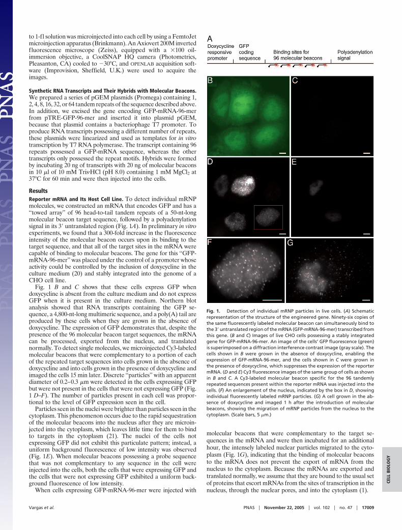
ResultsReporter mRNA and Its Host Cell Line. To detect individual mRNPmolecules, we constructed an mRNA that encodes GFP and has a‘‘towed array’’ of 96 head-to-tail tandem repeats of a 50-nt-longmolecular beacon target sequence, followed by a polyadenylationsignal in its 3� untranslated region (Fig. 1A). In preliminary in vitroexperiments, we found that a 300-fold increase in the fluorescenceintensity of the molecular beacon occurs upon its binding to thetarget sequence, and that all of the target sites in the mRNA werecapable of binding to molecular beacons. The gene for this ‘‘GFP-mRNA-96-mer’’ was placed under the control of a promoter whoseactivity could be controlled by the inclusion of doxycycline in theculture medium (20) and stably integrated into the genome of aCHO cell line.
Fig. 1 B and C shows that these cells express GFP whendoxycycline is absent from the culture medium and do not expressGFP when it is present in the culture medium. Northern blotanalysis showed that RNA transcripts containing the GFP se-quence, a 4,800-nt-long multimeric sequence, and a poly(A) tail areproduced by these cells when they are grown in the absence ofdoxycycline. The expression of GFP demonstrates that, despite thepresence of the 96 molecular beacon target sequences, the mRNAcan be processed, exported from the nucleus, and translatednormally. To detect single molecules, we microinjected Cy3-labeledmolecular beacons that were complementary to a portion of eachof the repeated target sequences into cells grown in the absence ofdoxycycline and into cells grown in the presence of doxycycline andimaged the cells 15 min later. Discrete ‘‘particles’’ with an apparentdiameter of 0.2–0.3 �m were detected in the cells expressing GFPbut were not present in the cells that were not expressing GFP (Fig.1 D–F). The number of particles present in each cell was propor-tional to the level of GFP expression seen in the cell.
Particles seen in the nuclei were brighter than particles seen in thecytoplasm. This phenomenon occurs due to the rapid sequestrationof the molecular beacons into the nucleus after they are microin-jected into the cytoplasm, which leaves little time for them to bindto targets in the cytoplasm (21). The nuclei of the cells notexpressing GFP did not exhibit this particulate pattern; instead, auniform background fluorescence of low intensity was observed(Fig. 1E). When molecular beacons possessing a probe sequencethat was not complementary to any sequence in the cell wereinjected into the cells, both the cells that were expressing GFP andthe cells that were not expressing GFP exhibited a uniform back-ground fluorescence of low intensity.
When cells expressing GFP-mRNA-96-mer were injected with
molecular beacons that were complementary to the target se-quences in the mRNA and were then incubated for an additionalhour, the intensely labeled nuclear particles migrated to the cyto-plasm (Fig. 1G), indicating that the binding of molecular beaconsto the mRNA does not prevent the export of mRNA from thenucleus to the cytoplasm. Because the mRNAs are exported andtranslated normally, we assume that they are bound to the usual setof proteins that escort mRNAs from the sites of transcription in thenucleus, through the nuclear pores, and into the cytoplasm (1).
Fig. 1. Detection of individual mRNP particles in live cells. (A) Schematicrepresentation of the structure of the engineered gene. Ninety-six copies ofthe same fluorescently labeled molecular beacon can simultaneously bind tothe 3� untranslated region of the mRNA (GFP-mRNA-96-mer) transcribed fromthis gene. (B and C) Images of live CHO cells possessing a stably integratedgene for GFP-mRNA-96-mer. An image of the cells’ GFP fluorescence (green)is superimposed on a diffraction interference contrast image (gray scale). Thecells shown in B were grown in the absence of doxycycline, enabling theexpression of GFP-mRNA-96-mer, and the cells shown in C were grown inthe presence of doxycycline, which suppresses the expression of the reportermRNA. (D and E) Cy3 fluorescence images of the same group of cells as shownin B and C. A Cy3-labeled molecular beacon specific for the 96 tandemlyrepeated sequences present within the reporter mRNA was injected into thecells. (F) An enlargement of the nucleus, indicated by the box in D, showingindividual fluorescently labeled mRNP particles. (G) A cell grown in the ab-sence of doxycycline and imaged 1 h after the introduction of molecularbeacons, showing the migration of mRNP particles from the nucleus to thecytoplasm. (Scale bars, 5 �m.)
Vargas et al. PNAS � November 22, 2005 � vol. 102 � no. 47 � 17009
CELL
BIO
LOG
Y
Demonstration That Each Particle Contains an Individual mRNA Mol-ecule. Previous studies have shown that �48 GFP molecules, or 70Cy3 moieties, can render a single molecular complex sufficientlyfluorescent to enable its detection by using fluorescence micro-scopes similar to ours (16, 22). Thus, it is likely that single mRNAmolecules hybridized to probes that aggregately contain 96 welldispersed Cy3 fluorophores would be similarly visible.
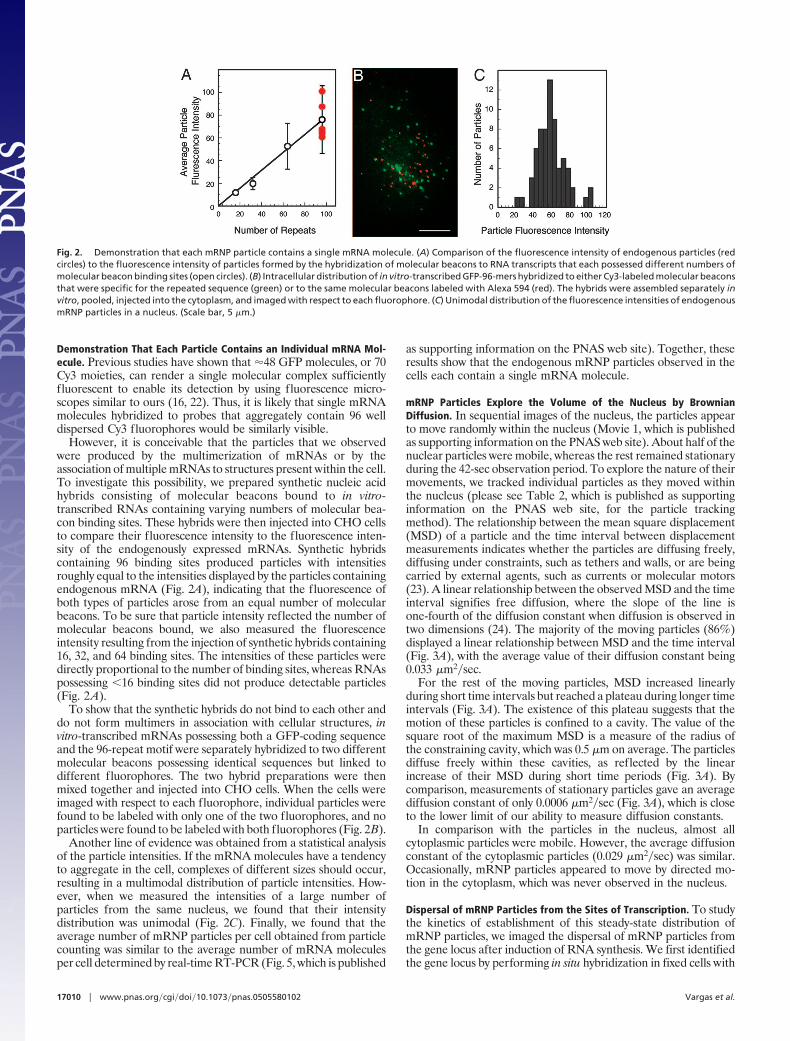
However, it is conceivable that the particles that we observedwere produced by the multimerization of mRNAs or by theassociation of multiple mRNAs to structures present within the cell.To investigate this possibility, we prepared synthetic nucleic acidhybrids consisting of molecular beacons bound to in vitro-transcribed RNAs containing varying numbers of molecular bea-con binding sites. These hybrids were then injected into CHO cellsto compare their fluorescence intensity to the fluorescence inten-sity of the endogenously expressed mRNAs. Synthetic hybridscontaining 96 binding sites produced particles with intensitiesroughly equal to the intensities displayed by the particles containingendogenous mRNA (Fig. 2A), indicating that the fluorescence ofboth types of particles arose from an equal number of molecularbeacons. To be sure that particle intensity reflected the number ofmolecular beacons bound, we also measured the fluorescenceintensity resulting from the injection of synthetic hybrids containing16, 32, and 64 binding sites. The intensities of these particles weredirectly proportional to the number of binding sites, whereas RNAspossessing �16 binding sites did not produce detectable particles(Fig. 2A).
To show that the synthetic hybrids do not bind to each other anddo not form multimers in association with cellular structures, invitro-transcribed mRNAs possessing both a GFP-coding sequenceand the 96-repeat motif were separately hybridized to two differentmolecular beacons possessing identical sequences but linked todifferent fluorophores. The two hybrid preparations were thenmixed together and injected into CHO cells. When the cells wereimaged with respect to each fluorophore, individual particles werefound to be labeled with only one of the two fluorophores, and noparticles were found to be labeled with both fluorophores (Fig. 2B).
Another line of evidence was obtained from a statistical analysisof the particle intensities. If the mRNA molecules have a tendencyto aggregate in the cell, complexes of different sizes should occur,resulting in a multimodal distribution of particle intensities. How-ever, when we measured the intensities of a large number ofparticles from the same nucleus, we found that their intensitydistribution was unimodal (Fig. 2C). Finally, we found that theaverage number of mRNP particles per cell obtained from particlecounting was similar to the average number of mRNA moleculesper cell determined by real-time RT-PCR (Fig. 5, which is published
as supporting information on the PNAS web site). Together, theseresults show that the endogenous mRNP particles observed in thecells each contain a single mRNA molecule.
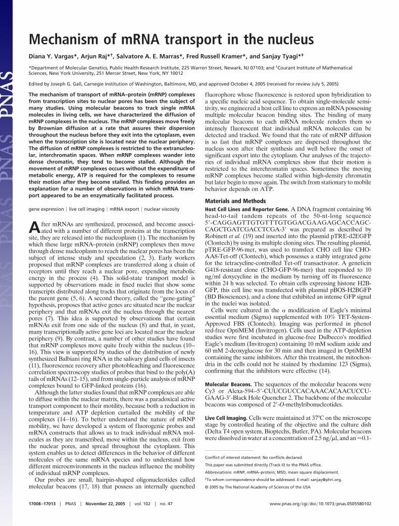
mRNP Particles Explore the Volume of the Nucleus by BrownianDiffusion. In sequential images of the nucleus, the particles appearto move randomly within the nucleus (Movie 1, which is publishedas supporting information on the PNAS web site). About half of thenuclear particles were mobile, whereas the rest remained stationaryduring the 42-sec observation period. To explore the nature of theirmovements, we tracked individual particles as they moved withinthe nucleus (please see Table 2, which is published as supportinginformation on the PNAS web site, for the particle trackingmethod). The relationship between the mean square displacement(MSD) of a particle and the time interval between displacementmeasurements indicates whether the particles are diffusing freely,diffusing under constraints, such as tethers and walls, or are beingcarried by external agents, such as currents or molecular motors(23). A linear relationship between the observed MSD and the timeinterval signifies free diffusion, where the slope of the line isone-fourth of the diffusion constant when diffusion is observed intwo dimensions (24). The majority of the moving particles (86%)displayed a linear relationship between MSD and the time interval(Fig. 3A), with the average value of their diffusion constant being0.033 �m2�sec.
For the rest of the moving particles, MSD increased linearlyduring short time intervals but reached a plateau during longer timeintervals (Fig. 3A). The existence of this plateau suggests that themotion of these particles is confined to a cavity. The value of thesquare root of the maximum MSD is a measure of the radius ofthe constraining cavity, which was 0.5 �m on average. The particlesdiffuse freely within these cavities, as reflected by the linearincrease of their MSD during short time periods (Fig. 3A). Bycomparison, measurements of stationary particles gave an averagediffusion constant of only 0.0006 �m2�sec (Fig. 3A), which is closeto the lower limit of our ability to measure diffusion constants.
In comparison with the particles in the nucleus, almost allcytoplasmic particles were mobile. However, the average diffusionconstant of the cytoplasmic particles (0.029 �m2�sec) was similar.Occasionally, mRNP particles appeared to move by directed mo-tion in the cytoplasm, which was never observed in the nucleus.
Dispersal of mRNP Particles from the Sites of Transcription. To studythe kinetics of establishment of this steady-state distribution ofmRNP particles, we imaged the dispersal of mRNP particles fromthe gene locus after induction of RNA synthesis. We first identifiedthe gene locus by performing in situ hybridization in fixed cells with
Fig. 2. Demonstration that each mRNP particle contains a single mRNA molecule. (A) Comparison of the fluorescence intensity of endogenous particles (redcircles) to the fluorescence intensity of particles formed by the hybridization of molecular beacons to RNA transcripts that each possessed different numbers ofmolecular beacon binding sites (open circles). (B) Intracellular distribution of in vitro-transcribed GFP-96-mers hybridized to either Cy3-labeled molecular beaconsthat were specific for the repeated sequence (green) or to the same molecular beacons labeled with Alexa 594 (red). The hybrids were assembled separately invitro, pooled, injected into the cytoplasm, and imaged with respect to each fluorophore. (C) Unimodal distribution of the fluorescence intensities of endogenousmRNP particles in a nucleus. (Scale bar, 5 �m.)
17010 � www.pnas.org�cgi�doi�10.1073�pnas.0505580102 Vargas et al.
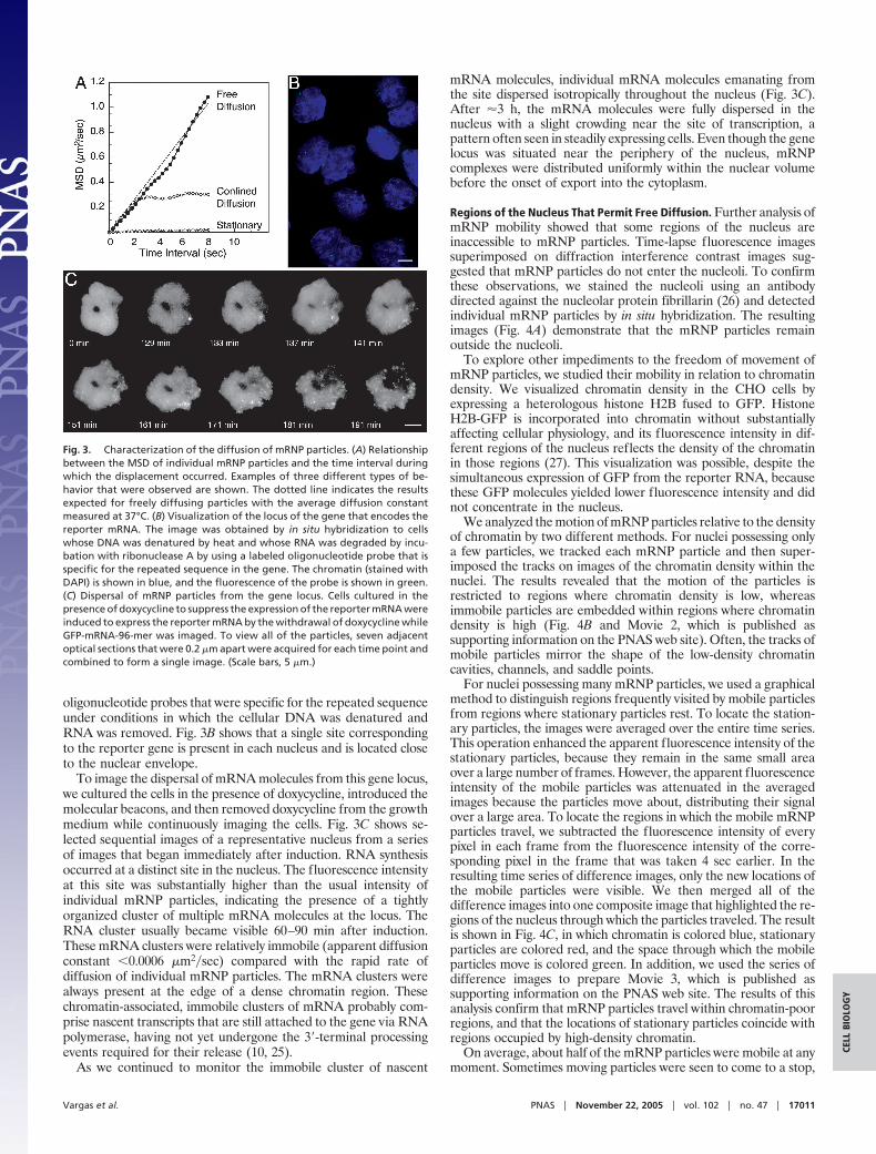
oligonucleotide probes that were specific for the repeated sequenceunder conditions in which the cellular DNA was denatured andRNA was removed. Fig. 3B shows that a single site correspondingto the reporter gene is present in each nucleus and is located closeto the nuclear envelope.
To image the dispersal of mRNA molecules from this gene locus,we cultured the cells in the presence of doxycycline, introduced themolecular beacons, and then removed doxycycline from the growthmedium while continuously imaging the cells. Fig. 3C shows se-lected sequential images of a representative nucleus from a seriesof images that began immediately after induction. RNA synthesisoccurred at a distinct site in the nucleus. The fluorescence intensityat this site was substantially higher than the usual intensity ofindividual mRNP particles, indicating the presence of a tightlyorganized cluster of multiple mRNA molecules at the locus. TheRNA cluster usually became visible 60–90 min after induction.These mRNA clusters were relatively immobile (apparent diffusionconstant �0.0006 �m2�sec) compared with the rapid rate ofdiffusion of individual mRNP particles. The mRNA clusters werealways present at the edge of a dense chromatin region. Thesechromatin-associated, immobile clusters of mRNA probably com-prise nascent transcripts that are still attached to the gene via RNApolymerase, having not yet undergone the 3�-terminal processingevents required for their release (10, 25).
As we continued to monitor the immobile cluster of nascent
mRNA molecules, individual mRNA molecules emanating fromthe site dispersed isotropically throughout the nucleus (Fig. 3C).After �3 h, the mRNA molecules were fully dispersed in thenucleus with a slight crowding near the site of transcription, apattern often seen in steadily expressing cells. Even though the genelocus was situated near the periphery of the nucleus, mRNPcomplexes were distributed uniformly within the nuclear volumebefore the onset of export into the cytoplasm.
Regions of the Nucleus That Permit Free Diffusion. Further analysis ofmRNP mobility showed that some regions of the nucleus areinaccessible to mRNP particles. Time-lapse fluorescence imagessuperimposed on diffraction interference contrast images sug-gested that mRNP particles do not enter the nucleoli. To confirmthese observations, we stained the nucleoli using an antibodydirected against the nucleolar protein fibrillarin (26) and detectedindividual mRNP particles by in situ hybridization. The resultingimages (Fig. 4A) demonstrate that the mRNP particles remainoutside the nucleoli.
To explore other impediments to the freedom of movement ofmRNP particles, we studied their mobility in relation to chromatindensity. We visualized chromatin density in the CHO cells byexpressing a heterologous histone H2B fused to GFP. HistoneH2B-GFP is incorporated into chromatin without substantiallyaffecting cellular physiology, and its fluorescence intensity in dif-ferent regions of the nucleus reflects the density of the chromatinin those regions (27). This visualization was possible, despite thesimultaneous expression of GFP from the reporter RNA, becausethese GFP molecules yielded lower fluorescence intensity and didnot concentrate in the nucleus.
We analyzed the motion of mRNP particles relative to the densityof chromatin by two different methods. For nuclei possessing onlya few particles, we tracked each mRNP particle and then super-imposed the tracks on images of the chromatin density within thenuclei. The results revealed that the motion of the particles isrestricted to regions where chromatin density is low, whereasimmobile particles are embedded within regions where chromatindensity is high (Fig. 4B and Movie 2, which is published assupporting information on the PNAS web site). Often, the tracks ofmobile particles mirror the shape of the low-density chromatincavities, channels, and saddle points.
For nuclei possessing many mRNP particles, we used a graphicalmethod to distinguish regions frequently visited by mobile particlesfrom regions where stationary particles rest. To locate the station-ary particles, the images were averaged over the entire time series.This operation enhanced the apparent fluorescence intensity of thestationary particles, because they remain in the same small areaover a large number of frames. However, the apparent fluorescenceintensity of the mobile particles was attenuated in the averagedimages because the particles move about, distributing their signalover a large area. To locate the regions in which the mobile mRNPparticles travel, we subtracted the fluorescence intensity of everypixel in each frame from the fluorescence intensity of the corre-sponding pixel in the frame that was taken 4 sec earlier. In theresulting time series of difference images, only the new locations ofthe mobile particles were visible. We then merged all of thedifference images into one composite image that highlighted the re-gions of the nucleus through which the particles traveled. The resultis shown in Fig. 4C, in which chromatin is colored blue, stationaryparticles are colored red, and the space through which the mobileparticles move is colored green. In addition, we used the series ofdifference images to prepare Movie 3, which is published assupporting information on the PNAS web site. The results of thisanalysis confirm that mRNP particles travel within chromatin-poorregions, and that the locations of stationary particles coincide withregions occupied by high-density chromatin.
On average, about half of the mRNP particles were mobile at anymoment. Sometimes moving particles were seen to come to a stop,
Fig. 3. Characterization of the diffusion of mRNP particles. (A) Relationshipbetween the MSD of individual mRNP particles and the time interval duringwhich the displacement occurred. Examples of three different types of be-havior that were observed are shown. The dotted line indicates the resultsexpected for freely diffusing particles with the average diffusion constantmeasured at 37°C. (B) Visualization of the locus of the gene that encodes thereporter mRNA. The image was obtained by in situ hybridization to cellswhose DNA was denatured by heat and whose RNA was degraded by incu-bation with ribonuclease A by using a labeled oligonucleotide probe that isspecific for the repeated sequence in the gene. The chromatin (stained withDAPI) is shown in blue, and the fluorescence of the probe is shown in green.(C) Dispersal of mRNP particles from the gene locus. Cells cultured in thepresence of doxycycline to suppress the expression of the reporter mRNA wereinduced to express the reporter mRNA by the withdrawal of doxycycline whileGFP-mRNA-96-mer was imaged. To view all of the particles, seven adjacentoptical sections that were 0.2 �m apart were acquired for each time point andcombined to form a single image. (Scale bars, 5 �m.)
Vargas et al. PNAS � November 22, 2005 � vol. 102 � no. 47 � 17011
CELL
BIO
LOG
Y
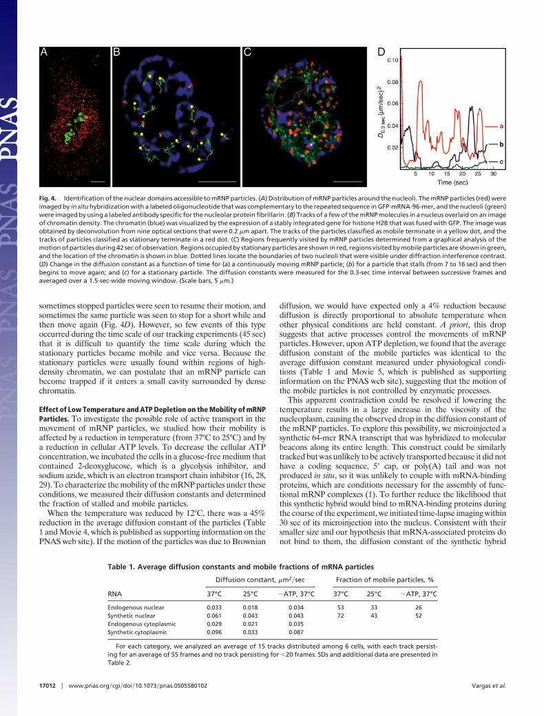
sometimes stopped particles were seen to resume their motion, andsometimes the same particle was seen to stop for a short while andthen move again (Fig. 4D). However, so few events of this typeoccurred during the time scale of our tracking experiments (45 sec)that it is difficult to quantify the time scale during which thestationary particles became mobile and vice versa. Because thestationary particles were usually found within regions of high-density chromatin, we can postulate that an mRNP particle canbecome trapped if it enters a small cavity surrounded by densechromatin.
Effect of Low Temperature and ATP Depletion on the Mobility of mRNPParticles. To investigate the possible role of active transport in themovement of mRNP particles, we studied how their mobility isaffected by a reduction in temperature (from 37°C to 25°C) and bya reduction in cellular ATP levels. To decrease the cellular ATPconcentration, we incubated the cells in a glucose-free medium thatcontained 2-deoxyglucose, which is a glycolysis inhibitor, andsodium azide, which is an electron transport chain inhibitor (16, 28,29). To characterize the mobility of the mRNP particles under theseconditions, we measured their diffusion constants and determinedthe fraction of stalled and mobile particles.
When the temperature was reduced by 12°C, there was a 45%reduction in the average diffusion constant of the particles (Table1 and Movie 4, which is published as supporting information on thePNAS web site). If the motion of the particles was due to Brownian
diffusion, we would have expected only a 4% reduction becausediffusion is directly proportional to absolute temperature whenother physical conditions are held constant. A priori, this dropsuggests that active processes control the movements of mRNPparticles. However, upon ATP depletion, we found that the averagediffusion constant of the mobile particles was identical to theaverage diffusion constant measured under physiological condi-tions (Table 1 and Movie 5, which is published as supportinginformation on the PNAS web site), suggesting that the motion ofthe mobile particles is not controlled by enzymatic processes.
This apparent contradiction could be resolved if lowering thetemperature results in a large increase in the viscosity of thenucleoplasm, causing the observed drop in the diffusion constant ofthe mRNP particles. To explore this possibility, we microinjected asynthetic 64-mer RNA transcript that was hybridized to molecularbeacons along its entire length. This construct could be similarlytracked but was unlikely to be actively transported because it did nothave a coding sequence, 5� cap, or poly(A) tail and was notproduced in situ, so it was unlikely to couple with mRNA-bindingproteins, which are conditions necessary for the assembly of func-tional mRNP complexes (1). To further reduce the likelihood thatthis synthetic hybrid would bind to mRNA-binding proteins duringthe course of the experiment, we initiated time-lapse imaging within30 sec of its microinjection into the nucleus. Consistent with theirsmaller size and our hypothesis that mRNA-associated proteins donot bind to them, the diffusion constant of the synthetic hybrid
Fig. 4. Identification of the nuclear domains accessible to mRNP particles. (A) Distribution of mRNP particles around the nucleoli. The mRNP particles (red) wereimaged by in situ hybridization with a labeled oligonucleotide that was complementary to the repeated sequence in GFP-mRNA-96-mer, and the nucleoli (green)were imaged by using a labeled antibody specific for the nucleolar protein fibrillarin. (B) Tracks of a few of the mRNP molecules in a nucleus overlaid on an imageof chromatin density. The chromatin (blue) was visualized by the expression of a stably integrated gene for histone H2B that was fused with GFP. The image wasobtained by deconvolution from nine optical sections that were 0.2 �m apart. The tracks of the particles classified as mobile terminate in a yellow dot, and thetracks of particles classified as stationary terminate in a red dot. (C) Regions frequently visited by mRNP particles determined from a graphical analysis of themotion of particles during 42 sec of observation. Regions occupied by stationary particles are shown in red, regions visited by mobile particles are shown in green,and the location of the chromatin is shown in blue. Dotted lines locate the boundaries of two nucleoli that were visible under diffraction interference contrast.(D) Change in the diffusion constant as a function of time for (a) a continuously moving mRNP particle; (b) for a particle that stalls (from 7 to 16 sec) and thenbegins to move again; and (c) for a stationary particle. The diffusion constants were measured for the 0.3-sec time interval between successive frames andaveraged over a 1.5-sec-wide moving window. (Scale bars, 5 �m.)
Table 1. Average diffusion constants and mobile fractions of mRNA particles
RNA
Diffusion constant, �m2�sec Fraction of mobile particles, %
37°C 25°C �ATP, 37°C 37°C 25°C �ATP, 37°C
Endogenous nuclear 0.033 0.018 0.034 53 33 26Synthetic nuclear 0.061 0.043 0.043 72 43 52Endogenous cytoplasmic 0.029 0.021 0.035Synthetic cytoplasmic 0.096 0.033 0.087
For each category, we analyzed an average of 15 tracks distributed among 6 cells, with each track persist-ing for an average of 55 frames and no track persisting for �20 frames. SDs and additional data are presented inTable 2.
17012 � www.pnas.org�cgi�doi�10.1073�pnas.0505580102 Vargas et al.
molecules was twice that of the endogenous mRNP particles underphysiological conditions. Upon reduction of the temperature, thesynthetic hybrids displayed a drop in diffusion constant that wassimilar in magnitude to the drop in the diffusion constant of theendogenous mRNP particles (Table 1). Because the synthetictranscripts were unlikely to be involved in enzymatic transportprocesses, these results suggest that the observed decrease in theaverage diffusion constant of the endogenous mRNP particles atlower temperatures is due to an increase in viscosity, rather than tothe involvement of an active process.
Both the reduction in temperature and the depletion of ATPdoubled the proportion of stalled mRNP particles (Table 1). Whenthe temperature was returned to 37°C or the level of ATP wasrestored (Movie 5), the proportion of stalled particles returned toits normal level. Assuming a dynamic equilibrium between themobile and the stalled states of the mRNP particles, this observa-tion suggests that the rescue of particles from the stalled state to themobile state is an ATP-dependent process.
DiscussionEarlier studies of the mobility of mRNA populations by usingpoly(A)-specific reporters led to seemingly contradictory conclu-sions that, although mRNP complexes move by diffusion, theirmobility is curtailed upon depletion of ATP from the cell (14, 15).Both our observations that mRNP particles tend to get stalled whenpassing through high-density chromatin and that, upon ATP de-pletion, this tendency is accentuated, resulting in a larger popula-tion of stalled particles, help to resolve this contradiction. Onepossible explanation is that some constituents of mRNP complexestend to bind to chromatin and that ATP is required to disrupt thesebonds. A second possibility, which we favor more, is that ATPdepletion alters the chromatin structure in such a way that a largernumber of mRNP particles become stalled. What kinds of struc-tural changes in chromatin may be able to bring this about? Arelevant observation is that the flexibility of chromatin is decreasedupon ATP depletion (30). Therefore, we postulate that highchromatin flexibility enables the frequent escape of mRNP particlesfrom their corralled or stalled states. Thus, ATP depletion willresult in an increase in the fraction of stalled particles withoutaffecting the diffusion constant of mobile particles. Along similarlines, Shav-Tal and colleagues (16) have suggested that ATPdepletion results in reduced ‘‘pore size’’ in the chromatin ‘‘mesh,’’which reduces the overall mobility of mRNP particles. Their viewis supported by observations of reversible curdling in chromatinupon ATP depletion (16, 31).
The underlying concern that prompted the formulation of thesolid-state active transport hypothesis (4, 5) and the gene-gatinghypothesis (7) was that interphase nuclei are likely to be so viscousthat large mRNP particles will not be able to diffuse freely withinthem. However, our analysis of the movements of individual mRNPparticles shows that the nucleus possesses at least two distinctmicroenvironments: dense chromatin, within which mRNP parti-cles do not move, and interchromatin spaces, within which theparticles move as freely as they move in the cytoplasm. Otherinvestigations that have explored the viscosity of nuclei support thisconclusion. When fluorescently labeled high-molecular-weight dex-trans are injected into nuclei, they distribute themselves throughoutthe interchromatin space and are excluded from dense areas ofchromatin (32). Fluorescence recovery after photobleaching mea-surements of the diffusion constants of these dextrans indicate thatthe viscosity within the interchromatin spaces is similar to theviscosity of the cytoplasm (33). Furthermore, single-particle track-ing of microspheres of 100 nm in diameter injected into nucleireveals the presence of two separate phases in the nucleus: inter-stitial spaces of low viscosity that permit free diffusion, and otherspaces of very high viscosity in which the microspheres are unableto move freely (34).
In all of the methods previously used to study the dynamics ofmRNP complexes, the probes fluoresced whether they were boundto the target, were bound nonspecifically to other molecules, orwere floating freely in the nucleoplasm. By comparison, the mo-lecular beacon probes are nonfluorescent until they bind to theirmRNA targets. Because we tracked discrete mRNP particles thatcontain a single mRNA target molecule, background fluorescencegenerated by the nonspecific association of molecular beacons withother molecules in the nucleus was uniformly distributed and hadno influence on our analysis.
We have described an effective method for the detection andtracking of individual mRNA molecules in living cells. Naturalgenes can be engineered to have multiple molecular beacon targetsites to study the mechanism of their transport in different cell types.This method will also be useful for the identification of cellular siteswhere other processes central to gene expression take place.Examples of such processes are mRNA splicing, maturation, export,decay, and localization. The ability to track multiple mRNAs taggedwith different multimeric target sequences by using differentlycolored molecular beacons in the same cell will be especially usefulin this regard.
We thank Diana Bratu, Musa M. Mhlanga, Charles S. Peskin, and BenGold for discussions. This work was supported by National Institutes ofHealth Grants GM-070357 and EB-000277.
1. Dreyfuss, G., Kim, V. N. & Kataoka, N. (2002) Nat. Rev. Mol. Cell Biol. 3, 195–205.2. Politz, J. C. & Pederson, T. (2000) J. Struct. Biol. 129, 252–257.3. Carmo-Fonseca, M., Platani, M. & Swedlow, J. R. (2002) Trends Cell Biol. 12, 491–495.4. Agutter, P. S. (1994) Cell Biol. Int. 18, 849–858.5. Lawrence, J. B., Singer, R. H. & Marselle, L. M. (1989) Cell 57, 493–502.6. Bridger, J. M., Kalla, C., Wodrich, H., Weitz, S., King, J. A., Khazaie, K., Krausslich, H. G.
& Lichter, P. (2005) Exp. Cell Res. 302, 180–193.7. Blobel, G. (1985) Proc. Natl. Acad. Sci. USA 82, 8527–8529.8. Colon-Ramos, D. A., Salisbury, J. L., Sanders, M. A., Shenoy, S. M., Singer, R. H. &
Garcia-Blanco, M. A. (2003) Dev. Cell 4, 941–952.9. Casolari, J. M., Brown, C. R., Komili, S., West, J., Hieronymus, H. & Silver, P. A. (2004)
Cell 117, 427–439.10. Zachar, Z., Kramer, J., Mims, I. P. & Bingham, P. M. (1993) J. Cell Biol. 121, 729–742.11. Singh, O. P., Bjorkroth, B., Masich, S., Wieslander, L. & Daneholt, B. (1999) Exp. Cell Res.
251, 135–146.12. Politz, J. C., Browne, E. S., Wolf, D. E. & Pederson, T. (1998) Proc. Natl. Acad. Sci. USA
95, 6043–6048.13. Politz, J. C., Tuft, R. A., Pederson, T. & Singer, R. H. (1999) Curr. Biol. 9, 285–291.14. Calapez, A., Pereira, H. M., Calado, A., Braga, J., Rino, J., Carvalho, C., Tavanez, J. P.,
Wahle, E., Rosa, A. C. & Carmo-Fonseca, M. (2002) J. Cell Biol. 159, 795–805.15. Molenaar, C., Abdulle, A., Gena, A., Tanke, H. J. & Dirks, R. W. (2004) J. Cell Biol. 165,
191–202.16. Shav-Tal, Y., Darzacq, X., Shenoy, S. M., Fusco, D., Janicki, S. M., Spector, D. L. & Singer,
R. H. (2004) Science 304, 1797–1800.
17. Tyagi, S. & Kramer, F. R. (1996) Nat. Biotechnol. 14, 303–308.18. Bratu, D. P., Cha, B. J., Mhlanga, M. M., Kramer, F. R. & Tyagi, S. (2003) Proc. Natl. Acad.
Sci. USA 100, 13308–13313.19. Robinett, C. C., Straight, A., Li, G., Willhelm, C., Sudlow, G., Murray, A. & Belmont, A. S.
(1996) J. Cell Biol. 135, 1685–1700.20. Gossen, M. & Bujard, H. (1992) Proc. Natl. Acad. Sci. USA 89, 5547–5551.21. Tyagi, S. & Alsmadi, O. (2004) Biophys. J. 87, 4153–4162.22. Babcock, H. P., Chen, C. & Zhuang, X. (2004) Biophys. J. 87, 2749–2758.23. Saxton, M. J. & Jacobson, K. (1997) Annu. Rev. Biophys. Biomol. Struct. 26, 373–399.24. Berg, H. C. (1983) Random Walks in Biology (Princeton Univ. Press, Princeton).25. Shatkin, A. J. & Manley, J. L. (2000) Nat. Struct. Biol. 7, 838–842.26. Kill, I. R. (1996) J. Cell Sci. 109, 1253–1263.27. Kanda, T., Sullivan, K. F. & Wahl, G. M. (1998) Curr. Biol. 8, 377–385.28. Wagner, S., Chiosea, S., Ivshina, M. & Nickerson, J. A. (2004) J. Cell Biol. 164, 843–850.29. Platani, M., Goldberg, I., Lamond, A. I. & Swedlow, J. R. (2002) Nat. Cell Biol. 4, 502–508.30. Heun, P., Laroche, T., Shimada, K., Furrer, P. & Gasser, S. M. (2001) Science 294, 2181–2186.31. Gorisch, S. M., Wachsmuth, M., Ittrich, C., Bacher, C. P., Rippe, K. & Lichter, P. (2004)
Proc. Natl. Acad. Sci. USA 101, 13221–13226.32. Verschure, P. J., van der Kraan, I., Manders, E. M., Hoogstraten, D., Houtsmuller, A. B.
& van Driel, R. (2003) EMBO Rep. 4, 861–866.33. Lukacs, G. L., Haggie, P., Seksek, O., Lechardeur, D., Freedman, N. & Verkman, A. S.
(2000) J. Biol. Chem. 275, 1625–1629.34. Tseng, Y., Lee, J. S. H., Kole, T. P., Jiang, I. & Wirtz, D. (2004) J. Cell Sci. 117, 2159–2167.
Vargas et al. PNAS � November 22, 2005 � vol. 102 � no. 47 � 17013
CELL
BIO
LOG
Y
Related Documents