Research Report MDMA treatment 6 months earlier attenuates the effects of CP-94,253, a 5-HT 1B receptor agonist, on motor control but not sleep inhibition Norbert Gyongyosi a , Brigitta Balogh a , Eszter Kirilly a , Tamas Kitka a , Sandor Kantor a , Gyorgy Bagdy a,b,c, ⁎ a Department of Pharmacology and Pharmacotherapy, Semmelweis University, Budapest, Hungary b Group of Neuropsychopharmacology, and Group of Neurochemistry, Hungarian Academy of Science and Semmelweis University, 1089 Budapest, Nagyvarad ter 4, Hungary c Department of Pharmacodynamics, Semmelweis University, Budapest, Hungary ARTICLE INFO ABSTRACT Article history: Accepted 26 June 2008 Available online 2 July 2008 The possible long-term effects of the recreational drug “ecstasy” (3,4-methylenedioxy- methamphetamine, MDMA) on the function of 5-hydroxytryptamine-1B (5-HT 1B ) receptor in sleep and motor control were investigated using a selective 5-HT 1B receptor agonist, 5-propoxy-3-(1,2,3,6-tetrahydro-4-pyrinzidyl)-1H-pyrrolo [3,2-b] pyridine hydrochloride (CP- 94,253; 5 mg/kg). CP-94,253 or vehicle was administered to freely moving rats pre-treated with MDMA (15 mg/kg) or vehicle 6 months earlier, and polygraphic recording for 24 h and motor activity measurements were performed. Active wake (AW), passive wake (PW), light slow wave sleep (SWS-1), deep slow wave sleep (SWS-2), paradoxical sleep (PS), and diurnal rhythm were analyzed for the whole period. In additional, the EEG power spectrum was calculated for the second hour after the acute treatment for AW, PW, SWS-1, and SWS-2. 5-HT transporter (5-HTT) immunohistochemistry was measured in brain areas related to sleep and motor control 6 months after MDMA treatment. CP-94,253 increased AW and PW, decreased SWS-2 and PS, and altered parameters of diurnal rhythm in control animals. CP- 94,253 decreased the EEG power spectra at higher frequencies. The effects of CP-94,253 on AW and diurnal rhythm were reduced or eliminated in MDMA-treated animals. MDMA treatment decreased 5-HTT fibre density in posterior hypothalamus, tuberomammillary nucleus, caudate putamen and ventrolateral striatum. These data suggest that long-term changes in 5-HT 1B receptor function occur after serotonergic damage caused by a single dose of MDMA. © 2008 Elsevier B.V. All rights reserved. Keywords: MDMA Motor activity EEG Sleep Serotonin 5HT 1B receptor 1. Introduction The ring-substituted amphetamine derivative (±)3,4-methyle- nedioxymethamphetamine (MDMA; ecstasy) has become a widely abused psychoactive drug among young people and is popular for its mood enhancing effects (Bond et al., 2004; Parrott, 2001; Steele et al., 1994). MDMA has a characteristic and well-documented two-phase effect on monoaminergic BRAIN RESEARCH 1231 (2008) 34 – 46 ⁎ Corresponding author. Fax: +36 1 4591494. E-mail address: [email protected] (G. Bagdy). 0006-8993/$ – see front matter © 2008 Elsevier B.V. All rights reserved. doi:10.1016/j.brainres.2008.06.099 available at www.sciencedirect.com www.elsevier.com/locate/brainres

Welcome message from author
This document is posted to help you gain knowledge. Please leave a comment to let me know what you think about it! Share it to your friends and learn new things together.
Transcript
B R A I N R E S E A R C H 1 2 3 1 ( 2 0 0 8 ) 3 4 – 4 6
ava i l ab l e a t www.sc i enced i rec t . com
www.e l sev i e r. com/ loca te /b ra in res
Research Report
MDMA treatment 6 months earlier attenuates the effects ofCP-94,253, a 5-HT1B receptor agonist, on motor control but notsleep inhibition
Norbert Gyongyosia, Brigitta Balogha, Eszter Kirillya, Tamas Kitkaa,Sandor Kantora, Gyorgy Bagdya,b,c,⁎aDepartment of Pharmacology and Pharmacotherapy, Semmelweis University, Budapest, HungarybGroup of Neuropsychopharmacology, and Group of Neurochemistry, Hungarian Academy of Science and Semmelweis University,1089 Budapest, Nagyvarad ter 4, HungarycDepartment of Pharmacodynamics, Semmelweis University, Budapest, Hungary
A R T I C L E I N F O
⁎ Corresponding author. Fax: +36 1 4591494.E-mail address: [email protected] (G.
0006-8993/$ – see front matter © 2008 Elsevdoi:10.1016/j.brainres.2008.06.099
A B S T R A C T
Article history:Accepted 26 June 2008Available online 2 July 2008
The possible long-term effects of the recreational drug “ecstasy” (3,4-methylenedioxy-methamphetamine, MDMA) on the function of 5-hydroxytryptamine-1B (5-HT1B) receptor insleep and motor control were investigated using a selective 5-HT1B receptor agonist,5-propoxy-3-(1,2,3,6-tetrahydro-4-pyrinzidyl)-1H-pyrrolo[3,2-b]pyridine hydrochloride (CP-94,253; 5 mg/kg). CP-94,253 or vehicle was administered to freely moving rats pre-treatedwith MDMA (15 mg/kg) or vehicle 6 months earlier, and polygraphic recording for 24 h andmotor activity measurements were performed. Active wake (AW), passive wake (PW), lightslow wave sleep (SWS-1), deep slow wave sleep (SWS-2), paradoxical sleep (PS), and diurnalrhythm were analyzed for the whole period. In additional, the EEG power spectrum wascalculated for the secondhour after the acute treatment forAW, PW, SWS-1, andSWS-2. 5-HTtransporter (5-HTT) immunohistochemistry was measured in brain areas related to sleepand motor control 6 months after MDMA treatment. CP-94,253 increased AW and PW,decreased SWS-2 and PS, and altered parameters of diurnal rhythm in control animals. CP-94,253decreased the EEGpower spectra at higher frequencies. The effects of CP-94,253onAWanddiurnal rhythmwere reduced or eliminated inMDMA-treated animals.MDMA treatmentdecreased 5-HTT fibre density in posterior hypothalamus, tuberomammillary nucleus,caudate putamen and ventrolateral striatum. These data suggest that long-term changes in5-HT1B receptor function occur after serotonergic damage caused by a single dose of MDMA.
© 2008 Elsevier B.V. All rights reserved.
Keywords:MDMAMotor activityEEGSleepSerotonin5HT1B receptor
1. Introduction
The ring-substituted amphetamine derivative (±)3,4-methyle-nedioxymethamphetamine (MDMA; ecstasy) has become a
Bagdy).
ier B.V. All rights reserve
widely abused psychoactive drug among young people and ispopular for its mood enhancing effects (Bond et al., 2004;Parrott, 2001; Steele et al., 1994).MDMAhas a characteristic andwell-documented two-phase effect on monoaminergic
d.
35B R A I N R E S E A R C H 1 2 3 1 ( 2 0 0 8 ) 3 4 – 4 6
systems in the central nervous system. First, shortly afteradministration, there is an acute release of 5-HT (Green et al.,2003; Shankaran and Gudelsky, 1998), dopamine and norepi-nephrine (Fitzgerald and Reid, 1990; Green et al., 2003; Roth-man et al., 2001; Yamamoto and Spanos, 1988). Moreover, inthe brain of rodents andnon-humanprimates,MDMAcauses along-term reduction in 5-HT, 5-hydroxyindoleacetic acid (5-HIAA) concentration and 5-HT transporter density (Adori etal., 2006; Green et al., 2003). Most investigators interpret thislatter effect as a neurotoxic one that involves the serotoner-gic axons and causes depletion of terminals (Green et al.,2003; O'Hearn et al., 1988; Ricaurte et al., 2000; Schmidt, 1987).Increased anxiety and impulsivity, behavioural disinhibition,sleep, learning and memory impairments have beendescribed as possible chronic functional effects of MDMA inanimals (Ando et al., 2006; Baggott et al., 2001; Balogh et al.,2004; Jerome and Baggott, 2003; Jerome, 2004; Jerome, 2005).Despite extensive studies, the long-term functional effects ofMDMA on animal and human behaviour are not fullyunderstood (Baggott et al., 2001; Jerome and Baggott, 2003;Jerome, 2004; Jerome, 2005). Because of its popularity andincreasing abuse, it is important to reveal the long-termeffects of MDMA in animal models in order to estimate thepotential dangers for human users of ecstasy.
The 5-hydroxytryptamine-1B (5-HT1B) receptors, widelyexpressed in the mammalian CNS, are located in serotonergicaxon terminals, where they act as terminal autoreceptors, orin non-serotonergic axons where they act as heteroreceptorsinhibiting the release of other neurotransmitters (e.g., GABA,glutamate, or acetylcholine). 5-HT1B receptors have beenshown to be involved in several physiological functions, e.g.,aggressive behaviour, sleep, learning and in psychiatricdiseases (Barnes and Sharp, 1999; Sari, 2004); thus, 5-HT1B
receptors have received considerable attention as a target fordrugs used in the treatment of anxiety, depression, andmigraine (Lance, 1991; Lin and Parsons, 2002; Sari, 2004;Saxena, 1995). Regarding sleep–wake control, treatment with5-HT1B agonists alters the sleep/wake cycle and reduces REMsleep (Bjorvatn and Ursin, 1994; Monti et al., 1995).
Evidence points toward 5-HT1B receptors playing a crucialrole in motor control. All basal ganglia receive serotonergicinnervation from the dorsal and medial raphe nuclei (Azmitiaand Segal, 1978; Steinbusch et al., 1981) and the presence of 5-HT1B receptors in motor control centres (e.g., the striatum,globus pallidus, substantia nigra and deep cerebellar nuclei)suggests their involvement in motor activity (Sari, 2004). More-over, loss of 5-HT neurons has been reported in patients withParkinson's disease (Chinaglia et al., 1993), which points to theimportance of the 5-HT system in motor control. Pharmacolo-gical activation of 5-HT1B receptors increases motor activity(Barnes and Sharp, 1999), and 5-HT1B receptors are involved inthe hyperlocomotor activity produced by the release of endo-genous 5-HT after MDMA treatment (Bankson and Cunning-ham, 2002; Callaway and Geyer, 1992; McCreary et al., 1999).
The aim of this study was to investigate whether anyfunctional alteration of 5-HT1B receptors could be detected 6months after a single dose of MDMA. Twenty-four hour longpolygraphic recording, motor activity measurements and EEGpower spectra analyses were performed after challenges witheither the 5-HT1B receptor agonist, CP-94,253, or vehicle in
animals pre-treatedwithMDMAandcontrol animals. For quan-tification of axonal damage, 5-HT transporter (5-HTT) immu-nohistochemistry was performed in several brain regions.
2. Results
2.1. Effect of CP-94,253 on vigilance in drug-naive rats
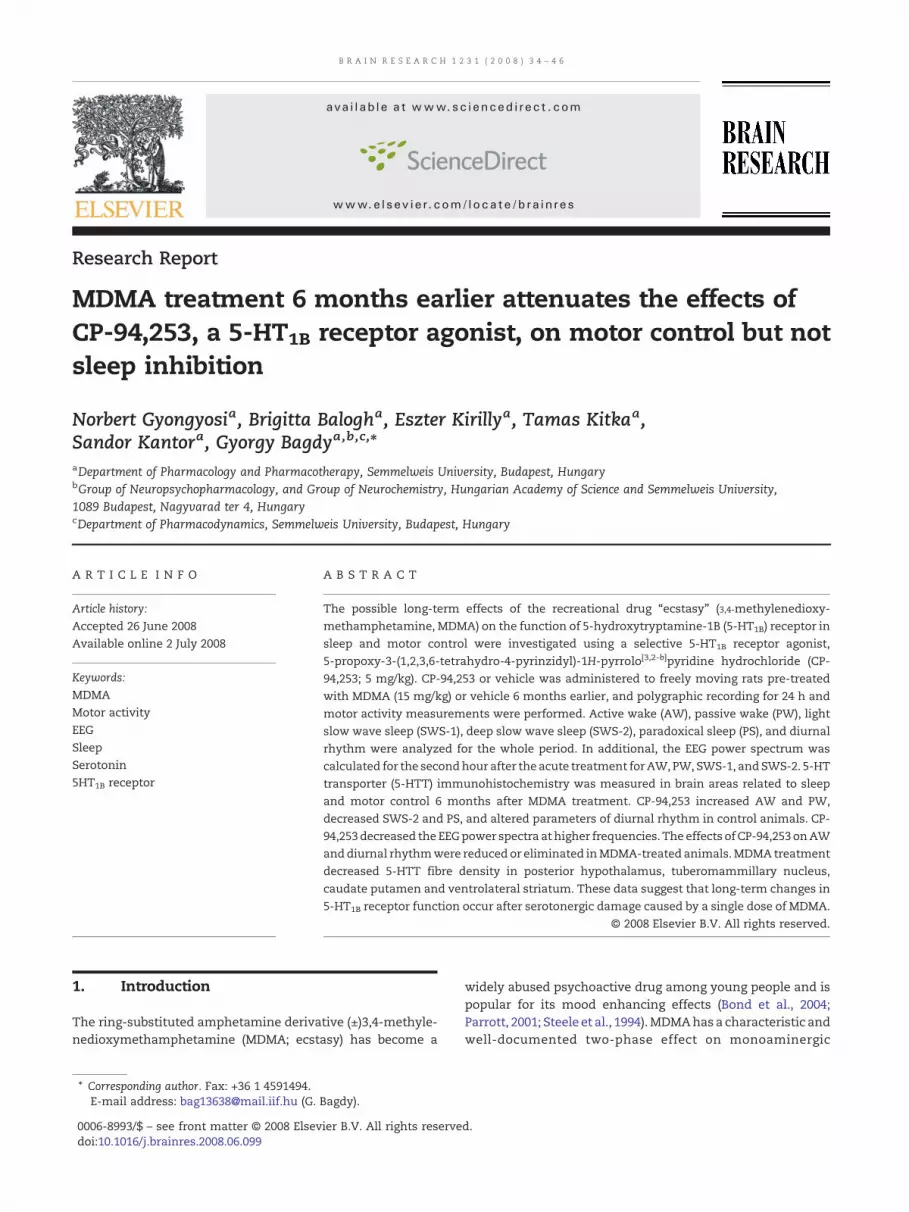
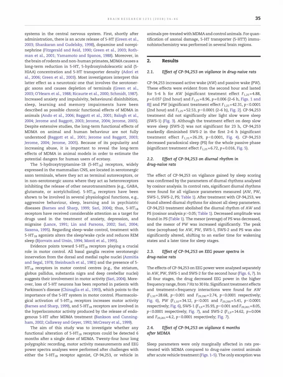
CP-94,253 increased active wake (AW) and passive wake (PW).These effects were evident from the second hour and lastedfor 5–6 h for AW [significant treatment effect F1,25=4.88,p=0.037 (2nd hour) and F1,25=8.96, p=0.006 (2–6 h, Figs. 1 and8)] and PW [significant treatment effect F1,25=42.31, pb0.0001(2nd hour) and F1,25=52.53, pb0.0001 (2–6 h), Fig. 2]. CP-94,253treatment did not significantly alter light slow wave sleep(SWS-1) (Fig. 3). Although the treatment effect on deep slowwave sleep (SWS-2) was not significant for 23 h, CP-94,253markedly diminished SWS-2 in the first 2–6 h (significanttreatment effect F1,25=26.29, pb0.0001, Fig. 4). CP-94,253decreased paradoxical sleep (PS) for the whole passive phase(significant treatment effect F1,25=6.72, p=0.016, Fig. 5).
2.2. Effect of CP-94,253 on diurnal rhythm indrug-naive rats
The effect of CP-94,253 on vigilance gained by sleep scoringwas confirmed by the parameters of diurnal rhythms analysedby cosinor analysis. In control rats, significant diurnal rhythmswere found for all vigilance parameters measured (AW, PW,SWS-1, SWS-2, PS; Table 1). After treatment with CP-94,253, wefound altered diurnal rhythms for almost all sleep parameters.CP-94,253 treatment abolished the diurnal rhythm of AW andPS (cosinor analysis pN0.05; Table 1). Decreased amplitude wasfound in PS (Table 1). The mesor (average) of PS was decreased,and the mesor of PW was increased significantly. The peaktime (acrophase) for AW, PW, SWS-1, SWS-2 and PS was alsosignificantly altered, shifting to an earlier time for wakeningstates and a later time for sleep stages.
2.3. Effect of CP-94,253 on EEG power spectra indrug-naive rats
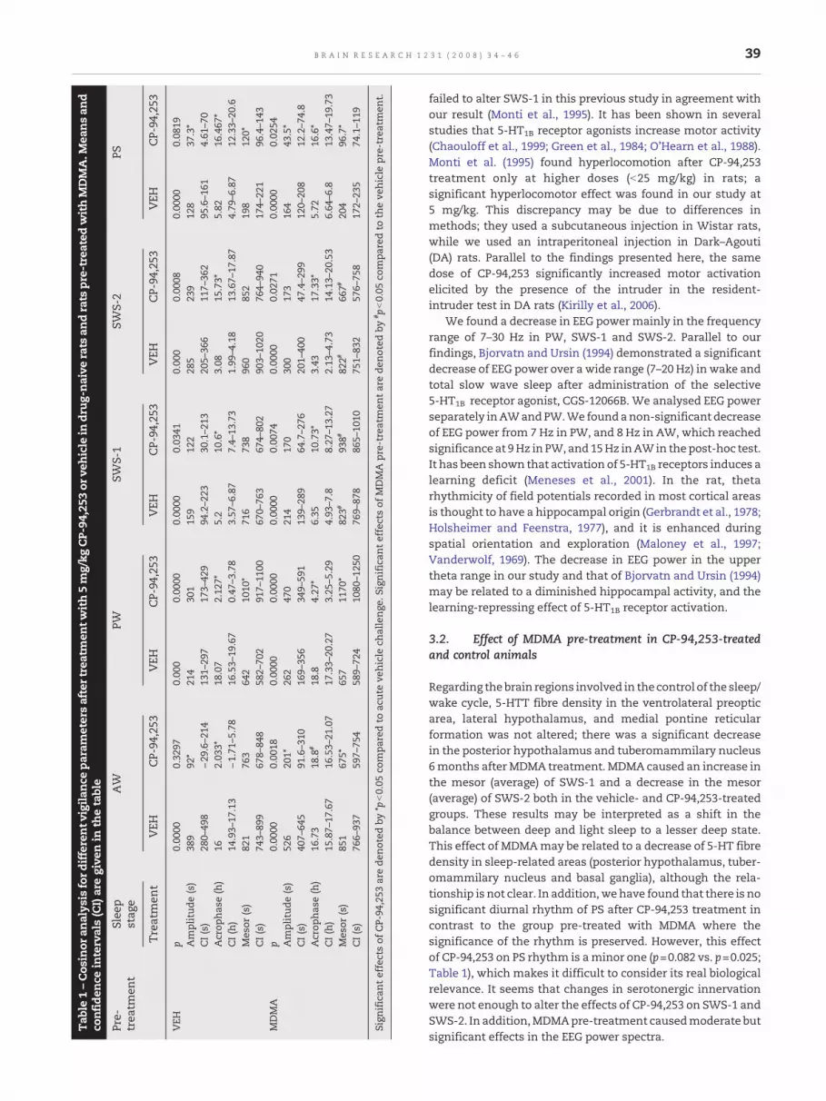
The effects of CP-94,253 on EEG power were analysed separatelyin AW, PW, SWS-1 and SWS-2 for the second hour (Figs. 6, 7). Inall four stages, the drug decreased EEG power in the higherfrequency range, from7Hz to30Hz. Significant treatmenteffectsand treatment×frequency interactions were found for AW(F1,10=28.68, pb0.001 and F29,290=2.74, pb0.0001 respectively;Fig. 6), PW (F1,10=34.12, pb0.001 and F29,290=5.41, pb0.0001respectively; Fig. 6), SWS-1 (F1,9=35.93, pb0.001 and F29,261=8.05,pb0.0001 respectively; Fig. 7), and SWS-2 (F1,9=14.62, p=0.004and F29,261=4.2, pb0.0001 respectively; Fig. 7).
2.4. Effect of CP-94,253 on vigilance 6 monthsafter MDMA
Sleep parameters were only marginally affected in rats pre-treated with MDMA compared to drug-naive control animalsafter acute vehicle treatment (Figs. 1–5). The only exceptionwas
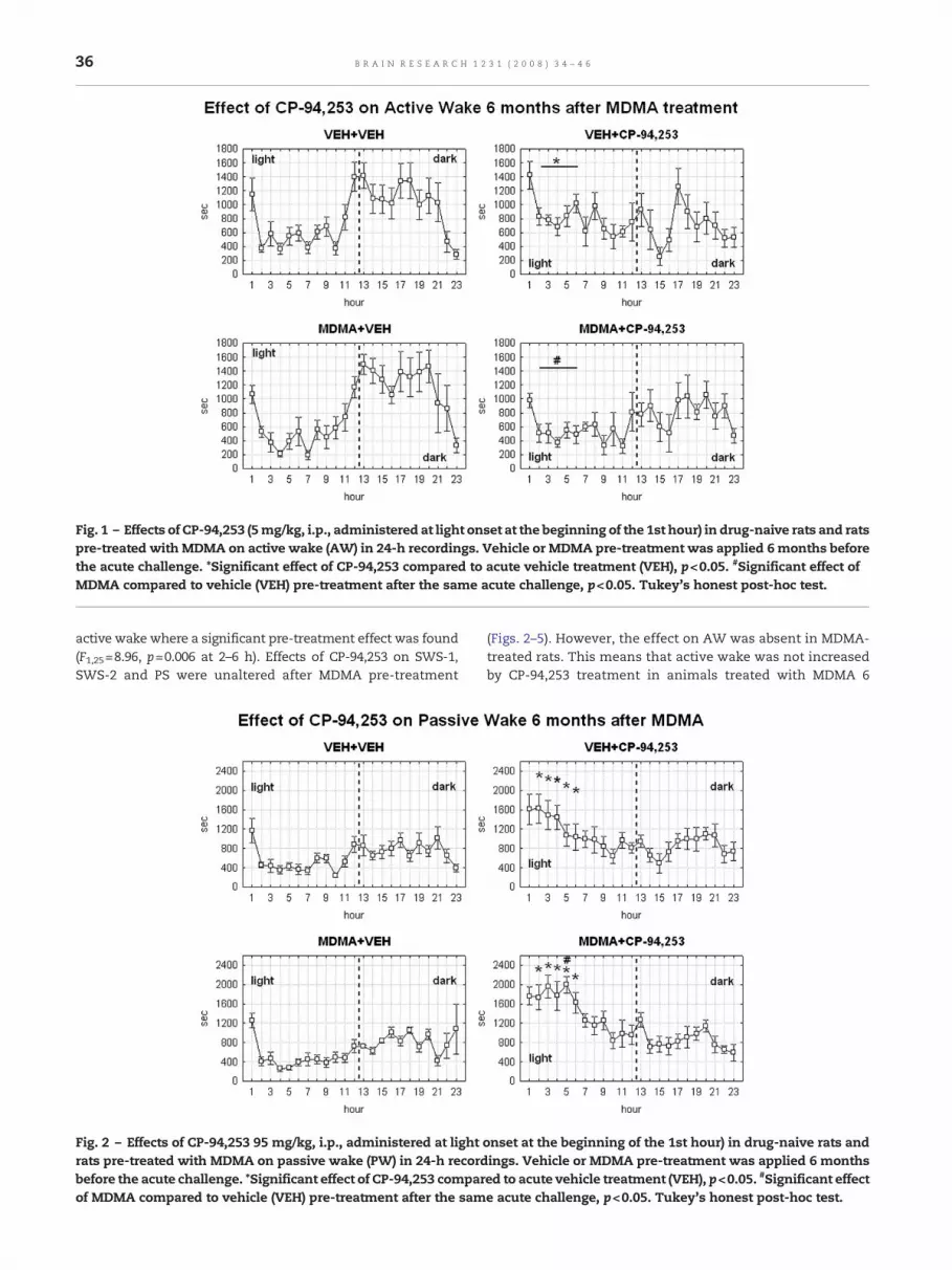
Fig. 1 – Effects of CP-94,253 (5mg/kg, i.p., administered at light onset at the beginning of the 1st hour) in drug-naive rats and ratspre-treated with MDMA on active wake (AW) in 24-h recordings. Vehicle or MDMA pre-treatment was applied 6 months beforethe acute challenge. *Significant effect of CP-94,253 compared to acute vehicle treatment (VEH), p<0.05. #Significant effect ofMDMA compared to vehicle (VEH) pre-treatment after the same acute challenge, p<0.05. Tukey's honest post-hoc test.
36 B R A I N R E S E A R C H 1 2 3 1 ( 2 0 0 8 ) 3 4 – 4 6
active wake where a significant pre-treatment effect was found(F1,25=8.96, p=0.006 at 2–6 h). Effects of CP-94,253 on SWS-1,SWS-2 and PS were unaltered after MDMA pre-treatment
Fig. 2 – Effects of CP-94,253 95 mg/kg, i.p., administered at lightrats pre-treated with MDMA on passive wake (PW) in 24-h recordbefore the acute challenge. *Significant effect of CP-94,253 comparof MDMA compared to vehicle (VEH) pre-treatment after the sam
(Figs. 2–5). However, the effect on AW was absent in MDMA-treated rats. This means that active wake was not increasedby CP-94,253 treatment in animals treated with MDMA 6
onset at the beginning of the 1st hour) in drug-naive rats andings. Vehicle or MDMA pre-treatment was applied 6 monthsed to acute vehicle treatment (VEH), p<0.05. #Significant effecte acute challenge, p<0.05. Tukey's honest post-hoc test.
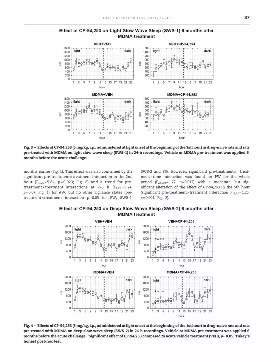
Fig. 3 – Effects of CP-94,253 (5mg/kg, i.p., administered at light onset at the beginning of the 1st hour) in drug-naive rats and ratspre-treated with MDMA on light slow wave sleep (SWS-1) in 24-h recordings. Vehicle or MDMA pre-treatment was applied 6months before the acute challenge.
37B R A I N R E S E A R C H 1 2 3 1 ( 2 0 0 8 ) 3 4 – 4 6
months earlier (Fig. 1). This effect was also confirmed by thesignificant pre-treatment×treatment interaction in the 2ndhour (F1,25 =5.84, p=0.023; Fig. 8) and a trend for pre-treatment×treatment interactions at 2–6 h (F1,25=3.24,p=0.07; Fig. 1) for AW, but no other vigilance states (pre-treatment×treatment interaction pN0.05 for PW, SWS-1,
Fig. 4 – Effects of CP-94,253 (5mg/kg, i.p., administered at light onpre-treated with MDMA on deep slow wave sleep (SWS-2) in 24-months before the acute challenge. *Significant effect of CP-94,25honest post-hoc test.
SWS-2 and PS). However, significant pre-treatment× treat-ment×time interaction was found for PW for the wholeperiod (F22,550=1.77, p=0.017) with a moderate, but sig-nificant alteration of the effect of CP-94,253 in the 5th hour(significant pre-treatment×treatment interaction F13,61=1.25,p=0.001; Fig. 2).
set at the beginning of the 1st hour) in drug-naive rats and ratsh recordings. Vehicle or MDMA pre-treatment was applied 63 compared to acute vehicle treatment (VEH), p<0.05. Tukey's
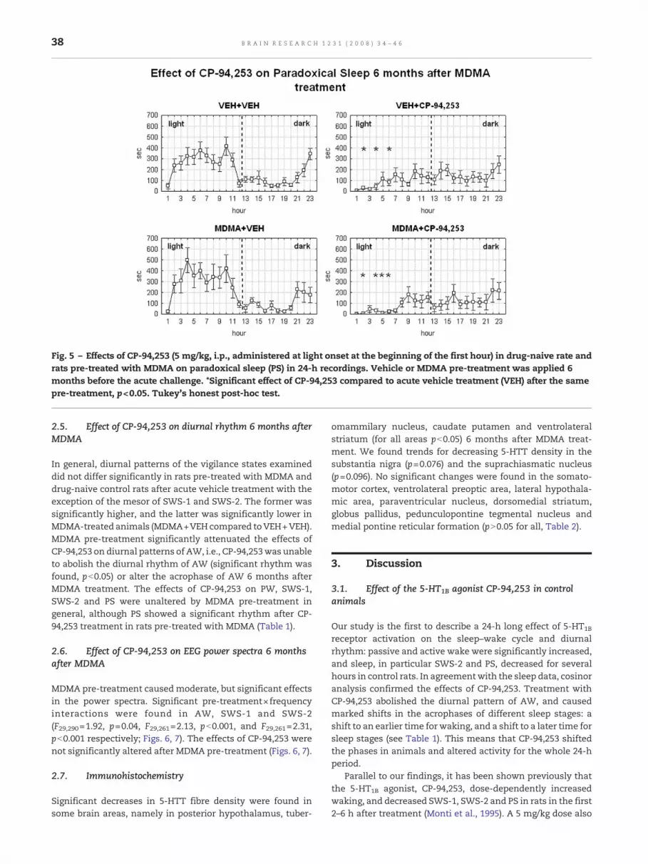
Fig. 5 – Effects of CP-94,253 (5 mg/kg, i.p., administered at light onset at the beginning of the first hour) in drug-naive rate andrats pre-treated with MDMA on paradoxical sleep (PS) in 24-h recordings. Vehicle or MDMA pre-treatment was applied 6months before the acute challenge. *Significant effect of CP-94,253 compared to acute vehicle treatment (VEH) after the samepre-treatment, p<0.05. Tukey's honest post-hoc test.
38 B R A I N R E S E A R C H 1 2 3 1 ( 2 0 0 8 ) 3 4 – 4 6
2.5. Effect of CP-94,253 on diurnal rhythm 6 months afterMDMA
In general, diurnal patterns of the vigilance states examineddid not differ significantly in rats pre-treated with MDMA anddrug-naive control rats after acute vehicle treatment with theexception of the mesor of SWS-1 and SWS-2. The former wassignificantly higher, and the latter was significantly lower inMDMA-treated animals (MDMA+VEHcompared toVEH+VEH).MDMA pre-treatment significantly attenuated the effects ofCP-94,253 on diurnal patterns of AW, i.e., CP-94,253was unableto abolish the diurnal rhythm of AW (significant rhythm wasfound, pb0.05) or alter the acrophase of AW 6 months afterMDMA treatment. The effects of CP-94,253 on PW, SWS-1,SWS-2 and PS were unaltered by MDMA pre-treatment ingeneral, although PS showed a significant rhythm after CP-94,253 treatment in rats pre-treated with MDMA (Table 1).
2.6. Effect of CP-94,253 on EEG power spectra 6 monthsafter MDMA
MDMA pre-treatment causedmoderate, but significant effectsin the power spectra. Significant pre-treatment×frequencyinteractions were found in AW, SWS-1 and SWS-2(F29,290=1.92, p=0.04, F29,261=2.13, pb0.001, and F29,261=2.31,pb0.001 respectively; Figs. 6, 7). The effects of CP-94,253 werenot significantly altered after MDMA pre-treatment (Figs. 6, 7).
2.7. Immunohistochemistry
Significant decreases in 5-HTT fibre density were found insome brain areas, namely in posterior hypothalamus, tuber-
omammilary nucleus, caudate putamen and ventrolateralstriatum (for all areas pb0.05) 6 months after MDMA treat-ment. We found trends for decreasing 5-HTT density in thesubstantia nigra (p=0.076) and the suprachiasmatic nucleus(p=0.096). No significant changes were found in the somato-motor cortex, ventrolateral preoptic area, lateral hypothala-mic area, paraventricular nucleus, dorsomedial striatum,globus pallidus, pedunculopontine tegmental nucleus andmedial pontine reticular formation (pN0.05 for all, Table 2).
3. Discussion
3.1. Effect of the 5-HT1B agonist CP-94,253 in controlanimals
Our study is the first to describe a 24-h long effect of 5-HT1B
receptor activation on the sleep–wake cycle and diurnalrhythm: passive and active wake were significantly increased,and sleep, in particular SWS-2 and PS, decreased for severalhours in control rats. In agreementwith the sleep data, cosinoranalysis confirmed the effects of CP-94,253. Treatment withCP-94,253 abolished the diurnal pattern of AW, and causedmarked shifts in the acrophases of different sleep stages: ashift to an earlier time for waking, and a shift to a later time forsleep stages (see Table 1). This means that CP-94,253 shiftedthe phases in animals and altered activity for the whole 24-hperiod.
Parallel to our findings, it has been shown previously thatthe 5-HT1B agonist, CP-94,253, dose-dependently increasedwaking, and decreased SWS-1, SWS-2 and PS in rats in the first2–6 h after treatment (Monti et al., 1995). A 5 mg/kg dose also
Tab
le1–Cos
inor
analys
isford
ifferentv
igila
nce
param
etersaftertreatm
entw
ith5m
g/kg
CP-
94,253
orve
hicle
indr
ug-naive
rats
andrats
pre-trea
tedwithMDMA.M
eansan
dco
nfide
nce
intervals(CI)aregive
nin
thetable
Pre-
trea
tmen
tSlee
pstag
eAW
PWSW
S-1
SWS-2
PS
Treatmen
tVEH
CP-94
,253
VEH
CP-94
,253
VEH
CP-94
,253
VEH
CP-94
,253
VEH
CP-94
,253
VEH
p0.00
000.32
970.00
00.00
000.00
000.03
410.00
00.00
080.00
000.08
19Amplitude
(s)
389
92⁎
214
301
159
122
285
239
128
37.3⁎
CI(s)
280–
498
−29
.6–2
1413
1–29
717
3–42
994
.2–2
2330
.1–2
1320
5–36
611
7–36
295
.6–1
614.61
–70
Acrop
has
e(h)
162.03
3⁎18
.07
2.12
7⁎5.2
10.6⁎
3.08
15.73⁎
5.82
16.467
⁎CI(h)
14.93–
17.13
−1.71
–5.78
16.53–
19.67
0.47
–3.78
3.57
–6.87
7.4–
13.73
1.99
–4.18
13.67–
17.87
4.79
–6.87
12.33–
20.6
Mes
or(s)
821
763
642
1010
⁎71
673
896
085
219
812
0⁎CI(s)
743–
899
678–
848
582–
702
917–
1100
670–
763
674–
802
903–
1020
764–
940
174–
221
96.4–1
43MDMA
p0.00
000.00
180.00
000.00
000.00
000.00
740.00
000.02
710.00
000.02
54Amplitude
(s)
526
201⁎
262
470
214
170
300
173
164
43.5⁎
CI(s)
407–
645
91.6–3
1016
9–35
634
9–59
113
9–28
964
.7–2
7620
1–40
047
.4–2
9912
0–20
812
.2–7
4.8
Acrop
has
e(h)
16.73
18.8
#18
.84.27
⁎6.35
10.73⁎
3.43
17.33⁎
5.72
16.6⁎
CI(h)
15.87–
17.67
16.53–
21.07
17.33–
20.27
3.25
–5.29
4.93
–7.8
8.27
–13.27
2.13
–4.73
14.13–
20.53
6.64
–6.8
13.47–
19.73
Mes
or(s)
851
675⁎
657
1170
⁎82
3#93
8#82
2#66
7#20
496
.7⁎
CI(s)
766–
937
597–
754
589–
724
1080
–125
076
9–87
886
5–10
1075
1–83
257
6–75
817
2–23
574
.1–1
19
Sign
ifican
teffectsof
CP-94
,253
arede
noted
by*p
b0.05
compa
redto
acute
vehicle
challenge
.Significan
teffectsof
MDMA
pre-trea
tmen
tarede
noted
by# p
b0.05
compa
redto
theve
hicle
pre-trea
tmen
t.
39B R A I N R E S E A R C H 1 2 3 1 ( 2 0 0 8 ) 3 4 – 4 6
failed to alter SWS-1 in this previous study in agreement withour result (Monti et al., 1995). It has been shown in severalstudies that 5-HT1B receptor agonists increase motor activity(Chaouloff et al., 1999; Green et al., 1984; O'Hearn et al., 1988).Monti et al. (1995) found hyperlocomotion after CP-94,253treatment only at higher doses (b25 mg/kg) in rats; asignificant hyperlocomotor effect was found in our study at5 mg/kg. This discrepancy may be due to differences inmethods; they used a subcutaneous injection in Wistar rats,while we used an intraperitoneal injection in Dark–Agouti(DA) rats. Parallel to the findings presented here, the samedose of CP-94,253 significantly increased motor activationelicited by the presence of the intruder in the resident-intruder test in DA rats (Kirilly et al., 2006).
We found a decrease in EEG powermainly in the frequencyrange of 7–30 Hz in PW, SWS-1 and SWS-2. Parallel to ourfindings, Bjorvatn and Ursin (1994) demonstrated a significantdecrease of EEG power over a wide range (7–20 Hz) in wake andtotal slow wave sleep after administration of the selective5-HT1B receptor agonist, CGS-12066B. We analysed EEG powerseparately inAWandPW.We foundanon-significant decreaseof EEG power from 7 Hz in PW, and 8 Hz in AW, which reachedsignificance at 9Hz in PW, and 15Hz inAWin thepost-hoc test.It has been shown that activation of 5-HT1B receptors induces alearning deficit (Meneses et al., 2001). In the rat, thetarhythmicity of field potentials recorded in most cortical areasis thought to have a hippocampal origin (Gerbrandt et al., 1978;Holsheimer and Feenstra, 1977), and it is enhanced duringspatial orientation and exploration (Maloney et al., 1997;Vanderwolf, 1969). The decrease in EEG power in the uppertheta range in our study and that of Bjorvatn and Ursin (1994)may be related to a diminished hippocampal activity, and thelearning-repressing effect of 5-HT1B receptor activation.
3.2. Effect of MDMA pre-treatment in CP-94,253-treatedand control animals
Regarding thebrain regions involved in the control of the sleep/wake cycle, 5-HTT fibre density in the ventrolateral preopticarea, lateral hypothalamus, and medial pontine reticularformation was not altered; there was a significant decreasein the posterior hypothalamus and tuberomammilary nucleus6months after MDMA treatment. MDMA caused an increase inthe mesor (average) of SWS-1 and a decrease in the mesor(average) of SWS-2 both in the vehicle- and CP-94,253-treatedgroups. These results may be interpreted as a shift in thebalance between deep and light sleep to a lesser deep state.This effect of MDMAmay be related to a decrease of 5-HT fibredensity in sleep-related areas (posterior hypothalamus, tuber-omammilary nucleus and basal ganglia), although the rela-tionship is not clear. In addition,wehave found that there is nosignificant diurnal rhythm of PS after CP-94,253 treatment incontrast to the group pre-treated with MDMA where thesignificance of the rhythm is preserved. However, this effectof CP-94,253 on PS rhythm is aminor one (p=0.082 vs. p=0.025;Table 1), which makes it difficult to consider its real biologicalrelevance. It seems that changes in serotonergic innervationwere not enough to alter the effects of CP-94,253 on SWS-1 andSWS-2. In addition,MDMApre-treatment causedmoderate butsignificant effects in the EEG power spectra.
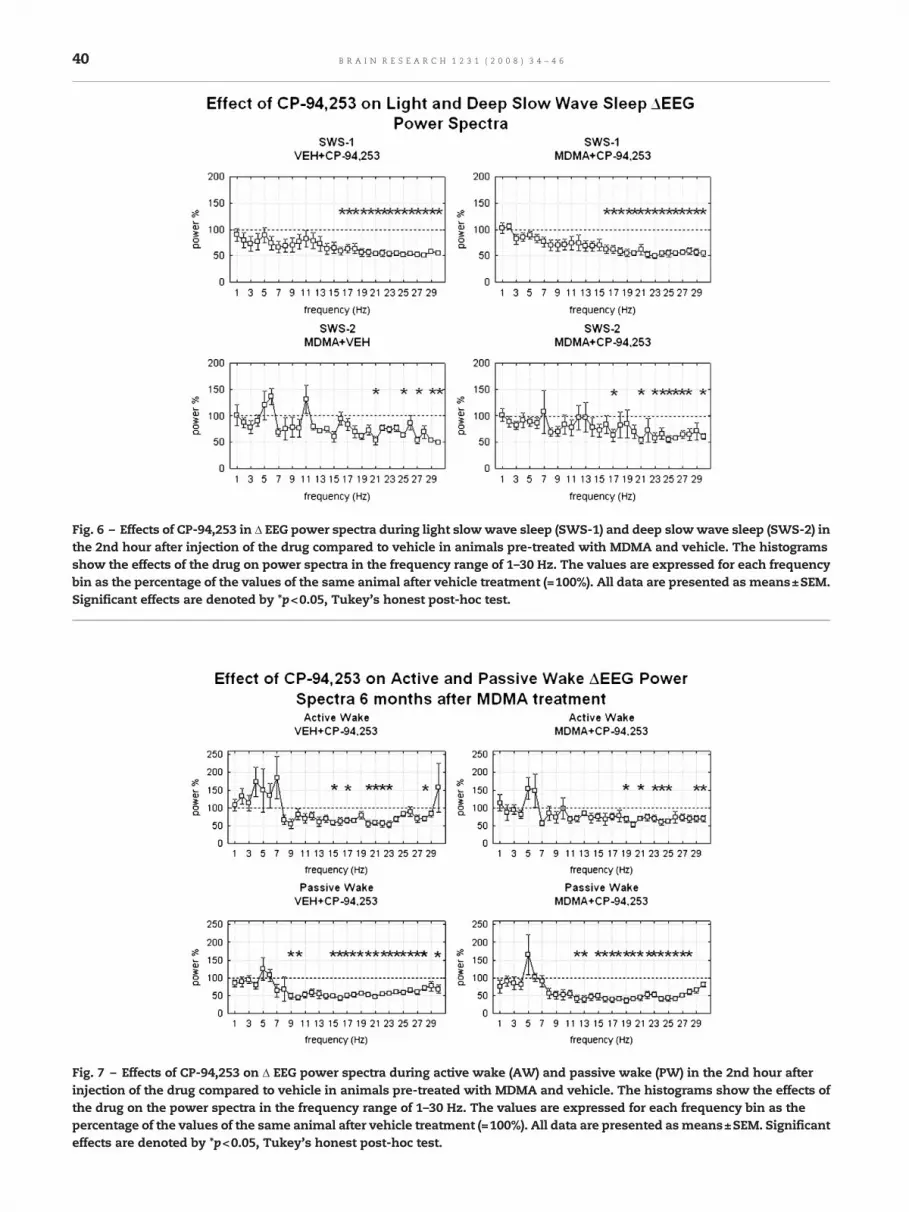
Fig. 6 – Effects of CP-94,253 in Δ EEG power spectra during light slowwave sleep (SWS-1) and deep slowwave sleep (SWS-2) inthe 2nd hour after injection of the drug compared to vehicle in animals pre-treated with MDMA and vehicle. The histogramsshow the effects of the drug on power spectra in the frequency range of 1–30 Hz. The values are expressed for each frequencybin as the percentage of the values of the same animal after vehicle treatment (=100%). All data are presented as means±SEM.Significant effects are denoted by *p<0.05, Tukey's honest post-hoc test.
Fig. 7 – Effects of CP-94,253 on Δ EEG power spectra during active wake (AW) and passive wake (PW) in the 2nd hour afterinjection of the drug compared to vehicle in animals pre-treated with MDMA and vehicle. The histograms show the effects ofthe drug on the power spectra in the frequency range of 1–30 Hz. The values are expressed for each frequency bin as thepercentage of the values of the same animal after vehicle treatment (=100%). All data are presented asmeans±SEM. Significanteffects are denoted by *p<0.05, Tukey's honest post-hoc test.
40 B R A I N R E S E A R C H 1 2 3 1 ( 2 0 0 8 ) 3 4 – 4 6
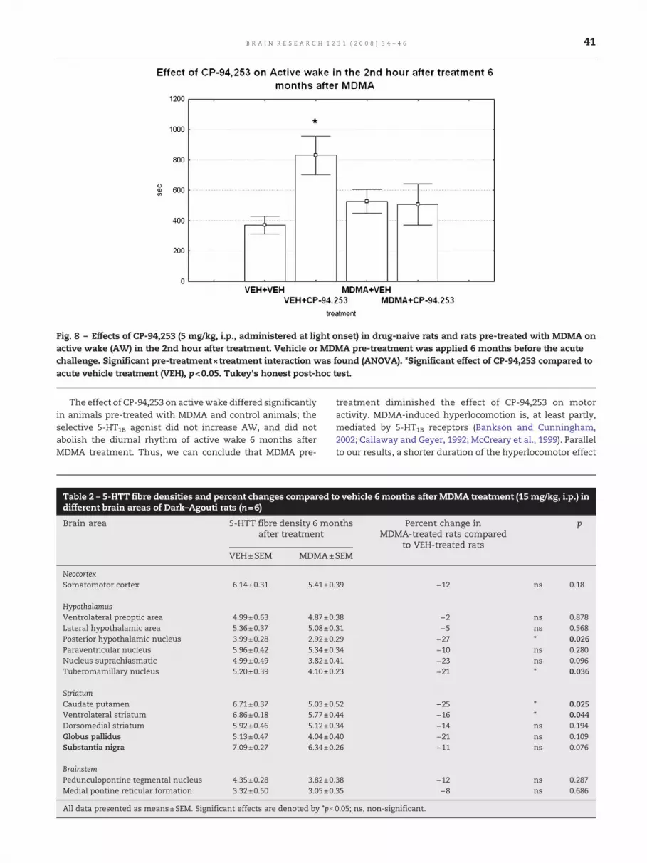
Fig. 8 – Effects of CP-94,253 (5 mg/kg, i.p., administered at light onset) in drug-naive rats and rats pre-treated with MDMA onactive wake (AW) in the 2nd hour after treatment. Vehicle or MDMA pre-treatment was applied 6 months before the acutechallenge. Significant pre-treatment×treatment interaction was found (ANOVA). *Significant effect of CP-94,253 compared toacute vehicle treatment (VEH), p<0.05. Tukey's honest post-hoc test.
41B R A I N R E S E A R C H 1 2 3 1 ( 2 0 0 8 ) 3 4 – 4 6
The effect of CP-94,253 on active wake differed significantlyin animals pre-treated with MDMA and control animals; theselective 5-HT1B agonist did not increase AW, and did notabolish the diurnal rhythm of active wake 6 months afterMDMA treatment. Thus, we can conclude that MDMA pre-
Table 2 – 5-HTT fibre densities and percent changes compared tdifferent brain areas of Dark–Agouti rats (n=6)
Brain area 5-HTT fibre density 6 moafter treatment
VEH±SEM MDMA±
NeocortexSomatomotor cortex 6.14±0.31 5.41±0
HypothalamusVentrolateral preoptic area 4.99±0.63 4.87±0Lateral hypothalamic area 5.36±0.37 5.08±0Posterior hypothalamic nucleus 3.99±0.28 2.92±0Paraventricular nucleus 5.96±0.42 5.34±0Nucleus suprachiasmatic 4.99±0.49 3.82±0Tuberomamillary nucleus 5.20±0.39 4.10±0
StriatumCaudate putamen 6.71±0.37 5.03±0Ventrolateral striatum 6.86±0.18 5.77±0Dorsomedial striatum 5.92±0.46 5.12±0Globus pallidus 5.13±0.47 4.04±0Substantia nigra 7.09±0.27 6.34±0
BrainstemPedunculopontine tegmental nucleus 4.35±0.28 3.82±0Medial pontine reticular formation 3.32±0.50 3.05±0
All data presented as means±SEM. Significant effects are denoted by *pb
treatment diminished the effect of CP-94,253 on motoractivity. MDMA-induced hyperlocomotion is, at least partly,mediated by 5-HT1B receptors (Bankson and Cunningham,2002; Callaway and Geyer, 1992; McCreary et al., 1999). Parallelto our results, a shorter duration of the hyperlocomotor effect
o vehicle 6 months after MDMA treatment (15 mg/kg, i.p.) in
nths Percent change inMDMA-treated rats compared
to VEH-treated rats
p
SEM
.39 −12 ns 0.18
.38 −2 ns 0.878
.31 −5 ns 0.568
.29 −27 ⁎ 0.026
.34 −10 ns 0.280
.41 −23 ns 0.096
.23 −21 ⁎ 0.036
.52 −25 ⁎ 0.025
.44 −16 ⁎ 0.044
.34 −14 ns 0.194
.40 −21 ns 0.109
.26 −11 ns 0.076
.38 −12 ns 0.287
.35 −8 ns 0.686
0.05; ns, non-significant.
42 B R A I N R E S E A R C H 1 2 3 1 ( 2 0 0 8 ) 3 4 – 4 6
was found in the acute response to MDMA in DA rats exposedto MDMA 3 weeks earlier (Balogh et al., 2004), and pre-treatment with MDMA 21 days earlier attenuated the hyper-activity exhibited by S-MDMA treatment (Callaway and Geyer,1992). A transient functional desensitization of 5-HT1B recep-tors following MDMA has also been described, but noalteration in the effect of a 5-HT1A/1B agonist, RU-24969, onmotor activity has been found 3 weeks after MDMA (Callawayand Geyer, 1992).
One possible explanation for the diminished effect of CP-94,253 on motor activity after MDMA treatment is that thedegeneration of 5-HT nerve endings in the basal ganglia maycause the absence of the hyperlocomotor effect of CP-94,253 inrats pre-treated with MDMA; we have found a significantdecrease in the density of 5-HTT in the ventrolateral striatum(16%), caudate putamen (25%), a strong trend for a decrease of5-HTT in the substantia nigra (11%), and a remarkable, butnon-significant reduction (21%) in the globus pallidus 6months after MDMA treatment (15 mg/kg; Table 2).
Data describing the long-term effects of MDMA on the 5-HTsystem in the basal ganglia are controversial, but most studieshave found more or less chronic alterations in the striatumand the globus pallidus. Callaway and Geyer (1992) founddiminished 5-HT and 5-HIAA levels in the striatum 36 daysafter MDMA. Sexton et al. (1999) found a decrease in [3H]-citalopram binding sites in the striatum 14 days after MDMA;they also found a transient increase of cyanopindolol bindingsites in the striatum and an increasing trend in the globuspallidus after MDMA treatment in Sprague–Dawley rats, bothreturning to the control levels by 14 days, without changes in5-HT1B mRNA density. In contrast, others have describedsignificant decrease of 5-HT1B binding sites in the globuspallidus but no significant reduction was found either in theserotonin transporter density or in 5-HT1B density in thecaudate putamen in Wistar rats 3 months after MDMA(McGregor et al., 2003). In the same study, decreases in 5-HTand 5-HIAA levels in the striatumwere described (McGregor etal., 2003). Significant decrease in the local cerebral metabolicrate of glucose utilisation was found 3 weeks after 15 mg/kgMDMA in DA rats in substantia nigra pars reticulata, globuspallidus and medial striatum (Balogh et al., 2004) and insubstantia nigra and caudate nucleus without alteration in[3H]paroxetine binding (Ando et al., 2006).
In our study, MDMA was administered to DA rats at a doseof 15 mg/kg and our earlier results provided evidence that thisdose ofMDMAsignificantly reduced axon densitymeasured bytryptophan-hydroxylase and5-HTT immunohistochemistry inmany brain regions 1 and 3 weeks after the administration ofMDMA (Adori et al., 2006; Ando et al., 2006; Kirilly et al., 2006).The Dark–Agouti rat strain possesses decreased microsomalCYP2D1 isoenzyme activity, which plays an important role inthe metabolism of MDMA, and thus the Dark–Agouti rat is apoormetabolizer of thedrug compared to other rat strains, e.g.,Wistar. The more pronounced vulnerability of DA ratscompared to Wistar and Sprague–Dawley rats and reductionin 5-HTTdensity in the striatummaybe the explanation for thedysfunction of 5-HT1B receptors in regulation ofmotor activity.
Our results suggest that the 5-HT neurotransmission isreduced in the striatum 6 months after MDMA. 5-HT1B
receptors may be located on intrastriatral connections, on
projections from the cortex and basal ganglia, and axonterminals from the dorsal raphe nucleus or other postsynapticneurons located in the striatum. It is suggested that 5-HT1B
receptors are localizedmainly on non-serotonergic, very likelyon GABAergic or glutamatergic neurons in the caudate puta-men (Sari, 2004). Neurons of the caudate putamen can inducemotor activity or select amotor program, andmay have a filterfunction to start or terminatemovements (Grillner et al., 2005).GABAergic neurons projecting from the striatum to globuspallidus externa contain met-enkephalin, and activation ofmet-enkephalin expressing GABAergic neurons results incessation of movement (Grillner et al., 2005). It has beenshown that met-enkephalin is accumulated in the globuspallidus 3 weeks after 5,7-DHT-induced lesion of the seroto-nergic pathway (Compan et al., 1997). Furthermore, it has beendemonstrated that either activation of 5-HT1B receptors orMDMA treatment decreases the met-enkephalin levels in theglobus pallidus and suggests that met-enkephalin containingGABAergic neurons projecting toward the globus pallidus playa significant role in 5-HT1B agonists and MDMA-inducedincrease in locomotor activity (Compan et al., 2003).
We have found a non-significant decrease in 5-HTT densityin the globus pallidus (−21%; pN0.05), and a significant decreasein the striatum (16 and 25%; pb0.05) after MDMA (Table 2). Itseems that an impairment in striatropallidal function exists 6months after MDMA treatment and functional loss of 5-HTnerve endings suggested by 5-HTT immunhistochemistry maycause long-term alteration of the function of the 5-HT1B
receptors. This impairment may result in the decreased motoractivity induced by the selective 5-HT1B agonist, CP-94,253, afterMDMA treatment. Previous studies indicate that locomotorhyperactivity produced by MDMA in rats ismediated via 5-HT1B
receptors, and it is possible that the alteration of 5-HT1B
receptor function contributes to the tolerance to MDMAreported in human ecstasy users and suggests a danger forimpairment in motor control in these people.
4. Conclusion
CP-94,253 caused an increase in motor activity and waking,and inhibited sleep, together with a depression in EEG powerspectra at the highest frequencies in all vigilance states. Theseeffects resulted inmarked shifts inmost circadian parametersof vigilance. A single dose pre-treatment with MDMA 6months earlier diminished the increase in motor activityinduced by acute CP-94,253 treatment and caused damage of5-HT nerve endings in the striatum. These results suggest thatregulation of motor activity is altered even 6 months afterMDMA-induced damage.
5. Experimental procedures
All animal experiments were carried out in accordance withthe European Communities Council Directive of 24 November1986 (86/609/EEC) and the National Institutes of Health“Principles of Laboratory Animal Care” (NIH Publications No.85-23, revised 1985), as well as specific national laws (theHungarian Governmental Regulations on animal studies,
43B R A I N R E S E A R C H 1 2 3 1 ( 2 0 0 8 ) 3 4 – 4 6
December 31, 1998). Permission was obtained from the localethical committees.
5.1. Pre-treatment
Male Dark–Agouti (DA) rats (Harlan, Olac Ltd., Shaw's Farm,Blackthorn, Bicester, Oxon, UK), aged 6 weeks, were randomlydivided into 2 pre-treatment groups 6months before the acutechallenge. Rats in group 1 received an intraperitoneal (i.p.)injection of 15 mg/kg (±)-3,4-methylenedioxymethampheta-mine hydrochloride (MDMA, certified reference compound,purityN99.5%, Sanofi-Synthelabo-Chinoin, Hungary) as pre-treatment. Rats in group 2 received an i.p. injection of 0.9%NaCl (vehicle) in a volume of 1 ml/kg at the same time as thepre-treatment (see Table 3 for pre-treatment groups). Theanimals (4 per cage) were kept under a controlled environ-ment (ambient temperature 21±1 °C, relative humidity 40–50%, 12 h light/dark cycle, lights on from 09:00 to 21:00 h).
5.2. Surgery
Five months and 2 weeks after the pre-treatment (rats were 7–8months old andweighed 300–350 g at implantation), animalsin groups 1 and 2 were equipped with EEG and EMG electrodesas described earlier (Kantor et al., 2004). Briefly, stainless steelscrew electrodes were implanted epidurally over the leftfrontal cortex (L: 2.0 mm and A: 2 mm to bregma) and leftparietal cortex (L: 2.0 mm and A: 2.0 mm to lambda) for fronto-parietal EEG recordings. The ground electrode was placed overthe cerebellum. In addition, EMG electrodes (stainless steelspring electrodes embedded in silicon rubber, Plastics One Inc.,Roanoke, VA, USA; size, 550 mm length, d=1.2 mm with thesilicon rubber) were placed in themuscles of the neck. Surgerywas performed under 2% halothane anaesthesia (Fluotec 3)using a Kopf stereotaxic instrument.
After surgery, the rats were kept in single cages in therecording chamber, maintained in a 12 h light/dark cycle (lightson from 09:00 to 21:00 h, daylight type fluorescent tubes, 18 W,approximately 300 lx) at an ambient temperature of 21±1 °C andrelative humidity of 40–50%. Food and water were available adlibitum. After a 7-day recovery period, in order to habituate theanimals to the recording conditions, the rats were attached tothepolygraphbya flexible recording cableandanelectric swivel,fixedabove the cages, permitting freemovement of the animals;i.p. injections of physiological saline were administered at lightonset for 7 days before the acute experiments. To assess motoractivity, thepotentialsgenerated inelectromagnetic transducersactivated by themovements of the recording cablewere used asdescribed earlier (Kantor et al, 2004). The animals remainedconnected to the recording cables throughout the study.
Table 3 – Study design and name of groups in vigilance studies
Group Treatment 6 months beforethe acute challenge
Acutechallenge
Group 1 Vehicle Vehicle VECP-94,253 VE
Group 2 MDMA Vehicle MDCP-94,253 MD
5.3. Acute challenge and recording
In the acute challenge (6 months after MDMA or vehicletreatment), rats from pre-treatment groups 1 and 2 receivedan i.p. injection of 5 mg/kg 5-propoxy-3-(1,2,3,6-tetrahydro-4-pyrinidyl)-1H-pyrrolo[3,2-b]pyridine hydrochloride (CP-94,253, Tocris Bioscience, catalogue number 1317) (marked asVEH+CP-94,253 and MDMA+CP-94,253 on the figures respec-tively) or vehicle (0.9% NaCl) treatment (marked as VEH+VEHand MDMA+VEH on the figures respectively) in randomorder in the recording chamber (4 days between treatments)starting at light onset as described earlier (Kantor et al., 2004)(see Table 3 for treatment groups). EEG, EMGandmotor activitywere recorded for 24 h after treatment. Ratswere not disturbedthroughout the recordings. Data were stored on computer forfurther analysis.
5.4. Vigilance analysis and scoring
The vigilance states were classified by SleepSign for Animalsleep analysis software (Kissei Comtec America, Inc., USA) for4-s periods over 24 h as follows: active wakefulness (AW), theEEG is characterized by low amplitude activity at beta (14–30 Hz) and alpha (8–13 Hz) frequencies accompanied by highEMG and motor activity; passive wakefulness (PW), the EEG ischaracterized by low amplitude activity at beta (14–30 Hz) andalpha (8–13 Hz) frequencies accompanied by high EMGactivity; light slow wave sleep (SWS-1), high voltage slowcortical waves (0.5–4 Hz) interrupted by low voltage fast EEGactivity (spindles 6–15 Hz) accompanied by reduced EMG andmotor activity; deep slowwave sleep (SWS-2), continuous highamplitude slow cortical waves (0.5–4 Hz) with reduced EMGand motor activity; paradoxical sleep (PS), low amplitudeand high frequency EEG activity with regular theta waves(5–9 Hz) accompanied by silent EMG and motor activity withoccasional twitching (Kantor et al., 2004). Results of manualand automatic analysis previously showed strong correla-tions for all vigilance states (Kantor et al., 2004).
5.5. EEG power spectral analysis (QEEG)
EEG power spectra were computed for consecutive 4-s epochsin the frequency range 0.5–30 Hz (FFT routine, Hanningwindow; frequency resolution 0.25 Hz). Epochs with artefactswere visually discarded on the basis of the polygraph records.Adjacent 0.25 Hz bins were summed into 1 Hz bins, and thoseabove 30 Hz were omitted. Bins are marked by their upperlimits, thus, 2 Hz refers to 1.25–2.00 Hz. The values ofconsecutive 4-s EEG epochs in AW, PW, SWS-1, SWS-2 wereaveraged in the second hour after treatment to obtain the
Name No. of animals invigilance study
No. of animals inQEEG analysis
H+VEH 10 6 (SWS 5)H+CP-94,253 7 6 (SWS 5)MA+VEH 6 6MA+CP-94,253 6 6
44 B R A I N R E S E A R C H 1 2 3 1 ( 2 0 0 8 ) 3 4 – 4 6
power density values for these vigilance states (Kantor et al.,2004). EEG power spectra were calculated for rats treated witheither vehicle or CP-94,253 (n=6 for AW, PW and n=5 for SWS-1, SWS-2 in the vehicle group, and n=6 in the MDMA pre-treated group for all stages).
5.6. Immunohistochemistry
Six months after the pre-treatment (VEH or MDMA), 6 animalsboth from the 1st and 2nd pre-treatment groups were deeplyanesthetized with pentobarbital (i.p.), thoracotomized, andperfused transcardially with Zamboni fixative (4% parafor-maldehyde (w/v%) and 15% saturated picric acid (v/v%) inphosphate buffer (0.1 M PB, pH 7.40). The brains were removedand were postfixed overnight at 4 °C in the fixative solution.
After fixation the brains were transferred into 20% glucosesolution 1 day before sectioning. The free-floating 40-μm thickcoronal sections were cut from the fixed brains using afreezing microtome then the sections were stored in cryopro-tectant at −20 °C until the immunohistochemical procedures.
For 5-HTT immunostaining we used peroxidase/DAB kit(EnVision™, DAKO, Glostrup, Denmark). Sections werebrought to room temperature and washed 3× in 0.1 M TBST(0.05 M Tris, 0.3 M NaCl containing 0.1% Tween20, pH 7.2–7.6).Endogenous peroxidase activity was blocked by application ofTBST containing 0.03% H2O2. After rinsing 3× with TBST theslides were blocked in 1% bovine serum albumin (BSA) for1 h. The presence of 5-HTT was detected using a 1:3000dilution of a rabbit polyclonal anti-5-HTT antibody (Onco-gene, San Diego, CA, USA). The primary antiserum wasdiluted in antibody diluent (DAKO, Glostrup, Denmark).Sections were incubated for 4 h at room temperature. Afterrinsing in TBST we used peroxidase-labelled polymer for 1 h.After the final rinse in TBST, immune complexes werevisualized in 3,3′-diaminobenzidine (DAB) substrate–chromo-gen solution. After washing in TBST, all sections weremounted on gelatine-coated slides, air-dried overnight andcoverslipped with Depex.
5.7. Quantitative analysis for immunohistochemistry
Digital images made using similar light microscopic settings(Olympus BX51, objective, aperture, exposure time) wereprocessed using Adobe Photoshop® 7.0 (similar sharpnessadjustment). Images were made using a 40× objective from4 randomly selected non-overlapping areas of each anato-mical region: ventrolateral preoptic area, lateral hypothala-mic area, posterior hypothalamus, paraventricular nucleus,suprachiasmatic nucleus, tuberomammilary nucleus, soma-tomotor cortex, ventrolateral striatum, dorsomedial striatum,caudate putamen, globus pallidus, substantia nigra, pedun-culopontine tegmental area, and medial pontine reticularformation.
To determine fibre density we used black andwhite imagesand the command “phase analysis” using the software,analySIS®. After the immunostained fibres were distin-guished from background by means of grey-level threshold-ing, the percent area within each image represented by fibreswas recorded. These percentages were averaged across thefour sections per brain region for each animal.
5.8. Statistical analysis
Statistical analysis was carried out using STATISTICA 7.0(Statsoft Inc., Tulsa, OK, USA). Values of vigilance states wereevaluated by ANOVA for measures with three main factors:pre-treatment (non-repeated, vehicle or MDMA); treatment(non-repeated, vehicle or CP-34,2583); and the third factor wastime (repeated, 1–23 h) for sleep analysis. Values of EEG powerwere evaluated by ANOVA for measures with three mainfactors: pre-treatment (non-repeated, vehicle or MDMA);treatment (repeated, vehicle or CP-34,2583), and the thirdfactor was frequency (repeated, 1–30 Hz). Tukey's honestsignificant difference test was used for post-hoc comparisons(see Table 3 for treatment groups). Amplitude, mesor (average)and acrophase (time of peak) values (±confidence limits) werecalculated by cosinor analysis (Nelson et al., 1979) using theprogram Time Series Analysis Seriel Cosinor 6.0 Lab View(Expert Soft Technologie, 1996–2004).
Data from 5-HTT immunohistochemistry were analyzedusing one-way analysis of variance (ANOVA) (treatment:MDMA or VEH) and Newman–Keuls test was performed forpost-hoc comparisons.
Data in all figures are expressed as mean±SEM.
Acknowledgments
This study was supported by the Sixth Framework Programmeof the EC, LSHM-CT-2004-503474, Hungarian Research FundGrants T020500 and M27976, a Ministry of Health ResearchGrant 460/2006, Fund Management of Ministry of EducationOMFB 01926/2002 and the PhD Fellowship Program of Sem-melweis University, Ministry of Education, Hungary.
R E F E R E N C E S
Adori, C., Ando, R.D., Kovacs, G.G., Bagdy, G., 2006. Damage ofserotonergic axons and immunolocalization of Hsp27, Hsp72,and Hsp90 molecular chaperones after a single dose of MDMAadministration in Dark Agouti rat: temporal, spatial, andcellular patterns. J. Comp. Neurol. 497, 251–269.
Ando, R.D., Benko, A., Ferrington, L., Kirilly, E., Kelly, P.A., Bagdy,G., 2006. Partial lesion of the serotonergic system by a singledose of MDMA results in behavioural disinhibition andenhances acute MDMA-induced social behaviour on the socialinteraction test. Neuropharmacology 50, 884–896.
Azmitia, E.C., Segal, M., 1978. An autoradiographic analysis of thedifferential ascending projections of the dorsal and medianraphe nuclei in the rat. J. Comp. Neurol. 179, 641–667.
Baggott, M., Jerome, L., Stuart, R., 2001. Methylenedioxymetham-phetamine (MDMA): A Review of the English-languageScientific and Medical Literature. http://www.maps.org.
Balogh, B., Molnar, E., Jakus, R., Quate, L., Olverman, H.J., Kelly,P.A., Kantor, S., Bagdy, G., 2004. Effects of a single dose of3,4-methylenedioxymethamphetamine on circadian patterns,motor activity and sleep in drug-naive rats and rats previouslyexposed to MDMA. Psychopharmacology (Berl.) 173, 296–309.
Bankson, M.G., Cunningham, K.A., 2002. Pharmacological studiesof the acute effects of (+)-3,4-methylenedioxymethampheta-mine on locomotor activity: role of 5-HT(1B/1D) and 5-HT(2)receptors. Neuropsychopharmacology 26, 40–52.
45B R A I N R E S E A R C H 1 2 3 1 ( 2 0 0 8 ) 3 4 – 4 6
Barnes, N.M., Sharp, T., 1999. A review of central 5-HT receptorsand their function. Neuropharmacology 38, 1083–1152.
Bjorvatn, B., Ursin, R., 1994. Effects of the selective 5-HT1B agonist,CGS 12066B, on sleep/waking stages and EEG power spectrumin rats. J. Sleep Res. 3, 97–105.
Bond, A.J., Verheyden, S.L., Wingrove, J., Curran, H.V., 2004. Angrycognitive bias, trait aggression and impulsivity in substanceusers. Psychopharmacology (Berl.) 171, 331–339.
Callaway, C.W., Geyer, M.A., 1992. Tolerance and cross-toleranceto the activating effects of 3,4-methylenedioxymethampheta-mine and a 5-hydroxytryptamine1B agonist. J. Pharmacol. Exp.Ther. 263, 318–326.
Chaouloff, F., Courvoisier, H., Moisan, M.P., Mormede, P., 1999. GR127935 reduces basal locomotor activity and prevents RU24969-, but not D-amphetamine-induced hyperlocomotion,in the Wistar–Kyoto hyperactive (WKHA) rat.Psychopharmacology (Berl.) 141, 326–331.
Chinaglia, G., Landwehrmeyer, B., Probst, A., Palacios, J.M., 1993.Serotoninergic terminal transporters are differentially affectedin Parkinson's disease and progressive supranuclear palsy: anautoradiographic study with [3H]citalopram. Neuroscience 54,691–699.
Compan, V., Salin, P., Daszuta, A., 1997. Selective effects ofpartial and severe lesions of the serotonergic systems onMet-enkephalin and substance P neurons in rat basal ganglia.Brain Res. Mol. Brain Res. 50, 246–256.
Compan, V., Scearce-Levie, K., Crosson, C., Daszuta, A., Hen, R.,2003. Enkephalin contributes to the locomotor stimulatingeffects of 3,4-methylenedioxy-N-methylamphetamine. Eur. J.Neurosci. 18, 383–390.
Fitzgerald, J.L., Reid, J.J., 1990. Effects of methylenedioxymetham-phetamine on the release of monoamines from rat brain slices.Eur. J. Pharmacol. 191, 217–220.
Gerbrandt, L.K., Lawrence, J.C., Eckardt, M.J., Lloyd, R.L., 1978.Origin of the neocortically monitored theta rhythm in thecurarized rat. Electroencephalogr. Clin. Neurophysiol. 45,454–467.
Green, A.R., Guy, A.P., Gardner, C.R., 1984. The behavioural effectsof RU 24969, a suggested 5-HT1 receptor agonist in rodents andthe effect on the behaviour of treatment with antidepressants.Neuropharmacology 23, 655–661.
Green, A.R., Mechan, A.O., Elliott, J.M., O'Shea, E., Colado, M.I.,2003. The pharmacology and clinical pharmacology of3,4-methylenedioxymethamphetamine (MDMA, qecstasyq).Pharmacol. Rev. 55, 463–508.
Grillner, S., Hellgren, J., Menard, A., Saitoh, K., Wikstrom, M.A.,2005. Mechanisms for selection of basic motor programs —roles for the striatum and pallidum. Trends Neurosci. 28,364–370.
Holsheimer, J., Feenstra, B.W., 1977. Volume conduction and EEGmeasurementswithin the brain: a quantitative approach to theinfluence of electrical spread on the linear relationship ofactivity measured at different locations. Electroencephalogr.Clin. Neurophysiol. 43, 52–58.
Jerome, L., Baggott, M., 2003. MAPS' MDMA Investigator's BrochureUpdate #1: A Review of Research in Humans and Non-HumanAnimals. http://www.maps.org.
Jerome, L., 2004. MAPS' MDMA Investigator's Brochure Update #2:A Review of Research in Humans and Non-Human Animals.http://www.maps.org.
Jerome, L., 2005. MDMA Literature Review Update: March2004–January 2005. http://www.maps.org.
Kantor, S., Jakus, R., Balogh, B., Benko, A., Bagdy, G., 2004.Increased wakefulness, motor activity and decreased thetaactivity after blockade of the 5-HT2B receptor by thesubtype-selective antagonist SB-215505. Br. J. Pharmacol. 142,1332–1342.
Kirilly, E., Benko, A., Ferrington, L., Ando, R.D., Kelly, P.A., Bagdy,G., 2006. Acute and long-term effects of a single dose of MDMA
on aggression in Dark Agouti rats. Int. J.Neuropsychopharmacol. 9, 63–76.
Lance, J.W., 1991. 5-Hydroxytryptamine and its role in migraine.Eur. Neurol. 31, 279–281.
Lin, D., Parsons, L.H., 2002. Anxiogenic-like effect of serotonin(1B)receptor stimulation in the rat elevated plus-maze. Pharmacol.Biochem. Behav. 71, 581–587.
Maloney, K.J., Cape, E.G., Gotman, J., Jones, B.E., 1997.High-frequency gamma electroencephalogram activity inassociation with sleep-wake states and spontaneous behaviorsin the rat. Neuroscience 76, 541–555.
McCreary, A.C., Bankson, M.G., Cunningham, K.A., 1999.Pharmacological studies of the acute and chronic effects of(+)-3, 4-methylenedioxymethamphetamine on locomotoractivity: role of 5-hydroxytryptamine(1A) and5-hydroxytryptamine(1B/1D) receptors. J. Pharmacol. Exp.Ther. 290, 965–973.
McGregor, I.S., Clemens, K.J., Van der Plasse, G., Li, K.M., Hunt, G.E.,Chen, F., Lawrence, A.J., 2003. Increased anxiety 3 months afterbrief exposure to MDMA (qEcstasyq) in rats: associationwith altered 5-HT transporter and receptor density.Neuropsychopharmacology 28, 1472–1484.
Meneses, P.I., Hajjar, K.A., Berns, K.I., Duvoisin, R.M., 2001.Recombinant angiostatin prevents retinal neovascularizationin a murine proliferative retinopathy model. Gene Ther. 8,646–648.
Monti, J.M.,Monti, D., Jantos, H., Ponzoni, A., 1995. Effects of selectiveactivation of the 5-HT1B receptor with CP-94,253 on sleep andwakefulness in the rat. Neuropharmacology 34, 1647–1651.
Nelson, W., Tong, Y.L., Lee, J.K., Halberg, F., 1979. Methods forcosinor-rhythmometry. Chronobiologia 6, 305–323.
O'Hearn, E., Battaglia, G., De Souza, E.B., Kuhar, M.J., Molliver, M.E.,1988. Methylenedioxyamphetamine (MDA) andmethylenedioxymethamphetamine (MDMA) cause selectiveablation of serotonergic axon terminals in forebrain:immunocytochemical evidence for neurotoxicity. J. Neurosci.8, 2788–2803.
Parrott, A.C., 2001. Human psychopharmacology of Ecstasy(MDMA): a review of 15 years of empirical research. Hum.Psychopharmacol. 16, 557–577.
Ricaurte, G.A., Yuan, J., McCann, U.D., 2000. (+/−)3,4-Methylene-dioxymethamphetamine ('Ecstasy')-induced serotoninneurotoxicity: studies in animals. Neuropsychobiology 42,5–10.
Rothman, R.B., Baumann, M.H., Dersch, C.M., Romero, D.V., Rice,K.C., Carroll, F.I., Partilla, J.S., 2001. Amphetamine-type centralnervous system stimulants release norepinephrine morepotently than they release dopamine and serotonin. Synapse39, 32–41.
Sari, Y., 2004. Serotonin1B receptors: from protein to physiologicalfunction and behavior. Neurosci. Biobehav. Rev. 28, 565–582.
Saxena, P.R., 1995. Serotonin receptors: subtypes, functionalresponses and therapeutic relevance. Pharmacol. Ther. 66,339–368.
Schmidt, C.J., 1987.Neurotoxicity of thepsychedelic amphetamine,methylenedioxymethamphetamine. J. Pharmacol. Exp. Ther.240, 1–7.
Sexton, T.J., McEvoy, C., Neumaier, J.F., 1999. (+) 3,4-Methylene-dioxymethamphetamine ('ecstasy') transiently increasesstriatal 5-HT1B binding sites without altering 5-HT1B mRNA inrat brain. Mol. Psychiatry 4, 572–579.
Shankaran, M., Gudelsky, G.A., 1998. Effect of 3,4-methylene-dioxymethamphetamine (MDMA) on hippocampal dopamineand serotonin. Pharmacol. Biochem. Behav. 61, 361–366.
Steele, T.D., McCann, U.D., Ricaurte, G.A., 1994. 3,4-Methylene-dioxymethamphetamine (MDMA, qEcstasyq): pharmacologyand toxicology in animals and humans. Addiction 89,539–551.
Steinbusch, H.W., Nieuwenhuys, R., Verhofstad, A.A., Van der

46 B R A I N R E S E A R C H 1 2 3 1 ( 2 0 0 8 ) 3 4 – 4 6
Kooy, D., 1981. The nucleus raphe dorsalis of the rat and itsprojection upon the caudatoputamen. A combinedcytoarchitectonic, immunohistochemical and retrogradetransport study. J. Physiol. (Paris) 77, 157–174.
Vanderwolf, C.H., 1969. Hippocampal electrical activity and
voluntary movement in the rat. Electroencephalogr. Clin.Neurophysiol. 26, 407–418.
Yamamoto, B.K., Spanos, L.J., 1988. The acute effects ofmethylenedioxymethamphetamine on dopamine release inthe awake-behaving rat. Eur. J. Pharmacol. 148, 195–203.
Related Documents

![[MDMA]MDMA Neurochemistry](https://static.cupdf.com/doc/110x72/577dab601a28ab223f8c57f3/mdmamdma-neurochemistry.jpg)