Summary Echinococcus granulosus is the causative agent of cystic echinococcosis (CE) in humans and many domestic animals, and still one of the most important global health problem in the world and in Turkey. Infection with metacestode causes severe illness and high economic losses. Several strains of Echinococcus have been identified based on the epidemiological and biological characteristics of strains. In this study, a total of 18 individual hydatid cyst samples from cattle were examined. They were obtained from central slaughterhouse in the province of Manisa/Turkey between 2010-2012. The total genomic DNA (gDNA) was extracted using RTA-DNA Isolation Kit (Gebze/Kocaeli, Turkey) according to manufacturer instructions from protoscoleces and cystic germinal membranes. The aim of this study was to provide molecular characterization of E. granulosus isolates which were obtained from cattles by using polymerase chain reaction (PCR) in Manisa province of Turkey. After PCR, to investigate the genetic characteristics of isolates, deoxyribonucleic acid sequencing of the mitochondrial cytochrome c oxidase subunit 1 (CO1) and nicotinamide adenine dinucleotide dehydrogenase subunit 1 (NAD1) genes were performed with ABI Prism Genetic Analyzer 3100 instrument. As a result of our study, all (18) cattle isolates were detected as E. granulosus sensu stricto (G1-G3 complex). This is the first molecular study report genotyping of Echinococcus isolates from cattle in Manisa province. Keywords: Echinococcus granulosus, Cattle, Genotyping, PCR, DNA Sequence, Turkey Manisa İlinde Echinococcus granulosus’un Sığır İzolatlarının Moleküler Analizi Özet Echinococcus granulosus insanda ve birçok evcil hayvanda kistik ekinokokkozise (KE) neden olan etkendir ve hala dünyada ve Türkiye’de en önemli sağlık problemlerinden biridir. Metasestodlarla infeksiyon şiddetli hastalıklara ve yüksek ekonomik kayıplara neden olur. Bazı Echinococcus suşları, suşların epidemiyolojik ve biyolojik karakteristiklerine dayanarak tanımlanmaktadır. Çalışmamızda sığırlardan elde edilen toplam 18 örnek incelenmiştir. Örnekler 2010-2012 yılları arasında Manisa merkez mezbahasından elde edilmiştir. Total genomik DNA (gDNA) üretici firmanın talimatları doğrultusunda protoskoleks ve kistik germinal membranlardan RTA- DNA İzolasyon Kiti kullanılarak (Gebze/Kocaeli, Türkiye) izole edilmiştir. Bu çalışmanın amacı, Türkiye’de Manisa ilindeki sığırlardan elde edilen E. granulosus izolatlarının Polimeraz Zincir Reaksiyonu (PZR) ile moleküler karakterizasyonunun elde edilmesidir. PZR’dan sonra, izolatların genetik karakteristiklerini araştırmak için mitokondrial sitokrom c oksidaz alt ünite 1 (CO1) ve nikotinamid adenin dinükleotit dehidrogenaz alt ünite 1 (NAD1) genleri deoksiribonükleik asit dizileme ile ABI Prism Genetik Analizör 3100 cihazıyla çalışıldı. Çalışmamızın sonucu olarak, tüm (18) sığır izolatları E. granulosus sensu stricto (G1-G3 kompleksi) olarak teşhis edildi. Bu çalışma Manisa ilindeki sığırlardan elde edilen Echinococcus izolatlarının ilk moleküler genotiplendirme çalışmasıdır. Anahtar sözcükler: Echinococcus granulosus, Sığır, Genotiplendirme, PZR, DNA Dizileme, Türkiye Molecular Analysis of Cattle Isolates of Echinococcus granulosus in Manisa Province of Turkey [1] Nuray ALTINTAS * Mustafa OZTATLICI * Nazmiye ALTINTAS ** Aysegul UNVER ** Aslan SAKARYA *** [1] * ** *** This study was supported financially by the Scientific Research Projects Foundation Unite of Celal Bayar University (Project No: 2011-065). And this study is part of the results of this project Celal Bayar University, Faculty of Medicine, Department of Medical Biology, TR-45030 Manisa - TURKEY Ege University, Faculty of Medicine, Department of Parasitology, TR-35100 İzmir - TURKEY Celal Bayar University, Faculty of Medicine, Department of Surgery, TR-45030 Manisa - TURKEY Makale Kodu (Article Code): KVFD-2012-8036 İleşim (Correspondence) +90 236 2331920/437 [email protected] JOURNAL HOME-PAGE: http://vetdergi.kafkas.edu.tr ONLINE SUBMISSION: http://vetdergikafkas.org RESEARCH ARTICLE Kafkas Univ Vet Fak Derg 19 (3): 455-459, 2013 DOI: 10.9775/kvfd.2012.8036

Welcome message from author
This document is posted to help you gain knowledge. Please leave a comment to let me know what you think about it! Share it to your friends and learn new things together.
Transcript
SummaryEchinococcus granulosus is the causative agent of cystic echinococcosis (CE) in humans and many domestic animals, and still one
of the most important global health problem in the world and in Turkey. Infection with metacestode causes severe illness and high economic losses. Several strains of Echinococcus have been identified based on the epidemiological and biological characteristics of strains. In this study, a total of 18 individual hydatid cyst samples from cattle were examined. They were obtained from central slaughterhouse in the province of Manisa/Turkey between 2010-2012. The total genomic DNA (gDNA) was extracted using RTA-DNA Isolation Kit (Gebze/Kocaeli, Turkey) according to manufacturer instructions from protoscoleces and cystic germinal membranes. The aim of this study was to provide molecular characterization of E. granulosus isolates which were obtained from cattles by using polymerase chain reaction (PCR) in Manisa province of Turkey. After PCR, to investigate the genetic characteristics of isolates, deoxyribonucleic acid sequencing of the mitochondrial cytochrome c oxidase subunit 1 (CO1) and nicotinamide adenine dinucleotide dehydrogenase subunit 1 (NAD1) genes were performed with ABI Prism Genetic Analyzer 3100 instrument. As a result of our study, all (18) cattle isolates were detected as E. granulosus sensu stricto (G1-G3 complex). This is the first molecular study report genotyping of Echinococcus isolates from cattle in Manisa province.
Keywords: Echinococcus granulosus, Cattle, Genotyping, PCR, DNA Sequence, Turkey
Manisa İlinde Echinococcus granulosus’un Sığır İzolatlarının Moleküler Analizi
ÖzetEchinococcus granulosus insanda ve birçok evcil hayvanda kistik ekinokokkozise (KE) neden olan etkendir ve hala dünyada ve
Türkiye’de en önemli sağlık problemlerinden biridir. Metasestodlarla infeksiyon şiddetli hastalıklara ve yüksek ekonomik kayıplara neden olur. Bazı Echinococcus suşları, suşların epidemiyolojik ve biyolojik karakteristiklerine dayanarak tanımlanmaktadır. Çalışmamızda sığırlardan elde edilen toplam 18 örnek incelenmiştir. Örnekler 2010-2012 yılları arasında Manisa merkez mezbahasından elde edilmiştir. Total genomik DNA (gDNA) üretici firmanın talimatları doğrultusunda protoskoleks ve kistik germinal membranlardan RTA-DNA İzolasyon Kiti kullanılarak (Gebze/Kocaeli, Türkiye) izole edilmiştir. Bu çalışmanın amacı, Türkiye’de Manisa ilindeki sığırlardan elde edilen E. granulosus izolatlarının Polimeraz Zincir Reaksiyonu (PZR) ile moleküler karakterizasyonunun elde edilmesidir. PZR’dan sonra, izolatların genetik karakteristiklerini araştırmak için mitokondrial sitokrom c oksidaz alt ünite 1 (CO1) ve nikotinamid adenin dinükleotit dehidrogenaz alt ünite 1 (NAD1) genleri deoksiribonükleik asit dizileme ile ABI Prism Genetik Analizör 3100 cihazıyla çalışıldı. Çalışmamızın sonucu olarak, tüm (18) sığır izolatları E. granulosus sensu stricto (G1-G3 kompleksi) olarak teşhis edildi. Bu çalışma Manisa ilindeki sığırlardan elde edilen Echinococcus izolatlarının ilk moleküler genotiplendirme çalışmasıdır.
Anahtar sözcükler: Echinococcus granulosus, Sığır, Genotiplendirme, PZR, DNA Dizileme, Türkiye
Molecular Analysis of Cattle Isolates of Echinococcus granulosus in Manisa Province of Turkey [1]
Nuray ALTINTAS * Mustafa OZTATLICI * Nazmiye ALTINTAS ** Aysegul UNVER ** Aslan SAKARYA ***
[1]
***
***
This study was supported financially by the Scientific Research Projects Foundation Unite of Celal Bayar University (Project No: 2011-065). And this study is part of the results of this projectCelal Bayar University, Faculty of Medicine, Department of Medical Biology, TR-45030 Manisa - TURKEYEge University, Faculty of Medicine, Department of Parasitology, TR-35100 İzmir - TURKEYCelal Bayar University, Faculty of Medicine, Department of Surgery, TR-45030 Manisa - TURKEY
Makale Kodu (Article Code): KVFD-2012-8036
İletişim (Correspondence) +90 236 2331920/437 [email protected]
Journal Home-Page: http://vetdergi.kafkas.edu.tronline SubmiSSion: http://vetdergikafkas.org RESEARCH ARTICLE
Kafkas Univ Vet Fak Derg19 (3): 455-459, 2013DOI: 10.9775/kvfd.2012.8036
456Molecular Analysis of Cattle ...
INTRODUCTION
Cystic echinococcosis (CE) is quite widespread in the world. It is one of the most important cestode infections causing significant morbidity and mortality in humans as well as significant economic losses in livestock animals.
The extensive intraspecific variation in E. granulosus is associated with change in life cycle pattern, host specificity, geographical distribution, transmission dynamics, infectivity to human, antigenicity and sensitivity to chemotherapeutic agents 1,2. This may have important implications for the design and development of diagnostic reagents, vaccines and control of echinococcosis. At least ten genotypically defined strains (G1–G10) were described within the E. granulosus complex, some of which exhibit marked biological and morphological differences. Such genotypes were recently proposed to merit species status, namely E. granulosus sensu stricto (G1–G3), E. equinus (G4), E. ortleppi (G5), and E. canadensis (G6–G10). E. granulosus sensu stricto is composed of three closely related genotypes, G1–G3. E. granulosus sensu stricto is known to be highly infective for humans 3.
To determine the perpetuation of echinococcosis, investigation must be done its spread in the definitive and intermediate hosts 4,5. Being largely confined with life cycles involving sheep and dogs, exposure of humans to E. granulosus is common in Turkey. The majority of people lives in rural areas and is engaged in animal husbandry. High prevalences of CE have been reported in animals in Turkey: 24% (at autopsy) and 62% (by ELISA) recorded in dogs, 66.4% (by Western blotting, EITB) and 51.9% (at autopsy) in sheep, 63.3% (by ELISA), 54.7% (by IFAT) and 39.7% (at autopsy) in cattle, and 22.1% (at autopsy) in goats 4,6.
Turkey is one of the countries where CE is of public health and economic importance. Despite its public health impact, relatively little informations avaliable on the presence of the different genotypes (strains, species) of E. granulosus. In Turkey, many studies have been performed regarding the prevalence of the disease in sheep and cattle but only few studies have been performed about genetic characterization of Echinococcus variants 3-9. Therefore the aim of the present study was to provide molecular characterization of E. granulosus isolates from cattle in Manisa province of Turkey.
MATERIAL and METHODS
Collection of Cyst Materials
In this study, a total of 18 individual hydatid cyst samples from cattle were examined. They were obtained from central slaughterhouse in the province of Manisa/Turkey. All livestock isolates of E. granulosus were obtained from
liver (16) and lung (2) hydatid cysts. Protoscoleces were detected under light microscope and all cysts were examined for their fertility (1 fertile, 17 sterile kist). Protoscoleces and cyst walls (germinal and laminar layer) were washed three times with phosphate buffered saline solution. The sediment was preserved in 70% ethanol and stored at -20ºC until used. Cyst walls were rinsed in sterile distilled water and then fixed in 70% ethanol and were stored at same conditions such as protoscoleces.
Molecular Analysis
Before the DNA isolation, protoscoleces and cut cyst walls were rinsed several times with sterile distilled water to remove ethanol. In order to determine the average number of protoscoleces in milliliters of a sample, the bottle was thoroughly shaked, 10 μl fluid was placed between the microscope slide and coverslip and then protoscoleces were counted under the light microscope. gDNA was extracted from samples which had 200 protoscoleces or upon 10. The total genomic DNA (gDNA) was extracted using RTA-DNA Isolation Kit (Gebze/Kocaeli, Turkey) according to manufacturer instructions from protoscoleces and cystic germinal membranes. Then the gDNA was examined with spectrophotometer (NanoDrop- ND1000) for qualitative and quantitative analyses (between 50-70 ng/μl). The gDNA was stored -20ºC until use.
The isolates were analyzed using amplification of two mitochondrial DNA regions which were cytochrome c oxidase subunit 1 (CO1) and nicotinamide adenine dinucleotide dehydrogenase subunit 1 (NAD1) genes separately. Amplicons of the CO1 mitochondrial gene were amplified using the JB3 (forward) (5’ -TTTTTTGGGCATCCT GAGGTTTAT- 3’)/JB4.5 (reverse) (5’-TAAAGAAAGAACATAA TGAAAATG- 3’) primers 6 and NAD1 mitochondrial gene’s amplicons were amplified using the MS1 (5’-CGTAGGTA TGTTGGTTTGTTTGGT-3’)/MS2 (5’-CCATAATCAAATGGCGTA CGAT- 3’) primers 11.
PCR amplification for CO1 carried out in a final volume of 25 μl including 3 μl gDNA, 2 μl of each primers (20 pmol), 12.5 μl of Amplitaq Gold Master Mix (Roche, Branchburg, New Jersey/USA), 4 μl GC enhancer (GML, Wollerau/Switzerland) 2.5 μl molecular grade water and 1 μl Hotstart Taq DNA polymerase (MBI, Fermantas, Lithuania). The PCR conditions were: 10 min at 95ºC (initial denaturation), 35 cycles of 50 s at 95ºC, 50 s at 47ºC and 50 s at 72ºC and finally 10 min at 72ºC (final extension). NAD1 carried out in a final volume of 25 μl including between 2.5 - 4 μl DNA, 2 μl each primers (20 pmol), 12.5 μl of Amplitaq Gold Master Mix (Roche, Branchburg, New Jersey/USA), 3 μl GC Enhancer (GML, Wollerau/Switzerland) and between 3.5-5 μl molecular grade water. The PCR conditions were: 10 min at 95ºC (initial denaturation), 35 cycles of 30 s at 95ºC, 30 s at 51ºC and 40 s at 72ºC and finally 10 min at 72ºC (final extension).
After the PCR, amplicons were fractionated in 1.5%
457
ALTINTAS, OZTATLICI, ALTINTASUNVER, SAKARYA
agarose gel which was including 5 μl ethidium bromide and then visualized under the UV light with gel imaging system (SYNGENE). For purification step, all PCR amplicons of both CO1 and NAD1 genes were purified with ExoSap-IT (GML, Wollerau/Switzerland) in a final volume of 7 μl including 5 μl each PCR product and 2 μl ExoSap-IT. The prufication step conditions were: 30 min at 37ºC and 15 min at 80ºC.
Forward and reverse primers which employed in the PCR were used in the Cycle Sequencing step. Cycle Sequencing carried out in a final volume of 10 μl including 2 μl BigDye Terminator v3.1 (Applied Biosystems, USA), 2 μl 5x sequencing buffer (Applied Biosystems, USA), 2 μl forward and reverse primers, 2 μl PCR product (prufied with ExoSap-IT) and 2 μl molecular grade water. The Cycle Sequencing conditions were: 10 min at 96ºC (initial denaturation), 25 cycle of 10 s at 96ºC, 5s at 47ºC and 4 min at 60ºC. In PCR applications, DNA which previously identified as sheep strain by DNA sequence analysis was used as a positive control and distilled water was used as a negative control.
RESULTS
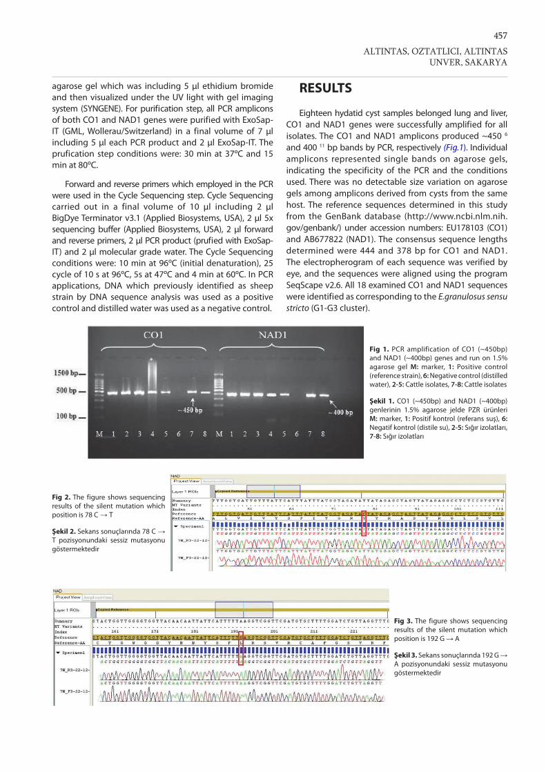
Eighteen hydatid cyst samples belonged lung and liver, CO1 and NAD1 genes were successfully amplified for all isolates. The CO1 and NAD1 amplicons produced ~450 6 and 400 11 bp bands by PCR, respectively (Fig.1). Individual amplicons represented single bands on agarose gels, indicating the specificity of the PCR and the conditions used. There was no detectable size variation on agarose gels among amplicons derived from cysts from the same host. The reference sequences determined in this study from the GenBank database (http://www.ncbi.nlm.nih.gov/genbank/) under accession numbers: EU178103 (CO1) and AB677822 (NAD1). The consensus sequence lengths determined were 444 and 378 bp for CO1 and NAD1. The electropherogram of each sequence was verified by eye, and the sequences were aligned using the program SeqScape v2.6. All 18 examined CO1 and NAD1 sequences were identified as corresponding to the E.granulosus sensu stricto (G1-G3 cluster).
Fig 1. PCR amplification of CO1 (~450bp) and NAD1 (~400bp) genes and run on 1.5% agarose gel M: marker, 1: Positive control (reference strain), 6: Negative control (distilled water), 2-5: Cattle isolates, 7-8: Cattle isolates
Şekil 1. CO1 (~450bp) and NAD1 (~400bp) genlerinin 1.5% agarose jelde PZR ürünleri M: marker, 1: Positif kontrol (referans suş), 6: Negatif kontrol (distile su), 2-5: Sığır izolatları, 7-8: Sığır izolatları
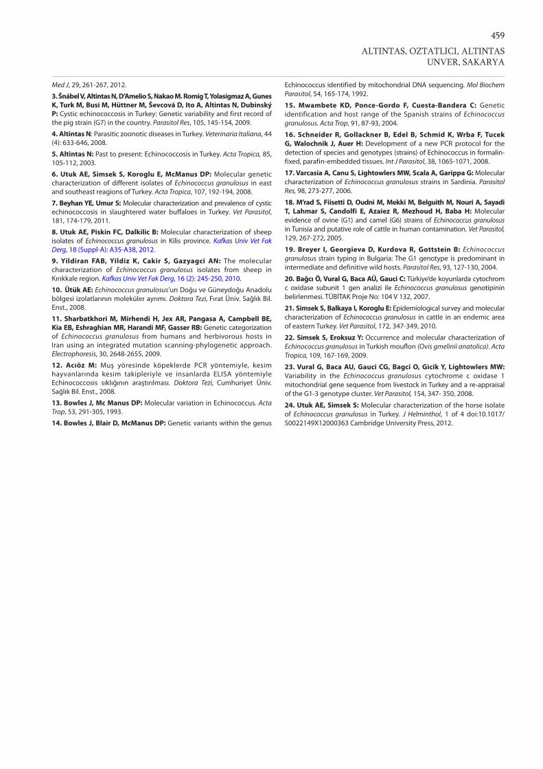
Fig 3. The figure shows sequencing results of the silent mutation which position is 192 G → A
Şekil 3. Sekans sonuçlarında 192 G → A pozisyonundaki sessiz mutasyonu göstermektedir
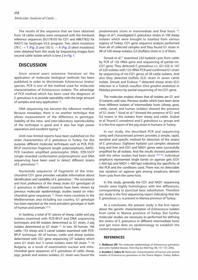
Fig 2. The figure shows sequencing results of the silent mutation which position is 78 C → T
Şekil 2. Sekans sonuçlarında 78 C → T pozisyonundaki sessiz mutasyonu göstermektedir
458Molecular Analysis of Cattle ...
The results of the sequence that we have obtained from 18 cattle isolates were compared with the Genbank reference sequences (EU178103 for CO1 and AB677822 for NAD1) by SeqScape V2.6 program. Two silent mutations [78 C → T (Fig. 2) and 192 G → A (Fig. 3) silent mutations] were obtained from this study by Sequencing images from second cattle isolate which is lane 2 in Fig. 1.
DISCUSSION
Since several years extensive literature on the application of molecular biological methods has been published in order to discriminate Echinococcus strains/species. PCR is one of the method used for molecular characterization of Echinococcus isolates. The advantage of PCR method which has been used the diagnosis of E. granulosus is to provide operability with the large amount of samples and easy application 12.
DNA sequencing has become the reference method. Because nowadays, there is no another technique that allows measurement of the difference in genotype. Stability of the intra- and inter-laboratory reproducibility of the technique is good and it also has high power separation and excellent typing 10.
Until now limited reports have been published on the strain characteristics of E. granulosus in Turkey. For this purpose, different molecular techniques such as PCR, PCR-RFLP (restriction fragment length polymorphism), RAPD-PCR (random amplified polymorphic DNA), PZR-SSCP (single stranded conformation polymorphism) and DNA sequencing have been used to detect different strains of E. granulosus 10.
Nucleotide sequences of fragments of the mito-chondrial CO1 gene provides valuable information about identification and variability of E. granulosus 1. The occurrence and host preference of the sheep strain (G1 genotype) of E. granulosus in different countries have been shown by previous molecular epidemiologic studies based on mito-chondrial gene sequences 13. From many countries in the Mediterranean area including our country, G1 genotype has been reported as the most prevalent genotype in both of human and animals 14-16.
In Sardinia, a total of 91 pieces of sheep, cattle and pig isolates examined with PCR-RFLP and DNA sequencing techniques and 89 isolates determined as G1 strain, 2 pig isolates determined as G7 strain 17. In Iran, 50 human, 166 cattle, 153 sheep and 3 camel isolates examined with PCR-RFLP technique. All of human, cattle and sheep isolates determined with CO1 gene sequencing (37 isolates) which were G1 strain but 3 camel isolates were G6 strain 18. In Bulgaria, as a result of examination nuclear and mito-chondrial gene sequences of E. granulosus of cattle, sheep, pigs, jackals and wolves isolates, G1 strain was found the
predominant strain in intermediate and final hosts 19. Bagcı et al.20, investigated E. granulosus strains in 100 sheep isolates which were brought to Istanbul from various regions of Turkey. CO1 gene sequence analysis performed from all of collected samples and they found G1 strain in 98 of 100 sheep isolates, G3 (buffalo) strain in 2 of them.
Simsek et al.21 examined 220 hydatid cysts from cattle by PCR of 12S rRNA gene and sequencing of partial mt-CO1 gene. They detected E. granulosus s.s. (G1-G3) in 147 of 220 isolates with 12S rRNA-PCR and confirmed G1 strain by sequencing of mt-CO1 genes of 28 cattle isolates. And also they detected buffalo (G3) strain in seven cattle isolate. Simsek and Eroksuz 22 detected sheep strain (G1) infection in a Turkish mouflon (Ovis gmelinii anatolica) in Malatya province by partial sequencing of mt-CO1 gene.
The molecular analysis shows that all isolates are G1 and G1variants until now. Previous studies which have been done from different isolates of intermediate hosts (sheep, goat, cattle, camel, and human isolates) showed the presence of G1 strain 6. Vural et al.23 showed the presence of G1 and G3 strains in the isolates from sheep and cattle. Snabel et al.3 found E. canadensis and E. granulosus s.s. groups and it is the first report of the pig strain in humans in Turkey.
In our study, the described PCR and sequencing using well characterized primers provides a simple, rapid, sensitive and specific method for detection of the strains of E. granulosus. Eighteen hydatid cyst samples obtained lung and liver and CO1 and NAD1 genes were successfully amplified for all isolates. And the results showed paralelism with the other studies had been done 10,21,24. Individual amplicons represented single bands on agarose gels (CO1 (~450 bp) and NAD1 (~400 bp)) indicating the specificity of the PCR and the conditions used. There was no detectable size variation on agarose gels among amplicons derived from cysts from the same host.
In this study, generally the CO1 and NAD1 sequencing results were highly homologous with few differences, corresponding to punctual base substitution. Therefore our study is the first sequencing report indicating that the E. granulosus s.s. is present in Manisa province of Turkey.
As a conclusion; the present study is the first report about the genetic characterization of Echinococcus isolates from cattle in Manisa province of Turkey. But further molecular studies are necessary to performed for defining the strains of E. granulosus in different intermediate hosts and get more data on epidemiology to establish the control programmes in Turkey.
REFERENCES
1. McManus DP: The molecular epidemiology of Echinococcus granulosus and cystic hydatid disease. Trans Roy Soc Med Hyg, 96, 151-157, 2002.
2. Eryıldız C, Sakru N: Molecular characterization of human and animal isolates of Echinococcus granulosus in the Thrace Region, Turkey. Balkan
459
Med J, 29, 261-267, 2012.
3. Šnábel V, Altintas N, D’Amelio S, Nakao M. Romig T, Yolasigmaz A, Gunes K, Turk M, Busi M, Hüttner M, Ševcová D, Ito A, Altintas N, Dubinský P: Cystic echinococcosis in Turkey: Genetic variability and first record of the pig strain (G7) in the country. Parasitol Res, 105, 145-154, 2009.
4. Altintas N: Parasitic zoonotic diseases in Turkey. Veterinaria Italiana, 44 (4): 633-646, 2008.
5. Altintas N: Past to present: Echinococcosis in Turkey. Acta Tropica, 85, 105-112, 2003.
6. Utuk AE, Simsek S, Koroglu E, McManus DP: Molecular genetic characterization of different isolates of Echinococcus granulosus in east and southeast reagions of Turkey. Acta Tropica, 107, 192-194, 2008.
7. Beyhan YE, Umur S: Molecular characterization and prevalence of cystic echinococcosis in slaughtered water buffaloes in Turkey. Vet Parasitol, 181, 174-179, 2011.
8. Utuk AE, Piskin FC, Dalkilic B: Molecular characterization of sheep isolates of Echinococcus granulosus in Kilis province. Kafkas Univ Vet Fak Derg, 18 (Suppl-A): A35-A38, 2012.
9. Yildiran FAB, Yildiz K, Cakir S, Gazyagci AN: The molecular characterization of Echinococcus granulosus isolates from sheep in Kırıkkale region. Kafkas Univ Vet Fak Derg, 16 (2): 245-250, 2010.
10. Ütük AE: Echinococcus granulosus’un Doğu ve Güneydoğu Anadolu bölgesi izolatlarının moleküler ayrımı. Doktora Tezi, Fırat Üniv. Sağlık Bil. Enst., 2008.
11. Sharbatkhori M, Mirhendi H, Jex AR, Pangasa A, Campbell BE, Kia EB, Eshraghian MR, Harandi MF, Gasser RB: Genetic categorization of Echinococcus granulosus from humans and herbivorous hosts in Iran using an integrated mutation scanning-phylogenetic approach. Electrophoresis, 30, 2648-2655, 2009.
12. Acıöz M: Muş yöresinde köpeklerde PCR yöntemiyle, kesim hayvanlarında kesim takipleriyle ve insanlarda ELISA yöntemiyle Echinococcosis sıklığının araştırılması. Doktora Tezi, Cumhuriyet Üniv. Sağlık Bil. Enst., 2008.
13. Bowles J, Mc Manus DP: Molecular variation in Echinococcus. Acta Trop, 53, 291-305, 1993.
14. Bowles J, Blair D, McManus DP: Genetic variants within the genus
Echinococcus identified by mitochondrial DNA sequencing. Mol Biochem Parasitol, 54, 165-174, 1992.
15. Mwambete KD, Ponce-Gordo F, Cuesta-Bandera C: Genetic identification and host range of the Spanish strains of Echinococcus granulosus. Acta Trop, 91, 87-93, 2004.
16. Schneider R, Gollackner B, Edel B, Schmid K, Wrba F, Tucek G, Walochnik J, Auer H: Development of a new PCR protocol for the detection of species and genotypes (strains) of Echinococcus in formalin-fixed, parafin-embedded tissues. Int J Parasitol, 38, 1065-1071, 2008.
17. Varcasia A, Canu S, Lightowlers MW, Scala A, Garippa G: Molecular characterization of Echinococcus granulosus strains in Sardinia. Parasitol Res, 98, 273-277, 2006.
18. M’rad S, Fiisetti D, Oudni M, Mekki M, Belguith M, Nouri A, Sayadi T, Lahmar S, Candolfi E, Azaiez R, Mezhoud H, Baba H: Molecular evidence of ovine (G1) and camel (G6) strains of Echinococcus granulosus in Tunisia and putative role of cattle in human contamination. Vet Parasitol, 129, 267-272, 2005.
19. Breyer I, Georgieva D, Kurdova R, Gottstein B: Echinococcus granulosus strain typing in Bulgaria: The G1 genotype is predominant in intermediate and definitive wild hosts. Parasitol Res, 93, 127-130, 2004.
20. Bağcı Ö, Vural G, Baca AÜ, Gauci C: Türkiye’de koyunlarda cytochrom c oxidase subunit 1 gen analizi ile Echinococcus granulosus genotipinin belirlenmesi. TÜBİTAK Proje No: 104 V 132, 2007.
21. Simsek S, Balkaya I, Koroglu E: Epidemiological survey and molecular characterization of Echinococcus granulosus in cattle in an endemic area of eastern Turkey. Vet Parasitol, 172, 347-349, 2010.
22. Simsek S, Eroksuz Y: Occurrence and molecular characterization of Echinococcus granulosus in Turkish mouflon (Ovis gmelinii anatolica). Acta Tropica, 109, 167-169, 2009.
23. Vural G, Baca AU, Gauci CG, Bagci O, Gicik Y, Lightowlers MW: Variability in the Echinococcus granulosus cytochrome c oxidase 1 mitochondrial gene sequence from livestock in Turkey and a re-appraisal of the G1-3 genotype cluster. Vet Parasitol, 154, 347- 350, 2008.
24. Utuk AE, Simsek S: Molecular characterization of the horse isolate of Echinococcus granulosus in Turkey. J Helminthol, 1 of 4 doi:10.1017/S0022149X12000363 Cambridge University Press, 2012.
ALTINTAS, OZTATLICI, ALTINTASUNVER, SAKARYA
Related Documents