Cervical spine imaging Thanks to Tim’s flexibility with coil combinations, MR imaging of the cervical spine is in 99% percent of the cases a swift and easy standard program evaluating degenerative disease. Figure 1 shows a case of herniated disk where you can clearly exclude myelopathie since the T2-weighted MEDIC sequence demonstrates the ‘butterfly’ of normal gray matter in the myelon. MAGNETOM Aera – Combining Throughput and Highest Quality Spine Imaging in an Optimized Clinical Workflow Johan Dehem, M.D. VZW Jan Yperman, Ieper, Belgium T2-weighted MEDIC sequence to exlude myelopathie. 1 1A 1C 1B 1D 2 MAGNETOM Flash | 5/2013 | www.siemens.com/magnetom-world Clinical Orthopedic Imaging

Welcome message from author
This document is posted to help you gain knowledge. Please leave a comment to let me know what you think about it! Share it to your friends and learn new things together.
Transcript
Cervical spine imaging
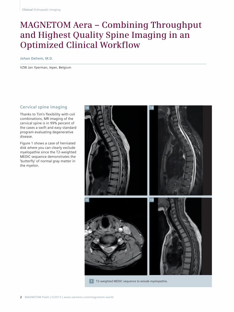
Thanks to Tim’s flexibility with coil combinations, MR imaging of the cervical spine is in 99% percent of the cases a swift and easy standard program evaluating degenerative disease.
Figure 1 shows a case of herniated disk where you can clearly exclude myelopathie since the T2-weighted MEDIC sequence demonstrates the ‘butterfly’ of normal gray matter in the myelon.
MAGNETOM Aera – Combining Throughput and Highest Quality Spine Imaging in an Optimized Clinical Workflow Johan Dehem, M.D.
VZW Jan Yperman, Ieper, Belgium
T2-weighted MEDIC sequence to exlude myelopathie.1
1A
1C
1B
1D
2 MAGNETOM Flash | 5/2013 | www.siemens.com/magnetom-world
Clinical Orthopedic Imaging
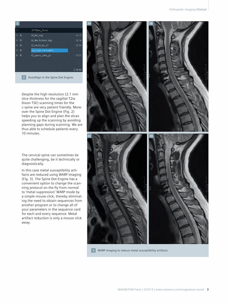
Despite the high resolution (2.1 mm slice thickness for the sagittal T2w Dixon TSE) scanning times for the c-spine are very patient friendly. More-over the Spine Dot Engine (Fig. 2) helps you to align and plan the slices speeding up the scanning by avoiding planning gaps during scanning. We are thus able to schedule patients every 10 minutes.
The cervical spine can sometimes be quite challenging, be it technically or diagnostically.
In this case metal susceptibility arti-facts are reduced using WARP imaging (Fig. 3). The Spine Dot Engine has a convenient option to change the scan-ning protocol on the fly from normal to ‘metal suppression’ WARP mode by a simple mouse click, thereby eliminat-ing the need to obtain sequences from another program or to change all of your parameters in the sequence card for each and every sequence. Metal artifact reduction is only a mouse click away.
AutoAlign in the Spine Dot Engine.2
WARP imaging to reduce metal susceptibility artifacts.3
3A
3C
3B
3D
2
MAGNETOM Flash | 5/2013 | www.siemens.com/magnetom-world 3
Orthopedic Imaging Clinical
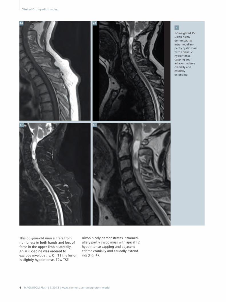
This 65-year-old man suffers from numbness in both hands and loss of force in the upper limb bilaterally. An MRI c-spine was ordered to exclude myelopathy. On T1 the lesion is slightly hypointense. T2w TSE
Dixon nicely demonstrates intramed-ullary partly cystic mass with apical T2 hypointense capping and adjacent edema cranially and caudally extend-ing (Fig. 4).
T2-weighted TSE Dixon nicely demonstrates intramedullary partly cystic mass with apical T2 hypointense capping and adjacent edema cranially and caudally extending.
4
4A
4C
4B
4D
4 MAGNETOM Flash | 5/2013 | www.siemens.com/magnetom-world
Clinical Orthopedic Imaging
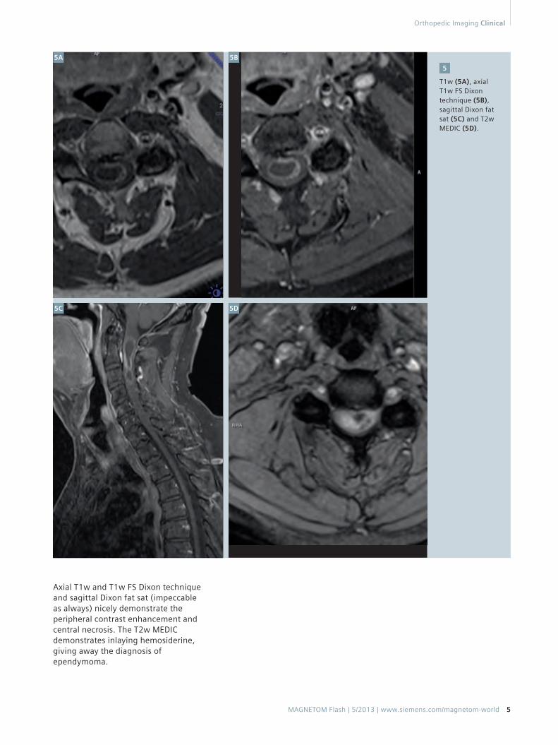
Axial T1w and T1w FS Dixon technique and sagittal Dixon fat sat (impeccable as always) nicely demonstrate the peripheral contrast enhancement and central necrosis. The T2w MEDIC demonstrates inlaying hemosiderine, giving away the diagnosis of ependymoma.
T1w (5A), axial T1w FS Dixon technique (5B), sagittal Dixon fat sat (5C) and T2w MEDIC (5D).
5
5A
5C
5B
5D
MAGNETOM Flash | 5/2013 | www.siemens.com/magnetom-world 5
Orthopedic Imaging Clinical
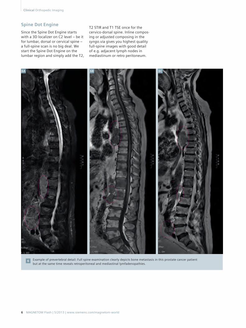
Spine Dot Engine
Since the Spine Dot Engine starts with a 3D localizer on C2 level – be it for lumbar, dorsal or cervical spine – a full-spine scan is no big deal. We start the Spine Dot Engine on the lumbar region and simply add the T2,
Example of prevertebral detail: Full spine examination clearly depicts bone metastasis in this prostate cancer patient but at the same time reveals retroperitoneal and mediastinal lymfadenopathies.
6
T2 STIR and T1 TSE once for the cervico-dorsal spine. Inline compos-ing or adjusted composing in the syngo.via gives you highest quality full-spine images with good detail of e.g. adjacent lymph nodes in mediastinum or retro peritoneum.
6A 6B 6C
6 MAGNETOM Flash | 5/2013 | www.siemens.com/magnetom-world
Clinical Orthopedic Imaging
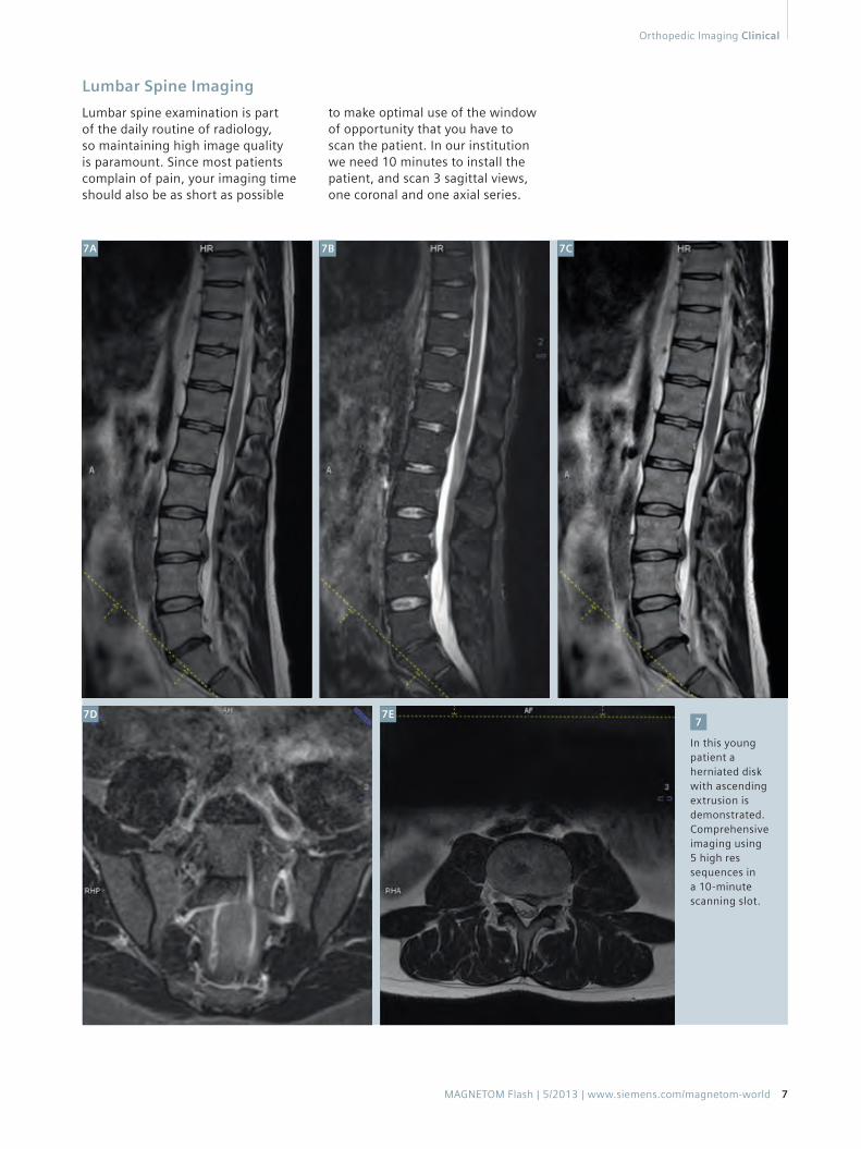
Lumbar Spine Imaging
Lumbar spine examination is part of the daily routine of radiology, so maintaining high image quality is paramount. Since most patients complain of pain, your imaging time should also be as short as possible
to make optimal use of the window of opportunity that you have to scan the patient. In our institution we need 10 minutes to install the patient, and scan 3 sagittal views, one coronal and one axial series.
In this young patient a herniated disk with ascending extrusion is demonstrated.Comprehensive imaging using 5 high res sequences in a 10-minute scanning slot.
7
7A
7D
7B
7E
7C
MAGNETOM Flash | 5/2013 | www.siemens.com/magnetom-world 7
Orthopedic Imaging Clinical
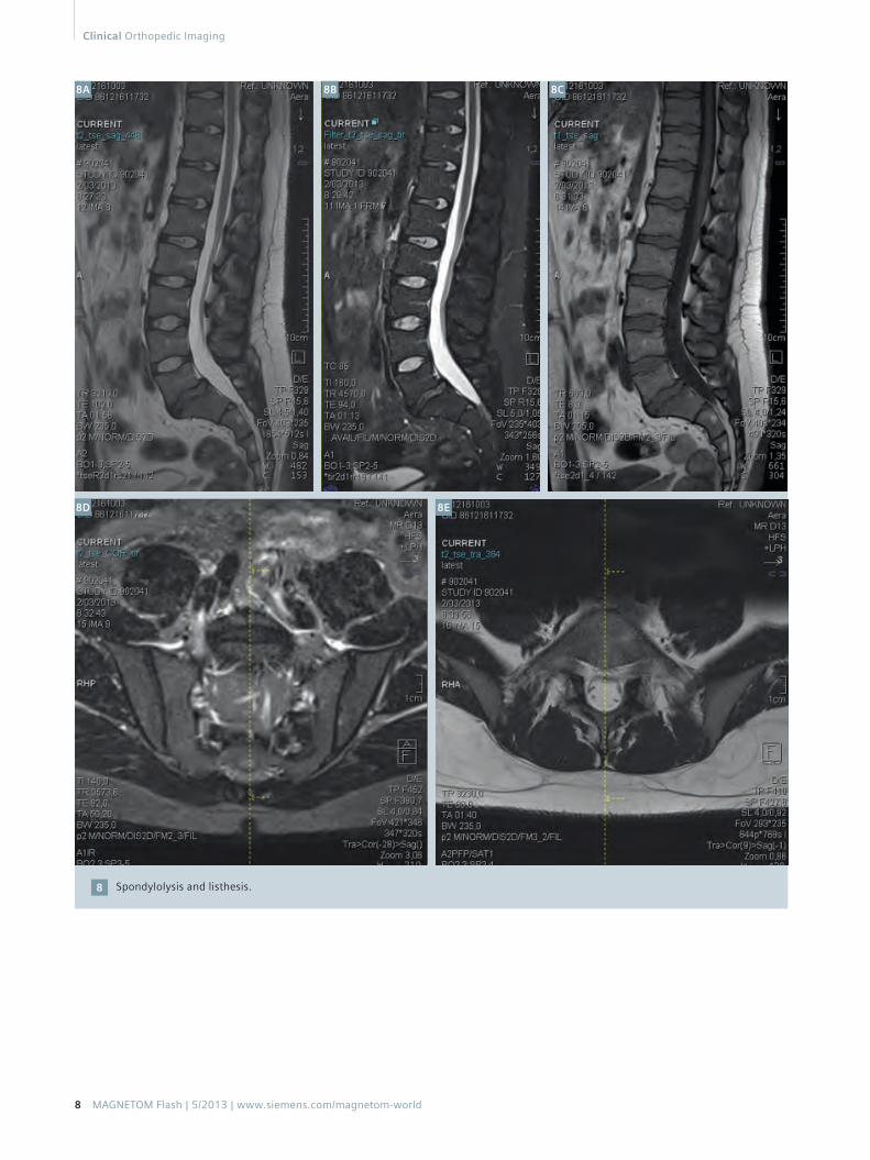
Spondylolysis and listhesis.8
8A 8B
8D 8E
8C
8 MAGNETOM Flash | 5/2013 | www.siemens.com/magnetom-world
Clinical Orthopedic Imaging
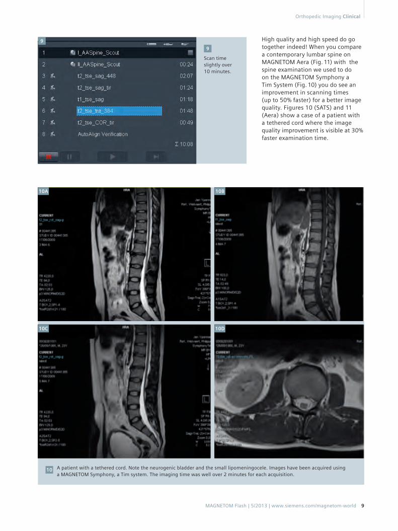
Scan time slightly over 10 minutes.
9
A patient with a tethered cord. Note the neurogenic bladder and the small lipomeningocele. Images have been acquired using a MAGNETOM Symphony, a Tim system. The imaging time was well over 2 minutes for each acquisition.
10
High quality and high speed do go together indeed! When you compare a contemporary lumbar spine on MAGNETOM Aera (Fig. 11) with the spine examination we used to do on the MAGNETOM Symphony a Tim System (Fig. 10) you do see an improvement in scanning times (up to 50% faster) for a better image quality. Figures 10 (SATS) and 11 (Aera) show a case of a patient with a tethered cord where the image quality improvement is visible at 30% faster examination time.
10A
10C
10B
10D
9
MAGNETOM Flash | 5/2013 | www.siemens.com/magnetom-world 9
Orthopedic Imaging Clinical
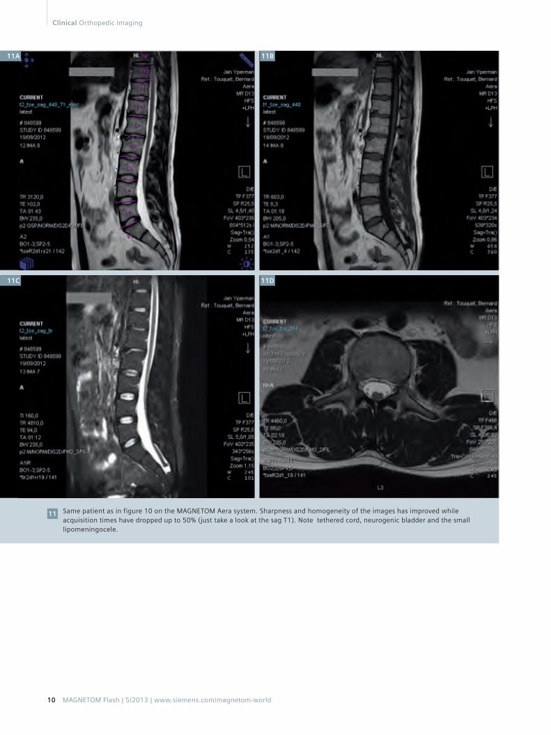
Same patient as in figure 10 on the MAGNETOM Aera system. Sharpness and homogeneity of the images has improved while acquisition times have dropped up to 50% (just take a look at the sag T1). Note tethered cord, neurogenic bladder and the small lipomeningocele.
11
11B
11D11C
11A
10 MAGNETOM Flash | 5/2013 | www.siemens.com/magnetom-world
Clinical Orthopedic Imaging
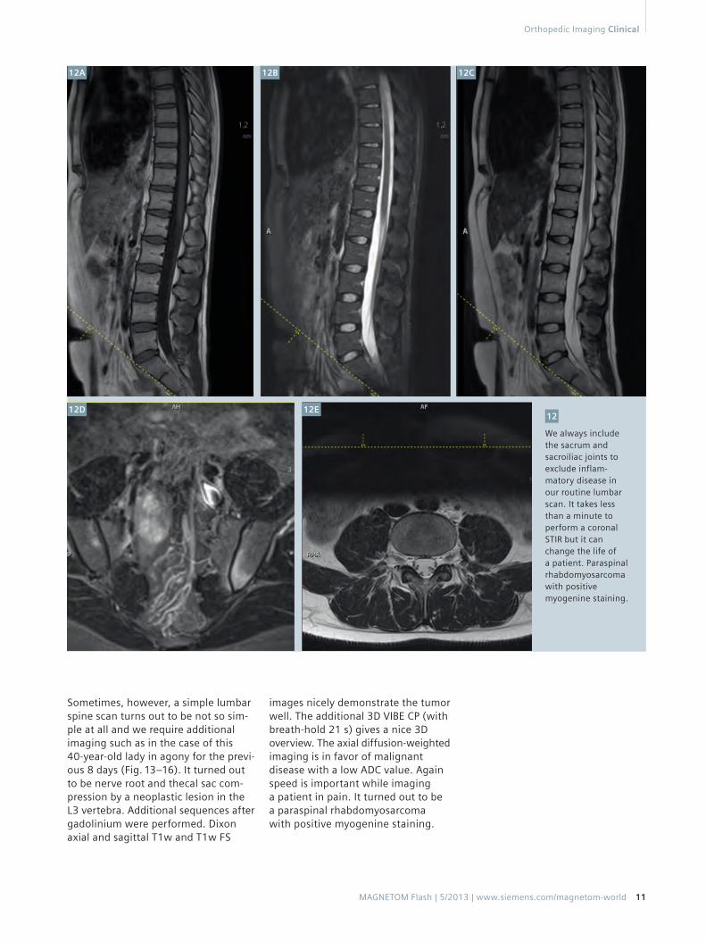
We always include the sacrum and sacroiliac joints to exclude inflam-matory disease in our routine lumbar scan. It takes less than a minute to perform a coronal STIR but it can change the life of a patient. Paraspinal rhabdomyosarcoma with positive myogenine staining.
12
Sometimes, however, a simple lumbar spine scan turns out to be not so sim-ple at all and we require additional imaging such as in the case of this 40-year-old lady in agony for the previ-ous 8 days (Fig. 13–16). It turned out to be nerve root and thecal sac com-pression by a neoplastic lesion in the L3 vertebra. Additional sequences after gadolinium were performed. Dixon axial and sagittal T1w and T1w FS
images nicely demonstrate the tumor well. The additional 3D VIBE CP (with breath-hold 21 s) gives a nice 3D overview. The axial diffusion-weighted imaging is in favor of malignant disease with a low ADC value. Again speed is important while imaging a patient in pain. It turned out to be a paraspinal rhabdomyosarcoma with positive myogenine staining.
12A 12B 12C
12D 12E
MAGNETOM Flash | 5/2013 | www.siemens.com/magnetom-world 11
Orthopedic Imaging Clinical
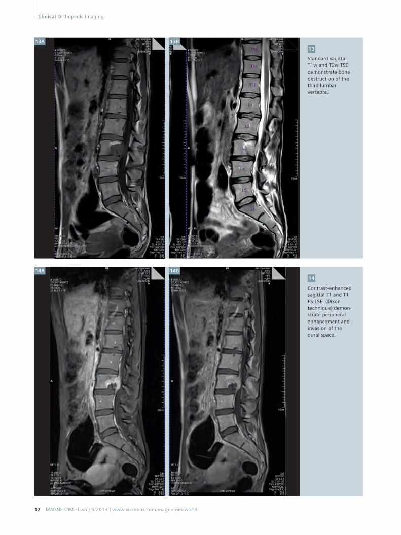
Standard sagittal T1w and T2w TSE demonstrate bone destruction of the third lumbar vertebra.
13
Contrast-enhanced sagittal T1 and T1 FS TSE (Dixon technique) demon-strate peripheral enhancement and invasion of the dural space.
14
13A
14A
13B
14B
12 MAGNETOM Flash | 5/2013 | www.siemens.com/magnetom-world
Clinical Orthopedic Imaging
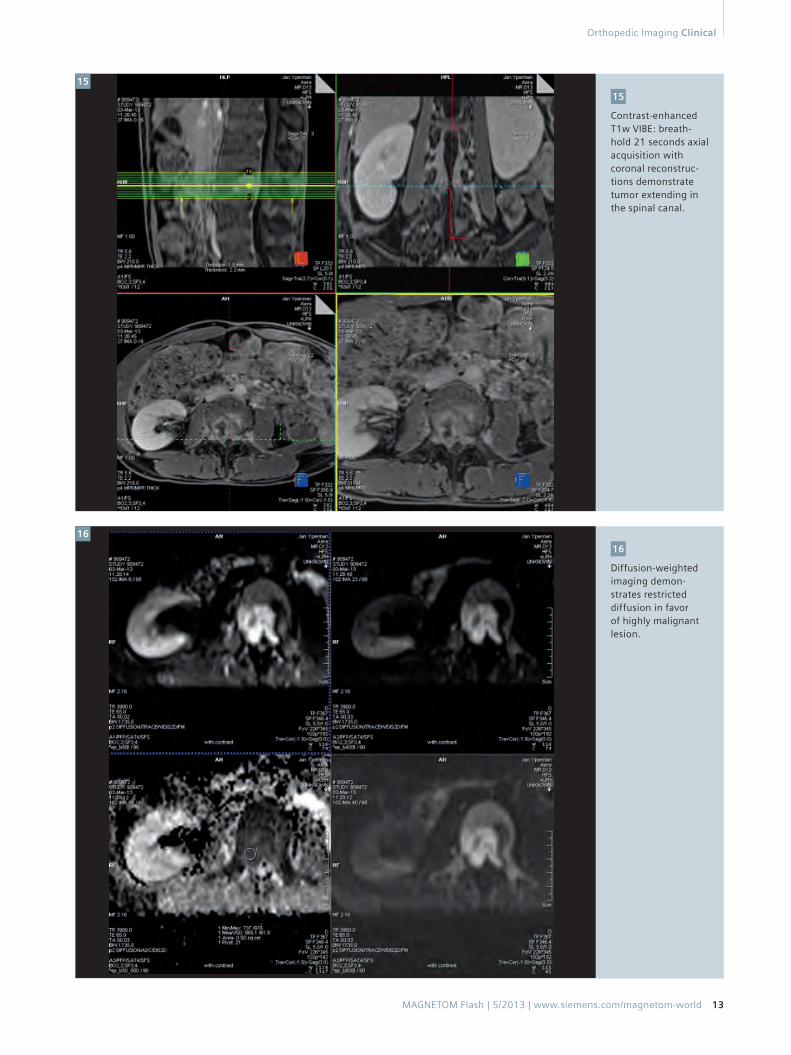
Contrast-enhanced T1w VIBE: breath-hold 21 seconds axial acquisition with coronal reconstruc-tions demonstrate tumor extending in the spinal canal.
15
Diffusion-weighted imaging demon-strates restricted diffusion in favor of highly malignant lesion.
16
15
16
MAGNETOM Flash | 5/2013 | www.siemens.com/magnetom-world 13
Orthopedic Imaging Clinical
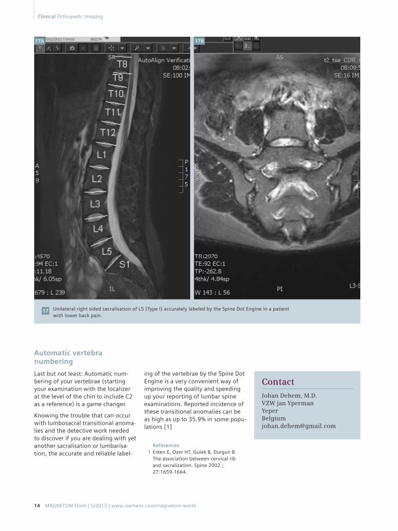
Automatic vertebra numbering
Last but not least: Automatic num-bering of your vertebrae (starting your examination with the localizer at the level of the chin to include C2 as a reference) is a game changer.
Knowing the trouble that can occur with lumbosacral transitional anoma-lies and the detective work needed to discover if you are dealing with yet another sacralisation or lumbarisa-tion, the accurate and reliable label-
Unilateral right sided sacralisation of L5 (Type I) accurately labeled by the Spine Dot Engine in a patient with lower back pain.
17
ing of the vertebrae by the Spine Dot Engine is a very convenient way of improving the quality and speeding up your reporting of lumbar spine examinations. Reported incidence of these transitional anomalies can be as high as up to 35.9% in some popu-lations [1]
Contact
Johan Dehem, M.D.VZW jan [email protected]
References1 Erken E, Ozer HT, Gulek B, Durgun B.
The association between cervical rib and sacralization. Spine 2002 ; 27:1659-1664.
17A 17B
14 MAGNETOM Flash | 5/2013 | www.siemens.com/magnetom-world
Clinical Orthopedic Imaging
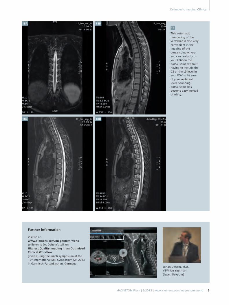
This automatic numbering of the vertebrae is also very convenient in the imaging of the dorsal spine where you can really focus your FOV on the dorsal spine without having to include the C2 or the L5 level in your FOV to be sure of your vertebral level. Scanning dorsal spine has become easy instead of tricky.
18
Further information
Visit us at www.siemens.com/magnetom-world to listen to Dr. Dehem’s talk on Highest Quality Imaging in an Optimized Clinical Workflow given during the lunch symposium at the 15th International MRI Symposium MR 2013 in Garmisch-Partenkirchen, Germany.
Johan Dehem, M.D. VZW Jan Yperman (Ieper, Belgium)
18A
18C
18B
18D
Orthopedic Imaging Clinical
MAGNETOM Flash | 5/2013 | www.siemens.com/magnetom-world 15
Related Documents
![[1] Magnetom Flash_Jan 2001](https://static.cupdf.com/doc/110x72/577cdc331a28ab9e78aa1b8e/1-magnetom-flashjan-2001.jpg)