ORIGINAL RESEARCH Open Access Longitudinal spectral domain optical coherence tomography changes in eyes with intraocular lymphoma Hyun Soo Jang 1 , Yasir J Sepah 1,2 , Raafay Sophie 1 , Millena G Bittencourt 1 , Daniel Ferraz 1,3 , Mostafa Hanout 1 , Hongting Liu 1 , Diana V Do 1,2 and Quan Dong Nguyen 1,2* Abstract Background: Cases of patients with primary intraocular lymphoma (PIOL) were retrospectively analyzed to describe the longitudinal intra-retinal morphological changes in PIOL as visualized on images obtained by spectral domain optical coherence tomography (SD-OCT). Results: In a retrospective case series, Heidelberg Spectralis SD-OCT images obtained in the longitudinal evaluation of patients with biopsy-proven PIOL were analyzed and assessed. The images were graded for the presence of macular edema (ME), pigment epithelial detachment (PED), subretinal fluid (SRF), and hyperreflective signals. SD-OCT scans of five eyes from five patients were assessed. Patients showed signs of inflammation, such as ME and SRF, which were resolved with treatments in some cases. Hyperreflective signals were found in all eyes in the form of nodules or bands across the retina, with the highest frequency of appearance in the ganglion cell layer, inner plexiform layer, photoreceptor layer, and retinal pigment epithelium; such signals increased with the progression of PIOL. Conclusion: SD-OCT may be employed to monitor the progression of PIOL. Hyperreflective signals on OCT may correspond with increase in disease activities, along with other findings such as ME, PED, and SRF. Keywords: Spectral domain optical coherence tomography (SD-OCT), Primary intraocular lymphoma, Primary retinal lymphoma, Primary vitreoretinal lymphoma Background Primary intraocular lymphoma (PIOL) is a subset of pri- mary central nervous system lymphoma (PCNSL). It is a rare non-Hodgkin's lymphoma that usually involves the retina and vitreous, mainly consisting of large B-cells. PIOL represents about 1% of non-Hodgkin's lymph- omas, 1% of intracranial tumors, and less than 1% of intraocular tumors [1]. It has been reported that 15% to 25% of PCNSL cases involve the eye; these may initially be present without concurrent central nervous system (CNS) involvement [2,3]. PIOL is the most common neoplastic disease that masquerades chronic posterior uveitis [4]. When mis- diagnosed as uveitis, patients may initially respond to cor- ticosteroids, which further contributes to the diagnostic challenge [2]. PIOL may also manifest in the anterior seg- ment as keratic precipitates, cells, and flare [5]. The identi- fication of atypical lymphoid cells in the eye is necessary to make the diagnosis [6,7]. Assessment of PIOL using ultrasonography, fluores- cein angiography, indocyanine green angiography, and optical coherence tomography has been reported with a common finding of intra- and sub-RPE (retinal pigment epithelium) infiltrates [2,8]. Additionally, a few case reports of PIOL have utilized spectral domain optical coherence tomography (SD-OCT) to capture high- resolution cross sections of the retina [9-11]. However, the common patterns of SD-OCT changes in PIOL have * Correspondence: [email protected] 1 Retinal Imaging Research and Reading Center, Wilmer Eye Institute, Johns Hopkins University School of Medicine, Baltimore, MD, USA 2 Stanley M. Truhlsen Eye Institute, University of Nebraska Medical Center, Omaha, NE, USA Full list of author information is available at the end of the article © 2013 Jang et al.; licensee Springer. This is an Open Access article distributed under the terms of the Creative Commons Attribution License (http://creativecommons.org/licenses/by/2.0), which permits unrestricted use, distribution, and reproduction in any medium, provided the original work is properly cited. Jang et al. Journal of Ophthalmic Inflammation and Infection 2013, 3:59 http://www.joii-journal.com/content/3/1/59

Welcome message from author
This document is posted to help you gain knowledge. Please leave a comment to let me know what you think about it! Share it to your friends and learn new things together.
Transcript
Jang et al. Journal of Ophthalmic Inflammation and Infection 2013, 3:59http://www.joii-journal.com/content/3/1/59
ORIGINAL RESEARCH Open Access
Longitudinal spectral domain optical coherencetomography changes in eyes with intraocularlymphomaHyun Soo Jang1, Yasir J Sepah1,2, Raafay Sophie1, Millena G Bittencourt1, Daniel Ferraz1,3, Mostafa Hanout1,Hongting Liu1, Diana V Do1,2 and Quan Dong Nguyen1,2*
Abstract
Background: Cases of patients with primary intraocular lymphoma (PIOL) were retrospectively analyzed to describethe longitudinal intra-retinal morphological changes in PIOL as visualized on images obtained by spectral domainoptical coherence tomography (SD-OCT).
Results: In a retrospective case series, Heidelberg Spectralis SD-OCT images obtained in the longitudinal evaluationof patients with biopsy-proven PIOL were analyzed and assessed. The images were graded for the presence ofmacular edema (ME), pigment epithelial detachment (PED), subretinal fluid (SRF), and hyperreflective signals.SD-OCT scans of five eyes from five patients were assessed. Patients showed signs of inflammation, such as ME andSRF, which were resolved with treatments in some cases. Hyperreflective signals were found in all eyes in the formof nodules or bands across the retina, with the highest frequency of appearance in the ganglion cell layer, innerplexiform layer, photoreceptor layer, and retinal pigment epithelium; such signals increased with the progression ofPIOL.
Conclusion: SD-OCT may be employed to monitor the progression of PIOL. Hyperreflective signals on OCT maycorrespond with increase in disease activities, along with other findings such as ME, PED, and SRF.
Keywords: Spectral domain optical coherence tomography (SD-OCT), Primary intraocular lymphoma, Primary retinallymphoma, Primary vitreoretinal lymphoma
BackgroundPrimary intraocular lymphoma (PIOL) is a subset of pri-mary central nervous system lymphoma (PCNSL). It is arare non-Hodgkin's lymphoma that usually involves theretina and vitreous, mainly consisting of large B-cells.PIOL represents about 1% of non-Hodgkin's lymph-omas, 1% of intracranial tumors, and less than 1% ofintraocular tumors [1]. It has been reported that 15% to25% of PCNSL cases involve the eye; these may initiallybe present without concurrent central nervous system(CNS) involvement [2,3].
* Correspondence: [email protected] Imaging Research and Reading Center, Wilmer Eye Institute, JohnsHopkins University School of Medicine, Baltimore, MD, USA2Stanley M. Truhlsen Eye Institute, University of Nebraska Medical Center,Omaha, NE, USAFull list of author information is available at the end of the article
© 2013 Jang et al.; licensee Springer. This is anAttribution License (http://creativecommons.orin any medium, provided the original work is p
PIOL is the most common neoplastic disease thatmasquerades chronic posterior uveitis [4]. When mis-diagnosed as uveitis, patients may initially respond to cor-ticosteroids, which further contributes to the diagnosticchallenge [2]. PIOL may also manifest in the anterior seg-ment as keratic precipitates, cells, and flare [5]. The identi-fication of atypical lymphoid cells in the eye is necessaryto make the diagnosis [6,7].Assessment of PIOL using ultrasonography, fluores-
cein angiography, indocyanine green angiography, andoptical coherence tomography has been reported with acommon finding of intra- and sub-RPE (retinal pigmentepithelium) infiltrates [2,8]. Additionally, a few casereports of PIOL have utilized spectral domain opticalcoherence tomography (SD-OCT) to capture high-resolution cross sections of the retina [9-11]. However,the common patterns of SD-OCT changes in PIOL have
Open Access article distributed under the terms of the Creative Commonsg/licenses/by/2.0), which permits unrestricted use, distribution, and reproductionroperly cited.
Jang et al. Journal of Ophthalmic Inflammation and Infection 2013, 3:59 Page 2 of 8http://www.joii-journal.com/content/3/1/59
not been established given the rarity of the disease andthe relative novelty of SD-OCT imaging. To the best ofour knowledge, progression of PIOL utilizing SD-OCThas been reported in one case report [11]. We hereinreport patterns of anatomic changes as visualized by SD-OCT in five patients of PIOL over a period of 161 ± 61days.
MethodsAn informed written consent that was approved by theInstitutional Review Board of Johns Hopkins Universitywas obtained from all patients included in the study.Patients with biopsy-proven PIOL who had been man-
aged by one of the authors (QDN) at the Wilmer EyeInstitute and followed using SD-OCT were includedin the index retrospective case series. Patients eitherreceived systemic high-dose methotrexate (MTX), sys-temic rituximab (RTX), and/or intravitreal MTX andRTX.For each patient, 30° SD-OCT scans of the macula
from three to four visits were analyzed carefully in theRetinal Imaging Research and Reading Center (RIRRC)at Wilmer, including scans at the first presentation ofsymptoms, a visit before treatment, and a post-treatmentvisit when available. The first visit was considered as thebaseline and was labeled Day 0. A senior grader selectedmultiple cross sections that were representative of theretinal changes in each case. These selected sectionswere evaluated by two independent graders in theRIRRC who were informed of the patients' diagnosis butmasked to the visits that were graded. Presence ofhyperreflective signals, macular edema (ME), pigmentepithelial detachment (PED), subretinal fluid (SRF), andany other abnormalities were assessed. The locations ofhyperreflective signals were marked as being present inthe following layers: nerve fiber layer (NFL), ganglioncell layer (GCL), inner plexiform layer (IPL), inner nu-clear layer (INL), outer plexiform layer (OPL), outer nu-clear layer (ONL), photoreceptor layer (PRL), RPE, andchoroid. Any disagreement between the two graders wasdecided by a third senior grader. The TruTrack™ activeeye tracking system in Spectralis SD-OCT allowed forcomparative analysis of the same cross sections betweenthe visits.
Table 1 Demographics of study subjects
Patient Age Gender Study eye Race
1 76 Male OD Caucasi
2 70 Male OS Caucasi
3 73 Male OS Caucasi
4 52 Female OD Caucasi
5 60 Female OS Caucasi
OD, right eye; OS, left eye; Time of observation, number of days between the basel
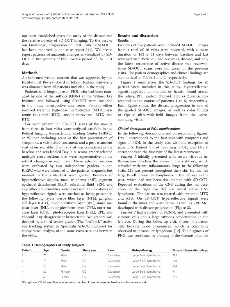
Results and discussionResultsFive eyes of five patients were included. SD-OCT imagesfrom a total of 16 visits were reviewed, with a meanduration of 163 ± 61 days between baseline and lastreviewed visit. Patient 5 had recurring disease, and onlythe latest recurrence of active disease was reviewed,since SD-OCT scans were not taken in the previousvisits. The patient demographics and clinical findings aresummarized in Tables 1 and 2, respectively.Figure 1 summarizes the SD-OCT findings for all
patient visits included in this study. Hyperreflectivesignals appeared as nodules or bands, found acrossthe retina, RPE, and/or choroid. Figures 2,3,4,5,6 cor-respond to the course of patients 1 to 5, respectively.Each figure shows the disease progression in one ofthe graded SD-OCT images, as well as color fundusor Optos® ultra-wide-field images from the corre-sponding visits.
Clinical description of PIOL manifestationIn the following descriptions and corresponding figures,Day 0 corresponds to the first visit with symptoms andsigns of PIOL in the study eye, with the exception ofpatient 5. Patient 5 had recurring PIOL, and Day 0corresponds to the first visit of the latest recurrence.Patient 1 initially presented with severe vitreous in-
flammation affecting the vision in the right eye, whichsubsided with anti-inflammatory drugs in the follow-upvisits. ME was present throughout the visits. He had hadlarge B-cell intraocular lymphoma in the left eye in thepast, which had not been documented with SD-OCT.Repeated evaluations of the CNS during the manifest-ation in the right eye did not reveal active CNSlymphoma. The patient was treated with systemic MTXand RTX. On SD-OCT, hyperreflective signals werefound in the inner and outer retina, as well as RPE. SRFdeveloped with disease progression (Figure 2).Patient 2 had a history of PCNSL and presented with
vitreous cells and a large vitreous condensation in theleft eye. During the follow-up visit, sheets of vitreouscells became more pronounced, which is commonlyobserved in intraocular lymphoma [12]. The diagnosis ofPIOL was confirmed by a biopsy of the vitreous obtained
Histopathology Time of observation (days)
an Large B-cell lymphoma 212
an Large B-cell lymphoma 119
an Large B-cell lymphoma 204
an Large B-cell lymphoma 77
an Large B-cell lymphoma 201
ine and last reviewed visit.
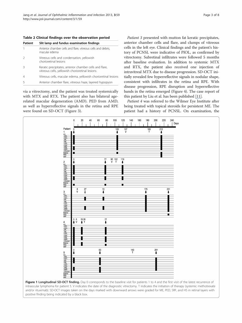
Table 2 Clinical findings over the observation period
Patient Slit lamp and fundus examination findings
1 Anterior chamber cells and flare, vitreous cells and debris,macular edema
2 Vitreous cells and condensation, yellowishchorioretinal lesions
3 Keratic precipitates, anterior chamber cells and flare,vitreous cells, yellowish chorioretinal lesions
4 Vitreous cells, macular edema, yellowish chorioretinal lesions
5 Anterior chamber flare, vitreous haze, layered hypopyon
Jang et al. Journal of Ophthalmic Inflammation and Infection 2013, 3:59 Page 3 of 8http://www.joii-journal.com/content/3/1/59
via a vitrectomy, and the patient was treated systemicallywith MTX and RTX. The patient also has bilateral age-related macular degeneration (AMD). PED from AMD,as well as hyperreflective signals in the retina and RPEwere found on SD-OCT (Figure 3).
Figure 1 Longitudinal SD-OCT finding. Day 0 corresponds to the baselinintraocular lymphoma for patient 5. V indicates the date of the diagnosticand/or rituximab). SD-OCT images taken on the days marked with downwapositive finding being indicated by a black box.
Patient 3 presented with mutton fat keratic precipitates,anterior chamber cells and flare, and clumps of vitreouscells in the left eye. Clinical findings and the patient's his-tory of PCNSL were indicative of PIOL, as confirmed byvitrectomy. Subretinal infiltrates were followed 5 monthsafter baseline evaluation. In addition to systemic MTXand RTX, the patient also received one injection ofintravitreal MTX due to disease progression. SD-OCT ini-tially revealed few hyperreflective signals in nodular shape,consistent with infiltrates in the retina and RPE. Withdisease progression, RPE disruption and hyperreflectivebands in the retina emerged (Figure 4). The case report ofthis patient by Liu et al. has been published [11].Patient 4 was referred to the Wilmer Eye Institute after
being treated with topical steroids for persistent ME. Thepatient had a history of PCNSL. On examination, the
e visit for patients 1 to 4 and the first visit of the latest recurrence ofvitrectomy. T indicates the initiation of therapy (systemic methotrexaterd arrows were graded for ME, PED, SRF, and HS in retinal layers with
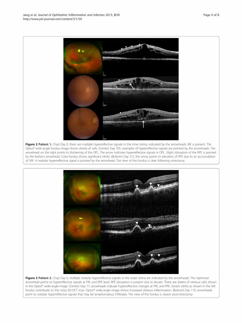
Figure 2 Patient 1. (Top) Day 0, there are multiple hyperreflective signals in the inner retina, indicated by the arrowheads. ME is present. TheOptos® wide-angle fundus image shows sheets of cells. (Center) Day 105, examples of hyperreflective signals are pointed by the arrowheads. Thearrowhead on the right points to thickening of the OPL. The arrow indicates hyperreflective signals in OPL. Slight disruption of the RPE is pointedby the bottom arrowhead. Color fundus shows significant vitritis. (Bottom) Day 212, the arrow points to elevation of RPE due to an accumulationof SRF. A nodular hyperreflective signal is pointed by the arrowhead. The view of the fundus is clear following vitrectomy.
Figure 3 Patient 2. (Top) Day 0, multiple nodular hyperreflective signals in the outer retina are indicated by the arrowheads. The rightmostarrowhead points to hyperreflective signals at PRL and RPE level. RPE disruption is present due to drusen. There are sheets of vitreous cells shownin the Optos® wide-angle image. (Center) Day 77, arrowheads indicate hyperreflective changes at PRL and RPE. Severe vitritis as shown in the leftfundus contributes to the noisy SD-OCT scan. Optos® wide-angle image shows increased vitreous inflammation. (Bottom) Day 119, arrowheadspoint to nodular hyperreflective signals that may be lymphomatous infiltrates. The view of the fundus is clearer post-vitrectomy.
Jang et al. Journal of Ophthalmic Inflammation and Infection 2013, 3:59 Page 4 of 8http://www.joii-journal.com/content/3/1/59
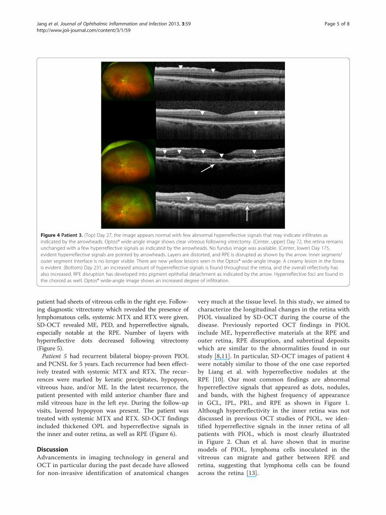
Figure 4 Patient 3. (Top) Day 27, the image appears normal with few abnormal hyperreflective signals that may indicate infiltrates asindicated by the arrowheads. Optos® wide-angle image shows clear vitreous following vitrectomy. (Center, upper) Day 72, the retina remainsunchanged with a few hyperreflective signals as indicated by the arrowheads. No fundus image was available. (Center, lower) Day 175,evident hyperreflective signals are pointed by arrowheads. Layers are distorted, and RPE is disrupted as shown by the arrow. Inner segment/outer segment interface is no longer visible. There are new yellow lesions seen in the Optos® wide-angle image. A creamy lesion in the foveais evident. (Bottom) Day 231, an increased amount of hyperreflective signals is found throughout the retina, and the overall reflectivity hasalso increased. RPE disruption has developed into pigment epithelial detachment as indicated by the arrow. Hyperreflective foci are found inthe choroid as well. Optos® wide-angle image shows an increased degree of infiltration.
Jang et al. Journal of Ophthalmic Inflammation and Infection 2013, 3:59 Page 5 of 8http://www.joii-journal.com/content/3/1/59
patient had sheets of vitreous cells in the right eye. Follow-ing diagnostic vitrectomy which revealed the presence oflymphomatous cells, systemic MTX and RTX were given.SD-OCT revealed ME, PED, and hyperreflective signals,especially notable at the RPE. Number of layers withhyperreflective dots decreased following vitrectomy(Figure 5).Patient 5 had recurrent bilateral biopsy-proven PIOL
and PCNSL for 5 years. Each recurrence had been effect-ively treated with systemic MTX and RTX. The recur-rences were marked by keratic precipitates, hypopyon,vitreous haze, and/or ME. In the latest recurrence, thepatient presented with mild anterior chamber flare andmild vitreous haze in the left eye. During the follow-upvisits, layered hypopyon was present. The patient wastreated with systemic MTX and RTX. SD-OCT findingsincluded thickened OPL and hyperreflective signals inthe inner and outer retina, as well as RPE (Figure 6).
DiscussionAdvancements in imaging technology in general andOCT in particular during the past decade have allowedfor non-invasive identification of anatomical changes
very much at the tissue level. In this study, we aimed tocharacterize the longitudinal changes in the retina withPIOL visualized by SD-OCT during the course of thedisease. Previously reported OCT findings in PIOLinclude ME, hyperreflective materials at the RPE andouter retina, RPE disruption, and subretinal depositswhich are similar to the abnormalities found in ourstudy [8,11]. In particular, SD-OCT images of patient 4were notably similar to those of the one case reportedby Liang et al. with hyperreflective nodules at theRPE [10]. Our most common findings are abnormalhyperreflective signals that appeared as dots, nodules,and bands, with the highest frequency of appearancein GCL, IPL, PRL, and RPE as shown in Figure 1.Although hyperreflectivity in the inner retina was notdiscussed in previous OCT studies of PIOL, we iden-tified hyperreflective signals in the inner retina of allpatients with PIOL, which is most clearly illustratedin Figure 2. Chan et al. have shown that in murinemodels of PIOL, lymphoma cells inoculated in thevitreous can migrate and gather between RPE andretina, suggesting that lymphoma cells can be foundacross the retina [13].
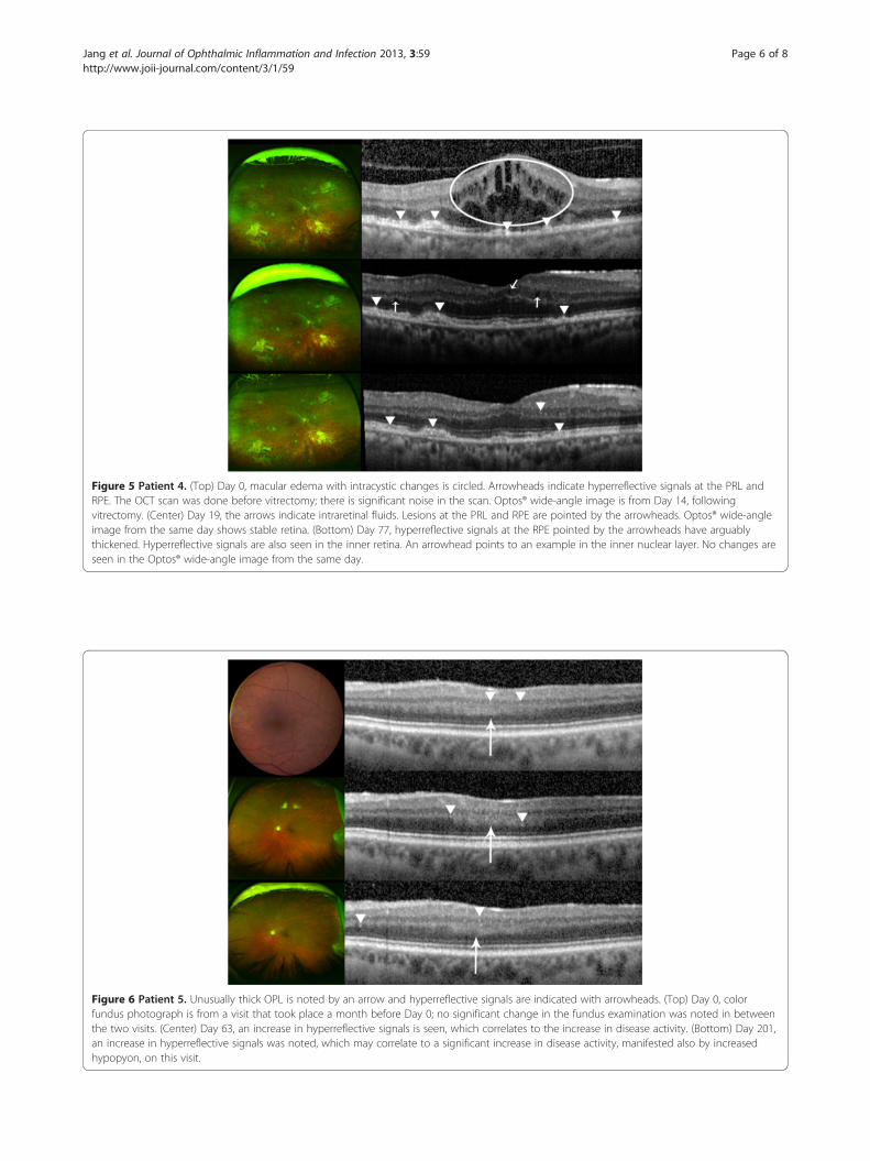
Figure 5 Patient 4. (Top) Day 0, macular edema with intracystic changes is circled. Arrowheads indicate hyperreflective signals at the PRL andRPE. The OCT scan was done before vitrectomy; there is significant noise in the scan. Optos® wide-angle image is from Day 14, followingvitrectomy. (Center) Day 19, the arrows indicate intraretinal fluids. Lesions at the PRL and RPE are pointed by the arrowheads. Optos® wide-angleimage from the same day shows stable retina. (Bottom) Day 77, hyperreflective signals at the RPE pointed by the arrowheads have arguablythickened. Hyperreflective signals are also seen in the inner retina. An arrowhead points to an example in the inner nuclear layer. No changes areseen in the Optos® wide-angle image from the same day.
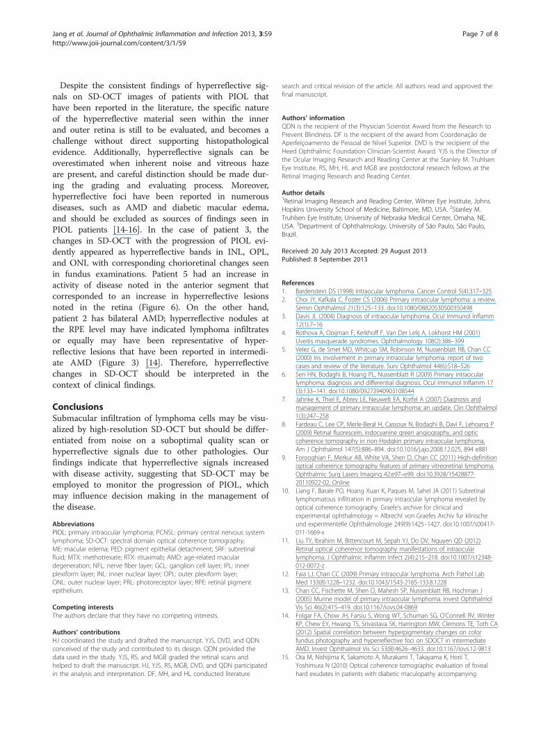
Figure 6 Patient 5. Unusually thick OPL is noted by an arrow and hyperreflective signals are indicated with arrowheads. (Top) Day 0, colorfundus photograph is from a visit that took place a month before Day 0; no significant change in the fundus examination was noted in betweenthe two visits. (Center) Day 63, an increase in hyperreflective signals is seen, which correlates to the increase in disease activity. (Bottom) Day 201,an increase in hyperreflective signals was noted, which may correlate to a significant increase in disease activity, manifested also by increasedhypopyon, on this visit.
Jang et al. Journal of Ophthalmic Inflammation and Infection 2013, 3:59 Page 6 of 8http://www.joii-journal.com/content/3/1/59
Jang et al. Journal of Ophthalmic Inflammation and Infection 2013, 3:59 Page 7 of 8http://www.joii-journal.com/content/3/1/59
Despite the consistent findings of hyperreflective sig-nals on SD-OCT images of patients with PIOL thathave been reported in the literature, the specific natureof the hyperreflective material seen within the innerand outer retina is still to be evaluated, and becomes achallenge without direct supporting histopathologicalevidence. Additionally, hyperreflective signals can beoverestimated when inherent noise and vitreous hazeare present, and careful distinction should be made dur-ing the grading and evaluating process. Moreover,hyperreflective foci have been reported in numerousdiseases, such as AMD and diabetic macular edema,and should be excluded as sources of findings seen inPIOL patients [14-16]. In the case of patient 3, thechanges in SD-OCT with the progression of PIOL evi-dently appeared as hyperreflective bands in INL, OPL,and ONL with corresponding chorioretinal changes seenin fundus examinations. Patient 5 had an increase inactivity of disease noted in the anterior segment thatcorresponded to an increase in hyperreflective lesionsnoted in the retina (Figure 6). On the other hand,patient 2 has bilateral AMD; hyperreflective nodules atthe RPE level may have indicated lymphoma infiltratesor equally may have been representative of hyper-reflective lesions that have been reported in intermedi-ate AMD (Figure 3) [14]. Therefore, hyperreflectivechanges in SD-OCT should be interpreted in thecontext of clinical findings.
ConclusionsSubmacular infiltration of lymphoma cells may be visu-alized by high-resolution SD-OCT but should be differ-entiated from noise on a suboptimal quality scan orhyperreflective signals due to other pathologies. Ourfindings indicate that hyperreflective signals increasedwith disease activity, suggesting that SD-OCT may beemployed to monitor the progression of PIOL, whichmay influence decision making in the management ofthe disease.
AbbreviationsPIOL: primary intraocular lymphoma; PCNSL: primary central nervous systemlymphoma; SD-OCT: spectral domain optical coherence tomography;ME: macular edema; PED: pigment epithelial detachment; SRF: subretinalfluid; MTX: methotrexate; RTX: rituximab; AMD: age-related maculardegeneration; NFL: nerve fiber layer; GCL: ganglion cell layer; IPL: innerplexiform layer; INL: inner nuclear layer; OPL: outer plexiform layer;ONL: outer nuclear layer; PRL: photoreceptor layer; RPE: retinal pigmentepithelium.
Competing interestsThe authors declare that they have no competing interests.
Authors’ contributionsHJ coordinated the study and drafted the manuscript. YJS, DVD, and QDNconceived of the study and contributed to its design. QDN provided thedata used in the study. YJS, RS, and MGB graded the retinal scans andhelped to draft the manuscript. HJ, YJS, RS, MGB, DVD, and QDN participatedin the analysis and interpretation. DF, MH, and HL conducted literature
search and critical revision of the article. All authors read and approved thefinal manuscript.
Authors’ informationQDN is the recipient of the Physician Scientist Award from the Research toPrevent Blindness. DF is the recipient of the award from Coordenação deAperfeiçoamento de Pessoal de Nível Superior. DVD is the recipient of theHeed Ophthalmic Foundation Clinician-Scientist Award. YJS is the Director ofthe Ocular Imaging Research and Reading Center at the Stanley M. TruhlsenEye Institute. RS, MH, HL and MGB are postdoctoral research fellows at theRetinal Imaging Research and Reading Center.
Author details1Retinal Imaging Research and Reading Center, Wilmer Eye Institute, JohnsHopkins University School of Medicine, Baltimore, MD, USA. 2Stanley M.Truhlsen Eye Institute, University of Nebraska Medical Center, Omaha, NE,USA. 3Department of Ophthalmology, University of São Paulo, São Paulo,Brazil.
Received: 20 July 2013 Accepted: 29 August 2013Published: 8 September 2013
References1. Bardenstein DS (1998) Intraocular lymphoma. Cancer Control 5(4):317–3252. Choi JY, Kafkala C, Foster CS (2006) Primary intraocular lymphoma: a review.
Semin Ophthalmol 21(3):125–133. doi:10.1080/088205305003504983. Davis JL (2004) Diagnosis of intraocular lymphoma. Ocul Immunol Inflamm
12(1):7–164. Rothova A, Ooijman F, Kerkhoff F, Van Der Lelij A, Lokhorst HM (2001)
Uveitis masquerade syndromes. Ophthalmology 108(2):386–3995. Velez G, de Smet MD, Whitcup SM, Robinson M, Nussenblatt RB, Chan CC
(2000) Iris involvement in primary intraocular lymphoma: report of twocases and review of the literature. Surv Ophthalmol 44(6):518–526
6. Sen HN, Bodaghi B, Hoang PL, Nussenblatt R (2009) Primary intraocularlymphoma: diagnosis and differential diagnosis. Ocul Immunol Inflamm 17(3):133–141. doi:10.1080/09273940903108544
7. Jahnke K, Thiel E, Abrey LE, Neuwelt EA, Korfel A (2007) Diagnosis andmanagement of primary intraocular lymphoma: an update. Clin Ophthalmol1(3):247–258
8. Fardeau C, Lee CP, Merle-Beral H, Cassoux N, Bodaghi B, Davi F, Lehoang P(2009) Retinal fluorescein, indocyanine green angiography, and opticcoherence tomography in non-Hodgkin primary intraocular lymphoma.Am J Ophthalmol 147(5):886–894. doi:10.1016/j.ajo.2008.12.025, 894 e881
9. Forooghian F, Merkur AB, White VA, Shen D, Chan CC (2011) High-definitionoptical coherence tomography features of primary vitreoretinal lymphoma.Ophthalmic Surg Lasers Imaging 42:e97–e99. doi:10.3928/15428877-20110922-02, Online
10. Liang F, Barale PO, Hoang Xuan K, Paques M, Sahel JA (2011) Subretinallymphomatous infiltration in primary intraocular lymphoma revealed byoptical coherence tomography. Graefe's archive for clinical andexperimental ophthalmology = Albrecht von Graefes Archiv fur klinischeund experimentelle Ophthalmologie 249(9):1425–1427. doi:10.1007/s00417-011-1669-x
11. Liu TY, Ibrahim M, Bittencourt M, Sepah YJ, Do DV, Nguyen QD (2012)Retinal optical coherence tomography manifestations of intraocularlymphoma. J Ophthalmic Inflamm Infect 2(4):215–218. doi:10.1007/s12348-012-0072-z
12. Faia LJ, Chan CC (2009) Primary intraocular lymphoma. Arch Pathol LabMed 133(8):1228–1232. doi:10.1043/1543-2165-133.8.1228
13. Chan CC, Fischette M, Shen D, Mahesh SP, Nussenblatt RB, Hochman J(2005) Murine model of primary intraocular lymphoma. Invest OphthalmolVis Sci 46(2):415–419. doi:10.1167/iovs.04-0869
14. Folgar FA, Chow JH, Farsiu S, Wong WT, Schuman SG, O'Connell RV, WinterKP, Chew EY, Hwang TS, Srivastava SK, Harrington MW, Clemons TE, Toth CA(2012) Spatial correlation between hyperpigmentary changes on colorfundus photography and hyperreflective foci on SDOCT in intermediateAMD. Invest Ophthalmol Vis Sci 53(8):4626–4633. doi:10.1167/iovs.12-9813
15. Ota M, Nishijima K, Sakamoto A, Murakami T, Takayama K, Horii T,Yoshimura N (2010) Optical coherence tomographic evaluation of fovealhard exudates in patients with diabetic maculopathy accompanying

Jang et al. Journal of Ophthalmic Inflammation and Infection 2013, 3:59 Page 8 of 8http://www.joii-journal.com/content/3/1/59
macular detachment. Ophthalmology 117(10):1996–2002. doi:10.1016/j.ophtha.2010.06.019
16. Bolz M, Schmidt-Erfurth U, Deak G, Mylonas G, Kriechbaum K, Scholda C(2009) Optical coherence tomographic hyperreflective foci: a morphologicsign of lipid extravasation in diabetic macular edema. Ophthalmology 116(5):914–920. doi:10.1016/j.ophtha.2008.12.039
doi:10.1186/1869-5760-3-59Cite this article as: Jang et al.: Longitudinal spectral domain opticalcoherence tomography changes in eyes with intraocular lymphoma.Journal of Ophthalmic Inflammation and Infection 2013 3:59.
Submit your manuscript to a journal and benefi t from:
7 Convenient online submission
7 Rigorous peer review
7 Immediate publication on acceptance
7 Open access: articles freely available online
7 High visibility within the fi eld
7 Retaining the copyright to your article
Submit your next manuscript at 7 springeropen.com
Related Documents