Lipid and Lipoprotein Basics Thomas Dayspring MD, FACP, FNLA Understanding atherosclerosis necessitates knowledge of various lipids (especially sterols and fatty acids) and their homeostasis, including how they are synthesized, incorporated into and transported or trafficked within lipoproteins. Since atherogenesis is a lipoprotein mediated disease, it is essential to review their synthesis, structure (surface protein and lipid composition), catabolism, kinetics (how they interact with each other and the arterial wall) and ability to be influenced by other metabolites (such as glucose, reactive oxygen species, etc.). Understanding lipoproteins also requires a realization that there is a constant, continually on-going, dynamic, flow and remodeling of particles where lipid molecules and apoproteins are gained and lost and acquired through complex pathways involving catabolism (lipolysis) and interchange between particles. Simply stated, lipoproteins and their lipid content are in a continuous state of constant dynamic flux. This can be very underappreciated by looking at concentrations reported in a standard lipid panel. The more we understand this, the more we can begin to understand the etiology of atherogenesis and the far better we can understand how to influence lipoprotein pathobiology using lifestyle and pharmacological methods. Of prime importance is to know what laboratory tests best help us identify and properly diagnose lipoprotein structure, composition and kinetics and then relate those parameters to risk. There are many ways of assaying lipid and lipoprotein concentrations and characteristics: particle numbers, particle sizes, particle lipid content, particle apolipoprotein content, etc. Where the subject gets complex and where real world clinicians cannot go at this time is understanding the functionality of the lipoproteins (are they doing what they are supposed to be): exactly where do the particles lipidate, and where do the delipidate and do they have normal catabolism (lipolysis): is it the liver, the jejunum, some peripheral tissue, or the artery intima? Are the sterols, entering the artery or being eliminated as fecal sterols (cholesterol or bile acid)? Before analyzing the effects of drugs on lipids, one must understand lipid biochemistry and nomenclature. Lipids are organic molecules used for energy and structural physiologic functions and broadly defined are simply molecules that are not or partially soluble in water. Polar molecules are soluble in water or aqueous solutions and nonpolar ones are not. Some lipids, called amphipathic molecules have unique structures where one end is polar and the other is not: at least the polar end would have some water solubility, the other end would not. Such molecules are essential for the structure lipid monolayers (lipoproteins and micelles) and lipid bilayers (cell membranes).

Welcome message from author
This document is posted to help you gain knowledge. Please leave a comment to let me know what you think about it! Share it to your friends and learn new things together.
Transcript
Lipid and Lipoprotein Basics Thomas Dayspring MD, FACP, FNLA
Understanding atherosclerosis necessitates knowledge of various lipids (especially sterols and fatty acids) and their homeostasis, including how they are synthesized, incorporated into and transported or trafficked within lipoproteins. Since atherogenesis is a lipoprotein mediated disease, it is essential to review their synthesis, structure (surface protein and lipid composition), catabolism, kinetics (how they interact with each other and the arterial wall) and ability to be influenced by other metabolites (such as glucose, reactive oxygen species, etc.). Understanding lipoproteins also requires a realization that there is a constant, continually on-going, dynamic, flow and remodeling of particles where lipid molecules and apoproteins are gained and lost and acquired through complex pathways involving catabolism (lipolysis) and interchange between particles. Simply stated, lipoproteins and their lipid content are in a continuous state of constant dynamic flux. This can be very underappreciated by looking at concentrations reported in a standard lipid panel. The more we understand this, the more we can begin to understand the etiology of atherogenesis and the far better we can understand how to influence lipoprotein pathobiology using lifestyle and pharmacological methods. Of prime importance is to know what laboratory tests best help us identify and properly diagnose lipoprotein structure, composition and kinetics and then relate those parameters to risk. There are many ways of assaying lipid and lipoprotein concentrations and characteristics: particle numbers, particle sizes, particle lipid content, particle apolipoprotein content, etc. Where the subject gets complex and where real world clinicians cannot go at this time is understanding the functionality of the lipoproteins (are they doing what they are supposed to be): exactly where do the particles lipidate, and where do the delipidate and do they have normal catabolism (lipolysis): is it the liver, the jejunum, some peripheral tissue, or the artery intima? Are the sterols, entering the artery or being eliminated as fecal sterols (cholesterol or bile acid)? Before analyzing the effects of drugs on lipids, one must understand lipid biochemistry and nomenclature. Lipids are organic molecules used for energy and structural physiologic functions and broadly defined are simply molecules that are not or partially soluble in water. Polar molecules are soluble in water or aqueous solutions and nonpolar ones are not. Some lipids, called amphipathic molecules have unique structures where one end is polar and the other is not: at least the polar end would have some water solubility, the other end would not. Such molecules are essential for the structure lipid monolayers (lipoproteins and micelles) and lipid bilayers (cell membranes).
Lipid and Lipoprotein Basics Thomas Dayspring MD, FACP, FNLA
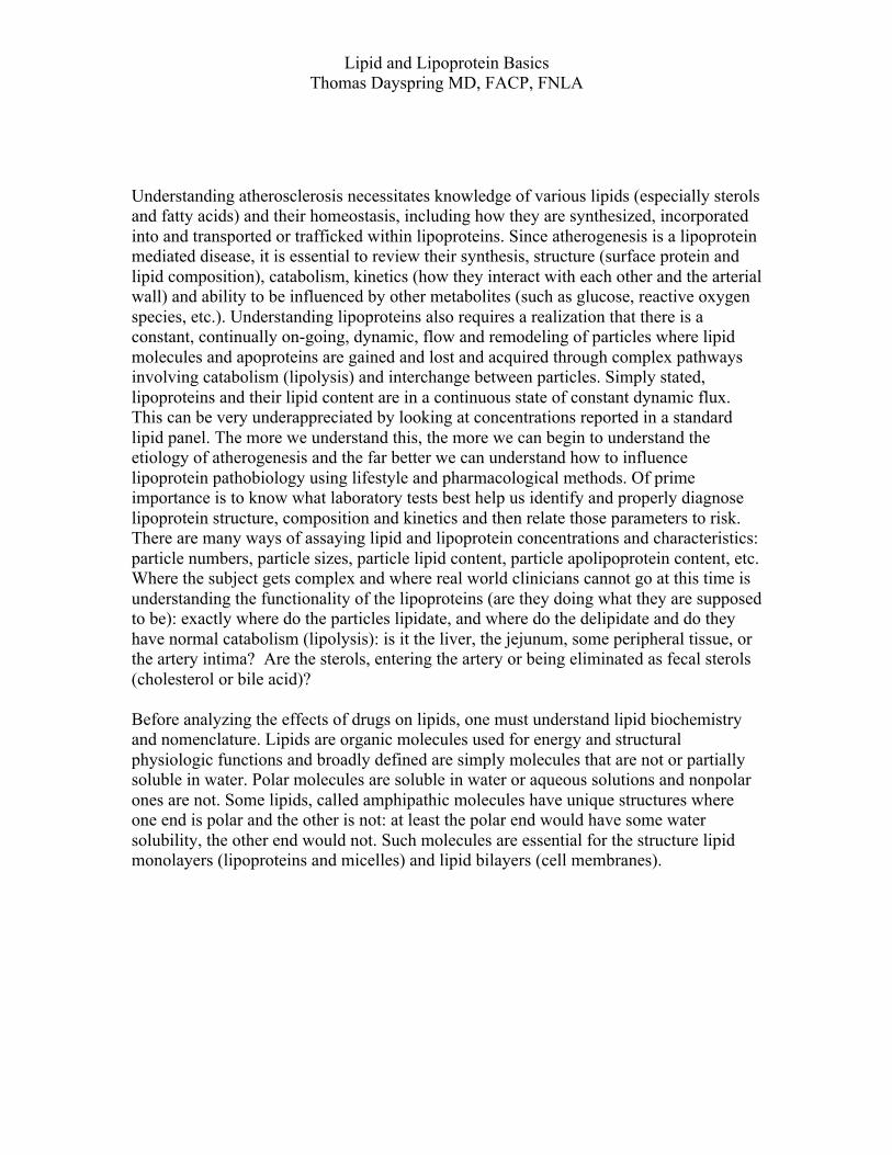
TG and PL are the major molecules in which fatty acids (FA) as well as phosphorus are delivered to tissues. Plasma PL not found on lipoproteins are bound to phospholipid transfer protein (PLTP) and plasma FA not found in PL or TG are bound to albumin and referred to as free fatty acid or nonesterified fatty acids. TG molecules contain 3 FA and PL 2 FA per molecule.
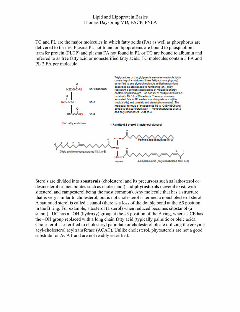
Sterols are divided into zoosterols (cholesterol and its precursors such as lathosterol or desmosterol or metabolites such as cholestanol) and phytosterols (several exist, with sitosterol and campesterol being the most common). Any molecule that has a structure that is very similar to cholesterol, but is not cholesterol is termed a noncholesterol sterol. A saturated sterol is called a stanol (there is a loss of the double bond at the Δ5 position in the B ring. For example, sitosterol (a sterol) when reduced becomes sitostanol (a stanol). UC has a –OH (hydroxy) group at the #3 position of the A ring, whereas CE has the –OH group replaced with a long chain fatty acid (typically palmitic or oleic acid). Cholesterol is esterified to cholesteryl palmitate or cholesterol oleate utilizing the enzyme acyl-cholesterol acyltransferase (ACAT). Unlike cholesterol, phytosterols are not a good substrate for ACAT and are not readily esterified.
Lipid and Lipoprotein Basics Thomas Dayspring MD, FACP, FNLA
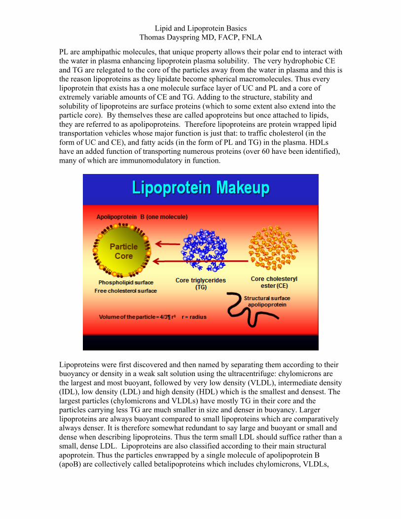
Lipoproteins are simply protein enwrapped lipid transportation vehicles which acquire (lipidate) several lipids namely UC, CE, noncholesterol sterols, TG and PL and transport them to and from various tissues. Biochemically speaking, lipoproteins are complex structures consisting of a central core of nonpolar (hydrophobic) TG and CE surrounded by a one molecule thick monolayer where UC is interspaced with PL. Since both UC and
Lipid and Lipoprotein Basics Thomas Dayspring MD, FACP, FNLA
PL are amphipathic molecules, that unique property allows their polar end to interact with the water in plasma enhancing lipoprotein plasma solubility. The very hydrophobic CE and TG are relegated to the core of the particles away from the water in plasma and this is the reason lipoproteins as they lipidate become spherical macromolecules. Thus every lipoprotein that exists has a one molecule surface layer of UC and PL and a core of extremely variable amounts of CE and TG. Adding to the structure, stability and solubility of lipoproteins are surface proteins (which to some extent also extend into the particle core). By themselves these are called apoproteins but once attached to lipids, they are referred to as apolipoproteins. Therefore lipoproteins are protein wrapped lipid transportation vehicles whose major function is just that: to traffic cholesterol (in the form of UC and CE), and fatty acids (in the form of PL and TG) in the plasma. HDLs have an added function of transporting numerous proteins (over 60 have been identified), many of which are immunomodulatory in function.
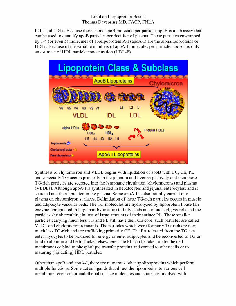
Lipoproteins were first discovered and then named by separating them according to their buoyancy or density in a weak salt solution using the ultracentrifuge: chylomicrons are the largest and most buoyant, followed by very low density (VLDL), intermediate density (IDL), low density (LDL) and high density (HDL) which is the smallest and densest. The largest particles (chylomicrons and VLDLs) have mostly TG in their core and the particles carrying less TG are much smaller in size and denser in buoyancy. Larger lipoproteins are always buoyant compared to small lipoproteins which are comparatively always denser. It is therefore somewhat redundant to say large and buoyant or small and dense when describing lipoproteins. Thus the term small LDL should suffice rather than a small, dense LDL. Lipoproteins are also classified according to their main structural apoprotein. Thus the particles enwrapped by a single molecule of apolipoprotein B (apoB) are collectively called betalipoproteins which includes chylomicrons, VLDLs,
Lipid and Lipoprotein Basics Thomas Dayspring MD, FACP, FNLA
IDLs and LDLs. Because there is one apoB molecule per particle, apoB is a lab assay that can be used to quantify apoB particles per deciliter of plasma. Those particles enwrapped by 1-4 (or even 5) molecules of apolipoprotein A-I (apoA-I) are the alphalipoproteins or HDLs. Because of the variable numbers of apoA-I molecules per particle, apoA-I is only an estimate of HDL particle concentration (HDL-P).
Synthesis of chylomicron and VLDL begins with lipidation of apoB with UC, CE, PL and especially TG occurs primarily in the jejunum and liver respectively and then these TG-rich particles are secreted into the lymphatic circulation (chylomicrons) and plasma (VLDLs). Although apoA-I is synthesized in hepatocytes and jejunal enterocytes, and is secreted and then lipidated in the plasma. Some apoA-I is also initially carried into plasma on chylomicron surfaces. Delipidation of these TG-rich particles occurs in muscle and adipocyte vascular beds. The TG molecules are hydrolyzed by lipoprotein lipase (an enzyme upregulated in large part by insulin) to fatty acids and monoacylglycerols and the particles shrink resulting in loss of large amounts of their surface PL. These smaller particles carrying much less TG and PL still have their CE core: such particles are called VLDL and chylomicron remnants. The particles which were formerly TG-rich are now much less TG-rich and are trafficking primarily CE. The FA released from the TG can enter myocytes to be oxidized for energy or enter adipocytes and be reconverted to TG or bind to albumin and be trafficked elsewhere. The PL can be taken up by the cell membranes or bind to phospholipid transfer proteins and carried to other cells or to maturing (lipidating) HDL particles. Other than apoB and apoA-I, there are numerous other apolipoproteins which perform multiple functions. Some act as ligands that direct the lipoproteins to various cell membrane receptors or endothelial surface molecules and some are involved with
Lipid and Lipoprotein Basics Thomas Dayspring MD, FACP, FNLA
activation or inactivation of various lipolytic enzymes such as lipoprotein lipase and other enzymes. Many of the apoproteins exist as different genetically determined isoforms, which create individual and population differences in lipoprotein metabolism. Many of the apoproteins can be modulated by drugs in ways which might enhance or retard what the lipoprotein is doing. There is much confusion with lipoprotein terminology. For example LDL pattern A (larger LDL particle) has nothing to do with lipoprotein little a [Lp(a)] or lipoprotein capital A [LP:A]. LP:A is a lipoprotein with a member of the apoA family attached. We know of apoA-I, A-II, A-IV and A-V. ApoA has nothing to do with apo(a) or pattern A LDL. LP:A-I is an HDL carrying only apoA-I, but LP:A-I,A-II would be an HDL carrying apoA-I and apoA-II. Likewise apoB is different than LDL pattern B (which refers to small LDL particles). Any lipoprotein that has apoB on its surface is termed a betalipoprotein and abbreviated as LP:B. Those with apoA-I are usually alphalipoproteins, but a chylomicron which is an apoB-containing particle also carries apoA-I and could be referred to as LP:B,A-I. No HDL particle ever carries apoB and thus all HDL nomenclature starts with LP:A. If a beta-lipoprotein also carries apoC-III it is termed LP:B-C-III. An alpha-lipoprotein that has apoA-I, A-II, C-III, D on its surface is termed LP:A-I,A-II, C-III,D. A VLDL with apoB, apoA-II, apo, C-I,C-II, C-III and apoE would be LP:B,A-II,C-I,C-II,C-III,E. Since most LDLs only carry apoB and no other apoproteins, they are simply LP:B BETALIPOPROTEINS: Decades ago before anyone knew what an apoprotein was; particles were separated by their buoyancy in the ultracentrifuge. As electrophoresis came into play as a way to separate lipoproteins it was obvious that there was a particle very different than the large TG-rich particles and since it migrated with beta-proteins it was called the beta (B) particle and it carried not TG but mostly cholesterol. Of course this turned out to be the LDL particle and measuring it was and is called betaquantification. This was and unfortunately still is often considered by some as the standard of quantitating LDL particles. Unfortunately betaquantification using the preparative ultracentrifuge to separate LDLs (and then measure their cholesterol) is extremely tedious and not available as a real world tool. As much as this is considered a religious measurement betaquantification has never be used in a clinical outcome trial whereas other lab assays used to measure LDL, namely LDL-C, non-HDL-C, apoB and LDL-P have. A long time ago betalipoproteins meant LDLs, but now the term is routinely used to collectively refer to all of the apoB-containing particles. There are actually two types of apo B: either B48 (chylomicron or intestinal origin) or B100 (liver origin). ApoB48 has 48% of the molecular weight of apoB100 (it is a truncated apoB). The apoB containing lipoproteins are all potentially atherogenic because once in the arterial intima, apoB adheres to arterial wall proteoglycans, where the particles are susceptible to modification by reactive oxygen species and lipoprotein associated phospholipase A2 (Lp-PLA2). Once modified, they are internalized by macrophages, creating foam cells, the hallmark of atherosclerosis. ApoB100 is recognized by the hepatic LDL receptor (LDLr) whereas apoB48 is not. Betalipoproteins can be collectively quantified by measuring apoB or individually quantified via nuclear

Lipid and Lipoprotein Basics Thomas Dayspring MD, FACP, FNLA
magnetic resonance spectroscopy (NMR LipoProfile performed by LipoScience). Directly measured apoB levels are a powerful measure of how many atherogenic lipoproteins are in a deciliter (dL) of plasma. Although one labs does it, apoB cannot be calculated (the formulas used to do so have never been published in peer reviewed lab journals). Because of apoB-particle half-life differences, > 90% of measured apoB represents LDL-P (LDLs have an extended half life of 2-3 days unlike the very short half life of chylomicrons, VLDL and IDLs). CHYLOMICRONS: large TG-rich lipoproteins formed in the intestine. Intestinally absorbed sterols and fatty acids are esterified and attached to apoB48 and form chylomicrons. More apolipoproteins are acquired during synthesis or as the particles travel through the lymphatic system before or after entering the plasma (C-II, C-III, E and others) from HDL. Chylomicrons rapidly undergo lipolysis (hydrolysis of triglycerides into fatty acids by lipoprotein lipase) and form smaller particles called chylomicron remnants. A protein crucial for chylomicron lipolysis by LPL is glycosylphosphatidylinositol-anchored high-density lipoprotein-binding protein 1 (GPIHBP1). The remnants are internalized by hepatic LDL receptors (LDLr) and LDL receptor related protein (LRP) using apoE as a ligand. A chylomicron half-life is less than an hour and most are gone in minutes. If chylomicrons are not rapidly cleared the patient will have severe postprandial and fasting lipemia (hypertriglyceridemia).
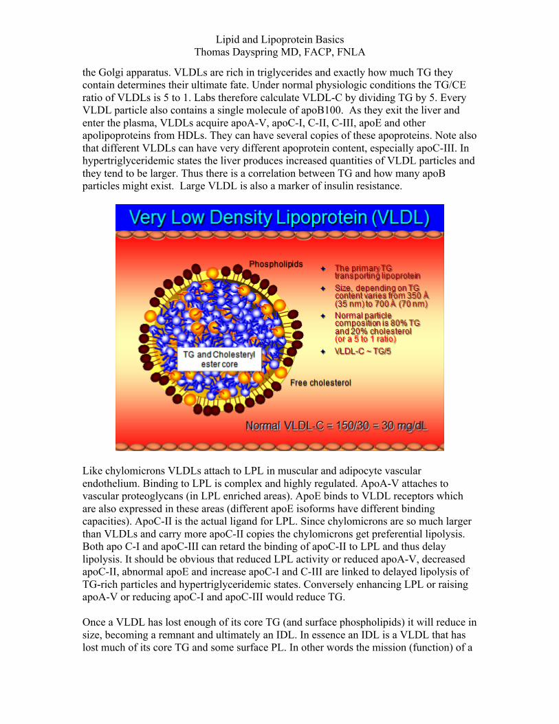
VLDLs (very low density lipoproteins) are synthesized in the liver. Their diameter varies from 40-200 nm, depending on their core lipid, especially TG, content. In the liver, TG and sterols (mostly cholesteryl ester) attach to apoB 100 utilizing microsomal triglyceride transfer protein (MTP). Ultimately they are phospholipidated in
Lipid and Lipoprotein Basics Thomas Dayspring MD, FACP, FNLA
the Golgi apparatus. VLDLs are rich in triglycerides and exactly how much TG they contain determines their ultimate fate. Under normal physiologic conditions the TG/CE ratio of VLDLs is 5 to 1. Labs therefore calculate VLDL-C by dividing TG by 5. Every VLDL particle also contains a single molecule of apoB100. As they exit the liver and enter the plasma, VLDLs acquire apoA-V, apoC-I, C-II, C-III, apoE and other apolipoproteins from HDLs. They can have several copies of these apoproteins. Note also that different VLDLs can have very different apoprotein content, especially apoC-III. In hypertriglyceridemic states the liver produces increased quantities of VLDL particles and they tend to be larger. Thus there is a correlation between TG and how many apoB particles might exist. Large VLDL is also a marker of insulin resistance.
Like chylomicrons VLDLs attach to LPL in muscular and adipocyte vascular endothelium. Binding to LPL is complex and highly regulated. ApoA-V attaches to vascular proteoglycans (in LPL enriched areas). ApoE binds to VLDL receptors which are also expressed in these areas (different apoE isoforms have different binding capacities). ApoC-II is the actual ligand for LPL. Since chylomicrons are so much larger than VLDLs and carry more apoC-II copies the chylomicrons get preferential lipolysis. Both apo C-I and apoC-III can retard the binding of apoC-II to LPL and thus delay lipolysis. It should be obvious that reduced LPL activity or reduced apoA-V, decreased apoC-II, abnormal apoE and increase apoC-I and C-III are linked to delayed lipolysis of TG-rich particles and hypertriglyceridemic states. Conversely enhancing LPL or raising apoA-V or reducing apoC-I and apoC-III would reduce TG. Once a VLDL has lost enough of its core TG (and surface phospholipids) it will reduce in size, becoming a remnant and ultimately an IDL. In essence an IDL is a VLDL that has lost much of its core TG and some surface PL. In other words the mission (function) of a
Lipid and Lipoprotein Basics Thomas Dayspring MD, FACP, FNLA
VLDL (and chylomicrons) is to traffic TG (energy in the form of FA) to muscle cells for use or to adipocytes for storage as well as to deliver PL to cells. VLDLs do not deliver cholesterol to the periphery: it gets passed onto their lipolytic end-product the IDLs.
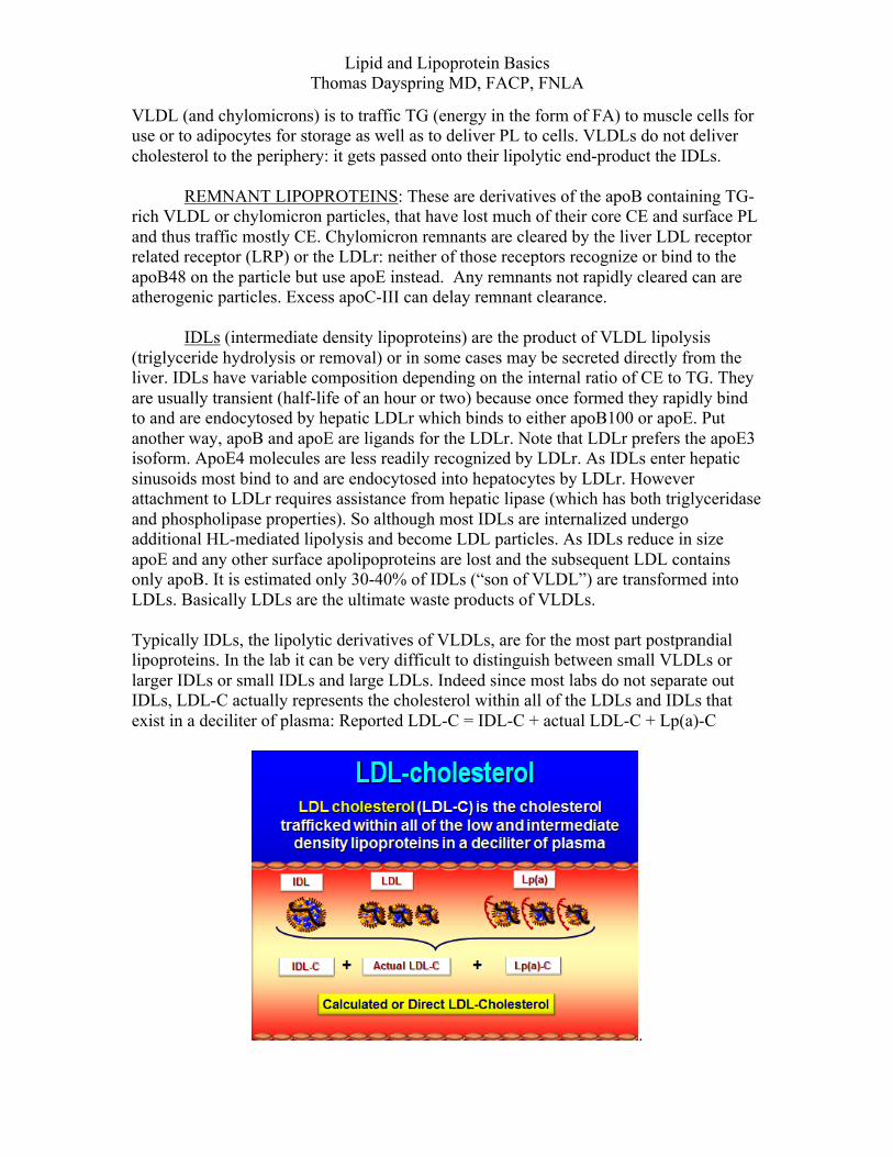
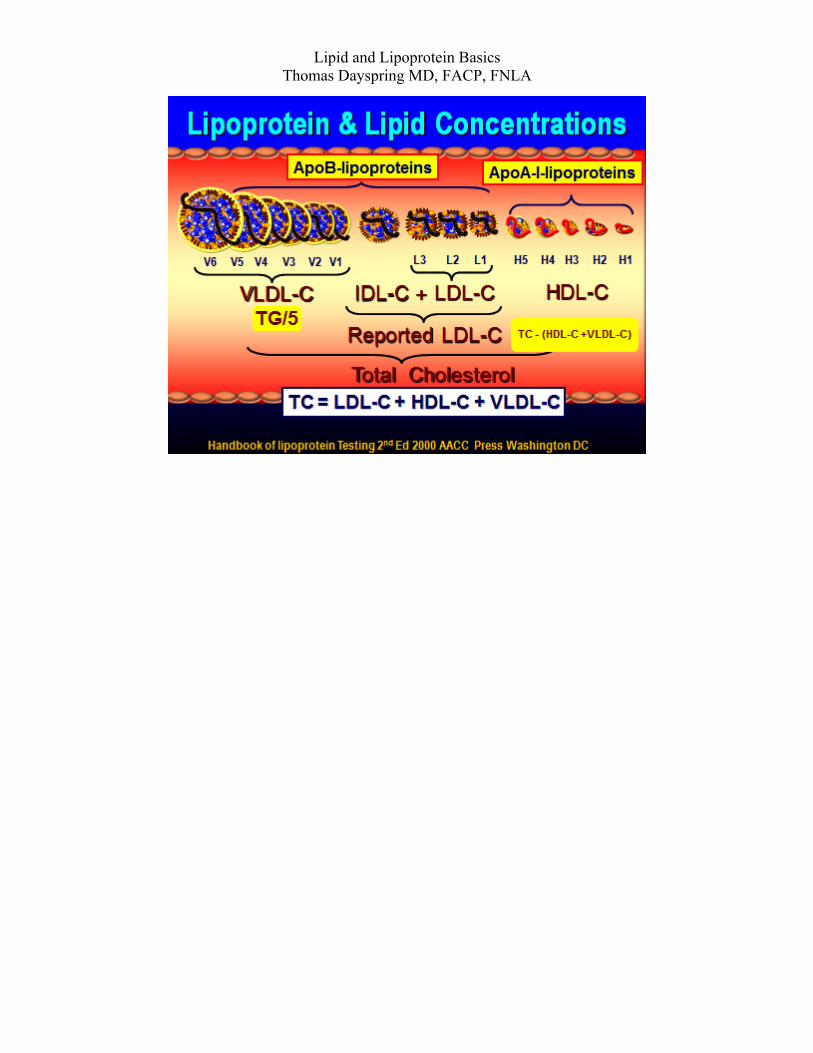
REMNANT LIPOPROTEINS: These are derivatives of the apoB containing TG-rich VLDL or chylomicron particles, that have lost much of their core CE and surface PL and thus traffic mostly CE. Chylomicron remnants are cleared by the liver LDL receptor related receptor (LRP) or the LDLr: neither of those receptors recognize or bind to the apoB48 on the particle but use apoE instead. Any remnants not rapidly cleared can are atherogenic particles. Excess apoC-III can delay remnant clearance. IDLs (intermediate density lipoproteins) are the product of VLDL lipolysis (triglyceride hydrolysis or removal) or in some cases may be secreted directly from the liver. IDLs have variable composition depending on the internal ratio of CE to TG. They are usually transient (half-life of an hour or two) because once formed they rapidly bind to and are endocytosed by hepatic LDLr which binds to either apoB100 or apoE. Put another way, apoB and apoE are ligands for the LDLr. Note that LDLr prefers the apoE3 isoform. ApoE4 molecules are less readily recognized by LDLr. As IDLs enter hepatic sinusoids most bind to and are endocytosed into hepatocytes by LDLr. However attachment to LDLr requires assistance from hepatic lipase (which has both triglyceridase and phospholipase properties). So although most IDLs are internalized undergo additional HL-mediated lipolysis and become LDL particles. As IDLs reduce in size apoE and any other surface apolipoproteins are lost and the subsequent LDL contains only apoB. It is estimated only 30-40% of IDLs (“son of VLDL”) are transformed into LDLs. Basically LDLs are the ultimate waste products of VLDLs. Typically IDLs, the lipolytic derivatives of VLDLs, are for the most part postprandial lipoproteins. In the lab it can be very difficult to distinguish between small VLDLs or larger IDLs or small IDLs and large LDLs. Indeed since most labs do not separate out IDLs, LDL-C actually represents the cholesterol within all of the LDLs and IDLs that exist in a deciliter of plasma: Reported LDL-C = IDL-C + actual LDL-C + Lp(a)-C
.
Lipid and Lipoprotein Basics Thomas Dayspring MD, FACP, FNLA
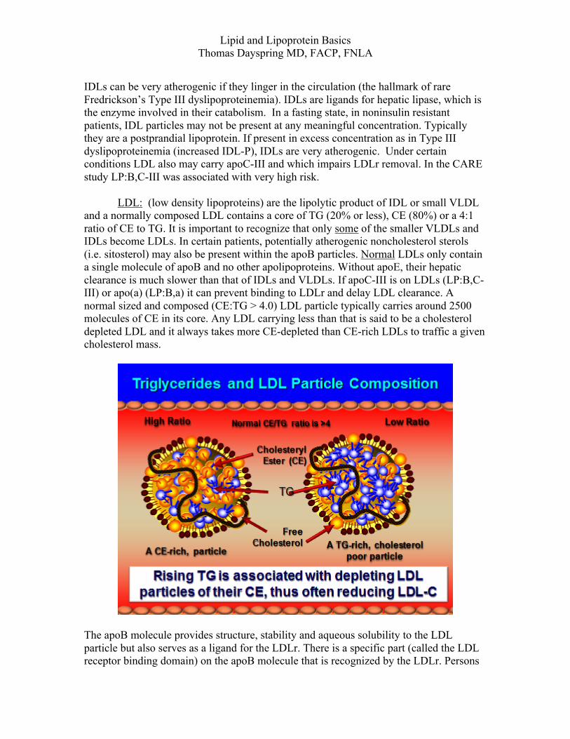
IDLs can be very atherogenic if they linger in the circulation (the hallmark of rare Fredrickson’s Type III dyslipoproteinemia). IDLs are ligands for hepatic lipase, which is the enzyme involved in their catabolism. In a fasting state, in noninsulin resistant patients, IDL particles may not be present at any meaningful concentration. Typically they are a postprandial lipoprotein. If present in excess concentration as in Type III dyslipoproteinemia (increased IDL-P), IDLs are very atherogenic. Under certain conditions LDL also may carry apoC-III and which impairs LDLr removal. In the CARE study LP:B,C-III was associated with very high risk. LDL: (low density lipoproteins) are the lipolytic product of IDL or small VLDL and a normally composed LDL contains a core of TG (20% or less), CE (80%) or a 4:1 ratio of CE to TG. It is important to recognize that only some of the smaller VLDLs and IDLs become LDLs. In certain patients, potentially atherogenic noncholesterol sterols (i.e. sitosterol) may also be present within the apoB particles. Normal LDLs only contain a single molecule of apoB and no other apolipoproteins. Without apoE, their hepatic clearance is much slower than that of IDLs and VLDLs. If apoC-III is on LDLs (LP:B,C-III) or apo(a) (LP:B,a) it can prevent binding to LDLr and delay LDL clearance. A normal sized and composed (CE:TG > 4.0) LDL particle typically carries around 2500 molecules of CE in its core. Any LDL carrying less than that is said to be a cholesterol depleted LDL and it always takes more CE-depleted than CE-rich LDLs to traffic a given cholesterol mass.
The apoB molecule provides structure, stability and aqueous solubility to the LDL particle but also serves as a ligand for the LDLr. There is a specific part (called the LDL receptor binding domain) on the apoB molecule that is recognized by the LDLr. Persons
Lipid and Lipoprotein Basics Thomas Dayspring MD, FACP, FNLA
with abnormal domain structure which interferes with LDLr-mediated clearance have very high apoB and LDL-C levels, a hypercholesterolemic condition called defective apoB. LDL particles are a heterogeneously sized group of particles ranging in size from large to small. Depending on the separation technique there can be as many as 15 different LDL species. In the two most commonly used techniques in clinical practice there are 7 LDL sizes reported by gel fractionation (Berkeley) and 2 reported by NMR spectroscopy (LipoScience). LDLs are grouped into two phenotypes: All people have a variety of LDL changing particle sizes, but we all have a predominant LDL particle size which is called LDL pattern or phenotype : Pattern A (large, buoyant) and Pattern B (small, dense). In-between sized particles were sometimes termed intermediate in size but that terminology has pretty much disappeared. When an LDL particle measures 20.5 or less nm via NMR technology), it is considered small, above that large. Since there is one apoB protein per LDL molecule, apoB measurements are another technique to determine how many LDL particles are present: because of its much longer half-life the vast majority (>90-95%) of apoB particles are LDLs. In actuality, except for unusual conditions (some very high TG states and Type III) at least 90% of measured apoB is from LDL, due to LDLs long half life (3 days). ApoB is associated with risk at levels > 80-100 mg/dl. Ideal is less than 80 mg/dL or perhaps even 60 mg/dL in very high risk patients. It is more accurate to measure LDL particle concentration (LDL-P) using the NMR (nuclear magnetic resonance spectroscopy) technique (n<1000 nmol/L). For Framingham and MESA apoB and LDL-P population percentile cutpoints see http://www.lipidcenter.com/pdf/Framingham.pdf Remember LDL cholesterol (LDL-C) is simply the amount (mass) of cholesterol within all of the LDL particles per unit of volume (typically reported as mg/dL or in other countries mmol/L). LDL-C serves as a surrogate of apoB (mg/dL) or LDL-P (LDL particle concentration reported as nmol/L). In certain patients both LDL-C and LDL-P correlate well and are said to be concordant with one another. Yet in others they do not correlate at all and are thus said to be discordant. In every study where discordance has occurred risk follows apoB or LDL-P, better than LDL-C or non-HDL-C. Thus in several studies, including Framingham and MESA, LDL-P is recognized as the best predictor of CV risk. LDL-P test is performed by LipoScience in Raleigh, NC (www.liposcience.com) and is termed the NMR LipoProfile. It is crucial to have a clear understanding of how particle size contributes to atherogenesis (it does not). All LDL particles, large or small are atherogenic (will enter the arterial wall) if present in increased numbers (elevated LDL-P). If LDL-P is low (physiologic), there is little risk whether one has large or small LDL particles. Indeed, once LDL-P is known, LDL size is no longer an independent risk factor for CHD. If LDL-P is high, CHD risk is high no matter whether the high LDL-P is driven by too many large LDLs, too many small LDLs or both. Keep in mind that the most lethal lipid/lipoprotein disorder is familial hypercholesterolemia. Such patients typically have very increased numbers of the larger (Pattern A) particles. All of the older studies that suggested risk is related to small LDLs per se, were never adjusted for LDL-P. Once that adjustment is made, the risk related to LDL size is no longer statistically significant.
Lipid and Lipoprotein Basics Thomas Dayspring MD, FACP, FNLA
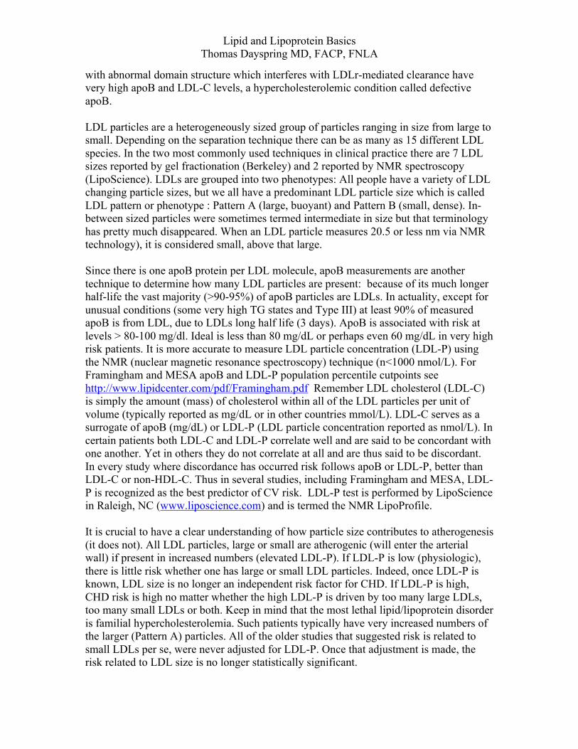
Because the volume of a sphere or circular particle is a third power of the radius (V = 4/3 π r3), there are considerable volume differences between particles that vary only slightly in diameter. Thus if a patient has an LDL-C of 100 mg/dL it will take 40 to 70% (depending on the exact diameter) more small LDL than large LDL to carry the 100 mg of cholesterol. Therefore, even though an LDL-C may be 100 mg/dL, the LDL-P will be 40-70% higher in patients with smaller rather than large LDL. Also if one has TG-rich and thus cholesterol-poor LDL particles, it will take increased numbers of particles to traffic a given level of cholesterol. And it is LDL-P that primarily determines risk. To use a dump truck analogy: If I have a mountain of cholesterol to traffic, it will take far fewer large LDL dump trucks (earth-moving dump trucks) than it would small LDL dump trucks (pickup trucks). However, if the larger earth-moving dump trucks where 50% full with something else (like TG), I’d need a lot more large of those dump trucks than I would empty ones. So it is not the size but the volume available for CE that matters. That available LDL volume dedicated to CE is dependent on particle size, particle TG content or both.
Since any apoB particle < 70 nm in diameter can penetrate the vascular endothelium, LDL size does not influence LDL particle entry into the subintimal space (the largest LDL is 23 nm. Likewise the number of cholesterol molecules per particle has no influence on LDL particle entry. The only variables that affect vascular entry are LDL-P and endothelial integrity. However, small particles carry lipoprotein-associated phospholipase A2 (LP-PLA2) are quite prone to oxidative forces and are also less readily cleared by hepatic LDL receptors, which more easily recognize and attach to the apoB
Lipid and Lipoprotein Basics Thomas Dayspring MD, FACP, FNLA
conformation on larger, normal-sized LDL. Thus almost all drug naïve patients with small LDLs have a very high LDL-P. The cholesterol in all of the beta lipoproteins (almost all of which are LDLs) is termed non-HDL-C and it is the best lipid concentration surrogate of atherogenic cholesterol and apoB levels. In effect non-HDL-C is the cholesterol that is not in the HDLs – therefore it has to be in the apoB particles. It is calculated by subtracting HDL-C from total cholesterol or by adding LDL-C plus VLDL-C. VLDL-C is calculated by TG/5. In reality non HDL-C is the cholesterol in all of the LDLs (regardless of size), VLDLs, IDLs, chylomicrons, remnants and Lp(a) particles. LDLs typically make up 90-95% of apoB particles and thus non-HDL-C is a better surrogate of apoB and LDL-P than is LDL-C. Non-HDL-C, like apoB or LDL-P does not require the patient to fast.
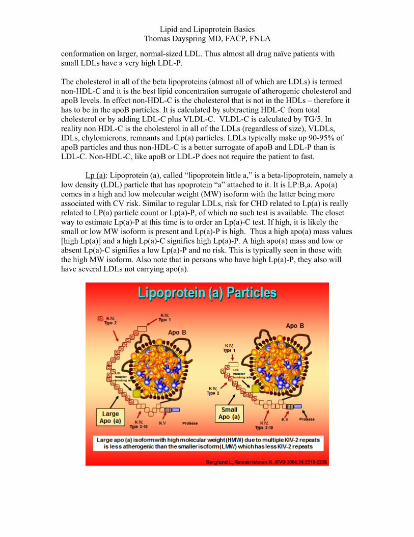
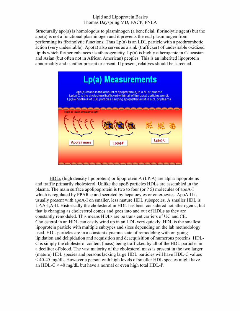
Lp (a): Lipoprotein (a), called “lipoprotein little a,” is a beta-lipoprotein, namely a low density (LDL) particle that has apoprotein “a” attached to it. It is LP:B,a. Apo(a) comes in a high and low molecular weight (MW) isoform with the latter being more associated with CV risk. Similar to regular LDLs, risk for CHD related to Lp(a) is really related to LP(a) particle count or Lp(a)-P, of which no such test is available. The closet way to estimate Lp(a)-P at this time is to order an Lp(a)-C test. If high, it is likely the small or low MW isoform is present and Lp(a)-P is high. Thus a high apo(a) mass values [high Lp(a)] and a high Lp(a)-C signifies high Lp(a)-P. A high apo(a) mass and low or absent Lp(a)-C signifies a low Lp(a)-P and no risk. This is typically seen in those with the high MW isoform. Also note that in persons who have high Lp(a)-P, they also will have several LDLs not carrying apo(a).
Lipid and Lipoprotein Basics Thomas Dayspring MD, FACP, FNLA
Structurally apo(a) is homologous to plasminogen (a beneficial, fibrinolytic agent) but the apo(a) is not a functional plasminogen and it prevents the real plasminogen from performing its fibrinolytic functions. Thus Lp(a) is an LDL particle with a prothrombotic action (very undesirable). Apo(a) also serves as a sink (trafficker) of undesirable oxidized lipids which further enhances its atherogenicity. Lp(a) is highly atherogenic in Caucasian and Asian (but often not in African American) peoples. This is an inherited lipoprotein abnormality and is either present or absent. If present, relatives should be screened.
HDLs (high density lipoprotein) or lipoprotein A (LP:A) are alpha-lipoproteins
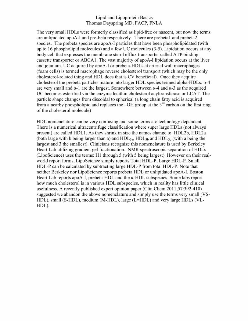
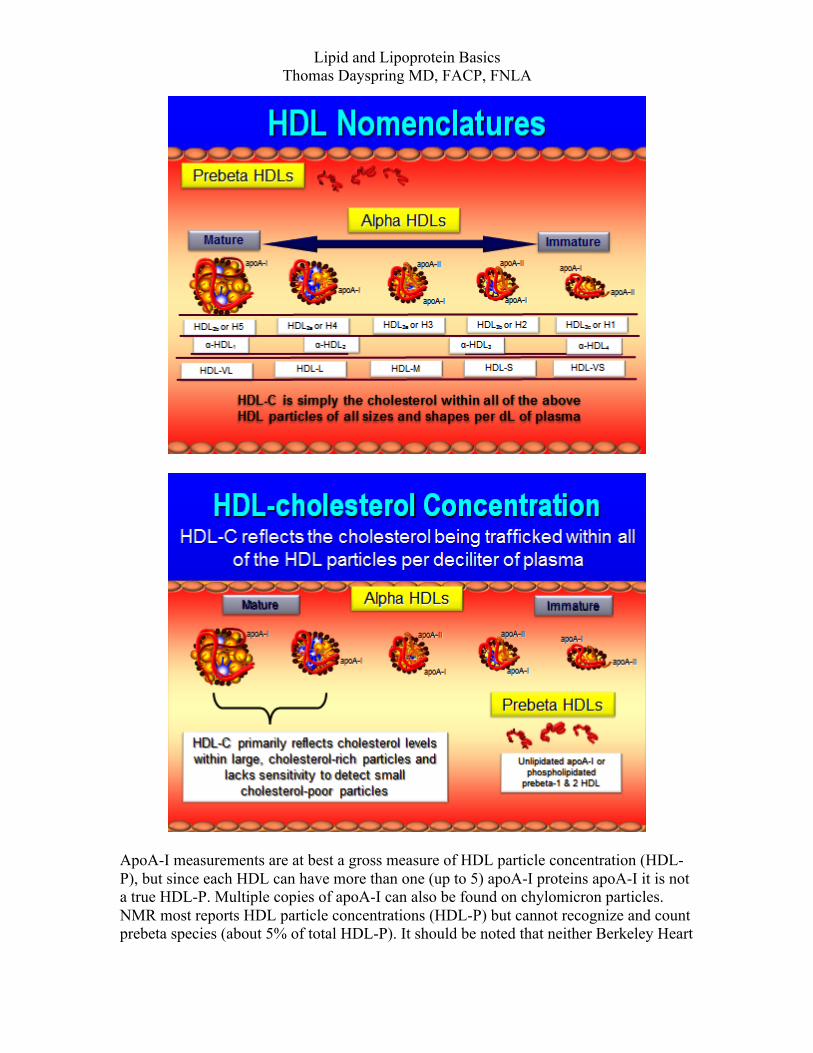
and traffic primarily cholesterol. Unlike the apoB particles HDLs are assembled in the plasma. The main surface apolipoprotein is two to four (or ? 5) molecules of apoA-I which is regulated by PPAR-α and secreted by hepatocytes or enterocytes. ApoA-II is usually present with apoA-I on smaller, less mature HDL subspecies. A smaller HDL is LP:A-I,A-II. Historically the cholesterol in HDL has been considered not atherogenic, but that is changing as cholesterol comes and goes into and out of HDLs as they are constantly remodeled. This means HDLs are be transient carriers of UC and CE. Cholesterol in an HDL can easily wind up in an LDL very quickly. HDL is the smallest lipoprotein particle with multiple subtypes and sizes depending on the lab methodology used. HDL particles are in a constant dynamic state of remodeling with on-going lipidation and delipidation and acquisition and deacquisition of numerous proteins. HDL-C is simply the cholesterol content (mass) being trafficked by all of the HDL particles in a deciliter of blood. The vast majority of the cholesterol mass is present in the two larger (mature) HDL species and persons lacking large HDL particles will have HDL-C values < 40-45 mg/dL. However a person with high levels of smaller HDL species might have an HDL-C < 40 mg/dL but have a normal or even high total HDL-P.
Lipid and Lipoprotein Basics Thomas Dayspring MD, FACP, FNLA
The very small HDLs were formerly classified as lipid-free or nascent, but now the terms are unlipidated apoA-I and pre-beta respectively. There are prebeta1 and prebeta2 species. The prebeta species are apoA-I particles that have been phospholipidated (with up to 16 phospholipid molecules) and a few UC molecules (3-5). Lipidation occurs at any body cell that expresses the membrane sterol efflux transporter called ATP binding cassette transporter or ABCA1. The vast majority of apoA-I lipidation occurs at the liver and jejunum. UC acquired by apoA-I or prebeta-HDLs at arterial wall macrophages (foam cells) is termed macrophage reverse cholesterol transport (which may be the only cholesterol-related thing and HDL does that is CV beneficial). Once they acquire cholesterol the prebeta particles mature into larger HDL species termed alpha-HDLs: α-4 are very small and α-1 are the largest. Somewhere between α-4 and α-3 as the acquired UC becomes esterified via the enzyme lecithin cholesterol acyltransferase or LCAT. The particle shape changes from discoidal to spherical (a long chain fatty acid is acquired from a nearby phospholipid and replaces the –OH group at the 3rd carbon on the first ring of the cholesterol molecule) HDL nomenclature can be very confusing and some terms are technology dependent. There is a numerical ultracentrifuge classification where super large HDLs (not always present) are called HDL1. As they shrink in size the names change to: HDL2b, HDL2a (both large with b being larger than a) and HDL3a, HDL3b and HDL3c (with a being the largest and 3 the smallest). Clinicians recognize this nomenclature is used by Berkeley Heart Lab utilizing gradient gel fractionation. NMR spectroscopic separation of HDLs (LipoScience) uses the terms: H1 through 5 (with 5 being largest). However on their real-world report forms, LipoScience simply reports Total HDL-P, Large HDL-P. Small HDL-P can be calculated by subtracting large HDL-P from total HDL-P. Note that neither Berkeley nor LipoScience reports prebeta HDL or unlipidated apoA-I. Boston Heart Lab reports apoA-I, prebeta-HDL and the α-HDL subspecies. Some labs report how much cholesterol is in various HDL subspecies, which in reality has little clinical usefulness. A recently published expert opinion paper (Clin Chem 2011;57:392-410) suggested we abandon the above nomenclature and simply use the terms very small (VS-HDL), small (S-HDL), medium (M-HDL), large (L=HDL) and very large HDLs (VL-HDL).
Lipid and Lipoprotein Basics Thomas Dayspring MD, FACP, FNLA
ApoA-I measurements are at best a gross measure of HDL particle concentration (HDL-P), but since each HDL can have more than one (up to 5) apoA-I proteins apoA-I it is not a true HDL-P. Multiple copies of apoA-I can also be found on chylomicron particles. NMR most reports HDL particle concentrations (HDL-P) but cannot recognize and count prebeta species (about 5% of total HDL-P). It should be noted that neither Berkeley Heart

Lipid and Lipoprotein Basics Thomas Dayspring MD, FACP, FNLA
Lab nor VAP analysis reports HDL particle counts. TG (discussed below) have a significant effect on HDL composition, HDL cholesterol content and HDL particle size. HDL particles potentially perform a multitude of antiatherogenic functions related to its proteome, its lipidome, and its ability to perform macrophage reverse cholesterol transport (RCT). One function of HDLs function is trafficking of lipids, especially cholesterol, in a process I prefer to call ApoA-I mediated trafficking of lipids (discussed in depth below). This process is immensely more complicated than classically taught and it is regulated by dozens of enzymes, cell receptors and lipid transfer proteins. It all starts with apoA-I synthesis, lipidation with UC, PL, esterification of UC to CE, protein acquisition, neutral exchange of lipids with other lipoproteins (homotypic and heterotypic), delipidation, cellular catabolism and renal excretion. Most startling is that almost nothing an HDL particle does is related to its cholesterol mass (HDL-C). About the only statement that has withstood the test of time is that in general a low HDL-C is (but certainly not always) a major cardiovascular risk factor. The statement that a high HDL-C value is a negative risk factor may be true in many but in some instances it is not and it is certainly possible to have severe atherosclerosis with high (> 60 mg/dL) levels. HUMAN LIPID TRANSPORTATION: So lipids are trafficked within lipoproteins and there are two pathways: the apoB-mediated and the apoA-I mediated pathways. Complicating the picture is that both pathways intersect. Let’s tie all of the above discussion into this wondrous if not miraculous and of course super complex process of lipid acquisition and transportation. All cells in the body can synthesize free or unesterified cholesterol de novo from acetate and acetoacetyl-CoA or UC can be absorbed from the jejunal lumen by enterocytes expressing the sterol influx protein (sterol permease) called the Niemann Pick C1 like 1 protein (NPC1L1). In addition to synthesis hepatocytes can acquire UC by back-flux from the bile using hepatobiliary expressed NPC1L1, or by endocytosing cholesterol-trafficking lipoproteins via various receptors (i.e. LDLr) or via a receptor called the scavenger receptor B1 (SR-B1) which binds to and delipidates mature HDLs. The major cells synthesizing TG are enterocytes, hepatocytes and adipocytes whereas the phospholipids usually have a hepatic or enterocytic origin. Because the vast majority of trafficked cholesterol is CE and not UC, cholesterol must be esterified within enterocytes, hepatocytes via ACAT or within the lipoproteins themselves (using LCAT). Note that phytosterols are poor substrates for ACAT and are not readily esterified. UC and any phytosterol still present will be found on lipoprotein surface interspaced with PL. The nonpolar molecules CE and TG will make up the core of the lipoproteins. ApoB-containing particle assembly in both enterocytes (chylomicron) and hepatocytes (VLDL) occurs when apoB48 or apoB100 are lipidated (utilizing by MTP) in the endoplasmic reticulum with CE and TG and phospholipidated in the Golgi apparatus. Chylomicrons also acquire copies of apoA-I and are secreted into the lymphatic circulation where they acquire additional apolipoproteins. VLDLs carry no apoA-I but also acquire other apoproteins during hepatic synthesis and after they are secreted into plasma.
Lipid and Lipoprotein Basics Thomas Dayspring MD, FACP, FNLA
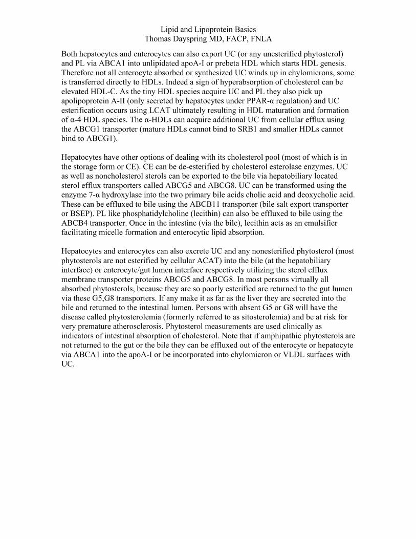
Both hepatocytes and enterocytes can also export UC (or any unesterified phytosterol) and PL via ABCA1 into unlipidated apoA-I or prebeta HDL which starts HDL genesis. Therefore not all enterocyte absorbed or synthesized UC winds up in chylomicrons, some is transferred directly to HDLs. Indeed a sign of hyperabsorption of cholesterol can be elevated HDL-C. As the tiny HDL species acquire UC and PL they also pick up apolipoprotein A-II (only secreted by hepatocytes under PPAR-α regulation) and UC esterification occurs using LCAT ultimately resulting in HDL maturation and formation of α-4 HDL species. The α-HDLs can acquire additional UC from cellular efflux using the ABCG1 transporter (mature HDLs cannot bind to SRB1 and smaller HDLs cannot bind to ABCG1). Hepatocytes have other options of dealing with its cholesterol pool (most of which is in the storage form or CE). CE can be de-esterified by cholesterol esterolase enzymes. UC as well as noncholesterol sterols can be exported to the bile via hepatobiliary located sterol efflux transporters called ABCG5 and ABCG8. UC can be transformed using the enzyme 7-α hydroxylase into the two primary bile acids cholic acid and deoxycholic acid. These can be effluxed to bile using the ABCB11 transporter (bile salt export transporter or BSEP). PL like phosphatidylcholine (lecithin) can also be effluxed to bile using the ABCB4 transporter. Once in the intestine (via the bile), lecithin acts as an emulsifier facilitating micelle formation and enterocytic lipid absorption. Hepatocytes and enterocytes can also excrete UC and any nonesterified phytosterol (most phytosterols are not esterified by cellular ACAT) into the bile (at the hepatobiliary interface) or enterocyte/gut lumen interface respectively utilizing the sterol efflux membrane transporter proteins ABCG5 and ABCG8. In most persons virtually all absorbed phytosterols, because they are so poorly esterified are returned to the gut lumen via these G5,G8 transporters. If any make it as far as the liver they are secreted into the bile and returned to the intestinal lumen. Persons with absent G5 or G8 will have the disease called phytosterolemia (formerly referred to as sitosterolemia) and be at risk for very premature atherosclerosis. Phytosterol measurements are used clinically as indicators of intestinal absorption of cholesterol. Note that if amphipathic phytosterols are not returned to the gut or the bile they can be effluxed out of the enterocyte or hepatocyte via ABCA1 into the apoA-I or be incorporated into chylomicron or VLDL surfaces with UC.
Lipid and Lipoprotein Basics Thomas Dayspring MD, FACP, FNLA
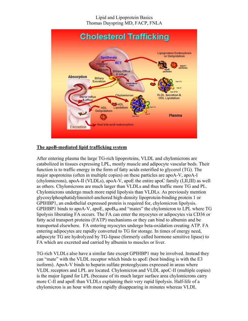
The apoB-mediated lipid trafficking system After entering plasma the large TG-rich lipoproteins, VLDL and chylomicrons are catabolized in tissues expressing LPL, mostly muscle and adipocyte vascular beds. Their function is to traffic energy in the form of fatty acids esterified to glycerol (TG). The major apoproteins (often in multiple copies) on these particles are apoA-V, apoA-I (chylomicrons), apoA-II (VLDLs), apoA-V, apoE the entire apoC family (I,II,III) as well as others. Chylomicrons are much larger than VLDLs and thus traffic more TG and PL. Chylomicrons undergo much more rapid lipolysis than VLDLs. As previously mention glycosylphosphatidylinositol-anchored high-density lipoprotein-binding protein 1 or GPIHBP1, an endothelial expressed protein is required for, chylomicron lipolysis. GPIHBP1 binds to apoA-V, apoE, apoB48 and “mates” the chylomicron to LPL where TG lipolysis liberating FA occurs. The FA can enter the myocytes or adipocytes via CD36 or fatty acid transport proteins (FATP) mechanisms or they can bind to albumin and be transported elsewhere. FA entering myocytes undergo beta-oxidation creating ATP. FA entering adipocytes are rapidly converted to TG for storage. In times of energy need, adipocyte TG are hydrolyzed by TG-lipase (formerly called hormone sensitive lipase) to FA which are excreted and carried by albumin to muscles or liver. TG-rich VLDLs also have a similar fate except GPIHBP1 may be involved. Instead they can “mate” with the VLDL receptor which binds to apoE (best binding is with the E3 isoform). ApoA-V binds to heparin sulfate proteoglycans expressed in areas where VLDL receptors and LPL are located. Chylomicron and VLDL apoC-II (multiple copies) is the major ligand for LPL (because of its much larger surface area chylomicrons carry more C-II and apoE than VLDLs explaining their very rapid lipolysis. Half-life of a chylomicron is an hour with most rapidly disappearing in minutes whereas VLDL
Lipid and Lipoprotein Basics Thomas Dayspring MD, FACP, FNLA
lipolysis takes 2-6 hours. As the particle lipolysis (hydrolyzing of TG) occurs the VLDL or chylomicron reduce in size and become remnants or in the case of VLDL, ultimately an IDL. As the particle shrinks large amounts of PL are liberated and these can enter cells or attach to PLTP and be trafficked elsewhere. Many are brought to maturing HDLs which need PL for their surface. As the chylomicron shrinks apoA-I can be liberated and become available for HDL particle synthesis or attaching to already mature HDLs. During shrinkage many apolipoproteins are also freed and these usually attach to HDL particles: a major part of HDL function is acting as a carrier of apoproteins needed for TG-rich apoB lipolysis (apoE, apoC-family, apoD better known as CETP as well as PLTP). The TG-rich lipoproteins can also participate in exchange of neutral (nonacidic) lipids, namely CE and TG) utilizing one of the lipid transfer proteins called cholesteryl ester transfer protein or CETP (also called apolipoprotein D). The VLDLs and chylomicrons can exchange one molecule of TG for one of CE in a homotypic transfer (with other apoB particles, namely IDLs and LDLs) or a heterotypic transfer with apoA-I (HDL) particles. In this process the VLDL and chylomicron remnants become TG-poorer and more enriched with CE whereas the LDLs and HDLs become CE-depleted and TG-poorer (keep in mind that a normally composed LDL has a 4:1 ratio of CE to TG and HDLs have a 9:1 ratio). After CETP modulation that ratio changes as the LDLs and HDLs becomes CE-depleted and more TG-rich. Lipolysis of such LDLs and HDLs by hepatic lipase will create smaller and denser LDLs and HDLs. This is one way a mature α-1or α-2 HDL retransforms into a prebeta or α-4 HDL. This avenue of lipid trafficking is another in which apoB and apoA-I particle lipolysis and lipid trafficking are intimately related. Paradoxically a cholesterol molecule in an HDL (classically called “good” cholesterol) can rapidly wind up in an apoB particle (where it conventionally would be called “bad” cholesterol). Another fate of a TG-rich LDL is to heterotypically exchange its newly acquired TG to HDL in exchange for CE. In this process CE goes from HDL to an LDL which can return it to the liver, a process which is now termed indirect reverse cholesterol transport. We now understand that the LDL-mediated pathway of returning cholesterol to the liver is often the major pathway of the RCT process and it is called indirect RCT. Chylomicron remnants are normally rapidly cleared by the hepatic LRP or LDLr via apoE binding. This is the major pathway of how enterocyte acquired cholesterol (in part dietary but mostly reabsorbed biliary supplied endogenous derived UC) gets to the liver. If any phytosterols were part of the chylomicron surface they also wind up in the liver. Further TG hydrolysis of TG by LP transforms VLDL or VLDL remnants into IDLs. The IDL contains less TG and surface PL than the VLDL and thus is a smaller sized lipoprotein. Classically the IDL serves as a ligand for (is the acted upon by) hepatic lipase (HL) and further lipolysis (removal) of TG and PL occurs. Note there are experts who believe IDL lipolysis is also accomplished by LPL and not HL. Regardless, when the IDL loses its core TG and surface PL it becomes a smaller lipoprotein (LDL). Again it is important to note that after delivering their TG, the majority of VLDLs and IDLs are cleared by hepatic LDLr utilizing apoE and apoB100 as ligands and do not become LDL particles. When cholesterol is returned to the liver by small VLDLs or IDLs, this is part of the indirect reverse cholesterol transport system. LDLs created by IDL lipolysis if
Lipid and Lipoprotein Basics Thomas Dayspring MD, FACP, FNLA
normal in size have a half-life (plasma residence time) of 2-3 days. One reason for the long half-life is that LDLs have no apoE (a favored ligand of the LDLr). LDLs are cleared by LDLr mostly in the liver: there is a specific area on apoB called the LDLr domain to which the LDLr attaches. When apoB conformation changes (as in smaller or very large LDL particles) that domain may be distorted resulting in much slower particle clearance and increase LDL plasma residence time. Thus the small LDL stays in plasma much longer than a normally composed one explaining why most people with small LDL have very high LDL-P or apoB levels. Scavenger receptor A molecules expressed on the endothelium of other cells can also endocytose LDL particles and this likely explains how xanthomas occur. There are also poorly understood non-receptor methods for some tissues to clear LDLs.
LDL size is determined by its lipid core, which normally should be 4 times more CE than TG. The less the amount of lipid that remains, the smaller and of course denser the LDL will be. When a lipase (LPL, HL, other phospholipase) acts on an IDL that is not TG rich, you are left with an LDL devoid of TG with a lot of cholesteryl ester: typically a larger and buoyant (Pattern A) LDL. When those lipases act on TG-rich and CE-poor IDLs (due to CETP activity), lipolysis will result in the formation of smaller (Pattern B) LDLs. HDL size is also determined by its lipid core, and normally composed HDLs have 90-95% core CE and very little core TG. However, HDLs can acquire TG via homotypic and heterotypic acquisition of CE mediated y CETP. HL, endothelial lipase (EL), and other phospholipases hydrolyze HDL core TG and surface PL resulting in smaller and of course denser HDL species. Many are then available for relipidation at ABCA1 but some
Lipid and Lipoprotein Basics Thomas Dayspring MD, FACP, FNLA
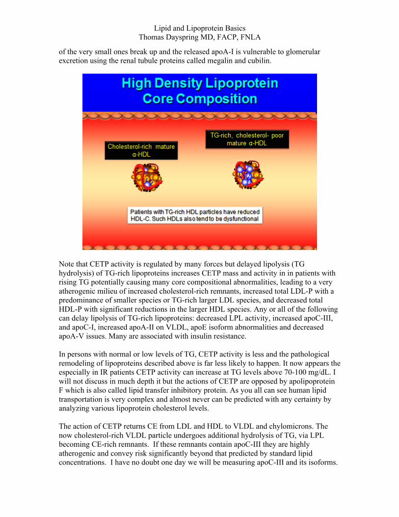
of the very small ones break up and the released apoA-I is vulnerable to glomerular excretion using the renal tubule proteins called megalin and cubilin.
Note that CETP activity is regulated by many forces but delayed lipolysis (TG hydrolysis) of TG-rich lipoproteins increases CETP mass and activity in in patients with rising TG potentially causing many core compositional abnormalities, leading to a very atherogenic milieu of increased cholesterol-rich remnants, increased total LDL-P with a predominance of smaller species or TG-rich larger LDL species, and decreased total HDL-P with significant reductions in the larger HDL species. Any or all of the following can delay lipolysis of TG-rich lipoproteins: decreased LPL activity, increased apoC-III, and apoC-I, increased apoA-II on VLDL, apoE isoform abnormalities and decreased apoA-V issues. Many are associated with insulin resistance. In persons with normal or low levels of TG, CETP activity is less and the pathological remodeling of lipoproteins described above is far less likely to happen. It now appears the especially in IR patients CETP activity can increase at TG levels above 70-100 mg/dL. I will not discuss in much depth it but the actions of CETP are opposed by apolipoprotein F which is also called lipid transfer inhibitory protein. As you all can see human lipid transportation is very complex and almost never can be predicted with any certainty by analyzing various lipoprotein cholesterol levels. The action of CETP returns CE from LDL and HDL to VLDL and chylomicrons. The now cholesterol-rich VLDL particle undergoes additional hydrolysis of TG, via LPL becoming CE-rich remnants. If these remnants contain apoC-III they are highly atherogenic and convey risk significantly beyond that predicted by standard lipid concentrations. I have no doubt one day we will be measuring apoC-III and its isoforms.
Lipid and Lipoprotein Basics Thomas Dayspring MD, FACP, FNLA
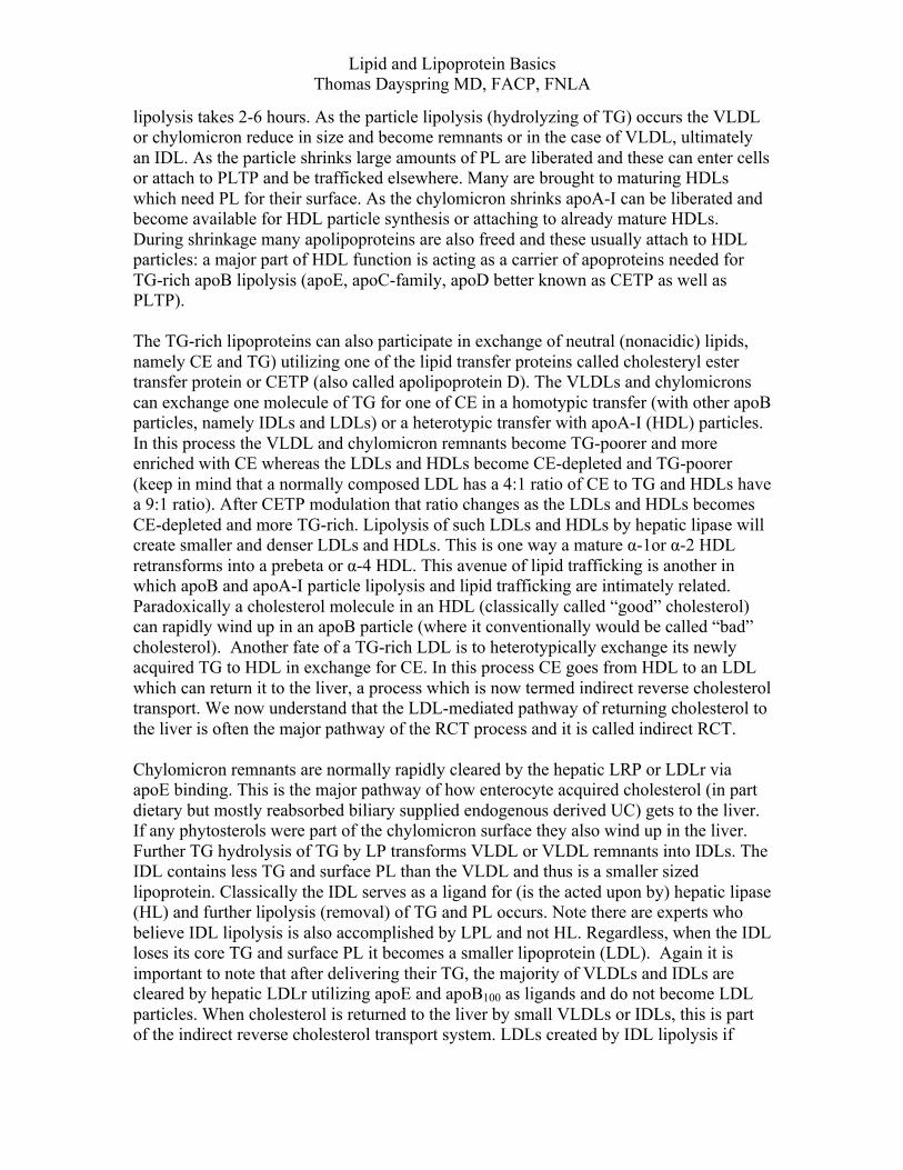
Note that persons with remnants can have very high VLDL-C (depending what you think a high VLDL-C is) which even in the face of an unremarkable LDL-C will elevate non-HDL-C (the poor man’s marker of LDL-P). AT this time since a TG of 150mg/dL is acceptable, a normal VLDL-C (TG/5) is 150/5 or 30 mg/dL. In fact a normal TG is < 70 mg/dL and a normal VLDL-C is closer to ~ 15 mg/dL, not 30 mg/dL. Since non-HDL-C = LDL-C plus VLDL-C, our non-HDL-C goals will have to become more strict. The apoA-I-mediated lipid trafficking system As discussed apoA-I is secreted by hepatocytes and jejunal enterocytes and apoA-II by hepatocytes when there is increased PPAR-α activity. Some estimate 70% is hepatic in origin and 30% intestinal but that is likely highly variable among individuals. ApoA-I can be lipidated at any tissue in the body including arterial wall foams cells (macrophage RCT). ApoA-I is not found in the CNS. Since the liver and intestine have major cholesterol pools, the vast majority of cholesterol effluxed into apoA-I is of hepatic and intestinal origin. So when looking at total HDL-C, well over 90% of that cholesterol mass is enterocytic or hepatocytic in origin. The rest is from peripheral cells and only a very tiny amount (not measurable using HDL-C assays) is from the arterial wall. Realize that the cholesterol mass in plaque is less than 1% of total body cholesterol and removal of such a small amount of total cholesterol (as crucial as that may be to CV health) will not affect any cholesterol measurement. As it is phospholipidated and cholesterol-lipidated by ABCA1, apoA-I becomes a prebeta HDL. ApoA-II is acquired and the UC is esterified by LCAT leading to α-4 HDL (other names: HDL3 or H1, H2, very small and small HDL). Further UC is acquired by ABCG1 lipidation and particle maturation continues. Maturation is also effected by both homotypic and heterotypic acquisition of lipids depending on the balance of apoD (CETP) and apoF activities. Once the CE-rich large α-2 HDL (HDL2, H4 or H5, large HDL) particles exist they have several options on what to do with that cholesterol. As the prebeta particles evolve into α-4 HDLs and then even larger species they acquire the PL for their surface from PLTP, which for the most part traffic PL acquired mostly during chylomicron and VLDL but also IDL lipolysis. Many are unaware that humans have far more HDL particles than they do apoB particles. ApoB particles are measured in nmol/L and HDLs in µmols/L. The primary lipid mission of HDLs is to deliver cholesterol to steroidogenic tissues (adrenal cortex or gonads): here the mature HDLs are delipidated by SR-B1. The reason HDLs have a 5 day half-life is to be available as a reservoir of cholesterol in case the adrenal has to start manufacturing large amounts of adrenocortical hormones (infections, inflammation, bodily crises). One could say the HDLs major job is to simple acquire cholesterol mostly from the liver and gut but also peripheral cells and bring it to and/or hold it in reserve for the adrenal. Rather than reverse cholesterol transport this aspect of what HDLs do is more aptly described as “forward” cholesterol transport. HDLs also supply significant amounts of cholesterol to adipocytes form their cell membranes. Adipocytes also can express SR-B1 or can express CETP both of which will transfer cholesterol from the
Lipid and Lipoprotein Basics Thomas Dayspring MD, FACP, FNLA
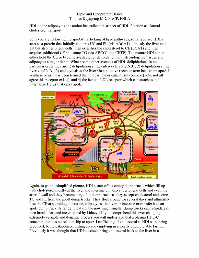
HDL to the adipocyte (one author has called this aspect of HDL function as “lateral cholesterol transport”). So if you are following the apoA-I trafficking of lipid pathways, so far you see HDLs start as a protein that initially acquires UC and PL (via ABCA1) at mostly the liver and gut but also peripheral cells, then esterifies the cholesterol to CE (LCAT) and then acquires additional CE and some TG (via ABCG1 and CETP). The mature HDLs then either hold the CE or become available for delipidation with steroidogenic tissues and adipocytes a major depot. What are the other avenues of HDL delipidation? In no particular order they are 1) delipidation at the enterocyte via SR-B1, 2) delipidation at the liver via SR-B1, 3) endocytosis at the liver via a putative receptor term beta-chain apoA-I synthase or as it has been termed the holoparticle or catabolism receptor (note: not all agree this receptor exists), and 4) the hepatic LDL receptor which can attach to and internalize HDLs that carry apoE.
Again, to paint a simplified picture, HDLs start off as empty dump trucks which fill up with cholesterol mostly at the liver and intestine but also at peripheral cells and even the arterial wall and they become large full dump trucks as they accept cholesterol and some TG and PL from the apoB dump trucks. They float around for several days and ultimately lose the CE at steroidogenic tissue, adipocytes, the liver or intestine or transfer it to an apoB dump truck. After delipidation, the now much smaller dump trucks can relipidate or then break apart and are excreted by kidneys. If you comprehend this ever changing, extremely variable and dynamic process you will understand that a plasma HDL-C concentration has no relationship to apoA-I trafficking of cholesterol as HDLs are being produced, being catabolized, filling up and emptying in a totally unpredictable fashion. Previously it was thought that HDLs existed bring cholesterol back to the liver in a
Lipid and Lipoprotein Basics Thomas Dayspring MD, FACP, FNLA
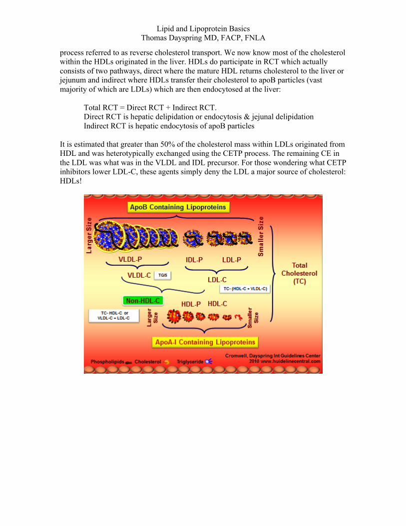
process referred to as reverse cholesterol transport. We now know most of the cholesterol within the HDLs originated in the liver. HDLs do participate in RCT which actually consists of two pathways, direct where the mature HDL returns cholesterol to the liver or jejunum and indirect where HDLs transfer their cholesterol to apoB particles (vast majority of which are LDLs) which are then endocytosed at the liver:
Total RCT = Direct RCT + Indirect RCT. Direct RCT is hepatic delipidation or endocytosis & jejunal delipidation Indirect RCT is hepatic endocytosis of apoB particles
It is estimated that greater than 50% of the cholesterol mass within LDLs originated from HDL and was heterotypically exchanged using the CETP process. The remaining CE in the LDL was what was in the VLDL and IDL precursor. For those wondering what CETP inhibitors lower LDL-C, these agents simply deny the LDL a major source of cholesterol: HDLs!
Lipid and Lipoprotein Basics Thomas Dayspring MD, FACP, FNLA
Related Documents