1 LECTURE : 09 Title: IMMUNOLOGICAL MECHANISM IN TISSUE DAMAGE TYPE-II CYTOTOXIC HYPERSENSITIVITY LEARNING OBJECTIVES : The student should be able to: • Describe type II hypersensitivity. • Determine if type II is antibody dependent or not? • Determine if type II systemic or local reactions. • Identify the type of immunoglobulin isotypes involved in the reaction. • Identify the type of immune cells involved in type II reaction. • Identify what causes cellular destruction. • Identify the target organ or tissue. • Determine type of antigen either endogenous or exogenous antigens. • Enumerate the type of immune cells that express the Fc receptors. • Discuss the mechanism of cytotoxic type II hypersensitivity reaction. • Provide some examples of clinical features of type II hypersensitivity. - Bed cell lysis: ♦ Intravascular hemolysis (ABO system incompatibility). ♦ Extravascular hemolysis (Rh system Rh incompatibility). ♦ Hemolytic disease of the new born (erythroblastosis fetalis). ♦ Autoimmune hemolytic disease (Warm and cold hemolytic anemia). - White cell lysis: ♦ Systemic lupus erythematosus (SLE) " Type II, and III reactions. ♦ Granulocytopenia. ♦ Idiopathic therompocytopeic purpura. - Nephrotoxic nephritis (Good pasture's syndrome). - Immunization: ♦ Myasthenia gravis (Acetylcholine receptors). - Bullous disease. LECTURE REFRENCE : 1. TEXTBOOK: ROITT, BROSTOFF, MALE IMMUNOLOGY. 6 th edition. Chapter 2. pp. 345-354. 2. HANDOUT.

Welcome message from author
This document is posted to help you gain knowledge. Please leave a comment to let me know what you think about it! Share it to your friends and learn new things together.
Transcript
1
LECTURE: 09
Title: IMMUNOLOGICAL MECHANISM IN TISSUE DAMAGETYPE-II CYTOTOXIC HYPERSENSITIVITY
LEARNING OBJECTIVES:
The student should be able to:
• Describe type II hypersensitivity. • Determine if type II is antibody dependent or not? • Determine if type II systemic or local reactions. • Identify the type of immunoglobulin isotypes involved in the reaction. • Identify the type of immune cells involved in type II reaction. • Identify what causes cellular destruction. • Identify the target organ or tissue. • Determine type of antigen either endogenous or exogenous antigens. • Enumerate the type of immune cells that express the Fc receptors. • Discuss the mechanism of cytotoxic type II hypersensitivity reaction. • Provide some examples of clinical features of type II hypersensitivity.
- Bed cell lysis:
♦ Intravascular hemolysis (ABO system incompatibility). ♦ Extravascular hemolysis (Rh system Rh incompatibility). ♦ Hemolytic disease of the new born (erythroblastosis fetalis). ♦ Autoimmune hemolytic disease (Warm and cold hemolytic anemia).
- White cell lysis:
♦ Systemic lupus erythematosus (SLE) " Type II, and III reactions. ♦ Granulocytopenia. ♦ Idiopathic therompocytopeic purpura.
- Nephrotoxic nephritis (Good pasture's syndrome).
- Immunization:
♦ Myasthenia gravis (Acetylcholine receptors).
- Bullous disease. LECTURE REFRENCE: 1. TEXTBOOK: ROITT, BROSTOFF, MALE IMMUNOLOGY. 6th edition. Chapter 2. pp. 345-354.
2. HANDOUT.
2
Hypersensitivity – Type II
Type II hypersensitivity reactions are caused by IgG or IgM antibodies against cell surface and extracellular matrix antigens. Antibodies to intracellular components also occur. These are not normally pathogenetic, although they may be diagnostically useful.
Transfusion reactions to erythrocytes are produced by antibodies to blood group antigens, which may occur naturally or may have been induced by previous contact with incompatible tissue or blood following transplantation, transfusion or during pregnancy.
The antibodies damage cells and tissues by activating complement, and by binding and activating effector cells carrying Fcγ receptors/
Haemolytic disease of the newborn occurs when maternal antibodies to fetal blood group antigens cross the placenta and destroy the fetal erythrocytes.
Damage to tissues may be produced by antibody to basement membranes, to intercellular adhesion molecules or to receptors. Examples include myasthenia gravis, pemphigus and Goodpasture's syndrome.
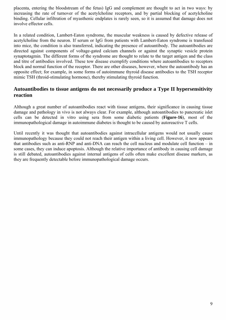
Type II hypersensitivity reactions are mediated by IgG and IgM antibodies binding to specific cells or tissues. The damage caused is thus restricted to the specific cells or tissues bearing the antigens. In general, those antibodies which are directed against cell surface antigens are usually pathogenic, while those against internal antigens usually are not so. The type II reactions therefore differ from Type III reactions which involve antibodies directed against soluble antigens in the serum, leading to the formation of circulating antigen-antibody complexes. Damage occurs when the complexes are deposited non-specifically onto tissue and/or organs. MECHANISMS OF DAMAGE Cells engage their targets using Fc and C3 receptors In Type II hypersensitivity, antibody directed against cell surface or tissue antigens interacts with complement and a variety of effector cells to bring about damage to the target cells (Figure-1) Once the antibody has attached itself to the surface of the cell or tissue, it can bind and activate complement component C1, with the following consequences:
• Complement fragments (C3a and C5a) generated by activation of complement attract macrophages and polymorphs to the site, and also stimulate mast cells and basophils to produce chemokines that attract and activate other effector cells.
• The classical complement pathway and activation loop lead to the deposition of C3b, C3bi and C3d on the target cell membrane.
• The classical complement pathway and lytic pathway result in the production of the C5b-9
membrane attack complex and insertion of the complex into the target cell membrane. Effector cells – in this case macrophages, neutrophils, eosinophils and K (killer) cells – bind either to the complexed antibody, via their Fc receptors, or to the membrane bound C3b, C3bi and C3d, via their C3 receptors (CR1, CR3, CR4).
3
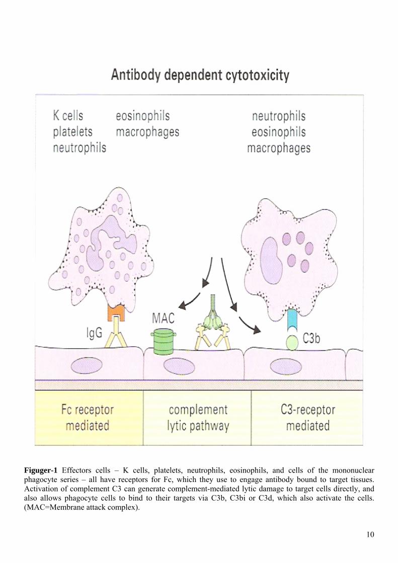
Cells damage targets by exocytosis of their normal immune effector molecules The mechanisms by which neutrophils and macrophages damage target cells in Type II hypersensitivity reactions reflect their normal methods of dealing with infectious pathogens (Figure-2). Normally pathogens would be internalized and then subjected to a barrage of microbicidal systems including defensins, reactive oxygen and nitrogen metabolites, hypohalites, enzymes, altered pH and other agents that infefere with metabolism. But if the target is too large to be phagocytosed, the granule and lysosome contents are released in apposition to the sensitized target in a process referred to as exocytosis. Cross-linking of the Fc and C3 receptors during this process causes activation of the phagocyte with production of reactive oxygen intermediates, as well as activation of phospholipase A2 and the consequent synthesis of prostaglandin and leukotrienes. In some situations, such as the eosinophil reaction against schistosomes, exocytosis of granule contents in normal and beneficial; however, when the target is host tissue that has been sensitized by antibody, the result is damaging (Figure-3). Antibodies may also mediate hypersensitivity by K cells. In this case, however, the nature of the target, and whether it can inhibit the K cells' cytotoxic actions, are as important as the presence of the sensitizing antibody. The resistance of a target cell to damage varies. Susceptibility depends on the amount of antigen expressed on the target cell's surface, and on the inherent ability of different target cells to sustain damage. For example, and erythrocyte may be lysed by a single C5 convertase site, whereas it takes many such sites to destroy most nucleated cells – their ion-pumping capacity and ability to maintain membrane integrity with anti-complementary defences is so much greater. The reminder of this chapter examines some of the instances where Type II hypersensitivity reactions are thought to be of prime importance in causing target cell destruction or immunopathological damage. REACTIONS AGAINST BLOOD CELLS AND PLATELETS Some of the most clear-cut examples of Type II reactions are seen in the responses to erythrocytes. Important examples are:
• Incompatible blood transfusions, where the recipient becomes sensitized to antigens on the surface of the donor's erythrocytes.
• Heamolytic disease of the newborn, where a pregnant woman has become sensitized to the fetal erythrocytes.
• Autoimmune haemolytic anaemias, where the patient becomes sensitized to his/her own erythrocytes.
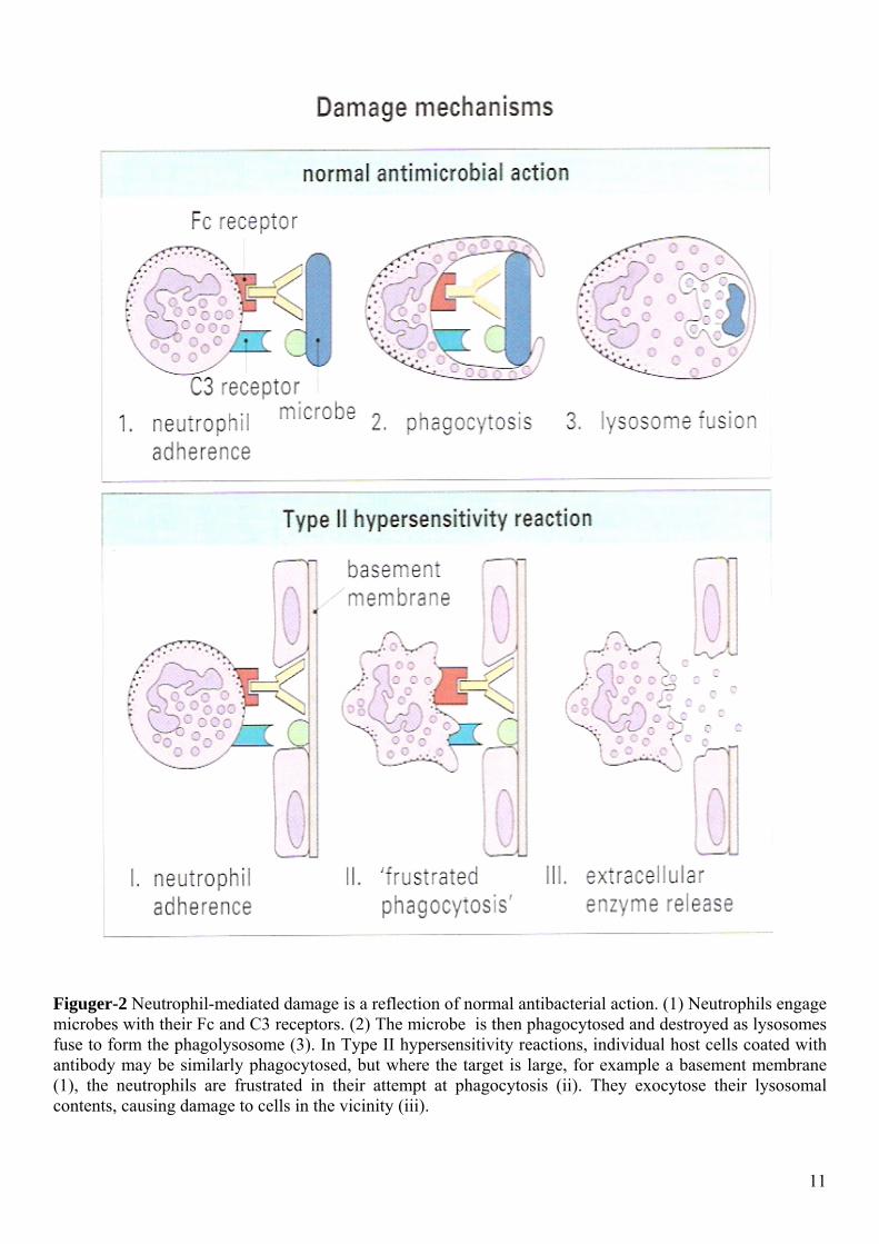
Transfusion reactions occur when a recipient has antibodies that react against donor erythrocytes More4 than 20 blood group systems, generating over 200 genetic variants of erythrocyte antigens, have been identified in man. A blood group system consists of a gene locus that specifies an antigen on the surface of blood cells (usually, but not always, erythrocytes). Within each system there may be two or more phenotypes. In the ABO system, for example, there are four phenotypes (A, B, AB and O), and thus four possible blood groups. An individual with a particular blood group can recognize erythrocytes carrying allogeneic (non-self) blood group antigens, and will produce antibodies against them. However, for some
4
blood group antigens such antibodies can also be produced 'naturally', i.e. without prior sensitization by foreign erythrocytes (see below). Transfusion of allogeneic erythrocytes into an individual who already has antibodies against them may produce erythrocyte destruction and symptoms of a 'transfusion reaction'. Some blood group systems (e.g. ABO and Rhesus) are characterized by antigens that are relatively strong immunogens; such antigens are more likely to induce antibodies. When planning a blood transfusion, it is important to ensure that donor and recipient blood thypes are compatible with respect to these major blood groups, otherwise transfusion reactions will occur. Some major human blood groups are listed in Figure-4. The ABO system – This blood group system is of primary importance. The epitopes concerned occur on may cell types in addition to erythrocytes and are located on the carbohydrate units of glycoproteins. The structure of these carbodydrates, and of those determining the related Lewis blood group system, is determined by genes coding for enzymes that transfer terminal sugars to a carbohydrate backbone (Figure-5). Most individuals develop antibodies to allogeneic specificities of the ABO system without prior sensitization by foreign erythrocytes; this sensitization occurs through contact with identical epitopes, coincidentally and routinely expressed on a wide variety of micro-organisms. Antibodies to ABO antigens are therefore extremely common, making it particularly important to match donor blood to the recipient for this system. However, all people are tolerant to the O antigen, and so O individuals are universal donors with respect to the ABO system. The Rhesus system – This system is also of great importance, as it is a mojor cause of haemolytic disease of the newborn (HDNB). Rhesus antigens are associated with membrane proteins of 30kDa which are expressed at moderate levels on the erythrocyte surface. The antigens are encoded by two closely linked loci, RhD and RhCcEe, with 92% homology. RhD is the most important clinically due to its high immunogenicity, but in RhD- individuals the RhD locus is missing completely. The RhCcEe locus encodes a molecule which expresses the RhC/c and RhE/e epitopes. Minor blood group systems – MN system epitopes are expressed on the N-terminal glycosylated region of glycophorin A, a glycoprotein present on the erythrocyte surface. Antigenicity is determined by polymorphisms at amino acids 1 and 5. The related Ss system antigens are carried on glycophorin B. Proteins expressed on erythrocytes which display allelic variation can also act as blood group antigens. Examples of these include the Kell antigen, a zinc endopeptidase, the Duffy antigen receptor for chemokines (DARC) and variants of decay accelerating factor (DAF). The relationship of the blood groups to erythrocyte surface proteins is listed in Figure-6. Transfusion reactions caused by the minor blood groups are relatively rare, unless repeated transfusions are given. Again, the risks are greatly reduced by accurately cross-matching the donor blood to that of recipient. Cross-matching – The aim of cross-matching is to ensure that the blood of a recipient does not contain antibodies that will be able to react with and destroy transfused (donor) erythrocytes. For example, antibodies to ABO system antigens cause incompatible cells to agglutinate in a clearly visible reaction. Minor blood group systems cause weaker reactions that may only be detectable by an indirect Coombs' test (Figure-10). If the individual is transfused with whole blood, it is also necessary to check that the donor's serum does not contain antibodies against the recipient's erythrocytes. However, transfusion of whole blood is unusual – most blood donations are separated into cellular and serum fractions, to be used individually. Transfusion reactions involve extensive destruction of donor blood cells Transfusion of erythrocytes into a recipient who has antibodies to those cells produces an immediate reaction. The symptom include fever, hypotension, nausea and vomiting, and pain in the back and chest. The severity of the reaction depends on the class and the amounts of antibodies involved. Antibodies to ABD system antigens are usually IgM, and cause agglutination, complement activation and intravascular haemolysis. Other blood groups induce IgG antibodies, which cause less agglutination than IgM. The IgG-sensitized cells are usually taken up by phagocytes in the liver and spleen, although severe reactions may cause erythrocyte destruction by complement activation. This can cause circulatory shock, and the released shock, and the released contents of the erythrocytes can produce acute tubular necrosis of
5
the kidneys. These acute transfusion reactions are often seen in previously unsensitized individuals, and develop over days or weeks as antibodies to the foreign cells are produced. This can result in anaemia or jaundice. Transfusion reactions to other components of blood may also occur, though their consequences are not usually as severe as reactions to erythrocytes. Hyperacute graft rejection is related to the transfusion reaction Hyperacute graft rejection occurs when a graft recipient has performed antibodies against the graft tissue. It is only seen in tissue that is revascularized directly after transplantation, in kidney grafts for example. The most severe reactions in this type of rejection are due to the ABO group antigens that are expressed on kidney cells. The damage is produced by antibody and complement activation in the blood vessels, with consequent recruitment and activation of neutrophils and platelets. However, donors and recipients are now always cross-matched for ABO antigens, and this reaction has become extremely rare. Antibodies to other graft antigens (e.g. MHC molecules) indeed by previous grafting can also produce this type of reaction. Haemolytic disease of the newborn is due to material IgG antibodies which react against the child's erythrocytes in utero Haemolytic disease of the newborn (HDNB) occurs when the mother has been sensitized to antigens on the infant's erythrocytes and makes IgG antibodies to these antigens. These antibodies cross the placenta and react with the fetal erythrocytes, causing their destruction (Figure-7, and 8). Rhesus D (RhD) is the most commonly involved antigen. A risk of HNDB arises when a Rh+ sensitized Rh- mother carries a second Rh+ infant. Sensitization of the Rh-
mother to the Rh+ erythrocytes usually occurs during birth of the first Rh+ infant, when some fetal erythrocytes leak back across the placenta into the material circulation and are recognized by the material immune system. Thus the first incompatible child is usually unaffected, whereas subsequent children have an increasing risk of being affected, as the mother is resensitized with each successive pregnancy. Reactions to other blood groups may also cause HDNB, the second most common being the Kell system K antigen. Reactionss due to anti-K are much less common than reactions due to RhD because of the relatively low frequency (95) and weaker antigenicity of the K antigen. The risk of HDNB due to Rhesus incompatibility is known to be reduced if the father is of a different ABO group to the mother. This observation led to the idea that these Rh- mothers were destroying Rh+ cells more rapidly, because they were also ABO incompatible. Consequently, fetal Rh+ erythrocytes would not be available to sensitize the material immune system to RhD antigen. This notion led to the development of Rhesus prophylaxis: performed anti-RhD antibodies are given to Rh- mother immediately after delivery of Rh+ infants, with the aim of destroying fetal Rh+ erythrocytes before they can cause Rh- sensitization. This practice has successfully reduced the incidence of HDNB due to Rhesus incompatibility (Figure-9). Although the number of cases of HDNB has fallen dramatically and progressively, the proportion of cases caused by other blood groups, including Kell and the ABO system, has increased. Autoimmune haemolytic anaemias arise spontaneously as autoimmune diseases, or may be induced as reactions to drugs Reactions to blood group antigens also occur spontaneously in the autoimmune haemolytic anaemias, in which patients produce antibodies to their own erythrocytes. Autoimmune haemolytic anaemia is suspected
6
if a patient gives a positive result on a direct antiglobulin test (Figure-10), which identifies antibodies present on the patient's erythrocytes. These are usually antibodies directed towards erythrocyte antigens, or immune complexes adsorbed onto the erythrocytes' surface. The direct antiglobulin test is also used to detect antibodies on red cells in mismatched transfusions, and in HDNB (see above). Autoimmune haemolytic anaemias can be divided into three types, depending upon whether they are due to:
• Warm-reactive autoantibodies, which can only react with antigen at below 370C. • Cold-reactive autoantibodies, which can only react with antigen at below 370C. • Antibodies provoked by allergic reactions to drugs.
Warm-reactive autoantibodies cause accelerated clearance of erythrocytes Warm-reactive autoantibodies are frequently found against Rhesus system antigen, including determinants of the RhC and RhE loci as well as RhD. They differ from the antibodies responsible for transfusion reactions, in that they appear to react with different epitopes. Warm-reactive autoantibodies to other blood group antigens exist, but are relatively rare. Most of these haemolytic anaemias are of unknown cause, but some are associated with other autoimmune diseases. The anaemia appears to be a result of accelerated clearance of the sensitized erythrocytes by spleen macrophages more often than being due to complement-mediated lysis. Cold-reactive autoantibodies cause erythrocytes lysis by complement fixation Cold-reactive autoantibodies are often present in higher titres than the warm-reactive autoantibodies. The antibodies are primarily IgM and fix complement strongly. In most cases they are specific for the Ii blood group system. The I and i epitopes are expressed on the precursor polysaccharides that produce the ABO system epitopes, and are the result of incomplete glycosylation of the core polysaccharide. The reaction of the antibody with the erythrocytes takes place in the peripheral circulation (particularly in winter), where the temperature in the capillary loops of exposed skin may fall below 300C. In severe cases, peripheral necrosis may occur due to aggregation and microthrombosis of small vessels caused by complement-mediated destruction in the periphery. The severity of the anaemia is therefore directly related to the complement-fixing ability of the patient's serum. (Fc-mediated removal of sensitized cells in the spleen and liver is not involved, because these organs are too warm for the antibodies to bind.) Most cold-reactive autoimmune haemolytic anaemias occur in older people. Their cause is unknown, but it is notable that the autoantibodies produced are usually of very limited clonality, indicating that a limited number of autoreactive clones are present. However, some cases may follow infection with Mycoplasma pneumoniae, and these are acute onset disease of short duration with polyclonal autoantibodies. Such cases are thought to be due to cross-reacting anigens on the bacteria and the erythrocytes, producing a bypass of normal tolerance mechanisms. Drug-induced reactions to blood components may be due either to antibodies binding to the drug absorbed to cells, or to breakdown of self tolerance Drugs (or their metabolites) can provoke hypersensitivity reactions against blood cells, including erythrocytes and platelets. This can occur in three different ways (Figure-11):
• The drug binds to the blood cells, and antibodies are produced against the drug. In this case it is necessary for both the drug and the antibody to be present to produce the reaction. This phenomenon was first recorded by Ackroyd, who noted thrombocytopenic purpura (destruction of platelets leading to purpuric rash) following administration of the drug Sedormid. Haemolytic anaemias have been
7
reported following the administration of a wide variety of drugs, including penicillin, quinine and sulphonamides. All these conditions are rare.
• Drug-antibody immune complexes are absorbed on to the erythrocyte cell membrane. Damage occurs by complement-mediated lysis.
• The drug induces an allergic reaction, and autoantibodies are directed against the erythrocyte
antigens themselves, as is the case with 0.3% of patients given α-methyldopa. The antibodies produced are similar to those in patients with warm-reactive antibody. However, the condition remits shortly after the cessation of drug treatment.
Reactions against other blood cells have been associated with systemic lupus erythematosus and with thrombocytopenia Antibodies to neutrophils and lymphocytes – Auto-antibodies to neutrophil cytoplasmic antigens (ANCAs) are associated with a number of diseases. For example, antibody to proteinase-3, a cytoplasmic antigen (C-ANCA), is associated with Wegener's granulomatosis, while antibodies to myeloperoxidase are seen more commonly in systemic lupus erythematosus (SLE), and are located in perinuclear granules (P-ANCA). Other granule components may also act as antigens in SLE but less commonly than myeloperoxidase. Such autoantigens are generally neutrophil specific and antibodies can be detected by immunofluorescent staining (Figure-12). (By contrast, antibodies to MHC antigens also seen in SLE are highly non-tissue specific.) Antibodies to P-ANCAs are particularly characteristic of vasculitis and glomerulonephritis. Their contribution to disease pathogenesis appears to be relatively small, although they have some diagnostic use. Antibodies to platelets – Autoantibodies to platelets are seen in up to 70% of cases of idiopathic thrombocytopenic purpura, a disorder in which there is accelerated removal of platelets from the circulation, mediated primarily by splenic macrophages. The mechanism of removal is via the immune adherence receptors on these cells. The condition most often develops after bacterial or viral infections, but may also be associated with autoimmune diseases including SLE. In SLE, antibodies to cardiolipin, which is present on platelets, can sometimes be detected. Autoantibodies to cardiolipin and other phospholipids can inhibit one aspect of blood clotting (lupus anticoagulant) and can be associated, in some cases, with venous thrombosis and recurrent abortions. Thrombocytopenia may also be induced by drugs, by similar mechanisms to those outlined in Figure-11. REACTIONS AGAINST TISSUE ANTGENS A number of autoimmune conditions occur in which antibodies to tissue antigens cause immunopathological damage by activation of Type II hypersensitivity mechanisms. The antigens are extracellular, and may be expressed on structural proteins or on the surface of cells. Descriptions of such diseases – Goodpasture's syndrome, pemphigus and myasthenia gravis – are given below. It is often possible to demonstrate autoantibodies to particular cell types, but in these cases the antigens are intracellular, and the importance of the Type II mechanisms is less well established. In these cases, recognition of autoantigen by T cells is probably more important pathologically, and the autoantibodies are of secondary importance. Antibodies against basement membranes produce nephritis in Goodpasture's syndrome
8
A number of patients with nephritis are found to have antibodies to collagen Type IV, which is a major component of basement membranes (Figure-3). Collagen Type IV undergoes alternate RNA splicing which produces a number of variant proteins (Goodpasture antigen), but the antibodies appear to bind just those forms which retain the characteristic N-terminus. The antibody is usually IgG and, in at least 50% of patients, it appears to fix complement. The condition usually results in severe necrosis of the glomerulus, with fibrin deposition. The association of this type of nephritis with lung haemorrhage was originally noticed by Goodpasture (hence Goodpasture's syndrome). Although the lung symptoms do not occur in all patients, the association of lung and kidney damage is due to cross-reactive autoantigens in the basement membranes of the two tissues. A number of animal models for Goodpasture's syndrome have been developed. In nephrotoxin serum nephritis (Masugi glomerulonephritis), heterologous antibodies to glomerular basement membrane are injected into rats or rabbits. The injected antibody is deposited on to the basement membranes, the this is followed by further deposition of host antibodies to the injected antibody; this precipitates acute nephritis. Development of nephritis and proteinuria depends on the accumulation of neutrophils, which bind via complement-dependent and complement-independent mechanisms. Similar lesions can be induced by immunization with heterologous basement membrane (Steblay model). Another animal model (Heymann nephritis), caused by raising autoantibodies to a protein present in the brush border of glomerular epithelial cells, resembles human membranous glomerulonephritis. In this model, the damage is mostly complement mediated: complement depletion of the animals alleviates the condition. Pemphigus is caused by autoantibodies to an intercellular adhesion molecule Pemphigus is a serious blistering disease of the skin and mucous membranes. Patients have autoantibodies against desmoglein-1 and desmoglein-3, components of desmosomes, which form junctions between epidermal cells (Figure-13). The antibodies disrupt cellular adhesion, leading to breakdown of the epidermis. Clinical disease profiles can be related to the specificity of the antibodies. For example, patients with only anit-desmoglein-3 tend to show mucosal disease, while those with anti-desmoglein-1 and -3 have affected skin and mucosa. Disease has been correlated with the incidence of IgG4 antibodies against a different part of the molecule. Pemphigus is strongly linked to a rare haplotype of HLA-DR4 (DRB1*0402), and this molecule has been shown to present a peptide of desmoglein-3, which other DR4 subtypes cannot. This is therefore a clear example of an autoimmune disease producing pathology by Type II mechanisms. Myasthenia gravis and Lambert-Eaton syndrome are caused by antibodies that reduce the availability of acetylcholine at motor endplates Myasthenia gravis, a condition in which there is extreme muscular weakness, is associated with antibodies to the acetylcholine receptors present on the surface of muscle membranes. The acetylcholine receptors are located at the motor endplate where the neuron contacts the muscle. Transmission of impulses from the nerve to the muscle takes place by the release of acetylcholine from the nerve terminal and its diffusion across the gap to the muscle fibre. It was noticed that immunization of experimental animals with purified acetylcholine receptors produced a condition of muscular weakness that closely resembled human myasthenia. This suggested a role for antibody to the acetylcholine receptor in the human disease. Analysis of the lesion in myasthenic muscles indicated that the disease was not there any problem in secreting it in response to a nerve impulse – the released acetylcholine was less effective at triggering depolarization of the muscle (Figure-14). Examination of neuromuscular endplates by immunochemical techniques had demonstrated IgG and the complement proteins, C3 and C9, on the postsynaptic folds of the muscle (Figure-15). (Further evidence for a pathogenetic role for IgG in this disease was furnished by the discovery of transient muscle weakness in babies born to myasthenic mothers. This is significant because it is known that IgG can and does cross the
9
placenta, entering the bloodstream of the fetus) IgG and complement are thought to act in two ways: by increasing the rate of turnover of the acetylcholine receptors, and by partial blocking of acetylcholine binding. Cellular infiltration of myasthenic endplates is rarely seen, so it is assumed that damage does not involve effector cells. In a related condition, Lambert-Eaton syndrome, the muscular weakness is caused by defective release of acetylcholine from the neuron. If serum or IgG from patients with Lambert-Eaton syndrome is transfused into mice, the condition is also transferred, indicating the presence of autoantibody. The autoantibodies are directed against components of voltage-gated calcium channels or against the synaptic vesicle protein synaptotagmin. The different forms of the syndrome are thought to relate to the target antigen and the class and titre of antibodies involved. These tow disease exemplify conditions where autoantibodies to receptors block and normal function of the receptor. There are other diseases, however, where the autoantibody has an opposite effect; for example, in some forms of autoimmune thyroid disease antibodies to the TSH receptor mimic TSH (throid-stimulating hormone), thereby stimulating thyroid function. Autoantibodies to tissue antigens do not necessarily produce a Type II hypersensitivity reaction Although a great number of autoantibodies react with tissue antigens, their significance in causing tissue damage and pathology in vivo is not always clear. For example, although autoantibodies to pancreatic islet cells can be detected in vitro using sera from some diabetic patients (Figure-16), most of the immunopathological damage in autoimmune diabetes is thought to be caused by autoreactive T cells. Until recently it was thought that autoantibodies against intracellular antigens would not usually cause immunopathology because they could not reach their antigen within a living cell. However, it now appears that antibodies such as anti-RNP and anti-DNA can reach the cell nucleus and modulate cell function – in some cases, they can induce apoptosis. Although the relative importance of antibody in causing cell damage is still debated, autoantibodies against internal antigens of cells often make excellent disease markers, as they are frequently detectable before immunopathological damage occurs.
Figuger-1 Effectors cells – K cells, platelets, neutrophils, eosinophils, and cells of the mononuclear phagocyte series – all have receptors for Fc, which they use to engage antibody bound to target tissues. Activation of complement C3 can generate complement-mediated lytic damage to target cells directly, and also allows phagocyte cells to bind to their targets via C3b, C3bi or C3d, which also activate the cells. (MAC=Membrane attack complex).
10
Figuger-2 Neutrophil-mediated damage is a reflection of normal antibacterial action. (1) Neutrophils engage microbes with their Fc and C3 receptors. (2) The microbe is then phagocytosed and destroyed as lysosomes fuse to form the phagolysosome (3). In Type II hypersensitivity reactions, individual host cells coated with antibody may be similarly phagocytosed, but where the target is large, for example a basement membrane (1), the neutrophils are frustrated in their attempt at phagocytosis (ii). They exocytose their lysosomal contents, causing damage to cells in the vicinity (iii).
11
Figuger-3 Phagocytes attacking a basement membrane, this electron-micrograph shows a neutrophil (N) and there monocytes (M) binding to the capillary basement membrane (B) in the kidney of a rabbit containing anti-basement membrane antibody. P= podocyte. X 3500
Figuger-4 Not all blood groups are equally antigenic in transfusion reactions: thus, RhD evokes a stronger reaction in an incompatible recipient than the other Rhesus antigens; and Fya is stronger than Fyb. Frequencies stated are for Caucasian populations – other races have different gene frequencies.
12
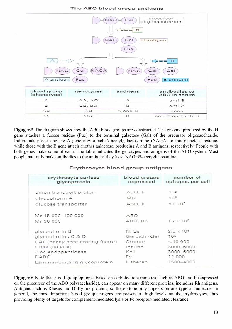
Figuger-5 The diagram shows how the ABO blood groups are constructed. The enzyme produced by the H gene attaches a fucose residue (Fuc) to the terminal galactose (Gal) of the precursor oligosaccharide. Individuals possessing the A gene now attach N-acetylgalactosamine (NAGA) to this galactose residue, while those with the B gene attach another galactose, producing A and B antigens, respectively. People with both genes make some of each. The table indicates the genotypes and antigens of the ABO system. Most people naturally make antibodies to the antigens they lack. NAG=N-acetyglucosamine.
Figuger-6 Note that blood group epitopes based on carbohydrate moieties, such as ABO and Ii (expressed on the precursor of the ABO polysaccharide), can appear on many different proteins, including Rh antigens. Antigens such as Rhesus and Duffy are proteins, so the epitope only appears on one type of molecule. In general, the most important blood group antigens are present at high levels on the erythrocytes, thus providing plenty of targets for complement-mediated lysis or Fc receptor-mediated clearance. 13
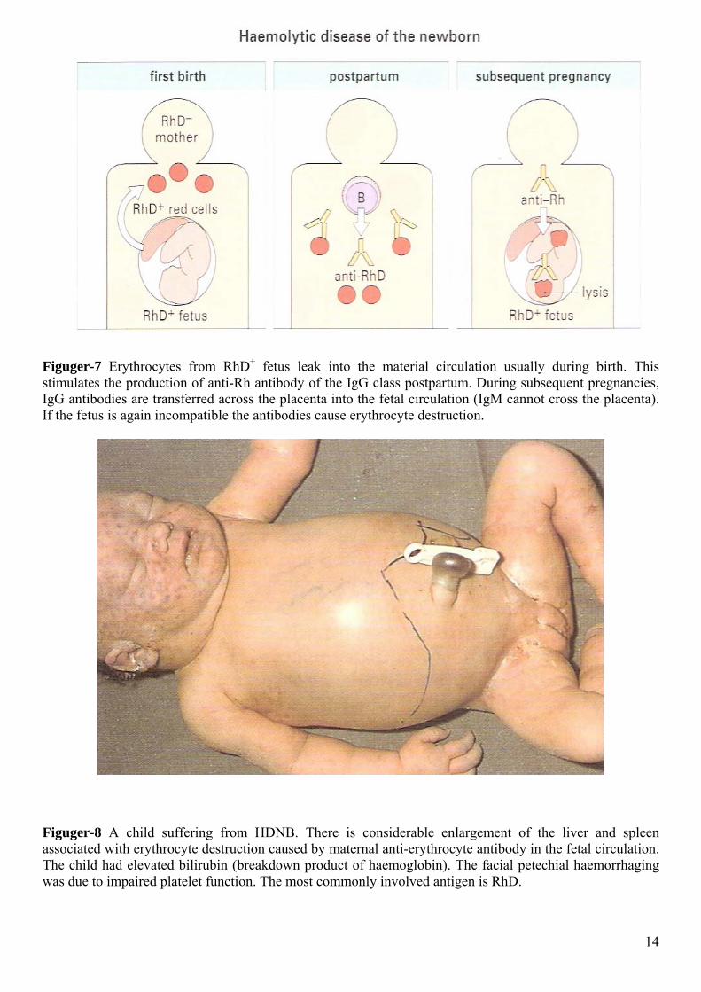
Figuger-7 Erythrocytes from RhD+ fetus leak into the material circulation usually during birth. This stimulates the production of anti-Rh antibody of the IgG class postpartum. During subsequent pregnancies, IgG antibodies are transferred across the placenta into the fetal circulation (IgM cannot cross the placenta). If the fetus is again incompatible the antibodies cause erythrocyte destruction.
Figuger-8 A child suffering from HDNB. There is considerable enlargement of the liver and spleen associated with erythrocyte destruction caused by maternal anti-erythrocyte antibody in the fetal circulation. The child had elevated bilirubin (breakdown product of haemoglobin). The facial petechial haemorrhaging was due to impaired platelet function. The most commonly involved antigen is RhD.
14
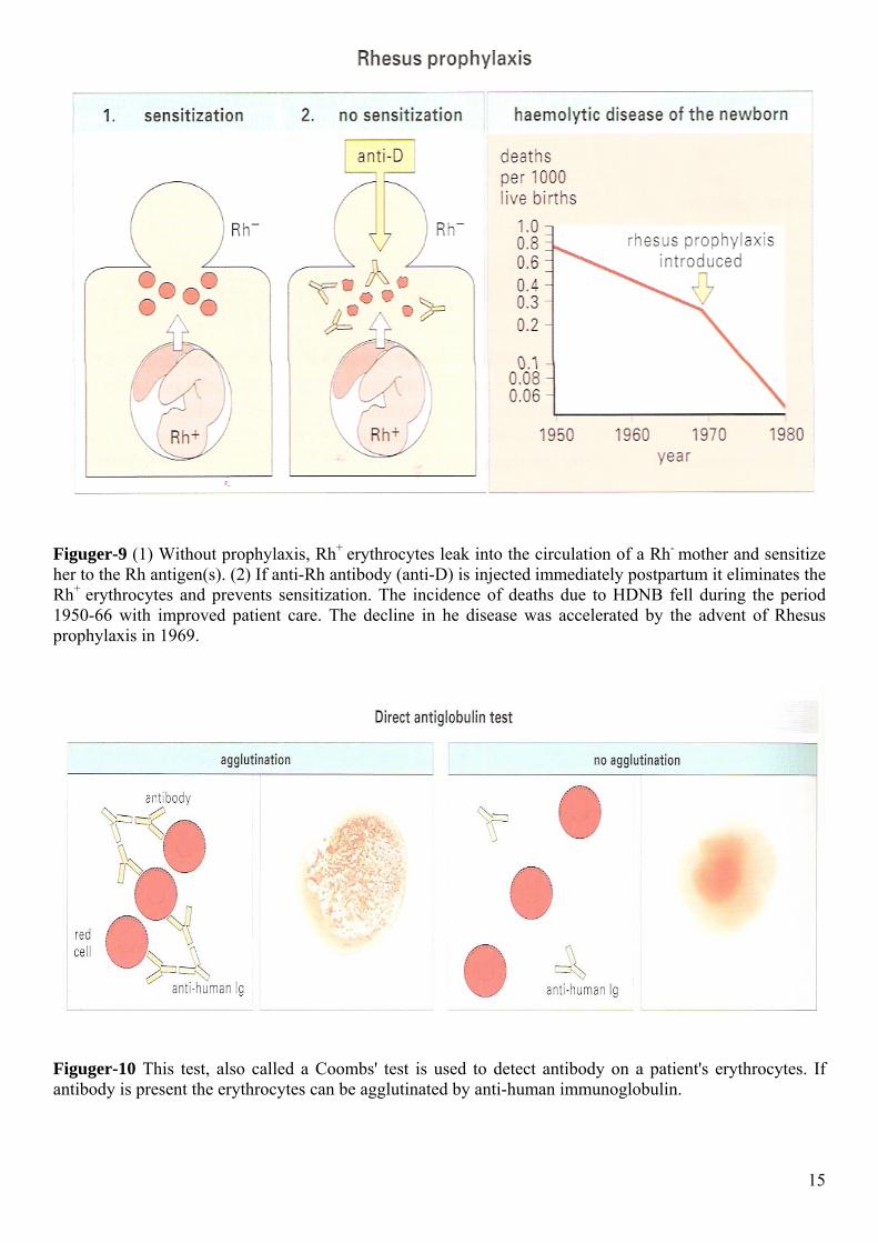
Figuger-9 (1) Without prophylaxis, Rh+ erythrocytes leak into the circulation of a Rh- mother and sensitize her to the Rh antigen(s). (2) If anti-Rh antibody (anti-D) is injected immediately postpartum it eliminates the Rh+ erythrocytes and prevents sensitization. The incidence of deaths due to HDNB fell during the period 1950-66 with improved patient care. The decline in he disease was accelerated by the advent of Rhesus prophylaxis in 1969.
Figuger-10 This test, also called a Coombs' test is used to detect antibody on a patient's erythrocytes. If antibody is present the erythrocytes can be agglutinated by anti-human immunoglobulin.
15
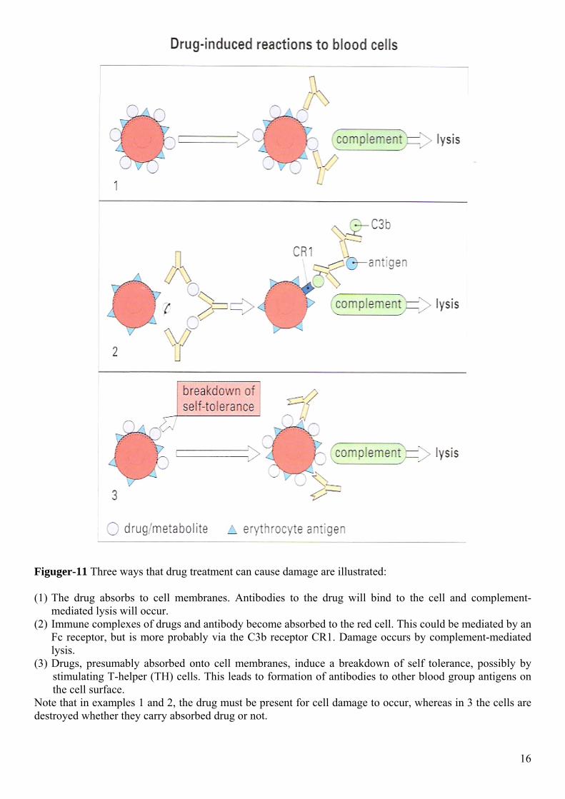
Figuger-11 Three ways that drug treatment can cause damage are illustrated: (1) The drug absorbs to cell membranes. Antibodies to the drug will bind to the cell and complement-
mediated lysis will occur. (2) Immune complexes of drugs and antibody become absorbed to the red cell. This could be mediated by an
Fc receptor, but is more probably via the C3b receptor CR1. Damage occurs by complement-mediated lysis.
(3) Drugs, presumably absorbed onto cell membranes, induce a breakdown of self tolerance, possibly by stimulating T-helper (TH) cells. This leads to formation of antibodies to other blood group antigens on the cell surface.
Note that in examples 1 and 2, the drug must be present for cell damage to occur, whereas in 3 the cells are destroyed whether they carry absorbed drug or not. 16
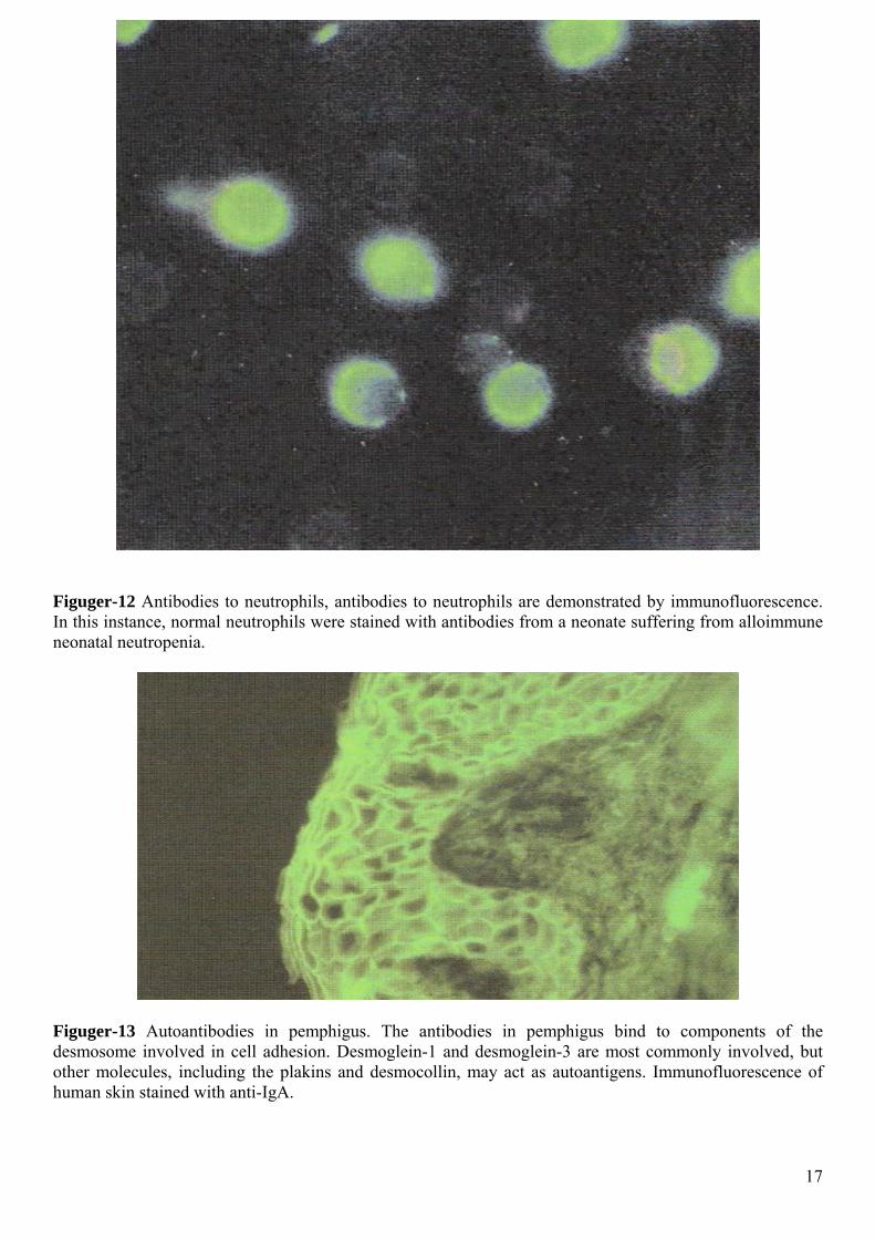
Figuger-12 Antibodies to neutrophils, antibodies to neutrophils are demonstrated by immunofluorescence. In this instance, normal neutrophils were stained with antibodies from a neonate suffering from alloimmune neonatal neutropenia.
Figuger-13 Autoantibodies in pemphigus. The antibodies in pemphigus bind to components of the desmosome involved in cell adhesion. Desmoglein-1 and desmoglein-3 are most commonly involved, but other molecules, including the plakins and desmocollin, may act as autoantigens. Immunofluorescence of human skin stained with anti-IgA.
17
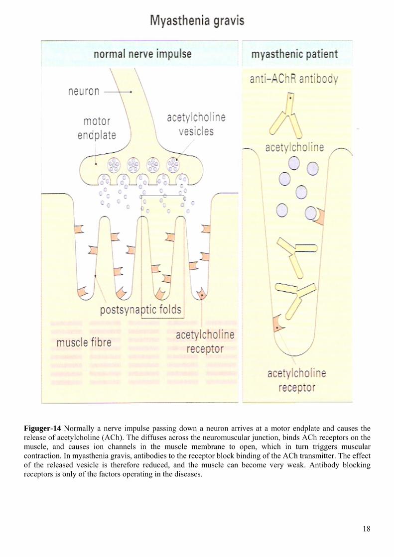
Figuger-14 Normally a nerve impulse passing down a neuron arrives at a motor endplate and causes the release of acetylcholine (ACh). The diffuses across the neuromuscular junction, binds ACh receptors on the muscle, and causes ion channels in the muscle membrane to open, which in turn triggers muscular contraction. In myasthenia gravis, antibodies to the receptor block binding of the ACh transmitter. The effect of the released vesicle is therefore reduced, and the muscle can become very weak. Antibody blocking receptors is only of the factors operating in the diseases.
18
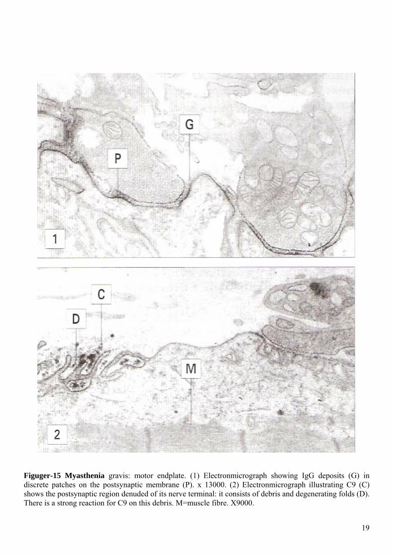
Figuger-15 Myasthenia gravis: motor endplate. (1) Electronmicrograph showing IgG deposits (G) in discrete patches on the postsynaptic membrane (P). x 13000. (2) Electronmicrograph illustrating C9 (C) shows the postsynaptic region denuded of its nerve terminal: it consists of debris and degenerating folds (D). There is a strong reaction for C9 on this debris. M=muscle fibre. X9000.
19
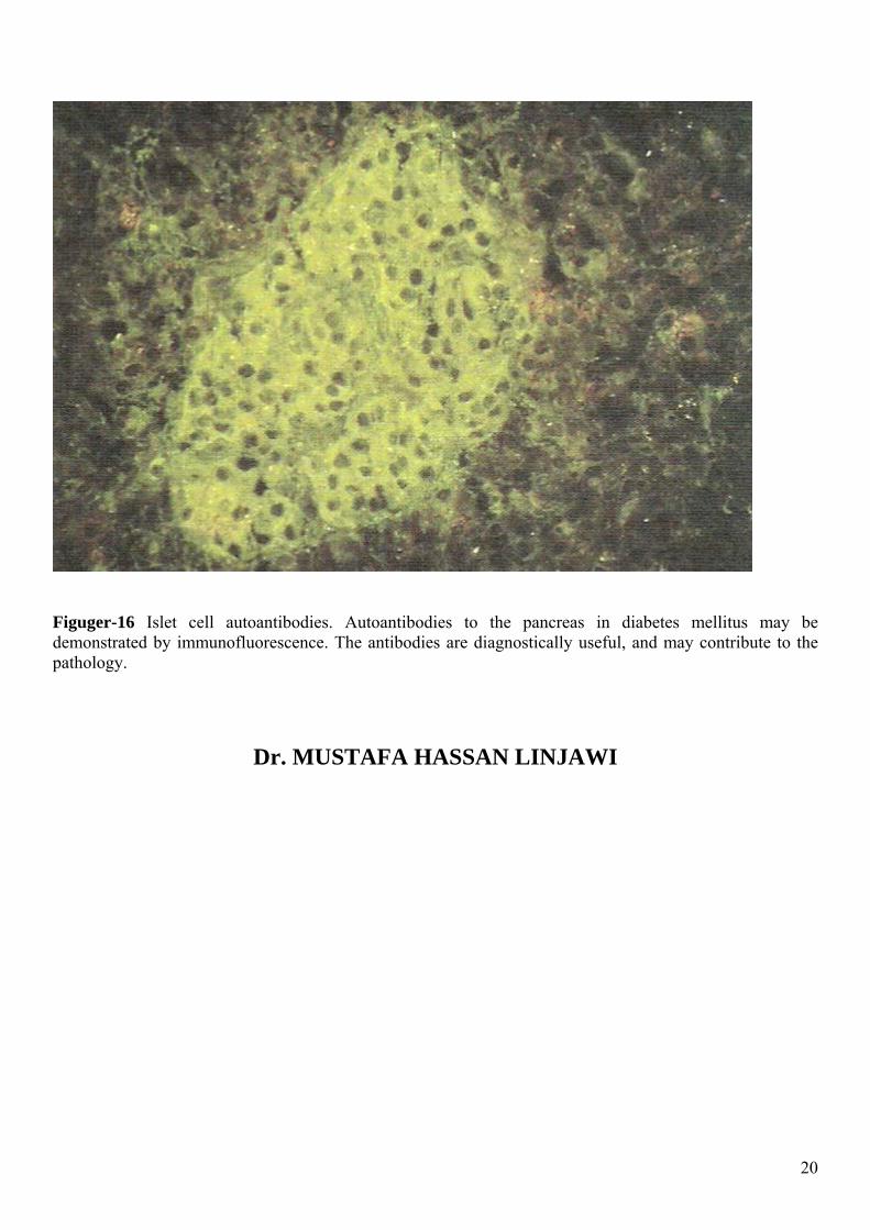
Figuger-16 Islet cell autoantibodies. Autoantibodies to the pancreas in diabetes mellitus may be demonstrated by immunofluorescence. The antibodies are diagnostically useful, and may contribute to the pathology.
Dr. MUSTAFA HASSAN LINJAWI
20
Related Documents