3 Keratoplasty in Contact Lens Related Acanthamoeba Keratitis Beata Kettesy, Laszlo Modis Jr., Andras Berta and Adam Kemeny-Beke Department of Ophthalmology, Medical and Health Science Center University of Debrecen Hungary 1. Introduction Acanthamoeba keratitis (AK) infection is a rather frequently occurring disease all over the world which can still cause serious or even total loss of vision despite improved diagnostic and therapeutic options. It may cause mostly keratitis, scleritis or chorioretinitis in people with competent immune systems. It mainly affects contact lens wearers with poor hygiene. Corneal trauma due to foreign body injury and exposure to contaminated water may also be associated with Acanthamoeba infection. Those with Acanthamoeba keratitis generally are immunocompetent. Nevertheless, these individuals do not develop protective immunity, and thus reinfection can occur. In the mid 1980s, an epidemic of Acanthamoeba keratitis occurred in the US which was attributed to increased contact lens use and poor lens hygiene. Conditions promoting the disease include not only poor contact lens hygiene but also the use of home-made saline solutions and corneal abrasions (Stehr-Green at al., 1989). In the United Kingdom, there was a marked rise in the number of cases in the first half of the 1990s, associated with the introduction and increasing popularity of disposable soft contact lenses shown to be due to irregular and/or chlorine based disinfection. After 1995 there was a decrease, perhaps resulting from an improvement in CL hygiene following the widespread dissemination of the results of a paper on Acanthamoeba keratitis as well as the gaining penetrance of new CL hygiene systems (Radford et al., 1995, 1998, 2002). 1.1 Features of Acanthamoeba Free-living amoebae belonging to the genus Acanthamoeba are the causative agents of granulomatosus amoebic encephalitis, a fatal disease of the central nervous system, cutaneous lesions and sinusitis in immonodeficient patients and amoebic keratitis, a painful sight-threatening disease of the eyes in otherwise healthy individuals. Acanthamoeba was first described by Castellani when he reported the presence of an amoeba in Cryptococcus pararoseus cultures. The genus Acanthamoeba was established later by Volkonsky in 1931 (Marciano-Cabral & Cabral, 2003). The first suggestion that Acanthamoeba could cause disease in humans came in 1958 during polio vaccine safety trials. Plaques appeared in cell cultures used to prepare vaccine and www.intechopen.com

Welcome message from author
This document is posted to help you gain knowledge. Please leave a comment to let me know what you think about it! Share it to your friends and learn new things together.
Transcript

3
Keratoplasty in Contact Lens Related Acanthamoeba Keratitis
Beata Kettesy, Laszlo Modis Jr., Andras Berta and Adam Kemeny-Beke
Department of Ophthalmology, Medical and Health Science Center University of Debrecen
Hungary
1. Introduction
Acanthamoeba keratitis (AK) infection is a rather frequently occurring disease all over the world which can still cause serious or even total loss of vision despite improved diagnostic and therapeutic options. It may cause mostly keratitis, scleritis or chorioretinitis in people with competent immune systems. It mainly affects contact lens wearers with poor hygiene. Corneal trauma due to foreign body injury and exposure to contaminated water may also be associated with Acanthamoeba infection. Those with Acanthamoeba keratitis generally are immunocompetent. Nevertheless, these individuals do not develop protective immunity, and thus reinfection can occur. In the mid 1980s, an epidemic of Acanthamoeba keratitis occurred in the US which was attributed to increased contact lens use and poor lens hygiene. Conditions promoting the disease include not only poor contact lens hygiene but also the use of home-made saline solutions and corneal abrasions (Stehr-Green at al., 1989). In the United Kingdom, there was a marked rise in the number of cases in the first half of the 1990s, associated with the introduction and increasing popularity of disposable soft contact lenses shown to be due to irregular and/or chlorine based disinfection. After 1995 there was a decrease, perhaps resulting from an improvement in CL hygiene following the widespread dissemination of the results of a paper on Acanthamoeba keratitis as well as the gaining penetrance of new CL hygiene systems (Radford et al., 1995, 1998, 2002).
1.1 Features of Acanthamoeba
Free-living amoebae belonging to the genus Acanthamoeba are the causative agents of granulomatosus amoebic encephalitis, a fatal disease of the central nervous system, cutaneous lesions and sinusitis in immonodeficient patients and amoebic keratitis, a painful sight-threatening disease of the eyes in otherwise healthy individuals. Acanthamoeba was first described by Castellani when he reported the presence of an amoeba in Cryptococcus pararoseus cultures. The genus Acanthamoeba was established later by Volkonsky in 1931 (Marciano-Cabral & Cabral, 2003). The first suggestion that Acanthamoeba could cause disease in humans came in 1958 during polio vaccine safety trials. Plaques appeared in cell cultures used to prepare vaccine and
www.intechopen.com
Keratoplasties – Surgical Techniques and Complications
32
were thought to be virus induced because mice and monkeys died from encephalitis following inoculation of tissue culture fluid. These observations of experimental animals dying from encephalitis led Culbertson et al. to predict a role for free-living amoebae as agents of human disease. Human cases of amoebic encephalitis were reported soon thereafter. The first cases which clearly established Acanthamoebae as causative agents of disease in humans were reported in the early 1970s with many more reports of various diseases following ever since (Dunand et al., 1997; Gulett et al., 1979; Illingworth & Cook 1998; Jones et al., 1975; Martinez et al., 1994). Acanthamoeba spp. are among the most prevalent protozoa found in the environment. They are distributed worldwide and have been isolated from soil, dust, air, natural and treated water, seawater, swimming pools, sewage, sediments, air-conditioning units, drinking water treatment plants, bottled water, dental treatment units, hospitals and dialysis units, eyewash stations, and contact lenses and lens cases and as contaminants in bacterial, yeast, and mammalian cell cultures Acanthamoeba spp. also have been isolated from vegetation, from animals including fish, amphibia, reptiles, and mammals, from the nasal mucosa and throats of apparently healthy humans, from infected brain and lung tissue, from skin lesions of immunosuppressed patients, and from corneal tissue of patients with Acanthamoeba
keratitis. It has been shown to live in domestic tap water pipelines especially in the cold water systems (Gray et al., 1995; Kilvington et al., 2004; Marciano-Cabral & Cabral, 2003; Radford et al., 1995). The life cycle of Acanthamoeba consists of two stages: an actively feeding, dividing trophozoite and a dormant cyst. The trophozoite varies in size from 25 to 40 µm and feeds on bacteria, algae, and yeast in the environment but can also exist axenically on nutrients in liquid taken up through pinocytosis. A double-walled wrinkled cyst composed of an ectocyst and an endocyst ranges in size from 13 to 20 µm and varies from species to species. Cyst formation occurs under adverse environmental conditions such as food deprivation, desiccation, and changes in temperature and pH. Cysts are resistant to biocides, chlorination, and antibiotics and survive low temperatures (0 to 2°C). But treatment with Freon or methylene oxide or autoclaving destroys cysts. Excystment occurs when trophozoites emerge from the cyst under suitable environmental conditions. It was demonstrated that cysts retained viable amoebae for over 24 years after storage in water at 4°C (Khan, 2001; Marciano-Cabral & Cabral, 2003). The cellular organization of Acanthamoeba has been studied using electron microscopy. Organelles typically found in higher eukaryotic cells have been identified in Acanthamoebae. They have a Golgi complex, smooth and rough endoplasmic reticula, free ribosomes, digestive vacuoles, mitochondria, and microtubules in Acanthamoeba trophozoites. A trilaminar plasma membrane was found to surround the cytoplasmic contents of the trophozoite. In addition, distinguishing features of the trophozoite were the presence of spiny surface projections called acanthopodia, a prominent contractile vacuole in the cytoplasm that controls the water content of the cell, and a nucleus with a large central nucleolus. Generally, the amoebae are uninucleated, although multinucleated cells are common when Acanthamoeba are maintained in suspension culture. Reproduction occurs by binary fission (Marciano-Cabral & Cabral, 2003). Pathogenic Acanthamoeba exhibited higher numbers (above hundred) of acanthopodia as compared to non-pathogens (below twenty) (Khan, 2001). Acanthamoeba species can be assigned to three different groups on the basis of their cyst morphology. Later the complete gene sequence of nuclear small ribosomal subunit RNA was determined and using this approach, Acanthamoeba species was classified into 53 isolates on the basis of 12 rDNA
www.intechopen.com
Keratoplasty in Contact Lens Related Acanthamoeba Keratitis
33
sequence types (RNA genotypes) designated typing units T1 to T12. Additional sequence types may exist. Sequences of either nuclear (RNA) or mitochondrial rRNA genes are suitable for classifying isolates. Current classification schemes integrate the morphological groups with the 12 sequence types (RNA genotypes) (T1 to T12) so that group I includes sequence types T7, T8, and T9, group II includes sequence types T3, T4, and T11, and group III includes sequence types T1, T2, T5, T6, T10, andT12. Studies in which clinical isolates have been identified based on sequence types have shown that the majority of strains causing keratitis belong to sequence type 4 (Marciano-Cabral & Cabral, 2003). Amoebae have been reported to exhibit different capabilities for binding and internalizing different species of bacteria. Bacterium amoebae interactions may lead to the establishment of an endosymbiotic state or, alternatively, to destruction of either the bacterium or the amoeba. The role of Acanthamoeba spp. as reservoirs or vectors for human pathogens has been examined. The intracellular growth of bacteria in Acanthamoeba has been associated with enhanced survival of bacteria in the environment, increased resistance of bacteria to biocides, and increased bacterial virulence. Intracellular survival within the amoebae has been postulated as a mode by which bacteria survive in substrate-limiting environmental ecosystems. Intracellular growth of bacteria in amoebae apparently also affects the resistance of bacteria to antibiotics (Marciano-Cabral & Cabral, 2003).
1.2 Pathogenesis
Pathogenic and nonpathogenic strains of Acanthamoebae have already been isolated from the environment, but the pathogenesis of infection and the biochemical determinants of virulence are poorly understood. Temperature tolerance, growth rate, adherence properties, cytolytic products produced by amoebae, and immune evasion mechanisms appear to constitute important factors in their pathogenicity. The virulence of pathogenic amoebae wanes during continuous culture in axenic medium but can be restored by brain passage in mice. It has been proposed that virulence may be related to distinct physiological characteristics of a strain and not to a dependence on environmental conditions (Marciano-Cabral & Cabral, 2003). Animal studies have confirmed the clinical impression that contact lenses are vectors for transmitting trophozoites to the corneal surface and facilitating trophozoite binding to the corneal epithelium. Adherence of trophozoites to cells followed by injury and invasion of tissue are thought to represent important steps in the establishment of infections. Clinical isolates of A. polyphaga, A. castellanii, and A. culbertsoni have been shown to attach to corneal epithelial cells through a process which involves binding to cell surface carbohydrate moieties. A. castellanii binds to mannose containing glycoproteins on the corneal epithelium through a 136-kDa mannose-binding protein on the amoeba surface. The adherence of Acanthamoeba to corneal epithelial cells can be inhibited by mannose and methylmannose pyranoside but not by other sugars (Clarke & Niederkorn, 2003; Hurt et al., 2003; Marciano-Cabral & Cabral, 2003). Glycolipids of corneal epithelium reactive with Acanthamoeba may also play a role in the pathogenesis of Acanthamoeba keratitis by mediating the adherence of the amoebae to the cornea. Gordon et al., using binding assays, reported that Acanthamoeba binds preferentially to collagen IV, laminin, and fibronectin (Gordon et al., 1993). Following adherence to cells, invasion and extensive tissue destruction occur in the host. Human epithelial cells, stromal keratocytes, and stromal cell homogenates have been used in vitro as models of Acanthamoeba keratitis. Damage to cells and tissue is thought to occur by phagocytic processes and by cytotoxic substances released by amoebae. Exposure to
www.intechopen.com
Keratoplasties – Surgical Techniques and Complications
34
mannose induces Acanthamoeba trophozoites to produce a 133-kDa protease termed mannose-induced-protein (MIP) 133. MIP133 produces contact independent cytolysis of corneal epithelial cells in vitro. Corneal organ culture studies have shown that binding to mannose glycoproteins on the traumatized corneal epithelium induces trophozoites to release cytopathic factors (including MIP133) that facilitate corneal destruction and invasion. Activated MMPs degrade components of the basement membrane and the extracellular matrix, including types I and II collagens, fibronectin and laminin (Clarke & Niederkorn, 2006; Hurt et al., 2003). Acanthamoeba trophozoites use multiple proteases with nonspecific collagenolytic activity to penetrate and degrade the stroma; these include serine proteases, cysteine protease, an elastase and a metalloproteinase. In addition, there is evidence that a collagenolytic enzyme could have a role in the generation of the ring-like stromal infiltrates and corneal lesions that are characteristic of Acanthamoeba keratitis. Trophozoite adherence was followed by penetration, a process which appeared to involve both secretion of lytic enzymes and phagocytosis (Clarke & Niederkorn, 2006; Hurt et al., 2003).
1.3 Epidemiology, risk factors
Acanthamoeba spp. are among the most prevalent protozoa found in the environment. This spp.
are worldwide and could been isolated from different sites so as soil, dust, air, natural and
treated water, seawater, swimming pools, domestic tap water, drinking water, or even bottled
water. The number of Acanthamoeba in a freshwater lake bottom can be 200-2100 amoeba/g.
In the Potomac River for example there was one amoeba per 3.4 L water. The highest
percentage of pathogens is during spring and fall (Nwachuku & Gerba, 2004). Acanthamoebae
have been detected in tap water and in swimming pools. In Mexico amoebae were found in
13% of the water samples from faucets in private residences. In Egypt 2/50 tap water samples
were contaminated. In Germany amoebae were found in 56% of hot water taps examined in
hospitals. In England amoebae were isolated from bathroom and kitchen cold water taps.
Amoebae may survive pool and spa disinfection (bromine, chlorine) procedures because of
their resistant cyst stages. In 1977 Acanthamoebae were recovered in the water of 27 out of 30
public pools. Acanthamoeba has been isolated in swimming pools in Germany, Mexico and in
Norway, temperature-tolerant strains of Acanthamoeba in spas in New Zealand and Spain.
Acanthamoeba was isolated from bottled water also in Mexico. Acanthamoeba cyst can be
isolated from air, dust, air conditioner, cooling towers (Kilvington et al., 2004; Nwachuku &
Gerba, 2004). Acanthamoebic keratitis first reported by Nagington et al. (Nagington et al.,
1974) in Great Britain and by Jones et al. Jones et al., 1975) in the United States is a painful
progressive sight-threatening corneal disease. Since acanthamoebic keratitis is not a reportable
disease, the true incidence is not known. Published works suggest an incidence rate of 0.58-
0.71 cases/1.000.000 in the general population, and 1.65-2.01/1.000.000 among contact lens
wearers (Nwachuku & Gerba, 2004). The given incidence among contact lens wearers is 3-
5/1.000.000 in Holland, and 33/1.000.000 in Hong Kong (Seal, 2003). Three recent multi centre
Questionnaire Reporting Surveys of Acanthamoeba keratitis were conducted in England
within the past twenty years. The first in 1992–96 gave an incidence rate of 0.25 per 10 000
contact lens wearers (CLW). The second and third surveys in England and Wales carried out
in 1997–99 with one case in 47 620 CLW (or 0.21 per 10 000) and in 1998–99 with one case in
555 CLW (or 0.18 per 10 000) found that 80% of all infections were manifest in lens wearers,
with 88% using hydrogel lenses and 12% rigid lenses The incidence of acanthamoeba keratitis
with gas permeable and rigid contact lenses is much lower than with soft hydrogel CL (Seal,
www.intechopen.com

Keratoplasty in Contact Lens Related Acanthamoeba Keratitis
35
2003). In non contact lens wearers history of trauma in a garden is a risk factor (Radford et al.,
1998).
Patient ages range from 4 to 64 years, with a mean age of 30 years, with no difference in genders (Radford et al., 1998; Sharma et al., 2000). Infection usually affects only one eye, although the infection is occasionally bilateral (Illingworth & Cook, 1998; Parthasarathy et al., 2007; Radford et al., 1998; Wilhelmus et al., 2008). Among non contact lens wearers trauma and exposure to contaminated water have been identified as major risk factors (Chynn et al., 1995; Sharma et al., 2000). Multivariable analysis showed that this is largely attributable to a lack of disinfection, the use of non-sterile saline, and the use of chlorine based disinfection rather than alternative chemical systems (Radford et al., 1995). Many CL users are still contaminating their contact lenses with tap water either directly (showering, face washing, handling with wet hands) or by using water to rinse their storage case (Radford et al., 2002; Seal et al., 1999). Disposable soft contact lenses seem to be a risk factor for contact lens induced keratitis. 89% of the patients with keratitis used this type of contact lens (Dejaco-Ruhswurm et al., 2001). It was shown that adherence of amoeba to the lens was higher among disposable contact lenses (etafilcon A) than among the conventional lenses (polymacon) (Beattie et al., 2003, Lema et al., 2001). Attachment of Acanthamoeba was affected significantly by lens material type (P<0.001), with higher numbers of trophozoites attaching to the first-generation lotrafilcon A silicone hydrogel lens, compared with the second-generation galyfilcon A lens and the conventional hydrogel lens. Patient wear and the presence of a bacterial biofilm had no significant effect on the attachment to the lotrafilcon A lens but did significantly increase attachment to the galyfilcon A and the etafilcon A (P = 0.009) lenses. If exposed to Acanthamoeba (e.g., when showering or swimming, through non-continuous wear and ineffective lens care regimes), first-generation silicone hydrogel lenses may promote a greater risk of Acanthamoeba infection due to the enhanced attachment characteristics of this lens material (Beattie et al., 2006). An initial coinfection occurs, with the bacteria providing an additional food source for the amoebae. The presence of bacterial biofilm may also affect amoebal sensitivity to disinfectants (Illingworth & Cook, 1998). Trophozoite and cyst adherence of two acanthamoeba keratitis strains to four types of unworn soft contact lens and their removal by cleaning agents were studied. Greater adherence of the trophozoites compared with the cysts was recorded. Trophozoites adhered in greater numbers to type I lenses (poly2-hydroxyethyl methacrylate), with no differences between type II (lidofilcon A), III (bufilcon A) and IV (etafilcon A) lenses. Adherence of the other trophozoites to type II lenses was lower compared with their adherence to the other lenses. Cysts of both strains showed greater adherence to type I and III lenses. Recommended cleaning procedures using three commercial solutions were effective in removing Acanthamoeba from the lenses (Kilvington, 1993).
1.4 Clinical symptoms
The clinical picture of acanthamoeba keratitis is remarkable for its varied manifestations, although these often seem to occur in a recognizable sequence. Most patients complain of symptoms of photophobia, pain, and tearing. The pain in acanthamoeba keratitis may be particularly severe, seemingly disproportionate to the signs, although the absence of severe pain does not preclude the diagnosis. Rarely, there may be an apparent precipitating event, such as an injury to the eye, swimming while wearing lenses, or insertion of a non-sterile lens. Symptoms may continue uninterrupted from the time of an injury, sometimes with
www.intechopen.com
Keratoplasties – Surgical Techniques and Complications
36
apparent failure of a corneal abrasion to heal, or rarely, there may be a delay of up to 2 weeks before the onset of symptoms. The earliest signs may be non-specific and may take the form of epithelial micro erosions, irregularities, opacities or microcystic oedema, often with patchy anterior stromal infiltrates. There may be no fluorescein staining at the onset. Commonly, there is a dendriform keratitis that is often initially treated as herpes simplex infection. Limbitis (limbal hyperaemia and oedema) is a very frequent finding in both early as well as late stages of the disease. A pattern of perineural infiltrates that occurs in a radial distribution (radial keratoneuritis) is virtually pathognomic for acanthamoeba keratitis. A ring infiltrate, usually with an overlying epithelial defect is commonly seen. Although initially the epithelium within the ring may be intact, in longstanding cases a central defect, which is often associated with stromal thinning, may occur. The ring may be incomplete, or occasionally it is double and concentric. The inflammation may involve the sclera, evidence of direct scleral invasion by amoebae has often been elusive, leading to the conclusion that scleritis is a secondary immunologic reaction. Posterior segment signs are rare, although occasional reports of optic nerve oedema, optic neuropathy and optic atrophy, retinal detachment, choroidal inflammation, and formation of a macular scar exist. Contra lateral chorioretinitis has also been observed. In up to 20% of cases cataract may occur, although this appears to be associated with severe or prolonged inflammation, and use of topical corticosteroids. Glaucoma is commonly reported, particularly in advanced stages (Illingworth & Cook, 1998; Radford et al., 1998; Reinhard & Sundmacher, 2000).
1.5 Diagnosis
The importance of early diagnosis cannot be overemphasized. We have to consider acanthamoeba keratitis when symptoms associated with trauma especially involving vegetable matter or exposure to contaminated water, such as lake -, sea-, or spring water are mentioned. Early diagnosis is essential to ensure a good prognosis. If effective therapy is delayed for 3 or more weeks the prognosis will deteriorate. Acanthamoeba keratitis should be considered in any case of corneal trauma complicated by exposure to soil or contaminated water, and in all CL wearers (Dart et al., 2009). We also have to question the patients about this during the history taking. Acanthamoebic keratitis has to be differentiated from bacterial or fungal or herpes simplex virus (HSV) keratitis. In addition, the disease must be considered when there is a failure to respond to first-line therapy for bacterial or herpes simplex virus keratitis, even when there has been a positive culture for another organism, because 10% to 23% of cases of Acanthamoeba keratitis may be polymicrobial or co-infected. Perineural and/or ring infiltrates are characteristic clinical signs of the disease (Dart et al., 2009; Marciano-Cabral & Cabral, 2003; Radford et al., 1998). Diagnosis may be achieved by using different methods, including non invasive confocal microscopy which is the preferred diagnostic technique in some centres, with sensitivity and specificity exceeding 90% (Dart et al., 2009). Another quick method is staining corneal scrapings with acridine orange, which reveal yellow-to-orange polygonal, cystic structures consistent with the appearance of Acanthamoeba among inflammatory cells and the corneal epithelial cells (Hahn et al., 1998). An additional advantage of the method is that no special solutions and staining techniques are required. A definitive diagnosis of Acanthamoeba
keratitis can only be made on the basis of culture or histology, or by the identification of the presence of amoebic DNA with PCR (Dart et al., 2009). Corneal scrapes or corneal biopsy specimens are used for culture or for the identification of cysts or trophozoites in stained
www.intechopen.com
Keratoplasty in Contact Lens Related Acanthamoeba Keratitis
37
tissue sections. For culture, material from a corneal scrape can be placed onto no nutrient agar containing E. coli. It should be incubated at 28 to 35°C and held for an extended interval (10 days or more to ensure time for excystation) because some species of Acanthamoeba do not grow well at 35°C or above. However, corneal scrapes may contain bacteria or yeast, which can confuse the diagnosis. Corneal biopsy has been suggested when repeated cultures of corneal scrapings are negative (Illingworth & Cook, 1998; Marciano-Cabral & Cabral, 2003). For a cytological diagnosis, various staining methods can be employed. The indirect immunofluorescent-antibody assay has been used to detect amoebae in corneal scrapings or in biopsy tissue. Calcofluor white, a chemo fluorescent dye with an affinity for the polysaccharide polymers of amoebic cysts, has been used to identify amoebic cysts in corneal tissue. Calcofluor white stains amoebic cyst walls bright apple green and this effect can be enhanced by prolonging the staining period. Evans blue is used to counter stain the background. Trophozoites and cysts in paraffin-embedded tissues can also be rapidly and differentially stained with calcofluor white (Marciano-Cabral & Cabral, 2003). For the rapid and sensitive identification of Acanthamoeba at the genus level, a polymerase chain reaction (PCR)-based method can be used. For typing Acanthamoeba isolates a restriction fragment length polymorphisms (RFLPs) is useful (Khan et al., 2001; Kilvington et al., 2004).
1.6 Treatment
The goals of medical therapy in Acanthamoeba keratitis include the eradication of viable cysts and trophozoites and rapid resolution of the associated inflammatory response.
1.6.1 Medical treatment
There is no single drug capable of eliminating the infection therefore drug combinations have been suggested as a treatment regimen. Several drugs are known to be amoebicid and cysticid. There are currently no drugs that are effective as monoterapy in Acanthamoeba keratitis, so
combinations are suggested. Acanthamoeba trophozoites are sensitive to most available
chemotherapeutic agents (antibiotics, antiseptics, antifungals, antiprotozoals including
metronidazole, antiviral, and antineoplastic agents). However, persistent infection is related
to the presence of Acanthamoeba cysts, against which very few of these agents have any effect
and only agents that are cysticidal in vitro against cysts can be expected to be effective as
therapy. The diamidines and biguanides are currently the most effective cysticidal
antiamoebics in vitro and their use is supported by substantial case series. Metronidazole
(Flagyl; Pfizer Inc, New York, New York, USA) has been used for several cases in one series,
but has proved to have no effect in vitro. For these reasons topical therapy with biguanides
with or without the addition of diamidines is currently the mainstay of treatment for
Acanthamoeba keratitis (Bacon et al., 1993; Dart et al., 2009; Illingworth et al., 1995;
Illingworth & Cook, 1998; McCellan et al., 2001; Seal, 2003). Biguanides: The two biguanides that are in use are polyhexamethylene biguanide (PHMB) 0.02% to 0.06% (200 to 600 g/ml) and chlorhexidine 0.02% to 0.2% (200 to 2000 g/ml). The biguanides interact with the cytoplasmic membrane, resulting in loss of cellular components and inhibition of respiratory enzymes. Both drugs have been effective clinically as primary therapy, as well as in cases where other agents have failed. Clinically, corneal epithelial toxicity has been minimal for both chlorhexidine 0.02% and PHMB 0.02%. Biguanides can be used as first-line treatment for Acanthamoeba keratitis either alone or in combination with diamidines, with which there may be a synergistic or additive effect (Bacon et al., 1993; Dart
www.intechopen.com

Keratoplasties – Surgical Techniques and Complications
38
et al., 2009; Illingworth et al., 1995; Illingworth & Cook, 1998; McCellan et al., 2001; Seal, 2003). Diamidines. Available diamidines include propamidine isethionate 0.1% (1000 g/ml) and hexamidine 0.1% (1000 g/ml); these are licensed as antibacterials in some European countries. The antimicrobial effects of the diamidines result from the cationic surface-active properties inducing structural membrane changes affecting cell permeability. When these molecules penetrate into the amoebic cytoplasm, denaturation of cytoplasmic proteins and enzymes occurs. Propamidine and hexamidine have been effective clinically against both the trophozoite and cyst forms of Acanthamoeba. Clinically, the diamidines are well tolerated by ocular tissues, although prolonged treatment with propamidine may lead to toxic keratopathy (Bacon et al., 1993; Dart et al., 2009; Illingworth et al., 1995; Illingworth & Cook, 1998; McCellan et al., 2001; Seal, 2003). Although neomycin has been widely used, it is ineffective against cysts in vitro. In addition, like all aminoglycosides it is toxic to the corneal epithelium and can often result in indolent corneal ulceration that may be incorrectly attributed to disease activity, nevertheless it is effective for the bacterial co-infection. Povidon-Iodine (Betadine) is amoebicid and cysticid generally, however it can be used in 0.5% concentrate as well (Bacon et al., 1993; Dart et al., 2009; Illingworth et al., 1995; Illingworth & Cook, 1998; McCellan et al., 2001; Seal, 2003). Topical corticosteroids. The role of steroids is controversial, but we believe that topical corticosteroids can have an important and beneficial role in the management of some cases of Acanthamoeba keratitis. The dead cysts persist in the corneal stroma and remain antigenic. This can give rise to a serious inflammatory reaction. Steroid treatment is unnecessary in most cases diagnosed early, which usually respond rapidly to antiamoebic drugs. However, persisting inflammation (anterior scleritis, severe pain, indolent ulcers, corneal inflammation, and anterior chamber inflammation) may respond dramatically to the addition of even low-potency topical steroid therapy, e.g. prednisolone 0.5%, or dexamethasone 0.1% 4 times daily. Clinicians should be careful to avoid use of corticosteroids if possible because they suppress the activity of the macrophage, which is the ‘scavenger’ phagocytic cell responsible for host immunity to Acanthamoeba. Use of nonsteroidal anti-inflammatory drugs, particularly flurbiprofen, is encouraged and also acts as both an analgesic and a mydriatic. When attendant to the inflammation secondary glaucoma appears, anti-glaucomatic drops are used. Pupil dilatators are also used for moving the pupil (Bacon et al., 1993; Dart et al., 2009; Illingworth et al., 1995; Illingworth & Cook, 1998; McCellan et al., 2001; Seal, 2003).
1.6.2 Surgical treatment
With respect to the severity of the disease as well as the complications accompanying it there is a range of surgical methods to choose from. Corneal abrasion or debridement of the affected area of corneal epithelium may be successful if performed at an early stage. Repeated debridement is used in some centres to improve drug penetration (Dart et al., 2009; Illingworth & Cook, 1998; Reinhard & Sundmacher, 2000). Cryosurgery may be valuable in treating Acanthamoeba keratitis cases. Cryocoagulation to the ring infiltrate and central cornea breaks the infected cells and cyst walls (Amoils & Heney, 1999; Reinhard & Sundmacher, 2000). Deep lamellar keratectomy with a conjunctival flap is a suitable approach to help control the infection and to help relieve pain in patients with advanced Acanthamoeba keratitis (Cremon et al., 2002; Parthasarathy & Tan, 2007).
www.intechopen.com
Keratoplasty in Contact Lens Related Acanthamoeba Keratitis
39
Instead of conjunctiva, amniotic membrane is usable. This suggests that amnion cells in
amniotic membrane release proteinase inhibitors and that stromal matrix selectively provide
adhesion molecules for polymorphonuclear cells (Kim et al., 2000). Amniotic membrane
transplantation may be a safe and effective treatment of severe Acanthamoeba keratitis,
particularly during the inflammation phase. It may permit penetrating keratoplasty to be
delayed (Bourcier et al., 2004).
The role of keratoplasty is now largely restricted to the visual rehabilitation of eyes in which a
medical cure has been achieved. In advanced cases corneal transplantation may be
necessary. Because of use of antiamoebal agents, penetrating keratoplasty is now usually
unnecessary in the acute phase unless the cornea has become very thin, with consequent risk
of perforation (Bacon et al, 1993; Dart et al., 2009; Illingworth & Cook, 1998; Reinhard &
Sundmacher, 2000).
Deep lamellar keratoplasty (DLK) with total removal of infected stromal tissue may be
performed in medically unresponsive cases of Acanthamoeba keratitis. Advantages of
DLK in infectious keratitis include less risk of intraocular entry of infectious organisms at
the time of surgery and the potential for improved graft survival rates caused by less
endothelial rejection and failure. In patients with severe disease involving the visual axis,
earlier surgery with DLK would allow debulking of the organisms as well as preservation
of autogenous endothelial cell function (Parthasarathy & Tan, 2007; Por et al., 2009;
Taenaka et al., 2007).
In some cases enucleation or evisceration is needed, because of inflammation, infection or
secundary glaucoma (Bacon et al., 1993; Radford et al., 1998; Reinhard & Sundmacher,
2000).
2. Mean headings
2.1 Patients, methods and results
Patients’ features and clinical data are summarised in Table 1.
pati- ent
age sex time till diagnosis
BCVA
diagnosis clinical features
medical treatment
keratoplasty surgical treatment
BCVA at latest follow up
1. 25 female 3 days 0.01 corneal scrape
corneal ulcus
dimopropamide+neomycine +ciprofloxacin +diclofenac +cyclopentholat
after 2 years urgency keratoplasty (7.75/8.0 mm)because descemetocele
aspiration lentis, scleral buckling, vitrectomy
light perception
2. 26 female 2 weeks 0.01 clinical findings
central ring-infiltration
dimopropamide+PHMB +neomycine +ciprofloxacin +prednisolon +atropin +cyclopentholat
after 3 month proposed keratoplasty (7.0/7.25 mm)
0.7
3. 41 female 4 weeks 0.03 histology central corneal ulcus, hypopyon
dimopropamide+PHMB +neomycine +ciprofloxacin +diclofenac
after 4 month proposed keratoplasty (7.0/7.75 mm)
1.0
www.intechopen.com
Keratoplasties – Surgical Techniques and Complications
40
pati- ent
age sex time till diagnosis
BCVA
diagnosis clinical features
medical treatment
keratoplasty surgical treatment
BCVA at latest follow up
4. 23 male 4 weeks 0.02 corneal scrape
central corneal abscess
dimopropamide+PHMB +neomycine +ciprofloxacin +diclofenac +timolol
after 1 month urgency keratoplasty (7.0/7.5 mm)
light perception
5. 33 female 4 weeks 0.02 histology central infiltration
dimopropamide+neomycine +fluorometholon+cyclopentholat
after 6 month urgency keratoplasty (7.5/7.5 mm)
corneal abrasion, phaco- emulsifikation+IOL
0.9
6. 65 female 2 months 0.15 clinical findings
central ring-infiltration
dimopropamide+PHMB +neomycine +ciprofloxacin +diclofenac
after 2 years urgency keratoplasty (7.7/8.25 mm)
ECCE+PCL+ trabeculectomy after 1 year
light perception
Table 1. Clinical data of our patients (BCVA=best corrected visual acuity)
2.1.1 Demographics
Between 2001 and 2006 we treated 11 patients with Acanthamoeba keratitis. The mean age of the patients was 30.2 years (16-65). The eight female and three male patients were all soft contact lens wearers. The right eye was affected in six and the left in five cases. 82% of the infections occurred in the summer period between June and September. The appropriate diagnosis of Acanthamoeba keratitis took 3 days to 2 months. Beside long-lasting conservative treatment perforating keratoplasty was performed in six cases. In the following we discuss these patients in details.
2.1.2 Risk factors
The infection was caused by poor and improper contact lens hygiene in each and every case. All lenses involved were hydrogel soft lenses: 1 daily, 4 monthly and 1 yearly disposable. None of the lenses have been applied for extended or continuous wear. Two patients rinsed the lenses in tap water, 4 patients went to swim in them. The half of the patients got the lenses from opticians, the other half from ophthalmologists.
2.1.3 Clinical findings and diagnosis
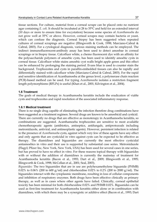
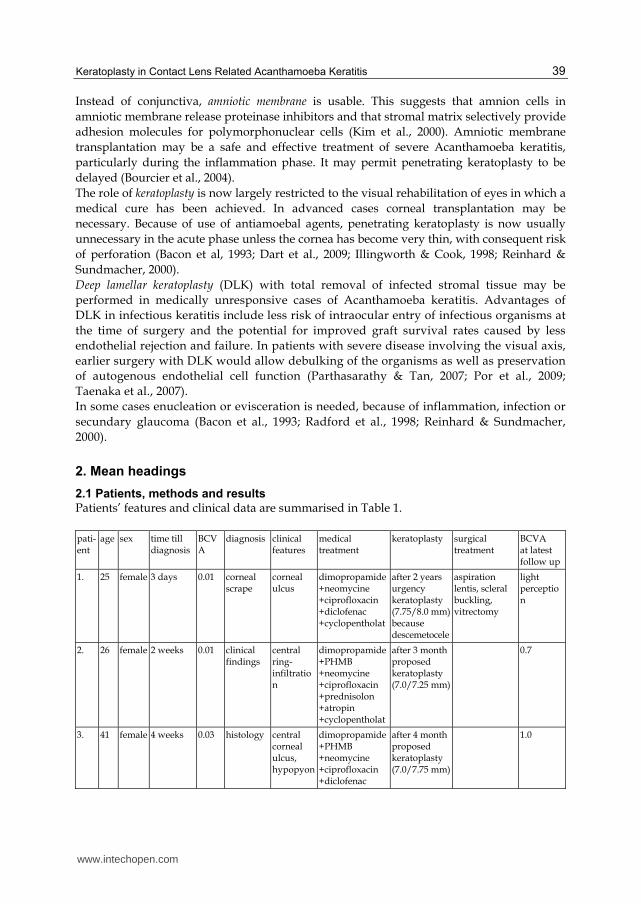
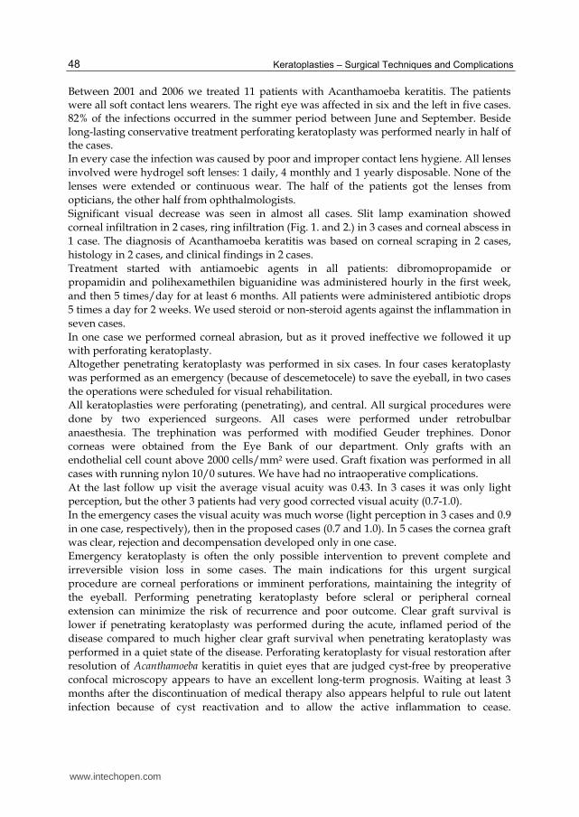
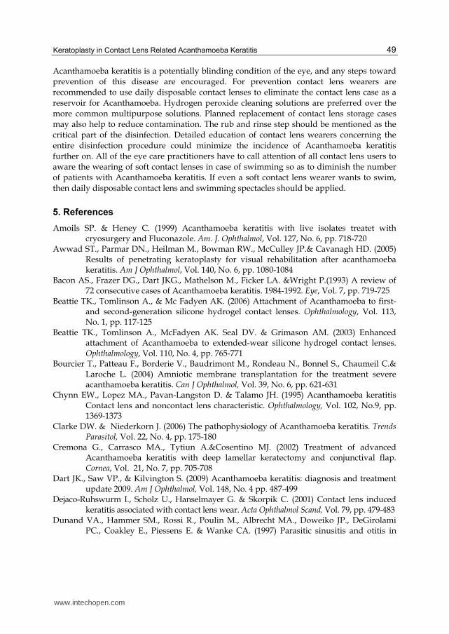
Significant visual decrease (0.15-0.01) was seen in almost all cases. Slit lamp examination showed corneal infiltration in 2 cases, ring infiltration (Fig. 1. and 2.) in 3 cases and corneal abscess in 1 case. The diagnosis was based on corneal scraping in 2 cases, on histology in 2 cases, and on clinical findings in 2 cases.
2.1.4 Medical treatment
Treatment started with antiamoebic agents in all patients: dibromopropamide or propamidin and polihexamethilen biguanidine was administerd hourly in the first week, and then 5 times/day for at least 6 months. All patients were administered antibiotic drops 5 times a day for 2 weeks. Two patients had to use antiglaucomatic drops because of secondary glaucoma. We used steroid or non-steroid agents against the inflammation in seven cases.
www.intechopen.com
Keratoplasty in Contact Lens Related Acanthamoeba Keratitis
41
Fig. 1. Anterior segment of patient No 5 before keratoplasty with corneal abscess
Fig. 2. Corneal scar of patient No 2 before optical keratoplasty
www.intechopen.com
Keratoplasties – Surgical Techniques and Complications
42
2.1.5 Indications for surgical intervention
In one case we performed corneal abrasion, but as it proved ineffective we followed it up
with perforating keratoplasty.
Perforating keratoplasty was performed in six cases. In four cases keratoplasty was
performed as an emergency to save the eyeball (because of descemetocele), in two cases the
operations were scheduled for visual rehabilitation. The emergency cases were the “late
diagnostic” cases.
Shape and size of keratoplasty and surgical techniques: All keratoplasties were perforating
(penetrating), and central. All surgical procedures were done by two experienced surgeons.
All cases were performed under retrobulbar anaesthesia. The trephination was performed
with modified Geuder trephines for the host cornea from the epithelial side, and the graft
from the endothelial side. Donor corneas were from the Eye Bank of our department. Only
grafts with an endothelial cell count above 2000 cells/mm² were used. The mean graft
diameter was 7.7 mm (range 7.25 to 8.25 mm), with an average over sizing of 0.5 mm. Graft
fixation was performed in all cases with running nylon 10/0 sutures. (Fig. 3.,4.)
2.1.6 Postoperative management
Topical antibiotic and steroid drops were used five times a day for 6 weeks, then only
steroid drops for 6 months. Anti amoebatic drops 5 times/day for at least six months.
2.1.7 Complications
We have had no intraoperative complications. Postoperative glaucoma appeared in 2 cases,
in one case trabeculectomy was performed. In 3 cases cataract operation was necessary after
some months. In one case retinal detachment developed and therefore scleral buckling
operation was needed.
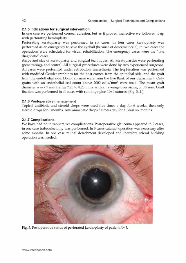
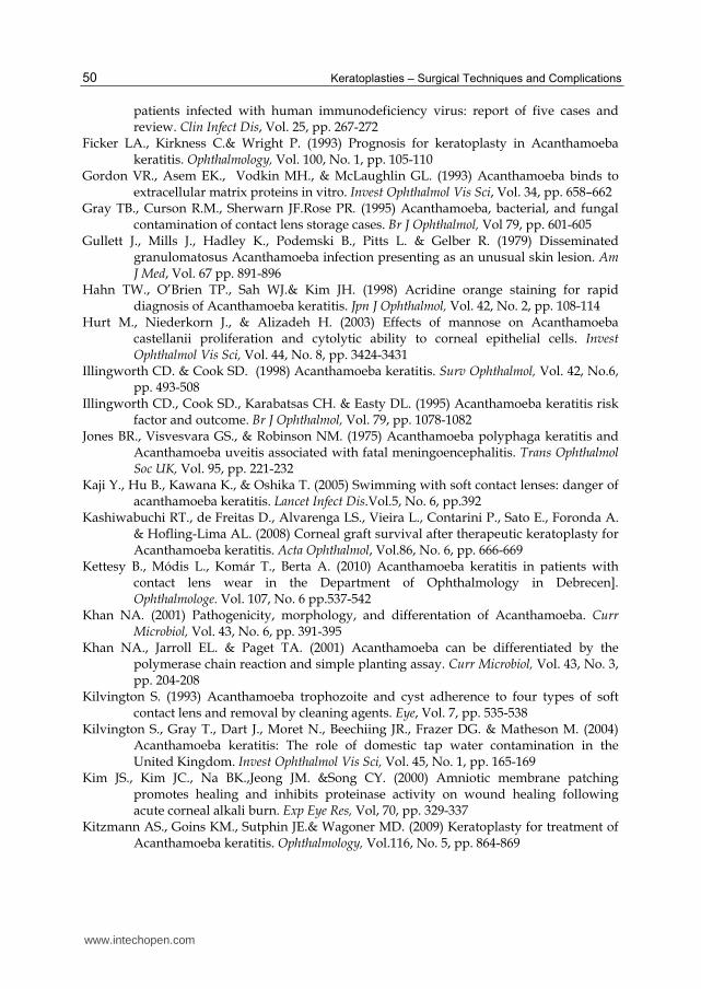
Fig. 3. Postoperative status of perforated keratoplasty of patient No 5.
www.intechopen.com
Keratoplasty in Contact Lens Related Acanthamoeba Keratitis
43
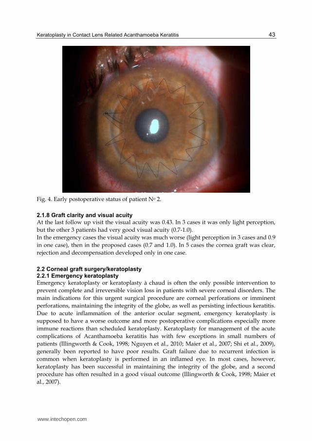
Fig. 4. Early postoperative status of patient No 2.
2.1.8 Graft clarity and visual acuity
At the last follow up visit the visual acuity was 0.43. In 3 cases it was only light perception,
but the other 3 patients had very good visual acuity (0.7-1.0).
In the emergency cases the visual acuity was much worse (light perception in 3 cases and 0.9
in one case), then in the proposed cases (0.7 and 1.0). In 5 cases the cornea graft was clear,
rejection and decompensation developed only in one case.
2.2 Corneal graft surgery/keratoplasty 2.2.1 Emergency keratoplasty
Emergency keratoplasty or keratoplasty à chaud is often the only possible intervention to
prevent complete and irreversible vision loss in patients with severe corneal disorders. The
main indications for this urgent surgical procedure are corneal perforations or imminent
perforations, maintaining the integrity of the globe, as well as persisting infectious keratitis.
Due to acute inflammation of the anterior ocular segment, emergency keratoplasty is
supposed to have a worse outcome and more postoperative complications especially more
immune reactions than scheduled keratoplasty. Keratoplasty for management of the acute
complications of Acanthamoeba keratitis has with few exceptions in small numbers of
patients (Illingworth & Cook, 1998; Nguyen et al., 2010; Maier et al., 2007; Shi et al., 2009),
generally been reported to have poor results. Graft failure due to recurrent infection is
common when keratoplasty is performed in an inflamed eye. In most cases, however,
keratoplasty has been successful in maintaining the integrity of the globe, and a second
procedure has often resulted in a good visual outcome (Illingworth & Cook, 1998; Maier et
al., 2007).
www.intechopen.com
Keratoplasties – Surgical Techniques and Complications
44
Since the introduction of the biguanides as medical therapy, PK has not been recommended as a treatment for the elimination of organisms; emergency keratoplasty should therefore be reserved as a treatment for corneal perforation, imminent perforation and fulminant corneal abscess. Many of these eyes will be severely inflamed with uncontrolled scleritis and limbitis, which should be treated before surgery with systemic immunsuppression using prednisolone (0.5 to 1 mg/kg/day) and/or cyclosporine (3 to 7.5 mg/kg/day), which is tapered as inflammation is controlled in the post graft period (Dart et al. 2009; Maier et al., 2007). Performing PK before scleral or peripheral corneal extension can minimize the risk of recurrence and poor outcome (Nguyen et al., 2010; Tanhehco & Colby, 2010).
2.2.2 Therapeutic/optical keratoplasty
Several authors (Awwad et al., 2005; Por et al., 2009) recommend observing for at least 3
months of clinical inactivity after completion of antiamoebic therapy before attempting PK.
Most recently, Kitzmann et al. (Kitzmann et al., 2009) compared outcomes of 22 eyes with
Acanthamoeba keratitis undergoing emergency penetrating or anterior lamellar keratoplasty
and 9 eyes undergoing optical penetrating or anterior lamellar keratoplasty. Although all eyes
ultimately achieved microbiological cure, there was a 41% recurrence rate of Acanthamoeba
keratitis after emergency keratoplasty compared with 22% recurrence rate after optical
keratoplasty. The Kaplan–Meier graft survival was 37.5% (95% confidence interval, 16.8–58.4)
at 5 years in the therapeutic group compared with 100% in the optical group. Shi et al. (Shi et
al., 2009) reported 28% recurrence after emergency PK. For acanthamoeba keratitis, Ficker et al.
(Ficker et al., 1993) found that clear graft survival was only 20% if penetrating keratoplasty
was performed during the acute, inflamed state of the disease compared to 100% clear graft
survival when penetrating keratoplasty was performed in a quiet state of the disease. The
reason for the difference compared to other results might possibly be differences in case
selection at the time of diagnosis, preoperative treatment and the time point of the decision for
emergency keratoplasty seem to be most important for the outcome of the graft (Maier et al.,
2007). Dart et al. (Dart et al., 2009) present cases, when therapeutic keratoplasty is rarely used
but lamellar or PK to improve vision is carried out in patients with scarred corneas and/or
irregular astigmatism. The outcome of corneal transplantation is good in this group of
patients. Exacerbations of scleritis and limbitis may occur following graft surgery in these eyes
and may need to be treated with systemic anti-inflammatory treatment (Dart et al., 2009).
Quian et al. (Qian et al., 2010) performed 8 optical perforating keratoplasty after at least 4
months medical treatment, with great success. Awwad et al. (Awwad et al., 2005) published an
article about 13 eyes which underwent optical PK for visual rehabilitation after acanthamoeba
keratitis. They mean that PK for visual restoration after resolution of Acanthamoeba keratitis in
quiet eyes that are judged cyst-free by preoperative confocal microscopy appears to have an
excellent long-term prognosis. Waiting at least 3 months after the discontinuation of medical
therapy also appears helpful to rule out latent infection because of cyst reactivation and to
allow the active inflammation to cease.
The success of keratoplasty in the literature are summarised in Table 2.
2.2.3 Donor size
The extent of the infected corneal tissue cannot be identified and should be assumed to include the entire cornea so that, unlike grafts for other corneal infections, which should be large enough to remove all contaminated tissue, the graft for Acanthamoeba keratitis should
www.intechopen.com

Keratoplasty in Contact Lens Related Acanthamoeba Keratitis
45
author year total acute success % optical success %
Kitzmann 2009 30 22 45.5 8 100
Nguyen 2010 9 9 100
Ficker 1993 13 7 20 6 100
Maier 2007 13 13 73
Kashiwabuchi 2008 32 32 43.8
Awwad 2005 13 13 100
Tanhehco 2010 8 8 50
Qian 2010 8 8 100
Por 2009 15 12 3
Illengworth 1995 9 9 100
Bacon 1993 34 21 35 13 69
Radford 1998 31
Sharma 2000 3 3 0
Table 2. Success rate of keratoplasty
be kept to the minimum size required to excise all ulcerated and necrotic tissue, retaining
clinically healthy (but usually subclinically infected) tissue. This is because of the risk of
rejection with large grafts and because repeating grafting may be needed as a result of
recurrence; a further graft represents a new food source for the organism and can be used to
attract residual amoebae. Recurrence of disease in a graft was frequent in the first 2 weeks
after surgery when keratoplasty was performed in an inflamed eye before the introduction
of biguanides; it typically involved the donor periphery, usually without clinical
involvement of the host. Late recurrences, several months after surgery, may also occur. The
use of large grafts worsens the prognosis because it increases the chances of an immune
response to the graft. Furthermore, the use of a large graft indicates a more severe
preoperative infection (Ficker et al., 1993).
Host trephination size ranged from 7 to 11 mm depending on the extent of the corneal
infiltration. The trephination should be performed beyond the clinical areas of infiltrate,
including satellite lesions. Confocal microscopy can be used to outline the extent of
involvement or the size of the trephination (Kashiwabuchiet al., 2008; Nguyen et al., 2010).
Meier et al. reported about a trend toward improved clear graft survival and fewer immune
reactions following emergency keratoplasty using smaller grafts (≤8.00 mm) (Awwad et al.,
2005; Maier et al., 2007).
Ideally, a corneal scar should be debulked as much as possible because residual cysts might still be present and might lead potentially to a recurrence of the disease when exposed to topical corticosteroids after PK. In those cases, rather than jeopardizing graft survival by trephining a large donor button (sizes of 9 mm and beyond), we suggest waiting for a longer period of time after the discontinuation of medical treatment, with repetitive confocal examination when available, before committing the patient to surgery (Awwad et al, 2005).
www.intechopen.com
Keratoplasties – Surgical Techniques and Complications
46
3. Prevention
Acanthamoeba keratitis is a potentially blinding condition of the eye, and any steps toward
prevention of this disease are encouraged. It is recommended contact lens wearers to use
daily disposable contact lenses to eliminate the contact lens case as a reservoir for
Acanthamoeba. If daily disposable contact lens can not be used, then hydrogen peroxide
cleaning solutions are preferred over the more common multipurpose solutions because of
the increased efficacy of hydrogen peroxide against Acanthamoeba cysts. Planned
replacement of contact lens storage cases may also help reduce contamination. It is
important for eye care professionals not only to emphasize proper contact lens wear and
hygiene in the prevention of infectious keratitis but also to maintain a high clinical suspicion
for Acanthamoeba keratitis because an early diagnosis may lead to a better visual outcome
(Tanhehco & Colby, 2010). The use of surfactant cleaner has been shown to be an effective
way of removing Acanthamoeba trophozoites and cysts (Kilvington, 1993). In the light of
these findings further research into the types of disinfectant used by patients with
Acanthamoeba keratitis is required, with a view to calculating the relative risks of different
lens type and disinfectant combinations. This series also show the importance of the fact that
contact lens practitioners should ensure at the time of prescribing lenses that patients have
understood all the disinfectant procedures (Illingworth et al., 1995). Gray et al. suggest
leaving contact lens case open to dry air after heat disinfection. If hydrogen peroxide
disinfection is the preferred solution, one has to use a two step hydrogen peroxide system. It
is necessary to call one’s attention to wash hands properly before handling contact lens cases
and what is more important is to avoid homemade saline. The contact lens case should be
replaced regularly. Nowadays the use of more frequently disposable contact lenses is highly
recommended (Gray et al., 1995). Also the rub and rinse step should be mentioned as the
critical part of the disinfection that may reduce significantly the microbiological load (Shih
et al., 1985). Detailed education of contact lens wearers concerning of the entire disinfection
procedure could reduce the incidence of Acanthamoeba keratitis further on. There is a
persistent need to educate contact lens wearers continuously about the possible risk factors
of Acanthamoeba keratitis, and since the frequency of Acanthamoeba keratitis appears to be
largely determined by the ever changing trends in contact lens use, continued monitoring is
indicated (Radford et al., 1998). All of the eye care practitioners have to call attention of all
contact lens users to aware the wearing of soft contact lenses in case of swimming so as to
reduce the number of patients with Acanthamoeba keratitis (Kaji et al., 2005; Kettesy et al.,
2010). If even a soft contact lens wearer wants to swim, then daily disposable contact lens
and swimming spectacles should be used.
4. Conclusion
Acanthamoebic keratitis, first reported by Nagington et al. in Great Britain and by Jones et
al. in the United States, is a painful progressive sight-threatening corneal disease. The
infection is a rather frequently occurring disease all over the world which can still cause
serious or even total loss of vision despite improved diagnostic and therapeutic options.
Acanthamoeba spp. are among the most prevalent protozoa found in the environment. They
are distributed worldwide and have been isolated from soil, dust, air, natural and treated
water, seawater, swimming pools, sewage, sediments, air-conditioning units, drinking water
www.intechopen.com
Keratoplasty in Contact Lens Related Acanthamoeba Keratitis
47
treatment plants, bottled water, dental treatment units, hospitals and dialysis units, eyewash
stations, and contact lenses and lens cases and as contaminants in bacterial, yeast, and
mammalian cells. It mainly affects contact lens wearers with poor hygiene. In non contact
lens wearers history of trauma in a garden is a risk factor. Patients’ ages range from 4 to 64
years, with a mean age of 30 years, with no difference in genders. Infection usually affects
only one eye, although it is occasionally bilateral. Most patients complain of symptoms of
photophobia, pain, and tearing. The earliest signs may be non-specific and may take the
form of epithelial micro erosions, irregularities, opacities or microcystic oedema, often with
patchy anterior stromal infiltrates. There may be no fluorescein staining at the onset.
Commonly, there is a dendriform keratitis that is often initially treated as herpes simplex
infection. A ring infiltrate, usually with an overlying epithelial defect, is commonly seen.
The ring may be incomplete, or occasionally it is double and concentric. The importance of
early diagnosis cannot be overemphasized. We have to consider Acanthamoeba keratitis
when symptoms associated with trauma especially involving vegetable matter or exposure
to contaminated water, such as lake-, sea-, or spring water are mentioned. Acanthamoebic
keratitis has to be differentiated from bacterial or fungal or herpes simplex virus keratitis.
Diagnosis may be achieved by using different methods, including non invasive confocal
microscopy, staining corneal scrapings with acridine orange, corneal scrapes or corneal
biopsy specimens onto no nutrient agar containing E. coli., cytological various staining
methods, like indirect immunofluorescent-antibody assay, polymerase chain reaction (PCR)-
based method. The goals of medical therapy in Acanthamoeba keratitis include the
eradication of viable cysts and trophozoites and rapid resolution of the associated
inflammatory response. There is no single drug capable of eliminating the infection
therefore drug combinations have been suggested as a treatment regimen. There are
currently no drugs that are effective as monotherapy in Acanthamoeba keratitis, hence
combinations are suggested. Acanthamoeba trophozoites are sensitive to most available
chemotherapeutic agents (antibiotics, antiseptics, antifungals, antiprotozoals including
metronidazole, antiviral, and antineoplastic agents). The diamidines and biguanides are
currently the most effective cysticidal antiamoebics. With respect to the severity of the
disease as well as the complications accompanying it there is a range of surgical methods to
choose from. Corneal abrasion, cryosurgery, deep lamellar keratectomy with a conjunctival
flap, amnion membrane transplantation, keratoplasty, deep lamellar keratoplasty. In some
cases enucleation or evisceration is needed, because of severe inflammation, infection or
secondary glaucoma. Antiamoebic therapy should be used before surgery and be continued
postoperatively using drugs and doses that will minimize or avoid signs of toxicity. PHMB
0.02% has low clinical toxicity in most patients and is clinically less than that with either of
the diamidines. We use PHMB 0.02% 6 to 8 times daily immediately after surgery, with an
adequate level of topical steroid to control inflammation. This should be continued for at
least 3 weeks while results of culture of the host keratectomy specimen are awaited. If viable
organisms are cultured it is prudent to continue antiamoebic therapy 4 times daily while
high-doses of steroids are needed usually for 6 months after surgery, as recurrent
Acanthamoeba keratitis has occurred up to 3 months after an initially successful transplant.
If culture of the excised host cornea is negative after 3 weeks we assume that most viable
amoebae have been treated, and the topical antiamoebic therapy is reduced to 4 times daily
and stopped after 1 month.
www.intechopen.com
Keratoplasties – Surgical Techniques and Complications
48
Between 2001 and 2006 we treated 11 patients with Acanthamoeba keratitis. The patients were all soft contact lens wearers. The right eye was affected in six and the left in five cases. 82% of the infections occurred in the summer period between June and September. Beside long-lasting conservative treatment perforating keratoplasty was performed nearly in half of the cases. In every case the infection was caused by poor and improper contact lens hygiene. All lenses
involved were hydrogel soft lenses: 1 daily, 4 monthly and 1 yearly disposable. None of the
lenses were extended or continuous wear. The half of the patients got the lenses from
opticians, the other half from ophthalmologists.
Significant visual decrease was seen in almost all cases. Slit lamp examination showed
corneal infiltration in 2 cases, ring infiltration (Fig. 1. and 2.) in 3 cases and corneal abscess in
1 case. The diagnosis of Acanthamoeba keratitis was based on corneal scraping in 2 cases,
histology in 2 cases, and clinical findings in 2 cases.
Treatment started with antiamoebic agents in all patients: dibromopropamide or
propamidin and polihexamethilen biguanidine was administered hourly in the first week,
and then 5 times/day for at least 6 months. All patients were administered antibiotic drops
5 times a day for 2 weeks. We used steroid or non-steroid agents against the inflammation in
seven cases.
In one case we performed corneal abrasion, but as it proved ineffective we followed it up with perforating keratoplasty. Altogether penetrating keratoplasty was performed in six cases. In four cases keratoplasty
was performed as an emergency (because of descemetocele) to save the eyeball, in two cases
the operations were scheduled for visual rehabilitation.
All keratoplasties were perforating (penetrating), and central. All surgical procedures were
done by two experienced surgeons. All cases were performed under retrobulbar
anaesthesia. The trephination was performed with modified Geuder trephines. Donor
corneas were obtained from the Eye Bank of our department. Only grafts with an
endothelial cell count above 2000 cells/mm² were used. Graft fixation was performed in all
cases with running nylon 10/0 sutures. We have had no intraoperative complications.
At the last follow up visit the average visual acuity was 0.43. In 3 cases it was only light
perception, but the other 3 patients had very good corrected visual acuity (0.7-1.0).
In the emergency cases the visual acuity was much worse (light perception in 3 cases and 0.9
in one case, respectively), then in the proposed cases (0.7 and 1.0). In 5 cases the cornea graft
was clear, rejection and decompensation developed only in one case.
Emergency keratoplasty is often the only possible intervention to prevent complete and
irreversible vision loss in some cases. The main indications for this urgent surgical
procedure are corneal perforations or imminent perforations, maintaining the integrity of
the eyeball. Performing penetrating keratoplasty before scleral or peripheral corneal
extension can minimize the risk of recurrence and poor outcome. Clear graft survival is
lower if penetrating keratoplasty was performed during the acute, inflamed period of the
disease compared to much higher clear graft survival when penetrating keratoplasty was
performed in a quiet state of the disease. Perforating keratoplasty for visual restoration after
resolution of Acanthamoeba keratitis in quiet eyes that are judged cyst-free by preoperative
confocal microscopy appears to have an excellent long-term prognosis. Waiting at least 3
months after the discontinuation of medical therapy also appears helpful to rule out latent
infection because of cyst reactivation and to allow the active inflammation to cease.
www.intechopen.com
Keratoplasty in Contact Lens Related Acanthamoeba Keratitis
49
Acanthamoeba keratitis is a potentially blinding condition of the eye, and any steps toward
prevention of this disease are encouraged. For prevention contact lens wearers are
recommended to use daily disposable contact lenses to eliminate the contact lens case as a
reservoir for Acanthamoeba. Hydrogen peroxide cleaning solutions are preferred over the
more common multipurpose solutions. Planned replacement of contact lens storage cases
may also help to reduce contamination. The rub and rinse step should be mentioned as the
critical part of the disinfection. Detailed education of contact lens wearers concerning the
entire disinfection procedure could minimize the incidence of Acanthamoeba keratitis
further on. All of the eye care practitioners have to call attention of all contact lens users to
aware the wearing of soft contact lenses in case of swimming so as to diminish the number
of patients with Acanthamoeba keratitis. If even a soft contact lens wearer wants to swim,
then daily disposable contact lens and swimming spectacles should be applied.
5. References
Amoils SP. & Heney C. (1999) Acanthamoeba keratitis with live isolates treatet with cryosurgery and Fluconazole. Am. J. Ophthalmol, Vol. 127, No. 6, pp. 718-720
Awwad ST., Parmar DN., Heilman M., Bowman RW., McCulley JP.& Cavanagh HD. (2005) Results of penetrating keratoplasty for visual rehabilitation after acanthamoeba keratitis. Am J Ophthalmol, Vol. 140, No. 6, pp. 1080-1084
Bacon AS., Frazer DG., Dart JKG., Mathelson M., Ficker LA. &Wright P.(1993) A review of 72 consecutive cases of Acanthamoeba keratitis. 1984-1992. Eye, Vol. 7, pp. 719-725
Beattie TK., Tomlinson A., & Mc Fadyen AK. (2006) Attachment of Acanthamoeba to first- and second-generation silicone hydrogel contact lenses. Ophthalmology, Vol. 113, No. 1, pp. 117-125
Beattie TK., Tomlinson A., McFadyen AK. Seal DV. & Grimason AM. (2003) Enhanced attachment of Acanthamoeba to extended-wear silicone hydrogel contact lenses. Ophthalmology, Vol. 110, No. 4, pp. 765-771
Bourcier T., Patteau F., Borderie V., Baudrimont M., Rondeau N., Bonnel S., Chaumeil C.& Laroche L. (2004) Amniotic membrane transplantation for the treatment severe acanthamoeba keratitis. Can J Ophthalmol, Vol. 39, No. 6, pp. 621-631
Chynn EW., Lopez MA., Pavan-Langston D. & Talamo JH. (1995) Acanthamoeba keratitis Contact lens and noncontact lens characteristic. Ophthalmology, Vol. 102, No.9, pp. 1369-1373
Clarke DW. & Niederkorn J. (2006) The pathophysiology of Acanthamoeba keratitis. Trends Parasitol, Vol. 22, No. 4, pp. 175-180
Cremona G., Carrasco MA., Tytiun A.&Cosentino MJ. (2002) Treatment of advanced Acanthamoeba keratitis with deep lamellar keratectomy and conjunctival flap. Cornea, Vol. 21, No. 7, pp. 705-708
Dart JK., Saw VP., & Kilvington S. (2009) Acanthamoeba keratitis: diagnosis and treatment update 2009. Am J Ophthalmol, Vol. 148, No. 4 pp. 487-499
Dejaco-Ruhswurm I., Scholz U., Hanselmayer G. & Skorpik C. (2001) Contact lens induced keratitis associated with contact lens wear. Acta Ophthalmol Scand, Vol. 79, pp. 479-483
Dunand VA., Hammer SM., Rossi R., Poulin M., Albrecht MA., Doweiko JP., DeGirolami PC., Coakley E., Piessens E. & Wanke CA. (1997) Parasitic sinusitis and otitis in
www.intechopen.com
Keratoplasties – Surgical Techniques and Complications
50
patients infected with human immunodeficiency virus: report of five cases and review. Clin Infect Dis, Vol. 25, pp. 267-272
Ficker LA., Kirkness C.& Wright P. (1993) Prognosis for keratoplasty in Acanthamoeba keratitis. Ophthalmology, Vol. 100, No. 1, pp. 105-110
Gordon VR., Asem EK., Vodkin MH., & McLaughlin GL. (1993) Acanthamoeba binds to extracellular matrix proteins in vitro. Invest Ophthalmol Vis Sci, Vol. 34, pp. 658–662
Gray TB., Curson R.M., Sherwarn JF.Rose PR. (1995) Acanthamoeba, bacterial, and fungal contamination of contact lens storage cases. Br J Ophthalmol, Vol 79, pp. 601-605
Gullett J., Mills J., Hadley K., Podemski B., Pitts L. & Gelber R. (1979) Disseminated granulomatosus Acanthamoeba infection presenting as an unusual skin lesion. Am J Med, Vol. 67 pp. 891-896
Hahn TW., O’Brien TP., Sah WJ.& Kim JH. (1998) Acridine orange staining for rapid diagnosis of Acanthamoeba keratitis. Jpn J Ophthalmol, Vol. 42, No. 2, pp. 108-114
Hurt M., Niederkorn J., & Alizadeh H. (2003) Effects of mannose on Acanthamoeba castellanii proliferation and cytolytic ability to corneal epithelial cells. Invest Ophthalmol Vis Sci, Vol. 44, No. 8, pp. 3424-3431
Illingworth CD. & Cook SD. (1998) Acanthamoeba keratitis. Surv Ophthalmol, Vol. 42, No.6, pp. 493-508
Illingworth CD., Cook SD., Karabatsas CH. & Easty DL. (1995) Acanthamoeba keratitis risk factor and outcome. Br J Ophthalmol, Vol. 79, pp. 1078-1082
Jones BR., Visvesvara GS., & Robinson NM. (1975) Acanthamoeba polyphaga keratitis and Acanthamoeba uveitis associated with fatal meningoencephalitis. Trans Ophthalmol Soc UK, Vol. 95, pp. 221-232
Kaji Y., Hu B., Kawana K., & Oshika T. (2005) Swimming with soft contact lenses: danger of acanthamoeba keratitis. Lancet Infect Dis.Vol.5, No. 6, pp.392
Kashiwabuchi RT., de Freitas D., Alvarenga LS., Vieira L., Contarini P., Sato E., Foronda A. & Hofling-Lima AL. (2008) Corneal graft survival after therapeutic keratoplasty for Acanthamoeba keratitis. Acta Ophthalmol, Vol.86, No. 6, pp. 666-669
Kettesy B., Módis L., Komár T., Berta A. (2010) Acanthamoeba keratitis in patients with contact lens wear in the Department of Ophthalmology in Debrecen]. Ophthalmologe. Vol. 107, No. 6 pp.537-542
Khan NA. (2001) Pathogenicity, morphology, and differentation of Acanthamoeba. Curr Microbiol, Vol. 43, No. 6, pp. 391-395
Khan NA., Jarroll EL. & Paget TA. (2001) Acanthamoeba can be differentiated by the polymerase chain reaction and simple planting assay. Curr Microbiol, Vol. 43, No. 3, pp. 204-208
Kilvington S. (1993) Acanthamoeba trophozoite and cyst adherence to four types of soft contact lens and removal by cleaning agents. Eye, Vol. 7, pp. 535-538
Kilvington S., Gray T., Dart J., Moret N., Beechiing JR., Frazer DG. & Matheson M. (2004) Acanthamoeba keratitis: The role of domestic tap water contamination in the United Kingdom. Invest Ophthalmol Vis Sci, Vol. 45, No. 1, pp. 165-169
Kim JS., Kim JC., Na BK.,Jeong JM. &Song CY. (2000) Amniotic membrane patching promotes healing and inhibits proteinase activity on wound healing following acute corneal alkali burn. Exp Eye Res, Vol, 70, pp. 329-337
Kitzmann AS., Goins KM., Sutphin JE.& Wagoner MD. (2009) Keratoplasty for treatment of Acanthamoeba keratitis. Ophthalmology, Vol.116, No. 5, pp. 864-869
www.intechopen.com
Keratoplasty in Contact Lens Related Acanthamoeba Keratitis
51
Lema I., Rodríguez-Ares MT., Gómez-Torreiro M. & Penalver MD. (2001) Adherence of Acanthamoeba to unworn conventional and disposable soft contact lenses. Cornea, Vol. 20, No. 6, pp. 635-638
Maier P., Böhringer D.& Reinhard T. (2007) Clear graft survival and immune reactions following emergency keratoplasty. Graefes Arch Clin Exp Ophthalmol, Vol. 245, No. 3, pp.351-359
Marciano-Cabral F., & Cabral G. (2003) Acanthamoeba ssp. as agent of disease in humans. Clin Microbiol Rev, Vol. 16, No. 2, pp. 273-307
Martinez AJ., Guerra AE., Garcia-Tamayo J., Céspedes G., Gonzalez-Alfonzo JE.&Visvesvara GS. (1994) Granulomatosus amoebic encephalitis: a review and report of a spontaneous case from Venezuela. Acta Neuropathol, Vol 87, No.4, pp. 430-434
Mc-Clellan K., Howard K., Niederkorn JY., & Alizadeh H. (2001) Effect of seroid on Acanthamoeba cyst and trophosoites. Invest Ophthalmol Vis Sci, Vol. 42, No. 12 pp. 2885-2893
Nagington J., Watson PG., Playfair TJ., McGill J., Jones BR. & Steele AD. (1974) Amoebic infection of the eye. Lancet, pp. 1537-1540
Nguyen TH., Weisenthal RW., Florakis GJ., Reidy JJ.,Gaster RN., & Tom D. (2010) Penetrating Keratoplasty in Active Acanthamoeba Keratitis. Cornea, Vol. 29, No. 9, pp. 1000-1004
Nwachuku N. & Gerba CP (2004) Health Effects of Acanthamoeba spp. and its potential for waterborne transmission. Rev Environ Contam Toxicol, Vol. 180, pp. 93-131
Parthasarathy A. & Tan DT. (2007) Deep lamellar keratoplasty for acanthamoeba keratitis. Cornea, Vol. 26, No. 8, pp. 1021-1023
Por YM., Mehta JS., Chua JL., Koh TH., Khor WB., Fong AC., Lim JW., Heng WJ., Loh RS., Lim L. & Tan DT. (2009) Acanthamoeba keratitis associated with contact lens wear in Singapore. Am J Ophthalmol, Vol. 148, No. 1, pp. 7-12
Qian Y., Meisler DM., Langston RH.& Jeng BH. (2010) Clinical experience with Acanthamoeba keratitis at the cole eye institute, 1999-2008. Cornea, Vol. 29, No. 9, pp. 1016-1021
Radford CF., Bacon AS., Dart JKG., & Minassian DC. (1995) Risk factors for acanthamoeba keratitis contact lens users: a case control study. BMJ, Vol. 310 pp.1567–1570
Radford CF., Lehmann OJ., & Dart JKG. (1998) Acanthamoeba keratitis: multicentre survey in England 1992–1996. Br J Ophthalmol, Vol. 82j pp.1387–1392
Radford CF., Minassian DC., & Dart JKG. (2002) Acanthamoeba keratitis in England and Wales: incidence, outcome, and risk factors. Br J Ophthalmol, Vol. 86j pp. 536–542
Reinhard T., & Sundmacher R. (2000) Klinik und Therapie der Akantamöbenkeratitis. Ophthalmologe, Vol. 97, pp. 446-459
Seal DV. 2003 Acanthamoeba keratitis update-incidence, molecular epidemiology and new drugs for treatment. Eye, Vol.17, pp. 893-905
Seal DV., Kirkness CM., Bennett HGB. & Peterson M. (1999) Acanthamoeba keratitis in Scotland: risk factors for contact lens wearers. Contact Lens Anterior Eye, Vol. 22, No. 2, pp. 58-68
Sharma S., Garg P. & Rao GN. (2000) Patient characteristics, diagnosis, and treatment of non-contact lens releated Acanthamoeba keratitis. Br J Ophthalmol, Vol. 84, pp. 1103-1108
www.intechopen.com
Keratoplasties – Surgical Techniques and Complications
52
Shi W., Liu M., Gao H., Li S. & Xie L. (2009) Perioperative treatment and prognostic factors for penetrating keratoplasty in Acanthamoeba keratitis unresponsive to medical treatment. Graefes Arch Clin Exp Ophthalmol, Vol. 247, No. 10, pp. 1383-1388
Shih KL., Hu J.& Sibley MJ. (1985) The microbiological benefit of cleaning and rinsing of contact lenses. Int Contact Lens Clin; Vol. 12. pp.235–242.
Stehr-Green JK., Bailey TM., & Visvesvara GS. (1989) The epidemiology of Acanthamoeba keratitis in the United States. Am J Ophthalmol, Vol. 107, No. 4, pp. 331–336
Taenaka N, Fukuda M, Hibino T, Kato Y, Arimura E, Ishii Y, & Shimomura Y. (2007) Surgical therapies for Acanthamoeba keratitis by phototherapeutic keratectomy and deep lamellar keratoplasty. Cornea, Vol. 26, No. 7, pp. 876-879
Tanhehco T.& Colby K. (2010) The clinical experience of Acanthamoeba keratitis at a tertiary care eye hospital. Cornea,Vol.29, No.9, pp.1005-1010
Wilhelmus KR., Jones DB., Matoba AY., Hamill MB., Pflugfelder SC., & Weikert MP.Bilateral acanthamoeba keratitis. Am J Ophthalmol, 2008 Vol. 145, No. 2, pp. 193-197
www.intechopen.com
Keratoplasties - Surgical techniques and complicationsEdited by Dr. Luigi Mosca
ISBN 978-953-307-809-0Hard cover, 134 pagesPublisher InTechPublished online 18, January, 2012Published in print edition January, 2012
InTech EuropeUniversity Campus STeP Ri Slavka Krautzeka 83/A 51000 Rijeka, Croatia Phone: +385 (51) 770 447 Fax: +385 (51) 686 166www.intechopen.com
InTech ChinaUnit 405, Office Block, Hotel Equatorial Shanghai No.65, Yan An Road (West), Shanghai, 200040, China
Phone: +86-21-62489820 Fax: +86-21-62489821
In this book, the authors illustrate different therapeutic and surgical approaches to treating various cornealpathologies. This edition in electronic format allows universal access to everybody regardless of the time ofday or setting, portability, and speed of information access. Such features show more feasibility for all readersand reduce the time necessary for research. This book will be a good tool for students as well as specialistsworking in the field of corneal transplantation, to improve their knowledge of treatment of corneal disease.
How to referenceIn order to correctly reference this scholarly work, feel free to copy and paste the following:
Beata Kettesy, Laszlo Modis Jr., Andras Berta and Adam Kemeny-Beke (2012). Keratoplasty in Contact LensRelated Acanthamoeba Keratitis, Keratoplasties - Surgical techniques and complications, Dr. Luigi Mosca(Ed.), ISBN: 978-953-307-809-0, InTech, Available from: http://www.intechopen.com/books/keratoplasties-surgical-techniques-and-complications/keratoplasty-in-contact-lens-related-acanthamoeba-keratitis

© 2012 The Author(s). Licensee IntechOpen. This is an open access articledistributed under the terms of the Creative Commons Attribution 3.0License, which permits unrestricted use, distribution, and reproduction inany medium, provided the original work is properly cited.
Related Documents


