INTRODUCTION OF EXCITATORY TISSUES By Dr. Mudassar Ali Roomi (MBBS, M.Phil.) Assistant Professor Physiology

Welcome message from author
This document is posted to help you gain knowledge. Please leave a comment to let me know what you think about it! Share it to your friends and learn new things together.
Transcript
7/27/2019 Introductory Lecture on Nervous System Physiology by Dr. Roomi 18-11-13
http://slidepdf.com/reader/full/introductory-lecture-on-nervous-system-physiology-by-dr-roomi-18-11-13 1/32
INTRODUCTION OF EXCITATORY TISSUES
By
Dr. Mudassar Ali Roomi (MBBS, M.Phil.)Assistant Professor Physiology
7/27/2019 Introductory Lecture on Nervous System Physiology by Dr. Roomi 18-11-13
http://slidepdf.com/reader/full/introductory-lecture-on-nervous-system-physiology-by-dr-roomi-18-11-13 2/32
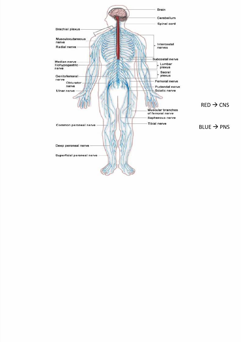
BLUE PNS
RED CNS
7/27/2019 Introductory Lecture on Nervous System Physiology by Dr. Roomi 18-11-13
http://slidepdf.com/reader/full/introductory-lecture-on-nervous-system-physiology-by-dr-roomi-18-11-13 3/32
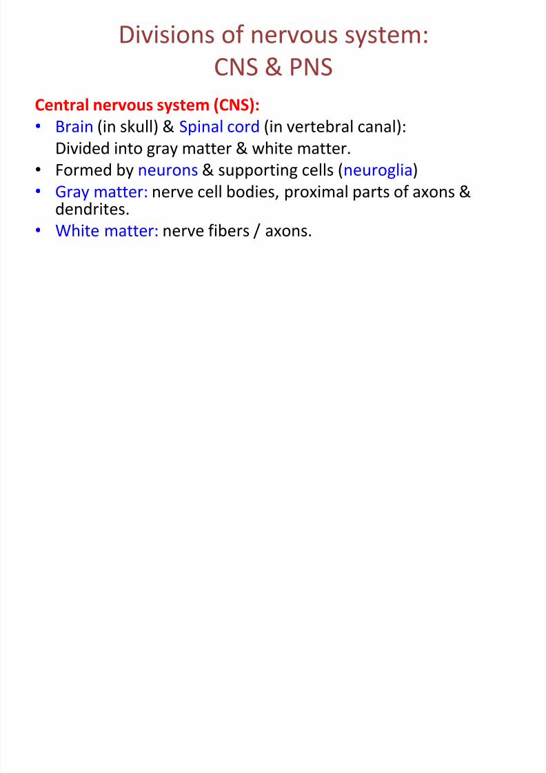
Divisions of nervous system:
CNS & PNS
Central nervous system (CNS):
• Brain (in skull) & Spinal cord (in vertebral canal):
Divided into gray matter & white matter.
• Formed by neurons & supporting cells (neuroglia)
• Gray matter: nerve cell bodies, proximal parts of axons &dendrites.
• White matter: nerve fibers / axons.
7/27/2019 Introductory Lecture on Nervous System Physiology by Dr. Roomi 18-11-13
http://slidepdf.com/reader/full/introductory-lecture-on-nervous-system-physiology-by-dr-roomi-18-11-13 4/32
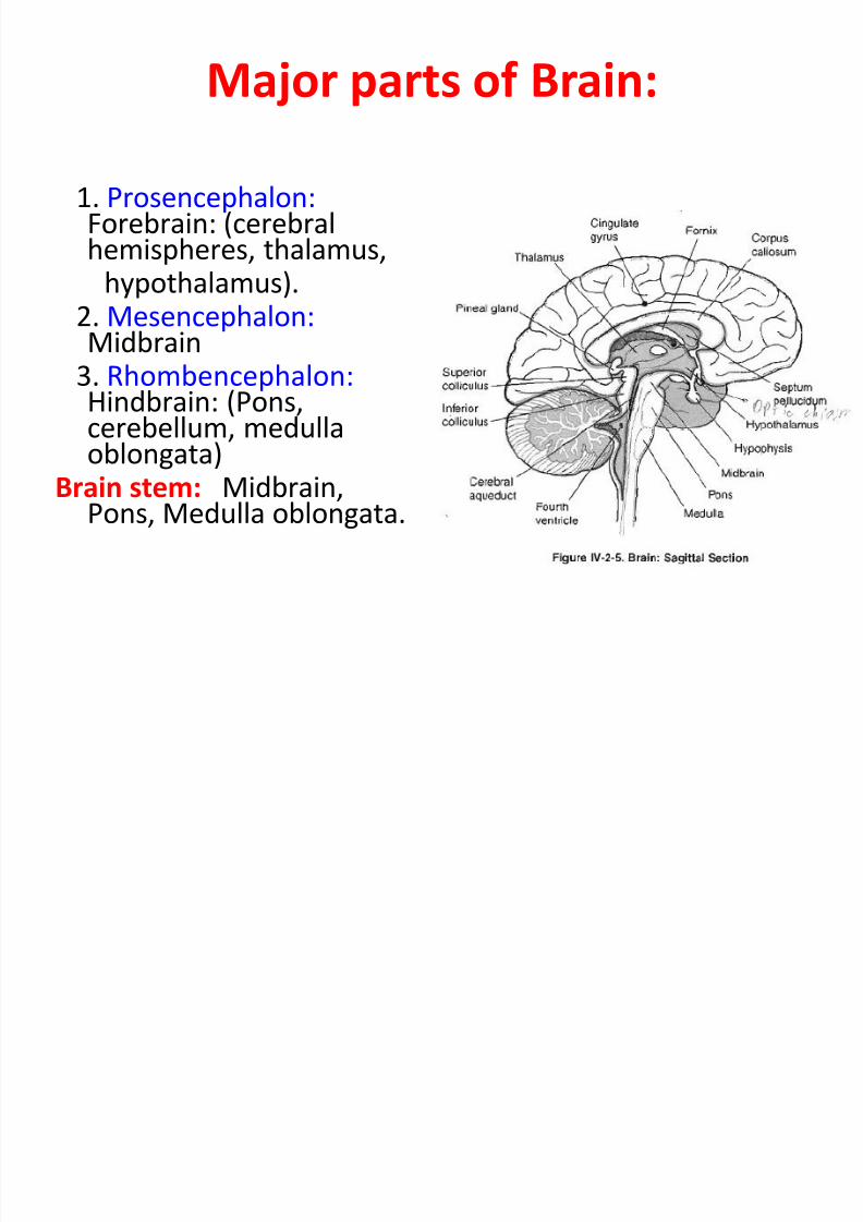
Major parts of Brain:
1. Prosencephalon: Forebrain: (cerebralhemispheres, thalamus,
hypothalamus).2. Mesencephalon: Midbrain
3. Rhombencephalon: Hindbrain: (Pons,cerebellum, medulla
oblongata)Brain stem: Midbrain,
Pons, Medulla oblongata.
7/27/2019 Introductory Lecture on Nervous System Physiology by Dr. Roomi 18-11-13
http://slidepdf.com/reader/full/introductory-lecture-on-nervous-system-physiology-by-dr-roomi-18-11-13 5/32
Peripheral nervous system (PNS)
• Formed by neurons & their processes outside CNS.
• Cranial nerves (from brain)
• Spinal nerves (from spinal cord)
• 2 divisions of PNS:
SOMATIC / VOLUNTARY NERVOUS SYSTEM:Nerves supplying skeletal muscles
Controls movements of body by acting on skeletal
muscles.
AUTONOMIC / VISCERAL / VEGETATIVE/ INVOLUNTARY NERVOUS SYSTEM:Function: Controls viscera
2 sub-divisions:
SYMPATHETIC
PARASYMPATHETIC
7/27/2019 Introductory Lecture on Nervous System Physiology by Dr. Roomi 18-11-13
http://slidepdf.com/reader/full/introductory-lecture-on-nervous-system-physiology-by-dr-roomi-18-11-13 6/32
Cells of nervous tissue
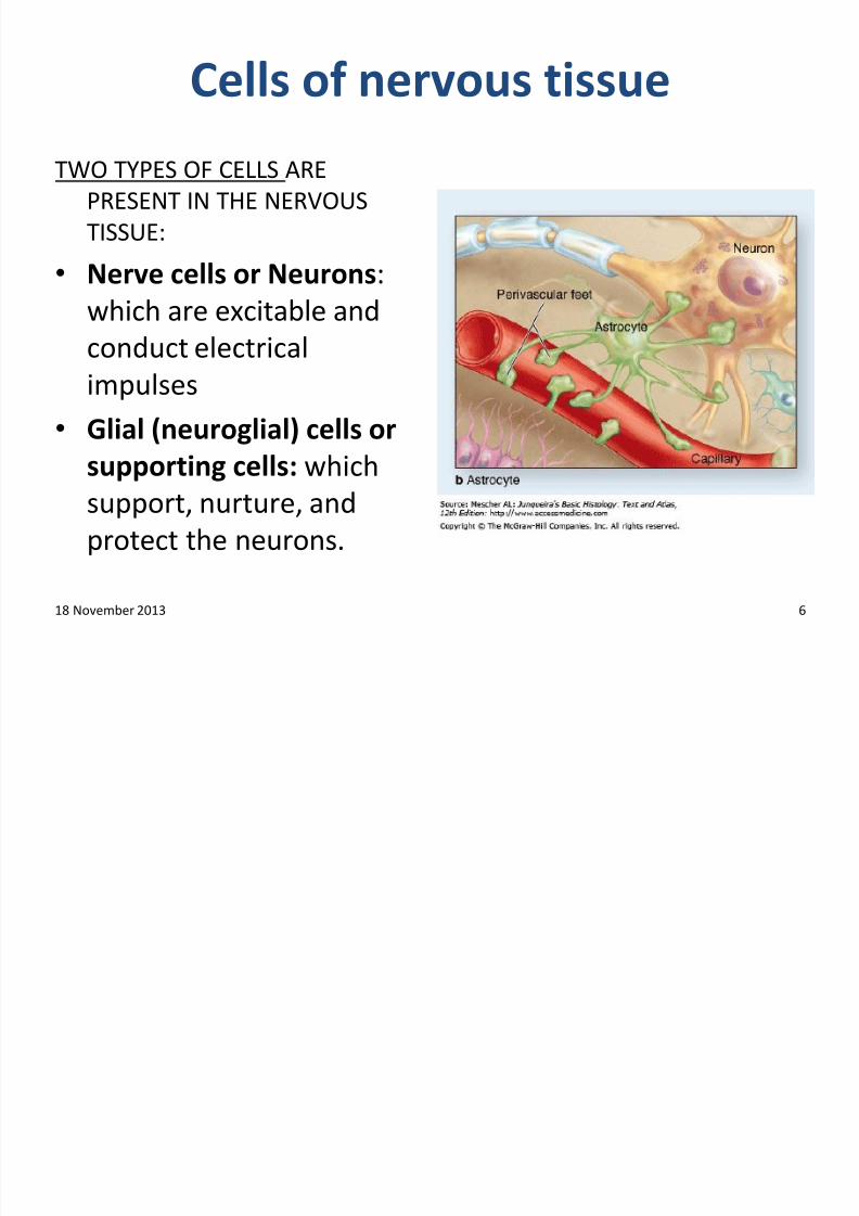
TWO TYPES OF CELLS ARE
PRESENT IN THE NERVOUS
TISSUE:
• Nerve cells or Neurons:
which are excitable and
conduct electrical
impulses
• Glial (neuroglial) cells or
supporting cells: which
support, nurture, and
protect the neurons.
18 November 2013 6
7/27/2019 Introductory Lecture on Nervous System Physiology by Dr. Roomi 18-11-13
http://slidepdf.com/reader/full/introductory-lecture-on-nervous-system-physiology-by-dr-roomi-18-11-13 7/32
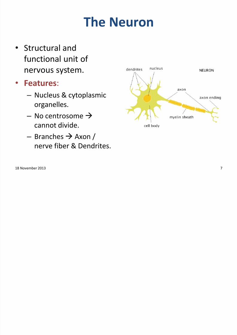
The Neuron
• Structural and
functional unit of
nervous system.
• Features: – Nucleus & cytoplasmic
organelles.
– No centrosome
cannot divide.
– Branches Axon /
nerve fiber & Dendrites.
18 November 2013 7
7/27/2019 Introductory Lecture on Nervous System Physiology by Dr. Roomi 18-11-13
http://slidepdf.com/reader/full/introductory-lecture-on-nervous-system-physiology-by-dr-roomi-18-11-13 8/32
Nerve cell body (perikaryon or soma)
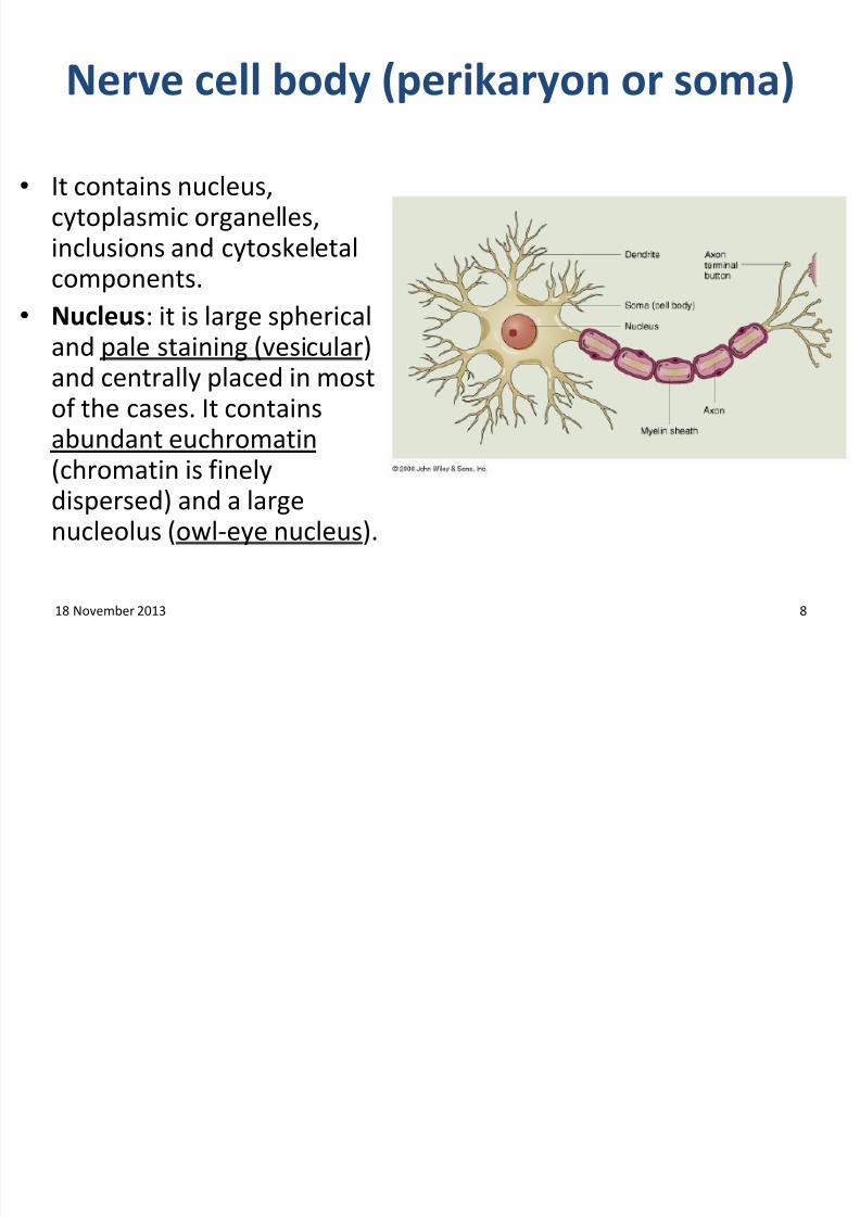
• It contains nucleus,cytoplasmic organelles,inclusions and cytoskeletalcomponents.
• Nucleus: it is large sphericaland pale staining (vesicular)and centrally placed in mostof the cases. It contains
abundant euchromatin(chromatin is finelydispersed) and a largenucleolus (owl-eye nucleus).
18 November 2013 8
7/27/2019 Introductory Lecture on Nervous System Physiology by Dr. Roomi 18-11-13
http://slidepdf.com/reader/full/introductory-lecture-on-nervous-system-physiology-by-dr-roomi-18-11-13 9/32
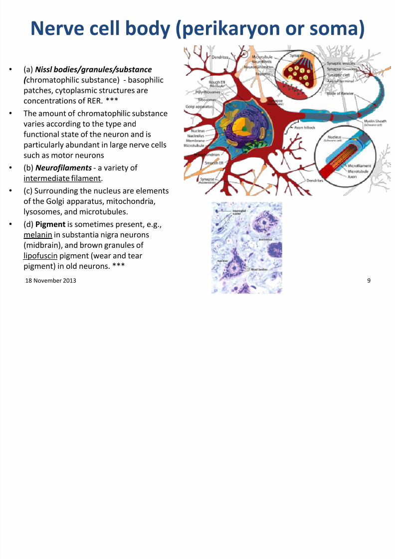
Nerve cell body (perikaryon or soma)
• (a) Nissl bodies/granules/substance( chromatophilic substance) - basophilic
patches, cytoplasmic structures are
concentrations of RER. ***
• The amount of chromatophilic substance
varies according to the type and
functional state of the neuron and isparticularly abundant in large nerve cells
such as motor neurons
• (b) Neurofilaments - a variety of
intermediate filament.
• (c) Surrounding the nucleus are elements
of the Golgi apparatus, mitochondria,
lysosomes, and microtubules.
• (d) Pigment is sometimes present, e.g.,
melanin in substantia nigra neurons
(midbrain), and brown granules of
lipofuscin pigment (wear and tear
pigment) in old neurons. ***
18 November 2013 9
7/27/2019 Introductory Lecture on Nervous System Physiology by Dr. Roomi 18-11-13
http://slidepdf.com/reader/full/introductory-lecture-on-nervous-system-physiology-by-dr-roomi-18-11-13 10/32
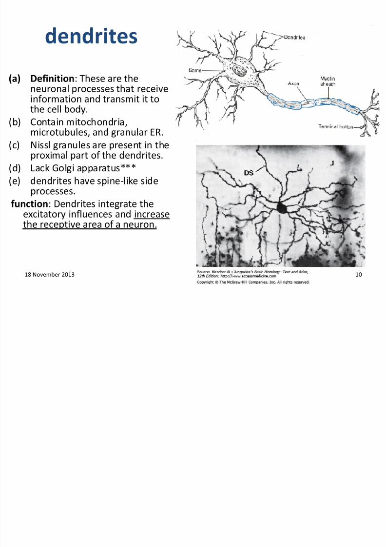
dendrites
(a) Definition: These are theneuronal processes that receiveinformation and transmit it tothe cell body.
(b) Contain mitochondria,
microtubules, and granular ER.(c) Nissl granules are present in the
proximal part of the dendrites.
(d) Lack Golgi apparatus***
(e) dendrites have spine-like sideprocesses.
function: Dendrites integrate theexcitatory influences and increasethe receptive area of a neuron.
18 November 2013 10
7/27/2019 Introductory Lecture on Nervous System Physiology by Dr. Roomi 18-11-13
http://slidepdf.com/reader/full/introductory-lecture-on-nervous-system-physiology-by-dr-roomi-18-11-13 11/32
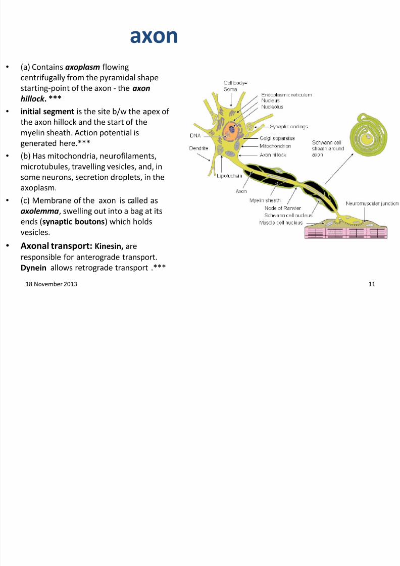
axon•
(a) Contains axoplasm flowingcentrifugally from the pyramidal shape
starting-point of the axon - the axon
hillock . ***
• initial segment is the site b/w the apex of
the axon hillock and the start of the
myelin sheath. Action potential is
generated here.***
• (b) Has mitochondria, neurofilaments,
microtubules, travelling vesicles, and, in
some neurons, secretion droplets, in the
axoplasm.
• (c) Membrane of the axon is called as
axolemma, swelling out into a bag at itsends (synaptic boutons) which holds
vesicles.
• Axonal transport: Kinesin, are
responsible for anterograde transport.
Dynein allows retrograde transport .***
18 November 2013 11
7/27/2019 Introductory Lecture on Nervous System Physiology by Dr. Roomi 18-11-13
http://slidepdf.com/reader/full/introductory-lecture-on-nervous-system-physiology-by-dr-roomi-18-11-13 12/32
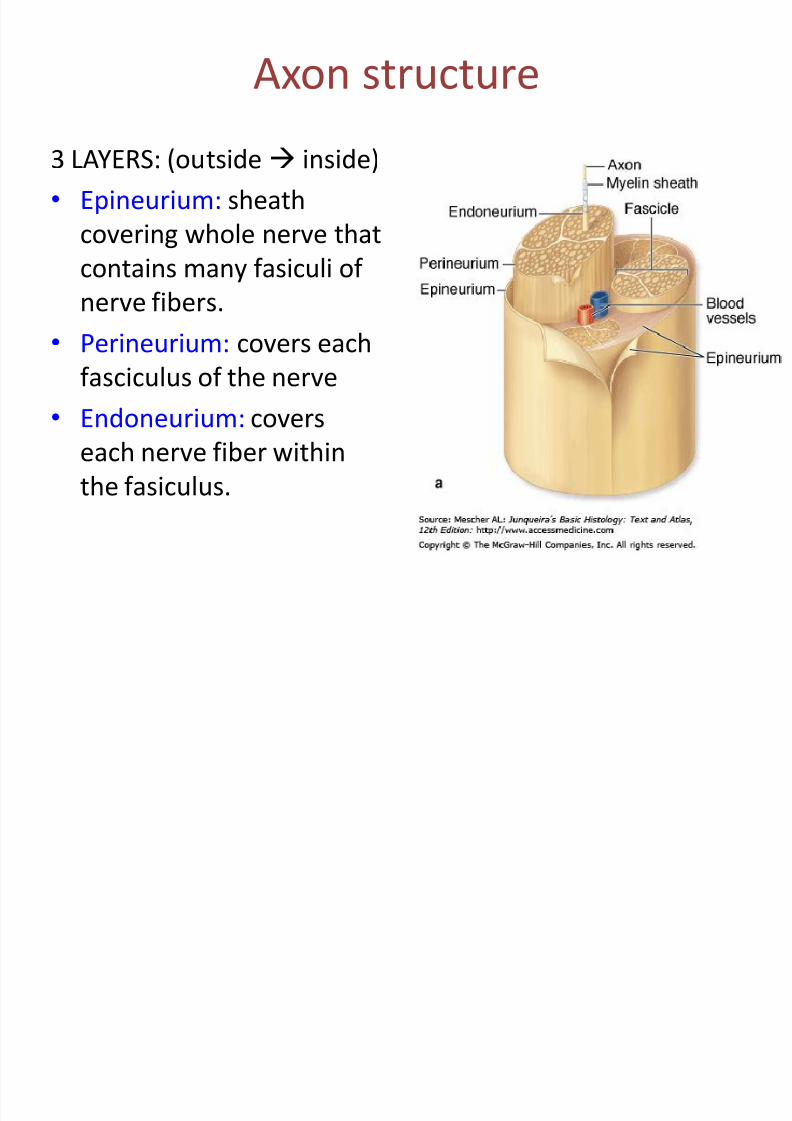
Axon structure
3 LAYERS: (outside inside)
• Epineurium: sheath
covering whole nerve that
contains many fasiculi ofnerve fibers.
• Perineurium: covers each
fasciculus of the nerve
• Endoneurium: coverseach nerve fiber within
the fasiculus.
7/27/2019 Introductory Lecture on Nervous System Physiology by Dr. Roomi 18-11-13
http://slidepdf.com/reader/full/introductory-lecture-on-nervous-system-physiology-by-dr-roomi-18-11-13 13/32
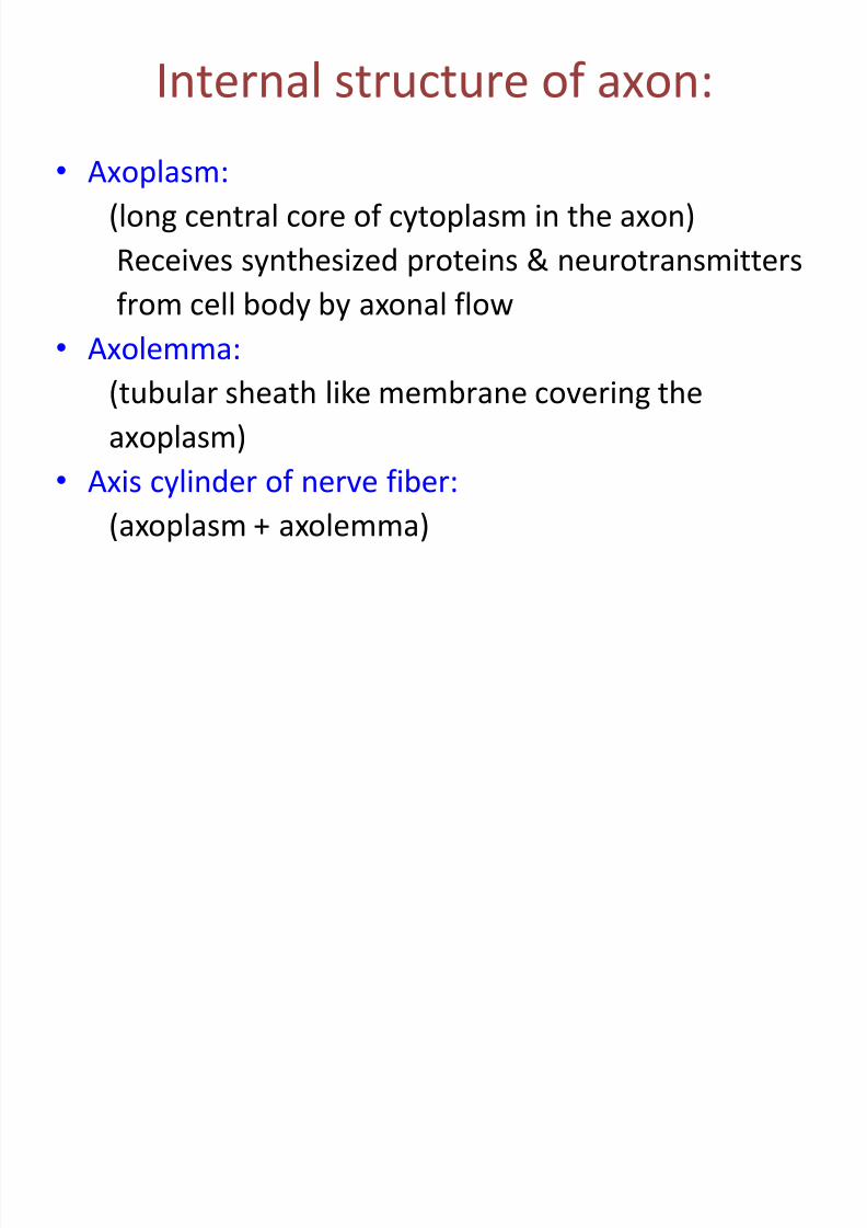
Internal structure of axon:
• Axoplasm:
(long central core of cytoplasm in the axon)
Receives synthesized proteins & neurotransmitters
from cell body by axonal flow• Axolemma:
(tubular sheath like membrane covering the
axoplasm)
• Axis cylinder of nerve fiber:
(axoplasm + axolemma)
7/27/2019 Introductory Lecture on Nervous System Physiology by Dr. Roomi 18-11-13
http://slidepdf.com/reader/full/introductory-lecture-on-nervous-system-physiology-by-dr-roomi-18-11-13 14/32
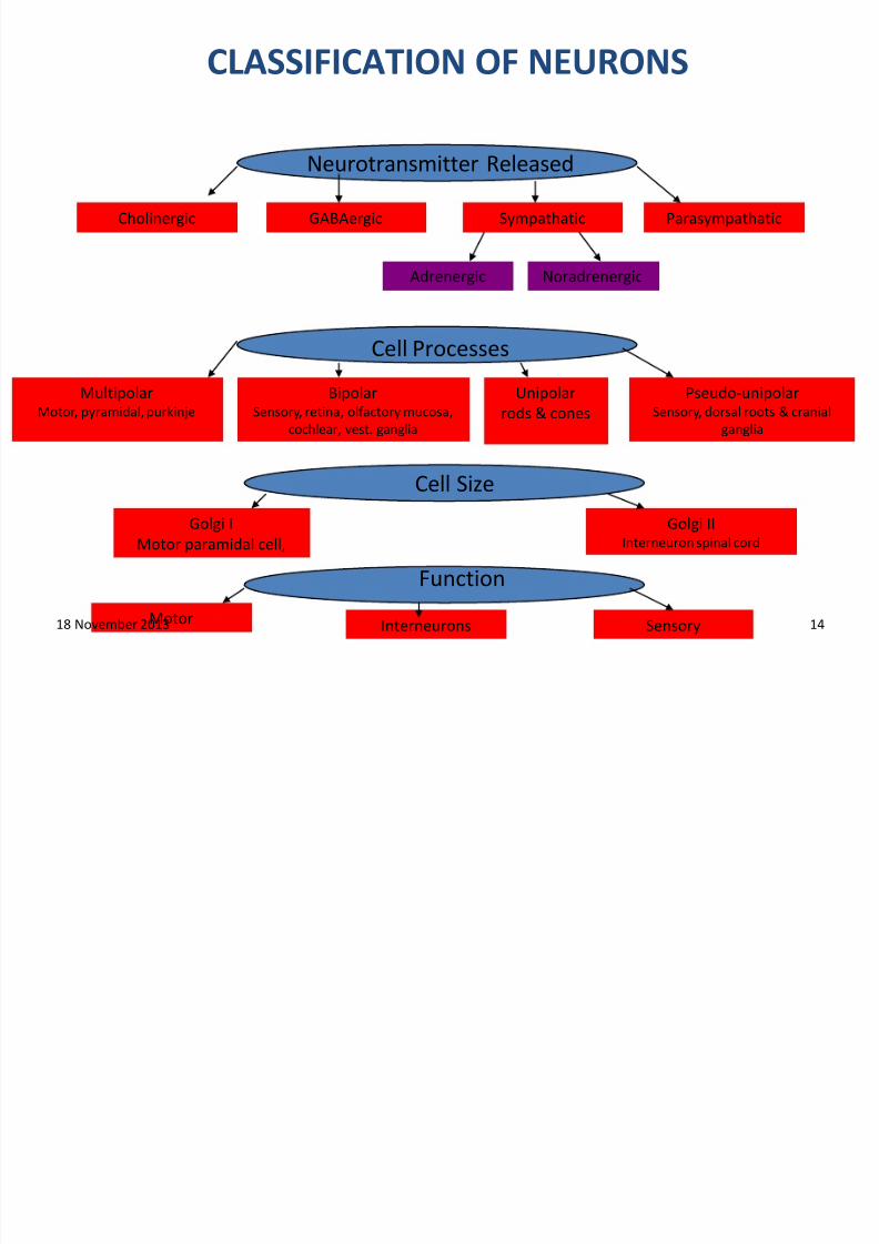
CLASSIFICATION OF NEURONS
Neurotransmitter Released
Cholinergic GABAergic
Cell Processes
MultipolarMotor, pyramidal, purkinje
BipolarSensory, retina, olfactory mucosa,
cochlear, vest. ganglia
Unipolar
rods & cones
Pseudo-unipolarSensory, dorsal roots & cranial
ganglia
Cell Size
Golgi I
Motor paramidal cell,
Golgi IIInterneuron spinal cord
Function
Motor Interneurons Sensory
Sympathatic Parasympathatic
Adrenergic Noradrenergic
18 November 2013 14
7/27/2019 Introductory Lecture on Nervous System Physiology by Dr. Roomi 18-11-13
http://slidepdf.com/reader/full/introductory-lecture-on-nervous-system-physiology-by-dr-roomi-18-11-13 15/32
Classification of neurons on the basis of structure
• Myelinated covered by myelin sheath
• Unmyelinated not covered by myelin
sheath
7/27/2019 Introductory Lecture on Nervous System Physiology by Dr. Roomi 18-11-13
http://slidepdf.com/reader/full/introductory-lecture-on-nervous-system-physiology-by-dr-roomi-18-11-13 16/32
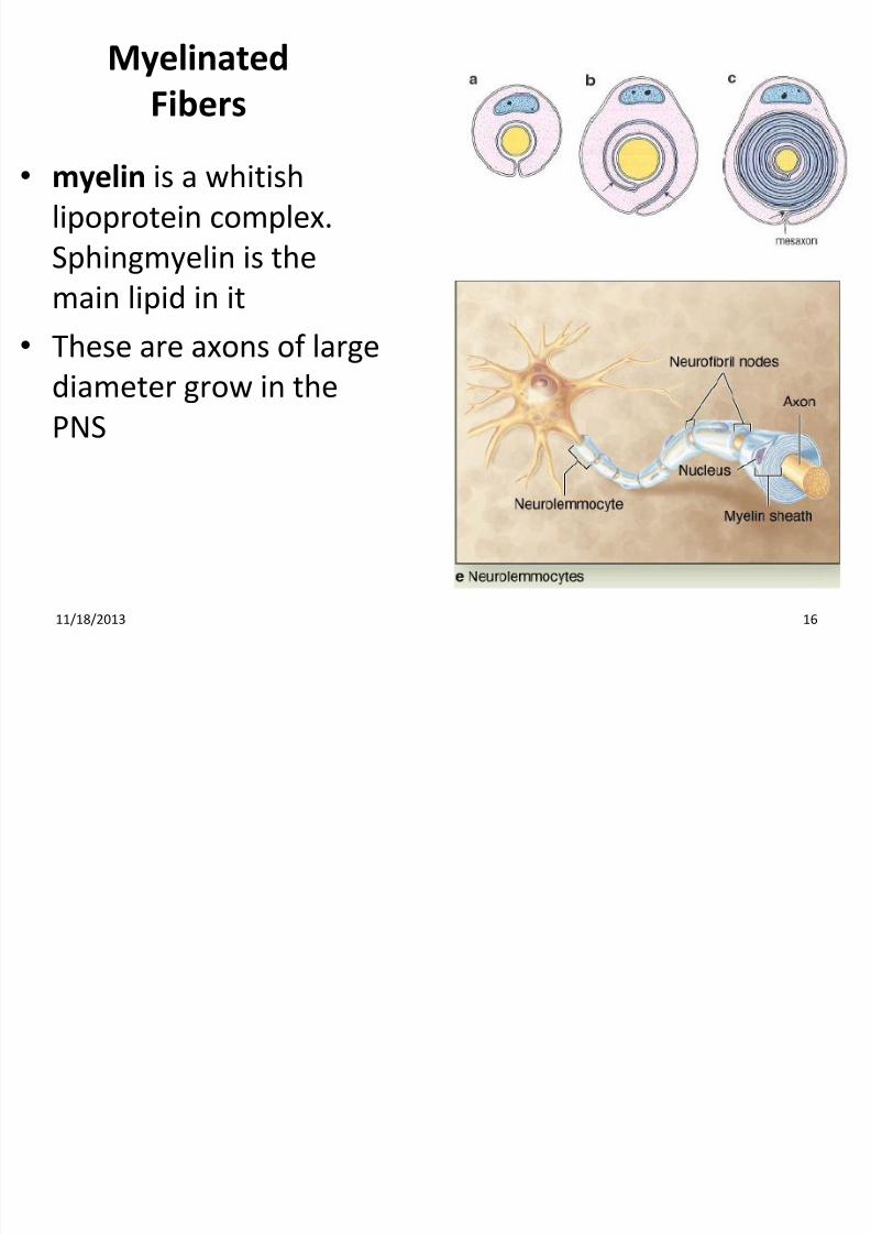
Myelinated
Fibers
• myelin is a whitish
lipoprotein complex.
Sphingmyelin is the
main lipid in it• These are axons of large
diameter grow in the
PNS
11/18/2013 16
7/27/2019 Introductory Lecture on Nervous System Physiology by Dr. Roomi 18-11-13
http://slidepdf.com/reader/full/introductory-lecture-on-nervous-system-physiology-by-dr-roomi-18-11-13 17/32
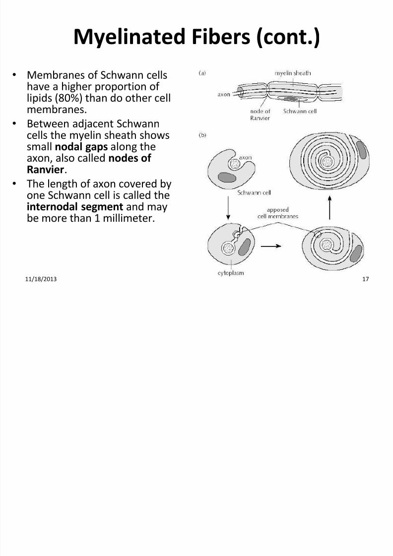
Myelinated Fibers (cont.)
• Membranes of Schwann cellshave a higher proportion oflipids (80%) than do other cellmembranes.
• Between adjacent Schwann
cells the myelin sheath showssmall nodal gaps along theaxon, also called nodes ofRanvier.
• The length of axon covered byone Schwann cell is called the
internodal segment and maybe more than 1 millimeter.
11/18/2013 17
7/27/2019 Introductory Lecture on Nervous System Physiology by Dr. Roomi 18-11-13
http://slidepdf.com/reader/full/introductory-lecture-on-nervous-system-physiology-by-dr-roomi-18-11-13 18/32
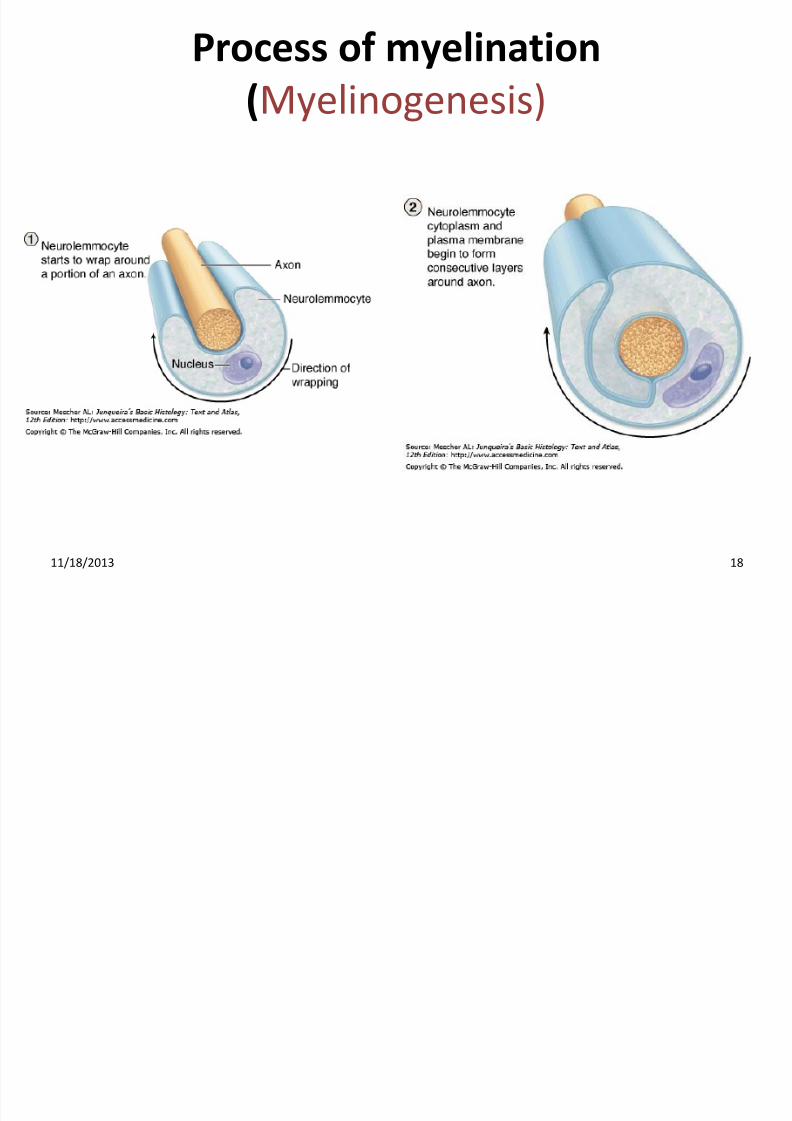
Process of myelination
(Myelinogenesis)
11/18/2013 18
7/27/2019 Introductory Lecture on Nervous System Physiology by Dr. Roomi 18-11-13
http://slidepdf.com/reader/full/introductory-lecture-on-nervous-system-physiology-by-dr-roomi-18-11-13 19/32
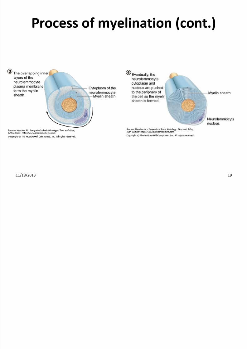
Process of myelination (cont.)
11/18/2013 19
7/27/2019 Introductory Lecture on Nervous System Physiology by Dr. Roomi 18-11-13
http://slidepdf.com/reader/full/introductory-lecture-on-nervous-system-physiology-by-dr-roomi-18-11-13 20/32
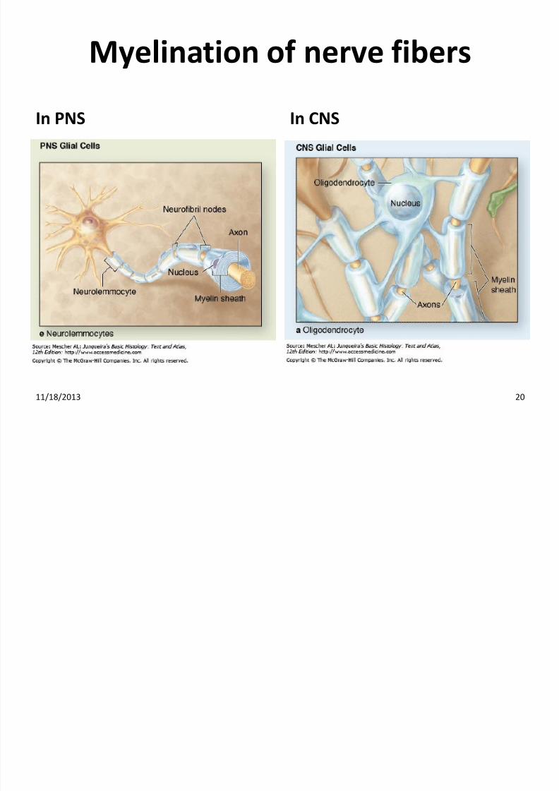
Myelination of nerve fibers
In PNS In CNS
11/18/2013 20
7/27/2019 Introductory Lecture on Nervous System Physiology by Dr. Roomi 18-11-13
http://slidepdf.com/reader/full/introductory-lecture-on-nervous-system-physiology-by-dr-roomi-18-11-13 21/32
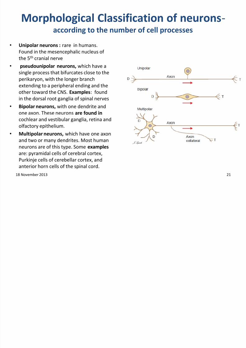
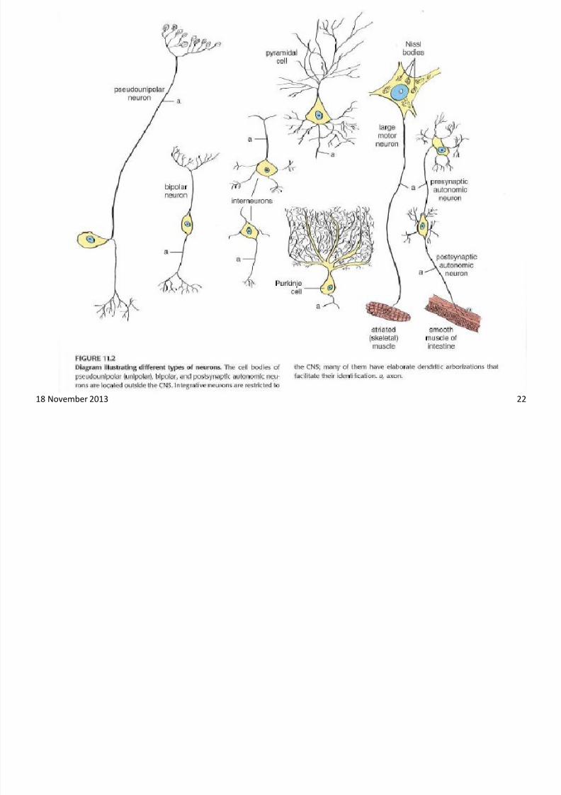
Morphological Classification of neurons-according to the number of cell processes
• Unipolar neurons : rare in humans.Found in the mesencephalic nucleus of
the 5th cranial nerve
• pseudounipolar neurons, which have a
single process that bifurcates close to the
perikaryon, with the longer branch
extending to a peripheral ending and theother toward the CNS. Examples: found
in the dorsal root ganglia of spinal nerves
• Bipolar neurons, with one dendrite and
one axon. These neurons are found in
cochlear and vestibular ganglia, retina and
olfactory epithelium.
• Multipolar neurons, which have one axon
and two or many dendrites. Most human
neurons are of this type. Some examples
are: pyramidal cells of cerebral cortex,
Purkinje cells of cerebellar cortex, and
anterior horn cells of the spinal cord.
18 November 2013 21
7/27/2019 Introductory Lecture on Nervous System Physiology by Dr. Roomi 18-11-13
http://slidepdf.com/reader/full/introductory-lecture-on-nervous-system-physiology-by-dr-roomi-18-11-13 22/32
18 November 2013 22
7/27/2019 Introductory Lecture on Nervous System Physiology by Dr. Roomi 18-11-13
http://slidepdf.com/reader/full/introductory-lecture-on-nervous-system-physiology-by-dr-roomi-18-11-13 23/32
Classification of Neurons by
Functional Role
Motor (efferent) neurons
• (CNS Periphery)
• Motor neurons control effector organs and
muscle fibers.Sensory (afferent) neurons
• (periphery CNS)
•
Sensory neurons receive sensory stimuli from theinternal or external environment and relay themto the CNS.
18 November 2013 23
7/27/2019 Introductory Lecture on Nervous System Physiology by Dr. Roomi 18-11-13
http://slidepdf.com/reader/full/introductory-lecture-on-nervous-system-physiology-by-dr-roomi-18-11-13 24/32
Classification of Neurons-according to
the length of their axons Golgi type I neurons: possess many dendrites and a very long
axon that leaves the grey matter in which its cell body ispresent.
Examples:
•
pyramidal cells of the cerebral cortex• anterior horn cells of the spinal cord.
Golgi type II neurons: possess many dendrites and a relativelyshort axon that does not leave the part of grey matter in
which the cell body of the neuron is present.Example:
• Interneurons.
18 November 2013 24
7/27/2019 Introductory Lecture on Nervous System Physiology by Dr. Roomi 18-11-13
http://slidepdf.com/reader/full/introductory-lecture-on-nervous-system-physiology-by-dr-roomi-18-11-13 25/32
Classification On the basis of distribution:
• Somatic supply skeletal muscles
• Visceral / Autonomic supply internal organs
7/27/2019 Introductory Lecture on Nervous System Physiology by Dr. Roomi 18-11-13
http://slidepdf.com/reader/full/introductory-lecture-on-nervous-system-physiology-by-dr-roomi-18-11-13 26/32
Classification on the basis of source of origin:
• Cranial from the brain
• Spinal from the spinal cord
7/27/2019 Introductory Lecture on Nervous System Physiology by Dr. Roomi 18-11-13
http://slidepdf.com/reader/full/introductory-lecture-on-nervous-system-physiology-by-dr-roomi-18-11-13 27/32
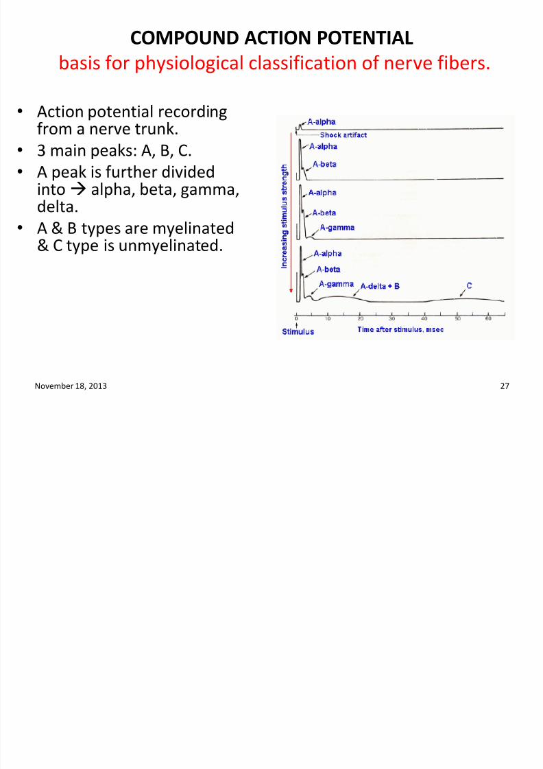
COMPOUND ACTION POTENTIAL
basis for physiological classification of nerve fibers.
• Action potential recordingfrom a nerve trunk.
• 3 main peaks: A, B, C.
• A peak is further divided
into alpha, beta, gamma,delta.
• A & B types are myelinated& C type is unmyelinated.
November 18, 2013 27
7/27/2019 Introductory Lecture on Nervous System Physiology by Dr. Roomi 18-11-13
http://slidepdf.com/reader/full/introductory-lecture-on-nervous-system-physiology-by-dr-roomi-18-11-13 28/32
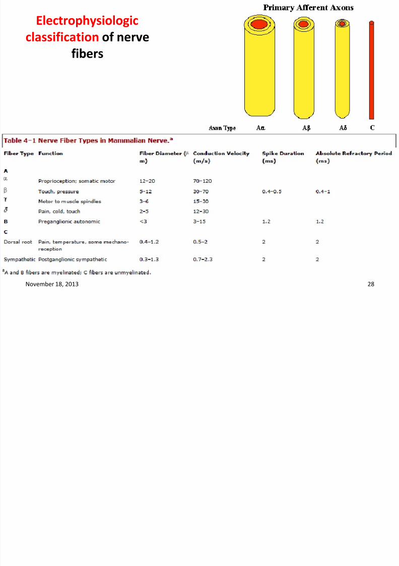
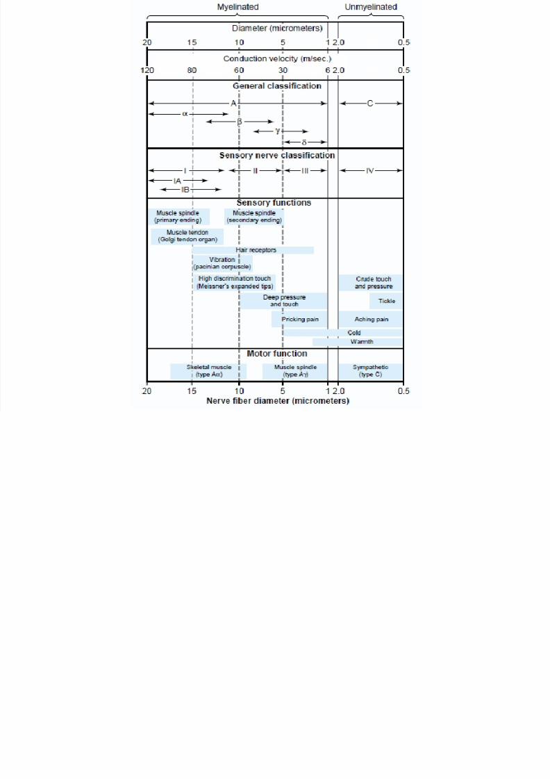
Electrophysiologic
classification of nerve
fibers
November 18, 2013 28
7/27/2019 Introductory Lecture on Nervous System Physiology by Dr. Roomi 18-11-13
http://slidepdf.com/reader/full/introductory-lecture-on-nervous-system-physiology-by-dr-roomi-18-11-13 29/32
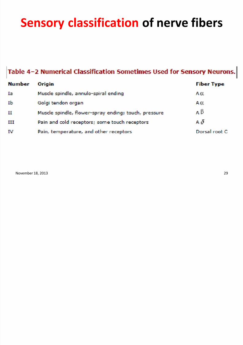
Sensory classification of nerve fibers
November 18, 2013 29
7/27/2019 Introductory Lecture on Nervous System Physiology by Dr. Roomi 18-11-13
http://slidepdf.com/reader/full/introductory-lecture-on-nervous-system-physiology-by-dr-roomi-18-11-13 30/32
Sensory classification Vs electrophysiological
classification
• Type A (myelinated): A-alpha (Group Ia & Ib), A-beta(Group II), A-gamma (Group II), A-delta (Group III).
• Type B (myelinated)
• Type C (unmyelinated): Group IV
Diameter is directly proportional to conductionvelocity.
Range of velocity: 120m/sec (in large myelinated fibers)to 0.5 m/sec (in smallest unmyelinated fibers).
7/27/2019 Introductory Lecture on Nervous System Physiology by Dr. Roomi 18-11-13
http://slidepdf.com/reader/full/introductory-lecture-on-nervous-system-physiology-by-dr-roomi-18-11-13 31/32
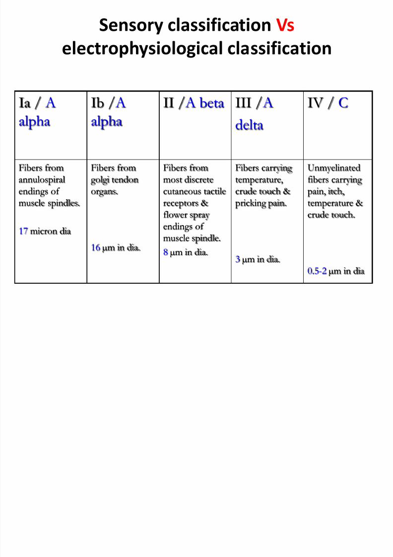
Sensory classification Vs
electrophysiological classification
Ia / A
alpha
Ib / A
alpha
II / A beta III / A
delta
IV / C
Fibers from
annulospiral
endings of
muscle spindles.
17 micron dia
Fibers from
golgi tendon
organs.
16 µm in dia.
Fibers from
most discrete
cutaneous tactile
receptors &
flower spray
endings of
muscle spindle.
8 µm in dia.
Fibers carrying
temperature,
crude touch &
pricking pain.
3 µm in dia.
Unmyelinated
fibers carrying
pain, itch,
temperature &
crude touch.
0.5-2 µm in dia
7/27/2019 Introductory Lecture on Nervous System Physiology by Dr. Roomi 18-11-13
http://slidepdf.com/reader/full/introductory-lecture-on-nervous-system-physiology-by-dr-roomi-18-11-13 32/32
Related Documents