Intestinal stromal cells in mucosal immunity and homeostasis BMJ Owens 1 and A Simmons 1,2 A growing body of evidence suggests that non-hematopoietic stromal cells of the intestine have multiple roles in immune responses and inflammation at this mucosal site. Despite this, many still consider gut stromal cells as passive structural entities, with past research focused heavily on their roles in fibrosis, tumor progression, and wound healing, rather than their contributions to immune function. In this review, we discuss our current knowledge of stromal cells in intestinal immunity, highlighting the many immunological axes in which stromal cells have a functional role. We also consider emerging data that broaden the potential scope of their contribution to immunity in the gut and argue that these so-called ‘‘non-immune’’ cells are reclassified in light of their diverse contributions to intestinal innate immunity and the maintenance of mucosal homeostasis. INTRODUCTION Immune responses at the intestinal mucosa are tightly controlled to remain tolerant of the vast commensal micro- biota, while simultaneously retaining the capacity to respond appropriately to harmful insult. Multiple cellular and molecular processes have evolved that contribute to this critical balance, although there is still much to be revealed regarding the precise mechanisms by which immunity and homeostasis in the gut is achieved. The vast majority of our knowledge regarding intestinal immune homeostasis and its breakdown in inflammatory disease has concerned the role of specialized hematopoietic ‘‘conventional’’ immune cell populations—both in isolation and in concert with the epithelial monolayer that comprises the functional barrier between the host and the microbiota. 1 This focused research has revealed several mechanisms by which aberrant hematopoietic immune cell functional pathways contribute to human disease, including defective nucleotide- binding oligomerization domain-containing protein 1 (NOD1) and NOD2—induced autophagy and bacterial handling by myeloid cells; 2,3 functional polymorphisms in the interleukin (IL)-23 receptor (IL-23R); 4 loss-of-function mutations in the IL-10R; 5 and variants in the inflammasome activator NOD-like receptor family, pyrin domain-containing 3 (NLRP3), 6 alongside many others. Although they have a crucial role in immunity and tolerance in the gut, hematopoietic immune and epithelial cells do not function in isolation but rather as part of a complex cellular network that contains several other distinct non-hematopoietic cell populations. Residing in the sub-epithelial compartment and providing much of the structural framework of the intestine are multiple populations of non-hematopoietic mesenchymal cells, distinct from the epithelium, that can be grouped as stromal cells—a nomenclature similar to that assigned to the cellular networks at the heart of secondary lymphoid organs. 7 Intestinal stromal cells dynamically interact with both epithelial and hemato- poietic immune cells at this mucosal site and, although not considered ‘‘professional’’ immune cells, there is mounting evidence that they can perform many of the functions often solely attributed to their hematopoietic immune cell counter- parts. 8 In this review, we move beyond the previously well- documented role of stromal cells in intestinal fibrosis, tumor progression, and wound healing, focusing instead on the immunological functions that support their re-designation as de facto intestinal immune cells. Intestinal stromal cells: ontogeny, location, and support of the intestinal epithelium The mesenchymal compartment of the intestinal laminar propria contains multiple stromal cell populations with distinct 1 Translational Gastroenterology Unit, Experimental Medicine Division, Nuffield Department of Medicine, John Radcliffe Hospital, University of Oxford, Oxford, UK and 2 MRC Human Immunology Unit, Weatherall Institute of Molecular Medicine, John Radcliffe Hospital, University of Oxford, Oxford, UK. Correspondence: BMJ Owens ([email protected]); A Simmons ([email protected]) Received 16 August 2012; accepted 13 November 2012; published online 19 December 2012. doi:10.1038/mi.2012.125 REVIEW nature publishing group 224 VOLUME 6 NUMBER 2 | MARCH 2013 | www.nature.com/mi

Welcome message from author
This document is posted to help you gain knowledge. Please leave a comment to let me know what you think about it! Share it to your friends and learn new things together.
Transcript
Intestinal stromal cells in mucosal immunity andhomeostasisBMJ Owens1 and A Simmons1,2
A growing body of evidence suggests that non-hematopoietic stromal cells of the intestine have multiple roles in immune
responses and inflammation at this mucosal site. Despite this, many still consider gut stromal cells as passive structural
entities, with past research focused heavily on their roles in fibrosis, tumor progression, and wound healing, rather than
their contributions to immune function. In this review, we discuss our current knowledge of stromal cells in intestinal
immunity, highlighting the many immunological axes in which stromal cells have a functional role. We also consider
emerging data that broaden the potential scope of their contribution to immunity in the gut and argue that these so-called
‘‘non-immune’’ cells are reclassified in light of their diverse contributions to intestinal innate immunity and the
maintenance of mucosal homeostasis.
INTRODUCTION
Immune responses at the intestinal mucosa are tightlycontrolled to remain tolerant of the vast commensal micro-biota, while simultaneously retaining the capacity to respondappropriately to harmful insult. Multiple cellular and molecularprocesses have evolved that contribute to this critical balance,although there is still much to be revealed regarding the precisemechanisms by which immunity and homeostasis in the gut isachieved.
The vast majority of our knowledge regarding intestinalimmune homeostasis and its breakdown in inflammatorydisease has concerned the role of specialized hematopoietic‘‘conventional’’ immune cell populations—both in isolationand in concert with the epithelial monolayer that comprises thefunctional barrier between the host and the microbiota.1 Thisfocused research has revealed several mechanisms by whichaberrant hematopoietic immune cell functional pathwayscontribute to human disease, including defective nucleotide-binding oligomerization domain-containing protein 1 (NOD1)and NOD2—induced autophagy and bacterial handling bymyeloid cells;2,3 functional polymorphisms in the interleukin(IL)-23 receptor (IL-23R);4 loss-of-function mutations in theIL-10R;5 and variants in the inflammasome activator NOD-likereceptor family, pyrin domain-containing 3 (NLRP3),6
alongside many others. Although they have a crucial role in
immunity and tolerance in the gut, hematopoietic immune andepithelial cells do not function in isolation but rather as part of acomplex cellular network that contains several other distinctnon-hematopoietic cell populations.
Residing in the sub-epithelial compartment and providingmuch of the structural framework of the intestine are multiplepopulations of non-hematopoietic mesenchymal cells, distinctfrom the epithelium, that can be grouped as stromal cells—anomenclature similar to that assigned to the cellular networksat the heart of secondary lymphoid organs.7 Intestinal stromalcells dynamically interact with both epithelial and hemato-poietic immune cells at this mucosal site and, although notconsidered ‘‘professional’’ immune cells, there is mountingevidence that they can perform many of the functions oftensolely attributed to their hematopoietic immune cell counter-parts.8 In this review, we move beyond the previously well-documented role of stromal cells in intestinal fibrosis, tumorprogression, and wound healing, focusing instead on theimmunological functions that support their re-designation asde facto intestinal immune cells.
Intestinal stromal cells: ontogeny, location, and support ofthe intestinal epithelium
The mesenchymal compartment of the intestinal laminarpropria contains multiple stromal cell populations with distinct
1Translational Gastroenterology Unit, Experimental Medicine Division, Nuffield Department of Medicine, John Radcliffe Hospital, University of Oxford, Oxford, UK and 2MRCHuman Immunology Unit, Weatherall Institute of Molecular Medicine, John Radcliffe Hospital, University of Oxford, Oxford, UK. Correspondence: BMJ Owens([email protected]); A Simmons ([email protected])
Received 16 August 2012; accepted 13 November 2012; published online 19 December 2012. doi:10.1038/mi.2012.125
REVIEW nature publishing group
224 VOLUME 6 NUMBER 2 |MARCH 2013 |www.nature.com/mi
localization, phenotype, and function. These include fibro-blasts, myofibroblasts, pericytes, endothelial cells, and smoothmuscle cells,9 in addition to less well-characterized populationsthat may represent distinct stromal subsets and suggestshitherto unknown phenotypic and functional heterogeneitywithin this compartment. The vast majority of publishedstudies relating to immunological functions of the intestinalmesenchymal cell compartment have focused on cells of thefibroblast family, in particular, the sub-epithelial myofibroblast.Although these cells likely have a major role in this context,taking a similar intellectual approach to that used in lymphoidtissue stromal cell biology,7 other intestinal mesenchymal cellsubsets—including smooth muscle cells, vascular endothelialcells, and lymphatic endothelial cells—can also be considered asstromal cell populations. As our current understanding of thephenotypic features and anatomical location of intestinalstromal cell subsets have been extensively covered recently,8,9 inthis review we consider intestinal stromal cells in a very broadsense and focus on the functional contributions of multiplesubsets to innate immunity and homeostasis in the gut.
Despite exhibiting features of non-hematopietic mesench-ymal cells, such as abundant collagen production, expression ofvimentin and a-smooth muscle actin (aSMA) filaments, and alack of surface CD45 expression,9 there is currently debate as tothe precise origins of intestinal stromal cells. It appears thatsuch cells can arise from both mesenchymal and hematopoieticstem cells,10,11 at least in murine models, although the relativecontributions of each source to the differentiated stromal poolin the intestine are not yet defined. During chronic inflamma-tion, tissue mesenchymal cell populations can also arise fromcirculating myeloid-lineage cells known as fibrocytes,12 withseveral alternative sources of tissue stromal cells reported inboth steady-state and disease contexts, including the inflamedcolon.13,14 Furthermore, recent evidence suggests that inexperimental animals, intestinal epithelial cells can act as adirect progenitor of fibroblasts during ongoing tissue fibrosisin vivo, converting to stromal cells via a process of epithelial-to-mesenchymal transition that is reciprocally regulated bytransforming growth factor b1 (TGFb1) and bone morpho-genetic protein-7.15 However, as such processes are currentlystill poorly defined for the intestine, detailed in vivo lineagetracing and fate mapping approaches are required in order todetermine the precise ontogeny of intestinal stromal cellpopulations in both homeostatic and inflammatory situations.
Adding to the confusion over intestinal stromal cell origin,there is currently much interest in the therapeutic potential andimmune functionality of mesenchymal stem cells (MSCs), oftenreferred to in the literature as ‘‘mesenchymal stromal cells’’.16,17
MSCs are multipotent progenitor cells that are distinct fromfully differentiated, intestinal-resident non-hematopoetic cells.Nevertheless, a better understanding of MSC function is likelyto be beneficial in the field of intestinal stromal cell biology,particularly with regard to the delineation of stromal celldifferentiation in vivo and the modulation of residentmesenchymal cell abundance/function in the context of stemcell transplantation for inflammatory bowel disease (IBD).10
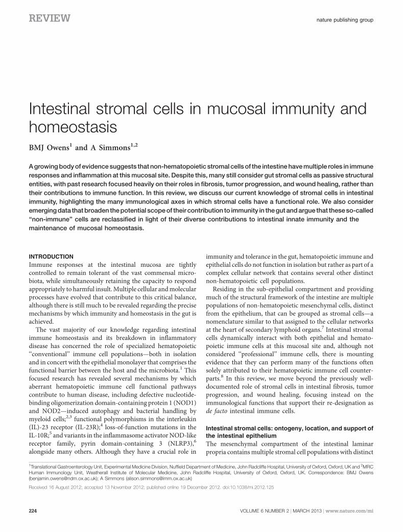
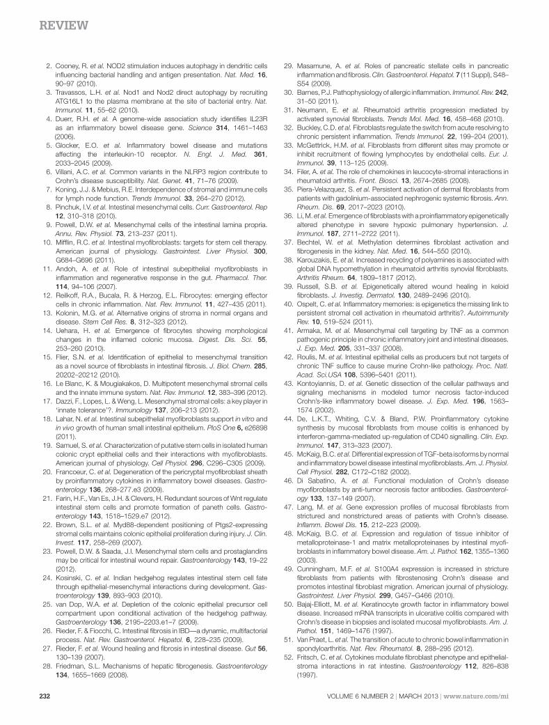
The sub-mucosal localization of intestinal stromal cellsmeans that they are critically placed to contribute to a range ofhomeostatic processes in the gut. Stromal cells form a densenetwork directly adjacent to and underlying the epithelium ofthe small and large intestine (Figure 1a,b, Pinchuk et al.8 andPowell et al.9). As this localization suggests, they interactthrough the basement membrane with various epithelial cellpopulations and have a critical role in supporting theirproliferation and function.18 This relies on multiple complexinteractions, mediated mostly by soluble mediators, andincludes communication between stromal cells and epithelialstem cells,19 with these essential interactions often disrupted asa result of IBD.20 Recent data has clarified some of themechanisms involved in this homeostatic axis, revealing amajor role for stromal cell-derived Wnt in epithelial stem cellhomeostasis.21 Interestingly, stromal cell support of intestinalepithelial differentiation and function is not merely astationary, passive process. In an elegant study using a murinemodel of colitis, Brown et al.22revealed that stromal cellsactively redistribute to the peri-cryptal area during inflamma-tion, produce prostaglandin-E2, and initiate epithelial renewalafter damage to the barrier. However, these cells appear to havefeatures of MSCs, rather than a differentiated stromal cellsubset.23
In addition, stromal–epithelial interactions appear to be bi-directional: Indian hedgehog produced by intestinal epitheliumis critical for the regulation of stromal cell development andproliferation in the mouse, and these stromal cells, in turn,regulate the faithful differentiation and regeneration of theintestinal epithelium.24 This process must be tightly regulated,as constitutive activation of hedgehog signaling in the colonleads to an accumulation of myofibroblasts, loss of epithelialprecursors, and crypt abnormalities in mice,25 highlighting thecritical importance of dynamic stromal–epithelial interactionsfor intestinal barrier function and repair. Although it iscurrently unclear whether stromal cells have a similar role inregulating epithelial function or regeneration after challengewith a mucosal pathogen, the potential innate immune roles ofstromal cells are discussed in more detail later in this review.
The beneficial contributions of intestinal stromal cells to thephysiological processes of epithelial development and regen-erative responses to barrier damage are clear; however, wheninappropriately regulated or activated, stromal cells can alsocontribute to many of the pathological mechanisms operatingin the context of intestinal fibrosis and dysregulated mucosalhealing in the gut. These areas of intestinal stromal cell biologyhave been the focus of intensive research over the years and arereviewed in detail elsewhere.11,26,27 However, in direct rele-vance to a more ‘‘classical’’ immunological role, stromal cellactivation and phenotypic modulation also appears to occurmore generally during chronic inflammatory immune-mediated disease.
Activation of stromal cells during inflammatory pathology
Stromal cell activation is implicated in a number of chronicinflammatory disorders of multiple tissues, including the lung,
REVIEW
MucosalImmunology | VOLUME 6 NUMBER 2 |MARCH 2013 225
liver, and pancreas.28–30 Perhaps best studied from a stromalcell angle, the diverse functional contributions of tissue-specificstromal cells to the immunopathology associated with rheu-matoid arthritis (RA) are now well accepted. This includestissue destruction, production of and response to inflammatorycytokines, activation of innate signaling pathways, and theinitiation of angiogenesis and thus regulates the transition fromacute-to-chronic inflammation (reviewed in Neumannet al.31and Buckley et al.32). Furthermore, stromal–stromaland stromal–leukocyte interactions have a major role in thepathophysiology of the inflamed joint.31,33,34 A cardinal featureof the modulated stromal cell phenotype during RA is that thesealterations appear to be stable and persistent. This has also beenobserved in other contexts, such as in systemic sclerosis wherethere is a persistent activation of dermal fibroblasts.35
Intriguingly, there is now mounting evidence that these stablealterations in stromal cell phenotype and function duringpersistent inflammatory disease may be as a result of imprintedepigenetic changes, with pro-inflammatory stromal epigeneticmodifications reported in murine pulmonary inflammation,36
human kidney fibrosis,37 RA,38 and keloid scarring.39 Ospeltet al.40 have, in fact, been led to propose that epigenetic changesin stromal cells during chronic inflammation lie at the heart oftheir capacity to have imprinted ‘‘inflammatory memories’’.Although not yet addressed in as much detail as RA, evidencefrom mouse models of colitis and cultured human cells ex vivosuggest that stromal cells are also involved in the chronicpathology associated with intestinal inflammation (Figure 1c).
The activation of intestinal stromal cells by tumor necrosisfactor-alpha (TNFa) is known to be sufficient for initiatingintestinal pathology in the TNFDARE mouse model ofCrohn’s disease (CD), via signaling through TNFR1 intoresident tissue mesenchymal cells.41 This activation comprisesrapid intercellular adhesion molecule 1 (ICAM-1) andmatrix metalloproteinase (MMP) upregulation by ilealCD90þaSMAþCD31� stromal cells, before inflammatorycell infiltration, in a process driven by TNFa secretion by theintestinal epithelium.42 These inflammatory signals, driven byTNFa signaling, are equally pathogenic when targeted at eitherhematopoietic immune cells or tissue mesenchymal cells andtherefore strongly support a major role for stromal cells in thismodel of IBD.43 In the SCID T-cell transfer model of intestinalinflammation, persistent pro-inflammatory activation of colo-nic fibroblasts is also observed and may be enhanced byinterferon-g signaling and CD40 ligation.44 However, otherthan generalized inflammatory signaling responses to TNFaactivation and the enhanced expression of adhesion molecules,there is currently little mechanistic data as to how activatedstromal cells contribute to immune-mediated pathology inmodels of chronic intestinal inflammation.
To date, evidence for the persistent activation of humanintestinal stromal cells during chronic inflammation is morelimited than in other human diseases. Although it is notnecessarily indicative of a pro-inflammatory response, persis-tent changes in TGFb isoform expression by colonic stromalcells isolated from healthy or inflamed intestine have been
Figure 1 Intestinal stromal cells actively support the intestinal epithelium and are phenotypically modulated during inflammation. (a) Intestinalstromal cells form a dense network immediately underlying the epithelium of the small and large intestine. (b) Multiple bi-directional interactionsbetween stromal cells and epithelial cells, mediated by a variety of soluble factors, can maintain homeostasis of the barrier. (c) Further, duringinflammation stromal cells can actively redistribute to damaged areas and aide in mucosal repair. (d) In the TNFDARE model of Crohn’sdisease, tumor necrosis factor a (TNFa) produced by the intestinal epithelium directly activates CD90þaSMAþCD31� ileal stromal cells thatupregulate inflammatory mediator expression and thus significantly contribute to pathology. CXCL7, C-X-C motif chemokine ligand 7; ICAM-1,intercellular adhesion molecule-1; IL-24, interleukin-24; MMP, matrix metalloproteinase; aSMA, a-smooth muscle actin; TNFR1, tumor necrosisfactor receptor 1.
REVIEW
226 VOLUME 6 NUMBER 2 |MARCH 2013 |www.nature.com/mi
reported and appear to be stable across multiple culturepassages in vitro.45 Furthermore, stromal cells cultured fromCD patients express higher levels of membrane-bound TNFathan those from uninflamed controls, and Infliximab has beenreported to modulate their pro-inflammatory phenotypein vitro.46 In addition, changes in gene expression are observedbetween uninflamed, stricture, and non-stricture fibroblastsfrom CD patients,47 MMP production by stromal cells is stablyenhanced during CD,48 and persistent changes in S100A4mRNA and protein expression by stricture fibroblasts has alsobeen reported,49 hinting at a potential for human intestinalstromal cells to exhibit stable phenotypic changes that are‘‘imprinted’’ during chronic inflammation.
Although not seen in CD, elevated expression of keratinocytegrowth factor mRNA is observed in myofibroblasts culturedfrom ulcerative colitis (UC) patients,50 further supporting thehypothesis that persistent alterations in the stromal compart-ment occur during chronic inflammation. This also suggeststhat diverse inflammatory stromal signatures are imprinted as aresult of these distinct forms of intestinal pathology, although amore rigorous assessment of stromal cells isolated frommultiple intestinal pathologies will be required to investigatethis fully. Despite highly divergent anatomy, localization, andimmune cell composition, persistent stromal cell activation hasbeen recently suggested as a major factor in regulating thetransition to chronic gut inflammation in patients with jointpathology linked to spondyloarthritis,51 and TNFa-dependentstromal activation contributes to the combined intestinal andsynovial pathology in TNFDARE experimental animals.41
The potential for imprinted pro-inflammatory alterations instromal cell phenotype and function across a range ofinflammatory disorders, highlights the timely need to applymodern high-throughput sequencing and immunologicalprofiling techniques to cells isolated during human disease.Furthermore, determining the similarities and differencesbetween the altered stromal cell phenotype of divergentinflamed tissues, such as the joint and gut, will allow for anassessment of the likelihood that shared pathological responsesmay operate at the level of the tissue mesenchyme, and such‘‘inflammatory stromal’’ pathways are likely to provide multiplenovel pathological axes to target during chronic inflammation.
Intestinal stromal cells and cytokine responses in healthand disease
Although much of the previous mechanistic work concerningcytokine responses by intestinal stromal cells has focused onTNFa, these cells produce and respond to a far more diversearray of cytokines. Several distinct groups of these proteins areassociated with various stromal homeostatic and immunolo-gical functions.
Cytokines have long been known to modulate the functionaloutcome of intestinal stromal cell–epithelial interactions,52 andthis aspect of their behavior is seemingly dependent on severalmechanisms (Figure 1b). Stromal cell production of IL-24activates mucin expression by intestinal epithelial cells,53
myofibroblast activation and MMP production stimulates
epithelial cells to produce C-X-C motif chemokine ligand 7leading to neutrophil recruitment,54 and colonic epithelial cellsare able to activate the mesenchymal compartment via solubleGalectin-3 production.55 This therefore suggests that atripartite of epithelial, stromal, and hematopoietic immunecell-derived cytokines synergise for effective cellular function atthe mucosal barrier.
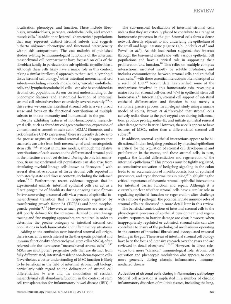
Intestinal stromal cells are also sensitive to multipleconventional immune cell-derived cytokines with relevanceto IBD (Figure 2). Intestinal stromal cells strongly respond tothe pro-inflammatory cytokines IL-1a and IL-1b, with a varietyof functional outcomes.56,57 In addition, the T helper type 17(Th17)-associated cytokine IL-17A induces the activation andproduction of IL-6, IL-8, and C-C motif chemokine ligand 2(CCL2) by intestinal stromal cells, through a mechanisminvolving nuclear factor (NF)-kB.58 Stromal responses to IL-17A and IL-17F appear to be strongest, as stimulation withother IL-17 family members (IL-17B/C/D/E) elicits a morelimited capacity to enhance the IL-1b-induced expression ofinflammatory mediators.59 In addition to being mediatedthrough NF-kB, the mechanism of IL-17A and IL-17F signalinghas been reported to be as a result of ERK (extracellular signal–regulated kinase) and p38MAPK (p38 mitogen-activatedprotein kinase) activity, indicating that divergent signalingmodules can be elicited in stromal cells by the same cytokines.
There is limited evidence that intestinal stromal cells canrespond to the IL-10 cytokine family member IL-22, with onereport describing a pro-inflammatory role for this cytokine inactivating myofibroblasts.60 If confirmed, this suggests that theimpact of IL-22 signaling on the mesenchymal compartment isdistinct from that of the epithelium, where IL-22 is generallyconsidered to be involved in protective responses.61 Interest-ingly, combined stimulation with IL-17A and IL-4 leads to thesynergistic induction of IL-6 release by stromal cells,62
suggesting a capacity to respond to cytokines associated withmultiple classes of CD4þ helper T cell. Supporting this isevidence that intestinal stromal cells respond to the classicalTh1 T-cell cytokine interferon-g by increasing their expressionof CD40 and MHCII (major histocompatibility complex classII), whereas downregulating aSMA expression, contributing totheir functional activation in vitro.63 It has been reported thatIL-21 signalling can synergise with TNFa to drive MMPproduction by intestinal stromal cells64 and attenuatedSTAT3 (signal transducer and activator of transcriptionfactor 3) expression within colonic stromal cells is associatedwith reduced tumor burden in IL-21� /� mice,65 indicating akey role for this hematopoietic immune cell-derived cytokinein activating stromal cells that can contribute to cancerprogression.
Multiple studies suggest that intestinal stromal cells arelinked to mucosal Th2-type responses. After IL-1b and TGFbstimulation, stromal cells are capable of abundant productionof IL-11, a cytokine linked to the priming of Th2 responses.63
Conversely, stromal cells are also able to respond to IL-31, acytokine produced by activated Th2 cells, in similar ways to thatseen with IL-17A.66 The extent to which redundancy exists
REVIEW
MucosalImmunology | VOLUME 6 NUMBER 2 |MARCH 2013 227
within these cytokine-induced activation pathways is not yetclear. Interestingly, levels of IL-33 are enhanced during UCand this Th2-associated alarmin appears to be preferentiallyexpressed by intestinal myofibroblasts.67,68 It is not knownwhether this is a protective or pathological response,although the potential for IL-33 to exacerbate inflammatorypathology in the intestine will be discussed later in thisreview.
Intestinal stromal cell–hematopoietic immune cellinteractions
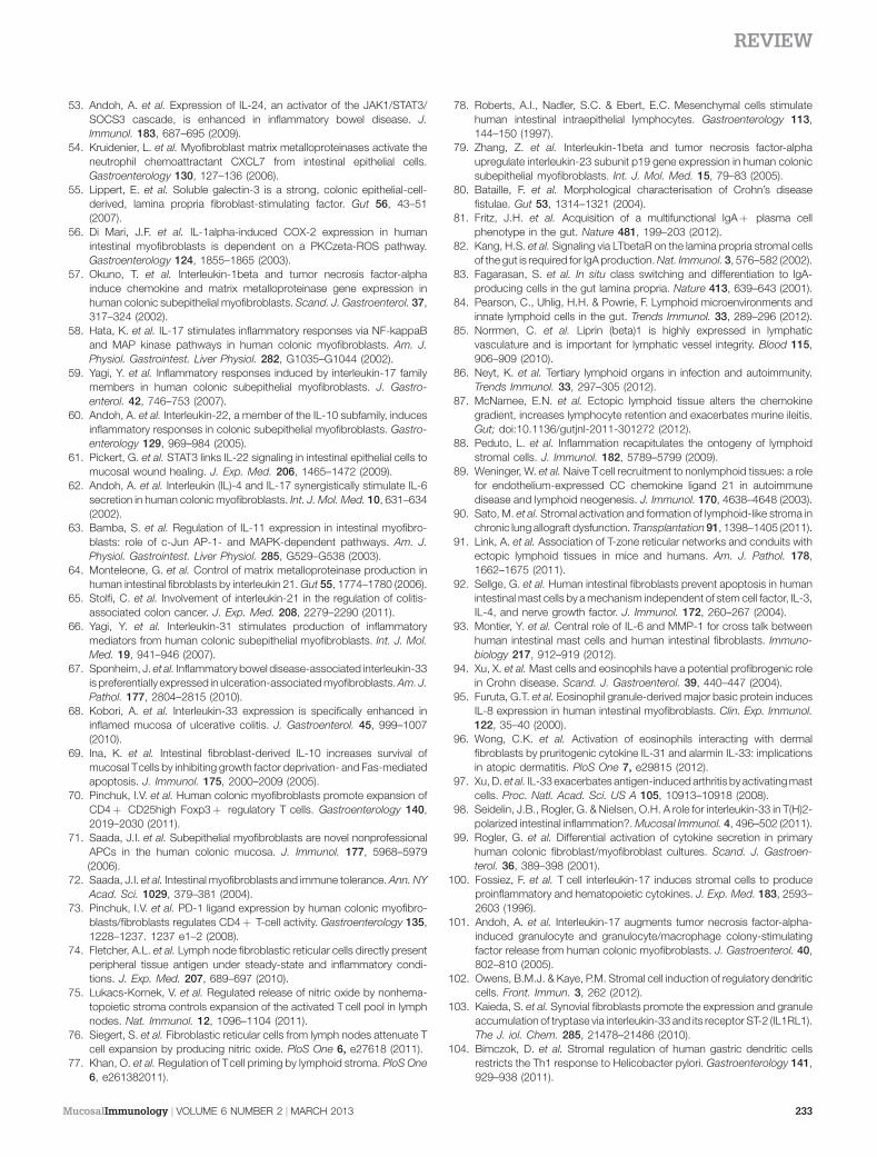
As well as responding to and modulating intestinal inflamma-tion in isolation, intestinal stromal cells also interact withmultiple populations of conventional hematopoietic immunecells outlined below (Figure 2).
T CELLS
Intestinal stromal cells are known to interact with effectorT cells in vitro, preventing their apoptosis via stromalproduction of IL-1069 and supporting CD25þ regulatoryT-cell expansion via expression of prostaglandin E2.70 Steady-state human colonic stromal cells also express human leukocyteantigen (HLA)-DR, can present model antigen, and activatea T-cell clone in vitro71—although nothing is known aboutthe mechanisms employed by these cells to elicit antigenpresentation after a more physiologically relevant challenge,for example, by an invasive bacterial species. It has been
proposed that intestinal stromal cells are capable of toleranceinduction72 and can regulate CD4þ T-cell proliferation byinhibiting IL-2 production via stromal expression of PD-L1/2(programmed death ligand 1/2),73 with this functionreminiscent of peripheral tissue antigen expression andtolerance induction by a subset of fibroblastic reticular cells(FRCs) in the peripheral lymph nodes.74 It is not yet clearwhether a subset of intestinal stromal cells can attenuate T-cellproliferation via provision of nitric oxide, as observed inperipheral lymph nodes.75–77
In addition to regulating conventional T-cell activation andproliferation, human stromal cells contribute to intraepitheliallymphocyte proliferation and survival in vitro by an HLA-DR-independent, a4b1 integrin–dependent mechanism.78 Cur-rently, there is a lack of convincing evidence that stromal cellscan directly induce T cells of a particular effector phenotype.However, it has been suggested that stromal cells can expressIL-23p19 (at least at the mRNA level) in response toinflammatory stimuli79 and as such could, in theory, supportTh17 differentiation. This hypothesis has not yet beenexperimentally validated and whether this capacity for‘‘myeloid’’ cytokine production is due to the differential origins(e.g., fibrocyte vs. MSC) of stromal populations is not known.
The many interactions between intestinal stromal cells and Tcells have been reviewed in more detail recently;9 however, theirfunctional interactions with other hematopoietic immune cellsare less well studied.
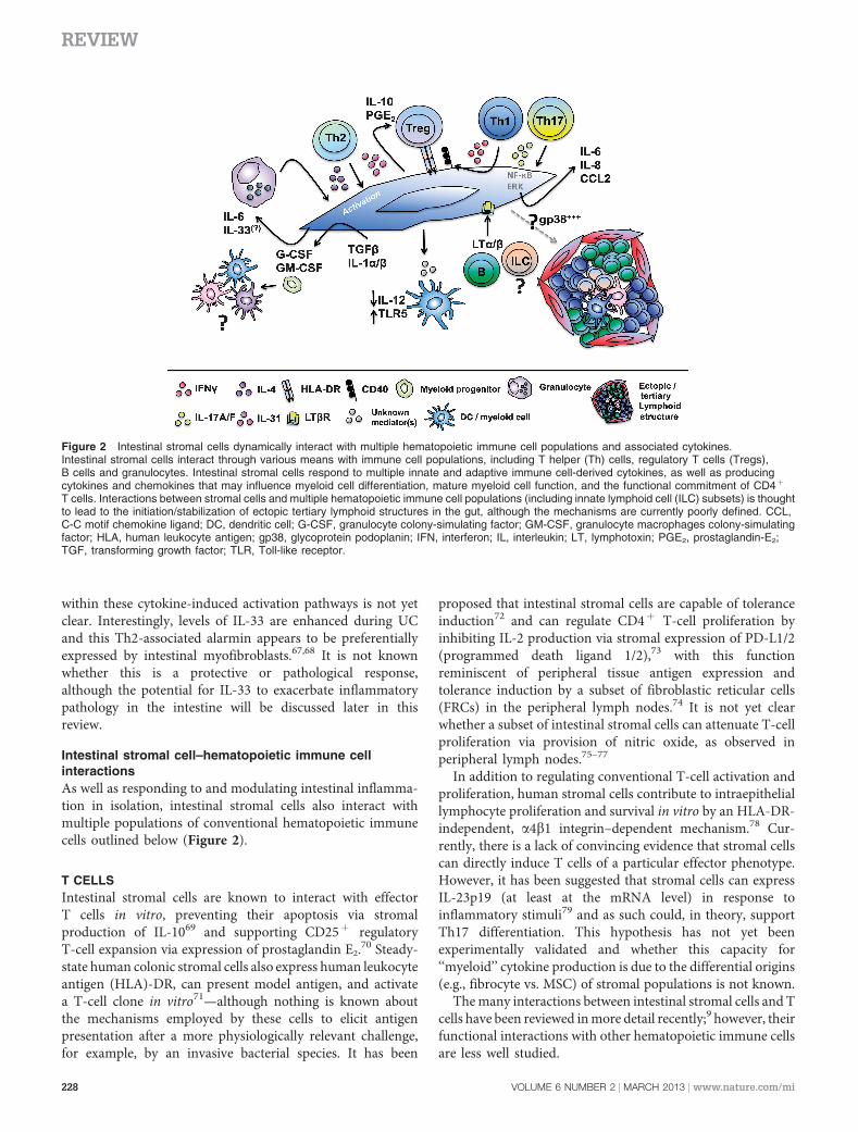
Figure 2 Intestinal stromal cells dynamically interact with multiple hematopoietic immune cell populations and associated cytokines.Intestinal stromal cells interact through various means with immune cell populations, including T helper (Th) cells, regulatory T cells (Tregs),B cells and granulocytes. Intestinal stromal cells respond to multiple innate and adaptive immune cell-derived cytokines, as well as producingcytokines and chemokines that may influence myeloid cell differentiation, mature myeloid cell function, and the functional commitment of CD4þ
T cells. Interactions between stromal cells and multiple hematopoietic immune cell populations (including innate lymphoid cell (ILC) subsets) is thoughtto lead to the initiation/stabilization of ectopic tertiary lymphoid structures in the gut, although the mechanisms are currently poorly defined. CCL,C-C motif chemokine ligand; DC, dendritic cell; G-CSF, granulocyte colony-simulating factor; GM-CSF, granulocyte macrophages colony-simulatingfactor; HLA, human leukocyte antigen; gp38, glycoprotein podoplanin; IFN, interferon; IL, interleukin; LT, lymphotoxin; PGE2, prostaglandin-E2;TGF, transforming growth factor; TLR, Toll-like receptor.
REVIEW
228 VOLUME 6 NUMBER 2 |MARCH 2013 |www.nature.com/mi
B CELLS
Reports describing intestinal stromal cell–B cell interactions aremore limited than those for T cells. A careful histologicalassessment of CD fistulae showed dense aggregates of CD20þ Bcells in the granulation tissue of fistulae, often adjacent to a layerof myofibroblasts and CD68þ macrophages.80 Whether suchinteractions are having a functional role in the tissue remodelingis not known. More recently, intestinal stromal cell–derivedfactors have been reported to co-operate with microbial stimuliin the generation of a unique (immunoglobulin Aþ ) IgAþ
‘‘multifunctional’’ plasma cell population, capable of abundantTNFa and induced nitric oxide synthase production.81
Although the precise mechanisms are not known, LTbRsignaling into intestinal stromal cells has been shown to becritical for IgA production,82 and intestinal stromal cells areable to induce B-cell IgM to IgA class switching in vitro,83
supporting the argument that stromal activation and stromalcell–B cell interactions have a role in the production of mucosalIgA. However, as the limited evidence currently availableattests, additional work will be required to determine whetherstromal cell–B cell interactions have further roles in thecomplex immunobiology of the intestine.
SECONDARY AND TERTIARY LYMPHOID ORGANS
The generation of several classes of secondary lymphoidstructures in the gut relies on exquisitely controlled spatio-temporal interactions between innate lymphoid cell popula-tions, such as lymphoid tissue inducer cells, and stromalorganizer cell populations.84 Unlike the development ofsecondary lymphoid organs in the periphery,7 the precisecontributions of intestinal stromal cells to lymphoid structuresin the gut are not yet known, nor is it fully clear whether thestromal subsets found in peripheral lymphoid organs arepresent within the intestine. Nevertheless, the isolation andculture of lymphatic endothelial cells (LECs) from humanintestine has been reported,85 with these cells bearing aphenotype analogous to the LEC stromal subset found withinthe peripheral lymph nodes.
Despite these processes being tightly regulated, the aberrantor ectopic accumulation of lymphoid structures in multipleorgans occurs during inflammation,86 including the intestine,87
although the precise contribution of intestinal stromal cells tode novo ectopic lymphoid organogenesis is not known.However, during murine models of inflammation, activatedileal-resident stromal cells exhibit some features of lymphoidorgan stromal cells, such as podoplanin (gp38) expression,88
hinting at the possibility of a contribution to ectopic/tertiarylymphoid organogenesis in the gut. Although not fullycharacterized for CD, CCL21 mRNA and protein expressionby stromal cells has been observed during UC,89 and stromalcell activation and the presence of CCL21þ FRC-like cells innascent T-cell zones associated with dendritic cell (DC) andlymphoid aggregates has been observed during other chronicinflammatory processes.90,91 Much is still to be clarifiedregarding the precise role of tissue stromal cells in thegeneration of tertiary lymphoid structures during chronic
intestinal inflammation, and there is a pressing need toinvestigate these mechanisms in humans. In addition to theinteractions with adaptive immune T and B cells described,multiple functional interactions between innate immune cellsand intestinal stromal cells have also been reported.
GRANULOCYTES
Intestinal stromal cells can support the survival of mast cellsin vitro92 through a mechanism dependent on the production ofIL-6.93 Interestingly, the interaction between stromal cells andgranulocytes appears to be bi-directional, as mast cell andeosinophil-derived factors can modulate intestinal stromal cellproliferation, phenotype, and function,93–95 at least in vitro.Although not yet determined for the gut, an IL-33-dependentinteraction between dermal stromal cells and eosinophilsenhances the pro-inflammatory activation of eosinophilsin vitro,96 suggesting that stromal cells can also directlymodulate granulocyte function in some mucosal contexts.Indeed, mast cell activation as a result of enhanced stromal IL-33 production in the inflamed synovium is proposed to be amajor pathological axis operating during experimental arthri-tis.97 As UC is associated with enhanced IL-33 expression byintestinal stromal cells,67,68 this raises the intriguing possibilitythat stromal cell–granulocyte interactions may contribute topathology associated with this form of IBD, by mechanismssimilar to those recently proposed for Th2-driven intestinalresponses more generally.98 Further characterization of intest-inal stromal cell and granulocyte interactions will be required toaddress this hypothesis.
MYELOID CELL DIFFERENTIATION AND FUNCTIONAL
CONDITIONING
It appears that intestinal stromal cells are capable of producingfactors required for the differentiation of myeloid populations,such as the NF-kB-dependent induction of M-CSF (macro-phage colony-stimulating factor)and GM-CSF (granulocytemacrophage colony-stimulating factor).99 This can be regulatedby cytokines implicated in IBD: indeed one of the earliestfunctions ascribed to human T-cell-derived IL-17A was theinduction of cytokines from stromal cells that created a nichefor myeloid cell differentiation.100 Supporting the existence ofthis axis in the gut, IL-17 directly enhances TNFa-inducedGM-CSF and G-CSF production by intestinal stromal cellsin vitro,101 although whether stromal cells are capable ofpolarizing myeloid precursors or monocytes toward distincteffector phenotypes is still unclear.
As previously stated, intestinal stromal cells can produceboth IL-10 and prostaglandin-E2, similar to stromal cells fromother organs, factors that in these distinct tissues can supportthe generation of regulatory DC populations.102 Conversely,stromal cell–derived IL-33—also abundantly expressed byintestinal stromal cells during UC—can modulate local pro-inflammatory myeloid cell function during RA.103 These datahighlight the possibility that stromal cells can functionally skewmyeloid populations to both pro- and anti-inflammatoryphenotypes, although the extent to which the stromal–myeloid
REVIEW
MucosalImmunology | VOLUME 6 NUMBER 2 |MARCH 2013 229
interactions apparent in other tissue sites are also operating inthe gut will require extensive further investigation.
Unlike mucosal epithelial cells, evidence for the conditioningof mature myeloid cell function by intestinal stromal cells islimited. However, gastric and intestinal stromal factors, mostlikely derived from the extracellular matrix, can regulate humanmyeloid DC function, downregulating IL-12 production andblunting the induction of Th1 responses by T cells in co-culture.104 Furthermore, stromal factors have also beenimplicated in the homeostatic regulation of TLR5 expressionin mucosal DCs,105 and stroma-conditioned media alsoregulates monocyte chemotaxis via IL-8 and TGFb.106 Verylittle else is known for the gut, and many highly pertinentquestions remain as to the extent of stromal cell–myeloid cellinteractions and their functional impact under steady-state andinflammatory conditions.
Stromal cells as direct mediators of innate immuneresponses in the intestine
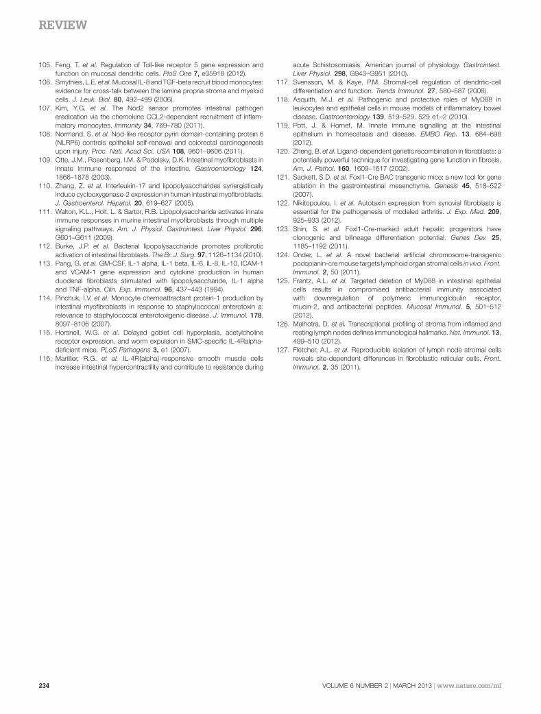
Growing evidence suggests that non-hematopoietic stromalcells of the intestine are capable of directly responding topathogens in a cell-intrinsic fashion (Figure 3). Although directin vivo evidence of the stromal contribution to microbialsensing is currently lacking, these cells appear to be equippedwith many mechanisms to facilitate such a functional capacity.
Recent murine data support a functional role forstromal expression of innate immune receptors in vivo, as
NOD2-dependent CCL2 production by intestinal stromal cells iskey for regulating inflammatory monocyte recruitment and thuspathogen clearance during Citrobacter rodentium infection, anexperimental model of human Enterohemorragic/pathogenicEscherichia coli infection.107 This function is associated with thedirect sensing of live bacteria and is specifically linked to NOD2-dependent bacterial-driven CCL2 production, as TNFa produc-tion is unaffected when NOD2-deficient colonic stromal cells areinfected with C. rodentium in vitro.
Data on the expression of other NLRs by murine intestinalstromal cells are limited; however, colonic myofibroblasts arethe major intestinal cell type expressing the innate inflamma-some-activating receptor NLRP6 in mice, through whichsignaling regulates epithelial reconstitution after bacterialchallenge in vivo.108 Although yet to be experimentallyvalidated, it is possible that sensing of bacteria by NLRs inthe stromal compartment after a breach in the epithelial barrierleads to the direct production of—as yet unknown—mediatorsthat facilitate epithelial repair. Interestingly, the re-localizationof stromal cells during epithelial regeneration is dependent onMyD88 (myeloid differentiation primary response gene (88)),22
a fact that lends support to the notion of innate immune sensinghaving a part in driving stromal cells toward an epithelialregenerative/healing response after a breach in the barrier andexposure to luminal bacteria.
Human intestinal stromal cells are known to express mRNAencoding the cytosolic innate receptors NOD1 and NOD2, and
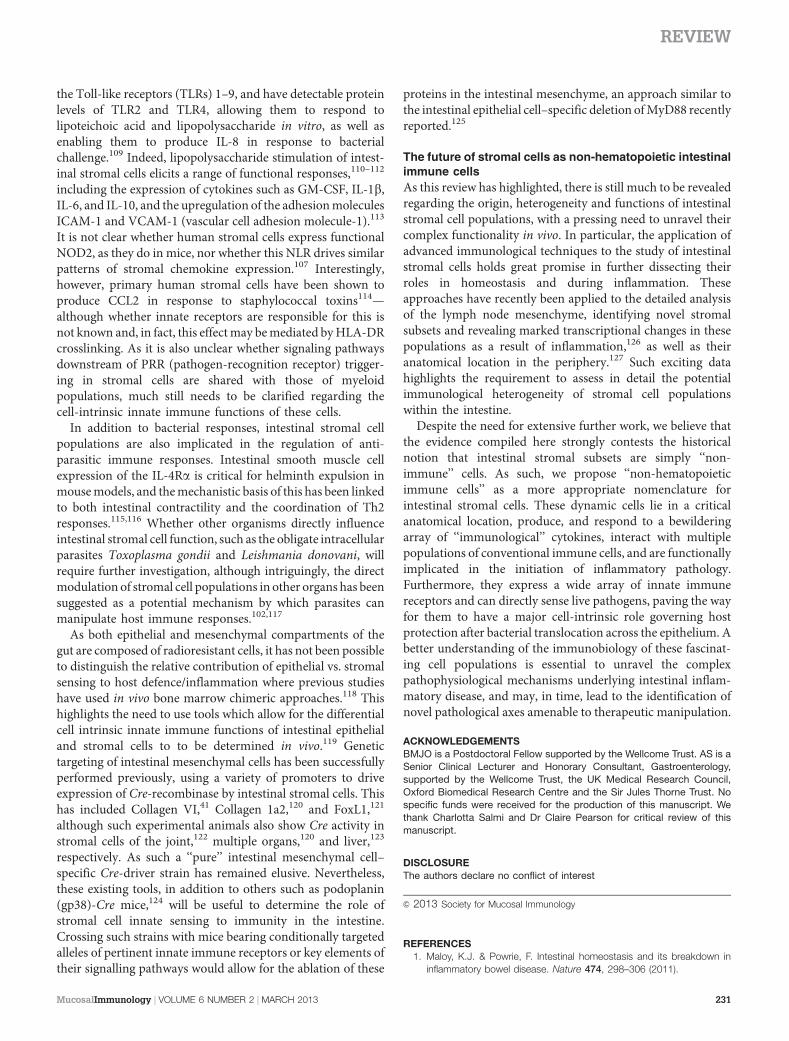
Figure 3 Intestinal stromal cells have a cell-intrinsic role in bacterial sensing at the intestinal barrier. Upon bacterial translocation across the epithelium,intestinal stromal cells are perfectly located to respond to the invading organisms. Murine stromal cells express NOD2 (nucleotide-bindingoligomerization domain-containing protein 2), which when triggered by Citrobacter rodentium initiates the production of C-C motif chemokine ligand2 (CCL2) by the intestinal stroma, recruitment of inflammatory monocytes, and the clearance of the organism. Human colonic stromal cellsexpress functional Toll-like receptor 2 (TLR2) and TLR4 (in addition to many pathogen-recognition receptors at the message level), which facilitate theproduction of multiple cytokines and other mediators after challenge with live bacteria or isolated bacterial ligands. Given their strategic locationand capacity to interact with epithelial cells, it is possible that stromal cells produce mucosal repair factors such as extracellular matrix (ECM)proteins, growth factors and other unknown factors after bacterial triggering, thus allowing the intestinal mesenchymal compartment to both recruithematopoietic immune cells (via chemokines) as well as begin the process of epithelial regeneration. GM-CSF, granulocyte macrophagescolony-simulating factor; ICAM-1, intercellular adhesion molecule-1; MyD88, myeloid differentiation primary response gene (88); NLR, NOD-likereceptor; PGE2, prostaglandin-E2; VCAM-1, vascular cell adhesion molecule-1.
REVIEW
230 VOLUME 6 NUMBER 2 |MARCH 2013 |www.nature.com/mi
the Toll-like receptors (TLRs) 1–9, and have detectable proteinlevels of TLR2 and TLR4, allowing them to respond tolipoteichoic acid and lipopolysaccharide in vitro, as well asenabling them to produce IL-8 in response to bacterialchallenge.109 Indeed, lipopolysaccharide stimulation of intest-inal stromal cells elicits a range of functional responses,110–112
including the expression of cytokines such as GM-CSF, IL-1b,IL-6, and IL-10, and the upregulation of the adhesion moleculesICAM-1 and VCAM-1 (vascular cell adhesion molecule-1).113
It is not clear whether human stromal cells express functionalNOD2, as they do in mice, nor whether this NLR drives similarpatterns of stromal chemokine expression.107 Interestingly,however, primary human stromal cells have been shown toproduce CCL2 in response to staphylococcal toxins114—although whether innate receptors are responsible for this isnot known and, in fact, this effect may be mediated by HLA-DRcrosslinking. As it is also unclear whether signaling pathwaysdownstream of PRR (pathogen-recognition receptor) trigger-ing in stromal cells are shared with those of myeloidpopulations, much still needs to be clarified regarding thecell-intrinsic innate immune functions of these cells.
In addition to bacterial responses, intestinal stromal cellpopulations are also implicated in the regulation of anti-parasitic immune responses. Intestinal smooth muscle cellexpression of the IL-4Ra is critical for helminth expulsion inmouse models, and the mechanistic basis of this has been linkedto both intestinal contractility and the coordination of Th2responses.115,116 Whether other organisms directly influenceintestinal stromal cell function, such as the obligate intracellularparasites Toxoplasma gondii and Leishmania donovani, willrequire further investigation, although intriguingly, the directmodulation of stromal cell populations in other organs has beensuggested as a potential mechanism by which parasites canmanipulate host immune responses.102,117
As both epithelial and mesenchymal compartments of thegut are composed of radioresistant cells, it has not been possibleto distinguish the relative contribution of epithelial vs. stromalsensing to host defence/inflammation where previous studieshave used in vivo bone marrow chimeric approaches.118 Thishighlights the need to use tools which allow for the differentialcell intrinsic innate immune functions of intestinal epithelialand stromal cells to to be determined in vivo.119 Genetictargeting of intestinal mesenchymal cells has been successfullyperformed previously, using a variety of promoters to driveexpression of Cre-recombinase by intestinal stromal cells. Thishas included Collagen VI,41 Collagen 1a2,120 and FoxL1,121
although such experimental animals also show Cre activity instromal cells of the joint,122 multiple organs,120 and liver,123
respectively. As such a ‘‘pure’’ intestinal mesenchymal cell–specific Cre-driver strain has remained elusive. Nevertheless,these existing tools, in addition to others such as podoplanin(gp38)-Cre mice,124 will be useful to determine the role ofstromal cell innate sensing to immunity in the intestine.Crossing such strains with mice bearing conditionally targetedalleles of pertinent innate immune receptors or key elements oftheir signalling pathways would allow for the ablation of these
proteins in the intestinal mesenchyme, an approach similar tothe intestinal epithelial cell–specific deletion of MyD88 recentlyreported.125
The future of stromal cells as non-hematopoietic intestinalimmune cells
As this review has highlighted, there is still much to be revealedregarding the origin, heterogeneity and functions of intestinalstromal cell populations, with a pressing need to unravel theircomplex functionality in vivo. In particular, the application ofadvanced immunological techniques to the study of intestinalstromal cells holds great promise in further dissecting theirroles in homeostasis and during inflammation. Theseapproaches have recently been applied to the detailed analysisof the lymph node mesenchyme, identifying novel stromalsubsets and revealing marked transcriptional changes in thesepopulations as a result of inflammation,126 as well as theiranatomical location in the periphery.127 Such exciting datahighlights the requirement to assess in detail the potentialimmunological heterogeneity of stromal cell populationswithin the intestine.
Despite the need for extensive further work, we believe thatthe evidence compiled here strongly contests the historicalnotion that intestinal stromal subsets are simply ‘‘non-immune’’ cells. As such, we propose ‘‘non-hematopoieticimmune cells’’ as a more appropriate nomenclature forintestinal stromal cells. These dynamic cells lie in a criticalanatomical location, produce, and respond to a bewilderingarray of ‘‘immunological’’ cytokines, interact with multiplepopulations of conventional immune cells, and are functionallyimplicated in the initiation of inflammatory pathology.Furthermore, they express a wide array of innate immunereceptors and can directly sense live pathogens, paving the wayfor them to have a major cell-intrinsic role governing hostprotection after bacterial translocation across the epithelium. Abetter understanding of the immunobiology of these fascinat-ing cell populations is essential to unravel the complexpathophysiological mechanisms underlying intestinal inflam-matory disease, and may, in time, lead to the identification ofnovel pathological axes amenable to therapeutic manipulation.
ACKNOWLEDGEMENTS
BMJO is a Postdoctoral Fellow supported by the Wellcome Trust. AS is a
Senior Clinical Lecturer and Honorary Consultant, Gastroenterology,
supported by the Wellcome Trust, the UK Medical Research Council,
Oxford Biomedical Research Centre and the Sir Jules Thorne Trust. No
specific funds were received for the production of this manuscript. We
thank Charlotta Salmi and Dr Claire Pearson for critical review of this
manuscript.
DISCLOSURE
The authors declare no conflict of interest
& 2013 Society for Mucosal Immunology
REFERENCES1. Maloy, K.J. & Powrie, F. Intestinal homeostasis and its breakdown in
inflammatory bowel disease. Nature 474, 298–306 (2011).
REVIEW
MucosalImmunology | VOLUME 6 NUMBER 2 |MARCH 2013 231
2. Cooney, R. et al. NOD2 stimulation induces autophagy in dendritic cells
influencing bacterial handling and antigen presentation. Nat. Med. 16,
90–97 (2010).3. Travassos, L.H. et al. Nod1 and Nod2 direct autophagy by recruiting
ATG16L1 to the plasma membrane at the site of bacterial entry. Nat.
Immunol. 11, 55–62 (2010).
4. Duerr, R.H. et al. A genome-wide association study identifies IL23R
as an inflammatory bowel disease gene. Science 314, 1461–1463
(2006).5. Glocker, E.O. et al. Inflammatory bowel disease and mutations
affecting the interleukin-10 receptor. N. Engl. J. Med. 361,
2033–2045 (2009).
6. Villani, A.C. et al. Common variants in the NLRP3 region contribute to
Crohn’s disease susceptibility. Nat. Genet. 41, 71–76 (2009).
7. Koning, J.J. & Mebius, R.E. Interdependence of stromal and immune cells
for lymph node function. Trends Immunol. 33, 264–270 (2012).8. Pinchuk, I.V. et al. Intestinal mesenchymal cells. Curr. Gastroenterol. Rep
12, 310–318 (2010).
9. Powell, D.W. et al. Mesenchymal cells of the intestinal lamina propria.
Annu. Rev. Physiol. 73, 213–237 (2011).
10. Mifflin, R.C. et al. Intestinal myofibroblasts: targets for stem cell therapy.
American journal of physiology. Gastrointest. Liver Physiol. 300,
G684–G696 (2011).
11. Andoh, A. et al. Role of intestinal subepithelial myofibroblasts in
inflammation and regenerative response in the gut. Pharmacol. Ther.
114, 94–106 (2007).
12. Reilkoff, R.A., Bucala, R. & Herzog, E.L. Fibrocytes: emerging effector
cells in chronic inflammation. Nat. Rev. Immunol. 11, 427–435 (2011).
13. Kolonin, M.G. et al. Alternative origins of stroma in normal organs and
disease. Stem Cell Res. 8, 312–323 (2012).
14. Uehara, H. et al. Emergence of fibrocytes showing morphological
changes in the inflamed colonic mucosa. Digest. Dis. Sci. 55,
253–260 (2010).
15. Flier, S.N. et al. Identification of epithelial to mesenchymal transition
as a novel source of fibroblasts in intestinal fibrosis. J. Biol. Chem. 285,
20202–20212 (2010).
16. Le Blanc, K. & Mougiakakos, D. Multipotent mesenchymal stromal cells
and the innate immune system. Nat. Rev. Immunol. 12, 383–396 (2012).
17. Dazzi, F., Lopes, L. & Weng, L. Mesenchymal stromal cells: a key player in
‘innate tolerance’?. Immunology 137, 206–213 (2012).
18. Lahar, N. et al. Intestinal subepithelial myofibroblasts support in vitro and
in vivo growth of human small intestinal epithelium. PloS One 6, e26898
(2011).
19. Samuel, S. et al. Characterization of putative stem cells in isolated human
colonic crypt epithelial cells and their interactions with myofibroblasts.
American journal of physiology. Cell Physiol. 296, C296–C305 (2009).
20. Francoeur, C. et al. Degeneration of the pericryptal myofibroblast sheath
by proinflammatory cytokines in inflammatory bowel diseases. Gastro-
enterology 136, 268–277.e3 (2009).
21. Farin, H.F., Van Es, J.H. & Clevers, H. Redundant sources of Wnt regulate
intestinal stem cells and promote formation of paneth cells. Gastro-
enterology 143, 1518–1529.e7 (2012).
22. Brown, S.L. et al. Myd88-dependent positioning of Ptgs2-expressing
stromal cells maintains colonic epithelial proliferation during injury. J. Clin.
Invest. 117, 258–269 (2007).
23. Powell, D.W. & Saada, J.I. Mesenchymal stem cells and prostaglandins
may be critical for intestinal wound repair. Gastroenterology 143, 19–22
(2012).
24. Kosinski, C. et al. Indian hedgehog regulates intestinal stem cell fate
through epithelial-mesenchymal interactions during development. Gas-
troenterology 139, 893–903 (2010).
25. van Dop, W.A. et al. Depletion of the colonic epithelial precursor cell
compartment upon conditional activation of the hedgehog pathway.
Gastroenterology 136, 2195–2203.e1–7 (2009).
26. Rieder, F. & Fiocchi, C. Intestinal fibrosis in IBD—a dynamic, multifactorial
process. Nat. Rev. Gastroenterol. Hepatol. 6, 228–235 (2009).
27. Rieder, F. et al. Wound healing and fibrosis in intestinal disease. Gut 56,
130–139 (2007).
28. Friedman, S.L. Mechanisms of hepatic fibrogenesis. Gastroenterology
134, 1655–1669 (2008).
29. Masamune, A. et al. Roles of pancreatic stellate cells in pancreatic
inflammation and fibrosis. Clin. Gastroenterol. Hepatol. 7 (11 Suppl), S48–
S54 (2009).
30. Barnes, P.J. Pathophysiology of allergic inflammation. Immunol. Rev. 242,
31–50 (2011).
31. Neumann, E. et al. Rheumatoid arthritis progression mediated by
activated synovial fibroblasts. Trends Mol. Med. 16, 458–468 (2010).32. Buckley, C.D. et al. Fibroblasts regulate the switch from acute resolving to
chronic persistent inflammation. Trends Immunol. 22, 199–204 (2001).
33. McGettrick, H.M. et al. Fibroblasts from different sites may promote or
inhibit recruitment of flowing lymphocytes by endothelial cells. Eur. J.
Immunol. 39, 113–125 (2009).
34. Filer, A. et al. The role of chemokines in leucocyte-stromal interactions in
rheumatoid arthritis. Front. Biosci. 13, 2674–2685 (2008).
35. Piera-Velazquez, S. et al. Persistent activation of dermal fibroblasts from
patients with gadolinium-associated nephrogenic systemic fibrosis. Ann.
Rheum. Dis. 69, 2017–2023 (2010).
36. Li, M. et al. Emergence of fibroblasts with a proinflammatory epigenetically
altered phenotype in severe hypoxic pulmonary hypertension. J.
Immunol. 187, 2711–2722 (2011).37. Bechtel, W. et al. Methylation determines fibroblast activation and
fibrogenesis in the kidney. Nat. Med. 16, 544–550 (2010).
38. Karouzakis, E. et al. Increased recycling of polyamines is associated with
global DNA hypomethylation in rheumatoid arthritis synovial fibroblasts.
Arthritis Rheum. 64, 1809–1817 (2012).
39. Russell, S.B. et al. Epigenetically altered wound healing in keloid
fibroblasts. J. Investig. Dermatol. 130, 2489–2496 (2010).
40. Ospelt, C. et al. Inflammatory memories: is epigenetics the missing link to
persistent stromal cell activation in rheumatoid arthritis?. Autoimmunity
Rev. 10, 519–524 (2011).
41. Armaka, M. et al. Mesenchymal cell targeting by TNF as a common
pathogenic principle in chronic inflammatory joint and intestinal diseases.
J. Exp. Med. 205, 331–337 (2008).42. Roulis, M. et al. Intestinal epithelial cells as producers but not targets of
chronic TNF suffice to cause murine Crohn-like pathology. Proc. Natl.
Acad. Sci.USA 108, 5396–5401 (2011).
43. Kontoyiannis, D. et al. Genetic dissection of the cellular pathways and
signaling mechanisms in modeled tumor necrosis factor-induced
Crohn’s-like inflammatory bowel disease. J. Exp. Med. 196, 1563–
1574 (2002).
44. De, L.K.T., Whiting, C.V. & Bland, P.W. Proinflammatory cytokine
synthesis by mucosal fibroblasts from mouse colitis is enhanced by
interferon-gamma-mediated up-regulation of CD40 signalling. Clin. Exp.
Immunol. 147, 313–323 (2007).
45. McKaig, B.C. et al. Differential expression of TGF-beta isoforms by normal
and inflammatory bowel disease intestinal myofibroblasts. Am. J. Physiol.
Cell Physiol. 282, C172–C182 (2002).
46. Di Sabatino, A. et al. Functional modulation of Crohn’s disease
myofibroblasts by anti-tumor necrosis factor antibodies. Gastroenterol-
ogy 133, 137–149 (2007).
47. Lang, M. et al. Gene expression profiles of mucosal fibroblasts from
strictured and nonstrictured areas of patients with Crohn’s disease.
Inflamm. Bowel Dis. 15, 212–223 (2009).
48. McKaig, B.C. et al. Expression and regulation of tissue inhibitor of
metalloproteinase-1 and matrix metalloproteinases by intestinal myofi-
broblasts in inflammatory bowel disease. Am. J. Pathol. 162, 1355–1360
(2003).
49. Cunningham, M.F. et al. S100A4 expression is increased in stricture
fibroblasts from patients with fibrostenosing Crohn’s disease and
promotes intestinal fibroblast migration. American journal of physiology.
Gastrointest. Liver Physiol. 299, G457–G466 (2010).
50. Bajaj-Elliott, M. et al. Keratinocyte growth factor in inflammatory bowel
disease. Increased mRNA transcripts in ulcerative colitis compared with
Crohn’s disease in biopsies and isolated mucosal myofibroblasts. Am. J.
Pathol. 151, 1469–1476 (1997).
51. Van Praet, L. et al. The transition of acute to chronic bowel inflammation in
spondyloarthritis. Nat. Rev. Rheumatol. 8, 288–295 (2012).
52. Fritsch, C. et al. Cytokines modulate fibroblast phenotype and epithelial-
stroma interactions in rat intestine. Gastroenterology 112, 826–838
(1997).
REVIEW
232 VOLUME 6 NUMBER 2 |MARCH 2013 |www.nature.com/mi
53. Andoh, A. et al. Expression of IL-24, an activator of the JAK1/STAT3/
SOCS3 cascade, is enhanced in inflammatory bowel disease. J.
Immunol. 183, 687–695 (2009).
54. Kruidenier, L. et al. Myofibroblast matrix metalloproteinases activate the
neutrophil chemoattractant CXCL7 from intestinal epithelial cells.
Gastroenterology 130, 127–136 (2006).
55. Lippert, E. et al. Soluble galectin-3 is a strong, colonic epithelial-cell-
derived, lamina propria fibroblast-stimulating factor. Gut 56, 43–51
(2007).
56. Di Mari, J.F. et al. IL-1alpha-induced COX-2 expression in human
intestinal myofibroblasts is dependent on a PKCzeta-ROS pathway.
Gastroenterology 124, 1855–1865 (2003).
57. Okuno, T. et al. Interleukin-1beta and tumor necrosis factor-alpha
induce chemokine and matrix metalloproteinase gene expression in
human colonic subepithelial myofibroblasts. Scand. J. Gastroenterol. 37,
317–324 (2002).
58. Hata, K. et al. IL-17 stimulates inflammatory responses via NF-kappaB
and MAP kinase pathways in human colonic myofibroblasts. Am. J.
Physiol. Gastrointest. Liver Physiol. 282, G1035–G1044 (2002).
59. Yagi, Y. et al. Inflammatory responses induced by interleukin-17 family
members in human colonic subepithelial myofibroblasts. J. Gastro-
enterol. 42, 746–753 (2007).
60. Andoh, A. et al. Interleukin-22, a member of the IL-10 subfamily, induces
inflammatory responses in colonic subepithelial myofibroblasts. Gastro-
enterology 129, 969–984 (2005).
61. Pickert, G. et al. STAT3 links IL-22 signaling in intestinal epithelial cells to
mucosal wound healing. J. Exp. Med. 206, 1465–1472 (2009).
62. Andoh, A. et al. Interleukin (IL)-4 and IL-17 synergistically stimulate IL-6
secretion in human colonic myofibroblasts. Int. J. Mol. Med. 10, 631–634
(2002).
63. Bamba, S. et al. Regulation of IL-11 expression in intestinal myofibro-
blasts: role of c-Jun AP-1- and MAPK-dependent pathways. Am. J.
Physiol. Gastrointest. Liver Physiol. 285, G529–G538 (2003).
64. Monteleone, G. et al. Control of matrix metalloproteinase production in
human intestinal fibroblasts by interleukin 21. Gut 55, 1774–1780 (2006).
65. Stolfi, C. et al. Involvement of interleukin-21 in the regulation of colitis-
associated colon cancer. J. Exp. Med. 208, 2279–2290 (2011).
66. Yagi, Y. et al. Interleukin-31 stimulates production of inflammatory
mediators from human colonic subepithelial myofibroblasts. Int. J. Mol.
Med. 19, 941–946 (2007).
67. Sponheim, J. et al. Inflammatory bowel disease-associated interleukin-33
is preferentially expressed in ulceration-associated myofibroblasts. Am. J.
Pathol. 177, 2804–2815 (2010).
68. Kobori, A. et al. Interleukin-33 expression is specifically enhanced in
inflamed mucosa of ulcerative colitis. J. Gastroenterol. 45, 999–1007
(2010).
69. Ina, K. et al. Intestinal fibroblast-derived IL-10 increases survival of
mucosal Tcells by inhibiting growth factor deprivation- and Fas-mediated
apoptosis. J. Immunol. 175, 2000–2009 (2005).
70. Pinchuk, I.V. et al. Human colonic myofibroblasts promote expansion of
CD4þ CD25high Foxp3þ regulatory T cells. Gastroenterology 140,
2019–2030 (2011).
71. Saada, J.I. et al. Subepithelial myofibroblasts are novel nonprofessional
APCs in the human colonic mucosa. J. Immunol. 177, 5968–5979
(2006).
72. Saada, J.I. et al. Intestinal myofibroblasts and immune tolerance. Ann. NY
Acad. Sci. 1029, 379–381 (2004).
73. Pinchuk, I.V. et al. PD-1 ligand expression by human colonic myofibro-
blasts/fibroblasts regulates CD4þ T-cell activity. Gastroenterology 135,
1228–1237. 1237 e1–2 (2008).
74. Fletcher, A.L. et al. Lymph node fibroblastic reticular cells directly present
peripheral tissue antigen under steady-state and inflammatory condi-
tions. J. Exp. Med. 207, 689–697 (2010).
75. Lukacs-Kornek, V. et al. Regulated release of nitric oxide by nonhema-
topoietic stroma controls expansion of the activated T cell pool in lymph
nodes. Nat. Immunol. 12, 1096–1104 (2011).
76. Siegert, S. et al. Fibroblastic reticular cells from lymph nodes attenuate T
cell expansion by producing nitric oxide. PloS One 6, e27618 (2011).
77. Khan, O. et al. Regulation of Tcell priming by lymphoid stroma. PloS One
6, e261382011).
78. Roberts, A.I., Nadler, S.C. & Ebert, E.C. Mesenchymal cells stimulate
human intestinal intraepithelial lymphocytes. Gastroenterology 113,
144–150 (1997).79. Zhang, Z. et al. Interleukin-1beta and tumor necrosis factor-alpha
upregulate interleukin-23 subunit p19 gene expression in human colonic
subepithelial myofibroblasts. Int. J. Mol. Med. 15, 79–83 (2005).
80. Bataille, F. et al. Morphological characterisation of Crohn’s disease
fistulae. Gut 53, 1314–1321 (2004).
81. Fritz, J.H. et al. Acquisition of a multifunctional IgAþ plasma cell
phenotype in the gut. Nature 481, 199–203 (2012).
82. Kang, H.S. et al. Signaling via LTbetaR on the lamina propria stromal cells
of the gut is required for IgA production. Nat. Immunol. 3, 576–582 (2002).
83. Fagarasan, S. et al. In situ class switching and differentiation to IgA-
producing cells in the gut lamina propria. Nature 413, 639–643 (2001).
84. Pearson, C., Uhlig, H.H. & Powrie, F. Lymphoid microenvironments and
innate lymphoid cells in the gut. Trends Immunol. 33, 289–296 (2012).
85. Norrmen, C. et al. Liprin (beta)1 is highly expressed in lymphatic
vasculature and is important for lymphatic vessel integrity. Blood 115,
906–909 (2010).
86. Neyt, K. et al. Tertiary lymphoid organs in infection and autoimmunity.
Trends Immunol. 33, 297–305 (2012).
87. McNamee, E.N. et al. Ectopic lymphoid tissue alters the chemokine
gradient, increases lymphocyte retention and exacerbates murine ileitis.
Gut; doi:10.1136/gutjnl-2011-301272 (2012).
88. Peduto, L. et al. Inflammation recapitulates the ontogeny of lymphoid
stromal cells. J. Immunol. 182, 5789–5799 (2009).
89. Weninger, W. et al. Naive Tcell recruitment to nonlymphoid tissues: a role
for endothelium-expressed CC chemokine ligand 21 in autoimmune
disease and lymphoid neogenesis. J. Immunol. 170, 4638–4648 (2003).
90. Sato, M. et al. Stromal activation and formation of lymphoid-like stroma in
chronic lung allograft dysfunction. Transplantation 91, 1398–1405 (2011).
91. Link, A. et al. Association of T-zone reticular networks and conduits with
ectopic lymphoid tissues in mice and humans. Am. J. Pathol. 178,
1662–1675 (2011).
92. Sellge, G. et al. Human intestinal fibroblasts prevent apoptosis in human
intestinal mast cells by a mechanism independent of stem cell factor, IL-3,
IL-4, and nerve growth factor. J. Immunol. 172, 260–267 (2004).
93. Montier, Y. et al. Central role of IL-6 and MMP-1 for cross talk between
human intestinal mast cells and human intestinal fibroblasts. Immuno-
biology 217, 912–919 (2012).
94. Xu, X. et al. Mast cells and eosinophils have a potential profibrogenic role
in Crohn disease. Scand. J. Gastroenterol. 39, 440–447 (2004).
95. Furuta, G.T. et al. Eosinophil granule-derived major basic protein induces
IL-8 expression in human intestinal myofibroblasts. Clin. Exp. Immunol.
122, 35–40 (2000).
96. Wong, C.K. et al. Activation of eosinophils interacting with dermal
fibroblasts by pruritogenic cytokine IL-31 and alarmin IL-33: implications
in atopic dermatitis. PloS One 7, e29815 (2012).
97. Xu, D. et al. IL-33 exacerbates antigen-induced arthritis by activating mast
cells. Proc. Natl. Acad. Sci. US A 105, 10913–10918 (2008).
98. Seidelin, J.B., Rogler, G. & Nielsen, O.H. A role for interleukin-33 in T(H)2-
polarized intestinal inflammation?. Mucosal Immunol. 4, 496–502 (2011).
99. Rogler, G. et al. Differential activation of cytokine secretion in primary
human colonic fibroblast/myofibroblast cultures. Scand. J. Gastroen-
terol. 36, 389–398 (2001).
100. Fossiez, F. et al. T cell interleukin-17 induces stromal cells to produce
proinflammatory and hematopoietic cytokines. J. Exp. Med. 183, 2593–
2603 (1996).
101. Andoh, A. et al. Interleukin-17 augments tumor necrosis factor-alpha-
induced granulocyte and granulocyte/macrophage colony-stimulating
factor release from human colonic myofibroblasts. J. Gastroenterol. 40,
802–810 (2005).
102. Owens, B.M.J. & Kaye, P.M. Stromal cell induction of regulatory dendritic
cells. Front. Immun. 3, 262 (2012).
103. Kaieda, S. et al. Synovial fibroblasts promote the expression and granule
accumulation of tryptase via interleukin-33 and its receptor ST-2 (IL1RL1).
The J. iol. Chem. 285, 21478–21486 (2010).
104. Bimczok, D. et al. Stromal regulation of human gastric dendritic cells
restricts the Th1 response to Helicobacter pylori. Gastroenterology 141,
929–938 (2011).
REVIEW
MucosalImmunology | VOLUME 6 NUMBER 2 |MARCH 2013 233
105. Feng, T. et al. Regulation of Toll-like receptor 5 gene expression and
function on mucosal dendritic cells. PloS One 7, e35918 (2012).
106. Smythies, L.E. et al. Mucosal IL-8 and TGF-beta recruit blood monocytes:
evidence for cross-talk between the lamina propria stroma and myeloid
cells. J. Leuk. Biol. 80, 492–499 (2006).
107. Kim, Y.G. et al. The Nod2 sensor promotes intestinal pathogen
eradication via the chemokine CCL2-dependent recruitment of inflam-
matory monocytes. Immunity 34, 769–780 (2011).
108. Normand, S. et al. Nod-like receptor pyrin domain-containing protein 6(NLRP6) controls epithelial self-renewal and colorectal carcinogenesis
upon injury. Proc. Natl. Acad Sci. USA 108, 9601–9606 (2011).
109. Otte, J.M., Rosenberg, I.M. & Podolsky, D.K. Intestinal myofibroblasts in
innate immune responses of the intestine. Gastroenterology 124,
1866–1878 (2003).
110. Zhang, Z. et al. Interleukin-17 and lipopolysaccharides synergistically
induce cyclooxygenase-2 expression in human intestinal myofibroblasts.
J. Gastroenterol. Hepatol. 20, 619–627 (2005).
111. Walton, K.L., Holt, L. & Sartor, R.B. Lipopolysaccharide activates innate
immune responses in murine intestinal myofibroblasts through multiple
signaling pathways. Am. J. Physiol. Gastrointest. Liver Physiol. 296,
G601–G611 (2009).
112. Burke, J.P. et al. Bacterial lipopolysaccharide promotes profibrotic
activation of intestinal fibroblasts. The Br. J. Surg. 97, 1126–1134 (2010).
113. Pang, G. et al. GM-CSF, IL-1 alpha, IL-1 beta, IL-6, IL-8, IL-10, ICAM-1
and VCAM-1 gene expression and cytokine production in human
duodenal fibroblasts stimulated with lipopolysaccharide, IL-1 alpha
and TNF-alpha. Clin. Exp. Immunol. 96, 437–443 (1994).114. Pinchuk, I.V. et al. Monocyte chemoattractant protein-1 production by
intestinal myofibroblasts in response to staphylococcal enterotoxin a:
relevance to staphylococcal enterotoxigenic disease. J. Immunol. 178,
8097–8106 (2007).
115. Horsnell, W.G. et al. Delayed goblet cell hyperplasia, acetylcholine
receptor expression, and worm expulsion in SMC-specific IL-4Ralpha-
deficient mice. PLoS Pathogens 3, e1 (2007).
116. Marillier, R.G. et al. IL-4R{alpha}-responsive smooth muscle cells
increase intestinal hypercontractility and contribute to resistance during
acute Schistosomiasis. American journal of physiology. Gastrointest.
Liver Physiol. 298, G943–G951 (2010).
117. Svensson, M. & Kaye, P.M. Stromal-cell regulation of dendritic-cell
differentiation and function. Trends Immunol. 27, 580–587 (2006).
118. Asquith, M.J. et al. Pathogenic and protective roles of MyD88 in
leukocytes and epithelial cells in mouse models of inflammatory bowel
disease. Gastroenterology 139, 519–529. 529 e1–2 (2010).
119. Pott, J. & Hornef, M. Innate immune signalling at the intestinal
epithelium in homeostasis and disease. EMBO Rep. 13, 684–698(2012).
120. Zheng, B. et al. Ligand-dependent genetic recombination in fibroblasts: a
potentially powerful technique for investigating gene function in fibrosis.
Am. J. Pathol. 160, 1609–1617 (2002).
121. Sackett, S.D. et al. Foxl1-Cre BAC transgenic mice: a new tool for gene
ablation in the gastrointestinal mesenchyme. Genesis 45, 518–522
(2007).
122. Nikitopoulou, I. et al. Autotaxin expression from synovial fibroblasts is
essential for the pathogenesis of modeled arthritis. J. Exp. Med. 209,
925–933 (2012).
123. Shin, S. et al. Foxl1-Cre-marked adult hepatic progenitors have
clonogenic and bilineage differentiation potential. Genes Dev. 25,
1185–1192 (2011).
124. Onder, L. et al. A novel bacterial artificial chromosome-transgenic
podoplanin-cre mouse targets lymphoid organ stromal cells in vivo. Front.
Immunol. 2, 50 (2011).
125. Frantz, A.L. et al. Targeted deletion of MyD88 in intestinal epithelial
cells results in compromised antibacterial immunity associatedwith downregulation of polymeric immunoglobulin receptor,
mucin-2, and antibacterial peptides. Mucosal Immunol. 5, 501–512
(2012).
126. Malhotra, D. et al. Transcriptional profiling of stroma from inflamed and
resting lymph nodes defines immunological hallmarks. Nat. Immunol. 13,
499–510 (2012).
127. Fletcher, A.L. et al. Reproducible isolation of lymph node stromal cells
reveals site-dependent differences in fibroblastic reticular cells. Front.
Immunol. 2, 35 (2011).
REVIEW
234 VOLUME 6 NUMBER 2 |MARCH 2013 |www.nature.com/mi
Related Documents