Insights into Advanced Retinopathy of Prematurity Using Handheld Spectral Domain Optical Coherence Tomography Imaging Sai H. Chavala, MD, Sina Farsiu, PhD, Ramiro Maldonado, MD, David K. Wallace, MD, Sharon F. Freedman, MD, Cynthia A. Toth, MD Purpose: To elucidate the subclinical anatomy of retinopathy of prematurity (ROP) using spectral domain optical coherence tomography (SD OCT). Design: Prospective, observational case series. Participants: Three low-birth-weight, severely premature infants. Methods: Clinical examination was performed using a portable slit lamp and indirect ophthalmoscope. Imaging was performed by using a handheld SD OCT device and Retcam (Clarity Medical Systems, Pleasanton, CA) or video-indirect recording. Spectral domain optical coherence tomography imaging was conducted without sedation at the bedside in the neonatal intensive care unit on 1 patient. The other 2 patients had an examination under anesthesia with SD OCT imaging in the operating room. Main Outcome Measures: In vivo determination of vitreoretinal morphology, anatomy, and pathology by clinical examination, imaging, and SD OCT. Results: Linear and volumetric imaging was achieved with the handheld system in infant eyes despite tunica vasculosa lentis and vitreous bands. Imaging was not possible in eyes with notable vitreous hemorrhage. Analysis of SD OCT images revealed preretinal structures (ranging from 409 to 2700 m in width and 212 to 440 m in height), retinoschisis, and retinal detachment in the posterior pole of patients with advanced ROP. Both the retinoschisis and the preretinal structures were not identified on conventional examination or imaging by expert pediatric ophthalmologists. The preretinal structures varied in location and size, and may represent preretinal fibrovascular proliferation. Some were found in close proximity to blood vessels, whereas others were near the optic nerve. Conclusions: Handheld SD OCT imaging can be performed on the sedated or nonsedated neonate and provides valuable subclinical anatomic information. This novel imaging modality can reveal the location and extent of posterior ROP pathology not evident on standard examination. This could affect future clinical decision-making if studies validate a management strategy based on findings from this imaging technique. Financial Disclosure(s): Proprietary or commercial disclosure may be found after the references. Ophthalmology 2009;116:2448 –2456 © 2009 by the American Academy of Ophthalmology. Despite advances in treatment, retinopathy of prematurity (ROP) is a major cause of vision loss in neonates. 1,2 This disease is characterized by an abnormal, premature arrest of developing retinal blood vessels. When this carefully or- chestrated process is pathologically disrupted by premature birth, a subset of patients with ROP develop abnormal fibrovascular proliferation that can cause retinal detachment and permanent vision loss. The Early Treatment of ROP study found that 9% of patients had an unfavorable anatomic outcome at 2 years despite laser ablation of high-risk eyes at prethreshold. 3 Prenner et al 4 showed that surgery for infants with stage 4 ROP without macula involvement is safe and may preserve the architecture of the macula leading to better visual out- comes. Similarly, Azuma et al 5 suggest that intervention with early vitreous surgery for aggressive posterior ROP may be indicated when laser ablation or cryotherapy does not prevent the progression to retinal detachment. 6 The ability to promptly recognize characteristics leading to ac- tive proliferation of fibrovascular tissue may lead to early intervention and better visual and anatomic outcomes. 7 The ability to recognize the early stage of retinal detachment is also important. In these studies, the extent of detachment and preretinal pathology was determined by ophthalmo- scopic examination or review of fundus photographs. Fundus examination with indirect ophthalmoscopy remains the gold standard for monitoring patients with ROP. 1,3,8 Fun- dus photography with computer-assisted tools is garnering 2448 © 2009 by the American Academy of Ophthalmology ISSN 0161-6420/09/$–see front matter Published by Elsevier Inc. doi:10.1016/j.ophtha.2009.06.003

Welcome message from author
This document is posted to help you gain knowledge. Please leave a comment to let me know what you think about it! Share it to your friends and learn new things together.
Transcript
Insights into Advanced Retinopathyof Prematurity Using HandheldSpectral Domain Optical CoherenceTomography Imaging
Sai H. Chavala, MD, Sina Farsiu, PhD, Ramiro Maldonado, MD, David K. Wallace, MD,Sharon F. Freedman, MD, Cynthia A. Toth, MD
Purpose: To elucidate the subclinical anatomy of retinopathy of prematurity (ROP) using spectral domainoptical coherence tomography (SD OCT).
Design: Prospective, observational case series.Participants: Three low-birth-weight, severely premature infants.Methods: Clinical examination was performed using a portable slit lamp and indirect ophthalmoscope.
Imaging was performed by using a handheld SD OCT device and Retcam (Clarity Medical Systems, Pleasanton,CA) or video-indirect recording. Spectral domain optical coherence tomography imaging was conducted withoutsedation at the bedside in the neonatal intensive care unit on 1 patient. The other 2 patients had an examinationunder anesthesia with SD OCT imaging in the operating room.
Main Outcome Measures: In vivo determination of vitreoretinal morphology, anatomy, and pathology byclinical examination, imaging, and SD OCT.
Results: Linear and volumetric imaging was achieved with the handheld system in infant eyes despite tunicavasculosa lentis and vitreous bands. Imaging was not possible in eyes with notable vitreous hemorrhage.Analysis of SD OCT images revealed preretinal structures (ranging from 409 to 2700 �m in width and 212 to 440�m in height), retinoschisis, and retinal detachment in the posterior pole of patients with advanced ROP. Both theretinoschisis and the preretinal structures were not identified on conventional examination or imaging by expertpediatric ophthalmologists. The preretinal structures varied in location and size, and may represent preretinalfibrovascular proliferation. Some were found in close proximity to blood vessels, whereas others were near theoptic nerve.
Conclusions: Handheld SD OCT imaging can be performed on the sedated or nonsedated neonate andprovides valuable subclinical anatomic information. This novel imaging modality can reveal the location andextent of posterior ROP pathology not evident on standard examination. This could affect future clinicaldecision-making if studies validate a management strategy based on findings from this imaging technique.
Financial Disclosure(s): Proprietary or commercial disclosure may be found after the references.Ophthalmology 2009;116:2448 –2456 © 2009 by the American Academy of Ophthalmology.
Despite advances in treatment, retinopathy of prematurity(ROP) is a major cause of vision loss in neonates.1,2 Thisdisease is characterized by an abnormal, premature arrest ofdeveloping retinal blood vessels. When this carefully or-chestrated process is pathologically disrupted by prematurebirth, a subset of patients with ROP develop abnormalfibrovascular proliferation that can cause retinal detachmentand permanent vision loss.
The Early Treatment of ROP study found that 9% ofpatients had an unfavorable anatomic outcome at 2 yearsdespite laser ablation of high-risk eyes at prethreshold.3
Prenner et al4 showed that surgery for infants with stage 4ROP without macula involvement is safe and may preserve
the architecture of the macula leading to better visual out-2448 © 2009 by the American Academy of OphthalmologyPublished by Elsevier Inc.
comes. Similarly, Azuma et al5 suggest that interventionwith early vitreous surgery for aggressive posterior ROPmay be indicated when laser ablation or cryotherapy doesnot prevent the progression to retinal detachment.6 Theability to promptly recognize characteristics leading to ac-tive proliferation of fibrovascular tissue may lead to earlyintervention and better visual and anatomic outcomes.7 Theability to recognize the early stage of retinal detachment isalso important. In these studies, the extent of detachmentand preretinal pathology was determined by ophthalmo-scopic examination or review of fundus photographs.
Fundus examination with indirect ophthalmoscopy remainsthe gold standard for monitoring patients with ROP.1,3,8 Fun-
dus photography with computer-assisted tools is garneringISSN 0161-6420/09/$–see front matterdoi:10.1016/j.ophtha.2009.06.003
Chavala et al � SD OCT in Advanced ROP
interest to reduce the subjectivity of diagnosing plus dis-ease.9–11 However, new imaging technology is necessary todetect subclinical pathology that may improve our under-standing of the underlying disease process and guide treat-ment decisions.
Optical coherence tomography (OCT) imaging has en-hanced our understanding and treatment of adult maculardiseases.12 Positioning and cooperation requirements limitneonatal imaging with commercial tabletop OCT systems.A time-domain OCT (TD OCT) system recently was mod-ified to image neonates with ROP in the operating room(OR).13,14 Both of these studies demonstrated subclinicalpathology, either retinoschisis or retinal detachment, inROP. However, because of slow image acquisition, TDOCT requires an immobile eye for 1 or more seconds toavoid motion artifact. This limits the use of this device toanesthetized or sedated young patients.
The capability of performing in vivo high-resolutionimaging would enhance our ability to detect, characterize,and follow ROP pathology. The higher capture rate ofspectral domain optical coherence tomography (SD OCT),which is more than 40 times faster than TD OCT, dimin-ishes motion artifact and makes handheld imaging possi-ble.15–17 Scott et al16 demonstrated that a handheld SD OCTdevice can be used to image the retina in full-term infantswith shaken baby syndrome. Chong et al17 used the samesystem to image a young child with albinism. Both of thesestudies imaged infants under sedation or general anesthesia.In this study, we extend the application of OCT by using anSD OCT system that combines high-speed imaging and theuse of a handheld probe, unavailable with existing TD OCT,to image the eyes of neonates with and without generalanesthesia. We report the results of high-resolution, cross-sectional, and 3-dimensional SD OCT images in nonsedatedand anesthetized neonates with advanced ROP.
Patients and Methods
Consent for this study was obtained as part of an approved studyin accordance with the Duke University Health System Institu-tional Review Board. The patients included 3 infants with ad-vanced ROP in both eyes. SD OCT imaging was performed in theneonatal intensive care unit (NICU) without sedation for 1 patientand in the OR under general anesthesia for 2 patients. We imagedall patients using a handheld SD OCT probe that replaced theconventional tabletop head on SD OCT systems (Bioptigen Inc.,Research Triangle Park, NC). The handheld probe was connectedby a flexible fiberoptic cable to the SD OCT system. For the thirdcase, the original larger tabletop SD OCT system (same handheldprobe) was switched to a more compact system on a moveable cart(Fig 1). All patients were supine during imaging. Both NICU andOR imaging were performed with continuous monitoring of vitalsigns. The NICU SD OCT imaging was performed on a nonintu-bated patient without sedation. A pacifier dipped with severaldrops of 24% sucrose solution (Toot Sweet, Hawaii Medical,Pembroke, MA) was orally administered during SD OCT imagingsessions in the NICU.18
In the NICU, SD OCT imaging was obtained 1 day before thenext standard care clinical examination. Video-indirect ophthal-moscopy was performed at the time of the examination, but be-
cause those images were of limited quality as the result of poorfocus, we used representative video-indirect images obtained 8days after SD OCT imaging in the NICU for comparison in thisstudy. Spectral domain optical coherence tomography imaging wasperformed in the NICU bed with a lid speculum and topicalproparacaine anesthesia. The examiner held the noncontact hand-piece over the infant’s eye, while resting her fingers on the faceand wrists on the side of the bed to stabilize the probe. The visiblelight emitted from the handpiece into the patient’s eye is a narrow,dim red light. There is no bright flash of light discharged during theexamination.
In the OR, the examiner had the option of using a wrist rest tostabilize her hands while holding the SD OCT handpiece (Fig 1).Color fundus photographs were captured using contact Retcamimaging (Clarity Medical Systems, Pleasanton, CA) with methyl-cellulose gel on the cornea.
Analysis and capture of SD OCT images were performed in amanner similar to the protocol described by Scott et al.16 Briefly,SD OCT images were captured using Bioptigen software version1755. The software allowed the user to set up length, orientation(0–360 degrees), number of A-scans per B-scan, and B-scans pervolumetric scan, as desired. For this study, our group defined linearscans as multiple B-scans of the same characteristics capturedfrom the same retinal location, volumetric-rectangular scans asmultiple B-scans stepped across a rectangular area of the retina,and mixed scans as low-resolution volumetric scans with higher-
Figure 1. Mobile workstation connected to a handheld probe forspectral domain optical coherence tomography (SD OCT) imaging inthe operating room. A neonate is positioned in the supine positionwith the operator using a wrist rest to stabilize the noncontact hand-held SD OCT probe.
resolution linear scans at the center line of the rectangle. Qualita-
2449
Ophthalmology Volume 116, Number 12, December 2009
tive image processing of the acquired scans were performed usingthe Duke Optical Coherence Tomography Retinal Analysis Pro-gram.19 Adobe Photoshop Creative Suite 3 (Adobe Inc., San Jose,CA), and ImageJ (National Institutes of Health, Bethesda, MD)software programs were used for image visualization, enhance-ment, and annotation of structures observed on cross-sectional SDOCT scans.15 Three-dimensional videos and composite imageswere created using AMIRA version 4.1 software (Visage Im-aging Inc., Carlsbad, CA). Duke Optical Coherence Tomogra-phy Retinal Analysis Program was also used to create anadaptive, contrast-enhanced, summed voxel projection (SVP)2-dimensional image analogous to a fundus photograph createdby axially collapsing B-scans.20 The anatomic findings visibleon Retcam, video-indirect images, and clinical examinationnotes were compared with SD OCT images. All SD OCT scanswere evaluated, and interpretable scans from each study wereused for analysis.
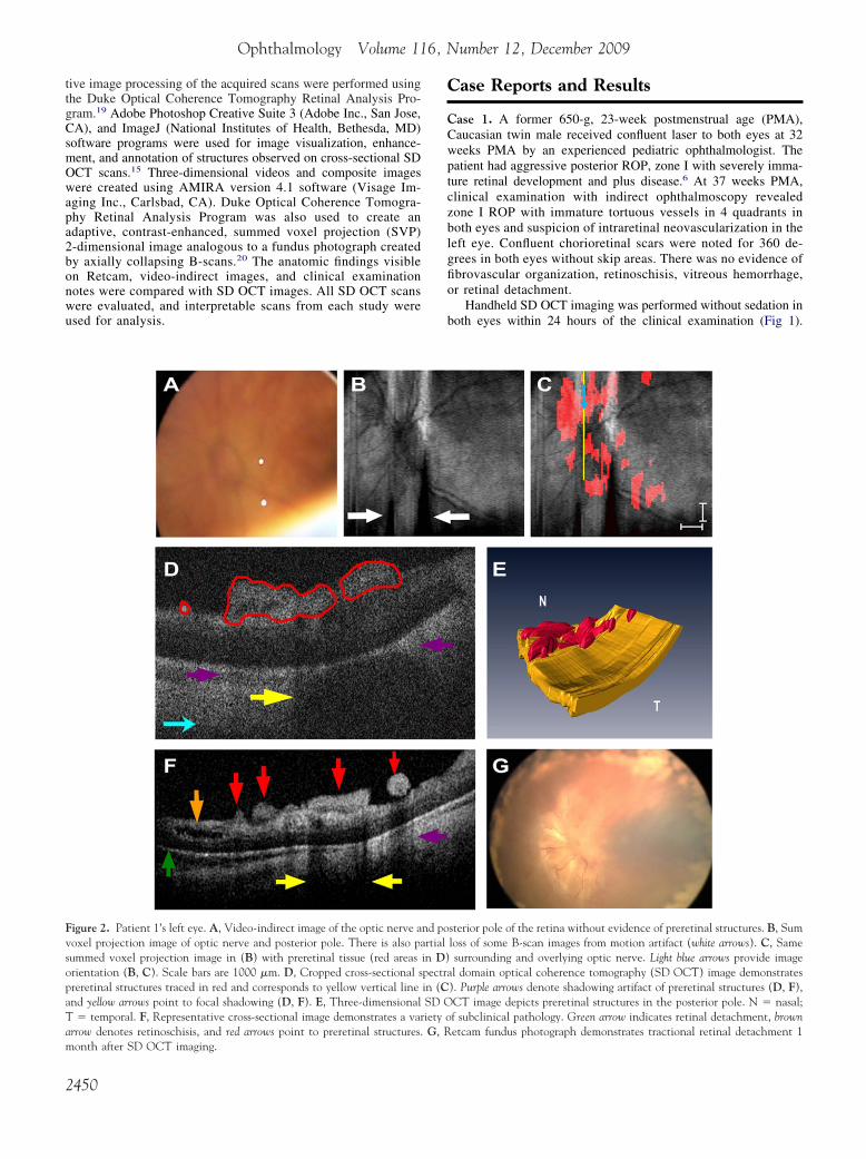
Figure 2. Patient 1’s left eye. A, Video-indirect image of the optic nerve avoxel projection image of optic nerve and posterior pole. There is also pasummed voxel projection image in (B) with preretinal tissue (red areasorientation (B, C). Scale bars are 1000 �m. D, Cropped cross-sectional spreretinal structures traced in red and corresponds to yellow vertical lineand yellow arrows point to focal shadowing (D, F). E, Three-dimensionalT � temporal. F, Representative cross-sectional image demonstrates a vararrow denotes retinoschisis, and red arrows point to preretinal structures.
month after SD OCT imaging.2450
Case Reports and Results
Case 1. A former 650-g, 23-week postmenstrual age (PMA),Caucasian twin male received confluent laser to both eyes at 32weeks PMA by an experienced pediatric ophthalmologist. Thepatient had aggressive posterior ROP, zone I with severely imma-ture retinal development and plus disease.6 At 37 weeks PMA,clinical examination with indirect ophthalmoscopy revealedzone I ROP with immature tortuous vessels in 4 quadrants inboth eyes and suspicion of intraretinal neovascularization in theleft eye. Confluent chorioretinal scars were noted for 360 de-grees in both eyes without skip areas. There was no evidence offibrovascular organization, retinoschisis, vitreous hemorrhage,or retinal detachment.
Handheld SD OCT imaging was performed without sedation inboth eyes within 24 hours of the clinical examination (Fig 1).
sterior pole of the retina without evidence of preretinal structures. B, Sumloss of some B-scan images from motion artifact (white arrows). C, Samesurrounding and overlying optic nerve. Light blue arrows provide imagel domain optical coherence tomography (SD OCT) image demonstrates
). Purple arrows denote shadowing artifact of preretinal structures (D, F),CT image depicts preretinal structures in the posterior pole. N � nasal;f subclinical pathology. Green arrow indicates retinal detachment, brownetcam fundus photograph demonstrates tractional retinal detachment 1
nd portial
in D)pectrain (CSD Oiety oG, R
Chavala et al � SD OCT in Advanced ROP
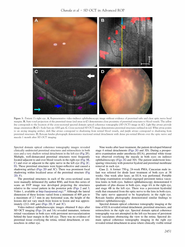
Spectral domain optical coherence tomography images revealedclinically undetected preretinal structures and retinoschisis in botheyes and a very shallow retinal detachment in the left eye (Fig 2F).Multiple, well-demarcated preretinal structures were frequentlylocated adjacent to and over blood vessels in the right eye (Fig 3B,C) and over or adjacent to the optic nerve in the left eye (Fig 2C,D). These preretinal structures were hyper-reflective and caused ashadowing artifact (Figs 2D and 3C). There was prominent focalshadowing within localized areas of the preretinal structure (Fig2D, F).
The preretinal structures in each of the cross-sectional scanswere manually delineated (by author RM), and from this series ofscans an SVP image was developed projecting the structuresrelative to the vessel pattern in the posterior pole (Figs 2 and 3;Video 1, available at http://aaojournal.org).15 Although the lateraldimension of these lesions varied from a minimum of 409 �m toa maximum of 2.7 mm in any horizontal scan, the height of thelesions did not vary much from lesion to lesion and was approx-imately (212–440 �m) (Figs 2D, F and 3C).
Video-indirect ophthalmoscopic images obtained 8 days afterSD OCT imaging (Figs 2A and 3A) revealed immature, tortuousretinal vasculature in both eyes with persistent neovascularizationbehind the laser margin in the left eye. There was no evidence ofpreretinal tissue overlying the retina, retinal detachment, or reti-
Figure 3. Patient 1’s right eye. A, Representative video-indirect ophthalmmargins. B, Sum voxel projection of the preretinal tissue (red areas in C) dline corresponds to the location of the cross-sectional spectral domain optimage orientation (B, C). Scale bars are 1000 �m. C, Cross-sectional SD Oto an arcing imaging artifact, dark blue arrows correspond to shadowingpreretinal structures. D, Retcam fundus photograph demonstrates tractionmacula 1 month after SD OCT imaging.
noschisis in either eye.
Nine weeks after laser treatment, the patient developed bilateralstage 4 retinal detachments (Figs 2G and 3D). During a preoper-ative examination under anesthesia (EUA), preretinal white tissuewas observed overlying the macula in both eyes on indirectophthalmoscopy (Figs 2G and 3D). The patient underwent lens-sparing vitrectomy with posterior hyaloid and preretinal membraneremoval in each eye.
Case 2. A former 750-g, 24-week PMA, Caucasian male in-fant was referred for diode laser treatment of both eyes at 38weeks. One week after laser, an EUA was performed. Portableslit-lamp examination revealed engorged persistent tunica vascu-losa lentis in both eyes. Indirect ophthalmoscopy demonstrated 4quadrants of plus disease in both eyes, stage 4A in the right eye,and stage 4B in the left eye. There was a persistent hyaloidalstalk that emanated from the optic nerve to the lens in both eyes.The optic nerve appeared to be hypoplastic in the right eye.Retcam fundus photography demonstrated similar findings toindirect ophthalmoscopy.
Spectral domain optical coherence tomography imaging at thetime of EUA revealed retinal detachment, preretinal structures, andretinoschisis in the right eye. Spectral domain optical coherencetomography was not attempted in the left eye because of persistentfetal vasculature obstructing the view to the retina. Spectral do-main optical coherence tomography imaging in the right eye
py image without evidence of preretinal tufts and clear optic nerve headstrates close proximity of preretinal structures to blood vessels. The yellowoherence tomography (SD OCT) image in (C). Light blue arrows provide
age demonstrates preretinal structures outlined in red. White arrow pointsretinal blood vessels, and purple arrows correspond to shadowing from
inal detachment with dense pre-retinal fibrosis over the optic nerve and
oscoemonical cCT imfromal ret
revealed retinal detachment in areas where clinically visible retinal
2451
.
Ophthalmology Volume 116, Number 12, December 2009
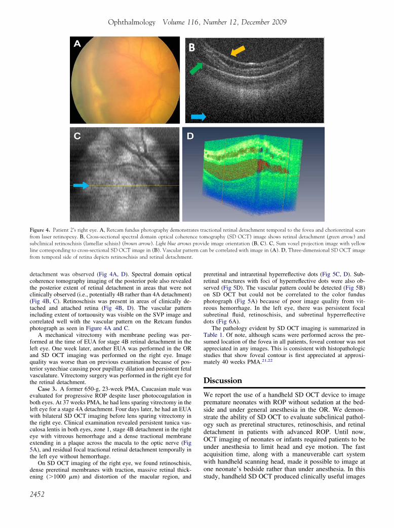
detachment was observed (Fig 4A, D). Spectral domain opticalcoherence tomography imaging of the posterior pole also revealedthe posterior extent of retinal detachment in areas that were notclinically observed (i.e., potentially 4B rather than 4A detachment)(Fig 4B, C). Retinoschisis was present in areas of clinically de-tached and attached retina (Fig 4B, D). The vascular patternincluding extent of tortuousity was visible on the SVP image andcorrelated well with the vascular pattern on the Retcam fundusphotograph as seen in Figure 4A and C.
A mechanical vitrectomy with membrane peeling was per-formed at the time of EUA for stage 4B retinal detachment in theleft eye. One week later, another EUA was performed in the ORand SD OCT imaging was performed on the right eye. Imagequality was worse than on previous examination because of pos-terior synechiae causing poor pupillary dilation and persistent fetalvasculature. Vitrectomy surgery was performed in the right eye forthe retinal detachment.
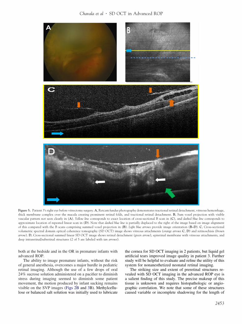
Case 3. A former 650-g, 23-week PMA, Caucasian male wasevaluated for progressive ROP despite laser photocoagulation inboth eyes. At 37 weeks PMA, he had lens sparing vitrectomy in theleft eye for a stage 4A detachment. Four days later, he had an EUAwith bilateral SD OCT imaging before lens sparing vitrectomy inthe right eye. Clinical examination revealed persistent tunica vas-culosa lentis in both eyes, zone 1, stage 4B detachment in the righteye with vitreous hemorrhage and a dense tractional membraneextending in a plaque across the macula to the optic nerve (Fig5A), and residual focal tractional retinal detachment temporally inthe left eye without hemorrhage.
On SD OCT imaging of the right eye, we found retinoschisis,dense preretinal membranes with traction, massive retinal thick-
Figure 4. Patient 2’s right eye. A, Retcam fundus photography demonstrafrom laser retinopexy. B, Cross-sectional spectral domain optical coherensubclinical retinoschisis (lamellar schisis) (brown arrow). Light blue arrowsline corresponding to cross-sectional SD OCT image in (B). Vascular pattfrom temporal side of retina depicts retinoschisis and retinal detachment
ening (�1000 �m) and distortion of the macular region, and
2452
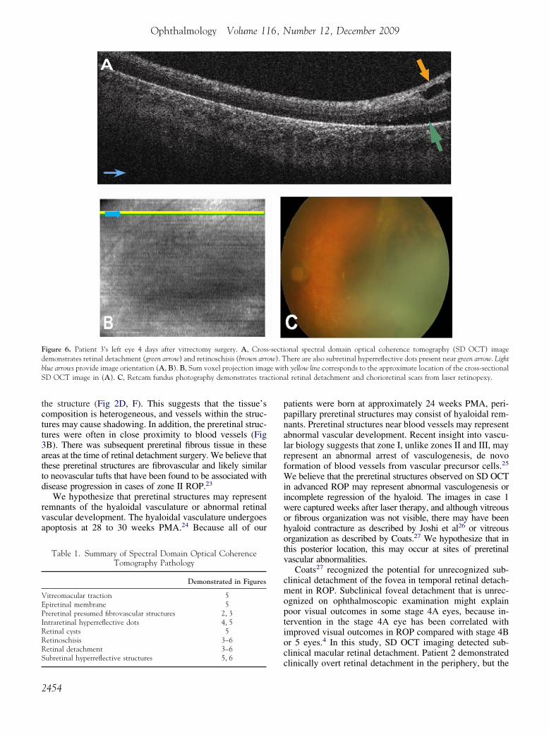
preretinal and intraretinal hyperreflective dots (Fig 5C, D). Sub-retinal structures with foci of hyperreflective dots were also ob-served (Fig 5D). The vascular pattern could be detected (Fig 5B)on SD OCT but could not be correlated to the color fundusphotograph (Fig 5A) because of poor image quality from vit-reous hemorrhage. In the left eye, there was persistent focalsubretinal fluid, retinoschisis, and subretinal hyperreflectivedots (Fig 6A).
The pathology evident by SD OCT imaging is summarized inTable 1. Of note, although scans were performed across the pre-sumed location of the fovea in all patients, foveal contour was notappreciated in any images. This is consistent with histopathologicstudies that show foveal contour is first appreciated at approxi-mately 40 weeks PMA.21,22
Discussion
We report the use of a handheld SD OCT device to imagepremature neonates with ROP without sedation at the bed-side and under general anesthesia in the OR. We demon-strate the ability of SD OCT to evaluate subclinical pathol-ogy such as preretinal structures, retinoschisis, and retinaldetachment in patients with advanced ROP. Until now,OCT imaging of neonates or infants required patients to beunder anesthesia to limit head and eye motion. The fastacquisition time, along with a maneuverable cart systemwith handheld scanning head, made it possible to image atone neonate’s bedside rather than under anesthesia. In this
actional retinal detachment temporal to the fovea and chorioretinal scarsmography (SD OCT) image shows retinal detachment (green arrow) andde image orientation (B, C). C, Sum voxel projection image with yellown be correlated with image in (A). D, Three-dimensional SD OCT image
tes trce toprovi
ern ca
study, handheld SD OCT produced clinically useful images
.
Chavala et al � SD OCT in Advanced ROP
both at the bedside and in the OR in premature infants withadvanced ROP.
The ability to image premature infants, without the riskof general anesthesia, overcomes a major hurdle in pediatricretinal imaging. Although the use of a few drops of oral24% sucrose solution administered on a pacifier to diminishstress during imaging seemed to diminish some patientmovement, the motion produced by infant sucking remainsvisible on the SVP images (Figs 2B and 3B). Methylcellu-
Figure 5. Patient 3’s right eye before vitrectomy surgery. A, Retcam fundusthick membrane complex over the macula creating prominent retinal fovascular pattern not seen clearly in (A). Yellow line corresponds to exactapproximate location of repeated linear scan in (D). Note that dashed bluof this compared with the B scans comprising summed voxel projectionvolumetric spectral domain optical coherence tomography (SD OCT) imarrow). D, Cross-sectional summed linear SD OCT image shows retinal ddeep intraretinal/subretinal structures (2 of 5 are labeled with tan arrows)
lose or balanced salt solution was initially used to lubricate
the cornea for SD OCT imaging in 2 patients, but liquid gelartificial tears improved image quality in patient 3. Furtherstudy will be helpful to evaluate and refine the utility of thissystem for nonanesthetized neonatal retinal imaging.
The striking size and extent of preretinal structures re-vealed with SD OCT imaging in the advanced ROP eye isa salient finding of this study. The precise makeup of thistissue is unknown and requires histopathologic or angio-graphic correlation. We note that some of these structures
ography demonstrates tractional retinal detachment, vitreous hemorrhage,nd tractional retinal detachment. B, Sum voxel projection with visibleion of cross-sectional B scan in (C), and dashed blue line corresponds tois partially displaced to the right of the image based on image alignment). Light blue arrows provide image orientation (B–D). C, Cross-sectionalows vitreous attachments (orange arrows C, D) and retinoschisis (brown
ment (green arrow), epiretinal membrane with vitreous attachments, and
photlds, alocat
e linein (Bage shetach
caused variable or incomplete shadowing for the length of
2453
ctiona
Ophthalmology Volume 116, Number 12, December 2009
the structure (Fig 2D, F). This suggests that the tissue’scomposition is heterogeneous, and vessels within the struc-tures may cause shadowing. In addition, the preretinal struc-tures were often in close proximity to blood vessels (Fig3B). There was subsequent preretinal fibrous tissue in theseareas at the time of retinal detachment surgery. We believe thatthese preretinal structures are fibrovascular and likely similarto neovascular tufts that have been found to be associated withdisease progression in cases of zone II ROP.23
We hypothesize that preretinal structures may representremnants of the hyaloidal vasculature or abnormal retinalvascular development. The hyaloidal vasculature undergoesapoptosis at 28 to 30 weeks PMA.24 Because all of our
Figure 6. Patient 3’s left eye 4 days after vitrectomy surgery. A, Crossdemonstrates retinal detachment (green arrow) and retinoschisis (brown arrblue arrows provide image orientation (A, B). B, Sum voxel projection imagSD OCT image in (A). C, Retcam fundus photography demonstrates tra
Table 1. Summary of Spectral Domain Optical CoherenceTomography Pathology
Demonstrated in Figures
Vitreomacular traction 5Epiretinal membrane 5Preretinal presumed fibrovascular structures 2, 3Intraretinal hyperreflective dots 4, 5Retinal cysts 5Retinoschisis 3–6Retinal detachment 3–6
Subretinal hyperreflective structures 5, 62454
patients were born at approximately 24 weeks PMA, peri-papillary preretinal structures may consist of hyaloidal rem-nants. Preretinal structures near blood vessels may representabnormal vascular development. Recent insight into vascu-lar biology suggests that zone I, unlike zones II and III, mayrepresent an abnormal arrest of vasculogenesis, de novoformation of blood vessels from vascular precursor cells.25
We believe that the preretinal structures observed on SD OCTin advanced ROP may represent abnormal vasculogenesis orincomplete regression of the hyaloid. The images in case 1were captured weeks after laser therapy, and although vitreousor fibrous organization was not visible, there may have beenhyaloid contracture as described by Joshi et al26 or vitreousorganization as described by Coats.27 We hypothesize that inthis posterior location, this may occur at sites of preretinalvascular abnormalities.
Coats27 recognized the potential for unrecognized sub-clinical detachment of the fovea in temporal retinal detach-ment in ROP. Subclinical foveal detachment that is unrec-ognized on ophthalmoscopic examination might explainpoor visual outcomes in some stage 4A eyes, because in-tervention in the stage 4A eye has been correlated withimproved visual outcomes in ROP compared with stage 4Bor 5 eyes.4 In this study, SD OCT imaging detected sub-clinical macular retinal detachment. Patient 2 demonstrated
onal spectral domain optical coherence tomography (SD OCT) imagehere are also subretinal hyperreflective dots present near green arrow. Light
h yellow line corresponds to the approximate location of the cross-sectionall retinal detachment and chorioretinal scars from laser retinopexy.
-sectiow). Te wit
clinically overt retinal detachment in the periphery, but the
Chavala et al � SD OCT in Advanced ROP
macula appeared to be attached on indirect ophthalmoscopyand Retcam photography (Fig 4A). Spectral domain opticalcoherence tomography imaging of the posterior pole (Fig4B, C) revealed a shallow retinal detachment across thepresumed area of the macula that was not clinically appre-ciated. Spectral domain optical coherence tomography maybe beneficial in detecting subclinical macular retinal detach-ment and may guide surgical decision-making. Spectraldomain optical coherence tomography may illustrate sub-clinical pathology and be a potential strength in ROPscreening in the NICU. First, however, studies are needed toevaluate potential management strategies based on findingsfrom this imaging technique.
Consistent with previous studies using TD OCT in stage4 ROP,14 all of our patients demonstrated a retinoschisis-like appearance of the retina (Figs 2, 4–6), none of whichwas visible on clinical examination. In some areas thisappeared as a splitting of the more highly reflective innerretinal layers (Figs 4B, 5C, and 6A) associated with vitreousattachment and upward tenting of the retina (Fig 5C),whereas in other areas this involved deeper retinal layerswith a “lamellar schisis-like change” similar to that de-scribed by Joshi et al14 (Figs 2F and 4D; Video 2, availableat http://aaojournal.org). From this limited study, we cannotdefinitively differentiate traction-associated schisis from ab-normal foveal development or diffuse cystoid macularedema. In patients 2 and 3, the subclinical retinoschisis(Figs 4B and 5D) was associated with clinically visibletractional detachment of the retina (Figs 4A and 5A). Inpatient 1, subclinical retinoschisis and retinal detachmentoccurred before the appearance of fibrovascular organiza-tion of the vitreous or stage 4 ROP in the left eye (Fig 3F).28
He subsequently progressed to clinically evident tractionalretinal detachments within 4 weeks (Figs 2G and 3D) of thisexamination. We hypothesize that some subclinical retinos-chisis may result from traction associated with fibrovascularorganization and may serve as a harbinger for tractionalretinal detachment. In diabetes, tractional elevations of theretina have been associated with either retinoschisis or retinaldetachment.29 In accordance with Joshi et al,14 we believe thatpoor visual outcomes after surgical intervention in ad-vanced ROP may be partially explained by undiagnosedlamellar schisis-like retinal changes at the time of interven-tion.30 Future study is required to determine whether detect-ing subclinical retinoschisis by SD OCT will help predictvisual outcomes.
Major limitations of this study include the small numberof patients, motion artifact in images from the nonsedatedsubject, and adjustment of the system for the prematureinfant eye. These issues are being addressed in a prospectiveNICU study with recruitment of a larger number of patientswith different stages of ROP. An experienced operator wasnecessary to use the handheld SD OCT probe to account formotion artifact from hand movement and patient motion.These artifacts can cause abnormal or complete loss ofB-scans requiring careful attention during analysis (Fig 2B,white arrows). Because of the shorter axial length of thepremature infant eye, the projected imaging area wassmaller than predicted by scan settings for the adult. Also, a
clipping effect was produced by the pupil on the B-scansbecause we used a standard adult reference arm length forthe SD OCT system (black margins on the right side of theimages in Fig 4B). We are working to correct these 2 issuesin future studies by adjusting scan length for the horizontalmagnification of the infant eye and modifying the referencearm for the axial length of the neonatal eye. The maximumextent of peripheral imaging was not assessed in this pilotstudy, which focused on posterior pole pathology in ad-vanced ROP. Ongoing studies of SD OCT imaging in zoneII ROP will assess the feasibility and limitations of imagingperipheral pathology.
In conclusion, this proof-of-principal study representsthe first use of SD OCT in a nonsedated patient with ROPand demonstrates that it may be possible to overcome themajor hurdle of the risk of anesthesia in neonatal retinalimaging. Moreover, in patients with advanced ROP, wedemonstrate the utility of SD OCT imaging in subclinicalpreretinal, intraretinal, and subretinal pathology that islikely to affect the precision of our assessment of the extentof disease. We believe that analysis of SD OCT images ineyes with ROP will provide insight into the pathogenesis ofROP. More work is needed to determine the frequency andpredictive value of these findings in patients with ROP.
Acknowledgment. Dr. Joseph Izatt provided essential input foroptimizing the SD OCT system imaging in the infant eye.
References
1. Tasman W. Multicenter trial of cryotherapy for retinopathy ofprematurity. Arch Ophthalmol 1988;106:463–4.
2. Early Treatment for Retinopathy of Prematurity CooperativeGroup. Revised indications for the treatment of retinopathy ofprematurity: results of the Early Treatment for Retinopathy ofPrematurity randomized trial. Arch Ophthalmol 2003;121:1684–94.
3. Early Treatment for Retinopathy of Prematurity CooperativeGroup. The Early Treatment for Retinopathy Of PrematurityStudy: structural findings at age 2 years. Br J Ophthalmol2006;90:1378–82.
4. Prenner JL, Capone A Jr, Trese MT. Visual outcomes afterlens-sparing vitrectomy for stage 4A retinopathy of prematu-rity. Ophthalmology 2004;111:2271–3.
5. Azuma N, Ishikawa K, Hama Y, et al. Early vitreous surgeryfor aggressive posterior retinopathy of prematurity. Am JOphthalmol 2006;142:636–43.
6. International Committee for the Classification of Retinopathy ofPrematurity. The International Classification of Retinopathy ofPrematurity revisited. Arch Ophthalmol 2005;123:991–9.
7. Coats DK, Miller AM, Hussein MA, et al. Involution ofretinopathy of prematurity after laser treatment: factors asso-ciated with development of retinal detachment. Am J Oph-thalmol 2005;140:214–22.
8. Committee for the Classification of Retinopathy of Prematu-rity. An international classification of retinopathy of prematu-rity. Arch Ophthalmol 1984;102:1130–4.
9. Wilson CM, Cocker KD, Moseley MJ, et al. Computerizedanalysis of retinal vessel width and tortuosity in prematureinfants. Invest Ophthalmol Vis Sci 2008;49:3577–85.
10. Wallace DK, Freedman SF, Zhao Z, Jung SH. Accuracy ofROPtool vs individual examiners in assessing retinal vascular
tortuosity. Arch Ophthalmol 2007;125:1523–30.2455
Ophthalmology Volume 116, Number 12, December 2009
11. Chiang MF, Gelman R, Williams SL, et al. Plus disease inretinopathy of prematurity: development of composite imagesby quantification of expert opinion. Invest Ophthalmol Vis Sci2008;49:4064–70.
12. Huang D, Swanson EA, Lin CP, et al. Optical coherencetomography. Science 1991;254:1178–81.
13. Patel CK. Optical coherence tomography in the managementof acute retinopathy of prematurity. Am J Ophthalmol 2006;141:582–4.
14. Joshi MM, Trese MT, Capone A Jr. Optical coherence tomog-raphy findings in stage 4A retinopathy of prematurity: a theoryfor visual variability. Ophthalmology 2006;113:657–60.
15. Stopa M, Bower BA, Davies E, et al. Correlation of pathologicfeatures in spectral domain optical coherence tomographywith conventional retinal studies. Retina 2008;28:298–308.
16. Scott AW, Farsiu S, Enyedi LB, et al. Imaging the infant retinawith hand-held spectral-domain optical coherence tomogra-phy. Am J Ophthalmol 2009;147:364–73.
17. Chong GT, Farsiu S, Freedman SF, et al. Abnormal fovealmorphology in ocular albinism imaged with spectral domainoptical coherence tomography. Arch Ophthalmol 2009;127:37–44.
18. Gal P, Kissling GE, Young WO, et al. Efficacy of sucrose toreduce pain in premature infants during eye examinationsfor retinopathy of prematurity. Ann Pharmacother 2005;39:1029–33.
19. Farsiu S, Chiu SJ, Izatt JA, Toth CA. Fast detection andsegmentation of drusen in retinal optical coherence tomogra-phy images. Proc SPIE 2008;6844:68440D.
20. Jiao S, Knighton R, Huang X, et al. Simultaneous acquisition ofsectional and fundus ophthalmic images with spectral-domain
optical coherence tomography. Opt Express [serial online] 2005;and Farsiu have a patent pending on OCT image processing techniques. Dr.
2456
13:444–52. Available at: http://www.opticsinfobase.org/oe/abstract.cfm?uri�oe-13-2-444. Accessed May 16, 2009.
21. Hendrickson AE, Yuodelis C. The morphological develop-ment of the human fovea. Ophthalmology 1984;91:603–12.
22. Yuodelis C, Hendrickson A. A qualitative and quantitativeanalysis of the human fovea during development. Vision Res1986;26:847–55.
23. Wallace DK, Kylstra JA, Greenman DB, Freedman SF. Sig-nificance of isolated neovascular tufts (“popcorn”) in retinop-athy of prematurity. J AAPOS 1998;2:52–6.
24. Eller AW, Jabbour NM, Hirose T, Schepens CL. Retinopathyof prematurity: the association of a persistent hyaloid artery.Ophthalmology 1987;94:444–8.
25. Flynn JT, Chan-Ling T. Retinopathy of prematurity: two dis-tinct mechanisms that underlie zone 1 and zone 2 disease.Am J Ophthalmol 2006;142:46–59.
26. Joshi MM, Ciaccia S, Trese MT, Capone A. Posterior hyaloidcontracture in pediatric vitreoretinopathies. Retina 2006;26(suppl):S38 – 41.
27. Coats DK. Retinopathy of prematurity: involution, factorspredisposing to retinal detachment, and expected utility ofpreemptive surgical reintervention. Trans Am Ophthalmol Soc2005;103:281–312.
28. Hartnett ME, McColm JR. Fibrovascular organization in thevitreous following laser for ROP: implications for prognosis.Retina 2006;26(suppl):S24–31.
29. Lincoff H, Serag Y, Chang S, et al. Tractional elevations ofthe retina in patients with diabetes. Am J Ophthalmol 1992;113:235–42.
30. Prenner JL, Capone A Jr, Ciaccia S, et al. Congenital X-linked
retinoschisis classification system. Retina 2006;26(suppl):S61–4.Footnotes and Financial Disclosures
Originally received: January 14, 2009.Final revision: May 31, 2009.Accepted: June 3, 2009.Available online: September 18, 2009. Manuscript no. 2009-59.
Department of Ophthalmology, Duke University Medical Center, DukeUniversity, Durham, North Carolina.
Financial Disclosure(s):The author(s) have made the following disclosure(s):
Dr. Toth receives research support through Duke University from AlconLaboratories, Bioptigen, Genentech, National Institutes of Health, NorthCarolina Biotechnology Center, and Sirion; is a consultant for AlconLaboratories and Genentech; and receives royalties for ophthalmic surgicaltechnologies through a Duke University agreement with Alcon. Drs. Toth
Farsiu receives research support through Duke University from Bioptigen.Dr. Joseph Izatt, who is principal investigator on National Institutes ofHealth grant R21EY017393 and co-investigator on North Carolina Bio-technology Center Collaborative Funding Grant 2007-CFG-8005 and DukeTranslational Medicine Institute Subcontract 12 of National Institutes ofHealth Grant 5ULT-RR024128-03, has consulting, intellectual property,and equity relationships with Bioptigen, Inc.
Supported by Angelica and Euan Baird, North Carolina BiotechnologyCenter Collaborative Funding Grant 2007-CFG-8005, National Institutesof Health R21 EY017393, and the Duke Translational Medicine InstituteSubcontract 12 of National Institutes of Health Grant 5ULT-RR024128-03.
Correspondence:Cynthia A. Toth, MD, Duke University Eye Center, PO Box 3802,
Durham, NC 27710. E-mail: [email protected].
Related Documents