7/30/2011 1 Image Processing for Optical Coherence Tomography Jonathan Oakley and Daniel Russakoff, Voxeleron LLC HISB 2011, July 29 th , 2011 Overview • Optical Coherence Tomography (OCT) – Brief History – Overview of the Modality • Methods and Applications in Ophthalmology – Image pre-processing – Layer Segmentation • Graph-based – 1d – Graph Cuts • Shape-based – Optic Nerve Segmentation – Image Registration • Other application domains of OCT • Future trends

Welcome message from author
This document is posted to help you gain knowledge. Please leave a comment to let me know what you think about it! Share it to your friends and learn new things together.
Transcript
7/30/2011
1
Image Processing for Optical Coherence Tomography
Jonathan Oakley and Daniel Russakoff, Voxeleron LLC
HISB 2011, July 29th, 2011
Overview
• Optical Coherence Tomography (OCT) – Brief History – Overview of the Modality
• Methods and Applications in Ophthalmology – Image pre-processing – Layer Segmentation
• Graph-based – 1d – Graph Cuts
• Shape-based
– Optic Nerve Segmentation – Image Registration
• Other application domains of OCT • Future trends
7/30/2011
2
OPTICAL COHERENCE TOMOGRAPHY
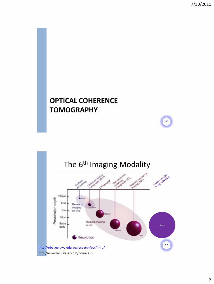
5mm
http://www.lantislaser.com/home.asp
http://obel.ee.uwa.edu.au/research/oct/intro/
The 6th Imaging Modality
7/30/2011
3
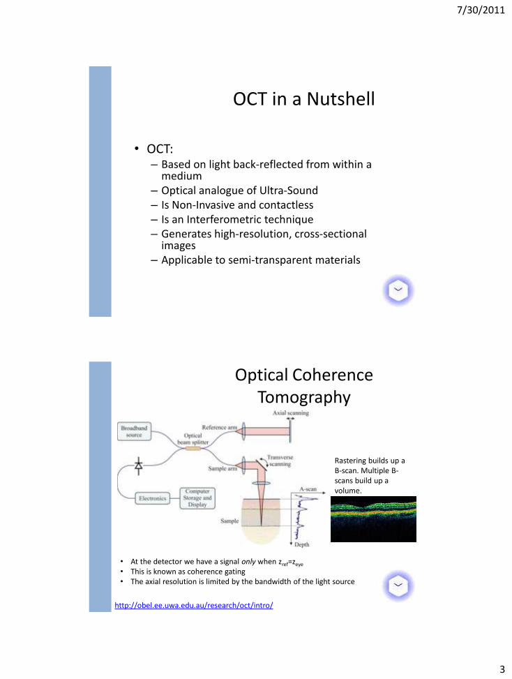
• OCT: – Based on light back-reflected from within a
medium – Optical analogue of Ultra-Sound – Is Non-Invasive and contactless – Is an Interferometric technique – Generates high-resolution, cross-sectional
images – Applicable to semi-transparent materials
OCT in a Nutshell
Optical Coherence Tomography
http://obel.ee.uwa.edu.au/research/oct/intro/
• At the detector we have a signal only when zref=zeye
• This is known as coherence gating • The axial resolution is limited by the bandwidth of the light source
Rastering builds up a B-scan. Multiple B-scans build up a volume.
7/30/2011
4
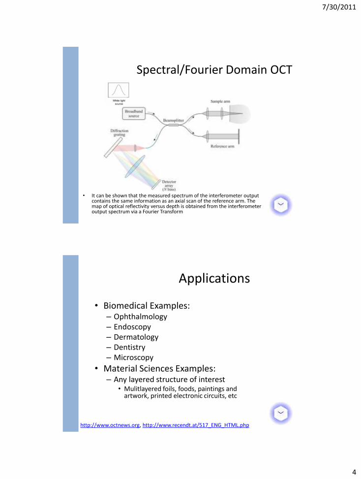
• It can be shown that the measured spectrum of the interferometer output contains the same information as an axial scan of the reference arm. The map of optical reflectivity versus depth is obtained from the interferometer output spectrum via a Fourier Transform
Spectral/Fourier Domain OCT
• Biomedical Examples: – Ophthalmology – Endoscopy – Dermatology – Dentistry – Microscopy
• Material Sciences Examples: – Any layered structure of interest
• Mulitlayered foils, foods, paintings and artwork, printed electronic circuits, etc
Applications
http://www.octnews.org, http://www.recendt.at/517_ENG_HTML.php
7/30/2011
5
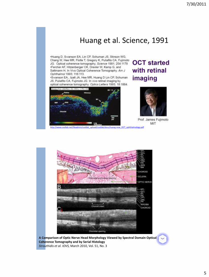
Huang et al. Science, 1991
http://www.coollab.net/fileadmin/coollab_upload/coollab/docs/huang-new_OCT_ophthalmology.pdf
A Comparison of Optic Nerve Head Morphology Viewed by Spectral Domain Optical Coherence Tomography and by Serial Histology Strouthidis et al. IOVS, March 2010, Vol. 51, No. 3
7/30/2011
6
OPHTHALMOLOGY AND OPTICAL COHERENCE TOMOGRAPHY
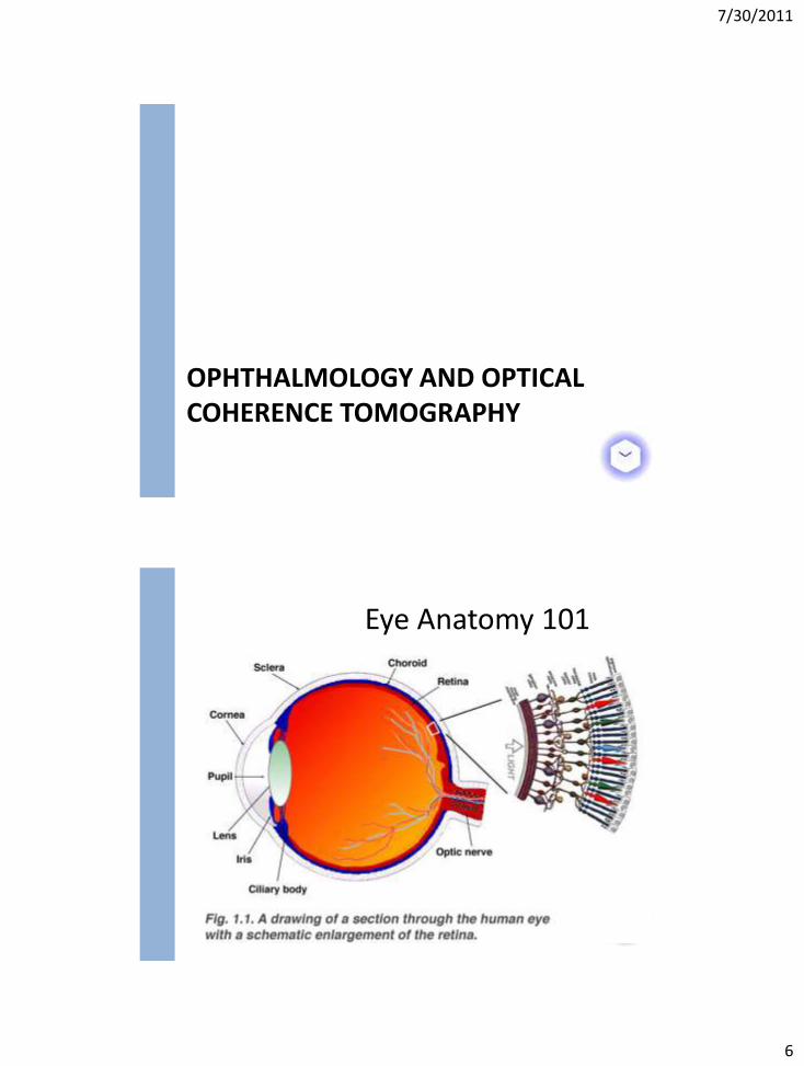
Eye Anatomy 101
7/30/2011
7
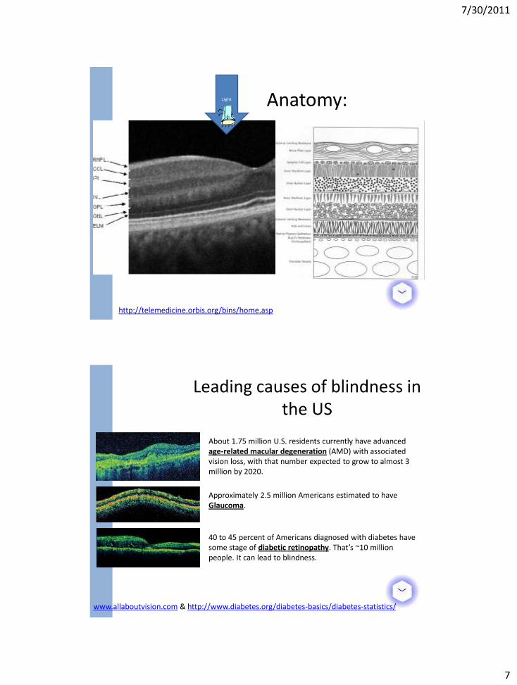
Light Anatomy:
http://telemedicine.orbis.org/bins/home.asp
Leading causes of blindness in the US
www.allaboutvision.com & http://www.diabetes.org/diabetes-basics/diabetes-statistics/
Approximately 2.5 million Americans estimated to have Glaucoma.
About 1.75 million U.S. residents currently have advanced age-related macular degeneration (AMD) with associated vision loss, with that number expected to grow to almost 3 million by 2020.
40 to 45 percent of Americans diagnosed with diabetes have some stage of diabetic retinopathy. That’s ~10 million people. It can lead to blindness.
7/30/2011
8
Ophthalmology
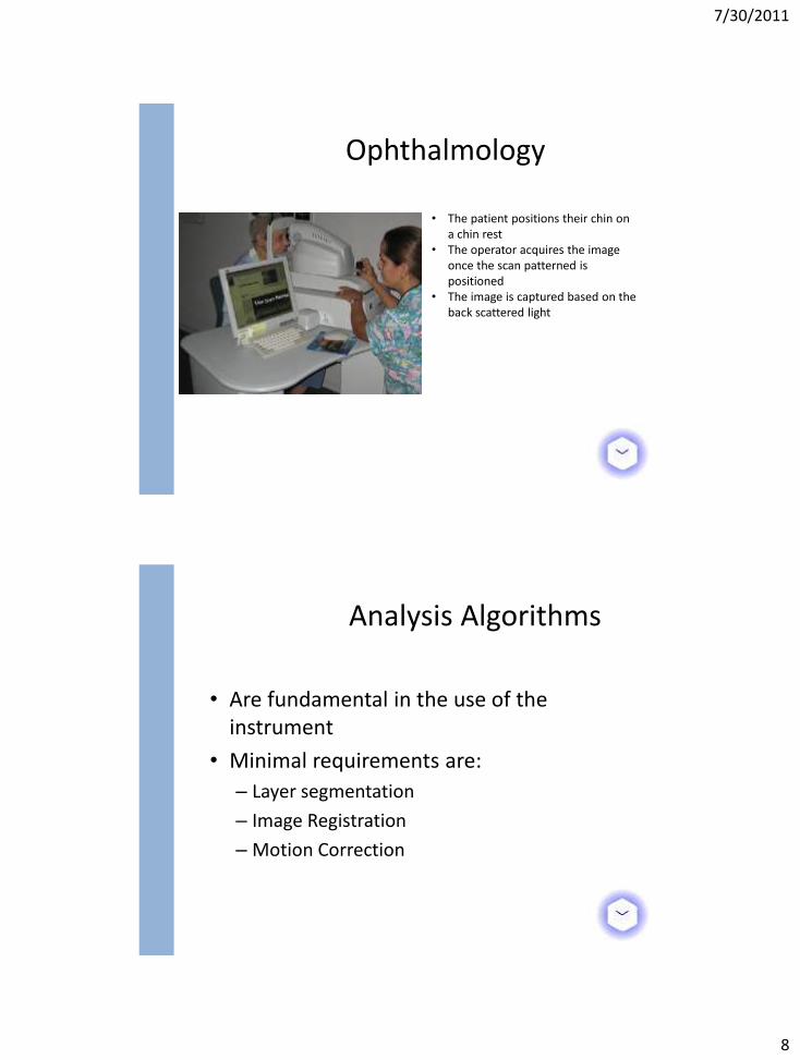
• The patient positions their chin on a chin rest
• The operator acquires the image once the scan patterned is positioned
• The image is captured based on the back scattered light
• Are fundamental in the use of the instrument
• Minimal requirements are:
– Layer segmentation
– Image Registration
– Motion Correction
Analysis Algorithms
7/30/2011
9
IMAGE PROCESSING IN OPHTHALMIC OCT
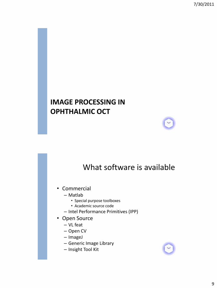
• Commercial – Matlab
• Special purpose toolboxes • Academic source code
– Intel Performance Primitives (IPP)
• Open Source – VL feat – Open CV – ImageJ – Generic Image Library – Insight Tool Kit
What software is available
7/30/2011
10
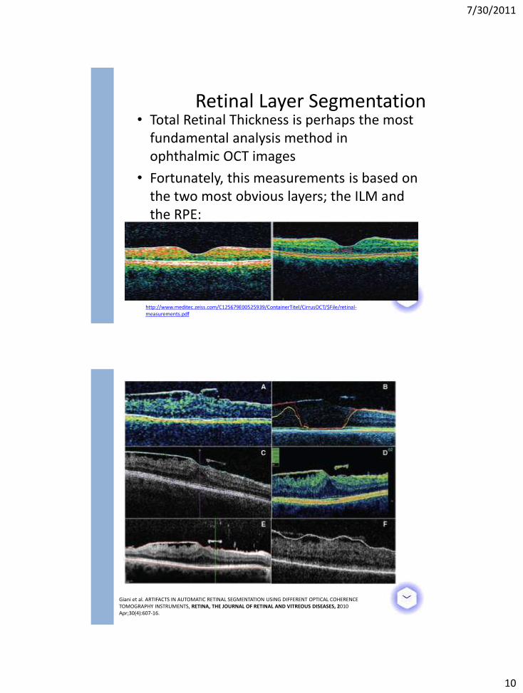
• Total Retinal Thickness is perhaps the most fundamental analysis method in ophthalmic OCT images
• Fortunately, this measurements is based on the two most obvious layers; the ILM and the RPE:
Retinal Layer Segmentation
http://www.meditec.zeiss.com/C125679E00525939/ContainerTitel/CirrusOCT/$File/retinal-measurements.pdf
Giani et al. ARTIFACTS IN AUTOMATIC RETINAL SEGMENTATION USING DIFFERENT OPTICAL COHERENCE TOMOGRAPHY INSTRUMENTS, RETINA, THE JOURNAL OF RETINAL AND VITREOUS DISEASES, 2010 Apr;30(4):607-16.
7/30/2011
11
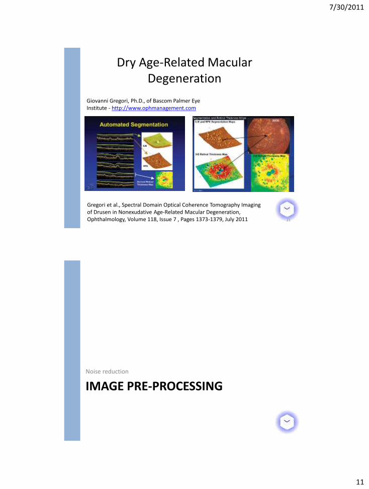
Dry Age-Related Macular Degeneration
21
Giovanni Gregori, Ph.D., of Bascom Palmer Eye Institute - http://www.ophmanagement.com
Gregori et al., Spectral Domain Optical Coherence Tomography Imaging of Drusen in Nonexudative Age-Related Macular Degeneration, Ophthalmology, Volume 118, Issue 7 , Pages 1373-1379, July 2011
IMAGE PRE-PROCESSING Noise reduction

7/30/2011
12
Speckle Noise
• Due to constructive and destructive interference
• Reduces contrast and makes boundaries between highly scattering structures difficult to resolve
• Approximately follows a Rayleigh distribution
Schmitt et al., SPECKLE IN OPTICAL COHERENCE TOMOGRAPHY, JOURNAL OF BIOMEDICAL OPTICS 4(1), 95–105 (JANUARY 1999)
• Median Filter • Sticks algorithm
– Directional Filtering • J. Rogowska and M. E. Brezinski, “Evaluation of
the adaptive speckle suppression filter for coronary optical coherence tomography imaging,” IEEE Trans. Med. Imaging, 19, 1261–6 (2000).
• Anisotropic Diffusion Filtering – Edge Preserving Smoothing
• Yu and Acton, Speckle Reducing Anisotropic Diffusion, IEEE TRANSACTIONS ON IMAGE PROCESSING, VOL. 11, NO. 11, NOVEMBER 2002
Speckle Noise Reduction
7/30/2011
13
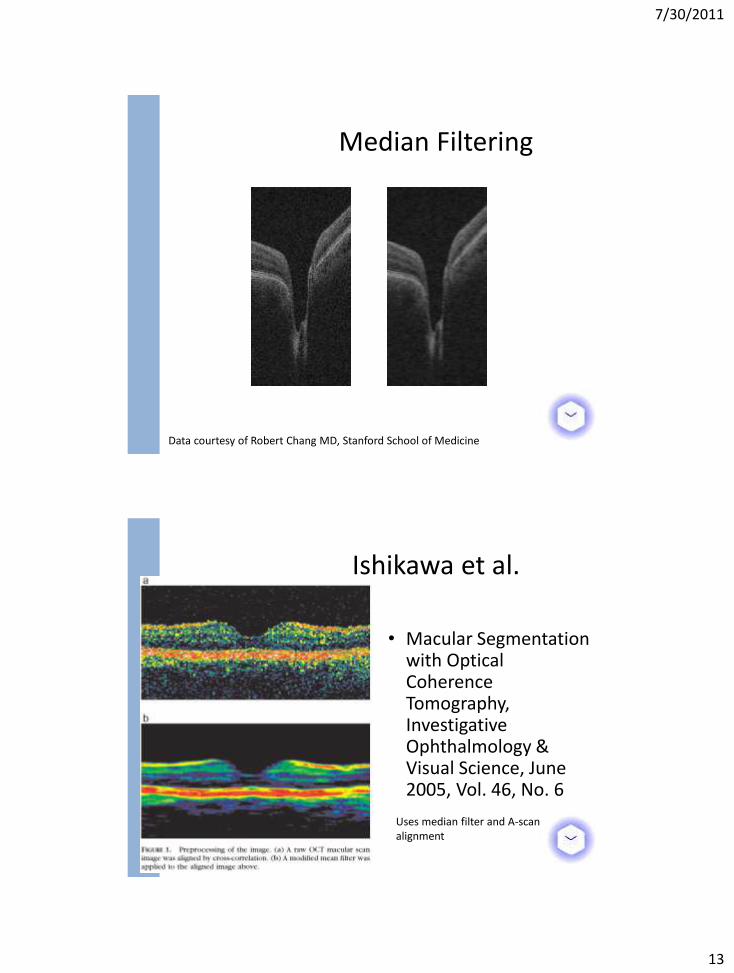
Median Filtering
Data courtesy of Robert Chang MD, Stanford School of Medicine
• Macular Segmentation with Optical Coherence Tomography, Investigative Ophthalmology & Visual Science, June 2005, Vol. 46, No. 6
Ishikawa et al.
Uses median filter and A-scan alignment
7/30/2011
14
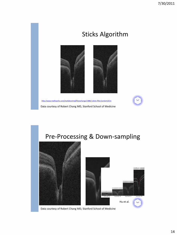
Sticks Algorithm
http://www.mathworks.com/matlabcentral/fileexchange/14862-sticks-filter/content/sf.m
Data courtesy of Robert Chang MD, Stanford School of Medicine
Pre-Processing & Down-sampling
Data courtesy of Robert Chang MD, Stanford School of Medicine
Hu et al.
7/30/2011
15
INTRA-RETINAL LAYER SEGMENTATION
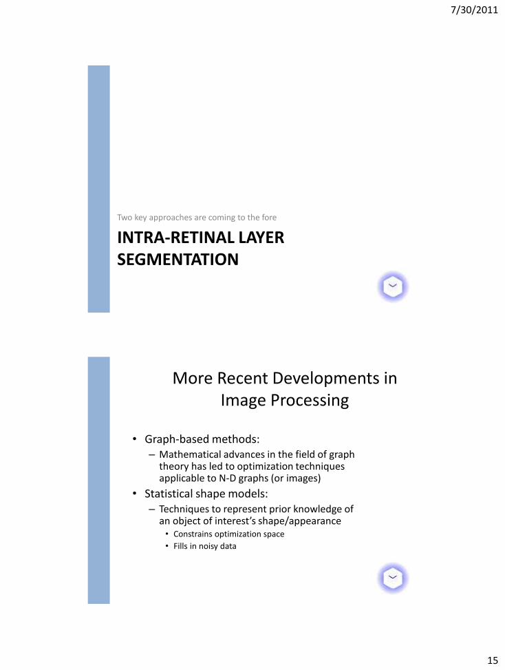
Two key approaches are coming to the fore
• Graph-based methods: – Mathematical advances in the field of graph
theory has led to optimization techniques applicable to N-D graphs (or images)
• Statistical shape models: – Techniques to represent prior knowledge of
an object of interest’s shape/appearance • Constrains optimization space
• Fills in noisy data
More Recent Developments in Image Processing
7/30/2011
16
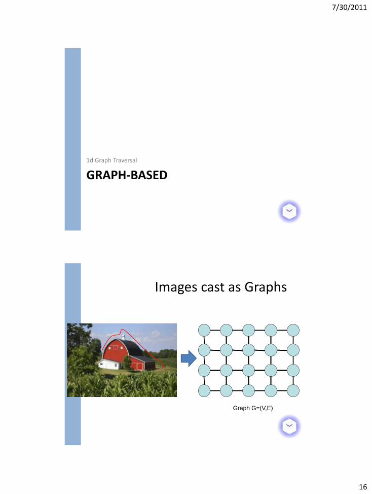
GRAPH-BASED 1d Graph Traversal
Images cast as Graphs
Graph G=(V,E)
7/30/2011
17
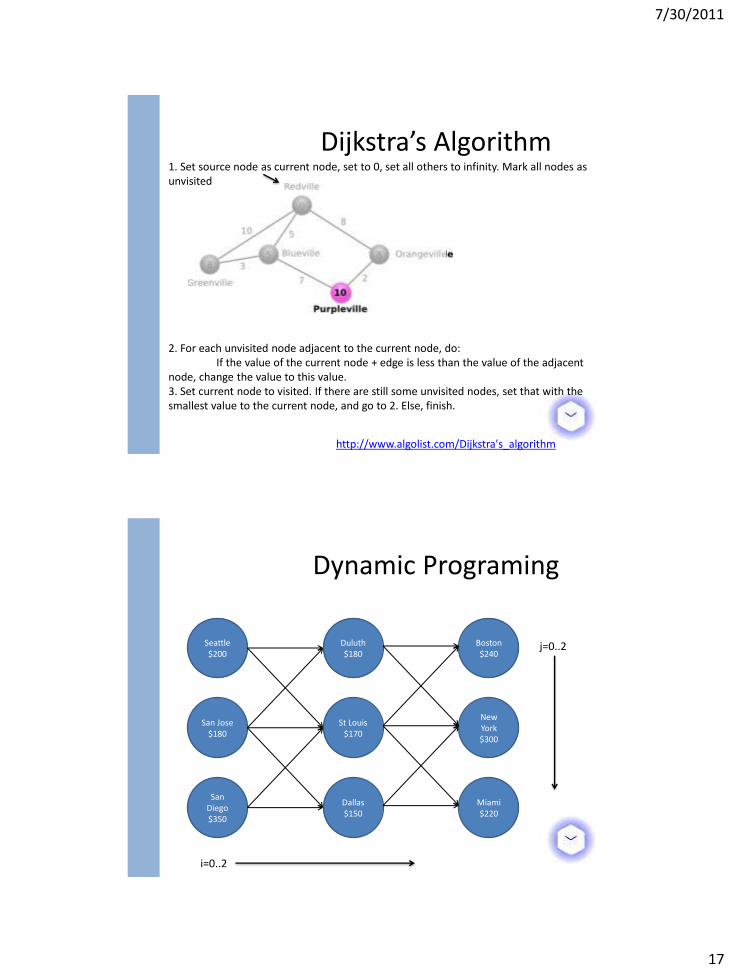
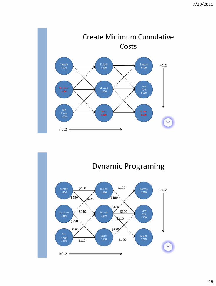
Dijkstra’s Algorithm
http://www.algolist.com/Dijkstra's_algorithm
1. Set source node as current node, set to 0, set all others to infinity. Mark all nodes as unvisited
2. For each unvisited node adjacent to the current node, do: If the value of the current node + edge is less than the value of the adjacent node, change the value to this value. 3. Set current node to visited. If there are still some unvisited nodes, set that with the smallest value to the current node, and go to 2. Else, finish.
Dynamic Programing
Seattle $200
San Jose $180
San Diego $350
Duluth $180
St Louis $170
Dallas $150
Boston $240
New York $300
Miami $220
i=0..2
j=0..2
7/30/2011
18
Create Minimum Cumulative Costs
Seattle $200
San Jose $180
San Diego $350
Duluth $360
St Louis $350
Dallas $330
Boston $590
New York $630
Miami $550
i=0..2
j=0..2
Miami $550
Dallas $330
San Jose $180
San Jose $180
St Louis $170
Dynamic Programing
Seattle $200
San Diego $350
Duluth $180
Dallas $150
Boston $240
New York $300
Miami $220
$150 $130
$180
$180
$190
$250
$110
$280
$110
$250
$290
$120
$210
$100
i=0..2
j=0..2
7/30/2011
19
San Jose $180
Boston $900
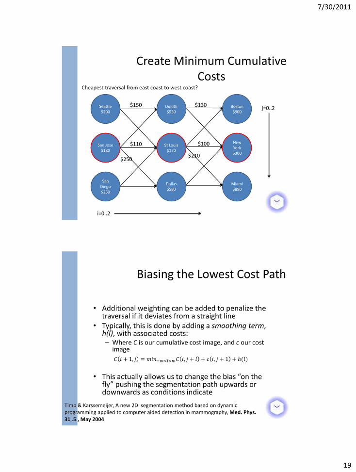
Create Minimum Cumulative Costs
Seattle $200
San Diego $250
Duluth $530
St Louis $460
Dallas $580
New York $860
Miami $890
$250
$110
$210
Cheapest traversal from east coast to west coast?
$150
i=0..2
j=0..2 $130
$100 San Jose $180
San Jose $180
St Louis $170
New York $300
• Additional weighting can be added to penalize the traversal if it deviates from a straight line
• Typically, this is done by adding a smoothing term, h(l), with associated costs: – Where C is our cumulative cost image, and c our cost
image
• This actually allows us to change the bias “on the
fly” pushing the segmentation path upwards or downwards as conditions indicate
Biasing the Lowest Cost Path
Timp & Karssemeijer, A new 2D segmentation method based on dynamic programming applied to computer aided detection in mammography, Med. Phys. 31 .5., May 2004
𝐶 𝑖 + 1, 𝑗 = 𝑚𝑖𝑛−𝑚<𝑙<𝑚𝐶 𝑖, 𝑗 + 𝑙 + 𝑐 𝑖, 𝑗 + 1 + ℎ(𝑙)
7/30/2011
20
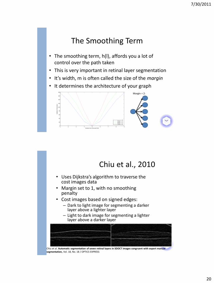
The Smoothing Term
• The smoothing term, h(l), affords you a lot of control over the path taken
• This is very important in retinal layer segmentation
• It’s width, m is often called the size of the margin
• It determines the architecture of your graph Margin = 2:
1 2 3 4 5 6 7 8 90
20
40
60
80
100
120
140
160
180
Deviation from Horizontal Path
Additative P
enaliz
ation
Margin Term
Margin = 2
Margin = 3
Margin = 4
• Uses Dijkstra’s algorithm to traverse the cost images data
• Margin set to 1, with no smoothing penalty
• Cost images based on signed edges: – Dark to light image for segmenting a darker
layer above a lighter layer – Light to dark image for segmenting a lighter
layer above a darker layer
Chiu et al., 2010
Chiu et al. Automatic segmentation of seven retinal layers in SDOCT images congruent with expert manual segmentation, Vol. 18, No. 18 / OPTICS EXPRESS
7/30/2011
21
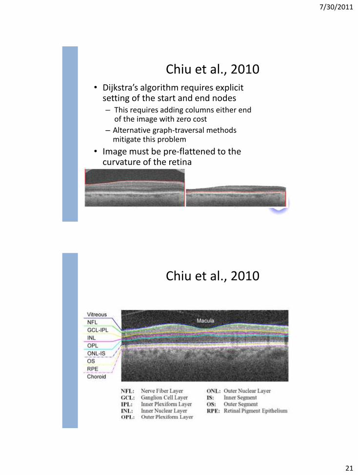
• Dijkstra’s algorithm requires explicit setting of the start and end nodes – This requires adding columns either end
of the image with zero cost
– Alternative graph-traversal methods mitigate this problem
• Image must be pre-flattened to the curvature of the retina
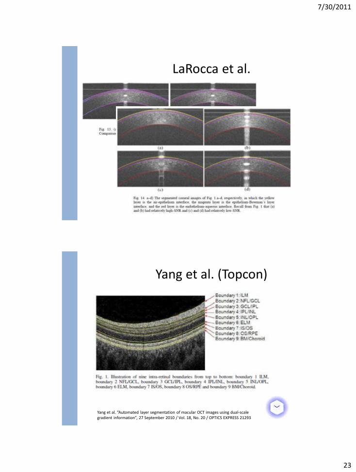
Chiu et al., 2010
Chiu et al., 2010
7/30/2011
22
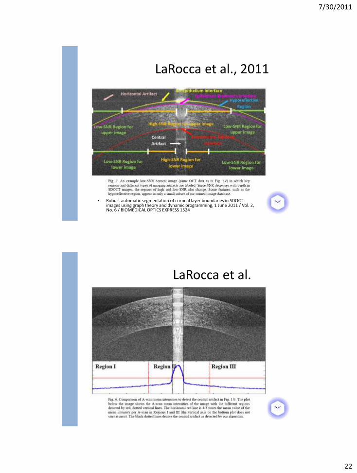
• Robust automatic segmentation of corneal layer boundaries in SDOCT images using graph theory and dynamic programming, 1 June 2011 / Vol. 2, No. 6 / BIOMEDICAL OPTICS EXPRESS 1524
LaRocca et al., 2011
LaRocca et al.
7/30/2011
23
LaRocca et al.
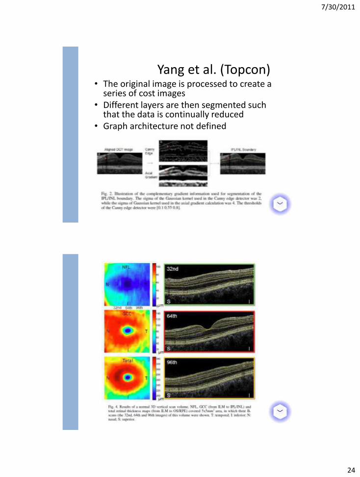
Yang et al. “Automated layer segmentation of macular OCT images using dual-scale gradient information”, 27 September 2010 / Vol. 18, No. 20 / OPTICS EXPRESS 21293
Yang et al. (Topcon)
7/30/2011
24
• The original image is processed to create a series of cost images
• Different layers are then segmented such that the data is continually reduced
• Graph architecture not defined
Yang et al. (Topcon)
7/30/2011
25
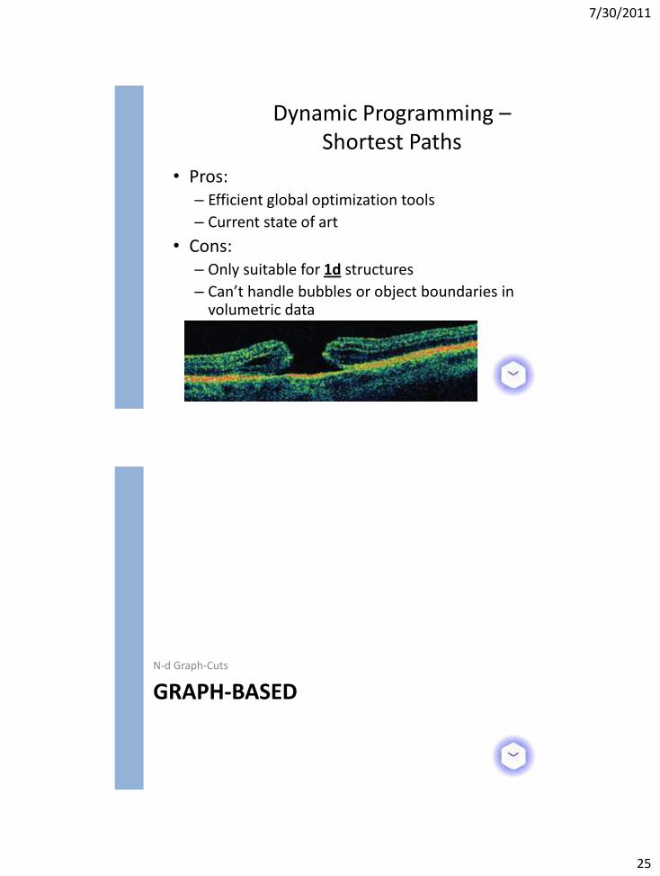
• Pros: – Efficient global optimization tools
– Current state of art
• Cons: – Only suitable for 1d structures
– Can’t handle bubbles or object boundaries in volumetric data
Dynamic Programming – Shortest Paths
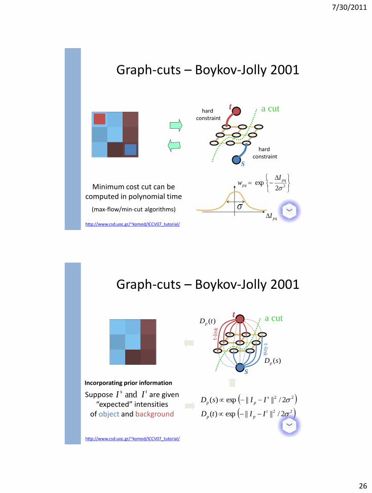
GRAPH-BASED N-d Graph-Cuts
7/30/2011
26
n-links
s
t a cut hard constraint
hard constraint
Minimum cost cut can be computed in polynomial time
(max-flow/min-cut algorithms)
22exp
pq
pq
Iw
pqI
Graph-cuts – Boykov-Jolly 2001
http://www.csd.uoc.gr/~komod/ICCV07_tutorial/
pqw
n-links
s
t a cut )(tDp
)(sDp
Incorporating prior information
Suppose are given “expected” intensities
of object and background
ts II and 22 2/||||exp)( s
pp IIsD
22 2/||||exp)( t
pp IItD
http://www.csd.uoc.gr/~komod/ICCV07_tutorial/
Graph-cuts – Boykov-Jolly 2001
7/30/2011
27
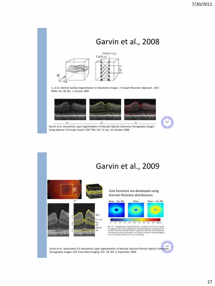
Garvin et al., 2008
Garvin et al. Intraretinal Layer Segmentation of Macular Optical Coherence Tomography Images Using Optimal 3-D Graph Search, IEEE TMI, Vol. 27, No. 10, October 2008
Li, et al. Optimal Surface Segmentation in Volumetric Images – A Graph-Theoretic Approach , IEEE PAMI, Vol. 28, No. 1, January 2006
Garvin et al., 2009
Garvin et al. Automated 3-D Intraretinal Layer Segmentation of Macular Spectral-Domain Optical Coherence Tomography Images, IEEE Trans Med Imaging, VOL. 28, NO. 9, September 2009
Cost functions are developed using learned thickness distributions:
7/30/2011
28
STATISTICAL SHAPE MODELS
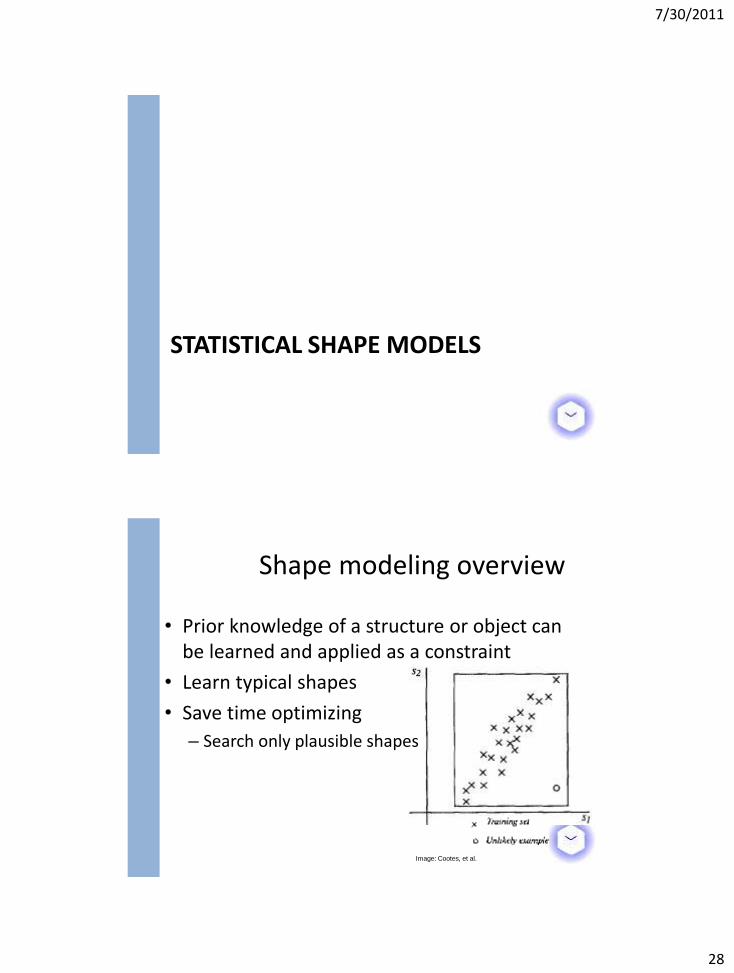
Shape modeling overview
Image: Cootes, et al.
• Prior knowledge of a structure or object can be learned and applied as a constraint
• Learn typical shapes
• Save time optimizing
– Search only plausible shapes
7/30/2011
29
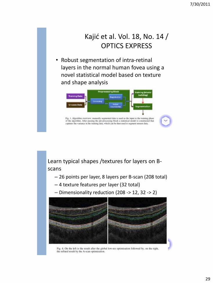
• Robust segmentation of intra-retinal layers in the normal human fovea using a novel statistical model based on texture and shape analysis
Kajić et al. Vol. 18, No. 14 / OPTICS EXPRESS
Learn typical shapes /textures for layers on B-scans
– 26 points per layer, 8 layers per B-scan (208 total)
– 4 texture features per layer (32 total)
– Dimensionality reduction (208 -> 12, 32 -> 2)
7/30/2011
30
CUP & DISC SEGMENTATION Example OCT Structure Segmentation Algorithms
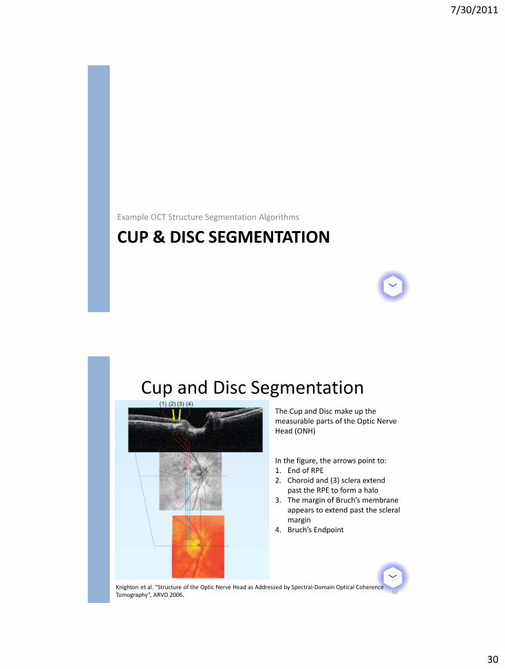
Cup and Disc Segmentation
60
The Cup and Disc make up the measurable parts of the Optic Nerve Head (ONH)
In the figure, the arrows point to: 1. End of RPE 2. Choroid and (3) sclera extend
past the RPE to form a halo 3. The margin of Bruch’s membrane
appears to extend past the scleral margin
4. Bruch’s Endpoint
Knighton et al. “Structure of the Optic Nerve Head as Addressed by Spectral-Domain Optical Coherence Tomography”, ARVO 2006.
7/30/2011
31
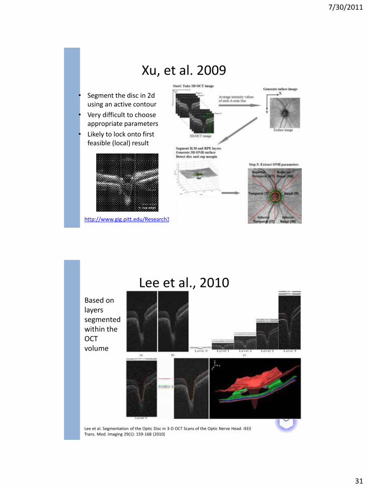
Xu, et al. 2009
• Segment the disc in 2d using an active contour
• Very difficult to choose appropriate parameters
• Likely to lock onto first feasible (local) result
61 http://www.gig.pitt.edu/Research3.html
Lee et al., 2010 Based on layers segmented within the OCT volume
Lee et al. Segmentation of the Optic Disc in 3-D OCT Scans of the Optic Nerve Head. IEEE Trans. Med. Imaging 29(1): 159-168 (2010)
7/30/2011
32
Lee et al., 2010
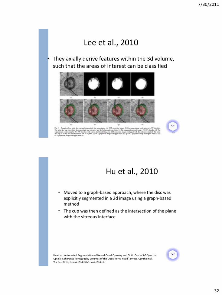
• They axially derive features within the 3d volume, such that the areas of interest can be classified
63
• Moved to a graph-based approach, where the disc was explicitly segmented in a 2d image using a graph-based method
• The cup was then defined as the intersection of the plane with the vitreous interface
Hu et al., 2010
Hu et al., Automated Segmentation of Neural Canal Opening and Optic Cup in 3-D Spectral Optical Coherence Tomography Volumes of the Optic Nerve Head”, Invest. Ophthalmol. Vis. Sci..2010; 0: iovs.09-4838v1-iovs.09-4838
7/30/2011
33
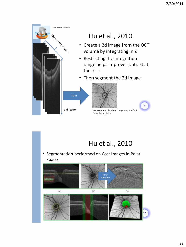
• Create a 2d image from the OCT volume by integrating in Z
• Restricting the integration range helps improve contrast at the disc
• Then segment the 2d image
Hu et al., 2010
Z-direction
Sum
Data courtesy of Robert Change MD, Stanford School of Medicine
From Topcon brochure
Hu et al., 2010
• Segmentation performed on Cost Images in Polar Space
Polar Transform
7/30/2011
34
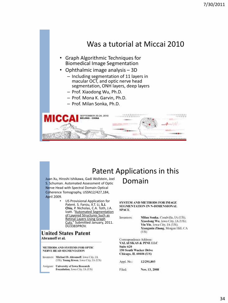
• Graph Algorithmic Techniques for Biomedical Image Segmentation
• Ophthalmic image analysis – 3D – Including segmentation of 11 layers in
macular OCT, and optic nerve head segmentation, ONH layers, deep layers
– Prof. Xiaodong Wu, Ph.D. – Prof. Mona K. Garvin, Ph.D. – Prof. Milan Sonka, Ph.D.
Was a tutorial at Miccai 2010
• US Provisional Application for Patent. S. Farsiu, X.T. Li, S.J. Chiu, P. Nicholas, C.A. Toth, J.A. Izatt. “Automated Segmentation of Layered Structures Such as Retinal Layers Using Graph Cuts.” Submitted January, 2011. DU3383PROV.
Patent Applications in this Domain Juan Xu, Hiroshi Ishikawa, Gadi Wollstein, Joel
S. Schuman. Automated Assessment of Optic Nerve Head with Spectral Domain Optical Coherence Tomography, USSN12/427,184, April 2009.
7/30/2011
35
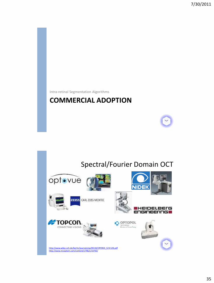
COMMERCIAL ADOPTION Intra-retinal Segmentation Algorithms
Spectral/Fourier Domain OCT
http://www.wiley-vch.de/berlin/journals/op/09-04/OP0904_S24-S28.pdf http://www.revoptom.com/content/i/796/c/14792/
7/30/2011
36
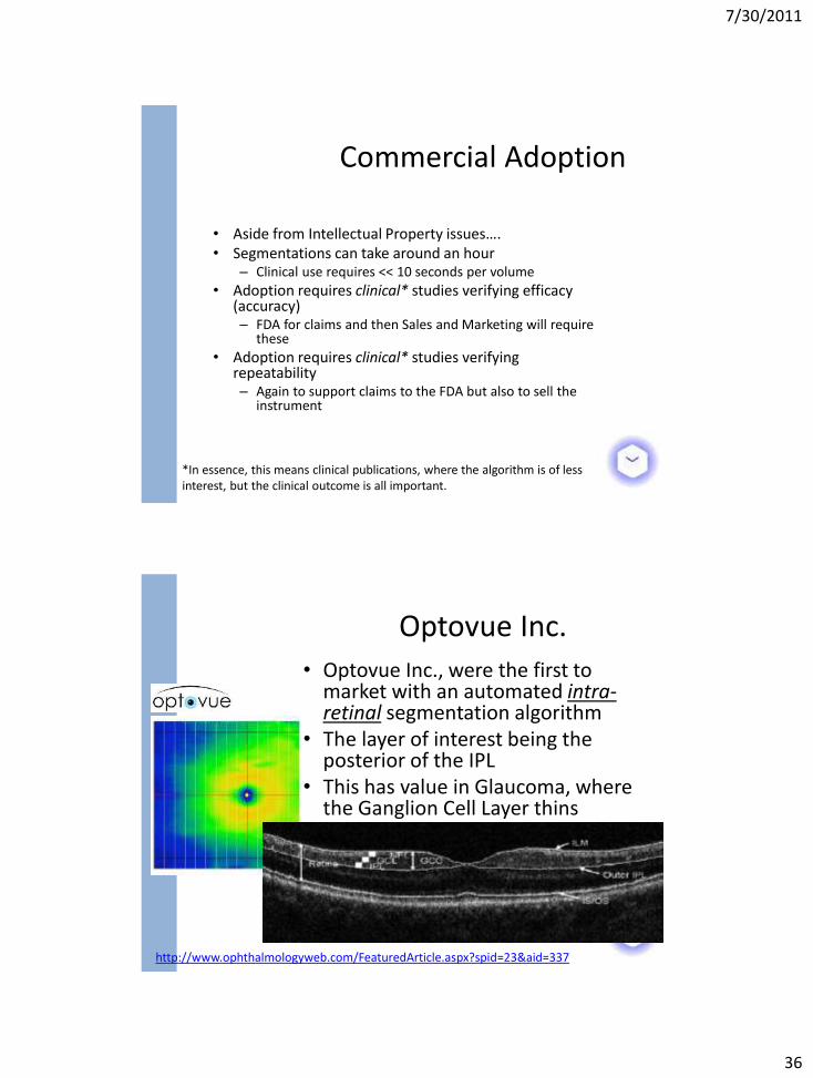
• Aside from Intellectual Property issues…. • Segmentations can take around an hour
– Clinical use requires << 10 seconds per volume
• Adoption requires clinical* studies verifying efficacy (accuracy) – FDA for claims and then Sales and Marketing will require
these
• Adoption requires clinical* studies verifying repeatability – Again to support claims to the FDA but also to sell the
instrument
Commercial Adoption
*In essence, this means clinical publications, where the algorithm is of less interest, but the clinical outcome is all important.
• Optovue Inc., were the first to market with an automated intra-retinal segmentation algorithm
• The layer of interest being the posterior of the IPL
• This has value in Glaucoma, where the Ganglion Cell Layer thins
Optovue Inc.
http://www.ophthalmologyweb.com/FeaturedArticle.aspx?spid=23&aid=337
7/30/2011
37
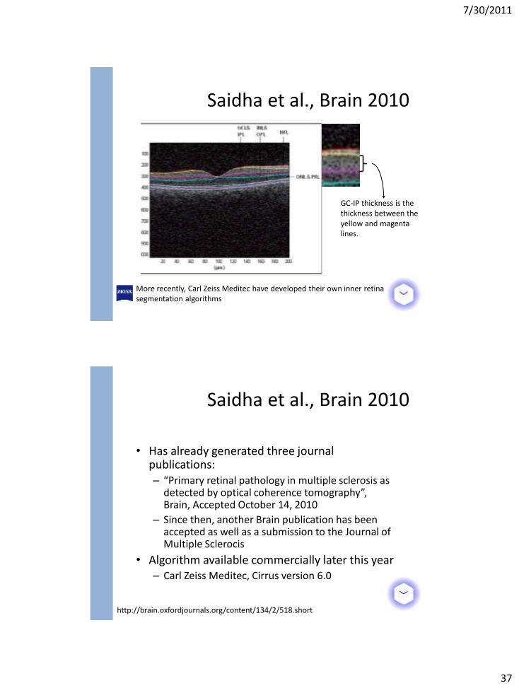
Saidha et al., Brain 2010
GC-IP thickness is the thickness between the yellow and magenta lines.
More recently, Carl Zeiss Meditec have developed their own inner retina segmentation algorithms
• Has already generated three journal publications: – “Primary retinal pathology in multiple sclerosis as
detected by optical coherence tomography”, Brain, Accepted October 14, 2010
– Since then, another Brain publication has been accepted as well as a submission to the Journal of Multiple Sclerocis
• Algorithm available commercially later this year – Carl Zeiss Meditec, Cirrus version 6.0
Saidha et al., Brain 2010
http://brain.oxfordjournals.org/content/134/2/518.short
7/30/2011
38
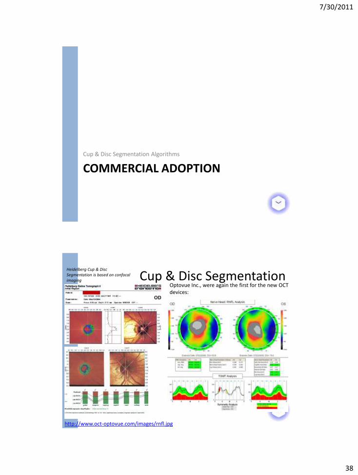
COMMERCIAL ADOPTION Cup & Disc Segmentation Algorithms
Cup & Disc Segmentation
http://www.oct-optovue.com/images/rnfl.jpg
Optovue Inc., were again the first for the new OCT devices:
Heidelberg Cup & Disc Segmentation is based on confocal imaging
7/30/2011
39
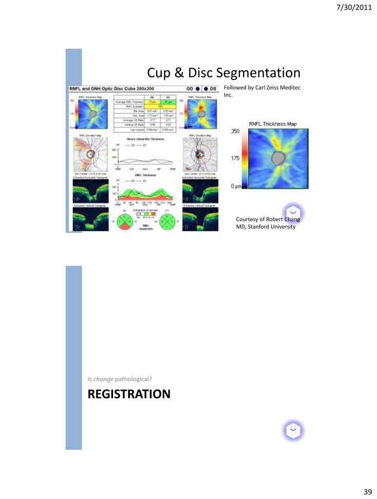
Cup & Disc Segmentation
Courtesy of Robert Chang MD, Stanford University
Followed by Carl Zeiss Meditec Inc.
REGISTRATION Is change pathological?
7/30/2011
40
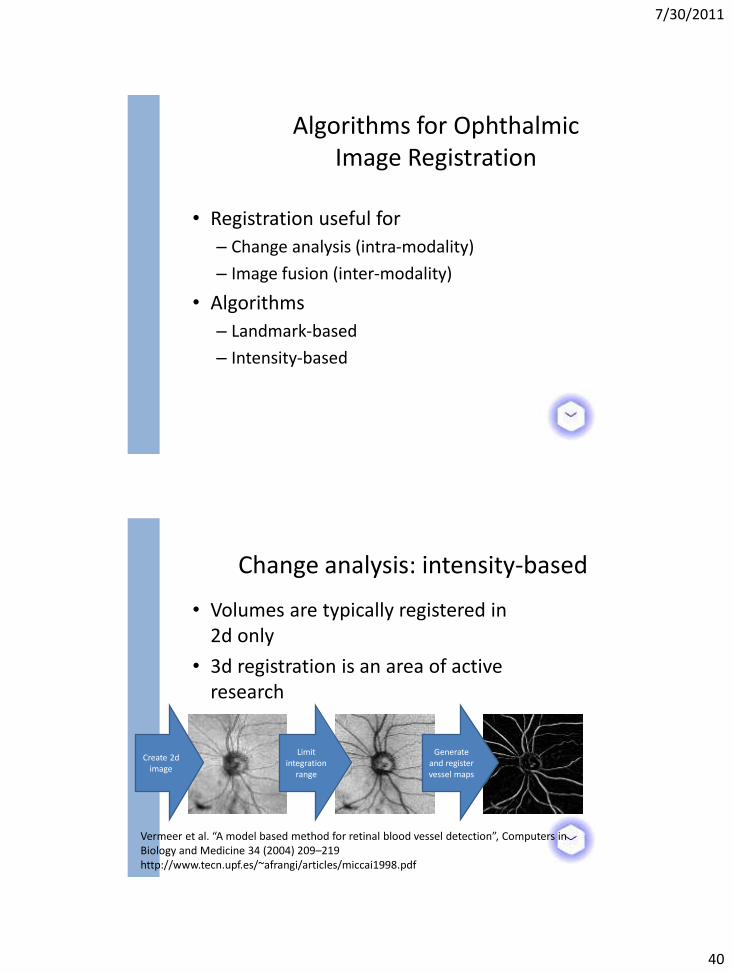
• Registration useful for
– Change analysis (intra-modality)
– Image fusion (inter-modality)
• Algorithms
– Landmark-based
– Intensity-based
Algorithms for Ophthalmic Image Registration
• Volumes are typically registered in 2d only
• 3d registration is an area of active research
Change analysis: intensity-based
Vermeer et al. “A model based method for retinal blood vessel detection”, Computers in Biology and Medicine 34 (2004) 209–219 http://www.tecn.upf.es/~afrangi/articles/miccai1998.pdf
Create 2d image
Limit integration
range
Generate and register vessel maps
7/30/2011
41
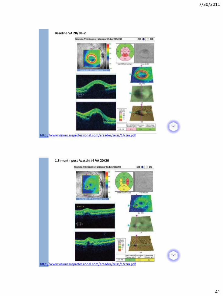
Baseline VA 20/30+2
http://www.visioncareprofessional.com/ereader/zeiss/1/czm.pdf
1.5 month post Avastin #4 VA 20/20
http://www.visioncareprofessional.com/ereader/zeiss/1/czm.pdf
7/30/2011
42
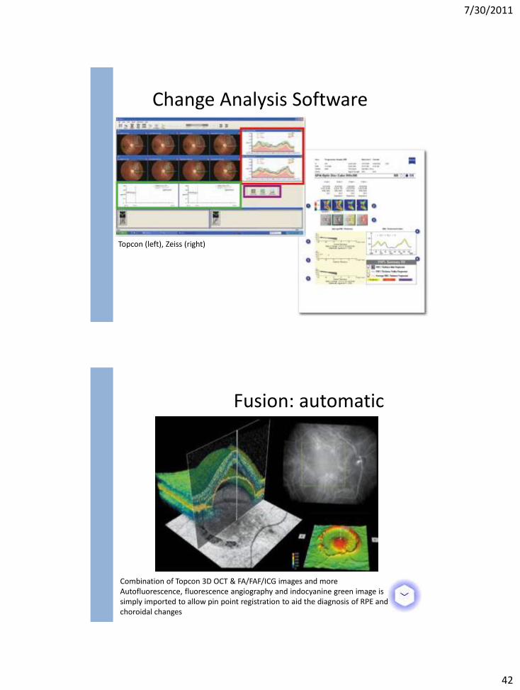
Change Analysis Software
Topcon (left), Zeiss (right)
Combination of Topcon 3D OCT & FA/FAF/ICG images and more Autofluorescence, fluorescence angiography and indocyanine green image is simply imported to allow pin point registration to aid the diagnosis of RPE and choroidal changes
Fusion: automatic
7/30/2011
43
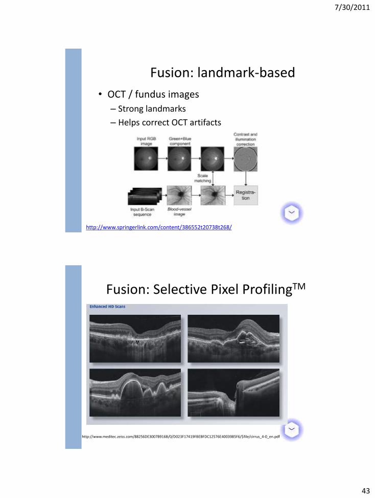
• OCT / fundus images
– Strong landmarks
– Helps correct OCT artifacts
Fusion: landmark-based
http://www.springerlink.com/content/386552t20738t268/
http://www.meditec.zeiss.com/88256DE3007B916B/0/D023F17419FBEBFDC12576E4003985F6/$file/cirrus_4-0_en.pdf
Fusion: Selective Pixel ProfilingTM
7/30/2011
44
OTHER APPLICATION DOMAINS
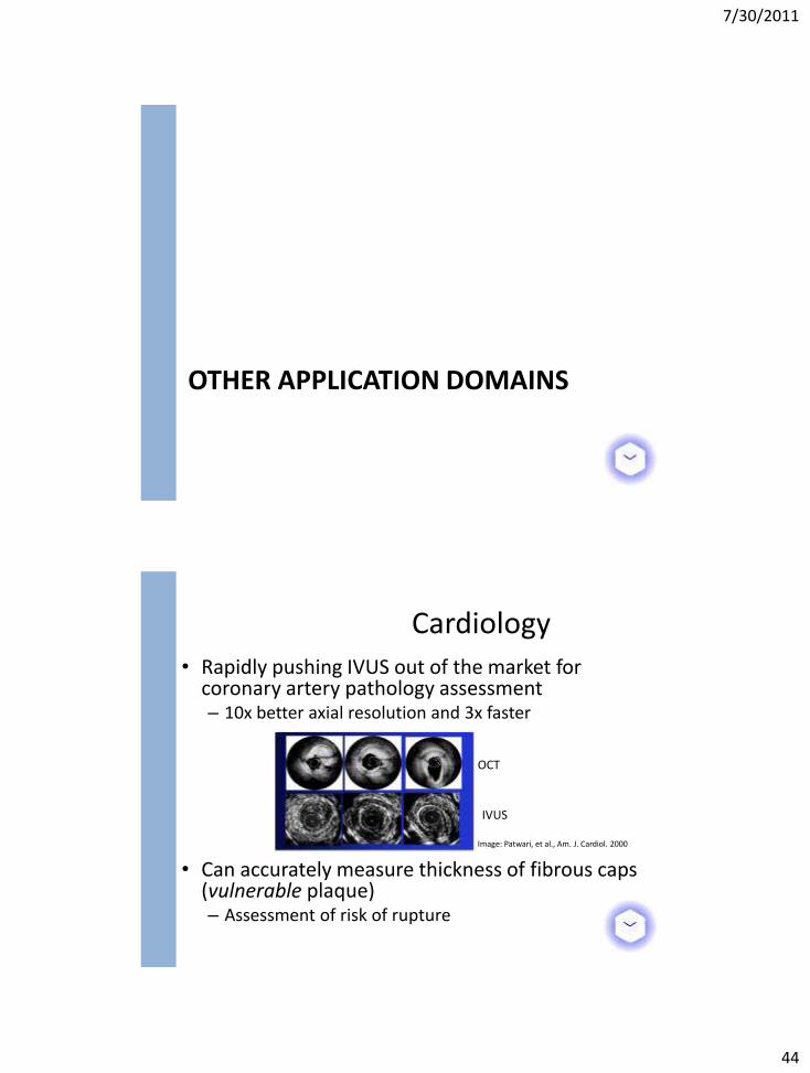
• Rapidly pushing IVUS out of the market for coronary artery pathology assessment – 10x better axial resolution and 3x faster
• Can accurately measure thickness of fibrous caps
(vulnerable plaque) – Assessment of risk of rupture
Cardiology
OCT
IVUS
Image: Patwari, et al., Am. J. Cardiol. 2000
7/30/2011
45
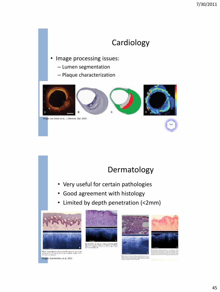
• Image processing issues:
– Lumen segmentation
– Plaque characterization
89
Image: van Soest et al., J. Biomed. Opt. 2010
Cardiology
• Very useful for certain pathologies
• Good agreement with histology
• Limited by depth penetration (<2mm)
Dermatology
Images: Gambichler, et al. 2011
7/30/2011
46
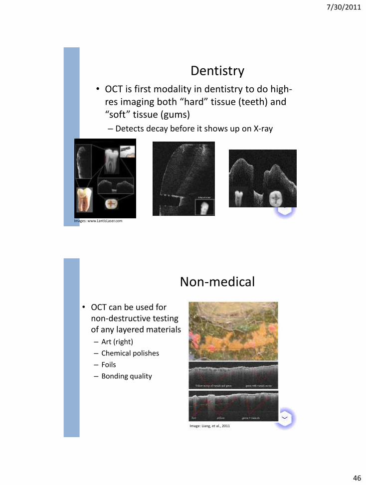
• OCT is first modality in dentistry to do high-res imaging both “hard” tissue (teeth) and “soft” tissue (gums)
– Detects decay before it shows up on X-ray
Dentistry
Images: www.LantisLaser.com
• OCT can be used for non-destructive testing of any layered materials
– Art (right)
– Chemical polishes
– Foils
– Bonding quality
Image: Liang, et al., 2011
Non-medical
7/30/2011
47
FUTURE TRENDS
• Steady evolution of the hardware
– Faster cameras
– Different wavelengths
– Combo modalities
– Lower costs, etc.
– Cheaper components
Hardware
7/30/2011
48
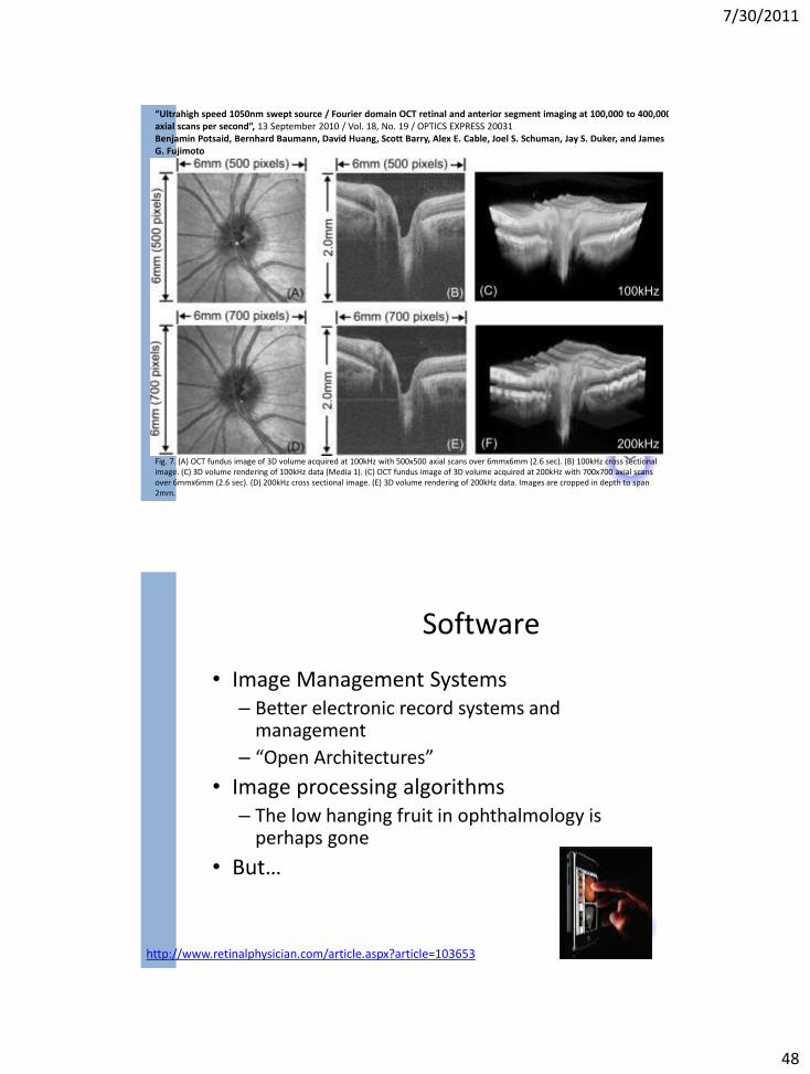
“Ultrahigh speed 1050nm swept source / Fourier domain OCT retinal and anterior segment imaging at 100,000 to 400,000 axial scans per second”, 13 September 2010 / Vol. 18, No. 19 / OPTICS EXPRESS 20031 Benjamin Potsaid, Bernhard Baumann, David Huang, Scott Barry, Alex E. Cable, Joel S. Schuman, Jay S. Duker, and James G. Fujimoto
Fig. 7. (A) OCT fundus image of 3D volume acquired at 100kHz with 500x500 axial scans over 6mmx6mm (2.6 sec). (B) 100kHz cross sectional image. (C) 3D volume rendering of 100kHz data (Media 1). (C) OCT fundus image of 3D volume acquired at 200kHz with 700x700 axial scans over 6mmx6mm (2.6 sec). (D) 200kHz cross sectional image. (E) 3D volume rendering of 200kHz data. Images are cropped in depth to span 2mm.
• Image Management Systems – Better electronic record systems and
management
– “Open Architectures”
• Image processing algorithms – The low hanging fruit in ophthalmology is
perhaps gone
• But…
Software
http://www.retinalphysician.com/article.aspx?article=103653
7/30/2011
49
– Sophisticated methods are too slow, and not, therefore, clinically applicable
– The fast methods are not sophisticated enough to be accurately applied across all disease states
– Ideally you would want to take the best of both worlds from all algorithms we have seen
– A marriage of shape and graph-based methods can leverage both
– Careful reductionist techniques may yet make them execute at clinically realistic speeds
– Lower quality devices will soon be ubiquitous, so the analysis tools offered will be a key differentiator
– Ultimately clinical usage is based on analysis performance
Analysis Algorithms
Thank you!
7/30/2011
50
References
• http://obel.ee.uwa.edu.au/research/oct/intro/ • http://www.lantislaser.com/home.asp • http://obel.ee.uwa.edu.au/research/oct/intro/ • http://www.octnews.org • http://www.recendt.at/517_ENG_HTML.php • http://www.coollab.net/fileadmin/coollab_upload/coollab/docs/huang-
new_OCT_ophthalmology.pdf
• A Comparison of Optic Nerve Head Morphology Viewed by Spectral Domain Optical Coherence Tomography and by Serial Histology
• Strouthidis et al. IOVS, March 2010, Vol. 51, No. 3 • http://telemedicine.orbis.org/bins/home.asp • www.allaboutvision.com & http://www.diabetes.org/diabetes-basics/diabetes-
statistics/ • http://www.meditec.zeiss.com/C125679E00525939/ContainerTitel/CirrusOCT/$Fil
e/retinal-measurements.pdf
References
• Giani et al. ARTIFACTS IN AUTOMATIC RETINAL SEGMENTATION USING DIFFERENT OPTICAL COHERENCE TOMOGRAPHY INSTRUMENTS, RETINA, THE JOURNAL OF RETINAL AND VITREOUS DISEASES, 2010 Apr;30(4):607-16.
• http://www.ophmanagement.com • Gregori et al., Spectral Domain Optical Coherence Tomography Imaging of Drusen
in Nonexudative Age-Related Macular Degeneration, Ophthalmology, Volume 118, Issue 7 , Pages 1373-1379, July 2011
• Schmitt et al., SPECKLE IN OPTICAL COHERENCE TOMOGRAPHY, JOURNAL OF BIOMEDICAL OPTICS 4(1), 95–105 (JANUARY 1999)
• J. Rogowska and M. E. Brezinski, “Evaluation of the adaptive speckle suppression filter for coronary optical coherence tomography imaging,” IEEE Trans. Med. Imaging, 19, 1261–6 (2000).
• Yu and Acton, Speckle Reducing Anisotropic Diffusion, IEEE TRANSACTIONS ON IMAGE PROCESSING, VOL. 11, NO. 11, NOVEMBER 2002
• Macular Segmentation with Optical Coherence Tomography, Investigative Ophthalmology & Visual Science, June 2005, Vol. 46, No. 6
100
7/30/2011
51
References
• http://www.mathworks.com/matlabcentral/fileexchange/14862-sticks-filter/content/sf.m
• http://www.algolist.com/Dijkstra's_algorithm • Timp & Karssemeijer, A new 2D segmentation method based on dynamic
programming applied to computer aided detection in mammography, Med. Phys. 31 .5., May 2004
• Chiu et al. Automatic segmentation of seven retinal layers in SDOCT images congruent with expert manual segmentation, Vol. 18, No. 18 / OPTICS EXPRESS
• Robust automatic segmentation of corneal layer boundaries in SDOCT images using graph theory and dynamic programming, 1 June 2011 / Vol. 2, No. 6 / BIOMEDICAL OPTICS EXPRESS 1524
• Yang et al. “Automated layer segmentation of macular OCT images using dual-scale gradient information”, 27 September 2010 / Vol. 18, No. 20 / OPTICS EXPRESS 21293
• Boykov-Jolly 2001 • http://www.csd.uoc.gr/~komod/ICCV07_tutorial/
101
References
• Garvin et al. Intraretinal Layer Segmentation of Macular Optical Coherence Tomography Images Using Optimal 3-D Graph Search, IEEE TMI, Vol. 27, No. 10, October 2008
• Garvin et al. Automated 3-D Intraretinal Layer Segmentation of Macular Spectral-Domain Optical Coherence Tomography Images, IEEE Trans Med Imaging, VOL. 28, NO. 9, September 2009
• Kajić et al. Vol. 18, No. 14 / OPTICS EXPRESS • Knighton et al. “Structure of the Optic Nerve Head as Addressed by Spectral-
Domain Optical Coherence Tomography”, ARVO 2006. • http://www.gig.pitt.edu/Research3.html • Lee et al. Segmentation of the Optic Disc in 3-D OCT Scans of the Optic Nerve
Head. IEEE Trans. Med. Imaging 29(1): 159-168 (2010) • Hu et al., Automated Segmentation of Neural Canal Opening and Optic Cup in 3-D
Spectral Optical Coherence Tomography Volumes of the Optic Nerve Head”, Invest. Ophthalmol. Vis. Sci..2010; 0: iovs.09-4838v1-iovs.09-4838
• http://www.wiley-vch.de/berlin/journals/op/09-04/OP0904_S24-S28.pdf
102
7/30/2011
52
References
• http://www.revoptom.com/content/i/796/c/14792/ • http://www.ophthalmologyweb.com/FeaturedArticle.aspx?spid=23&aid=337 • http://brain.oxfordjournals.org/content/134/2/518.short • http://www.oct-optovue.com/images/rnfl.jpg
• Vermeer et al. “A model based method for retinal blood vessel detection”, Computers in Biology and Medicine 34 (2004) 209–219
• http://www.tecn.upf.es/~afrangi/articles/miccai1998.pdf • http://www.visioncareprofessional.com/ereader/zeiss/1/czm.pdf • http://www.springerlink.com/content/386552t20738t268/ • http://www.meditec.zeiss.com/88256DE3007B916B/0/D023F17419FBEBFDC1257
6E4003985F6/$file/cirrus_4-0_en.pdf • Images: Patwari, et al., Am. J. Cardiol. 2000 • Images: van Soest et al., J. Biomed. Opt. 2010 • Images: Gambichler, et al. 2011 • Images: www.LantisLaser.com • Images: Liang, et al., 2011
103
References
• “Ultrahigh speed 1050nm swept source / Fourier domain OCT retinal and anterior segment imaging at 100,000 to 400,000 axial scans per second”, 13 September 2010 / Vol. 18, No. 19 / OPTICS EXPRESS 20031, Benjamin Potsaid, Bernhard Baumann, David Huang, Scott Barry, Alex E. Cable, Joel S. Schuman, Jay S. Duker, and James G. Fujimoto
• http://www.retinalphysician.com/article.aspx?article=103653
104
Related Documents