BRAIN A JOURNAL OF NEUROLOGY HTT-lowering reverses Huntington’s disease immune dysfunction caused by NFkB pathway dysregulation Ulrike Tra ¨ger, 1 Ralph Andre, 1 Nayana Lahiri, 1 Anna Magnusson-Lind, 1,2 Andreas Weiss, 3 Stephan Grueninger, 3 Chris McKinnon, 1 Eva Sirinathsinghji, 4 Shira Kahlon, 5 Edith L Pfister, 6 Roger Moser, 7 Holger Hummerich, 1 Michael Antoniou, 4 Gillian P Bates, 4 Ruth Luthi-Carter, 7,8 Mark W Lowdell, 9 Maria Bjo ¨ rkqvist, 2 Gary R Ostroff, 5 Neil Aronin 6 and Sarah J. Tabrizi 1 1 UCL Institute of Neurology, Department of Neurodegenerative Disease, London, UK 2 Wallenberg Neuroscience Centre, Department of Experimental Medical Science, Brain Disease Biomarker Unit, Lund University, Lund, Sweden 3 Novartis Institutes for BioMedical Research, Novartis Campus, Basel, Switzerland 4 King’s College London, Department of Medical and Molecular Genetics, Guy’s Hospital, London, UK 5 University of Massachusetts Medical School, Program in Molecular Medicine, Worcester, MA, USA 6 University of Massachusetts Medical School, Department of Medicine, Division Endocrinology and Metabolism, Worcester, MA, USA 7E ´ cole Polytechnique Fe ´de ´ rale de Lausanne, Brain Mind Institute, Lausanne, Switzerland 8 University of Leicester, Biological Sciences and Psychology, College of Medicine, Department of Cell Physiology and Pharmacology, Leicester, UK 9 UCL, Department of Haematology, Royal Free Campus, London, UK Correspondence to: Sarah J. Tabrizi, Department of Neurodegenerative Disease, UCL Institute of Neurology, Queen Square, London, WC1N 3BG, UK E-mail: [email protected] Huntington’s disease is an inherited neurodegenerative disorder caused by a CAG repeat expansion in the huntingtin gene. The peripheral innate immune system contributes to Huntington’s disease pathogenesis and has been targeted successfully to modulate disease progression, but mechanistic understanding relating this to mutant huntingtin expression in immune cells has been lacking. Here we demonstrate that human Huntington’s disease myeloid cells produce excessive inflammatory cytokines as a result of the cell-intrinsic effects of mutant huntingtin expression. A direct effect of mutant huntingtin on the NFkB pathway, whereby it interacts with IKKg, leads to increased degradation of IkB and subsequent nuclear translocation of RelA. Transcriptional alterations in intracellular immune signalling pathways are also observed. Using a novel method of small interfering RNA delivery to lower huntingtin expression, we show reversal of disease-associated alterations in cellular function–the first time this has been demon- strated in primary human cells. Glucan-encapsulated small interfering RNA particles were used to lower huntingtin levels in human Huntington’s disease monocytes/macrophages, resulting in a reversal of huntingtin-induced elevated cytokine production and transcriptional changes. These findings improve our understanding of the role of innate immunity in neurodegeneration, introduce glucan-encapsulated small interfering RNA particles as tool for studying cellular pathogenesis ex vivo in human cells and raise the prospect of immune cell-directed HTT-lowering as a therapeutic in Huntington’s disease. Keywords: Huntington’s disease; immunology; myeloid cells; gene lowering Abbreviations: GeRP = b1,3-D-glucan-encapsulated small interfering RNA particle; TR-FRET = time resolved fluorescence resonance energy transfer doi:10.1093/brain/awt355 Brain 2014: Page 1 of 15 | 1 Received June 18, 2013. Revised October 24, 2013. Accepted November 2, 2013. ß The Author (2014). Published by Oxford University Press on behalf of the Guarantors of Brain. All rights reserved. For Permissions, please email: [email protected] Brain Advance Access published January 22, 2014 at Queen Mary, University of London on February 11, 2014 http://brain.oxfordjournals.org/ Downloaded from

Welcome message from author
This document is posted to help you gain knowledge. Please leave a comment to let me know what you think about it! Share it to your friends and learn new things together.
Transcript
BRAINA JOURNAL OF NEUROLOGY
HTT-lowering reverses Huntington’s diseaseimmune dysfunction caused by NFkBpathway dysregulationUlrike Trager,1 Ralph Andre,1 Nayana Lahiri,1 Anna Magnusson-Lind,1,2 Andreas Weiss,3
Stephan Grueninger,3 Chris McKinnon,1 Eva Sirinathsinghji,4 Shira Kahlon,5 Edith L Pfister,6
Roger Moser,7 Holger Hummerich,1 Michael Antoniou,4 Gillian P Bates,4 Ruth Luthi-Carter,7,8
Mark W Lowdell,9 Maria Bjorkqvist,2 Gary R Ostroff,5 Neil Aronin6 and Sarah J. Tabrizi1
1 UCL Institute of Neurology, Department of Neurodegenerative Disease, London, UK
2 Wallenberg Neuroscience Centre, Department of Experimental Medical Science, Brain Disease Biomarker Unit, Lund University, Lund, Sweden
3 Novartis Institutes for BioMedical Research, Novartis Campus, Basel, Switzerland
4 King’s College London, Department of Medical and Molecular Genetics, Guy’s Hospital, London, UK
5 University of Massachusetts Medical School, Program in Molecular Medicine, Worcester, MA, USA
6 University of Massachusetts Medical School, Department of Medicine, Division Endocrinology and Metabolism, Worcester, MA, USA
7 Ecole Polytechnique Federale de Lausanne, Brain Mind Institute, Lausanne, Switzerland
8 University of Leicester, Biological Sciences and Psychology, College of Medicine, Department of Cell Physiology and Pharmacology, Leicester, UK
9 UCL, Department of Haematology, Royal Free Campus, London, UK
Correspondence to: Sarah J. Tabrizi,
Department of Neurodegenerative Disease,
UCL Institute of Neurology,
Queen Square,
London, WC1N 3BG, UK
E-mail: [email protected]
Huntington’s disease is an inherited neurodegenerative disorder caused by a CAG repeat expansion in the huntingtin gene. The
peripheral innate immune system contributes to Huntington’s disease pathogenesis and has been targeted successfully to modulate
disease progression, but mechanistic understanding relating this to mutant huntingtin expression in immune cells has been lacking.
Here we demonstrate that human Huntington’s disease myeloid cells produce excessive inflammatory cytokines as a result of the
cell-intrinsic effects of mutant huntingtin expression. A direct effect of mutant huntingtin on the NFkB pathway, whereby it
interacts with IKKg, leads to increased degradation of IkB and subsequent nuclear translocation of RelA. Transcriptional alterations
in intracellular immune signalling pathways are also observed. Using a novel method of small interfering RNA delivery to lower
huntingtin expression, we show reversal of disease-associated alterations in cellular function–the first time this has been demon-
strated in primary human cells. Glucan-encapsulated small interfering RNA particles were used to lower huntingtin levels in human
Huntington’s disease monocytes/macrophages, resulting in a reversal of huntingtin-induced elevated cytokine production and
transcriptional changes. These findings improve our understanding of the role of innate immunity in neurodegeneration, introduce
glucan-encapsulated small interfering RNA particles as tool for studying cellular pathogenesis ex vivo in human cells and raise the
prospect of immune cell-directed HTT-lowering as a therapeutic in Huntington’s disease.
Keywords: Huntington’s disease; immunology; myeloid cells; gene lowering
Abbreviations: GeRP = b1,3-D-glucan-encapsulated small interfering RNA particle; TR-FRET = time resolved fluorescence resonanceenergy transfer
doi:10.1093/brain/awt355 Brain 2014: Page 1 of 15 | 1
Received June 18, 2013. Revised October 24, 2013. Accepted November 2, 2013.
� The Author (2014). Published by Oxford University Press on behalf of the Guarantors of Brain. All rights reserved.
For Permissions, please email: [email protected]
Brain Advance Access published January 22, 2014 at Q
ueen Mary, U
niversity of London on February 11, 2014
http://brain.oxfordjournals.org/D
ownloaded from
IntroductionHuntington’s disease is an incurable, autosomal dominant neuro-
degenerative disorder caused by a CAG repeat expansion in exon
1 of the huntingtin (HTT) gene leading to an expanded stretch of
36 or more glutamine residues in the N-terminal region of the HTT
protein (The Huntington’s Disease Collaborative Research Group,
1993). The disease is characterized by progressive cognitive, psy-
chiatric and motor impairments caused by neuronal dysfunction
and cell death.
Although primary pathology in Huntington’s disease is believed
to arise from basal ganglia degeneration, HTT expression has been
found in all tissues studied (Li et al., 1993). Indeed, numerous
studies of patients with Huntington’s disease and mouse models
have described abnormalities in peripheral tissues, including weight
loss, muscle wasting, diabetes and changes in the neuro-endocrine
system (van der Burg et al., 2009). Mutant HTT expression in
non-neuronal cells in both the brain and the periphery may con-
tribute to Huntington’s disease neuropathology.
HTT is expressed in immune cells (Weiss et al., 2012), and both
central and peripheral immune system abnormalities have been
shown in patients with Huntington’s disease (Soulet and
Cicchetti, 2011). Microglia, the resident immune cells of the
brain (Ransohoff and Perry, 2009), are sustained by self-renewal
(Ajami et al., 2007); however, disrupting the blood–brain barrier
by irradiation has shown that blood monocytes are able to popu-
late the brain (Simard and Rivest, 2004). Microglial activation,
seen in post-mortem Huntington’s disease brain tissue (Sapp
et al., 2001) and by PET imaging, occurs in Huntington’s disease
gene carriers before symptom onset (Tai et al., 2007). We have
previously demonstrated peripheral immune system dysfunction in
Huntington’s disease, including changes in innate immune proteins
in patient plasma (Dalrymple et al., 2007). Moreover, elevated
plasma cytokine (Bjorkqvist et al., 2008) and chemokine (Wild
et al., 2011) levels in patients correlate with disease progression
and can be detected years before disease onset. We have shown
that primary human monocytes are hyper-reactive in response to
lipopolysaccharide, producing increased levels of interleukin (IL)-6.
This phenotype is replicated in murine mutant HTT expressing
macrophages and microglia, demonstrating that peripheral cells
could mirror pathology in the CNS in Huntington’s disease
(Bjorkqvist et al., 2008).
Furthermore, several recent studies have suggested that the
peripheral immune system can act as a modifier of Huntington’s
disease neuropathology. Transplantation of wild-type bone
marrow into Huntington’s disease mice partially rescues their
motor defects, increases synaptogenesis and reduces elevated
plasma cytokine levels (Kwan et al., 2012a). Peripheral adminis-
tration of a kynurenine 3-monooxygenase (KMO) inhibitor ex-
tends lifespan, prevents synaptic loss and decreases microglial
activation in Huntington’s disease mice. As the drug cannot
cross the blood–brain barrier, the neuroprotective effect is second-
ary to inhibition of KMO in peripheral immune cells (Zwilling
et al., 2011). Furthermore, treatment with a cannabinoid receptor
2 agonist known to dampen immune responses, suppresses motor
deficits and CNS inflammation while extending life span in a
Huntington’s disease mouse model. This positive effect can be
blocked with an antagonist that is restricted to the periphery,
demonstrating the importance of peripheral immune cells in mod-
ulating pathogenesis (Bouchard et al., 2012). These studies pro-
vide strong evidence that the immune system plays a disease-
modifying role in Huntington’s disease neuropathogenesis, but
the mechanism(s) by which mutant HTT expression in immune
cells causes this dysfunction has not yet been established.
Intracellular signalling pathways leading to the activation of the
transcription factor NFkB are important regulators of cytokine pro-
duction and play a key role in inflammation. Events such as the
activation of Toll-like receptors (TLRs) lead to signal transduction
through adapter proteins MyD88 and IRAK1, leading to the phos-
phorylation and activation of IKK. This kinase phosphorylates IkB,
which is then ubiquitinated and degraded by the proteasome,
whereby it dissociates from the NFkB transcription factor subunits
(RelA, RelB, cRel, NFkB1, NFkB2) that it sequesters in an in-
active state in the cytoplasm. The free NFkB molecules can
then translocate into the nucleus and activate gene transcription
(Hayden and Ghosh, 2012). The NFkB pathway has previously
been implicated in Huntington’s disease, with Khoshnan et al.
(2004) having shown in inducible PC12 cells and striatal
extracts from R6/2 Huntington’s disease mice that over-
expression of mutant HTT exon 1 can activate the NFkB pathway
by directly interacting with IKKg (Khoshnan et al., 2004). Similarly,
a recent study has shown enhanced NFkB signalling in astro-
cytes isolated from R6/2 mice (Hsiao et al., 2013). It remains
to be shown that this interaction also occurs in a human system
with expression of full-length HTT at normal allelic expression
levels.
The present work seeks to identify the mechanism of dysfunc-
tion in primary human Huntington’s disease monocytes and
macrophages ex vivo. We have characterized immune cell dys-
function by detailed cytokine profiling and study of upstream
intracellular signalling pathways, identifying NFkB pathway dysre-
gulation as the cause of immune dysfunction. We have used over-
expression studies and a novel small interfering RNA-mediated
knock-down technique to investigate the role cell-intrinsic HTT
plays in human Huntington’s disease monocyte and macrophage
function, demonstrating the feasibility of reversing peripheral
immune dysregulation by cell-targeted HTT-lowering.
Materials and methods
Collection and classification ofhuman samplesAll human experiments were performed in accordance with the
Declaration of Helsinki and approved by University College London
(UCL)/UCL Hospitals Joint Research Ethics Committee. All subjects
provided informed written consent. Classification of patients is detailed
in the Supplementary material. Subjects’ demographic are provided in
Supplementary Table 1.
2 | Brain 2014: Page 2 of 15 U. Trager et al.
at Queen M
ary, University of L
ondon on February 11, 2014http://brain.oxfordjournals.org/
Dow
nloaded from
Isolation of human monocytes andmacrophagesCells were isolated from whole blood, as previously described
(Bjorkqvist et al., 2008) and in the Supplementary material. Cells
were cultured in RPMI culture medium supplemented with 10%
foetal calf serum, 2 mM L-glutamine, 50 U/ml penicillin and 50 mg/ml
streptomycin (Invitrogen). Monocytes were allowed to rest for 16 h
before experimental use. Culture medium was supplemented
with 20 ng/ml granulocyte macrophage-colony stimulating factor
(GM-CSF) for 6 days to differentiate monocytes into macrophages.
Mutant HTT expression in U937 cellsU937 cells (Sundstrom and Nilsson, 1976) were transduced with lenti-
viral constructs containing human HTT exon 1 sequences with either
29, 71 or 129 CAG repeats, together with GFP, or a control vector
containing GFP but no HTT exon 1. For details of vectors, viral pro-
duction and transduction, see the online Supplementary material.
Transduced U937 cells were tested for HTT protein expression using
a time resolved fluorescence resonance energy transfer (TR-FRET)
immunoassay. HTT exon 1 expressing U937 cells were seeded into
24-well plates at 5 � 105 cells per well and differentiated into
mature monocytes using 10 nM phorbol 12-myristate 13-acetate
(PMA) for 3 days (Alciato et al., 2010) before cytokine profiling.
HTT silencingMonocytes and macrophages were incubated with b1,3-D-glucan-
encapsulated small interfering RNA particles (GeRPs) for 4 h, after
which fresh medium was added to the cultures. GeRP uptake was
visualized by seeding 1 � 105 monocytes per 13 mm coverslip, incu-
bating them with empty green fluorescent GeRPs for 12 h and mount-
ing onto slides with 1 mg/ml DAPI. Images were acquired using a Zeiss
510 meta microscope (objective �63/1.4 oil DIC, 1024 � 1024),
overlaying the bright-field image of the cells with the 405 nm and
488 nm fluorescence channels for DAPI and green fluorescence,
respectively. Macrophages, which were transfected on Day 3 of the
differentiation protocol, were transfected with green fluorescent
GeRPs containing no small interfering RNA at various ratios (1:1,
1:3, and 1:10) before uptake rates were measured by flow cytometry.
Cells were fixed with 3.7% paraformaldehyde for 10 min, washed with
fluorescence-activated cell sorting (FACS) buffer (PBS containing 1%
foetal calf serum and 0.02% sodium azide) and resuspended in 200 ml
FACS buffer for analysis by flow cytometry (FACSCalibur with
CellQuest Pro BD Bioscience). Data analysis was performed using
FlowJo 7.2.5 (Tree Star). To examine the effects of HTT knock-
down on cytokine production, macrophages were treated with either
scrambled or anti-HTT small interfering RNA containing GeRPs at a
1:10 cell: particle ratio on Day 3 of the differentiation protocol; stimu-
lation of the cells took place 3 days later. To examine the effects of
HTT knock-down on transcriptional dysregulation, monocytes were
treated with either scrambled or anti-HTT small interfering RNA con-
taining GeRPs at a 1:10 cell: particle ratio, before quantitative PCR
analysis 3 days later.
Cytokine profilingAll cells were seeded at 5 � 105 cells per well in 24-well plates and
isolated, differentiated and transduced as described above. For stimu-
lation, medium was changed to fresh cell culture medium containing
10 ng/ml IFNg (R & D Systems) and 2 mg/ml lipopolysaccharide
(Sigma-Aldrich, E.coli 055:B5, strain 1644-70. Cat. number L6529).
After 24 h, supernatants were harvested and analysed using MSD
multiplex assays, according to manufacturer’s instructions (MesoScale
Discovery). For monocytes the pro-inflammatory (7-plex) assay was
used, however, IFNg measures were not analysed as we used IFNgas stimulus. For all other cell types, the pro-inflammatory II (4-plex)
assay was used and all data are shown. Monocyte data were adjusted
to basal cytokine levels, whereas all other cell types were normalized
to total protein concentration in each well. Cells were lysed in 50 mM
Tris pH 8, 150 mM NaCl, 0.5% sodium deoxycholate, 0.5% TritonTM
X-100 and assayed for total protein concentration using a Bradford-
based protein assay (Bio-Rad).
Time resolved fluorescence resonanceenergy transfer quantification of HTTTR-FRET immunoassay quantification of total HTT and soluble mutant
HTT was performed as previously described (Baldo et al., 2012) and is
detailed in the Supplementary material.
Polymerase chain reaction arraysSABioscience Human NFkB Signaling Pathway RT2ProfilerTM PCR
Arrays were used in combination with the QIAGEN RNeasy� Mini
Kit for RNA isolation from 2 � 106 cells. RNA integrity was evaluated
using 2100 RNA Bioanalyser chips (Agilent). RNA was reverse
transcribed using the RT2 First Strand kit for complementary DNA
transcription, before the RT2 SYBR� Green qPCR Mastermix and
pre-primer coated PCR plates were used for quantitative PCR
(SABioscience). All kits were used according to the manufacturer’s
instructions.
For standard SYBR� Green protocols and bioinformatics used for
Supplementary Fig. 6, see Supplementary material.
Proximity ligation assaysProximity ligation assays were conducted on monocyte-derived macro-
phages seeded on 13 mm coverslips. Cells were fixed in 4% parafor-
maldehyde for 10 min and permeabilized with 100% ice cold methanol
at �20�C for 15 min. Coverslips were blocked with 10% bovine serum
albumin for 30 min at 37�C before staining with primary antibodies
was performed for 1 h at 37�C (mouse anti-HTT 4C9, 1:300, kind gift
from Novartis; rabbit anti-IKKg, 1:100, Santa Cruz; rabbit anti-IKKa/b,
1:25, Santa Cruz). Instead of using fluorescently labelled secondary
antibodies, a proximity ligation approach was applied following manu-
facturer’s instructions (Sigma). Briefly, samples were incubated
with secondary antibodies conjugated with DNA probes (minus anti-
mouse and plus anti-rabbit DNA probes). Probes were hybridized and
ligated before amplification of the DNA template in a rolling circle
amplification reaction. Detection solution was added to identify ampli-
fied DNA. Signals were detected using a Zeiss LSM 710 confocal
microscope (objective plan-apachromat �40/1.4 oil DIC M27,
1024 � 1024). Spots were quantified using Volocity (PerkinElmer) on
at least seven fields of view per subject, taken of random sides of each
coverslips.
Immunoblot analysis of IkB degradationMonocytes were seeded at 1 � 106 cells/well into 24-well plates and
rested for 16 h. Cells were stimulated with 2 mg/ml lipopolysaccharide
Reversing immune dysfunction in HD Brain 2014: Page 3 of 15 | 3
at Queen M
ary, University of L
ondon on February 11, 2014http://brain.oxfordjournals.org/
Dow
nloaded from
over a 2 h or 24 h time course before extraction of lysates for western
blotting (see Supplementary material for detail).
NFkB RelA translocationCD14 + monocytes were seeded at 2 � 106 into 6-well plates and left
to rest for 16 h. The cells were stimulated with 2 mg/ml lipopolysac-
charide before being scraped off the plates. Pelleted cells were fixed
for 15 min and permeabilized for 10 min using the eBioscience Fix/
Perm solutions, before NFkB p65/RelA XP antibody (1:200; Cell
Signaling) diluted in permeabilization buffer was added. After 30 min
incubation shaking at 4�C, cells were washed twice with FACS buffer
and spun for 5 min at 300g. Secondary anti-rabbit IgG phycoerythrin
(eBioscience) was added at 1:100 in FACS buffer and incubated for
30 min before washing the cells twice with FACS buffer. Cells were
resuspended in 80 ml FACS buffer and stained with 1 mg/ml DAPI just
before analysis. Samples were run on the ImageStreamX (Amnis) and
analysed using the IDEAS software. Briefly, gating on single cells, the
similarity feature {Similarity_Erode [Object (M04,BF,Tight)2]_Dapi
_RelA} was used to establish the rate of RelA translocation by measur-
ing the overlap of DAPI and RelA staining. Translocation rate was
normalized to baseline levels for each subject.
Statistical analysisFor cytokine profiling data, inter-group differences were identified by
one-way ANOVA with post hoc Tukey Honestly Significant Difference
testing to allow for multiple comparisons. Data were corrected for age
and gender before analysis. Linear regression with log10 transformed
data was used to establish whether cytokine production by primary
human monocytes and macrophages correlates with CAG repeat
length. Cytokine profiling data from U937 cells and knock-down
cells were analysed by two-way ANOVA with Bonferroni post-tests.
Gene expression changes measured by quantitative PCR were
analysed using unpaired two-tailed student t-tests. Paired two-tailed
student t-tests were used to analyse the effects of anti-HTT small
interfering RNA compared to scrambled small interfering RNA in
cells from the same individual. All error bars represent standard error
of the mean.
Results
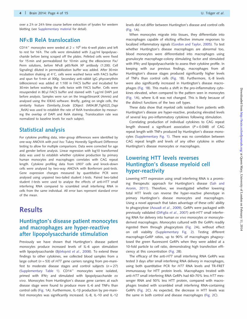
Huntington’s disease patient monocytesand macrophages are hyper-reactiveafter lipopolysaccharide stimulationPreviously we have shown that Huntington’s disease patient
monocytes produce increased levels of IL-6 upon stimulation
with lipopolysaccharide (Bjorkqvist et al., 2008). To extend these
findings to other cytokines, we collected blood samples from a
large cohort (n = 53) of HTT gene carriers ranging from pre-mani-
fest to moderate disease stages and control subjects (n = 27)
(Supplementary Table 1). CD14 + monocytes were isolated,
primed with IFNg and stimulated with lipopolysaccharide ex
vivo. Monocytes from Huntington’s disease gene carriers at each
disease stage were found to produce more IL-6 and TNFa than
control cells (Fig. 1A). Furthermore, IL-1b production by pre-mani-
fest monocytes was significantly increased. IL-8, IL-10 and IL-12
levels did not differ between Huntington’s disease and control cells
(Fig. 1A).
When monocytes migrate into tissues, they differentiate into
macrophages capable of eliciting effective immune responses to
localized inflammatory signals (Gordon and Taylor, 2005). To test
whether Huntington’s disease macrophages are abnormal too,
blood monocytes were differentiated into macrophages using
granulocyte macrophage-colony stimulating factor and stimulated
with IFNg and lipopolysaccharide to assess their cytokine profile. In
keeping with our previous findings, macrophages from all
Huntington’s disease stages produced significantly higher levels
of TNFa than control cells (Fig. 1B). Furthermore, IL-8 levels
were also significantly increased in Huntington’s disease macro-
phages (Fig. 1B). This marks a shift in the pro-inflammatory cyto-
kines elevated, when compared to the pattern seen in monocytes
(Fig. 1A), where IL-8 was not changed. This is likely because of
the distinct functions of the two cell types.
These data show that myeloid cells isolated from patients with
Huntington’s disease are hyper-reactive, producing elevated levels
of several key pro-inflammatory cytokines following stimulation.
Correlating production of individual cytokines to CAG repeat
length showed a significant association (P = 0.048) of CAG
repeat length with TNFa produced by Huntington’s disease mono-
cytes (Supplementary Fig. 1). There was no correlation between
CAG repeat length and levels of any other cytokine in either
Huntington’s disease monocytes or macrophages.
Lowering HTT levels reversesHuntington’s disease myeloid cellhyper-reactivityLowering HTT expression using small interfering RNA is a promis-
ing therapeutic approach for Huntington’s disease (Sah and
Aronin, 2011). Therefore, we investigated whether lowering
total HTT levels can reverse the hyper-reactive phenotype in
primary Huntington’s disease monocytes and macrophages.
Using a novel approach that takes advantage of these cells’ ability
to phagocytose (Aouadi et al., 2009), GeRPs were packaged with
previously validated (DiFiglia et al., 2007) anti-HTT small interfer-
ing RNA for delivery into human ex vivo monocytes or monocyte-
derived macrophages. Monocytes cultured with the GeRPs readily
ingested them through phagocytosis (Fig. 2A), without effect
on cell viability (Supplementary Fig. 2). Testing different
macrophage:GeRP ratios, up to 90% of macrophages phagocy-
tosed the green fluorescent GeRPs when they were added at a
10-fold particle to cell ratio, demonstrating high transfection effi-
ciency at this concentration (Fig. 2B).
The efficacy of the anti-HTT small interfering RNA GeRPs was
tested 3 days after small interfering RNA delivery in macrophages,
using both quantitative PCR for HTT RNA levels and TR-FRET
immunoassay for HTT protein levels. Macrophages treated with
anti-HTT small interfering RNA GeRPs had 60–70% less HTT mes-
senger RNA and 50% less HTT protein, compared with macro-
phages treated with scrambled small interfering RNA-containing
GeRPs (Fig. 2C). As expected, the decrease in HTT levels was
the same in both control and disease macrophages (Fig. 2C).
4 | Brain 2014: Page 4 of 15 U. Trager et al.
at Queen M
ary, University of L
ondon on February 11, 2014http://brain.oxfordjournals.org/
Dow
nloaded from
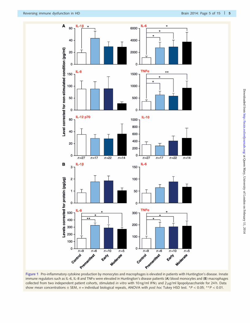
Figure 1 Pro-inflammatory cytokine production by monocytes and macrophages is elevated in patients with Huntington’s disease. Innate
immune regulators such as IL-6, IL-8 and TNFa were elevated in Huntington’s disease patients (A) blood monocytes and (B) macrophages
collected from two independent patient cohorts, stimulated in vitro with 10 ng/ml IFNg and 2mg/ml lipopolysaccharide for 24 h. Data
show mean concentrations � SEM, n = individual biological repeats, ANOVA with post hoc Tukey HSD test. *P50.05; **P5 0.01.
Reversing immune dysfunction in HD Brain 2014: Page 5 of 15 | 5
at Queen M
ary, University of L
ondon on February 11, 2014http://brain.oxfordjournals.org/
Dow
nloaded from
Next, we examined the effect of lowering total HTT levels on
cytokine production. After treating primary human monocyte-
derived macrophages with anti-HTT or scrambled small interfering
RNA GeRPs for 3 days, IFNg-primed macrophages were stimulated
with lipopolysaccharide and cytokine production was measured.
Validating our previous findings, IL-8 and TNFa levels were sig-
nificantly higher in Huntington’s disease than in control cells,
when both had been treated with scrambled small interfering
RNA (Fig. 3). However, lowering HTT levels in Huntington’s dis-
ease macrophages using anti-HTT GeRPs rescued this increase by
significantly decreasing the production of IL-6, IL-8 and TNFa(Fig. 3). IL-1b production showed a similar trend that did not
reach significance. Interestingly, lowering HTT levels also signifi-
cantly reduced IL-6, IL-8 and TNFa levels in control cells, suggest-
ing a role of wild-type HTT in cytokine production.
Thus, lowering HTT levels by 50% using this novel method of
small interfering RNA delivery can reverse the hyper-reactivity of
Huntington’s disease patient macrophages. The use of GeRPs to
achieve cell-targeted gene knock-down has to date shown signifi-
cant promise in mice, but this is the first report showing efficient
small interfering RNA delivery, pathogenic gene knock-down and
rescue of a deleterious phenotype using this method in primary
human immune cells.
The use of HTT-lowering in Huntington’s disease patient myeloid
cells demonstrates that their production of cytokines in response to
Figure 2 Glucan encapsulated small interfering RNA particles
(GeRPs) can effectively knock-down total HTT in primary human
immune cells. (A) GeRPs deliver small interfering RNA (siRNA)
efficiently when phagocytosed by myeloid cells, as shown in
primary human monocytes after 12 h incubation in culture
(GeRPs = green; DAPI = blue). (B) Ninety per cent of macro-
phages take up GeRPs when incubated at 1:10 cell: particle ratio
for 12 h as quantified by flow cytometry. Data shown as mean
[n = 2 for controls and n = 3 for Huntington’s disease
(HD)] � SEM. (C) Total HTT RNA measured by quantitative PCR
and protein levels measured by TR-FRET were reduced by 70%
and 50%, respectively, in macrophages treated for 3 days with
GeRPs containing anti-HTT small interfering RNA. Data shown
as mean HTT levels (each combining two independent experi-
ments, n = individual biological repeats) � SEM. Data are nor-
malized to the scrambled small interfering RNA treated condition
for each genotype.
Figure 3 Knock-down of total HTT reverses the hyper-reactive
cytokine production by Huntington’s disease macrophages.
Huntington’s disease (HD) and control macrophages were
treated with either anti-HTT or scrambled small interfering
RNA (siRNA) for 3 days, before the cells were stimulated with
10 ng/ml IFNg and 2 mg/ml lipopolysaccharide for 24 h.
Measuring cytokine production with multiplex ELISA assays
showed that lowering HTT levels reduces IL-6, IL-8 and TNFalevels after stimulation. Data shown as mean concentrations
(n = 9 for controls and n = 8 for Huntington’s disease, combined
from three independent experiments, n = individual biological
repeats) � SEM, two-way ANOVA with Bonferroni post-tests.
*P50.05; **P5 0.01, ***P50.001.
6 | Brain 2014: Page 6 of 15 U. Trager et al.
at Queen M
ary, University of L
ondon on February 11, 2014http://brain.oxfordjournals.org/
Dow
nloaded from
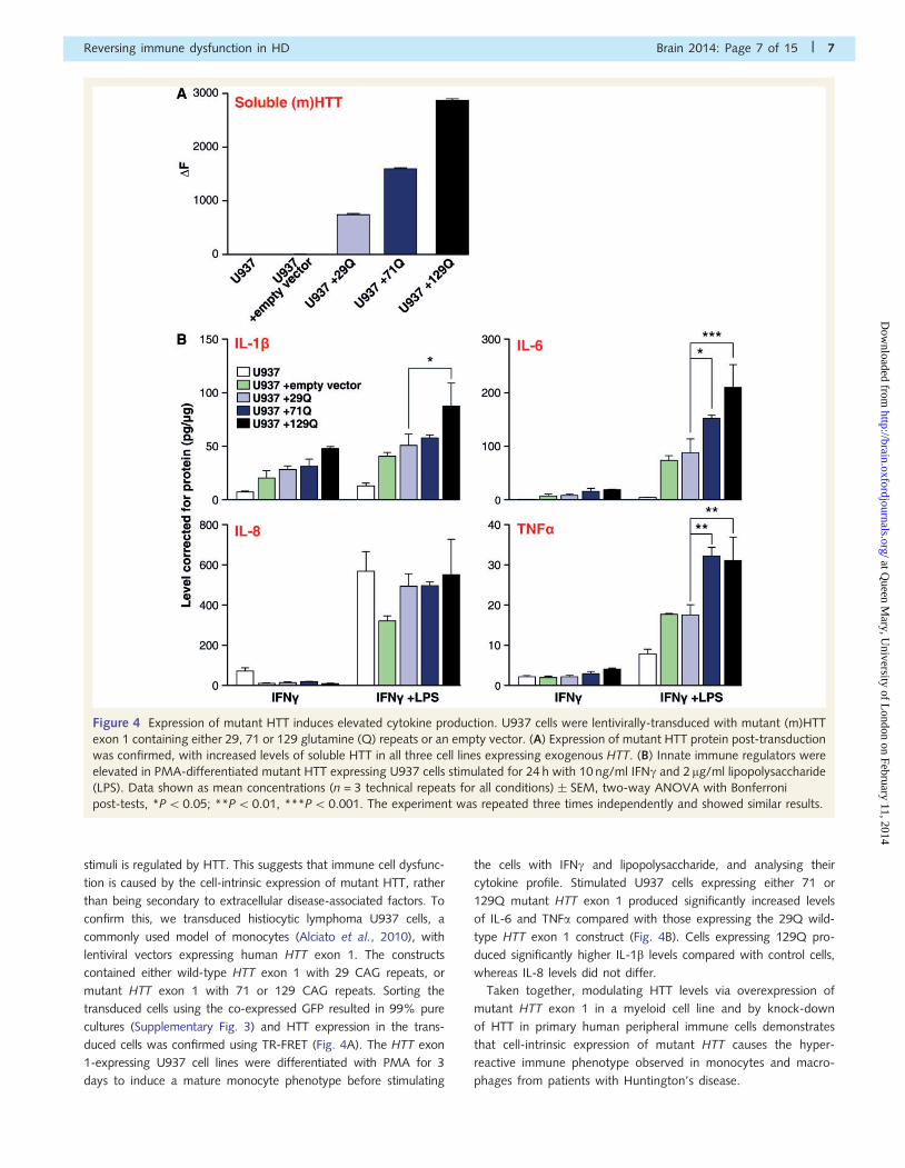
stimuli is regulated by HTT. This suggests that immune cell dysfunc-
tion is caused by the cell-intrinsic expression of mutant HTT, rather
than being secondary to extracellular disease-associated factors. To
confirm this, we transduced histiocytic lymphoma U937 cells, a
commonly used model of monocytes (Alciato et al., 2010), with
lentiviral vectors expressing human HTT exon 1. The constructs
contained either wild-type HTT exon 1 with 29 CAG repeats, or
mutant HTT exon 1 with 71 or 129 CAG repeats. Sorting the
transduced cells using the co-expressed GFP resulted in 99% pure
cultures (Supplementary Fig. 3) and HTT expression in the trans-
duced cells was confirmed using TR-FRET (Fig. 4A). The HTT exon
1-expressing U937 cell lines were differentiated with PMA for 3
days to induce a mature monocyte phenotype before stimulating
the cells with IFNg and lipopolysaccharide, and analysing their
cytokine profile. Stimulated U937 cells expressing either 71 or
129Q mutant HTT exon 1 produced significantly increased levels
of IL-6 and TNFa compared with those expressing the 29Q wild-
type HTT exon 1 construct (Fig. 4B). Cells expressing 129Q pro-
duced significantly higher IL-1b levels compared with control cells,
whereas IL-8 levels did not differ.
Taken together, modulating HTT levels via overexpression of
mutant HTT exon 1 in a myeloid cell line and by knock-down
of HTT in primary human peripheral immune cells demonstrates
that cell-intrinsic expression of mutant HTT causes the hyper-
reactive immune phenotype observed in monocytes and macro-
phages from patients with Huntington’s disease.
Figure 4 Expression of mutant HTT induces elevated cytokine production. U937 cells were lentivirally-transduced with mutant (m)HTT
exon 1 containing either 29, 71 or 129 glutamine (Q) repeats or an empty vector. (A) Expression of mutant HTT protein post-transduction
was confirmed, with increased levels of soluble HTT in all three cell lines expressing exogenous HTT. (B) Innate immune regulators were
elevated in PMA-differentiated mutant HTT expressing U937 cells stimulated for 24 h with 10 ng/ml IFNg and 2 mg/ml lipopolysaccharide
(LPS). Data shown as mean concentrations (n = 3 technical repeats for all conditions) � SEM, two-way ANOVA with Bonferroni
post-tests, *P5 0.05; **P50.01, ***P5 0.001. The experiment was repeated three times independently and showed similar results.
Reversing immune dysfunction in HD Brain 2014: Page 7 of 15 | 7
at Queen M
ary, University of L
ondon on February 11, 2014http://brain.oxfordjournals.org/
Dow
nloaded from
Mutant HTT interacts with the NFkBpathway in human Huntington’sdisease myeloid cellsThat Huntington’s disease patient monocytes and macrophages
resemble normal cells when unstimulated, but are hyper-reactive
in response to lipopolysaccharide, suggests that mutant HTT
affects the signalling cascade induced by lipopolysaccharide.
Expression of the main lipopolysaccharide receptor, TLR4, was
unaltered (Supplementary Fig. 4), suggesting downstream effects.
The NFkB pathway, a key signalling cascade downstream of TLR4,
has previously been shown to interact with mutant HTT exon 1 in
mice (Khoshnan et al., 2004).
To test whether this interaction occurs in human primary
immune cells, peripheral blood mononuclear cells from patients
with early-stage Huntington’s disease and control subjects were
isolated for co-immunoprecipitation experiments. Full-length HTT
was detectable in both the control and Huntington’s disease sam-
ples with two anti-HTT antibodies (2B7 and MAB2166), whereas
co-precipitation of IKKg was observed only in the Huntington’s
disease sample (Supplementary Fig. 5A). Given the high back-
ground signal in these experiments because of poor antibody
performance in the immunoprecipitation, we performed more
sensitive proximity ligation assays to detect native IKKg-HTT inter-
actions in the cells. As shown in Fig. 5A, specific IKKg-HTT protein
interactions, represented by red spots can be detected in both
control and disease macrophages. Quantification of the number
of spots per cell demonstrated more interaction between IKKg and
HTT in Huntington’s disease patient cells compared with controls
(Fig. 5B). Further evidence for a CAG repeat dependent interaction
between HTT and the IKK complex was given by an increased
number of interactions between HTT and the IKKa/b subunits in
Huntington’s disease samples (Fig. 5B). The fact that classical
immunoprecipitation did not pick up an interaction of the proteins
in control individual’s cells is likely to be because the method being
less sensitive. These data demonstrate for the first time a direct
interaction between the IKK complex and full-length HTT ex-
pressed at normal allelic expression levels in primary human cells.
Activation of the IKK complex leads to the phosphorylation and
degradation of IkB, the endogenous inhibitor of NFkB (Hayden
and Ghosh, 2012). To evaluate whether the increased interaction
of mutant HTT with IKKg leads to increased IKK complex activa-
tion and subsequent changes in IkB degradation, we stimulated
Huntington’s disease and control monocytes with lipopolysacchar-
ide over a time course of 2 h to analyse IkB levels by western blot.
Control monocytes demonstrated a drop in IkB levels over the first
15 min, before a recovery of IkB levels over the next 2 h, repre-
senting a normal pattern of NFkB activation on stimulation
(Fig. 5C) (Gross and Piwnica-Worms, 2005). After stimulation of
Huntington’s disease monocytes, we observed a different pattern:
IkB levels dropped within 5 mins and did not recover to baseline
levels within the 2 h time course (Fig. 5C). This demonstrates that
IkB is degraded more rapidly and over a prolonged period of time
in primary human Huntington’s disease monocytes as a result of
IKK activation. Similarly, levels of phosphorylated IkB were
increased over the 2 h period in monocytes isolated from patients
with Huntington’s disease compared with control subjects
(Supplementary Fig. 5B). To investigate by which time IkB levels
return to baseline in patients with Huntington’s disease, we per-
formed a prolonged time course over 24 h and found that IkB
levels returned to baseline levels or above (because of high level
re-synthesis of the protein) by 4 h post-stimulation (Supplementary
Fig. 5C). These findings demonstrate a transient effect of mutant
HTT expression on IkB levels after stimulation.
Under steady-state conditions, IkB binds NFkB and blocks its
translocation to the nucleus. Degradation of IkB allows the NFkB
transcription factors to enter the nucleus and influence transcrip-
tion (Beinke and Ley, 2004). To test whether increased IkB deg-
radation in Huntington’s disease monocytes leads to more rapid
nuclear translocation of NFkB, we analysed translocation of RelA,
one of five DNA-binding NFkB subunits, in Huntington’s disease
and control monocytes using imaging flow cytometry.
ImageStream technology, combining the high image content
information of microscopy with the high throughput analysis of
flow cytometry, is used to overcome the limitations of conven-
tional assays to produce highly reproducible and statistically robust
data (Maguire et al., 2011). Cells were stained with DAPI to mark
the nucleus and with anti-RelA antibodies (Fig. 5D). Analysis of
the levels of RelA and DAPI co-localization showed significantly
higher levels of RelA translocation in Huntington’s disease than
in control monocytes at 45 and 90 min post lipopolysaccharide
stimulation (Fig. 5E).
Thus, we demonstrate in primary Huntington’s disease patient
cells that mutant HTT binds IKKg and causes increased NFkB
activity by increased IkB degradation and subsequent NFkB trans-
location. We hypothesize that this causes altered transcription of
NFkB target genes, leading to increased cytokine production by
immune cells.
Transcriptional changes affectsignalling pathways in Huntington’sdisease myeloid cellsTranscriptional dysregulation is a central pathogenic mechanism in
Huntington’s disease (Hodges et al., 2006). Therefore, we tested
whether basal differences in transcription play a role in mutant
HTT induced immune hyper-reactivity by analysing differences in
the expression of genes related to the NFkB pathway. The mes-
senger RNA expression of 84 genes was tested in untreated
human monocytes using the SABioscience NFkB signalling path-
way PCR array. We identified seven genes that were significantly
upregulated (TLR2, LTBR, CD40, TMED4, AKT1, IL10, FR2) and
one gene that was significantly downregulated (CHUK) in
Huntington’s disease compared with control monocytes
(Table 1). Four of the upregulated genes showed a 51.5-fold
change: CD40 (1.5); AKT1 (1.5); IL10 (1.85) and F2R (2.23).
Furthermore, the adaptor molecules IRAK1, TICAM2, MYD88
and TRADD, were also upregulated (Table 1 and Supplementary
Fig. 6). Interestingly, CHUK, which encodes for IKKa, was found
to be downregulated, whereas all other parts of the IKK complex,
IkB and the NFkB transcription factors were unchanged.
8 | Brain 2014: Page 8 of 15 U. Trager et al.
at Queen M
ary, University of L
ondon on February 11, 2014http://brain.oxfordjournals.org/
Dow
nloaded from
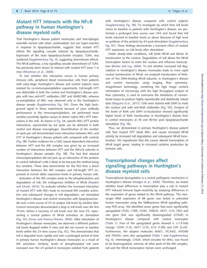
Figure 5 HTT interacts directly with the NFkB pathway, which is dysregulated in Huntington’s disease (HD). (A) HTT interacts directly
with IKK, as shown by proximity ligation assays. Monocyte-derived macrophages were differentiated on glass cover-slips and stained for
HTT and IKKg or IKKa/b before antibodies binding in close proximity were visualized using proximity ligation assay (PLA) probes as red
spots, shown here. Cells stained with a single primary antibody did not result in red spots. (B) Quantification of the number of spots per
cell using the Volocity software shows increased binding between IKKg and HTT in Huntington’s disease compared with control cells
(P = 0.06). Binding of HTT to the a and b subunit of IKK showed a similar, but smaller trend (P = 0.1). Two-tailed unpaired t-test used for
statistical analysis. (C) In control cells lipopolysaccharide (LPS)-induced degradation of IkB occurred within 15 min of stimulation and
recovered within 2 h, whereas Huntington’s disease monocytes demonstrated a more rapid loss of IkB and no recovery of the protein.
Shown is an example blot of samples from one control subject and one patient with Huntington’s disease. (D) Translocation of the NFkB
transcription factor RelA to the nucleus after lipopolysaccharide stimulation was measured using imaging flow cytometry; example images
are shown here. In untranslocated cells the green RelA staining surrounds the nuclear DAPI staining, whereas in cells demonstrating
translocation of RelA the colours merge. (E) Increased RelA translocation into the nucleus following lipopolysaccharide stimulation was
observed in Huntington’s disease monocytes (n = 7) compared to controls (n = 8). n = individual biological repeats. Data shown as mean
concentrations � SEM, two-way ANOVA with Bonferroni post-tests, *P5 0.05; **P5 0.01. All experiments were repeated at least twice
with the same results.
Reversing immune dysfunction in HD Brain 2014: Page 9 of 15 | 9
at Queen M
ary, University of L
ondon on February 11, 2014http://brain.oxfordjournals.org/
Dow
nloaded from
The array also screened intracellular signalling pathways closely
linked to the NFkB pathway, such as MAPK and PI3K/AKT path-
ways. Increased AKT protein levels have been found in
Huntington’s disease patient lymphoblasts (Colin et al., 2005)
and AKT1 is one of the genes upregulated in Huntington’s disease
monocytes (fold change = 1.5, P = 0.031). Moreover, the genes
composing the transcription factor AP-1, JUN and FOS, are also
upregulated in Huntington’s disease monocytes (Table 1 and
Supplementary Fig. 6). Therefore, both of these pathways may
also contribute to the increased immune response observed after
stimulation of Huntington’s disease monocytes.
To validate our findings, six candidate genes chosen on the basis
of array fold changes and their importance within the NFkB sig-
nalling cascade (CD40, AKT1, IRAK1, JUN, IL6 and IL10) were
quantified by quantitative PCR using different primer sets and
cells from a different patient cohort. The relative changes in
gene expression when comparing Huntington’s disease and con-
trol human monocytes matched our previous findings for all six
genes (Supplementary Fig. 7). Expression levels for CD40, IRAK1
and IL10 were significantly increased in Huntington’s disease com-
pared with control monocytes, whereas expression changes in
AKT1, JUN and IL6 demonstrated an upward trend, not reaching
statistical significance because of large interindividual differences.
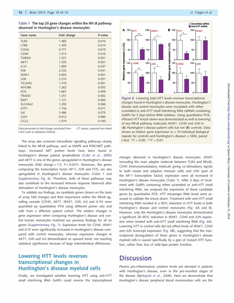
Lowering HTT levels reversestranscriptional changes inHuntington’s disease myeloid cellsFinally, we investigated whether lowering HTT using anti-HTT
small interfering RNA GeRPs could reverse the transcriptional
changes observed in Huntington’s disease monocytes. IRAK1
(encoding the main adapter molecule between TLR4 and NFkB),
CD40 (immunomodulatory molecule giving co-stimulatory signals
to both innate and adaptive immune cells) and JUN (part of
the AP-1 transcription factor) expression were all increased in
Huntington’s disease monocytes (Table 1). After 3 days of treat-
ment with GeRPs containing either scrambled or anti-HTT small
interfering RNA, we analysed the expression of these candidate
genes by quantitative PCR. HTT messenger RNA levels were as-
sessed to validate the knock-down. Treatment with anti-HTT small
interfering RNA resulted in a 50% reduction in HTT levels in both
Huntington’s disease and control monocytes (Fig. 6A and B).
However, only the Huntington’s disease monocytes demonstrated
a significant 20–30% reduction in IRAK1, CD40 and JUN expres-
sion when treated with anti-HTT small interfering RNA (Fig. 6A).
Lowering HTT in control cells did not affect levels of IRAK1, CD40
and JUN transcript expression (Fig. 6B), suggesting that the tran-
scriptional dysregulation of these genes in Huntington’s disease
myeloid cells is caused specifically by a gain of mutant HTT func-
tion, rather than loss of wild-type protein function.
DiscussionPlasma pro-inflammatory cytokine levels are elevated in patients
with Huntington’s disease, even in the pre-manifest stages of
the disease (Bjorkqvist et al., 2008). Here we demonstrate that
Huntington’s disease peripheral blood mononuclear cells are the
Figure 6 Lowering total HTT levels reverses transcriptional
changes found in Huntington’s disease monocytes. Huntington’s
disease and control monocytes were incubated with either
scrambled or anti-HTT small interfering RNA (siRNA) containing
GeRPs for 3 days before RNA isolation. Using quantitative PCR,
efficient HTT knock-down was demonstrated as well as lowering
of key NFkB pathway molecules IRAK1, CD40 and JUN in
(A) Huntington’s disease patient cells but not (B) controls. Data
shown as relative gene expression (n = 10 individual biological
repeats for controls and Huntington’s disease) � SEM, paired
t-test. *P50.05; **P5 0.01.
Table 1 The top 20 gene changes within the NFkB pathwayobserved in Huntington’s disease monocytes
Gene name Fold change P-value
TLR2 1.483 0.010
LTBR 1.355 0.014
CHUK 0.771 0.015
CD40 1.513 0.016
TMED4 1.227 0.021
AKT1 1.535 0.031
IL10 1.850 0.037
F2R 2.232 0.041
IRAK1 2.004 0.051
JUN 2.215 0.051
TICAM2 1.319 0.051
MYD88 1.262 0.053
FOS 1.867 0.059
TRADD 1.251 0.062
RAF1 1.121 0.065
SLC44A2 1.292 0.066
ATF1 1.156 0.071
IL6 2.486 0.075
GJA1 0.612 0.080
CCL2 1.574 0.160
Data presented as fold change calculated from ��-CT values, unpaired two-tailedt-test used as statistical method.
10 | Brain 2014: Page 10 of 15 U. Trager et al.
at Queen M
ary, University of L
ondon on February 11, 2014http://brain.oxfordjournals.org/
Dow
nloaded from
likely source of the increased pro-inflammatory cytokines, as both
monocytes and macrophages isolated from patients with
Huntington’s disease and stimulated with lipopolysaccharide pro-
duce significantly more IL-6, IL-8 and TNFa compared with control
subjects. Supporting our previous finding that plasma cytokine
levels are already elevated in pre-manifest subjects with a mean
of 16 years to clinical onset (Bjorkqvist et al., 2008), myeloid cells
isolated from patients with pre-manifest Huntington’s disease
were hyper-reactive to the same degree as cells isolated from
late-stage disease patients. Cytokine production seems CAG
repeat length independent and suggests an early deficit that is
already present many years before disease onset, which may be
a marker of when to intervene with potential modulatory thera-
pies. Modulating HTT expression by overexpression of mutant
HTT exon 1 in a monocyte-like cell line and lowering HTT levels
in primary human monocytes/macrophages demonstrated that this
hyper-reactive phenotype is because of a cell-intrinsic effect of
mutant HTT expression and not non-cell autonomous secondary
factors.
Importantly, we have been able to show that lowering total
HTT levels partially rescues this hyper-reactive phenotype, with a
reversal of both elevated cytokine production and transcriptional
changes observed in human Huntington’s disease myeloid cells ex
vivo. This is the first report showing that lowering HTT in cells
freshly isolated from patients with Huntington’s disease can re-
verse cellular dysfunction caused by mutant HTT expression—an
important first demonstration of the reversibility of cellular dys-
function after HTT-lowering in human tissue. HTT-lowering was
achieved using a novel phagocytosis dependent approach, in
which small interfering RNAs are packaged into glucan particles
isolated from yeast (Aouadi et al., 2009). This study is the first to
use this technique in primary human macrophages and demon-
strates that a 90% transfection rate can be achieved, much higher
than the 10–20% transfection rate achieved by traditional meth-
ods such as lentiviral transduction.
Our findings, that lowering total HTT levels by only 50% in
primary human Huntington’s disease macrophages and monocytes
can reverse the increased cytokine production and transcriptional
changes, respectively, validate the potential of HTT-lowering ther-
apy as well as the possibility of using peripheral cells to test small
interfering RNA efficiency, safety and efficacy. Interestingly, cyto-
kine release was also decreased in control macrophages treated
with anti-HTT small interfering RNA, indicating either that HTT
regulates cytokine production in a CAG dependent manner or
that wild-type HTT influences cytokine production in parallel
with mutant HTT. Wild-type HTT has been shown to play a role
in both actin remodelling (Munsie et al., 2011; Kwan et al.,
2012b) and microtubule-mediated transport (Gauthier et al.,
2004). As both processes are needed for the trafficking of cyto-
kines to the cell surface membrane for release (Lacy and Stow,
2011), a reduction of wild-type HTT levels might exert a loss of
function by hindering normal actin and microtubule remodelling
causing changes in cytokine release. A future study using allele-
specific silencing of mutant but not wild-type HTT will help deter-
mine the exact contributions that loss of wild-type HTT and gain
of mutant HTT function have on the myeloid cell dysfunction in
Huntington’s disease.
The NFkB pathway has been previously implicated in
Huntington’s disease in murine studies (Khoshnan et al., 2004;
Thompson et al., 2009; Steffan, 2010; Hsiao et al., 2013).
Investigating this pathway, we found that HTT binds the IKK com-
plex in a CAG repeat length dependent manner. Testing HTT
binding to both IKKg and IKKa/b subunits, we detected a stronger
interaction between HTT and IKKg, suggesting this subunit as the
direct interaction partner. IKKg is the regulatory subunit of the IKK
trimer, consisting of one regulatory (g) and two kinase subunits
(a and b), and is a critical component without which cells are
unresponsive to all upstream stimuli (Israel, 2000). During signal
transduction, polyubiquitin chains form the scaffold on which
TAK1/TAB2/3 and IKKa/b/g complexes are formed to induce
TAK1 dependent activation of IKKb (Miyamoto, 2011). In agree-
ment with previously described findings using non-primary human
cell model systems (Khoshnan and Patterson, 2011), we have
shown in primary human cells that HTT can function as an alter-
native scaffold for the NFkB pathway. The CAG repeat dependent
binding of HTT to IKKg is associated with increased IKK complex
formation and downstream signal transduction following lipopoly-
saccharide stimulation in Huntington’s disease myeloid cells
(Fig. 7). Previously, this interaction has only been observed in
cultured tumour cells (Khoshnan et al., 2004) or mouse models
expressing exon 1 mutant HTT (Khoshnan et al., 2004; Hsiao
et al., 2013). Here we demonstrate that the interaction also
takes place in primary human ex vivo cells expressing the full-
length protein at normal allelic expression levels. That mutant
HTT exon 1 fragments have previously been shown to bind
IKKg is consistent, however, with our finding that an N-terminal
human exon 1 mutant HTT fragment can induce elevated cytokine
production in a histiocytic cell line and our recent report demon-
strating increasing N-terminal fragmentation of mutant HTT in
human myeloid cells as the disease progresses (Weiss et al., 2012).
Interestingly, a recent study showed that activating the immune
modulator cannabinoid receptor 2, which is thought to dampen
NFkB signalling (Rajesh et al., 2007), reduces increased serum IL-6
levels while extending life span and reducing motor deficits in
Huntington’s disease mouse models (Bouchard et al., 2012).
Our finding that mutant HTT alters the NFkB pathway in human
Huntington’s disease monocytes is likely to be relevant to other
cell types and tissues, including those of the CNS. We previously
showed that hyper-reactivity of Huntington’s disease peripheral
myeloid cells is mirrored in microglia (Bjorkqvist et al., 2008).
The NFkB pathway is present and active in both neurons and
glial cells (O’Neill and Kaltschmidt, 1997). Pharmacological inhib-
ition of NFkB impairs memory and learning (Mattson and Meffert,
2006) and NFkB pathway activation is critical for neuronal survival
and neurite outgrowth (Teng and Tang, 2010). Increased levels of
NFkB activity have been shown in both Alzheimer’s disease
(Kaltschmidt et al., 1997) and Parkinson’s disease (Hunot et al.,
1997). Blocking NFkB function in mutant HTT exon 1 expressing
PC12 cells leads to reduced mutant HTT toxicity, implying that the
NFkB pathway contributes to neurotoxicity in Huntington’s disease
(Khoshnan et al., 2004). Indeed, a recent study showing that
mutant HTT enhances NFkB-mediated inflammation in astrocytes
to cause toxicity in the brain of Huntington’s disease mice
Reversing immune dysfunction in HD Brain 2014: Page 11 of 15 | 11
at Queen M
ary, University of L
ondon on February 11, 2014http://brain.oxfordjournals.org/
Dow
nloaded from
underlines the potential importance of NFkB in non-neuronal cells
during neurodegeneration (Hsiao et al., 2013).
Given the manifold roles NFkB signalling plays in the different
cell types, inhibiting the pathway to lower hyper-reactive immune
function in Huntington’s disease may also affect other cell types.
For example, compounds that target NFkB activity need to be
closely evaluated as to whether they cross the blood–brain barrier
and with regard to negative effects on synaptic activity and plas-
ticity. However, drugs that target this pathway will not necessarily
have negative effects. Laqinimod for example, an immunomodu-
latory compound inhibiting NFkB activity (Bruck et al., 2012), was
well tolerated and showed decreased progression rates in patients
with multiple sclerosis in clinical trials (Comi et al., 2012).
Furthermore, targeting the NFkB pathway further downstream,
for example at the level of cytokine secretion is also a possible
therapeutic target. In a Huntington’s disease mouse model, per-
ipherally administrated anti-IL-6 antibody treatment has shown
improvement of disease progression (Bouchard et al., 2012),
while perispinal administration of a TNFa inhibitor improves dis-
ease in patients with Alzheimer’s disease (Tobinick et al., 2006),
clearly demonstrating the positive effect of immunomodulatory
therapy for neurodegeneration.
We have identified gene expression changes in key molecules
involved in immune signalling in Huntington’s disease patients’
monocytes. Several adapter proteins downstream of TLR4, such as
IRAK1, TICAM2 and MyD88 were found to be slightly elevated in
native Huntington’s disease patients’ monocytes. A cumulative
baseline increase in expression of several of these adapter proteins
may lead to increased signal transduction from TLR4 to NFkB, fur-
ther increasing NFkB pathway dysregulation. Another gene found
to be upregulated in Huntington’s disease monocytes was CD40
which, together with its ligand CD154, mainly expressed on
T cells, regulates the immune response on several levels.
Monocytes are activated leading to upregulated cytokine production
and antigen presentation, and priming of the adaptive immune
system (Grewal and Flavell, 1998). This points to further functional
abnormalities in the immune system of patients with Huntington’s
disease, suggesting a possible deficit in the communication between
antigen presenting cells and the adaptive immune system.
Furthermore, CD40 mediates cell adhesion needed for leucocyte
trafficking (Alderson et al., 1993). Given recent studies showing
defective migration in Huntington’s disease because of defective
actin remodelling (Kwan et al., 2012b), the increase in CD40 ex-
pression could be a compensatory response of immune cells to
counteract their decreased migrative ability. FOS and JUN, subunits
of the AP-1 transcription factor, were also upregulated in primary
human myeloid Huntington’s disease cells. Interestingly, FOS and
JUN levels have been found to be increased in brain of patients with
Alzheimer’s disease (Anderson et al., 1994), and the MAP kinase
needed for JUN activation, JNK, is elevated and involved in neuro-
toxicity in Huntington’s disease mouse (Fan et al., 2012) and rat
models (Perrin et al., 2009). Thus, we cannot exclude that dysre-
gulation in these signalling pathways may also contribute to the
Huntington’s disease immune phenotype.
In addition to the pathways identified in this study, we cannot
exclude other previously described mechanisms, which may contrib-
ute to the transcriptional dysregulation we found in primary human
Huntington’s disease myeloid cells. For example, mutant HTT is
known to bind and thereby deplete transcription factors such as
CBP and p53 from their normal location causing changes in the
genes they control (Steffan et al., 2000; Nucifora et al., 2001).
Furthermore, HTT may alter DNA conformation upon direct bind-
ing, affecting transcription factors binding to their promoter regions
(Benn et al., 2008).
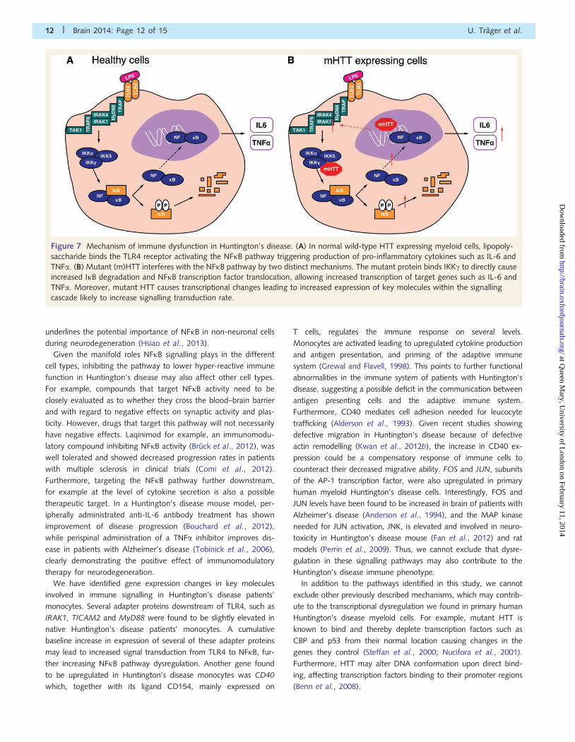
Figure 7 Mechanism of immune dysfunction in Huntington’s disease. (A) In normal wild-type HTT expressing myeloid cells, lipopoly-
saccharide binds the TLR4 receptor activating the NFkB pathway triggering production of pro-inflammatory cytokines such as IL-6 and
TNFa. (B) Mutant (m)HTT interferes with the NFkB pathway by two distinct mechanisms. The mutant protein binds IKKg to directly cause
increased IkB degradation and NFkB transcription factor translocation, allowing increased transcription of target genes such as IL-6 and
TNFa. Moreover, mutant HTT causes transcriptional changes leading to increased expression of key molecules within the signalling
cascade likely to increase signalling transduction rate.
12 | Brain 2014: Page 12 of 15 U. Trager et al.
at Queen M
ary, University of L
ondon on February 11, 2014http://brain.oxfordjournals.org/
Dow
nloaded from
This study demonstrates that the cellular dysregulation observed
in hyper-reactive immune cells in Huntington’s disease can be
reversed by HTT-lowering and represents the first demonstration
of phenotypic reversibility on HTT-lowering in primary human
cells in Huntington’s disease. It also identifies the underlying intra-
cellular mechanisms of immune dysfunction in human cells in
Huntington’s disease. This is important as the immune system
has been shown to be a powerful modifier of Huntington’s disease
pathogenesis in various mouse models (Zwilling et al., 2011;
Bouchard et al., 2012; Kwan et al., 2012a). There is currently a
search for genetic and environmental modifiers of Huntington’s
disease as the CAG repeat expansion only explains 50–70% of
variance in age of onset, and its role in modulating disease pro-
gression is variable (Andrew et al., 1993; Brinkman et al., 1997).
The remainder of the variance is likely due to environmental and
other genetic factors (Wexler et al., 2004). The immune system
may be a powerful modifier of Huntington’s disease age of onset
and progression, with an interaction of both genetic and environ-
mental factors. This has already been shown to be the case in
large genome-wide association studies in Alzheimer’s disease
where several key genes involved in the innate immune system
were shown to increase susceptibility to developing Alzheimer’s
disease (Harold et al., 2009; Lambert et al., 2009; Guerreiro
et al., 2013).
Our novel method of small interfering RNA delivery has poten-
tial therapeutic relevance to Huntington’s disease and other dis-
eases where immune dysregulation is a feature. Glucan particles
are a versatile phagocytic cell targeted delivery system and have
been administered by oral, subcutaneous, intraperitoneal and
intravenous routes in mice and rats. In our future studies, we
plan to administer GeRPs loaded with anti-HTT small interfering
RNA to reverse the inflammatory phenotype through intrathecal
administration to directly target phagocytic microglial cells and
infiltrating monocyte/macrophages, and through intravenous ad-
ministration to target circulating monocytes and peripheral mono-
nuclear cells, a precursor pool for inflammatory cells trafficking
into inflamed brain sites.
Finally, our work also suggests a potential new therapeutic
target for Huntington’s disease through modulating NFkB activa-
tion and downstream targets. The muscle wasting, weight loss and
depression that occurs in Huntington’s disease (van der Burg et al.,
2009) may be related to increased peripheral cytokine levels.
Therefore, modulating the immune system may have beneficial
effects in both the CNS and the periphery. Indeed, a peripherally
administered anti-inflammatory, anti-IL-6 antibody treatment in
R6/2 mice has already been show to improve both weight
loss and disease progression (Bouchard et al., 2012). This work
therefore has implications for both understanding the role of the
innate immune system as a modifier of neurodegeneration and
modulation of the immune system as a possible therapeutic in
Huntington’s disease.
AcknowledgementsWe thank the patients and control subjects who donated samples,
and the staff of the multidisciplinary Huntington’s disease clinic in
London; Dr Peter Klohn for his help with fluorescence-activated
cell sorting; P.J. Chana for his assistance with the imaging flow
cytometry; Dr Christian Landles for his advice on HTT immuno-
precipitations; Dr Edward Wild for his help with editing the manu-
script and Ray Young for his help with graphics.
FundingThis study was supported financially by UCL/UCLH Biomedical
Research Centre (PhD studentship to UT), Medical Research
Council, CHDI Foundation, EU FP7 grant (Paddington consor-
tium), the UK Dementia and Neurodegenerative Diseases
Network (DeNDRoN) and supported by the National Institute for
Health Research University College London Hospitals Biomedical
Research Centre. N.A. was supported by NHI: NS 38194 and the
CHDI foundation. The authors acknowledge financial support from
the Department of Health via the National Institute for Health
Research (NIHR) comprehensive Biomedical Research Centre
award to Guy’s & St Thomas’ NHS Foundation Trust in partnership
with King’s College London and King’s College Hospital NHS
Foundation Trust.
Supplementary materialSupplementary material is available at Brain online.
ReferencesAjami B, Bennett JL, Krieger C, Tetzlaff W, Rossi FM. Local self-renewal
can sustain CNS microglia maintenance and function throughout adult
life. Nat Neurosci 2007; 10: 1538–43.
Alciato F, Sainaghi PP, Sola D, Castello L, Avanzi GC. TNF-alpha, IL-6,
and IL-1 expression is inhibited by GAS6 in monocytes/macrophages.
J Leukoc Biol 2010; 87: 869–75.
Alderson MR, Armitage RJ, Tough TW, Strockbine L, Fanslow WC,
Spriggs MK. CD40 expression by human monocytes: regulation by
cytokines and activation of monocytes by the ligand for CD40.
J Exp Med 1993; 178: 669–74.Anderson AJ, Cummings BJ, Cotman CW. Increased immunoreactivity
for Jun- and Fos-related proteins in Alzheimer’s disease: association
with pathology. Exp Neurol 1994; 125: 286–95.Andrew SE, Goldberg YP, Kremer B, Telenius H, Theilmann J, Adam S,
et al. The relationship between trinucleotide (CAG) repeat length and
clinical features of Huntington’s disease. Nat Genet 1993; 4: 398–403.
Aouadi M, Tesz GJ, Nicoloro SM, Wang M, Chouinard M, Soto E, et al.
Orally delivered siRNA targeting macrophage Map4k4 suppresses sys-
temic inflammation. Nature 2009; 458: 1180–4.
Baldo B, Paganetti P, Grueninger S, Marcellin D, Kaltenbach LS, Lo DC,
et al. TR-FRET-based duplex immunoassay reveals an inverse correl-
ation of soluble and aggregated mutant huntingtin in huntington’s
disease. Chem Biol 2012; 19: 264–75.
Beinke S, Ley SC. Functions of NF-kappaB1 and NF-kappaB2 in immune
cell biology. Biochem J 2004; 382 (Pt 2): 393–409.
Benn CL, Sun T, Sadri-Vakili G, McFarland KN, DiRocco DP, Yohrling GJ,
et al. Huntingtin modulates transcription, occupies gene promoters
in vivo, and binds directly to DNA in a polyglutamine-dependent
manner. J Neurosci 2008; 28: 10720–33.
Bjorkqvist M, Wild EJ, Thiele J, Silvestroni A, Andre R, Lahiri N, et al. A
novel pathogenic pathway of immune activation detectable before
clinical onset in Huntington’s disease. J Exp Med 2008; 205: 1869–77.
Reversing immune dysfunction in HD Brain 2014: Page 13 of 15 | 13
at Queen M
ary, University of L
ondon on February 11, 2014http://brain.oxfordjournals.org/
Dow
nloaded from
Bouchard J, Truong J, Bouchard K, Dunkelberger D, Desrayaud S,
Moussaoui S, et al. Cannabinoid receptor 2 signaling in peripheral
immune cells modulates disease onset and severity in mouse models
of Huntington’s disease. J Neurosci 2012; 32: 18259–68.
Brinkman RR, Mezei MM, Theilmann J, Almqvist E, Hayden MR. The
likelihood of being affected with Huntington disease by a particular
age, for a specific CAG size. Am J Hum Genet 1997; 60: 1202–10.Bruck W, Pfortner R, Pham T, Zhang J, Hayardeny L, Piryatinsky V, et al.
Reduced astrocytic NF-kB activation by laquinimod protects from
cuprizone-induced demyelination. Acta Neuropathol 2012; 124:
411–24.
Colin E, Regulier E, Perrin V, Durr A, Brice A, Aebischer P, et al. Akt is
altered in an animal model of Huntington’s disease and in patients. Eur
J Neurosci 2005; 21: 1478–88.
Comi G, Jeffery D, Kappos L, Montalban X, Boyko A, Rocca MA, et al.
Placebo-controlled trial of oral laquinimod for multiple sclerosis. N Engl
J Med 2012; 366: 1000–9.
Dalrymple A, Wild EJ, Joubert R, Sathasivam K, Bjorkqvist M, Petersen A,
et al. Proteomic profiling of plasma in Huntington’s disease reveals
neuroinflammatory activation and biomarker candidates. J Proteome
Res 2007; 6: 2833–40.DiFiglia M, Sena-Esteves M, Chase K, Sapp E, Pfister E, Sass M, et al.
Therapeutic silencing of mutant huntingtin with siRNA attenuates stri-
atal and cortical neuropathology and behavioral deficits. Proc Natl
Acad Sci USA 2007; 104: 17204–9.
Fan J, Gladding CM, Wang L, Zhang LY, Kaufman AM, Milnerwood AJ,
et al. P38 MAPK is involved in enhanced NMDA receptor-dependent
excitotoxicity in YAC transgenic mouse model of Huntington disease.
Neurobiol Dis 2012; 45: 999–1009.Gauthier LR, Charrin BC, Borrell-Pages M, Dompierre JP, Rangone H,
Cordelieres FP, et al. Huntingtin controls neurotrophic support and
survival of neurons by enhancing BDNF vesicular transport along
microtubules. Cell 2004; 118: 127–38.
Gordon S, Taylor PR. Monocyte and macrophage heterogeneity. Nat Rev
Immunol 2005; 5: 953–64.
Grewal IS, Flavell RA. CD40 and CD154 in cell-mediated immunity.
Annu Rev Immunol 1998; 16: 111–35.
Gross S, Piwnica-Worms D. Real-time imaging of ligand-induced IKK
activation in intact cells and in living mice. Nat Methods 2005; 2:
607–14.
Guerreiro R, Wojtas A, Bras J, Carrasquillo M, Rogaeva E, Majounie E,
et al. TREM2 variants in Alzheimer’s disease. N Engl J Med 2013; 368:
117–27.
Harold D, Abraham R, Hollingworth P, Sims R, Gerrish A, Hamshere ML,
et al. Genome-wide association study identifies variants at CLU and
PICALM associated with Alzheimer’s disease. Nat Genet 2009; 41:
1088–93.Hayden MS, Ghosh S. NF-kB, the first quarter-century: remarkable pro-
gress and outstanding questions. Genes Dev 2012; 26: 203–34.Hodges A, Strand AD, Aragaki AK, Kuhn A, Sengstag T, Hughes G, et al.
Regional and cellular gene expression changes in human Huntington’s
disease brain. Hum Mol Genet 2006; 15: 965–77.
Hsiao HY, Chen YC, Chen HM, Tu PH, Chern Y. A critical role of
astrocyte-mediated nuclear factor-kB-dependent inflammation in
Huntington’s disease. Hum Mol Genet 2013; 22: 1826–42.
Hunot S, Brugg B, Ricard D, Michel PP, Muriel MP, Ruberg M, et al.
Nuclear translocation of NF-kappaB is increased in dopaminergic neu-
rons of patients with parkinson disease. Proc Natl Acad Sci USA 1997;
94: 7531–6.
Israel A. The IKK complex: an integrator of all signals that activate
NF-kappaB? Trends Cell Biol 2000; 10: 129–33.
Kaltschmidt B, Uherek M, Volk B, Baeuerle PA, Kaltschmidt C.
Transcription factor NF-kappaB is activated in primary neurons by
amyloid beta peptides and in neurons surrounding early plaques
from patients with Alzheimer disease. Proc Natl Acad Sci USA 1997;
94: 2642–7.
Khoshnan A, Ko J, Watkin EE, Paige LA, Reinhart PH, Patterson PH.
Activation of the IkappaB kinase complex and nuclear factor-kappaB
contributes to mutant huntingtin neurotoxicity. J Neurosci 2004; 24:
7999–8008.
Khoshnan A, Patterson PH. The role of IkB kinase complex in the neuro-
biology of Huntington’s disease. Neurobiol Dis 2011; 43: 305–11.
Kwan W, Magnusson A, Chou A, Adame A, Carson MJ, Kohsaka S,
et al. Bone marrow transplantation confers modest benefits in mouse
models of Huntington’s disease. J Neurosci 2012a; 32: 133–42.
Kwan W, Trager U, Davalos D, Chou A, Bouchard J, Andre R, et al.
Mutant huntingtin impairs immune cell migration in Huntington dis-
ease. J Clin Invest 2012b; 122: 4737–47.
Lacy P, Stow JL. Cytokine release from innate immune cells: associ-
ation with diverse membrane trafficking pathways. Blood 2011; 118:
9–18.
Lambert JC, Heath S, Even G, Campion D, Sleegers K, Hiltunen M, et al.
Genome-wide association study identifies variants at CLU and CR1
associated with Alzheimer’s disease. Nat Genet 2009; 41: 1094–9.Li SH, Schilling G, Young WS, Li XJ, Margolis RL, Stine OC, et al.
Huntington’s disease gene (IT15) is widely expressed in human and
rat tissues. Neuron 1993; 11: 985–93.
Maguire O, Collins C, O’Loughlin K, Miecznikowski J, Minderman H.
Quantifying nuclear p65 as a parameter for NF-kB activation: correl-
ation between ImageStream cytometry, microscopy, and Western blot.
Cytometry A 2011; 79: 461–9.Mattson MP, Meffert MK. Roles for NF-kappaB in nerve cell survival,
plasticity, and disease. Cell Death Differ 2006; 13: 852–60.Miyamoto S. Nuclear initiated NF-kB signaling: NEMO and ATM take
center stage. Cell Res 2011; 21: 116–30.
Munsie L, Caron N, Atwal RS, Marsden I, Wild EJ, Bamburg JR, et al.
Mutant huntingtin causes defective actin remodeling during stress:
defining a new role for transglutaminase 2 in neurodegenerative dis-
ease. Hum Mol Genet 2011; 20: 1937–51.Nucifora FC, Sasaki M, Peters MF, Huang H, Cooper JK, Yamada M,
et al. Interference by huntingtin and atrophin-1 with cbp-mediated
transcription leading to cellular toxicity. Science 2001; 291: 2423–8.
O’Neill LA, Kaltschmidt C. NF-kappa B: a crucial transcription factor for
glial and neuronal cell function. Trends Neurosci 1997; 20: 252–8.
Perrin V, Dufour N, Raoul C, Hassig R, Brouillet E, Aebischer P, et al.
Implication of the JNK pathway in a rat model of Huntington’s disease.
Exp Neurol 2009; 215: 191–200.
Rajesh M, Mukhopadhyay P, Batkai S, Hasko G, Liaudet L, Huffman JW,
et al. CB2-receptor stimulation attenuates TNF-alpha-induced human
endothelial cell activation, transendothelial migration of monocytes,
and monocyte-endothelial adhesion. Am J Physiol Heart Circ Physiol
2007; 293: H2210–8.
Ransohoff RM, Perry VH. Microglial physiology: unique stimuli, specia-
lized responses. Annu Rev Immunol 2009; 27: 119–45.
Sah DW, Aronin N. Oligonucleotide therapeutic approaches for
Huntington disease. J Clin Invest 2011; 121: 500–7.
Sapp E, Kegel KB, Aronin N, Hashikawa T, Uchiyama Y, Tohyama K,
et al. Early and progressive accumulation of reactive microglia in the
Huntington disease brain. J Neuropathol Exp Neurol 2001; 60:
161–72.
Simard AR, Rivest S. Bone marrow stem cells have the ability to populate
the entire central nervous system into fully differentiated parenchymal
microglia. FASEB J 2004; 18: 998–1000.Soulet D, Cicchetti F. The role of immunity in Huntington’s disease. Mol
Psychiatry 2011; 16: 889–902.Steffan JS. Does Huntingtin play a role in selective macroautophagy? Cell
Cycle 2010; 9: 3401–13.Steffan JS, Kazantsev A, Spasic-Boskovic O, Greenwald M, Zhu YZ,
Gohler H, et al. The Huntington’s disease protein interacts with p53
and CREB-binding protein and represses transcription. Proc Natl Acad
Sci USA 2000; 97: 6763–8.
Sundstrom C, Nilsson K. Establishment and characterization of a human
histiocytic lymphoma cell line (U-937). Int J Cancer 1976; 17: 565–77.
Tai YF, Pavese N, Gerhard A, Tabrizi SJ, Barker RA, Brooks DJ, et al.
Microglial activation in presymptomatic Huntington’s disease gene
carriers. Brain 2007; 130 (Pt 7): 1759–66.
14 | Brain 2014: Page 14 of 15 U. Trager et al.
at Queen M
ary, University of L
ondon on February 11, 2014http://brain.oxfordjournals.org/
Dow
nloaded from
Teng FY, Tang BL. NF-kappaB signaling in neurite growth and neuronalsurvival. Rev Neurosci 2010; 21: 299–313.
The Huntington’s Disease Collaborative Research Group. A novel gene
containing a trinucleotide repeat that is expanded and unstable on
Huntington’s disease chromosomes. The Huntington’s DiseaseCollaborative Research Group. Cell 1993; 72: 971–83.
Thompson LM, Aiken CT, Kaltenbach LS, Agrawal N, Illes K,
Khoshnan A, et al. IKK phosphorylates Huntingtin and targets it for
degradation by the proteasome and lysosome. J Cell Biol 2009; 187:1083–99.
Tobinick E, Gross H, Weinberger A, Cohen H. TNF-alpha modulation for
treatment of Alzheimer’s disease: a 6-month pilot study. MedGenMed2006; 8: 25.
van der Burg JM, Bjorkqvist M, Brundin P. Beyond the brain: widespread
pathology in Huntington’s disease. Lancet Neurol 2009; 8: 765–74.
Weiss A, Trager U, Wild EJ, Grueninger S, Farmer R, Landles C, et al.
Mutant huntingtin fragmentation in immune cells tracks Huntington’s
disease progression. J Clin Invest 2012; 122: 3731–6.
Wexler NS, Lorimer J, Porter J, Gomez F, Moskowitz C, Shackell E, et al.
Venezuelan kindreds reveal that genetic and environmental factors
modulate Huntington’s disease age of onset. Proc Natl Acad Sci USA
2004; 101: 3498–503.
Wild E, Magnusson A, Lahiri N, Krus U, Orth M, Tabrizi SJ, et al.
Abnormal peripheral chemokine profile in Huntington’s disease. PLoS
Curr 2011; 3: RRN1231.
Zwilling D, Huang SY, Sathyasaikumar KV, Notarangelo FM, Guidetti P,
Wu HQ, et al. Kynurenine 3-monooxygenase inhibition in blood ameli-
orates neurodegeneration. Cell 2011; 145: 863–74.
Reversing immune dysfunction in HD Brain 2014: Page 15 of 15 | 15
at Queen M
ary, University of L
ondon on February 11, 2014http://brain.oxfordjournals.org/
Dow
nloaded from
Related Documents