‘ ‘ How I do’ CMR in How I do’ CMR in DCM DCM Dr Sanjay Prasad, Royal Brompton Dr Sanjay Prasad, Royal Brompton Hospital Hospital London, UK. For SCMR August 2006 London, UK. For SCMR August 2006 This presentation is posted for members of scmr as This presentation is posted for members of scmr as an educational guide – it represents the views and an educational guide – it represents the views and practices of the author, and not necessarily those of practices of the author, and not necessarily those of SCMR. SCMR. [email protected]

‘How I do’ CMR in DCM Dr Sanjay Prasad, Royal Brompton Hospital London, UK. For SCMR August 2006 This presentation is posted for members of scmr as an.
Dec 14, 2015
Welcome message from author
This document is posted to help you gain knowledge. Please leave a comment to let me know what you think about it! Share it to your friends and learn new things together.
Transcript
‘‘How I do’ CMR in How I do’ CMR in DCMDCM
Dr Sanjay Prasad, Royal Brompton HospitalDr Sanjay Prasad, Royal Brompton Hospital
London, UK. For SCMR August 2006London, UK. For SCMR August 2006
This presentation is posted for members of This presentation is posted for members of scmr as an educational guide – it represents scmr as an educational guide – it represents
the views and practices of the author, and not the views and practices of the author, and not necessarily those of SCMR. necessarily those of SCMR.
CineCine
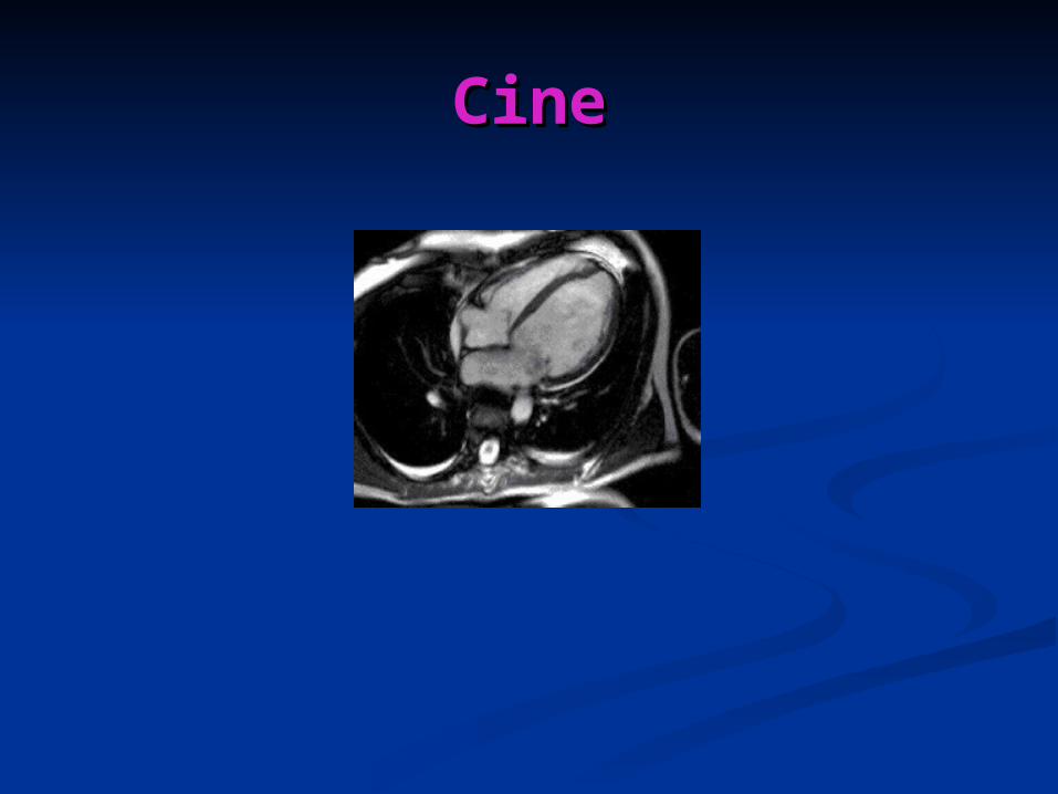
Dilated CardiomyopathyDilated Cardiomyopathy
Unexplained dilatation and impaired Unexplained dilatation and impaired contractile performance of the left ventricle in contractile performance of the left ventricle in the absence of significant coronary artery the absence of significant coronary artery diseasedisease
Prevalence – 38/ 100 000Prevalence – 38/ 100 000
Variable onsetVariable onset
A familial basis is seen in around 15% of A familial basis is seen in around 15% of patientspatients
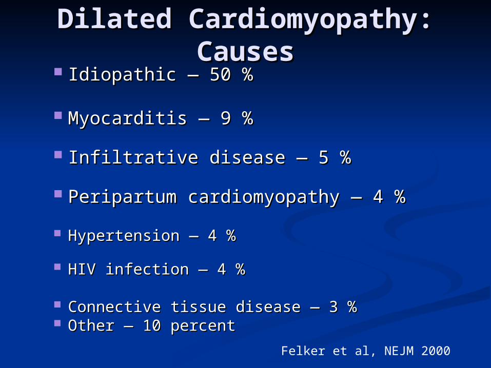
Dilated Cardiomyopathy: Dilated Cardiomyopathy: CausesCauses
Idiopathic — 50 %Idiopathic — 50 %
Myocarditis — 9 %Myocarditis — 9 %
Infiltrative disease — 5 %Infiltrative disease — 5 %
Peripartum cardiomyopathy — 4 %Peripartum cardiomyopathy — 4 %
Hypertension — 4 %Hypertension — 4 %
HIV infection — 4 %HIV infection — 4 %
Connective tissue disease — 3 %Connective tissue disease — 3 % Other — 10 percentOther — 10 percent
Felker et al, NEJM 2000
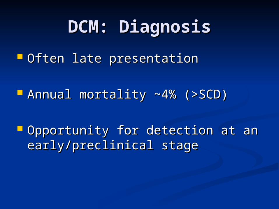
DCM: DiagnosisDCM: Diagnosis
Often late presentation Often late presentation
Annual mortality ~4% (>SCD)Annual mortality ~4% (>SCD)
Opportunity for detection at an Opportunity for detection at an early/preclinical stageearly/preclinical stage
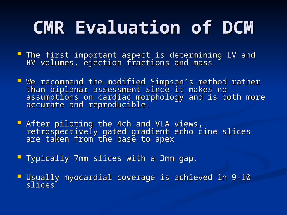
CMR Evaluation of DCMCMR Evaluation of DCM The first important aspect is determining LV and RV The first important aspect is determining LV and RV
volumes, ejection fractions and massvolumes, ejection fractions and mass
We recommend the modified Simpson’s method We recommend the modified Simpson’s method rather than biplanar assessment since it makes no rather than biplanar assessment since it makes no assumptions on cardiac morphology and is both more assumptions on cardiac morphology and is both more accurate and reproducible.accurate and reproducible.
After piloting the 4ch and VLA views, retrospectively After piloting the 4ch and VLA views, retrospectively gated gradient echo cine slices are taken from the gated gradient echo cine slices are taken from the base to apexbase to apex
Typically 7mm slices with a 3mm gap.Typically 7mm slices with a 3mm gap.
Usually myocardial coverage is achieved in 9-10 slicesUsually myocardial coverage is achieved in 9-10 slices
CMR QuantificationCMR Quantification
See the presentation ‘how I do’ LV volumes
Downloadable from www.scmr.org
T2 ImagingT2 Imaging
In patients with an acute presentation of In patients with an acute presentation of DCM, T2-weighted imaging is DCM, T2-weighted imaging is recommended to look for evidence of active recommended to look for evidence of active inflammation/oedema.inflammation/oedema.
The most likely cause for increased signal The most likely cause for increased signal in the absence of infarction will be due to in the absence of infarction will be due to myocarditismyocarditis
Occasionally other conditions such as Occasionally other conditions such as sarcoidosis may also manifest in this way.sarcoidosis may also manifest in this way.
T2 ImagingT2 Imaging
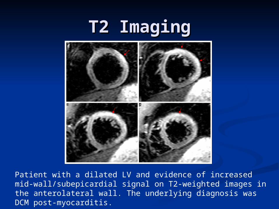
Patient with a dilated LV and evidence of increased mid-wall/subepicardial signal on T2-weighted images in the anterolateral wall. The underlying diagnosis was DCM post-myocarditis.
T2 ImagingT2 Imaging If the T2-weighted triple-inversion If the T2-weighted triple-inversion
recovery sequence is used, typical recovery sequence is used, typical sequence parameters are :-sequence parameters are :-
Slice thickness of 8mmSlice thickness of 8mm Surface and body coilsSurface and body coils TR – 2x RRTR – 2x RR TE- 65msTE- 65ms TI – 170msTI – 170ms Images are taken in the VLA, 4ch view, Images are taken in the VLA, 4ch view,
plus a full SA stackplus a full SA stack
Assessment of FibrosisAssessment of Fibrosis The presence of replacement fibrosis can be detected The presence of replacement fibrosis can be detected
using the inversion recovery late enhancement using the inversion recovery late enhancement technique following gadolinium-DTPA administration.technique following gadolinium-DTPA administration.
In several recent studies, about 60% of patients with In several recent studies, about 60% of patients with DCM showed no detectable fibrosis.DCM showed no detectable fibrosis.
30% showed mid-wall enhancement that correlates 30% showed mid-wall enhancement that correlates with fibrosis on histology.with fibrosis on histology.
10% showed a typical pattern of infarction with 10% showed a typical pattern of infarction with subendocardial rather than mid-wall enhancement. In subendocardial rather than mid-wall enhancement. In the presence of normal coronaries, this is likely to the presence of normal coronaries, this is likely to represent recannalisation, an embolic episode or represent recannalisation, an embolic episode or coronary spasm.coronary spasm.
McCrohon J et al, Circulation 2003

Detection of Fibrosis: Inversion Detection of Fibrosis: Inversion Recovery SequenceRecovery Sequence
Dosage of Gd-DTPA: 0.1-0.2mmol/kgDosage of Gd-DTPA: 0.1-0.2mmol/kg At 0.1mmol/kg, images are usually At 0.1mmol/kg, images are usually
acquired after about 5 minutes with acquired after about 5 minutes with a TI starting at ~340ms (every other a TI starting at ~340ms (every other heart beat).heart beat).
At 0.2 mmol/kg, scans are acquired At 0.2 mmol/kg, scans are acquired after about 15 minutes with a TI after about 15 minutes with a TI starting at ~250ms.starting at ~250ms.
Detection of Fibrosis: Inversion Detection of Fibrosis: Inversion Recovery SequenceRecovery Sequence
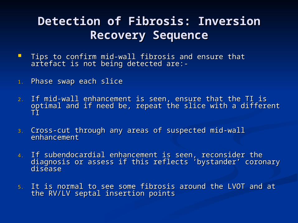
Tips to confirm mid-wall fibrosis and ensure that artefact is Tips to confirm mid-wall fibrosis and ensure that artefact is not being detected are:-not being detected are:-
1.1. Phase swap each slice Phase swap each slice
2.2. If mid-wall enhancement is seen, ensure that the TI is If mid-wall enhancement is seen, ensure that the TI is optimal and if need be, repeat the slice with a different TI optimal and if need be, repeat the slice with a different TI
3.3. Cross-cut through any areas of suspected mid-wall Cross-cut through any areas of suspected mid-wall enhancementenhancement
4.4. If subendocardial enhancement is seen, reconsider the If subendocardial enhancement is seen, reconsider the diagnosis or assess if this reflects ‘bystander’ coronary diagnosis or assess if this reflects ‘bystander’ coronary diseasedisease
5.5. It is normal to see some fibrosis around the LVOT and at the It is normal to see some fibrosis around the LVOT and at the RV/LV septal insertion pointsRV/LV septal insertion points
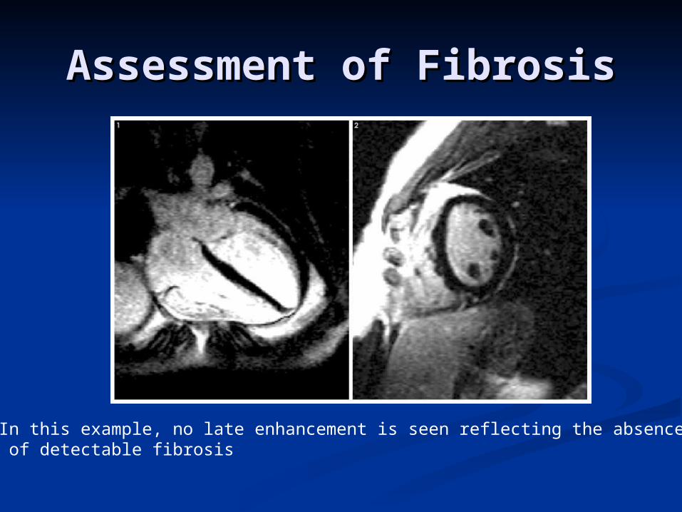
Assessment of FibrosisAssessment of Fibrosis
In this example, no late enhancement is seen reflecting the absence of detectable fibrosis
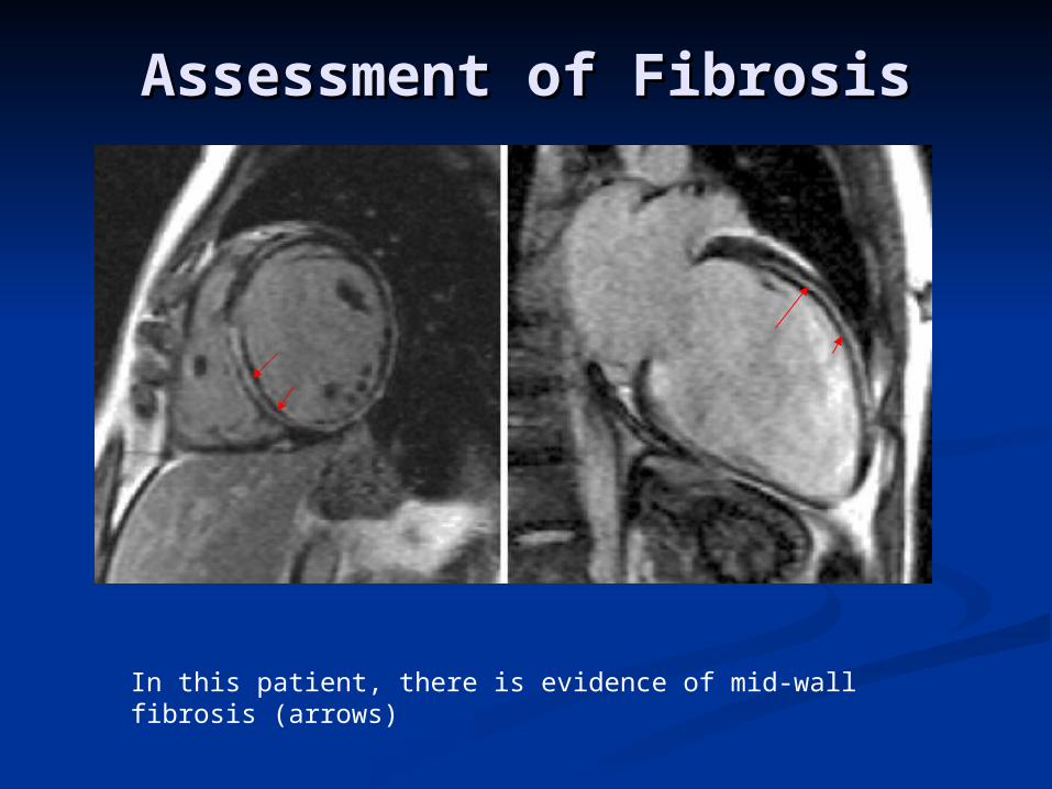
In this patient, there is evidence of mid-wall fibrosis (arrows)
Assessment of FibrosisAssessment of Fibrosis
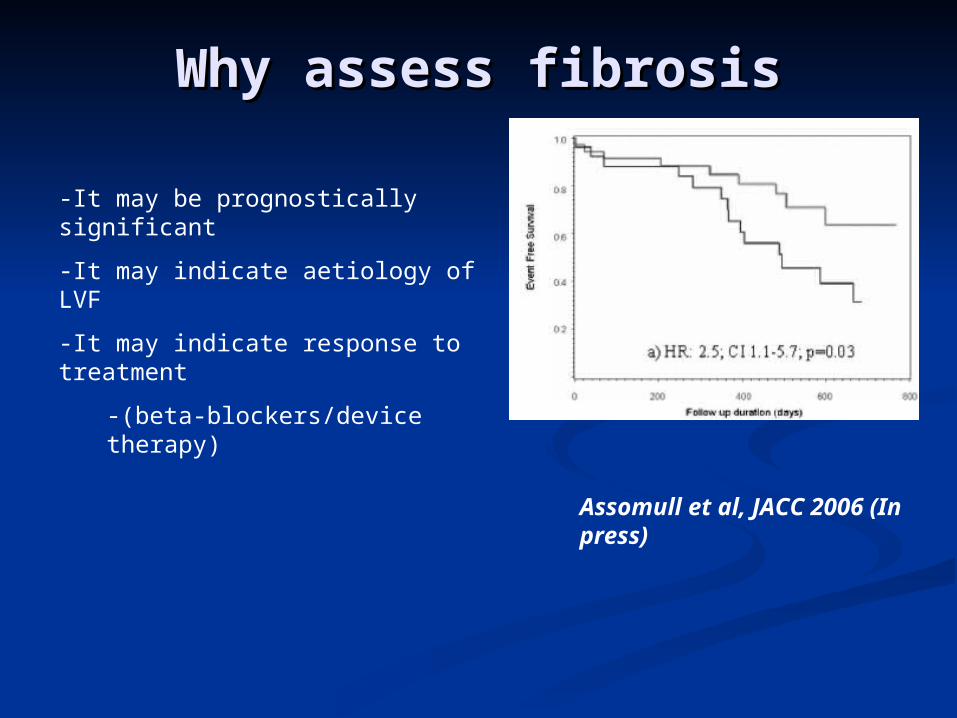
-It may be prognostically significant
-It may indicate aetiology of LVF
-It may indicate response to treatment
-(beta-blockers/device therapy)
Why assess fibrosisWhy assess fibrosis
Assomull et al, JACC 2006 (In press)
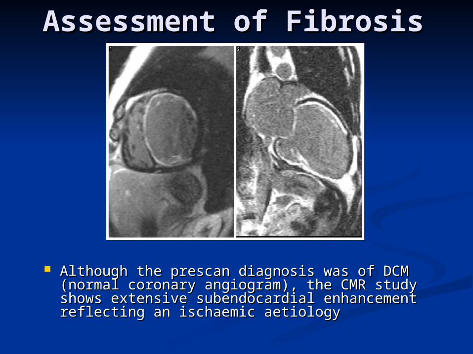
Assessment of FibrosisAssessment of Fibrosis
Although the prescan diagnosis was of DCM Although the prescan diagnosis was of DCM (normal coronary angiogram), the CMR study (normal coronary angiogram), the CMR study shows extensive subendocardial enhancement shows extensive subendocardial enhancement reflecting an ischaemic aetiologyreflecting an ischaemic aetiology
Follow-UpFollow-Up
This will be clinically determinedThis will be clinically determined
In a stable patient where the In a stable patient where the question is of determining reverse question is of determining reverse remodelling, a reasonable interscan remodelling, a reasonable interscan interval would be about 12 monthsinterval would be about 12 months
Related Documents