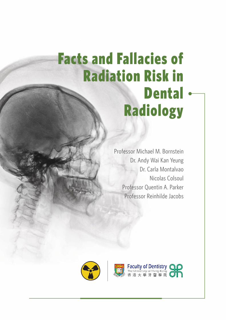
Professor Michael M. Bornstein Dr. Andy Wai Kan Yeung Dr. Carla Montalvao Nicolas Colsoul Professor Quentin A. Parker Professor Reinhilde Jacobs Facts and Fallacies of Radiation Risk in Dental Radiology

Welcome message from author
This document is posted to help you gain knowledge. Please leave a comment to let me know what you think about it! Share it to your friends and learn new things together.
Transcript
Professor Michael M. BornsteinDr. Andy Wai Kan Yeung
Dr. Carla MontalvaoNicolas Colsoul
Professor Quentin A. ParkerProfessor Reinhilde Jacobs
Facts and Fallacies of Radiation Risk in
Dental Radiology
1
Facts and Fallacies ofRadiation Risk inDental Radiology
Professor Michael M. Bornstein1
Dr. Andy Wai Kan Yeung1
Dr. Carla Montalvao1
Nicolas Colsoul1
Professor Quentin A. Parker2
Professor Reinhilde Jacobs3
1 Applied Oral Sciences, Faculty of Dentistry, The University of Hong Kong Applied Oral Sciences, Faculty of Dentistry, The University of Hong Kong Prince Philip Dental Hospital, Sai Ying Pun, Hong Kong SAR, China Prince Philip Dental Hospital, Sai Ying Pun, Hong Kong SAR, China
2 Faculty of Science / Department of Physics and Laboratory for Space Research Faculty of Science / Department of Physics and Laboratory for Space Research The University of Hong Kong, Cyberport 4, Hong Kong SAR, China The University of Hong Kong, Cyberport 4, Hong Kong SAR, China
3 OMFS IMPATH Research Group, Department of Imaging and Pathology OMFS IMPATH Research Group, Department of Imaging and Pathology Faculty of Medicine, University of Leuven, Leuven, Belgium and Faculty of Medicine, University of Leuven, Leuven, Belgium and Department of Oral and Maxillofacial Surgery, University Hospitals Leuven, Leuven Department of Oral and Maxillofacial Surgery, University Hospitals Leuven, Leuven Belgium and Visiting Professor, Department Dental Medicine, Karolinska Institute Belgium and Visiting Professor, Department Dental Medicine, Karolinska Institute Stockholm, Sweden Stockholm, Sweden

Printed in Hong Kong
ISBN 978-988-79680-0-9
© 2019 Faculty of Dentistry, The University of Hong Kong
Faculty of Dentistry, The University of Hong KongThe Prince Philip Dental Hospital, 34 Hospital Road, Hong Kong
http: //facdent.hku.hk
3
ContentsContents
1 What is radiology ? 4Intra-oral radiographs (X-rays taken inside the mouth) 4Extra-oral radiographs (X-rays taken outside the body/mouth) 5
2 What is radiation ? 8 Radiation dose 9
Radiation in dentistry 10
3 Radiation dose protection in Dentistry 1 2Children and radiation 12Pregnancy and radiation 14Elderly and radiation 14 Patient protection measures 15Safety and quality control 16
4 FAQs 18Are dental X-rays really necessary ? 18How often are dental X-rays recommended for adults and children ? 18If I refuse to take X-rays, can I still be treated without them ? 18Instead of taking X-rays now, can you use the X-rays from my previous dentist ? 18How can I protect myself from radiation during X-ray taking ? 19 How safe are dental X-rays during pregnancy ? 19Can radiation affect my child during breastfeeding ? 19Why do I stay alone in the room when X-rays are recorded ? 19Who owns my dental radiograph ? 19Why does my dentist need to retake an image ? 20How safe are dental X-rays ? 20
5 Acknowledgments 2 1
6 References 22 Publications 22
Books and brochures 22 Online resources 23
4What is a radiology ?
Radiology using X-rays is a key medical science. X-rays are high energy photons of electromagnetic energy. In medicine and dentistry it is common to use the word “X-rays” for images that are created when X-rays pass through tissue/bone and cause an exposure in a medium such as a photographic plate. Practical applications of radiology use various forms of imaging technologies for diagnostic purposes and for the treatment of diseases. Taking dental X-rays (dental radiology) is a sub-discipline of dental medicine. It includes the recording and interpretation of radiographic images (created using x-rays) used both for understanding what the problem is (diagnosis) and for the later planning of the treatment of any conditions, disorders or diseases of the mouth, jaws, and face that may be revealed. Experts within this fi eld are known as specialists in Dental and MaxilloFacial Radiology (DMFR) or Oral and MaxilloFacial Radiology (OMFR).
Th ere are diff erent types of X-rays (radiographs) used in dentistry. Th ese may be taken from outside the head/mouth area or from inside the mouth itself. Th e most common dental imaging techniques are the ones from inside the mouth, the bitewings and periapical X-rays (for details, please read below).
Intra-oral radiographs (X-rays taken inside the mouth)Th e X-ray fi lm is placed inside the mouth and can detect pathology. Th ese modalities include:
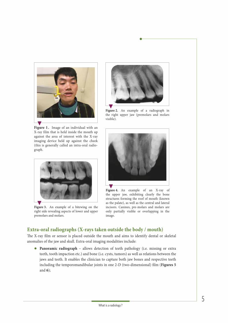
Small area X-rays (also known as periapicals) – these capture local images of teeth roots and surrounding bone. Th ey can detect “invisible” problems under the gums or within bone such as gum disease. A small X-ray fi lm is placed in the mouth and is exposed to a locally focused X-ray beam from a special machine. It has a restricted view being able to capture only two or three adjacent teeth at a time (see Figures 1 and 2);
“Bitewing” – used primarily to identify hidden cavities between back teeth (i.e. the crumbling of teeth or bone in the spaces between teeth – so called “interproximal caries”) or under existing restorations (i.e. recurrence of caries). Teeth on the same side on both upper and lower jaws can be captured simultaneously on one image (Figure 3);
“Occlusal radiographs” – these are useful to monitor and assess the front teeth in either the upper or lower jaw. Th is X-ray is more suited to visualize either the palate or fl oor of the mouth and respective bony anatomy (Figure 4).
Aft er taking the X-ray, two techniques can be used to show the image. Th e classic “analogue” technique is achieved in a darkroom by developing the exposed X-ray fi lm or by using an automatic X-ray fi lm processor. Th is technique is now largely replaced by modern “digital” technologies where images are created by a computer and digital X-ray sensors. Digital images require less X-ray radiation and can be easily adjusted on a screen to help in the diagnostic process. In addition, digital images can be easily stored, printed or electronically shared with other dental professionals.
1 What is a radiology ?radiology ?
5What is a radiology ?
Extra-oral radiographs (X-rays taken outside the body / mouth)Th e X-ray fi lm or sensor is placed outside the mouth and aims to identify dental or skeletal anomalies of the jaw and skull. Extra-oral imaging modalities include:
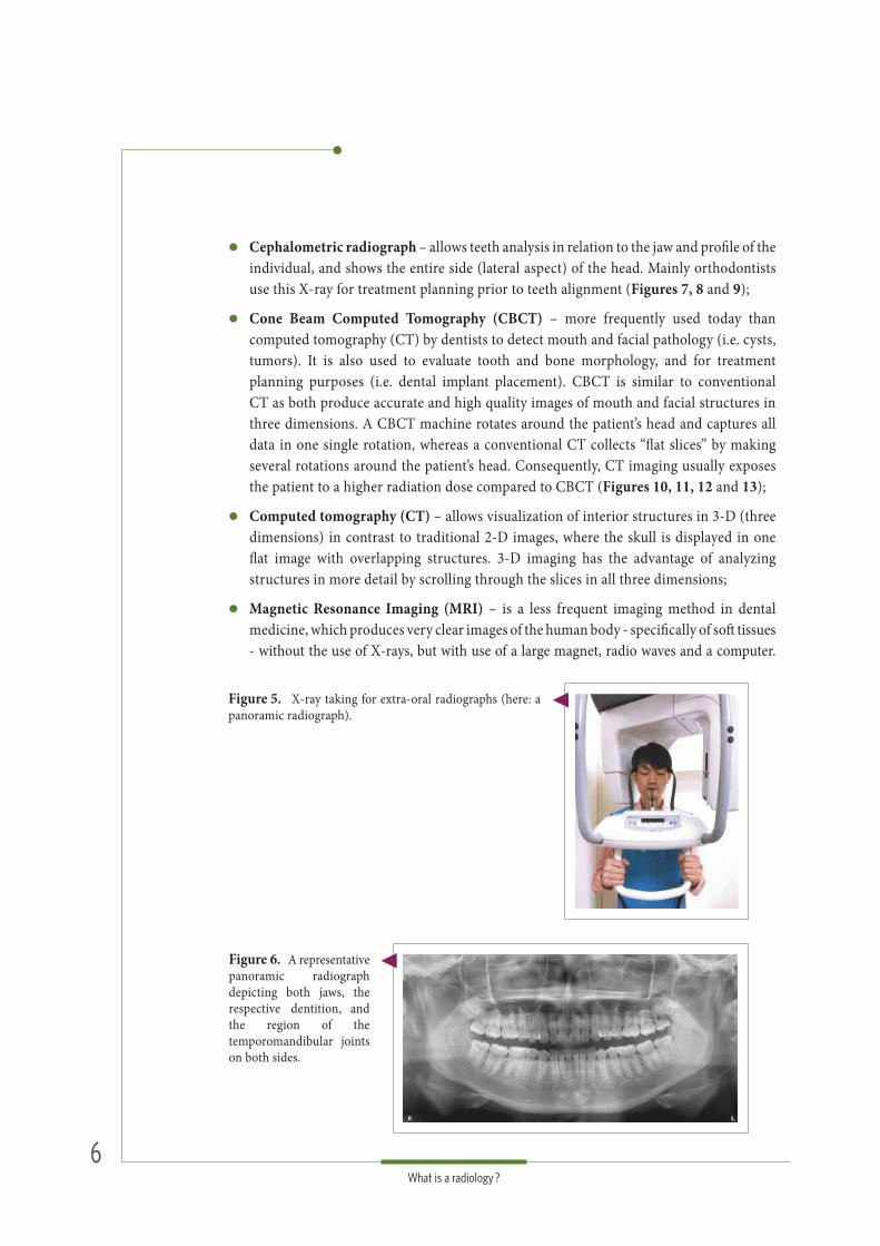
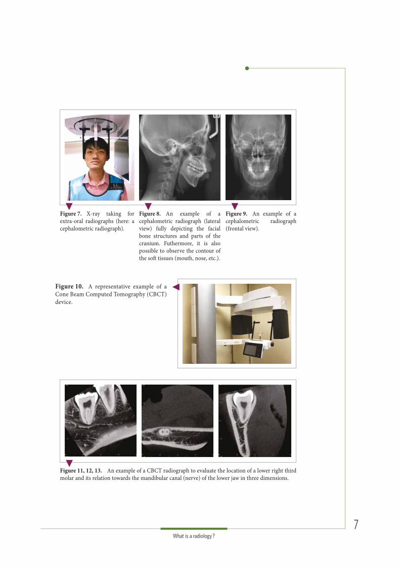
Panoramic radiograph – allows detection of teeth pathology (i.e. missing or extrateeth, tooth impaction etc.) and bone (i.e. cysts, tumors) as well as relations between thejaws and teeth. It enables the clinician to capture both jaw bones and respective teethincluding the temporomandibular joints in one 2-D (two-dimensional) fi lm (Figures 5and 6);
Figure 2. An example of a radiograph in the right upper jaw (premolars and molars visible).
Figure 3. An example of a bitewing on the right side revealing aspects of lower and upper premolars and molars.
Figure 1 . Image of an individual with an X-ray film that is held inside the mouth upagainst the area of interest with the X-rayimaging device held up against the cheek(this is generally called an intra-oral radio-graph.
Figure 4. An example of an X-ray of the upper jaw, exhibiting clearly the bone structures forming the roof of mouth (known as the palate), as well as the central and lateral incisors. Canines, pre-molars and molars are only partially visible or overlapping in the image.
6What is a radiology ?
Cephalometric radiograph – allows teeth analysis in relation to the jaw and profi le of the individual, and shows the entire side (lateral aspect) of the head. Mainly orthodontists use this X-ray for treatment planning prior to teeth alignment (Figures 7, 8 and 9);
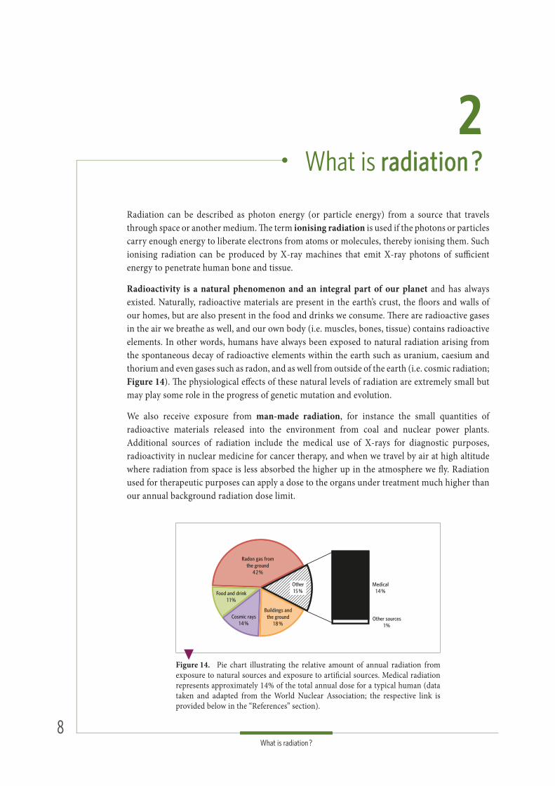
Cone Beam Computed Tomography (CBCT) – more frequently used today than computed tomography (CT) by dentists to detect mouth and facial pathology (i.e. cysts, tumors). It is also used to evaluate tooth and bone morphology, and for treatment planning purposes (i.e. dental implant placement). CBCT is similar to conventional CT as both produce accurate and high quality images of mouth and facial structures in three dimensions. A CBCT machine rotates around the patient’s head and captures all data in one single rotation, whereas a conventional CT collects “fl at slices” by making several rotations around the patient’s head. Consequently, CT imaging usually exposes the patient to a higher radiation dose compared to CBCT (Figures 10, 11, 12 and 13);
Computed tomography (CT) – allows visualization of interior structures in 3-D (three dimensions) in contrast to traditional 2-D images, where the skull is displayed in one fl at image with overlapping structures. 3-D imaging has the advantage of analyzing structures in more detail by scrolling through the slices in all three dimensions;
Magnetic Resonance Imaging (MRI) – is a less frequent imaging method in dental medicine, which produces very clear images of the human body - specifi cally of soft tissues - without the use of X-rays, but with use of a large magnet, radio waves and a computer.
Figure 5. X-ray taking for extra-oral radiographs (here: a panoramic radiograph).
Figure 6. A representative panoramic radiograph depicting both jaws, the respective dentition, and the region of the temporomandibular joints on both sides.
7What is a radiology ?
Figure 7. X-ray taking for extra-oral radiographs (here: a cephalometric radiograph).
Figure 8. An example of a cephalometric radiograph (lateral view) fully depicting the facial bone structures and parts of the cranium. Futhermore, it is also possible to observe the contour of the soft tissues (mouth, nose, etc.).
Figure 9. An example of a cephalometric radiograph (frontal view).
Figure 10. A representative example of a Cone Beam Computed Tomography (CBCT) device.
Figure 11, 12, 13. An example of a CBCT radiograph to evaluate the location of a lower right third molar and its relation towards the mandibular canal (nerve) of the lower jaw in three dimensions.
8What is radiation ?
Radiation can be described as photon energy (or particle energy) from a source that travels through space or another medium. Th e term ionising radiation is used if the photons or particles carry enough energy to liberate electrons from atoms or molecules, thereby ionising them. Such ionising radiation can be produced by X-ray machines that emit X-ray photons of suffi cient energy to penetrate human bone and tissue.
Radioactivity is a natural phenomenon and an integral part of our planet and has always existed. Naturally, radioactive materials are present in the earth’s crust, the fl oors and walls of our homes, but are also present in the food and drinks we consume. Th ere are radioactive gases in the air we breathe as well, and our own body (i.e. muscles, bones, tissue) contains radioactive elements. In other words, humans have always been exposed to natural radiation arising from the spontaneous decay of radioactive elements within the earth such as uranium, caesium and thorium and even gases such as radon, and as well from outside of the earth (i.e. cosmic radiation; Figure 14). Th e physiological eff ects of these natural levels of radiation are extremely small but may play some role in the progress of genetic mutation and evolution.
We also receive exposure from man-made radiation, for instance the small quantities of radioactive materials released into the environment from coal and nuclear power plants. Additional sources of radiation include the medical use of X-rays for diagnostic purposes, radioactivity in nuclear medicine for cancer therapy, and when we travel by air at high altitude where radiation from space is less absorbed the higher up in the atmosphere we fl y. Radiation used for therapeutic purposes can apply a dose to the organs under treatment much higher than our annual background radiation dose limit.
2 What is radiation ?radiation ?
Figure 14. Pie chart illustrating the relative amount of annual radiation from exposure to natural sources and exposure to artifi cial sources. Medical radiation represents approximately 14% of the total annual dose for a typical human (data taken and adapted from the World Nuclear Association; the respective link is provided below in the “References” section).
Radon gas fromthe ground
42 %
Cosmic rays14 %
Medical14 %
Other sources1 %
Other15 %Food and drink
1 1 %
Buildings andthe ground
18 %
9What is radiation ?
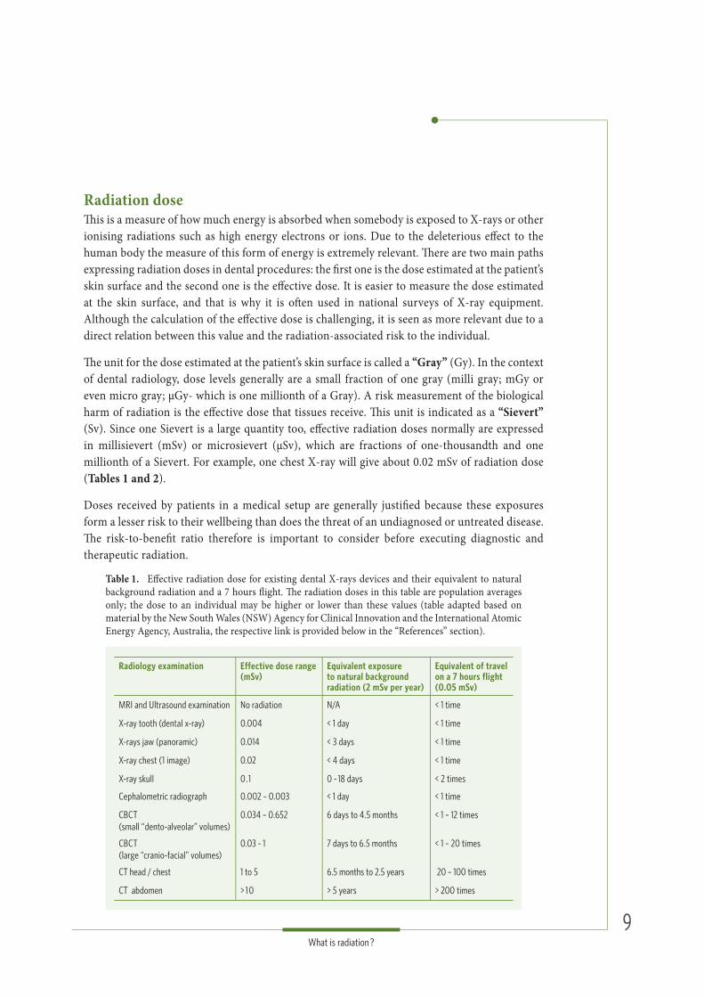
Radiation doseTh is is a measure of how much energy is absorbed when somebody is exposed to X-rays or other ionising radiations such as high energy electrons or ions. Due to the deleterious eff ect to the human body the measure of this form of energy is extremely relevant. Th ere are two main paths expressing radiation doses in dental procedures: the fi rst one is the dose estimated at the patient’s skin surface and the second one is the eff ective dose. It is easier to measure the dose estimated at the skin surface, and that is why it is oft en used in national surveys of X-ray equipment. Although the calculation of the eff ective dose is challenging, it is seen as more relevant due to a direct relation between this value and the radiation-associated risk to the individual.
Th e unit for the dose estimated at the patient’s skin surface is called a “Gray” (Gy). In the context of dental radiology, dose levels generally are a small fraction of one gray (milli gray; mGy or even micro gray; μGy- which is one millionth of a Gray). A risk measurement of the biological harm of radiation is the eff ective dose that tissues receive. Th is unit is indicated as a “Sievert”(Sv). Since one Sievert is a large quantity too, eff ective radiation doses normally are expressed in millisievert (mSv) or microsievert (μSv), which are fractions of one-thousandth and one millionth of a Sievert. For example, one chest X-ray will give about 0.02 mSv of radiation dose (Tables 1 and 2).
Doses received by patients in a medical setup are generally justifi ed because these exposures form a lesser risk to their wellbeing than does the threat of an undiagnosed or untreated disease. Th e risk-to-benefi t ratio therefore is important to consider before executing diagnostic and therapeutic radiation.
Table 1. Eff ective radiation dose for existing dental X-rays devices and their equivalent to natural background radiation and a 7 hours fl ight. Th e radiation doses in this table are population averages only; the dose to an individual may be higher or lower than these values (table adapted based on material by the New South Wales (NSW) Agency for Clinical Innovation and the International Atomic Energy Agency, Australia, the respective link is provided below in the “References” section).
Radiology examination Effective dose range (mSv)
Equivalent exposure to natural background radiation (2 mSv per year)
Equivalent of travel on a 7 hours flight(0.05 mSv)
MRI and Ultrasound examination No radiation N/A < 1 time
X-ray tooth (dental x-ray) 0.004 < 1 day < 1 time
X-rays jaw (panoramic) 0.014 < 3 days < 1 time
X-ray chest (1 image) 0.02 < 4 days < 1 time
X-ray skull 0 .1 0 - 18 days < 2 times
Cephalometric radiograph 0.002 – 0.003 < 1 day < 1 time
CBCT(small “dento-alveolar” volumes)
0.034 – 0.652 6 days to 4.5 months < 1 – 12 times
CBCT(large “cranio-facial” volumes)
0.03 - 1 7 days to 6.5 months < 1 – 20 times
CT head / chest 1 to 5 6.5 months to 2.5 years 20 – 100 times
CT abdomen > 1 0 > 5 years > 200 times
1 01 0What is radiation ?
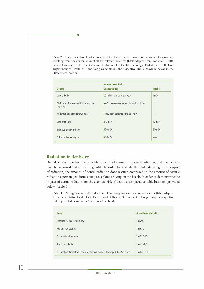
Table 2. Th e annual dose limit stipulated in the Radiation Ordinance for exposure of individuals resulting from the combination of all the relevant practices (table adapted from Radiation Health Series, Guidance Notes on Radiation Protection for Dental Radiology, Radiation Health Unit Department of Health of Hong Kong Government; the respective link is provided below in the “References” section).
Annual dose limit
Organs Occupational Public
Whole Body 20 mSv in any calendar year 1 mSv
Abdomen of woman with reproductive capacity
5 mSv in any consecutive 3 months interval -----
Abdomen of a pregnant woman 1 mSv from declaration to delivery -----
Lens of the eye 150 mSv 15 mSv
Skin, average over 1 cm2 500 mSv 50 mSv
Other individual organs 500 mSv ----
Table 3. Average annual risk of death in Hong Kong from some common causes (table adapted from the Radiation Health Unit, Department of Health, Government of Hong Kong; the respective link is provided below in the “References” section).
Cause Annual risk of death
Smoking 10 cigarettes a day 1 in 200
Malignant diseases 1 in 630
Occupational accidents 1 in 55 000
Traffic accidents 1 in 22 200
Occupational radiation exposure for local workers (average 0.15 mSv/year)5 1 in 170 333
Radiation in dentistryDental X-rays have been responsible for a small amount of patient radiation, and their eff ects have been considered almost negligible. In order to facilitate the understanding of the impact of radiation, the amount of dental radiation dose is oft en compared to the amount of natural radiation a person gets from sitting on a plane or lying on the beach. In order to demonstrate the impact of dental radiation on the eventual risk of death, a comparative table has been provided below (Table 3).
1 11 1What is radiation ?
Technology has changed substantially over the last few years. Th e change from classical fi lm-based radiology —the standard for decades in the fi eld—to digital imaging has had a great impact on the reduction of radiation dose. On the other hand, the advent of CBCT imaging or 3-D technology which has come with a higher radiation dose has more than off set reductions in dose to the public from switching from fi lm to digital imaging.
To image a tooth, which is a calcifi ed structure just like the other bones in our body, the amount of energy needed to penetrate is the same as for taking a bone tissue image. X-rays required for teeth are therefore very similar to X-rays for fi ngers, hands or feet. In reality, the processes of taking an X-ray of a tooth or a fi nger are almost the same. Following this principle of similarity, the amount of radiation dose required to penetrate the jaw to form an image on a panoramic X-ray has also a similar dose to the radiation needed to X-ray the elbow. Refer to Table 1 to observe the radiation dose among the several methods and types of diagnostic X-rays.
1 21 2Radiation dose protection in Dentistry
Optimization of radiation protection was developed as a result of the increasing knowledge on radiation and its potential negative effect on people. The acronym ALARA (“As Low As Reasonably Achievable”) has been used since 1973, to designate the optimization of X-ray doses in order to minimize radiation dose exposure. More recently, ALARA has been replaced by the acronym ALADA (“As Low As Diagnostically Acceptable”).
To establish ALADA goals, clinicians try to apply doses that are appropriate for the clinical purpose. This will influence the selection of imaging devices and the settings used. Practical measures for ALARA and ALADA in dental practice comprise an awareness of the field of view (for all types of imaging, but especially for CBCT), exposure time, anatomical relation (radiosensitive organs), diagnostic justification and shielding.
Patient selection guidelines for dental radiographic examinations have been developed to help the dentist’s professional judgment on how to optimize the use of diagnostic imaging. Further, the concept of radiographic justification as well as the effective use of X-rays in a dental practice has been elaborated in detail by the European and American Dental Association guidelines which suggests:
All radiographic examinations must be justifi ed on an individual case basis by considering that the benefi ts to the patient should outweigh the potential harm. Th e expected benefi t is that the radiographic examination can add new diagnostic information to facilitate the management of the patient;
Unless a medical history taking and clinical examination have been performed, no radiograph should be made. It means that the concept of ‘routine radiography’ is unacceptable;
When referring a patient for a radiographic examination, the dentist should provide enough clinical information to justify the examination.
Children and radiationDental radiography is a useful diagnostic aid in oral examination of children. In many cases the radiographic fi ndings add crucial information. However, the risks associated with radiography should not be neglected. Furthermore, it has been found that children are far more radiosensitive when compared with adults, they present approximately 3 to 5 times higher risk of mortality by cancer resultant from radiation. Th erefore, increased attention should be paid to minimize the medical radiation exposures to children. All radiological procedures carried out on children must
3 Radiation dose protection
in Dentistryin Dentistry
1 31 3Radiation dose protection in Dentistry
adapt to special radiation protection measures, which aims at recognizing and implementing possible dose reduction strategies in order to eliminate unnecessary and therefore unjustifi ed radiation exposure.
Since July 2007, the Alliance for Radiation Safety in Pediatric Imaging has created the Image Gently campaign to educate and raise the awareness regarding radiation safety. It is a worldwide coalition of healthcare organizations determined to provide safe, high quality pediatric imaging. Th e Society for Pediatric Radiology and over 33 other societies (such as the American Academy of Oral and Maxillofacial Radiology) formed this coalition, representing more than 500,000 worldwide health care professionals in radiology, dentistry, pediatrics, medical physics, and radiation safety. Th e Image Gently Campaign recommends to:
Perform imaging only if there is clear medical or dental benefi ts to the child;
Use the lowest amount of radiation for diagnostically acceptable imaging based on the body size of the child;
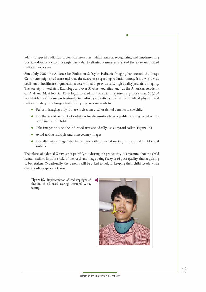
Take images only on the indicated area and ideally use a thyroid collar (Figure 15)
Avoid taking multiple and unnecessary images;
Use alternative diagnostic techniques without radiation (e.g. ultrasound or MRI), if suitable.
Th e taking of a dental X-ray is not painful, but during the procedure, it is essential that the child remains still to limit the risks of the resultant image being fuzzy or of poor quality, thus requiring to be retaken. Occasionally, the parents will be asked to help in keeping their child steady while dental radiographs are taken.
Figure 15. Representation of lead-impregnated thyroid shield used during intraoral X-ray taking.
1 41 4Radiation dose protection in Dentistry
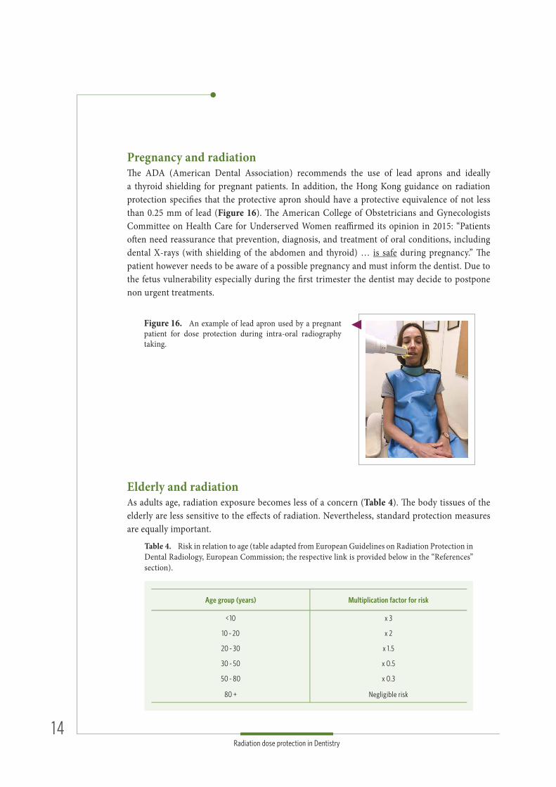
Pregnancy and radiationTh e ADA (American Dental Association) recommends the use of lead aprons and ideally a thyroid shielding for pregnant patients. In addition, the Hong Kong guidance on radiation protection specifi es that the protective apron should have a protective equivalence of not less than 0.25 mm of lead (Figure 16). Th e American College of Obstetricians and Gynecologists Committee on Health Care for Underserved Women reaffi rmed its opinion in 2015: “Patients oft en need reassurance that prevention, diagnosis, and treatment of oral conditions, including dental X-rays (with shielding of the abdomen and thyroid) … is safe during pregnancy.” Th e patient however needs to be aware of a possible pregnancy and must inform the dentist. Due to the fetus vulnerability especially during the fi rst trimester the dentist may decide to postpone non urgent treatments.
Elderly and radiationAs adults age, radiation exposure becomes less of a concern (Table 4). Th e body tissues of the elderly are less sensitive to the eff ects of radiation. Nevertheless, standard protection measures are equally important.
Figure 16. An example of lead apron used by a pregnant patient for dose protection during intra-oral radiography taking.
Table 4. Risk in relation to age (table adapted from European Guidelines on Radiation Protection in Dental Radiology, European Commission; the respective link is provided below in the “References” section).
Age group (years) Multiplication factor for risk
< 10 x 3
10 - 20 x 2
20 - 30 x 1.5
30 - 50 x 0.5
50 - 80 x 0.3
80 + Negligible risk
1 51 5Radiation dose protection in Dentistry
Patient protection measuresAlthough, dental X-ray taking for diagnostic purposes is decided on individual needs, the radiation protection guidelines are common for the majority of the procedures. Th e department for Radiation Protection in Dental Radiology, Radiation Health Unit, Department of Health, Hong Kong Government has stipulated for the use of intra-oral fi lms and extra-oral imaging the following guidelines:
A woman who is, or who may be, pregnant should have the body covered by a protective apron with a protective equivalence of not less than 0.25 mm lead.
Dental fi lms should be held by the patient and by no one else; when the patient cannot hold it (i.e. a child or a handicapped person) then the person providing support should be given appropriate instructions and should wear protective clothing (e.g. a lead apron) if practicable.
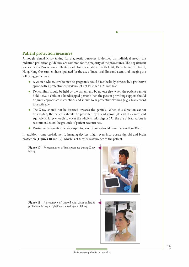
Th e X-ray should not be directed towards the genitals. When this direction cannot be avoided, the patients should be protected by a lead apron (at least 0.25 mm lead equivalent) large enough to cover the whole trunk (Figure 17); the use of lead aprons is recommended on the grounds of patient reassurance.
During cephalometry the focal-spot to skin distance should never be less than 30 cm.
In addition, some cephalometric imaging devices might even incorporate thyroid and brain protection (Figures 18 and 19), which is of further reassurance to the patient.
Figure 17. Representation of lead apron use during X-ray taking.
Figure 18. An example of thyroid and brain radiation protection during a cephalometric radiograph taking.

1 61 6Radiation dose protection in Dentistry
Safety and quality controlTh e radiation safety features of equipment should be maintained and checked according to the advice of the manufacturer or the supplier. At the Prince Philip Dental Hospital / Faculty of Dentistry of the University of Hong Kong these guidelines are followed by routine checks to detect if any deterioration in imaging quality or repeatability of radiographs occurs - and if the underlying cause is the deterioration in processing procedures or faulty X-ray equipment. Additionally, the Radiation Board will perform further routine and random inspections to assure patient and staff safety.
Th e Prince Philip Dental Hospital has also embraced preventive measures such as:
Personal wearing of dosimeters for staff working in the “Oral Imaging” clinic.
Use of barrier shielding for staff during exposures.
Protective panel for the operator.
Controlled areas for radiography:a. controlled areas are exhibiting a warning notice stating that the area is controlled. b. presence of radiation warning sign; may also include more detailed information,
such as the reason why the area is controlled, e.g. “X-radiation”, and whether entry is permitted or not (Figure 20).
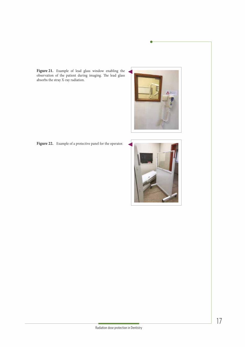
c. during radiography taking, staff are able to observe the condition of the patient through a lead glass window (Figure 21), or being protected by a control panel (Figure 22).
Figure 19. An example of cephalometric X-ray depicting the respective thyroid and brain radiation protection (white areas). The lack of image detail in the these areas demonstrate the effectiveness of the protection mechanism by preventing X-ray penetration of these tissues.
Figure 20. Example of a controlled area displaying a radiation warning sign and an active red light informing that entrance is currently not permitted.
1 71 7Radiation dose protection in Dentistry
Figure 21. Example of lead glass window enabling the observation of the patient during imaging. Th e lead glass absorbs the stray X-ray radiation.
Figure 22. Example of a protective panel for the operator.
1 81 8FAQs
1. Are dental X-rays really necessary ? Yes, as many diseases and conditions develop without any signs and symptoms. Tooth
decay (caries) for instance, which is frequently located in between teeth, can be impossible to visualize during a clinical examination alone - especially in posterior teeth. Also cysts and tumors which are located inside the jaw bone can best be depicted properly using 2-D or 3-D X-ray images. X-rays are crucial for problem identifi cation, diagnostic purposes, treatment planning, and to ensure a healthy condition of your mouth.
2. How oft en are dental X-rays recommended for adults and children ? Interval assessment of X-rays is made individually depending on your present oral health,
age, risk for disease, and any signs or symptoms of oral disease. It is not uncommon for children to have X-rays more oft en because their teeth and jaws are still developing. Additionally, they are also more prone to suff er from tooth decay. Nevertheless, extensive or repeated X-ray examinations especially of children and pregnant women should not be undertaken without considerable forethought. Your dentist will review your medical and dental history, and examine your mouth fi rst before deciding whether or not you need X-rays.
3. If I refuse to take X-rays, can I still be treated without them ? Yes, and this is part of the informed consent before initiating treatment. However, patients
should know that X-rays are essential for a complete diagnosis and during treatment as not everything can be detected during the clinical examination alone. If X-rays are refused, important information may be missing.
4. Instead of taking X-rays now, can you use the X-rays from my previous dentist ?
Yes, provided that they are recent and of acceptable diagnostic quality. Th e information of previous X-rays is actually very useful in the diagnostic process and disease management, as the evolution of a lesion can be carefully assessed. If x-rays are digitally available, ask your previous dentist for online access, and pass the respective images along to your current dentist. However, your dentist may still ask you to take new X-rays if he or she cannot be sure when or how the existing X-rays were taken.
4 FAQ sFAQ s
1 91 9FAQs
5. How can I protect myself from radiation during X-ray taking ? A lead apron (and thyroid collar as well) is part of the recommended radiation protection
measures to protect reproductive, blood-forming, and thyroid tissues from scatter radiation. Th e lead acts as a shield and prevents the radiation from reaching these radiosensitive organs, which is especially important within the developing tissues of both children and pregnant woman.
6. How safe are dental X-rays during pregnancy ? Future mothers should be aware that no single diagnostic dental X-ray has a radiation
dose signifi cant enough to cause adverse eff ects in a developing embryo or fetus. Th is is supported by various international organizations, who also emphasize the importance of appropriate shielding (i.e. lead apron) when pregnant. X-rays during emergencies are necessary for dental disease management. If not treated, the infection might spread through the bloodstream to the fetus, and (for example) periodontal disease may negatively aff ect pregnancy. X-rays during follow-up examinations however, can be postponed until aft er birth.
7. Can radiation aff ect my child during breastfeeding ? At present, there is no evidence supporting special precautions with regard to children
during breastfeeding. Dental X-rays are only present during the time that the image is being taken and leave no radiation or radioactivity in the body or in milk. Th erefore, dental X-rays have no known eff ect on the milk in the breast at the time of imaging, nor on milk production.
8. Why do I stay alone in the room when X-rays are recorded ? Th is is for the safety of the operator. When a patient is exposed to X-rays, they receive
diagnostic benefi ts, while the dentist or X-ray operator does not. Also, while a patient is likely to be exposed with limited X-rays per year or during his lifespan, the dentist has to take thousands of X-rays. Th is could result in multiple more times of radiation compared to the patient. Th e dentist must use proper protection measures, and one of the most eff ective ways to limit their X-ray exposure is to maintain adequate distance and shielding. Th is is why they usually step out of the room during the X-ray exposure.
9. Who owns my dental radiograph ? All your dental records, including the dental radiographs, are property of the dentist. Bound
by professional secrecy, the dentist is obligated to store them securely and is responsible for any violations. A patient however has the privilege of reasonable access to his own dental records, and can request these from the dentist.
2 02 0FAQs
10. Why does my dentist need to retake an image ? Several errors can occur during the X-ray image taking process such as motion artefacts,
wrong focus distance, the desired information may not be visible or clear enough on the current radiograph, mechanical errors, etc. Th ese complications might result in an X-ray that cannot be interpreted correctly, and thus is considered as not feasible for diagnosis. Th erefore, this image has to be retaken.
11. How safe are dental X-rays? Compared to medical X-rays, dental x-rays generally have a lower radiation dose, and
therefore should be safe when used with care and consideration by your dentist. Overall, the importance of making the right diagnosis and choosing the correct treatment makes X-rays far more benefi cial than they are dangerous. Nevertheless, the benefi ts of such procedure is always weighed up before it’s recommended.
2 12 1Acknowledgments
Th e authors thank Dr. Rainier Hogeveen, MSc, Department of Dental Radiology at ACTA Amsterdam, Th e Netherlands, for the provision of fi gures 18 and 19 (cephalometric radiology protection).
We thank also Prof. Cynthia Kar Yung Yiu, Paediatric Dentistry, Faculty of Dentistry, Th e University of Hong Kong, Hong Kong SAR, China, for sharing her expertise.
We further want to express our gratitude to Dr. Basak Harper, research assistant at Oral and Maxillofacial Radiology, Applied Oral Sciences, Faculty of Dentistry, Th e University of Hong Kong, Hong Kong SAR, China, for her help during the preparation of the manuscript.
5 AcknowledgmentsAcknowledgments
2 22 22 22 2References
PublicationsAbbott P. Are dental radiographs safe? Aust Dent J 2000; 45: 208-213.
American Dental Association Council on Scientific Affairs and U.S. Food and Drug Administration. Dental radiographic examinations: Recommendations for patient selection and limiting radiation exposure. ADA 2012: 16-17.
American Dental Association Council on Scientific Affairs. The use of dental radiographs. Update and recommendations. J Am Dent Assoc 2006; 137: 1304–1312.
Berkhout WE. The ALARA-principle. Backgrounds and enforcement in dental practices. Ned Tijdschr Tandheelkd 2015; 122: 263-270.
Evearitt A. The reality of dental radiation. Journal of the Colorado Dental Association 2016; 95: 28-30.
Giglio JA, Lanni SM, Laskin DM, Giglio NW. Oral health care for the pregnant patient. J Can Dent Assoc 2009; 75: 43-48.
ICRP International Commission on Radiological Protection. Radiological Protection in Medicine. ICRP Publication 105. Ann ICRP 37 (6).
Michalowicz BS, DiAngelis AJ, Novak MJ, Buchanan W, Papapanou PN, Mitchell DA, Curran AE, Lupo VR, Ferguson JE, Bofill J, Matseoane S, Deinard AS Jr, Rogers TB. Examining the safety of dental treatment in pregnant women. J Am Dent Assoc 2008; 139: 685-695.
Okano T, Sur J. Radiation dose and protection in dentistry. Japanese Dental Science Review 2010; 46: 112—121.
Books & BrochuresEuropean Guidelines on Radiation Protection in Dental Radiology:www.ec.europa.eu/energy/sites/ener/files/documents/136_0.pdf
Ianucci J, Howerton L. 2016, Dental Radiography Principles and Techniques. 5th Ed. Elsevier, Amsterdam.
International Commission on Radiological protection: www.icrp.org/docs/DRL_for_web.pdf
6 ReferencesReferences
2 32 32 32 3References
NSW Agency for Clinical Innovation, Government of New South Wales, Australia:www.aci.health.nsw.gov.au/__data/assets/pdf_file/0006/174552/MI-Clinician-Factsheet.pdf
Radiation Safety in Dental Practice – a study guide. California Dental Association:www.cda.org/Portals/0/pdfs/practice_support/radiation_safety_in_dental_practice.pdf
Radiation Health Series, Guidance notes on radiation protection for Dental Radiology, Radiation Health Unit, Department of Health of Hong Kong Government: www.info.gov.hk/dh-rhu/english/pdf/Pub4_english.pdf
Online resourcesAmerican Dental Association:www.ada.org/en/~/media/ADA/Science%20and%20Research/Files/topics_radiography_examinations(1)
Australian Nuclear Science and Technology Organization, Australian Government:www.ansto.gov.au/NuclearFacts/Whatisradiation/index.htm
International Atomic Energy Agency:www.iaea.org/resources/rpop/health-professionals/dentistry/pregnant-women
Image Gently Alliance:www.imagegently.org/Roles-What-can-I-do/Parent/Dental
National Health Service UK:https://www.nhs.uk/conditions/x-ray/
Radiation Health Unit, Department of Health, Government of Hong Kong Special Administrative Region: www.info.gov.hk/dh-rhu/english/html/understand_rad_english.htm
Radiation protection in dentistry. Recommended safety procedures for the use of dental x-ray equipment. Health Canada 1999, Government of Canada: www.canada.ca/en/health-canada/services/environmental-workplace-health/reports-publications/radiation/radiation-protection-dentistry-recommended-safety-procedures-use-dental-equipment-safety-code-30.html
World Nuclear Association: www.world-nuclear.org/nuclear-basics/what-is-radiation.aspx


The Prince Philip Dental Hospital34 Hospital Road
Hong Kong SAR, China
http: //facdent.hku.hk
Related Documents