----- __ 111 _____ HEALTH OF VIETNAM SUPPLEMENT A LABORATORY AND QUALITY CONTROL U.S. DEPARTMENT OF HEALTH AND HUMAN SEIIVICES PUBLIC HEALTH SERVICE Centers for Disease Control

Welcome message from author
This document is posted to help you gain knowledge. Please leave a comment to let me know what you think about it! Share it to your friends and learn new things together.
Transcript
-----__111 _____
HEALTH STATU~S
OF VIETNAM VETER~I~NS
SUPPLEMENT A LABORATORY METHOD~3
AND QUALITY CONTROL
US DEPARTMENT OF HEALTH AND HUMAN SEIIVICES PUBLIC HEALTH SERVICE Centers for Disease Control
-~------~~---------------------- _--shy
HEALTH STATUS OF VIETNAM VETERAN~)
SUPPLEMENT A LABORATORY METHODS AND QUALITY CONTRlt)L
The Centers for Disease Control Vietnam Experience Study
January 1989
tt
US DEPARTMENT OF HEALTH AND HUMAN SERVICES PUBLIC HEALTH SERVICE
Centers for Disease Control Center for Environmental Health and Injury Control
Atlanta Georgia 30333
Use of trade names is for identification only and does not constitute endorsement by the Public Health Service or by the US Department of Health and Human Services
1pound 22pound 1
ACKNOWLEDGEMENTS
This supplement was prepared by the Vietnam Experience Study staff of the Centers for Disease Control (CDC) and Lovelace Medical Foundation (LMF)
The CDC authors were Joseph L Annest (Division of Chronic Diseases Control) Adrian Hainline Jr (Division of Environmental Health Laboratory Sciences (DEHLS) Center for Environmental Health and Injury Control (CEHIC)) Sandra Emrich and Liz Cochran (Division of Chronic Diseases Control)
The LMF authors were Lee Gates Bruce S Edwards Susan R Flynn Mal) Westrich Jeanette Strauch and Thomas D Stewart
Other staff members who made important contributions were (CDC staff) John M Karon (LMF staff) Steve A Apodaca Anne M Baldonado Kathleen A Birmingham Janis L Burg Jan F Dean Beth G Goldberg Joseph R Good III Donna J Harper Wanda K Korn Loyola E Kratz Amy F Ludwig-Lupinetti Cheryl A Mattison Pamela J Morden Hectc A Nolla Janis H Prudhomme Robyn Richards Robin P Smith David C Smith Deborah J Spyker and Marguerite Townsend
Secretarial support for this supplement was provided by (CDC staff) Susan B Spangler
Consultants who made important contributions were David C Allison (Johm Hopkins University) Robert H Hill Jr (DEHLS CEHIC CDC) David F Katz (University of alifornia Davis) Gerald Kessler (Jewish Hospital St Louis Missouri) Harold Russell Clivision of Bacterial Diseases Center for Infectious Diseases CDC) Steven M Schrade (National Institute for Occupational Safety and Health Cincinnati Ohio) Carl C Stewart (Ls Alamos National Laboratories New Mexico) and William O Weigle (Scripps Clinic and Research Foundation San Diego California)
iii
SUMMARY
This supplement provides information on the standardized procedures and methods wed to perform a battery of laboratory assessments on blood urine and semen specimons obtained from veterans who participated in the medical examination component of he Vietnam Experience Study (VES) It includes detailed protocols for data management sp 3Cshyimen collection and processing analytic methods and quality control (QC) procedures In he final section QC data obtained over the 16-month examination period are summarized
The QC program used in this study was considerably more demanding than whu is considered normal for a clinical laboratory Each analytic run (usually including 23 particip mt samples per assay per day) contained bench and blind repeat QC samples Berlch controls were used to monitor precision of measurement within and among days over he course of the study Blind repeat controls (ie the technician was unaware of the co -ort status of the donor) were used to monitor the repeatability of the measurement () r a participant sample within the same analytic run QC data from bench controls ~re
summarized in this supplement QC data from blind repeat controls are summarizelj in Supplement B Medical and Psychological Data Quality
We implemented a statistical QC program to monitor the quality of laboratory d 3ta obtained over the study period Each respective technician maintained QC charts at he bench In addition two full-time QC supervisors and a board-certified clinical patholc list reviewed the participant and QC data daily To aid them in this labor-intensive proces~ a customized statistical QC program was put into use on a personal computer to ide-tify potential out-of-control runs Criteria for acceptable performance of daily runs were ba~ed on a contractual agreement with the Centers for Disease Control In that contIClct performance guidelines were given for each bioassay These guidelines were cloely followed Ultimately however the laboratory director decided whether the performance lias acceptable
The QC data were sent to CDC weekly by telephone Data were then carefully checked for completeness validity and accuracy We ran computer programs on these data as 1ltey accumulated To check for long-term trends we provided monthly computer-generated )C monitoring reports to Lovelace Clinical Laboratory for review there
In conclusion our overall review of QC data for these laboratory determinations sugge ts that the bioassays with one exception (the melioidosis titre) were performed with glod preCision and have provided high-quality laboratory data for use in assessing the currmt health status of veterans participating in the VES
iv
1
TABLE OF CONTENTS Page
Acknowledgements iii
Summary iv
I Introduction
II Laboratory Data Management Manual 3 A General Overview 3 B Data Flow 4
III Specimen Collection 13 A Specimen Collection Packet Preparation 13 B Blood Collection 16 C Random Urine Collection 23 D 12-Hour Urine Specimen Collection 24 E Semen Specimen Collection 25
IV Specimen Handling and Processing 26 A Blood Specimen Handling and Processing 26 B Urine Specimen Handling and Processing 34 C Semen Specimen Handling and Processing 37
V Analytical Methods 39 A Cell Parameters in Whole Blood (CBC)-Coulter s880 39 B Differential Counts of Whole Blood Smears-Geometric Data Hematrak
Automated Counter 43 C Prothrombin Time in Plasma-MLA 700 Automatic Coagulation Timer 47 D Erythrocyte Sedimentation Rate in Whole Blood 50 E Routine Urinalysis in a Random Urine 54 F D-Glucaric Acid in Urine-lon-Exchange Chromatography 60 G Quantitative Urinary Porphobilinogen-lon-Exchange Chromatography 67 H Quantitative Urinary Porphyrin Screen 73 I Urinary Porphyrins-High Performance Liquid Chromatography (HPLC) 76 J Routine Chemistries in Serum and Urine Creatinine-Kodak Ektachem 700 84 K Delta-Aminolevulinic Acid (D-ALA) in Serum-Manual Colorimetric Method 109 L Glycerol in Serum (Abott-VP) 114 M Cortisol in Serum-Aria HT 118 N T3 Uptake in Serum-Aria HT 123 O Thyroid Stimulating Hormone (hTSH) in Serum-Aria HT 130 P Thyroxine (T4) in Serum-Aria HT 135 Q Dehydroepiandrosterone Sulfate (DHEA-S) in Serum 140 R Follicle Stimulating Hormone in Serum (FSH) 146 S Luteinizing Hormone in Serum (LH) 152 T Testosterone in Serum 158 U T and B Lymphocytes in Serum-Flow Cytometric Fluorescence Determination 166 V Immunoglobulin Levels (lgG IgA IgM) in Serum-Beckman
Immunochemistry System 175 W Hepatitis B Surface Antigen (HBsAg) in Serum 179 X Antibody to Hepatitis B Core Antigen (HBcAb)
in Serum-Abbott Enzyme Immunoassay 184
v
Y Antibody to Hepatitis B Surface Antigen (HBsAb) in Serum 189 Z Semen Analysis 195 AA Alco-Sensor Breath Analyzer 205 BB Melioidosis Antibodies to Pseudomonas pseudomallei in Serum 209 ee Occult Blood in Stool Specimens 215 DO Serologic Test for Syphilis-Hynson Wescott and Dunning RPR 218
VI Quality Control for Analytical Procedures 221
VII Summary of Quality Control Data 233
vi
I INTRODUCTION
This manual provides documentation of the laboratory methods used and the )C results obtained for the bioanalysis of blood urine and semen specimens from veilrans who participated in the Vietnam Experience Study These analyses were performed b~r personnel in the Clinical and Research Division Department of Laboratories Lovelale Medical Foundation Albuquerque New Mexico
All laboratory procedures were accomplished by using standardized protocols In Section II we describe the data management practices used in the laboratory to ensure t~ ~ accuracy and integrity of the data Specimen collection handling and processing are dEscribed in Sections III and IV Analytical methods for each assay are described in Section I and QC procedures and criteria are presented in Section VI Finally in Section VII we summarize the QC data over the entire period of laboratory analyses
Blood random urine and 12-hour urine specimens were obtained from middot1462 study participants examined from June 1985 through September 1986 Specimens wei ~ collected on the morning of the physical examination and carefully transported to a procElsing area Specimens were then delivered to various laboratory benches for analysis usuall within the next 24 hours Calibration of equipment was verified daily A statistical QC prograrll was used to monitor within- and among-day variation for each assay Technologists were Illsponsible for maintaining QC charts and worksheets and problem logs at the bench Par1i ~ipant and QC data were reviewed daily by two QC supervisors and a laboratory director eekly QC data were transmitted electronically to Agent Orange Projects in Atlanta GeorgiE for further statistical evaluation of trends over time These data are presented in Section i II
Semen specimens were obtained from 571 study participants examined from May 1986 through September 1986 Specimens were processed within 2 hours after they were collected Videotapes and microslides of sperm preparations were made for analysis of sperm concentration motility morphology and morphometry by using a complJter system for image analysis Calibration of equipment was checked daily and statistic i II QC was employed similarly to that for other bioassays QC data are presented in Sectiorl VII
Technologists were thoroughly trained by two full-time QC supervisors in tile areas of computer data entry analytical methods and laboratory QC before performing cllalyses for this project Only certified medical technologists and medical laboratory techn I ians were employed Both QC supervisors had at least 10 years of experience in clinical laboratory work and management The laboratory director was a Board-certified clinical pat10logist A PhD statistician and a clinical chemist from CDC periodically visited the sit to review laboratory techniques and data and to ensure consistency in the quality of thE~ laboratory results
In this manual the use of trade names is for identification only and does net constitute endorsement by the Public Health Service or the US Department of Health H ld Human Services
I
2
_~11___________nllu_IIiIIIIIIIIIII
II LABORATORY DATA MANAGEMENT MANUAL
A General Overview
1 Goal
The goal of the laboratory is to collect process and repot accurate complete participant and quality control (QC) datil An IBM-PC-XT with the Community Health Computer (CHC) data mallagement system on the laboratory mainframe computer system provide the facility to capture analyze review and transmit both of hese types of data
2 System Description
The laboratory hardware configuration consists of a Mod Comf Classic Model 7830 CPU which controls the interpretation and execution of all instructions monitors all on-line instruments and hanles all communications to and from remote stations In addition ttere is disc storage of 134MB two tape drives two line printers End an operating console The capacity and structure of the hardwere is designed for application to both communications and scientific information processing
f
The Mod Comp Classic uses the Max IV operating system and sllpports the advanced hardware features of the Classic CPU includiIlL all context register files and peripheral devices All applicalion programs are written in ANSI Fortran IV central facility Ilograms execute in the background and remote station programs exeCte in the foreground
Incorporated into the laboratory information system (LIS) l software to download QC data to an IBM-PC-XT for evaluatiol by using a customized statistical QC program written by Jerry Gentry of CDC With the telecommunications software Crosstalk QC data car be directly transmitted weekly by telephone line to CDC for lnalysis and review
3 Data Collection Procedure
Laboratory personnel use a general purpose manual entry fU1Ltion from a microcomputer terminal (CRT) as the basic entry modi Results are entered into the LIS in an unverified state alii data are manually checked for validity In some cases interfal ed instruments transmit data in a unverified state directly f)m the instrument to the mainframe A manual check is then perfoned to validate the results by using a double review technique
3
B Data Flow J
j 1 Test Ordering Entry Systerr
a Participants test requests
(1) Laboratory assistants in specimen processing enter the orders
(2) About 1 week before the participants arrive the Clinical Data Management Section provides the laboratory a list of participants
(3) With the LIS function AE the participant is admitted to the system tests are requested and the collection date is specified
(4) Participant data to be used for the study are
(a) Participant number - seven digits unique for each participant
(b) Participant name defined as AOP XXX mmddyy where XXX = subjects initials mmddyy subjects birth date
(c) Participant location (ward) = CDC (d) Birth date (e) Sex
(5) The laboratory tests required for each study participant are entered by keying the number for the battery header tests When the battery header is requested the system orders the tests that are members of the battery
(6) To assign a collection date to the sample the laboratory assistant enters mmddD with mmdd being the month and date the participant will have his laboratory tests performed
(7) To request labels for the requested tests the laboratory assistant enters the line number of the label printer followed by an L
(8) After this information has been entered the system summarizes the entry assigns the sample number(s) and displays the requests on the CRT for review When verified the test requests are entered into the patient file and labels are printed
b Quality control (QC) test requests
(1) The laboratory system handles each control as a participant Each control material is assigned a participant number within a range of numbers specified by the LIS supervisor and is admitted to the CDCQ ward
4
(2) The LIS automatically orders the desired test(s) for each control and assigns sample numbers for use each day
(3) A list is generated showing all controls the tests requested and the assigned sample numbers
c Blind repeat test requests
(1) Five percent of all study specimens are repeated by blild analysis
(2) Because of the large amount of blood required for testin three randomly selected participants are used to make up a single complete battery of blind repeat tests Each rep~at sample is assigned a unique participant identification ([D) number Which can be linked through the LIS to the origilal number
(3) The laboratory QC supervisor randomly assigns the tests 0 be requested on each of the three participants to ensure thlt all repeat tests are requested
2 Sample Status
a Sample collection verification
(1) The phlebotomist verifies blood collection and receipt 0 the random urine in the LIS using the function VSS
( r (2) The laboratory assistants verify collection of the l2-hollr
urine specimens with the same function
(3) To verify collection the individual inputs VSS hishi r technician code the collection time and the sample numher of the specimen If no collection time is specified th LIS uses current system time This information is entered illto the paticipant file so that an audit trail can be maintaled
b Worksheets
(1) For each instrument or manual procedure the LIS generatl a worksheet which the technologist uses as a work-aid docllnent
(2) Worksheets contain the following information sample nWloer date and time collected participant name and ID number test(s) requested and a space for recording test resultB
c All QC sample numbers names of control products test(s) requested and a space for recording results are also on the worksheet
5
J
~
J
3 Data Entry and Verification Procedure
a Automated noninterfaced instruments
(1) The technologist who performs the tests will input the results into the LIS after the run has been accepted
(a) After results come off the instrument record the values on the prepared worksheet being careful to match the sample position on the run and worksheet
(b) Manually enter the results for each participant into the LIS via a CRT
(c) When all results are entered review for accuracy and leave the results unverified for further review
(d) Request the central facility (CF) to print a report for the results from a desired run and ask another technologist to verify the data entry
(2) The verifying technologist will check results on the printed computer report against the instrument output document read aloud by the performing technologist
(a) Carefully check (as it is read aloud) the result for each sample position on the instrument printout against the corresponding sample position on the printed report
(b) If the results on the printed report agree with the instrument printout initial the printed report attach it to the handwritten worksheet and instrument printout and return the set to the performing technologist Data entry verification is complete
(c) If the results on the printed report do NOT agree with the instrument printout notify the performing technologist of the discrepancy
(3) The technologist who performed the tests will determine the source of the discrepancy and make necessary corrections
(a) Check the instrument output document against the worksheet to determine if the result was recorded incorrectly
(b) Make necessary corrections on the worksheet if the result was written incorrectly Initial and document any changes on the worksheet
6
(c) Check the worksheet results against the results on thE computer printed report to determine if all results WEe keyed correctly
(d) Make necessary changes in the computer report via the CRT initial and document any changes on the printed chart report
(e) If no errors were discovered or changes made notify ne verifying technologist to recheck the results
(f) If changes were made in the computer request another printed report from the central facility and proceed through the verification checks again starting at paJt 3a(1)(d)
(4) After all verification procedures are complete and no en (Irs exist the verifying technologist initials the chart repct as reviewed and complete attaches the worksheet to the instrument printout and notifies the performing technoloeuro st that data entry review is complete
(5) The performing technologist confirms all results of the Im
in the LIS
(a) Verify the run in the LIS via a CRT
(b) Staple all paperwork for the run together and file f)r future reference
b Automated interfaced instruments
(1) The technologist who performed the run will review all results after the run has been accepted
(a) After the run is complete check each result in the computer with the result on the instrument printout
(b) If results do not agree notify the LIS supervisor oj the possible interface malfunction
(c) If results agree request a printed report for the rlln from the central facility and ask another technologilt to verify the data transfer
(2) The verifying technologist will check results on the pdllted computer report against the instrument output document J Iad aloud by the performing technologist
(a) Carefully check (as it is read aloud) the result fOJ each sample position on the instrument printout against 11e corresponding sample position on the printed report
7
(b) If the results of the printed report agree with the instrument printout initial the printed report attach it to the instrument printout and return the set to the performing technologist Data transfer verification is complete
(c) If the results of the printed report do NOT agree with the instrument printout notify the performing technologist of the discrepancy
(3) The performing technologist will determine the source of the discrepancy and all verification steps will be repeated starting at 3a(1)(a)
(4) After all verification procedures are complete and no errors exist the verifying technologist initials the printed report as reviewed and complete attaches it to the instrument printout and notifies the performing technologist that the data transfer review is complete
(5) The performing technologist confirms all results of the run in the LIS
(a) Verify the run in the computer via the CRT
(b) Staple all paperwork for the run together and file for future reference
c Manual tests
(1) The technologist who performs the test will input the results into the LIS after the run has been accepted
(a) After all results are completed and recorded on the worksheet manually enter the results for each participant into the LIS via a CRT
(b) When all results are entered review for accuracy and leave the results unverified for further review
(c) Request the central facility (CF) to print a report for the run and ask another technologist to verify data entry
(2) The verifying technologist will compare the worksheet results read aloud by the performing technologist with the printed computer report
(a) Carefully check (as it is read aloud) the worksheet results against the printed computer results for each patient
(b) If the results on the printed report agree with the worksheet results initial the printed report attach it
to the worksheet and return the set to the performlng technologist Data entry verification is complete
(c) If the results on the printed report do NOT agree ~lth the worksheet results notify the performing technologist of the discrepancy
(3) The technologist who performed the test will determine lhe source of the discrepancy and make necessary correction
(a) Carefully check the worksheet results against the plinted computer results for each patient
(b) If no errors are discovered notify verifying technologist to recheck the results
(c) If an error is detected make the necessary correctlon in the computer via a CRT and review for accuracy
(d) Initial and document all changes on the printed comluter report and request another printed report from the central facility
(e) Proceed through the verification checks again starling at 3a(1)(c)
d Calculations
(1) The LIS performs many calculations using the verified rEsults stored in the participant file Formulas for calculaticns are located in a separate file in the LIS
f (2) Calculations are routinely performed for the following
(a) l2-hour urine creatinine (b) Absolute triglycerides (c) Corrected glycerol blank (d) Absolute T and B lymphocytes (e) T4TS ratio (f) Urine porphobilinogen (g) D-glucaric acid (h) FTI (free thyroxine index)
(3) The calculations are automatically performed by enterin1 the test number followed by a C and the sample number [he verified results used in the calculation are displayed for review and a prompt for the calculated results appears on the CRT After NN and return are entered the calcultion is completed and displayed for review and verification l1 the technologist
9
4 Participant Test Result Reviews
a Supervisor review
(1) A report is generated at the end of the shift by the LIS for review by the QC supervisor
(2) This review includes the participant name ID number and age and is set up to print all test results for each participant in a ward report format
(3) The supervisor reviews all results and ensures that all work is complete for each participant
(4) Abnormal values are flagged H or L and reference ranges are printed on this report
(5) All results verified and unverified are printed
(6) Since all QC data are handled in the same manner as participant data QC results can also be printed on this type of a report and reviewed
b Abnormal test results review
(1) At the end of the shift a report of all abnormal test results is printed for review by the supervisor
(2) The abnormal values printed are those that fall outside the reference ranges established for the study
(3) Participant name ID number sample number test name and test result are included on the report with the results flagged H (for high) or L (for low)
c Incomplete list
(1) At the end of the shift a report of all incomplete tests is printed for review by the supervisor
(2) Incomplete tests are those that do not have verified results
(3) This list is used for follow-up to ensure that no tests are missing that no results are left unverified and that no answers to tests are missing
5 Participant Hardcopy Reports
a On the afternoon of day 3 of the medical examination a permanent participant results report is generated This report includes all the tests requested and the status of each -- either a result or an incomplete message
10
(
I
b The printed reports are reviewed for completeness and intenll consistency of results by the clinical pathologist After review they are ready for charting
c The medical records clerk picks up these reports and files 11em in the participants records
d If any test results are outstanding at the time the permanell report is printed the medical records clerk will be notifil~ i She or he in turn will alert the diagnosticians
e When all test results are completed and entered into the Ln a new permanent report is printed and given to medical record for filing in the participants chart The old incomplete repct is destroyed
e The medical records clerk will notify the medical director c the completion of these tests and he or she will contact the participant to give him the test results
6 Error Correction
It is anticipated that any and all errors will be detected and corrected before results are verified and stored in the LIS participant file When a result is found to be incorrect and Rllst be changed the following procedure will be followed
a The QC supervisor will document the needed change on a reque lt form and give it to the LIS supervisor or the QC supervisor li11 correct the result
b Only the LIS supervisor or the QC supervisor will be allowed to change a result in a participants history file
Co The LIS or QC supervisor through the background function of the system will remove the incorrect result and replace it with the correct result
d A new permanent participant report will be printed reviewed by the clinical pathologist and taken to the medical records cLerk for filing in the participants chart The old participant r~port will be destroyed
e Arrangements will be made with CDC and the Clinic Data Manag~nent Section to retransmit the correct laboratory data for that participant
f Accurate documentation of the action will be maintained in th~ LIS central facility Documentation will include participanl name ID number test name previous incorrect result new corrected result time and date the correction was made and he signature of the LIS or QC supervisor
11
1 7 Transfer of Data to CDC
a Participant data
Monthly the laboratory participant data are downloaded to a magnetic tape in the central facility of the mainframe computer These tapes are then sent to the Centers for Disease Control in Atlanta Georgia CDC will perform automated edit checks for record completeness and for validity and consistency of data Any edit rejections will be listed and sent back to the QC supervisor for correction or verification
b Quality control data
The laboratory QC supervisor has an IBM-PC-XT connected to the CHC system with a terminal emulator package Weekly quality control data are downloaded from the CHC system and formatted by using a software program The QC supervisor creates a data file of that weeks control data and transmits these data to CDC via Crosstalk for CDCs evaluation and review
I
f
t
r l~
(b~
III SPECIMEN COLLECTION
A Specimen Collection Packet Preparation
1 Principle
This procedure is used to prepare packets of collection bottles and tubes for blood and urine specimens obtained from each participant Computer-generated labels are applied withappropriate participant and assay identification to each collection container and then organized in a packet for each participart Some additional tubes are prepared for additional collectiors for blind repeat laboratory determinations
2 Supplies and Equipment
a Sodium carbonate b Labels identifying the preservative c Computer name labels d Semen cup labels e Log sheets f Ziploc bags g 5-oz urine collection cups h 2-oz specimen collection cups 1 Serum separation Vacutainer tubes--15 ml j Sodium citrate anticoagulated Vacutainer tubes--5 m1 k Sodium heparin anticoagulated Vacutainer tubes--7 m1 1 Sodium heparin anticoagulated Vacutainer tubes--3 m1 m Ethylenediamine tetra-acetate anticoagulated Vacutainer lubes--7
m1 n 6-oz Styrofoam paper cup with lid o Paper bags--size 10
3 Procedure
a 12-hour urine collection packet
(1) From the participant list provided order the batter) headers for the tests to be performed on the 12-hour urine collection Be sure to include the correct collectin day in your entry string
(2) Generate the computer labels
(3) Obtain a urine collection bottle containing the apprpriate preservative (20 g Na carbonate)
(4) Prepare a large adhesive urine container label with the appropriate identification information and preservatIve and affix it to the side of the urine collection bottle
13
I
14
111IIl III
(5) Also affix the computer (tube) label with the individual sample number participants identification requested tests and date to the side of the bottle
(6) On a separate blank computer label record the participants full name identification number and the dae the collection is to be started and affix this label to th~ urine container
(7) Retain the computer-generated aliquot labels in specimen processing for use when the specimen is submitted
(8) Prepare a l2-hour urine log sheet for each day that specimens will be submitted Ensure that participants identification and sample numbers match the collection packets prepared
b Fasting blood collection packet
(1) From the participant list provided order the battery headt~ for the tests to be performed Be sure to indicate the correct date of collection in your entry string
(2) Generate the computer labels
(3) Obtain the evacuated tubes necessary for the specimen collection and affix the appropriate computer (tube) label~ to the sides of the tubes
(a) Seven 15 ml SST tubes (serum separation tubes) (b) One 7 ml Na heparin (green-top) tube (sodium heparin) (c) Two 7 ml EDTA (purple-top) tube (ethylenediamine
tetra-acetate) (d) One 3 ml Na heparin (green-top) tube (e) One 5 ml Na citrate (blue top) tube (sodium citrate)
(4) Place the labeled evacuated tubes and all the computer aliquot labels into a Ziploc bag
(5) Label the outside of the packet with COMPLETE participant information (full name identification number and date of collection) bull
(6) Prepare a fasting blood specimen log sheet for each date of collection Ensure that participants identification and sample numbers match the labels on the evacuated tubes in the collection packet
c Random urine collection packet
(1) From the participant list provided order the battery headet for the random urinalysis Be sure to indicate the correct date of collection in your entry string
(2) Generate the computer labels
(3) Obtain an all-purpose specimen collection container and affix the computer (tube) label to the side of the contailer
(4) Retain the remaining computer-generated aliquot labels in specimen processing for use when the specimens are submited
(5) Place the labeled urine collection container into the appropriate fasting blood collection packet being very careful to match participant identification numbers and dates of collection
d Semen collection packet
(1) Place a preprinted label on a specimen container (2) Place the specimen container and a Styrofoam cup with a lld
in a paper bag for delivery to the participants
e Blind sample collection component
(1) From the participant list provided order the appropriate blind repeat tests on the participants designated
(2) Generate labels and add to packets (except l2-hour UA cUJ which will be aliquoted in the processing area)
For Blind A--12-hour urine 3 UA cups Random urinalysis 1 UA cup Hepatitis screen 1-7 ml SST Chemistry profile 1-7 ml SST
For Blind B--RIA testing 1-7 ml SST ICS testing 1-7 ml SST
For Blind C--Hematology testing 1-5 ml EDTA T and B testing 1-7 ml Na Hep Protime testing 1-5 ml Sodium Citrate
(3) The combination of blinds A B and C will equal one participant Once the blood is drawn from the original participant the blind tubes are separated from the othe~ tubes by the processer and treated as a separate participnt sample
15
III SPECIMEN COLLECTION
B Blood Collection
1 Principle
This procedure establishes criteria for the proper collection cf blood specimens by venipuncture Blood drawn by laboratory phlebotomists will be obtained ONLY from veins in the arm
2 Su~~lies and EQuipment
a Venipuncture couches b Needles (21-gauge preferred) c Sterile syringes d Vacutainers e Evacuated tubes f Blood pressure cuffs g Alcohol swabs h Gauze pads or cotton balls 1 Needle destroyer and disposal boxes j Adhesive bandages k 5 bleach solution
3 Procedure
a Table preparation
(1) Obtain specimen collection packet identified for the participant whose blood is to be drawn
(2) Remove all evacuated tubes and place on utility table fOe easy access during venipuncture procedure
(3) Check tube labels and verify that the identification numler and birth date correspond with the participant informatiorr on the outside of the packet and that all tubes have the same assigned sample number
b Participant identification
(1) Ask the participant to give his full name and blTth date
(2) Compare this information with the information on the participants identification bracelet and the informatior on the collection packet
(3) Have the participant sign in on the phlebotomy log sheet opposite his name
c Participant preparation verification
(1) Ask the participant if he is fasting and note the resporse on the questionnaire
(2) Ask the participant for the preparation history questionnaire (This may have already been collected bl the participant advocate)
d Participant reassurance
(1) If the person seems anxious take some time to explain hat is to be expected
(2) It is wise to tell the person when the needle enters thE skin so that he will not be frightened
(3) Never tell a participant This wont hurt
e Positioning the participant
(1) Be sure the participant is seated well back in the chait and is comfortable
(2) The arm should be supported firmly by the arm rest and extended to form a straignt line from the shoulder to te wrist
f Assembling supplies
(1) Assemble the following
(a) Labeled collection tubes (b) Blood pressure cuff (c) Alcohol swabs (d) Gauze pads or cotton balls (cotton balls should be sed
for participants with dermatitis)
(2) Select the system for drawing the blood specimen
(a) An evacuated system is preferred
(b) Plastic syringes may be used when blood is being drlwn from persons with fragile thready or rolly vein walls
g Selecting the vein site
(1) The median cubital and cephalic veins are preferred Wrist and hand veins are also acceptable for venipuncture
(2) With the index finger palpate and trace the path of th veins several times
17
(3) If superficial veins are not readily apparent you may
(a) Apply a blood pressure cuff briefly (b) Massage the arm from wrist to elbow (c) Tap at the vein site sharply with the index and second
finger (d) Apply a warm damp washcloth (40 OC) to proposed site (e) Lower the arm over the bedside or venipuncture chair
h Cleansing the venipuncture site
(1) Remove the prepared alcohol pad from its sterile package
(2) Cleanse the vein site with a circular motion from the center to the periphery
(3) Allow the area to dry
(4) If the vein site must be touched again it should be cleansed again
i Applying the blood pressure cuff
(1) Never leave the blood pressure cuff inflated for longer than 1 minute
(2) If you must apply a blood pressure cuff for preliminary vein selection release it and after waiting 2 minutes reapply it
(3) Wrap the blood pressure cuff around the arm 3 to 4 inches above the venipuncture site Stick the Velcro tabs on the blood pressure cuff to each other
j Inspecting the needle and syringe
~ I1 1 I
11 I
4
1 I
(1) The appropriate needle is attached to the Vacutainer or syringe
(2) When it is time to use the needle it should be removed from the sheath and examined visually to make sure that it is free of hooks at the end of the point and that the opening is clear of small particles
(3) If a syringe is being used move the plunger within the barrel to show that the syringe and needle are patent and that the plunger is moving freely
k Performing the venipuncture
(1) Evacuated tubes
(a) Collect the tubes in the same order every time See procedural notes
18
I i
t l
f t
---~----------------------------------------------------
(b) Insert the blood collection tube into the Vacutainer alIi into the needle up to the recessed guideline on the nuedle holder To prevent the loss of vacuum do not push the tube beyond the guideline
(c) Grasp the participants arm firmly using the thumb to draw the skin taut thus anchoring the vein
(d) With the bevel up line up the needle with the vein Push the needle into the vein Hold the needle holder with one hand and with the other hand depress the tubl forward until the butt end of the needle punctures the stopper and activates the vacuum action When the needle is in the vein keep the tube below the site
(e) Release the pressure from the blood pressure cuff as soon as the blood flow is established
(f) Fill the tube until the vacuum is exhausted and the blood flow ceases
(g) When the blood flow ceases remove the tube from the holder The shut-off valve recovers the point stoppill~ the blood flow until the next tube is inserted
(h) Immediately after drawing the blood mix the contents of each tube that contains an anticoagulant by gently inverting the tube 5 to 10 times
(i) To obtain additional specimens insert the next tube into the holder and repeat the procedure from step (f)
(2) Butterfly technique for difficult draws (This techniquerequires an assistant)
(a) Insert the needle of the butterfly into the vein allowing the tubing to fill by capillary action
(b) If necessary tape the needle to the arm to prevent it from moving
(c) Place the first syringe on the end of the tubing and 1) fill it gently pull the plunger
(d) Bend the tubing over the plastic cap near the syringe 1)
stop the blood flow Remove the full syringe and hand it to the assistant Place a clean syringe on the end release the tubing to continue the blood flow and proceed as in step (c)
19
(e) When the appropriate amount of blood is obtained remOVI the butterfly and treat the venipuncture with a cotton pad and pressure as ln a normal draw
(f) Treatment of svringe-drawn specimens
If evacuated tubes are filled from a syringe stoppers should NOT be removed The diaphragm of the rubber stopper on the appropriate tube MUST BE PUNCTURED and the correct amount of blood must be allowed to flow slowly into the tube Blood should NEVER be forced int(I a tube If the tube does not fill the plunger of the syringe may be pushed gently (an extremely important technique)
1 Removal of the needle
(1) Lightly place a gauze pad over the needle in the venipuncture site
(2) Apply slight pressure to the pad and remove the needle slowly while keeping the bevel in an upward position
(3) Apply mild pressure to the venipuncture site until the bleeding stops
(4) Apply an adhesive bandage over the venipuncture site and tell the participant to leave the bandage in place for at least 15 minutes
m Disposal of puncture unit
(1) Clip the needle in the needle cutter unscrew the hub from the holder or syringe and drop it into the box
(2) DO NOT throw the needle into a wastebasket
n Collection verification
(1) Verify collection in the computer using the VSS function (2) Initial the log sheet and record the time of the
venipuncture
o Specimen handling
(1) Deliver all blood specimens and the packet containing remaining labels to the preliminary processing site in the phlebotomy area
(2) Wash hands thoroughly before going to the next participant
20
----------------------- -~--~-
illLZ
4 poundrocedural Notes
a Difficulties in drawing blood
(1) If blood cannot be obtained after two venipuncture at1~mpts the clinical manager or other designated nurse will bl contacted to acquire the blood specimen
b Order of draw for multiple specimens
(1) Nonadditive tubes (~ red stopper) (2) Additive tubes in the following order
(a) Sodium citrate (blue stopper) (b) Sodium heparin (green stopper) (c) Ethylenediamine tetra-acetate (purple stopper)
f
f
I
c Prevention of hematomas (bruises)
(1) Do not go THROUGH the vein only IN to it
(2) Release blood pressure cuff pressure before removing Lle needle
(3) Apply sufficient pressure so that bleeding has stoppec before bandaging the venipuncture and dismissing the participant
d Prevention of hemolyzed specimens
(1) Mix anticoagulated specimens by gently inverting the 1lbes not by shaking them
(2) Avoid drawing the plunger back too forcefully when USilg a needle and syringe
(3) Avoid using a needle that is too small (A 2l-gauge I~edle is preferred)
e Blood spills or contamination
(1) Blood spills or contaminated tubes and equipment are 1 be cleaned up and disinfected by using a 5 bleach solutiln
f Participant problems
(1) If a participant feels faint lower the lounge back alloi administer an ammonia inhalant if necessary apply c( Ld compresses to the forehead and back of the neck If Lle participant does not respond notify the clinical malll~er or physician
21
(2) If a participant becomes nauseous make him as comfortable as possible and instruct him to breathe deeply and slowly Apply cold compresses to the forehead
(3) If a participant suffers convulsions prevent him from injuring himself but do not restrain the movements of the extremities completely Call the clinical manager or the physician
5 References
a Koebke J McFarland E Mein M Winkler B Slockbower JM Collection of blood specimens and venipuncture procedures In Slockbower JM Blumenfeld TA eds Collection and handling of laboratory specimens a practical guide Philadelphia JP Lippincott 19833-33
b Slockbower JM Belgeri KM Bruck E et al Procedure for the collection of diagnostic blood specimens by venipuncture NCCLS 1984 4(No5)
1
22 1 1
--------____~_____Jj
1
-~~~~--~~--=--------------------- _-- shy
III SPECIMEN COLLECTION
C Random Urine Collection
1 Principle
This procedure establishes criteria for the proper co11ectlon of random urine specimens for routine urinalysis
2 Supplies and Equipment
a 5-oz pre1abe1ed urine collection cup (to be placed in ne participant collection packet) (Sage catalog no 2200)
3 Procedure
a Obtain the specimen collection packet identified for th~
participant
b Ask the participant to give his full name and birth dat~ and compare this information with the information on the participants identification bracelet and information 01 the collection packet
c Remove the pre1abe1ed specimen containers from the packt and verify that the participants identification and sample number match the remaining labels and the information on the oltside of the collection packet
d Instruct the participant to collect the urine specimen lnd place it in the designated basket in the phlebotomy room
e Record the time of collection on the log sheet and initla1 the entry Verify the collection in the computer by using ne VSS function
f Store the urine sample in the refrigerator (2-8 OC) untl1 it is to be transported
g Specimens computer labels and log sheet are to be traJlsported to specimen processing or directly to the research area within 1 hour from the time of collection
(
4 Reference
Simindinger J Mansour FK Slockbower JM Collection of nlHlb100d specimens specimens for urinalysis In Slockbower JM Blumenfeld TA eds Collection and handling of laboratory specimens a practical guide Philadelphia JP Lippincot~ 1983103
23
1 1
1 I I
1 ~ 1
1 ~ i I
A
1 1
1 j
I
HI SPECIMEN COLLECTION
D 12-Hour Urine Specimen Collection
1 Principle
This section describes instructions to be given to the participant regarding the method of collecting of a l2-hour fasting urine specimen
2 Supplies and Equipment
a 4-pt container (Texberry catalog no 48009) with b 89 mm lid (Texberry catalog no 30548) c Igloo Playmate cooler - 10-quart size d Frozen ice pack 4 x 6 placed in each cooler
3 Instructions (to the participant)
a You are supplied with the proper container with preservatives added to ensure the stability of the analytes for the study ro further protect the specimen we ask that the urine container Je returned to the specimen cooler and covered between collections Moderate intake of water is allowed during the collection period
b All urine voided during a l2-hour period is required
(1) At 7 pm on the evening of starting the urine collection empty the bladder and DISCARD this urine Record the DATI and TIME this was done on the container label after the word start
(2) Collect ALL urine duri~g the next 12 hours until 7 am tll~ following morning
(3) At this time empty the bladder and ADD this urine into tIl ~ 12-hour collection of the urine Record the DATE and TI~ on the container label after the word end
c Following the above plan assures that the bladder is empty before the start of the collection and that it is empty upon the completion of the collection
IN CASE OF ACCIDENTAL INGESTION YOU SHOULD KNOW THAT THE PRESERVATIVE IN THE URINE CONTAINER IS SODIUM CARBONATE CONSULT A PHYSICIAN
24
III SPECIMEN COLLECTION
E Semen Specimen Collection
1 Principle
This section describes instructions to be given to the part~dpant regarding method of collection of a semen specimen Speciml~s are to be collected in the participants hotel room
2 Supplies and Equipment
a 2-oz prelabeled specimen collection cup (Sage catalog no 2210) b 6-oz Styrofoam cup with lid c Paper bag (Toreodore catalog no 10)
~
t
I r
3 Instructions (to the participant)
a Please DO NOT have sexual intercourse masturbate or engige in any other sexual activity that results in ejaculation fOlmiddot 2 days(36-48 hours) before submitting a semen sample for evaluation in this study The number of days of abstinence (days witholt sex) will affect the number of sperm in your sample
b After the necessary period of abstinence produce a semelL sample by masturbation and collect the entire amount in the plan tic container provided DONT use a condom (sheath) or any ocher method Place the lid securely on the container when thl semen collection is complete
c Write on the label when you collected the sample (date 1lme) and indicate whether any missed the jar or was spilled ~lso
note length of abstinence and whether a vasectomy has bel performed
d When all the label information is complete place the cOIIainer into the Styrofoam cup cap tightly and bring the cap tc the specimen processing room as soon as possible after you ccLlect the specimen preferably within 30 minutes
25
IV SPECIMEN HANDLING AND PROCESSING
A Blood Specimen Handling and Processing
1 Principle
This procedure establishes criteria for optimal samples and addresses the handling and processing of blood specimens for analytical determinations and for shipment of additional samplen to the Centers for Disease Control for long-term storage
2 Equipment
a Refrigerated centrifuge b Cold block c Polypropylene tubes d Glass culture tubes e Tube closure f Plastic transfer pipettes g Wheaton vials 5 ml h Refrigerator i Freezer j Adjustable macro MLA pipette k Wheaton rubber stopper 1 Wheaton aluminum seals m Dry ice - 100 lbwk
3 Procedure at Blood Collection Site
a Specimen handling
(1) Place the blue top (coagulation) tubes immediately into the cold block and maintain at 2-8 0e
(2) Properly preserve the blood specimen for T amp B cells into the labeled sodium heparin tube (approximately 5 ml) stopper and invert the tube gently to mix the contents Maintain in separate rack in upright position at room temperature until transport
(3) Place all SST tubes into another rack and allow to clot undisturbed at room temperature
(4) Place all EDTA tubes for hematology profiles into a separate rack
(5) Sort the computer labels and match with specimens (6) Place the labels into separate envelopes to accompany
each rack of specimen tubes
b Plasma a1iquots
(1) Within 30 minutes after venipuncture centrifuge the blue top (coagulation) tubes at 4 degc at 1000-1200 X g for 10 minutes
26
(2) While the blood tubes are centrifuging label polypropylene tubes with the appropriate aliquot 1a1ds and place these tubes in a rack
(3) On the edge of the rack in front of each tube place an extra computer label making sure it matches the identification and sample numbers on the tube label
(4) Carefully check and match identification and sample numbers on spun blue top tubes and aliquot tubes and transfer pipet Plasma must be free of excessive turbidity particulate matter and visible hemolysis
(5) Place the extra label from the edge of the rack onto a log sheet when ali quoting is complete for each samplE and initial the sheet This technique reduces the error of mismatching
(6) Tightly cap the aliquot tubes and maintain at 2-8 degc in the cold block until transport
(7) Dispose of original tube containing the red cells into disposal cans for autoclaving
c Transport all blood specimens the envelopes containing the aliquot labels and the specimen transport log sheet witin
l (
l
45 minutes from time of collection Include in each cooler a minimum-maximum thermometer that has been reset and is currently at 2-8 degC
4 Procedure at Laboratory Processing Area
a Receiving and specimen handling
(1) Initial the specimen transport log sheet and indicat the time the specimens arrived in the processing area Iiso record the minimum and maximum temperature of the cooler
(2) Specimens that do not require processing may be rout directly to the testing area ~ hematology profi~s and plasma aliquots for prothrombin time Initial alli note time of distribution on specimen acceptance log
(3) Place the SST tubes into the centrifuge in such a wa~
that all specimens are balanced Be sure stoppers rE~lnain
in place for centrifugation Spin at 1000-1200 X ~ for 10 minutes at room temperature (22-25 degC)
b Serum aliquots for analysis
(1) Before receipt of patient specimens in the processin~ area the following should be prepared
(a) Specimen processing checklist with the days particpants and blind sample test requests entere(
(b) Specimen processing sample log with labels affixec for each participant (this log will be used for preparation of the CDC aliquots in Section 4)
(c) A rack containing capped labelled aliquot tubes lor delta-ALA and melioidosis
27
(d) Three racks labeled as follows
RIA Ektachem Hepatitis
(2) Remove the SST tubes from the centrifuge and group them by sample numbers in a test tube rack Working with one patient set of tubes at a time place one tube each in the labeled racks for RIA (radioimmunoassay) Ektachem and hepatitis One tube will also go to the aliquot rack for D-ALA and one to the aliquot rack for melioidosis
The D-ALA tube will receive 35 ml of serum The melioidosis tube will receive 15 ml of serum
Extra tubes will go in another rack to be stored at 2-8 degc for later a1iquoting for CDC specimens
c Distribution and storage of a1iquots
(1) Notify the testing areas when samples are ready for analysis
(2) Store the aliquots appropriately (refer to storage instructions in card file) until all participant samples have been processed
(3) Have the receiving technologist note the time of receipt and initial the Specimen Acceptance Log (If a courier takes specimens to laboratory areas or the research building make sure that the technicians initials and time received by the performing technician are also recorded on the Specimen Acceptance Log)
d Spare serum specimens
For each participant
(1) Appropriately label three Wheaton vials containing the spare serum specimens for shipment to CDC Follow the CDC protocol for identification scheme (Section 5) Record the CDC ID number on the Specimen Processing log adjacent to our Community Health Computer (CHC) generated label
(2) Affix CHC-generated labels to one additional Wheaton vial to be utilized as a spare serum specimen by our laboratory (vial D)
(3) Upon return of the SST tubes used for RIA chemistry and hepatitis testing carefully match the specimens to the spare SST tubes that have been stored in the processing refrigerator
(4) Working from the Specimen Processing Log match the labelled SST tubes to the four labeled Wheaton vials
28
II
(5) With the adjustable MLA pipette measure 33 ml of serum into each of the Wheaton vials It is important that the three vials to be shipped to CDC are aliquoted first
(6) If any specimens are insufficient for all three CDC Wheaton vials aliquot the short amount into vial C Vials A and B should contain 33 ml each
(7) Stopper the serum bottles and put seal in place using the air-powered crimper
(8) Place bottle into the crimping jaws and depress foot valve Hold until bottle is sealed Release foot valve and remove sealed bottle Check seal for tightness and appearance
(9) Store the sealed vials in freezer at -20 degC (10) Record time of storage and initial the log sheet
Record on the log sheet the number of vials stored for CDC and our laboratory as well as the box number the vials were stored in
5 Protocol for Shipment of Spare Serum Specimens
a Supplies needed
(1) Wheaton 5 ml serum bottle-clear CMS no 026-306 288case Keep inventory stocked to 30-60 cases (do not let our supply drop below 20 cases)
(2) Wheaton rubber stopper gray butyl CMS no 026-443 1000case (at 5 cases-order to 10)
(3) Wheaton tear-off aluminum seal CMS no 131-698 1000case (at 5 cases-order to 10)
(4) CDC aliquot bottle label Time products DPC-8 (quote no 2137 for specs)
(5) Adjustable MLA pipet (1-5 ml) (6) MLA pipet tips Macro-9048
CMS 396-077 100pkg
b Identification number for each participant specimen AO-XXXXX-Z
Where AO = Agent Orange Projects identifier XXXXX = Participant ID number starting with 00001 and continuing consecutively Z = Specimen aliquot numbers for each participant (A B C and D)
c Instructions for completing Specimen Shipping List
Each Specimen Shipping List can accommodate participant ID information for 2 specimen boxes with 12 participantsbox
29
~
1 j
I
(1) Enter 3-digit ID number for Box A as described in Section d below
(2) Enter 5-digit participant ID numbers for 12 participants If only 1 or 2 specimens are shipped for a participant indicate the number in the No Specimens column If a completed set of 3 specimens is shipped then no entry is necessary in this column
IMPORTANT 12 PARTICIPANT SPECIMENS MUST BE INCLUDED IN EACH BOX
(3) Repeat steps (1) and (2) above for Box-B (4) After the Specimen Shipping List is completed for Boxes
A and B and the specimens are ready for shipment to CDC then complete the Shipped By line
d Instructions for labeling Specimen Boxes
Each 36-cell specimen box mailed to CDC must be labeled with a unique 3-digit ID number The studys first specimen box should be labeled 001 with each subsequent box being numbered consecutively for the entire study
A notebook containing a copy of all specimen shipping lists should be maintained at the contract laboratory to avoid duplicating or omitting box numbers Using a ballpoint pen write the number on the box top (at a corner) identifying the location of Box Position number 1 Write the box number in a position sufficiently low so that the box top will not cover the number See diagram of specimen box
Diagram of Specimen Box
II 11 I 11 I 11 I 12 I 12 I 12 I I 1___1___1___1___1___1___1
I I 9 I 9 I 9 I 10 I 10 I 10 II 1___1___1___1___1___1___1 I 171 7 I 7 I 8 I 8 I 8 II 1___1___1___1___1___1___1 I 151 5 I 5 I 6 I 6 I 6 I I 1___1___1___1___1___1___1 I 131 3 I 3 I 4 I 4 I 4 II 1___1___1___1___1___1___1 I 111 1 I 1 I 2 I 2 I 2 II 1___1___1___1___1___1___1 I I I I I I IIXXX j
30
poundZIll IIi amp
[ I
[
381 it m_ PI
Top View
xxx Specimen box ID number 1-12 = Specimen box position number for each
set of participant samples
e Instructions for packing specimen boxes
(1) For each participant a set of three frozen serum specimens (A B and C) will be shipped to the CDC
(2) Specimens A B and C are to be positioned in the box itS
shown in the Diagram of Specimen Box (shown above) using the box position number from the Specimen Shiptng List If a participant has less than three specimens leave the allocated space empty Specimens are packed left to right starting in the lower left corner usin the box number as a starting point
f Shipment of specimens to CDC
(1) Specimens are to be shipped to the CDC only on Tuesdayn
IMPORTANT Since the materials packed in accordance ~th the instructions below will remain frozen only about 2 12 days shipments should not arrive in Atlanta on weekends or on Federal holidays
(2) For all shipments do not pack the shippers with froze I specimens and dry ice until just before transport to tLe Express Mail office at Albuquerque International Airpolmiddott
(3) Dry ice is delivered every Monday by Argyle Welding Supply Inc Albuquerque New Mexico (Telephone numbel (505) 345-8101 (Specify 100 lbs dry ice 1- by 7- by 10-inch slabs)
Note When packing the shippers wear gloves when handling the dry ice to avoid burning the hands GlasEes or an eye shield should also be worn if the dry ice ca~es are to be broken into small pieces
(4) Instructions for packing and shipping frozen serum specimen vials A B and C to CDC
Remember Mail only totally filled shippers that contlin specimens from 24 participants in two-specimen boxes Any number of participant specimens not divisible by 21 must be stored at the contract laboratory until enough specimens to fill a full shipper are available for shipment
(a) Seal each specimen box with filament tape
31
(b) Pack four specimen boxes (Boxes A and B from two shipping lists) per Styrofoam shipper
(c) Fill the shipper with dry ice (it probably will hold 10-12 1bs) and place the Styrofoam lid on top of the shipper
(d) Secure the completed Specimen Shipping List in a Ziploc bag and attach to the top of the Styrofoam lid with filament tape In addition mail a photocopy copy of the Specimen Shipping List in a separate envelope to the same CDC address (see (f)(i) below) This is to ensure against loss and considerable time lag in detecting missing shippers Secure the outer carton lid on the shipper with filament tape
(e) Cover or remove previous labels on all shippers
(f) Additional Express Mail labels are available at the Post Office located in the Clinical Pharmacy on Gibson Street
(i) Preaddressed franked CDC mailing label with following address
Brenda Lewis Centers for Disease Control Chamblee Bldg 31 Rm 8 Atlanta GA 30333
(ii) Express Mail label with the same CDC address as above typed in
(iii) DRY ICE label with the weight of dry ice added
(iv) Dry ice form with weight of dry ice shippers address and CDC address
(v) HUMAN BLOOD label
(vi) Call the courier to pick up the shippers by 1130 am He or she is to deliver the shippers to the Express Mail drop at Albuquerque International Airport by 12 pm on Tuesday for delivery to CDC within 24 hours
(vii) Telephone the laboratory at CDC the day the shipment is mailed
32
1II_lIImllll______OOoo____ _ _______________ ______~
(5) Shipping Supplies - Contact the laboratory at CDC or requesting supplies a-d (a) Specimel boxes 6- by 6- by 2-inches high 36 ell (b) Styrofoam shippers (c) Filament tape for securing shippers (d) Prenumbered labels for the aliquot bottles (e) Preaddressed franked mailing labels (f) Dry ice labels (g) Transparent tape for securing specimen boxes (h) Specimen shipping list
l (i) Protocol (j) Ziploc bags(k) Human blood labels containing after-hours del rery
instructions
6 Procedure Notes
a Criteria for specimen rejection
(1) Inadequate specimen identification
(2) Inadequate volume of blood collected into an addiive tube
(3) Excessive hemolysis of specimen
(4) Improper transportation of samples
7 References
a Cal~~ RR Benoit SW DuBois JA~ Procedures fo the handling and processing of blood specimens NCCLS 1984j4(No9)
b Mansour FK Processing of specimens In Slockbower JM Blumenfeld TA eds Collection and handling of laborltory specimens a practical guide Philadelphia Lippincltt 1983172-91
33
1 ~ j I
I
Ii1
~ j
I J
J
IV SPECIMEN HANDLING AND PROCESSING
B Urine Specimen Handling and Processing
1 Principle
This procedure establishes criteria for handling and processing urine specimens to ensure optimal samples for analytical testing
2 Equipment
a Top loading balance b 100-ml specimen containers with screw-on lids c Preweighed 005-g a1iquots of Na4EDTA (tetra-sodium
ethylenediamine tetra-acetate) (SIGMA order no ED455) d 50 m1 Wheaton vials and stoppers
3 Procedure for 12-Hour Urine Specimen Processing
a Receiving and specimen handling
(1) Upon receipt of the l2-hour specimens denote time and initial the log sheet Ensure that the specimen identification numbers and sample numbers match those on he log sheet Record the preweight of the container with preservative This weight is recorded on the urine contaL~er and was measured before the container was delivered to th participant
(2) Weigh each 12-hour specimen and record weight (to the neaest tenth of a gram) on the container and on the log sheet carefully checking identification and sample numbers on container and log sheet
(3) Store specimens in the refrigerator (2-8 OC) until a1iquoting can be done
(4) Assume a constant specific gravity of 1012 for each l2-hclIr urine collection and calculate the total volume (TV) for Each specimen
TV = weight of specimen 1012
(5) Record the total volume on the log sheet (Report TV to tLe nearest whole milliliter)
b Aliquoting
(1) Affix computer aliquot labels to the appropriate aliquot containers
34
(a) porphyrins - lOO-ml specimen container (b) porphobilinogen (PBG) and D-glucaric acid (D-glucl shy
lOO-ml specimen container (c) creatinine - lOO-ml specimen container (d) storage aliquot - 50-ml Wheaton vial
(2) Remove corresponding 12-hour specimens from the refrilerator (do NOT process more than four specimens at one time)
(3) Carefully match specimens and aliquot containers by clecking participant identification number and sample number
(4) Mix specimen THOROUGHLY before aliquoting
(5) Pour urine from the collection container into the aPPlopriate container
(a) Porphyrins 50 ml (b) PBG and D-gluc = 10 ml minimum (c) Creatinine = 2 ml minimum (d) Storage 40-45 ml (if available)
(6) To each porphyrin aliquot (50 ml) add 005 g tetrascdum EDTA Cap and invert several times to mix
(7) Cap each aliquot container and store in the designatEcl areas of the refrigerator until samples are distributed
(8) The Wheaton vials are for stored specimens and are tc be frozen at -20 degC
(9) Enter the 12-hour urine total volume in the CHC compler and verify the urine collection
c Distribution
(1) Notify each testing area when all participant specimlls are aliquoted and ready for analysis Store the urine al Lquots at 2-8 degc if distribution to testing areas is delayec
(2) On specimen acceptance log sheet initial and record he time the samples are distributed for analysis
(3) Be sure appropriate computer labels accompany aliquot
(4) Record your initials on the log sheet when each specnen is completed and stored
35
l
ramp-middot==--~middot~=~~==~-=~==~~~~--~~~~~~--~~~~~~~~~~~~--~~~~ ----~~~~-
IV SPECIMEN HANDLING AND PROCESSING
C Semen Specimen Handling and Processing
1 Principle
This procedure describes the processing of semen specimens n preparation for automated semen analysis Semen specimenslre dropped in a mail-type holding bin outside the processing Nom (in the hotel where the samples are collected) by the participllt either in the morning or afternoon designated collection times 1 technologist is present in the processing room to receive Lld begin processing the sample Selected semen characteristics are middotloted at this time
2 EguiJment
a pH Meter-Orion 70lA b Incubation oven (30 OC) c Vortex (Thermolyne Mix Max) d Slide staining rack e Microscope slides-frosted end f Pasteur pipets - 5-34 inch g 5-cc syringes h 19-Gauge syringe needles i pH Paper 0-14 unit range j Buffer pH 7 k Buffer pH 10
f 1 95 Ethanol
3 Procedure
a Fill out a specimen collection worksheet with participall ts ID number name days of abstinence collection time vasetomy status and time of specimen arrival at the processing oom
b Keep specimen at 30 degC until ready for analysis Specilnen is ready for analysis 30 minutes after ejaculation
Note All processing including videotaping of the speimen should be completed within 2 hours of semen collection
c Vortex specimen in the collection container for 15 secomiddotlds WITH THE LID ON Allow specimen to sit for a few seconds afer vortexing to allow aerosols to dissipate before the conainer is opened
d Classify the color of the semen
(1) Normal (gray-white translucent) (2) Brownish reddish yellowish or white (abnormal)
37
e Classify the odor of the semen Do not purposely smell the specimen If a specimen is obviously not normal (musky smelling) it will be obvious when tne lid is removed Classif as
(1) Normal (2) Abnormal
f Measure the volume of the semen Use a 5-cc syringe with an l8-l9-gauge needle Measure to the nearest 01 mI Dispense back into the container
g Measure the pH of the semen to the nearest 01 unit
h Measure the viscosity of the semen 30 minutes after ejaculation (after measuring the volume) Aspirate a small amount of seminll fluid into a transfer pipet Slowly expel and grade as fo11owJg
(1) Normal (2) Minor moderate or severe (abnormal)
i Before placing semen in the Mackler Chamber (in preparation for videotaping the specimen for automated semen analysis) swirl the container to assure homogeneity of the sample
4 References
a Cannon DC Seminal fluid In Henry JB ed Clinical diagnosis and management by laboratory methods Philadelphia WB Saunders 1984516-9
b Schrader SM Semen analysis Cincinnati National Institute for Occupational Safety and Health 1983 SOP no EA-60(1)
c Urry R Seminal fluid In Kjeldsberg CR Knight JA eds Body fluids laboratory examination of amniotic cerebrospinal seminal serous and synovial fluids a textbook atlas Chicago American Society of Clinical Pathologists 1986 117-2
38
V ANALYTICAL METHODS
A Cell Parameters in Whole Blood (CBC) Coulter s880
1 Principle
The Coulter s880 System is a quantitative automated hematology analyzer for in-vitro diagnostic use in clinical laboratorie It is intended for the quantitative determination of the follong hematologic parameters WBC RBC HGB HCT MCV MGH MCHC and PLT
A sample of anticoagulated venous whole blood is required tc determine the blood parameters The whole blood is automatically diluted by the instrument and prepared for analysis Whi te Illood cells (leukocytes) and hemoglobin concentration are determiIwd via the WBC dilution and RBC and PLT are determined via the RBC dilution The particle counting is accomplished by the impecance
f principle
2 Specimen
a Blood is collected in sodium or potassium EDTA
b Macro Tubes (3 5 or 7 ml)--fill until vacuum is exhaustld Analyze within 4 hours of collection
c Micro Tubes--fill with 200-300 ul of whole lJlood Analyzl within 1 hour of collection
d Specimens may be stored at 2-6 degc for up to 24 hours for testing of all parameters except platelets (Platelet cOllnts must be stored at room temperature)
3 Reagents
The following reagents are stable at room temperature until expiration date stated on container
a Isoton III CMS Catalog no 165-951 (Diluent) b Lyse III CMS Catalog no 172-940 (Lysing Agent) c Isoterge III CMS Catalog no 179-630 (Cleansing Agent)
4 Calibration
Initial calibration will be done on five normal samples eact run in triplicate Calibration will be done on analysis of WEC RBC HGB HCT and platelets
All five samples will be run four times on the analyzer ThE first result will be discarded and the last three averaged
39
Calibration will be checked daily by running three participants specimens in triplicate for WEC RBC HGB HCT and platelets Percent differences retween the primary methods and the analyzer will be averaged and recorded
Action will be taken when the analyzer results deviate by more tLan 3 for RBC HGB and HCT 7 for WBC and 10 for platelets
5 Quality Control Material
I
1 J
1
a Normal (Catalog no 122) and elevated (Catalog no 123) levels of stabilized human red blood cells (Equinox controls made ty Hematronix)
(1) Store upright at 2-10 degC before and after opening
(2) Unopened--stable until expiration date (150 days from manufacture date)
(3) Opened--stable for 16 days Discard after 16 days (Stale 12 hours at room temperature)
(4) Warm vials to room temperature (18-30 OC) before handling Mix by inversion until cells have been resuspended
(5) Place on mixer for 5 minutes one time only when first opened
(6) Immediately before assaying invert vial five times
(7) Run each level of control in quadruplicate each analytica run
6 Procedure
a Preliminary Procedure
(1) Inspect the dilutor for disconnected or pinched tubing
(2) Turn on optic lamp
(3) Inspect the vacuum trap bottle on the pneumatic supply (No liquid should be present)
(4) Turn on the pneumatic supply by pressing the Power button Check the gauges
(a) Pressure--58-60 (b) 5 PSI--5 (c) 30 PSI--30 (d) Vacuum--20
40
(5) Verify that the meniscus of the manometer is on thn black reference mark
(6) Press RES 1 and RES 2 simultaneously to reset the computer (SELECT FUNCTION will appear on the dig tal readout if the reset was done properly)
(7) Press DRAIN and verify that the aperture baths and ~acuum
isolator drain into the waste chamber
(8) Press LYSE and verify that the lytic reagent enter the WBC bath without bubbles in the reagent line Press DHUN again
(9) Press RINSE and verify that a) the apertures fill lith diluent b) that the waste chamber drains and c) rrat the BSV rotates first to the right and then to the 1ef
(10) Press START UP When the cycle is completed pres3 PRINT and log the results
(11) Set the date and test number (000)
(12) Prime the instrument with whole blood by sampling 3ny specimen two times
b o CBC Procedure
[
(1) Mix samples at least 10 minutes on the mechanical t100d mixer but no more than 30 minutes Mix controls 0 morethan 5 minutes before initially opening vial (OncE vial is opened do not place on mechanical mixer again)
(2) Insert into the printer a report form with the participant identification written at the top
(3) Invert each control or participant sample five timEs after removing them from mixer and immediately before thE aspirating procedure
(4) Place the specimen tube under the whole blood aspitation tube inserting the aspiration tube 5-10 mm below the surface of the blood
(5) Depress the WHOLE BLOOD button Once the WIPE TIP appears on the digital display remove the specimen
(6) Wipe the aspiration tube with a damp Kimwipe
(7) The next sample can be aspirated when the SELECT FNCTION appears on the digital display
41
1 (8) The results will be automatically printed Each sample is
run in duplicate The first result is considered a prile and the second the accurate result (Note Both printcuts must show logical agreement or the test must be repeate)
7 References
a Coulter Electronics Coulter counter model s880 product reference manual Hialeah Florida Coulter Electronics ll85
b Coulter Electronics Coulter course guide Model s880 Hialeah Florida Coulter Electronics 1985
c Gilmer PR Williams LJ Koepke JA Bull BS Calibration metlods for automated hematology instruments Am J Clin Pathol 1977 68 (suppl 1)185-90
d Wintrobe MM The approach to hematologic problems In Clinical hematology 7th ed Philadelphia Lea amp Febiger 19743-38
42
V ANALYTICAL METHODS
B Differential Counts of Whole Blood Smears Geometric Data Hematrak Automated Counter
1 Principle
The Hematrak incorporates a mathematical approach to morphoogic analysis and a high resolution three-color video scanning ystem that detects significant leukocyte morphologic features
The video scanner searches the microscopes field until the color analyzer locates a nucleated cell The digitized image of lhat cell is passed into the image memory At this time the morphological analyzer takes over making significant measu]ements of each cell and performing morphologic analysis Included in this analysis are the nucleus nuclearcytoplasm ratio chromatic pattern and cytoplasm
The statistically derived and weighted factors are passed ilLtO the
t
[
f
recognition computer where they are compared with preprograrmed criteria in the reference memory The factors that make u~ the reference memory were developed by the analysis of hundreds of thousands of cells By matching each cells criteria agaillt the reference memory a classification is assigned to that cell by the instrument
2 Specimen
a Collect blood in sodium or potassium ethylenediamine tetra-acetate
b Store at room temperature until slide is made no longer than 4 hours
c Make a spun smear using the Hemaspinner (See Hemaspillller procedure)
d Stain with Wrights stain using the Hemastainer (See Hemastainer procedure)
e Place finished slides in frames five to a frame frostel end at the top Push all slides tightly to the left and up
3 Reagents and Equipment
a Hematrak immersion oil Geometric Data Catalog no 20-1(17-06 Type HT Store at room temperature
b Hematrak slide frames Geometric Data Catalog no 20-l0-02
43
4 j
j
4 Calibration
No calibration is needed with this instrument All parameters are preset and programmed by the manufacturer before the instrument is shipped
5 Quality Control Material
Two slides have been prepared from two separate blood specimens These will be run in quadruplicate each analytical run
6 Procedure
a Preliminary Procedure
(1) No start-up needed instrument remains on at all times
b Differential Procedure
(1) Press DIFF on Hematrak CRT LOOK AT THE SCREEN It sholld say Operator ID Date WBC 200 RBC 200 spun slide If it says Re-cap review insert slides Press GO Pres DIFF again If nothing changes press RECELL then DIFF
(2) Frame slides number frames consecutively enter frame number and slide sample numbers on the log sheet
(3) Place frame(s) in hopper
(4) Press 1D On page 1 change operator number and date Change WBC and RBC to 400 Enter the frame number on the seconl page and slide number Enter sample number of slide I then second slide etc After fifth slide enter frame 2 Slide 1 shows up automatically If you have more than one frare continue entering sample numbers until you have entered 311 of them If you make a mistake press DELETE before ENTER and correct your error If you ENTER then notice the mistake press ID (light goes off) ID (light goes on) and go through by pressing ENTER until you come to the error Press DELETE correct number enter
(5) When all numbers are entered press ID (light goes off)
(6) Now press GO All frames will go through automatically Allow 1 minute per slide (You may enter the first framE start it through then enter the ID numbers on the remainlng frames)
c RECAP Review
(1) After all frames are done the slides not meeting the prEset criteria for passing will be flagged for review Flageing
~ I
44 J i
1mB J
is indicated on the automatic printout and on the sCleen of the Hematrak CRT A dot means no review an F means review
(2) Remove the frames from the bin place in order in thE hopper Press GO Enter first frame number (number enter)
(3) The Hematrak automatically goes to the first slide il that frame that needs review and focuses on the first cel for review Look through the microscope adjust the fOCllS as needed and identify cell (or just GO for smudges stlins etc) with the proper diff key Press GO The next cell is found
Identify GO When done the bell rings
(4) Press COMPLETE The next slide for review is automatically found Continue as in step (3) until the frame is completed
(5) The screen asks for the next frame number Enter tll~ number and continue as above through all the frames
d Morphological Review of Slides
(1) All slides must be reviewed for the presence of hypersegmentation basophilic stippling rouleaux BILd Howell-Jolly bodies Also check the printout from tILe Hematrak for any RBC morphology comments called by tILe instrument Verify these and make a n~te on any RBC morphologic abnormalities you observe
(2) Review slides for morphology during RECAP Review (section c under Procedure) Flag all slides not flagged by thl~
Hematrak by entering an F for each one Comments aII~
written on the worksheet in the space to the right or patient information
(3) Morphology information is entered into the computer manually See the comment information sheets for t~Ie of answers used
7 Comments to Use In Reporting
(Enter with actual abbreviation designated) OIF - Oil Immelsion Field
45
Po1ychromasia
SL MOD MKD
(slight) (moderate) (marked)
2-4 cellsOIF 5-6 cellsOIF gt6 cellsOIF
Hypochromasia
SL MOD MKD
10-24 25-49 50+
Poikilocytosis
SL 4-10 cellsOIF MOD 11-20 cellsOIF MKD gt20 ce11s0IF
Rouleaux present are noted here in conjunction with the gradati(ln of poikilocytosis
Anisocytosis Microcytosis Macrocytosis
SL 5-20 MOD 21-40 MKD gt40
Basophilic Stippling
Rare 1 or 2 cellsseen on scan SL 3 or 4 cellsseen on scan MOD 1 per OIF MKD gt1 per OIF
Hypersegmentation
1+ gt5 polymorphonuclear neutrophils with five lobes100 ce1s 2+ 1 polymorphonuclear neutrophil with six or more lobesOO
cells 3+ Combination of 1+ and 2+ 4+ Large numbers of 1+ and 2+
Howell-Jolly Bodies
Rare = 1 or 2 on scan Few = 3 or 4 on scan MOD 1 per OIF MANY = gt1 per OIF
8 Reference
Geometric Data Hematrak automated differential system model middot90 operators manual Wayne Pennsylvania Geometric Data 1984
46
V ANALYTICAL METHODS
C Prothrombin Time in Plasma MLA 700 Automatic Coagulation Timer
1 Principle
The prothrombin time assesses extrinsic and common pathways 1 coagulation and is used to monitor antithrombin drugs
MLA has developed the photometric clot detection system Ligilt from a calibrated carefully controlled light source passesilrough a collimator in which the light is focused into a parallel eam The light illuminates the sample and the light passing throlgh falls on a photo detector which converts the light intensi t1 into an electrical signal
As the plasma sample clots in response to the addition of thromboplastin it becomes more opaque and the light fa11inl on the photo detector decreases This alters the electrical silnal A sensitive amplifier processes the signal and the instrumelts microprocessor determines when a clot has started to form [he instrument then records the time elapsed between the addi tiOl of reagent and the start of clotting This is the clot time
2 Specimen
a Collect blood in 38 sodium citrate anticoagulated tube (Ratio 91 45 m1 whole blood to 05 m1 citrate solution)
b Keep specimen cool Spin as soon as possible Separate the plasma into 10- x 75-mm plastic tube with cap
c Keep plasma cool (2-8 OC) until analyzed
d Analyze within 2 hours or freeze at -70 degC
e Minimum sample required 02 m1 plasma
3 Reagents and Equipment
a MLA 700 Model E-650 serial no 205
b Plastic tubes with caps 12 X 75 mm
c Plastic cuvettes red Scientific Products catalog no B middoti163-7
d Reservoir and heat exchange tubing Scientific Products clta1og no B 4163-2
47
4 Quality Control and Other Material
a C0ntrols Citrol Level I - Scientific Products catalog no B4224-l0 Dade
Citrol Level II - Scientific Products catalog no B4224-20 Dade
(1) Lyophilized preparations of human plasma stabilizers ani buffers
(2) Store unopened vials at 2-8 degC (3) Remove from refrigerator and allow to warm to room
temperature (4) Reconstitute with 10 ml distilled or deionized water
Stopper vial Label with date and time and initial and allow to stand at room temperature until dissolved approximately 15 minutes Invert gently to mix DO NOT SHAKE
(5) Reconstituted material is stable 8 hours Store at 2-8 0 when not in use
b Thromboplastin C Scientific Products catalog no B4216-50 Dade
(1) Lyophilized acetone-dehydrated rabbit brain thromboplastill 116 X 10-20M Ca++ stabilizers and preservative
(2) Store at 2-8 degC (3) Reconstitute vials with 10 ml deionized water Label with
date time and initials (4) Stable for 3 days if stored at 2-8 0C Mix well
5 Procedure
a Turn on machine and allow 20stabilize
-30 minutes for temperature to
b Reconstitute controls and thromboplastin-C
c Check the following systems
(1) Waste bin - empty (2) Test time - normal (3) Lever set for 300-second (4) Light source set on B (5) Lever on AUTO
test
d Fill cup with stirrer
a MAXIMUM of 8 ml of thrombo-C Add magnetic
e Prime tubing
(1) Check to make sure each reagent cup
plastic fitting is seated properly in
48
ITEa
lshy
f
l
l I f
(2) Prime reagent through tubing back into reagent cup Htil allbubbles are out of the line Note One bubble neal colored tip of line is supposed to be there
(3) Replace reagent cup lid
(4) Check to see if tubing is seated properly in the grc I)ves
(5) Place nozzle into nozzle holder marked TEST
f Run temperature check and confidence test
(1) Press PT 1 button (2) Press START button twice in succession (3) Printout should read
(4) Confidence = PT--199 plusmn 02 (5) Temperature = cold block--15plusmn3 degc (6) Reagent cups--15plusmn3 0c (7) Heat transfer pump block--375plusmn02 degc(8) Heat block--375plusmn02 degc
g Place the necessary number of cuvettes into the carouse] toinclude controls where scheduled Start with the first Hpace to the right of the decoder space
h Add Ol-ml amounts of control and participant samples tc eachcuvette position (Each cuvette runs in duplicate) PIIWet the tip before pipetting Carefully wipe the tip after samIing
i Press the START button once
j Two short beeps will indicate the run is complete
k The results will be printed out on tape
1 Discard reagents Wash cup with distilled water and dr)
m Flush tubing with deionized water Remove tubing
n Turn off machine
6 References
a Dade Thromboplastin-C package insert Aguada Puerto l~ico Dade 1982
b Medical Laboratory Automation Electra 700 automatic coagulation timer operators manual Mt Vernon New Ycrk Medical Laboratory Automation 1984
49
V ANALYTICAL METHODS
D Erythrocyte Sedlmentatlon Rate in Whole Blood
1 Principle
The erythrocyte sedimentation rate (ESR) test measures the settlirg of erythrocytes in human plasma over a specified time period The reported numerical value is derived from measuring in millimeter5 the distance from the bottom of the surface meniscus to the top 01 the erythrocyte sediment in a column of anticoagulated blood that has remained perpendicular in a special purpose pipet for 60 minutes
The empirical phenomenon depends on an interrelationship of a number of important inherent variables including plasma protein composition and concentration and erythrocyte configuration
In this ESR method an attempt has been made to control or eliminae the variability of environmental and technical factors that influence the results
The method is based on the Westergren method and calls for dilutiol of blood with autologous plasma to a standardized hematocrit Th change renders the method insensitive to hematocrit variations and provides a linear response throughout the range of clinical interest
2 Specimen
a EDTA (Na) anticoagulated whole blood
b Minimum sample size is 5 mlj optimum size is 7 mI
c Blood left at room temperature (19-25 OC) must be set up within 2 hours If blood is kept at 4 oe it must be set up wi thin 6 hours
3 Reagents and Equipment
a Anticoagulant Ethylene diamine tetra-acetic acid (Na) is used at a concentration of 14 to 16 mgml of blood
b The Westergren Pipet (Scientific Products catalog no B4504-l [Dade] )
(1) The pipet must have the following dimensions Overall length 3005plusmnO5 mm External diameter 55plusmnO5 mm Tube bore 270plusmn020 mm Uniformity of tube bore plusmnO05 mm
(2) The pipet must have the following markings
(a) The tube is inscribed Westergren
50
r (
(b) The tube has an inscribed graduated scale extendlg over the lower 200plusmn0 35 nun of the pipet The graduat ms are numbered from 0 at the top end of the scale to a~ least 180 no less than 20 nun from the lower end of thl~ pipet in steps of 10 or less
Maximal tolerated error between two adjacent milimeter markings is 02 nun tolerated error of the total graduated scale is 035
(c) The l-nun graduation marks are fine clearly mark~d permanent lines of uniform thickness (02 nun) ~Very 10th graduation mark is numbered
(d) The lengths of longer graduation marks are projet equally at each end beyond shorter markings Specifically the lengths are
(i) At the 0 mark 09-10 times the tube cir~lmference
(ii) At each single millimeter mark 0125 (18) times the tube circumference
(Ui) At each 5-nun mark 016 (16) times the tUle circumference
(iv) At each 10-nun mark 025 (14) times the tlbe circumference
(v) The tubes if disposable must be supplied clean and dry The manufacturer must ensure anl provide data on request that the tubes do not contain any factor that mey affect their U3e in the ESR test compared with WSP of the glas3 Westergren tubes described above
c Pipet Rack
(1) Equipped with a leveling bubble device (2) Constructed so that no leakage of the blood can OCCU[
4 Quality Control Material
Four participant samples will be run in duplicate each anal) tic run
51
1 S Procedure
a Blood Collection
(1) Collect whole blood in a 7-ml EDTA tube (2) Mix immediately and THOROUGHLY by gentle inversion
b Hematocrit Adjustment
(1) Obtain an initial hematocrit on the UNADJUSTED blood samp
(a) Spin for 3 minutes at 10000-11000 rpm in the microhematocrit centrifuge Read hematocrit on the accompanying reader
(b) Centrifuge the two equal aliquots of 2 ml each at 800 rpm for S minutes to adequately divide cells and plasrl i
(c) Remove plasma from or add plasma to one of the aliquo1l to obtain a sample with a hematocrit of 03S To calculate the volume required use the following formllLa
2 X hematocrit - 2 = ml plasma 035 required
(2) Determine the hematocrit of the ADJUSTED blood sample [USI
procedure as described in Sb(l)]
c Blood Cell Suspension
(1) Gently resuspend the blood cells thoroughly by placing on il
mechanical rocker for at least 10 minutes Blood should remain rocking until ready to aspirate
(2) Using a mechanical suction device aspirate a bubble-free sample into a clean and dry Westergren pipet exactly filling to the 0 mark
d Handling of Westergren pipet
(1) Set up singly Pick four of the samples and set them up ill duplicate
(2) Place the filled pipet in a vertical position in the pipet rack Area should be free of vibrations air draft and direct sunlight
52
e Reading of test
(1) After 60 minutes plusmn30 seconds read the distance of millimeters from the bottom of the plasma meniscus ltwhich should be at 0) to the top of the column of sedinmted erythrocytes Do not include buffy coat with the erythrocyte column
(2) Record the numerical value
6 References
a Bull BS Is a standard ESR possible Lab Med 1975 j 6110 11)31-5
b Koepke JA Bull BS Van Assendelft OW Reference procldure for the human erythrocyte sedimentation rate (ESR) test NCCLS 1983 3(no 7)251-64
53
J
~
j
1
V ANALYTICAL METHODS
E Routine Urinalysis in a Random Urine
1 Principle
Properly executed routine urinalysis will indicate the presence 01 renal disease (protein cells and casts) urinary tract infectior and diabetes mellitus (glucose) The routine urinalysis should bE done on a fairly concentrated specimen which is usually the first morning specimen especially when examined for protein and microscopic content
2 Specimen Collection and Storage
The urine sample must be collected in a clean dry container covered with a lid and examined as soon as possible Bacterial contamination may occur and leukocytes and casts disintegrate as the speciman ages Specimens should be examined within 1 hour of collection If this is not possible the specimen must be refrigerated at 4 degC until it is examined Minimum volume is 10 ml although 12 ml is optimum for the test
3 Reagents
a Ames Multistix Store at room temperature Gooc until expiration date on the bottle Ames Inc catalog no 4820
b Count-IO Trol II Urine Control (15-ml lyopholyZE ltl specimen) Store at 2-8 degC Good until expiration date on tl I ~
bottle (unopened) V-Tech Inc catalog no 10-0751
c Clinitek calibration strips Store at room temperature Good until expiration date on the box Ames Inc catalog no 5382
d Distilled water
e 4 Sodium chloride Store at room temperature Stable for 6 months
Procedure for preparing 4 NaCl
1 Weigh 4 g of dried NaCl crystals
54
2 Dissolve in distilled Hater in a 100-ml volumetric flask
3 QS to 100 mI
4 Quality Control Material
a Source
V-Tech Inc Count-IO-Trol Level II Each vial contait lyophilized material derived from human urine containin~ measured amounts of chemicals stabilized human erythrocrtes and simulated leukocytes
b Preparation
(1) Bring reagents to room temperature (2) Slowly vent cap on reagent vial to avoid reagent 101 l (3) Reconstitute each control vial with 15 ml of distil ~d
water Stable 5 days after reconstitution (4) Cap and swirl control vial for 20 seconds Allow 2(1 to 30
minutes for complete reconstitution (5) Remix reagent before each use
c Storage
Store controls at 2-8 degC Reconstituted controls remailL stable for 5 days at 2-8 degC Between use all controls ghould be stored at 2-8 degC Unopened controls are stable untL the expiration date on the bottle
5 Procedure
a Perform calibration on the Clinitek 200 and the Atago-UR-20 refractometer
b Prepare controls
c Mix specimen well
d Transfer 10-12 ml of patient urine and control to the pLastic centrifuge tubes labeled with specimen sample number
e Record color (straw yellow amber or specific color) ltHld appearance (clear hazy cloudy flocculent)
f Perform and record the specific gravity using the USR-2IJshyrefractometer Record the result to three decimal placlts (~ 1024)
g Perform chemical examination with a dipstick and the AmltS Clini tek 200
55
h Centrifuge urine tube for 5 minutes at 2000 rpm
i Decant tube without blotting
j Perform microscopic examination using the phase XIO lens and rre phase X40 lens
(1) Resuspend the sediment (2) Examine the drop of sediment on slide with 20 x 20
coverslip using the phase X10 lens first Look for cast1 and epithelial cells
Note It is necessary to distinguish between cast types Swi tch to the phase X40 lens and carefully scan 10middotmiddot12 separate fields of the slide Estimate average r~l~e of WBC RBC and renal tubular cells per high powe field (hpf)
(3) Standard nomenclature
(a) Cells (RBC and WBC) Range of numbers hpf (b) Casts Range of number per low power field (lpf) (c) Epithelial cells Report percentage of lpf
k Perform the daily shut-down procedure on the Clinitek 200 and Atago Refractometer
6 Codes and Comments To Use In Reporting
Assay Name
Hemoglobin Bilirubin
Comments
544 Negative 670 Small amount 406 Moderate amount ~
1 407 Large amount 2170 Trace
Ketones (mgd1)
544 Negative 5369 5-15 (trace) 5370 15-40 5371 40-80 5372 gt80
56
f
Urobilinogen (mgdl)
Codes Comments
5379 02-1 (- normal) 5380 1-2 5381 2-4 5374 4-8 5375 8-12 5382 )12
Protein (mgdl)
544 Negative 2170 Trace 5373 30-100 5378 100-300 5363 )300
Glucose (mgd])
544 Negative 5376 100-250 5377 250-500 5367 500-1000 5368 )1000
Color
5326 Straw yellow (normal) 510 Amber 513 Red
5327 Blue-green 2156 Brown 5328 Dark yellow 5329 Yellow-orange
645 Orange 5330 Red-brown 2160 Yellow
511 Colorless
Appearance
145 Clear (normal) 517 Hazy 518 Cloudy 147 Turbid
57
RBCs and Leukocytes Urine (cellshpf)
Codes Comments
396 None seen 1780 Rare 5331 Occasional 5332 Few = (1-5) 5333 1+ (6-20) 5334 2+ (21-50) 5335 3+ (51-100) 5336 4+ (gt100)
Renal Tybular Cells (ce11slpf)
1780 Rare 5331 Occasional 5337 1+ (1-5) 5338 2+ (6-10) 5339 3+ (11-30) 5340 4+ (gt30)
Casts (Hyaline Granular WBC RBC Other)(nolpf (XlOraquo
1780 Rare 5331 Occasional 5337 1+ (1-5) 5338 2+ = (6-10) 5339 3+ (11-30) 5340 4+ (gt30)
Epithelial Cells Urine (nolpf)
1780 Rare 5331 Occasional 5341 Few laquothan 25 of field) 5342 1+ (25 of field) 5343 2+ = (50 of field) 5344 3+ (75 of field) 5345 4+ (100 of field)
447 Packed
7 References
a Ames Division Miles Laboratories Clinitek 200 operating manual 4th ed Elkhart Indiana Ames Division Miles Laboratories 1985
b Bradley M Schumann GB Examination of urine IN Henry JB ed Clinical diagnosis and management by laboratory methods Philadelphia WE Saunders 1984380-58
58
c NSG Precision Instruction manual for the USR-20 refractometer Hicksville New York NSG Precision 198~
d V-Tech Count-10-Tro1 urine control package insert Pomna California V-Tech 1984
59
I
J
j
V ANALYTICAL METHODS
F D-Glucaric Acid in Urine Ion-Exchange Chromatography
1 Principle
For the quantitative determination of D-glucaric acid buffered urine specimens are added to an ion-exchange resin-filled column 0
separate D-glucaric acid from other interfering substances Afte elution from the column D-glucaric acid is oxidized with periodil acid to yield glyoxylic acid Glyoxylic acid is then oxidatively coupled with phenylhydrazine to yield the dye 15-diphenylformazan Absorbance at 520 mm is read and concentration determined by comparison to standard curve absorbanl e
D-glucaric acid is a product of glucuronic acid metabolism in the liver Urinary excretion reflects the activity of hepatic enzyme that hydroxylate and detoxify many foreign substances
Elevated levels are observed after the administration of drugs known to induce enzyme activity and anticonvulsant drugs and in association with exposure to insecticides
L-ascorbic acid (vitamin C) is know to falsely elevate D-glucaric acid levels obtained in this method therefore all urines are prescreened for ascorbic acid by using a dipstick method
2 Specimen Collection and Storage
a No participant preparation is necessary
b A well-mixed aliquot (4-10 ml) of a l2-hour urine collection l used for this test Urine specimens are collected in containell with 2 of sodium carbonate The specimen should be protected from light and kept cold during collection
c The specimen is stable for 24 hours at 2-8 0c For longer storage freeze at -20 0c
3 Reagents Supplies and Equipment
a Glass test tubes disposable 16 X 100 mm 16 X 150 mm 10 X i~i mm
b Pipets Eppendorf pipet l25-ml tips MLA 1 ml with disposable tips Accuprep (Beckman) capable of dispensing 1-1000 ul Repipet 5-ml (for addition of eluant to column)
60
RTFITUf
1m
c Class A volumetric flasks 2L lL 100 ml 50 ml 10 ml
d Analytical balance (Mettle~ AE 166)
e Spectrophotometer having band width of 1-2 nm 10-cm liht path (Beckman DU-7)
f Millipore filters
g Draining rack with waste container
h Plastic transfer pipets
i Magnetic stirrer
j Plastic caps 16 mm
k Stix (ascorbic acid reagent strips) Ames
1 Funnels (BioRad)
m Vortex mixer
n Test-tube racks
o Parafilm
p Timer
q Resin columns AG 1X-8 room temperature Refer
resin to
obtained from Bioexpiration date on
Rad box
Stoe at
r Sodium borate OlM
(1) Dissolve in volumetric flasks (a) Sodium borate--30596 g
Certified ACS Fisher (b) Reagent water type 1--8 L
(2) Place in polyethylene carboy Label as Sodium BoraL~ OlM with preparation date technician ID and an expiration date of 1 year Store at room temperaturl
s Sodium borate 005 Msodium sulfate 002 M
(1) Dissolve in volumetric flasks (a) Sodium borate--190685 g (b) Sodium sulfate--2840BO g
Anhydrous E M Science (c) Reagent water type 1--10 L
61
(2) Place in polyethylene carboy label as Sodium Borate 005MSodium sulfate 002M with preparation date tecrnician identification and an expiration date of 1 year Store at room temperature This quantity is sufficient for 1 week J
t Sodium borate OOSMsodium sulfate 01 M
(1) Dissolve in volumetric flasks (a) Sodium borate--190685 g (b) Sodium sulfate--142040 g (c) Reagent water type 1--10 L
(2) Place in polyethylene carboy label as Sodium Borate 005 M sodium sulfate OlM with preperation date technicians initials and an expiration date of 1 year Store at room temperature Aliquots are transferred to a repipet capable of dispensing 5 mI Label as above
u Saccharic acid (potassium hydrogen d-glucarate) Sigma storE in desiccator at room temperature
v Potassium biphosphate 10 Molar
(1) Dissolve in a 100-ml volumetric flask (a) Potassium phosphate monobasic--13609 g (b) Technical grade MCB (c) Reagent water type I--QS to 100 m1
(2) Transfer to glass bottle label as Potassium BiphosphatE 1 OM with preparation date technicians initials and III
expiration date of 2 weeks Store at 2-8 degC Mix well before using
w Periodic acid 001 Molar
(1) Dissolve in a SO-ml volumetric flask (a) Periodic acid--0114 g
Sigma (b) Reagent water type 1-- QS to 50 m1
(2) Transfer to brown glass bottle label as Periodic Acid OOlM with preparation date technicians initials and iLn expiration date of 1 week Store at 2-8 degC
x Phenylhydrazine Hydrochloride 1
(1) Dissolve in a 100-ml volumetric flask (a) Phenylhydrazine HCl--lO g
MCB (b) Reagent water Type I--QS to 100 m1
62
-----~
i
(2) Transfer to brown glass bottle label as Phenylhydllzine HCll with preparation date technician s initial and an expiration date of 1 week Store at 2-8 degC
y Hydrochloric Acid conc Reagent Grade MCB Reagents ~ ore under hood
z Potassium ferricyanide 5
(1) Dissolve in a 100-ml volumetric flask (a) Potassium ferricyanide--50 g
Technical grade MCB (b) Reagent water type I-QS to 100 ml
(2) Transfer to a brown glass bottle label as Potassi1Ln Ferricyanide 5 with preparation date technician initials an expiration date of 1 week Store at 2middotmiddot~ degC
aa Ascorbic Acid Control 25 mgdl
(1) Dissolve in an lL volumetric flask (a) 250 mg ascorbic acid ACS MCB reagents (b) Reagent water type I-QS to 1 L
(2) Aliquot approximately 3 ml in 10- x 75-mm tubes and store at -20 degC Stable for 6 months
bb Ascorbic acid dipsticks (Uralyte GK + A) Medical Sales Associates catalog no 8001
4 Calibration
a D-dlucaric acid stock std 1000 ugml
(1) Dissolve in a 100-ml volumetric flask (a) D-saccharic acid--lOO mg (b) Reagent water type I--QS to 100 ml
(2) Aliquot 1-2 ml in 10 X 75 mm tubes and store at -20 lC Stable for 3 months
b Using the Beckman Accuprep dilute working standards daiLy as follows
Prepare the working standards during the final elution (3tep 6d(3) of the procedure that follows)
c Prepare 100 ugml working stock standard
Dilute 1 ml (use a 1 ml volumetric pipette) of the 1000 ugml stock in a 10-ml volumetric flask QS to 10 ml with water reagent type I
63
Prepare Standards
D-glucarate ml of ml of std 100 ugml 005 m NaBoratel
ugml Stock Standard 01 m NaSulfate
2 0200 98 4 0400 96 6 0600 94 8 0800 92
10 1000 90
Standards at the 2- 4- 6- 8- and 10-ugml levels are run in duplicate with each assay
5 Quality Control Material
A control urine will be run in duplicate at the beginning and elll of each analytical run The control urine used is Lyphochek Quantitative Urine Control Abnormal Level II by BioRad The control is reconstituted by using a 25-ml volumetric pipet and reagent water type I It is made up at the beginning of each ~ek and then aliquoted and frozen for each days use A positive ahl negative ascorbic acid control will be run at the beginning and ~nd of the ascorbic acid screening procedure and ascorbic acid resul s for controls and participant samples will be recorded on the WOIC
list
6 Procedure
a Allow all reagents standards and participant sample specimElls to come to room temperature before testing
b Labell BioRad column for each participant and control Shdl~
column vigorously until resin is completely suspended No rEnin should remain in tip when the column is inverted Snap off ends attach funnels place in rack and drain into a waste container Do not insert pipets or other objects into the columns to further mix the contents while they are draining
c Screening for Ascorbic Acid
(1) In addition to each participant sample a positive ascorbc acid control and a negative control (reagent water type I must be tested for ascorbic acid Run these controls at 1he beginning and end of each analytic run
(2) Completely immerse the reagent area of an ascorbic acid dipstick in the well-mixed uncentrifuged urine or control sample
(3) Draw the edge of the strip against the side of the contairer to remove any excess liquid
64
j ill
(4) Compare the color of the reagent area with the color chart on the bottle label at exactly 60 seconds Hold the trip close to the color blocks and match carefully
(5) Record results in the 0-10 mgdl range as negative Jny results of 25 mgdl or greater are to be recorded on Ihe D-glucaric worksheet as positive and the D-glucaric IISUlt NOT reported Any positive ascorbic acid screen is til be reported to the QC supervisor
d Separation of D-glucaric Acid
(1) Label one 16- x 100-mm test tube for each participant and control Use a l-ml MLA to place 2 ml of the well-m_ ed l2-hour urine or control into its tube Using an Epp~ndorf pipet add 2 ml of OlM sodium borate to each tube lortex and let stand at room temperature 15 minutes
(2) Pour the urine mixture onto each drained column AL)w the samples to be completely absorbed on the resin (abou- 20 minutes) before proceeding
(3) Wash the columns with 100 ml of 005 M sodium borate--[)02 M sodium sulfate Drain completely into the waste recptacle (about 3 hours)
(4) Label one 16- X l50-mm test tube for each participantcontrol and place under the columns El-l te D-glucaric acid with lS ml (use the S-ml repipet) OJSM sodium borateOl sodium sulfate Let drain completly into tubes (about 1 hour)
e Colorimetric Procedure
Make up daily standards as described in the calibration section
(1) Cover with Parafilm and mix the eluate well by inversion With an MLA place 1 ml in each of two labeled 16- X 100-mm test tubes
(2) With an MLA place 1 ml of each of the standards int duplicate labeled 16- X 100-mm test tubes Two zero standards (reagent blank) are prepared by pipetting 1 ml of the OOSM sodium borate01M sodium sulfate buffer irto two labeled tubes
(3) Add 200 ul (use Accuprep program 1) of 1OM potassiunl biphosphate to each tube and vortex Make sure the potassium biphosphate reagent is well-mixed before udng
(4) Add 20 ul (use Accuprep program 4) of OOlM periodic acid vortex again and let stand at room temperature for _5 minutes
(5) Add 200 ul (use Accuprep program 1) of 1 phenylhydwzine hydrochloride vortex and let stand at room temperBIure for 10 minutes
(6) Using the accuprep program 2 add 1 ml of concentratld HClj vortex and follow immediately with the addition of ~OO ul S potassium ferricyanide (Accuprep) Vortex and lE - stand at room temperature 5-10 minutes
(7) Measure and record the absorbancy at 520 nm against -he reagent blank
65
7 Calibrations
a Use the linear regression program in the CHC or the IBM PCXT computer to evaluate the standard curve and obtain participar results (in ugml)
b After this result and the urine creatinine (creat) results hee been verified the force calculations mode is used to perforn the following calculation
Uncorrected D-Glucaric Acid (D-Glucaric Acid X 75) (in mgg creat) (in ugml)
Urine CreatIOO (in mgdl)
c Each duplicate control result is multiplied by 75 to correct for the dilution factor
8 References
a Hunter J Maxwell JD Carella M Stewart DA Urinary D-glucalic acid excretion as a test for hepatic enzyme induction in man Lancet 19711572-5
b Ishidate M Matsui M Okada M Biochemical studies on 1 14j
j
I ~
glucuronic acid and glucaric acid quantitative chemical determination of D-g1ucaric acid in urine Anal Biochem 1965 11176-89
c March J Turner WJ Shanley J Field J Values for urinary excretion of D-g1ucaric acid by normal individuals Clin Chel 1974 20(No9)1155-8
do McFadden BA Howes WV The determination of glyoxylic acid 11 biological systems Anal Biochem 1960 1240-8
66
1

I
2
_~11___________nllu_IIiIIIIIIIIIII
II LABORATORY DATA MANAGEMENT MANUAL
A General Overview
1 Goal
The goal of the laboratory is to collect process and repot accurate complete participant and quality control (QC) datil An IBM-PC-XT with the Community Health Computer (CHC) data mallagement system on the laboratory mainframe computer system provide the facility to capture analyze review and transmit both of hese types of data
2 System Description
The laboratory hardware configuration consists of a Mod Comf Classic Model 7830 CPU which controls the interpretation and execution of all instructions monitors all on-line instruments and hanles all communications to and from remote stations In addition ttere is disc storage of 134MB two tape drives two line printers End an operating console The capacity and structure of the hardwere is designed for application to both communications and scientific information processing
f
The Mod Comp Classic uses the Max IV operating system and sllpports the advanced hardware features of the Classic CPU includiIlL all context register files and peripheral devices All applicalion programs are written in ANSI Fortran IV central facility Ilograms execute in the background and remote station programs exeCte in the foreground
Incorporated into the laboratory information system (LIS) l software to download QC data to an IBM-PC-XT for evaluatiol by using a customized statistical QC program written by Jerry Gentry of CDC With the telecommunications software Crosstalk QC data car be directly transmitted weekly by telephone line to CDC for lnalysis and review
3 Data Collection Procedure
Laboratory personnel use a general purpose manual entry fU1Ltion from a microcomputer terminal (CRT) as the basic entry modi Results are entered into the LIS in an unverified state alii data are manually checked for validity In some cases interfal ed instruments transmit data in a unverified state directly f)m the instrument to the mainframe A manual check is then perfoned to validate the results by using a double review technique
3
B Data Flow J
j 1 Test Ordering Entry Systerr
a Participants test requests
(1) Laboratory assistants in specimen processing enter the orders
(2) About 1 week before the participants arrive the Clinical Data Management Section provides the laboratory a list of participants
(3) With the LIS function AE the participant is admitted to the system tests are requested and the collection date is specified
(4) Participant data to be used for the study are
(a) Participant number - seven digits unique for each participant
(b) Participant name defined as AOP XXX mmddyy where XXX = subjects initials mmddyy subjects birth date
(c) Participant location (ward) = CDC (d) Birth date (e) Sex
(5) The laboratory tests required for each study participant are entered by keying the number for the battery header tests When the battery header is requested the system orders the tests that are members of the battery
(6) To assign a collection date to the sample the laboratory assistant enters mmddD with mmdd being the month and date the participant will have his laboratory tests performed
(7) To request labels for the requested tests the laboratory assistant enters the line number of the label printer followed by an L
(8) After this information has been entered the system summarizes the entry assigns the sample number(s) and displays the requests on the CRT for review When verified the test requests are entered into the patient file and labels are printed
b Quality control (QC) test requests
(1) The laboratory system handles each control as a participant Each control material is assigned a participant number within a range of numbers specified by the LIS supervisor and is admitted to the CDCQ ward
4
(2) The LIS automatically orders the desired test(s) for each control and assigns sample numbers for use each day
(3) A list is generated showing all controls the tests requested and the assigned sample numbers
c Blind repeat test requests
(1) Five percent of all study specimens are repeated by blild analysis
(2) Because of the large amount of blood required for testin three randomly selected participants are used to make up a single complete battery of blind repeat tests Each rep~at sample is assigned a unique participant identification ([D) number Which can be linked through the LIS to the origilal number
(3) The laboratory QC supervisor randomly assigns the tests 0 be requested on each of the three participants to ensure thlt all repeat tests are requested
2 Sample Status
a Sample collection verification
(1) The phlebotomist verifies blood collection and receipt 0 the random urine in the LIS using the function VSS
( r (2) The laboratory assistants verify collection of the l2-hollr
urine specimens with the same function
(3) To verify collection the individual inputs VSS hishi r technician code the collection time and the sample numher of the specimen If no collection time is specified th LIS uses current system time This information is entered illto the paticipant file so that an audit trail can be maintaled
b Worksheets
(1) For each instrument or manual procedure the LIS generatl a worksheet which the technologist uses as a work-aid docllnent
(2) Worksheets contain the following information sample nWloer date and time collected participant name and ID number test(s) requested and a space for recording test resultB
c All QC sample numbers names of control products test(s) requested and a space for recording results are also on the worksheet
5
J
~
J
3 Data Entry and Verification Procedure
a Automated noninterfaced instruments
(1) The technologist who performs the tests will input the results into the LIS after the run has been accepted
(a) After results come off the instrument record the values on the prepared worksheet being careful to match the sample position on the run and worksheet
(b) Manually enter the results for each participant into the LIS via a CRT
(c) When all results are entered review for accuracy and leave the results unverified for further review
(d) Request the central facility (CF) to print a report for the results from a desired run and ask another technologist to verify the data entry
(2) The verifying technologist will check results on the printed computer report against the instrument output document read aloud by the performing technologist
(a) Carefully check (as it is read aloud) the result for each sample position on the instrument printout against the corresponding sample position on the printed report
(b) If the results on the printed report agree with the instrument printout initial the printed report attach it to the handwritten worksheet and instrument printout and return the set to the performing technologist Data entry verification is complete
(c) If the results on the printed report do NOT agree with the instrument printout notify the performing technologist of the discrepancy
(3) The technologist who performed the tests will determine the source of the discrepancy and make necessary corrections
(a) Check the instrument output document against the worksheet to determine if the result was recorded incorrectly
(b) Make necessary corrections on the worksheet if the result was written incorrectly Initial and document any changes on the worksheet
6
(c) Check the worksheet results against the results on thE computer printed report to determine if all results WEe keyed correctly
(d) Make necessary changes in the computer report via the CRT initial and document any changes on the printed chart report
(e) If no errors were discovered or changes made notify ne verifying technologist to recheck the results
(f) If changes were made in the computer request another printed report from the central facility and proceed through the verification checks again starting at paJt 3a(1)(d)
(4) After all verification procedures are complete and no en (Irs exist the verifying technologist initials the chart repct as reviewed and complete attaches the worksheet to the instrument printout and notifies the performing technoloeuro st that data entry review is complete
(5) The performing technologist confirms all results of the Im
in the LIS
(a) Verify the run in the LIS via a CRT
(b) Staple all paperwork for the run together and file f)r future reference
b Automated interfaced instruments
(1) The technologist who performed the run will review all results after the run has been accepted
(a) After the run is complete check each result in the computer with the result on the instrument printout
(b) If results do not agree notify the LIS supervisor oj the possible interface malfunction
(c) If results agree request a printed report for the rlln from the central facility and ask another technologilt to verify the data transfer
(2) The verifying technologist will check results on the pdllted computer report against the instrument output document J Iad aloud by the performing technologist
(a) Carefully check (as it is read aloud) the result fOJ each sample position on the instrument printout against 11e corresponding sample position on the printed report
7
(b) If the results of the printed report agree with the instrument printout initial the printed report attach it to the instrument printout and return the set to the performing technologist Data transfer verification is complete
(c) If the results of the printed report do NOT agree with the instrument printout notify the performing technologist of the discrepancy
(3) The performing technologist will determine the source of the discrepancy and all verification steps will be repeated starting at 3a(1)(a)
(4) After all verification procedures are complete and no errors exist the verifying technologist initials the printed report as reviewed and complete attaches it to the instrument printout and notifies the performing technologist that the data transfer review is complete
(5) The performing technologist confirms all results of the run in the LIS
(a) Verify the run in the computer via the CRT
(b) Staple all paperwork for the run together and file for future reference
c Manual tests
(1) The technologist who performs the test will input the results into the LIS after the run has been accepted
(a) After all results are completed and recorded on the worksheet manually enter the results for each participant into the LIS via a CRT
(b) When all results are entered review for accuracy and leave the results unverified for further review
(c) Request the central facility (CF) to print a report for the run and ask another technologist to verify data entry
(2) The verifying technologist will compare the worksheet results read aloud by the performing technologist with the printed computer report
(a) Carefully check (as it is read aloud) the worksheet results against the printed computer results for each patient
(b) If the results on the printed report agree with the worksheet results initial the printed report attach it
to the worksheet and return the set to the performlng technologist Data entry verification is complete
(c) If the results on the printed report do NOT agree ~lth the worksheet results notify the performing technologist of the discrepancy
(3) The technologist who performed the test will determine lhe source of the discrepancy and make necessary correction
(a) Carefully check the worksheet results against the plinted computer results for each patient
(b) If no errors are discovered notify verifying technologist to recheck the results
(c) If an error is detected make the necessary correctlon in the computer via a CRT and review for accuracy
(d) Initial and document all changes on the printed comluter report and request another printed report from the central facility
(e) Proceed through the verification checks again starling at 3a(1)(c)
d Calculations
(1) The LIS performs many calculations using the verified rEsults stored in the participant file Formulas for calculaticns are located in a separate file in the LIS
f (2) Calculations are routinely performed for the following
(a) l2-hour urine creatinine (b) Absolute triglycerides (c) Corrected glycerol blank (d) Absolute T and B lymphocytes (e) T4TS ratio (f) Urine porphobilinogen (g) D-glucaric acid (h) FTI (free thyroxine index)
(3) The calculations are automatically performed by enterin1 the test number followed by a C and the sample number [he verified results used in the calculation are displayed for review and a prompt for the calculated results appears on the CRT After NN and return are entered the calcultion is completed and displayed for review and verification l1 the technologist
9
4 Participant Test Result Reviews
a Supervisor review
(1) A report is generated at the end of the shift by the LIS for review by the QC supervisor
(2) This review includes the participant name ID number and age and is set up to print all test results for each participant in a ward report format
(3) The supervisor reviews all results and ensures that all work is complete for each participant
(4) Abnormal values are flagged H or L and reference ranges are printed on this report
(5) All results verified and unverified are printed
(6) Since all QC data are handled in the same manner as participant data QC results can also be printed on this type of a report and reviewed
b Abnormal test results review
(1) At the end of the shift a report of all abnormal test results is printed for review by the supervisor
(2) The abnormal values printed are those that fall outside the reference ranges established for the study
(3) Participant name ID number sample number test name and test result are included on the report with the results flagged H (for high) or L (for low)
c Incomplete list
(1) At the end of the shift a report of all incomplete tests is printed for review by the supervisor
(2) Incomplete tests are those that do not have verified results
(3) This list is used for follow-up to ensure that no tests are missing that no results are left unverified and that no answers to tests are missing
5 Participant Hardcopy Reports
a On the afternoon of day 3 of the medical examination a permanent participant results report is generated This report includes all the tests requested and the status of each -- either a result or an incomplete message
10
(
I
b The printed reports are reviewed for completeness and intenll consistency of results by the clinical pathologist After review they are ready for charting
c The medical records clerk picks up these reports and files 11em in the participants records
d If any test results are outstanding at the time the permanell report is printed the medical records clerk will be notifil~ i She or he in turn will alert the diagnosticians
e When all test results are completed and entered into the Ln a new permanent report is printed and given to medical record for filing in the participants chart The old incomplete repct is destroyed
e The medical records clerk will notify the medical director c the completion of these tests and he or she will contact the participant to give him the test results
6 Error Correction
It is anticipated that any and all errors will be detected and corrected before results are verified and stored in the LIS participant file When a result is found to be incorrect and Rllst be changed the following procedure will be followed
a The QC supervisor will document the needed change on a reque lt form and give it to the LIS supervisor or the QC supervisor li11 correct the result
b Only the LIS supervisor or the QC supervisor will be allowed to change a result in a participants history file
Co The LIS or QC supervisor through the background function of the system will remove the incorrect result and replace it with the correct result
d A new permanent participant report will be printed reviewed by the clinical pathologist and taken to the medical records cLerk for filing in the participants chart The old participant r~port will be destroyed
e Arrangements will be made with CDC and the Clinic Data Manag~nent Section to retransmit the correct laboratory data for that participant
f Accurate documentation of the action will be maintained in th~ LIS central facility Documentation will include participanl name ID number test name previous incorrect result new corrected result time and date the correction was made and he signature of the LIS or QC supervisor
11
1 7 Transfer of Data to CDC
a Participant data
Monthly the laboratory participant data are downloaded to a magnetic tape in the central facility of the mainframe computer These tapes are then sent to the Centers for Disease Control in Atlanta Georgia CDC will perform automated edit checks for record completeness and for validity and consistency of data Any edit rejections will be listed and sent back to the QC supervisor for correction or verification
b Quality control data
The laboratory QC supervisor has an IBM-PC-XT connected to the CHC system with a terminal emulator package Weekly quality control data are downloaded from the CHC system and formatted by using a software program The QC supervisor creates a data file of that weeks control data and transmits these data to CDC via Crosstalk for CDCs evaluation and review
I
f
t
r l~
(b~
III SPECIMEN COLLECTION
A Specimen Collection Packet Preparation
1 Principle
This procedure is used to prepare packets of collection bottles and tubes for blood and urine specimens obtained from each participant Computer-generated labels are applied withappropriate participant and assay identification to each collection container and then organized in a packet for each participart Some additional tubes are prepared for additional collectiors for blind repeat laboratory determinations
2 Supplies and Equipment
a Sodium carbonate b Labels identifying the preservative c Computer name labels d Semen cup labels e Log sheets f Ziploc bags g 5-oz urine collection cups h 2-oz specimen collection cups 1 Serum separation Vacutainer tubes--15 ml j Sodium citrate anticoagulated Vacutainer tubes--5 m1 k Sodium heparin anticoagulated Vacutainer tubes--7 m1 1 Sodium heparin anticoagulated Vacutainer tubes--3 m1 m Ethylenediamine tetra-acetate anticoagulated Vacutainer lubes--7
m1 n 6-oz Styrofoam paper cup with lid o Paper bags--size 10
3 Procedure
a 12-hour urine collection packet
(1) From the participant list provided order the batter) headers for the tests to be performed on the 12-hour urine collection Be sure to include the correct collectin day in your entry string
(2) Generate the computer labels
(3) Obtain a urine collection bottle containing the apprpriate preservative (20 g Na carbonate)
(4) Prepare a large adhesive urine container label with the appropriate identification information and preservatIve and affix it to the side of the urine collection bottle
13
I
14
111IIl III
(5) Also affix the computer (tube) label with the individual sample number participants identification requested tests and date to the side of the bottle
(6) On a separate blank computer label record the participants full name identification number and the dae the collection is to be started and affix this label to th~ urine container
(7) Retain the computer-generated aliquot labels in specimen processing for use when the specimen is submitted
(8) Prepare a l2-hour urine log sheet for each day that specimens will be submitted Ensure that participants identification and sample numbers match the collection packets prepared
b Fasting blood collection packet
(1) From the participant list provided order the battery headt~ for the tests to be performed Be sure to indicate the correct date of collection in your entry string
(2) Generate the computer labels
(3) Obtain the evacuated tubes necessary for the specimen collection and affix the appropriate computer (tube) label~ to the sides of the tubes
(a) Seven 15 ml SST tubes (serum separation tubes) (b) One 7 ml Na heparin (green-top) tube (sodium heparin) (c) Two 7 ml EDTA (purple-top) tube (ethylenediamine
tetra-acetate) (d) One 3 ml Na heparin (green-top) tube (e) One 5 ml Na citrate (blue top) tube (sodium citrate)
(4) Place the labeled evacuated tubes and all the computer aliquot labels into a Ziploc bag
(5) Label the outside of the packet with COMPLETE participant information (full name identification number and date of collection) bull
(6) Prepare a fasting blood specimen log sheet for each date of collection Ensure that participants identification and sample numbers match the labels on the evacuated tubes in the collection packet
c Random urine collection packet
(1) From the participant list provided order the battery headet for the random urinalysis Be sure to indicate the correct date of collection in your entry string
(2) Generate the computer labels
(3) Obtain an all-purpose specimen collection container and affix the computer (tube) label to the side of the contailer
(4) Retain the remaining computer-generated aliquot labels in specimen processing for use when the specimens are submited
(5) Place the labeled urine collection container into the appropriate fasting blood collection packet being very careful to match participant identification numbers and dates of collection
d Semen collection packet
(1) Place a preprinted label on a specimen container (2) Place the specimen container and a Styrofoam cup with a lld
in a paper bag for delivery to the participants
e Blind sample collection component
(1) From the participant list provided order the appropriate blind repeat tests on the participants designated
(2) Generate labels and add to packets (except l2-hour UA cUJ which will be aliquoted in the processing area)
For Blind A--12-hour urine 3 UA cups Random urinalysis 1 UA cup Hepatitis screen 1-7 ml SST Chemistry profile 1-7 ml SST
For Blind B--RIA testing 1-7 ml SST ICS testing 1-7 ml SST
For Blind C--Hematology testing 1-5 ml EDTA T and B testing 1-7 ml Na Hep Protime testing 1-5 ml Sodium Citrate
(3) The combination of blinds A B and C will equal one participant Once the blood is drawn from the original participant the blind tubes are separated from the othe~ tubes by the processer and treated as a separate participnt sample
15
III SPECIMEN COLLECTION
B Blood Collection
1 Principle
This procedure establishes criteria for the proper collection cf blood specimens by venipuncture Blood drawn by laboratory phlebotomists will be obtained ONLY from veins in the arm
2 Su~~lies and EQuipment
a Venipuncture couches b Needles (21-gauge preferred) c Sterile syringes d Vacutainers e Evacuated tubes f Blood pressure cuffs g Alcohol swabs h Gauze pads or cotton balls 1 Needle destroyer and disposal boxes j Adhesive bandages k 5 bleach solution
3 Procedure
a Table preparation
(1) Obtain specimen collection packet identified for the participant whose blood is to be drawn
(2) Remove all evacuated tubes and place on utility table fOe easy access during venipuncture procedure
(3) Check tube labels and verify that the identification numler and birth date correspond with the participant informatiorr on the outside of the packet and that all tubes have the same assigned sample number
b Participant identification
(1) Ask the participant to give his full name and blTth date
(2) Compare this information with the information on the participants identification bracelet and the informatior on the collection packet
(3) Have the participant sign in on the phlebotomy log sheet opposite his name
c Participant preparation verification
(1) Ask the participant if he is fasting and note the resporse on the questionnaire
(2) Ask the participant for the preparation history questionnaire (This may have already been collected bl the participant advocate)
d Participant reassurance
(1) If the person seems anxious take some time to explain hat is to be expected
(2) It is wise to tell the person when the needle enters thE skin so that he will not be frightened
(3) Never tell a participant This wont hurt
e Positioning the participant
(1) Be sure the participant is seated well back in the chait and is comfortable
(2) The arm should be supported firmly by the arm rest and extended to form a straignt line from the shoulder to te wrist
f Assembling supplies
(1) Assemble the following
(a) Labeled collection tubes (b) Blood pressure cuff (c) Alcohol swabs (d) Gauze pads or cotton balls (cotton balls should be sed
for participants with dermatitis)
(2) Select the system for drawing the blood specimen
(a) An evacuated system is preferred
(b) Plastic syringes may be used when blood is being drlwn from persons with fragile thready or rolly vein walls
g Selecting the vein site
(1) The median cubital and cephalic veins are preferred Wrist and hand veins are also acceptable for venipuncture
(2) With the index finger palpate and trace the path of th veins several times
17
(3) If superficial veins are not readily apparent you may
(a) Apply a blood pressure cuff briefly (b) Massage the arm from wrist to elbow (c) Tap at the vein site sharply with the index and second
finger (d) Apply a warm damp washcloth (40 OC) to proposed site (e) Lower the arm over the bedside or venipuncture chair
h Cleansing the venipuncture site
(1) Remove the prepared alcohol pad from its sterile package
(2) Cleanse the vein site with a circular motion from the center to the periphery
(3) Allow the area to dry
(4) If the vein site must be touched again it should be cleansed again
i Applying the blood pressure cuff
(1) Never leave the blood pressure cuff inflated for longer than 1 minute
(2) If you must apply a blood pressure cuff for preliminary vein selection release it and after waiting 2 minutes reapply it
(3) Wrap the blood pressure cuff around the arm 3 to 4 inches above the venipuncture site Stick the Velcro tabs on the blood pressure cuff to each other
j Inspecting the needle and syringe
~ I1 1 I
11 I
4
1 I
(1) The appropriate needle is attached to the Vacutainer or syringe
(2) When it is time to use the needle it should be removed from the sheath and examined visually to make sure that it is free of hooks at the end of the point and that the opening is clear of small particles
(3) If a syringe is being used move the plunger within the barrel to show that the syringe and needle are patent and that the plunger is moving freely
k Performing the venipuncture
(1) Evacuated tubes
(a) Collect the tubes in the same order every time See procedural notes
18
I i
t l
f t
---~----------------------------------------------------
(b) Insert the blood collection tube into the Vacutainer alIi into the needle up to the recessed guideline on the nuedle holder To prevent the loss of vacuum do not push the tube beyond the guideline
(c) Grasp the participants arm firmly using the thumb to draw the skin taut thus anchoring the vein
(d) With the bevel up line up the needle with the vein Push the needle into the vein Hold the needle holder with one hand and with the other hand depress the tubl forward until the butt end of the needle punctures the stopper and activates the vacuum action When the needle is in the vein keep the tube below the site
(e) Release the pressure from the blood pressure cuff as soon as the blood flow is established
(f) Fill the tube until the vacuum is exhausted and the blood flow ceases
(g) When the blood flow ceases remove the tube from the holder The shut-off valve recovers the point stoppill~ the blood flow until the next tube is inserted
(h) Immediately after drawing the blood mix the contents of each tube that contains an anticoagulant by gently inverting the tube 5 to 10 times
(i) To obtain additional specimens insert the next tube into the holder and repeat the procedure from step (f)
(2) Butterfly technique for difficult draws (This techniquerequires an assistant)
(a) Insert the needle of the butterfly into the vein allowing the tubing to fill by capillary action
(b) If necessary tape the needle to the arm to prevent it from moving
(c) Place the first syringe on the end of the tubing and 1) fill it gently pull the plunger
(d) Bend the tubing over the plastic cap near the syringe 1)
stop the blood flow Remove the full syringe and hand it to the assistant Place a clean syringe on the end release the tubing to continue the blood flow and proceed as in step (c)
19
(e) When the appropriate amount of blood is obtained remOVI the butterfly and treat the venipuncture with a cotton pad and pressure as ln a normal draw
(f) Treatment of svringe-drawn specimens
If evacuated tubes are filled from a syringe stoppers should NOT be removed The diaphragm of the rubber stopper on the appropriate tube MUST BE PUNCTURED and the correct amount of blood must be allowed to flow slowly into the tube Blood should NEVER be forced int(I a tube If the tube does not fill the plunger of the syringe may be pushed gently (an extremely important technique)
1 Removal of the needle
(1) Lightly place a gauze pad over the needle in the venipuncture site
(2) Apply slight pressure to the pad and remove the needle slowly while keeping the bevel in an upward position
(3) Apply mild pressure to the venipuncture site until the bleeding stops
(4) Apply an adhesive bandage over the venipuncture site and tell the participant to leave the bandage in place for at least 15 minutes
m Disposal of puncture unit
(1) Clip the needle in the needle cutter unscrew the hub from the holder or syringe and drop it into the box
(2) DO NOT throw the needle into a wastebasket
n Collection verification
(1) Verify collection in the computer using the VSS function (2) Initial the log sheet and record the time of the
venipuncture
o Specimen handling
(1) Deliver all blood specimens and the packet containing remaining labels to the preliminary processing site in the phlebotomy area
(2) Wash hands thoroughly before going to the next participant
20
----------------------- -~--~-
illLZ
4 poundrocedural Notes
a Difficulties in drawing blood
(1) If blood cannot be obtained after two venipuncture at1~mpts the clinical manager or other designated nurse will bl contacted to acquire the blood specimen
b Order of draw for multiple specimens
(1) Nonadditive tubes (~ red stopper) (2) Additive tubes in the following order
(a) Sodium citrate (blue stopper) (b) Sodium heparin (green stopper) (c) Ethylenediamine tetra-acetate (purple stopper)
f
f
I
c Prevention of hematomas (bruises)
(1) Do not go THROUGH the vein only IN to it
(2) Release blood pressure cuff pressure before removing Lle needle
(3) Apply sufficient pressure so that bleeding has stoppec before bandaging the venipuncture and dismissing the participant
d Prevention of hemolyzed specimens
(1) Mix anticoagulated specimens by gently inverting the 1lbes not by shaking them
(2) Avoid drawing the plunger back too forcefully when USilg a needle and syringe
(3) Avoid using a needle that is too small (A 2l-gauge I~edle is preferred)
e Blood spills or contamination
(1) Blood spills or contaminated tubes and equipment are 1 be cleaned up and disinfected by using a 5 bleach solutiln
f Participant problems
(1) If a participant feels faint lower the lounge back alloi administer an ammonia inhalant if necessary apply c( Ld compresses to the forehead and back of the neck If Lle participant does not respond notify the clinical malll~er or physician
21
(2) If a participant becomes nauseous make him as comfortable as possible and instruct him to breathe deeply and slowly Apply cold compresses to the forehead
(3) If a participant suffers convulsions prevent him from injuring himself but do not restrain the movements of the extremities completely Call the clinical manager or the physician
5 References
a Koebke J McFarland E Mein M Winkler B Slockbower JM Collection of blood specimens and venipuncture procedures In Slockbower JM Blumenfeld TA eds Collection and handling of laboratory specimens a practical guide Philadelphia JP Lippincott 19833-33
b Slockbower JM Belgeri KM Bruck E et al Procedure for the collection of diagnostic blood specimens by venipuncture NCCLS 1984 4(No5)
1
22 1 1
--------____~_____Jj
1
-~~~~--~~--=--------------------- _-- shy
III SPECIMEN COLLECTION
C Random Urine Collection
1 Principle
This procedure establishes criteria for the proper co11ectlon of random urine specimens for routine urinalysis
2 Supplies and Equipment
a 5-oz pre1abe1ed urine collection cup (to be placed in ne participant collection packet) (Sage catalog no 2200)
3 Procedure
a Obtain the specimen collection packet identified for th~
participant
b Ask the participant to give his full name and birth dat~ and compare this information with the information on the participants identification bracelet and information 01 the collection packet
c Remove the pre1abe1ed specimen containers from the packt and verify that the participants identification and sample number match the remaining labels and the information on the oltside of the collection packet
d Instruct the participant to collect the urine specimen lnd place it in the designated basket in the phlebotomy room
e Record the time of collection on the log sheet and initla1 the entry Verify the collection in the computer by using ne VSS function
f Store the urine sample in the refrigerator (2-8 OC) untl1 it is to be transported
g Specimens computer labels and log sheet are to be traJlsported to specimen processing or directly to the research area within 1 hour from the time of collection
(
4 Reference
Simindinger J Mansour FK Slockbower JM Collection of nlHlb100d specimens specimens for urinalysis In Slockbower JM Blumenfeld TA eds Collection and handling of laboratory specimens a practical guide Philadelphia JP Lippincot~ 1983103
23
1 1
1 I I
1 ~ 1
1 ~ i I
A
1 1
1 j
I
HI SPECIMEN COLLECTION
D 12-Hour Urine Specimen Collection
1 Principle
This section describes instructions to be given to the participant regarding the method of collecting of a l2-hour fasting urine specimen
2 Supplies and Equipment
a 4-pt container (Texberry catalog no 48009) with b 89 mm lid (Texberry catalog no 30548) c Igloo Playmate cooler - 10-quart size d Frozen ice pack 4 x 6 placed in each cooler
3 Instructions (to the participant)
a You are supplied with the proper container with preservatives added to ensure the stability of the analytes for the study ro further protect the specimen we ask that the urine container Je returned to the specimen cooler and covered between collections Moderate intake of water is allowed during the collection period
b All urine voided during a l2-hour period is required
(1) At 7 pm on the evening of starting the urine collection empty the bladder and DISCARD this urine Record the DATI and TIME this was done on the container label after the word start
(2) Collect ALL urine duri~g the next 12 hours until 7 am tll~ following morning
(3) At this time empty the bladder and ADD this urine into tIl ~ 12-hour collection of the urine Record the DATE and TI~ on the container label after the word end
c Following the above plan assures that the bladder is empty before the start of the collection and that it is empty upon the completion of the collection
IN CASE OF ACCIDENTAL INGESTION YOU SHOULD KNOW THAT THE PRESERVATIVE IN THE URINE CONTAINER IS SODIUM CARBONATE CONSULT A PHYSICIAN
24
III SPECIMEN COLLECTION
E Semen Specimen Collection
1 Principle
This section describes instructions to be given to the part~dpant regarding method of collection of a semen specimen Speciml~s are to be collected in the participants hotel room
2 Supplies and Equipment
a 2-oz prelabeled specimen collection cup (Sage catalog no 2210) b 6-oz Styrofoam cup with lid c Paper bag (Toreodore catalog no 10)
~
t
I r
3 Instructions (to the participant)
a Please DO NOT have sexual intercourse masturbate or engige in any other sexual activity that results in ejaculation fOlmiddot 2 days(36-48 hours) before submitting a semen sample for evaluation in this study The number of days of abstinence (days witholt sex) will affect the number of sperm in your sample
b After the necessary period of abstinence produce a semelL sample by masturbation and collect the entire amount in the plan tic container provided DONT use a condom (sheath) or any ocher method Place the lid securely on the container when thl semen collection is complete
c Write on the label when you collected the sample (date 1lme) and indicate whether any missed the jar or was spilled ~lso
note length of abstinence and whether a vasectomy has bel performed
d When all the label information is complete place the cOIIainer into the Styrofoam cup cap tightly and bring the cap tc the specimen processing room as soon as possible after you ccLlect the specimen preferably within 30 minutes
25
IV SPECIMEN HANDLING AND PROCESSING
A Blood Specimen Handling and Processing
1 Principle
This procedure establishes criteria for optimal samples and addresses the handling and processing of blood specimens for analytical determinations and for shipment of additional samplen to the Centers for Disease Control for long-term storage
2 Equipment
a Refrigerated centrifuge b Cold block c Polypropylene tubes d Glass culture tubes e Tube closure f Plastic transfer pipettes g Wheaton vials 5 ml h Refrigerator i Freezer j Adjustable macro MLA pipette k Wheaton rubber stopper 1 Wheaton aluminum seals m Dry ice - 100 lbwk
3 Procedure at Blood Collection Site
a Specimen handling
(1) Place the blue top (coagulation) tubes immediately into the cold block and maintain at 2-8 0e
(2) Properly preserve the blood specimen for T amp B cells into the labeled sodium heparin tube (approximately 5 ml) stopper and invert the tube gently to mix the contents Maintain in separate rack in upright position at room temperature until transport
(3) Place all SST tubes into another rack and allow to clot undisturbed at room temperature
(4) Place all EDTA tubes for hematology profiles into a separate rack
(5) Sort the computer labels and match with specimens (6) Place the labels into separate envelopes to accompany
each rack of specimen tubes
b Plasma a1iquots
(1) Within 30 minutes after venipuncture centrifuge the blue top (coagulation) tubes at 4 degc at 1000-1200 X g for 10 minutes
26
(2) While the blood tubes are centrifuging label polypropylene tubes with the appropriate aliquot 1a1ds and place these tubes in a rack
(3) On the edge of the rack in front of each tube place an extra computer label making sure it matches the identification and sample numbers on the tube label
(4) Carefully check and match identification and sample numbers on spun blue top tubes and aliquot tubes and transfer pipet Plasma must be free of excessive turbidity particulate matter and visible hemolysis
(5) Place the extra label from the edge of the rack onto a log sheet when ali quoting is complete for each samplE and initial the sheet This technique reduces the error of mismatching
(6) Tightly cap the aliquot tubes and maintain at 2-8 degc in the cold block until transport
(7) Dispose of original tube containing the red cells into disposal cans for autoclaving
c Transport all blood specimens the envelopes containing the aliquot labels and the specimen transport log sheet witin
l (
l
45 minutes from time of collection Include in each cooler a minimum-maximum thermometer that has been reset and is currently at 2-8 degC
4 Procedure at Laboratory Processing Area
a Receiving and specimen handling
(1) Initial the specimen transport log sheet and indicat the time the specimens arrived in the processing area Iiso record the minimum and maximum temperature of the cooler
(2) Specimens that do not require processing may be rout directly to the testing area ~ hematology profi~s and plasma aliquots for prothrombin time Initial alli note time of distribution on specimen acceptance log
(3) Place the SST tubes into the centrifuge in such a wa~
that all specimens are balanced Be sure stoppers rE~lnain
in place for centrifugation Spin at 1000-1200 X ~ for 10 minutes at room temperature (22-25 degC)
b Serum aliquots for analysis
(1) Before receipt of patient specimens in the processin~ area the following should be prepared
(a) Specimen processing checklist with the days particpants and blind sample test requests entere(
(b) Specimen processing sample log with labels affixec for each participant (this log will be used for preparation of the CDC aliquots in Section 4)
(c) A rack containing capped labelled aliquot tubes lor delta-ALA and melioidosis
27
(d) Three racks labeled as follows
RIA Ektachem Hepatitis
(2) Remove the SST tubes from the centrifuge and group them by sample numbers in a test tube rack Working with one patient set of tubes at a time place one tube each in the labeled racks for RIA (radioimmunoassay) Ektachem and hepatitis One tube will also go to the aliquot rack for D-ALA and one to the aliquot rack for melioidosis
The D-ALA tube will receive 35 ml of serum The melioidosis tube will receive 15 ml of serum
Extra tubes will go in another rack to be stored at 2-8 degc for later a1iquoting for CDC specimens
c Distribution and storage of a1iquots
(1) Notify the testing areas when samples are ready for analysis
(2) Store the aliquots appropriately (refer to storage instructions in card file) until all participant samples have been processed
(3) Have the receiving technologist note the time of receipt and initial the Specimen Acceptance Log (If a courier takes specimens to laboratory areas or the research building make sure that the technicians initials and time received by the performing technician are also recorded on the Specimen Acceptance Log)
d Spare serum specimens
For each participant
(1) Appropriately label three Wheaton vials containing the spare serum specimens for shipment to CDC Follow the CDC protocol for identification scheme (Section 5) Record the CDC ID number on the Specimen Processing log adjacent to our Community Health Computer (CHC) generated label
(2) Affix CHC-generated labels to one additional Wheaton vial to be utilized as a spare serum specimen by our laboratory (vial D)
(3) Upon return of the SST tubes used for RIA chemistry and hepatitis testing carefully match the specimens to the spare SST tubes that have been stored in the processing refrigerator
(4) Working from the Specimen Processing Log match the labelled SST tubes to the four labeled Wheaton vials
28
II
(5) With the adjustable MLA pipette measure 33 ml of serum into each of the Wheaton vials It is important that the three vials to be shipped to CDC are aliquoted first
(6) If any specimens are insufficient for all three CDC Wheaton vials aliquot the short amount into vial C Vials A and B should contain 33 ml each
(7) Stopper the serum bottles and put seal in place using the air-powered crimper
(8) Place bottle into the crimping jaws and depress foot valve Hold until bottle is sealed Release foot valve and remove sealed bottle Check seal for tightness and appearance
(9) Store the sealed vials in freezer at -20 degC (10) Record time of storage and initial the log sheet
Record on the log sheet the number of vials stored for CDC and our laboratory as well as the box number the vials were stored in
5 Protocol for Shipment of Spare Serum Specimens
a Supplies needed
(1) Wheaton 5 ml serum bottle-clear CMS no 026-306 288case Keep inventory stocked to 30-60 cases (do not let our supply drop below 20 cases)
(2) Wheaton rubber stopper gray butyl CMS no 026-443 1000case (at 5 cases-order to 10)
(3) Wheaton tear-off aluminum seal CMS no 131-698 1000case (at 5 cases-order to 10)
(4) CDC aliquot bottle label Time products DPC-8 (quote no 2137 for specs)
(5) Adjustable MLA pipet (1-5 ml) (6) MLA pipet tips Macro-9048
CMS 396-077 100pkg
b Identification number for each participant specimen AO-XXXXX-Z
Where AO = Agent Orange Projects identifier XXXXX = Participant ID number starting with 00001 and continuing consecutively Z = Specimen aliquot numbers for each participant (A B C and D)
c Instructions for completing Specimen Shipping List
Each Specimen Shipping List can accommodate participant ID information for 2 specimen boxes with 12 participantsbox
29
~
1 j
I
(1) Enter 3-digit ID number for Box A as described in Section d below
(2) Enter 5-digit participant ID numbers for 12 participants If only 1 or 2 specimens are shipped for a participant indicate the number in the No Specimens column If a completed set of 3 specimens is shipped then no entry is necessary in this column
IMPORTANT 12 PARTICIPANT SPECIMENS MUST BE INCLUDED IN EACH BOX
(3) Repeat steps (1) and (2) above for Box-B (4) After the Specimen Shipping List is completed for Boxes
A and B and the specimens are ready for shipment to CDC then complete the Shipped By line
d Instructions for labeling Specimen Boxes
Each 36-cell specimen box mailed to CDC must be labeled with a unique 3-digit ID number The studys first specimen box should be labeled 001 with each subsequent box being numbered consecutively for the entire study
A notebook containing a copy of all specimen shipping lists should be maintained at the contract laboratory to avoid duplicating or omitting box numbers Using a ballpoint pen write the number on the box top (at a corner) identifying the location of Box Position number 1 Write the box number in a position sufficiently low so that the box top will not cover the number See diagram of specimen box
Diagram of Specimen Box
II 11 I 11 I 11 I 12 I 12 I 12 I I 1___1___1___1___1___1___1
I I 9 I 9 I 9 I 10 I 10 I 10 II 1___1___1___1___1___1___1 I 171 7 I 7 I 8 I 8 I 8 II 1___1___1___1___1___1___1 I 151 5 I 5 I 6 I 6 I 6 I I 1___1___1___1___1___1___1 I 131 3 I 3 I 4 I 4 I 4 II 1___1___1___1___1___1___1 I 111 1 I 1 I 2 I 2 I 2 II 1___1___1___1___1___1___1 I I I I I I IIXXX j
30
poundZIll IIi amp
[ I
[
381 it m_ PI
Top View
xxx Specimen box ID number 1-12 = Specimen box position number for each
set of participant samples
e Instructions for packing specimen boxes
(1) For each participant a set of three frozen serum specimens (A B and C) will be shipped to the CDC
(2) Specimens A B and C are to be positioned in the box itS
shown in the Diagram of Specimen Box (shown above) using the box position number from the Specimen Shiptng List If a participant has less than three specimens leave the allocated space empty Specimens are packed left to right starting in the lower left corner usin the box number as a starting point
f Shipment of specimens to CDC
(1) Specimens are to be shipped to the CDC only on Tuesdayn
IMPORTANT Since the materials packed in accordance ~th the instructions below will remain frozen only about 2 12 days shipments should not arrive in Atlanta on weekends or on Federal holidays
(2) For all shipments do not pack the shippers with froze I specimens and dry ice until just before transport to tLe Express Mail office at Albuquerque International Airpolmiddott
(3) Dry ice is delivered every Monday by Argyle Welding Supply Inc Albuquerque New Mexico (Telephone numbel (505) 345-8101 (Specify 100 lbs dry ice 1- by 7- by 10-inch slabs)
Note When packing the shippers wear gloves when handling the dry ice to avoid burning the hands GlasEes or an eye shield should also be worn if the dry ice ca~es are to be broken into small pieces
(4) Instructions for packing and shipping frozen serum specimen vials A B and C to CDC
Remember Mail only totally filled shippers that contlin specimens from 24 participants in two-specimen boxes Any number of participant specimens not divisible by 21 must be stored at the contract laboratory until enough specimens to fill a full shipper are available for shipment
(a) Seal each specimen box with filament tape
31
(b) Pack four specimen boxes (Boxes A and B from two shipping lists) per Styrofoam shipper
(c) Fill the shipper with dry ice (it probably will hold 10-12 1bs) and place the Styrofoam lid on top of the shipper
(d) Secure the completed Specimen Shipping List in a Ziploc bag and attach to the top of the Styrofoam lid with filament tape In addition mail a photocopy copy of the Specimen Shipping List in a separate envelope to the same CDC address (see (f)(i) below) This is to ensure against loss and considerable time lag in detecting missing shippers Secure the outer carton lid on the shipper with filament tape
(e) Cover or remove previous labels on all shippers
(f) Additional Express Mail labels are available at the Post Office located in the Clinical Pharmacy on Gibson Street
(i) Preaddressed franked CDC mailing label with following address
Brenda Lewis Centers for Disease Control Chamblee Bldg 31 Rm 8 Atlanta GA 30333
(ii) Express Mail label with the same CDC address as above typed in
(iii) DRY ICE label with the weight of dry ice added
(iv) Dry ice form with weight of dry ice shippers address and CDC address
(v) HUMAN BLOOD label
(vi) Call the courier to pick up the shippers by 1130 am He or she is to deliver the shippers to the Express Mail drop at Albuquerque International Airport by 12 pm on Tuesday for delivery to CDC within 24 hours
(vii) Telephone the laboratory at CDC the day the shipment is mailed
32
1II_lIImllll______OOoo____ _ _______________ ______~
(5) Shipping Supplies - Contact the laboratory at CDC or requesting supplies a-d (a) Specimel boxes 6- by 6- by 2-inches high 36 ell (b) Styrofoam shippers (c) Filament tape for securing shippers (d) Prenumbered labels for the aliquot bottles (e) Preaddressed franked mailing labels (f) Dry ice labels (g) Transparent tape for securing specimen boxes (h) Specimen shipping list
l (i) Protocol (j) Ziploc bags(k) Human blood labels containing after-hours del rery
instructions
6 Procedure Notes
a Criteria for specimen rejection
(1) Inadequate specimen identification
(2) Inadequate volume of blood collected into an addiive tube
(3) Excessive hemolysis of specimen
(4) Improper transportation of samples
7 References
a Cal~~ RR Benoit SW DuBois JA~ Procedures fo the handling and processing of blood specimens NCCLS 1984j4(No9)
b Mansour FK Processing of specimens In Slockbower JM Blumenfeld TA eds Collection and handling of laborltory specimens a practical guide Philadelphia Lippincltt 1983172-91
33
1 ~ j I
I
Ii1
~ j
I J
J
IV SPECIMEN HANDLING AND PROCESSING
B Urine Specimen Handling and Processing
1 Principle
This procedure establishes criteria for handling and processing urine specimens to ensure optimal samples for analytical testing
2 Equipment
a Top loading balance b 100-ml specimen containers with screw-on lids c Preweighed 005-g a1iquots of Na4EDTA (tetra-sodium
ethylenediamine tetra-acetate) (SIGMA order no ED455) d 50 m1 Wheaton vials and stoppers
3 Procedure for 12-Hour Urine Specimen Processing
a Receiving and specimen handling
(1) Upon receipt of the l2-hour specimens denote time and initial the log sheet Ensure that the specimen identification numbers and sample numbers match those on he log sheet Record the preweight of the container with preservative This weight is recorded on the urine contaL~er and was measured before the container was delivered to th participant
(2) Weigh each 12-hour specimen and record weight (to the neaest tenth of a gram) on the container and on the log sheet carefully checking identification and sample numbers on container and log sheet
(3) Store specimens in the refrigerator (2-8 OC) until a1iquoting can be done
(4) Assume a constant specific gravity of 1012 for each l2-hclIr urine collection and calculate the total volume (TV) for Each specimen
TV = weight of specimen 1012
(5) Record the total volume on the log sheet (Report TV to tLe nearest whole milliliter)
b Aliquoting
(1) Affix computer aliquot labels to the appropriate aliquot containers
34
(a) porphyrins - lOO-ml specimen container (b) porphobilinogen (PBG) and D-glucaric acid (D-glucl shy
lOO-ml specimen container (c) creatinine - lOO-ml specimen container (d) storage aliquot - 50-ml Wheaton vial
(2) Remove corresponding 12-hour specimens from the refrilerator (do NOT process more than four specimens at one time)
(3) Carefully match specimens and aliquot containers by clecking participant identification number and sample number
(4) Mix specimen THOROUGHLY before aliquoting
(5) Pour urine from the collection container into the aPPlopriate container
(a) Porphyrins 50 ml (b) PBG and D-gluc = 10 ml minimum (c) Creatinine = 2 ml minimum (d) Storage 40-45 ml (if available)
(6) To each porphyrin aliquot (50 ml) add 005 g tetrascdum EDTA Cap and invert several times to mix
(7) Cap each aliquot container and store in the designatEcl areas of the refrigerator until samples are distributed
(8) The Wheaton vials are for stored specimens and are tc be frozen at -20 degC
(9) Enter the 12-hour urine total volume in the CHC compler and verify the urine collection
c Distribution
(1) Notify each testing area when all participant specimlls are aliquoted and ready for analysis Store the urine al Lquots at 2-8 degc if distribution to testing areas is delayec
(2) On specimen acceptance log sheet initial and record he time the samples are distributed for analysis
(3) Be sure appropriate computer labels accompany aliquot
(4) Record your initials on the log sheet when each specnen is completed and stored
35
l
ramp-middot==--~middot~=~~==~-=~==~~~~--~~~~~~--~~~~~~~~~~~~--~~~~ ----~~~~-
IV SPECIMEN HANDLING AND PROCESSING
C Semen Specimen Handling and Processing
1 Principle
This procedure describes the processing of semen specimens n preparation for automated semen analysis Semen specimenslre dropped in a mail-type holding bin outside the processing Nom (in the hotel where the samples are collected) by the participllt either in the morning or afternoon designated collection times 1 technologist is present in the processing room to receive Lld begin processing the sample Selected semen characteristics are middotloted at this time
2 EguiJment
a pH Meter-Orion 70lA b Incubation oven (30 OC) c Vortex (Thermolyne Mix Max) d Slide staining rack e Microscope slides-frosted end f Pasteur pipets - 5-34 inch g 5-cc syringes h 19-Gauge syringe needles i pH Paper 0-14 unit range j Buffer pH 7 k Buffer pH 10
f 1 95 Ethanol
3 Procedure
a Fill out a specimen collection worksheet with participall ts ID number name days of abstinence collection time vasetomy status and time of specimen arrival at the processing oom
b Keep specimen at 30 degC until ready for analysis Specilnen is ready for analysis 30 minutes after ejaculation
Note All processing including videotaping of the speimen should be completed within 2 hours of semen collection
c Vortex specimen in the collection container for 15 secomiddotlds WITH THE LID ON Allow specimen to sit for a few seconds afer vortexing to allow aerosols to dissipate before the conainer is opened
d Classify the color of the semen
(1) Normal (gray-white translucent) (2) Brownish reddish yellowish or white (abnormal)
37
e Classify the odor of the semen Do not purposely smell the specimen If a specimen is obviously not normal (musky smelling) it will be obvious when tne lid is removed Classif as
(1) Normal (2) Abnormal
f Measure the volume of the semen Use a 5-cc syringe with an l8-l9-gauge needle Measure to the nearest 01 mI Dispense back into the container
g Measure the pH of the semen to the nearest 01 unit
h Measure the viscosity of the semen 30 minutes after ejaculation (after measuring the volume) Aspirate a small amount of seminll fluid into a transfer pipet Slowly expel and grade as fo11owJg
(1) Normal (2) Minor moderate or severe (abnormal)
i Before placing semen in the Mackler Chamber (in preparation for videotaping the specimen for automated semen analysis) swirl the container to assure homogeneity of the sample
4 References
a Cannon DC Seminal fluid In Henry JB ed Clinical diagnosis and management by laboratory methods Philadelphia WB Saunders 1984516-9
b Schrader SM Semen analysis Cincinnati National Institute for Occupational Safety and Health 1983 SOP no EA-60(1)
c Urry R Seminal fluid In Kjeldsberg CR Knight JA eds Body fluids laboratory examination of amniotic cerebrospinal seminal serous and synovial fluids a textbook atlas Chicago American Society of Clinical Pathologists 1986 117-2
38
V ANALYTICAL METHODS
A Cell Parameters in Whole Blood (CBC) Coulter s880
1 Principle
The Coulter s880 System is a quantitative automated hematology analyzer for in-vitro diagnostic use in clinical laboratorie It is intended for the quantitative determination of the follong hematologic parameters WBC RBC HGB HCT MCV MGH MCHC and PLT
A sample of anticoagulated venous whole blood is required tc determine the blood parameters The whole blood is automatically diluted by the instrument and prepared for analysis Whi te Illood cells (leukocytes) and hemoglobin concentration are determiIwd via the WBC dilution and RBC and PLT are determined via the RBC dilution The particle counting is accomplished by the impecance
f principle
2 Specimen
a Blood is collected in sodium or potassium EDTA
b Macro Tubes (3 5 or 7 ml)--fill until vacuum is exhaustld Analyze within 4 hours of collection
c Micro Tubes--fill with 200-300 ul of whole lJlood Analyzl within 1 hour of collection
d Specimens may be stored at 2-6 degc for up to 24 hours for testing of all parameters except platelets (Platelet cOllnts must be stored at room temperature)
3 Reagents
The following reagents are stable at room temperature until expiration date stated on container
a Isoton III CMS Catalog no 165-951 (Diluent) b Lyse III CMS Catalog no 172-940 (Lysing Agent) c Isoterge III CMS Catalog no 179-630 (Cleansing Agent)
4 Calibration
Initial calibration will be done on five normal samples eact run in triplicate Calibration will be done on analysis of WEC RBC HGB HCT and platelets
All five samples will be run four times on the analyzer ThE first result will be discarded and the last three averaged
39
Calibration will be checked daily by running three participants specimens in triplicate for WEC RBC HGB HCT and platelets Percent differences retween the primary methods and the analyzer will be averaged and recorded
Action will be taken when the analyzer results deviate by more tLan 3 for RBC HGB and HCT 7 for WBC and 10 for platelets
5 Quality Control Material
I
1 J
1
a Normal (Catalog no 122) and elevated (Catalog no 123) levels of stabilized human red blood cells (Equinox controls made ty Hematronix)
(1) Store upright at 2-10 degC before and after opening
(2) Unopened--stable until expiration date (150 days from manufacture date)
(3) Opened--stable for 16 days Discard after 16 days (Stale 12 hours at room temperature)
(4) Warm vials to room temperature (18-30 OC) before handling Mix by inversion until cells have been resuspended
(5) Place on mixer for 5 minutes one time only when first opened
(6) Immediately before assaying invert vial five times
(7) Run each level of control in quadruplicate each analytica run
6 Procedure
a Preliminary Procedure
(1) Inspect the dilutor for disconnected or pinched tubing
(2) Turn on optic lamp
(3) Inspect the vacuum trap bottle on the pneumatic supply (No liquid should be present)
(4) Turn on the pneumatic supply by pressing the Power button Check the gauges
(a) Pressure--58-60 (b) 5 PSI--5 (c) 30 PSI--30 (d) Vacuum--20
40
(5) Verify that the meniscus of the manometer is on thn black reference mark
(6) Press RES 1 and RES 2 simultaneously to reset the computer (SELECT FUNCTION will appear on the dig tal readout if the reset was done properly)
(7) Press DRAIN and verify that the aperture baths and ~acuum
isolator drain into the waste chamber
(8) Press LYSE and verify that the lytic reagent enter the WBC bath without bubbles in the reagent line Press DHUN again
(9) Press RINSE and verify that a) the apertures fill lith diluent b) that the waste chamber drains and c) rrat the BSV rotates first to the right and then to the 1ef
(10) Press START UP When the cycle is completed pres3 PRINT and log the results
(11) Set the date and test number (000)
(12) Prime the instrument with whole blood by sampling 3ny specimen two times
b o CBC Procedure
[
(1) Mix samples at least 10 minutes on the mechanical t100d mixer but no more than 30 minutes Mix controls 0 morethan 5 minutes before initially opening vial (OncE vial is opened do not place on mechanical mixer again)
(2) Insert into the printer a report form with the participant identification written at the top
(3) Invert each control or participant sample five timEs after removing them from mixer and immediately before thE aspirating procedure
(4) Place the specimen tube under the whole blood aspitation tube inserting the aspiration tube 5-10 mm below the surface of the blood
(5) Depress the WHOLE BLOOD button Once the WIPE TIP appears on the digital display remove the specimen
(6) Wipe the aspiration tube with a damp Kimwipe
(7) The next sample can be aspirated when the SELECT FNCTION appears on the digital display
41
1 (8) The results will be automatically printed Each sample is
run in duplicate The first result is considered a prile and the second the accurate result (Note Both printcuts must show logical agreement or the test must be repeate)
7 References
a Coulter Electronics Coulter counter model s880 product reference manual Hialeah Florida Coulter Electronics ll85
b Coulter Electronics Coulter course guide Model s880 Hialeah Florida Coulter Electronics 1985
c Gilmer PR Williams LJ Koepke JA Bull BS Calibration metlods for automated hematology instruments Am J Clin Pathol 1977 68 (suppl 1)185-90
d Wintrobe MM The approach to hematologic problems In Clinical hematology 7th ed Philadelphia Lea amp Febiger 19743-38
42
V ANALYTICAL METHODS
B Differential Counts of Whole Blood Smears Geometric Data Hematrak Automated Counter
1 Principle
The Hematrak incorporates a mathematical approach to morphoogic analysis and a high resolution three-color video scanning ystem that detects significant leukocyte morphologic features
The video scanner searches the microscopes field until the color analyzer locates a nucleated cell The digitized image of lhat cell is passed into the image memory At this time the morphological analyzer takes over making significant measu]ements of each cell and performing morphologic analysis Included in this analysis are the nucleus nuclearcytoplasm ratio chromatic pattern and cytoplasm
The statistically derived and weighted factors are passed ilLtO the
t
[
f
recognition computer where they are compared with preprograrmed criteria in the reference memory The factors that make u~ the reference memory were developed by the analysis of hundreds of thousands of cells By matching each cells criteria agaillt the reference memory a classification is assigned to that cell by the instrument
2 Specimen
a Collect blood in sodium or potassium ethylenediamine tetra-acetate
b Store at room temperature until slide is made no longer than 4 hours
c Make a spun smear using the Hemaspinner (See Hemaspillller procedure)
d Stain with Wrights stain using the Hemastainer (See Hemastainer procedure)
e Place finished slides in frames five to a frame frostel end at the top Push all slides tightly to the left and up
3 Reagents and Equipment
a Hematrak immersion oil Geometric Data Catalog no 20-1(17-06 Type HT Store at room temperature
b Hematrak slide frames Geometric Data Catalog no 20-l0-02
43
4 j
j
4 Calibration
No calibration is needed with this instrument All parameters are preset and programmed by the manufacturer before the instrument is shipped
5 Quality Control Material
Two slides have been prepared from two separate blood specimens These will be run in quadruplicate each analytical run
6 Procedure
a Preliminary Procedure
(1) No start-up needed instrument remains on at all times
b Differential Procedure
(1) Press DIFF on Hematrak CRT LOOK AT THE SCREEN It sholld say Operator ID Date WBC 200 RBC 200 spun slide If it says Re-cap review insert slides Press GO Pres DIFF again If nothing changes press RECELL then DIFF
(2) Frame slides number frames consecutively enter frame number and slide sample numbers on the log sheet
(3) Place frame(s) in hopper
(4) Press 1D On page 1 change operator number and date Change WBC and RBC to 400 Enter the frame number on the seconl page and slide number Enter sample number of slide I then second slide etc After fifth slide enter frame 2 Slide 1 shows up automatically If you have more than one frare continue entering sample numbers until you have entered 311 of them If you make a mistake press DELETE before ENTER and correct your error If you ENTER then notice the mistake press ID (light goes off) ID (light goes on) and go through by pressing ENTER until you come to the error Press DELETE correct number enter
(5) When all numbers are entered press ID (light goes off)
(6) Now press GO All frames will go through automatically Allow 1 minute per slide (You may enter the first framE start it through then enter the ID numbers on the remainlng frames)
c RECAP Review
(1) After all frames are done the slides not meeting the prEset criteria for passing will be flagged for review Flageing
~ I
44 J i
1mB J
is indicated on the automatic printout and on the sCleen of the Hematrak CRT A dot means no review an F means review
(2) Remove the frames from the bin place in order in thE hopper Press GO Enter first frame number (number enter)
(3) The Hematrak automatically goes to the first slide il that frame that needs review and focuses on the first cel for review Look through the microscope adjust the fOCllS as needed and identify cell (or just GO for smudges stlins etc) with the proper diff key Press GO The next cell is found
Identify GO When done the bell rings
(4) Press COMPLETE The next slide for review is automatically found Continue as in step (3) until the frame is completed
(5) The screen asks for the next frame number Enter tll~ number and continue as above through all the frames
d Morphological Review of Slides
(1) All slides must be reviewed for the presence of hypersegmentation basophilic stippling rouleaux BILd Howell-Jolly bodies Also check the printout from tILe Hematrak for any RBC morphology comments called by tILe instrument Verify these and make a n~te on any RBC morphologic abnormalities you observe
(2) Review slides for morphology during RECAP Review (section c under Procedure) Flag all slides not flagged by thl~
Hematrak by entering an F for each one Comments aII~
written on the worksheet in the space to the right or patient information
(3) Morphology information is entered into the computer manually See the comment information sheets for t~Ie of answers used
7 Comments to Use In Reporting
(Enter with actual abbreviation designated) OIF - Oil Immelsion Field
45
Po1ychromasia
SL MOD MKD
(slight) (moderate) (marked)
2-4 cellsOIF 5-6 cellsOIF gt6 cellsOIF
Hypochromasia
SL MOD MKD
10-24 25-49 50+
Poikilocytosis
SL 4-10 cellsOIF MOD 11-20 cellsOIF MKD gt20 ce11s0IF
Rouleaux present are noted here in conjunction with the gradati(ln of poikilocytosis
Anisocytosis Microcytosis Macrocytosis
SL 5-20 MOD 21-40 MKD gt40
Basophilic Stippling
Rare 1 or 2 cellsseen on scan SL 3 or 4 cellsseen on scan MOD 1 per OIF MKD gt1 per OIF
Hypersegmentation
1+ gt5 polymorphonuclear neutrophils with five lobes100 ce1s 2+ 1 polymorphonuclear neutrophil with six or more lobesOO
cells 3+ Combination of 1+ and 2+ 4+ Large numbers of 1+ and 2+
Howell-Jolly Bodies
Rare = 1 or 2 on scan Few = 3 or 4 on scan MOD 1 per OIF MANY = gt1 per OIF
8 Reference
Geometric Data Hematrak automated differential system model middot90 operators manual Wayne Pennsylvania Geometric Data 1984
46
V ANALYTICAL METHODS
C Prothrombin Time in Plasma MLA 700 Automatic Coagulation Timer
1 Principle
The prothrombin time assesses extrinsic and common pathways 1 coagulation and is used to monitor antithrombin drugs
MLA has developed the photometric clot detection system Ligilt from a calibrated carefully controlled light source passesilrough a collimator in which the light is focused into a parallel eam The light illuminates the sample and the light passing throlgh falls on a photo detector which converts the light intensi t1 into an electrical signal
As the plasma sample clots in response to the addition of thromboplastin it becomes more opaque and the light fa11inl on the photo detector decreases This alters the electrical silnal A sensitive amplifier processes the signal and the instrumelts microprocessor determines when a clot has started to form [he instrument then records the time elapsed between the addi tiOl of reagent and the start of clotting This is the clot time
2 Specimen
a Collect blood in 38 sodium citrate anticoagulated tube (Ratio 91 45 m1 whole blood to 05 m1 citrate solution)
b Keep specimen cool Spin as soon as possible Separate the plasma into 10- x 75-mm plastic tube with cap
c Keep plasma cool (2-8 OC) until analyzed
d Analyze within 2 hours or freeze at -70 degC
e Minimum sample required 02 m1 plasma
3 Reagents and Equipment
a MLA 700 Model E-650 serial no 205
b Plastic tubes with caps 12 X 75 mm
c Plastic cuvettes red Scientific Products catalog no B middoti163-7
d Reservoir and heat exchange tubing Scientific Products clta1og no B 4163-2
47
4 Quality Control and Other Material
a C0ntrols Citrol Level I - Scientific Products catalog no B4224-l0 Dade
Citrol Level II - Scientific Products catalog no B4224-20 Dade
(1) Lyophilized preparations of human plasma stabilizers ani buffers
(2) Store unopened vials at 2-8 degC (3) Remove from refrigerator and allow to warm to room
temperature (4) Reconstitute with 10 ml distilled or deionized water
Stopper vial Label with date and time and initial and allow to stand at room temperature until dissolved approximately 15 minutes Invert gently to mix DO NOT SHAKE
(5) Reconstituted material is stable 8 hours Store at 2-8 0 when not in use
b Thromboplastin C Scientific Products catalog no B4216-50 Dade
(1) Lyophilized acetone-dehydrated rabbit brain thromboplastill 116 X 10-20M Ca++ stabilizers and preservative
(2) Store at 2-8 degC (3) Reconstitute vials with 10 ml deionized water Label with
date time and initials (4) Stable for 3 days if stored at 2-8 0C Mix well
5 Procedure
a Turn on machine and allow 20stabilize
-30 minutes for temperature to
b Reconstitute controls and thromboplastin-C
c Check the following systems
(1) Waste bin - empty (2) Test time - normal (3) Lever set for 300-second (4) Light source set on B (5) Lever on AUTO
test
d Fill cup with stirrer
a MAXIMUM of 8 ml of thrombo-C Add magnetic
e Prime tubing
(1) Check to make sure each reagent cup
plastic fitting is seated properly in
48
ITEa
lshy
f
l
l I f
(2) Prime reagent through tubing back into reagent cup Htil allbubbles are out of the line Note One bubble neal colored tip of line is supposed to be there
(3) Replace reagent cup lid
(4) Check to see if tubing is seated properly in the grc I)ves
(5) Place nozzle into nozzle holder marked TEST
f Run temperature check and confidence test
(1) Press PT 1 button (2) Press START button twice in succession (3) Printout should read
(4) Confidence = PT--199 plusmn 02 (5) Temperature = cold block--15plusmn3 degc (6) Reagent cups--15plusmn3 0c (7) Heat transfer pump block--375plusmn02 degc(8) Heat block--375plusmn02 degc
g Place the necessary number of cuvettes into the carouse] toinclude controls where scheduled Start with the first Hpace to the right of the decoder space
h Add Ol-ml amounts of control and participant samples tc eachcuvette position (Each cuvette runs in duplicate) PIIWet the tip before pipetting Carefully wipe the tip after samIing
i Press the START button once
j Two short beeps will indicate the run is complete
k The results will be printed out on tape
1 Discard reagents Wash cup with distilled water and dr)
m Flush tubing with deionized water Remove tubing
n Turn off machine
6 References
a Dade Thromboplastin-C package insert Aguada Puerto l~ico Dade 1982
b Medical Laboratory Automation Electra 700 automatic coagulation timer operators manual Mt Vernon New Ycrk Medical Laboratory Automation 1984
49
V ANALYTICAL METHODS
D Erythrocyte Sedlmentatlon Rate in Whole Blood
1 Principle
The erythrocyte sedimentation rate (ESR) test measures the settlirg of erythrocytes in human plasma over a specified time period The reported numerical value is derived from measuring in millimeter5 the distance from the bottom of the surface meniscus to the top 01 the erythrocyte sediment in a column of anticoagulated blood that has remained perpendicular in a special purpose pipet for 60 minutes
The empirical phenomenon depends on an interrelationship of a number of important inherent variables including plasma protein composition and concentration and erythrocyte configuration
In this ESR method an attempt has been made to control or eliminae the variability of environmental and technical factors that influence the results
The method is based on the Westergren method and calls for dilutiol of blood with autologous plasma to a standardized hematocrit Th change renders the method insensitive to hematocrit variations and provides a linear response throughout the range of clinical interest
2 Specimen
a EDTA (Na) anticoagulated whole blood
b Minimum sample size is 5 mlj optimum size is 7 mI
c Blood left at room temperature (19-25 OC) must be set up within 2 hours If blood is kept at 4 oe it must be set up wi thin 6 hours
3 Reagents and Equipment
a Anticoagulant Ethylene diamine tetra-acetic acid (Na) is used at a concentration of 14 to 16 mgml of blood
b The Westergren Pipet (Scientific Products catalog no B4504-l [Dade] )
(1) The pipet must have the following dimensions Overall length 3005plusmnO5 mm External diameter 55plusmnO5 mm Tube bore 270plusmn020 mm Uniformity of tube bore plusmnO05 mm
(2) The pipet must have the following markings
(a) The tube is inscribed Westergren
50
r (
(b) The tube has an inscribed graduated scale extendlg over the lower 200plusmn0 35 nun of the pipet The graduat ms are numbered from 0 at the top end of the scale to a~ least 180 no less than 20 nun from the lower end of thl~ pipet in steps of 10 or less
Maximal tolerated error between two adjacent milimeter markings is 02 nun tolerated error of the total graduated scale is 035
(c) The l-nun graduation marks are fine clearly mark~d permanent lines of uniform thickness (02 nun) ~Very 10th graduation mark is numbered
(d) The lengths of longer graduation marks are projet equally at each end beyond shorter markings Specifically the lengths are
(i) At the 0 mark 09-10 times the tube cir~lmference
(ii) At each single millimeter mark 0125 (18) times the tube circumference
(Ui) At each 5-nun mark 016 (16) times the tUle circumference
(iv) At each 10-nun mark 025 (14) times the tlbe circumference
(v) The tubes if disposable must be supplied clean and dry The manufacturer must ensure anl provide data on request that the tubes do not contain any factor that mey affect their U3e in the ESR test compared with WSP of the glas3 Westergren tubes described above
c Pipet Rack
(1) Equipped with a leveling bubble device (2) Constructed so that no leakage of the blood can OCCU[
4 Quality Control Material
Four participant samples will be run in duplicate each anal) tic run
51
1 S Procedure
a Blood Collection
(1) Collect whole blood in a 7-ml EDTA tube (2) Mix immediately and THOROUGHLY by gentle inversion
b Hematocrit Adjustment
(1) Obtain an initial hematocrit on the UNADJUSTED blood samp
(a) Spin for 3 minutes at 10000-11000 rpm in the microhematocrit centrifuge Read hematocrit on the accompanying reader
(b) Centrifuge the two equal aliquots of 2 ml each at 800 rpm for S minutes to adequately divide cells and plasrl i
(c) Remove plasma from or add plasma to one of the aliquo1l to obtain a sample with a hematocrit of 03S To calculate the volume required use the following formllLa
2 X hematocrit - 2 = ml plasma 035 required
(2) Determine the hematocrit of the ADJUSTED blood sample [USI
procedure as described in Sb(l)]
c Blood Cell Suspension
(1) Gently resuspend the blood cells thoroughly by placing on il
mechanical rocker for at least 10 minutes Blood should remain rocking until ready to aspirate
(2) Using a mechanical suction device aspirate a bubble-free sample into a clean and dry Westergren pipet exactly filling to the 0 mark
d Handling of Westergren pipet
(1) Set up singly Pick four of the samples and set them up ill duplicate
(2) Place the filled pipet in a vertical position in the pipet rack Area should be free of vibrations air draft and direct sunlight
52
e Reading of test
(1) After 60 minutes plusmn30 seconds read the distance of millimeters from the bottom of the plasma meniscus ltwhich should be at 0) to the top of the column of sedinmted erythrocytes Do not include buffy coat with the erythrocyte column
(2) Record the numerical value
6 References
a Bull BS Is a standard ESR possible Lab Med 1975 j 6110 11)31-5
b Koepke JA Bull BS Van Assendelft OW Reference procldure for the human erythrocyte sedimentation rate (ESR) test NCCLS 1983 3(no 7)251-64
53
J
~
j
1
V ANALYTICAL METHODS
E Routine Urinalysis in a Random Urine
1 Principle
Properly executed routine urinalysis will indicate the presence 01 renal disease (protein cells and casts) urinary tract infectior and diabetes mellitus (glucose) The routine urinalysis should bE done on a fairly concentrated specimen which is usually the first morning specimen especially when examined for protein and microscopic content
2 Specimen Collection and Storage
The urine sample must be collected in a clean dry container covered with a lid and examined as soon as possible Bacterial contamination may occur and leukocytes and casts disintegrate as the speciman ages Specimens should be examined within 1 hour of collection If this is not possible the specimen must be refrigerated at 4 degC until it is examined Minimum volume is 10 ml although 12 ml is optimum for the test
3 Reagents
a Ames Multistix Store at room temperature Gooc until expiration date on the bottle Ames Inc catalog no 4820
b Count-IO Trol II Urine Control (15-ml lyopholyZE ltl specimen) Store at 2-8 degC Good until expiration date on tl I ~
bottle (unopened) V-Tech Inc catalog no 10-0751
c Clinitek calibration strips Store at room temperature Good until expiration date on the box Ames Inc catalog no 5382
d Distilled water
e 4 Sodium chloride Store at room temperature Stable for 6 months
Procedure for preparing 4 NaCl
1 Weigh 4 g of dried NaCl crystals
54
2 Dissolve in distilled Hater in a 100-ml volumetric flask
3 QS to 100 mI
4 Quality Control Material
a Source
V-Tech Inc Count-IO-Trol Level II Each vial contait lyophilized material derived from human urine containin~ measured amounts of chemicals stabilized human erythrocrtes and simulated leukocytes
b Preparation
(1) Bring reagents to room temperature (2) Slowly vent cap on reagent vial to avoid reagent 101 l (3) Reconstitute each control vial with 15 ml of distil ~d
water Stable 5 days after reconstitution (4) Cap and swirl control vial for 20 seconds Allow 2(1 to 30
minutes for complete reconstitution (5) Remix reagent before each use
c Storage
Store controls at 2-8 degC Reconstituted controls remailL stable for 5 days at 2-8 degC Between use all controls ghould be stored at 2-8 degC Unopened controls are stable untL the expiration date on the bottle
5 Procedure
a Perform calibration on the Clinitek 200 and the Atago-UR-20 refractometer
b Prepare controls
c Mix specimen well
d Transfer 10-12 ml of patient urine and control to the pLastic centrifuge tubes labeled with specimen sample number
e Record color (straw yellow amber or specific color) ltHld appearance (clear hazy cloudy flocculent)
f Perform and record the specific gravity using the USR-2IJshyrefractometer Record the result to three decimal placlts (~ 1024)
g Perform chemical examination with a dipstick and the AmltS Clini tek 200
55
h Centrifuge urine tube for 5 minutes at 2000 rpm
i Decant tube without blotting
j Perform microscopic examination using the phase XIO lens and rre phase X40 lens
(1) Resuspend the sediment (2) Examine the drop of sediment on slide with 20 x 20
coverslip using the phase X10 lens first Look for cast1 and epithelial cells
Note It is necessary to distinguish between cast types Swi tch to the phase X40 lens and carefully scan 10middotmiddot12 separate fields of the slide Estimate average r~l~e of WBC RBC and renal tubular cells per high powe field (hpf)
(3) Standard nomenclature
(a) Cells (RBC and WBC) Range of numbers hpf (b) Casts Range of number per low power field (lpf) (c) Epithelial cells Report percentage of lpf
k Perform the daily shut-down procedure on the Clinitek 200 and Atago Refractometer
6 Codes and Comments To Use In Reporting
Assay Name
Hemoglobin Bilirubin
Comments
544 Negative 670 Small amount 406 Moderate amount ~
1 407 Large amount 2170 Trace
Ketones (mgd1)
544 Negative 5369 5-15 (trace) 5370 15-40 5371 40-80 5372 gt80
56
f
Urobilinogen (mgdl)
Codes Comments
5379 02-1 (- normal) 5380 1-2 5381 2-4 5374 4-8 5375 8-12 5382 )12
Protein (mgdl)
544 Negative 2170 Trace 5373 30-100 5378 100-300 5363 )300
Glucose (mgd])
544 Negative 5376 100-250 5377 250-500 5367 500-1000 5368 )1000
Color
5326 Straw yellow (normal) 510 Amber 513 Red
5327 Blue-green 2156 Brown 5328 Dark yellow 5329 Yellow-orange
645 Orange 5330 Red-brown 2160 Yellow
511 Colorless
Appearance
145 Clear (normal) 517 Hazy 518 Cloudy 147 Turbid
57
RBCs and Leukocytes Urine (cellshpf)
Codes Comments
396 None seen 1780 Rare 5331 Occasional 5332 Few = (1-5) 5333 1+ (6-20) 5334 2+ (21-50) 5335 3+ (51-100) 5336 4+ (gt100)
Renal Tybular Cells (ce11slpf)
1780 Rare 5331 Occasional 5337 1+ (1-5) 5338 2+ (6-10) 5339 3+ (11-30) 5340 4+ (gt30)
Casts (Hyaline Granular WBC RBC Other)(nolpf (XlOraquo
1780 Rare 5331 Occasional 5337 1+ (1-5) 5338 2+ = (6-10) 5339 3+ (11-30) 5340 4+ (gt30)
Epithelial Cells Urine (nolpf)
1780 Rare 5331 Occasional 5341 Few laquothan 25 of field) 5342 1+ (25 of field) 5343 2+ = (50 of field) 5344 3+ (75 of field) 5345 4+ (100 of field)
447 Packed
7 References
a Ames Division Miles Laboratories Clinitek 200 operating manual 4th ed Elkhart Indiana Ames Division Miles Laboratories 1985
b Bradley M Schumann GB Examination of urine IN Henry JB ed Clinical diagnosis and management by laboratory methods Philadelphia WE Saunders 1984380-58
58
c NSG Precision Instruction manual for the USR-20 refractometer Hicksville New York NSG Precision 198~
d V-Tech Count-10-Tro1 urine control package insert Pomna California V-Tech 1984
59
I
J
j
V ANALYTICAL METHODS
F D-Glucaric Acid in Urine Ion-Exchange Chromatography
1 Principle
For the quantitative determination of D-glucaric acid buffered urine specimens are added to an ion-exchange resin-filled column 0
separate D-glucaric acid from other interfering substances Afte elution from the column D-glucaric acid is oxidized with periodil acid to yield glyoxylic acid Glyoxylic acid is then oxidatively coupled with phenylhydrazine to yield the dye 15-diphenylformazan Absorbance at 520 mm is read and concentration determined by comparison to standard curve absorbanl e
D-glucaric acid is a product of glucuronic acid metabolism in the liver Urinary excretion reflects the activity of hepatic enzyme that hydroxylate and detoxify many foreign substances
Elevated levels are observed after the administration of drugs known to induce enzyme activity and anticonvulsant drugs and in association with exposure to insecticides
L-ascorbic acid (vitamin C) is know to falsely elevate D-glucaric acid levels obtained in this method therefore all urines are prescreened for ascorbic acid by using a dipstick method
2 Specimen Collection and Storage
a No participant preparation is necessary
b A well-mixed aliquot (4-10 ml) of a l2-hour urine collection l used for this test Urine specimens are collected in containell with 2 of sodium carbonate The specimen should be protected from light and kept cold during collection
c The specimen is stable for 24 hours at 2-8 0c For longer storage freeze at -20 0c
3 Reagents Supplies and Equipment
a Glass test tubes disposable 16 X 100 mm 16 X 150 mm 10 X i~i mm
b Pipets Eppendorf pipet l25-ml tips MLA 1 ml with disposable tips Accuprep (Beckman) capable of dispensing 1-1000 ul Repipet 5-ml (for addition of eluant to column)
60
RTFITUf
1m
c Class A volumetric flasks 2L lL 100 ml 50 ml 10 ml
d Analytical balance (Mettle~ AE 166)
e Spectrophotometer having band width of 1-2 nm 10-cm liht path (Beckman DU-7)
f Millipore filters
g Draining rack with waste container
h Plastic transfer pipets
i Magnetic stirrer
j Plastic caps 16 mm
k Stix (ascorbic acid reagent strips) Ames
1 Funnels (BioRad)
m Vortex mixer
n Test-tube racks
o Parafilm
p Timer
q Resin columns AG 1X-8 room temperature Refer
resin to
obtained from Bioexpiration date on
Rad box
Stoe at
r Sodium borate OlM
(1) Dissolve in volumetric flasks (a) Sodium borate--30596 g
Certified ACS Fisher (b) Reagent water type 1--8 L
(2) Place in polyethylene carboy Label as Sodium BoraL~ OlM with preparation date technician ID and an expiration date of 1 year Store at room temperaturl
s Sodium borate 005 Msodium sulfate 002 M
(1) Dissolve in volumetric flasks (a) Sodium borate--190685 g (b) Sodium sulfate--2840BO g
Anhydrous E M Science (c) Reagent water type 1--10 L
61
(2) Place in polyethylene carboy label as Sodium Borate 005MSodium sulfate 002M with preparation date tecrnician identification and an expiration date of 1 year Store at room temperature This quantity is sufficient for 1 week J
t Sodium borate OOSMsodium sulfate 01 M
(1) Dissolve in volumetric flasks (a) Sodium borate--190685 g (b) Sodium sulfate--142040 g (c) Reagent water type 1--10 L
(2) Place in polyethylene carboy label as Sodium Borate 005 M sodium sulfate OlM with preperation date technicians initials and an expiration date of 1 year Store at room temperature Aliquots are transferred to a repipet capable of dispensing 5 mI Label as above
u Saccharic acid (potassium hydrogen d-glucarate) Sigma storE in desiccator at room temperature
v Potassium biphosphate 10 Molar
(1) Dissolve in a 100-ml volumetric flask (a) Potassium phosphate monobasic--13609 g (b) Technical grade MCB (c) Reagent water type I--QS to 100 m1
(2) Transfer to glass bottle label as Potassium BiphosphatE 1 OM with preparation date technicians initials and III
expiration date of 2 weeks Store at 2-8 degC Mix well before using
w Periodic acid 001 Molar
(1) Dissolve in a SO-ml volumetric flask (a) Periodic acid--0114 g
Sigma (b) Reagent water type 1-- QS to 50 m1
(2) Transfer to brown glass bottle label as Periodic Acid OOlM with preparation date technicians initials and iLn expiration date of 1 week Store at 2-8 degC
x Phenylhydrazine Hydrochloride 1
(1) Dissolve in a 100-ml volumetric flask (a) Phenylhydrazine HCl--lO g
MCB (b) Reagent water Type I--QS to 100 m1
62
-----~
i
(2) Transfer to brown glass bottle label as Phenylhydllzine HCll with preparation date technician s initial and an expiration date of 1 week Store at 2-8 degC
y Hydrochloric Acid conc Reagent Grade MCB Reagents ~ ore under hood
z Potassium ferricyanide 5
(1) Dissolve in a 100-ml volumetric flask (a) Potassium ferricyanide--50 g
Technical grade MCB (b) Reagent water type I-QS to 100 ml
(2) Transfer to a brown glass bottle label as Potassi1Ln Ferricyanide 5 with preparation date technician initials an expiration date of 1 week Store at 2middotmiddot~ degC
aa Ascorbic Acid Control 25 mgdl
(1) Dissolve in an lL volumetric flask (a) 250 mg ascorbic acid ACS MCB reagents (b) Reagent water type I-QS to 1 L
(2) Aliquot approximately 3 ml in 10- x 75-mm tubes and store at -20 degC Stable for 6 months
bb Ascorbic acid dipsticks (Uralyte GK + A) Medical Sales Associates catalog no 8001
4 Calibration
a D-dlucaric acid stock std 1000 ugml
(1) Dissolve in a 100-ml volumetric flask (a) D-saccharic acid--lOO mg (b) Reagent water type I--QS to 100 ml
(2) Aliquot 1-2 ml in 10 X 75 mm tubes and store at -20 lC Stable for 3 months
b Using the Beckman Accuprep dilute working standards daiLy as follows
Prepare the working standards during the final elution (3tep 6d(3) of the procedure that follows)
c Prepare 100 ugml working stock standard
Dilute 1 ml (use a 1 ml volumetric pipette) of the 1000 ugml stock in a 10-ml volumetric flask QS to 10 ml with water reagent type I
63
Prepare Standards
D-glucarate ml of ml of std 100 ugml 005 m NaBoratel
ugml Stock Standard 01 m NaSulfate
2 0200 98 4 0400 96 6 0600 94 8 0800 92
10 1000 90
Standards at the 2- 4- 6- 8- and 10-ugml levels are run in duplicate with each assay
5 Quality Control Material
A control urine will be run in duplicate at the beginning and elll of each analytical run The control urine used is Lyphochek Quantitative Urine Control Abnormal Level II by BioRad The control is reconstituted by using a 25-ml volumetric pipet and reagent water type I It is made up at the beginning of each ~ek and then aliquoted and frozen for each days use A positive ahl negative ascorbic acid control will be run at the beginning and ~nd of the ascorbic acid screening procedure and ascorbic acid resul s for controls and participant samples will be recorded on the WOIC
list
6 Procedure
a Allow all reagents standards and participant sample specimElls to come to room temperature before testing
b Labell BioRad column for each participant and control Shdl~
column vigorously until resin is completely suspended No rEnin should remain in tip when the column is inverted Snap off ends attach funnels place in rack and drain into a waste container Do not insert pipets or other objects into the columns to further mix the contents while they are draining
c Screening for Ascorbic Acid
(1) In addition to each participant sample a positive ascorbc acid control and a negative control (reagent water type I must be tested for ascorbic acid Run these controls at 1he beginning and end of each analytic run
(2) Completely immerse the reagent area of an ascorbic acid dipstick in the well-mixed uncentrifuged urine or control sample
(3) Draw the edge of the strip against the side of the contairer to remove any excess liquid
64
j ill
(4) Compare the color of the reagent area with the color chart on the bottle label at exactly 60 seconds Hold the trip close to the color blocks and match carefully
(5) Record results in the 0-10 mgdl range as negative Jny results of 25 mgdl or greater are to be recorded on Ihe D-glucaric worksheet as positive and the D-glucaric IISUlt NOT reported Any positive ascorbic acid screen is til be reported to the QC supervisor
d Separation of D-glucaric Acid
(1) Label one 16- x 100-mm test tube for each participant and control Use a l-ml MLA to place 2 ml of the well-m_ ed l2-hour urine or control into its tube Using an Epp~ndorf pipet add 2 ml of OlM sodium borate to each tube lortex and let stand at room temperature 15 minutes
(2) Pour the urine mixture onto each drained column AL)w the samples to be completely absorbed on the resin (abou- 20 minutes) before proceeding
(3) Wash the columns with 100 ml of 005 M sodium borate--[)02 M sodium sulfate Drain completely into the waste recptacle (about 3 hours)
(4) Label one 16- X l50-mm test tube for each participantcontrol and place under the columns El-l te D-glucaric acid with lS ml (use the S-ml repipet) OJSM sodium borateOl sodium sulfate Let drain completly into tubes (about 1 hour)
e Colorimetric Procedure
Make up daily standards as described in the calibration section
(1) Cover with Parafilm and mix the eluate well by inversion With an MLA place 1 ml in each of two labeled 16- X 100-mm test tubes
(2) With an MLA place 1 ml of each of the standards int duplicate labeled 16- X 100-mm test tubes Two zero standards (reagent blank) are prepared by pipetting 1 ml of the OOSM sodium borate01M sodium sulfate buffer irto two labeled tubes
(3) Add 200 ul (use Accuprep program 1) of 1OM potassiunl biphosphate to each tube and vortex Make sure the potassium biphosphate reagent is well-mixed before udng
(4) Add 20 ul (use Accuprep program 4) of OOlM periodic acid vortex again and let stand at room temperature for _5 minutes
(5) Add 200 ul (use Accuprep program 1) of 1 phenylhydwzine hydrochloride vortex and let stand at room temperBIure for 10 minutes
(6) Using the accuprep program 2 add 1 ml of concentratld HClj vortex and follow immediately with the addition of ~OO ul S potassium ferricyanide (Accuprep) Vortex and lE - stand at room temperature 5-10 minutes
(7) Measure and record the absorbancy at 520 nm against -he reagent blank
65
7 Calibrations
a Use the linear regression program in the CHC or the IBM PCXT computer to evaluate the standard curve and obtain participar results (in ugml)
b After this result and the urine creatinine (creat) results hee been verified the force calculations mode is used to perforn the following calculation
Uncorrected D-Glucaric Acid (D-Glucaric Acid X 75) (in mgg creat) (in ugml)
Urine CreatIOO (in mgdl)
c Each duplicate control result is multiplied by 75 to correct for the dilution factor
8 References
a Hunter J Maxwell JD Carella M Stewart DA Urinary D-glucalic acid excretion as a test for hepatic enzyme induction in man Lancet 19711572-5
b Ishidate M Matsui M Okada M Biochemical studies on 1 14j
j
I ~
glucuronic acid and glucaric acid quantitative chemical determination of D-g1ucaric acid in urine Anal Biochem 1965 11176-89
c March J Turner WJ Shanley J Field J Values for urinary excretion of D-g1ucaric acid by normal individuals Clin Chel 1974 20(No9)1155-8
do McFadden BA Howes WV The determination of glyoxylic acid 11 biological systems Anal Biochem 1960 1240-8
66
1
J
~
J
3 Data Entry and Verification Procedure
a Automated noninterfaced instruments
(1) The technologist who performs the tests will input the results into the LIS after the run has been accepted
(a) After results come off the instrument record the values on the prepared worksheet being careful to match the sample position on the run and worksheet
(b) Manually enter the results for each participant into the LIS via a CRT
(c) When all results are entered review for accuracy and leave the results unverified for further review
(d) Request the central facility (CF) to print a report for the results from a desired run and ask another technologist to verify the data entry
(2) The verifying technologist will check results on the printed computer report against the instrument output document read aloud by the performing technologist
(a) Carefully check (as it is read aloud) the result for each sample position on the instrument printout against the corresponding sample position on the printed report
(b) If the results on the printed report agree with the instrument printout initial the printed report attach it to the handwritten worksheet and instrument printout and return the set to the performing technologist Data entry verification is complete
(c) If the results on the printed report do NOT agree with the instrument printout notify the performing technologist of the discrepancy
(3) The technologist who performed the tests will determine the source of the discrepancy and make necessary corrections
(a) Check the instrument output document against the worksheet to determine if the result was recorded incorrectly
(b) Make necessary corrections on the worksheet if the result was written incorrectly Initial and document any changes on the worksheet
6
(c) Check the worksheet results against the results on thE computer printed report to determine if all results WEe keyed correctly
(d) Make necessary changes in the computer report via the CRT initial and document any changes on the printed chart report
(e) If no errors were discovered or changes made notify ne verifying technologist to recheck the results
(f) If changes were made in the computer request another printed report from the central facility and proceed through the verification checks again starting at paJt 3a(1)(d)
(4) After all verification procedures are complete and no en (Irs exist the verifying technologist initials the chart repct as reviewed and complete attaches the worksheet to the instrument printout and notifies the performing technoloeuro st that data entry review is complete
(5) The performing technologist confirms all results of the Im
in the LIS
(a) Verify the run in the LIS via a CRT
(b) Staple all paperwork for the run together and file f)r future reference
b Automated interfaced instruments
(1) The technologist who performed the run will review all results after the run has been accepted
(a) After the run is complete check each result in the computer with the result on the instrument printout
(b) If results do not agree notify the LIS supervisor oj the possible interface malfunction
(c) If results agree request a printed report for the rlln from the central facility and ask another technologilt to verify the data transfer
(2) The verifying technologist will check results on the pdllted computer report against the instrument output document J Iad aloud by the performing technologist
(a) Carefully check (as it is read aloud) the result fOJ each sample position on the instrument printout against 11e corresponding sample position on the printed report
7
(b) If the results of the printed report agree with the instrument printout initial the printed report attach it to the instrument printout and return the set to the performing technologist Data transfer verification is complete
(c) If the results of the printed report do NOT agree with the instrument printout notify the performing technologist of the discrepancy
(3) The performing technologist will determine the source of the discrepancy and all verification steps will be repeated starting at 3a(1)(a)
(4) After all verification procedures are complete and no errors exist the verifying technologist initials the printed report as reviewed and complete attaches it to the instrument printout and notifies the performing technologist that the data transfer review is complete
(5) The performing technologist confirms all results of the run in the LIS
(a) Verify the run in the computer via the CRT
(b) Staple all paperwork for the run together and file for future reference
c Manual tests
(1) The technologist who performs the test will input the results into the LIS after the run has been accepted
(a) After all results are completed and recorded on the worksheet manually enter the results for each participant into the LIS via a CRT
(b) When all results are entered review for accuracy and leave the results unverified for further review
(c) Request the central facility (CF) to print a report for the run and ask another technologist to verify data entry
(2) The verifying technologist will compare the worksheet results read aloud by the performing technologist with the printed computer report
(a) Carefully check (as it is read aloud) the worksheet results against the printed computer results for each patient
(b) If the results on the printed report agree with the worksheet results initial the printed report attach it
to the worksheet and return the set to the performlng technologist Data entry verification is complete
(c) If the results on the printed report do NOT agree ~lth the worksheet results notify the performing technologist of the discrepancy
(3) The technologist who performed the test will determine lhe source of the discrepancy and make necessary correction
(a) Carefully check the worksheet results against the plinted computer results for each patient
(b) If no errors are discovered notify verifying technologist to recheck the results
(c) If an error is detected make the necessary correctlon in the computer via a CRT and review for accuracy
(d) Initial and document all changes on the printed comluter report and request another printed report from the central facility
(e) Proceed through the verification checks again starling at 3a(1)(c)
d Calculations
(1) The LIS performs many calculations using the verified rEsults stored in the participant file Formulas for calculaticns are located in a separate file in the LIS
f (2) Calculations are routinely performed for the following
(a) l2-hour urine creatinine (b) Absolute triglycerides (c) Corrected glycerol blank (d) Absolute T and B lymphocytes (e) T4TS ratio (f) Urine porphobilinogen (g) D-glucaric acid (h) FTI (free thyroxine index)
(3) The calculations are automatically performed by enterin1 the test number followed by a C and the sample number [he verified results used in the calculation are displayed for review and a prompt for the calculated results appears on the CRT After NN and return are entered the calcultion is completed and displayed for review and verification l1 the technologist
9
4 Participant Test Result Reviews
a Supervisor review
(1) A report is generated at the end of the shift by the LIS for review by the QC supervisor
(2) This review includes the participant name ID number and age and is set up to print all test results for each participant in a ward report format
(3) The supervisor reviews all results and ensures that all work is complete for each participant
(4) Abnormal values are flagged H or L and reference ranges are printed on this report
(5) All results verified and unverified are printed
(6) Since all QC data are handled in the same manner as participant data QC results can also be printed on this type of a report and reviewed
b Abnormal test results review
(1) At the end of the shift a report of all abnormal test results is printed for review by the supervisor
(2) The abnormal values printed are those that fall outside the reference ranges established for the study
(3) Participant name ID number sample number test name and test result are included on the report with the results flagged H (for high) or L (for low)
c Incomplete list
(1) At the end of the shift a report of all incomplete tests is printed for review by the supervisor
(2) Incomplete tests are those that do not have verified results
(3) This list is used for follow-up to ensure that no tests are missing that no results are left unverified and that no answers to tests are missing
5 Participant Hardcopy Reports
a On the afternoon of day 3 of the medical examination a permanent participant results report is generated This report includes all the tests requested and the status of each -- either a result or an incomplete message
10
(
I
b The printed reports are reviewed for completeness and intenll consistency of results by the clinical pathologist After review they are ready for charting
c The medical records clerk picks up these reports and files 11em in the participants records
d If any test results are outstanding at the time the permanell report is printed the medical records clerk will be notifil~ i She or he in turn will alert the diagnosticians
e When all test results are completed and entered into the Ln a new permanent report is printed and given to medical record for filing in the participants chart The old incomplete repct is destroyed
e The medical records clerk will notify the medical director c the completion of these tests and he or she will contact the participant to give him the test results
6 Error Correction
It is anticipated that any and all errors will be detected and corrected before results are verified and stored in the LIS participant file When a result is found to be incorrect and Rllst be changed the following procedure will be followed
a The QC supervisor will document the needed change on a reque lt form and give it to the LIS supervisor or the QC supervisor li11 correct the result
b Only the LIS supervisor or the QC supervisor will be allowed to change a result in a participants history file
Co The LIS or QC supervisor through the background function of the system will remove the incorrect result and replace it with the correct result
d A new permanent participant report will be printed reviewed by the clinical pathologist and taken to the medical records cLerk for filing in the participants chart The old participant r~port will be destroyed
e Arrangements will be made with CDC and the Clinic Data Manag~nent Section to retransmit the correct laboratory data for that participant
f Accurate documentation of the action will be maintained in th~ LIS central facility Documentation will include participanl name ID number test name previous incorrect result new corrected result time and date the correction was made and he signature of the LIS or QC supervisor
11
1 7 Transfer of Data to CDC
a Participant data
Monthly the laboratory participant data are downloaded to a magnetic tape in the central facility of the mainframe computer These tapes are then sent to the Centers for Disease Control in Atlanta Georgia CDC will perform automated edit checks for record completeness and for validity and consistency of data Any edit rejections will be listed and sent back to the QC supervisor for correction or verification
b Quality control data
The laboratory QC supervisor has an IBM-PC-XT connected to the CHC system with a terminal emulator package Weekly quality control data are downloaded from the CHC system and formatted by using a software program The QC supervisor creates a data file of that weeks control data and transmits these data to CDC via Crosstalk for CDCs evaluation and review
I
f
t
r l~
(b~
III SPECIMEN COLLECTION
A Specimen Collection Packet Preparation
1 Principle
This procedure is used to prepare packets of collection bottles and tubes for blood and urine specimens obtained from each participant Computer-generated labels are applied withappropriate participant and assay identification to each collection container and then organized in a packet for each participart Some additional tubes are prepared for additional collectiors for blind repeat laboratory determinations
2 Supplies and Equipment
a Sodium carbonate b Labels identifying the preservative c Computer name labels d Semen cup labels e Log sheets f Ziploc bags g 5-oz urine collection cups h 2-oz specimen collection cups 1 Serum separation Vacutainer tubes--15 ml j Sodium citrate anticoagulated Vacutainer tubes--5 m1 k Sodium heparin anticoagulated Vacutainer tubes--7 m1 1 Sodium heparin anticoagulated Vacutainer tubes--3 m1 m Ethylenediamine tetra-acetate anticoagulated Vacutainer lubes--7
m1 n 6-oz Styrofoam paper cup with lid o Paper bags--size 10
3 Procedure
a 12-hour urine collection packet
(1) From the participant list provided order the batter) headers for the tests to be performed on the 12-hour urine collection Be sure to include the correct collectin day in your entry string
(2) Generate the computer labels
(3) Obtain a urine collection bottle containing the apprpriate preservative (20 g Na carbonate)
(4) Prepare a large adhesive urine container label with the appropriate identification information and preservatIve and affix it to the side of the urine collection bottle
13
I
14
111IIl III
(5) Also affix the computer (tube) label with the individual sample number participants identification requested tests and date to the side of the bottle
(6) On a separate blank computer label record the participants full name identification number and the dae the collection is to be started and affix this label to th~ urine container
(7) Retain the computer-generated aliquot labels in specimen processing for use when the specimen is submitted
(8) Prepare a l2-hour urine log sheet for each day that specimens will be submitted Ensure that participants identification and sample numbers match the collection packets prepared
b Fasting blood collection packet
(1) From the participant list provided order the battery headt~ for the tests to be performed Be sure to indicate the correct date of collection in your entry string
(2) Generate the computer labels
(3) Obtain the evacuated tubes necessary for the specimen collection and affix the appropriate computer (tube) label~ to the sides of the tubes
(a) Seven 15 ml SST tubes (serum separation tubes) (b) One 7 ml Na heparin (green-top) tube (sodium heparin) (c) Two 7 ml EDTA (purple-top) tube (ethylenediamine
tetra-acetate) (d) One 3 ml Na heparin (green-top) tube (e) One 5 ml Na citrate (blue top) tube (sodium citrate)
(4) Place the labeled evacuated tubes and all the computer aliquot labels into a Ziploc bag
(5) Label the outside of the packet with COMPLETE participant information (full name identification number and date of collection) bull
(6) Prepare a fasting blood specimen log sheet for each date of collection Ensure that participants identification and sample numbers match the labels on the evacuated tubes in the collection packet
c Random urine collection packet
(1) From the participant list provided order the battery headet for the random urinalysis Be sure to indicate the correct date of collection in your entry string
(2) Generate the computer labels
(3) Obtain an all-purpose specimen collection container and affix the computer (tube) label to the side of the contailer
(4) Retain the remaining computer-generated aliquot labels in specimen processing for use when the specimens are submited
(5) Place the labeled urine collection container into the appropriate fasting blood collection packet being very careful to match participant identification numbers and dates of collection
d Semen collection packet
(1) Place a preprinted label on a specimen container (2) Place the specimen container and a Styrofoam cup with a lld
in a paper bag for delivery to the participants
e Blind sample collection component
(1) From the participant list provided order the appropriate blind repeat tests on the participants designated
(2) Generate labels and add to packets (except l2-hour UA cUJ which will be aliquoted in the processing area)
For Blind A--12-hour urine 3 UA cups Random urinalysis 1 UA cup Hepatitis screen 1-7 ml SST Chemistry profile 1-7 ml SST
For Blind B--RIA testing 1-7 ml SST ICS testing 1-7 ml SST
For Blind C--Hematology testing 1-5 ml EDTA T and B testing 1-7 ml Na Hep Protime testing 1-5 ml Sodium Citrate
(3) The combination of blinds A B and C will equal one participant Once the blood is drawn from the original participant the blind tubes are separated from the othe~ tubes by the processer and treated as a separate participnt sample
15
III SPECIMEN COLLECTION
B Blood Collection
1 Principle
This procedure establishes criteria for the proper collection cf blood specimens by venipuncture Blood drawn by laboratory phlebotomists will be obtained ONLY from veins in the arm
2 Su~~lies and EQuipment
a Venipuncture couches b Needles (21-gauge preferred) c Sterile syringes d Vacutainers e Evacuated tubes f Blood pressure cuffs g Alcohol swabs h Gauze pads or cotton balls 1 Needle destroyer and disposal boxes j Adhesive bandages k 5 bleach solution
3 Procedure
a Table preparation
(1) Obtain specimen collection packet identified for the participant whose blood is to be drawn
(2) Remove all evacuated tubes and place on utility table fOe easy access during venipuncture procedure
(3) Check tube labels and verify that the identification numler and birth date correspond with the participant informatiorr on the outside of the packet and that all tubes have the same assigned sample number
b Participant identification
(1) Ask the participant to give his full name and blTth date
(2) Compare this information with the information on the participants identification bracelet and the informatior on the collection packet
(3) Have the participant sign in on the phlebotomy log sheet opposite his name
c Participant preparation verification
(1) Ask the participant if he is fasting and note the resporse on the questionnaire
(2) Ask the participant for the preparation history questionnaire (This may have already been collected bl the participant advocate)
d Participant reassurance
(1) If the person seems anxious take some time to explain hat is to be expected
(2) It is wise to tell the person when the needle enters thE skin so that he will not be frightened
(3) Never tell a participant This wont hurt
e Positioning the participant
(1) Be sure the participant is seated well back in the chait and is comfortable
(2) The arm should be supported firmly by the arm rest and extended to form a straignt line from the shoulder to te wrist
f Assembling supplies
(1) Assemble the following
(a) Labeled collection tubes (b) Blood pressure cuff (c) Alcohol swabs (d) Gauze pads or cotton balls (cotton balls should be sed
for participants with dermatitis)
(2) Select the system for drawing the blood specimen
(a) An evacuated system is preferred
(b) Plastic syringes may be used when blood is being drlwn from persons with fragile thready or rolly vein walls
g Selecting the vein site
(1) The median cubital and cephalic veins are preferred Wrist and hand veins are also acceptable for venipuncture
(2) With the index finger palpate and trace the path of th veins several times
17
(3) If superficial veins are not readily apparent you may
(a) Apply a blood pressure cuff briefly (b) Massage the arm from wrist to elbow (c) Tap at the vein site sharply with the index and second
finger (d) Apply a warm damp washcloth (40 OC) to proposed site (e) Lower the arm over the bedside or venipuncture chair
h Cleansing the venipuncture site
(1) Remove the prepared alcohol pad from its sterile package
(2) Cleanse the vein site with a circular motion from the center to the periphery
(3) Allow the area to dry
(4) If the vein site must be touched again it should be cleansed again
i Applying the blood pressure cuff
(1) Never leave the blood pressure cuff inflated for longer than 1 minute
(2) If you must apply a blood pressure cuff for preliminary vein selection release it and after waiting 2 minutes reapply it
(3) Wrap the blood pressure cuff around the arm 3 to 4 inches above the venipuncture site Stick the Velcro tabs on the blood pressure cuff to each other
j Inspecting the needle and syringe
~ I1 1 I
11 I
4
1 I
(1) The appropriate needle is attached to the Vacutainer or syringe
(2) When it is time to use the needle it should be removed from the sheath and examined visually to make sure that it is free of hooks at the end of the point and that the opening is clear of small particles
(3) If a syringe is being used move the plunger within the barrel to show that the syringe and needle are patent and that the plunger is moving freely
k Performing the venipuncture
(1) Evacuated tubes
(a) Collect the tubes in the same order every time See procedural notes
18
I i
t l
f t
---~----------------------------------------------------
(b) Insert the blood collection tube into the Vacutainer alIi into the needle up to the recessed guideline on the nuedle holder To prevent the loss of vacuum do not push the tube beyond the guideline
(c) Grasp the participants arm firmly using the thumb to draw the skin taut thus anchoring the vein
(d) With the bevel up line up the needle with the vein Push the needle into the vein Hold the needle holder with one hand and with the other hand depress the tubl forward until the butt end of the needle punctures the stopper and activates the vacuum action When the needle is in the vein keep the tube below the site
(e) Release the pressure from the blood pressure cuff as soon as the blood flow is established
(f) Fill the tube until the vacuum is exhausted and the blood flow ceases
(g) When the blood flow ceases remove the tube from the holder The shut-off valve recovers the point stoppill~ the blood flow until the next tube is inserted
(h) Immediately after drawing the blood mix the contents of each tube that contains an anticoagulant by gently inverting the tube 5 to 10 times
(i) To obtain additional specimens insert the next tube into the holder and repeat the procedure from step (f)
(2) Butterfly technique for difficult draws (This techniquerequires an assistant)
(a) Insert the needle of the butterfly into the vein allowing the tubing to fill by capillary action
(b) If necessary tape the needle to the arm to prevent it from moving
(c) Place the first syringe on the end of the tubing and 1) fill it gently pull the plunger
(d) Bend the tubing over the plastic cap near the syringe 1)
stop the blood flow Remove the full syringe and hand it to the assistant Place a clean syringe on the end release the tubing to continue the blood flow and proceed as in step (c)
19
(e) When the appropriate amount of blood is obtained remOVI the butterfly and treat the venipuncture with a cotton pad and pressure as ln a normal draw
(f) Treatment of svringe-drawn specimens
If evacuated tubes are filled from a syringe stoppers should NOT be removed The diaphragm of the rubber stopper on the appropriate tube MUST BE PUNCTURED and the correct amount of blood must be allowed to flow slowly into the tube Blood should NEVER be forced int(I a tube If the tube does not fill the plunger of the syringe may be pushed gently (an extremely important technique)
1 Removal of the needle
(1) Lightly place a gauze pad over the needle in the venipuncture site
(2) Apply slight pressure to the pad and remove the needle slowly while keeping the bevel in an upward position
(3) Apply mild pressure to the venipuncture site until the bleeding stops
(4) Apply an adhesive bandage over the venipuncture site and tell the participant to leave the bandage in place for at least 15 minutes
m Disposal of puncture unit
(1) Clip the needle in the needle cutter unscrew the hub from the holder or syringe and drop it into the box
(2) DO NOT throw the needle into a wastebasket
n Collection verification
(1) Verify collection in the computer using the VSS function (2) Initial the log sheet and record the time of the
venipuncture
o Specimen handling
(1) Deliver all blood specimens and the packet containing remaining labels to the preliminary processing site in the phlebotomy area
(2) Wash hands thoroughly before going to the next participant
20
----------------------- -~--~-
illLZ
4 poundrocedural Notes
a Difficulties in drawing blood
(1) If blood cannot be obtained after two venipuncture at1~mpts the clinical manager or other designated nurse will bl contacted to acquire the blood specimen
b Order of draw for multiple specimens
(1) Nonadditive tubes (~ red stopper) (2) Additive tubes in the following order
(a) Sodium citrate (blue stopper) (b) Sodium heparin (green stopper) (c) Ethylenediamine tetra-acetate (purple stopper)
f
f
I
c Prevention of hematomas (bruises)
(1) Do not go THROUGH the vein only IN to it
(2) Release blood pressure cuff pressure before removing Lle needle
(3) Apply sufficient pressure so that bleeding has stoppec before bandaging the venipuncture and dismissing the participant
d Prevention of hemolyzed specimens
(1) Mix anticoagulated specimens by gently inverting the 1lbes not by shaking them
(2) Avoid drawing the plunger back too forcefully when USilg a needle and syringe
(3) Avoid using a needle that is too small (A 2l-gauge I~edle is preferred)
e Blood spills or contamination
(1) Blood spills or contaminated tubes and equipment are 1 be cleaned up and disinfected by using a 5 bleach solutiln
f Participant problems
(1) If a participant feels faint lower the lounge back alloi administer an ammonia inhalant if necessary apply c( Ld compresses to the forehead and back of the neck If Lle participant does not respond notify the clinical malll~er or physician
21
(2) If a participant becomes nauseous make him as comfortable as possible and instruct him to breathe deeply and slowly Apply cold compresses to the forehead
(3) If a participant suffers convulsions prevent him from injuring himself but do not restrain the movements of the extremities completely Call the clinical manager or the physician
5 References
a Koebke J McFarland E Mein M Winkler B Slockbower JM Collection of blood specimens and venipuncture procedures In Slockbower JM Blumenfeld TA eds Collection and handling of laboratory specimens a practical guide Philadelphia JP Lippincott 19833-33
b Slockbower JM Belgeri KM Bruck E et al Procedure for the collection of diagnostic blood specimens by venipuncture NCCLS 1984 4(No5)
1
22 1 1
--------____~_____Jj
1
-~~~~--~~--=--------------------- _-- shy
III SPECIMEN COLLECTION
C Random Urine Collection
1 Principle
This procedure establishes criteria for the proper co11ectlon of random urine specimens for routine urinalysis
2 Supplies and Equipment
a 5-oz pre1abe1ed urine collection cup (to be placed in ne participant collection packet) (Sage catalog no 2200)
3 Procedure
a Obtain the specimen collection packet identified for th~
participant
b Ask the participant to give his full name and birth dat~ and compare this information with the information on the participants identification bracelet and information 01 the collection packet
c Remove the pre1abe1ed specimen containers from the packt and verify that the participants identification and sample number match the remaining labels and the information on the oltside of the collection packet
d Instruct the participant to collect the urine specimen lnd place it in the designated basket in the phlebotomy room
e Record the time of collection on the log sheet and initla1 the entry Verify the collection in the computer by using ne VSS function
f Store the urine sample in the refrigerator (2-8 OC) untl1 it is to be transported
g Specimens computer labels and log sheet are to be traJlsported to specimen processing or directly to the research area within 1 hour from the time of collection
(
4 Reference
Simindinger J Mansour FK Slockbower JM Collection of nlHlb100d specimens specimens for urinalysis In Slockbower JM Blumenfeld TA eds Collection and handling of laboratory specimens a practical guide Philadelphia JP Lippincot~ 1983103
23
1 1
1 I I
1 ~ 1
1 ~ i I
A
1 1
1 j
I
HI SPECIMEN COLLECTION
D 12-Hour Urine Specimen Collection
1 Principle
This section describes instructions to be given to the participant regarding the method of collecting of a l2-hour fasting urine specimen
2 Supplies and Equipment
a 4-pt container (Texberry catalog no 48009) with b 89 mm lid (Texberry catalog no 30548) c Igloo Playmate cooler - 10-quart size d Frozen ice pack 4 x 6 placed in each cooler
3 Instructions (to the participant)
a You are supplied with the proper container with preservatives added to ensure the stability of the analytes for the study ro further protect the specimen we ask that the urine container Je returned to the specimen cooler and covered between collections Moderate intake of water is allowed during the collection period
b All urine voided during a l2-hour period is required
(1) At 7 pm on the evening of starting the urine collection empty the bladder and DISCARD this urine Record the DATI and TIME this was done on the container label after the word start
(2) Collect ALL urine duri~g the next 12 hours until 7 am tll~ following morning
(3) At this time empty the bladder and ADD this urine into tIl ~ 12-hour collection of the urine Record the DATE and TI~ on the container label after the word end
c Following the above plan assures that the bladder is empty before the start of the collection and that it is empty upon the completion of the collection
IN CASE OF ACCIDENTAL INGESTION YOU SHOULD KNOW THAT THE PRESERVATIVE IN THE URINE CONTAINER IS SODIUM CARBONATE CONSULT A PHYSICIAN
24
III SPECIMEN COLLECTION
E Semen Specimen Collection
1 Principle
This section describes instructions to be given to the part~dpant regarding method of collection of a semen specimen Speciml~s are to be collected in the participants hotel room
2 Supplies and Equipment
a 2-oz prelabeled specimen collection cup (Sage catalog no 2210) b 6-oz Styrofoam cup with lid c Paper bag (Toreodore catalog no 10)
~
t
I r
3 Instructions (to the participant)
a Please DO NOT have sexual intercourse masturbate or engige in any other sexual activity that results in ejaculation fOlmiddot 2 days(36-48 hours) before submitting a semen sample for evaluation in this study The number of days of abstinence (days witholt sex) will affect the number of sperm in your sample
b After the necessary period of abstinence produce a semelL sample by masturbation and collect the entire amount in the plan tic container provided DONT use a condom (sheath) or any ocher method Place the lid securely on the container when thl semen collection is complete
c Write on the label when you collected the sample (date 1lme) and indicate whether any missed the jar or was spilled ~lso
note length of abstinence and whether a vasectomy has bel performed
d When all the label information is complete place the cOIIainer into the Styrofoam cup cap tightly and bring the cap tc the specimen processing room as soon as possible after you ccLlect the specimen preferably within 30 minutes
25
IV SPECIMEN HANDLING AND PROCESSING
A Blood Specimen Handling and Processing
1 Principle
This procedure establishes criteria for optimal samples and addresses the handling and processing of blood specimens for analytical determinations and for shipment of additional samplen to the Centers for Disease Control for long-term storage
2 Equipment
a Refrigerated centrifuge b Cold block c Polypropylene tubes d Glass culture tubes e Tube closure f Plastic transfer pipettes g Wheaton vials 5 ml h Refrigerator i Freezer j Adjustable macro MLA pipette k Wheaton rubber stopper 1 Wheaton aluminum seals m Dry ice - 100 lbwk
3 Procedure at Blood Collection Site
a Specimen handling
(1) Place the blue top (coagulation) tubes immediately into the cold block and maintain at 2-8 0e
(2) Properly preserve the blood specimen for T amp B cells into the labeled sodium heparin tube (approximately 5 ml) stopper and invert the tube gently to mix the contents Maintain in separate rack in upright position at room temperature until transport
(3) Place all SST tubes into another rack and allow to clot undisturbed at room temperature
(4) Place all EDTA tubes for hematology profiles into a separate rack
(5) Sort the computer labels and match with specimens (6) Place the labels into separate envelopes to accompany
each rack of specimen tubes
b Plasma a1iquots
(1) Within 30 minutes after venipuncture centrifuge the blue top (coagulation) tubes at 4 degc at 1000-1200 X g for 10 minutes
26
(2) While the blood tubes are centrifuging label polypropylene tubes with the appropriate aliquot 1a1ds and place these tubes in a rack
(3) On the edge of the rack in front of each tube place an extra computer label making sure it matches the identification and sample numbers on the tube label
(4) Carefully check and match identification and sample numbers on spun blue top tubes and aliquot tubes and transfer pipet Plasma must be free of excessive turbidity particulate matter and visible hemolysis
(5) Place the extra label from the edge of the rack onto a log sheet when ali quoting is complete for each samplE and initial the sheet This technique reduces the error of mismatching
(6) Tightly cap the aliquot tubes and maintain at 2-8 degc in the cold block until transport
(7) Dispose of original tube containing the red cells into disposal cans for autoclaving
c Transport all blood specimens the envelopes containing the aliquot labels and the specimen transport log sheet witin
l (
l
45 minutes from time of collection Include in each cooler a minimum-maximum thermometer that has been reset and is currently at 2-8 degC
4 Procedure at Laboratory Processing Area
a Receiving and specimen handling
(1) Initial the specimen transport log sheet and indicat the time the specimens arrived in the processing area Iiso record the minimum and maximum temperature of the cooler
(2) Specimens that do not require processing may be rout directly to the testing area ~ hematology profi~s and plasma aliquots for prothrombin time Initial alli note time of distribution on specimen acceptance log
(3) Place the SST tubes into the centrifuge in such a wa~
that all specimens are balanced Be sure stoppers rE~lnain
in place for centrifugation Spin at 1000-1200 X ~ for 10 minutes at room temperature (22-25 degC)
b Serum aliquots for analysis
(1) Before receipt of patient specimens in the processin~ area the following should be prepared
(a) Specimen processing checklist with the days particpants and blind sample test requests entere(
(b) Specimen processing sample log with labels affixec for each participant (this log will be used for preparation of the CDC aliquots in Section 4)
(c) A rack containing capped labelled aliquot tubes lor delta-ALA and melioidosis
27
(d) Three racks labeled as follows
RIA Ektachem Hepatitis
(2) Remove the SST tubes from the centrifuge and group them by sample numbers in a test tube rack Working with one patient set of tubes at a time place one tube each in the labeled racks for RIA (radioimmunoassay) Ektachem and hepatitis One tube will also go to the aliquot rack for D-ALA and one to the aliquot rack for melioidosis
The D-ALA tube will receive 35 ml of serum The melioidosis tube will receive 15 ml of serum
Extra tubes will go in another rack to be stored at 2-8 degc for later a1iquoting for CDC specimens
c Distribution and storage of a1iquots
(1) Notify the testing areas when samples are ready for analysis
(2) Store the aliquots appropriately (refer to storage instructions in card file) until all participant samples have been processed
(3) Have the receiving technologist note the time of receipt and initial the Specimen Acceptance Log (If a courier takes specimens to laboratory areas or the research building make sure that the technicians initials and time received by the performing technician are also recorded on the Specimen Acceptance Log)
d Spare serum specimens
For each participant
(1) Appropriately label three Wheaton vials containing the spare serum specimens for shipment to CDC Follow the CDC protocol for identification scheme (Section 5) Record the CDC ID number on the Specimen Processing log adjacent to our Community Health Computer (CHC) generated label
(2) Affix CHC-generated labels to one additional Wheaton vial to be utilized as a spare serum specimen by our laboratory (vial D)
(3) Upon return of the SST tubes used for RIA chemistry and hepatitis testing carefully match the specimens to the spare SST tubes that have been stored in the processing refrigerator
(4) Working from the Specimen Processing Log match the labelled SST tubes to the four labeled Wheaton vials
28
II
(5) With the adjustable MLA pipette measure 33 ml of serum into each of the Wheaton vials It is important that the three vials to be shipped to CDC are aliquoted first
(6) If any specimens are insufficient for all three CDC Wheaton vials aliquot the short amount into vial C Vials A and B should contain 33 ml each
(7) Stopper the serum bottles and put seal in place using the air-powered crimper
(8) Place bottle into the crimping jaws and depress foot valve Hold until bottle is sealed Release foot valve and remove sealed bottle Check seal for tightness and appearance
(9) Store the sealed vials in freezer at -20 degC (10) Record time of storage and initial the log sheet
Record on the log sheet the number of vials stored for CDC and our laboratory as well as the box number the vials were stored in
5 Protocol for Shipment of Spare Serum Specimens
a Supplies needed
(1) Wheaton 5 ml serum bottle-clear CMS no 026-306 288case Keep inventory stocked to 30-60 cases (do not let our supply drop below 20 cases)
(2) Wheaton rubber stopper gray butyl CMS no 026-443 1000case (at 5 cases-order to 10)
(3) Wheaton tear-off aluminum seal CMS no 131-698 1000case (at 5 cases-order to 10)
(4) CDC aliquot bottle label Time products DPC-8 (quote no 2137 for specs)
(5) Adjustable MLA pipet (1-5 ml) (6) MLA pipet tips Macro-9048
CMS 396-077 100pkg
b Identification number for each participant specimen AO-XXXXX-Z
Where AO = Agent Orange Projects identifier XXXXX = Participant ID number starting with 00001 and continuing consecutively Z = Specimen aliquot numbers for each participant (A B C and D)
c Instructions for completing Specimen Shipping List
Each Specimen Shipping List can accommodate participant ID information for 2 specimen boxes with 12 participantsbox
29
~
1 j
I
(1) Enter 3-digit ID number for Box A as described in Section d below
(2) Enter 5-digit participant ID numbers for 12 participants If only 1 or 2 specimens are shipped for a participant indicate the number in the No Specimens column If a completed set of 3 specimens is shipped then no entry is necessary in this column
IMPORTANT 12 PARTICIPANT SPECIMENS MUST BE INCLUDED IN EACH BOX
(3) Repeat steps (1) and (2) above for Box-B (4) After the Specimen Shipping List is completed for Boxes
A and B and the specimens are ready for shipment to CDC then complete the Shipped By line
d Instructions for labeling Specimen Boxes
Each 36-cell specimen box mailed to CDC must be labeled with a unique 3-digit ID number The studys first specimen box should be labeled 001 with each subsequent box being numbered consecutively for the entire study
A notebook containing a copy of all specimen shipping lists should be maintained at the contract laboratory to avoid duplicating or omitting box numbers Using a ballpoint pen write the number on the box top (at a corner) identifying the location of Box Position number 1 Write the box number in a position sufficiently low so that the box top will not cover the number See diagram of specimen box
Diagram of Specimen Box
II 11 I 11 I 11 I 12 I 12 I 12 I I 1___1___1___1___1___1___1
I I 9 I 9 I 9 I 10 I 10 I 10 II 1___1___1___1___1___1___1 I 171 7 I 7 I 8 I 8 I 8 II 1___1___1___1___1___1___1 I 151 5 I 5 I 6 I 6 I 6 I I 1___1___1___1___1___1___1 I 131 3 I 3 I 4 I 4 I 4 II 1___1___1___1___1___1___1 I 111 1 I 1 I 2 I 2 I 2 II 1___1___1___1___1___1___1 I I I I I I IIXXX j
30
poundZIll IIi amp
[ I
[
381 it m_ PI
Top View
xxx Specimen box ID number 1-12 = Specimen box position number for each
set of participant samples
e Instructions for packing specimen boxes
(1) For each participant a set of three frozen serum specimens (A B and C) will be shipped to the CDC
(2) Specimens A B and C are to be positioned in the box itS
shown in the Diagram of Specimen Box (shown above) using the box position number from the Specimen Shiptng List If a participant has less than three specimens leave the allocated space empty Specimens are packed left to right starting in the lower left corner usin the box number as a starting point
f Shipment of specimens to CDC
(1) Specimens are to be shipped to the CDC only on Tuesdayn
IMPORTANT Since the materials packed in accordance ~th the instructions below will remain frozen only about 2 12 days shipments should not arrive in Atlanta on weekends or on Federal holidays
(2) For all shipments do not pack the shippers with froze I specimens and dry ice until just before transport to tLe Express Mail office at Albuquerque International Airpolmiddott
(3) Dry ice is delivered every Monday by Argyle Welding Supply Inc Albuquerque New Mexico (Telephone numbel (505) 345-8101 (Specify 100 lbs dry ice 1- by 7- by 10-inch slabs)
Note When packing the shippers wear gloves when handling the dry ice to avoid burning the hands GlasEes or an eye shield should also be worn if the dry ice ca~es are to be broken into small pieces
(4) Instructions for packing and shipping frozen serum specimen vials A B and C to CDC
Remember Mail only totally filled shippers that contlin specimens from 24 participants in two-specimen boxes Any number of participant specimens not divisible by 21 must be stored at the contract laboratory until enough specimens to fill a full shipper are available for shipment
(a) Seal each specimen box with filament tape
31
(b) Pack four specimen boxes (Boxes A and B from two shipping lists) per Styrofoam shipper
(c) Fill the shipper with dry ice (it probably will hold 10-12 1bs) and place the Styrofoam lid on top of the shipper
(d) Secure the completed Specimen Shipping List in a Ziploc bag and attach to the top of the Styrofoam lid with filament tape In addition mail a photocopy copy of the Specimen Shipping List in a separate envelope to the same CDC address (see (f)(i) below) This is to ensure against loss and considerable time lag in detecting missing shippers Secure the outer carton lid on the shipper with filament tape
(e) Cover or remove previous labels on all shippers
(f) Additional Express Mail labels are available at the Post Office located in the Clinical Pharmacy on Gibson Street
(i) Preaddressed franked CDC mailing label with following address
Brenda Lewis Centers for Disease Control Chamblee Bldg 31 Rm 8 Atlanta GA 30333
(ii) Express Mail label with the same CDC address as above typed in
(iii) DRY ICE label with the weight of dry ice added
(iv) Dry ice form with weight of dry ice shippers address and CDC address
(v) HUMAN BLOOD label
(vi) Call the courier to pick up the shippers by 1130 am He or she is to deliver the shippers to the Express Mail drop at Albuquerque International Airport by 12 pm on Tuesday for delivery to CDC within 24 hours
(vii) Telephone the laboratory at CDC the day the shipment is mailed
32
1II_lIImllll______OOoo____ _ _______________ ______~
(5) Shipping Supplies - Contact the laboratory at CDC or requesting supplies a-d (a) Specimel boxes 6- by 6- by 2-inches high 36 ell (b) Styrofoam shippers (c) Filament tape for securing shippers (d) Prenumbered labels for the aliquot bottles (e) Preaddressed franked mailing labels (f) Dry ice labels (g) Transparent tape for securing specimen boxes (h) Specimen shipping list
l (i) Protocol (j) Ziploc bags(k) Human blood labels containing after-hours del rery
instructions
6 Procedure Notes
a Criteria for specimen rejection
(1) Inadequate specimen identification
(2) Inadequate volume of blood collected into an addiive tube
(3) Excessive hemolysis of specimen
(4) Improper transportation of samples
7 References
a Cal~~ RR Benoit SW DuBois JA~ Procedures fo the handling and processing of blood specimens NCCLS 1984j4(No9)
b Mansour FK Processing of specimens In Slockbower JM Blumenfeld TA eds Collection and handling of laborltory specimens a practical guide Philadelphia Lippincltt 1983172-91
33
1 ~ j I
I
Ii1
~ j
I J
J
IV SPECIMEN HANDLING AND PROCESSING
B Urine Specimen Handling and Processing
1 Principle
This procedure establishes criteria for handling and processing urine specimens to ensure optimal samples for analytical testing
2 Equipment
a Top loading balance b 100-ml specimen containers with screw-on lids c Preweighed 005-g a1iquots of Na4EDTA (tetra-sodium
ethylenediamine tetra-acetate) (SIGMA order no ED455) d 50 m1 Wheaton vials and stoppers
3 Procedure for 12-Hour Urine Specimen Processing
a Receiving and specimen handling
(1) Upon receipt of the l2-hour specimens denote time and initial the log sheet Ensure that the specimen identification numbers and sample numbers match those on he log sheet Record the preweight of the container with preservative This weight is recorded on the urine contaL~er and was measured before the container was delivered to th participant
(2) Weigh each 12-hour specimen and record weight (to the neaest tenth of a gram) on the container and on the log sheet carefully checking identification and sample numbers on container and log sheet
(3) Store specimens in the refrigerator (2-8 OC) until a1iquoting can be done
(4) Assume a constant specific gravity of 1012 for each l2-hclIr urine collection and calculate the total volume (TV) for Each specimen
TV = weight of specimen 1012
(5) Record the total volume on the log sheet (Report TV to tLe nearest whole milliliter)
b Aliquoting
(1) Affix computer aliquot labels to the appropriate aliquot containers
34
(a) porphyrins - lOO-ml specimen container (b) porphobilinogen (PBG) and D-glucaric acid (D-glucl shy
lOO-ml specimen container (c) creatinine - lOO-ml specimen container (d) storage aliquot - 50-ml Wheaton vial
(2) Remove corresponding 12-hour specimens from the refrilerator (do NOT process more than four specimens at one time)
(3) Carefully match specimens and aliquot containers by clecking participant identification number and sample number
(4) Mix specimen THOROUGHLY before aliquoting
(5) Pour urine from the collection container into the aPPlopriate container
(a) Porphyrins 50 ml (b) PBG and D-gluc = 10 ml minimum (c) Creatinine = 2 ml minimum (d) Storage 40-45 ml (if available)
(6) To each porphyrin aliquot (50 ml) add 005 g tetrascdum EDTA Cap and invert several times to mix
(7) Cap each aliquot container and store in the designatEcl areas of the refrigerator until samples are distributed
(8) The Wheaton vials are for stored specimens and are tc be frozen at -20 degC
(9) Enter the 12-hour urine total volume in the CHC compler and verify the urine collection
c Distribution
(1) Notify each testing area when all participant specimlls are aliquoted and ready for analysis Store the urine al Lquots at 2-8 degc if distribution to testing areas is delayec
(2) On specimen acceptance log sheet initial and record he time the samples are distributed for analysis
(3) Be sure appropriate computer labels accompany aliquot
(4) Record your initials on the log sheet when each specnen is completed and stored
35
l
ramp-middot==--~middot~=~~==~-=~==~~~~--~~~~~~--~~~~~~~~~~~~--~~~~ ----~~~~-
IV SPECIMEN HANDLING AND PROCESSING
C Semen Specimen Handling and Processing
1 Principle
This procedure describes the processing of semen specimens n preparation for automated semen analysis Semen specimenslre dropped in a mail-type holding bin outside the processing Nom (in the hotel where the samples are collected) by the participllt either in the morning or afternoon designated collection times 1 technologist is present in the processing room to receive Lld begin processing the sample Selected semen characteristics are middotloted at this time
2 EguiJment
a pH Meter-Orion 70lA b Incubation oven (30 OC) c Vortex (Thermolyne Mix Max) d Slide staining rack e Microscope slides-frosted end f Pasteur pipets - 5-34 inch g 5-cc syringes h 19-Gauge syringe needles i pH Paper 0-14 unit range j Buffer pH 7 k Buffer pH 10
f 1 95 Ethanol
3 Procedure
a Fill out a specimen collection worksheet with participall ts ID number name days of abstinence collection time vasetomy status and time of specimen arrival at the processing oom
b Keep specimen at 30 degC until ready for analysis Specilnen is ready for analysis 30 minutes after ejaculation
Note All processing including videotaping of the speimen should be completed within 2 hours of semen collection
c Vortex specimen in the collection container for 15 secomiddotlds WITH THE LID ON Allow specimen to sit for a few seconds afer vortexing to allow aerosols to dissipate before the conainer is opened
d Classify the color of the semen
(1) Normal (gray-white translucent) (2) Brownish reddish yellowish or white (abnormal)
37
e Classify the odor of the semen Do not purposely smell the specimen If a specimen is obviously not normal (musky smelling) it will be obvious when tne lid is removed Classif as
(1) Normal (2) Abnormal
f Measure the volume of the semen Use a 5-cc syringe with an l8-l9-gauge needle Measure to the nearest 01 mI Dispense back into the container
g Measure the pH of the semen to the nearest 01 unit
h Measure the viscosity of the semen 30 minutes after ejaculation (after measuring the volume) Aspirate a small amount of seminll fluid into a transfer pipet Slowly expel and grade as fo11owJg
(1) Normal (2) Minor moderate or severe (abnormal)
i Before placing semen in the Mackler Chamber (in preparation for videotaping the specimen for automated semen analysis) swirl the container to assure homogeneity of the sample
4 References
a Cannon DC Seminal fluid In Henry JB ed Clinical diagnosis and management by laboratory methods Philadelphia WB Saunders 1984516-9
b Schrader SM Semen analysis Cincinnati National Institute for Occupational Safety and Health 1983 SOP no EA-60(1)
c Urry R Seminal fluid In Kjeldsberg CR Knight JA eds Body fluids laboratory examination of amniotic cerebrospinal seminal serous and synovial fluids a textbook atlas Chicago American Society of Clinical Pathologists 1986 117-2
38
V ANALYTICAL METHODS
A Cell Parameters in Whole Blood (CBC) Coulter s880
1 Principle
The Coulter s880 System is a quantitative automated hematology analyzer for in-vitro diagnostic use in clinical laboratorie It is intended for the quantitative determination of the follong hematologic parameters WBC RBC HGB HCT MCV MGH MCHC and PLT
A sample of anticoagulated venous whole blood is required tc determine the blood parameters The whole blood is automatically diluted by the instrument and prepared for analysis Whi te Illood cells (leukocytes) and hemoglobin concentration are determiIwd via the WBC dilution and RBC and PLT are determined via the RBC dilution The particle counting is accomplished by the impecance
f principle
2 Specimen
a Blood is collected in sodium or potassium EDTA
b Macro Tubes (3 5 or 7 ml)--fill until vacuum is exhaustld Analyze within 4 hours of collection
c Micro Tubes--fill with 200-300 ul of whole lJlood Analyzl within 1 hour of collection
d Specimens may be stored at 2-6 degc for up to 24 hours for testing of all parameters except platelets (Platelet cOllnts must be stored at room temperature)
3 Reagents
The following reagents are stable at room temperature until expiration date stated on container
a Isoton III CMS Catalog no 165-951 (Diluent) b Lyse III CMS Catalog no 172-940 (Lysing Agent) c Isoterge III CMS Catalog no 179-630 (Cleansing Agent)
4 Calibration
Initial calibration will be done on five normal samples eact run in triplicate Calibration will be done on analysis of WEC RBC HGB HCT and platelets
All five samples will be run four times on the analyzer ThE first result will be discarded and the last three averaged
39
Calibration will be checked daily by running three participants specimens in triplicate for WEC RBC HGB HCT and platelets Percent differences retween the primary methods and the analyzer will be averaged and recorded
Action will be taken when the analyzer results deviate by more tLan 3 for RBC HGB and HCT 7 for WBC and 10 for platelets
5 Quality Control Material
I
1 J
1
a Normal (Catalog no 122) and elevated (Catalog no 123) levels of stabilized human red blood cells (Equinox controls made ty Hematronix)
(1) Store upright at 2-10 degC before and after opening
(2) Unopened--stable until expiration date (150 days from manufacture date)
(3) Opened--stable for 16 days Discard after 16 days (Stale 12 hours at room temperature)
(4) Warm vials to room temperature (18-30 OC) before handling Mix by inversion until cells have been resuspended
(5) Place on mixer for 5 minutes one time only when first opened
(6) Immediately before assaying invert vial five times
(7) Run each level of control in quadruplicate each analytica run
6 Procedure
a Preliminary Procedure
(1) Inspect the dilutor for disconnected or pinched tubing
(2) Turn on optic lamp
(3) Inspect the vacuum trap bottle on the pneumatic supply (No liquid should be present)
(4) Turn on the pneumatic supply by pressing the Power button Check the gauges
(a) Pressure--58-60 (b) 5 PSI--5 (c) 30 PSI--30 (d) Vacuum--20
40
(5) Verify that the meniscus of the manometer is on thn black reference mark
(6) Press RES 1 and RES 2 simultaneously to reset the computer (SELECT FUNCTION will appear on the dig tal readout if the reset was done properly)
(7) Press DRAIN and verify that the aperture baths and ~acuum
isolator drain into the waste chamber
(8) Press LYSE and verify that the lytic reagent enter the WBC bath without bubbles in the reagent line Press DHUN again
(9) Press RINSE and verify that a) the apertures fill lith diluent b) that the waste chamber drains and c) rrat the BSV rotates first to the right and then to the 1ef
(10) Press START UP When the cycle is completed pres3 PRINT and log the results
(11) Set the date and test number (000)
(12) Prime the instrument with whole blood by sampling 3ny specimen two times
b o CBC Procedure
[
(1) Mix samples at least 10 minutes on the mechanical t100d mixer but no more than 30 minutes Mix controls 0 morethan 5 minutes before initially opening vial (OncE vial is opened do not place on mechanical mixer again)
(2) Insert into the printer a report form with the participant identification written at the top
(3) Invert each control or participant sample five timEs after removing them from mixer and immediately before thE aspirating procedure
(4) Place the specimen tube under the whole blood aspitation tube inserting the aspiration tube 5-10 mm below the surface of the blood
(5) Depress the WHOLE BLOOD button Once the WIPE TIP appears on the digital display remove the specimen
(6) Wipe the aspiration tube with a damp Kimwipe
(7) The next sample can be aspirated when the SELECT FNCTION appears on the digital display
41
1 (8) The results will be automatically printed Each sample is
run in duplicate The first result is considered a prile and the second the accurate result (Note Both printcuts must show logical agreement or the test must be repeate)
7 References
a Coulter Electronics Coulter counter model s880 product reference manual Hialeah Florida Coulter Electronics ll85
b Coulter Electronics Coulter course guide Model s880 Hialeah Florida Coulter Electronics 1985
c Gilmer PR Williams LJ Koepke JA Bull BS Calibration metlods for automated hematology instruments Am J Clin Pathol 1977 68 (suppl 1)185-90
d Wintrobe MM The approach to hematologic problems In Clinical hematology 7th ed Philadelphia Lea amp Febiger 19743-38
42
V ANALYTICAL METHODS
B Differential Counts of Whole Blood Smears Geometric Data Hematrak Automated Counter
1 Principle
The Hematrak incorporates a mathematical approach to morphoogic analysis and a high resolution three-color video scanning ystem that detects significant leukocyte morphologic features
The video scanner searches the microscopes field until the color analyzer locates a nucleated cell The digitized image of lhat cell is passed into the image memory At this time the morphological analyzer takes over making significant measu]ements of each cell and performing morphologic analysis Included in this analysis are the nucleus nuclearcytoplasm ratio chromatic pattern and cytoplasm
The statistically derived and weighted factors are passed ilLtO the
t
[
f
recognition computer where they are compared with preprograrmed criteria in the reference memory The factors that make u~ the reference memory were developed by the analysis of hundreds of thousands of cells By matching each cells criteria agaillt the reference memory a classification is assigned to that cell by the instrument
2 Specimen
a Collect blood in sodium or potassium ethylenediamine tetra-acetate
b Store at room temperature until slide is made no longer than 4 hours
c Make a spun smear using the Hemaspinner (See Hemaspillller procedure)
d Stain with Wrights stain using the Hemastainer (See Hemastainer procedure)
e Place finished slides in frames five to a frame frostel end at the top Push all slides tightly to the left and up
3 Reagents and Equipment
a Hematrak immersion oil Geometric Data Catalog no 20-1(17-06 Type HT Store at room temperature
b Hematrak slide frames Geometric Data Catalog no 20-l0-02
43
4 j
j
4 Calibration
No calibration is needed with this instrument All parameters are preset and programmed by the manufacturer before the instrument is shipped
5 Quality Control Material
Two slides have been prepared from two separate blood specimens These will be run in quadruplicate each analytical run
6 Procedure
a Preliminary Procedure
(1) No start-up needed instrument remains on at all times
b Differential Procedure
(1) Press DIFF on Hematrak CRT LOOK AT THE SCREEN It sholld say Operator ID Date WBC 200 RBC 200 spun slide If it says Re-cap review insert slides Press GO Pres DIFF again If nothing changes press RECELL then DIFF
(2) Frame slides number frames consecutively enter frame number and slide sample numbers on the log sheet
(3) Place frame(s) in hopper
(4) Press 1D On page 1 change operator number and date Change WBC and RBC to 400 Enter the frame number on the seconl page and slide number Enter sample number of slide I then second slide etc After fifth slide enter frame 2 Slide 1 shows up automatically If you have more than one frare continue entering sample numbers until you have entered 311 of them If you make a mistake press DELETE before ENTER and correct your error If you ENTER then notice the mistake press ID (light goes off) ID (light goes on) and go through by pressing ENTER until you come to the error Press DELETE correct number enter
(5) When all numbers are entered press ID (light goes off)
(6) Now press GO All frames will go through automatically Allow 1 minute per slide (You may enter the first framE start it through then enter the ID numbers on the remainlng frames)
c RECAP Review
(1) After all frames are done the slides not meeting the prEset criteria for passing will be flagged for review Flageing
~ I
44 J i
1mB J
is indicated on the automatic printout and on the sCleen of the Hematrak CRT A dot means no review an F means review
(2) Remove the frames from the bin place in order in thE hopper Press GO Enter first frame number (number enter)
(3) The Hematrak automatically goes to the first slide il that frame that needs review and focuses on the first cel for review Look through the microscope adjust the fOCllS as needed and identify cell (or just GO for smudges stlins etc) with the proper diff key Press GO The next cell is found
Identify GO When done the bell rings
(4) Press COMPLETE The next slide for review is automatically found Continue as in step (3) until the frame is completed
(5) The screen asks for the next frame number Enter tll~ number and continue as above through all the frames
d Morphological Review of Slides
(1) All slides must be reviewed for the presence of hypersegmentation basophilic stippling rouleaux BILd Howell-Jolly bodies Also check the printout from tILe Hematrak for any RBC morphology comments called by tILe instrument Verify these and make a n~te on any RBC morphologic abnormalities you observe
(2) Review slides for morphology during RECAP Review (section c under Procedure) Flag all slides not flagged by thl~
Hematrak by entering an F for each one Comments aII~
written on the worksheet in the space to the right or patient information
(3) Morphology information is entered into the computer manually See the comment information sheets for t~Ie of answers used
7 Comments to Use In Reporting
(Enter with actual abbreviation designated) OIF - Oil Immelsion Field
45
Po1ychromasia
SL MOD MKD
(slight) (moderate) (marked)
2-4 cellsOIF 5-6 cellsOIF gt6 cellsOIF
Hypochromasia
SL MOD MKD
10-24 25-49 50+
Poikilocytosis
SL 4-10 cellsOIF MOD 11-20 cellsOIF MKD gt20 ce11s0IF
Rouleaux present are noted here in conjunction with the gradati(ln of poikilocytosis
Anisocytosis Microcytosis Macrocytosis
SL 5-20 MOD 21-40 MKD gt40
Basophilic Stippling
Rare 1 or 2 cellsseen on scan SL 3 or 4 cellsseen on scan MOD 1 per OIF MKD gt1 per OIF
Hypersegmentation
1+ gt5 polymorphonuclear neutrophils with five lobes100 ce1s 2+ 1 polymorphonuclear neutrophil with six or more lobesOO
cells 3+ Combination of 1+ and 2+ 4+ Large numbers of 1+ and 2+
Howell-Jolly Bodies
Rare = 1 or 2 on scan Few = 3 or 4 on scan MOD 1 per OIF MANY = gt1 per OIF
8 Reference
Geometric Data Hematrak automated differential system model middot90 operators manual Wayne Pennsylvania Geometric Data 1984
46
V ANALYTICAL METHODS
C Prothrombin Time in Plasma MLA 700 Automatic Coagulation Timer
1 Principle
The prothrombin time assesses extrinsic and common pathways 1 coagulation and is used to monitor antithrombin drugs
MLA has developed the photometric clot detection system Ligilt from a calibrated carefully controlled light source passesilrough a collimator in which the light is focused into a parallel eam The light illuminates the sample and the light passing throlgh falls on a photo detector which converts the light intensi t1 into an electrical signal
As the plasma sample clots in response to the addition of thromboplastin it becomes more opaque and the light fa11inl on the photo detector decreases This alters the electrical silnal A sensitive amplifier processes the signal and the instrumelts microprocessor determines when a clot has started to form [he instrument then records the time elapsed between the addi tiOl of reagent and the start of clotting This is the clot time
2 Specimen
a Collect blood in 38 sodium citrate anticoagulated tube (Ratio 91 45 m1 whole blood to 05 m1 citrate solution)
b Keep specimen cool Spin as soon as possible Separate the plasma into 10- x 75-mm plastic tube with cap
c Keep plasma cool (2-8 OC) until analyzed
d Analyze within 2 hours or freeze at -70 degC
e Minimum sample required 02 m1 plasma
3 Reagents and Equipment
a MLA 700 Model E-650 serial no 205
b Plastic tubes with caps 12 X 75 mm
c Plastic cuvettes red Scientific Products catalog no B middoti163-7
d Reservoir and heat exchange tubing Scientific Products clta1og no B 4163-2
47
4 Quality Control and Other Material
a C0ntrols Citrol Level I - Scientific Products catalog no B4224-l0 Dade
Citrol Level II - Scientific Products catalog no B4224-20 Dade
(1) Lyophilized preparations of human plasma stabilizers ani buffers
(2) Store unopened vials at 2-8 degC (3) Remove from refrigerator and allow to warm to room
temperature (4) Reconstitute with 10 ml distilled or deionized water
Stopper vial Label with date and time and initial and allow to stand at room temperature until dissolved approximately 15 minutes Invert gently to mix DO NOT SHAKE
(5) Reconstituted material is stable 8 hours Store at 2-8 0 when not in use
b Thromboplastin C Scientific Products catalog no B4216-50 Dade
(1) Lyophilized acetone-dehydrated rabbit brain thromboplastill 116 X 10-20M Ca++ stabilizers and preservative
(2) Store at 2-8 degC (3) Reconstitute vials with 10 ml deionized water Label with
date time and initials (4) Stable for 3 days if stored at 2-8 0C Mix well
5 Procedure
a Turn on machine and allow 20stabilize
-30 minutes for temperature to
b Reconstitute controls and thromboplastin-C
c Check the following systems
(1) Waste bin - empty (2) Test time - normal (3) Lever set for 300-second (4) Light source set on B (5) Lever on AUTO
test
d Fill cup with stirrer
a MAXIMUM of 8 ml of thrombo-C Add magnetic
e Prime tubing
(1) Check to make sure each reagent cup
plastic fitting is seated properly in
48
ITEa
lshy
f
l
l I f
(2) Prime reagent through tubing back into reagent cup Htil allbubbles are out of the line Note One bubble neal colored tip of line is supposed to be there
(3) Replace reagent cup lid
(4) Check to see if tubing is seated properly in the grc I)ves
(5) Place nozzle into nozzle holder marked TEST
f Run temperature check and confidence test
(1) Press PT 1 button (2) Press START button twice in succession (3) Printout should read
(4) Confidence = PT--199 plusmn 02 (5) Temperature = cold block--15plusmn3 degc (6) Reagent cups--15plusmn3 0c (7) Heat transfer pump block--375plusmn02 degc(8) Heat block--375plusmn02 degc
g Place the necessary number of cuvettes into the carouse] toinclude controls where scheduled Start with the first Hpace to the right of the decoder space
h Add Ol-ml amounts of control and participant samples tc eachcuvette position (Each cuvette runs in duplicate) PIIWet the tip before pipetting Carefully wipe the tip after samIing
i Press the START button once
j Two short beeps will indicate the run is complete
k The results will be printed out on tape
1 Discard reagents Wash cup with distilled water and dr)
m Flush tubing with deionized water Remove tubing
n Turn off machine
6 References
a Dade Thromboplastin-C package insert Aguada Puerto l~ico Dade 1982
b Medical Laboratory Automation Electra 700 automatic coagulation timer operators manual Mt Vernon New Ycrk Medical Laboratory Automation 1984
49
V ANALYTICAL METHODS
D Erythrocyte Sedlmentatlon Rate in Whole Blood
1 Principle
The erythrocyte sedimentation rate (ESR) test measures the settlirg of erythrocytes in human plasma over a specified time period The reported numerical value is derived from measuring in millimeter5 the distance from the bottom of the surface meniscus to the top 01 the erythrocyte sediment in a column of anticoagulated blood that has remained perpendicular in a special purpose pipet for 60 minutes
The empirical phenomenon depends on an interrelationship of a number of important inherent variables including plasma protein composition and concentration and erythrocyte configuration
In this ESR method an attempt has been made to control or eliminae the variability of environmental and technical factors that influence the results
The method is based on the Westergren method and calls for dilutiol of blood with autologous plasma to a standardized hematocrit Th change renders the method insensitive to hematocrit variations and provides a linear response throughout the range of clinical interest
2 Specimen
a EDTA (Na) anticoagulated whole blood
b Minimum sample size is 5 mlj optimum size is 7 mI
c Blood left at room temperature (19-25 OC) must be set up within 2 hours If blood is kept at 4 oe it must be set up wi thin 6 hours
3 Reagents and Equipment
a Anticoagulant Ethylene diamine tetra-acetic acid (Na) is used at a concentration of 14 to 16 mgml of blood
b The Westergren Pipet (Scientific Products catalog no B4504-l [Dade] )
(1) The pipet must have the following dimensions Overall length 3005plusmnO5 mm External diameter 55plusmnO5 mm Tube bore 270plusmn020 mm Uniformity of tube bore plusmnO05 mm
(2) The pipet must have the following markings
(a) The tube is inscribed Westergren
50
r (
(b) The tube has an inscribed graduated scale extendlg over the lower 200plusmn0 35 nun of the pipet The graduat ms are numbered from 0 at the top end of the scale to a~ least 180 no less than 20 nun from the lower end of thl~ pipet in steps of 10 or less
Maximal tolerated error between two adjacent milimeter markings is 02 nun tolerated error of the total graduated scale is 035
(c) The l-nun graduation marks are fine clearly mark~d permanent lines of uniform thickness (02 nun) ~Very 10th graduation mark is numbered
(d) The lengths of longer graduation marks are projet equally at each end beyond shorter markings Specifically the lengths are
(i) At the 0 mark 09-10 times the tube cir~lmference
(ii) At each single millimeter mark 0125 (18) times the tube circumference
(Ui) At each 5-nun mark 016 (16) times the tUle circumference
(iv) At each 10-nun mark 025 (14) times the tlbe circumference
(v) The tubes if disposable must be supplied clean and dry The manufacturer must ensure anl provide data on request that the tubes do not contain any factor that mey affect their U3e in the ESR test compared with WSP of the glas3 Westergren tubes described above
c Pipet Rack
(1) Equipped with a leveling bubble device (2) Constructed so that no leakage of the blood can OCCU[
4 Quality Control Material
Four participant samples will be run in duplicate each anal) tic run
51
1 S Procedure
a Blood Collection
(1) Collect whole blood in a 7-ml EDTA tube (2) Mix immediately and THOROUGHLY by gentle inversion
b Hematocrit Adjustment
(1) Obtain an initial hematocrit on the UNADJUSTED blood samp
(a) Spin for 3 minutes at 10000-11000 rpm in the microhematocrit centrifuge Read hematocrit on the accompanying reader
(b) Centrifuge the two equal aliquots of 2 ml each at 800 rpm for S minutes to adequately divide cells and plasrl i
(c) Remove plasma from or add plasma to one of the aliquo1l to obtain a sample with a hematocrit of 03S To calculate the volume required use the following formllLa
2 X hematocrit - 2 = ml plasma 035 required
(2) Determine the hematocrit of the ADJUSTED blood sample [USI
procedure as described in Sb(l)]
c Blood Cell Suspension
(1) Gently resuspend the blood cells thoroughly by placing on il
mechanical rocker for at least 10 minutes Blood should remain rocking until ready to aspirate
(2) Using a mechanical suction device aspirate a bubble-free sample into a clean and dry Westergren pipet exactly filling to the 0 mark
d Handling of Westergren pipet
(1) Set up singly Pick four of the samples and set them up ill duplicate
(2) Place the filled pipet in a vertical position in the pipet rack Area should be free of vibrations air draft and direct sunlight
52
e Reading of test
(1) After 60 minutes plusmn30 seconds read the distance of millimeters from the bottom of the plasma meniscus ltwhich should be at 0) to the top of the column of sedinmted erythrocytes Do not include buffy coat with the erythrocyte column
(2) Record the numerical value
6 References
a Bull BS Is a standard ESR possible Lab Med 1975 j 6110 11)31-5
b Koepke JA Bull BS Van Assendelft OW Reference procldure for the human erythrocyte sedimentation rate (ESR) test NCCLS 1983 3(no 7)251-64
53
J
~
j
1
V ANALYTICAL METHODS
E Routine Urinalysis in a Random Urine
1 Principle
Properly executed routine urinalysis will indicate the presence 01 renal disease (protein cells and casts) urinary tract infectior and diabetes mellitus (glucose) The routine urinalysis should bE done on a fairly concentrated specimen which is usually the first morning specimen especially when examined for protein and microscopic content
2 Specimen Collection and Storage
The urine sample must be collected in a clean dry container covered with a lid and examined as soon as possible Bacterial contamination may occur and leukocytes and casts disintegrate as the speciman ages Specimens should be examined within 1 hour of collection If this is not possible the specimen must be refrigerated at 4 degC until it is examined Minimum volume is 10 ml although 12 ml is optimum for the test
3 Reagents
a Ames Multistix Store at room temperature Gooc until expiration date on the bottle Ames Inc catalog no 4820
b Count-IO Trol II Urine Control (15-ml lyopholyZE ltl specimen) Store at 2-8 degC Good until expiration date on tl I ~
bottle (unopened) V-Tech Inc catalog no 10-0751
c Clinitek calibration strips Store at room temperature Good until expiration date on the box Ames Inc catalog no 5382
d Distilled water
e 4 Sodium chloride Store at room temperature Stable for 6 months
Procedure for preparing 4 NaCl
1 Weigh 4 g of dried NaCl crystals
54
2 Dissolve in distilled Hater in a 100-ml volumetric flask
3 QS to 100 mI
4 Quality Control Material
a Source
V-Tech Inc Count-IO-Trol Level II Each vial contait lyophilized material derived from human urine containin~ measured amounts of chemicals stabilized human erythrocrtes and simulated leukocytes
b Preparation
(1) Bring reagents to room temperature (2) Slowly vent cap on reagent vial to avoid reagent 101 l (3) Reconstitute each control vial with 15 ml of distil ~d
water Stable 5 days after reconstitution (4) Cap and swirl control vial for 20 seconds Allow 2(1 to 30
minutes for complete reconstitution (5) Remix reagent before each use
c Storage
Store controls at 2-8 degC Reconstituted controls remailL stable for 5 days at 2-8 degC Between use all controls ghould be stored at 2-8 degC Unopened controls are stable untL the expiration date on the bottle
5 Procedure
a Perform calibration on the Clinitek 200 and the Atago-UR-20 refractometer
b Prepare controls
c Mix specimen well
d Transfer 10-12 ml of patient urine and control to the pLastic centrifuge tubes labeled with specimen sample number
e Record color (straw yellow amber or specific color) ltHld appearance (clear hazy cloudy flocculent)
f Perform and record the specific gravity using the USR-2IJshyrefractometer Record the result to three decimal placlts (~ 1024)
g Perform chemical examination with a dipstick and the AmltS Clini tek 200
55
h Centrifuge urine tube for 5 minutes at 2000 rpm
i Decant tube without blotting
j Perform microscopic examination using the phase XIO lens and rre phase X40 lens
(1) Resuspend the sediment (2) Examine the drop of sediment on slide with 20 x 20
coverslip using the phase X10 lens first Look for cast1 and epithelial cells
Note It is necessary to distinguish between cast types Swi tch to the phase X40 lens and carefully scan 10middotmiddot12 separate fields of the slide Estimate average r~l~e of WBC RBC and renal tubular cells per high powe field (hpf)
(3) Standard nomenclature
(a) Cells (RBC and WBC) Range of numbers hpf (b) Casts Range of number per low power field (lpf) (c) Epithelial cells Report percentage of lpf
k Perform the daily shut-down procedure on the Clinitek 200 and Atago Refractometer
6 Codes and Comments To Use In Reporting
Assay Name
Hemoglobin Bilirubin
Comments
544 Negative 670 Small amount 406 Moderate amount ~
1 407 Large amount 2170 Trace
Ketones (mgd1)
544 Negative 5369 5-15 (trace) 5370 15-40 5371 40-80 5372 gt80
56
f
Urobilinogen (mgdl)
Codes Comments
5379 02-1 (- normal) 5380 1-2 5381 2-4 5374 4-8 5375 8-12 5382 )12
Protein (mgdl)
544 Negative 2170 Trace 5373 30-100 5378 100-300 5363 )300
Glucose (mgd])
544 Negative 5376 100-250 5377 250-500 5367 500-1000 5368 )1000
Color
5326 Straw yellow (normal) 510 Amber 513 Red
5327 Blue-green 2156 Brown 5328 Dark yellow 5329 Yellow-orange
645 Orange 5330 Red-brown 2160 Yellow
511 Colorless
Appearance
145 Clear (normal) 517 Hazy 518 Cloudy 147 Turbid
57
RBCs and Leukocytes Urine (cellshpf)
Codes Comments
396 None seen 1780 Rare 5331 Occasional 5332 Few = (1-5) 5333 1+ (6-20) 5334 2+ (21-50) 5335 3+ (51-100) 5336 4+ (gt100)
Renal Tybular Cells (ce11slpf)
1780 Rare 5331 Occasional 5337 1+ (1-5) 5338 2+ (6-10) 5339 3+ (11-30) 5340 4+ (gt30)
Casts (Hyaline Granular WBC RBC Other)(nolpf (XlOraquo
1780 Rare 5331 Occasional 5337 1+ (1-5) 5338 2+ = (6-10) 5339 3+ (11-30) 5340 4+ (gt30)
Epithelial Cells Urine (nolpf)
1780 Rare 5331 Occasional 5341 Few laquothan 25 of field) 5342 1+ (25 of field) 5343 2+ = (50 of field) 5344 3+ (75 of field) 5345 4+ (100 of field)
447 Packed
7 References
a Ames Division Miles Laboratories Clinitek 200 operating manual 4th ed Elkhart Indiana Ames Division Miles Laboratories 1985
b Bradley M Schumann GB Examination of urine IN Henry JB ed Clinical diagnosis and management by laboratory methods Philadelphia WE Saunders 1984380-58
58
c NSG Precision Instruction manual for the USR-20 refractometer Hicksville New York NSG Precision 198~
d V-Tech Count-10-Tro1 urine control package insert Pomna California V-Tech 1984
59
I
J
j
V ANALYTICAL METHODS
F D-Glucaric Acid in Urine Ion-Exchange Chromatography
1 Principle
For the quantitative determination of D-glucaric acid buffered urine specimens are added to an ion-exchange resin-filled column 0
separate D-glucaric acid from other interfering substances Afte elution from the column D-glucaric acid is oxidized with periodil acid to yield glyoxylic acid Glyoxylic acid is then oxidatively coupled with phenylhydrazine to yield the dye 15-diphenylformazan Absorbance at 520 mm is read and concentration determined by comparison to standard curve absorbanl e
D-glucaric acid is a product of glucuronic acid metabolism in the liver Urinary excretion reflects the activity of hepatic enzyme that hydroxylate and detoxify many foreign substances
Elevated levels are observed after the administration of drugs known to induce enzyme activity and anticonvulsant drugs and in association with exposure to insecticides
L-ascorbic acid (vitamin C) is know to falsely elevate D-glucaric acid levels obtained in this method therefore all urines are prescreened for ascorbic acid by using a dipstick method
2 Specimen Collection and Storage
a No participant preparation is necessary
b A well-mixed aliquot (4-10 ml) of a l2-hour urine collection l used for this test Urine specimens are collected in containell with 2 of sodium carbonate The specimen should be protected from light and kept cold during collection
c The specimen is stable for 24 hours at 2-8 0c For longer storage freeze at -20 0c
3 Reagents Supplies and Equipment
a Glass test tubes disposable 16 X 100 mm 16 X 150 mm 10 X i~i mm
b Pipets Eppendorf pipet l25-ml tips MLA 1 ml with disposable tips Accuprep (Beckman) capable of dispensing 1-1000 ul Repipet 5-ml (for addition of eluant to column)
60
RTFITUf
1m
c Class A volumetric flasks 2L lL 100 ml 50 ml 10 ml
d Analytical balance (Mettle~ AE 166)
e Spectrophotometer having band width of 1-2 nm 10-cm liht path (Beckman DU-7)
f Millipore filters
g Draining rack with waste container
h Plastic transfer pipets
i Magnetic stirrer
j Plastic caps 16 mm
k Stix (ascorbic acid reagent strips) Ames
1 Funnels (BioRad)
m Vortex mixer
n Test-tube racks
o Parafilm
p Timer
q Resin columns AG 1X-8 room temperature Refer
resin to
obtained from Bioexpiration date on
Rad box
Stoe at
r Sodium borate OlM
(1) Dissolve in volumetric flasks (a) Sodium borate--30596 g
Certified ACS Fisher (b) Reagent water type 1--8 L
(2) Place in polyethylene carboy Label as Sodium BoraL~ OlM with preparation date technician ID and an expiration date of 1 year Store at room temperaturl
s Sodium borate 005 Msodium sulfate 002 M
(1) Dissolve in volumetric flasks (a) Sodium borate--190685 g (b) Sodium sulfate--2840BO g
Anhydrous E M Science (c) Reagent water type 1--10 L
61
(2) Place in polyethylene carboy label as Sodium Borate 005MSodium sulfate 002M with preparation date tecrnician identification and an expiration date of 1 year Store at room temperature This quantity is sufficient for 1 week J
t Sodium borate OOSMsodium sulfate 01 M
(1) Dissolve in volumetric flasks (a) Sodium borate--190685 g (b) Sodium sulfate--142040 g (c) Reagent water type 1--10 L
(2) Place in polyethylene carboy label as Sodium Borate 005 M sodium sulfate OlM with preperation date technicians initials and an expiration date of 1 year Store at room temperature Aliquots are transferred to a repipet capable of dispensing 5 mI Label as above
u Saccharic acid (potassium hydrogen d-glucarate) Sigma storE in desiccator at room temperature
v Potassium biphosphate 10 Molar
(1) Dissolve in a 100-ml volumetric flask (a) Potassium phosphate monobasic--13609 g (b) Technical grade MCB (c) Reagent water type I--QS to 100 m1
(2) Transfer to glass bottle label as Potassium BiphosphatE 1 OM with preparation date technicians initials and III
expiration date of 2 weeks Store at 2-8 degC Mix well before using
w Periodic acid 001 Molar
(1) Dissolve in a SO-ml volumetric flask (a) Periodic acid--0114 g
Sigma (b) Reagent water type 1-- QS to 50 m1
(2) Transfer to brown glass bottle label as Periodic Acid OOlM with preparation date technicians initials and iLn expiration date of 1 week Store at 2-8 degC
x Phenylhydrazine Hydrochloride 1
(1) Dissolve in a 100-ml volumetric flask (a) Phenylhydrazine HCl--lO g
MCB (b) Reagent water Type I--QS to 100 m1
62
-----~
i
(2) Transfer to brown glass bottle label as Phenylhydllzine HCll with preparation date technician s initial and an expiration date of 1 week Store at 2-8 degC
y Hydrochloric Acid conc Reagent Grade MCB Reagents ~ ore under hood
z Potassium ferricyanide 5
(1) Dissolve in a 100-ml volumetric flask (a) Potassium ferricyanide--50 g
Technical grade MCB (b) Reagent water type I-QS to 100 ml
(2) Transfer to a brown glass bottle label as Potassi1Ln Ferricyanide 5 with preparation date technician initials an expiration date of 1 week Store at 2middotmiddot~ degC
aa Ascorbic Acid Control 25 mgdl
(1) Dissolve in an lL volumetric flask (a) 250 mg ascorbic acid ACS MCB reagents (b) Reagent water type I-QS to 1 L
(2) Aliquot approximately 3 ml in 10- x 75-mm tubes and store at -20 degC Stable for 6 months
bb Ascorbic acid dipsticks (Uralyte GK + A) Medical Sales Associates catalog no 8001
4 Calibration
a D-dlucaric acid stock std 1000 ugml
(1) Dissolve in a 100-ml volumetric flask (a) D-saccharic acid--lOO mg (b) Reagent water type I--QS to 100 ml
(2) Aliquot 1-2 ml in 10 X 75 mm tubes and store at -20 lC Stable for 3 months
b Using the Beckman Accuprep dilute working standards daiLy as follows
Prepare the working standards during the final elution (3tep 6d(3) of the procedure that follows)
c Prepare 100 ugml working stock standard
Dilute 1 ml (use a 1 ml volumetric pipette) of the 1000 ugml stock in a 10-ml volumetric flask QS to 10 ml with water reagent type I
63
Prepare Standards
D-glucarate ml of ml of std 100 ugml 005 m NaBoratel
ugml Stock Standard 01 m NaSulfate
2 0200 98 4 0400 96 6 0600 94 8 0800 92
10 1000 90
Standards at the 2- 4- 6- 8- and 10-ugml levels are run in duplicate with each assay
5 Quality Control Material
A control urine will be run in duplicate at the beginning and elll of each analytical run The control urine used is Lyphochek Quantitative Urine Control Abnormal Level II by BioRad The control is reconstituted by using a 25-ml volumetric pipet and reagent water type I It is made up at the beginning of each ~ek and then aliquoted and frozen for each days use A positive ahl negative ascorbic acid control will be run at the beginning and ~nd of the ascorbic acid screening procedure and ascorbic acid resul s for controls and participant samples will be recorded on the WOIC
list
6 Procedure
a Allow all reagents standards and participant sample specimElls to come to room temperature before testing
b Labell BioRad column for each participant and control Shdl~
column vigorously until resin is completely suspended No rEnin should remain in tip when the column is inverted Snap off ends attach funnels place in rack and drain into a waste container Do not insert pipets or other objects into the columns to further mix the contents while they are draining
c Screening for Ascorbic Acid
(1) In addition to each participant sample a positive ascorbc acid control and a negative control (reagent water type I must be tested for ascorbic acid Run these controls at 1he beginning and end of each analytic run
(2) Completely immerse the reagent area of an ascorbic acid dipstick in the well-mixed uncentrifuged urine or control sample
(3) Draw the edge of the strip against the side of the contairer to remove any excess liquid
64
j ill
(4) Compare the color of the reagent area with the color chart on the bottle label at exactly 60 seconds Hold the trip close to the color blocks and match carefully
(5) Record results in the 0-10 mgdl range as negative Jny results of 25 mgdl or greater are to be recorded on Ihe D-glucaric worksheet as positive and the D-glucaric IISUlt NOT reported Any positive ascorbic acid screen is til be reported to the QC supervisor
d Separation of D-glucaric Acid
(1) Label one 16- x 100-mm test tube for each participant and control Use a l-ml MLA to place 2 ml of the well-m_ ed l2-hour urine or control into its tube Using an Epp~ndorf pipet add 2 ml of OlM sodium borate to each tube lortex and let stand at room temperature 15 minutes
(2) Pour the urine mixture onto each drained column AL)w the samples to be completely absorbed on the resin (abou- 20 minutes) before proceeding
(3) Wash the columns with 100 ml of 005 M sodium borate--[)02 M sodium sulfate Drain completely into the waste recptacle (about 3 hours)
(4) Label one 16- X l50-mm test tube for each participantcontrol and place under the columns El-l te D-glucaric acid with lS ml (use the S-ml repipet) OJSM sodium borateOl sodium sulfate Let drain completly into tubes (about 1 hour)
e Colorimetric Procedure
Make up daily standards as described in the calibration section
(1) Cover with Parafilm and mix the eluate well by inversion With an MLA place 1 ml in each of two labeled 16- X 100-mm test tubes
(2) With an MLA place 1 ml of each of the standards int duplicate labeled 16- X 100-mm test tubes Two zero standards (reagent blank) are prepared by pipetting 1 ml of the OOSM sodium borate01M sodium sulfate buffer irto two labeled tubes
(3) Add 200 ul (use Accuprep program 1) of 1OM potassiunl biphosphate to each tube and vortex Make sure the potassium biphosphate reagent is well-mixed before udng
(4) Add 20 ul (use Accuprep program 4) of OOlM periodic acid vortex again and let stand at room temperature for _5 minutes
(5) Add 200 ul (use Accuprep program 1) of 1 phenylhydwzine hydrochloride vortex and let stand at room temperBIure for 10 minutes
(6) Using the accuprep program 2 add 1 ml of concentratld HClj vortex and follow immediately with the addition of ~OO ul S potassium ferricyanide (Accuprep) Vortex and lE - stand at room temperature 5-10 minutes
(7) Measure and record the absorbancy at 520 nm against -he reagent blank
65
7 Calibrations
a Use the linear regression program in the CHC or the IBM PCXT computer to evaluate the standard curve and obtain participar results (in ugml)
b After this result and the urine creatinine (creat) results hee been verified the force calculations mode is used to perforn the following calculation
Uncorrected D-Glucaric Acid (D-Glucaric Acid X 75) (in mgg creat) (in ugml)
Urine CreatIOO (in mgdl)
c Each duplicate control result is multiplied by 75 to correct for the dilution factor
8 References
a Hunter J Maxwell JD Carella M Stewart DA Urinary D-glucalic acid excretion as a test for hepatic enzyme induction in man Lancet 19711572-5
b Ishidate M Matsui M Okada M Biochemical studies on 1 14j
j
I ~
glucuronic acid and glucaric acid quantitative chemical determination of D-g1ucaric acid in urine Anal Biochem 1965 11176-89
c March J Turner WJ Shanley J Field J Values for urinary excretion of D-g1ucaric acid by normal individuals Clin Chel 1974 20(No9)1155-8
do McFadden BA Howes WV The determination of glyoxylic acid 11 biological systems Anal Biochem 1960 1240-8
66
1
(
I
b The printed reports are reviewed for completeness and intenll consistency of results by the clinical pathologist After review they are ready for charting
c The medical records clerk picks up these reports and files 11em in the participants records
d If any test results are outstanding at the time the permanell report is printed the medical records clerk will be notifil~ i She or he in turn will alert the diagnosticians
e When all test results are completed and entered into the Ln a new permanent report is printed and given to medical record for filing in the participants chart The old incomplete repct is destroyed
e The medical records clerk will notify the medical director c the completion of these tests and he or she will contact the participant to give him the test results
6 Error Correction
It is anticipated that any and all errors will be detected and corrected before results are verified and stored in the LIS participant file When a result is found to be incorrect and Rllst be changed the following procedure will be followed
a The QC supervisor will document the needed change on a reque lt form and give it to the LIS supervisor or the QC supervisor li11 correct the result
b Only the LIS supervisor or the QC supervisor will be allowed to change a result in a participants history file
Co The LIS or QC supervisor through the background function of the system will remove the incorrect result and replace it with the correct result
d A new permanent participant report will be printed reviewed by the clinical pathologist and taken to the medical records cLerk for filing in the participants chart The old participant r~port will be destroyed
e Arrangements will be made with CDC and the Clinic Data Manag~nent Section to retransmit the correct laboratory data for that participant
f Accurate documentation of the action will be maintained in th~ LIS central facility Documentation will include participanl name ID number test name previous incorrect result new corrected result time and date the correction was made and he signature of the LIS or QC supervisor
11
1 7 Transfer of Data to CDC
a Participant data
Monthly the laboratory participant data are downloaded to a magnetic tape in the central facility of the mainframe computer These tapes are then sent to the Centers for Disease Control in Atlanta Georgia CDC will perform automated edit checks for record completeness and for validity and consistency of data Any edit rejections will be listed and sent back to the QC supervisor for correction or verification
b Quality control data
The laboratory QC supervisor has an IBM-PC-XT connected to the CHC system with a terminal emulator package Weekly quality control data are downloaded from the CHC system and formatted by using a software program The QC supervisor creates a data file of that weeks control data and transmits these data to CDC via Crosstalk for CDCs evaluation and review
I
f
t
r l~
(b~
III SPECIMEN COLLECTION
A Specimen Collection Packet Preparation
1 Principle
This procedure is used to prepare packets of collection bottles and tubes for blood and urine specimens obtained from each participant Computer-generated labels are applied withappropriate participant and assay identification to each collection container and then organized in a packet for each participart Some additional tubes are prepared for additional collectiors for blind repeat laboratory determinations
2 Supplies and Equipment
a Sodium carbonate b Labels identifying the preservative c Computer name labels d Semen cup labels e Log sheets f Ziploc bags g 5-oz urine collection cups h 2-oz specimen collection cups 1 Serum separation Vacutainer tubes--15 ml j Sodium citrate anticoagulated Vacutainer tubes--5 m1 k Sodium heparin anticoagulated Vacutainer tubes--7 m1 1 Sodium heparin anticoagulated Vacutainer tubes--3 m1 m Ethylenediamine tetra-acetate anticoagulated Vacutainer lubes--7
m1 n 6-oz Styrofoam paper cup with lid o Paper bags--size 10
3 Procedure
a 12-hour urine collection packet
(1) From the participant list provided order the batter) headers for the tests to be performed on the 12-hour urine collection Be sure to include the correct collectin day in your entry string
(2) Generate the computer labels
(3) Obtain a urine collection bottle containing the apprpriate preservative (20 g Na carbonate)
(4) Prepare a large adhesive urine container label with the appropriate identification information and preservatIve and affix it to the side of the urine collection bottle
13
I
14
111IIl III
(5) Also affix the computer (tube) label with the individual sample number participants identification requested tests and date to the side of the bottle
(6) On a separate blank computer label record the participants full name identification number and the dae the collection is to be started and affix this label to th~ urine container
(7) Retain the computer-generated aliquot labels in specimen processing for use when the specimen is submitted
(8) Prepare a l2-hour urine log sheet for each day that specimens will be submitted Ensure that participants identification and sample numbers match the collection packets prepared
b Fasting blood collection packet
(1) From the participant list provided order the battery headt~ for the tests to be performed Be sure to indicate the correct date of collection in your entry string
(2) Generate the computer labels
(3) Obtain the evacuated tubes necessary for the specimen collection and affix the appropriate computer (tube) label~ to the sides of the tubes
(a) Seven 15 ml SST tubes (serum separation tubes) (b) One 7 ml Na heparin (green-top) tube (sodium heparin) (c) Two 7 ml EDTA (purple-top) tube (ethylenediamine
tetra-acetate) (d) One 3 ml Na heparin (green-top) tube (e) One 5 ml Na citrate (blue top) tube (sodium citrate)
(4) Place the labeled evacuated tubes and all the computer aliquot labels into a Ziploc bag
(5) Label the outside of the packet with COMPLETE participant information (full name identification number and date of collection) bull
(6) Prepare a fasting blood specimen log sheet for each date of collection Ensure that participants identification and sample numbers match the labels on the evacuated tubes in the collection packet
c Random urine collection packet
(1) From the participant list provided order the battery headet for the random urinalysis Be sure to indicate the correct date of collection in your entry string
(2) Generate the computer labels
(3) Obtain an all-purpose specimen collection container and affix the computer (tube) label to the side of the contailer
(4) Retain the remaining computer-generated aliquot labels in specimen processing for use when the specimens are submited
(5) Place the labeled urine collection container into the appropriate fasting blood collection packet being very careful to match participant identification numbers and dates of collection
d Semen collection packet
(1) Place a preprinted label on a specimen container (2) Place the specimen container and a Styrofoam cup with a lld
in a paper bag for delivery to the participants
e Blind sample collection component
(1) From the participant list provided order the appropriate blind repeat tests on the participants designated
(2) Generate labels and add to packets (except l2-hour UA cUJ which will be aliquoted in the processing area)
For Blind A--12-hour urine 3 UA cups Random urinalysis 1 UA cup Hepatitis screen 1-7 ml SST Chemistry profile 1-7 ml SST
For Blind B--RIA testing 1-7 ml SST ICS testing 1-7 ml SST
For Blind C--Hematology testing 1-5 ml EDTA T and B testing 1-7 ml Na Hep Protime testing 1-5 ml Sodium Citrate
(3) The combination of blinds A B and C will equal one participant Once the blood is drawn from the original participant the blind tubes are separated from the othe~ tubes by the processer and treated as a separate participnt sample
15
III SPECIMEN COLLECTION
B Blood Collection
1 Principle
This procedure establishes criteria for the proper collection cf blood specimens by venipuncture Blood drawn by laboratory phlebotomists will be obtained ONLY from veins in the arm
2 Su~~lies and EQuipment
a Venipuncture couches b Needles (21-gauge preferred) c Sterile syringes d Vacutainers e Evacuated tubes f Blood pressure cuffs g Alcohol swabs h Gauze pads or cotton balls 1 Needle destroyer and disposal boxes j Adhesive bandages k 5 bleach solution
3 Procedure
a Table preparation
(1) Obtain specimen collection packet identified for the participant whose blood is to be drawn
(2) Remove all evacuated tubes and place on utility table fOe easy access during venipuncture procedure
(3) Check tube labels and verify that the identification numler and birth date correspond with the participant informatiorr on the outside of the packet and that all tubes have the same assigned sample number
b Participant identification
(1) Ask the participant to give his full name and blTth date
(2) Compare this information with the information on the participants identification bracelet and the informatior on the collection packet
(3) Have the participant sign in on the phlebotomy log sheet opposite his name
c Participant preparation verification
(1) Ask the participant if he is fasting and note the resporse on the questionnaire
(2) Ask the participant for the preparation history questionnaire (This may have already been collected bl the participant advocate)
d Participant reassurance
(1) If the person seems anxious take some time to explain hat is to be expected
(2) It is wise to tell the person when the needle enters thE skin so that he will not be frightened
(3) Never tell a participant This wont hurt
e Positioning the participant
(1) Be sure the participant is seated well back in the chait and is comfortable
(2) The arm should be supported firmly by the arm rest and extended to form a straignt line from the shoulder to te wrist
f Assembling supplies
(1) Assemble the following
(a) Labeled collection tubes (b) Blood pressure cuff (c) Alcohol swabs (d) Gauze pads or cotton balls (cotton balls should be sed
for participants with dermatitis)
(2) Select the system for drawing the blood specimen
(a) An evacuated system is preferred
(b) Plastic syringes may be used when blood is being drlwn from persons with fragile thready or rolly vein walls
g Selecting the vein site
(1) The median cubital and cephalic veins are preferred Wrist and hand veins are also acceptable for venipuncture
(2) With the index finger palpate and trace the path of th veins several times
17
(3) If superficial veins are not readily apparent you may
(a) Apply a blood pressure cuff briefly (b) Massage the arm from wrist to elbow (c) Tap at the vein site sharply with the index and second
finger (d) Apply a warm damp washcloth (40 OC) to proposed site (e) Lower the arm over the bedside or venipuncture chair
h Cleansing the venipuncture site
(1) Remove the prepared alcohol pad from its sterile package
(2) Cleanse the vein site with a circular motion from the center to the periphery
(3) Allow the area to dry
(4) If the vein site must be touched again it should be cleansed again
i Applying the blood pressure cuff
(1) Never leave the blood pressure cuff inflated for longer than 1 minute
(2) If you must apply a blood pressure cuff for preliminary vein selection release it and after waiting 2 minutes reapply it
(3) Wrap the blood pressure cuff around the arm 3 to 4 inches above the venipuncture site Stick the Velcro tabs on the blood pressure cuff to each other
j Inspecting the needle and syringe
~ I1 1 I
11 I
4
1 I
(1) The appropriate needle is attached to the Vacutainer or syringe
(2) When it is time to use the needle it should be removed from the sheath and examined visually to make sure that it is free of hooks at the end of the point and that the opening is clear of small particles
(3) If a syringe is being used move the plunger within the barrel to show that the syringe and needle are patent and that the plunger is moving freely
k Performing the venipuncture
(1) Evacuated tubes
(a) Collect the tubes in the same order every time See procedural notes
18
I i
t l
f t
---~----------------------------------------------------
(b) Insert the blood collection tube into the Vacutainer alIi into the needle up to the recessed guideline on the nuedle holder To prevent the loss of vacuum do not push the tube beyond the guideline
(c) Grasp the participants arm firmly using the thumb to draw the skin taut thus anchoring the vein
(d) With the bevel up line up the needle with the vein Push the needle into the vein Hold the needle holder with one hand and with the other hand depress the tubl forward until the butt end of the needle punctures the stopper and activates the vacuum action When the needle is in the vein keep the tube below the site
(e) Release the pressure from the blood pressure cuff as soon as the blood flow is established
(f) Fill the tube until the vacuum is exhausted and the blood flow ceases
(g) When the blood flow ceases remove the tube from the holder The shut-off valve recovers the point stoppill~ the blood flow until the next tube is inserted
(h) Immediately after drawing the blood mix the contents of each tube that contains an anticoagulant by gently inverting the tube 5 to 10 times
(i) To obtain additional specimens insert the next tube into the holder and repeat the procedure from step (f)
(2) Butterfly technique for difficult draws (This techniquerequires an assistant)
(a) Insert the needle of the butterfly into the vein allowing the tubing to fill by capillary action
(b) If necessary tape the needle to the arm to prevent it from moving
(c) Place the first syringe on the end of the tubing and 1) fill it gently pull the plunger
(d) Bend the tubing over the plastic cap near the syringe 1)
stop the blood flow Remove the full syringe and hand it to the assistant Place a clean syringe on the end release the tubing to continue the blood flow and proceed as in step (c)
19
(e) When the appropriate amount of blood is obtained remOVI the butterfly and treat the venipuncture with a cotton pad and pressure as ln a normal draw
(f) Treatment of svringe-drawn specimens
If evacuated tubes are filled from a syringe stoppers should NOT be removed The diaphragm of the rubber stopper on the appropriate tube MUST BE PUNCTURED and the correct amount of blood must be allowed to flow slowly into the tube Blood should NEVER be forced int(I a tube If the tube does not fill the plunger of the syringe may be pushed gently (an extremely important technique)
1 Removal of the needle
(1) Lightly place a gauze pad over the needle in the venipuncture site
(2) Apply slight pressure to the pad and remove the needle slowly while keeping the bevel in an upward position
(3) Apply mild pressure to the venipuncture site until the bleeding stops
(4) Apply an adhesive bandage over the venipuncture site and tell the participant to leave the bandage in place for at least 15 minutes
m Disposal of puncture unit
(1) Clip the needle in the needle cutter unscrew the hub from the holder or syringe and drop it into the box
(2) DO NOT throw the needle into a wastebasket
n Collection verification
(1) Verify collection in the computer using the VSS function (2) Initial the log sheet and record the time of the
venipuncture
o Specimen handling
(1) Deliver all blood specimens and the packet containing remaining labels to the preliminary processing site in the phlebotomy area
(2) Wash hands thoroughly before going to the next participant
20
----------------------- -~--~-
illLZ
4 poundrocedural Notes
a Difficulties in drawing blood
(1) If blood cannot be obtained after two venipuncture at1~mpts the clinical manager or other designated nurse will bl contacted to acquire the blood specimen
b Order of draw for multiple specimens
(1) Nonadditive tubes (~ red stopper) (2) Additive tubes in the following order
(a) Sodium citrate (blue stopper) (b) Sodium heparin (green stopper) (c) Ethylenediamine tetra-acetate (purple stopper)
f
f
I
c Prevention of hematomas (bruises)
(1) Do not go THROUGH the vein only IN to it
(2) Release blood pressure cuff pressure before removing Lle needle
(3) Apply sufficient pressure so that bleeding has stoppec before bandaging the venipuncture and dismissing the participant
d Prevention of hemolyzed specimens
(1) Mix anticoagulated specimens by gently inverting the 1lbes not by shaking them
(2) Avoid drawing the plunger back too forcefully when USilg a needle and syringe
(3) Avoid using a needle that is too small (A 2l-gauge I~edle is preferred)
e Blood spills or contamination
(1) Blood spills or contaminated tubes and equipment are 1 be cleaned up and disinfected by using a 5 bleach solutiln
f Participant problems
(1) If a participant feels faint lower the lounge back alloi administer an ammonia inhalant if necessary apply c( Ld compresses to the forehead and back of the neck If Lle participant does not respond notify the clinical malll~er or physician
21
(2) If a participant becomes nauseous make him as comfortable as possible and instruct him to breathe deeply and slowly Apply cold compresses to the forehead
(3) If a participant suffers convulsions prevent him from injuring himself but do not restrain the movements of the extremities completely Call the clinical manager or the physician
5 References
a Koebke J McFarland E Mein M Winkler B Slockbower JM Collection of blood specimens and venipuncture procedures In Slockbower JM Blumenfeld TA eds Collection and handling of laboratory specimens a practical guide Philadelphia JP Lippincott 19833-33
b Slockbower JM Belgeri KM Bruck E et al Procedure for the collection of diagnostic blood specimens by venipuncture NCCLS 1984 4(No5)
1
22 1 1
--------____~_____Jj
1
-~~~~--~~--=--------------------- _-- shy
III SPECIMEN COLLECTION
C Random Urine Collection
1 Principle
This procedure establishes criteria for the proper co11ectlon of random urine specimens for routine urinalysis
2 Supplies and Equipment
a 5-oz pre1abe1ed urine collection cup (to be placed in ne participant collection packet) (Sage catalog no 2200)
3 Procedure
a Obtain the specimen collection packet identified for th~
participant
b Ask the participant to give his full name and birth dat~ and compare this information with the information on the participants identification bracelet and information 01 the collection packet
c Remove the pre1abe1ed specimen containers from the packt and verify that the participants identification and sample number match the remaining labels and the information on the oltside of the collection packet
d Instruct the participant to collect the urine specimen lnd place it in the designated basket in the phlebotomy room
e Record the time of collection on the log sheet and initla1 the entry Verify the collection in the computer by using ne VSS function
f Store the urine sample in the refrigerator (2-8 OC) untl1 it is to be transported
g Specimens computer labels and log sheet are to be traJlsported to specimen processing or directly to the research area within 1 hour from the time of collection
(
4 Reference
Simindinger J Mansour FK Slockbower JM Collection of nlHlb100d specimens specimens for urinalysis In Slockbower JM Blumenfeld TA eds Collection and handling of laboratory specimens a practical guide Philadelphia JP Lippincot~ 1983103
23
1 1
1 I I
1 ~ 1
1 ~ i I
A
1 1
1 j
I
HI SPECIMEN COLLECTION
D 12-Hour Urine Specimen Collection
1 Principle
This section describes instructions to be given to the participant regarding the method of collecting of a l2-hour fasting urine specimen
2 Supplies and Equipment
a 4-pt container (Texberry catalog no 48009) with b 89 mm lid (Texberry catalog no 30548) c Igloo Playmate cooler - 10-quart size d Frozen ice pack 4 x 6 placed in each cooler
3 Instructions (to the participant)
a You are supplied with the proper container with preservatives added to ensure the stability of the analytes for the study ro further protect the specimen we ask that the urine container Je returned to the specimen cooler and covered between collections Moderate intake of water is allowed during the collection period
b All urine voided during a l2-hour period is required
(1) At 7 pm on the evening of starting the urine collection empty the bladder and DISCARD this urine Record the DATI and TIME this was done on the container label after the word start
(2) Collect ALL urine duri~g the next 12 hours until 7 am tll~ following morning
(3) At this time empty the bladder and ADD this urine into tIl ~ 12-hour collection of the urine Record the DATE and TI~ on the container label after the word end
c Following the above plan assures that the bladder is empty before the start of the collection and that it is empty upon the completion of the collection
IN CASE OF ACCIDENTAL INGESTION YOU SHOULD KNOW THAT THE PRESERVATIVE IN THE URINE CONTAINER IS SODIUM CARBONATE CONSULT A PHYSICIAN
24
III SPECIMEN COLLECTION
E Semen Specimen Collection
1 Principle
This section describes instructions to be given to the part~dpant regarding method of collection of a semen specimen Speciml~s are to be collected in the participants hotel room
2 Supplies and Equipment
a 2-oz prelabeled specimen collection cup (Sage catalog no 2210) b 6-oz Styrofoam cup with lid c Paper bag (Toreodore catalog no 10)
~
t
I r
3 Instructions (to the participant)
a Please DO NOT have sexual intercourse masturbate or engige in any other sexual activity that results in ejaculation fOlmiddot 2 days(36-48 hours) before submitting a semen sample for evaluation in this study The number of days of abstinence (days witholt sex) will affect the number of sperm in your sample
b After the necessary period of abstinence produce a semelL sample by masturbation and collect the entire amount in the plan tic container provided DONT use a condom (sheath) or any ocher method Place the lid securely on the container when thl semen collection is complete
c Write on the label when you collected the sample (date 1lme) and indicate whether any missed the jar or was spilled ~lso
note length of abstinence and whether a vasectomy has bel performed
d When all the label information is complete place the cOIIainer into the Styrofoam cup cap tightly and bring the cap tc the specimen processing room as soon as possible after you ccLlect the specimen preferably within 30 minutes
25
IV SPECIMEN HANDLING AND PROCESSING
A Blood Specimen Handling and Processing
1 Principle
This procedure establishes criteria for optimal samples and addresses the handling and processing of blood specimens for analytical determinations and for shipment of additional samplen to the Centers for Disease Control for long-term storage
2 Equipment
a Refrigerated centrifuge b Cold block c Polypropylene tubes d Glass culture tubes e Tube closure f Plastic transfer pipettes g Wheaton vials 5 ml h Refrigerator i Freezer j Adjustable macro MLA pipette k Wheaton rubber stopper 1 Wheaton aluminum seals m Dry ice - 100 lbwk
3 Procedure at Blood Collection Site
a Specimen handling
(1) Place the blue top (coagulation) tubes immediately into the cold block and maintain at 2-8 0e
(2) Properly preserve the blood specimen for T amp B cells into the labeled sodium heparin tube (approximately 5 ml) stopper and invert the tube gently to mix the contents Maintain in separate rack in upright position at room temperature until transport
(3) Place all SST tubes into another rack and allow to clot undisturbed at room temperature
(4) Place all EDTA tubes for hematology profiles into a separate rack
(5) Sort the computer labels and match with specimens (6) Place the labels into separate envelopes to accompany
each rack of specimen tubes
b Plasma a1iquots
(1) Within 30 minutes after venipuncture centrifuge the blue top (coagulation) tubes at 4 degc at 1000-1200 X g for 10 minutes
26
(2) While the blood tubes are centrifuging label polypropylene tubes with the appropriate aliquot 1a1ds and place these tubes in a rack
(3) On the edge of the rack in front of each tube place an extra computer label making sure it matches the identification and sample numbers on the tube label
(4) Carefully check and match identification and sample numbers on spun blue top tubes and aliquot tubes and transfer pipet Plasma must be free of excessive turbidity particulate matter and visible hemolysis
(5) Place the extra label from the edge of the rack onto a log sheet when ali quoting is complete for each samplE and initial the sheet This technique reduces the error of mismatching
(6) Tightly cap the aliquot tubes and maintain at 2-8 degc in the cold block until transport
(7) Dispose of original tube containing the red cells into disposal cans for autoclaving
c Transport all blood specimens the envelopes containing the aliquot labels and the specimen transport log sheet witin
l (
l
45 minutes from time of collection Include in each cooler a minimum-maximum thermometer that has been reset and is currently at 2-8 degC
4 Procedure at Laboratory Processing Area
a Receiving and specimen handling
(1) Initial the specimen transport log sheet and indicat the time the specimens arrived in the processing area Iiso record the minimum and maximum temperature of the cooler
(2) Specimens that do not require processing may be rout directly to the testing area ~ hematology profi~s and plasma aliquots for prothrombin time Initial alli note time of distribution on specimen acceptance log
(3) Place the SST tubes into the centrifuge in such a wa~
that all specimens are balanced Be sure stoppers rE~lnain
in place for centrifugation Spin at 1000-1200 X ~ for 10 minutes at room temperature (22-25 degC)
b Serum aliquots for analysis
(1) Before receipt of patient specimens in the processin~ area the following should be prepared
(a) Specimen processing checklist with the days particpants and blind sample test requests entere(
(b) Specimen processing sample log with labels affixec for each participant (this log will be used for preparation of the CDC aliquots in Section 4)
(c) A rack containing capped labelled aliquot tubes lor delta-ALA and melioidosis
27
(d) Three racks labeled as follows
RIA Ektachem Hepatitis
(2) Remove the SST tubes from the centrifuge and group them by sample numbers in a test tube rack Working with one patient set of tubes at a time place one tube each in the labeled racks for RIA (radioimmunoassay) Ektachem and hepatitis One tube will also go to the aliquot rack for D-ALA and one to the aliquot rack for melioidosis
The D-ALA tube will receive 35 ml of serum The melioidosis tube will receive 15 ml of serum
Extra tubes will go in another rack to be stored at 2-8 degc for later a1iquoting for CDC specimens
c Distribution and storage of a1iquots
(1) Notify the testing areas when samples are ready for analysis
(2) Store the aliquots appropriately (refer to storage instructions in card file) until all participant samples have been processed
(3) Have the receiving technologist note the time of receipt and initial the Specimen Acceptance Log (If a courier takes specimens to laboratory areas or the research building make sure that the technicians initials and time received by the performing technician are also recorded on the Specimen Acceptance Log)
d Spare serum specimens
For each participant
(1) Appropriately label three Wheaton vials containing the spare serum specimens for shipment to CDC Follow the CDC protocol for identification scheme (Section 5) Record the CDC ID number on the Specimen Processing log adjacent to our Community Health Computer (CHC) generated label
(2) Affix CHC-generated labels to one additional Wheaton vial to be utilized as a spare serum specimen by our laboratory (vial D)
(3) Upon return of the SST tubes used for RIA chemistry and hepatitis testing carefully match the specimens to the spare SST tubes that have been stored in the processing refrigerator
(4) Working from the Specimen Processing Log match the labelled SST tubes to the four labeled Wheaton vials
28
II
(5) With the adjustable MLA pipette measure 33 ml of serum into each of the Wheaton vials It is important that the three vials to be shipped to CDC are aliquoted first
(6) If any specimens are insufficient for all three CDC Wheaton vials aliquot the short amount into vial C Vials A and B should contain 33 ml each
(7) Stopper the serum bottles and put seal in place using the air-powered crimper
(8) Place bottle into the crimping jaws and depress foot valve Hold until bottle is sealed Release foot valve and remove sealed bottle Check seal for tightness and appearance
(9) Store the sealed vials in freezer at -20 degC (10) Record time of storage and initial the log sheet
Record on the log sheet the number of vials stored for CDC and our laboratory as well as the box number the vials were stored in
5 Protocol for Shipment of Spare Serum Specimens
a Supplies needed
(1) Wheaton 5 ml serum bottle-clear CMS no 026-306 288case Keep inventory stocked to 30-60 cases (do not let our supply drop below 20 cases)
(2) Wheaton rubber stopper gray butyl CMS no 026-443 1000case (at 5 cases-order to 10)
(3) Wheaton tear-off aluminum seal CMS no 131-698 1000case (at 5 cases-order to 10)
(4) CDC aliquot bottle label Time products DPC-8 (quote no 2137 for specs)
(5) Adjustable MLA pipet (1-5 ml) (6) MLA pipet tips Macro-9048
CMS 396-077 100pkg
b Identification number for each participant specimen AO-XXXXX-Z
Where AO = Agent Orange Projects identifier XXXXX = Participant ID number starting with 00001 and continuing consecutively Z = Specimen aliquot numbers for each participant (A B C and D)
c Instructions for completing Specimen Shipping List
Each Specimen Shipping List can accommodate participant ID information for 2 specimen boxes with 12 participantsbox
29
~
1 j
I
(1) Enter 3-digit ID number for Box A as described in Section d below
(2) Enter 5-digit participant ID numbers for 12 participants If only 1 or 2 specimens are shipped for a participant indicate the number in the No Specimens column If a completed set of 3 specimens is shipped then no entry is necessary in this column
IMPORTANT 12 PARTICIPANT SPECIMENS MUST BE INCLUDED IN EACH BOX
(3) Repeat steps (1) and (2) above for Box-B (4) After the Specimen Shipping List is completed for Boxes
A and B and the specimens are ready for shipment to CDC then complete the Shipped By line
d Instructions for labeling Specimen Boxes
Each 36-cell specimen box mailed to CDC must be labeled with a unique 3-digit ID number The studys first specimen box should be labeled 001 with each subsequent box being numbered consecutively for the entire study
A notebook containing a copy of all specimen shipping lists should be maintained at the contract laboratory to avoid duplicating or omitting box numbers Using a ballpoint pen write the number on the box top (at a corner) identifying the location of Box Position number 1 Write the box number in a position sufficiently low so that the box top will not cover the number See diagram of specimen box
Diagram of Specimen Box
II 11 I 11 I 11 I 12 I 12 I 12 I I 1___1___1___1___1___1___1
I I 9 I 9 I 9 I 10 I 10 I 10 II 1___1___1___1___1___1___1 I 171 7 I 7 I 8 I 8 I 8 II 1___1___1___1___1___1___1 I 151 5 I 5 I 6 I 6 I 6 I I 1___1___1___1___1___1___1 I 131 3 I 3 I 4 I 4 I 4 II 1___1___1___1___1___1___1 I 111 1 I 1 I 2 I 2 I 2 II 1___1___1___1___1___1___1 I I I I I I IIXXX j
30
poundZIll IIi amp
[ I
[
381 it m_ PI
Top View
xxx Specimen box ID number 1-12 = Specimen box position number for each
set of participant samples
e Instructions for packing specimen boxes
(1) For each participant a set of three frozen serum specimens (A B and C) will be shipped to the CDC
(2) Specimens A B and C are to be positioned in the box itS
shown in the Diagram of Specimen Box (shown above) using the box position number from the Specimen Shiptng List If a participant has less than three specimens leave the allocated space empty Specimens are packed left to right starting in the lower left corner usin the box number as a starting point
f Shipment of specimens to CDC
(1) Specimens are to be shipped to the CDC only on Tuesdayn
IMPORTANT Since the materials packed in accordance ~th the instructions below will remain frozen only about 2 12 days shipments should not arrive in Atlanta on weekends or on Federal holidays
(2) For all shipments do not pack the shippers with froze I specimens and dry ice until just before transport to tLe Express Mail office at Albuquerque International Airpolmiddott
(3) Dry ice is delivered every Monday by Argyle Welding Supply Inc Albuquerque New Mexico (Telephone numbel (505) 345-8101 (Specify 100 lbs dry ice 1- by 7- by 10-inch slabs)
Note When packing the shippers wear gloves when handling the dry ice to avoid burning the hands GlasEes or an eye shield should also be worn if the dry ice ca~es are to be broken into small pieces
(4) Instructions for packing and shipping frozen serum specimen vials A B and C to CDC
Remember Mail only totally filled shippers that contlin specimens from 24 participants in two-specimen boxes Any number of participant specimens not divisible by 21 must be stored at the contract laboratory until enough specimens to fill a full shipper are available for shipment
(a) Seal each specimen box with filament tape
31
(b) Pack four specimen boxes (Boxes A and B from two shipping lists) per Styrofoam shipper
(c) Fill the shipper with dry ice (it probably will hold 10-12 1bs) and place the Styrofoam lid on top of the shipper
(d) Secure the completed Specimen Shipping List in a Ziploc bag and attach to the top of the Styrofoam lid with filament tape In addition mail a photocopy copy of the Specimen Shipping List in a separate envelope to the same CDC address (see (f)(i) below) This is to ensure against loss and considerable time lag in detecting missing shippers Secure the outer carton lid on the shipper with filament tape
(e) Cover or remove previous labels on all shippers
(f) Additional Express Mail labels are available at the Post Office located in the Clinical Pharmacy on Gibson Street
(i) Preaddressed franked CDC mailing label with following address
Brenda Lewis Centers for Disease Control Chamblee Bldg 31 Rm 8 Atlanta GA 30333
(ii) Express Mail label with the same CDC address as above typed in
(iii) DRY ICE label with the weight of dry ice added
(iv) Dry ice form with weight of dry ice shippers address and CDC address
(v) HUMAN BLOOD label
(vi) Call the courier to pick up the shippers by 1130 am He or she is to deliver the shippers to the Express Mail drop at Albuquerque International Airport by 12 pm on Tuesday for delivery to CDC within 24 hours
(vii) Telephone the laboratory at CDC the day the shipment is mailed
32
1II_lIImllll______OOoo____ _ _______________ ______~
(5) Shipping Supplies - Contact the laboratory at CDC or requesting supplies a-d (a) Specimel boxes 6- by 6- by 2-inches high 36 ell (b) Styrofoam shippers (c) Filament tape for securing shippers (d) Prenumbered labels for the aliquot bottles (e) Preaddressed franked mailing labels (f) Dry ice labels (g) Transparent tape for securing specimen boxes (h) Specimen shipping list
l (i) Protocol (j) Ziploc bags(k) Human blood labels containing after-hours del rery
instructions
6 Procedure Notes
a Criteria for specimen rejection
(1) Inadequate specimen identification
(2) Inadequate volume of blood collected into an addiive tube
(3) Excessive hemolysis of specimen
(4) Improper transportation of samples
7 References
a Cal~~ RR Benoit SW DuBois JA~ Procedures fo the handling and processing of blood specimens NCCLS 1984j4(No9)
b Mansour FK Processing of specimens In Slockbower JM Blumenfeld TA eds Collection and handling of laborltory specimens a practical guide Philadelphia Lippincltt 1983172-91
33
1 ~ j I
I
Ii1
~ j
I J
J
IV SPECIMEN HANDLING AND PROCESSING
B Urine Specimen Handling and Processing
1 Principle
This procedure establishes criteria for handling and processing urine specimens to ensure optimal samples for analytical testing
2 Equipment
a Top loading balance b 100-ml specimen containers with screw-on lids c Preweighed 005-g a1iquots of Na4EDTA (tetra-sodium
ethylenediamine tetra-acetate) (SIGMA order no ED455) d 50 m1 Wheaton vials and stoppers
3 Procedure for 12-Hour Urine Specimen Processing
a Receiving and specimen handling
(1) Upon receipt of the l2-hour specimens denote time and initial the log sheet Ensure that the specimen identification numbers and sample numbers match those on he log sheet Record the preweight of the container with preservative This weight is recorded on the urine contaL~er and was measured before the container was delivered to th participant
(2) Weigh each 12-hour specimen and record weight (to the neaest tenth of a gram) on the container and on the log sheet carefully checking identification and sample numbers on container and log sheet
(3) Store specimens in the refrigerator (2-8 OC) until a1iquoting can be done
(4) Assume a constant specific gravity of 1012 for each l2-hclIr urine collection and calculate the total volume (TV) for Each specimen
TV = weight of specimen 1012
(5) Record the total volume on the log sheet (Report TV to tLe nearest whole milliliter)
b Aliquoting
(1) Affix computer aliquot labels to the appropriate aliquot containers
34
(a) porphyrins - lOO-ml specimen container (b) porphobilinogen (PBG) and D-glucaric acid (D-glucl shy
lOO-ml specimen container (c) creatinine - lOO-ml specimen container (d) storage aliquot - 50-ml Wheaton vial
(2) Remove corresponding 12-hour specimens from the refrilerator (do NOT process more than four specimens at one time)
(3) Carefully match specimens and aliquot containers by clecking participant identification number and sample number
(4) Mix specimen THOROUGHLY before aliquoting
(5) Pour urine from the collection container into the aPPlopriate container
(a) Porphyrins 50 ml (b) PBG and D-gluc = 10 ml minimum (c) Creatinine = 2 ml minimum (d) Storage 40-45 ml (if available)
(6) To each porphyrin aliquot (50 ml) add 005 g tetrascdum EDTA Cap and invert several times to mix
(7) Cap each aliquot container and store in the designatEcl areas of the refrigerator until samples are distributed
(8) The Wheaton vials are for stored specimens and are tc be frozen at -20 degC
(9) Enter the 12-hour urine total volume in the CHC compler and verify the urine collection
c Distribution
(1) Notify each testing area when all participant specimlls are aliquoted and ready for analysis Store the urine al Lquots at 2-8 degc if distribution to testing areas is delayec
(2) On specimen acceptance log sheet initial and record he time the samples are distributed for analysis
(3) Be sure appropriate computer labels accompany aliquot
(4) Record your initials on the log sheet when each specnen is completed and stored
35
l
ramp-middot==--~middot~=~~==~-=~==~~~~--~~~~~~--~~~~~~~~~~~~--~~~~ ----~~~~-
IV SPECIMEN HANDLING AND PROCESSING
C Semen Specimen Handling and Processing
1 Principle
This procedure describes the processing of semen specimens n preparation for automated semen analysis Semen specimenslre dropped in a mail-type holding bin outside the processing Nom (in the hotel where the samples are collected) by the participllt either in the morning or afternoon designated collection times 1 technologist is present in the processing room to receive Lld begin processing the sample Selected semen characteristics are middotloted at this time
2 EguiJment
a pH Meter-Orion 70lA b Incubation oven (30 OC) c Vortex (Thermolyne Mix Max) d Slide staining rack e Microscope slides-frosted end f Pasteur pipets - 5-34 inch g 5-cc syringes h 19-Gauge syringe needles i pH Paper 0-14 unit range j Buffer pH 7 k Buffer pH 10
f 1 95 Ethanol
3 Procedure
a Fill out a specimen collection worksheet with participall ts ID number name days of abstinence collection time vasetomy status and time of specimen arrival at the processing oom
b Keep specimen at 30 degC until ready for analysis Specilnen is ready for analysis 30 minutes after ejaculation
Note All processing including videotaping of the speimen should be completed within 2 hours of semen collection
c Vortex specimen in the collection container for 15 secomiddotlds WITH THE LID ON Allow specimen to sit for a few seconds afer vortexing to allow aerosols to dissipate before the conainer is opened
d Classify the color of the semen
(1) Normal (gray-white translucent) (2) Brownish reddish yellowish or white (abnormal)
37
e Classify the odor of the semen Do not purposely smell the specimen If a specimen is obviously not normal (musky smelling) it will be obvious when tne lid is removed Classif as
(1) Normal (2) Abnormal
f Measure the volume of the semen Use a 5-cc syringe with an l8-l9-gauge needle Measure to the nearest 01 mI Dispense back into the container
g Measure the pH of the semen to the nearest 01 unit
h Measure the viscosity of the semen 30 minutes after ejaculation (after measuring the volume) Aspirate a small amount of seminll fluid into a transfer pipet Slowly expel and grade as fo11owJg
(1) Normal (2) Minor moderate or severe (abnormal)
i Before placing semen in the Mackler Chamber (in preparation for videotaping the specimen for automated semen analysis) swirl the container to assure homogeneity of the sample
4 References
a Cannon DC Seminal fluid In Henry JB ed Clinical diagnosis and management by laboratory methods Philadelphia WB Saunders 1984516-9
b Schrader SM Semen analysis Cincinnati National Institute for Occupational Safety and Health 1983 SOP no EA-60(1)
c Urry R Seminal fluid In Kjeldsberg CR Knight JA eds Body fluids laboratory examination of amniotic cerebrospinal seminal serous and synovial fluids a textbook atlas Chicago American Society of Clinical Pathologists 1986 117-2
38
V ANALYTICAL METHODS
A Cell Parameters in Whole Blood (CBC) Coulter s880
1 Principle
The Coulter s880 System is a quantitative automated hematology analyzer for in-vitro diagnostic use in clinical laboratorie It is intended for the quantitative determination of the follong hematologic parameters WBC RBC HGB HCT MCV MGH MCHC and PLT
A sample of anticoagulated venous whole blood is required tc determine the blood parameters The whole blood is automatically diluted by the instrument and prepared for analysis Whi te Illood cells (leukocytes) and hemoglobin concentration are determiIwd via the WBC dilution and RBC and PLT are determined via the RBC dilution The particle counting is accomplished by the impecance
f principle
2 Specimen
a Blood is collected in sodium or potassium EDTA
b Macro Tubes (3 5 or 7 ml)--fill until vacuum is exhaustld Analyze within 4 hours of collection
c Micro Tubes--fill with 200-300 ul of whole lJlood Analyzl within 1 hour of collection
d Specimens may be stored at 2-6 degc for up to 24 hours for testing of all parameters except platelets (Platelet cOllnts must be stored at room temperature)
3 Reagents
The following reagents are stable at room temperature until expiration date stated on container
a Isoton III CMS Catalog no 165-951 (Diluent) b Lyse III CMS Catalog no 172-940 (Lysing Agent) c Isoterge III CMS Catalog no 179-630 (Cleansing Agent)
4 Calibration
Initial calibration will be done on five normal samples eact run in triplicate Calibration will be done on analysis of WEC RBC HGB HCT and platelets
All five samples will be run four times on the analyzer ThE first result will be discarded and the last three averaged
39
Calibration will be checked daily by running three participants specimens in triplicate for WEC RBC HGB HCT and platelets Percent differences retween the primary methods and the analyzer will be averaged and recorded
Action will be taken when the analyzer results deviate by more tLan 3 for RBC HGB and HCT 7 for WBC and 10 for platelets
5 Quality Control Material
I
1 J
1
a Normal (Catalog no 122) and elevated (Catalog no 123) levels of stabilized human red blood cells (Equinox controls made ty Hematronix)
(1) Store upright at 2-10 degC before and after opening
(2) Unopened--stable until expiration date (150 days from manufacture date)
(3) Opened--stable for 16 days Discard after 16 days (Stale 12 hours at room temperature)
(4) Warm vials to room temperature (18-30 OC) before handling Mix by inversion until cells have been resuspended
(5) Place on mixer for 5 minutes one time only when first opened
(6) Immediately before assaying invert vial five times
(7) Run each level of control in quadruplicate each analytica run
6 Procedure
a Preliminary Procedure
(1) Inspect the dilutor for disconnected or pinched tubing
(2) Turn on optic lamp
(3) Inspect the vacuum trap bottle on the pneumatic supply (No liquid should be present)
(4) Turn on the pneumatic supply by pressing the Power button Check the gauges
(a) Pressure--58-60 (b) 5 PSI--5 (c) 30 PSI--30 (d) Vacuum--20
40
(5) Verify that the meniscus of the manometer is on thn black reference mark
(6) Press RES 1 and RES 2 simultaneously to reset the computer (SELECT FUNCTION will appear on the dig tal readout if the reset was done properly)
(7) Press DRAIN and verify that the aperture baths and ~acuum
isolator drain into the waste chamber
(8) Press LYSE and verify that the lytic reagent enter the WBC bath without bubbles in the reagent line Press DHUN again
(9) Press RINSE and verify that a) the apertures fill lith diluent b) that the waste chamber drains and c) rrat the BSV rotates first to the right and then to the 1ef
(10) Press START UP When the cycle is completed pres3 PRINT and log the results
(11) Set the date and test number (000)
(12) Prime the instrument with whole blood by sampling 3ny specimen two times
b o CBC Procedure
[
(1) Mix samples at least 10 minutes on the mechanical t100d mixer but no more than 30 minutes Mix controls 0 morethan 5 minutes before initially opening vial (OncE vial is opened do not place on mechanical mixer again)
(2) Insert into the printer a report form with the participant identification written at the top
(3) Invert each control or participant sample five timEs after removing them from mixer and immediately before thE aspirating procedure
(4) Place the specimen tube under the whole blood aspitation tube inserting the aspiration tube 5-10 mm below the surface of the blood
(5) Depress the WHOLE BLOOD button Once the WIPE TIP appears on the digital display remove the specimen
(6) Wipe the aspiration tube with a damp Kimwipe
(7) The next sample can be aspirated when the SELECT FNCTION appears on the digital display
41
1 (8) The results will be automatically printed Each sample is
run in duplicate The first result is considered a prile and the second the accurate result (Note Both printcuts must show logical agreement or the test must be repeate)
7 References
a Coulter Electronics Coulter counter model s880 product reference manual Hialeah Florida Coulter Electronics ll85
b Coulter Electronics Coulter course guide Model s880 Hialeah Florida Coulter Electronics 1985
c Gilmer PR Williams LJ Koepke JA Bull BS Calibration metlods for automated hematology instruments Am J Clin Pathol 1977 68 (suppl 1)185-90
d Wintrobe MM The approach to hematologic problems In Clinical hematology 7th ed Philadelphia Lea amp Febiger 19743-38
42
V ANALYTICAL METHODS
B Differential Counts of Whole Blood Smears Geometric Data Hematrak Automated Counter
1 Principle
The Hematrak incorporates a mathematical approach to morphoogic analysis and a high resolution three-color video scanning ystem that detects significant leukocyte morphologic features
The video scanner searches the microscopes field until the color analyzer locates a nucleated cell The digitized image of lhat cell is passed into the image memory At this time the morphological analyzer takes over making significant measu]ements of each cell and performing morphologic analysis Included in this analysis are the nucleus nuclearcytoplasm ratio chromatic pattern and cytoplasm
The statistically derived and weighted factors are passed ilLtO the
t
[
f
recognition computer where they are compared with preprograrmed criteria in the reference memory The factors that make u~ the reference memory were developed by the analysis of hundreds of thousands of cells By matching each cells criteria agaillt the reference memory a classification is assigned to that cell by the instrument
2 Specimen
a Collect blood in sodium or potassium ethylenediamine tetra-acetate
b Store at room temperature until slide is made no longer than 4 hours
c Make a spun smear using the Hemaspinner (See Hemaspillller procedure)
d Stain with Wrights stain using the Hemastainer (See Hemastainer procedure)
e Place finished slides in frames five to a frame frostel end at the top Push all slides tightly to the left and up
3 Reagents and Equipment
a Hematrak immersion oil Geometric Data Catalog no 20-1(17-06 Type HT Store at room temperature
b Hematrak slide frames Geometric Data Catalog no 20-l0-02
43
4 j
j
4 Calibration
No calibration is needed with this instrument All parameters are preset and programmed by the manufacturer before the instrument is shipped
5 Quality Control Material
Two slides have been prepared from two separate blood specimens These will be run in quadruplicate each analytical run
6 Procedure
a Preliminary Procedure
(1) No start-up needed instrument remains on at all times
b Differential Procedure
(1) Press DIFF on Hematrak CRT LOOK AT THE SCREEN It sholld say Operator ID Date WBC 200 RBC 200 spun slide If it says Re-cap review insert slides Press GO Pres DIFF again If nothing changes press RECELL then DIFF
(2) Frame slides number frames consecutively enter frame number and slide sample numbers on the log sheet
(3) Place frame(s) in hopper
(4) Press 1D On page 1 change operator number and date Change WBC and RBC to 400 Enter the frame number on the seconl page and slide number Enter sample number of slide I then second slide etc After fifth slide enter frame 2 Slide 1 shows up automatically If you have more than one frare continue entering sample numbers until you have entered 311 of them If you make a mistake press DELETE before ENTER and correct your error If you ENTER then notice the mistake press ID (light goes off) ID (light goes on) and go through by pressing ENTER until you come to the error Press DELETE correct number enter
(5) When all numbers are entered press ID (light goes off)
(6) Now press GO All frames will go through automatically Allow 1 minute per slide (You may enter the first framE start it through then enter the ID numbers on the remainlng frames)
c RECAP Review
(1) After all frames are done the slides not meeting the prEset criteria for passing will be flagged for review Flageing
~ I
44 J i
1mB J
is indicated on the automatic printout and on the sCleen of the Hematrak CRT A dot means no review an F means review
(2) Remove the frames from the bin place in order in thE hopper Press GO Enter first frame number (number enter)
(3) The Hematrak automatically goes to the first slide il that frame that needs review and focuses on the first cel for review Look through the microscope adjust the fOCllS as needed and identify cell (or just GO for smudges stlins etc) with the proper diff key Press GO The next cell is found
Identify GO When done the bell rings
(4) Press COMPLETE The next slide for review is automatically found Continue as in step (3) until the frame is completed
(5) The screen asks for the next frame number Enter tll~ number and continue as above through all the frames
d Morphological Review of Slides
(1) All slides must be reviewed for the presence of hypersegmentation basophilic stippling rouleaux BILd Howell-Jolly bodies Also check the printout from tILe Hematrak for any RBC morphology comments called by tILe instrument Verify these and make a n~te on any RBC morphologic abnormalities you observe
(2) Review slides for morphology during RECAP Review (section c under Procedure) Flag all slides not flagged by thl~
Hematrak by entering an F for each one Comments aII~
written on the worksheet in the space to the right or patient information
(3) Morphology information is entered into the computer manually See the comment information sheets for t~Ie of answers used
7 Comments to Use In Reporting
(Enter with actual abbreviation designated) OIF - Oil Immelsion Field
45
Po1ychromasia
SL MOD MKD
(slight) (moderate) (marked)
2-4 cellsOIF 5-6 cellsOIF gt6 cellsOIF
Hypochromasia
SL MOD MKD
10-24 25-49 50+
Poikilocytosis
SL 4-10 cellsOIF MOD 11-20 cellsOIF MKD gt20 ce11s0IF
Rouleaux present are noted here in conjunction with the gradati(ln of poikilocytosis
Anisocytosis Microcytosis Macrocytosis
SL 5-20 MOD 21-40 MKD gt40
Basophilic Stippling
Rare 1 or 2 cellsseen on scan SL 3 or 4 cellsseen on scan MOD 1 per OIF MKD gt1 per OIF
Hypersegmentation
1+ gt5 polymorphonuclear neutrophils with five lobes100 ce1s 2+ 1 polymorphonuclear neutrophil with six or more lobesOO
cells 3+ Combination of 1+ and 2+ 4+ Large numbers of 1+ and 2+
Howell-Jolly Bodies
Rare = 1 or 2 on scan Few = 3 or 4 on scan MOD 1 per OIF MANY = gt1 per OIF
8 Reference
Geometric Data Hematrak automated differential system model middot90 operators manual Wayne Pennsylvania Geometric Data 1984
46
V ANALYTICAL METHODS
C Prothrombin Time in Plasma MLA 700 Automatic Coagulation Timer
1 Principle
The prothrombin time assesses extrinsic and common pathways 1 coagulation and is used to monitor antithrombin drugs
MLA has developed the photometric clot detection system Ligilt from a calibrated carefully controlled light source passesilrough a collimator in which the light is focused into a parallel eam The light illuminates the sample and the light passing throlgh falls on a photo detector which converts the light intensi t1 into an electrical signal
As the plasma sample clots in response to the addition of thromboplastin it becomes more opaque and the light fa11inl on the photo detector decreases This alters the electrical silnal A sensitive amplifier processes the signal and the instrumelts microprocessor determines when a clot has started to form [he instrument then records the time elapsed between the addi tiOl of reagent and the start of clotting This is the clot time
2 Specimen
a Collect blood in 38 sodium citrate anticoagulated tube (Ratio 91 45 m1 whole blood to 05 m1 citrate solution)
b Keep specimen cool Spin as soon as possible Separate the plasma into 10- x 75-mm plastic tube with cap
c Keep plasma cool (2-8 OC) until analyzed
d Analyze within 2 hours or freeze at -70 degC
e Minimum sample required 02 m1 plasma
3 Reagents and Equipment
a MLA 700 Model E-650 serial no 205
b Plastic tubes with caps 12 X 75 mm
c Plastic cuvettes red Scientific Products catalog no B middoti163-7
d Reservoir and heat exchange tubing Scientific Products clta1og no B 4163-2
47
4 Quality Control and Other Material
a C0ntrols Citrol Level I - Scientific Products catalog no B4224-l0 Dade
Citrol Level II - Scientific Products catalog no B4224-20 Dade
(1) Lyophilized preparations of human plasma stabilizers ani buffers
(2) Store unopened vials at 2-8 degC (3) Remove from refrigerator and allow to warm to room
temperature (4) Reconstitute with 10 ml distilled or deionized water
Stopper vial Label with date and time and initial and allow to stand at room temperature until dissolved approximately 15 minutes Invert gently to mix DO NOT SHAKE
(5) Reconstituted material is stable 8 hours Store at 2-8 0 when not in use
b Thromboplastin C Scientific Products catalog no B4216-50 Dade
(1) Lyophilized acetone-dehydrated rabbit brain thromboplastill 116 X 10-20M Ca++ stabilizers and preservative
(2) Store at 2-8 degC (3) Reconstitute vials with 10 ml deionized water Label with
date time and initials (4) Stable for 3 days if stored at 2-8 0C Mix well
5 Procedure
a Turn on machine and allow 20stabilize
-30 minutes for temperature to
b Reconstitute controls and thromboplastin-C
c Check the following systems
(1) Waste bin - empty (2) Test time - normal (3) Lever set for 300-second (4) Light source set on B (5) Lever on AUTO
test
d Fill cup with stirrer
a MAXIMUM of 8 ml of thrombo-C Add magnetic
e Prime tubing
(1) Check to make sure each reagent cup
plastic fitting is seated properly in
48
ITEa
lshy
f
l
l I f
(2) Prime reagent through tubing back into reagent cup Htil allbubbles are out of the line Note One bubble neal colored tip of line is supposed to be there
(3) Replace reagent cup lid
(4) Check to see if tubing is seated properly in the grc I)ves
(5) Place nozzle into nozzle holder marked TEST
f Run temperature check and confidence test
(1) Press PT 1 button (2) Press START button twice in succession (3) Printout should read
(4) Confidence = PT--199 plusmn 02 (5) Temperature = cold block--15plusmn3 degc (6) Reagent cups--15plusmn3 0c (7) Heat transfer pump block--375plusmn02 degc(8) Heat block--375plusmn02 degc
g Place the necessary number of cuvettes into the carouse] toinclude controls where scheduled Start with the first Hpace to the right of the decoder space
h Add Ol-ml amounts of control and participant samples tc eachcuvette position (Each cuvette runs in duplicate) PIIWet the tip before pipetting Carefully wipe the tip after samIing
i Press the START button once
j Two short beeps will indicate the run is complete
k The results will be printed out on tape
1 Discard reagents Wash cup with distilled water and dr)
m Flush tubing with deionized water Remove tubing
n Turn off machine
6 References
a Dade Thromboplastin-C package insert Aguada Puerto l~ico Dade 1982
b Medical Laboratory Automation Electra 700 automatic coagulation timer operators manual Mt Vernon New Ycrk Medical Laboratory Automation 1984
49
V ANALYTICAL METHODS
D Erythrocyte Sedlmentatlon Rate in Whole Blood
1 Principle
The erythrocyte sedimentation rate (ESR) test measures the settlirg of erythrocytes in human plasma over a specified time period The reported numerical value is derived from measuring in millimeter5 the distance from the bottom of the surface meniscus to the top 01 the erythrocyte sediment in a column of anticoagulated blood that has remained perpendicular in a special purpose pipet for 60 minutes
The empirical phenomenon depends on an interrelationship of a number of important inherent variables including plasma protein composition and concentration and erythrocyte configuration
In this ESR method an attempt has been made to control or eliminae the variability of environmental and technical factors that influence the results
The method is based on the Westergren method and calls for dilutiol of blood with autologous plasma to a standardized hematocrit Th change renders the method insensitive to hematocrit variations and provides a linear response throughout the range of clinical interest
2 Specimen
a EDTA (Na) anticoagulated whole blood
b Minimum sample size is 5 mlj optimum size is 7 mI
c Blood left at room temperature (19-25 OC) must be set up within 2 hours If blood is kept at 4 oe it must be set up wi thin 6 hours
3 Reagents and Equipment
a Anticoagulant Ethylene diamine tetra-acetic acid (Na) is used at a concentration of 14 to 16 mgml of blood
b The Westergren Pipet (Scientific Products catalog no B4504-l [Dade] )
(1) The pipet must have the following dimensions Overall length 3005plusmnO5 mm External diameter 55plusmnO5 mm Tube bore 270plusmn020 mm Uniformity of tube bore plusmnO05 mm
(2) The pipet must have the following markings
(a) The tube is inscribed Westergren
50
r (
(b) The tube has an inscribed graduated scale extendlg over the lower 200plusmn0 35 nun of the pipet The graduat ms are numbered from 0 at the top end of the scale to a~ least 180 no less than 20 nun from the lower end of thl~ pipet in steps of 10 or less
Maximal tolerated error between two adjacent milimeter markings is 02 nun tolerated error of the total graduated scale is 035
(c) The l-nun graduation marks are fine clearly mark~d permanent lines of uniform thickness (02 nun) ~Very 10th graduation mark is numbered
(d) The lengths of longer graduation marks are projet equally at each end beyond shorter markings Specifically the lengths are
(i) At the 0 mark 09-10 times the tube cir~lmference
(ii) At each single millimeter mark 0125 (18) times the tube circumference
(Ui) At each 5-nun mark 016 (16) times the tUle circumference
(iv) At each 10-nun mark 025 (14) times the tlbe circumference
(v) The tubes if disposable must be supplied clean and dry The manufacturer must ensure anl provide data on request that the tubes do not contain any factor that mey affect their U3e in the ESR test compared with WSP of the glas3 Westergren tubes described above
c Pipet Rack
(1) Equipped with a leveling bubble device (2) Constructed so that no leakage of the blood can OCCU[
4 Quality Control Material
Four participant samples will be run in duplicate each anal) tic run
51
1 S Procedure
a Blood Collection
(1) Collect whole blood in a 7-ml EDTA tube (2) Mix immediately and THOROUGHLY by gentle inversion
b Hematocrit Adjustment
(1) Obtain an initial hematocrit on the UNADJUSTED blood samp
(a) Spin for 3 minutes at 10000-11000 rpm in the microhematocrit centrifuge Read hematocrit on the accompanying reader
(b) Centrifuge the two equal aliquots of 2 ml each at 800 rpm for S minutes to adequately divide cells and plasrl i
(c) Remove plasma from or add plasma to one of the aliquo1l to obtain a sample with a hematocrit of 03S To calculate the volume required use the following formllLa
2 X hematocrit - 2 = ml plasma 035 required
(2) Determine the hematocrit of the ADJUSTED blood sample [USI
procedure as described in Sb(l)]
c Blood Cell Suspension
(1) Gently resuspend the blood cells thoroughly by placing on il
mechanical rocker for at least 10 minutes Blood should remain rocking until ready to aspirate
(2) Using a mechanical suction device aspirate a bubble-free sample into a clean and dry Westergren pipet exactly filling to the 0 mark
d Handling of Westergren pipet
(1) Set up singly Pick four of the samples and set them up ill duplicate
(2) Place the filled pipet in a vertical position in the pipet rack Area should be free of vibrations air draft and direct sunlight
52
e Reading of test
(1) After 60 minutes plusmn30 seconds read the distance of millimeters from the bottom of the plasma meniscus ltwhich should be at 0) to the top of the column of sedinmted erythrocytes Do not include buffy coat with the erythrocyte column
(2) Record the numerical value
6 References
a Bull BS Is a standard ESR possible Lab Med 1975 j 6110 11)31-5
b Koepke JA Bull BS Van Assendelft OW Reference procldure for the human erythrocyte sedimentation rate (ESR) test NCCLS 1983 3(no 7)251-64
53
J
~
j
1
V ANALYTICAL METHODS
E Routine Urinalysis in a Random Urine
1 Principle
Properly executed routine urinalysis will indicate the presence 01 renal disease (protein cells and casts) urinary tract infectior and diabetes mellitus (glucose) The routine urinalysis should bE done on a fairly concentrated specimen which is usually the first morning specimen especially when examined for protein and microscopic content
2 Specimen Collection and Storage
The urine sample must be collected in a clean dry container covered with a lid and examined as soon as possible Bacterial contamination may occur and leukocytes and casts disintegrate as the speciman ages Specimens should be examined within 1 hour of collection If this is not possible the specimen must be refrigerated at 4 degC until it is examined Minimum volume is 10 ml although 12 ml is optimum for the test
3 Reagents
a Ames Multistix Store at room temperature Gooc until expiration date on the bottle Ames Inc catalog no 4820
b Count-IO Trol II Urine Control (15-ml lyopholyZE ltl specimen) Store at 2-8 degC Good until expiration date on tl I ~
bottle (unopened) V-Tech Inc catalog no 10-0751
c Clinitek calibration strips Store at room temperature Good until expiration date on the box Ames Inc catalog no 5382
d Distilled water
e 4 Sodium chloride Store at room temperature Stable for 6 months
Procedure for preparing 4 NaCl
1 Weigh 4 g of dried NaCl crystals
54
2 Dissolve in distilled Hater in a 100-ml volumetric flask
3 QS to 100 mI
4 Quality Control Material
a Source
V-Tech Inc Count-IO-Trol Level II Each vial contait lyophilized material derived from human urine containin~ measured amounts of chemicals stabilized human erythrocrtes and simulated leukocytes
b Preparation
(1) Bring reagents to room temperature (2) Slowly vent cap on reagent vial to avoid reagent 101 l (3) Reconstitute each control vial with 15 ml of distil ~d
water Stable 5 days after reconstitution (4) Cap and swirl control vial for 20 seconds Allow 2(1 to 30
minutes for complete reconstitution (5) Remix reagent before each use
c Storage
Store controls at 2-8 degC Reconstituted controls remailL stable for 5 days at 2-8 degC Between use all controls ghould be stored at 2-8 degC Unopened controls are stable untL the expiration date on the bottle
5 Procedure
a Perform calibration on the Clinitek 200 and the Atago-UR-20 refractometer
b Prepare controls
c Mix specimen well
d Transfer 10-12 ml of patient urine and control to the pLastic centrifuge tubes labeled with specimen sample number
e Record color (straw yellow amber or specific color) ltHld appearance (clear hazy cloudy flocculent)
f Perform and record the specific gravity using the USR-2IJshyrefractometer Record the result to three decimal placlts (~ 1024)
g Perform chemical examination with a dipstick and the AmltS Clini tek 200
55
h Centrifuge urine tube for 5 minutes at 2000 rpm
i Decant tube without blotting
j Perform microscopic examination using the phase XIO lens and rre phase X40 lens
(1) Resuspend the sediment (2) Examine the drop of sediment on slide with 20 x 20
coverslip using the phase X10 lens first Look for cast1 and epithelial cells
Note It is necessary to distinguish between cast types Swi tch to the phase X40 lens and carefully scan 10middotmiddot12 separate fields of the slide Estimate average r~l~e of WBC RBC and renal tubular cells per high powe field (hpf)
(3) Standard nomenclature
(a) Cells (RBC and WBC) Range of numbers hpf (b) Casts Range of number per low power field (lpf) (c) Epithelial cells Report percentage of lpf
k Perform the daily shut-down procedure on the Clinitek 200 and Atago Refractometer
6 Codes and Comments To Use In Reporting
Assay Name
Hemoglobin Bilirubin
Comments
544 Negative 670 Small amount 406 Moderate amount ~
1 407 Large amount 2170 Trace
Ketones (mgd1)
544 Negative 5369 5-15 (trace) 5370 15-40 5371 40-80 5372 gt80
56
f
Urobilinogen (mgdl)
Codes Comments
5379 02-1 (- normal) 5380 1-2 5381 2-4 5374 4-8 5375 8-12 5382 )12
Protein (mgdl)
544 Negative 2170 Trace 5373 30-100 5378 100-300 5363 )300
Glucose (mgd])
544 Negative 5376 100-250 5377 250-500 5367 500-1000 5368 )1000
Color
5326 Straw yellow (normal) 510 Amber 513 Red
5327 Blue-green 2156 Brown 5328 Dark yellow 5329 Yellow-orange
645 Orange 5330 Red-brown 2160 Yellow
511 Colorless
Appearance
145 Clear (normal) 517 Hazy 518 Cloudy 147 Turbid
57
RBCs and Leukocytes Urine (cellshpf)
Codes Comments
396 None seen 1780 Rare 5331 Occasional 5332 Few = (1-5) 5333 1+ (6-20) 5334 2+ (21-50) 5335 3+ (51-100) 5336 4+ (gt100)
Renal Tybular Cells (ce11slpf)
1780 Rare 5331 Occasional 5337 1+ (1-5) 5338 2+ (6-10) 5339 3+ (11-30) 5340 4+ (gt30)
Casts (Hyaline Granular WBC RBC Other)(nolpf (XlOraquo
1780 Rare 5331 Occasional 5337 1+ (1-5) 5338 2+ = (6-10) 5339 3+ (11-30) 5340 4+ (gt30)
Epithelial Cells Urine (nolpf)
1780 Rare 5331 Occasional 5341 Few laquothan 25 of field) 5342 1+ (25 of field) 5343 2+ = (50 of field) 5344 3+ (75 of field) 5345 4+ (100 of field)
447 Packed
7 References
a Ames Division Miles Laboratories Clinitek 200 operating manual 4th ed Elkhart Indiana Ames Division Miles Laboratories 1985
b Bradley M Schumann GB Examination of urine IN Henry JB ed Clinical diagnosis and management by laboratory methods Philadelphia WE Saunders 1984380-58
58
c NSG Precision Instruction manual for the USR-20 refractometer Hicksville New York NSG Precision 198~
d V-Tech Count-10-Tro1 urine control package insert Pomna California V-Tech 1984
59
I
J
j
V ANALYTICAL METHODS
F D-Glucaric Acid in Urine Ion-Exchange Chromatography
1 Principle
For the quantitative determination of D-glucaric acid buffered urine specimens are added to an ion-exchange resin-filled column 0
separate D-glucaric acid from other interfering substances Afte elution from the column D-glucaric acid is oxidized with periodil acid to yield glyoxylic acid Glyoxylic acid is then oxidatively coupled with phenylhydrazine to yield the dye 15-diphenylformazan Absorbance at 520 mm is read and concentration determined by comparison to standard curve absorbanl e
D-glucaric acid is a product of glucuronic acid metabolism in the liver Urinary excretion reflects the activity of hepatic enzyme that hydroxylate and detoxify many foreign substances
Elevated levels are observed after the administration of drugs known to induce enzyme activity and anticonvulsant drugs and in association with exposure to insecticides
L-ascorbic acid (vitamin C) is know to falsely elevate D-glucaric acid levels obtained in this method therefore all urines are prescreened for ascorbic acid by using a dipstick method
2 Specimen Collection and Storage
a No participant preparation is necessary
b A well-mixed aliquot (4-10 ml) of a l2-hour urine collection l used for this test Urine specimens are collected in containell with 2 of sodium carbonate The specimen should be protected from light and kept cold during collection
c The specimen is stable for 24 hours at 2-8 0c For longer storage freeze at -20 0c
3 Reagents Supplies and Equipment
a Glass test tubes disposable 16 X 100 mm 16 X 150 mm 10 X i~i mm
b Pipets Eppendorf pipet l25-ml tips MLA 1 ml with disposable tips Accuprep (Beckman) capable of dispensing 1-1000 ul Repipet 5-ml (for addition of eluant to column)
60
RTFITUf
1m
c Class A volumetric flasks 2L lL 100 ml 50 ml 10 ml
d Analytical balance (Mettle~ AE 166)
e Spectrophotometer having band width of 1-2 nm 10-cm liht path (Beckman DU-7)
f Millipore filters
g Draining rack with waste container
h Plastic transfer pipets
i Magnetic stirrer
j Plastic caps 16 mm
k Stix (ascorbic acid reagent strips) Ames
1 Funnels (BioRad)
m Vortex mixer
n Test-tube racks
o Parafilm
p Timer
q Resin columns AG 1X-8 room temperature Refer
resin to
obtained from Bioexpiration date on
Rad box
Stoe at
r Sodium borate OlM
(1) Dissolve in volumetric flasks (a) Sodium borate--30596 g
Certified ACS Fisher (b) Reagent water type 1--8 L
(2) Place in polyethylene carboy Label as Sodium BoraL~ OlM with preparation date technician ID and an expiration date of 1 year Store at room temperaturl
s Sodium borate 005 Msodium sulfate 002 M
(1) Dissolve in volumetric flasks (a) Sodium borate--190685 g (b) Sodium sulfate--2840BO g
Anhydrous E M Science (c) Reagent water type 1--10 L
61
(2) Place in polyethylene carboy label as Sodium Borate 005MSodium sulfate 002M with preparation date tecrnician identification and an expiration date of 1 year Store at room temperature This quantity is sufficient for 1 week J
t Sodium borate OOSMsodium sulfate 01 M
(1) Dissolve in volumetric flasks (a) Sodium borate--190685 g (b) Sodium sulfate--142040 g (c) Reagent water type 1--10 L
(2) Place in polyethylene carboy label as Sodium Borate 005 M sodium sulfate OlM with preperation date technicians initials and an expiration date of 1 year Store at room temperature Aliquots are transferred to a repipet capable of dispensing 5 mI Label as above
u Saccharic acid (potassium hydrogen d-glucarate) Sigma storE in desiccator at room temperature
v Potassium biphosphate 10 Molar
(1) Dissolve in a 100-ml volumetric flask (a) Potassium phosphate monobasic--13609 g (b) Technical grade MCB (c) Reagent water type I--QS to 100 m1
(2) Transfer to glass bottle label as Potassium BiphosphatE 1 OM with preparation date technicians initials and III
expiration date of 2 weeks Store at 2-8 degC Mix well before using
w Periodic acid 001 Molar
(1) Dissolve in a SO-ml volumetric flask (a) Periodic acid--0114 g
Sigma (b) Reagent water type 1-- QS to 50 m1
(2) Transfer to brown glass bottle label as Periodic Acid OOlM with preparation date technicians initials and iLn expiration date of 1 week Store at 2-8 degC
x Phenylhydrazine Hydrochloride 1
(1) Dissolve in a 100-ml volumetric flask (a) Phenylhydrazine HCl--lO g
MCB (b) Reagent water Type I--QS to 100 m1
62
-----~
i
(2) Transfer to brown glass bottle label as Phenylhydllzine HCll with preparation date technician s initial and an expiration date of 1 week Store at 2-8 degC
y Hydrochloric Acid conc Reagent Grade MCB Reagents ~ ore under hood
z Potassium ferricyanide 5
(1) Dissolve in a 100-ml volumetric flask (a) Potassium ferricyanide--50 g
Technical grade MCB (b) Reagent water type I-QS to 100 ml
(2) Transfer to a brown glass bottle label as Potassi1Ln Ferricyanide 5 with preparation date technician initials an expiration date of 1 week Store at 2middotmiddot~ degC
aa Ascorbic Acid Control 25 mgdl
(1) Dissolve in an lL volumetric flask (a) 250 mg ascorbic acid ACS MCB reagents (b) Reagent water type I-QS to 1 L
(2) Aliquot approximately 3 ml in 10- x 75-mm tubes and store at -20 degC Stable for 6 months
bb Ascorbic acid dipsticks (Uralyte GK + A) Medical Sales Associates catalog no 8001
4 Calibration
a D-dlucaric acid stock std 1000 ugml
(1) Dissolve in a 100-ml volumetric flask (a) D-saccharic acid--lOO mg (b) Reagent water type I--QS to 100 ml
(2) Aliquot 1-2 ml in 10 X 75 mm tubes and store at -20 lC Stable for 3 months
b Using the Beckman Accuprep dilute working standards daiLy as follows
Prepare the working standards during the final elution (3tep 6d(3) of the procedure that follows)
c Prepare 100 ugml working stock standard
Dilute 1 ml (use a 1 ml volumetric pipette) of the 1000 ugml stock in a 10-ml volumetric flask QS to 10 ml with water reagent type I
63
Prepare Standards
D-glucarate ml of ml of std 100 ugml 005 m NaBoratel
ugml Stock Standard 01 m NaSulfate
2 0200 98 4 0400 96 6 0600 94 8 0800 92
10 1000 90
Standards at the 2- 4- 6- 8- and 10-ugml levels are run in duplicate with each assay
5 Quality Control Material
A control urine will be run in duplicate at the beginning and elll of each analytical run The control urine used is Lyphochek Quantitative Urine Control Abnormal Level II by BioRad The control is reconstituted by using a 25-ml volumetric pipet and reagent water type I It is made up at the beginning of each ~ek and then aliquoted and frozen for each days use A positive ahl negative ascorbic acid control will be run at the beginning and ~nd of the ascorbic acid screening procedure and ascorbic acid resul s for controls and participant samples will be recorded on the WOIC
list
6 Procedure
a Allow all reagents standards and participant sample specimElls to come to room temperature before testing
b Labell BioRad column for each participant and control Shdl~
column vigorously until resin is completely suspended No rEnin should remain in tip when the column is inverted Snap off ends attach funnels place in rack and drain into a waste container Do not insert pipets or other objects into the columns to further mix the contents while they are draining
c Screening for Ascorbic Acid
(1) In addition to each participant sample a positive ascorbc acid control and a negative control (reagent water type I must be tested for ascorbic acid Run these controls at 1he beginning and end of each analytic run
(2) Completely immerse the reagent area of an ascorbic acid dipstick in the well-mixed uncentrifuged urine or control sample
(3) Draw the edge of the strip against the side of the contairer to remove any excess liquid
64
j ill
(4) Compare the color of the reagent area with the color chart on the bottle label at exactly 60 seconds Hold the trip close to the color blocks and match carefully
(5) Record results in the 0-10 mgdl range as negative Jny results of 25 mgdl or greater are to be recorded on Ihe D-glucaric worksheet as positive and the D-glucaric IISUlt NOT reported Any positive ascorbic acid screen is til be reported to the QC supervisor
d Separation of D-glucaric Acid
(1) Label one 16- x 100-mm test tube for each participant and control Use a l-ml MLA to place 2 ml of the well-m_ ed l2-hour urine or control into its tube Using an Epp~ndorf pipet add 2 ml of OlM sodium borate to each tube lortex and let stand at room temperature 15 minutes
(2) Pour the urine mixture onto each drained column AL)w the samples to be completely absorbed on the resin (abou- 20 minutes) before proceeding
(3) Wash the columns with 100 ml of 005 M sodium borate--[)02 M sodium sulfate Drain completely into the waste recptacle (about 3 hours)
(4) Label one 16- X l50-mm test tube for each participantcontrol and place under the columns El-l te D-glucaric acid with lS ml (use the S-ml repipet) OJSM sodium borateOl sodium sulfate Let drain completly into tubes (about 1 hour)
e Colorimetric Procedure
Make up daily standards as described in the calibration section
(1) Cover with Parafilm and mix the eluate well by inversion With an MLA place 1 ml in each of two labeled 16- X 100-mm test tubes
(2) With an MLA place 1 ml of each of the standards int duplicate labeled 16- X 100-mm test tubes Two zero standards (reagent blank) are prepared by pipetting 1 ml of the OOSM sodium borate01M sodium sulfate buffer irto two labeled tubes
(3) Add 200 ul (use Accuprep program 1) of 1OM potassiunl biphosphate to each tube and vortex Make sure the potassium biphosphate reagent is well-mixed before udng
(4) Add 20 ul (use Accuprep program 4) of OOlM periodic acid vortex again and let stand at room temperature for _5 minutes
(5) Add 200 ul (use Accuprep program 1) of 1 phenylhydwzine hydrochloride vortex and let stand at room temperBIure for 10 minutes
(6) Using the accuprep program 2 add 1 ml of concentratld HClj vortex and follow immediately with the addition of ~OO ul S potassium ferricyanide (Accuprep) Vortex and lE - stand at room temperature 5-10 minutes
(7) Measure and record the absorbancy at 520 nm against -he reagent blank
65
7 Calibrations
a Use the linear regression program in the CHC or the IBM PCXT computer to evaluate the standard curve and obtain participar results (in ugml)
b After this result and the urine creatinine (creat) results hee been verified the force calculations mode is used to perforn the following calculation
Uncorrected D-Glucaric Acid (D-Glucaric Acid X 75) (in mgg creat) (in ugml)
Urine CreatIOO (in mgdl)
c Each duplicate control result is multiplied by 75 to correct for the dilution factor
8 References
a Hunter J Maxwell JD Carella M Stewart DA Urinary D-glucalic acid excretion as a test for hepatic enzyme induction in man Lancet 19711572-5
b Ishidate M Matsui M Okada M Biochemical studies on 1 14j
j
I ~
glucuronic acid and glucaric acid quantitative chemical determination of D-g1ucaric acid in urine Anal Biochem 1965 11176-89
c March J Turner WJ Shanley J Field J Values for urinary excretion of D-g1ucaric acid by normal individuals Clin Chel 1974 20(No9)1155-8
do McFadden BA Howes WV The determination of glyoxylic acid 11 biological systems Anal Biochem 1960 1240-8
66
1
I
f
t
r l~
(b~
III SPECIMEN COLLECTION
A Specimen Collection Packet Preparation
1 Principle
This procedure is used to prepare packets of collection bottles and tubes for blood and urine specimens obtained from each participant Computer-generated labels are applied withappropriate participant and assay identification to each collection container and then organized in a packet for each participart Some additional tubes are prepared for additional collectiors for blind repeat laboratory determinations
2 Supplies and Equipment
a Sodium carbonate b Labels identifying the preservative c Computer name labels d Semen cup labels e Log sheets f Ziploc bags g 5-oz urine collection cups h 2-oz specimen collection cups 1 Serum separation Vacutainer tubes--15 ml j Sodium citrate anticoagulated Vacutainer tubes--5 m1 k Sodium heparin anticoagulated Vacutainer tubes--7 m1 1 Sodium heparin anticoagulated Vacutainer tubes--3 m1 m Ethylenediamine tetra-acetate anticoagulated Vacutainer lubes--7
m1 n 6-oz Styrofoam paper cup with lid o Paper bags--size 10
3 Procedure
a 12-hour urine collection packet
(1) From the participant list provided order the batter) headers for the tests to be performed on the 12-hour urine collection Be sure to include the correct collectin day in your entry string
(2) Generate the computer labels
(3) Obtain a urine collection bottle containing the apprpriate preservative (20 g Na carbonate)
(4) Prepare a large adhesive urine container label with the appropriate identification information and preservatIve and affix it to the side of the urine collection bottle
13
I
14
111IIl III
(5) Also affix the computer (tube) label with the individual sample number participants identification requested tests and date to the side of the bottle
(6) On a separate blank computer label record the participants full name identification number and the dae the collection is to be started and affix this label to th~ urine container
(7) Retain the computer-generated aliquot labels in specimen processing for use when the specimen is submitted
(8) Prepare a l2-hour urine log sheet for each day that specimens will be submitted Ensure that participants identification and sample numbers match the collection packets prepared
b Fasting blood collection packet
(1) From the participant list provided order the battery headt~ for the tests to be performed Be sure to indicate the correct date of collection in your entry string
(2) Generate the computer labels
(3) Obtain the evacuated tubes necessary for the specimen collection and affix the appropriate computer (tube) label~ to the sides of the tubes
(a) Seven 15 ml SST tubes (serum separation tubes) (b) One 7 ml Na heparin (green-top) tube (sodium heparin) (c) Two 7 ml EDTA (purple-top) tube (ethylenediamine
tetra-acetate) (d) One 3 ml Na heparin (green-top) tube (e) One 5 ml Na citrate (blue top) tube (sodium citrate)
(4) Place the labeled evacuated tubes and all the computer aliquot labels into a Ziploc bag
(5) Label the outside of the packet with COMPLETE participant information (full name identification number and date of collection) bull
(6) Prepare a fasting blood specimen log sheet for each date of collection Ensure that participants identification and sample numbers match the labels on the evacuated tubes in the collection packet
c Random urine collection packet
(1) From the participant list provided order the battery headet for the random urinalysis Be sure to indicate the correct date of collection in your entry string
(2) Generate the computer labels
(3) Obtain an all-purpose specimen collection container and affix the computer (tube) label to the side of the contailer
(4) Retain the remaining computer-generated aliquot labels in specimen processing for use when the specimens are submited
(5) Place the labeled urine collection container into the appropriate fasting blood collection packet being very careful to match participant identification numbers and dates of collection
d Semen collection packet
(1) Place a preprinted label on a specimen container (2) Place the specimen container and a Styrofoam cup with a lld
in a paper bag for delivery to the participants
e Blind sample collection component
(1) From the participant list provided order the appropriate blind repeat tests on the participants designated
(2) Generate labels and add to packets (except l2-hour UA cUJ which will be aliquoted in the processing area)
For Blind A--12-hour urine 3 UA cups Random urinalysis 1 UA cup Hepatitis screen 1-7 ml SST Chemistry profile 1-7 ml SST
For Blind B--RIA testing 1-7 ml SST ICS testing 1-7 ml SST
For Blind C--Hematology testing 1-5 ml EDTA T and B testing 1-7 ml Na Hep Protime testing 1-5 ml Sodium Citrate
(3) The combination of blinds A B and C will equal one participant Once the blood is drawn from the original participant the blind tubes are separated from the othe~ tubes by the processer and treated as a separate participnt sample
15
III SPECIMEN COLLECTION
B Blood Collection
1 Principle
This procedure establishes criteria for the proper collection cf blood specimens by venipuncture Blood drawn by laboratory phlebotomists will be obtained ONLY from veins in the arm
2 Su~~lies and EQuipment
a Venipuncture couches b Needles (21-gauge preferred) c Sterile syringes d Vacutainers e Evacuated tubes f Blood pressure cuffs g Alcohol swabs h Gauze pads or cotton balls 1 Needle destroyer and disposal boxes j Adhesive bandages k 5 bleach solution
3 Procedure
a Table preparation
(1) Obtain specimen collection packet identified for the participant whose blood is to be drawn
(2) Remove all evacuated tubes and place on utility table fOe easy access during venipuncture procedure
(3) Check tube labels and verify that the identification numler and birth date correspond with the participant informatiorr on the outside of the packet and that all tubes have the same assigned sample number
b Participant identification
(1) Ask the participant to give his full name and blTth date
(2) Compare this information with the information on the participants identification bracelet and the informatior on the collection packet
(3) Have the participant sign in on the phlebotomy log sheet opposite his name
c Participant preparation verification
(1) Ask the participant if he is fasting and note the resporse on the questionnaire
(2) Ask the participant for the preparation history questionnaire (This may have already been collected bl the participant advocate)
d Participant reassurance
(1) If the person seems anxious take some time to explain hat is to be expected
(2) It is wise to tell the person when the needle enters thE skin so that he will not be frightened
(3) Never tell a participant This wont hurt
e Positioning the participant
(1) Be sure the participant is seated well back in the chait and is comfortable
(2) The arm should be supported firmly by the arm rest and extended to form a straignt line from the shoulder to te wrist
f Assembling supplies
(1) Assemble the following
(a) Labeled collection tubes (b) Blood pressure cuff (c) Alcohol swabs (d) Gauze pads or cotton balls (cotton balls should be sed
for participants with dermatitis)
(2) Select the system for drawing the blood specimen
(a) An evacuated system is preferred
(b) Plastic syringes may be used when blood is being drlwn from persons with fragile thready or rolly vein walls
g Selecting the vein site
(1) The median cubital and cephalic veins are preferred Wrist and hand veins are also acceptable for venipuncture
(2) With the index finger palpate and trace the path of th veins several times
17
(3) If superficial veins are not readily apparent you may
(a) Apply a blood pressure cuff briefly (b) Massage the arm from wrist to elbow (c) Tap at the vein site sharply with the index and second
finger (d) Apply a warm damp washcloth (40 OC) to proposed site (e) Lower the arm over the bedside or venipuncture chair
h Cleansing the venipuncture site
(1) Remove the prepared alcohol pad from its sterile package
(2) Cleanse the vein site with a circular motion from the center to the periphery
(3) Allow the area to dry
(4) If the vein site must be touched again it should be cleansed again
i Applying the blood pressure cuff
(1) Never leave the blood pressure cuff inflated for longer than 1 minute
(2) If you must apply a blood pressure cuff for preliminary vein selection release it and after waiting 2 minutes reapply it
(3) Wrap the blood pressure cuff around the arm 3 to 4 inches above the venipuncture site Stick the Velcro tabs on the blood pressure cuff to each other
j Inspecting the needle and syringe
~ I1 1 I
11 I
4
1 I
(1) The appropriate needle is attached to the Vacutainer or syringe
(2) When it is time to use the needle it should be removed from the sheath and examined visually to make sure that it is free of hooks at the end of the point and that the opening is clear of small particles
(3) If a syringe is being used move the plunger within the barrel to show that the syringe and needle are patent and that the plunger is moving freely
k Performing the venipuncture
(1) Evacuated tubes
(a) Collect the tubes in the same order every time See procedural notes
18
I i
t l
f t
---~----------------------------------------------------
(b) Insert the blood collection tube into the Vacutainer alIi into the needle up to the recessed guideline on the nuedle holder To prevent the loss of vacuum do not push the tube beyond the guideline
(c) Grasp the participants arm firmly using the thumb to draw the skin taut thus anchoring the vein
(d) With the bevel up line up the needle with the vein Push the needle into the vein Hold the needle holder with one hand and with the other hand depress the tubl forward until the butt end of the needle punctures the stopper and activates the vacuum action When the needle is in the vein keep the tube below the site
(e) Release the pressure from the blood pressure cuff as soon as the blood flow is established
(f) Fill the tube until the vacuum is exhausted and the blood flow ceases
(g) When the blood flow ceases remove the tube from the holder The shut-off valve recovers the point stoppill~ the blood flow until the next tube is inserted
(h) Immediately after drawing the blood mix the contents of each tube that contains an anticoagulant by gently inverting the tube 5 to 10 times
(i) To obtain additional specimens insert the next tube into the holder and repeat the procedure from step (f)
(2) Butterfly technique for difficult draws (This techniquerequires an assistant)
(a) Insert the needle of the butterfly into the vein allowing the tubing to fill by capillary action
(b) If necessary tape the needle to the arm to prevent it from moving
(c) Place the first syringe on the end of the tubing and 1) fill it gently pull the plunger
(d) Bend the tubing over the plastic cap near the syringe 1)
stop the blood flow Remove the full syringe and hand it to the assistant Place a clean syringe on the end release the tubing to continue the blood flow and proceed as in step (c)
19
(e) When the appropriate amount of blood is obtained remOVI the butterfly and treat the venipuncture with a cotton pad and pressure as ln a normal draw
(f) Treatment of svringe-drawn specimens
If evacuated tubes are filled from a syringe stoppers should NOT be removed The diaphragm of the rubber stopper on the appropriate tube MUST BE PUNCTURED and the correct amount of blood must be allowed to flow slowly into the tube Blood should NEVER be forced int(I a tube If the tube does not fill the plunger of the syringe may be pushed gently (an extremely important technique)
1 Removal of the needle
(1) Lightly place a gauze pad over the needle in the venipuncture site
(2) Apply slight pressure to the pad and remove the needle slowly while keeping the bevel in an upward position
(3) Apply mild pressure to the venipuncture site until the bleeding stops
(4) Apply an adhesive bandage over the venipuncture site and tell the participant to leave the bandage in place for at least 15 minutes
m Disposal of puncture unit
(1) Clip the needle in the needle cutter unscrew the hub from the holder or syringe and drop it into the box
(2) DO NOT throw the needle into a wastebasket
n Collection verification
(1) Verify collection in the computer using the VSS function (2) Initial the log sheet and record the time of the
venipuncture
o Specimen handling
(1) Deliver all blood specimens and the packet containing remaining labels to the preliminary processing site in the phlebotomy area
(2) Wash hands thoroughly before going to the next participant
20
----------------------- -~--~-
illLZ
4 poundrocedural Notes
a Difficulties in drawing blood
(1) If blood cannot be obtained after two venipuncture at1~mpts the clinical manager or other designated nurse will bl contacted to acquire the blood specimen
b Order of draw for multiple specimens
(1) Nonadditive tubes (~ red stopper) (2) Additive tubes in the following order
(a) Sodium citrate (blue stopper) (b) Sodium heparin (green stopper) (c) Ethylenediamine tetra-acetate (purple stopper)
f
f
I
c Prevention of hematomas (bruises)
(1) Do not go THROUGH the vein only IN to it
(2) Release blood pressure cuff pressure before removing Lle needle
(3) Apply sufficient pressure so that bleeding has stoppec before bandaging the venipuncture and dismissing the participant
d Prevention of hemolyzed specimens
(1) Mix anticoagulated specimens by gently inverting the 1lbes not by shaking them
(2) Avoid drawing the plunger back too forcefully when USilg a needle and syringe
(3) Avoid using a needle that is too small (A 2l-gauge I~edle is preferred)
e Blood spills or contamination
(1) Blood spills or contaminated tubes and equipment are 1 be cleaned up and disinfected by using a 5 bleach solutiln
f Participant problems
(1) If a participant feels faint lower the lounge back alloi administer an ammonia inhalant if necessary apply c( Ld compresses to the forehead and back of the neck If Lle participant does not respond notify the clinical malll~er or physician
21
(2) If a participant becomes nauseous make him as comfortable as possible and instruct him to breathe deeply and slowly Apply cold compresses to the forehead
(3) If a participant suffers convulsions prevent him from injuring himself but do not restrain the movements of the extremities completely Call the clinical manager or the physician
5 References
a Koebke J McFarland E Mein M Winkler B Slockbower JM Collection of blood specimens and venipuncture procedures In Slockbower JM Blumenfeld TA eds Collection and handling of laboratory specimens a practical guide Philadelphia JP Lippincott 19833-33
b Slockbower JM Belgeri KM Bruck E et al Procedure for the collection of diagnostic blood specimens by venipuncture NCCLS 1984 4(No5)
1
22 1 1
--------____~_____Jj
1
-~~~~--~~--=--------------------- _-- shy
III SPECIMEN COLLECTION
C Random Urine Collection
1 Principle
This procedure establishes criteria for the proper co11ectlon of random urine specimens for routine urinalysis
2 Supplies and Equipment
a 5-oz pre1abe1ed urine collection cup (to be placed in ne participant collection packet) (Sage catalog no 2200)
3 Procedure
a Obtain the specimen collection packet identified for th~
participant
b Ask the participant to give his full name and birth dat~ and compare this information with the information on the participants identification bracelet and information 01 the collection packet
c Remove the pre1abe1ed specimen containers from the packt and verify that the participants identification and sample number match the remaining labels and the information on the oltside of the collection packet
d Instruct the participant to collect the urine specimen lnd place it in the designated basket in the phlebotomy room
e Record the time of collection on the log sheet and initla1 the entry Verify the collection in the computer by using ne VSS function
f Store the urine sample in the refrigerator (2-8 OC) untl1 it is to be transported
g Specimens computer labels and log sheet are to be traJlsported to specimen processing or directly to the research area within 1 hour from the time of collection
(
4 Reference
Simindinger J Mansour FK Slockbower JM Collection of nlHlb100d specimens specimens for urinalysis In Slockbower JM Blumenfeld TA eds Collection and handling of laboratory specimens a practical guide Philadelphia JP Lippincot~ 1983103
23
1 1
1 I I
1 ~ 1
1 ~ i I
A
1 1
1 j
I
HI SPECIMEN COLLECTION
D 12-Hour Urine Specimen Collection
1 Principle
This section describes instructions to be given to the participant regarding the method of collecting of a l2-hour fasting urine specimen
2 Supplies and Equipment
a 4-pt container (Texberry catalog no 48009) with b 89 mm lid (Texberry catalog no 30548) c Igloo Playmate cooler - 10-quart size d Frozen ice pack 4 x 6 placed in each cooler
3 Instructions (to the participant)
a You are supplied with the proper container with preservatives added to ensure the stability of the analytes for the study ro further protect the specimen we ask that the urine container Je returned to the specimen cooler and covered between collections Moderate intake of water is allowed during the collection period
b All urine voided during a l2-hour period is required
(1) At 7 pm on the evening of starting the urine collection empty the bladder and DISCARD this urine Record the DATI and TIME this was done on the container label after the word start
(2) Collect ALL urine duri~g the next 12 hours until 7 am tll~ following morning
(3) At this time empty the bladder and ADD this urine into tIl ~ 12-hour collection of the urine Record the DATE and TI~ on the container label after the word end
c Following the above plan assures that the bladder is empty before the start of the collection and that it is empty upon the completion of the collection
IN CASE OF ACCIDENTAL INGESTION YOU SHOULD KNOW THAT THE PRESERVATIVE IN THE URINE CONTAINER IS SODIUM CARBONATE CONSULT A PHYSICIAN
24
III SPECIMEN COLLECTION
E Semen Specimen Collection
1 Principle
This section describes instructions to be given to the part~dpant regarding method of collection of a semen specimen Speciml~s are to be collected in the participants hotel room
2 Supplies and Equipment
a 2-oz prelabeled specimen collection cup (Sage catalog no 2210) b 6-oz Styrofoam cup with lid c Paper bag (Toreodore catalog no 10)
~
t
I r
3 Instructions (to the participant)
a Please DO NOT have sexual intercourse masturbate or engige in any other sexual activity that results in ejaculation fOlmiddot 2 days(36-48 hours) before submitting a semen sample for evaluation in this study The number of days of abstinence (days witholt sex) will affect the number of sperm in your sample
b After the necessary period of abstinence produce a semelL sample by masturbation and collect the entire amount in the plan tic container provided DONT use a condom (sheath) or any ocher method Place the lid securely on the container when thl semen collection is complete
c Write on the label when you collected the sample (date 1lme) and indicate whether any missed the jar or was spilled ~lso
note length of abstinence and whether a vasectomy has bel performed
d When all the label information is complete place the cOIIainer into the Styrofoam cup cap tightly and bring the cap tc the specimen processing room as soon as possible after you ccLlect the specimen preferably within 30 minutes
25
IV SPECIMEN HANDLING AND PROCESSING
A Blood Specimen Handling and Processing
1 Principle
This procedure establishes criteria for optimal samples and addresses the handling and processing of blood specimens for analytical determinations and for shipment of additional samplen to the Centers for Disease Control for long-term storage
2 Equipment
a Refrigerated centrifuge b Cold block c Polypropylene tubes d Glass culture tubes e Tube closure f Plastic transfer pipettes g Wheaton vials 5 ml h Refrigerator i Freezer j Adjustable macro MLA pipette k Wheaton rubber stopper 1 Wheaton aluminum seals m Dry ice - 100 lbwk
3 Procedure at Blood Collection Site
a Specimen handling
(1) Place the blue top (coagulation) tubes immediately into the cold block and maintain at 2-8 0e
(2) Properly preserve the blood specimen for T amp B cells into the labeled sodium heparin tube (approximately 5 ml) stopper and invert the tube gently to mix the contents Maintain in separate rack in upright position at room temperature until transport
(3) Place all SST tubes into another rack and allow to clot undisturbed at room temperature
(4) Place all EDTA tubes for hematology profiles into a separate rack
(5) Sort the computer labels and match with specimens (6) Place the labels into separate envelopes to accompany
each rack of specimen tubes
b Plasma a1iquots
(1) Within 30 minutes after venipuncture centrifuge the blue top (coagulation) tubes at 4 degc at 1000-1200 X g for 10 minutes
26
(2) While the blood tubes are centrifuging label polypropylene tubes with the appropriate aliquot 1a1ds and place these tubes in a rack
(3) On the edge of the rack in front of each tube place an extra computer label making sure it matches the identification and sample numbers on the tube label
(4) Carefully check and match identification and sample numbers on spun blue top tubes and aliquot tubes and transfer pipet Plasma must be free of excessive turbidity particulate matter and visible hemolysis
(5) Place the extra label from the edge of the rack onto a log sheet when ali quoting is complete for each samplE and initial the sheet This technique reduces the error of mismatching
(6) Tightly cap the aliquot tubes and maintain at 2-8 degc in the cold block until transport
(7) Dispose of original tube containing the red cells into disposal cans for autoclaving
c Transport all blood specimens the envelopes containing the aliquot labels and the specimen transport log sheet witin
l (
l
45 minutes from time of collection Include in each cooler a minimum-maximum thermometer that has been reset and is currently at 2-8 degC
4 Procedure at Laboratory Processing Area
a Receiving and specimen handling
(1) Initial the specimen transport log sheet and indicat the time the specimens arrived in the processing area Iiso record the minimum and maximum temperature of the cooler
(2) Specimens that do not require processing may be rout directly to the testing area ~ hematology profi~s and plasma aliquots for prothrombin time Initial alli note time of distribution on specimen acceptance log
(3) Place the SST tubes into the centrifuge in such a wa~
that all specimens are balanced Be sure stoppers rE~lnain
in place for centrifugation Spin at 1000-1200 X ~ for 10 minutes at room temperature (22-25 degC)
b Serum aliquots for analysis
(1) Before receipt of patient specimens in the processin~ area the following should be prepared
(a) Specimen processing checklist with the days particpants and blind sample test requests entere(
(b) Specimen processing sample log with labels affixec for each participant (this log will be used for preparation of the CDC aliquots in Section 4)
(c) A rack containing capped labelled aliquot tubes lor delta-ALA and melioidosis
27
(d) Three racks labeled as follows
RIA Ektachem Hepatitis
(2) Remove the SST tubes from the centrifuge and group them by sample numbers in a test tube rack Working with one patient set of tubes at a time place one tube each in the labeled racks for RIA (radioimmunoassay) Ektachem and hepatitis One tube will also go to the aliquot rack for D-ALA and one to the aliquot rack for melioidosis
The D-ALA tube will receive 35 ml of serum The melioidosis tube will receive 15 ml of serum
Extra tubes will go in another rack to be stored at 2-8 degc for later a1iquoting for CDC specimens
c Distribution and storage of a1iquots
(1) Notify the testing areas when samples are ready for analysis
(2) Store the aliquots appropriately (refer to storage instructions in card file) until all participant samples have been processed
(3) Have the receiving technologist note the time of receipt and initial the Specimen Acceptance Log (If a courier takes specimens to laboratory areas or the research building make sure that the technicians initials and time received by the performing technician are also recorded on the Specimen Acceptance Log)
d Spare serum specimens
For each participant
(1) Appropriately label three Wheaton vials containing the spare serum specimens for shipment to CDC Follow the CDC protocol for identification scheme (Section 5) Record the CDC ID number on the Specimen Processing log adjacent to our Community Health Computer (CHC) generated label
(2) Affix CHC-generated labels to one additional Wheaton vial to be utilized as a spare serum specimen by our laboratory (vial D)
(3) Upon return of the SST tubes used for RIA chemistry and hepatitis testing carefully match the specimens to the spare SST tubes that have been stored in the processing refrigerator
(4) Working from the Specimen Processing Log match the labelled SST tubes to the four labeled Wheaton vials
28
II
(5) With the adjustable MLA pipette measure 33 ml of serum into each of the Wheaton vials It is important that the three vials to be shipped to CDC are aliquoted first
(6) If any specimens are insufficient for all three CDC Wheaton vials aliquot the short amount into vial C Vials A and B should contain 33 ml each
(7) Stopper the serum bottles and put seal in place using the air-powered crimper
(8) Place bottle into the crimping jaws and depress foot valve Hold until bottle is sealed Release foot valve and remove sealed bottle Check seal for tightness and appearance
(9) Store the sealed vials in freezer at -20 degC (10) Record time of storage and initial the log sheet
Record on the log sheet the number of vials stored for CDC and our laboratory as well as the box number the vials were stored in
5 Protocol for Shipment of Spare Serum Specimens
a Supplies needed
(1) Wheaton 5 ml serum bottle-clear CMS no 026-306 288case Keep inventory stocked to 30-60 cases (do not let our supply drop below 20 cases)
(2) Wheaton rubber stopper gray butyl CMS no 026-443 1000case (at 5 cases-order to 10)
(3) Wheaton tear-off aluminum seal CMS no 131-698 1000case (at 5 cases-order to 10)
(4) CDC aliquot bottle label Time products DPC-8 (quote no 2137 for specs)
(5) Adjustable MLA pipet (1-5 ml) (6) MLA pipet tips Macro-9048
CMS 396-077 100pkg
b Identification number for each participant specimen AO-XXXXX-Z
Where AO = Agent Orange Projects identifier XXXXX = Participant ID number starting with 00001 and continuing consecutively Z = Specimen aliquot numbers for each participant (A B C and D)
c Instructions for completing Specimen Shipping List
Each Specimen Shipping List can accommodate participant ID information for 2 specimen boxes with 12 participantsbox
29
~
1 j
I
(1) Enter 3-digit ID number for Box A as described in Section d below
(2) Enter 5-digit participant ID numbers for 12 participants If only 1 or 2 specimens are shipped for a participant indicate the number in the No Specimens column If a completed set of 3 specimens is shipped then no entry is necessary in this column
IMPORTANT 12 PARTICIPANT SPECIMENS MUST BE INCLUDED IN EACH BOX
(3) Repeat steps (1) and (2) above for Box-B (4) After the Specimen Shipping List is completed for Boxes
A and B and the specimens are ready for shipment to CDC then complete the Shipped By line
d Instructions for labeling Specimen Boxes
Each 36-cell specimen box mailed to CDC must be labeled with a unique 3-digit ID number The studys first specimen box should be labeled 001 with each subsequent box being numbered consecutively for the entire study
A notebook containing a copy of all specimen shipping lists should be maintained at the contract laboratory to avoid duplicating or omitting box numbers Using a ballpoint pen write the number on the box top (at a corner) identifying the location of Box Position number 1 Write the box number in a position sufficiently low so that the box top will not cover the number See diagram of specimen box
Diagram of Specimen Box
II 11 I 11 I 11 I 12 I 12 I 12 I I 1___1___1___1___1___1___1
I I 9 I 9 I 9 I 10 I 10 I 10 II 1___1___1___1___1___1___1 I 171 7 I 7 I 8 I 8 I 8 II 1___1___1___1___1___1___1 I 151 5 I 5 I 6 I 6 I 6 I I 1___1___1___1___1___1___1 I 131 3 I 3 I 4 I 4 I 4 II 1___1___1___1___1___1___1 I 111 1 I 1 I 2 I 2 I 2 II 1___1___1___1___1___1___1 I I I I I I IIXXX j
30
poundZIll IIi amp
[ I
[
381 it m_ PI
Top View
xxx Specimen box ID number 1-12 = Specimen box position number for each
set of participant samples
e Instructions for packing specimen boxes
(1) For each participant a set of three frozen serum specimens (A B and C) will be shipped to the CDC
(2) Specimens A B and C are to be positioned in the box itS
shown in the Diagram of Specimen Box (shown above) using the box position number from the Specimen Shiptng List If a participant has less than three specimens leave the allocated space empty Specimens are packed left to right starting in the lower left corner usin the box number as a starting point
f Shipment of specimens to CDC
(1) Specimens are to be shipped to the CDC only on Tuesdayn
IMPORTANT Since the materials packed in accordance ~th the instructions below will remain frozen only about 2 12 days shipments should not arrive in Atlanta on weekends or on Federal holidays
(2) For all shipments do not pack the shippers with froze I specimens and dry ice until just before transport to tLe Express Mail office at Albuquerque International Airpolmiddott
(3) Dry ice is delivered every Monday by Argyle Welding Supply Inc Albuquerque New Mexico (Telephone numbel (505) 345-8101 (Specify 100 lbs dry ice 1- by 7- by 10-inch slabs)
Note When packing the shippers wear gloves when handling the dry ice to avoid burning the hands GlasEes or an eye shield should also be worn if the dry ice ca~es are to be broken into small pieces
(4) Instructions for packing and shipping frozen serum specimen vials A B and C to CDC
Remember Mail only totally filled shippers that contlin specimens from 24 participants in two-specimen boxes Any number of participant specimens not divisible by 21 must be stored at the contract laboratory until enough specimens to fill a full shipper are available for shipment
(a) Seal each specimen box with filament tape
31
(b) Pack four specimen boxes (Boxes A and B from two shipping lists) per Styrofoam shipper
(c) Fill the shipper with dry ice (it probably will hold 10-12 1bs) and place the Styrofoam lid on top of the shipper
(d) Secure the completed Specimen Shipping List in a Ziploc bag and attach to the top of the Styrofoam lid with filament tape In addition mail a photocopy copy of the Specimen Shipping List in a separate envelope to the same CDC address (see (f)(i) below) This is to ensure against loss and considerable time lag in detecting missing shippers Secure the outer carton lid on the shipper with filament tape
(e) Cover or remove previous labels on all shippers
(f) Additional Express Mail labels are available at the Post Office located in the Clinical Pharmacy on Gibson Street
(i) Preaddressed franked CDC mailing label with following address
Brenda Lewis Centers for Disease Control Chamblee Bldg 31 Rm 8 Atlanta GA 30333
(ii) Express Mail label with the same CDC address as above typed in
(iii) DRY ICE label with the weight of dry ice added
(iv) Dry ice form with weight of dry ice shippers address and CDC address
(v) HUMAN BLOOD label
(vi) Call the courier to pick up the shippers by 1130 am He or she is to deliver the shippers to the Express Mail drop at Albuquerque International Airport by 12 pm on Tuesday for delivery to CDC within 24 hours
(vii) Telephone the laboratory at CDC the day the shipment is mailed
32
1II_lIImllll______OOoo____ _ _______________ ______~
(5) Shipping Supplies - Contact the laboratory at CDC or requesting supplies a-d (a) Specimel boxes 6- by 6- by 2-inches high 36 ell (b) Styrofoam shippers (c) Filament tape for securing shippers (d) Prenumbered labels for the aliquot bottles (e) Preaddressed franked mailing labels (f) Dry ice labels (g) Transparent tape for securing specimen boxes (h) Specimen shipping list
l (i) Protocol (j) Ziploc bags(k) Human blood labels containing after-hours del rery
instructions
6 Procedure Notes
a Criteria for specimen rejection
(1) Inadequate specimen identification
(2) Inadequate volume of blood collected into an addiive tube
(3) Excessive hemolysis of specimen
(4) Improper transportation of samples
7 References
a Cal~~ RR Benoit SW DuBois JA~ Procedures fo the handling and processing of blood specimens NCCLS 1984j4(No9)
b Mansour FK Processing of specimens In Slockbower JM Blumenfeld TA eds Collection and handling of laborltory specimens a practical guide Philadelphia Lippincltt 1983172-91
33
1 ~ j I
I
Ii1
~ j
I J
J
IV SPECIMEN HANDLING AND PROCESSING
B Urine Specimen Handling and Processing
1 Principle
This procedure establishes criteria for handling and processing urine specimens to ensure optimal samples for analytical testing
2 Equipment
a Top loading balance b 100-ml specimen containers with screw-on lids c Preweighed 005-g a1iquots of Na4EDTA (tetra-sodium
ethylenediamine tetra-acetate) (SIGMA order no ED455) d 50 m1 Wheaton vials and stoppers
3 Procedure for 12-Hour Urine Specimen Processing
a Receiving and specimen handling
(1) Upon receipt of the l2-hour specimens denote time and initial the log sheet Ensure that the specimen identification numbers and sample numbers match those on he log sheet Record the preweight of the container with preservative This weight is recorded on the urine contaL~er and was measured before the container was delivered to th participant
(2) Weigh each 12-hour specimen and record weight (to the neaest tenth of a gram) on the container and on the log sheet carefully checking identification and sample numbers on container and log sheet
(3) Store specimens in the refrigerator (2-8 OC) until a1iquoting can be done
(4) Assume a constant specific gravity of 1012 for each l2-hclIr urine collection and calculate the total volume (TV) for Each specimen
TV = weight of specimen 1012
(5) Record the total volume on the log sheet (Report TV to tLe nearest whole milliliter)
b Aliquoting
(1) Affix computer aliquot labels to the appropriate aliquot containers
34
(a) porphyrins - lOO-ml specimen container (b) porphobilinogen (PBG) and D-glucaric acid (D-glucl shy
lOO-ml specimen container (c) creatinine - lOO-ml specimen container (d) storage aliquot - 50-ml Wheaton vial
(2) Remove corresponding 12-hour specimens from the refrilerator (do NOT process more than four specimens at one time)
(3) Carefully match specimens and aliquot containers by clecking participant identification number and sample number
(4) Mix specimen THOROUGHLY before aliquoting
(5) Pour urine from the collection container into the aPPlopriate container
(a) Porphyrins 50 ml (b) PBG and D-gluc = 10 ml minimum (c) Creatinine = 2 ml minimum (d) Storage 40-45 ml (if available)
(6) To each porphyrin aliquot (50 ml) add 005 g tetrascdum EDTA Cap and invert several times to mix
(7) Cap each aliquot container and store in the designatEcl areas of the refrigerator until samples are distributed
(8) The Wheaton vials are for stored specimens and are tc be frozen at -20 degC
(9) Enter the 12-hour urine total volume in the CHC compler and verify the urine collection
c Distribution
(1) Notify each testing area when all participant specimlls are aliquoted and ready for analysis Store the urine al Lquots at 2-8 degc if distribution to testing areas is delayec
(2) On specimen acceptance log sheet initial and record he time the samples are distributed for analysis
(3) Be sure appropriate computer labels accompany aliquot
(4) Record your initials on the log sheet when each specnen is completed and stored
35
l
ramp-middot==--~middot~=~~==~-=~==~~~~--~~~~~~--~~~~~~~~~~~~--~~~~ ----~~~~-
IV SPECIMEN HANDLING AND PROCESSING
C Semen Specimen Handling and Processing
1 Principle
This procedure describes the processing of semen specimens n preparation for automated semen analysis Semen specimenslre dropped in a mail-type holding bin outside the processing Nom (in the hotel where the samples are collected) by the participllt either in the morning or afternoon designated collection times 1 technologist is present in the processing room to receive Lld begin processing the sample Selected semen characteristics are middotloted at this time
2 EguiJment
a pH Meter-Orion 70lA b Incubation oven (30 OC) c Vortex (Thermolyne Mix Max) d Slide staining rack e Microscope slides-frosted end f Pasteur pipets - 5-34 inch g 5-cc syringes h 19-Gauge syringe needles i pH Paper 0-14 unit range j Buffer pH 7 k Buffer pH 10
f 1 95 Ethanol
3 Procedure
a Fill out a specimen collection worksheet with participall ts ID number name days of abstinence collection time vasetomy status and time of specimen arrival at the processing oom
b Keep specimen at 30 degC until ready for analysis Specilnen is ready for analysis 30 minutes after ejaculation
Note All processing including videotaping of the speimen should be completed within 2 hours of semen collection
c Vortex specimen in the collection container for 15 secomiddotlds WITH THE LID ON Allow specimen to sit for a few seconds afer vortexing to allow aerosols to dissipate before the conainer is opened
d Classify the color of the semen
(1) Normal (gray-white translucent) (2) Brownish reddish yellowish or white (abnormal)
37
e Classify the odor of the semen Do not purposely smell the specimen If a specimen is obviously not normal (musky smelling) it will be obvious when tne lid is removed Classif as
(1) Normal (2) Abnormal
f Measure the volume of the semen Use a 5-cc syringe with an l8-l9-gauge needle Measure to the nearest 01 mI Dispense back into the container
g Measure the pH of the semen to the nearest 01 unit
h Measure the viscosity of the semen 30 minutes after ejaculation (after measuring the volume) Aspirate a small amount of seminll fluid into a transfer pipet Slowly expel and grade as fo11owJg
(1) Normal (2) Minor moderate or severe (abnormal)
i Before placing semen in the Mackler Chamber (in preparation for videotaping the specimen for automated semen analysis) swirl the container to assure homogeneity of the sample
4 References
a Cannon DC Seminal fluid In Henry JB ed Clinical diagnosis and management by laboratory methods Philadelphia WB Saunders 1984516-9
b Schrader SM Semen analysis Cincinnati National Institute for Occupational Safety and Health 1983 SOP no EA-60(1)
c Urry R Seminal fluid In Kjeldsberg CR Knight JA eds Body fluids laboratory examination of amniotic cerebrospinal seminal serous and synovial fluids a textbook atlas Chicago American Society of Clinical Pathologists 1986 117-2
38
V ANALYTICAL METHODS
A Cell Parameters in Whole Blood (CBC) Coulter s880
1 Principle
The Coulter s880 System is a quantitative automated hematology analyzer for in-vitro diagnostic use in clinical laboratorie It is intended for the quantitative determination of the follong hematologic parameters WBC RBC HGB HCT MCV MGH MCHC and PLT
A sample of anticoagulated venous whole blood is required tc determine the blood parameters The whole blood is automatically diluted by the instrument and prepared for analysis Whi te Illood cells (leukocytes) and hemoglobin concentration are determiIwd via the WBC dilution and RBC and PLT are determined via the RBC dilution The particle counting is accomplished by the impecance
f principle
2 Specimen
a Blood is collected in sodium or potassium EDTA
b Macro Tubes (3 5 or 7 ml)--fill until vacuum is exhaustld Analyze within 4 hours of collection
c Micro Tubes--fill with 200-300 ul of whole lJlood Analyzl within 1 hour of collection
d Specimens may be stored at 2-6 degc for up to 24 hours for testing of all parameters except platelets (Platelet cOllnts must be stored at room temperature)
3 Reagents
The following reagents are stable at room temperature until expiration date stated on container
a Isoton III CMS Catalog no 165-951 (Diluent) b Lyse III CMS Catalog no 172-940 (Lysing Agent) c Isoterge III CMS Catalog no 179-630 (Cleansing Agent)
4 Calibration
Initial calibration will be done on five normal samples eact run in triplicate Calibration will be done on analysis of WEC RBC HGB HCT and platelets
All five samples will be run four times on the analyzer ThE first result will be discarded and the last three averaged
39
Calibration will be checked daily by running three participants specimens in triplicate for WEC RBC HGB HCT and platelets Percent differences retween the primary methods and the analyzer will be averaged and recorded
Action will be taken when the analyzer results deviate by more tLan 3 for RBC HGB and HCT 7 for WBC and 10 for platelets
5 Quality Control Material
I
1 J
1
a Normal (Catalog no 122) and elevated (Catalog no 123) levels of stabilized human red blood cells (Equinox controls made ty Hematronix)
(1) Store upright at 2-10 degC before and after opening
(2) Unopened--stable until expiration date (150 days from manufacture date)
(3) Opened--stable for 16 days Discard after 16 days (Stale 12 hours at room temperature)
(4) Warm vials to room temperature (18-30 OC) before handling Mix by inversion until cells have been resuspended
(5) Place on mixer for 5 minutes one time only when first opened
(6) Immediately before assaying invert vial five times
(7) Run each level of control in quadruplicate each analytica run
6 Procedure
a Preliminary Procedure
(1) Inspect the dilutor for disconnected or pinched tubing
(2) Turn on optic lamp
(3) Inspect the vacuum trap bottle on the pneumatic supply (No liquid should be present)
(4) Turn on the pneumatic supply by pressing the Power button Check the gauges
(a) Pressure--58-60 (b) 5 PSI--5 (c) 30 PSI--30 (d) Vacuum--20
40
(5) Verify that the meniscus of the manometer is on thn black reference mark
(6) Press RES 1 and RES 2 simultaneously to reset the computer (SELECT FUNCTION will appear on the dig tal readout if the reset was done properly)
(7) Press DRAIN and verify that the aperture baths and ~acuum
isolator drain into the waste chamber
(8) Press LYSE and verify that the lytic reagent enter the WBC bath without bubbles in the reagent line Press DHUN again
(9) Press RINSE and verify that a) the apertures fill lith diluent b) that the waste chamber drains and c) rrat the BSV rotates first to the right and then to the 1ef
(10) Press START UP When the cycle is completed pres3 PRINT and log the results
(11) Set the date and test number (000)
(12) Prime the instrument with whole blood by sampling 3ny specimen two times
b o CBC Procedure
[
(1) Mix samples at least 10 minutes on the mechanical t100d mixer but no more than 30 minutes Mix controls 0 morethan 5 minutes before initially opening vial (OncE vial is opened do not place on mechanical mixer again)
(2) Insert into the printer a report form with the participant identification written at the top
(3) Invert each control or participant sample five timEs after removing them from mixer and immediately before thE aspirating procedure
(4) Place the specimen tube under the whole blood aspitation tube inserting the aspiration tube 5-10 mm below the surface of the blood
(5) Depress the WHOLE BLOOD button Once the WIPE TIP appears on the digital display remove the specimen
(6) Wipe the aspiration tube with a damp Kimwipe
(7) The next sample can be aspirated when the SELECT FNCTION appears on the digital display
41
1 (8) The results will be automatically printed Each sample is
run in duplicate The first result is considered a prile and the second the accurate result (Note Both printcuts must show logical agreement or the test must be repeate)
7 References
a Coulter Electronics Coulter counter model s880 product reference manual Hialeah Florida Coulter Electronics ll85
b Coulter Electronics Coulter course guide Model s880 Hialeah Florida Coulter Electronics 1985
c Gilmer PR Williams LJ Koepke JA Bull BS Calibration metlods for automated hematology instruments Am J Clin Pathol 1977 68 (suppl 1)185-90
d Wintrobe MM The approach to hematologic problems In Clinical hematology 7th ed Philadelphia Lea amp Febiger 19743-38
42
V ANALYTICAL METHODS
B Differential Counts of Whole Blood Smears Geometric Data Hematrak Automated Counter
1 Principle
The Hematrak incorporates a mathematical approach to morphoogic analysis and a high resolution three-color video scanning ystem that detects significant leukocyte morphologic features
The video scanner searches the microscopes field until the color analyzer locates a nucleated cell The digitized image of lhat cell is passed into the image memory At this time the morphological analyzer takes over making significant measu]ements of each cell and performing morphologic analysis Included in this analysis are the nucleus nuclearcytoplasm ratio chromatic pattern and cytoplasm
The statistically derived and weighted factors are passed ilLtO the
t
[
f
recognition computer where they are compared with preprograrmed criteria in the reference memory The factors that make u~ the reference memory were developed by the analysis of hundreds of thousands of cells By matching each cells criteria agaillt the reference memory a classification is assigned to that cell by the instrument
2 Specimen
a Collect blood in sodium or potassium ethylenediamine tetra-acetate
b Store at room temperature until slide is made no longer than 4 hours
c Make a spun smear using the Hemaspinner (See Hemaspillller procedure)
d Stain with Wrights stain using the Hemastainer (See Hemastainer procedure)
e Place finished slides in frames five to a frame frostel end at the top Push all slides tightly to the left and up
3 Reagents and Equipment
a Hematrak immersion oil Geometric Data Catalog no 20-1(17-06 Type HT Store at room temperature
b Hematrak slide frames Geometric Data Catalog no 20-l0-02
43
4 j
j
4 Calibration
No calibration is needed with this instrument All parameters are preset and programmed by the manufacturer before the instrument is shipped
5 Quality Control Material
Two slides have been prepared from two separate blood specimens These will be run in quadruplicate each analytical run
6 Procedure
a Preliminary Procedure
(1) No start-up needed instrument remains on at all times
b Differential Procedure
(1) Press DIFF on Hematrak CRT LOOK AT THE SCREEN It sholld say Operator ID Date WBC 200 RBC 200 spun slide If it says Re-cap review insert slides Press GO Pres DIFF again If nothing changes press RECELL then DIFF
(2) Frame slides number frames consecutively enter frame number and slide sample numbers on the log sheet
(3) Place frame(s) in hopper
(4) Press 1D On page 1 change operator number and date Change WBC and RBC to 400 Enter the frame number on the seconl page and slide number Enter sample number of slide I then second slide etc After fifth slide enter frame 2 Slide 1 shows up automatically If you have more than one frare continue entering sample numbers until you have entered 311 of them If you make a mistake press DELETE before ENTER and correct your error If you ENTER then notice the mistake press ID (light goes off) ID (light goes on) and go through by pressing ENTER until you come to the error Press DELETE correct number enter
(5) When all numbers are entered press ID (light goes off)
(6) Now press GO All frames will go through automatically Allow 1 minute per slide (You may enter the first framE start it through then enter the ID numbers on the remainlng frames)
c RECAP Review
(1) After all frames are done the slides not meeting the prEset criteria for passing will be flagged for review Flageing
~ I
44 J i
1mB J
is indicated on the automatic printout and on the sCleen of the Hematrak CRT A dot means no review an F means review
(2) Remove the frames from the bin place in order in thE hopper Press GO Enter first frame number (number enter)
(3) The Hematrak automatically goes to the first slide il that frame that needs review and focuses on the first cel for review Look through the microscope adjust the fOCllS as needed and identify cell (or just GO for smudges stlins etc) with the proper diff key Press GO The next cell is found
Identify GO When done the bell rings
(4) Press COMPLETE The next slide for review is automatically found Continue as in step (3) until the frame is completed
(5) The screen asks for the next frame number Enter tll~ number and continue as above through all the frames
d Morphological Review of Slides
(1) All slides must be reviewed for the presence of hypersegmentation basophilic stippling rouleaux BILd Howell-Jolly bodies Also check the printout from tILe Hematrak for any RBC morphology comments called by tILe instrument Verify these and make a n~te on any RBC morphologic abnormalities you observe
(2) Review slides for morphology during RECAP Review (section c under Procedure) Flag all slides not flagged by thl~
Hematrak by entering an F for each one Comments aII~
written on the worksheet in the space to the right or patient information
(3) Morphology information is entered into the computer manually See the comment information sheets for t~Ie of answers used
7 Comments to Use In Reporting
(Enter with actual abbreviation designated) OIF - Oil Immelsion Field
45
Po1ychromasia
SL MOD MKD
(slight) (moderate) (marked)
2-4 cellsOIF 5-6 cellsOIF gt6 cellsOIF
Hypochromasia
SL MOD MKD
10-24 25-49 50+
Poikilocytosis
SL 4-10 cellsOIF MOD 11-20 cellsOIF MKD gt20 ce11s0IF
Rouleaux present are noted here in conjunction with the gradati(ln of poikilocytosis
Anisocytosis Microcytosis Macrocytosis
SL 5-20 MOD 21-40 MKD gt40
Basophilic Stippling
Rare 1 or 2 cellsseen on scan SL 3 or 4 cellsseen on scan MOD 1 per OIF MKD gt1 per OIF
Hypersegmentation
1+ gt5 polymorphonuclear neutrophils with five lobes100 ce1s 2+ 1 polymorphonuclear neutrophil with six or more lobesOO
cells 3+ Combination of 1+ and 2+ 4+ Large numbers of 1+ and 2+
Howell-Jolly Bodies
Rare = 1 or 2 on scan Few = 3 or 4 on scan MOD 1 per OIF MANY = gt1 per OIF
8 Reference
Geometric Data Hematrak automated differential system model middot90 operators manual Wayne Pennsylvania Geometric Data 1984
46
V ANALYTICAL METHODS
C Prothrombin Time in Plasma MLA 700 Automatic Coagulation Timer
1 Principle
The prothrombin time assesses extrinsic and common pathways 1 coagulation and is used to monitor antithrombin drugs
MLA has developed the photometric clot detection system Ligilt from a calibrated carefully controlled light source passesilrough a collimator in which the light is focused into a parallel eam The light illuminates the sample and the light passing throlgh falls on a photo detector which converts the light intensi t1 into an electrical signal
As the plasma sample clots in response to the addition of thromboplastin it becomes more opaque and the light fa11inl on the photo detector decreases This alters the electrical silnal A sensitive amplifier processes the signal and the instrumelts microprocessor determines when a clot has started to form [he instrument then records the time elapsed between the addi tiOl of reagent and the start of clotting This is the clot time
2 Specimen
a Collect blood in 38 sodium citrate anticoagulated tube (Ratio 91 45 m1 whole blood to 05 m1 citrate solution)
b Keep specimen cool Spin as soon as possible Separate the plasma into 10- x 75-mm plastic tube with cap
c Keep plasma cool (2-8 OC) until analyzed
d Analyze within 2 hours or freeze at -70 degC
e Minimum sample required 02 m1 plasma
3 Reagents and Equipment
a MLA 700 Model E-650 serial no 205
b Plastic tubes with caps 12 X 75 mm
c Plastic cuvettes red Scientific Products catalog no B middoti163-7
d Reservoir and heat exchange tubing Scientific Products clta1og no B 4163-2
47
4 Quality Control and Other Material
a C0ntrols Citrol Level I - Scientific Products catalog no B4224-l0 Dade
Citrol Level II - Scientific Products catalog no B4224-20 Dade
(1) Lyophilized preparations of human plasma stabilizers ani buffers
(2) Store unopened vials at 2-8 degC (3) Remove from refrigerator and allow to warm to room
temperature (4) Reconstitute with 10 ml distilled or deionized water
Stopper vial Label with date and time and initial and allow to stand at room temperature until dissolved approximately 15 minutes Invert gently to mix DO NOT SHAKE
(5) Reconstituted material is stable 8 hours Store at 2-8 0 when not in use
b Thromboplastin C Scientific Products catalog no B4216-50 Dade
(1) Lyophilized acetone-dehydrated rabbit brain thromboplastill 116 X 10-20M Ca++ stabilizers and preservative
(2) Store at 2-8 degC (3) Reconstitute vials with 10 ml deionized water Label with
date time and initials (4) Stable for 3 days if stored at 2-8 0C Mix well
5 Procedure
a Turn on machine and allow 20stabilize
-30 minutes for temperature to
b Reconstitute controls and thromboplastin-C
c Check the following systems
(1) Waste bin - empty (2) Test time - normal (3) Lever set for 300-second (4) Light source set on B (5) Lever on AUTO
test
d Fill cup with stirrer
a MAXIMUM of 8 ml of thrombo-C Add magnetic
e Prime tubing
(1) Check to make sure each reagent cup
plastic fitting is seated properly in
48
ITEa
lshy
f
l
l I f
(2) Prime reagent through tubing back into reagent cup Htil allbubbles are out of the line Note One bubble neal colored tip of line is supposed to be there
(3) Replace reagent cup lid
(4) Check to see if tubing is seated properly in the grc I)ves
(5) Place nozzle into nozzle holder marked TEST
f Run temperature check and confidence test
(1) Press PT 1 button (2) Press START button twice in succession (3) Printout should read
(4) Confidence = PT--199 plusmn 02 (5) Temperature = cold block--15plusmn3 degc (6) Reagent cups--15plusmn3 0c (7) Heat transfer pump block--375plusmn02 degc(8) Heat block--375plusmn02 degc
g Place the necessary number of cuvettes into the carouse] toinclude controls where scheduled Start with the first Hpace to the right of the decoder space
h Add Ol-ml amounts of control and participant samples tc eachcuvette position (Each cuvette runs in duplicate) PIIWet the tip before pipetting Carefully wipe the tip after samIing
i Press the START button once
j Two short beeps will indicate the run is complete
k The results will be printed out on tape
1 Discard reagents Wash cup with distilled water and dr)
m Flush tubing with deionized water Remove tubing
n Turn off machine
6 References
a Dade Thromboplastin-C package insert Aguada Puerto l~ico Dade 1982
b Medical Laboratory Automation Electra 700 automatic coagulation timer operators manual Mt Vernon New Ycrk Medical Laboratory Automation 1984
49
V ANALYTICAL METHODS
D Erythrocyte Sedlmentatlon Rate in Whole Blood
1 Principle
The erythrocyte sedimentation rate (ESR) test measures the settlirg of erythrocytes in human plasma over a specified time period The reported numerical value is derived from measuring in millimeter5 the distance from the bottom of the surface meniscus to the top 01 the erythrocyte sediment in a column of anticoagulated blood that has remained perpendicular in a special purpose pipet for 60 minutes
The empirical phenomenon depends on an interrelationship of a number of important inherent variables including plasma protein composition and concentration and erythrocyte configuration
In this ESR method an attempt has been made to control or eliminae the variability of environmental and technical factors that influence the results
The method is based on the Westergren method and calls for dilutiol of blood with autologous plasma to a standardized hematocrit Th change renders the method insensitive to hematocrit variations and provides a linear response throughout the range of clinical interest
2 Specimen
a EDTA (Na) anticoagulated whole blood
b Minimum sample size is 5 mlj optimum size is 7 mI
c Blood left at room temperature (19-25 OC) must be set up within 2 hours If blood is kept at 4 oe it must be set up wi thin 6 hours
3 Reagents and Equipment
a Anticoagulant Ethylene diamine tetra-acetic acid (Na) is used at a concentration of 14 to 16 mgml of blood
b The Westergren Pipet (Scientific Products catalog no B4504-l [Dade] )
(1) The pipet must have the following dimensions Overall length 3005plusmnO5 mm External diameter 55plusmnO5 mm Tube bore 270plusmn020 mm Uniformity of tube bore plusmnO05 mm
(2) The pipet must have the following markings
(a) The tube is inscribed Westergren
50
r (
(b) The tube has an inscribed graduated scale extendlg over the lower 200plusmn0 35 nun of the pipet The graduat ms are numbered from 0 at the top end of the scale to a~ least 180 no less than 20 nun from the lower end of thl~ pipet in steps of 10 or less
Maximal tolerated error between two adjacent milimeter markings is 02 nun tolerated error of the total graduated scale is 035
(c) The l-nun graduation marks are fine clearly mark~d permanent lines of uniform thickness (02 nun) ~Very 10th graduation mark is numbered
(d) The lengths of longer graduation marks are projet equally at each end beyond shorter markings Specifically the lengths are
(i) At the 0 mark 09-10 times the tube cir~lmference
(ii) At each single millimeter mark 0125 (18) times the tube circumference
(Ui) At each 5-nun mark 016 (16) times the tUle circumference
(iv) At each 10-nun mark 025 (14) times the tlbe circumference
(v) The tubes if disposable must be supplied clean and dry The manufacturer must ensure anl provide data on request that the tubes do not contain any factor that mey affect their U3e in the ESR test compared with WSP of the glas3 Westergren tubes described above
c Pipet Rack
(1) Equipped with a leveling bubble device (2) Constructed so that no leakage of the blood can OCCU[
4 Quality Control Material
Four participant samples will be run in duplicate each anal) tic run
51
1 S Procedure
a Blood Collection
(1) Collect whole blood in a 7-ml EDTA tube (2) Mix immediately and THOROUGHLY by gentle inversion
b Hematocrit Adjustment
(1) Obtain an initial hematocrit on the UNADJUSTED blood samp
(a) Spin for 3 minutes at 10000-11000 rpm in the microhematocrit centrifuge Read hematocrit on the accompanying reader
(b) Centrifuge the two equal aliquots of 2 ml each at 800 rpm for S minutes to adequately divide cells and plasrl i
(c) Remove plasma from or add plasma to one of the aliquo1l to obtain a sample with a hematocrit of 03S To calculate the volume required use the following formllLa
2 X hematocrit - 2 = ml plasma 035 required
(2) Determine the hematocrit of the ADJUSTED blood sample [USI
procedure as described in Sb(l)]
c Blood Cell Suspension
(1) Gently resuspend the blood cells thoroughly by placing on il
mechanical rocker for at least 10 minutes Blood should remain rocking until ready to aspirate
(2) Using a mechanical suction device aspirate a bubble-free sample into a clean and dry Westergren pipet exactly filling to the 0 mark
d Handling of Westergren pipet
(1) Set up singly Pick four of the samples and set them up ill duplicate
(2) Place the filled pipet in a vertical position in the pipet rack Area should be free of vibrations air draft and direct sunlight
52
e Reading of test
(1) After 60 minutes plusmn30 seconds read the distance of millimeters from the bottom of the plasma meniscus ltwhich should be at 0) to the top of the column of sedinmted erythrocytes Do not include buffy coat with the erythrocyte column
(2) Record the numerical value
6 References
a Bull BS Is a standard ESR possible Lab Med 1975 j 6110 11)31-5
b Koepke JA Bull BS Van Assendelft OW Reference procldure for the human erythrocyte sedimentation rate (ESR) test NCCLS 1983 3(no 7)251-64
53
J
~
j
1
V ANALYTICAL METHODS
E Routine Urinalysis in a Random Urine
1 Principle
Properly executed routine urinalysis will indicate the presence 01 renal disease (protein cells and casts) urinary tract infectior and diabetes mellitus (glucose) The routine urinalysis should bE done on a fairly concentrated specimen which is usually the first morning specimen especially when examined for protein and microscopic content
2 Specimen Collection and Storage
The urine sample must be collected in a clean dry container covered with a lid and examined as soon as possible Bacterial contamination may occur and leukocytes and casts disintegrate as the speciman ages Specimens should be examined within 1 hour of collection If this is not possible the specimen must be refrigerated at 4 degC until it is examined Minimum volume is 10 ml although 12 ml is optimum for the test
3 Reagents
a Ames Multistix Store at room temperature Gooc until expiration date on the bottle Ames Inc catalog no 4820
b Count-IO Trol II Urine Control (15-ml lyopholyZE ltl specimen) Store at 2-8 degC Good until expiration date on tl I ~
bottle (unopened) V-Tech Inc catalog no 10-0751
c Clinitek calibration strips Store at room temperature Good until expiration date on the box Ames Inc catalog no 5382
d Distilled water
e 4 Sodium chloride Store at room temperature Stable for 6 months
Procedure for preparing 4 NaCl
1 Weigh 4 g of dried NaCl crystals
54
2 Dissolve in distilled Hater in a 100-ml volumetric flask
3 QS to 100 mI
4 Quality Control Material
a Source
V-Tech Inc Count-IO-Trol Level II Each vial contait lyophilized material derived from human urine containin~ measured amounts of chemicals stabilized human erythrocrtes and simulated leukocytes
b Preparation
(1) Bring reagents to room temperature (2) Slowly vent cap on reagent vial to avoid reagent 101 l (3) Reconstitute each control vial with 15 ml of distil ~d
water Stable 5 days after reconstitution (4) Cap and swirl control vial for 20 seconds Allow 2(1 to 30
minutes for complete reconstitution (5) Remix reagent before each use
c Storage
Store controls at 2-8 degC Reconstituted controls remailL stable for 5 days at 2-8 degC Between use all controls ghould be stored at 2-8 degC Unopened controls are stable untL the expiration date on the bottle
5 Procedure
a Perform calibration on the Clinitek 200 and the Atago-UR-20 refractometer
b Prepare controls
c Mix specimen well
d Transfer 10-12 ml of patient urine and control to the pLastic centrifuge tubes labeled with specimen sample number
e Record color (straw yellow amber or specific color) ltHld appearance (clear hazy cloudy flocculent)
f Perform and record the specific gravity using the USR-2IJshyrefractometer Record the result to three decimal placlts (~ 1024)
g Perform chemical examination with a dipstick and the AmltS Clini tek 200
55
h Centrifuge urine tube for 5 minutes at 2000 rpm
i Decant tube without blotting
j Perform microscopic examination using the phase XIO lens and rre phase X40 lens
(1) Resuspend the sediment (2) Examine the drop of sediment on slide with 20 x 20
coverslip using the phase X10 lens first Look for cast1 and epithelial cells
Note It is necessary to distinguish between cast types Swi tch to the phase X40 lens and carefully scan 10middotmiddot12 separate fields of the slide Estimate average r~l~e of WBC RBC and renal tubular cells per high powe field (hpf)
(3) Standard nomenclature
(a) Cells (RBC and WBC) Range of numbers hpf (b) Casts Range of number per low power field (lpf) (c) Epithelial cells Report percentage of lpf
k Perform the daily shut-down procedure on the Clinitek 200 and Atago Refractometer
6 Codes and Comments To Use In Reporting
Assay Name
Hemoglobin Bilirubin
Comments
544 Negative 670 Small amount 406 Moderate amount ~
1 407 Large amount 2170 Trace
Ketones (mgd1)
544 Negative 5369 5-15 (trace) 5370 15-40 5371 40-80 5372 gt80
56
f
Urobilinogen (mgdl)
Codes Comments
5379 02-1 (- normal) 5380 1-2 5381 2-4 5374 4-8 5375 8-12 5382 )12
Protein (mgdl)
544 Negative 2170 Trace 5373 30-100 5378 100-300 5363 )300
Glucose (mgd])
544 Negative 5376 100-250 5377 250-500 5367 500-1000 5368 )1000
Color
5326 Straw yellow (normal) 510 Amber 513 Red
5327 Blue-green 2156 Brown 5328 Dark yellow 5329 Yellow-orange
645 Orange 5330 Red-brown 2160 Yellow
511 Colorless
Appearance
145 Clear (normal) 517 Hazy 518 Cloudy 147 Turbid
57
RBCs and Leukocytes Urine (cellshpf)
Codes Comments
396 None seen 1780 Rare 5331 Occasional 5332 Few = (1-5) 5333 1+ (6-20) 5334 2+ (21-50) 5335 3+ (51-100) 5336 4+ (gt100)
Renal Tybular Cells (ce11slpf)
1780 Rare 5331 Occasional 5337 1+ (1-5) 5338 2+ (6-10) 5339 3+ (11-30) 5340 4+ (gt30)
Casts (Hyaline Granular WBC RBC Other)(nolpf (XlOraquo
1780 Rare 5331 Occasional 5337 1+ (1-5) 5338 2+ = (6-10) 5339 3+ (11-30) 5340 4+ (gt30)
Epithelial Cells Urine (nolpf)
1780 Rare 5331 Occasional 5341 Few laquothan 25 of field) 5342 1+ (25 of field) 5343 2+ = (50 of field) 5344 3+ (75 of field) 5345 4+ (100 of field)
447 Packed
7 References
a Ames Division Miles Laboratories Clinitek 200 operating manual 4th ed Elkhart Indiana Ames Division Miles Laboratories 1985
b Bradley M Schumann GB Examination of urine IN Henry JB ed Clinical diagnosis and management by laboratory methods Philadelphia WE Saunders 1984380-58
58
c NSG Precision Instruction manual for the USR-20 refractometer Hicksville New York NSG Precision 198~
d V-Tech Count-10-Tro1 urine control package insert Pomna California V-Tech 1984
59
I
J
j
V ANALYTICAL METHODS
F D-Glucaric Acid in Urine Ion-Exchange Chromatography
1 Principle
For the quantitative determination of D-glucaric acid buffered urine specimens are added to an ion-exchange resin-filled column 0
separate D-glucaric acid from other interfering substances Afte elution from the column D-glucaric acid is oxidized with periodil acid to yield glyoxylic acid Glyoxylic acid is then oxidatively coupled with phenylhydrazine to yield the dye 15-diphenylformazan Absorbance at 520 mm is read and concentration determined by comparison to standard curve absorbanl e
D-glucaric acid is a product of glucuronic acid metabolism in the liver Urinary excretion reflects the activity of hepatic enzyme that hydroxylate and detoxify many foreign substances
Elevated levels are observed after the administration of drugs known to induce enzyme activity and anticonvulsant drugs and in association with exposure to insecticides
L-ascorbic acid (vitamin C) is know to falsely elevate D-glucaric acid levels obtained in this method therefore all urines are prescreened for ascorbic acid by using a dipstick method
2 Specimen Collection and Storage
a No participant preparation is necessary
b A well-mixed aliquot (4-10 ml) of a l2-hour urine collection l used for this test Urine specimens are collected in containell with 2 of sodium carbonate The specimen should be protected from light and kept cold during collection
c The specimen is stable for 24 hours at 2-8 0c For longer storage freeze at -20 0c
3 Reagents Supplies and Equipment
a Glass test tubes disposable 16 X 100 mm 16 X 150 mm 10 X i~i mm
b Pipets Eppendorf pipet l25-ml tips MLA 1 ml with disposable tips Accuprep (Beckman) capable of dispensing 1-1000 ul Repipet 5-ml (for addition of eluant to column)
60
RTFITUf
1m
c Class A volumetric flasks 2L lL 100 ml 50 ml 10 ml
d Analytical balance (Mettle~ AE 166)
e Spectrophotometer having band width of 1-2 nm 10-cm liht path (Beckman DU-7)
f Millipore filters
g Draining rack with waste container
h Plastic transfer pipets
i Magnetic stirrer
j Plastic caps 16 mm
k Stix (ascorbic acid reagent strips) Ames
1 Funnels (BioRad)
m Vortex mixer
n Test-tube racks
o Parafilm
p Timer
q Resin columns AG 1X-8 room temperature Refer
resin to
obtained from Bioexpiration date on
Rad box
Stoe at
r Sodium borate OlM
(1) Dissolve in volumetric flasks (a) Sodium borate--30596 g
Certified ACS Fisher (b) Reagent water type 1--8 L
(2) Place in polyethylene carboy Label as Sodium BoraL~ OlM with preparation date technician ID and an expiration date of 1 year Store at room temperaturl
s Sodium borate 005 Msodium sulfate 002 M
(1) Dissolve in volumetric flasks (a) Sodium borate--190685 g (b) Sodium sulfate--2840BO g
Anhydrous E M Science (c) Reagent water type 1--10 L
61
(2) Place in polyethylene carboy label as Sodium Borate 005MSodium sulfate 002M with preparation date tecrnician identification and an expiration date of 1 year Store at room temperature This quantity is sufficient for 1 week J
t Sodium borate OOSMsodium sulfate 01 M
(1) Dissolve in volumetric flasks (a) Sodium borate--190685 g (b) Sodium sulfate--142040 g (c) Reagent water type 1--10 L
(2) Place in polyethylene carboy label as Sodium Borate 005 M sodium sulfate OlM with preperation date technicians initials and an expiration date of 1 year Store at room temperature Aliquots are transferred to a repipet capable of dispensing 5 mI Label as above
u Saccharic acid (potassium hydrogen d-glucarate) Sigma storE in desiccator at room temperature
v Potassium biphosphate 10 Molar
(1) Dissolve in a 100-ml volumetric flask (a) Potassium phosphate monobasic--13609 g (b) Technical grade MCB (c) Reagent water type I--QS to 100 m1
(2) Transfer to glass bottle label as Potassium BiphosphatE 1 OM with preparation date technicians initials and III
expiration date of 2 weeks Store at 2-8 degC Mix well before using
w Periodic acid 001 Molar
(1) Dissolve in a SO-ml volumetric flask (a) Periodic acid--0114 g
Sigma (b) Reagent water type 1-- QS to 50 m1
(2) Transfer to brown glass bottle label as Periodic Acid OOlM with preparation date technicians initials and iLn expiration date of 1 week Store at 2-8 degC
x Phenylhydrazine Hydrochloride 1
(1) Dissolve in a 100-ml volumetric flask (a) Phenylhydrazine HCl--lO g
MCB (b) Reagent water Type I--QS to 100 m1
62
-----~
i
(2) Transfer to brown glass bottle label as Phenylhydllzine HCll with preparation date technician s initial and an expiration date of 1 week Store at 2-8 degC
y Hydrochloric Acid conc Reagent Grade MCB Reagents ~ ore under hood
z Potassium ferricyanide 5
(1) Dissolve in a 100-ml volumetric flask (a) Potassium ferricyanide--50 g
Technical grade MCB (b) Reagent water type I-QS to 100 ml
(2) Transfer to a brown glass bottle label as Potassi1Ln Ferricyanide 5 with preparation date technician initials an expiration date of 1 week Store at 2middotmiddot~ degC
aa Ascorbic Acid Control 25 mgdl
(1) Dissolve in an lL volumetric flask (a) 250 mg ascorbic acid ACS MCB reagents (b) Reagent water type I-QS to 1 L
(2) Aliquot approximately 3 ml in 10- x 75-mm tubes and store at -20 degC Stable for 6 months
bb Ascorbic acid dipsticks (Uralyte GK + A) Medical Sales Associates catalog no 8001
4 Calibration
a D-dlucaric acid stock std 1000 ugml
(1) Dissolve in a 100-ml volumetric flask (a) D-saccharic acid--lOO mg (b) Reagent water type I--QS to 100 ml
(2) Aliquot 1-2 ml in 10 X 75 mm tubes and store at -20 lC Stable for 3 months
b Using the Beckman Accuprep dilute working standards daiLy as follows
Prepare the working standards during the final elution (3tep 6d(3) of the procedure that follows)
c Prepare 100 ugml working stock standard
Dilute 1 ml (use a 1 ml volumetric pipette) of the 1000 ugml stock in a 10-ml volumetric flask QS to 10 ml with water reagent type I
63
Prepare Standards
D-glucarate ml of ml of std 100 ugml 005 m NaBoratel
ugml Stock Standard 01 m NaSulfate
2 0200 98 4 0400 96 6 0600 94 8 0800 92
10 1000 90
Standards at the 2- 4- 6- 8- and 10-ugml levels are run in duplicate with each assay
5 Quality Control Material
A control urine will be run in duplicate at the beginning and elll of each analytical run The control urine used is Lyphochek Quantitative Urine Control Abnormal Level II by BioRad The control is reconstituted by using a 25-ml volumetric pipet and reagent water type I It is made up at the beginning of each ~ek and then aliquoted and frozen for each days use A positive ahl negative ascorbic acid control will be run at the beginning and ~nd of the ascorbic acid screening procedure and ascorbic acid resul s for controls and participant samples will be recorded on the WOIC
list
6 Procedure
a Allow all reagents standards and participant sample specimElls to come to room temperature before testing
b Labell BioRad column for each participant and control Shdl~
column vigorously until resin is completely suspended No rEnin should remain in tip when the column is inverted Snap off ends attach funnels place in rack and drain into a waste container Do not insert pipets or other objects into the columns to further mix the contents while they are draining
c Screening for Ascorbic Acid
(1) In addition to each participant sample a positive ascorbc acid control and a negative control (reagent water type I must be tested for ascorbic acid Run these controls at 1he beginning and end of each analytic run
(2) Completely immerse the reagent area of an ascorbic acid dipstick in the well-mixed uncentrifuged urine or control sample
(3) Draw the edge of the strip against the side of the contairer to remove any excess liquid
64
j ill
(4) Compare the color of the reagent area with the color chart on the bottle label at exactly 60 seconds Hold the trip close to the color blocks and match carefully
(5) Record results in the 0-10 mgdl range as negative Jny results of 25 mgdl or greater are to be recorded on Ihe D-glucaric worksheet as positive and the D-glucaric IISUlt NOT reported Any positive ascorbic acid screen is til be reported to the QC supervisor
d Separation of D-glucaric Acid
(1) Label one 16- x 100-mm test tube for each participant and control Use a l-ml MLA to place 2 ml of the well-m_ ed l2-hour urine or control into its tube Using an Epp~ndorf pipet add 2 ml of OlM sodium borate to each tube lortex and let stand at room temperature 15 minutes
(2) Pour the urine mixture onto each drained column AL)w the samples to be completely absorbed on the resin (abou- 20 minutes) before proceeding
(3) Wash the columns with 100 ml of 005 M sodium borate--[)02 M sodium sulfate Drain completely into the waste recptacle (about 3 hours)
(4) Label one 16- X l50-mm test tube for each participantcontrol and place under the columns El-l te D-glucaric acid with lS ml (use the S-ml repipet) OJSM sodium borateOl sodium sulfate Let drain completly into tubes (about 1 hour)
e Colorimetric Procedure
Make up daily standards as described in the calibration section
(1) Cover with Parafilm and mix the eluate well by inversion With an MLA place 1 ml in each of two labeled 16- X 100-mm test tubes
(2) With an MLA place 1 ml of each of the standards int duplicate labeled 16- X 100-mm test tubes Two zero standards (reagent blank) are prepared by pipetting 1 ml of the OOSM sodium borate01M sodium sulfate buffer irto two labeled tubes
(3) Add 200 ul (use Accuprep program 1) of 1OM potassiunl biphosphate to each tube and vortex Make sure the potassium biphosphate reagent is well-mixed before udng
(4) Add 20 ul (use Accuprep program 4) of OOlM periodic acid vortex again and let stand at room temperature for _5 minutes
(5) Add 200 ul (use Accuprep program 1) of 1 phenylhydwzine hydrochloride vortex and let stand at room temperBIure for 10 minutes
(6) Using the accuprep program 2 add 1 ml of concentratld HClj vortex and follow immediately with the addition of ~OO ul S potassium ferricyanide (Accuprep) Vortex and lE - stand at room temperature 5-10 minutes
(7) Measure and record the absorbancy at 520 nm against -he reagent blank
65
7 Calibrations
a Use the linear regression program in the CHC or the IBM PCXT computer to evaluate the standard curve and obtain participar results (in ugml)
b After this result and the urine creatinine (creat) results hee been verified the force calculations mode is used to perforn the following calculation
Uncorrected D-Glucaric Acid (D-Glucaric Acid X 75) (in mgg creat) (in ugml)
Urine CreatIOO (in mgdl)
c Each duplicate control result is multiplied by 75 to correct for the dilution factor
8 References
a Hunter J Maxwell JD Carella M Stewart DA Urinary D-glucalic acid excretion as a test for hepatic enzyme induction in man Lancet 19711572-5
b Ishidate M Matsui M Okada M Biochemical studies on 1 14j
j
I ~
glucuronic acid and glucaric acid quantitative chemical determination of D-g1ucaric acid in urine Anal Biochem 1965 11176-89
c March J Turner WJ Shanley J Field J Values for urinary excretion of D-g1ucaric acid by normal individuals Clin Chel 1974 20(No9)1155-8
do McFadden BA Howes WV The determination of glyoxylic acid 11 biological systems Anal Biochem 1960 1240-8
66
1
III SPECIMEN COLLECTION
B Blood Collection
1 Principle
This procedure establishes criteria for the proper collection cf blood specimens by venipuncture Blood drawn by laboratory phlebotomists will be obtained ONLY from veins in the arm
2 Su~~lies and EQuipment
a Venipuncture couches b Needles (21-gauge preferred) c Sterile syringes d Vacutainers e Evacuated tubes f Blood pressure cuffs g Alcohol swabs h Gauze pads or cotton balls 1 Needle destroyer and disposal boxes j Adhesive bandages k 5 bleach solution
3 Procedure
a Table preparation
(1) Obtain specimen collection packet identified for the participant whose blood is to be drawn
(2) Remove all evacuated tubes and place on utility table fOe easy access during venipuncture procedure
(3) Check tube labels and verify that the identification numler and birth date correspond with the participant informatiorr on the outside of the packet and that all tubes have the same assigned sample number
b Participant identification
(1) Ask the participant to give his full name and blTth date
(2) Compare this information with the information on the participants identification bracelet and the informatior on the collection packet
(3) Have the participant sign in on the phlebotomy log sheet opposite his name
c Participant preparation verification
(1) Ask the participant if he is fasting and note the resporse on the questionnaire
(2) Ask the participant for the preparation history questionnaire (This may have already been collected bl the participant advocate)
d Participant reassurance
(1) If the person seems anxious take some time to explain hat is to be expected
(2) It is wise to tell the person when the needle enters thE skin so that he will not be frightened
(3) Never tell a participant This wont hurt
e Positioning the participant
(1) Be sure the participant is seated well back in the chait and is comfortable
(2) The arm should be supported firmly by the arm rest and extended to form a straignt line from the shoulder to te wrist
f Assembling supplies
(1) Assemble the following
(a) Labeled collection tubes (b) Blood pressure cuff (c) Alcohol swabs (d) Gauze pads or cotton balls (cotton balls should be sed
for participants with dermatitis)
(2) Select the system for drawing the blood specimen
(a) An evacuated system is preferred
(b) Plastic syringes may be used when blood is being drlwn from persons with fragile thready or rolly vein walls
g Selecting the vein site
(1) The median cubital and cephalic veins are preferred Wrist and hand veins are also acceptable for venipuncture
(2) With the index finger palpate and trace the path of th veins several times
17
(3) If superficial veins are not readily apparent you may
(a) Apply a blood pressure cuff briefly (b) Massage the arm from wrist to elbow (c) Tap at the vein site sharply with the index and second
finger (d) Apply a warm damp washcloth (40 OC) to proposed site (e) Lower the arm over the bedside or venipuncture chair
h Cleansing the venipuncture site
(1) Remove the prepared alcohol pad from its sterile package
(2) Cleanse the vein site with a circular motion from the center to the periphery
(3) Allow the area to dry
(4) If the vein site must be touched again it should be cleansed again
i Applying the blood pressure cuff
(1) Never leave the blood pressure cuff inflated for longer than 1 minute
(2) If you must apply a blood pressure cuff for preliminary vein selection release it and after waiting 2 minutes reapply it
(3) Wrap the blood pressure cuff around the arm 3 to 4 inches above the venipuncture site Stick the Velcro tabs on the blood pressure cuff to each other
j Inspecting the needle and syringe
~ I1 1 I
11 I
4
1 I
(1) The appropriate needle is attached to the Vacutainer or syringe
(2) When it is time to use the needle it should be removed from the sheath and examined visually to make sure that it is free of hooks at the end of the point and that the opening is clear of small particles
(3) If a syringe is being used move the plunger within the barrel to show that the syringe and needle are patent and that the plunger is moving freely
k Performing the venipuncture
(1) Evacuated tubes
(a) Collect the tubes in the same order every time See procedural notes
18
I i
t l
f t
---~----------------------------------------------------
(b) Insert the blood collection tube into the Vacutainer alIi into the needle up to the recessed guideline on the nuedle holder To prevent the loss of vacuum do not push the tube beyond the guideline
(c) Grasp the participants arm firmly using the thumb to draw the skin taut thus anchoring the vein
(d) With the bevel up line up the needle with the vein Push the needle into the vein Hold the needle holder with one hand and with the other hand depress the tubl forward until the butt end of the needle punctures the stopper and activates the vacuum action When the needle is in the vein keep the tube below the site
(e) Release the pressure from the blood pressure cuff as soon as the blood flow is established
(f) Fill the tube until the vacuum is exhausted and the blood flow ceases
(g) When the blood flow ceases remove the tube from the holder The shut-off valve recovers the point stoppill~ the blood flow until the next tube is inserted
(h) Immediately after drawing the blood mix the contents of each tube that contains an anticoagulant by gently inverting the tube 5 to 10 times
(i) To obtain additional specimens insert the next tube into the holder and repeat the procedure from step (f)
(2) Butterfly technique for difficult draws (This techniquerequires an assistant)
(a) Insert the needle of the butterfly into the vein allowing the tubing to fill by capillary action
(b) If necessary tape the needle to the arm to prevent it from moving
(c) Place the first syringe on the end of the tubing and 1) fill it gently pull the plunger
(d) Bend the tubing over the plastic cap near the syringe 1)
stop the blood flow Remove the full syringe and hand it to the assistant Place a clean syringe on the end release the tubing to continue the blood flow and proceed as in step (c)
19
(e) When the appropriate amount of blood is obtained remOVI the butterfly and treat the venipuncture with a cotton pad and pressure as ln a normal draw
(f) Treatment of svringe-drawn specimens
If evacuated tubes are filled from a syringe stoppers should NOT be removed The diaphragm of the rubber stopper on the appropriate tube MUST BE PUNCTURED and the correct amount of blood must be allowed to flow slowly into the tube Blood should NEVER be forced int(I a tube If the tube does not fill the plunger of the syringe may be pushed gently (an extremely important technique)
1 Removal of the needle
(1) Lightly place a gauze pad over the needle in the venipuncture site
(2) Apply slight pressure to the pad and remove the needle slowly while keeping the bevel in an upward position
(3) Apply mild pressure to the venipuncture site until the bleeding stops
(4) Apply an adhesive bandage over the venipuncture site and tell the participant to leave the bandage in place for at least 15 minutes
m Disposal of puncture unit
(1) Clip the needle in the needle cutter unscrew the hub from the holder or syringe and drop it into the box
(2) DO NOT throw the needle into a wastebasket
n Collection verification
(1) Verify collection in the computer using the VSS function (2) Initial the log sheet and record the time of the
venipuncture
o Specimen handling
(1) Deliver all blood specimens and the packet containing remaining labels to the preliminary processing site in the phlebotomy area
(2) Wash hands thoroughly before going to the next participant
20
----------------------- -~--~-
illLZ
4 poundrocedural Notes
a Difficulties in drawing blood
(1) If blood cannot be obtained after two venipuncture at1~mpts the clinical manager or other designated nurse will bl contacted to acquire the blood specimen
b Order of draw for multiple specimens
(1) Nonadditive tubes (~ red stopper) (2) Additive tubes in the following order
(a) Sodium citrate (blue stopper) (b) Sodium heparin (green stopper) (c) Ethylenediamine tetra-acetate (purple stopper)
f
f
I
c Prevention of hematomas (bruises)
(1) Do not go THROUGH the vein only IN to it
(2) Release blood pressure cuff pressure before removing Lle needle
(3) Apply sufficient pressure so that bleeding has stoppec before bandaging the venipuncture and dismissing the participant
d Prevention of hemolyzed specimens
(1) Mix anticoagulated specimens by gently inverting the 1lbes not by shaking them
(2) Avoid drawing the plunger back too forcefully when USilg a needle and syringe
(3) Avoid using a needle that is too small (A 2l-gauge I~edle is preferred)
e Blood spills or contamination
(1) Blood spills or contaminated tubes and equipment are 1 be cleaned up and disinfected by using a 5 bleach solutiln
f Participant problems
(1) If a participant feels faint lower the lounge back alloi administer an ammonia inhalant if necessary apply c( Ld compresses to the forehead and back of the neck If Lle participant does not respond notify the clinical malll~er or physician
21
(2) If a participant becomes nauseous make him as comfortable as possible and instruct him to breathe deeply and slowly Apply cold compresses to the forehead
(3) If a participant suffers convulsions prevent him from injuring himself but do not restrain the movements of the extremities completely Call the clinical manager or the physician
5 References
a Koebke J McFarland E Mein M Winkler B Slockbower JM Collection of blood specimens and venipuncture procedures In Slockbower JM Blumenfeld TA eds Collection and handling of laboratory specimens a practical guide Philadelphia JP Lippincott 19833-33
b Slockbower JM Belgeri KM Bruck E et al Procedure for the collection of diagnostic blood specimens by venipuncture NCCLS 1984 4(No5)
1
22 1 1
--------____~_____Jj
1
-~~~~--~~--=--------------------- _-- shy
III SPECIMEN COLLECTION
C Random Urine Collection
1 Principle
This procedure establishes criteria for the proper co11ectlon of random urine specimens for routine urinalysis
2 Supplies and Equipment
a 5-oz pre1abe1ed urine collection cup (to be placed in ne participant collection packet) (Sage catalog no 2200)
3 Procedure
a Obtain the specimen collection packet identified for th~
participant
b Ask the participant to give his full name and birth dat~ and compare this information with the information on the participants identification bracelet and information 01 the collection packet
c Remove the pre1abe1ed specimen containers from the packt and verify that the participants identification and sample number match the remaining labels and the information on the oltside of the collection packet
d Instruct the participant to collect the urine specimen lnd place it in the designated basket in the phlebotomy room
e Record the time of collection on the log sheet and initla1 the entry Verify the collection in the computer by using ne VSS function
f Store the urine sample in the refrigerator (2-8 OC) untl1 it is to be transported
g Specimens computer labels and log sheet are to be traJlsported to specimen processing or directly to the research area within 1 hour from the time of collection
(
4 Reference
Simindinger J Mansour FK Slockbower JM Collection of nlHlb100d specimens specimens for urinalysis In Slockbower JM Blumenfeld TA eds Collection and handling of laboratory specimens a practical guide Philadelphia JP Lippincot~ 1983103
23
1 1
1 I I
1 ~ 1
1 ~ i I
A
1 1
1 j
I
HI SPECIMEN COLLECTION
D 12-Hour Urine Specimen Collection
1 Principle
This section describes instructions to be given to the participant regarding the method of collecting of a l2-hour fasting urine specimen
2 Supplies and Equipment
a 4-pt container (Texberry catalog no 48009) with b 89 mm lid (Texberry catalog no 30548) c Igloo Playmate cooler - 10-quart size d Frozen ice pack 4 x 6 placed in each cooler
3 Instructions (to the participant)
a You are supplied with the proper container with preservatives added to ensure the stability of the analytes for the study ro further protect the specimen we ask that the urine container Je returned to the specimen cooler and covered between collections Moderate intake of water is allowed during the collection period
b All urine voided during a l2-hour period is required
(1) At 7 pm on the evening of starting the urine collection empty the bladder and DISCARD this urine Record the DATI and TIME this was done on the container label after the word start
(2) Collect ALL urine duri~g the next 12 hours until 7 am tll~ following morning
(3) At this time empty the bladder and ADD this urine into tIl ~ 12-hour collection of the urine Record the DATE and TI~ on the container label after the word end
c Following the above plan assures that the bladder is empty before the start of the collection and that it is empty upon the completion of the collection
IN CASE OF ACCIDENTAL INGESTION YOU SHOULD KNOW THAT THE PRESERVATIVE IN THE URINE CONTAINER IS SODIUM CARBONATE CONSULT A PHYSICIAN
24
III SPECIMEN COLLECTION
E Semen Specimen Collection
1 Principle
This section describes instructions to be given to the part~dpant regarding method of collection of a semen specimen Speciml~s are to be collected in the participants hotel room
2 Supplies and Equipment
a 2-oz prelabeled specimen collection cup (Sage catalog no 2210) b 6-oz Styrofoam cup with lid c Paper bag (Toreodore catalog no 10)
~
t
I r
3 Instructions (to the participant)
a Please DO NOT have sexual intercourse masturbate or engige in any other sexual activity that results in ejaculation fOlmiddot 2 days(36-48 hours) before submitting a semen sample for evaluation in this study The number of days of abstinence (days witholt sex) will affect the number of sperm in your sample
b After the necessary period of abstinence produce a semelL sample by masturbation and collect the entire amount in the plan tic container provided DONT use a condom (sheath) or any ocher method Place the lid securely on the container when thl semen collection is complete
c Write on the label when you collected the sample (date 1lme) and indicate whether any missed the jar or was spilled ~lso
note length of abstinence and whether a vasectomy has bel performed
d When all the label information is complete place the cOIIainer into the Styrofoam cup cap tightly and bring the cap tc the specimen processing room as soon as possible after you ccLlect the specimen preferably within 30 minutes
25
IV SPECIMEN HANDLING AND PROCESSING
A Blood Specimen Handling and Processing
1 Principle
This procedure establishes criteria for optimal samples and addresses the handling and processing of blood specimens for analytical determinations and for shipment of additional samplen to the Centers for Disease Control for long-term storage
2 Equipment
a Refrigerated centrifuge b Cold block c Polypropylene tubes d Glass culture tubes e Tube closure f Plastic transfer pipettes g Wheaton vials 5 ml h Refrigerator i Freezer j Adjustable macro MLA pipette k Wheaton rubber stopper 1 Wheaton aluminum seals m Dry ice - 100 lbwk
3 Procedure at Blood Collection Site
a Specimen handling
(1) Place the blue top (coagulation) tubes immediately into the cold block and maintain at 2-8 0e
(2) Properly preserve the blood specimen for T amp B cells into the labeled sodium heparin tube (approximately 5 ml) stopper and invert the tube gently to mix the contents Maintain in separate rack in upright position at room temperature until transport
(3) Place all SST tubes into another rack and allow to clot undisturbed at room temperature
(4) Place all EDTA tubes for hematology profiles into a separate rack
(5) Sort the computer labels and match with specimens (6) Place the labels into separate envelopes to accompany
each rack of specimen tubes
b Plasma a1iquots
(1) Within 30 minutes after venipuncture centrifuge the blue top (coagulation) tubes at 4 degc at 1000-1200 X g for 10 minutes
26
(2) While the blood tubes are centrifuging label polypropylene tubes with the appropriate aliquot 1a1ds and place these tubes in a rack
(3) On the edge of the rack in front of each tube place an extra computer label making sure it matches the identification and sample numbers on the tube label
(4) Carefully check and match identification and sample numbers on spun blue top tubes and aliquot tubes and transfer pipet Plasma must be free of excessive turbidity particulate matter and visible hemolysis
(5) Place the extra label from the edge of the rack onto a log sheet when ali quoting is complete for each samplE and initial the sheet This technique reduces the error of mismatching
(6) Tightly cap the aliquot tubes and maintain at 2-8 degc in the cold block until transport
(7) Dispose of original tube containing the red cells into disposal cans for autoclaving
c Transport all blood specimens the envelopes containing the aliquot labels and the specimen transport log sheet witin
l (
l
45 minutes from time of collection Include in each cooler a minimum-maximum thermometer that has been reset and is currently at 2-8 degC
4 Procedure at Laboratory Processing Area
a Receiving and specimen handling
(1) Initial the specimen transport log sheet and indicat the time the specimens arrived in the processing area Iiso record the minimum and maximum temperature of the cooler
(2) Specimens that do not require processing may be rout directly to the testing area ~ hematology profi~s and plasma aliquots for prothrombin time Initial alli note time of distribution on specimen acceptance log
(3) Place the SST tubes into the centrifuge in such a wa~
that all specimens are balanced Be sure stoppers rE~lnain
in place for centrifugation Spin at 1000-1200 X ~ for 10 minutes at room temperature (22-25 degC)
b Serum aliquots for analysis
(1) Before receipt of patient specimens in the processin~ area the following should be prepared
(a) Specimen processing checklist with the days particpants and blind sample test requests entere(
(b) Specimen processing sample log with labels affixec for each participant (this log will be used for preparation of the CDC aliquots in Section 4)
(c) A rack containing capped labelled aliquot tubes lor delta-ALA and melioidosis
27
(d) Three racks labeled as follows
RIA Ektachem Hepatitis
(2) Remove the SST tubes from the centrifuge and group them by sample numbers in a test tube rack Working with one patient set of tubes at a time place one tube each in the labeled racks for RIA (radioimmunoassay) Ektachem and hepatitis One tube will also go to the aliquot rack for D-ALA and one to the aliquot rack for melioidosis
The D-ALA tube will receive 35 ml of serum The melioidosis tube will receive 15 ml of serum
Extra tubes will go in another rack to be stored at 2-8 degc for later a1iquoting for CDC specimens
c Distribution and storage of a1iquots
(1) Notify the testing areas when samples are ready for analysis
(2) Store the aliquots appropriately (refer to storage instructions in card file) until all participant samples have been processed
(3) Have the receiving technologist note the time of receipt and initial the Specimen Acceptance Log (If a courier takes specimens to laboratory areas or the research building make sure that the technicians initials and time received by the performing technician are also recorded on the Specimen Acceptance Log)
d Spare serum specimens
For each participant
(1) Appropriately label three Wheaton vials containing the spare serum specimens for shipment to CDC Follow the CDC protocol for identification scheme (Section 5) Record the CDC ID number on the Specimen Processing log adjacent to our Community Health Computer (CHC) generated label
(2) Affix CHC-generated labels to one additional Wheaton vial to be utilized as a spare serum specimen by our laboratory (vial D)
(3) Upon return of the SST tubes used for RIA chemistry and hepatitis testing carefully match the specimens to the spare SST tubes that have been stored in the processing refrigerator
(4) Working from the Specimen Processing Log match the labelled SST tubes to the four labeled Wheaton vials
28
II
(5) With the adjustable MLA pipette measure 33 ml of serum into each of the Wheaton vials It is important that the three vials to be shipped to CDC are aliquoted first
(6) If any specimens are insufficient for all three CDC Wheaton vials aliquot the short amount into vial C Vials A and B should contain 33 ml each
(7) Stopper the serum bottles and put seal in place using the air-powered crimper
(8) Place bottle into the crimping jaws and depress foot valve Hold until bottle is sealed Release foot valve and remove sealed bottle Check seal for tightness and appearance
(9) Store the sealed vials in freezer at -20 degC (10) Record time of storage and initial the log sheet
Record on the log sheet the number of vials stored for CDC and our laboratory as well as the box number the vials were stored in
5 Protocol for Shipment of Spare Serum Specimens
a Supplies needed
(1) Wheaton 5 ml serum bottle-clear CMS no 026-306 288case Keep inventory stocked to 30-60 cases (do not let our supply drop below 20 cases)
(2) Wheaton rubber stopper gray butyl CMS no 026-443 1000case (at 5 cases-order to 10)
(3) Wheaton tear-off aluminum seal CMS no 131-698 1000case (at 5 cases-order to 10)
(4) CDC aliquot bottle label Time products DPC-8 (quote no 2137 for specs)
(5) Adjustable MLA pipet (1-5 ml) (6) MLA pipet tips Macro-9048
CMS 396-077 100pkg
b Identification number for each participant specimen AO-XXXXX-Z
Where AO = Agent Orange Projects identifier XXXXX = Participant ID number starting with 00001 and continuing consecutively Z = Specimen aliquot numbers for each participant (A B C and D)
c Instructions for completing Specimen Shipping List
Each Specimen Shipping List can accommodate participant ID information for 2 specimen boxes with 12 participantsbox
29
~
1 j
I
(1) Enter 3-digit ID number for Box A as described in Section d below
(2) Enter 5-digit participant ID numbers for 12 participants If only 1 or 2 specimens are shipped for a participant indicate the number in the No Specimens column If a completed set of 3 specimens is shipped then no entry is necessary in this column
IMPORTANT 12 PARTICIPANT SPECIMENS MUST BE INCLUDED IN EACH BOX
(3) Repeat steps (1) and (2) above for Box-B (4) After the Specimen Shipping List is completed for Boxes
A and B and the specimens are ready for shipment to CDC then complete the Shipped By line
d Instructions for labeling Specimen Boxes
Each 36-cell specimen box mailed to CDC must be labeled with a unique 3-digit ID number The studys first specimen box should be labeled 001 with each subsequent box being numbered consecutively for the entire study
A notebook containing a copy of all specimen shipping lists should be maintained at the contract laboratory to avoid duplicating or omitting box numbers Using a ballpoint pen write the number on the box top (at a corner) identifying the location of Box Position number 1 Write the box number in a position sufficiently low so that the box top will not cover the number See diagram of specimen box
Diagram of Specimen Box
II 11 I 11 I 11 I 12 I 12 I 12 I I 1___1___1___1___1___1___1
I I 9 I 9 I 9 I 10 I 10 I 10 II 1___1___1___1___1___1___1 I 171 7 I 7 I 8 I 8 I 8 II 1___1___1___1___1___1___1 I 151 5 I 5 I 6 I 6 I 6 I I 1___1___1___1___1___1___1 I 131 3 I 3 I 4 I 4 I 4 II 1___1___1___1___1___1___1 I 111 1 I 1 I 2 I 2 I 2 II 1___1___1___1___1___1___1 I I I I I I IIXXX j
30
poundZIll IIi amp
[ I
[
381 it m_ PI
Top View
xxx Specimen box ID number 1-12 = Specimen box position number for each
set of participant samples
e Instructions for packing specimen boxes
(1) For each participant a set of three frozen serum specimens (A B and C) will be shipped to the CDC
(2) Specimens A B and C are to be positioned in the box itS
shown in the Diagram of Specimen Box (shown above) using the box position number from the Specimen Shiptng List If a participant has less than three specimens leave the allocated space empty Specimens are packed left to right starting in the lower left corner usin the box number as a starting point
f Shipment of specimens to CDC
(1) Specimens are to be shipped to the CDC only on Tuesdayn
IMPORTANT Since the materials packed in accordance ~th the instructions below will remain frozen only about 2 12 days shipments should not arrive in Atlanta on weekends or on Federal holidays
(2) For all shipments do not pack the shippers with froze I specimens and dry ice until just before transport to tLe Express Mail office at Albuquerque International Airpolmiddott
(3) Dry ice is delivered every Monday by Argyle Welding Supply Inc Albuquerque New Mexico (Telephone numbel (505) 345-8101 (Specify 100 lbs dry ice 1- by 7- by 10-inch slabs)
Note When packing the shippers wear gloves when handling the dry ice to avoid burning the hands GlasEes or an eye shield should also be worn if the dry ice ca~es are to be broken into small pieces
(4) Instructions for packing and shipping frozen serum specimen vials A B and C to CDC
Remember Mail only totally filled shippers that contlin specimens from 24 participants in two-specimen boxes Any number of participant specimens not divisible by 21 must be stored at the contract laboratory until enough specimens to fill a full shipper are available for shipment
(a) Seal each specimen box with filament tape
31
(b) Pack four specimen boxes (Boxes A and B from two shipping lists) per Styrofoam shipper
(c) Fill the shipper with dry ice (it probably will hold 10-12 1bs) and place the Styrofoam lid on top of the shipper
(d) Secure the completed Specimen Shipping List in a Ziploc bag and attach to the top of the Styrofoam lid with filament tape In addition mail a photocopy copy of the Specimen Shipping List in a separate envelope to the same CDC address (see (f)(i) below) This is to ensure against loss and considerable time lag in detecting missing shippers Secure the outer carton lid on the shipper with filament tape
(e) Cover or remove previous labels on all shippers
(f) Additional Express Mail labels are available at the Post Office located in the Clinical Pharmacy on Gibson Street
(i) Preaddressed franked CDC mailing label with following address
Brenda Lewis Centers for Disease Control Chamblee Bldg 31 Rm 8 Atlanta GA 30333
(ii) Express Mail label with the same CDC address as above typed in
(iii) DRY ICE label with the weight of dry ice added
(iv) Dry ice form with weight of dry ice shippers address and CDC address
(v) HUMAN BLOOD label
(vi) Call the courier to pick up the shippers by 1130 am He or she is to deliver the shippers to the Express Mail drop at Albuquerque International Airport by 12 pm on Tuesday for delivery to CDC within 24 hours
(vii) Telephone the laboratory at CDC the day the shipment is mailed
32
1II_lIImllll______OOoo____ _ _______________ ______~
(5) Shipping Supplies - Contact the laboratory at CDC or requesting supplies a-d (a) Specimel boxes 6- by 6- by 2-inches high 36 ell (b) Styrofoam shippers (c) Filament tape for securing shippers (d) Prenumbered labels for the aliquot bottles (e) Preaddressed franked mailing labels (f) Dry ice labels (g) Transparent tape for securing specimen boxes (h) Specimen shipping list
l (i) Protocol (j) Ziploc bags(k) Human blood labels containing after-hours del rery
instructions
6 Procedure Notes
a Criteria for specimen rejection
(1) Inadequate specimen identification
(2) Inadequate volume of blood collected into an addiive tube
(3) Excessive hemolysis of specimen
(4) Improper transportation of samples
7 References
a Cal~~ RR Benoit SW DuBois JA~ Procedures fo the handling and processing of blood specimens NCCLS 1984j4(No9)
b Mansour FK Processing of specimens In Slockbower JM Blumenfeld TA eds Collection and handling of laborltory specimens a practical guide Philadelphia Lippincltt 1983172-91
33
1 ~ j I
I
Ii1
~ j
I J
J
IV SPECIMEN HANDLING AND PROCESSING
B Urine Specimen Handling and Processing
1 Principle
This procedure establishes criteria for handling and processing urine specimens to ensure optimal samples for analytical testing
2 Equipment
a Top loading balance b 100-ml specimen containers with screw-on lids c Preweighed 005-g a1iquots of Na4EDTA (tetra-sodium
ethylenediamine tetra-acetate) (SIGMA order no ED455) d 50 m1 Wheaton vials and stoppers
3 Procedure for 12-Hour Urine Specimen Processing
a Receiving and specimen handling
(1) Upon receipt of the l2-hour specimens denote time and initial the log sheet Ensure that the specimen identification numbers and sample numbers match those on he log sheet Record the preweight of the container with preservative This weight is recorded on the urine contaL~er and was measured before the container was delivered to th participant
(2) Weigh each 12-hour specimen and record weight (to the neaest tenth of a gram) on the container and on the log sheet carefully checking identification and sample numbers on container and log sheet
(3) Store specimens in the refrigerator (2-8 OC) until a1iquoting can be done
(4) Assume a constant specific gravity of 1012 for each l2-hclIr urine collection and calculate the total volume (TV) for Each specimen
TV = weight of specimen 1012
(5) Record the total volume on the log sheet (Report TV to tLe nearest whole milliliter)
b Aliquoting
(1) Affix computer aliquot labels to the appropriate aliquot containers
34
(a) porphyrins - lOO-ml specimen container (b) porphobilinogen (PBG) and D-glucaric acid (D-glucl shy
lOO-ml specimen container (c) creatinine - lOO-ml specimen container (d) storage aliquot - 50-ml Wheaton vial
(2) Remove corresponding 12-hour specimens from the refrilerator (do NOT process more than four specimens at one time)
(3) Carefully match specimens and aliquot containers by clecking participant identification number and sample number
(4) Mix specimen THOROUGHLY before aliquoting
(5) Pour urine from the collection container into the aPPlopriate container
(a) Porphyrins 50 ml (b) PBG and D-gluc = 10 ml minimum (c) Creatinine = 2 ml minimum (d) Storage 40-45 ml (if available)
(6) To each porphyrin aliquot (50 ml) add 005 g tetrascdum EDTA Cap and invert several times to mix
(7) Cap each aliquot container and store in the designatEcl areas of the refrigerator until samples are distributed
(8) The Wheaton vials are for stored specimens and are tc be frozen at -20 degC
(9) Enter the 12-hour urine total volume in the CHC compler and verify the urine collection
c Distribution
(1) Notify each testing area when all participant specimlls are aliquoted and ready for analysis Store the urine al Lquots at 2-8 degc if distribution to testing areas is delayec
(2) On specimen acceptance log sheet initial and record he time the samples are distributed for analysis
(3) Be sure appropriate computer labels accompany aliquot
(4) Record your initials on the log sheet when each specnen is completed and stored
35
l
ramp-middot==--~middot~=~~==~-=~==~~~~--~~~~~~--~~~~~~~~~~~~--~~~~ ----~~~~-
IV SPECIMEN HANDLING AND PROCESSING
C Semen Specimen Handling and Processing
1 Principle
This procedure describes the processing of semen specimens n preparation for automated semen analysis Semen specimenslre dropped in a mail-type holding bin outside the processing Nom (in the hotel where the samples are collected) by the participllt either in the morning or afternoon designated collection times 1 technologist is present in the processing room to receive Lld begin processing the sample Selected semen characteristics are middotloted at this time
2 EguiJment
a pH Meter-Orion 70lA b Incubation oven (30 OC) c Vortex (Thermolyne Mix Max) d Slide staining rack e Microscope slides-frosted end f Pasteur pipets - 5-34 inch g 5-cc syringes h 19-Gauge syringe needles i pH Paper 0-14 unit range j Buffer pH 7 k Buffer pH 10
f 1 95 Ethanol
3 Procedure
a Fill out a specimen collection worksheet with participall ts ID number name days of abstinence collection time vasetomy status and time of specimen arrival at the processing oom
b Keep specimen at 30 degC until ready for analysis Specilnen is ready for analysis 30 minutes after ejaculation
Note All processing including videotaping of the speimen should be completed within 2 hours of semen collection
c Vortex specimen in the collection container for 15 secomiddotlds WITH THE LID ON Allow specimen to sit for a few seconds afer vortexing to allow aerosols to dissipate before the conainer is opened
d Classify the color of the semen
(1) Normal (gray-white translucent) (2) Brownish reddish yellowish or white (abnormal)
37
e Classify the odor of the semen Do not purposely smell the specimen If a specimen is obviously not normal (musky smelling) it will be obvious when tne lid is removed Classif as
(1) Normal (2) Abnormal
f Measure the volume of the semen Use a 5-cc syringe with an l8-l9-gauge needle Measure to the nearest 01 mI Dispense back into the container
g Measure the pH of the semen to the nearest 01 unit
h Measure the viscosity of the semen 30 minutes after ejaculation (after measuring the volume) Aspirate a small amount of seminll fluid into a transfer pipet Slowly expel and grade as fo11owJg
(1) Normal (2) Minor moderate or severe (abnormal)
i Before placing semen in the Mackler Chamber (in preparation for videotaping the specimen for automated semen analysis) swirl the container to assure homogeneity of the sample
4 References
a Cannon DC Seminal fluid In Henry JB ed Clinical diagnosis and management by laboratory methods Philadelphia WB Saunders 1984516-9
b Schrader SM Semen analysis Cincinnati National Institute for Occupational Safety and Health 1983 SOP no EA-60(1)
c Urry R Seminal fluid In Kjeldsberg CR Knight JA eds Body fluids laboratory examination of amniotic cerebrospinal seminal serous and synovial fluids a textbook atlas Chicago American Society of Clinical Pathologists 1986 117-2
38
V ANALYTICAL METHODS
A Cell Parameters in Whole Blood (CBC) Coulter s880
1 Principle
The Coulter s880 System is a quantitative automated hematology analyzer for in-vitro diagnostic use in clinical laboratorie It is intended for the quantitative determination of the follong hematologic parameters WBC RBC HGB HCT MCV MGH MCHC and PLT
A sample of anticoagulated venous whole blood is required tc determine the blood parameters The whole blood is automatically diluted by the instrument and prepared for analysis Whi te Illood cells (leukocytes) and hemoglobin concentration are determiIwd via the WBC dilution and RBC and PLT are determined via the RBC dilution The particle counting is accomplished by the impecance
f principle
2 Specimen
a Blood is collected in sodium or potassium EDTA
b Macro Tubes (3 5 or 7 ml)--fill until vacuum is exhaustld Analyze within 4 hours of collection
c Micro Tubes--fill with 200-300 ul of whole lJlood Analyzl within 1 hour of collection
d Specimens may be stored at 2-6 degc for up to 24 hours for testing of all parameters except platelets (Platelet cOllnts must be stored at room temperature)
3 Reagents
The following reagents are stable at room temperature until expiration date stated on container
a Isoton III CMS Catalog no 165-951 (Diluent) b Lyse III CMS Catalog no 172-940 (Lysing Agent) c Isoterge III CMS Catalog no 179-630 (Cleansing Agent)
4 Calibration
Initial calibration will be done on five normal samples eact run in triplicate Calibration will be done on analysis of WEC RBC HGB HCT and platelets
All five samples will be run four times on the analyzer ThE first result will be discarded and the last three averaged
39
Calibration will be checked daily by running three participants specimens in triplicate for WEC RBC HGB HCT and platelets Percent differences retween the primary methods and the analyzer will be averaged and recorded
Action will be taken when the analyzer results deviate by more tLan 3 for RBC HGB and HCT 7 for WBC and 10 for platelets
5 Quality Control Material
I
1 J
1
a Normal (Catalog no 122) and elevated (Catalog no 123) levels of stabilized human red blood cells (Equinox controls made ty Hematronix)
(1) Store upright at 2-10 degC before and after opening
(2) Unopened--stable until expiration date (150 days from manufacture date)
(3) Opened--stable for 16 days Discard after 16 days (Stale 12 hours at room temperature)
(4) Warm vials to room temperature (18-30 OC) before handling Mix by inversion until cells have been resuspended
(5) Place on mixer for 5 minutes one time only when first opened
(6) Immediately before assaying invert vial five times
(7) Run each level of control in quadruplicate each analytica run
6 Procedure
a Preliminary Procedure
(1) Inspect the dilutor for disconnected or pinched tubing
(2) Turn on optic lamp
(3) Inspect the vacuum trap bottle on the pneumatic supply (No liquid should be present)
(4) Turn on the pneumatic supply by pressing the Power button Check the gauges
(a) Pressure--58-60 (b) 5 PSI--5 (c) 30 PSI--30 (d) Vacuum--20
40
(5) Verify that the meniscus of the manometer is on thn black reference mark
(6) Press RES 1 and RES 2 simultaneously to reset the computer (SELECT FUNCTION will appear on the dig tal readout if the reset was done properly)
(7) Press DRAIN and verify that the aperture baths and ~acuum
isolator drain into the waste chamber
(8) Press LYSE and verify that the lytic reagent enter the WBC bath without bubbles in the reagent line Press DHUN again
(9) Press RINSE and verify that a) the apertures fill lith diluent b) that the waste chamber drains and c) rrat the BSV rotates first to the right and then to the 1ef
(10) Press START UP When the cycle is completed pres3 PRINT and log the results
(11) Set the date and test number (000)
(12) Prime the instrument with whole blood by sampling 3ny specimen two times
b o CBC Procedure
[
(1) Mix samples at least 10 minutes on the mechanical t100d mixer but no more than 30 minutes Mix controls 0 morethan 5 minutes before initially opening vial (OncE vial is opened do not place on mechanical mixer again)
(2) Insert into the printer a report form with the participant identification written at the top
(3) Invert each control or participant sample five timEs after removing them from mixer and immediately before thE aspirating procedure
(4) Place the specimen tube under the whole blood aspitation tube inserting the aspiration tube 5-10 mm below the surface of the blood
(5) Depress the WHOLE BLOOD button Once the WIPE TIP appears on the digital display remove the specimen
(6) Wipe the aspiration tube with a damp Kimwipe
(7) The next sample can be aspirated when the SELECT FNCTION appears on the digital display
41
1 (8) The results will be automatically printed Each sample is
run in duplicate The first result is considered a prile and the second the accurate result (Note Both printcuts must show logical agreement or the test must be repeate)
7 References
a Coulter Electronics Coulter counter model s880 product reference manual Hialeah Florida Coulter Electronics ll85
b Coulter Electronics Coulter course guide Model s880 Hialeah Florida Coulter Electronics 1985
c Gilmer PR Williams LJ Koepke JA Bull BS Calibration metlods for automated hematology instruments Am J Clin Pathol 1977 68 (suppl 1)185-90
d Wintrobe MM The approach to hematologic problems In Clinical hematology 7th ed Philadelphia Lea amp Febiger 19743-38
42
V ANALYTICAL METHODS
B Differential Counts of Whole Blood Smears Geometric Data Hematrak Automated Counter
1 Principle
The Hematrak incorporates a mathematical approach to morphoogic analysis and a high resolution three-color video scanning ystem that detects significant leukocyte morphologic features
The video scanner searches the microscopes field until the color analyzer locates a nucleated cell The digitized image of lhat cell is passed into the image memory At this time the morphological analyzer takes over making significant measu]ements of each cell and performing morphologic analysis Included in this analysis are the nucleus nuclearcytoplasm ratio chromatic pattern and cytoplasm
The statistically derived and weighted factors are passed ilLtO the
t
[
f
recognition computer where they are compared with preprograrmed criteria in the reference memory The factors that make u~ the reference memory were developed by the analysis of hundreds of thousands of cells By matching each cells criteria agaillt the reference memory a classification is assigned to that cell by the instrument
2 Specimen
a Collect blood in sodium or potassium ethylenediamine tetra-acetate
b Store at room temperature until slide is made no longer than 4 hours
c Make a spun smear using the Hemaspinner (See Hemaspillller procedure)
d Stain with Wrights stain using the Hemastainer (See Hemastainer procedure)
e Place finished slides in frames five to a frame frostel end at the top Push all slides tightly to the left and up
3 Reagents and Equipment
a Hematrak immersion oil Geometric Data Catalog no 20-1(17-06 Type HT Store at room temperature
b Hematrak slide frames Geometric Data Catalog no 20-l0-02
43
4 j
j
4 Calibration
No calibration is needed with this instrument All parameters are preset and programmed by the manufacturer before the instrument is shipped
5 Quality Control Material
Two slides have been prepared from two separate blood specimens These will be run in quadruplicate each analytical run
6 Procedure
a Preliminary Procedure
(1) No start-up needed instrument remains on at all times
b Differential Procedure
(1) Press DIFF on Hematrak CRT LOOK AT THE SCREEN It sholld say Operator ID Date WBC 200 RBC 200 spun slide If it says Re-cap review insert slides Press GO Pres DIFF again If nothing changes press RECELL then DIFF
(2) Frame slides number frames consecutively enter frame number and slide sample numbers on the log sheet
(3) Place frame(s) in hopper
(4) Press 1D On page 1 change operator number and date Change WBC and RBC to 400 Enter the frame number on the seconl page and slide number Enter sample number of slide I then second slide etc After fifth slide enter frame 2 Slide 1 shows up automatically If you have more than one frare continue entering sample numbers until you have entered 311 of them If you make a mistake press DELETE before ENTER and correct your error If you ENTER then notice the mistake press ID (light goes off) ID (light goes on) and go through by pressing ENTER until you come to the error Press DELETE correct number enter
(5) When all numbers are entered press ID (light goes off)
(6) Now press GO All frames will go through automatically Allow 1 minute per slide (You may enter the first framE start it through then enter the ID numbers on the remainlng frames)
c RECAP Review
(1) After all frames are done the slides not meeting the prEset criteria for passing will be flagged for review Flageing
~ I
44 J i
1mB J
is indicated on the automatic printout and on the sCleen of the Hematrak CRT A dot means no review an F means review
(2) Remove the frames from the bin place in order in thE hopper Press GO Enter first frame number (number enter)
(3) The Hematrak automatically goes to the first slide il that frame that needs review and focuses on the first cel for review Look through the microscope adjust the fOCllS as needed and identify cell (or just GO for smudges stlins etc) with the proper diff key Press GO The next cell is found
Identify GO When done the bell rings
(4) Press COMPLETE The next slide for review is automatically found Continue as in step (3) until the frame is completed
(5) The screen asks for the next frame number Enter tll~ number and continue as above through all the frames
d Morphological Review of Slides
(1) All slides must be reviewed for the presence of hypersegmentation basophilic stippling rouleaux BILd Howell-Jolly bodies Also check the printout from tILe Hematrak for any RBC morphology comments called by tILe instrument Verify these and make a n~te on any RBC morphologic abnormalities you observe
(2) Review slides for morphology during RECAP Review (section c under Procedure) Flag all slides not flagged by thl~
Hematrak by entering an F for each one Comments aII~
written on the worksheet in the space to the right or patient information
(3) Morphology information is entered into the computer manually See the comment information sheets for t~Ie of answers used
7 Comments to Use In Reporting
(Enter with actual abbreviation designated) OIF - Oil Immelsion Field
45
Po1ychromasia
SL MOD MKD
(slight) (moderate) (marked)
2-4 cellsOIF 5-6 cellsOIF gt6 cellsOIF
Hypochromasia
SL MOD MKD
10-24 25-49 50+
Poikilocytosis
SL 4-10 cellsOIF MOD 11-20 cellsOIF MKD gt20 ce11s0IF
Rouleaux present are noted here in conjunction with the gradati(ln of poikilocytosis
Anisocytosis Microcytosis Macrocytosis
SL 5-20 MOD 21-40 MKD gt40
Basophilic Stippling
Rare 1 or 2 cellsseen on scan SL 3 or 4 cellsseen on scan MOD 1 per OIF MKD gt1 per OIF
Hypersegmentation
1+ gt5 polymorphonuclear neutrophils with five lobes100 ce1s 2+ 1 polymorphonuclear neutrophil with six or more lobesOO
cells 3+ Combination of 1+ and 2+ 4+ Large numbers of 1+ and 2+
Howell-Jolly Bodies
Rare = 1 or 2 on scan Few = 3 or 4 on scan MOD 1 per OIF MANY = gt1 per OIF
8 Reference
Geometric Data Hematrak automated differential system model middot90 operators manual Wayne Pennsylvania Geometric Data 1984
46
V ANALYTICAL METHODS
C Prothrombin Time in Plasma MLA 700 Automatic Coagulation Timer
1 Principle
The prothrombin time assesses extrinsic and common pathways 1 coagulation and is used to monitor antithrombin drugs
MLA has developed the photometric clot detection system Ligilt from a calibrated carefully controlled light source passesilrough a collimator in which the light is focused into a parallel eam The light illuminates the sample and the light passing throlgh falls on a photo detector which converts the light intensi t1 into an electrical signal
As the plasma sample clots in response to the addition of thromboplastin it becomes more opaque and the light fa11inl on the photo detector decreases This alters the electrical silnal A sensitive amplifier processes the signal and the instrumelts microprocessor determines when a clot has started to form [he instrument then records the time elapsed between the addi tiOl of reagent and the start of clotting This is the clot time
2 Specimen
a Collect blood in 38 sodium citrate anticoagulated tube (Ratio 91 45 m1 whole blood to 05 m1 citrate solution)
b Keep specimen cool Spin as soon as possible Separate the plasma into 10- x 75-mm plastic tube with cap
c Keep plasma cool (2-8 OC) until analyzed
d Analyze within 2 hours or freeze at -70 degC
e Minimum sample required 02 m1 plasma
3 Reagents and Equipment
a MLA 700 Model E-650 serial no 205
b Plastic tubes with caps 12 X 75 mm
c Plastic cuvettes red Scientific Products catalog no B middoti163-7
d Reservoir and heat exchange tubing Scientific Products clta1og no B 4163-2
47
4 Quality Control and Other Material
a C0ntrols Citrol Level I - Scientific Products catalog no B4224-l0 Dade
Citrol Level II - Scientific Products catalog no B4224-20 Dade
(1) Lyophilized preparations of human plasma stabilizers ani buffers
(2) Store unopened vials at 2-8 degC (3) Remove from refrigerator and allow to warm to room
temperature (4) Reconstitute with 10 ml distilled or deionized water
Stopper vial Label with date and time and initial and allow to stand at room temperature until dissolved approximately 15 minutes Invert gently to mix DO NOT SHAKE
(5) Reconstituted material is stable 8 hours Store at 2-8 0 when not in use
b Thromboplastin C Scientific Products catalog no B4216-50 Dade
(1) Lyophilized acetone-dehydrated rabbit brain thromboplastill 116 X 10-20M Ca++ stabilizers and preservative
(2) Store at 2-8 degC (3) Reconstitute vials with 10 ml deionized water Label with
date time and initials (4) Stable for 3 days if stored at 2-8 0C Mix well
5 Procedure
a Turn on machine and allow 20stabilize
-30 minutes for temperature to
b Reconstitute controls and thromboplastin-C
c Check the following systems
(1) Waste bin - empty (2) Test time - normal (3) Lever set for 300-second (4) Light source set on B (5) Lever on AUTO
test
d Fill cup with stirrer
a MAXIMUM of 8 ml of thrombo-C Add magnetic
e Prime tubing
(1) Check to make sure each reagent cup
plastic fitting is seated properly in
48
ITEa
lshy
f
l
l I f
(2) Prime reagent through tubing back into reagent cup Htil allbubbles are out of the line Note One bubble neal colored tip of line is supposed to be there
(3) Replace reagent cup lid
(4) Check to see if tubing is seated properly in the grc I)ves
(5) Place nozzle into nozzle holder marked TEST
f Run temperature check and confidence test
(1) Press PT 1 button (2) Press START button twice in succession (3) Printout should read
(4) Confidence = PT--199 plusmn 02 (5) Temperature = cold block--15plusmn3 degc (6) Reagent cups--15plusmn3 0c (7) Heat transfer pump block--375plusmn02 degc(8) Heat block--375plusmn02 degc
g Place the necessary number of cuvettes into the carouse] toinclude controls where scheduled Start with the first Hpace to the right of the decoder space
h Add Ol-ml amounts of control and participant samples tc eachcuvette position (Each cuvette runs in duplicate) PIIWet the tip before pipetting Carefully wipe the tip after samIing
i Press the START button once
j Two short beeps will indicate the run is complete
k The results will be printed out on tape
1 Discard reagents Wash cup with distilled water and dr)
m Flush tubing with deionized water Remove tubing
n Turn off machine
6 References
a Dade Thromboplastin-C package insert Aguada Puerto l~ico Dade 1982
b Medical Laboratory Automation Electra 700 automatic coagulation timer operators manual Mt Vernon New Ycrk Medical Laboratory Automation 1984
49
V ANALYTICAL METHODS
D Erythrocyte Sedlmentatlon Rate in Whole Blood
1 Principle
The erythrocyte sedimentation rate (ESR) test measures the settlirg of erythrocytes in human plasma over a specified time period The reported numerical value is derived from measuring in millimeter5 the distance from the bottom of the surface meniscus to the top 01 the erythrocyte sediment in a column of anticoagulated blood that has remained perpendicular in a special purpose pipet for 60 minutes
The empirical phenomenon depends on an interrelationship of a number of important inherent variables including plasma protein composition and concentration and erythrocyte configuration
In this ESR method an attempt has been made to control or eliminae the variability of environmental and technical factors that influence the results
The method is based on the Westergren method and calls for dilutiol of blood with autologous plasma to a standardized hematocrit Th change renders the method insensitive to hematocrit variations and provides a linear response throughout the range of clinical interest
2 Specimen
a EDTA (Na) anticoagulated whole blood
b Minimum sample size is 5 mlj optimum size is 7 mI
c Blood left at room temperature (19-25 OC) must be set up within 2 hours If blood is kept at 4 oe it must be set up wi thin 6 hours
3 Reagents and Equipment
a Anticoagulant Ethylene diamine tetra-acetic acid (Na) is used at a concentration of 14 to 16 mgml of blood
b The Westergren Pipet (Scientific Products catalog no B4504-l [Dade] )
(1) The pipet must have the following dimensions Overall length 3005plusmnO5 mm External diameter 55plusmnO5 mm Tube bore 270plusmn020 mm Uniformity of tube bore plusmnO05 mm
(2) The pipet must have the following markings
(a) The tube is inscribed Westergren
50
r (
(b) The tube has an inscribed graduated scale extendlg over the lower 200plusmn0 35 nun of the pipet The graduat ms are numbered from 0 at the top end of the scale to a~ least 180 no less than 20 nun from the lower end of thl~ pipet in steps of 10 or less
Maximal tolerated error between two adjacent milimeter markings is 02 nun tolerated error of the total graduated scale is 035
(c) The l-nun graduation marks are fine clearly mark~d permanent lines of uniform thickness (02 nun) ~Very 10th graduation mark is numbered
(d) The lengths of longer graduation marks are projet equally at each end beyond shorter markings Specifically the lengths are
(i) At the 0 mark 09-10 times the tube cir~lmference
(ii) At each single millimeter mark 0125 (18) times the tube circumference
(Ui) At each 5-nun mark 016 (16) times the tUle circumference
(iv) At each 10-nun mark 025 (14) times the tlbe circumference
(v) The tubes if disposable must be supplied clean and dry The manufacturer must ensure anl provide data on request that the tubes do not contain any factor that mey affect their U3e in the ESR test compared with WSP of the glas3 Westergren tubes described above
c Pipet Rack
(1) Equipped with a leveling bubble device (2) Constructed so that no leakage of the blood can OCCU[
4 Quality Control Material
Four participant samples will be run in duplicate each anal) tic run
51
1 S Procedure
a Blood Collection
(1) Collect whole blood in a 7-ml EDTA tube (2) Mix immediately and THOROUGHLY by gentle inversion
b Hematocrit Adjustment
(1) Obtain an initial hematocrit on the UNADJUSTED blood samp
(a) Spin for 3 minutes at 10000-11000 rpm in the microhematocrit centrifuge Read hematocrit on the accompanying reader
(b) Centrifuge the two equal aliquots of 2 ml each at 800 rpm for S minutes to adequately divide cells and plasrl i
(c) Remove plasma from or add plasma to one of the aliquo1l to obtain a sample with a hematocrit of 03S To calculate the volume required use the following formllLa
2 X hematocrit - 2 = ml plasma 035 required
(2) Determine the hematocrit of the ADJUSTED blood sample [USI
procedure as described in Sb(l)]
c Blood Cell Suspension
(1) Gently resuspend the blood cells thoroughly by placing on il
mechanical rocker for at least 10 minutes Blood should remain rocking until ready to aspirate
(2) Using a mechanical suction device aspirate a bubble-free sample into a clean and dry Westergren pipet exactly filling to the 0 mark
d Handling of Westergren pipet
(1) Set up singly Pick four of the samples and set them up ill duplicate
(2) Place the filled pipet in a vertical position in the pipet rack Area should be free of vibrations air draft and direct sunlight
52
e Reading of test
(1) After 60 minutes plusmn30 seconds read the distance of millimeters from the bottom of the plasma meniscus ltwhich should be at 0) to the top of the column of sedinmted erythrocytes Do not include buffy coat with the erythrocyte column
(2) Record the numerical value
6 References
a Bull BS Is a standard ESR possible Lab Med 1975 j 6110 11)31-5
b Koepke JA Bull BS Van Assendelft OW Reference procldure for the human erythrocyte sedimentation rate (ESR) test NCCLS 1983 3(no 7)251-64
53
J
~
j
1
V ANALYTICAL METHODS
E Routine Urinalysis in a Random Urine
1 Principle
Properly executed routine urinalysis will indicate the presence 01 renal disease (protein cells and casts) urinary tract infectior and diabetes mellitus (glucose) The routine urinalysis should bE done on a fairly concentrated specimen which is usually the first morning specimen especially when examined for protein and microscopic content
2 Specimen Collection and Storage
The urine sample must be collected in a clean dry container covered with a lid and examined as soon as possible Bacterial contamination may occur and leukocytes and casts disintegrate as the speciman ages Specimens should be examined within 1 hour of collection If this is not possible the specimen must be refrigerated at 4 degC until it is examined Minimum volume is 10 ml although 12 ml is optimum for the test
3 Reagents
a Ames Multistix Store at room temperature Gooc until expiration date on the bottle Ames Inc catalog no 4820
b Count-IO Trol II Urine Control (15-ml lyopholyZE ltl specimen) Store at 2-8 degC Good until expiration date on tl I ~
bottle (unopened) V-Tech Inc catalog no 10-0751
c Clinitek calibration strips Store at room temperature Good until expiration date on the box Ames Inc catalog no 5382
d Distilled water
e 4 Sodium chloride Store at room temperature Stable for 6 months
Procedure for preparing 4 NaCl
1 Weigh 4 g of dried NaCl crystals
54
2 Dissolve in distilled Hater in a 100-ml volumetric flask
3 QS to 100 mI
4 Quality Control Material
a Source
V-Tech Inc Count-IO-Trol Level II Each vial contait lyophilized material derived from human urine containin~ measured amounts of chemicals stabilized human erythrocrtes and simulated leukocytes
b Preparation
(1) Bring reagents to room temperature (2) Slowly vent cap on reagent vial to avoid reagent 101 l (3) Reconstitute each control vial with 15 ml of distil ~d
water Stable 5 days after reconstitution (4) Cap and swirl control vial for 20 seconds Allow 2(1 to 30
minutes for complete reconstitution (5) Remix reagent before each use
c Storage
Store controls at 2-8 degC Reconstituted controls remailL stable for 5 days at 2-8 degC Between use all controls ghould be stored at 2-8 degC Unopened controls are stable untL the expiration date on the bottle
5 Procedure
a Perform calibration on the Clinitek 200 and the Atago-UR-20 refractometer
b Prepare controls
c Mix specimen well
d Transfer 10-12 ml of patient urine and control to the pLastic centrifuge tubes labeled with specimen sample number
e Record color (straw yellow amber or specific color) ltHld appearance (clear hazy cloudy flocculent)
f Perform and record the specific gravity using the USR-2IJshyrefractometer Record the result to three decimal placlts (~ 1024)
g Perform chemical examination with a dipstick and the AmltS Clini tek 200
55
h Centrifuge urine tube for 5 minutes at 2000 rpm
i Decant tube without blotting
j Perform microscopic examination using the phase XIO lens and rre phase X40 lens
(1) Resuspend the sediment (2) Examine the drop of sediment on slide with 20 x 20
coverslip using the phase X10 lens first Look for cast1 and epithelial cells
Note It is necessary to distinguish between cast types Swi tch to the phase X40 lens and carefully scan 10middotmiddot12 separate fields of the slide Estimate average r~l~e of WBC RBC and renal tubular cells per high powe field (hpf)
(3) Standard nomenclature
(a) Cells (RBC and WBC) Range of numbers hpf (b) Casts Range of number per low power field (lpf) (c) Epithelial cells Report percentage of lpf
k Perform the daily shut-down procedure on the Clinitek 200 and Atago Refractometer
6 Codes and Comments To Use In Reporting
Assay Name
Hemoglobin Bilirubin
Comments
544 Negative 670 Small amount 406 Moderate amount ~
1 407 Large amount 2170 Trace
Ketones (mgd1)
544 Negative 5369 5-15 (trace) 5370 15-40 5371 40-80 5372 gt80
56
f
Urobilinogen (mgdl)
Codes Comments
5379 02-1 (- normal) 5380 1-2 5381 2-4 5374 4-8 5375 8-12 5382 )12
Protein (mgdl)
544 Negative 2170 Trace 5373 30-100 5378 100-300 5363 )300
Glucose (mgd])
544 Negative 5376 100-250 5377 250-500 5367 500-1000 5368 )1000
Color
5326 Straw yellow (normal) 510 Amber 513 Red
5327 Blue-green 2156 Brown 5328 Dark yellow 5329 Yellow-orange
645 Orange 5330 Red-brown 2160 Yellow
511 Colorless
Appearance
145 Clear (normal) 517 Hazy 518 Cloudy 147 Turbid
57
RBCs and Leukocytes Urine (cellshpf)
Codes Comments
396 None seen 1780 Rare 5331 Occasional 5332 Few = (1-5) 5333 1+ (6-20) 5334 2+ (21-50) 5335 3+ (51-100) 5336 4+ (gt100)
Renal Tybular Cells (ce11slpf)
1780 Rare 5331 Occasional 5337 1+ (1-5) 5338 2+ (6-10) 5339 3+ (11-30) 5340 4+ (gt30)
Casts (Hyaline Granular WBC RBC Other)(nolpf (XlOraquo
1780 Rare 5331 Occasional 5337 1+ (1-5) 5338 2+ = (6-10) 5339 3+ (11-30) 5340 4+ (gt30)
Epithelial Cells Urine (nolpf)
1780 Rare 5331 Occasional 5341 Few laquothan 25 of field) 5342 1+ (25 of field) 5343 2+ = (50 of field) 5344 3+ (75 of field) 5345 4+ (100 of field)
447 Packed
7 References
a Ames Division Miles Laboratories Clinitek 200 operating manual 4th ed Elkhart Indiana Ames Division Miles Laboratories 1985
b Bradley M Schumann GB Examination of urine IN Henry JB ed Clinical diagnosis and management by laboratory methods Philadelphia WE Saunders 1984380-58
58
c NSG Precision Instruction manual for the USR-20 refractometer Hicksville New York NSG Precision 198~
d V-Tech Count-10-Tro1 urine control package insert Pomna California V-Tech 1984
59
I
J
j
V ANALYTICAL METHODS
F D-Glucaric Acid in Urine Ion-Exchange Chromatography
1 Principle
For the quantitative determination of D-glucaric acid buffered urine specimens are added to an ion-exchange resin-filled column 0
separate D-glucaric acid from other interfering substances Afte elution from the column D-glucaric acid is oxidized with periodil acid to yield glyoxylic acid Glyoxylic acid is then oxidatively coupled with phenylhydrazine to yield the dye 15-diphenylformazan Absorbance at 520 mm is read and concentration determined by comparison to standard curve absorbanl e
D-glucaric acid is a product of glucuronic acid metabolism in the liver Urinary excretion reflects the activity of hepatic enzyme that hydroxylate and detoxify many foreign substances
Elevated levels are observed after the administration of drugs known to induce enzyme activity and anticonvulsant drugs and in association with exposure to insecticides
L-ascorbic acid (vitamin C) is know to falsely elevate D-glucaric acid levels obtained in this method therefore all urines are prescreened for ascorbic acid by using a dipstick method
2 Specimen Collection and Storage
a No participant preparation is necessary
b A well-mixed aliquot (4-10 ml) of a l2-hour urine collection l used for this test Urine specimens are collected in containell with 2 of sodium carbonate The specimen should be protected from light and kept cold during collection
c The specimen is stable for 24 hours at 2-8 0c For longer storage freeze at -20 0c
3 Reagents Supplies and Equipment
a Glass test tubes disposable 16 X 100 mm 16 X 150 mm 10 X i~i mm
b Pipets Eppendorf pipet l25-ml tips MLA 1 ml with disposable tips Accuprep (Beckman) capable of dispensing 1-1000 ul Repipet 5-ml (for addition of eluant to column)
60
RTFITUf
1m
c Class A volumetric flasks 2L lL 100 ml 50 ml 10 ml
d Analytical balance (Mettle~ AE 166)
e Spectrophotometer having band width of 1-2 nm 10-cm liht path (Beckman DU-7)
f Millipore filters
g Draining rack with waste container
h Plastic transfer pipets
i Magnetic stirrer
j Plastic caps 16 mm
k Stix (ascorbic acid reagent strips) Ames
1 Funnels (BioRad)
m Vortex mixer
n Test-tube racks
o Parafilm
p Timer
q Resin columns AG 1X-8 room temperature Refer
resin to
obtained from Bioexpiration date on
Rad box
Stoe at
r Sodium borate OlM
(1) Dissolve in volumetric flasks (a) Sodium borate--30596 g
Certified ACS Fisher (b) Reagent water type 1--8 L
(2) Place in polyethylene carboy Label as Sodium BoraL~ OlM with preparation date technician ID and an expiration date of 1 year Store at room temperaturl
s Sodium borate 005 Msodium sulfate 002 M
(1) Dissolve in volumetric flasks (a) Sodium borate--190685 g (b) Sodium sulfate--2840BO g
Anhydrous E M Science (c) Reagent water type 1--10 L
61
(2) Place in polyethylene carboy label as Sodium Borate 005MSodium sulfate 002M with preparation date tecrnician identification and an expiration date of 1 year Store at room temperature This quantity is sufficient for 1 week J
t Sodium borate OOSMsodium sulfate 01 M
(1) Dissolve in volumetric flasks (a) Sodium borate--190685 g (b) Sodium sulfate--142040 g (c) Reagent water type 1--10 L
(2) Place in polyethylene carboy label as Sodium Borate 005 M sodium sulfate OlM with preperation date technicians initials and an expiration date of 1 year Store at room temperature Aliquots are transferred to a repipet capable of dispensing 5 mI Label as above
u Saccharic acid (potassium hydrogen d-glucarate) Sigma storE in desiccator at room temperature
v Potassium biphosphate 10 Molar
(1) Dissolve in a 100-ml volumetric flask (a) Potassium phosphate monobasic--13609 g (b) Technical grade MCB (c) Reagent water type I--QS to 100 m1
(2) Transfer to glass bottle label as Potassium BiphosphatE 1 OM with preparation date technicians initials and III
expiration date of 2 weeks Store at 2-8 degC Mix well before using
w Periodic acid 001 Molar
(1) Dissolve in a SO-ml volumetric flask (a) Periodic acid--0114 g
Sigma (b) Reagent water type 1-- QS to 50 m1
(2) Transfer to brown glass bottle label as Periodic Acid OOlM with preparation date technicians initials and iLn expiration date of 1 week Store at 2-8 degC
x Phenylhydrazine Hydrochloride 1
(1) Dissolve in a 100-ml volumetric flask (a) Phenylhydrazine HCl--lO g
MCB (b) Reagent water Type I--QS to 100 m1
62
-----~
i
(2) Transfer to brown glass bottle label as Phenylhydllzine HCll with preparation date technician s initial and an expiration date of 1 week Store at 2-8 degC
y Hydrochloric Acid conc Reagent Grade MCB Reagents ~ ore under hood
z Potassium ferricyanide 5
(1) Dissolve in a 100-ml volumetric flask (a) Potassium ferricyanide--50 g
Technical grade MCB (b) Reagent water type I-QS to 100 ml
(2) Transfer to a brown glass bottle label as Potassi1Ln Ferricyanide 5 with preparation date technician initials an expiration date of 1 week Store at 2middotmiddot~ degC
aa Ascorbic Acid Control 25 mgdl
(1) Dissolve in an lL volumetric flask (a) 250 mg ascorbic acid ACS MCB reagents (b) Reagent water type I-QS to 1 L
(2) Aliquot approximately 3 ml in 10- x 75-mm tubes and store at -20 degC Stable for 6 months
bb Ascorbic acid dipsticks (Uralyte GK + A) Medical Sales Associates catalog no 8001
4 Calibration
a D-dlucaric acid stock std 1000 ugml
(1) Dissolve in a 100-ml volumetric flask (a) D-saccharic acid--lOO mg (b) Reagent water type I--QS to 100 ml
(2) Aliquot 1-2 ml in 10 X 75 mm tubes and store at -20 lC Stable for 3 months
b Using the Beckman Accuprep dilute working standards daiLy as follows
Prepare the working standards during the final elution (3tep 6d(3) of the procedure that follows)
c Prepare 100 ugml working stock standard
Dilute 1 ml (use a 1 ml volumetric pipette) of the 1000 ugml stock in a 10-ml volumetric flask QS to 10 ml with water reagent type I
63
Prepare Standards
D-glucarate ml of ml of std 100 ugml 005 m NaBoratel
ugml Stock Standard 01 m NaSulfate
2 0200 98 4 0400 96 6 0600 94 8 0800 92
10 1000 90
Standards at the 2- 4- 6- 8- and 10-ugml levels are run in duplicate with each assay
5 Quality Control Material
A control urine will be run in duplicate at the beginning and elll of each analytical run The control urine used is Lyphochek Quantitative Urine Control Abnormal Level II by BioRad The control is reconstituted by using a 25-ml volumetric pipet and reagent water type I It is made up at the beginning of each ~ek and then aliquoted and frozen for each days use A positive ahl negative ascorbic acid control will be run at the beginning and ~nd of the ascorbic acid screening procedure and ascorbic acid resul s for controls and participant samples will be recorded on the WOIC
list
6 Procedure
a Allow all reagents standards and participant sample specimElls to come to room temperature before testing
b Labell BioRad column for each participant and control Shdl~
column vigorously until resin is completely suspended No rEnin should remain in tip when the column is inverted Snap off ends attach funnels place in rack and drain into a waste container Do not insert pipets or other objects into the columns to further mix the contents while they are draining
c Screening for Ascorbic Acid
(1) In addition to each participant sample a positive ascorbc acid control and a negative control (reagent water type I must be tested for ascorbic acid Run these controls at 1he beginning and end of each analytic run
(2) Completely immerse the reagent area of an ascorbic acid dipstick in the well-mixed uncentrifuged urine or control sample
(3) Draw the edge of the strip against the side of the contairer to remove any excess liquid
64
j ill
(4) Compare the color of the reagent area with the color chart on the bottle label at exactly 60 seconds Hold the trip close to the color blocks and match carefully
(5) Record results in the 0-10 mgdl range as negative Jny results of 25 mgdl or greater are to be recorded on Ihe D-glucaric worksheet as positive and the D-glucaric IISUlt NOT reported Any positive ascorbic acid screen is til be reported to the QC supervisor
d Separation of D-glucaric Acid
(1) Label one 16- x 100-mm test tube for each participant and control Use a l-ml MLA to place 2 ml of the well-m_ ed l2-hour urine or control into its tube Using an Epp~ndorf pipet add 2 ml of OlM sodium borate to each tube lortex and let stand at room temperature 15 minutes
(2) Pour the urine mixture onto each drained column AL)w the samples to be completely absorbed on the resin (abou- 20 minutes) before proceeding
(3) Wash the columns with 100 ml of 005 M sodium borate--[)02 M sodium sulfate Drain completely into the waste recptacle (about 3 hours)
(4) Label one 16- X l50-mm test tube for each participantcontrol and place under the columns El-l te D-glucaric acid with lS ml (use the S-ml repipet) OJSM sodium borateOl sodium sulfate Let drain completly into tubes (about 1 hour)
e Colorimetric Procedure
Make up daily standards as described in the calibration section
(1) Cover with Parafilm and mix the eluate well by inversion With an MLA place 1 ml in each of two labeled 16- X 100-mm test tubes
(2) With an MLA place 1 ml of each of the standards int duplicate labeled 16- X 100-mm test tubes Two zero standards (reagent blank) are prepared by pipetting 1 ml of the OOSM sodium borate01M sodium sulfate buffer irto two labeled tubes
(3) Add 200 ul (use Accuprep program 1) of 1OM potassiunl biphosphate to each tube and vortex Make sure the potassium biphosphate reagent is well-mixed before udng
(4) Add 20 ul (use Accuprep program 4) of OOlM periodic acid vortex again and let stand at room temperature for _5 minutes
(5) Add 200 ul (use Accuprep program 1) of 1 phenylhydwzine hydrochloride vortex and let stand at room temperBIure for 10 minutes
(6) Using the accuprep program 2 add 1 ml of concentratld HClj vortex and follow immediately with the addition of ~OO ul S potassium ferricyanide (Accuprep) Vortex and lE - stand at room temperature 5-10 minutes
(7) Measure and record the absorbancy at 520 nm against -he reagent blank
65
7 Calibrations
a Use the linear regression program in the CHC or the IBM PCXT computer to evaluate the standard curve and obtain participar results (in ugml)
b After this result and the urine creatinine (creat) results hee been verified the force calculations mode is used to perforn the following calculation
Uncorrected D-Glucaric Acid (D-Glucaric Acid X 75) (in mgg creat) (in ugml)
Urine CreatIOO (in mgdl)
c Each duplicate control result is multiplied by 75 to correct for the dilution factor
8 References
a Hunter J Maxwell JD Carella M Stewart DA Urinary D-glucalic acid excretion as a test for hepatic enzyme induction in man Lancet 19711572-5
b Ishidate M Matsui M Okada M Biochemical studies on 1 14j
j
I ~
glucuronic acid and glucaric acid quantitative chemical determination of D-g1ucaric acid in urine Anal Biochem 1965 11176-89
c March J Turner WJ Shanley J Field J Values for urinary excretion of D-g1ucaric acid by normal individuals Clin Chel 1974 20(No9)1155-8
do McFadden BA Howes WV The determination of glyoxylic acid 11 biological systems Anal Biochem 1960 1240-8
66
1
1 1
1 I I
1 ~ 1
1 ~ i I
A
1 1
1 j
I
HI SPECIMEN COLLECTION
D 12-Hour Urine Specimen Collection
1 Principle
This section describes instructions to be given to the participant regarding the method of collecting of a l2-hour fasting urine specimen
2 Supplies and Equipment
a 4-pt container (Texberry catalog no 48009) with b 89 mm lid (Texberry catalog no 30548) c Igloo Playmate cooler - 10-quart size d Frozen ice pack 4 x 6 placed in each cooler
3 Instructions (to the participant)
a You are supplied with the proper container with preservatives added to ensure the stability of the analytes for the study ro further protect the specimen we ask that the urine container Je returned to the specimen cooler and covered between collections Moderate intake of water is allowed during the collection period
b All urine voided during a l2-hour period is required
(1) At 7 pm on the evening of starting the urine collection empty the bladder and DISCARD this urine Record the DATI and TIME this was done on the container label after the word start
(2) Collect ALL urine duri~g the next 12 hours until 7 am tll~ following morning
(3) At this time empty the bladder and ADD this urine into tIl ~ 12-hour collection of the urine Record the DATE and TI~ on the container label after the word end
c Following the above plan assures that the bladder is empty before the start of the collection and that it is empty upon the completion of the collection
IN CASE OF ACCIDENTAL INGESTION YOU SHOULD KNOW THAT THE PRESERVATIVE IN THE URINE CONTAINER IS SODIUM CARBONATE CONSULT A PHYSICIAN
24
III SPECIMEN COLLECTION
E Semen Specimen Collection
1 Principle
This section describes instructions to be given to the part~dpant regarding method of collection of a semen specimen Speciml~s are to be collected in the participants hotel room
2 Supplies and Equipment
a 2-oz prelabeled specimen collection cup (Sage catalog no 2210) b 6-oz Styrofoam cup with lid c Paper bag (Toreodore catalog no 10)
~
t
I r
3 Instructions (to the participant)
a Please DO NOT have sexual intercourse masturbate or engige in any other sexual activity that results in ejaculation fOlmiddot 2 days(36-48 hours) before submitting a semen sample for evaluation in this study The number of days of abstinence (days witholt sex) will affect the number of sperm in your sample
b After the necessary period of abstinence produce a semelL sample by masturbation and collect the entire amount in the plan tic container provided DONT use a condom (sheath) or any ocher method Place the lid securely on the container when thl semen collection is complete
c Write on the label when you collected the sample (date 1lme) and indicate whether any missed the jar or was spilled ~lso
note length of abstinence and whether a vasectomy has bel performed
d When all the label information is complete place the cOIIainer into the Styrofoam cup cap tightly and bring the cap tc the specimen processing room as soon as possible after you ccLlect the specimen preferably within 30 minutes
25
IV SPECIMEN HANDLING AND PROCESSING
A Blood Specimen Handling and Processing
1 Principle
This procedure establishes criteria for optimal samples and addresses the handling and processing of blood specimens for analytical determinations and for shipment of additional samplen to the Centers for Disease Control for long-term storage
2 Equipment
a Refrigerated centrifuge b Cold block c Polypropylene tubes d Glass culture tubes e Tube closure f Plastic transfer pipettes g Wheaton vials 5 ml h Refrigerator i Freezer j Adjustable macro MLA pipette k Wheaton rubber stopper 1 Wheaton aluminum seals m Dry ice - 100 lbwk
3 Procedure at Blood Collection Site
a Specimen handling
(1) Place the blue top (coagulation) tubes immediately into the cold block and maintain at 2-8 0e
(2) Properly preserve the blood specimen for T amp B cells into the labeled sodium heparin tube (approximately 5 ml) stopper and invert the tube gently to mix the contents Maintain in separate rack in upright position at room temperature until transport
(3) Place all SST tubes into another rack and allow to clot undisturbed at room temperature
(4) Place all EDTA tubes for hematology profiles into a separate rack
(5) Sort the computer labels and match with specimens (6) Place the labels into separate envelopes to accompany
each rack of specimen tubes
b Plasma a1iquots
(1) Within 30 minutes after venipuncture centrifuge the blue top (coagulation) tubes at 4 degc at 1000-1200 X g for 10 minutes
26
(2) While the blood tubes are centrifuging label polypropylene tubes with the appropriate aliquot 1a1ds and place these tubes in a rack
(3) On the edge of the rack in front of each tube place an extra computer label making sure it matches the identification and sample numbers on the tube label
(4) Carefully check and match identification and sample numbers on spun blue top tubes and aliquot tubes and transfer pipet Plasma must be free of excessive turbidity particulate matter and visible hemolysis
(5) Place the extra label from the edge of the rack onto a log sheet when ali quoting is complete for each samplE and initial the sheet This technique reduces the error of mismatching
(6) Tightly cap the aliquot tubes and maintain at 2-8 degc in the cold block until transport
(7) Dispose of original tube containing the red cells into disposal cans for autoclaving
c Transport all blood specimens the envelopes containing the aliquot labels and the specimen transport log sheet witin
l (
l
45 minutes from time of collection Include in each cooler a minimum-maximum thermometer that has been reset and is currently at 2-8 degC
4 Procedure at Laboratory Processing Area
a Receiving and specimen handling
(1) Initial the specimen transport log sheet and indicat the time the specimens arrived in the processing area Iiso record the minimum and maximum temperature of the cooler
(2) Specimens that do not require processing may be rout directly to the testing area ~ hematology profi~s and plasma aliquots for prothrombin time Initial alli note time of distribution on specimen acceptance log
(3) Place the SST tubes into the centrifuge in such a wa~
that all specimens are balanced Be sure stoppers rE~lnain
in place for centrifugation Spin at 1000-1200 X ~ for 10 minutes at room temperature (22-25 degC)
b Serum aliquots for analysis
(1) Before receipt of patient specimens in the processin~ area the following should be prepared
(a) Specimen processing checklist with the days particpants and blind sample test requests entere(
(b) Specimen processing sample log with labels affixec for each participant (this log will be used for preparation of the CDC aliquots in Section 4)
(c) A rack containing capped labelled aliquot tubes lor delta-ALA and melioidosis
27
(d) Three racks labeled as follows
RIA Ektachem Hepatitis
(2) Remove the SST tubes from the centrifuge and group them by sample numbers in a test tube rack Working with one patient set of tubes at a time place one tube each in the labeled racks for RIA (radioimmunoassay) Ektachem and hepatitis One tube will also go to the aliquot rack for D-ALA and one to the aliquot rack for melioidosis
The D-ALA tube will receive 35 ml of serum The melioidosis tube will receive 15 ml of serum
Extra tubes will go in another rack to be stored at 2-8 degc for later a1iquoting for CDC specimens
c Distribution and storage of a1iquots
(1) Notify the testing areas when samples are ready for analysis
(2) Store the aliquots appropriately (refer to storage instructions in card file) until all participant samples have been processed
(3) Have the receiving technologist note the time of receipt and initial the Specimen Acceptance Log (If a courier takes specimens to laboratory areas or the research building make sure that the technicians initials and time received by the performing technician are also recorded on the Specimen Acceptance Log)
d Spare serum specimens
For each participant
(1) Appropriately label three Wheaton vials containing the spare serum specimens for shipment to CDC Follow the CDC protocol for identification scheme (Section 5) Record the CDC ID number on the Specimen Processing log adjacent to our Community Health Computer (CHC) generated label
(2) Affix CHC-generated labels to one additional Wheaton vial to be utilized as a spare serum specimen by our laboratory (vial D)
(3) Upon return of the SST tubes used for RIA chemistry and hepatitis testing carefully match the specimens to the spare SST tubes that have been stored in the processing refrigerator
(4) Working from the Specimen Processing Log match the labelled SST tubes to the four labeled Wheaton vials
28
II
(5) With the adjustable MLA pipette measure 33 ml of serum into each of the Wheaton vials It is important that the three vials to be shipped to CDC are aliquoted first
(6) If any specimens are insufficient for all three CDC Wheaton vials aliquot the short amount into vial C Vials A and B should contain 33 ml each
(7) Stopper the serum bottles and put seal in place using the air-powered crimper
(8) Place bottle into the crimping jaws and depress foot valve Hold until bottle is sealed Release foot valve and remove sealed bottle Check seal for tightness and appearance
(9) Store the sealed vials in freezer at -20 degC (10) Record time of storage and initial the log sheet
Record on the log sheet the number of vials stored for CDC and our laboratory as well as the box number the vials were stored in
5 Protocol for Shipment of Spare Serum Specimens
a Supplies needed
(1) Wheaton 5 ml serum bottle-clear CMS no 026-306 288case Keep inventory stocked to 30-60 cases (do not let our supply drop below 20 cases)
(2) Wheaton rubber stopper gray butyl CMS no 026-443 1000case (at 5 cases-order to 10)
(3) Wheaton tear-off aluminum seal CMS no 131-698 1000case (at 5 cases-order to 10)
(4) CDC aliquot bottle label Time products DPC-8 (quote no 2137 for specs)
(5) Adjustable MLA pipet (1-5 ml) (6) MLA pipet tips Macro-9048
CMS 396-077 100pkg
b Identification number for each participant specimen AO-XXXXX-Z
Where AO = Agent Orange Projects identifier XXXXX = Participant ID number starting with 00001 and continuing consecutively Z = Specimen aliquot numbers for each participant (A B C and D)
c Instructions for completing Specimen Shipping List
Each Specimen Shipping List can accommodate participant ID information for 2 specimen boxes with 12 participantsbox
29
~
1 j
I
(1) Enter 3-digit ID number for Box A as described in Section d below
(2) Enter 5-digit participant ID numbers for 12 participants If only 1 or 2 specimens are shipped for a participant indicate the number in the No Specimens column If a completed set of 3 specimens is shipped then no entry is necessary in this column
IMPORTANT 12 PARTICIPANT SPECIMENS MUST BE INCLUDED IN EACH BOX
(3) Repeat steps (1) and (2) above for Box-B (4) After the Specimen Shipping List is completed for Boxes
A and B and the specimens are ready for shipment to CDC then complete the Shipped By line
d Instructions for labeling Specimen Boxes
Each 36-cell specimen box mailed to CDC must be labeled with a unique 3-digit ID number The studys first specimen box should be labeled 001 with each subsequent box being numbered consecutively for the entire study
A notebook containing a copy of all specimen shipping lists should be maintained at the contract laboratory to avoid duplicating or omitting box numbers Using a ballpoint pen write the number on the box top (at a corner) identifying the location of Box Position number 1 Write the box number in a position sufficiently low so that the box top will not cover the number See diagram of specimen box
Diagram of Specimen Box
II 11 I 11 I 11 I 12 I 12 I 12 I I 1___1___1___1___1___1___1
I I 9 I 9 I 9 I 10 I 10 I 10 II 1___1___1___1___1___1___1 I 171 7 I 7 I 8 I 8 I 8 II 1___1___1___1___1___1___1 I 151 5 I 5 I 6 I 6 I 6 I I 1___1___1___1___1___1___1 I 131 3 I 3 I 4 I 4 I 4 II 1___1___1___1___1___1___1 I 111 1 I 1 I 2 I 2 I 2 II 1___1___1___1___1___1___1 I I I I I I IIXXX j
30
poundZIll IIi amp
[ I
[
381 it m_ PI
Top View
xxx Specimen box ID number 1-12 = Specimen box position number for each
set of participant samples
e Instructions for packing specimen boxes
(1) For each participant a set of three frozen serum specimens (A B and C) will be shipped to the CDC
(2) Specimens A B and C are to be positioned in the box itS
shown in the Diagram of Specimen Box (shown above) using the box position number from the Specimen Shiptng List If a participant has less than three specimens leave the allocated space empty Specimens are packed left to right starting in the lower left corner usin the box number as a starting point
f Shipment of specimens to CDC
(1) Specimens are to be shipped to the CDC only on Tuesdayn
IMPORTANT Since the materials packed in accordance ~th the instructions below will remain frozen only about 2 12 days shipments should not arrive in Atlanta on weekends or on Federal holidays
(2) For all shipments do not pack the shippers with froze I specimens and dry ice until just before transport to tLe Express Mail office at Albuquerque International Airpolmiddott
(3) Dry ice is delivered every Monday by Argyle Welding Supply Inc Albuquerque New Mexico (Telephone numbel (505) 345-8101 (Specify 100 lbs dry ice 1- by 7- by 10-inch slabs)
Note When packing the shippers wear gloves when handling the dry ice to avoid burning the hands GlasEes or an eye shield should also be worn if the dry ice ca~es are to be broken into small pieces
(4) Instructions for packing and shipping frozen serum specimen vials A B and C to CDC
Remember Mail only totally filled shippers that contlin specimens from 24 participants in two-specimen boxes Any number of participant specimens not divisible by 21 must be stored at the contract laboratory until enough specimens to fill a full shipper are available for shipment
(a) Seal each specimen box with filament tape
31
(b) Pack four specimen boxes (Boxes A and B from two shipping lists) per Styrofoam shipper
(c) Fill the shipper with dry ice (it probably will hold 10-12 1bs) and place the Styrofoam lid on top of the shipper
(d) Secure the completed Specimen Shipping List in a Ziploc bag and attach to the top of the Styrofoam lid with filament tape In addition mail a photocopy copy of the Specimen Shipping List in a separate envelope to the same CDC address (see (f)(i) below) This is to ensure against loss and considerable time lag in detecting missing shippers Secure the outer carton lid on the shipper with filament tape
(e) Cover or remove previous labels on all shippers
(f) Additional Express Mail labels are available at the Post Office located in the Clinical Pharmacy on Gibson Street
(i) Preaddressed franked CDC mailing label with following address
Brenda Lewis Centers for Disease Control Chamblee Bldg 31 Rm 8 Atlanta GA 30333
(ii) Express Mail label with the same CDC address as above typed in
(iii) DRY ICE label with the weight of dry ice added
(iv) Dry ice form with weight of dry ice shippers address and CDC address
(v) HUMAN BLOOD label
(vi) Call the courier to pick up the shippers by 1130 am He or she is to deliver the shippers to the Express Mail drop at Albuquerque International Airport by 12 pm on Tuesday for delivery to CDC within 24 hours
(vii) Telephone the laboratory at CDC the day the shipment is mailed
32
1II_lIImllll______OOoo____ _ _______________ ______~
(5) Shipping Supplies - Contact the laboratory at CDC or requesting supplies a-d (a) Specimel boxes 6- by 6- by 2-inches high 36 ell (b) Styrofoam shippers (c) Filament tape for securing shippers (d) Prenumbered labels for the aliquot bottles (e) Preaddressed franked mailing labels (f) Dry ice labels (g) Transparent tape for securing specimen boxes (h) Specimen shipping list
l (i) Protocol (j) Ziploc bags(k) Human blood labels containing after-hours del rery
instructions
6 Procedure Notes
a Criteria for specimen rejection
(1) Inadequate specimen identification
(2) Inadequate volume of blood collected into an addiive tube
(3) Excessive hemolysis of specimen
(4) Improper transportation of samples
7 References
a Cal~~ RR Benoit SW DuBois JA~ Procedures fo the handling and processing of blood specimens NCCLS 1984j4(No9)
b Mansour FK Processing of specimens In Slockbower JM Blumenfeld TA eds Collection and handling of laborltory specimens a practical guide Philadelphia Lippincltt 1983172-91
33
1 ~ j I
I
Ii1
~ j
I J
J
IV SPECIMEN HANDLING AND PROCESSING
B Urine Specimen Handling and Processing
1 Principle
This procedure establishes criteria for handling and processing urine specimens to ensure optimal samples for analytical testing
2 Equipment
a Top loading balance b 100-ml specimen containers with screw-on lids c Preweighed 005-g a1iquots of Na4EDTA (tetra-sodium
ethylenediamine tetra-acetate) (SIGMA order no ED455) d 50 m1 Wheaton vials and stoppers
3 Procedure for 12-Hour Urine Specimen Processing
a Receiving and specimen handling
(1) Upon receipt of the l2-hour specimens denote time and initial the log sheet Ensure that the specimen identification numbers and sample numbers match those on he log sheet Record the preweight of the container with preservative This weight is recorded on the urine contaL~er and was measured before the container was delivered to th participant
(2) Weigh each 12-hour specimen and record weight (to the neaest tenth of a gram) on the container and on the log sheet carefully checking identification and sample numbers on container and log sheet
(3) Store specimens in the refrigerator (2-8 OC) until a1iquoting can be done
(4) Assume a constant specific gravity of 1012 for each l2-hclIr urine collection and calculate the total volume (TV) for Each specimen
TV = weight of specimen 1012
(5) Record the total volume on the log sheet (Report TV to tLe nearest whole milliliter)
b Aliquoting
(1) Affix computer aliquot labels to the appropriate aliquot containers
34
(a) porphyrins - lOO-ml specimen container (b) porphobilinogen (PBG) and D-glucaric acid (D-glucl shy
lOO-ml specimen container (c) creatinine - lOO-ml specimen container (d) storage aliquot - 50-ml Wheaton vial
(2) Remove corresponding 12-hour specimens from the refrilerator (do NOT process more than four specimens at one time)
(3) Carefully match specimens and aliquot containers by clecking participant identification number and sample number
(4) Mix specimen THOROUGHLY before aliquoting
(5) Pour urine from the collection container into the aPPlopriate container
(a) Porphyrins 50 ml (b) PBG and D-gluc = 10 ml minimum (c) Creatinine = 2 ml minimum (d) Storage 40-45 ml (if available)
(6) To each porphyrin aliquot (50 ml) add 005 g tetrascdum EDTA Cap and invert several times to mix
(7) Cap each aliquot container and store in the designatEcl areas of the refrigerator until samples are distributed
(8) The Wheaton vials are for stored specimens and are tc be frozen at -20 degC
(9) Enter the 12-hour urine total volume in the CHC compler and verify the urine collection
c Distribution
(1) Notify each testing area when all participant specimlls are aliquoted and ready for analysis Store the urine al Lquots at 2-8 degc if distribution to testing areas is delayec
(2) On specimen acceptance log sheet initial and record he time the samples are distributed for analysis
(3) Be sure appropriate computer labels accompany aliquot
(4) Record your initials on the log sheet when each specnen is completed and stored
35
l
ramp-middot==--~middot~=~~==~-=~==~~~~--~~~~~~--~~~~~~~~~~~~--~~~~ ----~~~~-
IV SPECIMEN HANDLING AND PROCESSING
C Semen Specimen Handling and Processing
1 Principle
This procedure describes the processing of semen specimens n preparation for automated semen analysis Semen specimenslre dropped in a mail-type holding bin outside the processing Nom (in the hotel where the samples are collected) by the participllt either in the morning or afternoon designated collection times 1 technologist is present in the processing room to receive Lld begin processing the sample Selected semen characteristics are middotloted at this time
2 EguiJment
a pH Meter-Orion 70lA b Incubation oven (30 OC) c Vortex (Thermolyne Mix Max) d Slide staining rack e Microscope slides-frosted end f Pasteur pipets - 5-34 inch g 5-cc syringes h 19-Gauge syringe needles i pH Paper 0-14 unit range j Buffer pH 7 k Buffer pH 10
f 1 95 Ethanol
3 Procedure
a Fill out a specimen collection worksheet with participall ts ID number name days of abstinence collection time vasetomy status and time of specimen arrival at the processing oom
b Keep specimen at 30 degC until ready for analysis Specilnen is ready for analysis 30 minutes after ejaculation
Note All processing including videotaping of the speimen should be completed within 2 hours of semen collection
c Vortex specimen in the collection container for 15 secomiddotlds WITH THE LID ON Allow specimen to sit for a few seconds afer vortexing to allow aerosols to dissipate before the conainer is opened
d Classify the color of the semen
(1) Normal (gray-white translucent) (2) Brownish reddish yellowish or white (abnormal)
37
e Classify the odor of the semen Do not purposely smell the specimen If a specimen is obviously not normal (musky smelling) it will be obvious when tne lid is removed Classif as
(1) Normal (2) Abnormal
f Measure the volume of the semen Use a 5-cc syringe with an l8-l9-gauge needle Measure to the nearest 01 mI Dispense back into the container
g Measure the pH of the semen to the nearest 01 unit
h Measure the viscosity of the semen 30 minutes after ejaculation (after measuring the volume) Aspirate a small amount of seminll fluid into a transfer pipet Slowly expel and grade as fo11owJg
(1) Normal (2) Minor moderate or severe (abnormal)
i Before placing semen in the Mackler Chamber (in preparation for videotaping the specimen for automated semen analysis) swirl the container to assure homogeneity of the sample
4 References
a Cannon DC Seminal fluid In Henry JB ed Clinical diagnosis and management by laboratory methods Philadelphia WB Saunders 1984516-9
b Schrader SM Semen analysis Cincinnati National Institute for Occupational Safety and Health 1983 SOP no EA-60(1)
c Urry R Seminal fluid In Kjeldsberg CR Knight JA eds Body fluids laboratory examination of amniotic cerebrospinal seminal serous and synovial fluids a textbook atlas Chicago American Society of Clinical Pathologists 1986 117-2
38
V ANALYTICAL METHODS
A Cell Parameters in Whole Blood (CBC) Coulter s880
1 Principle
The Coulter s880 System is a quantitative automated hematology analyzer for in-vitro diagnostic use in clinical laboratorie It is intended for the quantitative determination of the follong hematologic parameters WBC RBC HGB HCT MCV MGH MCHC and PLT
A sample of anticoagulated venous whole blood is required tc determine the blood parameters The whole blood is automatically diluted by the instrument and prepared for analysis Whi te Illood cells (leukocytes) and hemoglobin concentration are determiIwd via the WBC dilution and RBC and PLT are determined via the RBC dilution The particle counting is accomplished by the impecance
f principle
2 Specimen
a Blood is collected in sodium or potassium EDTA
b Macro Tubes (3 5 or 7 ml)--fill until vacuum is exhaustld Analyze within 4 hours of collection
c Micro Tubes--fill with 200-300 ul of whole lJlood Analyzl within 1 hour of collection
d Specimens may be stored at 2-6 degc for up to 24 hours for testing of all parameters except platelets (Platelet cOllnts must be stored at room temperature)
3 Reagents
The following reagents are stable at room temperature until expiration date stated on container
a Isoton III CMS Catalog no 165-951 (Diluent) b Lyse III CMS Catalog no 172-940 (Lysing Agent) c Isoterge III CMS Catalog no 179-630 (Cleansing Agent)
4 Calibration
Initial calibration will be done on five normal samples eact run in triplicate Calibration will be done on analysis of WEC RBC HGB HCT and platelets
All five samples will be run four times on the analyzer ThE first result will be discarded and the last three averaged
39
Calibration will be checked daily by running three participants specimens in triplicate for WEC RBC HGB HCT and platelets Percent differences retween the primary methods and the analyzer will be averaged and recorded
Action will be taken when the analyzer results deviate by more tLan 3 for RBC HGB and HCT 7 for WBC and 10 for platelets
5 Quality Control Material
I
1 J
1
a Normal (Catalog no 122) and elevated (Catalog no 123) levels of stabilized human red blood cells (Equinox controls made ty Hematronix)
(1) Store upright at 2-10 degC before and after opening
(2) Unopened--stable until expiration date (150 days from manufacture date)
(3) Opened--stable for 16 days Discard after 16 days (Stale 12 hours at room temperature)
(4) Warm vials to room temperature (18-30 OC) before handling Mix by inversion until cells have been resuspended
(5) Place on mixer for 5 minutes one time only when first opened
(6) Immediately before assaying invert vial five times
(7) Run each level of control in quadruplicate each analytica run
6 Procedure
a Preliminary Procedure
(1) Inspect the dilutor for disconnected or pinched tubing
(2) Turn on optic lamp
(3) Inspect the vacuum trap bottle on the pneumatic supply (No liquid should be present)
(4) Turn on the pneumatic supply by pressing the Power button Check the gauges
(a) Pressure--58-60 (b) 5 PSI--5 (c) 30 PSI--30 (d) Vacuum--20
40
(5) Verify that the meniscus of the manometer is on thn black reference mark
(6) Press RES 1 and RES 2 simultaneously to reset the computer (SELECT FUNCTION will appear on the dig tal readout if the reset was done properly)
(7) Press DRAIN and verify that the aperture baths and ~acuum
isolator drain into the waste chamber
(8) Press LYSE and verify that the lytic reagent enter the WBC bath without bubbles in the reagent line Press DHUN again
(9) Press RINSE and verify that a) the apertures fill lith diluent b) that the waste chamber drains and c) rrat the BSV rotates first to the right and then to the 1ef
(10) Press START UP When the cycle is completed pres3 PRINT and log the results
(11) Set the date and test number (000)
(12) Prime the instrument with whole blood by sampling 3ny specimen two times
b o CBC Procedure
[
(1) Mix samples at least 10 minutes on the mechanical t100d mixer but no more than 30 minutes Mix controls 0 morethan 5 minutes before initially opening vial (OncE vial is opened do not place on mechanical mixer again)
(2) Insert into the printer a report form with the participant identification written at the top
(3) Invert each control or participant sample five timEs after removing them from mixer and immediately before thE aspirating procedure
(4) Place the specimen tube under the whole blood aspitation tube inserting the aspiration tube 5-10 mm below the surface of the blood
(5) Depress the WHOLE BLOOD button Once the WIPE TIP appears on the digital display remove the specimen
(6) Wipe the aspiration tube with a damp Kimwipe
(7) The next sample can be aspirated when the SELECT FNCTION appears on the digital display
41
1 (8) The results will be automatically printed Each sample is
run in duplicate The first result is considered a prile and the second the accurate result (Note Both printcuts must show logical agreement or the test must be repeate)
7 References
a Coulter Electronics Coulter counter model s880 product reference manual Hialeah Florida Coulter Electronics ll85
b Coulter Electronics Coulter course guide Model s880 Hialeah Florida Coulter Electronics 1985
c Gilmer PR Williams LJ Koepke JA Bull BS Calibration metlods for automated hematology instruments Am J Clin Pathol 1977 68 (suppl 1)185-90
d Wintrobe MM The approach to hematologic problems In Clinical hematology 7th ed Philadelphia Lea amp Febiger 19743-38
42
V ANALYTICAL METHODS
B Differential Counts of Whole Blood Smears Geometric Data Hematrak Automated Counter
1 Principle
The Hematrak incorporates a mathematical approach to morphoogic analysis and a high resolution three-color video scanning ystem that detects significant leukocyte morphologic features
The video scanner searches the microscopes field until the color analyzer locates a nucleated cell The digitized image of lhat cell is passed into the image memory At this time the morphological analyzer takes over making significant measu]ements of each cell and performing morphologic analysis Included in this analysis are the nucleus nuclearcytoplasm ratio chromatic pattern and cytoplasm
The statistically derived and weighted factors are passed ilLtO the
t
[
f
recognition computer where they are compared with preprograrmed criteria in the reference memory The factors that make u~ the reference memory were developed by the analysis of hundreds of thousands of cells By matching each cells criteria agaillt the reference memory a classification is assigned to that cell by the instrument
2 Specimen
a Collect blood in sodium or potassium ethylenediamine tetra-acetate
b Store at room temperature until slide is made no longer than 4 hours
c Make a spun smear using the Hemaspinner (See Hemaspillller procedure)
d Stain with Wrights stain using the Hemastainer (See Hemastainer procedure)
e Place finished slides in frames five to a frame frostel end at the top Push all slides tightly to the left and up
3 Reagents and Equipment
a Hematrak immersion oil Geometric Data Catalog no 20-1(17-06 Type HT Store at room temperature
b Hematrak slide frames Geometric Data Catalog no 20-l0-02
43
4 j
j
4 Calibration
No calibration is needed with this instrument All parameters are preset and programmed by the manufacturer before the instrument is shipped
5 Quality Control Material
Two slides have been prepared from two separate blood specimens These will be run in quadruplicate each analytical run
6 Procedure
a Preliminary Procedure
(1) No start-up needed instrument remains on at all times
b Differential Procedure
(1) Press DIFF on Hematrak CRT LOOK AT THE SCREEN It sholld say Operator ID Date WBC 200 RBC 200 spun slide If it says Re-cap review insert slides Press GO Pres DIFF again If nothing changes press RECELL then DIFF
(2) Frame slides number frames consecutively enter frame number and slide sample numbers on the log sheet
(3) Place frame(s) in hopper
(4) Press 1D On page 1 change operator number and date Change WBC and RBC to 400 Enter the frame number on the seconl page and slide number Enter sample number of slide I then second slide etc After fifth slide enter frame 2 Slide 1 shows up automatically If you have more than one frare continue entering sample numbers until you have entered 311 of them If you make a mistake press DELETE before ENTER and correct your error If you ENTER then notice the mistake press ID (light goes off) ID (light goes on) and go through by pressing ENTER until you come to the error Press DELETE correct number enter
(5) When all numbers are entered press ID (light goes off)
(6) Now press GO All frames will go through automatically Allow 1 minute per slide (You may enter the first framE start it through then enter the ID numbers on the remainlng frames)
c RECAP Review
(1) After all frames are done the slides not meeting the prEset criteria for passing will be flagged for review Flageing
~ I
44 J i
1mB J
is indicated on the automatic printout and on the sCleen of the Hematrak CRT A dot means no review an F means review
(2) Remove the frames from the bin place in order in thE hopper Press GO Enter first frame number (number enter)
(3) The Hematrak automatically goes to the first slide il that frame that needs review and focuses on the first cel for review Look through the microscope adjust the fOCllS as needed and identify cell (or just GO for smudges stlins etc) with the proper diff key Press GO The next cell is found
Identify GO When done the bell rings
(4) Press COMPLETE The next slide for review is automatically found Continue as in step (3) until the frame is completed
(5) The screen asks for the next frame number Enter tll~ number and continue as above through all the frames
d Morphological Review of Slides
(1) All slides must be reviewed for the presence of hypersegmentation basophilic stippling rouleaux BILd Howell-Jolly bodies Also check the printout from tILe Hematrak for any RBC morphology comments called by tILe instrument Verify these and make a n~te on any RBC morphologic abnormalities you observe
(2) Review slides for morphology during RECAP Review (section c under Procedure) Flag all slides not flagged by thl~
Hematrak by entering an F for each one Comments aII~
written on the worksheet in the space to the right or patient information
(3) Morphology information is entered into the computer manually See the comment information sheets for t~Ie of answers used
7 Comments to Use In Reporting
(Enter with actual abbreviation designated) OIF - Oil Immelsion Field
45
Po1ychromasia
SL MOD MKD
(slight) (moderate) (marked)
2-4 cellsOIF 5-6 cellsOIF gt6 cellsOIF
Hypochromasia
SL MOD MKD
10-24 25-49 50+
Poikilocytosis
SL 4-10 cellsOIF MOD 11-20 cellsOIF MKD gt20 ce11s0IF
Rouleaux present are noted here in conjunction with the gradati(ln of poikilocytosis
Anisocytosis Microcytosis Macrocytosis
SL 5-20 MOD 21-40 MKD gt40
Basophilic Stippling
Rare 1 or 2 cellsseen on scan SL 3 or 4 cellsseen on scan MOD 1 per OIF MKD gt1 per OIF
Hypersegmentation
1+ gt5 polymorphonuclear neutrophils with five lobes100 ce1s 2+ 1 polymorphonuclear neutrophil with six or more lobesOO
cells 3+ Combination of 1+ and 2+ 4+ Large numbers of 1+ and 2+
Howell-Jolly Bodies
Rare = 1 or 2 on scan Few = 3 or 4 on scan MOD 1 per OIF MANY = gt1 per OIF
8 Reference
Geometric Data Hematrak automated differential system model middot90 operators manual Wayne Pennsylvania Geometric Data 1984
46
V ANALYTICAL METHODS
C Prothrombin Time in Plasma MLA 700 Automatic Coagulation Timer
1 Principle
The prothrombin time assesses extrinsic and common pathways 1 coagulation and is used to monitor antithrombin drugs
MLA has developed the photometric clot detection system Ligilt from a calibrated carefully controlled light source passesilrough a collimator in which the light is focused into a parallel eam The light illuminates the sample and the light passing throlgh falls on a photo detector which converts the light intensi t1 into an electrical signal
As the plasma sample clots in response to the addition of thromboplastin it becomes more opaque and the light fa11inl on the photo detector decreases This alters the electrical silnal A sensitive amplifier processes the signal and the instrumelts microprocessor determines when a clot has started to form [he instrument then records the time elapsed between the addi tiOl of reagent and the start of clotting This is the clot time
2 Specimen
a Collect blood in 38 sodium citrate anticoagulated tube (Ratio 91 45 m1 whole blood to 05 m1 citrate solution)
b Keep specimen cool Spin as soon as possible Separate the plasma into 10- x 75-mm plastic tube with cap
c Keep plasma cool (2-8 OC) until analyzed
d Analyze within 2 hours or freeze at -70 degC
e Minimum sample required 02 m1 plasma
3 Reagents and Equipment
a MLA 700 Model E-650 serial no 205
b Plastic tubes with caps 12 X 75 mm
c Plastic cuvettes red Scientific Products catalog no B middoti163-7
d Reservoir and heat exchange tubing Scientific Products clta1og no B 4163-2
47
4 Quality Control and Other Material
a C0ntrols Citrol Level I - Scientific Products catalog no B4224-l0 Dade
Citrol Level II - Scientific Products catalog no B4224-20 Dade
(1) Lyophilized preparations of human plasma stabilizers ani buffers
(2) Store unopened vials at 2-8 degC (3) Remove from refrigerator and allow to warm to room
temperature (4) Reconstitute with 10 ml distilled or deionized water
Stopper vial Label with date and time and initial and allow to stand at room temperature until dissolved approximately 15 minutes Invert gently to mix DO NOT SHAKE
(5) Reconstituted material is stable 8 hours Store at 2-8 0 when not in use
b Thromboplastin C Scientific Products catalog no B4216-50 Dade
(1) Lyophilized acetone-dehydrated rabbit brain thromboplastill 116 X 10-20M Ca++ stabilizers and preservative
(2) Store at 2-8 degC (3) Reconstitute vials with 10 ml deionized water Label with
date time and initials (4) Stable for 3 days if stored at 2-8 0C Mix well
5 Procedure
a Turn on machine and allow 20stabilize
-30 minutes for temperature to
b Reconstitute controls and thromboplastin-C
c Check the following systems
(1) Waste bin - empty (2) Test time - normal (3) Lever set for 300-second (4) Light source set on B (5) Lever on AUTO
test
d Fill cup with stirrer
a MAXIMUM of 8 ml of thrombo-C Add magnetic
e Prime tubing
(1) Check to make sure each reagent cup
plastic fitting is seated properly in
48
ITEa
lshy
f
l
l I f
(2) Prime reagent through tubing back into reagent cup Htil allbubbles are out of the line Note One bubble neal colored tip of line is supposed to be there
(3) Replace reagent cup lid
(4) Check to see if tubing is seated properly in the grc I)ves
(5) Place nozzle into nozzle holder marked TEST
f Run temperature check and confidence test
(1) Press PT 1 button (2) Press START button twice in succession (3) Printout should read
(4) Confidence = PT--199 plusmn 02 (5) Temperature = cold block--15plusmn3 degc (6) Reagent cups--15plusmn3 0c (7) Heat transfer pump block--375plusmn02 degc(8) Heat block--375plusmn02 degc
g Place the necessary number of cuvettes into the carouse] toinclude controls where scheduled Start with the first Hpace to the right of the decoder space
h Add Ol-ml amounts of control and participant samples tc eachcuvette position (Each cuvette runs in duplicate) PIIWet the tip before pipetting Carefully wipe the tip after samIing
i Press the START button once
j Two short beeps will indicate the run is complete
k The results will be printed out on tape
1 Discard reagents Wash cup with distilled water and dr)
m Flush tubing with deionized water Remove tubing
n Turn off machine
6 References
a Dade Thromboplastin-C package insert Aguada Puerto l~ico Dade 1982
b Medical Laboratory Automation Electra 700 automatic coagulation timer operators manual Mt Vernon New Ycrk Medical Laboratory Automation 1984
49
V ANALYTICAL METHODS
D Erythrocyte Sedlmentatlon Rate in Whole Blood
1 Principle
The erythrocyte sedimentation rate (ESR) test measures the settlirg of erythrocytes in human plasma over a specified time period The reported numerical value is derived from measuring in millimeter5 the distance from the bottom of the surface meniscus to the top 01 the erythrocyte sediment in a column of anticoagulated blood that has remained perpendicular in a special purpose pipet for 60 minutes
The empirical phenomenon depends on an interrelationship of a number of important inherent variables including plasma protein composition and concentration and erythrocyte configuration
In this ESR method an attempt has been made to control or eliminae the variability of environmental and technical factors that influence the results
The method is based on the Westergren method and calls for dilutiol of blood with autologous plasma to a standardized hematocrit Th change renders the method insensitive to hematocrit variations and provides a linear response throughout the range of clinical interest
2 Specimen
a EDTA (Na) anticoagulated whole blood
b Minimum sample size is 5 mlj optimum size is 7 mI
c Blood left at room temperature (19-25 OC) must be set up within 2 hours If blood is kept at 4 oe it must be set up wi thin 6 hours
3 Reagents and Equipment
a Anticoagulant Ethylene diamine tetra-acetic acid (Na) is used at a concentration of 14 to 16 mgml of blood
b The Westergren Pipet (Scientific Products catalog no B4504-l [Dade] )
(1) The pipet must have the following dimensions Overall length 3005plusmnO5 mm External diameter 55plusmnO5 mm Tube bore 270plusmn020 mm Uniformity of tube bore plusmnO05 mm
(2) The pipet must have the following markings
(a) The tube is inscribed Westergren
50
r (
(b) The tube has an inscribed graduated scale extendlg over the lower 200plusmn0 35 nun of the pipet The graduat ms are numbered from 0 at the top end of the scale to a~ least 180 no less than 20 nun from the lower end of thl~ pipet in steps of 10 or less
Maximal tolerated error between two adjacent milimeter markings is 02 nun tolerated error of the total graduated scale is 035
(c) The l-nun graduation marks are fine clearly mark~d permanent lines of uniform thickness (02 nun) ~Very 10th graduation mark is numbered
(d) The lengths of longer graduation marks are projet equally at each end beyond shorter markings Specifically the lengths are
(i) At the 0 mark 09-10 times the tube cir~lmference
(ii) At each single millimeter mark 0125 (18) times the tube circumference
(Ui) At each 5-nun mark 016 (16) times the tUle circumference
(iv) At each 10-nun mark 025 (14) times the tlbe circumference
(v) The tubes if disposable must be supplied clean and dry The manufacturer must ensure anl provide data on request that the tubes do not contain any factor that mey affect their U3e in the ESR test compared with WSP of the glas3 Westergren tubes described above
c Pipet Rack
(1) Equipped with a leveling bubble device (2) Constructed so that no leakage of the blood can OCCU[
4 Quality Control Material
Four participant samples will be run in duplicate each anal) tic run
51
1 S Procedure
a Blood Collection
(1) Collect whole blood in a 7-ml EDTA tube (2) Mix immediately and THOROUGHLY by gentle inversion
b Hematocrit Adjustment
(1) Obtain an initial hematocrit on the UNADJUSTED blood samp
(a) Spin for 3 minutes at 10000-11000 rpm in the microhematocrit centrifuge Read hematocrit on the accompanying reader
(b) Centrifuge the two equal aliquots of 2 ml each at 800 rpm for S minutes to adequately divide cells and plasrl i
(c) Remove plasma from or add plasma to one of the aliquo1l to obtain a sample with a hematocrit of 03S To calculate the volume required use the following formllLa
2 X hematocrit - 2 = ml plasma 035 required
(2) Determine the hematocrit of the ADJUSTED blood sample [USI
procedure as described in Sb(l)]
c Blood Cell Suspension
(1) Gently resuspend the blood cells thoroughly by placing on il
mechanical rocker for at least 10 minutes Blood should remain rocking until ready to aspirate
(2) Using a mechanical suction device aspirate a bubble-free sample into a clean and dry Westergren pipet exactly filling to the 0 mark
d Handling of Westergren pipet
(1) Set up singly Pick four of the samples and set them up ill duplicate
(2) Place the filled pipet in a vertical position in the pipet rack Area should be free of vibrations air draft and direct sunlight
52
e Reading of test
(1) After 60 minutes plusmn30 seconds read the distance of millimeters from the bottom of the plasma meniscus ltwhich should be at 0) to the top of the column of sedinmted erythrocytes Do not include buffy coat with the erythrocyte column
(2) Record the numerical value
6 References
a Bull BS Is a standard ESR possible Lab Med 1975 j 6110 11)31-5
b Koepke JA Bull BS Van Assendelft OW Reference procldure for the human erythrocyte sedimentation rate (ESR) test NCCLS 1983 3(no 7)251-64
53
J
~
j
1
V ANALYTICAL METHODS
E Routine Urinalysis in a Random Urine
1 Principle
Properly executed routine urinalysis will indicate the presence 01 renal disease (protein cells and casts) urinary tract infectior and diabetes mellitus (glucose) The routine urinalysis should bE done on a fairly concentrated specimen which is usually the first morning specimen especially when examined for protein and microscopic content
2 Specimen Collection and Storage
The urine sample must be collected in a clean dry container covered with a lid and examined as soon as possible Bacterial contamination may occur and leukocytes and casts disintegrate as the speciman ages Specimens should be examined within 1 hour of collection If this is not possible the specimen must be refrigerated at 4 degC until it is examined Minimum volume is 10 ml although 12 ml is optimum for the test
3 Reagents
a Ames Multistix Store at room temperature Gooc until expiration date on the bottle Ames Inc catalog no 4820
b Count-IO Trol II Urine Control (15-ml lyopholyZE ltl specimen) Store at 2-8 degC Good until expiration date on tl I ~
bottle (unopened) V-Tech Inc catalog no 10-0751
c Clinitek calibration strips Store at room temperature Good until expiration date on the box Ames Inc catalog no 5382
d Distilled water
e 4 Sodium chloride Store at room temperature Stable for 6 months
Procedure for preparing 4 NaCl
1 Weigh 4 g of dried NaCl crystals
54
2 Dissolve in distilled Hater in a 100-ml volumetric flask
3 QS to 100 mI
4 Quality Control Material
a Source
V-Tech Inc Count-IO-Trol Level II Each vial contait lyophilized material derived from human urine containin~ measured amounts of chemicals stabilized human erythrocrtes and simulated leukocytes
b Preparation
(1) Bring reagents to room temperature (2) Slowly vent cap on reagent vial to avoid reagent 101 l (3) Reconstitute each control vial with 15 ml of distil ~d
water Stable 5 days after reconstitution (4) Cap and swirl control vial for 20 seconds Allow 2(1 to 30
minutes for complete reconstitution (5) Remix reagent before each use
c Storage
Store controls at 2-8 degC Reconstituted controls remailL stable for 5 days at 2-8 degC Between use all controls ghould be stored at 2-8 degC Unopened controls are stable untL the expiration date on the bottle
5 Procedure
a Perform calibration on the Clinitek 200 and the Atago-UR-20 refractometer
b Prepare controls
c Mix specimen well
d Transfer 10-12 ml of patient urine and control to the pLastic centrifuge tubes labeled with specimen sample number
e Record color (straw yellow amber or specific color) ltHld appearance (clear hazy cloudy flocculent)
f Perform and record the specific gravity using the USR-2IJshyrefractometer Record the result to three decimal placlts (~ 1024)
g Perform chemical examination with a dipstick and the AmltS Clini tek 200
55
h Centrifuge urine tube for 5 minutes at 2000 rpm
i Decant tube without blotting
j Perform microscopic examination using the phase XIO lens and rre phase X40 lens
(1) Resuspend the sediment (2) Examine the drop of sediment on slide with 20 x 20
coverslip using the phase X10 lens first Look for cast1 and epithelial cells
Note It is necessary to distinguish between cast types Swi tch to the phase X40 lens and carefully scan 10middotmiddot12 separate fields of the slide Estimate average r~l~e of WBC RBC and renal tubular cells per high powe field (hpf)
(3) Standard nomenclature
(a) Cells (RBC and WBC) Range of numbers hpf (b) Casts Range of number per low power field (lpf) (c) Epithelial cells Report percentage of lpf
k Perform the daily shut-down procedure on the Clinitek 200 and Atago Refractometer
6 Codes and Comments To Use In Reporting
Assay Name
Hemoglobin Bilirubin
Comments
544 Negative 670 Small amount 406 Moderate amount ~
1 407 Large amount 2170 Trace
Ketones (mgd1)
544 Negative 5369 5-15 (trace) 5370 15-40 5371 40-80 5372 gt80
56
f
Urobilinogen (mgdl)
Codes Comments
5379 02-1 (- normal) 5380 1-2 5381 2-4 5374 4-8 5375 8-12 5382 )12
Protein (mgdl)
544 Negative 2170 Trace 5373 30-100 5378 100-300 5363 )300
Glucose (mgd])
544 Negative 5376 100-250 5377 250-500 5367 500-1000 5368 )1000
Color
5326 Straw yellow (normal) 510 Amber 513 Red
5327 Blue-green 2156 Brown 5328 Dark yellow 5329 Yellow-orange
645 Orange 5330 Red-brown 2160 Yellow
511 Colorless
Appearance
145 Clear (normal) 517 Hazy 518 Cloudy 147 Turbid
57
RBCs and Leukocytes Urine (cellshpf)
Codes Comments
396 None seen 1780 Rare 5331 Occasional 5332 Few = (1-5) 5333 1+ (6-20) 5334 2+ (21-50) 5335 3+ (51-100) 5336 4+ (gt100)
Renal Tybular Cells (ce11slpf)
1780 Rare 5331 Occasional 5337 1+ (1-5) 5338 2+ (6-10) 5339 3+ (11-30) 5340 4+ (gt30)
Casts (Hyaline Granular WBC RBC Other)(nolpf (XlOraquo
1780 Rare 5331 Occasional 5337 1+ (1-5) 5338 2+ = (6-10) 5339 3+ (11-30) 5340 4+ (gt30)
Epithelial Cells Urine (nolpf)
1780 Rare 5331 Occasional 5341 Few laquothan 25 of field) 5342 1+ (25 of field) 5343 2+ = (50 of field) 5344 3+ (75 of field) 5345 4+ (100 of field)
447 Packed
7 References
a Ames Division Miles Laboratories Clinitek 200 operating manual 4th ed Elkhart Indiana Ames Division Miles Laboratories 1985
b Bradley M Schumann GB Examination of urine IN Henry JB ed Clinical diagnosis and management by laboratory methods Philadelphia WE Saunders 1984380-58
58
c NSG Precision Instruction manual for the USR-20 refractometer Hicksville New York NSG Precision 198~
d V-Tech Count-10-Tro1 urine control package insert Pomna California V-Tech 1984
59
I
J
j
V ANALYTICAL METHODS
F D-Glucaric Acid in Urine Ion-Exchange Chromatography
1 Principle
For the quantitative determination of D-glucaric acid buffered urine specimens are added to an ion-exchange resin-filled column 0
separate D-glucaric acid from other interfering substances Afte elution from the column D-glucaric acid is oxidized with periodil acid to yield glyoxylic acid Glyoxylic acid is then oxidatively coupled with phenylhydrazine to yield the dye 15-diphenylformazan Absorbance at 520 mm is read and concentration determined by comparison to standard curve absorbanl e
D-glucaric acid is a product of glucuronic acid metabolism in the liver Urinary excretion reflects the activity of hepatic enzyme that hydroxylate and detoxify many foreign substances
Elevated levels are observed after the administration of drugs known to induce enzyme activity and anticonvulsant drugs and in association with exposure to insecticides
L-ascorbic acid (vitamin C) is know to falsely elevate D-glucaric acid levels obtained in this method therefore all urines are prescreened for ascorbic acid by using a dipstick method
2 Specimen Collection and Storage
a No participant preparation is necessary
b A well-mixed aliquot (4-10 ml) of a l2-hour urine collection l used for this test Urine specimens are collected in containell with 2 of sodium carbonate The specimen should be protected from light and kept cold during collection
c The specimen is stable for 24 hours at 2-8 0c For longer storage freeze at -20 0c
3 Reagents Supplies and Equipment
a Glass test tubes disposable 16 X 100 mm 16 X 150 mm 10 X i~i mm
b Pipets Eppendorf pipet l25-ml tips MLA 1 ml with disposable tips Accuprep (Beckman) capable of dispensing 1-1000 ul Repipet 5-ml (for addition of eluant to column)
60
RTFITUf
1m
c Class A volumetric flasks 2L lL 100 ml 50 ml 10 ml
d Analytical balance (Mettle~ AE 166)
e Spectrophotometer having band width of 1-2 nm 10-cm liht path (Beckman DU-7)
f Millipore filters
g Draining rack with waste container
h Plastic transfer pipets
i Magnetic stirrer
j Plastic caps 16 mm
k Stix (ascorbic acid reagent strips) Ames
1 Funnels (BioRad)
m Vortex mixer
n Test-tube racks
o Parafilm
p Timer
q Resin columns AG 1X-8 room temperature Refer
resin to
obtained from Bioexpiration date on
Rad box
Stoe at
r Sodium borate OlM
(1) Dissolve in volumetric flasks (a) Sodium borate--30596 g
Certified ACS Fisher (b) Reagent water type 1--8 L
(2) Place in polyethylene carboy Label as Sodium BoraL~ OlM with preparation date technician ID and an expiration date of 1 year Store at room temperaturl
s Sodium borate 005 Msodium sulfate 002 M
(1) Dissolve in volumetric flasks (a) Sodium borate--190685 g (b) Sodium sulfate--2840BO g
Anhydrous E M Science (c) Reagent water type 1--10 L
61
(2) Place in polyethylene carboy label as Sodium Borate 005MSodium sulfate 002M with preparation date tecrnician identification and an expiration date of 1 year Store at room temperature This quantity is sufficient for 1 week J
t Sodium borate OOSMsodium sulfate 01 M
(1) Dissolve in volumetric flasks (a) Sodium borate--190685 g (b) Sodium sulfate--142040 g (c) Reagent water type 1--10 L
(2) Place in polyethylene carboy label as Sodium Borate 005 M sodium sulfate OlM with preperation date technicians initials and an expiration date of 1 year Store at room temperature Aliquots are transferred to a repipet capable of dispensing 5 mI Label as above
u Saccharic acid (potassium hydrogen d-glucarate) Sigma storE in desiccator at room temperature
v Potassium biphosphate 10 Molar
(1) Dissolve in a 100-ml volumetric flask (a) Potassium phosphate monobasic--13609 g (b) Technical grade MCB (c) Reagent water type I--QS to 100 m1
(2) Transfer to glass bottle label as Potassium BiphosphatE 1 OM with preparation date technicians initials and III
expiration date of 2 weeks Store at 2-8 degC Mix well before using
w Periodic acid 001 Molar
(1) Dissolve in a SO-ml volumetric flask (a) Periodic acid--0114 g
Sigma (b) Reagent water type 1-- QS to 50 m1
(2) Transfer to brown glass bottle label as Periodic Acid OOlM with preparation date technicians initials and iLn expiration date of 1 week Store at 2-8 degC
x Phenylhydrazine Hydrochloride 1
(1) Dissolve in a 100-ml volumetric flask (a) Phenylhydrazine HCl--lO g
MCB (b) Reagent water Type I--QS to 100 m1
62
-----~
i
(2) Transfer to brown glass bottle label as Phenylhydllzine HCll with preparation date technician s initial and an expiration date of 1 week Store at 2-8 degC
y Hydrochloric Acid conc Reagent Grade MCB Reagents ~ ore under hood
z Potassium ferricyanide 5
(1) Dissolve in a 100-ml volumetric flask (a) Potassium ferricyanide--50 g
Technical grade MCB (b) Reagent water type I-QS to 100 ml
(2) Transfer to a brown glass bottle label as Potassi1Ln Ferricyanide 5 with preparation date technician initials an expiration date of 1 week Store at 2middotmiddot~ degC
aa Ascorbic Acid Control 25 mgdl
(1) Dissolve in an lL volumetric flask (a) 250 mg ascorbic acid ACS MCB reagents (b) Reagent water type I-QS to 1 L
(2) Aliquot approximately 3 ml in 10- x 75-mm tubes and store at -20 degC Stable for 6 months
bb Ascorbic acid dipsticks (Uralyte GK + A) Medical Sales Associates catalog no 8001
4 Calibration
a D-dlucaric acid stock std 1000 ugml
(1) Dissolve in a 100-ml volumetric flask (a) D-saccharic acid--lOO mg (b) Reagent water type I--QS to 100 ml
(2) Aliquot 1-2 ml in 10 X 75 mm tubes and store at -20 lC Stable for 3 months
b Using the Beckman Accuprep dilute working standards daiLy as follows
Prepare the working standards during the final elution (3tep 6d(3) of the procedure that follows)
c Prepare 100 ugml working stock standard
Dilute 1 ml (use a 1 ml volumetric pipette) of the 1000 ugml stock in a 10-ml volumetric flask QS to 10 ml with water reagent type I
63
Prepare Standards
D-glucarate ml of ml of std 100 ugml 005 m NaBoratel
ugml Stock Standard 01 m NaSulfate
2 0200 98 4 0400 96 6 0600 94 8 0800 92
10 1000 90
Standards at the 2- 4- 6- 8- and 10-ugml levels are run in duplicate with each assay
5 Quality Control Material
A control urine will be run in duplicate at the beginning and elll of each analytical run The control urine used is Lyphochek Quantitative Urine Control Abnormal Level II by BioRad The control is reconstituted by using a 25-ml volumetric pipet and reagent water type I It is made up at the beginning of each ~ek and then aliquoted and frozen for each days use A positive ahl negative ascorbic acid control will be run at the beginning and ~nd of the ascorbic acid screening procedure and ascorbic acid resul s for controls and participant samples will be recorded on the WOIC
list
6 Procedure
a Allow all reagents standards and participant sample specimElls to come to room temperature before testing
b Labell BioRad column for each participant and control Shdl~
column vigorously until resin is completely suspended No rEnin should remain in tip when the column is inverted Snap off ends attach funnels place in rack and drain into a waste container Do not insert pipets or other objects into the columns to further mix the contents while they are draining
c Screening for Ascorbic Acid
(1) In addition to each participant sample a positive ascorbc acid control and a negative control (reagent water type I must be tested for ascorbic acid Run these controls at 1he beginning and end of each analytic run
(2) Completely immerse the reagent area of an ascorbic acid dipstick in the well-mixed uncentrifuged urine or control sample
(3) Draw the edge of the strip against the side of the contairer to remove any excess liquid
64
j ill
(4) Compare the color of the reagent area with the color chart on the bottle label at exactly 60 seconds Hold the trip close to the color blocks and match carefully
(5) Record results in the 0-10 mgdl range as negative Jny results of 25 mgdl or greater are to be recorded on Ihe D-glucaric worksheet as positive and the D-glucaric IISUlt NOT reported Any positive ascorbic acid screen is til be reported to the QC supervisor
d Separation of D-glucaric Acid
(1) Label one 16- x 100-mm test tube for each participant and control Use a l-ml MLA to place 2 ml of the well-m_ ed l2-hour urine or control into its tube Using an Epp~ndorf pipet add 2 ml of OlM sodium borate to each tube lortex and let stand at room temperature 15 minutes
(2) Pour the urine mixture onto each drained column AL)w the samples to be completely absorbed on the resin (abou- 20 minutes) before proceeding
(3) Wash the columns with 100 ml of 005 M sodium borate--[)02 M sodium sulfate Drain completely into the waste recptacle (about 3 hours)
(4) Label one 16- X l50-mm test tube for each participantcontrol and place under the columns El-l te D-glucaric acid with lS ml (use the S-ml repipet) OJSM sodium borateOl sodium sulfate Let drain completly into tubes (about 1 hour)
e Colorimetric Procedure
Make up daily standards as described in the calibration section
(1) Cover with Parafilm and mix the eluate well by inversion With an MLA place 1 ml in each of two labeled 16- X 100-mm test tubes
(2) With an MLA place 1 ml of each of the standards int duplicate labeled 16- X 100-mm test tubes Two zero standards (reagent blank) are prepared by pipetting 1 ml of the OOSM sodium borate01M sodium sulfate buffer irto two labeled tubes
(3) Add 200 ul (use Accuprep program 1) of 1OM potassiunl biphosphate to each tube and vortex Make sure the potassium biphosphate reagent is well-mixed before udng
(4) Add 20 ul (use Accuprep program 4) of OOlM periodic acid vortex again and let stand at room temperature for _5 minutes
(5) Add 200 ul (use Accuprep program 1) of 1 phenylhydwzine hydrochloride vortex and let stand at room temperBIure for 10 minutes
(6) Using the accuprep program 2 add 1 ml of concentratld HClj vortex and follow immediately with the addition of ~OO ul S potassium ferricyanide (Accuprep) Vortex and lE - stand at room temperature 5-10 minutes
(7) Measure and record the absorbancy at 520 nm against -he reagent blank
65
7 Calibrations
a Use the linear regression program in the CHC or the IBM PCXT computer to evaluate the standard curve and obtain participar results (in ugml)
b After this result and the urine creatinine (creat) results hee been verified the force calculations mode is used to perforn the following calculation
Uncorrected D-Glucaric Acid (D-Glucaric Acid X 75) (in mgg creat) (in ugml)
Urine CreatIOO (in mgdl)
c Each duplicate control result is multiplied by 75 to correct for the dilution factor
8 References
a Hunter J Maxwell JD Carella M Stewart DA Urinary D-glucalic acid excretion as a test for hepatic enzyme induction in man Lancet 19711572-5
b Ishidate M Matsui M Okada M Biochemical studies on 1 14j
j
I ~
glucuronic acid and glucaric acid quantitative chemical determination of D-g1ucaric acid in urine Anal Biochem 1965 11176-89
c March J Turner WJ Shanley J Field J Values for urinary excretion of D-g1ucaric acid by normal individuals Clin Chel 1974 20(No9)1155-8
do McFadden BA Howes WV The determination of glyoxylic acid 11 biological systems Anal Biochem 1960 1240-8
66
1

IV SPECIMEN HANDLING AND PROCESSING
A Blood Specimen Handling and Processing
1 Principle
This procedure establishes criteria for optimal samples and addresses the handling and processing of blood specimens for analytical determinations and for shipment of additional samplen to the Centers for Disease Control for long-term storage
2 Equipment
a Refrigerated centrifuge b Cold block c Polypropylene tubes d Glass culture tubes e Tube closure f Plastic transfer pipettes g Wheaton vials 5 ml h Refrigerator i Freezer j Adjustable macro MLA pipette k Wheaton rubber stopper 1 Wheaton aluminum seals m Dry ice - 100 lbwk
3 Procedure at Blood Collection Site
a Specimen handling
(1) Place the blue top (coagulation) tubes immediately into the cold block and maintain at 2-8 0e
(2) Properly preserve the blood specimen for T amp B cells into the labeled sodium heparin tube (approximately 5 ml) stopper and invert the tube gently to mix the contents Maintain in separate rack in upright position at room temperature until transport
(3) Place all SST tubes into another rack and allow to clot undisturbed at room temperature
(4) Place all EDTA tubes for hematology profiles into a separate rack
(5) Sort the computer labels and match with specimens (6) Place the labels into separate envelopes to accompany
each rack of specimen tubes
b Plasma a1iquots
(1) Within 30 minutes after venipuncture centrifuge the blue top (coagulation) tubes at 4 degc at 1000-1200 X g for 10 minutes
26
(2) While the blood tubes are centrifuging label polypropylene tubes with the appropriate aliquot 1a1ds and place these tubes in a rack
(3) On the edge of the rack in front of each tube place an extra computer label making sure it matches the identification and sample numbers on the tube label
(4) Carefully check and match identification and sample numbers on spun blue top tubes and aliquot tubes and transfer pipet Plasma must be free of excessive turbidity particulate matter and visible hemolysis
(5) Place the extra label from the edge of the rack onto a log sheet when ali quoting is complete for each samplE and initial the sheet This technique reduces the error of mismatching
(6) Tightly cap the aliquot tubes and maintain at 2-8 degc in the cold block until transport
(7) Dispose of original tube containing the red cells into disposal cans for autoclaving
c Transport all blood specimens the envelopes containing the aliquot labels and the specimen transport log sheet witin
l (
l
45 minutes from time of collection Include in each cooler a minimum-maximum thermometer that has been reset and is currently at 2-8 degC
4 Procedure at Laboratory Processing Area
a Receiving and specimen handling
(1) Initial the specimen transport log sheet and indicat the time the specimens arrived in the processing area Iiso record the minimum and maximum temperature of the cooler
(2) Specimens that do not require processing may be rout directly to the testing area ~ hematology profi~s and plasma aliquots for prothrombin time Initial alli note time of distribution on specimen acceptance log
(3) Place the SST tubes into the centrifuge in such a wa~
that all specimens are balanced Be sure stoppers rE~lnain
in place for centrifugation Spin at 1000-1200 X ~ for 10 minutes at room temperature (22-25 degC)
b Serum aliquots for analysis
(1) Before receipt of patient specimens in the processin~ area the following should be prepared
(a) Specimen processing checklist with the days particpants and blind sample test requests entere(
(b) Specimen processing sample log with labels affixec for each participant (this log will be used for preparation of the CDC aliquots in Section 4)
(c) A rack containing capped labelled aliquot tubes lor delta-ALA and melioidosis
27
(d) Three racks labeled as follows
RIA Ektachem Hepatitis
(2) Remove the SST tubes from the centrifuge and group them by sample numbers in a test tube rack Working with one patient set of tubes at a time place one tube each in the labeled racks for RIA (radioimmunoassay) Ektachem and hepatitis One tube will also go to the aliquot rack for D-ALA and one to the aliquot rack for melioidosis
The D-ALA tube will receive 35 ml of serum The melioidosis tube will receive 15 ml of serum
Extra tubes will go in another rack to be stored at 2-8 degc for later a1iquoting for CDC specimens
c Distribution and storage of a1iquots
(1) Notify the testing areas when samples are ready for analysis
(2) Store the aliquots appropriately (refer to storage instructions in card file) until all participant samples have been processed
(3) Have the receiving technologist note the time of receipt and initial the Specimen Acceptance Log (If a courier takes specimens to laboratory areas or the research building make sure that the technicians initials and time received by the performing technician are also recorded on the Specimen Acceptance Log)
d Spare serum specimens
For each participant
(1) Appropriately label three Wheaton vials containing the spare serum specimens for shipment to CDC Follow the CDC protocol for identification scheme (Section 5) Record the CDC ID number on the Specimen Processing log adjacent to our Community Health Computer (CHC) generated label
(2) Affix CHC-generated labels to one additional Wheaton vial to be utilized as a spare serum specimen by our laboratory (vial D)
(3) Upon return of the SST tubes used for RIA chemistry and hepatitis testing carefully match the specimens to the spare SST tubes that have been stored in the processing refrigerator
(4) Working from the Specimen Processing Log match the labelled SST tubes to the four labeled Wheaton vials
28
II
(5) With the adjustable MLA pipette measure 33 ml of serum into each of the Wheaton vials It is important that the three vials to be shipped to CDC are aliquoted first
(6) If any specimens are insufficient for all three CDC Wheaton vials aliquot the short amount into vial C Vials A and B should contain 33 ml each
(7) Stopper the serum bottles and put seal in place using the air-powered crimper
(8) Place bottle into the crimping jaws and depress foot valve Hold until bottle is sealed Release foot valve and remove sealed bottle Check seal for tightness and appearance
(9) Store the sealed vials in freezer at -20 degC (10) Record time of storage and initial the log sheet
Record on the log sheet the number of vials stored for CDC and our laboratory as well as the box number the vials were stored in
5 Protocol for Shipment of Spare Serum Specimens
a Supplies needed
(1) Wheaton 5 ml serum bottle-clear CMS no 026-306 288case Keep inventory stocked to 30-60 cases (do not let our supply drop below 20 cases)
(2) Wheaton rubber stopper gray butyl CMS no 026-443 1000case (at 5 cases-order to 10)
(3) Wheaton tear-off aluminum seal CMS no 131-698 1000case (at 5 cases-order to 10)
(4) CDC aliquot bottle label Time products DPC-8 (quote no 2137 for specs)
(5) Adjustable MLA pipet (1-5 ml) (6) MLA pipet tips Macro-9048
CMS 396-077 100pkg
b Identification number for each participant specimen AO-XXXXX-Z
Where AO = Agent Orange Projects identifier XXXXX = Participant ID number starting with 00001 and continuing consecutively Z = Specimen aliquot numbers for each participant (A B C and D)
c Instructions for completing Specimen Shipping List
Each Specimen Shipping List can accommodate participant ID information for 2 specimen boxes with 12 participantsbox
29
~
1 j
I
(1) Enter 3-digit ID number for Box A as described in Section d below
(2) Enter 5-digit participant ID numbers for 12 participants If only 1 or 2 specimens are shipped for a participant indicate the number in the No Specimens column If a completed set of 3 specimens is shipped then no entry is necessary in this column
IMPORTANT 12 PARTICIPANT SPECIMENS MUST BE INCLUDED IN EACH BOX
(3) Repeat steps (1) and (2) above for Box-B (4) After the Specimen Shipping List is completed for Boxes
A and B and the specimens are ready for shipment to CDC then complete the Shipped By line
d Instructions for labeling Specimen Boxes
Each 36-cell specimen box mailed to CDC must be labeled with a unique 3-digit ID number The studys first specimen box should be labeled 001 with each subsequent box being numbered consecutively for the entire study
A notebook containing a copy of all specimen shipping lists should be maintained at the contract laboratory to avoid duplicating or omitting box numbers Using a ballpoint pen write the number on the box top (at a corner) identifying the location of Box Position number 1 Write the box number in a position sufficiently low so that the box top will not cover the number See diagram of specimen box
Diagram of Specimen Box
II 11 I 11 I 11 I 12 I 12 I 12 I I 1___1___1___1___1___1___1
I I 9 I 9 I 9 I 10 I 10 I 10 II 1___1___1___1___1___1___1 I 171 7 I 7 I 8 I 8 I 8 II 1___1___1___1___1___1___1 I 151 5 I 5 I 6 I 6 I 6 I I 1___1___1___1___1___1___1 I 131 3 I 3 I 4 I 4 I 4 II 1___1___1___1___1___1___1 I 111 1 I 1 I 2 I 2 I 2 II 1___1___1___1___1___1___1 I I I I I I IIXXX j
30
poundZIll IIi amp
[ I
[
381 it m_ PI
Top View
xxx Specimen box ID number 1-12 = Specimen box position number for each
set of participant samples
e Instructions for packing specimen boxes
(1) For each participant a set of three frozen serum specimens (A B and C) will be shipped to the CDC
(2) Specimens A B and C are to be positioned in the box itS
shown in the Diagram of Specimen Box (shown above) using the box position number from the Specimen Shiptng List If a participant has less than three specimens leave the allocated space empty Specimens are packed left to right starting in the lower left corner usin the box number as a starting point
f Shipment of specimens to CDC
(1) Specimens are to be shipped to the CDC only on Tuesdayn
IMPORTANT Since the materials packed in accordance ~th the instructions below will remain frozen only about 2 12 days shipments should not arrive in Atlanta on weekends or on Federal holidays
(2) For all shipments do not pack the shippers with froze I specimens and dry ice until just before transport to tLe Express Mail office at Albuquerque International Airpolmiddott
(3) Dry ice is delivered every Monday by Argyle Welding Supply Inc Albuquerque New Mexico (Telephone numbel (505) 345-8101 (Specify 100 lbs dry ice 1- by 7- by 10-inch slabs)
Note When packing the shippers wear gloves when handling the dry ice to avoid burning the hands GlasEes or an eye shield should also be worn if the dry ice ca~es are to be broken into small pieces
(4) Instructions for packing and shipping frozen serum specimen vials A B and C to CDC
Remember Mail only totally filled shippers that contlin specimens from 24 participants in two-specimen boxes Any number of participant specimens not divisible by 21 must be stored at the contract laboratory until enough specimens to fill a full shipper are available for shipment
(a) Seal each specimen box with filament tape
31
(b) Pack four specimen boxes (Boxes A and B from two shipping lists) per Styrofoam shipper
(c) Fill the shipper with dry ice (it probably will hold 10-12 1bs) and place the Styrofoam lid on top of the shipper
(d) Secure the completed Specimen Shipping List in a Ziploc bag and attach to the top of the Styrofoam lid with filament tape In addition mail a photocopy copy of the Specimen Shipping List in a separate envelope to the same CDC address (see (f)(i) below) This is to ensure against loss and considerable time lag in detecting missing shippers Secure the outer carton lid on the shipper with filament tape
(e) Cover or remove previous labels on all shippers
(f) Additional Express Mail labels are available at the Post Office located in the Clinical Pharmacy on Gibson Street
(i) Preaddressed franked CDC mailing label with following address
Brenda Lewis Centers for Disease Control Chamblee Bldg 31 Rm 8 Atlanta GA 30333
(ii) Express Mail label with the same CDC address as above typed in
(iii) DRY ICE label with the weight of dry ice added
(iv) Dry ice form with weight of dry ice shippers address and CDC address
(v) HUMAN BLOOD label
(vi) Call the courier to pick up the shippers by 1130 am He or she is to deliver the shippers to the Express Mail drop at Albuquerque International Airport by 12 pm on Tuesday for delivery to CDC within 24 hours
(vii) Telephone the laboratory at CDC the day the shipment is mailed
32
1II_lIImllll______OOoo____ _ _______________ ______~
(5) Shipping Supplies - Contact the laboratory at CDC or requesting supplies a-d (a) Specimel boxes 6- by 6- by 2-inches high 36 ell (b) Styrofoam shippers (c) Filament tape for securing shippers (d) Prenumbered labels for the aliquot bottles (e) Preaddressed franked mailing labels (f) Dry ice labels (g) Transparent tape for securing specimen boxes (h) Specimen shipping list
l (i) Protocol (j) Ziploc bags(k) Human blood labels containing after-hours del rery
instructions
6 Procedure Notes
a Criteria for specimen rejection
(1) Inadequate specimen identification
(2) Inadequate volume of blood collected into an addiive tube
(3) Excessive hemolysis of specimen
(4) Improper transportation of samples
7 References
a Cal~~ RR Benoit SW DuBois JA~ Procedures fo the handling and processing of blood specimens NCCLS 1984j4(No9)
b Mansour FK Processing of specimens In Slockbower JM Blumenfeld TA eds Collection and handling of laborltory specimens a practical guide Philadelphia Lippincltt 1983172-91
33
1 ~ j I
I
Ii1
~ j
I J
J
IV SPECIMEN HANDLING AND PROCESSING
B Urine Specimen Handling and Processing
1 Principle
This procedure establishes criteria for handling and processing urine specimens to ensure optimal samples for analytical testing
2 Equipment
a Top loading balance b 100-ml specimen containers with screw-on lids c Preweighed 005-g a1iquots of Na4EDTA (tetra-sodium
ethylenediamine tetra-acetate) (SIGMA order no ED455) d 50 m1 Wheaton vials and stoppers
3 Procedure for 12-Hour Urine Specimen Processing
a Receiving and specimen handling
(1) Upon receipt of the l2-hour specimens denote time and initial the log sheet Ensure that the specimen identification numbers and sample numbers match those on he log sheet Record the preweight of the container with preservative This weight is recorded on the urine contaL~er and was measured before the container was delivered to th participant
(2) Weigh each 12-hour specimen and record weight (to the neaest tenth of a gram) on the container and on the log sheet carefully checking identification and sample numbers on container and log sheet
(3) Store specimens in the refrigerator (2-8 OC) until a1iquoting can be done
(4) Assume a constant specific gravity of 1012 for each l2-hclIr urine collection and calculate the total volume (TV) for Each specimen
TV = weight of specimen 1012
(5) Record the total volume on the log sheet (Report TV to tLe nearest whole milliliter)
b Aliquoting
(1) Affix computer aliquot labels to the appropriate aliquot containers
34
(a) porphyrins - lOO-ml specimen container (b) porphobilinogen (PBG) and D-glucaric acid (D-glucl shy
lOO-ml specimen container (c) creatinine - lOO-ml specimen container (d) storage aliquot - 50-ml Wheaton vial
(2) Remove corresponding 12-hour specimens from the refrilerator (do NOT process more than four specimens at one time)
(3) Carefully match specimens and aliquot containers by clecking participant identification number and sample number
(4) Mix specimen THOROUGHLY before aliquoting
(5) Pour urine from the collection container into the aPPlopriate container
(a) Porphyrins 50 ml (b) PBG and D-gluc = 10 ml minimum (c) Creatinine = 2 ml minimum (d) Storage 40-45 ml (if available)
(6) To each porphyrin aliquot (50 ml) add 005 g tetrascdum EDTA Cap and invert several times to mix
(7) Cap each aliquot container and store in the designatEcl areas of the refrigerator until samples are distributed
(8) The Wheaton vials are for stored specimens and are tc be frozen at -20 degC
(9) Enter the 12-hour urine total volume in the CHC compler and verify the urine collection
c Distribution
(1) Notify each testing area when all participant specimlls are aliquoted and ready for analysis Store the urine al Lquots at 2-8 degc if distribution to testing areas is delayec
(2) On specimen acceptance log sheet initial and record he time the samples are distributed for analysis
(3) Be sure appropriate computer labels accompany aliquot
(4) Record your initials on the log sheet when each specnen is completed and stored
35
l
ramp-middot==--~middot~=~~==~-=~==~~~~--~~~~~~--~~~~~~~~~~~~--~~~~ ----~~~~-
IV SPECIMEN HANDLING AND PROCESSING
C Semen Specimen Handling and Processing
1 Principle
This procedure describes the processing of semen specimens n preparation for automated semen analysis Semen specimenslre dropped in a mail-type holding bin outside the processing Nom (in the hotel where the samples are collected) by the participllt either in the morning or afternoon designated collection times 1 technologist is present in the processing room to receive Lld begin processing the sample Selected semen characteristics are middotloted at this time
2 EguiJment
a pH Meter-Orion 70lA b Incubation oven (30 OC) c Vortex (Thermolyne Mix Max) d Slide staining rack e Microscope slides-frosted end f Pasteur pipets - 5-34 inch g 5-cc syringes h 19-Gauge syringe needles i pH Paper 0-14 unit range j Buffer pH 7 k Buffer pH 10
f 1 95 Ethanol
3 Procedure
a Fill out a specimen collection worksheet with participall ts ID number name days of abstinence collection time vasetomy status and time of specimen arrival at the processing oom
b Keep specimen at 30 degC until ready for analysis Specilnen is ready for analysis 30 minutes after ejaculation
Note All processing including videotaping of the speimen should be completed within 2 hours of semen collection
c Vortex specimen in the collection container for 15 secomiddotlds WITH THE LID ON Allow specimen to sit for a few seconds afer vortexing to allow aerosols to dissipate before the conainer is opened
d Classify the color of the semen
(1) Normal (gray-white translucent) (2) Brownish reddish yellowish or white (abnormal)
37
e Classify the odor of the semen Do not purposely smell the specimen If a specimen is obviously not normal (musky smelling) it will be obvious when tne lid is removed Classif as
(1) Normal (2) Abnormal
f Measure the volume of the semen Use a 5-cc syringe with an l8-l9-gauge needle Measure to the nearest 01 mI Dispense back into the container
g Measure the pH of the semen to the nearest 01 unit
h Measure the viscosity of the semen 30 minutes after ejaculation (after measuring the volume) Aspirate a small amount of seminll fluid into a transfer pipet Slowly expel and grade as fo11owJg
(1) Normal (2) Minor moderate or severe (abnormal)
i Before placing semen in the Mackler Chamber (in preparation for videotaping the specimen for automated semen analysis) swirl the container to assure homogeneity of the sample
4 References
a Cannon DC Seminal fluid In Henry JB ed Clinical diagnosis and management by laboratory methods Philadelphia WB Saunders 1984516-9
b Schrader SM Semen analysis Cincinnati National Institute for Occupational Safety and Health 1983 SOP no EA-60(1)
c Urry R Seminal fluid In Kjeldsberg CR Knight JA eds Body fluids laboratory examination of amniotic cerebrospinal seminal serous and synovial fluids a textbook atlas Chicago American Society of Clinical Pathologists 1986 117-2
38
V ANALYTICAL METHODS
A Cell Parameters in Whole Blood (CBC) Coulter s880
1 Principle
The Coulter s880 System is a quantitative automated hematology analyzer for in-vitro diagnostic use in clinical laboratorie It is intended for the quantitative determination of the follong hematologic parameters WBC RBC HGB HCT MCV MGH MCHC and PLT
A sample of anticoagulated venous whole blood is required tc determine the blood parameters The whole blood is automatically diluted by the instrument and prepared for analysis Whi te Illood cells (leukocytes) and hemoglobin concentration are determiIwd via the WBC dilution and RBC and PLT are determined via the RBC dilution The particle counting is accomplished by the impecance
f principle
2 Specimen
a Blood is collected in sodium or potassium EDTA
b Macro Tubes (3 5 or 7 ml)--fill until vacuum is exhaustld Analyze within 4 hours of collection
c Micro Tubes--fill with 200-300 ul of whole lJlood Analyzl within 1 hour of collection
d Specimens may be stored at 2-6 degc for up to 24 hours for testing of all parameters except platelets (Platelet cOllnts must be stored at room temperature)
3 Reagents
The following reagents are stable at room temperature until expiration date stated on container
a Isoton III CMS Catalog no 165-951 (Diluent) b Lyse III CMS Catalog no 172-940 (Lysing Agent) c Isoterge III CMS Catalog no 179-630 (Cleansing Agent)
4 Calibration
Initial calibration will be done on five normal samples eact run in triplicate Calibration will be done on analysis of WEC RBC HGB HCT and platelets
All five samples will be run four times on the analyzer ThE first result will be discarded and the last three averaged
39
Calibration will be checked daily by running three participants specimens in triplicate for WEC RBC HGB HCT and platelets Percent differences retween the primary methods and the analyzer will be averaged and recorded
Action will be taken when the analyzer results deviate by more tLan 3 for RBC HGB and HCT 7 for WBC and 10 for platelets
5 Quality Control Material
I
1 J
1
a Normal (Catalog no 122) and elevated (Catalog no 123) levels of stabilized human red blood cells (Equinox controls made ty Hematronix)
(1) Store upright at 2-10 degC before and after opening
(2) Unopened--stable until expiration date (150 days from manufacture date)
(3) Opened--stable for 16 days Discard after 16 days (Stale 12 hours at room temperature)
(4) Warm vials to room temperature (18-30 OC) before handling Mix by inversion until cells have been resuspended
(5) Place on mixer for 5 minutes one time only when first opened
(6) Immediately before assaying invert vial five times
(7) Run each level of control in quadruplicate each analytica run
6 Procedure
a Preliminary Procedure
(1) Inspect the dilutor for disconnected or pinched tubing
(2) Turn on optic lamp
(3) Inspect the vacuum trap bottle on the pneumatic supply (No liquid should be present)
(4) Turn on the pneumatic supply by pressing the Power button Check the gauges
(a) Pressure--58-60 (b) 5 PSI--5 (c) 30 PSI--30 (d) Vacuum--20
40
(5) Verify that the meniscus of the manometer is on thn black reference mark
(6) Press RES 1 and RES 2 simultaneously to reset the computer (SELECT FUNCTION will appear on the dig tal readout if the reset was done properly)
(7) Press DRAIN and verify that the aperture baths and ~acuum
isolator drain into the waste chamber
(8) Press LYSE and verify that the lytic reagent enter the WBC bath without bubbles in the reagent line Press DHUN again
(9) Press RINSE and verify that a) the apertures fill lith diluent b) that the waste chamber drains and c) rrat the BSV rotates first to the right and then to the 1ef
(10) Press START UP When the cycle is completed pres3 PRINT and log the results
(11) Set the date and test number (000)
(12) Prime the instrument with whole blood by sampling 3ny specimen two times
b o CBC Procedure
[
(1) Mix samples at least 10 minutes on the mechanical t100d mixer but no more than 30 minutes Mix controls 0 morethan 5 minutes before initially opening vial (OncE vial is opened do not place on mechanical mixer again)
(2) Insert into the printer a report form with the participant identification written at the top
(3) Invert each control or participant sample five timEs after removing them from mixer and immediately before thE aspirating procedure
(4) Place the specimen tube under the whole blood aspitation tube inserting the aspiration tube 5-10 mm below the surface of the blood
(5) Depress the WHOLE BLOOD button Once the WIPE TIP appears on the digital display remove the specimen
(6) Wipe the aspiration tube with a damp Kimwipe
(7) The next sample can be aspirated when the SELECT FNCTION appears on the digital display
41
1 (8) The results will be automatically printed Each sample is
run in duplicate The first result is considered a prile and the second the accurate result (Note Both printcuts must show logical agreement or the test must be repeate)
7 References
a Coulter Electronics Coulter counter model s880 product reference manual Hialeah Florida Coulter Electronics ll85
b Coulter Electronics Coulter course guide Model s880 Hialeah Florida Coulter Electronics 1985
c Gilmer PR Williams LJ Koepke JA Bull BS Calibration metlods for automated hematology instruments Am J Clin Pathol 1977 68 (suppl 1)185-90
d Wintrobe MM The approach to hematologic problems In Clinical hematology 7th ed Philadelphia Lea amp Febiger 19743-38
42
V ANALYTICAL METHODS
B Differential Counts of Whole Blood Smears Geometric Data Hematrak Automated Counter
1 Principle
The Hematrak incorporates a mathematical approach to morphoogic analysis and a high resolution three-color video scanning ystem that detects significant leukocyte morphologic features
The video scanner searches the microscopes field until the color analyzer locates a nucleated cell The digitized image of lhat cell is passed into the image memory At this time the morphological analyzer takes over making significant measu]ements of each cell and performing morphologic analysis Included in this analysis are the nucleus nuclearcytoplasm ratio chromatic pattern and cytoplasm
The statistically derived and weighted factors are passed ilLtO the
t
[
f
recognition computer where they are compared with preprograrmed criteria in the reference memory The factors that make u~ the reference memory were developed by the analysis of hundreds of thousands of cells By matching each cells criteria agaillt the reference memory a classification is assigned to that cell by the instrument
2 Specimen
a Collect blood in sodium or potassium ethylenediamine tetra-acetate
b Store at room temperature until slide is made no longer than 4 hours
c Make a spun smear using the Hemaspinner (See Hemaspillller procedure)
d Stain with Wrights stain using the Hemastainer (See Hemastainer procedure)
e Place finished slides in frames five to a frame frostel end at the top Push all slides tightly to the left and up
3 Reagents and Equipment
a Hematrak immersion oil Geometric Data Catalog no 20-1(17-06 Type HT Store at room temperature
b Hematrak slide frames Geometric Data Catalog no 20-l0-02
43
4 j
j
4 Calibration
No calibration is needed with this instrument All parameters are preset and programmed by the manufacturer before the instrument is shipped
5 Quality Control Material
Two slides have been prepared from two separate blood specimens These will be run in quadruplicate each analytical run
6 Procedure
a Preliminary Procedure
(1) No start-up needed instrument remains on at all times
b Differential Procedure
(1) Press DIFF on Hematrak CRT LOOK AT THE SCREEN It sholld say Operator ID Date WBC 200 RBC 200 spun slide If it says Re-cap review insert slides Press GO Pres DIFF again If nothing changes press RECELL then DIFF
(2) Frame slides number frames consecutively enter frame number and slide sample numbers on the log sheet
(3) Place frame(s) in hopper
(4) Press 1D On page 1 change operator number and date Change WBC and RBC to 400 Enter the frame number on the seconl page and slide number Enter sample number of slide I then second slide etc After fifth slide enter frame 2 Slide 1 shows up automatically If you have more than one frare continue entering sample numbers until you have entered 311 of them If you make a mistake press DELETE before ENTER and correct your error If you ENTER then notice the mistake press ID (light goes off) ID (light goes on) and go through by pressing ENTER until you come to the error Press DELETE correct number enter
(5) When all numbers are entered press ID (light goes off)
(6) Now press GO All frames will go through automatically Allow 1 minute per slide (You may enter the first framE start it through then enter the ID numbers on the remainlng frames)
c RECAP Review
(1) After all frames are done the slides not meeting the prEset criteria for passing will be flagged for review Flageing
~ I
44 J i
1mB J
is indicated on the automatic printout and on the sCleen of the Hematrak CRT A dot means no review an F means review
(2) Remove the frames from the bin place in order in thE hopper Press GO Enter first frame number (number enter)
(3) The Hematrak automatically goes to the first slide il that frame that needs review and focuses on the first cel for review Look through the microscope adjust the fOCllS as needed and identify cell (or just GO for smudges stlins etc) with the proper diff key Press GO The next cell is found
Identify GO When done the bell rings
(4) Press COMPLETE The next slide for review is automatically found Continue as in step (3) until the frame is completed
(5) The screen asks for the next frame number Enter tll~ number and continue as above through all the frames
d Morphological Review of Slides
(1) All slides must be reviewed for the presence of hypersegmentation basophilic stippling rouleaux BILd Howell-Jolly bodies Also check the printout from tILe Hematrak for any RBC morphology comments called by tILe instrument Verify these and make a n~te on any RBC morphologic abnormalities you observe
(2) Review slides for morphology during RECAP Review (section c under Procedure) Flag all slides not flagged by thl~
Hematrak by entering an F for each one Comments aII~
written on the worksheet in the space to the right or patient information
(3) Morphology information is entered into the computer manually See the comment information sheets for t~Ie of answers used
7 Comments to Use In Reporting
(Enter with actual abbreviation designated) OIF - Oil Immelsion Field
45
Po1ychromasia
SL MOD MKD
(slight) (moderate) (marked)
2-4 cellsOIF 5-6 cellsOIF gt6 cellsOIF
Hypochromasia
SL MOD MKD
10-24 25-49 50+
Poikilocytosis
SL 4-10 cellsOIF MOD 11-20 cellsOIF MKD gt20 ce11s0IF
Rouleaux present are noted here in conjunction with the gradati(ln of poikilocytosis
Anisocytosis Microcytosis Macrocytosis
SL 5-20 MOD 21-40 MKD gt40
Basophilic Stippling
Rare 1 or 2 cellsseen on scan SL 3 or 4 cellsseen on scan MOD 1 per OIF MKD gt1 per OIF
Hypersegmentation
1+ gt5 polymorphonuclear neutrophils with five lobes100 ce1s 2+ 1 polymorphonuclear neutrophil with six or more lobesOO
cells 3+ Combination of 1+ and 2+ 4+ Large numbers of 1+ and 2+
Howell-Jolly Bodies
Rare = 1 or 2 on scan Few = 3 or 4 on scan MOD 1 per OIF MANY = gt1 per OIF
8 Reference
Geometric Data Hematrak automated differential system model middot90 operators manual Wayne Pennsylvania Geometric Data 1984
46
V ANALYTICAL METHODS
C Prothrombin Time in Plasma MLA 700 Automatic Coagulation Timer
1 Principle
The prothrombin time assesses extrinsic and common pathways 1 coagulation and is used to monitor antithrombin drugs
MLA has developed the photometric clot detection system Ligilt from a calibrated carefully controlled light source passesilrough a collimator in which the light is focused into a parallel eam The light illuminates the sample and the light passing throlgh falls on a photo detector which converts the light intensi t1 into an electrical signal
As the plasma sample clots in response to the addition of thromboplastin it becomes more opaque and the light fa11inl on the photo detector decreases This alters the electrical silnal A sensitive amplifier processes the signal and the instrumelts microprocessor determines when a clot has started to form [he instrument then records the time elapsed between the addi tiOl of reagent and the start of clotting This is the clot time
2 Specimen
a Collect blood in 38 sodium citrate anticoagulated tube (Ratio 91 45 m1 whole blood to 05 m1 citrate solution)
b Keep specimen cool Spin as soon as possible Separate the plasma into 10- x 75-mm plastic tube with cap
c Keep plasma cool (2-8 OC) until analyzed
d Analyze within 2 hours or freeze at -70 degC
e Minimum sample required 02 m1 plasma
3 Reagents and Equipment
a MLA 700 Model E-650 serial no 205
b Plastic tubes with caps 12 X 75 mm
c Plastic cuvettes red Scientific Products catalog no B middoti163-7
d Reservoir and heat exchange tubing Scientific Products clta1og no B 4163-2
47
4 Quality Control and Other Material
a C0ntrols Citrol Level I - Scientific Products catalog no B4224-l0 Dade
Citrol Level II - Scientific Products catalog no B4224-20 Dade
(1) Lyophilized preparations of human plasma stabilizers ani buffers
(2) Store unopened vials at 2-8 degC (3) Remove from refrigerator and allow to warm to room
temperature (4) Reconstitute with 10 ml distilled or deionized water
Stopper vial Label with date and time and initial and allow to stand at room temperature until dissolved approximately 15 minutes Invert gently to mix DO NOT SHAKE
(5) Reconstituted material is stable 8 hours Store at 2-8 0 when not in use
b Thromboplastin C Scientific Products catalog no B4216-50 Dade
(1) Lyophilized acetone-dehydrated rabbit brain thromboplastill 116 X 10-20M Ca++ stabilizers and preservative
(2) Store at 2-8 degC (3) Reconstitute vials with 10 ml deionized water Label with
date time and initials (4) Stable for 3 days if stored at 2-8 0C Mix well
5 Procedure
a Turn on machine and allow 20stabilize
-30 minutes for temperature to
b Reconstitute controls and thromboplastin-C
c Check the following systems
(1) Waste bin - empty (2) Test time - normal (3) Lever set for 300-second (4) Light source set on B (5) Lever on AUTO
test
d Fill cup with stirrer
a MAXIMUM of 8 ml of thrombo-C Add magnetic
e Prime tubing
(1) Check to make sure each reagent cup
plastic fitting is seated properly in
48
ITEa
lshy
f
l
l I f
(2) Prime reagent through tubing back into reagent cup Htil allbubbles are out of the line Note One bubble neal colored tip of line is supposed to be there
(3) Replace reagent cup lid
(4) Check to see if tubing is seated properly in the grc I)ves
(5) Place nozzle into nozzle holder marked TEST
f Run temperature check and confidence test
(1) Press PT 1 button (2) Press START button twice in succession (3) Printout should read
(4) Confidence = PT--199 plusmn 02 (5) Temperature = cold block--15plusmn3 degc (6) Reagent cups--15plusmn3 0c (7) Heat transfer pump block--375plusmn02 degc(8) Heat block--375plusmn02 degc
g Place the necessary number of cuvettes into the carouse] toinclude controls where scheduled Start with the first Hpace to the right of the decoder space
h Add Ol-ml amounts of control and participant samples tc eachcuvette position (Each cuvette runs in duplicate) PIIWet the tip before pipetting Carefully wipe the tip after samIing
i Press the START button once
j Two short beeps will indicate the run is complete
k The results will be printed out on tape
1 Discard reagents Wash cup with distilled water and dr)
m Flush tubing with deionized water Remove tubing
n Turn off machine
6 References
a Dade Thromboplastin-C package insert Aguada Puerto l~ico Dade 1982
b Medical Laboratory Automation Electra 700 automatic coagulation timer operators manual Mt Vernon New Ycrk Medical Laboratory Automation 1984
49
V ANALYTICAL METHODS
D Erythrocyte Sedlmentatlon Rate in Whole Blood
1 Principle
The erythrocyte sedimentation rate (ESR) test measures the settlirg of erythrocytes in human plasma over a specified time period The reported numerical value is derived from measuring in millimeter5 the distance from the bottom of the surface meniscus to the top 01 the erythrocyte sediment in a column of anticoagulated blood that has remained perpendicular in a special purpose pipet for 60 minutes
The empirical phenomenon depends on an interrelationship of a number of important inherent variables including plasma protein composition and concentration and erythrocyte configuration
In this ESR method an attempt has been made to control or eliminae the variability of environmental and technical factors that influence the results
The method is based on the Westergren method and calls for dilutiol of blood with autologous plasma to a standardized hematocrit Th change renders the method insensitive to hematocrit variations and provides a linear response throughout the range of clinical interest
2 Specimen
a EDTA (Na) anticoagulated whole blood
b Minimum sample size is 5 mlj optimum size is 7 mI
c Blood left at room temperature (19-25 OC) must be set up within 2 hours If blood is kept at 4 oe it must be set up wi thin 6 hours
3 Reagents and Equipment
a Anticoagulant Ethylene diamine tetra-acetic acid (Na) is used at a concentration of 14 to 16 mgml of blood
b The Westergren Pipet (Scientific Products catalog no B4504-l [Dade] )
(1) The pipet must have the following dimensions Overall length 3005plusmnO5 mm External diameter 55plusmnO5 mm Tube bore 270plusmn020 mm Uniformity of tube bore plusmnO05 mm
(2) The pipet must have the following markings
(a) The tube is inscribed Westergren
50
r (
(b) The tube has an inscribed graduated scale extendlg over the lower 200plusmn0 35 nun of the pipet The graduat ms are numbered from 0 at the top end of the scale to a~ least 180 no less than 20 nun from the lower end of thl~ pipet in steps of 10 or less
Maximal tolerated error between two adjacent milimeter markings is 02 nun tolerated error of the total graduated scale is 035
(c) The l-nun graduation marks are fine clearly mark~d permanent lines of uniform thickness (02 nun) ~Very 10th graduation mark is numbered
(d) The lengths of longer graduation marks are projet equally at each end beyond shorter markings Specifically the lengths are
(i) At the 0 mark 09-10 times the tube cir~lmference
(ii) At each single millimeter mark 0125 (18) times the tube circumference
(Ui) At each 5-nun mark 016 (16) times the tUle circumference
(iv) At each 10-nun mark 025 (14) times the tlbe circumference
(v) The tubes if disposable must be supplied clean and dry The manufacturer must ensure anl provide data on request that the tubes do not contain any factor that mey affect their U3e in the ESR test compared with WSP of the glas3 Westergren tubes described above
c Pipet Rack
(1) Equipped with a leveling bubble device (2) Constructed so that no leakage of the blood can OCCU[
4 Quality Control Material
Four participant samples will be run in duplicate each anal) tic run
51
1 S Procedure
a Blood Collection
(1) Collect whole blood in a 7-ml EDTA tube (2) Mix immediately and THOROUGHLY by gentle inversion
b Hematocrit Adjustment
(1) Obtain an initial hematocrit on the UNADJUSTED blood samp
(a) Spin for 3 minutes at 10000-11000 rpm in the microhematocrit centrifuge Read hematocrit on the accompanying reader
(b) Centrifuge the two equal aliquots of 2 ml each at 800 rpm for S minutes to adequately divide cells and plasrl i
(c) Remove plasma from or add plasma to one of the aliquo1l to obtain a sample with a hematocrit of 03S To calculate the volume required use the following formllLa
2 X hematocrit - 2 = ml plasma 035 required
(2) Determine the hematocrit of the ADJUSTED blood sample [USI
procedure as described in Sb(l)]
c Blood Cell Suspension
(1) Gently resuspend the blood cells thoroughly by placing on il
mechanical rocker for at least 10 minutes Blood should remain rocking until ready to aspirate
(2) Using a mechanical suction device aspirate a bubble-free sample into a clean and dry Westergren pipet exactly filling to the 0 mark
d Handling of Westergren pipet
(1) Set up singly Pick four of the samples and set them up ill duplicate
(2) Place the filled pipet in a vertical position in the pipet rack Area should be free of vibrations air draft and direct sunlight
52
e Reading of test
(1) After 60 minutes plusmn30 seconds read the distance of millimeters from the bottom of the plasma meniscus ltwhich should be at 0) to the top of the column of sedinmted erythrocytes Do not include buffy coat with the erythrocyte column
(2) Record the numerical value
6 References
a Bull BS Is a standard ESR possible Lab Med 1975 j 6110 11)31-5
b Koepke JA Bull BS Van Assendelft OW Reference procldure for the human erythrocyte sedimentation rate (ESR) test NCCLS 1983 3(no 7)251-64
53
J
~
j
1
V ANALYTICAL METHODS
E Routine Urinalysis in a Random Urine
1 Principle
Properly executed routine urinalysis will indicate the presence 01 renal disease (protein cells and casts) urinary tract infectior and diabetes mellitus (glucose) The routine urinalysis should bE done on a fairly concentrated specimen which is usually the first morning specimen especially when examined for protein and microscopic content
2 Specimen Collection and Storage
The urine sample must be collected in a clean dry container covered with a lid and examined as soon as possible Bacterial contamination may occur and leukocytes and casts disintegrate as the speciman ages Specimens should be examined within 1 hour of collection If this is not possible the specimen must be refrigerated at 4 degC until it is examined Minimum volume is 10 ml although 12 ml is optimum for the test
3 Reagents
a Ames Multistix Store at room temperature Gooc until expiration date on the bottle Ames Inc catalog no 4820
b Count-IO Trol II Urine Control (15-ml lyopholyZE ltl specimen) Store at 2-8 degC Good until expiration date on tl I ~
bottle (unopened) V-Tech Inc catalog no 10-0751
c Clinitek calibration strips Store at room temperature Good until expiration date on the box Ames Inc catalog no 5382
d Distilled water
e 4 Sodium chloride Store at room temperature Stable for 6 months
Procedure for preparing 4 NaCl
1 Weigh 4 g of dried NaCl crystals
54
2 Dissolve in distilled Hater in a 100-ml volumetric flask
3 QS to 100 mI
4 Quality Control Material
a Source
V-Tech Inc Count-IO-Trol Level II Each vial contait lyophilized material derived from human urine containin~ measured amounts of chemicals stabilized human erythrocrtes and simulated leukocytes
b Preparation
(1) Bring reagents to room temperature (2) Slowly vent cap on reagent vial to avoid reagent 101 l (3) Reconstitute each control vial with 15 ml of distil ~d
water Stable 5 days after reconstitution (4) Cap and swirl control vial for 20 seconds Allow 2(1 to 30
minutes for complete reconstitution (5) Remix reagent before each use
c Storage
Store controls at 2-8 degC Reconstituted controls remailL stable for 5 days at 2-8 degC Between use all controls ghould be stored at 2-8 degC Unopened controls are stable untL the expiration date on the bottle
5 Procedure
a Perform calibration on the Clinitek 200 and the Atago-UR-20 refractometer
b Prepare controls
c Mix specimen well
d Transfer 10-12 ml of patient urine and control to the pLastic centrifuge tubes labeled with specimen sample number
e Record color (straw yellow amber or specific color) ltHld appearance (clear hazy cloudy flocculent)
f Perform and record the specific gravity using the USR-2IJshyrefractometer Record the result to three decimal placlts (~ 1024)
g Perform chemical examination with a dipstick and the AmltS Clini tek 200
55
h Centrifuge urine tube for 5 minutes at 2000 rpm
i Decant tube without blotting
j Perform microscopic examination using the phase XIO lens and rre phase X40 lens
(1) Resuspend the sediment (2) Examine the drop of sediment on slide with 20 x 20
coverslip using the phase X10 lens first Look for cast1 and epithelial cells
Note It is necessary to distinguish between cast types Swi tch to the phase X40 lens and carefully scan 10middotmiddot12 separate fields of the slide Estimate average r~l~e of WBC RBC and renal tubular cells per high powe field (hpf)
(3) Standard nomenclature
(a) Cells (RBC and WBC) Range of numbers hpf (b) Casts Range of number per low power field (lpf) (c) Epithelial cells Report percentage of lpf
k Perform the daily shut-down procedure on the Clinitek 200 and Atago Refractometer
6 Codes and Comments To Use In Reporting
Assay Name
Hemoglobin Bilirubin
Comments
544 Negative 670 Small amount 406 Moderate amount ~
1 407 Large amount 2170 Trace
Ketones (mgd1)
544 Negative 5369 5-15 (trace) 5370 15-40 5371 40-80 5372 gt80
56
f
Urobilinogen (mgdl)
Codes Comments
5379 02-1 (- normal) 5380 1-2 5381 2-4 5374 4-8 5375 8-12 5382 )12
Protein (mgdl)
544 Negative 2170 Trace 5373 30-100 5378 100-300 5363 )300
Glucose (mgd])
544 Negative 5376 100-250 5377 250-500 5367 500-1000 5368 )1000
Color
5326 Straw yellow (normal) 510 Amber 513 Red
5327 Blue-green 2156 Brown 5328 Dark yellow 5329 Yellow-orange
645 Orange 5330 Red-brown 2160 Yellow
511 Colorless
Appearance
145 Clear (normal) 517 Hazy 518 Cloudy 147 Turbid
57
RBCs and Leukocytes Urine (cellshpf)
Codes Comments
396 None seen 1780 Rare 5331 Occasional 5332 Few = (1-5) 5333 1+ (6-20) 5334 2+ (21-50) 5335 3+ (51-100) 5336 4+ (gt100)
Renal Tybular Cells (ce11slpf)
1780 Rare 5331 Occasional 5337 1+ (1-5) 5338 2+ (6-10) 5339 3+ (11-30) 5340 4+ (gt30)
Casts (Hyaline Granular WBC RBC Other)(nolpf (XlOraquo
1780 Rare 5331 Occasional 5337 1+ (1-5) 5338 2+ = (6-10) 5339 3+ (11-30) 5340 4+ (gt30)
Epithelial Cells Urine (nolpf)
1780 Rare 5331 Occasional 5341 Few laquothan 25 of field) 5342 1+ (25 of field) 5343 2+ = (50 of field) 5344 3+ (75 of field) 5345 4+ (100 of field)
447 Packed
7 References
a Ames Division Miles Laboratories Clinitek 200 operating manual 4th ed Elkhart Indiana Ames Division Miles Laboratories 1985
b Bradley M Schumann GB Examination of urine IN Henry JB ed Clinical diagnosis and management by laboratory methods Philadelphia WE Saunders 1984380-58
58
c NSG Precision Instruction manual for the USR-20 refractometer Hicksville New York NSG Precision 198~
d V-Tech Count-10-Tro1 urine control package insert Pomna California V-Tech 1984
59
I
J
j
V ANALYTICAL METHODS
F D-Glucaric Acid in Urine Ion-Exchange Chromatography
1 Principle
For the quantitative determination of D-glucaric acid buffered urine specimens are added to an ion-exchange resin-filled column 0
separate D-glucaric acid from other interfering substances Afte elution from the column D-glucaric acid is oxidized with periodil acid to yield glyoxylic acid Glyoxylic acid is then oxidatively coupled with phenylhydrazine to yield the dye 15-diphenylformazan Absorbance at 520 mm is read and concentration determined by comparison to standard curve absorbanl e
D-glucaric acid is a product of glucuronic acid metabolism in the liver Urinary excretion reflects the activity of hepatic enzyme that hydroxylate and detoxify many foreign substances
Elevated levels are observed after the administration of drugs known to induce enzyme activity and anticonvulsant drugs and in association with exposure to insecticides
L-ascorbic acid (vitamin C) is know to falsely elevate D-glucaric acid levels obtained in this method therefore all urines are prescreened for ascorbic acid by using a dipstick method
2 Specimen Collection and Storage
a No participant preparation is necessary
b A well-mixed aliquot (4-10 ml) of a l2-hour urine collection l used for this test Urine specimens are collected in containell with 2 of sodium carbonate The specimen should be protected from light and kept cold during collection
c The specimen is stable for 24 hours at 2-8 0c For longer storage freeze at -20 0c
3 Reagents Supplies and Equipment
a Glass test tubes disposable 16 X 100 mm 16 X 150 mm 10 X i~i mm
b Pipets Eppendorf pipet l25-ml tips MLA 1 ml with disposable tips Accuprep (Beckman) capable of dispensing 1-1000 ul Repipet 5-ml (for addition of eluant to column)
60
RTFITUf
1m
c Class A volumetric flasks 2L lL 100 ml 50 ml 10 ml
d Analytical balance (Mettle~ AE 166)
e Spectrophotometer having band width of 1-2 nm 10-cm liht path (Beckman DU-7)
f Millipore filters
g Draining rack with waste container
h Plastic transfer pipets
i Magnetic stirrer
j Plastic caps 16 mm
k Stix (ascorbic acid reagent strips) Ames
1 Funnels (BioRad)
m Vortex mixer
n Test-tube racks
o Parafilm
p Timer
q Resin columns AG 1X-8 room temperature Refer
resin to
obtained from Bioexpiration date on
Rad box
Stoe at
r Sodium borate OlM
(1) Dissolve in volumetric flasks (a) Sodium borate--30596 g
Certified ACS Fisher (b) Reagent water type 1--8 L
(2) Place in polyethylene carboy Label as Sodium BoraL~ OlM with preparation date technician ID and an expiration date of 1 year Store at room temperaturl
s Sodium borate 005 Msodium sulfate 002 M
(1) Dissolve in volumetric flasks (a) Sodium borate--190685 g (b) Sodium sulfate--2840BO g
Anhydrous E M Science (c) Reagent water type 1--10 L
61
(2) Place in polyethylene carboy label as Sodium Borate 005MSodium sulfate 002M with preparation date tecrnician identification and an expiration date of 1 year Store at room temperature This quantity is sufficient for 1 week J
t Sodium borate OOSMsodium sulfate 01 M
(1) Dissolve in volumetric flasks (a) Sodium borate--190685 g (b) Sodium sulfate--142040 g (c) Reagent water type 1--10 L
(2) Place in polyethylene carboy label as Sodium Borate 005 M sodium sulfate OlM with preperation date technicians initials and an expiration date of 1 year Store at room temperature Aliquots are transferred to a repipet capable of dispensing 5 mI Label as above
u Saccharic acid (potassium hydrogen d-glucarate) Sigma storE in desiccator at room temperature
v Potassium biphosphate 10 Molar
(1) Dissolve in a 100-ml volumetric flask (a) Potassium phosphate monobasic--13609 g (b) Technical grade MCB (c) Reagent water type I--QS to 100 m1
(2) Transfer to glass bottle label as Potassium BiphosphatE 1 OM with preparation date technicians initials and III
expiration date of 2 weeks Store at 2-8 degC Mix well before using
w Periodic acid 001 Molar
(1) Dissolve in a SO-ml volumetric flask (a) Periodic acid--0114 g
Sigma (b) Reagent water type 1-- QS to 50 m1
(2) Transfer to brown glass bottle label as Periodic Acid OOlM with preparation date technicians initials and iLn expiration date of 1 week Store at 2-8 degC
x Phenylhydrazine Hydrochloride 1
(1) Dissolve in a 100-ml volumetric flask (a) Phenylhydrazine HCl--lO g
MCB (b) Reagent water Type I--QS to 100 m1
62
-----~
i
(2) Transfer to brown glass bottle label as Phenylhydllzine HCll with preparation date technician s initial and an expiration date of 1 week Store at 2-8 degC
y Hydrochloric Acid conc Reagent Grade MCB Reagents ~ ore under hood
z Potassium ferricyanide 5
(1) Dissolve in a 100-ml volumetric flask (a) Potassium ferricyanide--50 g
Technical grade MCB (b) Reagent water type I-QS to 100 ml
(2) Transfer to a brown glass bottle label as Potassi1Ln Ferricyanide 5 with preparation date technician initials an expiration date of 1 week Store at 2middotmiddot~ degC
aa Ascorbic Acid Control 25 mgdl
(1) Dissolve in an lL volumetric flask (a) 250 mg ascorbic acid ACS MCB reagents (b) Reagent water type I-QS to 1 L
(2) Aliquot approximately 3 ml in 10- x 75-mm tubes and store at -20 degC Stable for 6 months
bb Ascorbic acid dipsticks (Uralyte GK + A) Medical Sales Associates catalog no 8001
4 Calibration
a D-dlucaric acid stock std 1000 ugml
(1) Dissolve in a 100-ml volumetric flask (a) D-saccharic acid--lOO mg (b) Reagent water type I--QS to 100 ml
(2) Aliquot 1-2 ml in 10 X 75 mm tubes and store at -20 lC Stable for 3 months
b Using the Beckman Accuprep dilute working standards daiLy as follows
Prepare the working standards during the final elution (3tep 6d(3) of the procedure that follows)
c Prepare 100 ugml working stock standard
Dilute 1 ml (use a 1 ml volumetric pipette) of the 1000 ugml stock in a 10-ml volumetric flask QS to 10 ml with water reagent type I
63
Prepare Standards
D-glucarate ml of ml of std 100 ugml 005 m NaBoratel
ugml Stock Standard 01 m NaSulfate
2 0200 98 4 0400 96 6 0600 94 8 0800 92
10 1000 90
Standards at the 2- 4- 6- 8- and 10-ugml levels are run in duplicate with each assay
5 Quality Control Material
A control urine will be run in duplicate at the beginning and elll of each analytical run The control urine used is Lyphochek Quantitative Urine Control Abnormal Level II by BioRad The control is reconstituted by using a 25-ml volumetric pipet and reagent water type I It is made up at the beginning of each ~ek and then aliquoted and frozen for each days use A positive ahl negative ascorbic acid control will be run at the beginning and ~nd of the ascorbic acid screening procedure and ascorbic acid resul s for controls and participant samples will be recorded on the WOIC
list
6 Procedure
a Allow all reagents standards and participant sample specimElls to come to room temperature before testing
b Labell BioRad column for each participant and control Shdl~
column vigorously until resin is completely suspended No rEnin should remain in tip when the column is inverted Snap off ends attach funnels place in rack and drain into a waste container Do not insert pipets or other objects into the columns to further mix the contents while they are draining
c Screening for Ascorbic Acid
(1) In addition to each participant sample a positive ascorbc acid control and a negative control (reagent water type I must be tested for ascorbic acid Run these controls at 1he beginning and end of each analytic run
(2) Completely immerse the reagent area of an ascorbic acid dipstick in the well-mixed uncentrifuged urine or control sample
(3) Draw the edge of the strip against the side of the contairer to remove any excess liquid
64
j ill
(4) Compare the color of the reagent area with the color chart on the bottle label at exactly 60 seconds Hold the trip close to the color blocks and match carefully
(5) Record results in the 0-10 mgdl range as negative Jny results of 25 mgdl or greater are to be recorded on Ihe D-glucaric worksheet as positive and the D-glucaric IISUlt NOT reported Any positive ascorbic acid screen is til be reported to the QC supervisor
d Separation of D-glucaric Acid
(1) Label one 16- x 100-mm test tube for each participant and control Use a l-ml MLA to place 2 ml of the well-m_ ed l2-hour urine or control into its tube Using an Epp~ndorf pipet add 2 ml of OlM sodium borate to each tube lortex and let stand at room temperature 15 minutes
(2) Pour the urine mixture onto each drained column AL)w the samples to be completely absorbed on the resin (abou- 20 minutes) before proceeding
(3) Wash the columns with 100 ml of 005 M sodium borate--[)02 M sodium sulfate Drain completely into the waste recptacle (about 3 hours)
(4) Label one 16- X l50-mm test tube for each participantcontrol and place under the columns El-l te D-glucaric acid with lS ml (use the S-ml repipet) OJSM sodium borateOl sodium sulfate Let drain completly into tubes (about 1 hour)
e Colorimetric Procedure
Make up daily standards as described in the calibration section
(1) Cover with Parafilm and mix the eluate well by inversion With an MLA place 1 ml in each of two labeled 16- X 100-mm test tubes
(2) With an MLA place 1 ml of each of the standards int duplicate labeled 16- X 100-mm test tubes Two zero standards (reagent blank) are prepared by pipetting 1 ml of the OOSM sodium borate01M sodium sulfate buffer irto two labeled tubes
(3) Add 200 ul (use Accuprep program 1) of 1OM potassiunl biphosphate to each tube and vortex Make sure the potassium biphosphate reagent is well-mixed before udng
(4) Add 20 ul (use Accuprep program 4) of OOlM periodic acid vortex again and let stand at room temperature for _5 minutes
(5) Add 200 ul (use Accuprep program 1) of 1 phenylhydwzine hydrochloride vortex and let stand at room temperBIure for 10 minutes
(6) Using the accuprep program 2 add 1 ml of concentratld HClj vortex and follow immediately with the addition of ~OO ul S potassium ferricyanide (Accuprep) Vortex and lE - stand at room temperature 5-10 minutes
(7) Measure and record the absorbancy at 520 nm against -he reagent blank
65
7 Calibrations
a Use the linear regression program in the CHC or the IBM PCXT computer to evaluate the standard curve and obtain participar results (in ugml)
b After this result and the urine creatinine (creat) results hee been verified the force calculations mode is used to perforn the following calculation
Uncorrected D-Glucaric Acid (D-Glucaric Acid X 75) (in mgg creat) (in ugml)
Urine CreatIOO (in mgdl)
c Each duplicate control result is multiplied by 75 to correct for the dilution factor
8 References
a Hunter J Maxwell JD Carella M Stewart DA Urinary D-glucalic acid excretion as a test for hepatic enzyme induction in man Lancet 19711572-5
b Ishidate M Matsui M Okada M Biochemical studies on 1 14j
j
I ~
glucuronic acid and glucaric acid quantitative chemical determination of D-g1ucaric acid in urine Anal Biochem 1965 11176-89
c March J Turner WJ Shanley J Field J Values for urinary excretion of D-g1ucaric acid by normal individuals Clin Chel 1974 20(No9)1155-8
do McFadden BA Howes WV The determination of glyoxylic acid 11 biological systems Anal Biochem 1960 1240-8
66
1
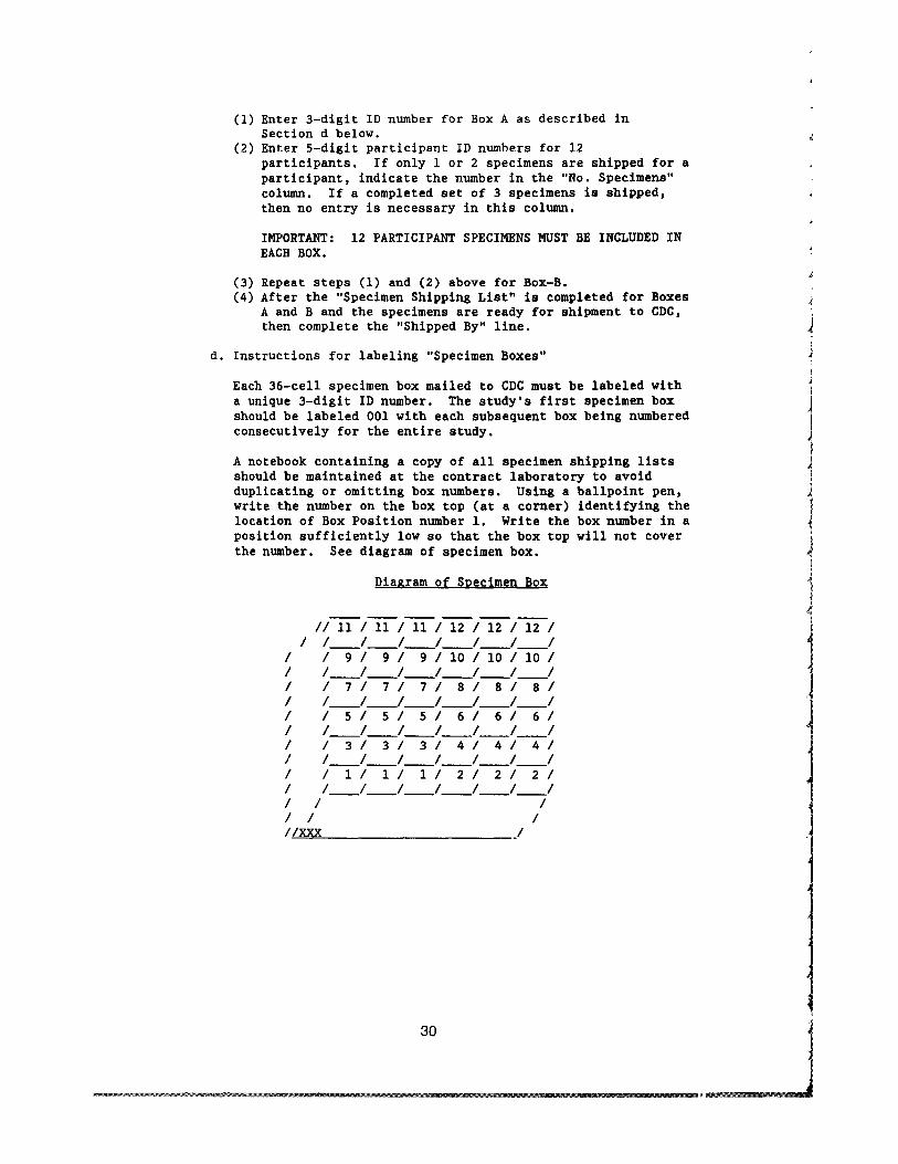
~
1 j
I
(1) Enter 3-digit ID number for Box A as described in Section d below
(2) Enter 5-digit participant ID numbers for 12 participants If only 1 or 2 specimens are shipped for a participant indicate the number in the No Specimens column If a completed set of 3 specimens is shipped then no entry is necessary in this column
IMPORTANT 12 PARTICIPANT SPECIMENS MUST BE INCLUDED IN EACH BOX
(3) Repeat steps (1) and (2) above for Box-B (4) After the Specimen Shipping List is completed for Boxes
A and B and the specimens are ready for shipment to CDC then complete the Shipped By line
d Instructions for labeling Specimen Boxes
Each 36-cell specimen box mailed to CDC must be labeled with a unique 3-digit ID number The studys first specimen box should be labeled 001 with each subsequent box being numbered consecutively for the entire study
A notebook containing a copy of all specimen shipping lists should be maintained at the contract laboratory to avoid duplicating or omitting box numbers Using a ballpoint pen write the number on the box top (at a corner) identifying the location of Box Position number 1 Write the box number in a position sufficiently low so that the box top will not cover the number See diagram of specimen box
Diagram of Specimen Box
II 11 I 11 I 11 I 12 I 12 I 12 I I 1___1___1___1___1___1___1
I I 9 I 9 I 9 I 10 I 10 I 10 II 1___1___1___1___1___1___1 I 171 7 I 7 I 8 I 8 I 8 II 1___1___1___1___1___1___1 I 151 5 I 5 I 6 I 6 I 6 I I 1___1___1___1___1___1___1 I 131 3 I 3 I 4 I 4 I 4 II 1___1___1___1___1___1___1 I 111 1 I 1 I 2 I 2 I 2 II 1___1___1___1___1___1___1 I I I I I I IIXXX j
30
poundZIll IIi amp
[ I
[
381 it m_ PI
Top View
xxx Specimen box ID number 1-12 = Specimen box position number for each
set of participant samples
e Instructions for packing specimen boxes
(1) For each participant a set of three frozen serum specimens (A B and C) will be shipped to the CDC
(2) Specimens A B and C are to be positioned in the box itS
shown in the Diagram of Specimen Box (shown above) using the box position number from the Specimen Shiptng List If a participant has less than three specimens leave the allocated space empty Specimens are packed left to right starting in the lower left corner usin the box number as a starting point
f Shipment of specimens to CDC
(1) Specimens are to be shipped to the CDC only on Tuesdayn
IMPORTANT Since the materials packed in accordance ~th the instructions below will remain frozen only about 2 12 days shipments should not arrive in Atlanta on weekends or on Federal holidays
(2) For all shipments do not pack the shippers with froze I specimens and dry ice until just before transport to tLe Express Mail office at Albuquerque International Airpolmiddott
(3) Dry ice is delivered every Monday by Argyle Welding Supply Inc Albuquerque New Mexico (Telephone numbel (505) 345-8101 (Specify 100 lbs dry ice 1- by 7- by 10-inch slabs)
Note When packing the shippers wear gloves when handling the dry ice to avoid burning the hands GlasEes or an eye shield should also be worn if the dry ice ca~es are to be broken into small pieces
(4) Instructions for packing and shipping frozen serum specimen vials A B and C to CDC
Remember Mail only totally filled shippers that contlin specimens from 24 participants in two-specimen boxes Any number of participant specimens not divisible by 21 must be stored at the contract laboratory until enough specimens to fill a full shipper are available for shipment
(a) Seal each specimen box with filament tape
31
(b) Pack four specimen boxes (Boxes A and B from two shipping lists) per Styrofoam shipper
(c) Fill the shipper with dry ice (it probably will hold 10-12 1bs) and place the Styrofoam lid on top of the shipper
(d) Secure the completed Specimen Shipping List in a Ziploc bag and attach to the top of the Styrofoam lid with filament tape In addition mail a photocopy copy of the Specimen Shipping List in a separate envelope to the same CDC address (see (f)(i) below) This is to ensure against loss and considerable time lag in detecting missing shippers Secure the outer carton lid on the shipper with filament tape
(e) Cover or remove previous labels on all shippers
(f) Additional Express Mail labels are available at the Post Office located in the Clinical Pharmacy on Gibson Street
(i) Preaddressed franked CDC mailing label with following address
Brenda Lewis Centers for Disease Control Chamblee Bldg 31 Rm 8 Atlanta GA 30333
(ii) Express Mail label with the same CDC address as above typed in
(iii) DRY ICE label with the weight of dry ice added
(iv) Dry ice form with weight of dry ice shippers address and CDC address
(v) HUMAN BLOOD label
(vi) Call the courier to pick up the shippers by 1130 am He or she is to deliver the shippers to the Express Mail drop at Albuquerque International Airport by 12 pm on Tuesday for delivery to CDC within 24 hours
(vii) Telephone the laboratory at CDC the day the shipment is mailed
32
1II_lIImllll______OOoo____ _ _______________ ______~
(5) Shipping Supplies - Contact the laboratory at CDC or requesting supplies a-d (a) Specimel boxes 6- by 6- by 2-inches high 36 ell (b) Styrofoam shippers (c) Filament tape for securing shippers (d) Prenumbered labels for the aliquot bottles (e) Preaddressed franked mailing labels (f) Dry ice labels (g) Transparent tape for securing specimen boxes (h) Specimen shipping list
l (i) Protocol (j) Ziploc bags(k) Human blood labels containing after-hours del rery
instructions
6 Procedure Notes
a Criteria for specimen rejection
(1) Inadequate specimen identification
(2) Inadequate volume of blood collected into an addiive tube
(3) Excessive hemolysis of specimen
(4) Improper transportation of samples
7 References
a Cal~~ RR Benoit SW DuBois JA~ Procedures fo the handling and processing of blood specimens NCCLS 1984j4(No9)
b Mansour FK Processing of specimens In Slockbower JM Blumenfeld TA eds Collection and handling of laborltory specimens a practical guide Philadelphia Lippincltt 1983172-91
33
1 ~ j I
I
Ii1
~ j
I J
J
IV SPECIMEN HANDLING AND PROCESSING
B Urine Specimen Handling and Processing
1 Principle
This procedure establishes criteria for handling and processing urine specimens to ensure optimal samples for analytical testing
2 Equipment
a Top loading balance b 100-ml specimen containers with screw-on lids c Preweighed 005-g a1iquots of Na4EDTA (tetra-sodium
ethylenediamine tetra-acetate) (SIGMA order no ED455) d 50 m1 Wheaton vials and stoppers
3 Procedure for 12-Hour Urine Specimen Processing
a Receiving and specimen handling
(1) Upon receipt of the l2-hour specimens denote time and initial the log sheet Ensure that the specimen identification numbers and sample numbers match those on he log sheet Record the preweight of the container with preservative This weight is recorded on the urine contaL~er and was measured before the container was delivered to th participant
(2) Weigh each 12-hour specimen and record weight (to the neaest tenth of a gram) on the container and on the log sheet carefully checking identification and sample numbers on container and log sheet
(3) Store specimens in the refrigerator (2-8 OC) until a1iquoting can be done
(4) Assume a constant specific gravity of 1012 for each l2-hclIr urine collection and calculate the total volume (TV) for Each specimen
TV = weight of specimen 1012
(5) Record the total volume on the log sheet (Report TV to tLe nearest whole milliliter)
b Aliquoting
(1) Affix computer aliquot labels to the appropriate aliquot containers
34
(a) porphyrins - lOO-ml specimen container (b) porphobilinogen (PBG) and D-glucaric acid (D-glucl shy
lOO-ml specimen container (c) creatinine - lOO-ml specimen container (d) storage aliquot - 50-ml Wheaton vial
(2) Remove corresponding 12-hour specimens from the refrilerator (do NOT process more than four specimens at one time)
(3) Carefully match specimens and aliquot containers by clecking participant identification number and sample number
(4) Mix specimen THOROUGHLY before aliquoting
(5) Pour urine from the collection container into the aPPlopriate container
(a) Porphyrins 50 ml (b) PBG and D-gluc = 10 ml minimum (c) Creatinine = 2 ml minimum (d) Storage 40-45 ml (if available)
(6) To each porphyrin aliquot (50 ml) add 005 g tetrascdum EDTA Cap and invert several times to mix
(7) Cap each aliquot container and store in the designatEcl areas of the refrigerator until samples are distributed
(8) The Wheaton vials are for stored specimens and are tc be frozen at -20 degC
(9) Enter the 12-hour urine total volume in the CHC compler and verify the urine collection
c Distribution
(1) Notify each testing area when all participant specimlls are aliquoted and ready for analysis Store the urine al Lquots at 2-8 degc if distribution to testing areas is delayec
(2) On specimen acceptance log sheet initial and record he time the samples are distributed for analysis
(3) Be sure appropriate computer labels accompany aliquot
(4) Record your initials on the log sheet when each specnen is completed and stored
35
l
ramp-middot==--~middot~=~~==~-=~==~~~~--~~~~~~--~~~~~~~~~~~~--~~~~ ----~~~~-
IV SPECIMEN HANDLING AND PROCESSING
C Semen Specimen Handling and Processing
1 Principle
This procedure describes the processing of semen specimens n preparation for automated semen analysis Semen specimenslre dropped in a mail-type holding bin outside the processing Nom (in the hotel where the samples are collected) by the participllt either in the morning or afternoon designated collection times 1 technologist is present in the processing room to receive Lld begin processing the sample Selected semen characteristics are middotloted at this time
2 EguiJment
a pH Meter-Orion 70lA b Incubation oven (30 OC) c Vortex (Thermolyne Mix Max) d Slide staining rack e Microscope slides-frosted end f Pasteur pipets - 5-34 inch g 5-cc syringes h 19-Gauge syringe needles i pH Paper 0-14 unit range j Buffer pH 7 k Buffer pH 10
f 1 95 Ethanol
3 Procedure
a Fill out a specimen collection worksheet with participall ts ID number name days of abstinence collection time vasetomy status and time of specimen arrival at the processing oom
b Keep specimen at 30 degC until ready for analysis Specilnen is ready for analysis 30 minutes after ejaculation
Note All processing including videotaping of the speimen should be completed within 2 hours of semen collection
c Vortex specimen in the collection container for 15 secomiddotlds WITH THE LID ON Allow specimen to sit for a few seconds afer vortexing to allow aerosols to dissipate before the conainer is opened
d Classify the color of the semen
(1) Normal (gray-white translucent) (2) Brownish reddish yellowish or white (abnormal)
37
e Classify the odor of the semen Do not purposely smell the specimen If a specimen is obviously not normal (musky smelling) it will be obvious when tne lid is removed Classif as
(1) Normal (2) Abnormal
f Measure the volume of the semen Use a 5-cc syringe with an l8-l9-gauge needle Measure to the nearest 01 mI Dispense back into the container
g Measure the pH of the semen to the nearest 01 unit
h Measure the viscosity of the semen 30 minutes after ejaculation (after measuring the volume) Aspirate a small amount of seminll fluid into a transfer pipet Slowly expel and grade as fo11owJg
(1) Normal (2) Minor moderate or severe (abnormal)
i Before placing semen in the Mackler Chamber (in preparation for videotaping the specimen for automated semen analysis) swirl the container to assure homogeneity of the sample
4 References
a Cannon DC Seminal fluid In Henry JB ed Clinical diagnosis and management by laboratory methods Philadelphia WB Saunders 1984516-9
b Schrader SM Semen analysis Cincinnati National Institute for Occupational Safety and Health 1983 SOP no EA-60(1)
c Urry R Seminal fluid In Kjeldsberg CR Knight JA eds Body fluids laboratory examination of amniotic cerebrospinal seminal serous and synovial fluids a textbook atlas Chicago American Society of Clinical Pathologists 1986 117-2
38
V ANALYTICAL METHODS
A Cell Parameters in Whole Blood (CBC) Coulter s880
1 Principle
The Coulter s880 System is a quantitative automated hematology analyzer for in-vitro diagnostic use in clinical laboratorie It is intended for the quantitative determination of the follong hematologic parameters WBC RBC HGB HCT MCV MGH MCHC and PLT
A sample of anticoagulated venous whole blood is required tc determine the blood parameters The whole blood is automatically diluted by the instrument and prepared for analysis Whi te Illood cells (leukocytes) and hemoglobin concentration are determiIwd via the WBC dilution and RBC and PLT are determined via the RBC dilution The particle counting is accomplished by the impecance
f principle
2 Specimen
a Blood is collected in sodium or potassium EDTA
b Macro Tubes (3 5 or 7 ml)--fill until vacuum is exhaustld Analyze within 4 hours of collection
c Micro Tubes--fill with 200-300 ul of whole lJlood Analyzl within 1 hour of collection
d Specimens may be stored at 2-6 degc for up to 24 hours for testing of all parameters except platelets (Platelet cOllnts must be stored at room temperature)
3 Reagents
The following reagents are stable at room temperature until expiration date stated on container
a Isoton III CMS Catalog no 165-951 (Diluent) b Lyse III CMS Catalog no 172-940 (Lysing Agent) c Isoterge III CMS Catalog no 179-630 (Cleansing Agent)
4 Calibration
Initial calibration will be done on five normal samples eact run in triplicate Calibration will be done on analysis of WEC RBC HGB HCT and platelets
All five samples will be run four times on the analyzer ThE first result will be discarded and the last three averaged
39
Calibration will be checked daily by running three participants specimens in triplicate for WEC RBC HGB HCT and platelets Percent differences retween the primary methods and the analyzer will be averaged and recorded
Action will be taken when the analyzer results deviate by more tLan 3 for RBC HGB and HCT 7 for WBC and 10 for platelets
5 Quality Control Material
I
1 J
1
a Normal (Catalog no 122) and elevated (Catalog no 123) levels of stabilized human red blood cells (Equinox controls made ty Hematronix)
(1) Store upright at 2-10 degC before and after opening
(2) Unopened--stable until expiration date (150 days from manufacture date)
(3) Opened--stable for 16 days Discard after 16 days (Stale 12 hours at room temperature)
(4) Warm vials to room temperature (18-30 OC) before handling Mix by inversion until cells have been resuspended
(5) Place on mixer for 5 minutes one time only when first opened
(6) Immediately before assaying invert vial five times
(7) Run each level of control in quadruplicate each analytica run
6 Procedure
a Preliminary Procedure
(1) Inspect the dilutor for disconnected or pinched tubing
(2) Turn on optic lamp
(3) Inspect the vacuum trap bottle on the pneumatic supply (No liquid should be present)
(4) Turn on the pneumatic supply by pressing the Power button Check the gauges
(a) Pressure--58-60 (b) 5 PSI--5 (c) 30 PSI--30 (d) Vacuum--20
40
(5) Verify that the meniscus of the manometer is on thn black reference mark
(6) Press RES 1 and RES 2 simultaneously to reset the computer (SELECT FUNCTION will appear on the dig tal readout if the reset was done properly)
(7) Press DRAIN and verify that the aperture baths and ~acuum
isolator drain into the waste chamber
(8) Press LYSE and verify that the lytic reagent enter the WBC bath without bubbles in the reagent line Press DHUN again
(9) Press RINSE and verify that a) the apertures fill lith diluent b) that the waste chamber drains and c) rrat the BSV rotates first to the right and then to the 1ef
(10) Press START UP When the cycle is completed pres3 PRINT and log the results
(11) Set the date and test number (000)
(12) Prime the instrument with whole blood by sampling 3ny specimen two times
b o CBC Procedure
[
(1) Mix samples at least 10 minutes on the mechanical t100d mixer but no more than 30 minutes Mix controls 0 morethan 5 minutes before initially opening vial (OncE vial is opened do not place on mechanical mixer again)
(2) Insert into the printer a report form with the participant identification written at the top
(3) Invert each control or participant sample five timEs after removing them from mixer and immediately before thE aspirating procedure
(4) Place the specimen tube under the whole blood aspitation tube inserting the aspiration tube 5-10 mm below the surface of the blood
(5) Depress the WHOLE BLOOD button Once the WIPE TIP appears on the digital display remove the specimen
(6) Wipe the aspiration tube with a damp Kimwipe
(7) The next sample can be aspirated when the SELECT FNCTION appears on the digital display
41
1 (8) The results will be automatically printed Each sample is
run in duplicate The first result is considered a prile and the second the accurate result (Note Both printcuts must show logical agreement or the test must be repeate)
7 References
a Coulter Electronics Coulter counter model s880 product reference manual Hialeah Florida Coulter Electronics ll85
b Coulter Electronics Coulter course guide Model s880 Hialeah Florida Coulter Electronics 1985
c Gilmer PR Williams LJ Koepke JA Bull BS Calibration metlods for automated hematology instruments Am J Clin Pathol 1977 68 (suppl 1)185-90
d Wintrobe MM The approach to hematologic problems In Clinical hematology 7th ed Philadelphia Lea amp Febiger 19743-38
42
V ANALYTICAL METHODS
B Differential Counts of Whole Blood Smears Geometric Data Hematrak Automated Counter
1 Principle
The Hematrak incorporates a mathematical approach to morphoogic analysis and a high resolution three-color video scanning ystem that detects significant leukocyte morphologic features
The video scanner searches the microscopes field until the color analyzer locates a nucleated cell The digitized image of lhat cell is passed into the image memory At this time the morphological analyzer takes over making significant measu]ements of each cell and performing morphologic analysis Included in this analysis are the nucleus nuclearcytoplasm ratio chromatic pattern and cytoplasm
The statistically derived and weighted factors are passed ilLtO the
t
[
f
recognition computer where they are compared with preprograrmed criteria in the reference memory The factors that make u~ the reference memory were developed by the analysis of hundreds of thousands of cells By matching each cells criteria agaillt the reference memory a classification is assigned to that cell by the instrument
2 Specimen
a Collect blood in sodium or potassium ethylenediamine tetra-acetate
b Store at room temperature until slide is made no longer than 4 hours
c Make a spun smear using the Hemaspinner (See Hemaspillller procedure)
d Stain with Wrights stain using the Hemastainer (See Hemastainer procedure)
e Place finished slides in frames five to a frame frostel end at the top Push all slides tightly to the left and up
3 Reagents and Equipment
a Hematrak immersion oil Geometric Data Catalog no 20-1(17-06 Type HT Store at room temperature
b Hematrak slide frames Geometric Data Catalog no 20-l0-02
43
4 j
j
4 Calibration
No calibration is needed with this instrument All parameters are preset and programmed by the manufacturer before the instrument is shipped
5 Quality Control Material
Two slides have been prepared from two separate blood specimens These will be run in quadruplicate each analytical run
6 Procedure
a Preliminary Procedure
(1) No start-up needed instrument remains on at all times
b Differential Procedure
(1) Press DIFF on Hematrak CRT LOOK AT THE SCREEN It sholld say Operator ID Date WBC 200 RBC 200 spun slide If it says Re-cap review insert slides Press GO Pres DIFF again If nothing changes press RECELL then DIFF
(2) Frame slides number frames consecutively enter frame number and slide sample numbers on the log sheet
(3) Place frame(s) in hopper
(4) Press 1D On page 1 change operator number and date Change WBC and RBC to 400 Enter the frame number on the seconl page and slide number Enter sample number of slide I then second slide etc After fifth slide enter frame 2 Slide 1 shows up automatically If you have more than one frare continue entering sample numbers until you have entered 311 of them If you make a mistake press DELETE before ENTER and correct your error If you ENTER then notice the mistake press ID (light goes off) ID (light goes on) and go through by pressing ENTER until you come to the error Press DELETE correct number enter
(5) When all numbers are entered press ID (light goes off)
(6) Now press GO All frames will go through automatically Allow 1 minute per slide (You may enter the first framE start it through then enter the ID numbers on the remainlng frames)
c RECAP Review
(1) After all frames are done the slides not meeting the prEset criteria for passing will be flagged for review Flageing
~ I
44 J i
1mB J
is indicated on the automatic printout and on the sCleen of the Hematrak CRT A dot means no review an F means review
(2) Remove the frames from the bin place in order in thE hopper Press GO Enter first frame number (number enter)
(3) The Hematrak automatically goes to the first slide il that frame that needs review and focuses on the first cel for review Look through the microscope adjust the fOCllS as needed and identify cell (or just GO for smudges stlins etc) with the proper diff key Press GO The next cell is found
Identify GO When done the bell rings
(4) Press COMPLETE The next slide for review is automatically found Continue as in step (3) until the frame is completed
(5) The screen asks for the next frame number Enter tll~ number and continue as above through all the frames
d Morphological Review of Slides
(1) All slides must be reviewed for the presence of hypersegmentation basophilic stippling rouleaux BILd Howell-Jolly bodies Also check the printout from tILe Hematrak for any RBC morphology comments called by tILe instrument Verify these and make a n~te on any RBC morphologic abnormalities you observe
(2) Review slides for morphology during RECAP Review (section c under Procedure) Flag all slides not flagged by thl~
Hematrak by entering an F for each one Comments aII~
written on the worksheet in the space to the right or patient information
(3) Morphology information is entered into the computer manually See the comment information sheets for t~Ie of answers used
7 Comments to Use In Reporting
(Enter with actual abbreviation designated) OIF - Oil Immelsion Field
45
Po1ychromasia
SL MOD MKD
(slight) (moderate) (marked)
2-4 cellsOIF 5-6 cellsOIF gt6 cellsOIF
Hypochromasia
SL MOD MKD
10-24 25-49 50+
Poikilocytosis
SL 4-10 cellsOIF MOD 11-20 cellsOIF MKD gt20 ce11s0IF
Rouleaux present are noted here in conjunction with the gradati(ln of poikilocytosis
Anisocytosis Microcytosis Macrocytosis
SL 5-20 MOD 21-40 MKD gt40
Basophilic Stippling
Rare 1 or 2 cellsseen on scan SL 3 or 4 cellsseen on scan MOD 1 per OIF MKD gt1 per OIF
Hypersegmentation
1+ gt5 polymorphonuclear neutrophils with five lobes100 ce1s 2+ 1 polymorphonuclear neutrophil with six or more lobesOO
cells 3+ Combination of 1+ and 2+ 4+ Large numbers of 1+ and 2+
Howell-Jolly Bodies
Rare = 1 or 2 on scan Few = 3 or 4 on scan MOD 1 per OIF MANY = gt1 per OIF
8 Reference
Geometric Data Hematrak automated differential system model middot90 operators manual Wayne Pennsylvania Geometric Data 1984
46
V ANALYTICAL METHODS
C Prothrombin Time in Plasma MLA 700 Automatic Coagulation Timer
1 Principle
The prothrombin time assesses extrinsic and common pathways 1 coagulation and is used to monitor antithrombin drugs
MLA has developed the photometric clot detection system Ligilt from a calibrated carefully controlled light source passesilrough a collimator in which the light is focused into a parallel eam The light illuminates the sample and the light passing throlgh falls on a photo detector which converts the light intensi t1 into an electrical signal
As the plasma sample clots in response to the addition of thromboplastin it becomes more opaque and the light fa11inl on the photo detector decreases This alters the electrical silnal A sensitive amplifier processes the signal and the instrumelts microprocessor determines when a clot has started to form [he instrument then records the time elapsed between the addi tiOl of reagent and the start of clotting This is the clot time
2 Specimen
a Collect blood in 38 sodium citrate anticoagulated tube (Ratio 91 45 m1 whole blood to 05 m1 citrate solution)
b Keep specimen cool Spin as soon as possible Separate the plasma into 10- x 75-mm plastic tube with cap
c Keep plasma cool (2-8 OC) until analyzed
d Analyze within 2 hours or freeze at -70 degC
e Minimum sample required 02 m1 plasma
3 Reagents and Equipment
a MLA 700 Model E-650 serial no 205
b Plastic tubes with caps 12 X 75 mm
c Plastic cuvettes red Scientific Products catalog no B middoti163-7
d Reservoir and heat exchange tubing Scientific Products clta1og no B 4163-2
47
4 Quality Control and Other Material
a C0ntrols Citrol Level I - Scientific Products catalog no B4224-l0 Dade
Citrol Level II - Scientific Products catalog no B4224-20 Dade
(1) Lyophilized preparations of human plasma stabilizers ani buffers
(2) Store unopened vials at 2-8 degC (3) Remove from refrigerator and allow to warm to room
temperature (4) Reconstitute with 10 ml distilled or deionized water
Stopper vial Label with date and time and initial and allow to stand at room temperature until dissolved approximately 15 minutes Invert gently to mix DO NOT SHAKE
(5) Reconstituted material is stable 8 hours Store at 2-8 0 when not in use
b Thromboplastin C Scientific Products catalog no B4216-50 Dade
(1) Lyophilized acetone-dehydrated rabbit brain thromboplastill 116 X 10-20M Ca++ stabilizers and preservative
(2) Store at 2-8 degC (3) Reconstitute vials with 10 ml deionized water Label with
date time and initials (4) Stable for 3 days if stored at 2-8 0C Mix well
5 Procedure
a Turn on machine and allow 20stabilize
-30 minutes for temperature to
b Reconstitute controls and thromboplastin-C
c Check the following systems
(1) Waste bin - empty (2) Test time - normal (3) Lever set for 300-second (4) Light source set on B (5) Lever on AUTO
test
d Fill cup with stirrer
a MAXIMUM of 8 ml of thrombo-C Add magnetic
e Prime tubing
(1) Check to make sure each reagent cup
plastic fitting is seated properly in
48
ITEa
lshy
f
l
l I f
(2) Prime reagent through tubing back into reagent cup Htil allbubbles are out of the line Note One bubble neal colored tip of line is supposed to be there
(3) Replace reagent cup lid
(4) Check to see if tubing is seated properly in the grc I)ves
(5) Place nozzle into nozzle holder marked TEST
f Run temperature check and confidence test
(1) Press PT 1 button (2) Press START button twice in succession (3) Printout should read
(4) Confidence = PT--199 plusmn 02 (5) Temperature = cold block--15plusmn3 degc (6) Reagent cups--15plusmn3 0c (7) Heat transfer pump block--375plusmn02 degc(8) Heat block--375plusmn02 degc
g Place the necessary number of cuvettes into the carouse] toinclude controls where scheduled Start with the first Hpace to the right of the decoder space
h Add Ol-ml amounts of control and participant samples tc eachcuvette position (Each cuvette runs in duplicate) PIIWet the tip before pipetting Carefully wipe the tip after samIing
i Press the START button once
j Two short beeps will indicate the run is complete
k The results will be printed out on tape
1 Discard reagents Wash cup with distilled water and dr)
m Flush tubing with deionized water Remove tubing
n Turn off machine
6 References
a Dade Thromboplastin-C package insert Aguada Puerto l~ico Dade 1982
b Medical Laboratory Automation Electra 700 automatic coagulation timer operators manual Mt Vernon New Ycrk Medical Laboratory Automation 1984
49
V ANALYTICAL METHODS
D Erythrocyte Sedlmentatlon Rate in Whole Blood
1 Principle
The erythrocyte sedimentation rate (ESR) test measures the settlirg of erythrocytes in human plasma over a specified time period The reported numerical value is derived from measuring in millimeter5 the distance from the bottom of the surface meniscus to the top 01 the erythrocyte sediment in a column of anticoagulated blood that has remained perpendicular in a special purpose pipet for 60 minutes
The empirical phenomenon depends on an interrelationship of a number of important inherent variables including plasma protein composition and concentration and erythrocyte configuration
In this ESR method an attempt has been made to control or eliminae the variability of environmental and technical factors that influence the results
The method is based on the Westergren method and calls for dilutiol of blood with autologous plasma to a standardized hematocrit Th change renders the method insensitive to hematocrit variations and provides a linear response throughout the range of clinical interest
2 Specimen
a EDTA (Na) anticoagulated whole blood
b Minimum sample size is 5 mlj optimum size is 7 mI
c Blood left at room temperature (19-25 OC) must be set up within 2 hours If blood is kept at 4 oe it must be set up wi thin 6 hours
3 Reagents and Equipment
a Anticoagulant Ethylene diamine tetra-acetic acid (Na) is used at a concentration of 14 to 16 mgml of blood
b The Westergren Pipet (Scientific Products catalog no B4504-l [Dade] )
(1) The pipet must have the following dimensions Overall length 3005plusmnO5 mm External diameter 55plusmnO5 mm Tube bore 270plusmn020 mm Uniformity of tube bore plusmnO05 mm
(2) The pipet must have the following markings
(a) The tube is inscribed Westergren
50
r (
(b) The tube has an inscribed graduated scale extendlg over the lower 200plusmn0 35 nun of the pipet The graduat ms are numbered from 0 at the top end of the scale to a~ least 180 no less than 20 nun from the lower end of thl~ pipet in steps of 10 or less
Maximal tolerated error between two adjacent milimeter markings is 02 nun tolerated error of the total graduated scale is 035
(c) The l-nun graduation marks are fine clearly mark~d permanent lines of uniform thickness (02 nun) ~Very 10th graduation mark is numbered
(d) The lengths of longer graduation marks are projet equally at each end beyond shorter markings Specifically the lengths are
(i) At the 0 mark 09-10 times the tube cir~lmference
(ii) At each single millimeter mark 0125 (18) times the tube circumference
(Ui) At each 5-nun mark 016 (16) times the tUle circumference
(iv) At each 10-nun mark 025 (14) times the tlbe circumference
(v) The tubes if disposable must be supplied clean and dry The manufacturer must ensure anl provide data on request that the tubes do not contain any factor that mey affect their U3e in the ESR test compared with WSP of the glas3 Westergren tubes described above
c Pipet Rack
(1) Equipped with a leveling bubble device (2) Constructed so that no leakage of the blood can OCCU[
4 Quality Control Material
Four participant samples will be run in duplicate each anal) tic run
51
1 S Procedure
a Blood Collection
(1) Collect whole blood in a 7-ml EDTA tube (2) Mix immediately and THOROUGHLY by gentle inversion
b Hematocrit Adjustment
(1) Obtain an initial hematocrit on the UNADJUSTED blood samp
(a) Spin for 3 minutes at 10000-11000 rpm in the microhematocrit centrifuge Read hematocrit on the accompanying reader
(b) Centrifuge the two equal aliquots of 2 ml each at 800 rpm for S minutes to adequately divide cells and plasrl i
(c) Remove plasma from or add plasma to one of the aliquo1l to obtain a sample with a hematocrit of 03S To calculate the volume required use the following formllLa
2 X hematocrit - 2 = ml plasma 035 required
(2) Determine the hematocrit of the ADJUSTED blood sample [USI
procedure as described in Sb(l)]
c Blood Cell Suspension
(1) Gently resuspend the blood cells thoroughly by placing on il
mechanical rocker for at least 10 minutes Blood should remain rocking until ready to aspirate
(2) Using a mechanical suction device aspirate a bubble-free sample into a clean and dry Westergren pipet exactly filling to the 0 mark
d Handling of Westergren pipet
(1) Set up singly Pick four of the samples and set them up ill duplicate
(2) Place the filled pipet in a vertical position in the pipet rack Area should be free of vibrations air draft and direct sunlight
52
e Reading of test
(1) After 60 minutes plusmn30 seconds read the distance of millimeters from the bottom of the plasma meniscus ltwhich should be at 0) to the top of the column of sedinmted erythrocytes Do not include buffy coat with the erythrocyte column
(2) Record the numerical value
6 References
a Bull BS Is a standard ESR possible Lab Med 1975 j 6110 11)31-5
b Koepke JA Bull BS Van Assendelft OW Reference procldure for the human erythrocyte sedimentation rate (ESR) test NCCLS 1983 3(no 7)251-64
53
J
~
j
1
V ANALYTICAL METHODS
E Routine Urinalysis in a Random Urine
1 Principle
Properly executed routine urinalysis will indicate the presence 01 renal disease (protein cells and casts) urinary tract infectior and diabetes mellitus (glucose) The routine urinalysis should bE done on a fairly concentrated specimen which is usually the first morning specimen especially when examined for protein and microscopic content
2 Specimen Collection and Storage
The urine sample must be collected in a clean dry container covered with a lid and examined as soon as possible Bacterial contamination may occur and leukocytes and casts disintegrate as the speciman ages Specimens should be examined within 1 hour of collection If this is not possible the specimen must be refrigerated at 4 degC until it is examined Minimum volume is 10 ml although 12 ml is optimum for the test
3 Reagents
a Ames Multistix Store at room temperature Gooc until expiration date on the bottle Ames Inc catalog no 4820
b Count-IO Trol II Urine Control (15-ml lyopholyZE ltl specimen) Store at 2-8 degC Good until expiration date on tl I ~
bottle (unopened) V-Tech Inc catalog no 10-0751
c Clinitek calibration strips Store at room temperature Good until expiration date on the box Ames Inc catalog no 5382
d Distilled water
e 4 Sodium chloride Store at room temperature Stable for 6 months
Procedure for preparing 4 NaCl
1 Weigh 4 g of dried NaCl crystals
54
2 Dissolve in distilled Hater in a 100-ml volumetric flask
3 QS to 100 mI
4 Quality Control Material
a Source
V-Tech Inc Count-IO-Trol Level II Each vial contait lyophilized material derived from human urine containin~ measured amounts of chemicals stabilized human erythrocrtes and simulated leukocytes
b Preparation
(1) Bring reagents to room temperature (2) Slowly vent cap on reagent vial to avoid reagent 101 l (3) Reconstitute each control vial with 15 ml of distil ~d
water Stable 5 days after reconstitution (4) Cap and swirl control vial for 20 seconds Allow 2(1 to 30
minutes for complete reconstitution (5) Remix reagent before each use
c Storage
Store controls at 2-8 degC Reconstituted controls remailL stable for 5 days at 2-8 degC Between use all controls ghould be stored at 2-8 degC Unopened controls are stable untL the expiration date on the bottle
5 Procedure
a Perform calibration on the Clinitek 200 and the Atago-UR-20 refractometer
b Prepare controls
c Mix specimen well
d Transfer 10-12 ml of patient urine and control to the pLastic centrifuge tubes labeled with specimen sample number
e Record color (straw yellow amber or specific color) ltHld appearance (clear hazy cloudy flocculent)
f Perform and record the specific gravity using the USR-2IJshyrefractometer Record the result to three decimal placlts (~ 1024)
g Perform chemical examination with a dipstick and the AmltS Clini tek 200
55
h Centrifuge urine tube for 5 minutes at 2000 rpm
i Decant tube without blotting
j Perform microscopic examination using the phase XIO lens and rre phase X40 lens
(1) Resuspend the sediment (2) Examine the drop of sediment on slide with 20 x 20
coverslip using the phase X10 lens first Look for cast1 and epithelial cells
Note It is necessary to distinguish between cast types Swi tch to the phase X40 lens and carefully scan 10middotmiddot12 separate fields of the slide Estimate average r~l~e of WBC RBC and renal tubular cells per high powe field (hpf)
(3) Standard nomenclature
(a) Cells (RBC and WBC) Range of numbers hpf (b) Casts Range of number per low power field (lpf) (c) Epithelial cells Report percentage of lpf
k Perform the daily shut-down procedure on the Clinitek 200 and Atago Refractometer
6 Codes and Comments To Use In Reporting
Assay Name
Hemoglobin Bilirubin
Comments
544 Negative 670 Small amount 406 Moderate amount ~
1 407 Large amount 2170 Trace
Ketones (mgd1)
544 Negative 5369 5-15 (trace) 5370 15-40 5371 40-80 5372 gt80
56
f
Urobilinogen (mgdl)
Codes Comments
5379 02-1 (- normal) 5380 1-2 5381 2-4 5374 4-8 5375 8-12 5382 )12
Protein (mgdl)
544 Negative 2170 Trace 5373 30-100 5378 100-300 5363 )300
Glucose (mgd])
544 Negative 5376 100-250 5377 250-500 5367 500-1000 5368 )1000
Color
5326 Straw yellow (normal) 510 Amber 513 Red
5327 Blue-green 2156 Brown 5328 Dark yellow 5329 Yellow-orange
645 Orange 5330 Red-brown 2160 Yellow
511 Colorless
Appearance
145 Clear (normal) 517 Hazy 518 Cloudy 147 Turbid
57
RBCs and Leukocytes Urine (cellshpf)
Codes Comments
396 None seen 1780 Rare 5331 Occasional 5332 Few = (1-5) 5333 1+ (6-20) 5334 2+ (21-50) 5335 3+ (51-100) 5336 4+ (gt100)
Renal Tybular Cells (ce11slpf)
1780 Rare 5331 Occasional 5337 1+ (1-5) 5338 2+ (6-10) 5339 3+ (11-30) 5340 4+ (gt30)
Casts (Hyaline Granular WBC RBC Other)(nolpf (XlOraquo
1780 Rare 5331 Occasional 5337 1+ (1-5) 5338 2+ = (6-10) 5339 3+ (11-30) 5340 4+ (gt30)
Epithelial Cells Urine (nolpf)
1780 Rare 5331 Occasional 5341 Few laquothan 25 of field) 5342 1+ (25 of field) 5343 2+ = (50 of field) 5344 3+ (75 of field) 5345 4+ (100 of field)
447 Packed
7 References
a Ames Division Miles Laboratories Clinitek 200 operating manual 4th ed Elkhart Indiana Ames Division Miles Laboratories 1985
b Bradley M Schumann GB Examination of urine IN Henry JB ed Clinical diagnosis and management by laboratory methods Philadelphia WE Saunders 1984380-58
58
c NSG Precision Instruction manual for the USR-20 refractometer Hicksville New York NSG Precision 198~
d V-Tech Count-10-Tro1 urine control package insert Pomna California V-Tech 1984
59
I
J
j
V ANALYTICAL METHODS
F D-Glucaric Acid in Urine Ion-Exchange Chromatography
1 Principle
For the quantitative determination of D-glucaric acid buffered urine specimens are added to an ion-exchange resin-filled column 0
separate D-glucaric acid from other interfering substances Afte elution from the column D-glucaric acid is oxidized with periodil acid to yield glyoxylic acid Glyoxylic acid is then oxidatively coupled with phenylhydrazine to yield the dye 15-diphenylformazan Absorbance at 520 mm is read and concentration determined by comparison to standard curve absorbanl e
D-glucaric acid is a product of glucuronic acid metabolism in the liver Urinary excretion reflects the activity of hepatic enzyme that hydroxylate and detoxify many foreign substances
Elevated levels are observed after the administration of drugs known to induce enzyme activity and anticonvulsant drugs and in association with exposure to insecticides
L-ascorbic acid (vitamin C) is know to falsely elevate D-glucaric acid levels obtained in this method therefore all urines are prescreened for ascorbic acid by using a dipstick method
2 Specimen Collection and Storage
a No participant preparation is necessary
b A well-mixed aliquot (4-10 ml) of a l2-hour urine collection l used for this test Urine specimens are collected in containell with 2 of sodium carbonate The specimen should be protected from light and kept cold during collection
c The specimen is stable for 24 hours at 2-8 0c For longer storage freeze at -20 0c
3 Reagents Supplies and Equipment
a Glass test tubes disposable 16 X 100 mm 16 X 150 mm 10 X i~i mm
b Pipets Eppendorf pipet l25-ml tips MLA 1 ml with disposable tips Accuprep (Beckman) capable of dispensing 1-1000 ul Repipet 5-ml (for addition of eluant to column)
60
RTFITUf
1m
c Class A volumetric flasks 2L lL 100 ml 50 ml 10 ml
d Analytical balance (Mettle~ AE 166)
e Spectrophotometer having band width of 1-2 nm 10-cm liht path (Beckman DU-7)
f Millipore filters
g Draining rack with waste container
h Plastic transfer pipets
i Magnetic stirrer
j Plastic caps 16 mm
k Stix (ascorbic acid reagent strips) Ames
1 Funnels (BioRad)
m Vortex mixer
n Test-tube racks
o Parafilm
p Timer
q Resin columns AG 1X-8 room temperature Refer
resin to
obtained from Bioexpiration date on
Rad box
Stoe at
r Sodium borate OlM
(1) Dissolve in volumetric flasks (a) Sodium borate--30596 g
Certified ACS Fisher (b) Reagent water type 1--8 L
(2) Place in polyethylene carboy Label as Sodium BoraL~ OlM with preparation date technician ID and an expiration date of 1 year Store at room temperaturl
s Sodium borate 005 Msodium sulfate 002 M
(1) Dissolve in volumetric flasks (a) Sodium borate--190685 g (b) Sodium sulfate--2840BO g
Anhydrous E M Science (c) Reagent water type 1--10 L
61
(2) Place in polyethylene carboy label as Sodium Borate 005MSodium sulfate 002M with preparation date tecrnician identification and an expiration date of 1 year Store at room temperature This quantity is sufficient for 1 week J
t Sodium borate OOSMsodium sulfate 01 M
(1) Dissolve in volumetric flasks (a) Sodium borate--190685 g (b) Sodium sulfate--142040 g (c) Reagent water type 1--10 L
(2) Place in polyethylene carboy label as Sodium Borate 005 M sodium sulfate OlM with preperation date technicians initials and an expiration date of 1 year Store at room temperature Aliquots are transferred to a repipet capable of dispensing 5 mI Label as above
u Saccharic acid (potassium hydrogen d-glucarate) Sigma storE in desiccator at room temperature
v Potassium biphosphate 10 Molar
(1) Dissolve in a 100-ml volumetric flask (a) Potassium phosphate monobasic--13609 g (b) Technical grade MCB (c) Reagent water type I--QS to 100 m1
(2) Transfer to glass bottle label as Potassium BiphosphatE 1 OM with preparation date technicians initials and III
expiration date of 2 weeks Store at 2-8 degC Mix well before using
w Periodic acid 001 Molar
(1) Dissolve in a SO-ml volumetric flask (a) Periodic acid--0114 g
Sigma (b) Reagent water type 1-- QS to 50 m1
(2) Transfer to brown glass bottle label as Periodic Acid OOlM with preparation date technicians initials and iLn expiration date of 1 week Store at 2-8 degC
x Phenylhydrazine Hydrochloride 1
(1) Dissolve in a 100-ml volumetric flask (a) Phenylhydrazine HCl--lO g
MCB (b) Reagent water Type I--QS to 100 m1
62
-----~
i
(2) Transfer to brown glass bottle label as Phenylhydllzine HCll with preparation date technician s initial and an expiration date of 1 week Store at 2-8 degC
y Hydrochloric Acid conc Reagent Grade MCB Reagents ~ ore under hood
z Potassium ferricyanide 5
(1) Dissolve in a 100-ml volumetric flask (a) Potassium ferricyanide--50 g
Technical grade MCB (b) Reagent water type I-QS to 100 ml
(2) Transfer to a brown glass bottle label as Potassi1Ln Ferricyanide 5 with preparation date technician initials an expiration date of 1 week Store at 2middotmiddot~ degC
aa Ascorbic Acid Control 25 mgdl
(1) Dissolve in an lL volumetric flask (a) 250 mg ascorbic acid ACS MCB reagents (b) Reagent water type I-QS to 1 L
(2) Aliquot approximately 3 ml in 10- x 75-mm tubes and store at -20 degC Stable for 6 months
bb Ascorbic acid dipsticks (Uralyte GK + A) Medical Sales Associates catalog no 8001
4 Calibration
a D-dlucaric acid stock std 1000 ugml
(1) Dissolve in a 100-ml volumetric flask (a) D-saccharic acid--lOO mg (b) Reagent water type I--QS to 100 ml
(2) Aliquot 1-2 ml in 10 X 75 mm tubes and store at -20 lC Stable for 3 months
b Using the Beckman Accuprep dilute working standards daiLy as follows
Prepare the working standards during the final elution (3tep 6d(3) of the procedure that follows)
c Prepare 100 ugml working stock standard
Dilute 1 ml (use a 1 ml volumetric pipette) of the 1000 ugml stock in a 10-ml volumetric flask QS to 10 ml with water reagent type I
63
Prepare Standards
D-glucarate ml of ml of std 100 ugml 005 m NaBoratel
ugml Stock Standard 01 m NaSulfate
2 0200 98 4 0400 96 6 0600 94 8 0800 92
10 1000 90
Standards at the 2- 4- 6- 8- and 10-ugml levels are run in duplicate with each assay
5 Quality Control Material
A control urine will be run in duplicate at the beginning and elll of each analytical run The control urine used is Lyphochek Quantitative Urine Control Abnormal Level II by BioRad The control is reconstituted by using a 25-ml volumetric pipet and reagent water type I It is made up at the beginning of each ~ek and then aliquoted and frozen for each days use A positive ahl negative ascorbic acid control will be run at the beginning and ~nd of the ascorbic acid screening procedure and ascorbic acid resul s for controls and participant samples will be recorded on the WOIC
list
6 Procedure
a Allow all reagents standards and participant sample specimElls to come to room temperature before testing
b Labell BioRad column for each participant and control Shdl~
column vigorously until resin is completely suspended No rEnin should remain in tip when the column is inverted Snap off ends attach funnels place in rack and drain into a waste container Do not insert pipets or other objects into the columns to further mix the contents while they are draining
c Screening for Ascorbic Acid
(1) In addition to each participant sample a positive ascorbc acid control and a negative control (reagent water type I must be tested for ascorbic acid Run these controls at 1he beginning and end of each analytic run
(2) Completely immerse the reagent area of an ascorbic acid dipstick in the well-mixed uncentrifuged urine or control sample
(3) Draw the edge of the strip against the side of the contairer to remove any excess liquid
64
j ill
(4) Compare the color of the reagent area with the color chart on the bottle label at exactly 60 seconds Hold the trip close to the color blocks and match carefully
(5) Record results in the 0-10 mgdl range as negative Jny results of 25 mgdl or greater are to be recorded on Ihe D-glucaric worksheet as positive and the D-glucaric IISUlt NOT reported Any positive ascorbic acid screen is til be reported to the QC supervisor
d Separation of D-glucaric Acid
(1) Label one 16- x 100-mm test tube for each participant and control Use a l-ml MLA to place 2 ml of the well-m_ ed l2-hour urine or control into its tube Using an Epp~ndorf pipet add 2 ml of OlM sodium borate to each tube lortex and let stand at room temperature 15 minutes
(2) Pour the urine mixture onto each drained column AL)w the samples to be completely absorbed on the resin (abou- 20 minutes) before proceeding
(3) Wash the columns with 100 ml of 005 M sodium borate--[)02 M sodium sulfate Drain completely into the waste recptacle (about 3 hours)
(4) Label one 16- X l50-mm test tube for each participantcontrol and place under the columns El-l te D-glucaric acid with lS ml (use the S-ml repipet) OJSM sodium borateOl sodium sulfate Let drain completly into tubes (about 1 hour)
e Colorimetric Procedure
Make up daily standards as described in the calibration section
(1) Cover with Parafilm and mix the eluate well by inversion With an MLA place 1 ml in each of two labeled 16- X 100-mm test tubes
(2) With an MLA place 1 ml of each of the standards int duplicate labeled 16- X 100-mm test tubes Two zero standards (reagent blank) are prepared by pipetting 1 ml of the OOSM sodium borate01M sodium sulfate buffer irto two labeled tubes
(3) Add 200 ul (use Accuprep program 1) of 1OM potassiunl biphosphate to each tube and vortex Make sure the potassium biphosphate reagent is well-mixed before udng
(4) Add 20 ul (use Accuprep program 4) of OOlM periodic acid vortex again and let stand at room temperature for _5 minutes
(5) Add 200 ul (use Accuprep program 1) of 1 phenylhydwzine hydrochloride vortex and let stand at room temperBIure for 10 minutes
(6) Using the accuprep program 2 add 1 ml of concentratld HClj vortex and follow immediately with the addition of ~OO ul S potassium ferricyanide (Accuprep) Vortex and lE - stand at room temperature 5-10 minutes
(7) Measure and record the absorbancy at 520 nm against -he reagent blank
65
7 Calibrations
a Use the linear regression program in the CHC or the IBM PCXT computer to evaluate the standard curve and obtain participar results (in ugml)
b After this result and the urine creatinine (creat) results hee been verified the force calculations mode is used to perforn the following calculation
Uncorrected D-Glucaric Acid (D-Glucaric Acid X 75) (in mgg creat) (in ugml)
Urine CreatIOO (in mgdl)
c Each duplicate control result is multiplied by 75 to correct for the dilution factor
8 References
a Hunter J Maxwell JD Carella M Stewart DA Urinary D-glucalic acid excretion as a test for hepatic enzyme induction in man Lancet 19711572-5
b Ishidate M Matsui M Okada M Biochemical studies on 1 14j
j
I ~
glucuronic acid and glucaric acid quantitative chemical determination of D-g1ucaric acid in urine Anal Biochem 1965 11176-89
c March J Turner WJ Shanley J Field J Values for urinary excretion of D-g1ucaric acid by normal individuals Clin Chel 1974 20(No9)1155-8
do McFadden BA Howes WV The determination of glyoxylic acid 11 biological systems Anal Biochem 1960 1240-8
66
1
l
ramp-middot==--~middot~=~~==~-=~==~~~~--~~~~~~--~~~~~~~~~~~~--~~~~ ----~~~~-
IV SPECIMEN HANDLING AND PROCESSING
C Semen Specimen Handling and Processing
1 Principle
This procedure describes the processing of semen specimens n preparation for automated semen analysis Semen specimenslre dropped in a mail-type holding bin outside the processing Nom (in the hotel where the samples are collected) by the participllt either in the morning or afternoon designated collection times 1 technologist is present in the processing room to receive Lld begin processing the sample Selected semen characteristics are middotloted at this time
2 EguiJment
a pH Meter-Orion 70lA b Incubation oven (30 OC) c Vortex (Thermolyne Mix Max) d Slide staining rack e Microscope slides-frosted end f Pasteur pipets - 5-34 inch g 5-cc syringes h 19-Gauge syringe needles i pH Paper 0-14 unit range j Buffer pH 7 k Buffer pH 10
f 1 95 Ethanol
3 Procedure
a Fill out a specimen collection worksheet with participall ts ID number name days of abstinence collection time vasetomy status and time of specimen arrival at the processing oom
b Keep specimen at 30 degC until ready for analysis Specilnen is ready for analysis 30 minutes after ejaculation
Note All processing including videotaping of the speimen should be completed within 2 hours of semen collection
c Vortex specimen in the collection container for 15 secomiddotlds WITH THE LID ON Allow specimen to sit for a few seconds afer vortexing to allow aerosols to dissipate before the conainer is opened
d Classify the color of the semen
(1) Normal (gray-white translucent) (2) Brownish reddish yellowish or white (abnormal)
37
e Classify the odor of the semen Do not purposely smell the specimen If a specimen is obviously not normal (musky smelling) it will be obvious when tne lid is removed Classif as
(1) Normal (2) Abnormal
f Measure the volume of the semen Use a 5-cc syringe with an l8-l9-gauge needle Measure to the nearest 01 mI Dispense back into the container
g Measure the pH of the semen to the nearest 01 unit
h Measure the viscosity of the semen 30 minutes after ejaculation (after measuring the volume) Aspirate a small amount of seminll fluid into a transfer pipet Slowly expel and grade as fo11owJg
(1) Normal (2) Minor moderate or severe (abnormal)
i Before placing semen in the Mackler Chamber (in preparation for videotaping the specimen for automated semen analysis) swirl the container to assure homogeneity of the sample
4 References
a Cannon DC Seminal fluid In Henry JB ed Clinical diagnosis and management by laboratory methods Philadelphia WB Saunders 1984516-9
b Schrader SM Semen analysis Cincinnati National Institute for Occupational Safety and Health 1983 SOP no EA-60(1)
c Urry R Seminal fluid In Kjeldsberg CR Knight JA eds Body fluids laboratory examination of amniotic cerebrospinal seminal serous and synovial fluids a textbook atlas Chicago American Society of Clinical Pathologists 1986 117-2
38
V ANALYTICAL METHODS
A Cell Parameters in Whole Blood (CBC) Coulter s880
1 Principle
The Coulter s880 System is a quantitative automated hematology analyzer for in-vitro diagnostic use in clinical laboratorie It is intended for the quantitative determination of the follong hematologic parameters WBC RBC HGB HCT MCV MGH MCHC and PLT
A sample of anticoagulated venous whole blood is required tc determine the blood parameters The whole blood is automatically diluted by the instrument and prepared for analysis Whi te Illood cells (leukocytes) and hemoglobin concentration are determiIwd via the WBC dilution and RBC and PLT are determined via the RBC dilution The particle counting is accomplished by the impecance
f principle
2 Specimen
a Blood is collected in sodium or potassium EDTA
b Macro Tubes (3 5 or 7 ml)--fill until vacuum is exhaustld Analyze within 4 hours of collection
c Micro Tubes--fill with 200-300 ul of whole lJlood Analyzl within 1 hour of collection
d Specimens may be stored at 2-6 degc for up to 24 hours for testing of all parameters except platelets (Platelet cOllnts must be stored at room temperature)
3 Reagents
The following reagents are stable at room temperature until expiration date stated on container
a Isoton III CMS Catalog no 165-951 (Diluent) b Lyse III CMS Catalog no 172-940 (Lysing Agent) c Isoterge III CMS Catalog no 179-630 (Cleansing Agent)
4 Calibration
Initial calibration will be done on five normal samples eact run in triplicate Calibration will be done on analysis of WEC RBC HGB HCT and platelets
All five samples will be run four times on the analyzer ThE first result will be discarded and the last three averaged
39
Calibration will be checked daily by running three participants specimens in triplicate for WEC RBC HGB HCT and platelets Percent differences retween the primary methods and the analyzer will be averaged and recorded
Action will be taken when the analyzer results deviate by more tLan 3 for RBC HGB and HCT 7 for WBC and 10 for platelets
5 Quality Control Material
I
1 J
1
a Normal (Catalog no 122) and elevated (Catalog no 123) levels of stabilized human red blood cells (Equinox controls made ty Hematronix)
(1) Store upright at 2-10 degC before and after opening
(2) Unopened--stable until expiration date (150 days from manufacture date)
(3) Opened--stable for 16 days Discard after 16 days (Stale 12 hours at room temperature)
(4) Warm vials to room temperature (18-30 OC) before handling Mix by inversion until cells have been resuspended
(5) Place on mixer for 5 minutes one time only when first opened
(6) Immediately before assaying invert vial five times
(7) Run each level of control in quadruplicate each analytica run
6 Procedure
a Preliminary Procedure
(1) Inspect the dilutor for disconnected or pinched tubing
(2) Turn on optic lamp
(3) Inspect the vacuum trap bottle on the pneumatic supply (No liquid should be present)
(4) Turn on the pneumatic supply by pressing the Power button Check the gauges
(a) Pressure--58-60 (b) 5 PSI--5 (c) 30 PSI--30 (d) Vacuum--20
40
(5) Verify that the meniscus of the manometer is on thn black reference mark
(6) Press RES 1 and RES 2 simultaneously to reset the computer (SELECT FUNCTION will appear on the dig tal readout if the reset was done properly)
(7) Press DRAIN and verify that the aperture baths and ~acuum
isolator drain into the waste chamber
(8) Press LYSE and verify that the lytic reagent enter the WBC bath without bubbles in the reagent line Press DHUN again
(9) Press RINSE and verify that a) the apertures fill lith diluent b) that the waste chamber drains and c) rrat the BSV rotates first to the right and then to the 1ef
(10) Press START UP When the cycle is completed pres3 PRINT and log the results
(11) Set the date and test number (000)
(12) Prime the instrument with whole blood by sampling 3ny specimen two times
b o CBC Procedure
[
(1) Mix samples at least 10 minutes on the mechanical t100d mixer but no more than 30 minutes Mix controls 0 morethan 5 minutes before initially opening vial (OncE vial is opened do not place on mechanical mixer again)
(2) Insert into the printer a report form with the participant identification written at the top
(3) Invert each control or participant sample five timEs after removing them from mixer and immediately before thE aspirating procedure
(4) Place the specimen tube under the whole blood aspitation tube inserting the aspiration tube 5-10 mm below the surface of the blood
(5) Depress the WHOLE BLOOD button Once the WIPE TIP appears on the digital display remove the specimen
(6) Wipe the aspiration tube with a damp Kimwipe
(7) The next sample can be aspirated when the SELECT FNCTION appears on the digital display
41
1 (8) The results will be automatically printed Each sample is
run in duplicate The first result is considered a prile and the second the accurate result (Note Both printcuts must show logical agreement or the test must be repeate)
7 References
a Coulter Electronics Coulter counter model s880 product reference manual Hialeah Florida Coulter Electronics ll85
b Coulter Electronics Coulter course guide Model s880 Hialeah Florida Coulter Electronics 1985
c Gilmer PR Williams LJ Koepke JA Bull BS Calibration metlods for automated hematology instruments Am J Clin Pathol 1977 68 (suppl 1)185-90
d Wintrobe MM The approach to hematologic problems In Clinical hematology 7th ed Philadelphia Lea amp Febiger 19743-38
42
V ANALYTICAL METHODS
B Differential Counts of Whole Blood Smears Geometric Data Hematrak Automated Counter
1 Principle
The Hematrak incorporates a mathematical approach to morphoogic analysis and a high resolution three-color video scanning ystem that detects significant leukocyte morphologic features
The video scanner searches the microscopes field until the color analyzer locates a nucleated cell The digitized image of lhat cell is passed into the image memory At this time the morphological analyzer takes over making significant measu]ements of each cell and performing morphologic analysis Included in this analysis are the nucleus nuclearcytoplasm ratio chromatic pattern and cytoplasm
The statistically derived and weighted factors are passed ilLtO the
t
[
f
recognition computer where they are compared with preprograrmed criteria in the reference memory The factors that make u~ the reference memory were developed by the analysis of hundreds of thousands of cells By matching each cells criteria agaillt the reference memory a classification is assigned to that cell by the instrument
2 Specimen
a Collect blood in sodium or potassium ethylenediamine tetra-acetate
b Store at room temperature until slide is made no longer than 4 hours
c Make a spun smear using the Hemaspinner (See Hemaspillller procedure)
d Stain with Wrights stain using the Hemastainer (See Hemastainer procedure)
e Place finished slides in frames five to a frame frostel end at the top Push all slides tightly to the left and up
3 Reagents and Equipment
a Hematrak immersion oil Geometric Data Catalog no 20-1(17-06 Type HT Store at room temperature
b Hematrak slide frames Geometric Data Catalog no 20-l0-02
43
4 j
j
4 Calibration
No calibration is needed with this instrument All parameters are preset and programmed by the manufacturer before the instrument is shipped
5 Quality Control Material
Two slides have been prepared from two separate blood specimens These will be run in quadruplicate each analytical run
6 Procedure
a Preliminary Procedure
(1) No start-up needed instrument remains on at all times
b Differential Procedure
(1) Press DIFF on Hematrak CRT LOOK AT THE SCREEN It sholld say Operator ID Date WBC 200 RBC 200 spun slide If it says Re-cap review insert slides Press GO Pres DIFF again If nothing changes press RECELL then DIFF
(2) Frame slides number frames consecutively enter frame number and slide sample numbers on the log sheet
(3) Place frame(s) in hopper
(4) Press 1D On page 1 change operator number and date Change WBC and RBC to 400 Enter the frame number on the seconl page and slide number Enter sample number of slide I then second slide etc After fifth slide enter frame 2 Slide 1 shows up automatically If you have more than one frare continue entering sample numbers until you have entered 311 of them If you make a mistake press DELETE before ENTER and correct your error If you ENTER then notice the mistake press ID (light goes off) ID (light goes on) and go through by pressing ENTER until you come to the error Press DELETE correct number enter
(5) When all numbers are entered press ID (light goes off)
(6) Now press GO All frames will go through automatically Allow 1 minute per slide (You may enter the first framE start it through then enter the ID numbers on the remainlng frames)
c RECAP Review
(1) After all frames are done the slides not meeting the prEset criteria for passing will be flagged for review Flageing
~ I
44 J i
1mB J
is indicated on the automatic printout and on the sCleen of the Hematrak CRT A dot means no review an F means review
(2) Remove the frames from the bin place in order in thE hopper Press GO Enter first frame number (number enter)
(3) The Hematrak automatically goes to the first slide il that frame that needs review and focuses on the first cel for review Look through the microscope adjust the fOCllS as needed and identify cell (or just GO for smudges stlins etc) with the proper diff key Press GO The next cell is found
Identify GO When done the bell rings
(4) Press COMPLETE The next slide for review is automatically found Continue as in step (3) until the frame is completed
(5) The screen asks for the next frame number Enter tll~ number and continue as above through all the frames
d Morphological Review of Slides
(1) All slides must be reviewed for the presence of hypersegmentation basophilic stippling rouleaux BILd Howell-Jolly bodies Also check the printout from tILe Hematrak for any RBC morphology comments called by tILe instrument Verify these and make a n~te on any RBC morphologic abnormalities you observe
(2) Review slides for morphology during RECAP Review (section c under Procedure) Flag all slides not flagged by thl~
Hematrak by entering an F for each one Comments aII~
written on the worksheet in the space to the right or patient information
(3) Morphology information is entered into the computer manually See the comment information sheets for t~Ie of answers used
7 Comments to Use In Reporting
(Enter with actual abbreviation designated) OIF - Oil Immelsion Field
45
Po1ychromasia
SL MOD MKD
(slight) (moderate) (marked)
2-4 cellsOIF 5-6 cellsOIF gt6 cellsOIF
Hypochromasia
SL MOD MKD
10-24 25-49 50+
Poikilocytosis
SL 4-10 cellsOIF MOD 11-20 cellsOIF MKD gt20 ce11s0IF
Rouleaux present are noted here in conjunction with the gradati(ln of poikilocytosis
Anisocytosis Microcytosis Macrocytosis
SL 5-20 MOD 21-40 MKD gt40
Basophilic Stippling
Rare 1 or 2 cellsseen on scan SL 3 or 4 cellsseen on scan MOD 1 per OIF MKD gt1 per OIF
Hypersegmentation
1+ gt5 polymorphonuclear neutrophils with five lobes100 ce1s 2+ 1 polymorphonuclear neutrophil with six or more lobesOO
cells 3+ Combination of 1+ and 2+ 4+ Large numbers of 1+ and 2+
Howell-Jolly Bodies
Rare = 1 or 2 on scan Few = 3 or 4 on scan MOD 1 per OIF MANY = gt1 per OIF
8 Reference
Geometric Data Hematrak automated differential system model middot90 operators manual Wayne Pennsylvania Geometric Data 1984
46
V ANALYTICAL METHODS
C Prothrombin Time in Plasma MLA 700 Automatic Coagulation Timer
1 Principle
The prothrombin time assesses extrinsic and common pathways 1 coagulation and is used to monitor antithrombin drugs
MLA has developed the photometric clot detection system Ligilt from a calibrated carefully controlled light source passesilrough a collimator in which the light is focused into a parallel eam The light illuminates the sample and the light passing throlgh falls on a photo detector which converts the light intensi t1 into an electrical signal
As the plasma sample clots in response to the addition of thromboplastin it becomes more opaque and the light fa11inl on the photo detector decreases This alters the electrical silnal A sensitive amplifier processes the signal and the instrumelts microprocessor determines when a clot has started to form [he instrument then records the time elapsed between the addi tiOl of reagent and the start of clotting This is the clot time
2 Specimen
a Collect blood in 38 sodium citrate anticoagulated tube (Ratio 91 45 m1 whole blood to 05 m1 citrate solution)
b Keep specimen cool Spin as soon as possible Separate the plasma into 10- x 75-mm plastic tube with cap
c Keep plasma cool (2-8 OC) until analyzed
d Analyze within 2 hours or freeze at -70 degC
e Minimum sample required 02 m1 plasma
3 Reagents and Equipment
a MLA 700 Model E-650 serial no 205
b Plastic tubes with caps 12 X 75 mm
c Plastic cuvettes red Scientific Products catalog no B middoti163-7
d Reservoir and heat exchange tubing Scientific Products clta1og no B 4163-2
47
4 Quality Control and Other Material
a C0ntrols Citrol Level I - Scientific Products catalog no B4224-l0 Dade
Citrol Level II - Scientific Products catalog no B4224-20 Dade
(1) Lyophilized preparations of human plasma stabilizers ani buffers
(2) Store unopened vials at 2-8 degC (3) Remove from refrigerator and allow to warm to room
temperature (4) Reconstitute with 10 ml distilled or deionized water
Stopper vial Label with date and time and initial and allow to stand at room temperature until dissolved approximately 15 minutes Invert gently to mix DO NOT SHAKE
(5) Reconstituted material is stable 8 hours Store at 2-8 0 when not in use
b Thromboplastin C Scientific Products catalog no B4216-50 Dade
(1) Lyophilized acetone-dehydrated rabbit brain thromboplastill 116 X 10-20M Ca++ stabilizers and preservative
(2) Store at 2-8 degC (3) Reconstitute vials with 10 ml deionized water Label with
date time and initials (4) Stable for 3 days if stored at 2-8 0C Mix well
5 Procedure
a Turn on machine and allow 20stabilize
-30 minutes for temperature to
b Reconstitute controls and thromboplastin-C
c Check the following systems
(1) Waste bin - empty (2) Test time - normal (3) Lever set for 300-second (4) Light source set on B (5) Lever on AUTO
test
d Fill cup with stirrer
a MAXIMUM of 8 ml of thrombo-C Add magnetic
e Prime tubing
(1) Check to make sure each reagent cup
plastic fitting is seated properly in
48
ITEa
lshy
f
l
l I f
(2) Prime reagent through tubing back into reagent cup Htil allbubbles are out of the line Note One bubble neal colored tip of line is supposed to be there
(3) Replace reagent cup lid
(4) Check to see if tubing is seated properly in the grc I)ves
(5) Place nozzle into nozzle holder marked TEST
f Run temperature check and confidence test
(1) Press PT 1 button (2) Press START button twice in succession (3) Printout should read
(4) Confidence = PT--199 plusmn 02 (5) Temperature = cold block--15plusmn3 degc (6) Reagent cups--15plusmn3 0c (7) Heat transfer pump block--375plusmn02 degc(8) Heat block--375plusmn02 degc
g Place the necessary number of cuvettes into the carouse] toinclude controls where scheduled Start with the first Hpace to the right of the decoder space
h Add Ol-ml amounts of control and participant samples tc eachcuvette position (Each cuvette runs in duplicate) PIIWet the tip before pipetting Carefully wipe the tip after samIing
i Press the START button once
j Two short beeps will indicate the run is complete
k The results will be printed out on tape
1 Discard reagents Wash cup with distilled water and dr)
m Flush tubing with deionized water Remove tubing
n Turn off machine
6 References
a Dade Thromboplastin-C package insert Aguada Puerto l~ico Dade 1982
b Medical Laboratory Automation Electra 700 automatic coagulation timer operators manual Mt Vernon New Ycrk Medical Laboratory Automation 1984
49
V ANALYTICAL METHODS
D Erythrocyte Sedlmentatlon Rate in Whole Blood
1 Principle
The erythrocyte sedimentation rate (ESR) test measures the settlirg of erythrocytes in human plasma over a specified time period The reported numerical value is derived from measuring in millimeter5 the distance from the bottom of the surface meniscus to the top 01 the erythrocyte sediment in a column of anticoagulated blood that has remained perpendicular in a special purpose pipet for 60 minutes
The empirical phenomenon depends on an interrelationship of a number of important inherent variables including plasma protein composition and concentration and erythrocyte configuration
In this ESR method an attempt has been made to control or eliminae the variability of environmental and technical factors that influence the results
The method is based on the Westergren method and calls for dilutiol of blood with autologous plasma to a standardized hematocrit Th change renders the method insensitive to hematocrit variations and provides a linear response throughout the range of clinical interest
2 Specimen
a EDTA (Na) anticoagulated whole blood
b Minimum sample size is 5 mlj optimum size is 7 mI
c Blood left at room temperature (19-25 OC) must be set up within 2 hours If blood is kept at 4 oe it must be set up wi thin 6 hours
3 Reagents and Equipment
a Anticoagulant Ethylene diamine tetra-acetic acid (Na) is used at a concentration of 14 to 16 mgml of blood
b The Westergren Pipet (Scientific Products catalog no B4504-l [Dade] )
(1) The pipet must have the following dimensions Overall length 3005plusmnO5 mm External diameter 55plusmnO5 mm Tube bore 270plusmn020 mm Uniformity of tube bore plusmnO05 mm
(2) The pipet must have the following markings
(a) The tube is inscribed Westergren
50
r (
(b) The tube has an inscribed graduated scale extendlg over the lower 200plusmn0 35 nun of the pipet The graduat ms are numbered from 0 at the top end of the scale to a~ least 180 no less than 20 nun from the lower end of thl~ pipet in steps of 10 or less
Maximal tolerated error between two adjacent milimeter markings is 02 nun tolerated error of the total graduated scale is 035
(c) The l-nun graduation marks are fine clearly mark~d permanent lines of uniform thickness (02 nun) ~Very 10th graduation mark is numbered
(d) The lengths of longer graduation marks are projet equally at each end beyond shorter markings Specifically the lengths are
(i) At the 0 mark 09-10 times the tube cir~lmference
(ii) At each single millimeter mark 0125 (18) times the tube circumference
(Ui) At each 5-nun mark 016 (16) times the tUle circumference
(iv) At each 10-nun mark 025 (14) times the tlbe circumference
(v) The tubes if disposable must be supplied clean and dry The manufacturer must ensure anl provide data on request that the tubes do not contain any factor that mey affect their U3e in the ESR test compared with WSP of the glas3 Westergren tubes described above
c Pipet Rack
(1) Equipped with a leveling bubble device (2) Constructed so that no leakage of the blood can OCCU[
4 Quality Control Material
Four participant samples will be run in duplicate each anal) tic run
51
1 S Procedure
a Blood Collection
(1) Collect whole blood in a 7-ml EDTA tube (2) Mix immediately and THOROUGHLY by gentle inversion
b Hematocrit Adjustment
(1) Obtain an initial hematocrit on the UNADJUSTED blood samp
(a) Spin for 3 minutes at 10000-11000 rpm in the microhematocrit centrifuge Read hematocrit on the accompanying reader
(b) Centrifuge the two equal aliquots of 2 ml each at 800 rpm for S minutes to adequately divide cells and plasrl i
(c) Remove plasma from or add plasma to one of the aliquo1l to obtain a sample with a hematocrit of 03S To calculate the volume required use the following formllLa
2 X hematocrit - 2 = ml plasma 035 required
(2) Determine the hematocrit of the ADJUSTED blood sample [USI
procedure as described in Sb(l)]
c Blood Cell Suspension
(1) Gently resuspend the blood cells thoroughly by placing on il
mechanical rocker for at least 10 minutes Blood should remain rocking until ready to aspirate
(2) Using a mechanical suction device aspirate a bubble-free sample into a clean and dry Westergren pipet exactly filling to the 0 mark
d Handling of Westergren pipet
(1) Set up singly Pick four of the samples and set them up ill duplicate
(2) Place the filled pipet in a vertical position in the pipet rack Area should be free of vibrations air draft and direct sunlight
52
e Reading of test
(1) After 60 minutes plusmn30 seconds read the distance of millimeters from the bottom of the plasma meniscus ltwhich should be at 0) to the top of the column of sedinmted erythrocytes Do not include buffy coat with the erythrocyte column
(2) Record the numerical value
6 References
a Bull BS Is a standard ESR possible Lab Med 1975 j 6110 11)31-5
b Koepke JA Bull BS Van Assendelft OW Reference procldure for the human erythrocyte sedimentation rate (ESR) test NCCLS 1983 3(no 7)251-64
53
J
~
j
1
V ANALYTICAL METHODS
E Routine Urinalysis in a Random Urine
1 Principle
Properly executed routine urinalysis will indicate the presence 01 renal disease (protein cells and casts) urinary tract infectior and diabetes mellitus (glucose) The routine urinalysis should bE done on a fairly concentrated specimen which is usually the first morning specimen especially when examined for protein and microscopic content
2 Specimen Collection and Storage
The urine sample must be collected in a clean dry container covered with a lid and examined as soon as possible Bacterial contamination may occur and leukocytes and casts disintegrate as the speciman ages Specimens should be examined within 1 hour of collection If this is not possible the specimen must be refrigerated at 4 degC until it is examined Minimum volume is 10 ml although 12 ml is optimum for the test
3 Reagents
a Ames Multistix Store at room temperature Gooc until expiration date on the bottle Ames Inc catalog no 4820
b Count-IO Trol II Urine Control (15-ml lyopholyZE ltl specimen) Store at 2-8 degC Good until expiration date on tl I ~
bottle (unopened) V-Tech Inc catalog no 10-0751
c Clinitek calibration strips Store at room temperature Good until expiration date on the box Ames Inc catalog no 5382
d Distilled water
e 4 Sodium chloride Store at room temperature Stable for 6 months
Procedure for preparing 4 NaCl
1 Weigh 4 g of dried NaCl crystals
54
2 Dissolve in distilled Hater in a 100-ml volumetric flask
3 QS to 100 mI
4 Quality Control Material
a Source
V-Tech Inc Count-IO-Trol Level II Each vial contait lyophilized material derived from human urine containin~ measured amounts of chemicals stabilized human erythrocrtes and simulated leukocytes
b Preparation
(1) Bring reagents to room temperature (2) Slowly vent cap on reagent vial to avoid reagent 101 l (3) Reconstitute each control vial with 15 ml of distil ~d
water Stable 5 days after reconstitution (4) Cap and swirl control vial for 20 seconds Allow 2(1 to 30
minutes for complete reconstitution (5) Remix reagent before each use
c Storage
Store controls at 2-8 degC Reconstituted controls remailL stable for 5 days at 2-8 degC Between use all controls ghould be stored at 2-8 degC Unopened controls are stable untL the expiration date on the bottle
5 Procedure
a Perform calibration on the Clinitek 200 and the Atago-UR-20 refractometer
b Prepare controls
c Mix specimen well
d Transfer 10-12 ml of patient urine and control to the pLastic centrifuge tubes labeled with specimen sample number
e Record color (straw yellow amber or specific color) ltHld appearance (clear hazy cloudy flocculent)
f Perform and record the specific gravity using the USR-2IJshyrefractometer Record the result to three decimal placlts (~ 1024)
g Perform chemical examination with a dipstick and the AmltS Clini tek 200
55
h Centrifuge urine tube for 5 minutes at 2000 rpm
i Decant tube without blotting
j Perform microscopic examination using the phase XIO lens and rre phase X40 lens
(1) Resuspend the sediment (2) Examine the drop of sediment on slide with 20 x 20
coverslip using the phase X10 lens first Look for cast1 and epithelial cells
Note It is necessary to distinguish between cast types Swi tch to the phase X40 lens and carefully scan 10middotmiddot12 separate fields of the slide Estimate average r~l~e of WBC RBC and renal tubular cells per high powe field (hpf)
(3) Standard nomenclature
(a) Cells (RBC and WBC) Range of numbers hpf (b) Casts Range of number per low power field (lpf) (c) Epithelial cells Report percentage of lpf
k Perform the daily shut-down procedure on the Clinitek 200 and Atago Refractometer
6 Codes and Comments To Use In Reporting
Assay Name
Hemoglobin Bilirubin
Comments
544 Negative 670 Small amount 406 Moderate amount ~
1 407 Large amount 2170 Trace
Ketones (mgd1)
544 Negative 5369 5-15 (trace) 5370 15-40 5371 40-80 5372 gt80
56
f
Urobilinogen (mgdl)
Codes Comments
5379 02-1 (- normal) 5380 1-2 5381 2-4 5374 4-8 5375 8-12 5382 )12
Protein (mgdl)
544 Negative 2170 Trace 5373 30-100 5378 100-300 5363 )300
Glucose (mgd])
544 Negative 5376 100-250 5377 250-500 5367 500-1000 5368 )1000
Color
5326 Straw yellow (normal) 510 Amber 513 Red
5327 Blue-green 2156 Brown 5328 Dark yellow 5329 Yellow-orange
645 Orange 5330 Red-brown 2160 Yellow
511 Colorless
Appearance
145 Clear (normal) 517 Hazy 518 Cloudy 147 Turbid
57
RBCs and Leukocytes Urine (cellshpf)
Codes Comments
396 None seen 1780 Rare 5331 Occasional 5332 Few = (1-5) 5333 1+ (6-20) 5334 2+ (21-50) 5335 3+ (51-100) 5336 4+ (gt100)
Renal Tybular Cells (ce11slpf)
1780 Rare 5331 Occasional 5337 1+ (1-5) 5338 2+ (6-10) 5339 3+ (11-30) 5340 4+ (gt30)
Casts (Hyaline Granular WBC RBC Other)(nolpf (XlOraquo
1780 Rare 5331 Occasional 5337 1+ (1-5) 5338 2+ = (6-10) 5339 3+ (11-30) 5340 4+ (gt30)
Epithelial Cells Urine (nolpf)
1780 Rare 5331 Occasional 5341 Few laquothan 25 of field) 5342 1+ (25 of field) 5343 2+ = (50 of field) 5344 3+ (75 of field) 5345 4+ (100 of field)
447 Packed
7 References
a Ames Division Miles Laboratories Clinitek 200 operating manual 4th ed Elkhart Indiana Ames Division Miles Laboratories 1985
b Bradley M Schumann GB Examination of urine IN Henry JB ed Clinical diagnosis and management by laboratory methods Philadelphia WE Saunders 1984380-58
58
c NSG Precision Instruction manual for the USR-20 refractometer Hicksville New York NSG Precision 198~
d V-Tech Count-10-Tro1 urine control package insert Pomna California V-Tech 1984
59
I
J
j
V ANALYTICAL METHODS
F D-Glucaric Acid in Urine Ion-Exchange Chromatography
1 Principle
For the quantitative determination of D-glucaric acid buffered urine specimens are added to an ion-exchange resin-filled column 0
separate D-glucaric acid from other interfering substances Afte elution from the column D-glucaric acid is oxidized with periodil acid to yield glyoxylic acid Glyoxylic acid is then oxidatively coupled with phenylhydrazine to yield the dye 15-diphenylformazan Absorbance at 520 mm is read and concentration determined by comparison to standard curve absorbanl e
D-glucaric acid is a product of glucuronic acid metabolism in the liver Urinary excretion reflects the activity of hepatic enzyme that hydroxylate and detoxify many foreign substances
Elevated levels are observed after the administration of drugs known to induce enzyme activity and anticonvulsant drugs and in association with exposure to insecticides
L-ascorbic acid (vitamin C) is know to falsely elevate D-glucaric acid levels obtained in this method therefore all urines are prescreened for ascorbic acid by using a dipstick method
2 Specimen Collection and Storage
a No participant preparation is necessary
b A well-mixed aliquot (4-10 ml) of a l2-hour urine collection l used for this test Urine specimens are collected in containell with 2 of sodium carbonate The specimen should be protected from light and kept cold during collection
c The specimen is stable for 24 hours at 2-8 0c For longer storage freeze at -20 0c
3 Reagents Supplies and Equipment
a Glass test tubes disposable 16 X 100 mm 16 X 150 mm 10 X i~i mm
b Pipets Eppendorf pipet l25-ml tips MLA 1 ml with disposable tips Accuprep (Beckman) capable of dispensing 1-1000 ul Repipet 5-ml (for addition of eluant to column)
60
RTFITUf
1m
c Class A volumetric flasks 2L lL 100 ml 50 ml 10 ml
d Analytical balance (Mettle~ AE 166)
e Spectrophotometer having band width of 1-2 nm 10-cm liht path (Beckman DU-7)
f Millipore filters
g Draining rack with waste container
h Plastic transfer pipets
i Magnetic stirrer
j Plastic caps 16 mm
k Stix (ascorbic acid reagent strips) Ames
1 Funnels (BioRad)
m Vortex mixer
n Test-tube racks
o Parafilm
p Timer
q Resin columns AG 1X-8 room temperature Refer
resin to
obtained from Bioexpiration date on
Rad box
Stoe at
r Sodium borate OlM
(1) Dissolve in volumetric flasks (a) Sodium borate--30596 g
Certified ACS Fisher (b) Reagent water type 1--8 L
(2) Place in polyethylene carboy Label as Sodium BoraL~ OlM with preparation date technician ID and an expiration date of 1 year Store at room temperaturl
s Sodium borate 005 Msodium sulfate 002 M
(1) Dissolve in volumetric flasks (a) Sodium borate--190685 g (b) Sodium sulfate--2840BO g
Anhydrous E M Science (c) Reagent water type 1--10 L
61
(2) Place in polyethylene carboy label as Sodium Borate 005MSodium sulfate 002M with preparation date tecrnician identification and an expiration date of 1 year Store at room temperature This quantity is sufficient for 1 week J
t Sodium borate OOSMsodium sulfate 01 M
(1) Dissolve in volumetric flasks (a) Sodium borate--190685 g (b) Sodium sulfate--142040 g (c) Reagent water type 1--10 L
(2) Place in polyethylene carboy label as Sodium Borate 005 M sodium sulfate OlM with preperation date technicians initials and an expiration date of 1 year Store at room temperature Aliquots are transferred to a repipet capable of dispensing 5 mI Label as above
u Saccharic acid (potassium hydrogen d-glucarate) Sigma storE in desiccator at room temperature
v Potassium biphosphate 10 Molar
(1) Dissolve in a 100-ml volumetric flask (a) Potassium phosphate monobasic--13609 g (b) Technical grade MCB (c) Reagent water type I--QS to 100 m1
(2) Transfer to glass bottle label as Potassium BiphosphatE 1 OM with preparation date technicians initials and III
expiration date of 2 weeks Store at 2-8 degC Mix well before using
w Periodic acid 001 Molar
(1) Dissolve in a SO-ml volumetric flask (a) Periodic acid--0114 g
Sigma (b) Reagent water type 1-- QS to 50 m1
(2) Transfer to brown glass bottle label as Periodic Acid OOlM with preparation date technicians initials and iLn expiration date of 1 week Store at 2-8 degC
x Phenylhydrazine Hydrochloride 1
(1) Dissolve in a 100-ml volumetric flask (a) Phenylhydrazine HCl--lO g
MCB (b) Reagent water Type I--QS to 100 m1
62
-----~
i
(2) Transfer to brown glass bottle label as Phenylhydllzine HCll with preparation date technician s initial and an expiration date of 1 week Store at 2-8 degC
y Hydrochloric Acid conc Reagent Grade MCB Reagents ~ ore under hood
z Potassium ferricyanide 5
(1) Dissolve in a 100-ml volumetric flask (a) Potassium ferricyanide--50 g
Technical grade MCB (b) Reagent water type I-QS to 100 ml
(2) Transfer to a brown glass bottle label as Potassi1Ln Ferricyanide 5 with preparation date technician initials an expiration date of 1 week Store at 2middotmiddot~ degC
aa Ascorbic Acid Control 25 mgdl
(1) Dissolve in an lL volumetric flask (a) 250 mg ascorbic acid ACS MCB reagents (b) Reagent water type I-QS to 1 L
(2) Aliquot approximately 3 ml in 10- x 75-mm tubes and store at -20 degC Stable for 6 months
bb Ascorbic acid dipsticks (Uralyte GK + A) Medical Sales Associates catalog no 8001
4 Calibration
a D-dlucaric acid stock std 1000 ugml
(1) Dissolve in a 100-ml volumetric flask (a) D-saccharic acid--lOO mg (b) Reagent water type I--QS to 100 ml
(2) Aliquot 1-2 ml in 10 X 75 mm tubes and store at -20 lC Stable for 3 months
b Using the Beckman Accuprep dilute working standards daiLy as follows
Prepare the working standards during the final elution (3tep 6d(3) of the procedure that follows)
c Prepare 100 ugml working stock standard
Dilute 1 ml (use a 1 ml volumetric pipette) of the 1000 ugml stock in a 10-ml volumetric flask QS to 10 ml with water reagent type I
63
Prepare Standards
D-glucarate ml of ml of std 100 ugml 005 m NaBoratel
ugml Stock Standard 01 m NaSulfate
2 0200 98 4 0400 96 6 0600 94 8 0800 92
10 1000 90
Standards at the 2- 4- 6- 8- and 10-ugml levels are run in duplicate with each assay
5 Quality Control Material
A control urine will be run in duplicate at the beginning and elll of each analytical run The control urine used is Lyphochek Quantitative Urine Control Abnormal Level II by BioRad The control is reconstituted by using a 25-ml volumetric pipet and reagent water type I It is made up at the beginning of each ~ek and then aliquoted and frozen for each days use A positive ahl negative ascorbic acid control will be run at the beginning and ~nd of the ascorbic acid screening procedure and ascorbic acid resul s for controls and participant samples will be recorded on the WOIC
list
6 Procedure
a Allow all reagents standards and participant sample specimElls to come to room temperature before testing
b Labell BioRad column for each participant and control Shdl~
column vigorously until resin is completely suspended No rEnin should remain in tip when the column is inverted Snap off ends attach funnels place in rack and drain into a waste container Do not insert pipets or other objects into the columns to further mix the contents while they are draining
c Screening for Ascorbic Acid
(1) In addition to each participant sample a positive ascorbc acid control and a negative control (reagent water type I must be tested for ascorbic acid Run these controls at 1he beginning and end of each analytic run
(2) Completely immerse the reagent area of an ascorbic acid dipstick in the well-mixed uncentrifuged urine or control sample
(3) Draw the edge of the strip against the side of the contairer to remove any excess liquid
64
j ill
(4) Compare the color of the reagent area with the color chart on the bottle label at exactly 60 seconds Hold the trip close to the color blocks and match carefully
(5) Record results in the 0-10 mgdl range as negative Jny results of 25 mgdl or greater are to be recorded on Ihe D-glucaric worksheet as positive and the D-glucaric IISUlt NOT reported Any positive ascorbic acid screen is til be reported to the QC supervisor
d Separation of D-glucaric Acid
(1) Label one 16- x 100-mm test tube for each participant and control Use a l-ml MLA to place 2 ml of the well-m_ ed l2-hour urine or control into its tube Using an Epp~ndorf pipet add 2 ml of OlM sodium borate to each tube lortex and let stand at room temperature 15 minutes
(2) Pour the urine mixture onto each drained column AL)w the samples to be completely absorbed on the resin (abou- 20 minutes) before proceeding
(3) Wash the columns with 100 ml of 005 M sodium borate--[)02 M sodium sulfate Drain completely into the waste recptacle (about 3 hours)
(4) Label one 16- X l50-mm test tube for each participantcontrol and place under the columns El-l te D-glucaric acid with lS ml (use the S-ml repipet) OJSM sodium borateOl sodium sulfate Let drain completly into tubes (about 1 hour)
e Colorimetric Procedure
Make up daily standards as described in the calibration section
(1) Cover with Parafilm and mix the eluate well by inversion With an MLA place 1 ml in each of two labeled 16- X 100-mm test tubes
(2) With an MLA place 1 ml of each of the standards int duplicate labeled 16- X 100-mm test tubes Two zero standards (reagent blank) are prepared by pipetting 1 ml of the OOSM sodium borate01M sodium sulfate buffer irto two labeled tubes
(3) Add 200 ul (use Accuprep program 1) of 1OM potassiunl biphosphate to each tube and vortex Make sure the potassium biphosphate reagent is well-mixed before udng
(4) Add 20 ul (use Accuprep program 4) of OOlM periodic acid vortex again and let stand at room temperature for _5 minutes
(5) Add 200 ul (use Accuprep program 1) of 1 phenylhydwzine hydrochloride vortex and let stand at room temperBIure for 10 minutes
(6) Using the accuprep program 2 add 1 ml of concentratld HClj vortex and follow immediately with the addition of ~OO ul S potassium ferricyanide (Accuprep) Vortex and lE - stand at room temperature 5-10 minutes
(7) Measure and record the absorbancy at 520 nm against -he reagent blank
65
7 Calibrations
a Use the linear regression program in the CHC or the IBM PCXT computer to evaluate the standard curve and obtain participar results (in ugml)
b After this result and the urine creatinine (creat) results hee been verified the force calculations mode is used to perforn the following calculation
Uncorrected D-Glucaric Acid (D-Glucaric Acid X 75) (in mgg creat) (in ugml)
Urine CreatIOO (in mgdl)
c Each duplicate control result is multiplied by 75 to correct for the dilution factor
8 References
a Hunter J Maxwell JD Carella M Stewart DA Urinary D-glucalic acid excretion as a test for hepatic enzyme induction in man Lancet 19711572-5
b Ishidate M Matsui M Okada M Biochemical studies on 1 14j
j
I ~
glucuronic acid and glucaric acid quantitative chemical determination of D-g1ucaric acid in urine Anal Biochem 1965 11176-89
c March J Turner WJ Shanley J Field J Values for urinary excretion of D-g1ucaric acid by normal individuals Clin Chel 1974 20(No9)1155-8
do McFadden BA Howes WV The determination of glyoxylic acid 11 biological systems Anal Biochem 1960 1240-8
66
1
V ANALYTICAL METHODS
A Cell Parameters in Whole Blood (CBC) Coulter s880
1 Principle
The Coulter s880 System is a quantitative automated hematology analyzer for in-vitro diagnostic use in clinical laboratorie It is intended for the quantitative determination of the follong hematologic parameters WBC RBC HGB HCT MCV MGH MCHC and PLT
A sample of anticoagulated venous whole blood is required tc determine the blood parameters The whole blood is automatically diluted by the instrument and prepared for analysis Whi te Illood cells (leukocytes) and hemoglobin concentration are determiIwd via the WBC dilution and RBC and PLT are determined via the RBC dilution The particle counting is accomplished by the impecance
f principle
2 Specimen
a Blood is collected in sodium or potassium EDTA
b Macro Tubes (3 5 or 7 ml)--fill until vacuum is exhaustld Analyze within 4 hours of collection
c Micro Tubes--fill with 200-300 ul of whole lJlood Analyzl within 1 hour of collection
d Specimens may be stored at 2-6 degc for up to 24 hours for testing of all parameters except platelets (Platelet cOllnts must be stored at room temperature)
3 Reagents
The following reagents are stable at room temperature until expiration date stated on container
a Isoton III CMS Catalog no 165-951 (Diluent) b Lyse III CMS Catalog no 172-940 (Lysing Agent) c Isoterge III CMS Catalog no 179-630 (Cleansing Agent)
4 Calibration
Initial calibration will be done on five normal samples eact run in triplicate Calibration will be done on analysis of WEC RBC HGB HCT and platelets
All five samples will be run four times on the analyzer ThE first result will be discarded and the last three averaged
39
Calibration will be checked daily by running three participants specimens in triplicate for WEC RBC HGB HCT and platelets Percent differences retween the primary methods and the analyzer will be averaged and recorded
Action will be taken when the analyzer results deviate by more tLan 3 for RBC HGB and HCT 7 for WBC and 10 for platelets
5 Quality Control Material
I
1 J
1
a Normal (Catalog no 122) and elevated (Catalog no 123) levels of stabilized human red blood cells (Equinox controls made ty Hematronix)
(1) Store upright at 2-10 degC before and after opening
(2) Unopened--stable until expiration date (150 days from manufacture date)
(3) Opened--stable for 16 days Discard after 16 days (Stale 12 hours at room temperature)
(4) Warm vials to room temperature (18-30 OC) before handling Mix by inversion until cells have been resuspended
(5) Place on mixer for 5 minutes one time only when first opened
(6) Immediately before assaying invert vial five times
(7) Run each level of control in quadruplicate each analytica run
6 Procedure
a Preliminary Procedure
(1) Inspect the dilutor for disconnected or pinched tubing
(2) Turn on optic lamp
(3) Inspect the vacuum trap bottle on the pneumatic supply (No liquid should be present)
(4) Turn on the pneumatic supply by pressing the Power button Check the gauges
(a) Pressure--58-60 (b) 5 PSI--5 (c) 30 PSI--30 (d) Vacuum--20
40
(5) Verify that the meniscus of the manometer is on thn black reference mark
(6) Press RES 1 and RES 2 simultaneously to reset the computer (SELECT FUNCTION will appear on the dig tal readout if the reset was done properly)
(7) Press DRAIN and verify that the aperture baths and ~acuum
isolator drain into the waste chamber
(8) Press LYSE and verify that the lytic reagent enter the WBC bath without bubbles in the reagent line Press DHUN again
(9) Press RINSE and verify that a) the apertures fill lith diluent b) that the waste chamber drains and c) rrat the BSV rotates first to the right and then to the 1ef
(10) Press START UP When the cycle is completed pres3 PRINT and log the results
(11) Set the date and test number (000)
(12) Prime the instrument with whole blood by sampling 3ny specimen two times
b o CBC Procedure
[
(1) Mix samples at least 10 minutes on the mechanical t100d mixer but no more than 30 minutes Mix controls 0 morethan 5 minutes before initially opening vial (OncE vial is opened do not place on mechanical mixer again)
(2) Insert into the printer a report form with the participant identification written at the top
(3) Invert each control or participant sample five timEs after removing them from mixer and immediately before thE aspirating procedure
(4) Place the specimen tube under the whole blood aspitation tube inserting the aspiration tube 5-10 mm below the surface of the blood
(5) Depress the WHOLE BLOOD button Once the WIPE TIP appears on the digital display remove the specimen
(6) Wipe the aspiration tube with a damp Kimwipe
(7) The next sample can be aspirated when the SELECT FNCTION appears on the digital display
41
1 (8) The results will be automatically printed Each sample is
run in duplicate The first result is considered a prile and the second the accurate result (Note Both printcuts must show logical agreement or the test must be repeate)
7 References
a Coulter Electronics Coulter counter model s880 product reference manual Hialeah Florida Coulter Electronics ll85
b Coulter Electronics Coulter course guide Model s880 Hialeah Florida Coulter Electronics 1985
c Gilmer PR Williams LJ Koepke JA Bull BS Calibration metlods for automated hematology instruments Am J Clin Pathol 1977 68 (suppl 1)185-90
d Wintrobe MM The approach to hematologic problems In Clinical hematology 7th ed Philadelphia Lea amp Febiger 19743-38
42
V ANALYTICAL METHODS
B Differential Counts of Whole Blood Smears Geometric Data Hematrak Automated Counter
1 Principle
The Hematrak incorporates a mathematical approach to morphoogic analysis and a high resolution three-color video scanning ystem that detects significant leukocyte morphologic features
The video scanner searches the microscopes field until the color analyzer locates a nucleated cell The digitized image of lhat cell is passed into the image memory At this time the morphological analyzer takes over making significant measu]ements of each cell and performing morphologic analysis Included in this analysis are the nucleus nuclearcytoplasm ratio chromatic pattern and cytoplasm
The statistically derived and weighted factors are passed ilLtO the
t
[
f
recognition computer where they are compared with preprograrmed criteria in the reference memory The factors that make u~ the reference memory were developed by the analysis of hundreds of thousands of cells By matching each cells criteria agaillt the reference memory a classification is assigned to that cell by the instrument
2 Specimen
a Collect blood in sodium or potassium ethylenediamine tetra-acetate
b Store at room temperature until slide is made no longer than 4 hours
c Make a spun smear using the Hemaspinner (See Hemaspillller procedure)
d Stain with Wrights stain using the Hemastainer (See Hemastainer procedure)
e Place finished slides in frames five to a frame frostel end at the top Push all slides tightly to the left and up
3 Reagents and Equipment
a Hematrak immersion oil Geometric Data Catalog no 20-1(17-06 Type HT Store at room temperature
b Hematrak slide frames Geometric Data Catalog no 20-l0-02
43
4 j
j
4 Calibration
No calibration is needed with this instrument All parameters are preset and programmed by the manufacturer before the instrument is shipped
5 Quality Control Material
Two slides have been prepared from two separate blood specimens These will be run in quadruplicate each analytical run
6 Procedure
a Preliminary Procedure
(1) No start-up needed instrument remains on at all times
b Differential Procedure
(1) Press DIFF on Hematrak CRT LOOK AT THE SCREEN It sholld say Operator ID Date WBC 200 RBC 200 spun slide If it says Re-cap review insert slides Press GO Pres DIFF again If nothing changes press RECELL then DIFF
(2) Frame slides number frames consecutively enter frame number and slide sample numbers on the log sheet
(3) Place frame(s) in hopper
(4) Press 1D On page 1 change operator number and date Change WBC and RBC to 400 Enter the frame number on the seconl page and slide number Enter sample number of slide I then second slide etc After fifth slide enter frame 2 Slide 1 shows up automatically If you have more than one frare continue entering sample numbers until you have entered 311 of them If you make a mistake press DELETE before ENTER and correct your error If you ENTER then notice the mistake press ID (light goes off) ID (light goes on) and go through by pressing ENTER until you come to the error Press DELETE correct number enter
(5) When all numbers are entered press ID (light goes off)
(6) Now press GO All frames will go through automatically Allow 1 minute per slide (You may enter the first framE start it through then enter the ID numbers on the remainlng frames)
c RECAP Review
(1) After all frames are done the slides not meeting the prEset criteria for passing will be flagged for review Flageing
~ I
44 J i
1mB J
is indicated on the automatic printout and on the sCleen of the Hematrak CRT A dot means no review an F means review
(2) Remove the frames from the bin place in order in thE hopper Press GO Enter first frame number (number enter)
(3) The Hematrak automatically goes to the first slide il that frame that needs review and focuses on the first cel for review Look through the microscope adjust the fOCllS as needed and identify cell (or just GO for smudges stlins etc) with the proper diff key Press GO The next cell is found
Identify GO When done the bell rings
(4) Press COMPLETE The next slide for review is automatically found Continue as in step (3) until the frame is completed
(5) The screen asks for the next frame number Enter tll~ number and continue as above through all the frames
d Morphological Review of Slides
(1) All slides must be reviewed for the presence of hypersegmentation basophilic stippling rouleaux BILd Howell-Jolly bodies Also check the printout from tILe Hematrak for any RBC morphology comments called by tILe instrument Verify these and make a n~te on any RBC morphologic abnormalities you observe
(2) Review slides for morphology during RECAP Review (section c under Procedure) Flag all slides not flagged by thl~
Hematrak by entering an F for each one Comments aII~
written on the worksheet in the space to the right or patient information
(3) Morphology information is entered into the computer manually See the comment information sheets for t~Ie of answers used
7 Comments to Use In Reporting
(Enter with actual abbreviation designated) OIF - Oil Immelsion Field
45
Po1ychromasia
SL MOD MKD
(slight) (moderate) (marked)
2-4 cellsOIF 5-6 cellsOIF gt6 cellsOIF
Hypochromasia
SL MOD MKD
10-24 25-49 50+
Poikilocytosis
SL 4-10 cellsOIF MOD 11-20 cellsOIF MKD gt20 ce11s0IF
Rouleaux present are noted here in conjunction with the gradati(ln of poikilocytosis
Anisocytosis Microcytosis Macrocytosis
SL 5-20 MOD 21-40 MKD gt40
Basophilic Stippling
Rare 1 or 2 cellsseen on scan SL 3 or 4 cellsseen on scan MOD 1 per OIF MKD gt1 per OIF
Hypersegmentation
1+ gt5 polymorphonuclear neutrophils with five lobes100 ce1s 2+ 1 polymorphonuclear neutrophil with six or more lobesOO
cells 3+ Combination of 1+ and 2+ 4+ Large numbers of 1+ and 2+
Howell-Jolly Bodies
Rare = 1 or 2 on scan Few = 3 or 4 on scan MOD 1 per OIF MANY = gt1 per OIF
8 Reference
Geometric Data Hematrak automated differential system model middot90 operators manual Wayne Pennsylvania Geometric Data 1984
46
V ANALYTICAL METHODS
C Prothrombin Time in Plasma MLA 700 Automatic Coagulation Timer
1 Principle
The prothrombin time assesses extrinsic and common pathways 1 coagulation and is used to monitor antithrombin drugs
MLA has developed the photometric clot detection system Ligilt from a calibrated carefully controlled light source passesilrough a collimator in which the light is focused into a parallel eam The light illuminates the sample and the light passing throlgh falls on a photo detector which converts the light intensi t1 into an electrical signal
As the plasma sample clots in response to the addition of thromboplastin it becomes more opaque and the light fa11inl on the photo detector decreases This alters the electrical silnal A sensitive amplifier processes the signal and the instrumelts microprocessor determines when a clot has started to form [he instrument then records the time elapsed between the addi tiOl of reagent and the start of clotting This is the clot time
2 Specimen
a Collect blood in 38 sodium citrate anticoagulated tube (Ratio 91 45 m1 whole blood to 05 m1 citrate solution)
b Keep specimen cool Spin as soon as possible Separate the plasma into 10- x 75-mm plastic tube with cap
c Keep plasma cool (2-8 OC) until analyzed
d Analyze within 2 hours or freeze at -70 degC
e Minimum sample required 02 m1 plasma
3 Reagents and Equipment
a MLA 700 Model E-650 serial no 205
b Plastic tubes with caps 12 X 75 mm
c Plastic cuvettes red Scientific Products catalog no B middoti163-7
d Reservoir and heat exchange tubing Scientific Products clta1og no B 4163-2
47
4 Quality Control and Other Material
a C0ntrols Citrol Level I - Scientific Products catalog no B4224-l0 Dade
Citrol Level II - Scientific Products catalog no B4224-20 Dade
(1) Lyophilized preparations of human plasma stabilizers ani buffers
(2) Store unopened vials at 2-8 degC (3) Remove from refrigerator and allow to warm to room
temperature (4) Reconstitute with 10 ml distilled or deionized water
Stopper vial Label with date and time and initial and allow to stand at room temperature until dissolved approximately 15 minutes Invert gently to mix DO NOT SHAKE
(5) Reconstituted material is stable 8 hours Store at 2-8 0 when not in use
b Thromboplastin C Scientific Products catalog no B4216-50 Dade
(1) Lyophilized acetone-dehydrated rabbit brain thromboplastill 116 X 10-20M Ca++ stabilizers and preservative
(2) Store at 2-8 degC (3) Reconstitute vials with 10 ml deionized water Label with
date time and initials (4) Stable for 3 days if stored at 2-8 0C Mix well
5 Procedure
a Turn on machine and allow 20stabilize
-30 minutes for temperature to
b Reconstitute controls and thromboplastin-C
c Check the following systems
(1) Waste bin - empty (2) Test time - normal (3) Lever set for 300-second (4) Light source set on B (5) Lever on AUTO
test
d Fill cup with stirrer
a MAXIMUM of 8 ml of thrombo-C Add magnetic
e Prime tubing
(1) Check to make sure each reagent cup
plastic fitting is seated properly in
48
ITEa
lshy
f
l
l I f
(2) Prime reagent through tubing back into reagent cup Htil allbubbles are out of the line Note One bubble neal colored tip of line is supposed to be there
(3) Replace reagent cup lid
(4) Check to see if tubing is seated properly in the grc I)ves
(5) Place nozzle into nozzle holder marked TEST
f Run temperature check and confidence test
(1) Press PT 1 button (2) Press START button twice in succession (3) Printout should read
(4) Confidence = PT--199 plusmn 02 (5) Temperature = cold block--15plusmn3 degc (6) Reagent cups--15plusmn3 0c (7) Heat transfer pump block--375plusmn02 degc(8) Heat block--375plusmn02 degc
g Place the necessary number of cuvettes into the carouse] toinclude controls where scheduled Start with the first Hpace to the right of the decoder space
h Add Ol-ml amounts of control and participant samples tc eachcuvette position (Each cuvette runs in duplicate) PIIWet the tip before pipetting Carefully wipe the tip after samIing
i Press the START button once
j Two short beeps will indicate the run is complete
k The results will be printed out on tape
1 Discard reagents Wash cup with distilled water and dr)
m Flush tubing with deionized water Remove tubing
n Turn off machine
6 References
a Dade Thromboplastin-C package insert Aguada Puerto l~ico Dade 1982
b Medical Laboratory Automation Electra 700 automatic coagulation timer operators manual Mt Vernon New Ycrk Medical Laboratory Automation 1984
49
V ANALYTICAL METHODS
D Erythrocyte Sedlmentatlon Rate in Whole Blood
1 Principle
The erythrocyte sedimentation rate (ESR) test measures the settlirg of erythrocytes in human plasma over a specified time period The reported numerical value is derived from measuring in millimeter5 the distance from the bottom of the surface meniscus to the top 01 the erythrocyte sediment in a column of anticoagulated blood that has remained perpendicular in a special purpose pipet for 60 minutes
The empirical phenomenon depends on an interrelationship of a number of important inherent variables including plasma protein composition and concentration and erythrocyte configuration
In this ESR method an attempt has been made to control or eliminae the variability of environmental and technical factors that influence the results
The method is based on the Westergren method and calls for dilutiol of blood with autologous plasma to a standardized hematocrit Th change renders the method insensitive to hematocrit variations and provides a linear response throughout the range of clinical interest
2 Specimen
a EDTA (Na) anticoagulated whole blood
b Minimum sample size is 5 mlj optimum size is 7 mI
c Blood left at room temperature (19-25 OC) must be set up within 2 hours If blood is kept at 4 oe it must be set up wi thin 6 hours
3 Reagents and Equipment
a Anticoagulant Ethylene diamine tetra-acetic acid (Na) is used at a concentration of 14 to 16 mgml of blood
b The Westergren Pipet (Scientific Products catalog no B4504-l [Dade] )
(1) The pipet must have the following dimensions Overall length 3005plusmnO5 mm External diameter 55plusmnO5 mm Tube bore 270plusmn020 mm Uniformity of tube bore plusmnO05 mm
(2) The pipet must have the following markings
(a) The tube is inscribed Westergren
50
r (
(b) The tube has an inscribed graduated scale extendlg over the lower 200plusmn0 35 nun of the pipet The graduat ms are numbered from 0 at the top end of the scale to a~ least 180 no less than 20 nun from the lower end of thl~ pipet in steps of 10 or less
Maximal tolerated error between two adjacent milimeter markings is 02 nun tolerated error of the total graduated scale is 035
(c) The l-nun graduation marks are fine clearly mark~d permanent lines of uniform thickness (02 nun) ~Very 10th graduation mark is numbered
(d) The lengths of longer graduation marks are projet equally at each end beyond shorter markings Specifically the lengths are
(i) At the 0 mark 09-10 times the tube cir~lmference
(ii) At each single millimeter mark 0125 (18) times the tube circumference
(Ui) At each 5-nun mark 016 (16) times the tUle circumference
(iv) At each 10-nun mark 025 (14) times the tlbe circumference
(v) The tubes if disposable must be supplied clean and dry The manufacturer must ensure anl provide data on request that the tubes do not contain any factor that mey affect their U3e in the ESR test compared with WSP of the glas3 Westergren tubes described above
c Pipet Rack
(1) Equipped with a leveling bubble device (2) Constructed so that no leakage of the blood can OCCU[
4 Quality Control Material
Four participant samples will be run in duplicate each anal) tic run
51
1 S Procedure
a Blood Collection
(1) Collect whole blood in a 7-ml EDTA tube (2) Mix immediately and THOROUGHLY by gentle inversion
b Hematocrit Adjustment
(1) Obtain an initial hematocrit on the UNADJUSTED blood samp
(a) Spin for 3 minutes at 10000-11000 rpm in the microhematocrit centrifuge Read hematocrit on the accompanying reader
(b) Centrifuge the two equal aliquots of 2 ml each at 800 rpm for S minutes to adequately divide cells and plasrl i
(c) Remove plasma from or add plasma to one of the aliquo1l to obtain a sample with a hematocrit of 03S To calculate the volume required use the following formllLa
2 X hematocrit - 2 = ml plasma 035 required
(2) Determine the hematocrit of the ADJUSTED blood sample [USI
procedure as described in Sb(l)]
c Blood Cell Suspension
(1) Gently resuspend the blood cells thoroughly by placing on il
mechanical rocker for at least 10 minutes Blood should remain rocking until ready to aspirate
(2) Using a mechanical suction device aspirate a bubble-free sample into a clean and dry Westergren pipet exactly filling to the 0 mark
d Handling of Westergren pipet
(1) Set up singly Pick four of the samples and set them up ill duplicate
(2) Place the filled pipet in a vertical position in the pipet rack Area should be free of vibrations air draft and direct sunlight
52
e Reading of test
(1) After 60 minutes plusmn30 seconds read the distance of millimeters from the bottom of the plasma meniscus ltwhich should be at 0) to the top of the column of sedinmted erythrocytes Do not include buffy coat with the erythrocyte column
(2) Record the numerical value
6 References
a Bull BS Is a standard ESR possible Lab Med 1975 j 6110 11)31-5
b Koepke JA Bull BS Van Assendelft OW Reference procldure for the human erythrocyte sedimentation rate (ESR) test NCCLS 1983 3(no 7)251-64
53
J
~
j
1
V ANALYTICAL METHODS
E Routine Urinalysis in a Random Urine
1 Principle
Properly executed routine urinalysis will indicate the presence 01 renal disease (protein cells and casts) urinary tract infectior and diabetes mellitus (glucose) The routine urinalysis should bE done on a fairly concentrated specimen which is usually the first morning specimen especially when examined for protein and microscopic content
2 Specimen Collection and Storage
The urine sample must be collected in a clean dry container covered with a lid and examined as soon as possible Bacterial contamination may occur and leukocytes and casts disintegrate as the speciman ages Specimens should be examined within 1 hour of collection If this is not possible the specimen must be refrigerated at 4 degC until it is examined Minimum volume is 10 ml although 12 ml is optimum for the test
3 Reagents
a Ames Multistix Store at room temperature Gooc until expiration date on the bottle Ames Inc catalog no 4820
b Count-IO Trol II Urine Control (15-ml lyopholyZE ltl specimen) Store at 2-8 degC Good until expiration date on tl I ~
bottle (unopened) V-Tech Inc catalog no 10-0751
c Clinitek calibration strips Store at room temperature Good until expiration date on the box Ames Inc catalog no 5382
d Distilled water
e 4 Sodium chloride Store at room temperature Stable for 6 months
Procedure for preparing 4 NaCl
1 Weigh 4 g of dried NaCl crystals
54
2 Dissolve in distilled Hater in a 100-ml volumetric flask
3 QS to 100 mI
4 Quality Control Material
a Source
V-Tech Inc Count-IO-Trol Level II Each vial contait lyophilized material derived from human urine containin~ measured amounts of chemicals stabilized human erythrocrtes and simulated leukocytes
b Preparation
(1) Bring reagents to room temperature (2) Slowly vent cap on reagent vial to avoid reagent 101 l (3) Reconstitute each control vial with 15 ml of distil ~d
water Stable 5 days after reconstitution (4) Cap and swirl control vial for 20 seconds Allow 2(1 to 30
minutes for complete reconstitution (5) Remix reagent before each use
c Storage
Store controls at 2-8 degC Reconstituted controls remailL stable for 5 days at 2-8 degC Between use all controls ghould be stored at 2-8 degC Unopened controls are stable untL the expiration date on the bottle
5 Procedure
a Perform calibration on the Clinitek 200 and the Atago-UR-20 refractometer
b Prepare controls
c Mix specimen well
d Transfer 10-12 ml of patient urine and control to the pLastic centrifuge tubes labeled with specimen sample number
e Record color (straw yellow amber or specific color) ltHld appearance (clear hazy cloudy flocculent)
f Perform and record the specific gravity using the USR-2IJshyrefractometer Record the result to three decimal placlts (~ 1024)
g Perform chemical examination with a dipstick and the AmltS Clini tek 200
55
h Centrifuge urine tube for 5 minutes at 2000 rpm
i Decant tube without blotting
j Perform microscopic examination using the phase XIO lens and rre phase X40 lens
(1) Resuspend the sediment (2) Examine the drop of sediment on slide with 20 x 20
coverslip using the phase X10 lens first Look for cast1 and epithelial cells
Note It is necessary to distinguish between cast types Swi tch to the phase X40 lens and carefully scan 10middotmiddot12 separate fields of the slide Estimate average r~l~e of WBC RBC and renal tubular cells per high powe field (hpf)
(3) Standard nomenclature
(a) Cells (RBC and WBC) Range of numbers hpf (b) Casts Range of number per low power field (lpf) (c) Epithelial cells Report percentage of lpf
k Perform the daily shut-down procedure on the Clinitek 200 and Atago Refractometer
6 Codes and Comments To Use In Reporting
Assay Name
Hemoglobin Bilirubin
Comments
544 Negative 670 Small amount 406 Moderate amount ~
1 407 Large amount 2170 Trace
Ketones (mgd1)
544 Negative 5369 5-15 (trace) 5370 15-40 5371 40-80 5372 gt80
56
f
Urobilinogen (mgdl)
Codes Comments
5379 02-1 (- normal) 5380 1-2 5381 2-4 5374 4-8 5375 8-12 5382 )12
Protein (mgdl)
544 Negative 2170 Trace 5373 30-100 5378 100-300 5363 )300
Glucose (mgd])
544 Negative 5376 100-250 5377 250-500 5367 500-1000 5368 )1000
Color
5326 Straw yellow (normal) 510 Amber 513 Red
5327 Blue-green 2156 Brown 5328 Dark yellow 5329 Yellow-orange
645 Orange 5330 Red-brown 2160 Yellow
511 Colorless
Appearance
145 Clear (normal) 517 Hazy 518 Cloudy 147 Turbid
57
RBCs and Leukocytes Urine (cellshpf)
Codes Comments
396 None seen 1780 Rare 5331 Occasional 5332 Few = (1-5) 5333 1+ (6-20) 5334 2+ (21-50) 5335 3+ (51-100) 5336 4+ (gt100)
Renal Tybular Cells (ce11slpf)
1780 Rare 5331 Occasional 5337 1+ (1-5) 5338 2+ (6-10) 5339 3+ (11-30) 5340 4+ (gt30)
Casts (Hyaline Granular WBC RBC Other)(nolpf (XlOraquo
1780 Rare 5331 Occasional 5337 1+ (1-5) 5338 2+ = (6-10) 5339 3+ (11-30) 5340 4+ (gt30)
Epithelial Cells Urine (nolpf)
1780 Rare 5331 Occasional 5341 Few laquothan 25 of field) 5342 1+ (25 of field) 5343 2+ = (50 of field) 5344 3+ (75 of field) 5345 4+ (100 of field)
447 Packed
7 References
a Ames Division Miles Laboratories Clinitek 200 operating manual 4th ed Elkhart Indiana Ames Division Miles Laboratories 1985
b Bradley M Schumann GB Examination of urine IN Henry JB ed Clinical diagnosis and management by laboratory methods Philadelphia WE Saunders 1984380-58
58
c NSG Precision Instruction manual for the USR-20 refractometer Hicksville New York NSG Precision 198~
d V-Tech Count-10-Tro1 urine control package insert Pomna California V-Tech 1984
59
I
J
j
V ANALYTICAL METHODS
F D-Glucaric Acid in Urine Ion-Exchange Chromatography
1 Principle
For the quantitative determination of D-glucaric acid buffered urine specimens are added to an ion-exchange resin-filled column 0
separate D-glucaric acid from other interfering substances Afte elution from the column D-glucaric acid is oxidized with periodil acid to yield glyoxylic acid Glyoxylic acid is then oxidatively coupled with phenylhydrazine to yield the dye 15-diphenylformazan Absorbance at 520 mm is read and concentration determined by comparison to standard curve absorbanl e
D-glucaric acid is a product of glucuronic acid metabolism in the liver Urinary excretion reflects the activity of hepatic enzyme that hydroxylate and detoxify many foreign substances
Elevated levels are observed after the administration of drugs known to induce enzyme activity and anticonvulsant drugs and in association with exposure to insecticides
L-ascorbic acid (vitamin C) is know to falsely elevate D-glucaric acid levels obtained in this method therefore all urines are prescreened for ascorbic acid by using a dipstick method
2 Specimen Collection and Storage
a No participant preparation is necessary
b A well-mixed aliquot (4-10 ml) of a l2-hour urine collection l used for this test Urine specimens are collected in containell with 2 of sodium carbonate The specimen should be protected from light and kept cold during collection
c The specimen is stable for 24 hours at 2-8 0c For longer storage freeze at -20 0c
3 Reagents Supplies and Equipment
a Glass test tubes disposable 16 X 100 mm 16 X 150 mm 10 X i~i mm
b Pipets Eppendorf pipet l25-ml tips MLA 1 ml with disposable tips Accuprep (Beckman) capable of dispensing 1-1000 ul Repipet 5-ml (for addition of eluant to column)
60
RTFITUf
1m
c Class A volumetric flasks 2L lL 100 ml 50 ml 10 ml
d Analytical balance (Mettle~ AE 166)
e Spectrophotometer having band width of 1-2 nm 10-cm liht path (Beckman DU-7)
f Millipore filters
g Draining rack with waste container
h Plastic transfer pipets
i Magnetic stirrer
j Plastic caps 16 mm
k Stix (ascorbic acid reagent strips) Ames
1 Funnels (BioRad)
m Vortex mixer
n Test-tube racks
o Parafilm
p Timer
q Resin columns AG 1X-8 room temperature Refer
resin to
obtained from Bioexpiration date on
Rad box
Stoe at
r Sodium borate OlM
(1) Dissolve in volumetric flasks (a) Sodium borate--30596 g
Certified ACS Fisher (b) Reagent water type 1--8 L
(2) Place in polyethylene carboy Label as Sodium BoraL~ OlM with preparation date technician ID and an expiration date of 1 year Store at room temperaturl
s Sodium borate 005 Msodium sulfate 002 M
(1) Dissolve in volumetric flasks (a) Sodium borate--190685 g (b) Sodium sulfate--2840BO g
Anhydrous E M Science (c) Reagent water type 1--10 L
61
(2) Place in polyethylene carboy label as Sodium Borate 005MSodium sulfate 002M with preparation date tecrnician identification and an expiration date of 1 year Store at room temperature This quantity is sufficient for 1 week J
t Sodium borate OOSMsodium sulfate 01 M
(1) Dissolve in volumetric flasks (a) Sodium borate--190685 g (b) Sodium sulfate--142040 g (c) Reagent water type 1--10 L
(2) Place in polyethylene carboy label as Sodium Borate 005 M sodium sulfate OlM with preperation date technicians initials and an expiration date of 1 year Store at room temperature Aliquots are transferred to a repipet capable of dispensing 5 mI Label as above
u Saccharic acid (potassium hydrogen d-glucarate) Sigma storE in desiccator at room temperature
v Potassium biphosphate 10 Molar
(1) Dissolve in a 100-ml volumetric flask (a) Potassium phosphate monobasic--13609 g (b) Technical grade MCB (c) Reagent water type I--QS to 100 m1
(2) Transfer to glass bottle label as Potassium BiphosphatE 1 OM with preparation date technicians initials and III
expiration date of 2 weeks Store at 2-8 degC Mix well before using
w Periodic acid 001 Molar
(1) Dissolve in a SO-ml volumetric flask (a) Periodic acid--0114 g
Sigma (b) Reagent water type 1-- QS to 50 m1
(2) Transfer to brown glass bottle label as Periodic Acid OOlM with preparation date technicians initials and iLn expiration date of 1 week Store at 2-8 degC
x Phenylhydrazine Hydrochloride 1
(1) Dissolve in a 100-ml volumetric flask (a) Phenylhydrazine HCl--lO g
MCB (b) Reagent water Type I--QS to 100 m1
62
-----~
i
(2) Transfer to brown glass bottle label as Phenylhydllzine HCll with preparation date technician s initial and an expiration date of 1 week Store at 2-8 degC
y Hydrochloric Acid conc Reagent Grade MCB Reagents ~ ore under hood
z Potassium ferricyanide 5
(1) Dissolve in a 100-ml volumetric flask (a) Potassium ferricyanide--50 g
Technical grade MCB (b) Reagent water type I-QS to 100 ml
(2) Transfer to a brown glass bottle label as Potassi1Ln Ferricyanide 5 with preparation date technician initials an expiration date of 1 week Store at 2middotmiddot~ degC
aa Ascorbic Acid Control 25 mgdl
(1) Dissolve in an lL volumetric flask (a) 250 mg ascorbic acid ACS MCB reagents (b) Reagent water type I-QS to 1 L
(2) Aliquot approximately 3 ml in 10- x 75-mm tubes and store at -20 degC Stable for 6 months
bb Ascorbic acid dipsticks (Uralyte GK + A) Medical Sales Associates catalog no 8001
4 Calibration
a D-dlucaric acid stock std 1000 ugml
(1) Dissolve in a 100-ml volumetric flask (a) D-saccharic acid--lOO mg (b) Reagent water type I--QS to 100 ml
(2) Aliquot 1-2 ml in 10 X 75 mm tubes and store at -20 lC Stable for 3 months
b Using the Beckman Accuprep dilute working standards daiLy as follows
Prepare the working standards during the final elution (3tep 6d(3) of the procedure that follows)
c Prepare 100 ugml working stock standard
Dilute 1 ml (use a 1 ml volumetric pipette) of the 1000 ugml stock in a 10-ml volumetric flask QS to 10 ml with water reagent type I
63
Prepare Standards
D-glucarate ml of ml of std 100 ugml 005 m NaBoratel
ugml Stock Standard 01 m NaSulfate
2 0200 98 4 0400 96 6 0600 94 8 0800 92
10 1000 90
Standards at the 2- 4- 6- 8- and 10-ugml levels are run in duplicate with each assay
5 Quality Control Material
A control urine will be run in duplicate at the beginning and elll of each analytical run The control urine used is Lyphochek Quantitative Urine Control Abnormal Level II by BioRad The control is reconstituted by using a 25-ml volumetric pipet and reagent water type I It is made up at the beginning of each ~ek and then aliquoted and frozen for each days use A positive ahl negative ascorbic acid control will be run at the beginning and ~nd of the ascorbic acid screening procedure and ascorbic acid resul s for controls and participant samples will be recorded on the WOIC
list
6 Procedure
a Allow all reagents standards and participant sample specimElls to come to room temperature before testing
b Labell BioRad column for each participant and control Shdl~
column vigorously until resin is completely suspended No rEnin should remain in tip when the column is inverted Snap off ends attach funnels place in rack and drain into a waste container Do not insert pipets or other objects into the columns to further mix the contents while they are draining
c Screening for Ascorbic Acid
(1) In addition to each participant sample a positive ascorbc acid control and a negative control (reagent water type I must be tested for ascorbic acid Run these controls at 1he beginning and end of each analytic run
(2) Completely immerse the reagent area of an ascorbic acid dipstick in the well-mixed uncentrifuged urine or control sample
(3) Draw the edge of the strip against the side of the contairer to remove any excess liquid
64
j ill
(4) Compare the color of the reagent area with the color chart on the bottle label at exactly 60 seconds Hold the trip close to the color blocks and match carefully
(5) Record results in the 0-10 mgdl range as negative Jny results of 25 mgdl or greater are to be recorded on Ihe D-glucaric worksheet as positive and the D-glucaric IISUlt NOT reported Any positive ascorbic acid screen is til be reported to the QC supervisor
d Separation of D-glucaric Acid
(1) Label one 16- x 100-mm test tube for each participant and control Use a l-ml MLA to place 2 ml of the well-m_ ed l2-hour urine or control into its tube Using an Epp~ndorf pipet add 2 ml of OlM sodium borate to each tube lortex and let stand at room temperature 15 minutes
(2) Pour the urine mixture onto each drained column AL)w the samples to be completely absorbed on the resin (abou- 20 minutes) before proceeding
(3) Wash the columns with 100 ml of 005 M sodium borate--[)02 M sodium sulfate Drain completely into the waste recptacle (about 3 hours)
(4) Label one 16- X l50-mm test tube for each participantcontrol and place under the columns El-l te D-glucaric acid with lS ml (use the S-ml repipet) OJSM sodium borateOl sodium sulfate Let drain completly into tubes (about 1 hour)
e Colorimetric Procedure
Make up daily standards as described in the calibration section
(1) Cover with Parafilm and mix the eluate well by inversion With an MLA place 1 ml in each of two labeled 16- X 100-mm test tubes
(2) With an MLA place 1 ml of each of the standards int duplicate labeled 16- X 100-mm test tubes Two zero standards (reagent blank) are prepared by pipetting 1 ml of the OOSM sodium borate01M sodium sulfate buffer irto two labeled tubes
(3) Add 200 ul (use Accuprep program 1) of 1OM potassiunl biphosphate to each tube and vortex Make sure the potassium biphosphate reagent is well-mixed before udng
(4) Add 20 ul (use Accuprep program 4) of OOlM periodic acid vortex again and let stand at room temperature for _5 minutes
(5) Add 200 ul (use Accuprep program 1) of 1 phenylhydwzine hydrochloride vortex and let stand at room temperBIure for 10 minutes
(6) Using the accuprep program 2 add 1 ml of concentratld HClj vortex and follow immediately with the addition of ~OO ul S potassium ferricyanide (Accuprep) Vortex and lE - stand at room temperature 5-10 minutes
(7) Measure and record the absorbancy at 520 nm against -he reagent blank
65
7 Calibrations
a Use the linear regression program in the CHC or the IBM PCXT computer to evaluate the standard curve and obtain participar results (in ugml)
b After this result and the urine creatinine (creat) results hee been verified the force calculations mode is used to perforn the following calculation
Uncorrected D-Glucaric Acid (D-Glucaric Acid X 75) (in mgg creat) (in ugml)
Urine CreatIOO (in mgdl)
c Each duplicate control result is multiplied by 75 to correct for the dilution factor
8 References
a Hunter J Maxwell JD Carella M Stewart DA Urinary D-glucalic acid excretion as a test for hepatic enzyme induction in man Lancet 19711572-5
b Ishidate M Matsui M Okada M Biochemical studies on 1 14j
j
I ~
glucuronic acid and glucaric acid quantitative chemical determination of D-g1ucaric acid in urine Anal Biochem 1965 11176-89
c March J Turner WJ Shanley J Field J Values for urinary excretion of D-g1ucaric acid by normal individuals Clin Chel 1974 20(No9)1155-8
do McFadden BA Howes WV The determination of glyoxylic acid 11 biological systems Anal Biochem 1960 1240-8
66
1
V ANALYTICAL METHODS
B Differential Counts of Whole Blood Smears Geometric Data Hematrak Automated Counter
1 Principle
The Hematrak incorporates a mathematical approach to morphoogic analysis and a high resolution three-color video scanning ystem that detects significant leukocyte morphologic features
The video scanner searches the microscopes field until the color analyzer locates a nucleated cell The digitized image of lhat cell is passed into the image memory At this time the morphological analyzer takes over making significant measu]ements of each cell and performing morphologic analysis Included in this analysis are the nucleus nuclearcytoplasm ratio chromatic pattern and cytoplasm
The statistically derived and weighted factors are passed ilLtO the
t
[
f
recognition computer where they are compared with preprograrmed criteria in the reference memory The factors that make u~ the reference memory were developed by the analysis of hundreds of thousands of cells By matching each cells criteria agaillt the reference memory a classification is assigned to that cell by the instrument
2 Specimen
a Collect blood in sodium or potassium ethylenediamine tetra-acetate
b Store at room temperature until slide is made no longer than 4 hours
c Make a spun smear using the Hemaspinner (See Hemaspillller procedure)
d Stain with Wrights stain using the Hemastainer (See Hemastainer procedure)
e Place finished slides in frames five to a frame frostel end at the top Push all slides tightly to the left and up
3 Reagents and Equipment
a Hematrak immersion oil Geometric Data Catalog no 20-1(17-06 Type HT Store at room temperature
b Hematrak slide frames Geometric Data Catalog no 20-l0-02
43
4 j
j
4 Calibration
No calibration is needed with this instrument All parameters are preset and programmed by the manufacturer before the instrument is shipped
5 Quality Control Material
Two slides have been prepared from two separate blood specimens These will be run in quadruplicate each analytical run
6 Procedure
a Preliminary Procedure
(1) No start-up needed instrument remains on at all times
b Differential Procedure
(1) Press DIFF on Hematrak CRT LOOK AT THE SCREEN It sholld say Operator ID Date WBC 200 RBC 200 spun slide If it says Re-cap review insert slides Press GO Pres DIFF again If nothing changes press RECELL then DIFF
(2) Frame slides number frames consecutively enter frame number and slide sample numbers on the log sheet
(3) Place frame(s) in hopper
(4) Press 1D On page 1 change operator number and date Change WBC and RBC to 400 Enter the frame number on the seconl page and slide number Enter sample number of slide I then second slide etc After fifth slide enter frame 2 Slide 1 shows up automatically If you have more than one frare continue entering sample numbers until you have entered 311 of them If you make a mistake press DELETE before ENTER and correct your error If you ENTER then notice the mistake press ID (light goes off) ID (light goes on) and go through by pressing ENTER until you come to the error Press DELETE correct number enter
(5) When all numbers are entered press ID (light goes off)
(6) Now press GO All frames will go through automatically Allow 1 minute per slide (You may enter the first framE start it through then enter the ID numbers on the remainlng frames)
c RECAP Review
(1) After all frames are done the slides not meeting the prEset criteria for passing will be flagged for review Flageing
~ I
44 J i
1mB J
is indicated on the automatic printout and on the sCleen of the Hematrak CRT A dot means no review an F means review
(2) Remove the frames from the bin place in order in thE hopper Press GO Enter first frame number (number enter)
(3) The Hematrak automatically goes to the first slide il that frame that needs review and focuses on the first cel for review Look through the microscope adjust the fOCllS as needed and identify cell (or just GO for smudges stlins etc) with the proper diff key Press GO The next cell is found
Identify GO When done the bell rings
(4) Press COMPLETE The next slide for review is automatically found Continue as in step (3) until the frame is completed
(5) The screen asks for the next frame number Enter tll~ number and continue as above through all the frames
d Morphological Review of Slides
(1) All slides must be reviewed for the presence of hypersegmentation basophilic stippling rouleaux BILd Howell-Jolly bodies Also check the printout from tILe Hematrak for any RBC morphology comments called by tILe instrument Verify these and make a n~te on any RBC morphologic abnormalities you observe
(2) Review slides for morphology during RECAP Review (section c under Procedure) Flag all slides not flagged by thl~
Hematrak by entering an F for each one Comments aII~
written on the worksheet in the space to the right or patient information
(3) Morphology information is entered into the computer manually See the comment information sheets for t~Ie of answers used
7 Comments to Use In Reporting
(Enter with actual abbreviation designated) OIF - Oil Immelsion Field
45
Po1ychromasia
SL MOD MKD
(slight) (moderate) (marked)
2-4 cellsOIF 5-6 cellsOIF gt6 cellsOIF
Hypochromasia
SL MOD MKD
10-24 25-49 50+
Poikilocytosis
SL 4-10 cellsOIF MOD 11-20 cellsOIF MKD gt20 ce11s0IF
Rouleaux present are noted here in conjunction with the gradati(ln of poikilocytosis
Anisocytosis Microcytosis Macrocytosis
SL 5-20 MOD 21-40 MKD gt40
Basophilic Stippling
Rare 1 or 2 cellsseen on scan SL 3 or 4 cellsseen on scan MOD 1 per OIF MKD gt1 per OIF
Hypersegmentation
1+ gt5 polymorphonuclear neutrophils with five lobes100 ce1s 2+ 1 polymorphonuclear neutrophil with six or more lobesOO
cells 3+ Combination of 1+ and 2+ 4+ Large numbers of 1+ and 2+
Howell-Jolly Bodies
Rare = 1 or 2 on scan Few = 3 or 4 on scan MOD 1 per OIF MANY = gt1 per OIF
8 Reference
Geometric Data Hematrak automated differential system model middot90 operators manual Wayne Pennsylvania Geometric Data 1984
46
V ANALYTICAL METHODS
C Prothrombin Time in Plasma MLA 700 Automatic Coagulation Timer
1 Principle
The prothrombin time assesses extrinsic and common pathways 1 coagulation and is used to monitor antithrombin drugs
MLA has developed the photometric clot detection system Ligilt from a calibrated carefully controlled light source passesilrough a collimator in which the light is focused into a parallel eam The light illuminates the sample and the light passing throlgh falls on a photo detector which converts the light intensi t1 into an electrical signal
As the plasma sample clots in response to the addition of thromboplastin it becomes more opaque and the light fa11inl on the photo detector decreases This alters the electrical silnal A sensitive amplifier processes the signal and the instrumelts microprocessor determines when a clot has started to form [he instrument then records the time elapsed between the addi tiOl of reagent and the start of clotting This is the clot time
2 Specimen
a Collect blood in 38 sodium citrate anticoagulated tube (Ratio 91 45 m1 whole blood to 05 m1 citrate solution)
b Keep specimen cool Spin as soon as possible Separate the plasma into 10- x 75-mm plastic tube with cap
c Keep plasma cool (2-8 OC) until analyzed
d Analyze within 2 hours or freeze at -70 degC
e Minimum sample required 02 m1 plasma
3 Reagents and Equipment
a MLA 700 Model E-650 serial no 205
b Plastic tubes with caps 12 X 75 mm
c Plastic cuvettes red Scientific Products catalog no B middoti163-7
d Reservoir and heat exchange tubing Scientific Products clta1og no B 4163-2
47
4 Quality Control and Other Material
a C0ntrols Citrol Level I - Scientific Products catalog no B4224-l0 Dade
Citrol Level II - Scientific Products catalog no B4224-20 Dade
(1) Lyophilized preparations of human plasma stabilizers ani buffers
(2) Store unopened vials at 2-8 degC (3) Remove from refrigerator and allow to warm to room
temperature (4) Reconstitute with 10 ml distilled or deionized water
Stopper vial Label with date and time and initial and allow to stand at room temperature until dissolved approximately 15 minutes Invert gently to mix DO NOT SHAKE
(5) Reconstituted material is stable 8 hours Store at 2-8 0 when not in use
b Thromboplastin C Scientific Products catalog no B4216-50 Dade
(1) Lyophilized acetone-dehydrated rabbit brain thromboplastill 116 X 10-20M Ca++ stabilizers and preservative
(2) Store at 2-8 degC (3) Reconstitute vials with 10 ml deionized water Label with
date time and initials (4) Stable for 3 days if stored at 2-8 0C Mix well
5 Procedure
a Turn on machine and allow 20stabilize
-30 minutes for temperature to
b Reconstitute controls and thromboplastin-C
c Check the following systems
(1) Waste bin - empty (2) Test time - normal (3) Lever set for 300-second (4) Light source set on B (5) Lever on AUTO
test
d Fill cup with stirrer
a MAXIMUM of 8 ml of thrombo-C Add magnetic
e Prime tubing
(1) Check to make sure each reagent cup
plastic fitting is seated properly in
48
ITEa
lshy
f
l
l I f
(2) Prime reagent through tubing back into reagent cup Htil allbubbles are out of the line Note One bubble neal colored tip of line is supposed to be there
(3) Replace reagent cup lid
(4) Check to see if tubing is seated properly in the grc I)ves
(5) Place nozzle into nozzle holder marked TEST
f Run temperature check and confidence test
(1) Press PT 1 button (2) Press START button twice in succession (3) Printout should read
(4) Confidence = PT--199 plusmn 02 (5) Temperature = cold block--15plusmn3 degc (6) Reagent cups--15plusmn3 0c (7) Heat transfer pump block--375plusmn02 degc(8) Heat block--375plusmn02 degc
g Place the necessary number of cuvettes into the carouse] toinclude controls where scheduled Start with the first Hpace to the right of the decoder space
h Add Ol-ml amounts of control and participant samples tc eachcuvette position (Each cuvette runs in duplicate) PIIWet the tip before pipetting Carefully wipe the tip after samIing
i Press the START button once
j Two short beeps will indicate the run is complete
k The results will be printed out on tape
1 Discard reagents Wash cup with distilled water and dr)
m Flush tubing with deionized water Remove tubing
n Turn off machine
6 References
a Dade Thromboplastin-C package insert Aguada Puerto l~ico Dade 1982
b Medical Laboratory Automation Electra 700 automatic coagulation timer operators manual Mt Vernon New Ycrk Medical Laboratory Automation 1984
49
V ANALYTICAL METHODS
D Erythrocyte Sedlmentatlon Rate in Whole Blood
1 Principle
The erythrocyte sedimentation rate (ESR) test measures the settlirg of erythrocytes in human plasma over a specified time period The reported numerical value is derived from measuring in millimeter5 the distance from the bottom of the surface meniscus to the top 01 the erythrocyte sediment in a column of anticoagulated blood that has remained perpendicular in a special purpose pipet for 60 minutes
The empirical phenomenon depends on an interrelationship of a number of important inherent variables including plasma protein composition and concentration and erythrocyte configuration
In this ESR method an attempt has been made to control or eliminae the variability of environmental and technical factors that influence the results
The method is based on the Westergren method and calls for dilutiol of blood with autologous plasma to a standardized hematocrit Th change renders the method insensitive to hematocrit variations and provides a linear response throughout the range of clinical interest
2 Specimen
a EDTA (Na) anticoagulated whole blood
b Minimum sample size is 5 mlj optimum size is 7 mI
c Blood left at room temperature (19-25 OC) must be set up within 2 hours If blood is kept at 4 oe it must be set up wi thin 6 hours
3 Reagents and Equipment
a Anticoagulant Ethylene diamine tetra-acetic acid (Na) is used at a concentration of 14 to 16 mgml of blood
b The Westergren Pipet (Scientific Products catalog no B4504-l [Dade] )
(1) The pipet must have the following dimensions Overall length 3005plusmnO5 mm External diameter 55plusmnO5 mm Tube bore 270plusmn020 mm Uniformity of tube bore plusmnO05 mm
(2) The pipet must have the following markings
(a) The tube is inscribed Westergren
50
r (
(b) The tube has an inscribed graduated scale extendlg over the lower 200plusmn0 35 nun of the pipet The graduat ms are numbered from 0 at the top end of the scale to a~ least 180 no less than 20 nun from the lower end of thl~ pipet in steps of 10 or less
Maximal tolerated error between two adjacent milimeter markings is 02 nun tolerated error of the total graduated scale is 035
(c) The l-nun graduation marks are fine clearly mark~d permanent lines of uniform thickness (02 nun) ~Very 10th graduation mark is numbered
(d) The lengths of longer graduation marks are projet equally at each end beyond shorter markings Specifically the lengths are
(i) At the 0 mark 09-10 times the tube cir~lmference
(ii) At each single millimeter mark 0125 (18) times the tube circumference
(Ui) At each 5-nun mark 016 (16) times the tUle circumference
(iv) At each 10-nun mark 025 (14) times the tlbe circumference
(v) The tubes if disposable must be supplied clean and dry The manufacturer must ensure anl provide data on request that the tubes do not contain any factor that mey affect their U3e in the ESR test compared with WSP of the glas3 Westergren tubes described above
c Pipet Rack
(1) Equipped with a leveling bubble device (2) Constructed so that no leakage of the blood can OCCU[
4 Quality Control Material
Four participant samples will be run in duplicate each anal) tic run
51
1 S Procedure
a Blood Collection
(1) Collect whole blood in a 7-ml EDTA tube (2) Mix immediately and THOROUGHLY by gentle inversion
b Hematocrit Adjustment
(1) Obtain an initial hematocrit on the UNADJUSTED blood samp
(a) Spin for 3 minutes at 10000-11000 rpm in the microhematocrit centrifuge Read hematocrit on the accompanying reader
(b) Centrifuge the two equal aliquots of 2 ml each at 800 rpm for S minutes to adequately divide cells and plasrl i
(c) Remove plasma from or add plasma to one of the aliquo1l to obtain a sample with a hematocrit of 03S To calculate the volume required use the following formllLa
2 X hematocrit - 2 = ml plasma 035 required
(2) Determine the hematocrit of the ADJUSTED blood sample [USI
procedure as described in Sb(l)]
c Blood Cell Suspension
(1) Gently resuspend the blood cells thoroughly by placing on il
mechanical rocker for at least 10 minutes Blood should remain rocking until ready to aspirate
(2) Using a mechanical suction device aspirate a bubble-free sample into a clean and dry Westergren pipet exactly filling to the 0 mark
d Handling of Westergren pipet
(1) Set up singly Pick four of the samples and set them up ill duplicate
(2) Place the filled pipet in a vertical position in the pipet rack Area should be free of vibrations air draft and direct sunlight
52
e Reading of test
(1) After 60 minutes plusmn30 seconds read the distance of millimeters from the bottom of the plasma meniscus ltwhich should be at 0) to the top of the column of sedinmted erythrocytes Do not include buffy coat with the erythrocyte column
(2) Record the numerical value
6 References
a Bull BS Is a standard ESR possible Lab Med 1975 j 6110 11)31-5
b Koepke JA Bull BS Van Assendelft OW Reference procldure for the human erythrocyte sedimentation rate (ESR) test NCCLS 1983 3(no 7)251-64
53
J
~
j
1
V ANALYTICAL METHODS
E Routine Urinalysis in a Random Urine
1 Principle
Properly executed routine urinalysis will indicate the presence 01 renal disease (protein cells and casts) urinary tract infectior and diabetes mellitus (glucose) The routine urinalysis should bE done on a fairly concentrated specimen which is usually the first morning specimen especially when examined for protein and microscopic content
2 Specimen Collection and Storage
The urine sample must be collected in a clean dry container covered with a lid and examined as soon as possible Bacterial contamination may occur and leukocytes and casts disintegrate as the speciman ages Specimens should be examined within 1 hour of collection If this is not possible the specimen must be refrigerated at 4 degC until it is examined Minimum volume is 10 ml although 12 ml is optimum for the test
3 Reagents
a Ames Multistix Store at room temperature Gooc until expiration date on the bottle Ames Inc catalog no 4820
b Count-IO Trol II Urine Control (15-ml lyopholyZE ltl specimen) Store at 2-8 degC Good until expiration date on tl I ~
bottle (unopened) V-Tech Inc catalog no 10-0751
c Clinitek calibration strips Store at room temperature Good until expiration date on the box Ames Inc catalog no 5382
d Distilled water
e 4 Sodium chloride Store at room temperature Stable for 6 months
Procedure for preparing 4 NaCl
1 Weigh 4 g of dried NaCl crystals
54
2 Dissolve in distilled Hater in a 100-ml volumetric flask
3 QS to 100 mI
4 Quality Control Material
a Source
V-Tech Inc Count-IO-Trol Level II Each vial contait lyophilized material derived from human urine containin~ measured amounts of chemicals stabilized human erythrocrtes and simulated leukocytes
b Preparation
(1) Bring reagents to room temperature (2) Slowly vent cap on reagent vial to avoid reagent 101 l (3) Reconstitute each control vial with 15 ml of distil ~d
water Stable 5 days after reconstitution (4) Cap and swirl control vial for 20 seconds Allow 2(1 to 30
minutes for complete reconstitution (5) Remix reagent before each use
c Storage
Store controls at 2-8 degC Reconstituted controls remailL stable for 5 days at 2-8 degC Between use all controls ghould be stored at 2-8 degC Unopened controls are stable untL the expiration date on the bottle
5 Procedure
a Perform calibration on the Clinitek 200 and the Atago-UR-20 refractometer
b Prepare controls
c Mix specimen well
d Transfer 10-12 ml of patient urine and control to the pLastic centrifuge tubes labeled with specimen sample number
e Record color (straw yellow amber or specific color) ltHld appearance (clear hazy cloudy flocculent)
f Perform and record the specific gravity using the USR-2IJshyrefractometer Record the result to three decimal placlts (~ 1024)
g Perform chemical examination with a dipstick and the AmltS Clini tek 200
55
h Centrifuge urine tube for 5 minutes at 2000 rpm
i Decant tube without blotting
j Perform microscopic examination using the phase XIO lens and rre phase X40 lens
(1) Resuspend the sediment (2) Examine the drop of sediment on slide with 20 x 20
coverslip using the phase X10 lens first Look for cast1 and epithelial cells
Note It is necessary to distinguish between cast types Swi tch to the phase X40 lens and carefully scan 10middotmiddot12 separate fields of the slide Estimate average r~l~e of WBC RBC and renal tubular cells per high powe field (hpf)
(3) Standard nomenclature
(a) Cells (RBC and WBC) Range of numbers hpf (b) Casts Range of number per low power field (lpf) (c) Epithelial cells Report percentage of lpf
k Perform the daily shut-down procedure on the Clinitek 200 and Atago Refractometer
6 Codes and Comments To Use In Reporting
Assay Name
Hemoglobin Bilirubin
Comments
544 Negative 670 Small amount 406 Moderate amount ~
1 407 Large amount 2170 Trace
Ketones (mgd1)
544 Negative 5369 5-15 (trace) 5370 15-40 5371 40-80 5372 gt80
56
f
Urobilinogen (mgdl)
Codes Comments
5379 02-1 (- normal) 5380 1-2 5381 2-4 5374 4-8 5375 8-12 5382 )12
Protein (mgdl)
544 Negative 2170 Trace 5373 30-100 5378 100-300 5363 )300
Glucose (mgd])
544 Negative 5376 100-250 5377 250-500 5367 500-1000 5368 )1000
Color
5326 Straw yellow (normal) 510 Amber 513 Red
5327 Blue-green 2156 Brown 5328 Dark yellow 5329 Yellow-orange
645 Orange 5330 Red-brown 2160 Yellow
511 Colorless
Appearance
145 Clear (normal) 517 Hazy 518 Cloudy 147 Turbid
57
RBCs and Leukocytes Urine (cellshpf)
Codes Comments
396 None seen 1780 Rare 5331 Occasional 5332 Few = (1-5) 5333 1+ (6-20) 5334 2+ (21-50) 5335 3+ (51-100) 5336 4+ (gt100)
Renal Tybular Cells (ce11slpf)
1780 Rare 5331 Occasional 5337 1+ (1-5) 5338 2+ (6-10) 5339 3+ (11-30) 5340 4+ (gt30)
Casts (Hyaline Granular WBC RBC Other)(nolpf (XlOraquo
1780 Rare 5331 Occasional 5337 1+ (1-5) 5338 2+ = (6-10) 5339 3+ (11-30) 5340 4+ (gt30)
Epithelial Cells Urine (nolpf)
1780 Rare 5331 Occasional 5341 Few laquothan 25 of field) 5342 1+ (25 of field) 5343 2+ = (50 of field) 5344 3+ (75 of field) 5345 4+ (100 of field)
447 Packed
7 References
a Ames Division Miles Laboratories Clinitek 200 operating manual 4th ed Elkhart Indiana Ames Division Miles Laboratories 1985
b Bradley M Schumann GB Examination of urine IN Henry JB ed Clinical diagnosis and management by laboratory methods Philadelphia WE Saunders 1984380-58
58
c NSG Precision Instruction manual for the USR-20 refractometer Hicksville New York NSG Precision 198~
d V-Tech Count-10-Tro1 urine control package insert Pomna California V-Tech 1984
59
I
J
j
V ANALYTICAL METHODS
F D-Glucaric Acid in Urine Ion-Exchange Chromatography
1 Principle
For the quantitative determination of D-glucaric acid buffered urine specimens are added to an ion-exchange resin-filled column 0
separate D-glucaric acid from other interfering substances Afte elution from the column D-glucaric acid is oxidized with periodil acid to yield glyoxylic acid Glyoxylic acid is then oxidatively coupled with phenylhydrazine to yield the dye 15-diphenylformazan Absorbance at 520 mm is read and concentration determined by comparison to standard curve absorbanl e
D-glucaric acid is a product of glucuronic acid metabolism in the liver Urinary excretion reflects the activity of hepatic enzyme that hydroxylate and detoxify many foreign substances
Elevated levels are observed after the administration of drugs known to induce enzyme activity and anticonvulsant drugs and in association with exposure to insecticides
L-ascorbic acid (vitamin C) is know to falsely elevate D-glucaric acid levels obtained in this method therefore all urines are prescreened for ascorbic acid by using a dipstick method
2 Specimen Collection and Storage
a No participant preparation is necessary
b A well-mixed aliquot (4-10 ml) of a l2-hour urine collection l used for this test Urine specimens are collected in containell with 2 of sodium carbonate The specimen should be protected from light and kept cold during collection
c The specimen is stable for 24 hours at 2-8 0c For longer storage freeze at -20 0c
3 Reagents Supplies and Equipment
a Glass test tubes disposable 16 X 100 mm 16 X 150 mm 10 X i~i mm
b Pipets Eppendorf pipet l25-ml tips MLA 1 ml with disposable tips Accuprep (Beckman) capable of dispensing 1-1000 ul Repipet 5-ml (for addition of eluant to column)
60
RTFITUf
1m
c Class A volumetric flasks 2L lL 100 ml 50 ml 10 ml
d Analytical balance (Mettle~ AE 166)
e Spectrophotometer having band width of 1-2 nm 10-cm liht path (Beckman DU-7)
f Millipore filters
g Draining rack with waste container
h Plastic transfer pipets
i Magnetic stirrer
j Plastic caps 16 mm
k Stix (ascorbic acid reagent strips) Ames
1 Funnels (BioRad)
m Vortex mixer
n Test-tube racks
o Parafilm
p Timer
q Resin columns AG 1X-8 room temperature Refer
resin to
obtained from Bioexpiration date on
Rad box
Stoe at
r Sodium borate OlM
(1) Dissolve in volumetric flasks (a) Sodium borate--30596 g
Certified ACS Fisher (b) Reagent water type 1--8 L
(2) Place in polyethylene carboy Label as Sodium BoraL~ OlM with preparation date technician ID and an expiration date of 1 year Store at room temperaturl
s Sodium borate 005 Msodium sulfate 002 M
(1) Dissolve in volumetric flasks (a) Sodium borate--190685 g (b) Sodium sulfate--2840BO g
Anhydrous E M Science (c) Reagent water type 1--10 L
61
(2) Place in polyethylene carboy label as Sodium Borate 005MSodium sulfate 002M with preparation date tecrnician identification and an expiration date of 1 year Store at room temperature This quantity is sufficient for 1 week J
t Sodium borate OOSMsodium sulfate 01 M
(1) Dissolve in volumetric flasks (a) Sodium borate--190685 g (b) Sodium sulfate--142040 g (c) Reagent water type 1--10 L
(2) Place in polyethylene carboy label as Sodium Borate 005 M sodium sulfate OlM with preperation date technicians initials and an expiration date of 1 year Store at room temperature Aliquots are transferred to a repipet capable of dispensing 5 mI Label as above
u Saccharic acid (potassium hydrogen d-glucarate) Sigma storE in desiccator at room temperature
v Potassium biphosphate 10 Molar
(1) Dissolve in a 100-ml volumetric flask (a) Potassium phosphate monobasic--13609 g (b) Technical grade MCB (c) Reagent water type I--QS to 100 m1
(2) Transfer to glass bottle label as Potassium BiphosphatE 1 OM with preparation date technicians initials and III
expiration date of 2 weeks Store at 2-8 degC Mix well before using
w Periodic acid 001 Molar
(1) Dissolve in a SO-ml volumetric flask (a) Periodic acid--0114 g
Sigma (b) Reagent water type 1-- QS to 50 m1
(2) Transfer to brown glass bottle label as Periodic Acid OOlM with preparation date technicians initials and iLn expiration date of 1 week Store at 2-8 degC
x Phenylhydrazine Hydrochloride 1
(1) Dissolve in a 100-ml volumetric flask (a) Phenylhydrazine HCl--lO g
MCB (b) Reagent water Type I--QS to 100 m1
62
-----~
i
(2) Transfer to brown glass bottle label as Phenylhydllzine HCll with preparation date technician s initial and an expiration date of 1 week Store at 2-8 degC
y Hydrochloric Acid conc Reagent Grade MCB Reagents ~ ore under hood
z Potassium ferricyanide 5
(1) Dissolve in a 100-ml volumetric flask (a) Potassium ferricyanide--50 g
Technical grade MCB (b) Reagent water type I-QS to 100 ml
(2) Transfer to a brown glass bottle label as Potassi1Ln Ferricyanide 5 with preparation date technician initials an expiration date of 1 week Store at 2middotmiddot~ degC
aa Ascorbic Acid Control 25 mgdl
(1) Dissolve in an lL volumetric flask (a) 250 mg ascorbic acid ACS MCB reagents (b) Reagent water type I-QS to 1 L
(2) Aliquot approximately 3 ml in 10- x 75-mm tubes and store at -20 degC Stable for 6 months
bb Ascorbic acid dipsticks (Uralyte GK + A) Medical Sales Associates catalog no 8001
4 Calibration
a D-dlucaric acid stock std 1000 ugml
(1) Dissolve in a 100-ml volumetric flask (a) D-saccharic acid--lOO mg (b) Reagent water type I--QS to 100 ml
(2) Aliquot 1-2 ml in 10 X 75 mm tubes and store at -20 lC Stable for 3 months
b Using the Beckman Accuprep dilute working standards daiLy as follows
Prepare the working standards during the final elution (3tep 6d(3) of the procedure that follows)
c Prepare 100 ugml working stock standard
Dilute 1 ml (use a 1 ml volumetric pipette) of the 1000 ugml stock in a 10-ml volumetric flask QS to 10 ml with water reagent type I
63
Prepare Standards
D-glucarate ml of ml of std 100 ugml 005 m NaBoratel
ugml Stock Standard 01 m NaSulfate
2 0200 98 4 0400 96 6 0600 94 8 0800 92
10 1000 90
Standards at the 2- 4- 6- 8- and 10-ugml levels are run in duplicate with each assay
5 Quality Control Material
A control urine will be run in duplicate at the beginning and elll of each analytical run The control urine used is Lyphochek Quantitative Urine Control Abnormal Level II by BioRad The control is reconstituted by using a 25-ml volumetric pipet and reagent water type I It is made up at the beginning of each ~ek and then aliquoted and frozen for each days use A positive ahl negative ascorbic acid control will be run at the beginning and ~nd of the ascorbic acid screening procedure and ascorbic acid resul s for controls and participant samples will be recorded on the WOIC
list
6 Procedure
a Allow all reagents standards and participant sample specimElls to come to room temperature before testing
b Labell BioRad column for each participant and control Shdl~
column vigorously until resin is completely suspended No rEnin should remain in tip when the column is inverted Snap off ends attach funnels place in rack and drain into a waste container Do not insert pipets or other objects into the columns to further mix the contents while they are draining
c Screening for Ascorbic Acid
(1) In addition to each participant sample a positive ascorbc acid control and a negative control (reagent water type I must be tested for ascorbic acid Run these controls at 1he beginning and end of each analytic run
(2) Completely immerse the reagent area of an ascorbic acid dipstick in the well-mixed uncentrifuged urine or control sample
(3) Draw the edge of the strip against the side of the contairer to remove any excess liquid
64
j ill
(4) Compare the color of the reagent area with the color chart on the bottle label at exactly 60 seconds Hold the trip close to the color blocks and match carefully
(5) Record results in the 0-10 mgdl range as negative Jny results of 25 mgdl or greater are to be recorded on Ihe D-glucaric worksheet as positive and the D-glucaric IISUlt NOT reported Any positive ascorbic acid screen is til be reported to the QC supervisor
d Separation of D-glucaric Acid
(1) Label one 16- x 100-mm test tube for each participant and control Use a l-ml MLA to place 2 ml of the well-m_ ed l2-hour urine or control into its tube Using an Epp~ndorf pipet add 2 ml of OlM sodium borate to each tube lortex and let stand at room temperature 15 minutes
(2) Pour the urine mixture onto each drained column AL)w the samples to be completely absorbed on the resin (abou- 20 minutes) before proceeding
(3) Wash the columns with 100 ml of 005 M sodium borate--[)02 M sodium sulfate Drain completely into the waste recptacle (about 3 hours)
(4) Label one 16- X l50-mm test tube for each participantcontrol and place under the columns El-l te D-glucaric acid with lS ml (use the S-ml repipet) OJSM sodium borateOl sodium sulfate Let drain completly into tubes (about 1 hour)
e Colorimetric Procedure
Make up daily standards as described in the calibration section
(1) Cover with Parafilm and mix the eluate well by inversion With an MLA place 1 ml in each of two labeled 16- X 100-mm test tubes
(2) With an MLA place 1 ml of each of the standards int duplicate labeled 16- X 100-mm test tubes Two zero standards (reagent blank) are prepared by pipetting 1 ml of the OOSM sodium borate01M sodium sulfate buffer irto two labeled tubes
(3) Add 200 ul (use Accuprep program 1) of 1OM potassiunl biphosphate to each tube and vortex Make sure the potassium biphosphate reagent is well-mixed before udng
(4) Add 20 ul (use Accuprep program 4) of OOlM periodic acid vortex again and let stand at room temperature for _5 minutes
(5) Add 200 ul (use Accuprep program 1) of 1 phenylhydwzine hydrochloride vortex and let stand at room temperBIure for 10 minutes
(6) Using the accuprep program 2 add 1 ml of concentratld HClj vortex and follow immediately with the addition of ~OO ul S potassium ferricyanide (Accuprep) Vortex and lE - stand at room temperature 5-10 minutes
(7) Measure and record the absorbancy at 520 nm against -he reagent blank
65
7 Calibrations
a Use the linear regression program in the CHC or the IBM PCXT computer to evaluate the standard curve and obtain participar results (in ugml)
b After this result and the urine creatinine (creat) results hee been verified the force calculations mode is used to perforn the following calculation
Uncorrected D-Glucaric Acid (D-Glucaric Acid X 75) (in mgg creat) (in ugml)
Urine CreatIOO (in mgdl)
c Each duplicate control result is multiplied by 75 to correct for the dilution factor
8 References
a Hunter J Maxwell JD Carella M Stewart DA Urinary D-glucalic acid excretion as a test for hepatic enzyme induction in man Lancet 19711572-5
b Ishidate M Matsui M Okada M Biochemical studies on 1 14j
j
I ~
glucuronic acid and glucaric acid quantitative chemical determination of D-g1ucaric acid in urine Anal Biochem 1965 11176-89
c March J Turner WJ Shanley J Field J Values for urinary excretion of D-g1ucaric acid by normal individuals Clin Chel 1974 20(No9)1155-8
do McFadden BA Howes WV The determination of glyoxylic acid 11 biological systems Anal Biochem 1960 1240-8
66
1
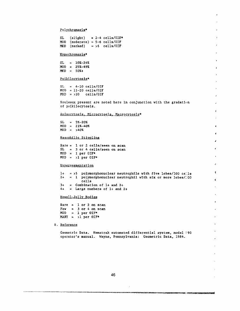
V ANALYTICAL METHODS
C Prothrombin Time in Plasma MLA 700 Automatic Coagulation Timer
1 Principle
The prothrombin time assesses extrinsic and common pathways 1 coagulation and is used to monitor antithrombin drugs
MLA has developed the photometric clot detection system Ligilt from a calibrated carefully controlled light source passesilrough a collimator in which the light is focused into a parallel eam The light illuminates the sample and the light passing throlgh falls on a photo detector which converts the light intensi t1 into an electrical signal
As the plasma sample clots in response to the addition of thromboplastin it becomes more opaque and the light fa11inl on the photo detector decreases This alters the electrical silnal A sensitive amplifier processes the signal and the instrumelts microprocessor determines when a clot has started to form [he instrument then records the time elapsed between the addi tiOl of reagent and the start of clotting This is the clot time
2 Specimen
a Collect blood in 38 sodium citrate anticoagulated tube (Ratio 91 45 m1 whole blood to 05 m1 citrate solution)
b Keep specimen cool Spin as soon as possible Separate the plasma into 10- x 75-mm plastic tube with cap
c Keep plasma cool (2-8 OC) until analyzed
d Analyze within 2 hours or freeze at -70 degC
e Minimum sample required 02 m1 plasma
3 Reagents and Equipment
a MLA 700 Model E-650 serial no 205
b Plastic tubes with caps 12 X 75 mm
c Plastic cuvettes red Scientific Products catalog no B middoti163-7
d Reservoir and heat exchange tubing Scientific Products clta1og no B 4163-2
47
4 Quality Control and Other Material
a C0ntrols Citrol Level I - Scientific Products catalog no B4224-l0 Dade
Citrol Level II - Scientific Products catalog no B4224-20 Dade
(1) Lyophilized preparations of human plasma stabilizers ani buffers
(2) Store unopened vials at 2-8 degC (3) Remove from refrigerator and allow to warm to room
temperature (4) Reconstitute with 10 ml distilled or deionized water
Stopper vial Label with date and time and initial and allow to stand at room temperature until dissolved approximately 15 minutes Invert gently to mix DO NOT SHAKE
(5) Reconstituted material is stable 8 hours Store at 2-8 0 when not in use
b Thromboplastin C Scientific Products catalog no B4216-50 Dade
(1) Lyophilized acetone-dehydrated rabbit brain thromboplastill 116 X 10-20M Ca++ stabilizers and preservative
(2) Store at 2-8 degC (3) Reconstitute vials with 10 ml deionized water Label with
date time and initials (4) Stable for 3 days if stored at 2-8 0C Mix well
5 Procedure
a Turn on machine and allow 20stabilize
-30 minutes for temperature to
b Reconstitute controls and thromboplastin-C
c Check the following systems
(1) Waste bin - empty (2) Test time - normal (3) Lever set for 300-second (4) Light source set on B (5) Lever on AUTO
test
d Fill cup with stirrer
a MAXIMUM of 8 ml of thrombo-C Add magnetic
e Prime tubing
(1) Check to make sure each reagent cup
plastic fitting is seated properly in
48
ITEa
lshy
f
l
l I f
(2) Prime reagent through tubing back into reagent cup Htil allbubbles are out of the line Note One bubble neal colored tip of line is supposed to be there
(3) Replace reagent cup lid
(4) Check to see if tubing is seated properly in the grc I)ves
(5) Place nozzle into nozzle holder marked TEST
f Run temperature check and confidence test
(1) Press PT 1 button (2) Press START button twice in succession (3) Printout should read
(4) Confidence = PT--199 plusmn 02 (5) Temperature = cold block--15plusmn3 degc (6) Reagent cups--15plusmn3 0c (7) Heat transfer pump block--375plusmn02 degc(8) Heat block--375plusmn02 degc
g Place the necessary number of cuvettes into the carouse] toinclude controls where scheduled Start with the first Hpace to the right of the decoder space
h Add Ol-ml amounts of control and participant samples tc eachcuvette position (Each cuvette runs in duplicate) PIIWet the tip before pipetting Carefully wipe the tip after samIing
i Press the START button once
j Two short beeps will indicate the run is complete
k The results will be printed out on tape
1 Discard reagents Wash cup with distilled water and dr)
m Flush tubing with deionized water Remove tubing
n Turn off machine
6 References
a Dade Thromboplastin-C package insert Aguada Puerto l~ico Dade 1982
b Medical Laboratory Automation Electra 700 automatic coagulation timer operators manual Mt Vernon New Ycrk Medical Laboratory Automation 1984
49
V ANALYTICAL METHODS
D Erythrocyte Sedlmentatlon Rate in Whole Blood
1 Principle
The erythrocyte sedimentation rate (ESR) test measures the settlirg of erythrocytes in human plasma over a specified time period The reported numerical value is derived from measuring in millimeter5 the distance from the bottom of the surface meniscus to the top 01 the erythrocyte sediment in a column of anticoagulated blood that has remained perpendicular in a special purpose pipet for 60 minutes
The empirical phenomenon depends on an interrelationship of a number of important inherent variables including plasma protein composition and concentration and erythrocyte configuration
In this ESR method an attempt has been made to control or eliminae the variability of environmental and technical factors that influence the results
The method is based on the Westergren method and calls for dilutiol of blood with autologous plasma to a standardized hematocrit Th change renders the method insensitive to hematocrit variations and provides a linear response throughout the range of clinical interest
2 Specimen
a EDTA (Na) anticoagulated whole blood
b Minimum sample size is 5 mlj optimum size is 7 mI
c Blood left at room temperature (19-25 OC) must be set up within 2 hours If blood is kept at 4 oe it must be set up wi thin 6 hours
3 Reagents and Equipment
a Anticoagulant Ethylene diamine tetra-acetic acid (Na) is used at a concentration of 14 to 16 mgml of blood
b The Westergren Pipet (Scientific Products catalog no B4504-l [Dade] )
(1) The pipet must have the following dimensions Overall length 3005plusmnO5 mm External diameter 55plusmnO5 mm Tube bore 270plusmn020 mm Uniformity of tube bore plusmnO05 mm
(2) The pipet must have the following markings
(a) The tube is inscribed Westergren
50
r (
(b) The tube has an inscribed graduated scale extendlg over the lower 200plusmn0 35 nun of the pipet The graduat ms are numbered from 0 at the top end of the scale to a~ least 180 no less than 20 nun from the lower end of thl~ pipet in steps of 10 or less
Maximal tolerated error between two adjacent milimeter markings is 02 nun tolerated error of the total graduated scale is 035
(c) The l-nun graduation marks are fine clearly mark~d permanent lines of uniform thickness (02 nun) ~Very 10th graduation mark is numbered
(d) The lengths of longer graduation marks are projet equally at each end beyond shorter markings Specifically the lengths are
(i) At the 0 mark 09-10 times the tube cir~lmference
(ii) At each single millimeter mark 0125 (18) times the tube circumference
(Ui) At each 5-nun mark 016 (16) times the tUle circumference
(iv) At each 10-nun mark 025 (14) times the tlbe circumference
(v) The tubes if disposable must be supplied clean and dry The manufacturer must ensure anl provide data on request that the tubes do not contain any factor that mey affect their U3e in the ESR test compared with WSP of the glas3 Westergren tubes described above
c Pipet Rack
(1) Equipped with a leveling bubble device (2) Constructed so that no leakage of the blood can OCCU[
4 Quality Control Material
Four participant samples will be run in duplicate each anal) tic run
51
1 S Procedure
a Blood Collection
(1) Collect whole blood in a 7-ml EDTA tube (2) Mix immediately and THOROUGHLY by gentle inversion
b Hematocrit Adjustment
(1) Obtain an initial hematocrit on the UNADJUSTED blood samp
(a) Spin for 3 minutes at 10000-11000 rpm in the microhematocrit centrifuge Read hematocrit on the accompanying reader
(b) Centrifuge the two equal aliquots of 2 ml each at 800 rpm for S minutes to adequately divide cells and plasrl i
(c) Remove plasma from or add plasma to one of the aliquo1l to obtain a sample with a hematocrit of 03S To calculate the volume required use the following formllLa
2 X hematocrit - 2 = ml plasma 035 required
(2) Determine the hematocrit of the ADJUSTED blood sample [USI
procedure as described in Sb(l)]
c Blood Cell Suspension
(1) Gently resuspend the blood cells thoroughly by placing on il
mechanical rocker for at least 10 minutes Blood should remain rocking until ready to aspirate
(2) Using a mechanical suction device aspirate a bubble-free sample into a clean and dry Westergren pipet exactly filling to the 0 mark
d Handling of Westergren pipet
(1) Set up singly Pick four of the samples and set them up ill duplicate
(2) Place the filled pipet in a vertical position in the pipet rack Area should be free of vibrations air draft and direct sunlight
52
e Reading of test
(1) After 60 minutes plusmn30 seconds read the distance of millimeters from the bottom of the plasma meniscus ltwhich should be at 0) to the top of the column of sedinmted erythrocytes Do not include buffy coat with the erythrocyte column
(2) Record the numerical value
6 References
a Bull BS Is a standard ESR possible Lab Med 1975 j 6110 11)31-5
b Koepke JA Bull BS Van Assendelft OW Reference procldure for the human erythrocyte sedimentation rate (ESR) test NCCLS 1983 3(no 7)251-64
53
J
~
j
1
V ANALYTICAL METHODS
E Routine Urinalysis in a Random Urine
1 Principle
Properly executed routine urinalysis will indicate the presence 01 renal disease (protein cells and casts) urinary tract infectior and diabetes mellitus (glucose) The routine urinalysis should bE done on a fairly concentrated specimen which is usually the first morning specimen especially when examined for protein and microscopic content
2 Specimen Collection and Storage
The urine sample must be collected in a clean dry container covered with a lid and examined as soon as possible Bacterial contamination may occur and leukocytes and casts disintegrate as the speciman ages Specimens should be examined within 1 hour of collection If this is not possible the specimen must be refrigerated at 4 degC until it is examined Minimum volume is 10 ml although 12 ml is optimum for the test
3 Reagents
a Ames Multistix Store at room temperature Gooc until expiration date on the bottle Ames Inc catalog no 4820
b Count-IO Trol II Urine Control (15-ml lyopholyZE ltl specimen) Store at 2-8 degC Good until expiration date on tl I ~
bottle (unopened) V-Tech Inc catalog no 10-0751
c Clinitek calibration strips Store at room temperature Good until expiration date on the box Ames Inc catalog no 5382
d Distilled water
e 4 Sodium chloride Store at room temperature Stable for 6 months
Procedure for preparing 4 NaCl
1 Weigh 4 g of dried NaCl crystals
54
2 Dissolve in distilled Hater in a 100-ml volumetric flask
3 QS to 100 mI
4 Quality Control Material
a Source
V-Tech Inc Count-IO-Trol Level II Each vial contait lyophilized material derived from human urine containin~ measured amounts of chemicals stabilized human erythrocrtes and simulated leukocytes
b Preparation
(1) Bring reagents to room temperature (2) Slowly vent cap on reagent vial to avoid reagent 101 l (3) Reconstitute each control vial with 15 ml of distil ~d
water Stable 5 days after reconstitution (4) Cap and swirl control vial for 20 seconds Allow 2(1 to 30
minutes for complete reconstitution (5) Remix reagent before each use
c Storage
Store controls at 2-8 degC Reconstituted controls remailL stable for 5 days at 2-8 degC Between use all controls ghould be stored at 2-8 degC Unopened controls are stable untL the expiration date on the bottle
5 Procedure
a Perform calibration on the Clinitek 200 and the Atago-UR-20 refractometer
b Prepare controls
c Mix specimen well
d Transfer 10-12 ml of patient urine and control to the pLastic centrifuge tubes labeled with specimen sample number
e Record color (straw yellow amber or specific color) ltHld appearance (clear hazy cloudy flocculent)
f Perform and record the specific gravity using the USR-2IJshyrefractometer Record the result to three decimal placlts (~ 1024)
g Perform chemical examination with a dipstick and the AmltS Clini tek 200
55
h Centrifuge urine tube for 5 minutes at 2000 rpm
i Decant tube without blotting
j Perform microscopic examination using the phase XIO lens and rre phase X40 lens
(1) Resuspend the sediment (2) Examine the drop of sediment on slide with 20 x 20
coverslip using the phase X10 lens first Look for cast1 and epithelial cells
Note It is necessary to distinguish between cast types Swi tch to the phase X40 lens and carefully scan 10middotmiddot12 separate fields of the slide Estimate average r~l~e of WBC RBC and renal tubular cells per high powe field (hpf)
(3) Standard nomenclature
(a) Cells (RBC and WBC) Range of numbers hpf (b) Casts Range of number per low power field (lpf) (c) Epithelial cells Report percentage of lpf
k Perform the daily shut-down procedure on the Clinitek 200 and Atago Refractometer
6 Codes and Comments To Use In Reporting
Assay Name
Hemoglobin Bilirubin
Comments
544 Negative 670 Small amount 406 Moderate amount ~
1 407 Large amount 2170 Trace
Ketones (mgd1)
544 Negative 5369 5-15 (trace) 5370 15-40 5371 40-80 5372 gt80
56
f
Urobilinogen (mgdl)
Codes Comments
5379 02-1 (- normal) 5380 1-2 5381 2-4 5374 4-8 5375 8-12 5382 )12
Protein (mgdl)
544 Negative 2170 Trace 5373 30-100 5378 100-300 5363 )300
Glucose (mgd])
544 Negative 5376 100-250 5377 250-500 5367 500-1000 5368 )1000
Color
5326 Straw yellow (normal) 510 Amber 513 Red
5327 Blue-green 2156 Brown 5328 Dark yellow 5329 Yellow-orange
645 Orange 5330 Red-brown 2160 Yellow
511 Colorless
Appearance
145 Clear (normal) 517 Hazy 518 Cloudy 147 Turbid
57
RBCs and Leukocytes Urine (cellshpf)
Codes Comments
396 None seen 1780 Rare 5331 Occasional 5332 Few = (1-5) 5333 1+ (6-20) 5334 2+ (21-50) 5335 3+ (51-100) 5336 4+ (gt100)
Renal Tybular Cells (ce11slpf)
1780 Rare 5331 Occasional 5337 1+ (1-5) 5338 2+ (6-10) 5339 3+ (11-30) 5340 4+ (gt30)
Casts (Hyaline Granular WBC RBC Other)(nolpf (XlOraquo
1780 Rare 5331 Occasional 5337 1+ (1-5) 5338 2+ = (6-10) 5339 3+ (11-30) 5340 4+ (gt30)
Epithelial Cells Urine (nolpf)
1780 Rare 5331 Occasional 5341 Few laquothan 25 of field) 5342 1+ (25 of field) 5343 2+ = (50 of field) 5344 3+ (75 of field) 5345 4+ (100 of field)
447 Packed
7 References
a Ames Division Miles Laboratories Clinitek 200 operating manual 4th ed Elkhart Indiana Ames Division Miles Laboratories 1985
b Bradley M Schumann GB Examination of urine IN Henry JB ed Clinical diagnosis and management by laboratory methods Philadelphia WE Saunders 1984380-58
58
c NSG Precision Instruction manual for the USR-20 refractometer Hicksville New York NSG Precision 198~
d V-Tech Count-10-Tro1 urine control package insert Pomna California V-Tech 1984
59
I
J
j
V ANALYTICAL METHODS
F D-Glucaric Acid in Urine Ion-Exchange Chromatography
1 Principle
For the quantitative determination of D-glucaric acid buffered urine specimens are added to an ion-exchange resin-filled column 0
separate D-glucaric acid from other interfering substances Afte elution from the column D-glucaric acid is oxidized with periodil acid to yield glyoxylic acid Glyoxylic acid is then oxidatively coupled with phenylhydrazine to yield the dye 15-diphenylformazan Absorbance at 520 mm is read and concentration determined by comparison to standard curve absorbanl e
D-glucaric acid is a product of glucuronic acid metabolism in the liver Urinary excretion reflects the activity of hepatic enzyme that hydroxylate and detoxify many foreign substances
Elevated levels are observed after the administration of drugs known to induce enzyme activity and anticonvulsant drugs and in association with exposure to insecticides
L-ascorbic acid (vitamin C) is know to falsely elevate D-glucaric acid levels obtained in this method therefore all urines are prescreened for ascorbic acid by using a dipstick method
2 Specimen Collection and Storage
a No participant preparation is necessary
b A well-mixed aliquot (4-10 ml) of a l2-hour urine collection l used for this test Urine specimens are collected in containell with 2 of sodium carbonate The specimen should be protected from light and kept cold during collection
c The specimen is stable for 24 hours at 2-8 0c For longer storage freeze at -20 0c
3 Reagents Supplies and Equipment
a Glass test tubes disposable 16 X 100 mm 16 X 150 mm 10 X i~i mm
b Pipets Eppendorf pipet l25-ml tips MLA 1 ml with disposable tips Accuprep (Beckman) capable of dispensing 1-1000 ul Repipet 5-ml (for addition of eluant to column)
60
RTFITUf
1m
c Class A volumetric flasks 2L lL 100 ml 50 ml 10 ml
d Analytical balance (Mettle~ AE 166)
e Spectrophotometer having band width of 1-2 nm 10-cm liht path (Beckman DU-7)
f Millipore filters
g Draining rack with waste container
h Plastic transfer pipets
i Magnetic stirrer
j Plastic caps 16 mm
k Stix (ascorbic acid reagent strips) Ames
1 Funnels (BioRad)
m Vortex mixer
n Test-tube racks
o Parafilm
p Timer
q Resin columns AG 1X-8 room temperature Refer
resin to
obtained from Bioexpiration date on
Rad box
Stoe at
r Sodium borate OlM
(1) Dissolve in volumetric flasks (a) Sodium borate--30596 g
Certified ACS Fisher (b) Reagent water type 1--8 L
(2) Place in polyethylene carboy Label as Sodium BoraL~ OlM with preparation date technician ID and an expiration date of 1 year Store at room temperaturl
s Sodium borate 005 Msodium sulfate 002 M
(1) Dissolve in volumetric flasks (a) Sodium borate--190685 g (b) Sodium sulfate--2840BO g
Anhydrous E M Science (c) Reagent water type 1--10 L
61
(2) Place in polyethylene carboy label as Sodium Borate 005MSodium sulfate 002M with preparation date tecrnician identification and an expiration date of 1 year Store at room temperature This quantity is sufficient for 1 week J
t Sodium borate OOSMsodium sulfate 01 M
(1) Dissolve in volumetric flasks (a) Sodium borate--190685 g (b) Sodium sulfate--142040 g (c) Reagent water type 1--10 L
(2) Place in polyethylene carboy label as Sodium Borate 005 M sodium sulfate OlM with preperation date technicians initials and an expiration date of 1 year Store at room temperature Aliquots are transferred to a repipet capable of dispensing 5 mI Label as above
u Saccharic acid (potassium hydrogen d-glucarate) Sigma storE in desiccator at room temperature
v Potassium biphosphate 10 Molar
(1) Dissolve in a 100-ml volumetric flask (a) Potassium phosphate monobasic--13609 g (b) Technical grade MCB (c) Reagent water type I--QS to 100 m1
(2) Transfer to glass bottle label as Potassium BiphosphatE 1 OM with preparation date technicians initials and III
expiration date of 2 weeks Store at 2-8 degC Mix well before using
w Periodic acid 001 Molar
(1) Dissolve in a SO-ml volumetric flask (a) Periodic acid--0114 g
Sigma (b) Reagent water type 1-- QS to 50 m1
(2) Transfer to brown glass bottle label as Periodic Acid OOlM with preparation date technicians initials and iLn expiration date of 1 week Store at 2-8 degC
x Phenylhydrazine Hydrochloride 1
(1) Dissolve in a 100-ml volumetric flask (a) Phenylhydrazine HCl--lO g
MCB (b) Reagent water Type I--QS to 100 m1
62
-----~
i
(2) Transfer to brown glass bottle label as Phenylhydllzine HCll with preparation date technician s initial and an expiration date of 1 week Store at 2-8 degC
y Hydrochloric Acid conc Reagent Grade MCB Reagents ~ ore under hood
z Potassium ferricyanide 5
(1) Dissolve in a 100-ml volumetric flask (a) Potassium ferricyanide--50 g
Technical grade MCB (b) Reagent water type I-QS to 100 ml
(2) Transfer to a brown glass bottle label as Potassi1Ln Ferricyanide 5 with preparation date technician initials an expiration date of 1 week Store at 2middotmiddot~ degC
aa Ascorbic Acid Control 25 mgdl
(1) Dissolve in an lL volumetric flask (a) 250 mg ascorbic acid ACS MCB reagents (b) Reagent water type I-QS to 1 L
(2) Aliquot approximately 3 ml in 10- x 75-mm tubes and store at -20 degC Stable for 6 months
bb Ascorbic acid dipsticks (Uralyte GK + A) Medical Sales Associates catalog no 8001
4 Calibration
a D-dlucaric acid stock std 1000 ugml
(1) Dissolve in a 100-ml volumetric flask (a) D-saccharic acid--lOO mg (b) Reagent water type I--QS to 100 ml
(2) Aliquot 1-2 ml in 10 X 75 mm tubes and store at -20 lC Stable for 3 months
b Using the Beckman Accuprep dilute working standards daiLy as follows
Prepare the working standards during the final elution (3tep 6d(3) of the procedure that follows)
c Prepare 100 ugml working stock standard
Dilute 1 ml (use a 1 ml volumetric pipette) of the 1000 ugml stock in a 10-ml volumetric flask QS to 10 ml with water reagent type I
63
Prepare Standards
D-glucarate ml of ml of std 100 ugml 005 m NaBoratel
ugml Stock Standard 01 m NaSulfate
2 0200 98 4 0400 96 6 0600 94 8 0800 92
10 1000 90
Standards at the 2- 4- 6- 8- and 10-ugml levels are run in duplicate with each assay
5 Quality Control Material
A control urine will be run in duplicate at the beginning and elll of each analytical run The control urine used is Lyphochek Quantitative Urine Control Abnormal Level II by BioRad The control is reconstituted by using a 25-ml volumetric pipet and reagent water type I It is made up at the beginning of each ~ek and then aliquoted and frozen for each days use A positive ahl negative ascorbic acid control will be run at the beginning and ~nd of the ascorbic acid screening procedure and ascorbic acid resul s for controls and participant samples will be recorded on the WOIC
list
6 Procedure
a Allow all reagents standards and participant sample specimElls to come to room temperature before testing
b Labell BioRad column for each participant and control Shdl~
column vigorously until resin is completely suspended No rEnin should remain in tip when the column is inverted Snap off ends attach funnels place in rack and drain into a waste container Do not insert pipets or other objects into the columns to further mix the contents while they are draining
c Screening for Ascorbic Acid
(1) In addition to each participant sample a positive ascorbc acid control and a negative control (reagent water type I must be tested for ascorbic acid Run these controls at 1he beginning and end of each analytic run
(2) Completely immerse the reagent area of an ascorbic acid dipstick in the well-mixed uncentrifuged urine or control sample
(3) Draw the edge of the strip against the side of the contairer to remove any excess liquid
64
j ill
(4) Compare the color of the reagent area with the color chart on the bottle label at exactly 60 seconds Hold the trip close to the color blocks and match carefully
(5) Record results in the 0-10 mgdl range as negative Jny results of 25 mgdl or greater are to be recorded on Ihe D-glucaric worksheet as positive and the D-glucaric IISUlt NOT reported Any positive ascorbic acid screen is til be reported to the QC supervisor
d Separation of D-glucaric Acid
(1) Label one 16- x 100-mm test tube for each participant and control Use a l-ml MLA to place 2 ml of the well-m_ ed l2-hour urine or control into its tube Using an Epp~ndorf pipet add 2 ml of OlM sodium borate to each tube lortex and let stand at room temperature 15 minutes
(2) Pour the urine mixture onto each drained column AL)w the samples to be completely absorbed on the resin (abou- 20 minutes) before proceeding
(3) Wash the columns with 100 ml of 005 M sodium borate--[)02 M sodium sulfate Drain completely into the waste recptacle (about 3 hours)
(4) Label one 16- X l50-mm test tube for each participantcontrol and place under the columns El-l te D-glucaric acid with lS ml (use the S-ml repipet) OJSM sodium borateOl sodium sulfate Let drain completly into tubes (about 1 hour)
e Colorimetric Procedure
Make up daily standards as described in the calibration section
(1) Cover with Parafilm and mix the eluate well by inversion With an MLA place 1 ml in each of two labeled 16- X 100-mm test tubes
(2) With an MLA place 1 ml of each of the standards int duplicate labeled 16- X 100-mm test tubes Two zero standards (reagent blank) are prepared by pipetting 1 ml of the OOSM sodium borate01M sodium sulfate buffer irto two labeled tubes
(3) Add 200 ul (use Accuprep program 1) of 1OM potassiunl biphosphate to each tube and vortex Make sure the potassium biphosphate reagent is well-mixed before udng
(4) Add 20 ul (use Accuprep program 4) of OOlM periodic acid vortex again and let stand at room temperature for _5 minutes
(5) Add 200 ul (use Accuprep program 1) of 1 phenylhydwzine hydrochloride vortex and let stand at room temperBIure for 10 minutes
(6) Using the accuprep program 2 add 1 ml of concentratld HClj vortex and follow immediately with the addition of ~OO ul S potassium ferricyanide (Accuprep) Vortex and lE - stand at room temperature 5-10 minutes
(7) Measure and record the absorbancy at 520 nm against -he reagent blank
65
7 Calibrations
a Use the linear regression program in the CHC or the IBM PCXT computer to evaluate the standard curve and obtain participar results (in ugml)
b After this result and the urine creatinine (creat) results hee been verified the force calculations mode is used to perforn the following calculation
Uncorrected D-Glucaric Acid (D-Glucaric Acid X 75) (in mgg creat) (in ugml)
Urine CreatIOO (in mgdl)
c Each duplicate control result is multiplied by 75 to correct for the dilution factor
8 References
a Hunter J Maxwell JD Carella M Stewart DA Urinary D-glucalic acid excretion as a test for hepatic enzyme induction in man Lancet 19711572-5
b Ishidate M Matsui M Okada M Biochemical studies on 1 14j
j
I ~
glucuronic acid and glucaric acid quantitative chemical determination of D-g1ucaric acid in urine Anal Biochem 1965 11176-89
c March J Turner WJ Shanley J Field J Values for urinary excretion of D-g1ucaric acid by normal individuals Clin Chel 1974 20(No9)1155-8
do McFadden BA Howes WV The determination of glyoxylic acid 11 biological systems Anal Biochem 1960 1240-8
66
1
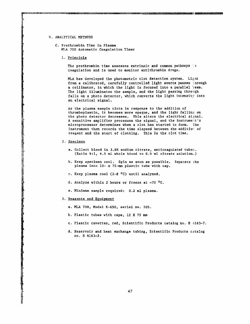
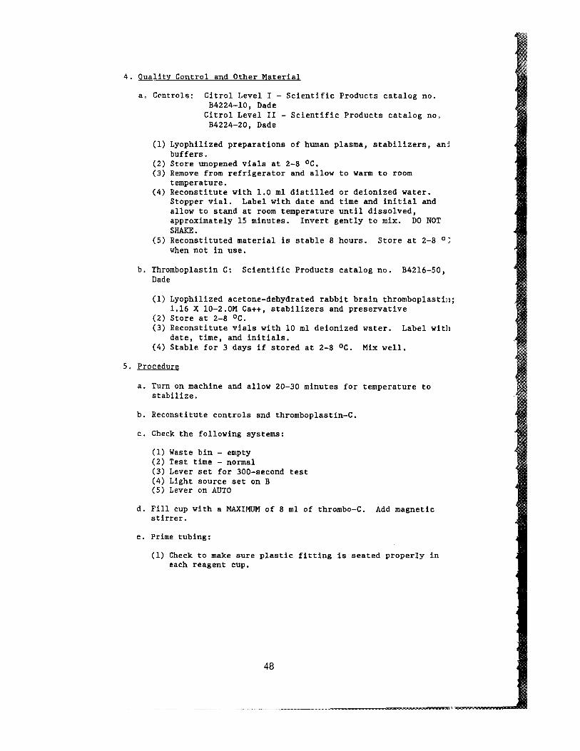
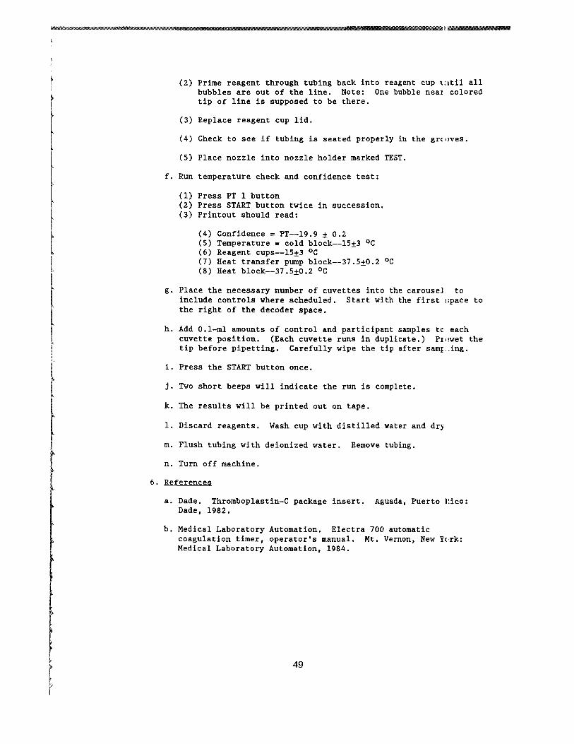
V ANALYTICAL METHODS
D Erythrocyte Sedlmentatlon Rate in Whole Blood
1 Principle
The erythrocyte sedimentation rate (ESR) test measures the settlirg of erythrocytes in human plasma over a specified time period The reported numerical value is derived from measuring in millimeter5 the distance from the bottom of the surface meniscus to the top 01 the erythrocyte sediment in a column of anticoagulated blood that has remained perpendicular in a special purpose pipet for 60 minutes
The empirical phenomenon depends on an interrelationship of a number of important inherent variables including plasma protein composition and concentration and erythrocyte configuration
In this ESR method an attempt has been made to control or eliminae the variability of environmental and technical factors that influence the results
The method is based on the Westergren method and calls for dilutiol of blood with autologous plasma to a standardized hematocrit Th change renders the method insensitive to hematocrit variations and provides a linear response throughout the range of clinical interest
2 Specimen
a EDTA (Na) anticoagulated whole blood
b Minimum sample size is 5 mlj optimum size is 7 mI
c Blood left at room temperature (19-25 OC) must be set up within 2 hours If blood is kept at 4 oe it must be set up wi thin 6 hours
3 Reagents and Equipment
a Anticoagulant Ethylene diamine tetra-acetic acid (Na) is used at a concentration of 14 to 16 mgml of blood
b The Westergren Pipet (Scientific Products catalog no B4504-l [Dade] )
(1) The pipet must have the following dimensions Overall length 3005plusmnO5 mm External diameter 55plusmnO5 mm Tube bore 270plusmn020 mm Uniformity of tube bore plusmnO05 mm
(2) The pipet must have the following markings
(a) The tube is inscribed Westergren
50
r (
(b) The tube has an inscribed graduated scale extendlg over the lower 200plusmn0 35 nun of the pipet The graduat ms are numbered from 0 at the top end of the scale to a~ least 180 no less than 20 nun from the lower end of thl~ pipet in steps of 10 or less
Maximal tolerated error between two adjacent milimeter markings is 02 nun tolerated error of the total graduated scale is 035
(c) The l-nun graduation marks are fine clearly mark~d permanent lines of uniform thickness (02 nun) ~Very 10th graduation mark is numbered
(d) The lengths of longer graduation marks are projet equally at each end beyond shorter markings Specifically the lengths are
(i) At the 0 mark 09-10 times the tube cir~lmference
(ii) At each single millimeter mark 0125 (18) times the tube circumference
(Ui) At each 5-nun mark 016 (16) times the tUle circumference
(iv) At each 10-nun mark 025 (14) times the tlbe circumference
(v) The tubes if disposable must be supplied clean and dry The manufacturer must ensure anl provide data on request that the tubes do not contain any factor that mey affect their U3e in the ESR test compared with WSP of the glas3 Westergren tubes described above
c Pipet Rack
(1) Equipped with a leveling bubble device (2) Constructed so that no leakage of the blood can OCCU[
4 Quality Control Material
Four participant samples will be run in duplicate each anal) tic run
51
1 S Procedure
a Blood Collection
(1) Collect whole blood in a 7-ml EDTA tube (2) Mix immediately and THOROUGHLY by gentle inversion
b Hematocrit Adjustment
(1) Obtain an initial hematocrit on the UNADJUSTED blood samp
(a) Spin for 3 minutes at 10000-11000 rpm in the microhematocrit centrifuge Read hematocrit on the accompanying reader
(b) Centrifuge the two equal aliquots of 2 ml each at 800 rpm for S minutes to adequately divide cells and plasrl i
(c) Remove plasma from or add plasma to one of the aliquo1l to obtain a sample with a hematocrit of 03S To calculate the volume required use the following formllLa
2 X hematocrit - 2 = ml plasma 035 required
(2) Determine the hematocrit of the ADJUSTED blood sample [USI
procedure as described in Sb(l)]
c Blood Cell Suspension
(1) Gently resuspend the blood cells thoroughly by placing on il
mechanical rocker for at least 10 minutes Blood should remain rocking until ready to aspirate
(2) Using a mechanical suction device aspirate a bubble-free sample into a clean and dry Westergren pipet exactly filling to the 0 mark
d Handling of Westergren pipet
(1) Set up singly Pick four of the samples and set them up ill duplicate
(2) Place the filled pipet in a vertical position in the pipet rack Area should be free of vibrations air draft and direct sunlight
52
e Reading of test
(1) After 60 minutes plusmn30 seconds read the distance of millimeters from the bottom of the plasma meniscus ltwhich should be at 0) to the top of the column of sedinmted erythrocytes Do not include buffy coat with the erythrocyte column
(2) Record the numerical value
6 References
a Bull BS Is a standard ESR possible Lab Med 1975 j 6110 11)31-5
b Koepke JA Bull BS Van Assendelft OW Reference procldure for the human erythrocyte sedimentation rate (ESR) test NCCLS 1983 3(no 7)251-64
53
J
~
j
1
V ANALYTICAL METHODS
E Routine Urinalysis in a Random Urine
1 Principle
Properly executed routine urinalysis will indicate the presence 01 renal disease (protein cells and casts) urinary tract infectior and diabetes mellitus (glucose) The routine urinalysis should bE done on a fairly concentrated specimen which is usually the first morning specimen especially when examined for protein and microscopic content
2 Specimen Collection and Storage
The urine sample must be collected in a clean dry container covered with a lid and examined as soon as possible Bacterial contamination may occur and leukocytes and casts disintegrate as the speciman ages Specimens should be examined within 1 hour of collection If this is not possible the specimen must be refrigerated at 4 degC until it is examined Minimum volume is 10 ml although 12 ml is optimum for the test
3 Reagents
a Ames Multistix Store at room temperature Gooc until expiration date on the bottle Ames Inc catalog no 4820
b Count-IO Trol II Urine Control (15-ml lyopholyZE ltl specimen) Store at 2-8 degC Good until expiration date on tl I ~
bottle (unopened) V-Tech Inc catalog no 10-0751
c Clinitek calibration strips Store at room temperature Good until expiration date on the box Ames Inc catalog no 5382
d Distilled water
e 4 Sodium chloride Store at room temperature Stable for 6 months
Procedure for preparing 4 NaCl
1 Weigh 4 g of dried NaCl crystals
54
2 Dissolve in distilled Hater in a 100-ml volumetric flask
3 QS to 100 mI
4 Quality Control Material
a Source
V-Tech Inc Count-IO-Trol Level II Each vial contait lyophilized material derived from human urine containin~ measured amounts of chemicals stabilized human erythrocrtes and simulated leukocytes
b Preparation
(1) Bring reagents to room temperature (2) Slowly vent cap on reagent vial to avoid reagent 101 l (3) Reconstitute each control vial with 15 ml of distil ~d
water Stable 5 days after reconstitution (4) Cap and swirl control vial for 20 seconds Allow 2(1 to 30
minutes for complete reconstitution (5) Remix reagent before each use
c Storage
Store controls at 2-8 degC Reconstituted controls remailL stable for 5 days at 2-8 degC Between use all controls ghould be stored at 2-8 degC Unopened controls are stable untL the expiration date on the bottle
5 Procedure
a Perform calibration on the Clinitek 200 and the Atago-UR-20 refractometer
b Prepare controls
c Mix specimen well
d Transfer 10-12 ml of patient urine and control to the pLastic centrifuge tubes labeled with specimen sample number
e Record color (straw yellow amber or specific color) ltHld appearance (clear hazy cloudy flocculent)
f Perform and record the specific gravity using the USR-2IJshyrefractometer Record the result to three decimal placlts (~ 1024)
g Perform chemical examination with a dipstick and the AmltS Clini tek 200
55
h Centrifuge urine tube for 5 minutes at 2000 rpm
i Decant tube without blotting
j Perform microscopic examination using the phase XIO lens and rre phase X40 lens
(1) Resuspend the sediment (2) Examine the drop of sediment on slide with 20 x 20
coverslip using the phase X10 lens first Look for cast1 and epithelial cells
Note It is necessary to distinguish between cast types Swi tch to the phase X40 lens and carefully scan 10middotmiddot12 separate fields of the slide Estimate average r~l~e of WBC RBC and renal tubular cells per high powe field (hpf)
(3) Standard nomenclature
(a) Cells (RBC and WBC) Range of numbers hpf (b) Casts Range of number per low power field (lpf) (c) Epithelial cells Report percentage of lpf
k Perform the daily shut-down procedure on the Clinitek 200 and Atago Refractometer
6 Codes and Comments To Use In Reporting
Assay Name
Hemoglobin Bilirubin
Comments
544 Negative 670 Small amount 406 Moderate amount ~
1 407 Large amount 2170 Trace
Ketones (mgd1)
544 Negative 5369 5-15 (trace) 5370 15-40 5371 40-80 5372 gt80
56
f
Urobilinogen (mgdl)
Codes Comments
5379 02-1 (- normal) 5380 1-2 5381 2-4 5374 4-8 5375 8-12 5382 )12
Protein (mgdl)
544 Negative 2170 Trace 5373 30-100 5378 100-300 5363 )300
Glucose (mgd])
544 Negative 5376 100-250 5377 250-500 5367 500-1000 5368 )1000
Color
5326 Straw yellow (normal) 510 Amber 513 Red
5327 Blue-green 2156 Brown 5328 Dark yellow 5329 Yellow-orange
645 Orange 5330 Red-brown 2160 Yellow
511 Colorless
Appearance
145 Clear (normal) 517 Hazy 518 Cloudy 147 Turbid
57
RBCs and Leukocytes Urine (cellshpf)
Codes Comments
396 None seen 1780 Rare 5331 Occasional 5332 Few = (1-5) 5333 1+ (6-20) 5334 2+ (21-50) 5335 3+ (51-100) 5336 4+ (gt100)
Renal Tybular Cells (ce11slpf)
1780 Rare 5331 Occasional 5337 1+ (1-5) 5338 2+ (6-10) 5339 3+ (11-30) 5340 4+ (gt30)
Casts (Hyaline Granular WBC RBC Other)(nolpf (XlOraquo
1780 Rare 5331 Occasional 5337 1+ (1-5) 5338 2+ = (6-10) 5339 3+ (11-30) 5340 4+ (gt30)
Epithelial Cells Urine (nolpf)
1780 Rare 5331 Occasional 5341 Few laquothan 25 of field) 5342 1+ (25 of field) 5343 2+ = (50 of field) 5344 3+ (75 of field) 5345 4+ (100 of field)
447 Packed
7 References
a Ames Division Miles Laboratories Clinitek 200 operating manual 4th ed Elkhart Indiana Ames Division Miles Laboratories 1985
b Bradley M Schumann GB Examination of urine IN Henry JB ed Clinical diagnosis and management by laboratory methods Philadelphia WE Saunders 1984380-58
58
c NSG Precision Instruction manual for the USR-20 refractometer Hicksville New York NSG Precision 198~
d V-Tech Count-10-Tro1 urine control package insert Pomna California V-Tech 1984
59
I
J
j
V ANALYTICAL METHODS
F D-Glucaric Acid in Urine Ion-Exchange Chromatography
1 Principle
For the quantitative determination of D-glucaric acid buffered urine specimens are added to an ion-exchange resin-filled column 0
separate D-glucaric acid from other interfering substances Afte elution from the column D-glucaric acid is oxidized with periodil acid to yield glyoxylic acid Glyoxylic acid is then oxidatively coupled with phenylhydrazine to yield the dye 15-diphenylformazan Absorbance at 520 mm is read and concentration determined by comparison to standard curve absorbanl e
D-glucaric acid is a product of glucuronic acid metabolism in the liver Urinary excretion reflects the activity of hepatic enzyme that hydroxylate and detoxify many foreign substances
Elevated levels are observed after the administration of drugs known to induce enzyme activity and anticonvulsant drugs and in association with exposure to insecticides
L-ascorbic acid (vitamin C) is know to falsely elevate D-glucaric acid levels obtained in this method therefore all urines are prescreened for ascorbic acid by using a dipstick method
2 Specimen Collection and Storage
a No participant preparation is necessary
b A well-mixed aliquot (4-10 ml) of a l2-hour urine collection l used for this test Urine specimens are collected in containell with 2 of sodium carbonate The specimen should be protected from light and kept cold during collection
c The specimen is stable for 24 hours at 2-8 0c For longer storage freeze at -20 0c
3 Reagents Supplies and Equipment
a Glass test tubes disposable 16 X 100 mm 16 X 150 mm 10 X i~i mm
b Pipets Eppendorf pipet l25-ml tips MLA 1 ml with disposable tips Accuprep (Beckman) capable of dispensing 1-1000 ul Repipet 5-ml (for addition of eluant to column)
60
RTFITUf
1m
c Class A volumetric flasks 2L lL 100 ml 50 ml 10 ml
d Analytical balance (Mettle~ AE 166)
e Spectrophotometer having band width of 1-2 nm 10-cm liht path (Beckman DU-7)
f Millipore filters
g Draining rack with waste container
h Plastic transfer pipets
i Magnetic stirrer
j Plastic caps 16 mm
k Stix (ascorbic acid reagent strips) Ames
1 Funnels (BioRad)
m Vortex mixer
n Test-tube racks
o Parafilm
p Timer
q Resin columns AG 1X-8 room temperature Refer
resin to
obtained from Bioexpiration date on
Rad box
Stoe at
r Sodium borate OlM
(1) Dissolve in volumetric flasks (a) Sodium borate--30596 g
Certified ACS Fisher (b) Reagent water type 1--8 L
(2) Place in polyethylene carboy Label as Sodium BoraL~ OlM with preparation date technician ID and an expiration date of 1 year Store at room temperaturl
s Sodium borate 005 Msodium sulfate 002 M
(1) Dissolve in volumetric flasks (a) Sodium borate--190685 g (b) Sodium sulfate--2840BO g
Anhydrous E M Science (c) Reagent water type 1--10 L
61
(2) Place in polyethylene carboy label as Sodium Borate 005MSodium sulfate 002M with preparation date tecrnician identification and an expiration date of 1 year Store at room temperature This quantity is sufficient for 1 week J
t Sodium borate OOSMsodium sulfate 01 M
(1) Dissolve in volumetric flasks (a) Sodium borate--190685 g (b) Sodium sulfate--142040 g (c) Reagent water type 1--10 L
(2) Place in polyethylene carboy label as Sodium Borate 005 M sodium sulfate OlM with preperation date technicians initials and an expiration date of 1 year Store at room temperature Aliquots are transferred to a repipet capable of dispensing 5 mI Label as above
u Saccharic acid (potassium hydrogen d-glucarate) Sigma storE in desiccator at room temperature
v Potassium biphosphate 10 Molar
(1) Dissolve in a 100-ml volumetric flask (a) Potassium phosphate monobasic--13609 g (b) Technical grade MCB (c) Reagent water type I--QS to 100 m1
(2) Transfer to glass bottle label as Potassium BiphosphatE 1 OM with preparation date technicians initials and III
expiration date of 2 weeks Store at 2-8 degC Mix well before using
w Periodic acid 001 Molar
(1) Dissolve in a SO-ml volumetric flask (a) Periodic acid--0114 g
Sigma (b) Reagent water type 1-- QS to 50 m1
(2) Transfer to brown glass bottle label as Periodic Acid OOlM with preparation date technicians initials and iLn expiration date of 1 week Store at 2-8 degC
x Phenylhydrazine Hydrochloride 1
(1) Dissolve in a 100-ml volumetric flask (a) Phenylhydrazine HCl--lO g
MCB (b) Reagent water Type I--QS to 100 m1
62
-----~
i
(2) Transfer to brown glass bottle label as Phenylhydllzine HCll with preparation date technician s initial and an expiration date of 1 week Store at 2-8 degC
y Hydrochloric Acid conc Reagent Grade MCB Reagents ~ ore under hood
z Potassium ferricyanide 5
(1) Dissolve in a 100-ml volumetric flask (a) Potassium ferricyanide--50 g
Technical grade MCB (b) Reagent water type I-QS to 100 ml
(2) Transfer to a brown glass bottle label as Potassi1Ln Ferricyanide 5 with preparation date technician initials an expiration date of 1 week Store at 2middotmiddot~ degC
aa Ascorbic Acid Control 25 mgdl
(1) Dissolve in an lL volumetric flask (a) 250 mg ascorbic acid ACS MCB reagents (b) Reagent water type I-QS to 1 L
(2) Aliquot approximately 3 ml in 10- x 75-mm tubes and store at -20 degC Stable for 6 months
bb Ascorbic acid dipsticks (Uralyte GK + A) Medical Sales Associates catalog no 8001
4 Calibration
a D-dlucaric acid stock std 1000 ugml
(1) Dissolve in a 100-ml volumetric flask (a) D-saccharic acid--lOO mg (b) Reagent water type I--QS to 100 ml
(2) Aliquot 1-2 ml in 10 X 75 mm tubes and store at -20 lC Stable for 3 months
b Using the Beckman Accuprep dilute working standards daiLy as follows
Prepare the working standards during the final elution (3tep 6d(3) of the procedure that follows)
c Prepare 100 ugml working stock standard
Dilute 1 ml (use a 1 ml volumetric pipette) of the 1000 ugml stock in a 10-ml volumetric flask QS to 10 ml with water reagent type I
63
Prepare Standards
D-glucarate ml of ml of std 100 ugml 005 m NaBoratel
ugml Stock Standard 01 m NaSulfate
2 0200 98 4 0400 96 6 0600 94 8 0800 92
10 1000 90
Standards at the 2- 4- 6- 8- and 10-ugml levels are run in duplicate with each assay
5 Quality Control Material
A control urine will be run in duplicate at the beginning and elll of each analytical run The control urine used is Lyphochek Quantitative Urine Control Abnormal Level II by BioRad The control is reconstituted by using a 25-ml volumetric pipet and reagent water type I It is made up at the beginning of each ~ek and then aliquoted and frozen for each days use A positive ahl negative ascorbic acid control will be run at the beginning and ~nd of the ascorbic acid screening procedure and ascorbic acid resul s for controls and participant samples will be recorded on the WOIC
list
6 Procedure
a Allow all reagents standards and participant sample specimElls to come to room temperature before testing
b Labell BioRad column for each participant and control Shdl~
column vigorously until resin is completely suspended No rEnin should remain in tip when the column is inverted Snap off ends attach funnels place in rack and drain into a waste container Do not insert pipets or other objects into the columns to further mix the contents while they are draining
c Screening for Ascorbic Acid
(1) In addition to each participant sample a positive ascorbc acid control and a negative control (reagent water type I must be tested for ascorbic acid Run these controls at 1he beginning and end of each analytic run
(2) Completely immerse the reagent area of an ascorbic acid dipstick in the well-mixed uncentrifuged urine or control sample
(3) Draw the edge of the strip against the side of the contairer to remove any excess liquid
64
j ill
(4) Compare the color of the reagent area with the color chart on the bottle label at exactly 60 seconds Hold the trip close to the color blocks and match carefully
(5) Record results in the 0-10 mgdl range as negative Jny results of 25 mgdl or greater are to be recorded on Ihe D-glucaric worksheet as positive and the D-glucaric IISUlt NOT reported Any positive ascorbic acid screen is til be reported to the QC supervisor
d Separation of D-glucaric Acid
(1) Label one 16- x 100-mm test tube for each participant and control Use a l-ml MLA to place 2 ml of the well-m_ ed l2-hour urine or control into its tube Using an Epp~ndorf pipet add 2 ml of OlM sodium borate to each tube lortex and let stand at room temperature 15 minutes
(2) Pour the urine mixture onto each drained column AL)w the samples to be completely absorbed on the resin (abou- 20 minutes) before proceeding
(3) Wash the columns with 100 ml of 005 M sodium borate--[)02 M sodium sulfate Drain completely into the waste recptacle (about 3 hours)
(4) Label one 16- X l50-mm test tube for each participantcontrol and place under the columns El-l te D-glucaric acid with lS ml (use the S-ml repipet) OJSM sodium borateOl sodium sulfate Let drain completly into tubes (about 1 hour)
e Colorimetric Procedure
Make up daily standards as described in the calibration section
(1) Cover with Parafilm and mix the eluate well by inversion With an MLA place 1 ml in each of two labeled 16- X 100-mm test tubes
(2) With an MLA place 1 ml of each of the standards int duplicate labeled 16- X 100-mm test tubes Two zero standards (reagent blank) are prepared by pipetting 1 ml of the OOSM sodium borate01M sodium sulfate buffer irto two labeled tubes
(3) Add 200 ul (use Accuprep program 1) of 1OM potassiunl biphosphate to each tube and vortex Make sure the potassium biphosphate reagent is well-mixed before udng
(4) Add 20 ul (use Accuprep program 4) of OOlM periodic acid vortex again and let stand at room temperature for _5 minutes
(5) Add 200 ul (use Accuprep program 1) of 1 phenylhydwzine hydrochloride vortex and let stand at room temperBIure for 10 minutes
(6) Using the accuprep program 2 add 1 ml of concentratld HClj vortex and follow immediately with the addition of ~OO ul S potassium ferricyanide (Accuprep) Vortex and lE - stand at room temperature 5-10 minutes
(7) Measure and record the absorbancy at 520 nm against -he reagent blank
65
7 Calibrations
a Use the linear regression program in the CHC or the IBM PCXT computer to evaluate the standard curve and obtain participar results (in ugml)
b After this result and the urine creatinine (creat) results hee been verified the force calculations mode is used to perforn the following calculation
Uncorrected D-Glucaric Acid (D-Glucaric Acid X 75) (in mgg creat) (in ugml)
Urine CreatIOO (in mgdl)
c Each duplicate control result is multiplied by 75 to correct for the dilution factor
8 References
a Hunter J Maxwell JD Carella M Stewart DA Urinary D-glucalic acid excretion as a test for hepatic enzyme induction in man Lancet 19711572-5
b Ishidate M Matsui M Okada M Biochemical studies on 1 14j
j
I ~
glucuronic acid and glucaric acid quantitative chemical determination of D-g1ucaric acid in urine Anal Biochem 1965 11176-89
c March J Turner WJ Shanley J Field J Values for urinary excretion of D-g1ucaric acid by normal individuals Clin Chel 1974 20(No9)1155-8
do McFadden BA Howes WV The determination of glyoxylic acid 11 biological systems Anal Biochem 1960 1240-8
66
1
J
~
j
1
V ANALYTICAL METHODS
E Routine Urinalysis in a Random Urine
1 Principle
Properly executed routine urinalysis will indicate the presence 01 renal disease (protein cells and casts) urinary tract infectior and diabetes mellitus (glucose) The routine urinalysis should bE done on a fairly concentrated specimen which is usually the first morning specimen especially when examined for protein and microscopic content
2 Specimen Collection and Storage
The urine sample must be collected in a clean dry container covered with a lid and examined as soon as possible Bacterial contamination may occur and leukocytes and casts disintegrate as the speciman ages Specimens should be examined within 1 hour of collection If this is not possible the specimen must be refrigerated at 4 degC until it is examined Minimum volume is 10 ml although 12 ml is optimum for the test
3 Reagents
a Ames Multistix Store at room temperature Gooc until expiration date on the bottle Ames Inc catalog no 4820
b Count-IO Trol II Urine Control (15-ml lyopholyZE ltl specimen) Store at 2-8 degC Good until expiration date on tl I ~
bottle (unopened) V-Tech Inc catalog no 10-0751
c Clinitek calibration strips Store at room temperature Good until expiration date on the box Ames Inc catalog no 5382
d Distilled water
e 4 Sodium chloride Store at room temperature Stable for 6 months
Procedure for preparing 4 NaCl
1 Weigh 4 g of dried NaCl crystals
54
2 Dissolve in distilled Hater in a 100-ml volumetric flask
3 QS to 100 mI
4 Quality Control Material
a Source
V-Tech Inc Count-IO-Trol Level II Each vial contait lyophilized material derived from human urine containin~ measured amounts of chemicals stabilized human erythrocrtes and simulated leukocytes
b Preparation
(1) Bring reagents to room temperature (2) Slowly vent cap on reagent vial to avoid reagent 101 l (3) Reconstitute each control vial with 15 ml of distil ~d
water Stable 5 days after reconstitution (4) Cap and swirl control vial for 20 seconds Allow 2(1 to 30
minutes for complete reconstitution (5) Remix reagent before each use
c Storage
Store controls at 2-8 degC Reconstituted controls remailL stable for 5 days at 2-8 degC Between use all controls ghould be stored at 2-8 degC Unopened controls are stable untL the expiration date on the bottle
5 Procedure
a Perform calibration on the Clinitek 200 and the Atago-UR-20 refractometer
b Prepare controls
c Mix specimen well
d Transfer 10-12 ml of patient urine and control to the pLastic centrifuge tubes labeled with specimen sample number
e Record color (straw yellow amber or specific color) ltHld appearance (clear hazy cloudy flocculent)
f Perform and record the specific gravity using the USR-2IJshyrefractometer Record the result to three decimal placlts (~ 1024)
g Perform chemical examination with a dipstick and the AmltS Clini tek 200
55
h Centrifuge urine tube for 5 minutes at 2000 rpm
i Decant tube without blotting
j Perform microscopic examination using the phase XIO lens and rre phase X40 lens
(1) Resuspend the sediment (2) Examine the drop of sediment on slide with 20 x 20
coverslip using the phase X10 lens first Look for cast1 and epithelial cells
Note It is necessary to distinguish between cast types Swi tch to the phase X40 lens and carefully scan 10middotmiddot12 separate fields of the slide Estimate average r~l~e of WBC RBC and renal tubular cells per high powe field (hpf)
(3) Standard nomenclature
(a) Cells (RBC and WBC) Range of numbers hpf (b) Casts Range of number per low power field (lpf) (c) Epithelial cells Report percentage of lpf
k Perform the daily shut-down procedure on the Clinitek 200 and Atago Refractometer
6 Codes and Comments To Use In Reporting
Assay Name
Hemoglobin Bilirubin
Comments
544 Negative 670 Small amount 406 Moderate amount ~
1 407 Large amount 2170 Trace
Ketones (mgd1)
544 Negative 5369 5-15 (trace) 5370 15-40 5371 40-80 5372 gt80
56
f
Urobilinogen (mgdl)
Codes Comments
5379 02-1 (- normal) 5380 1-2 5381 2-4 5374 4-8 5375 8-12 5382 )12
Protein (mgdl)
544 Negative 2170 Trace 5373 30-100 5378 100-300 5363 )300
Glucose (mgd])
544 Negative 5376 100-250 5377 250-500 5367 500-1000 5368 )1000
Color
5326 Straw yellow (normal) 510 Amber 513 Red
5327 Blue-green 2156 Brown 5328 Dark yellow 5329 Yellow-orange
645 Orange 5330 Red-brown 2160 Yellow
511 Colorless
Appearance
145 Clear (normal) 517 Hazy 518 Cloudy 147 Turbid
57
RBCs and Leukocytes Urine (cellshpf)
Codes Comments
396 None seen 1780 Rare 5331 Occasional 5332 Few = (1-5) 5333 1+ (6-20) 5334 2+ (21-50) 5335 3+ (51-100) 5336 4+ (gt100)
Renal Tybular Cells (ce11slpf)
1780 Rare 5331 Occasional 5337 1+ (1-5) 5338 2+ (6-10) 5339 3+ (11-30) 5340 4+ (gt30)
Casts (Hyaline Granular WBC RBC Other)(nolpf (XlOraquo
1780 Rare 5331 Occasional 5337 1+ (1-5) 5338 2+ = (6-10) 5339 3+ (11-30) 5340 4+ (gt30)
Epithelial Cells Urine (nolpf)
1780 Rare 5331 Occasional 5341 Few laquothan 25 of field) 5342 1+ (25 of field) 5343 2+ = (50 of field) 5344 3+ (75 of field) 5345 4+ (100 of field)
447 Packed
7 References
a Ames Division Miles Laboratories Clinitek 200 operating manual 4th ed Elkhart Indiana Ames Division Miles Laboratories 1985
b Bradley M Schumann GB Examination of urine IN Henry JB ed Clinical diagnosis and management by laboratory methods Philadelphia WE Saunders 1984380-58
58
c NSG Precision Instruction manual for the USR-20 refractometer Hicksville New York NSG Precision 198~
d V-Tech Count-10-Tro1 urine control package insert Pomna California V-Tech 1984
59
I
J
j
V ANALYTICAL METHODS
F D-Glucaric Acid in Urine Ion-Exchange Chromatography
1 Principle
For the quantitative determination of D-glucaric acid buffered urine specimens are added to an ion-exchange resin-filled column 0
separate D-glucaric acid from other interfering substances Afte elution from the column D-glucaric acid is oxidized with periodil acid to yield glyoxylic acid Glyoxylic acid is then oxidatively coupled with phenylhydrazine to yield the dye 15-diphenylformazan Absorbance at 520 mm is read and concentration determined by comparison to standard curve absorbanl e
D-glucaric acid is a product of glucuronic acid metabolism in the liver Urinary excretion reflects the activity of hepatic enzyme that hydroxylate and detoxify many foreign substances
Elevated levels are observed after the administration of drugs known to induce enzyme activity and anticonvulsant drugs and in association with exposure to insecticides
L-ascorbic acid (vitamin C) is know to falsely elevate D-glucaric acid levels obtained in this method therefore all urines are prescreened for ascorbic acid by using a dipstick method
2 Specimen Collection and Storage
a No participant preparation is necessary
b A well-mixed aliquot (4-10 ml) of a l2-hour urine collection l used for this test Urine specimens are collected in containell with 2 of sodium carbonate The specimen should be protected from light and kept cold during collection
c The specimen is stable for 24 hours at 2-8 0c For longer storage freeze at -20 0c
3 Reagents Supplies and Equipment
a Glass test tubes disposable 16 X 100 mm 16 X 150 mm 10 X i~i mm
b Pipets Eppendorf pipet l25-ml tips MLA 1 ml with disposable tips Accuprep (Beckman) capable of dispensing 1-1000 ul Repipet 5-ml (for addition of eluant to column)
60
RTFITUf
1m
c Class A volumetric flasks 2L lL 100 ml 50 ml 10 ml
d Analytical balance (Mettle~ AE 166)
e Spectrophotometer having band width of 1-2 nm 10-cm liht path (Beckman DU-7)
f Millipore filters
g Draining rack with waste container
h Plastic transfer pipets
i Magnetic stirrer
j Plastic caps 16 mm
k Stix (ascorbic acid reagent strips) Ames
1 Funnels (BioRad)
m Vortex mixer
n Test-tube racks
o Parafilm
p Timer
q Resin columns AG 1X-8 room temperature Refer
resin to
obtained from Bioexpiration date on
Rad box
Stoe at
r Sodium borate OlM
(1) Dissolve in volumetric flasks (a) Sodium borate--30596 g
Certified ACS Fisher (b) Reagent water type 1--8 L
(2) Place in polyethylene carboy Label as Sodium BoraL~ OlM with preparation date technician ID and an expiration date of 1 year Store at room temperaturl
s Sodium borate 005 Msodium sulfate 002 M
(1) Dissolve in volumetric flasks (a) Sodium borate--190685 g (b) Sodium sulfate--2840BO g
Anhydrous E M Science (c) Reagent water type 1--10 L
61
(2) Place in polyethylene carboy label as Sodium Borate 005MSodium sulfate 002M with preparation date tecrnician identification and an expiration date of 1 year Store at room temperature This quantity is sufficient for 1 week J
t Sodium borate OOSMsodium sulfate 01 M
(1) Dissolve in volumetric flasks (a) Sodium borate--190685 g (b) Sodium sulfate--142040 g (c) Reagent water type 1--10 L
(2) Place in polyethylene carboy label as Sodium Borate 005 M sodium sulfate OlM with preperation date technicians initials and an expiration date of 1 year Store at room temperature Aliquots are transferred to a repipet capable of dispensing 5 mI Label as above
u Saccharic acid (potassium hydrogen d-glucarate) Sigma storE in desiccator at room temperature
v Potassium biphosphate 10 Molar
(1) Dissolve in a 100-ml volumetric flask (a) Potassium phosphate monobasic--13609 g (b) Technical grade MCB (c) Reagent water type I--QS to 100 m1
(2) Transfer to glass bottle label as Potassium BiphosphatE 1 OM with preparation date technicians initials and III
expiration date of 2 weeks Store at 2-8 degC Mix well before using
w Periodic acid 001 Molar
(1) Dissolve in a SO-ml volumetric flask (a) Periodic acid--0114 g
Sigma (b) Reagent water type 1-- QS to 50 m1
(2) Transfer to brown glass bottle label as Periodic Acid OOlM with preparation date technicians initials and iLn expiration date of 1 week Store at 2-8 degC
x Phenylhydrazine Hydrochloride 1
(1) Dissolve in a 100-ml volumetric flask (a) Phenylhydrazine HCl--lO g
MCB (b) Reagent water Type I--QS to 100 m1
62
-----~
i
(2) Transfer to brown glass bottle label as Phenylhydllzine HCll with preparation date technician s initial and an expiration date of 1 week Store at 2-8 degC
y Hydrochloric Acid conc Reagent Grade MCB Reagents ~ ore under hood
z Potassium ferricyanide 5
(1) Dissolve in a 100-ml volumetric flask (a) Potassium ferricyanide--50 g
Technical grade MCB (b) Reagent water type I-QS to 100 ml
(2) Transfer to a brown glass bottle label as Potassi1Ln Ferricyanide 5 with preparation date technician initials an expiration date of 1 week Store at 2middotmiddot~ degC
aa Ascorbic Acid Control 25 mgdl
(1) Dissolve in an lL volumetric flask (a) 250 mg ascorbic acid ACS MCB reagents (b) Reagent water type I-QS to 1 L
(2) Aliquot approximately 3 ml in 10- x 75-mm tubes and store at -20 degC Stable for 6 months
bb Ascorbic acid dipsticks (Uralyte GK + A) Medical Sales Associates catalog no 8001
4 Calibration
a D-dlucaric acid stock std 1000 ugml
(1) Dissolve in a 100-ml volumetric flask (a) D-saccharic acid--lOO mg (b) Reagent water type I--QS to 100 ml
(2) Aliquot 1-2 ml in 10 X 75 mm tubes and store at -20 lC Stable for 3 months
b Using the Beckman Accuprep dilute working standards daiLy as follows
Prepare the working standards during the final elution (3tep 6d(3) of the procedure that follows)
c Prepare 100 ugml working stock standard
Dilute 1 ml (use a 1 ml volumetric pipette) of the 1000 ugml stock in a 10-ml volumetric flask QS to 10 ml with water reagent type I
63
Prepare Standards
D-glucarate ml of ml of std 100 ugml 005 m NaBoratel
ugml Stock Standard 01 m NaSulfate
2 0200 98 4 0400 96 6 0600 94 8 0800 92
10 1000 90
Standards at the 2- 4- 6- 8- and 10-ugml levels are run in duplicate with each assay
5 Quality Control Material
A control urine will be run in duplicate at the beginning and elll of each analytical run The control urine used is Lyphochek Quantitative Urine Control Abnormal Level II by BioRad The control is reconstituted by using a 25-ml volumetric pipet and reagent water type I It is made up at the beginning of each ~ek and then aliquoted and frozen for each days use A positive ahl negative ascorbic acid control will be run at the beginning and ~nd of the ascorbic acid screening procedure and ascorbic acid resul s for controls and participant samples will be recorded on the WOIC
list
6 Procedure
a Allow all reagents standards and participant sample specimElls to come to room temperature before testing
b Labell BioRad column for each participant and control Shdl~
column vigorously until resin is completely suspended No rEnin should remain in tip when the column is inverted Snap off ends attach funnels place in rack and drain into a waste container Do not insert pipets or other objects into the columns to further mix the contents while they are draining
c Screening for Ascorbic Acid
(1) In addition to each participant sample a positive ascorbc acid control and a negative control (reagent water type I must be tested for ascorbic acid Run these controls at 1he beginning and end of each analytic run
(2) Completely immerse the reagent area of an ascorbic acid dipstick in the well-mixed uncentrifuged urine or control sample
(3) Draw the edge of the strip against the side of the contairer to remove any excess liquid
64
j ill
(4) Compare the color of the reagent area with the color chart on the bottle label at exactly 60 seconds Hold the trip close to the color blocks and match carefully
(5) Record results in the 0-10 mgdl range as negative Jny results of 25 mgdl or greater are to be recorded on Ihe D-glucaric worksheet as positive and the D-glucaric IISUlt NOT reported Any positive ascorbic acid screen is til be reported to the QC supervisor
d Separation of D-glucaric Acid
(1) Label one 16- x 100-mm test tube for each participant and control Use a l-ml MLA to place 2 ml of the well-m_ ed l2-hour urine or control into its tube Using an Epp~ndorf pipet add 2 ml of OlM sodium borate to each tube lortex and let stand at room temperature 15 minutes
(2) Pour the urine mixture onto each drained column AL)w the samples to be completely absorbed on the resin (abou- 20 minutes) before proceeding
(3) Wash the columns with 100 ml of 005 M sodium borate--[)02 M sodium sulfate Drain completely into the waste recptacle (about 3 hours)
(4) Label one 16- X l50-mm test tube for each participantcontrol and place under the columns El-l te D-glucaric acid with lS ml (use the S-ml repipet) OJSM sodium borateOl sodium sulfate Let drain completly into tubes (about 1 hour)
e Colorimetric Procedure
Make up daily standards as described in the calibration section
(1) Cover with Parafilm and mix the eluate well by inversion With an MLA place 1 ml in each of two labeled 16- X 100-mm test tubes
(2) With an MLA place 1 ml of each of the standards int duplicate labeled 16- X 100-mm test tubes Two zero standards (reagent blank) are prepared by pipetting 1 ml of the OOSM sodium borate01M sodium sulfate buffer irto two labeled tubes
(3) Add 200 ul (use Accuprep program 1) of 1OM potassiunl biphosphate to each tube and vortex Make sure the potassium biphosphate reagent is well-mixed before udng
(4) Add 20 ul (use Accuprep program 4) of OOlM periodic acid vortex again and let stand at room temperature for _5 minutes
(5) Add 200 ul (use Accuprep program 1) of 1 phenylhydwzine hydrochloride vortex and let stand at room temperBIure for 10 minutes
(6) Using the accuprep program 2 add 1 ml of concentratld HClj vortex and follow immediately with the addition of ~OO ul S potassium ferricyanide (Accuprep) Vortex and lE - stand at room temperature 5-10 minutes
(7) Measure and record the absorbancy at 520 nm against -he reagent blank
65
7 Calibrations
a Use the linear regression program in the CHC or the IBM PCXT computer to evaluate the standard curve and obtain participar results (in ugml)
b After this result and the urine creatinine (creat) results hee been verified the force calculations mode is used to perforn the following calculation
Uncorrected D-Glucaric Acid (D-Glucaric Acid X 75) (in mgg creat) (in ugml)
Urine CreatIOO (in mgdl)
c Each duplicate control result is multiplied by 75 to correct for the dilution factor
8 References
a Hunter J Maxwell JD Carella M Stewart DA Urinary D-glucalic acid excretion as a test for hepatic enzyme induction in man Lancet 19711572-5
b Ishidate M Matsui M Okada M Biochemical studies on 1 14j
j
I ~
glucuronic acid and glucaric acid quantitative chemical determination of D-g1ucaric acid in urine Anal Biochem 1965 11176-89
c March J Turner WJ Shanley J Field J Values for urinary excretion of D-g1ucaric acid by normal individuals Clin Chel 1974 20(No9)1155-8
do McFadden BA Howes WV The determination of glyoxylic acid 11 biological systems Anal Biochem 1960 1240-8
66
1
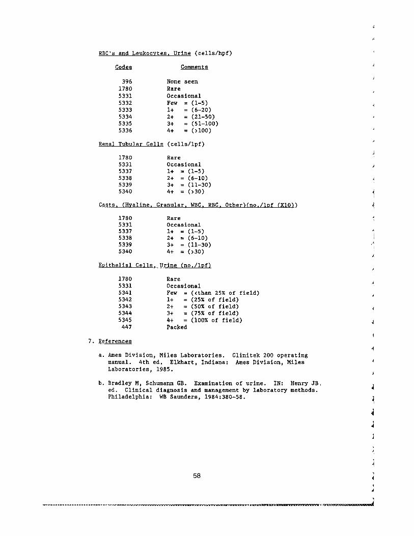
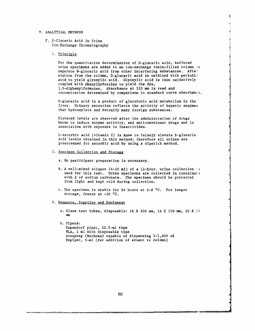
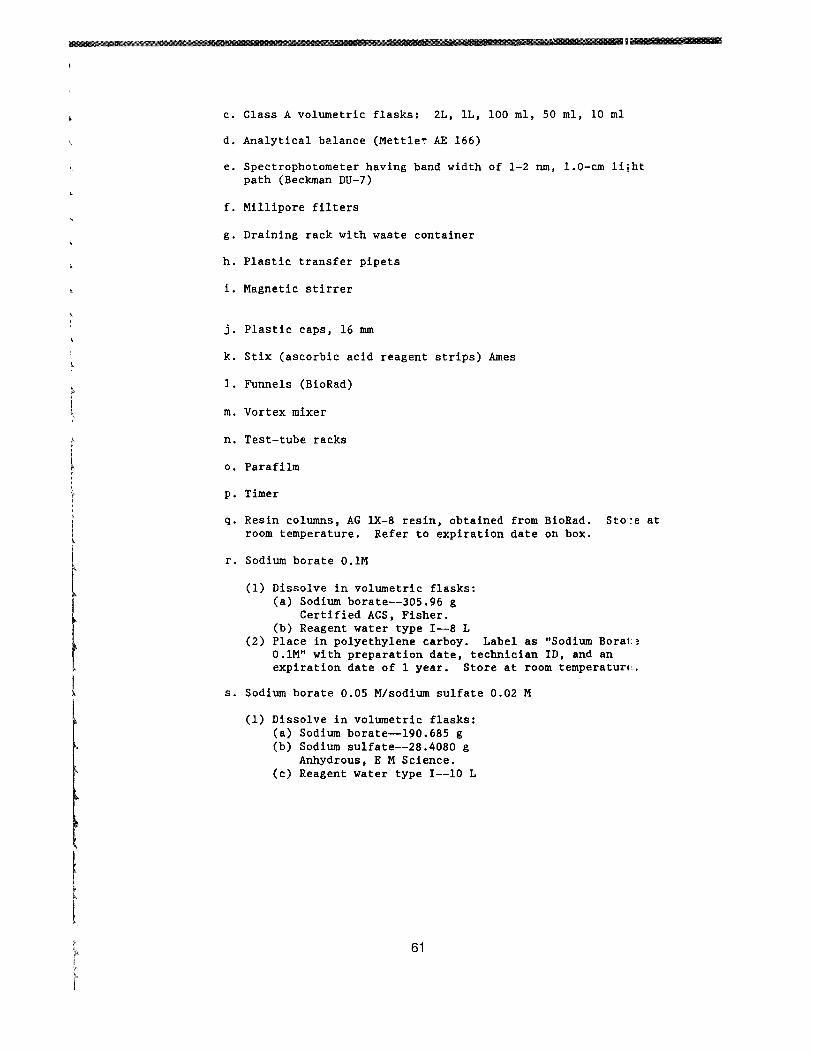
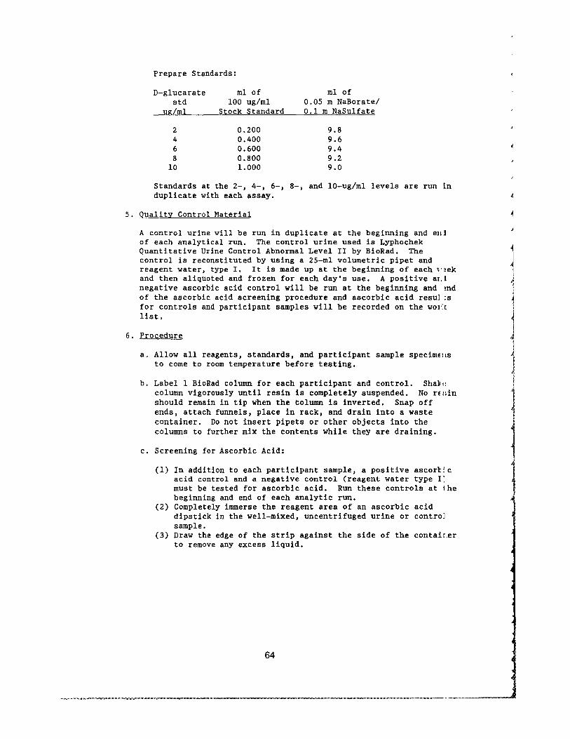
Related Documents