GUIDED BY Mr. Chaitanya Srinivas L.V. Sujeet Blessing Assistant Professor 08MBE026 SBST VIT University VIT University Vellore Vellore 2-D Comparative Gait Kinematics Using a Single Video Camera and EMG Signal Analysis

GUIDED BY Mr. Chaitanya Srinivas L.V. Sujeet Blessing Assistant Professor 08MBE026 SBSTVIT University VIT UniversityVellore Vellore 2-D Comparative Gait.
Dec 30, 2015
Welcome message from author
This document is posted to help you gain knowledge. Please leave a comment to let me know what you think about it! Share it to your friends and learn new things together.
Transcript
GUIDED BY
Mr. Chaitanya Srinivas L.V. Sujeet Blessing
Assistant Professor 08MBE026
SBST VIT University
VIT University Vellore
Vellore
2-D Comparative Gait Kinematics Using a Single Video Camera and EMG Signal
Analysis

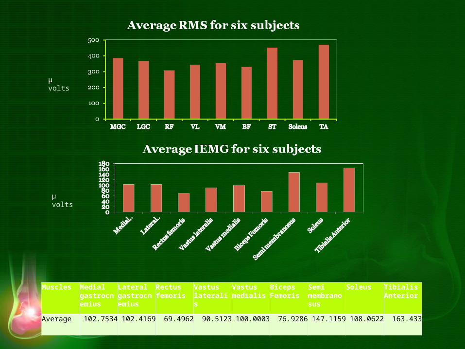
SUMMARY OF WORK
• Acquisition and Processing of EMG for six subjects from nine muscles
• Stride analysis for six subjects• Kinematics analysis for six subjects
• Marker based automated video-graphic analysis
• Marker-less automated video-graphic analysis
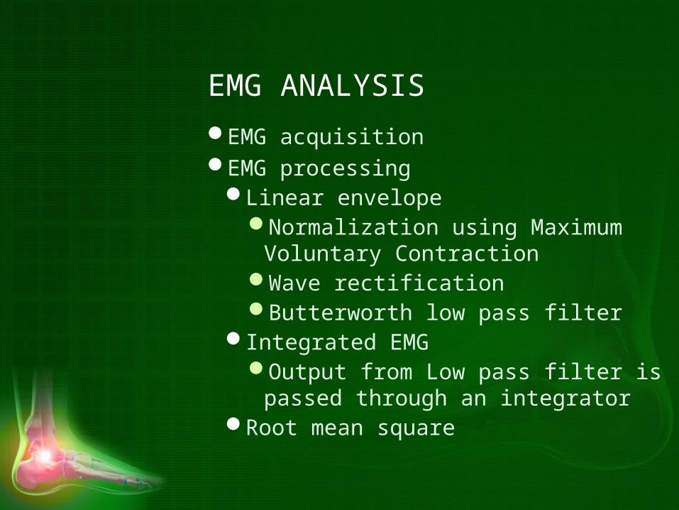
EMG ANALYSIS
EMG acquisitionEMG processing
Linear envelopeNormalization using Maximum
Voluntary ContractionWave rectificationButterworth low pass filter
Integrated EMGOutput from Low pass filter is passed
through an integratorRoot mean square

Biceps Femoris
Vastus Medialis
Vastus Lateralis
Semi Tendinosus
Rectus Femoris
Medial Gastrocnemius
Lateral Gastrocnemius
Soleus Tibialis Anterior
Linear envelope of EMG during one gait cycle
µ volts
µ volts
µ volts
µ volts
µ volts
µ volts
µ volts
µ volts
µ volts
Normal
µ volts
µ volts
Muscles Medial gastrocnemius
Lateral gastrocnemius
Rectus femoris
Vastus lateralis
Vastus medialis
Biceps Femoris
Semi membranosus
Soleus Tibialis Anterior
Average 102.7534 102.4169 69.4962 90.5123 100.0003 76.9286 147.1159 108.0622 163.433
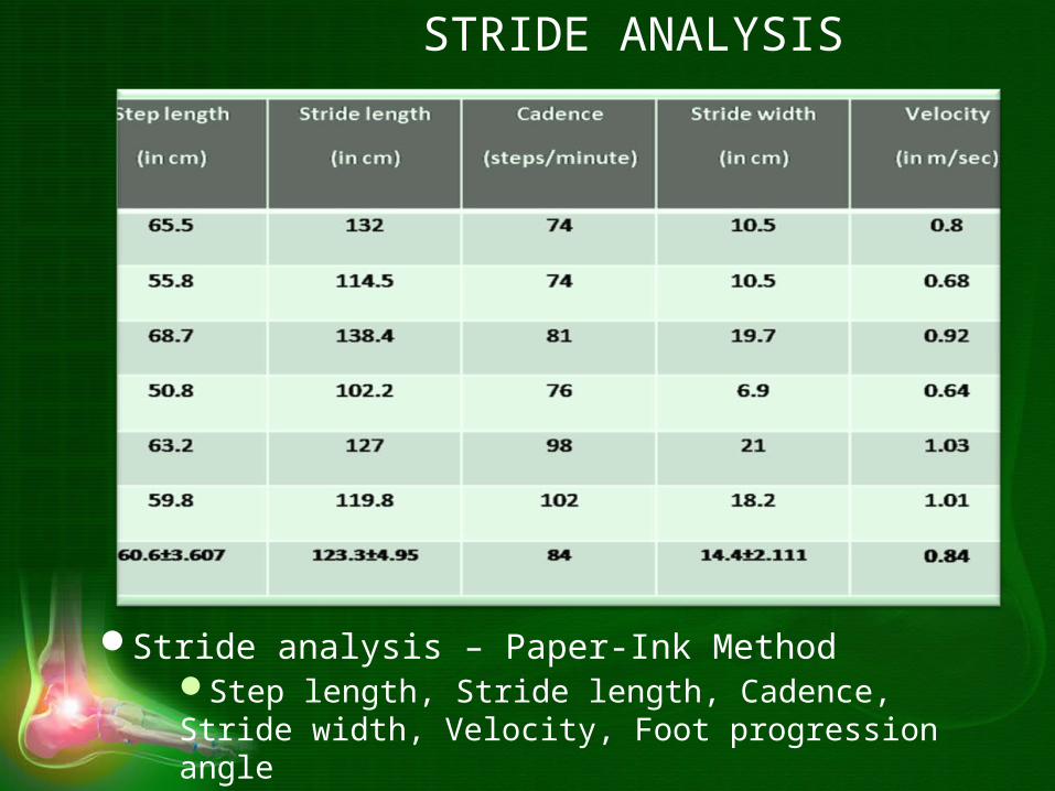
STRIDE ANALYSIS
Stride analysis – Paper-Ink MethodStep length, Stride length, Cadence, Stride width, Velocity, Foot progression angle
KINEMATIC ANALYSIS
• The motion of objects without consideration of the causes leading to the motion
• Determinants of position• Active – EMG• Passive – Force
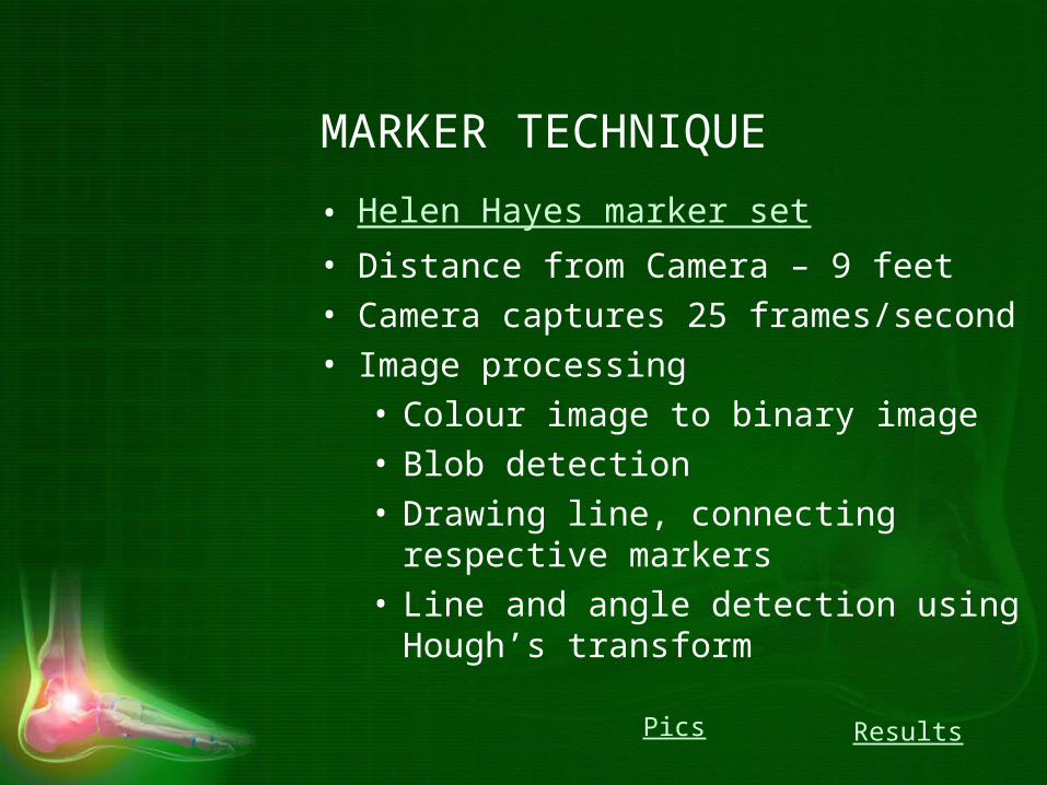
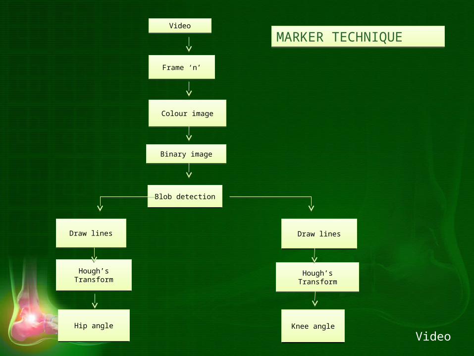
MARKER TECHNIQUE
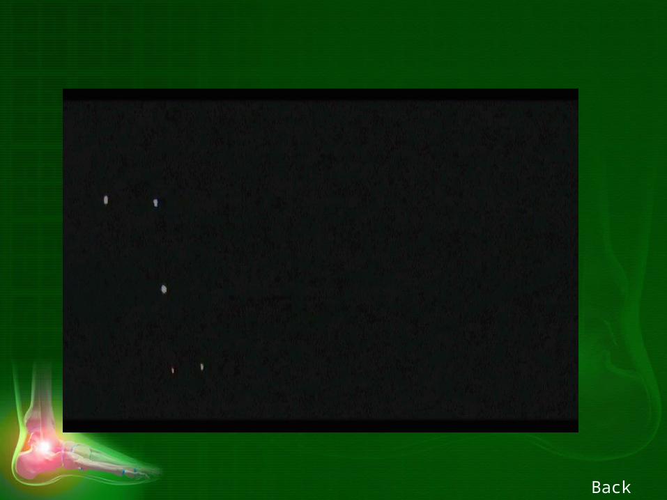
• Helen Hayes marker set• Distance from Camera – 9 feet • Camera captures 25 frames/second• Image processing
• Colour image to binary image• Blob detection• Drawing line, connecting respective
markers• Line and angle detection using Hough’s
transform
ResultsPics
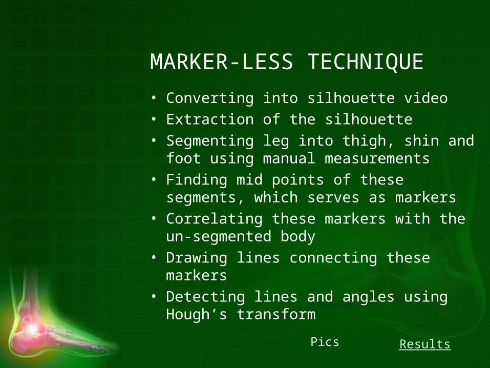
MARKER-LESS TECHNIQUE
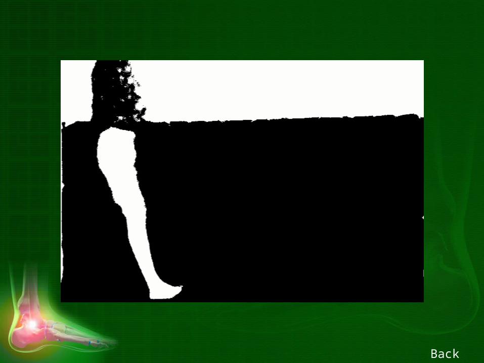
• Converting into silhouette video• Extraction of the silhouette• Segmenting leg into thigh, shin and foot
using manual measurements• Finding mid points of these segments,
which serves as markers• Correlating these markers with the un-
segmented body• Drawing lines connecting these markers• Detecting lines and angles using Hough’s
transformResultsPics
Colour imageColour image
Binary imageBinary image
VideoVideo
Frame ‘n’Frame ‘n’
Blob detectionBlob detection
Draw linesDraw lines
Hough’s TransformHough’s Transform
Draw linesDraw lines
Hough’s TransformHough’s Transform
Hip angleHip angle Knee angleKnee angle
MARKER TECHNIQUEMARKER TECHNIQUE
Video

Video (in RGB)
Video (in RGB)
Silhouette extractionSilhouette extraction
Frame ‘n’Frame ‘n’
Swing Phase Algorithm
Swing Phase Algorithm
Stance Phase Algorithm
Stance Phase Algorithm
Segmentation and Detection of Markers
Segmentation and Detection of Markers
Segmentation and Detection of
Markers
Segmentation and Detection of
Markers
Adjusting Leg Shortening using extraction
Adjusting Leg Shortening using extraction
Drawing LinesDrawing Lines
Drawing LinesDrawing Lines
Angle DetectionAngle Detection
Angle DetectionAngle Detection
MARKER-LESS TECHNIQUEMARKER-LESS TECHNIQUE
Video
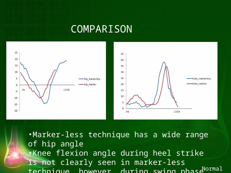
COMPARISON
•Marker-less technique has a wide range of hip angle•Knee flexion angle during heel strike is not clearly seen in marker-less technique, however, during swing phase, it has a good range
Normal
CONCLUSION
• Stride analysis was carried out using paper-ink method
• Emg was acquired from nine muscles from six subjects, processed and averaged
• Kinematic analysis was done on the same six subjects
• Marker and Marker-less automated video-graphic techniques were developed and the results were compared

REFERENCE• Richard Baker, “Gait analysis methods in rehabilitation”, Journal of
NeuroEngineering and Rehabilitation, 2006, 3:4.
• Mary M. Rodgers, “Dynamic biomechanics of the normal foot and ankle during walking and running”, Physical Therapy, 1988, 1822-30.
• Michela Goffredo, Imed Bouchrika, John N. Carter and Mark S. Nixon, “Performance analysis for gait in camera networks”, Association of Computing Machinery, 2008, 73-80.
• Y.P. Ivanenko, R.E. Poppele and F. Lacquaniti, “Five basic muscle activation patterns account for muscle activity during human locomotion”, American Journal of Physiology, 2004, 267-282.
• M.B.I. Reaz, M.S. Hussain and F. Mohd-Yasin, “Techniques of EMG signal analysis: Detection, processing, classification and applications”, Biological Procedures, 2006, 8(1): 11-35.
• Noraxon EMG and Sensor System, “Clinical SEMG Electrode Sites.” www.noraxon.com.
• Helen Hayes Marker System, www.helenhayeshospital.org.
Queries???

THANK YOU.
Back
Back
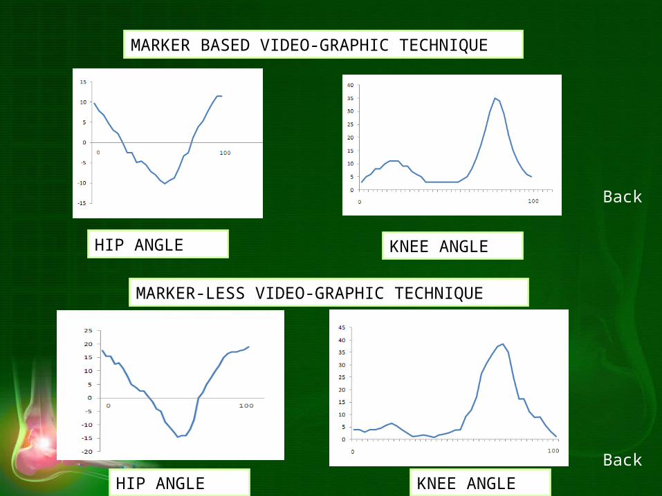
HIP ANGLE KNEE ANGLE
MARKER BASED VIDEO-GRAPHIC TECHNIQUE
MARKER-LESS VIDEO-GRAPHIC TECHNIQUE
HIP ANGLE KNEE ANGLE
Back
Back

MUSCLES
• Lateral gastrocnemius, Medial gastrocnemius, Vastus lateralis, Vastus medialis, Rectus femoris, Biceps femoris, Semi tendinosus, Soleus, Tibialis anterior
Back

LG MG
VLRFVM
TA
BF
SOLEUS
ST
% Stride
µ volts
Data Taken From Winter (1991)
Normal Hip Angle
Normal Knee Angle
Back
Back

From Helen Hayes official websiteFrom Helen Hayes official website
Back

a – one frame of an original video; b – grey image; c, d – binary image; e – blob detection; f – for hip angle
estimation; g – for knee angle estimation; h – detected lines by Hough’s transform for hip angle;
i – detected lines by Hough’s transform for knee angle
Back
h i
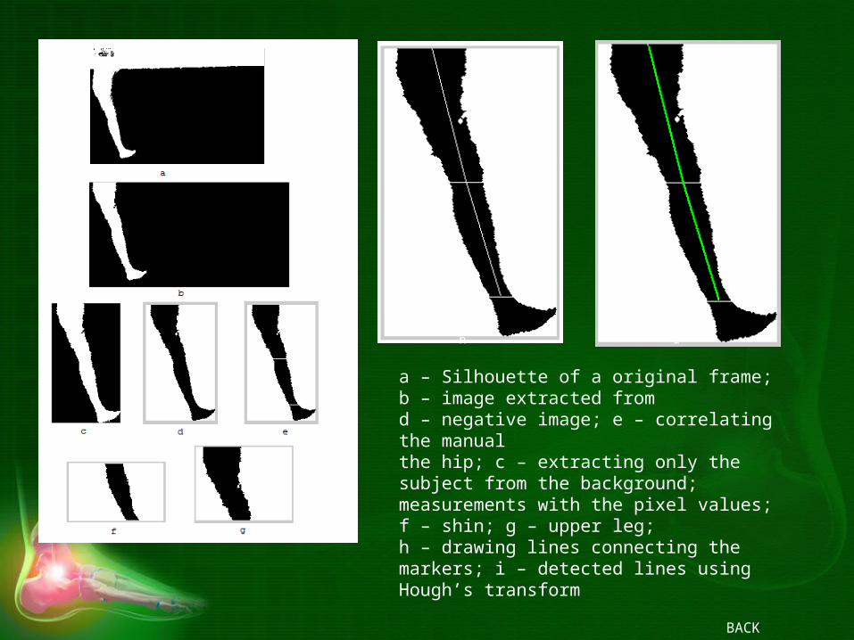
a – Silhouette of a original frame; b – image extracted from d – negative image; e – correlating the manual the hip; c – extracting only the subject from the background; measurements with the pixel values; f – shin; g – upper leg; h – drawing lines connecting the markers; i – detected lines using Hough’s transform
BACK
Related Documents