Fossil protists and fungi in amber and rock substrates Carmen Ascaso 1 , Jacek Wierzchos 2 , Mariela Speranza 3 , Juan Carlos Gutiérrez 4 , Ana Martín González 4 , Asuncion de los Ríos 1 , and Jesús Alonso 5 1 Centro de Ciencias Medioambientales, CSIC, Serrano 115 bis, 28006 Madrid, Spain email: [email protected] 2 Servicio de Microscopía Electrónica, Universidad de Lleida, Rovira Roure 44, 25198 Lleida, Spain 3 Laboratorio de Micologia, Facultad de Ciencias, Universidad de la República, Julio Herrera y Reissig 565, 11400 Montevideo, Uruguay 4 Departamento de Microbiologia III, Facultad de Biología, Universidad Complutense, 28040 Madrid, Spain 5 Museo de Ciencias Naturales de Álava, C/ Siervas de Jesús 24, 01001 Vitoria-Gasteiz, Spain ABSTRACT: The present study was designed to gain insight into the protists and fungi that made up the microbiota in the past, fossilized in two different substrates: amber and sandstone. The amber, dated as Lower Cretaceous, was from Álava in northern Spain, while fos- sil-bearing sandstone rocks were collected from the Linnaeus terrace and Mount Fleming regions of Antarctica. When examining this type of inclusion in hard substrates, it first has to be established whether the microorganism is mummified or only partially mineralized. In the latter case, some of the organism’s autofluorescence may be preserved. In our amber samples, light microscopy revealed a very well preserved microcenosis in what must have been a semiaquatic habitat comprised of several types of protozoa including Amoeba, Paramecium and Astasia (Euglena), Amebas limax and the colpodid ciliate Pseudoplatyophrya nana, as well as an abundant fossilized mycelium. The SEM-BSE procedure provided us with ultrastructural details of the fungi and protozoa, especially amoebae and flagel- lates. In the sandstone samples from Linnaeus terrace, it was possible to identify presumptive diatoms. Ultrastructural details were well-preserved in a fossil Trebouxia-type microalga from Mount Flemming, including the inner chloroplast area normally occupied by the pyrenoid. This fossilized microalga was shown by energy dispersive spectroscopy (EDS) to contain high Si levels in the pyrenoid zone, and high Fe levels in the area corresponding to the chloroplast periphery, peripheric cytoplasm and cell wall. In sandstone from Linnaeus Terrace, fossilized algae showed no central core containing Si, and the entire cell appeared to be completely filled with Al, K and Fe, with Fe most intensely observed within the algal wall.The present observations suggest that a well-preserved cell ultrastructure is the best criterion of biogenicity. INTRODUCTION The term biocomplexity has been recently used with reference to the study of an ecosystem at the highest level. When examin- ing an ecosystem on a scale as small as hundreds of cubic mi- crons, all its components need to be analysed at the microscale level. Analysis of the components of microcenoses in translu- cent resins such as amber can reveal the components and possi- ble functioning of certain microecosystems in the past. The finding of fossilized microorganisms in lithic substrates has helped us reconstruct the biotic and abiotic elements of these microecosystems before the appropriate conditions for permin- eralization of the microbiota were reached, also millions of years ago (e.g., Ascaso and Wierzchos 2003). In some regions of Antarctica, certain microorganisms are presently able to survive its extremely cold and dry conditions. Microbial ecology studies of the living microorganisms in the Dry Valley area of the Ross Desert commenced almost 30 years ago, yet investigations on fossilized microbiota have only very recently got underway (Wierzchos and Ascaso 2001). Careful interpretation of the morphological features of fossilized cells, such as preserved cell walls in algae, fungi and bacteria, cyto- plasm elements such as chloroplast membranes in algae and the spatial context of these structures enabled their identification as cryotoendolithic microfossils (Wierzchos and Ascaso 2002). In later studies, biomarkers of Antarctic microorganism fossils were detected. These biomarkers - traces left behind by micro- organisms after their death - are the result of the biological ac- tivity of these endolithic microorganisms while they were still alive. Fe rich diagenetic minerals in the form of iron hydroxide nanocrystals and biogenic clays are deposited around endolithic hyphae (Wierzchos et al. 2003). Biomarkers in the form of physicochemical weathering patterns and calcium oxalate and silica deposits have also been detected in pieces of granite rock from the Antarctic Ross Sea coast (Ascaso and Wierzchos 2003). Both in geological and palaeontological terms, amber from Álava (N Spain) is considered exceptional (see Alonso et al. 2000). Along with Lebanese amber (Schlee and Dietrich 1970) and the recently uncovered amber from Charente-Maritime, France (Néraudeau et al. 2002), this is one of the only three known highly fossiliferous amber deposits of the Lower Creta- ceous. According to Alonso et al. (2000), this amber was depos- ited in areas of a distal fluvial environment, where stagnant waters allowed the formation of lignite and pyrite nodules. Studies on microbiota inclusions in amber started with those by Poinar (1992) and Wier et al. (2002) using transmission electron microscopy (TEM), by Kohring (1995) using scanning electron microscopy with secondary electron imaging (SEM-SE), and by Waggoner (1994) and Schönborn et al. (1999) using light mi- croscopy (LM). Recently, Grimaldi used SEM in secondary electron mode to examine inclusions in amber mainly com- posed of wood, and fragments of insects (Grimaldi 2000a; 2000b). Cano and Boruki (1995) even purportedly revived and identified Bacillus bacteria in amber from the Dominican Re- public. Lambert et al. (1998) were also able to isolate a Staphy- lococcus species from soil and plant fragments embedded in micropaleontology, vol. 51, no. 1, pp. 59-72, text-figures 1-6, 2005 59

Welcome message from author
This document is posted to help you gain knowledge. Please leave a comment to let me know what you think about it! Share it to your friends and learn new things together.
Transcript
Fossil protists and fungi in amber and rock substrates
Carmen Ascaso1, Jacek Wierzchos2, Mariela Speranza3, Juan Carlos Gutiérrez4,Ana Martín González4, Asuncion de los Ríos1, and Jesús Alonso5
1Centro de Ciencias Medioambientales, CSIC, Serrano 115 bis, 28006 Madrid, Spainemail: [email protected]
2Servicio de Microscopía Electrónica, Universidad de Lleida, Rovira Roure 44, 25198 Lleida, Spain3 Laboratorio de Micologia, Facultad de Ciencias, Universidad de la República,
Julio Herrera y Reissig 565, 11400 Montevideo, Uruguay4Departamento de Microbiologia III, Facultad de Biología, Universidad Complutense, 28040 Madrid, Spain
5Museo de Ciencias Naturales de Álava, C/ Siervas de Jesús 24, 01001 Vitoria-Gasteiz, Spain
ABSTRACT: The present study was designed to gain insight into the protists and fungi that made up the microbiota in the past, fossilizedin two different substrates: amber and sandstone. The amber, dated as Lower Cretaceous, was from Álava in northern Spain, while fos-sil-bearing sandstone rocks were collected from the Linnaeus terrace and Mount Fleming regions of Antarctica. When examining thistype of inclusion in hard substrates, it first has to be established whether the microorganism is mummified or only partially mineralized.In the latter case, some of the organism’s autofluorescence may be preserved. In our amber samples, light microscopy revealed a verywell preserved microcenosis in what must have been a semiaquatic habitat comprised of several types of protozoa including Amoeba,Paramecium and Astasia (Euglena), Amebas limax and the colpodid ciliate Pseudoplatyophrya nana, as well as an abundant fossilizedmycelium. The SEM-BSE procedure provided us with ultrastructural details of the fungi and protozoa, especially amoebae and flagel-lates. In the sandstone samples from Linnaeus terrace, it was possible to identify presumptive diatoms. Ultrastructural details werewell-preserved in a fossil Trebouxia-type microalga from Mount Flemming, including the inner chloroplast area normally occupied bythe pyrenoid. This fossilized microalga was shown by energy dispersive spectroscopy (EDS) to contain high Si levels in the pyrenoidzone, and high Fe levels in the area corresponding to the chloroplast periphery, peripheric cytoplasm and cell wall. In sandstone fromLinnaeus Terrace, fossilized algae showed no central core containing Si, and the entire cell appeared to be completely filled with Al, Kand Fe, with Fe most intensely observed within the algal wall.The present observations suggest that a well-preserved cell ultrastructure isthe best criterion of biogenicity.
INTRODUCTION
The term biocomplexity has been recently used with referenceto the study of an ecosystem at the highest level. When examin-ing an ecosystem on a scale as small as hundreds of cubic mi-crons, all its components need to be analysed at the microscalelevel. Analysis of the components of microcenoses in translu-cent resins such as amber can reveal the components and possi-ble functioning of certain microecosystems in the past. Thefinding of fossilized microorganisms in lithic substrates hashelped us reconstruct the biotic and abiotic elements of thesemicroecosystems before the appropriate conditions for permin-eralization of the microbiota were reached, also millions ofyears ago (e.g., Ascaso and Wierzchos 2003).
In some regions of Antarctica, certain microorganisms arepresently able to survive its extremely cold and dry conditions.Microbial ecology studies of the living microorganisms in theDry Valley area of the Ross Desert commenced almost 30 yearsago, yet investigations on fossilized microbiota have only veryrecently got underway (Wierzchos and Ascaso 2001). Carefulinterpretation of the morphological features of fossilized cells,such as preserved cell walls in algae, fungi and bacteria, cyto-plasm elements such as chloroplast membranes in algae and thespatial context of these structures enabled their identification ascryotoendolithic microfossils (Wierzchos and Ascaso 2002). Inlater studies, biomarkers of Antarctic microorganism fossilswere detected. These biomarkers - traces left behind by micro-organisms after their death - are the result of the biological ac-tivity of these endolithic microorganisms while they were still
alive. Fe rich diagenetic minerals in the form of iron hydroxidenanocrystals and biogenic clays are deposited around endolithichyphae (Wierzchos et al. 2003). Biomarkers in the form ofphysicochemical weathering patterns and calcium oxalate andsilica deposits have also been detected in pieces of granite rockfrom the Antarctic Ross Sea coast (Ascaso and Wierzchos2003).
Both in geological and palaeontological terms, amber fromÁlava (N Spain) is considered exceptional (see Alonso et al.2000). Along with Lebanese amber (Schlee and Dietrich 1970)and the recently uncovered amber from Charente-Maritime,France (Néraudeau et al. 2002), this is one of the only threeknown highly fossiliferous amber deposits of the Lower Creta-ceous. According to Alonso et al. (2000), this amber was depos-ited in areas of a distal fluvial environment, where stagnantwaters allowed the formation of lignite and pyrite nodules.Studies on microbiota inclusions in amber started with those byPoinar (1992) and Wier et al. (2002) using transmission electronmicroscopy (TEM), by Kohring (1995) using scanning electronmicroscopy with secondary electron imaging (SEM-SE), and byWaggoner (1994) and Schönborn et al. (1999) using light mi-croscopy (LM). Recently, Grimaldi used SEM in secondaryelectron mode to examine inclusions in amber mainly com-posed of wood, and fragments of insects (Grimaldi 2000a;2000b). Cano and Boruki (1995) even purportedly revived andidentified Bacillus bacteria in amber from the Dominican Re-public. Lambert et al. (1998) were also able to isolate a Staphy-lococcus species from soil and plant fragments embedded in
micropaleontology, vol. 51, no. 1, pp. 59-72, text-figures 1-6, 2005 59

amber. Further, some authors have argued that DNA is pre-served in amber (Smith and Austin 1997, Stankiewicz et al.1998). Several genera of fungi have been described by differentauthors using LM. Thus, there are descriptions of Aspergillusand Entomophtoralean species and higher fungi fossilized inamber from the Dominican Republic (Thomas and Poinar 1988;Poinar and Thomas 1982; Poinar and Singer 1990; Rikkinenand Poinar 2001). Protozoa have also been identified in amber(Poinar 1992; Waggoner 1994; Schönborn et al. 1999). Ascasoet al. (2003) combined several innovative microscopy tech-niques for the in depth ultrastructural analysis of the compo-nents of the microbiota preserved in amber from Álava. Thisallowed them to observe the 3-dimensional structure of themicrocenosis and to simultaneously discern which parts of theprotist and fungal structures were most mineralized. Scanningelectron microscopy with backscattered electron imaging(SEM-BSE) was one of the techniques applied.
Although fungi are an extremely diverse group of microorgan-isms, only around one hundred fossilized fungi and very fewgenera have been identified in fossil resins (Pirozynski 1976;Rikkinen and Poinar 2001). In lithic substrates, fossilized fungiwere first, and very recently, described in the Antarctic sand-stones of Mount Flemming in the Ross Desert (McMurdo DryValleys) (Wierzchos and Ascaso 2002; Ascaso and Wierzchos2003). These fungi were observed alongside accompanyingfossil microalgae (protists).
The study of fungi and protists fossilized in amber and Antarc-tic rock substrates could contribute to the field of microbialecology, since the micropalaeontological data obtained willhelp gain insight into the evolution of these microecosystems.
The aim of the present study was to analyse the protists andfungi fossilized in amber and Antarctic rocks that made up themicrobiota in the past, and also to try to improve our under-standing of permineralization phenomena by determining thechemical elements present in the microbiota at the nanoscale.
MATERIALS
Amber
Amber collection and preparation. For details of the collectionand preparation of amber deposits dated as Lower Cretaceous,see Corral et al. (1999). Detailed references on the macroscopicinclusions observed by other authors in these deposits and a de-scription of the techniques used to observe microcenoses andmicrodebris (plant and animal remnants) can be found inAscaso et al. (2003).
Lithic Substrate
Pieces of sandstone rock thought to contain fossils were col-lected by E. I. Friedmann over the year 1983/84 from theLinnaeus Terrace region (77.60o S, 161.08o E, 1600 m a.s.l) ofthe Ross Desert. Mean annual air temperature in 1993 was re-ported as – 23.2oC for this area (Friedmann et al. 1994). Furthersandstone samples were collected from the harsh Mount Flem-ing region (77o 33’ S, 160o 06’ E, 2200 m alt.) of the RossDesert, where the mean annual air temperature in 1993 was-24.9oC (Friedmann et al., 1994). The rocks were air-dried, andstored in an air-conditioned room until use.
METHODS
When examining fossil protists and fungi included in hard sub-strates, one has to dispense of all molecular techniques and ap-ply the more suitable methods of confocal and electronmicroscopy. It also has to be considered whether the microor-ganism is mummified or only partially mineralized. In the lattercase, some of the organism’s autofluorescence may be pre-served, as in some protists and fungi found in amber. Theprotists and fungi fossilized in Antarctic rocks examined here,were fully mineralized such that they emit no autofluorescence.
Despite its lower power of resolution, light microscopy may beused for a preliminary view of the microorganisms mummifiedor mineralized in the amber and should also be used to classify
60
Carmen Ascaso et al.: Fossil protists and fungi in amber and rock substrates
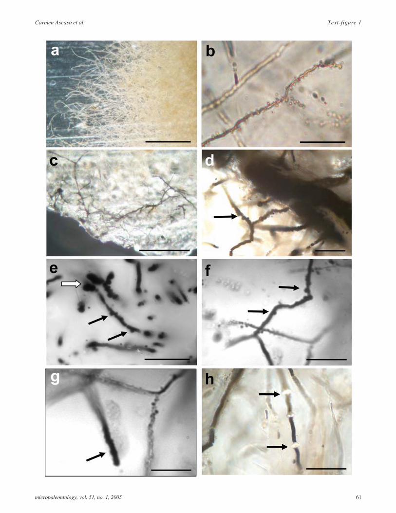
TEXT-FIGURE 1
a Fossilized hyaline mycelium found in amber; scalebar = 50�m.
b Closer view of the fungal colony composed bycoenocytic hyphae. Most hyphae are 0.75-1 µm widerand few bifurcates branches were found at the expand-ing mycelium zone. Note the abundant vesiclesspore-like structures in the hyphae. Other fungal rem-nants can also detected in the sample; scale bar =20�m.
c A dark mycelium was detected in the border zone ofthe amber sample, which strongly resemble asaprophytic fungus; scale bar = 50�m.
d Dark mycelium comprised of thick-walled vegetativehyphae, 1.5 to 3�m in width, with numerous simpleclam connections and no septa (black arrow); scale bar= 20�m.
e Globose and irregularly constricted cystidia likestructures are best observed by fluorescence micros-copy (white arrow). Simple clamp connectios markedby black arrow; scale bar = 20�m.
f Simple clamp connections (black arrows); scale bar =20�m.
g Long apical hyphae with irregular surface and en-crusted material resembled gloeocystidia forms pres-ents in present-day Basidiomycetes (black arrow);scale bar = 20�m.
h Hyphae segments were produce by constriction andrefractive intercalary structure are present (black ar-rows); scale bar = 20�m.
micropaleontology, vol. 51, no. 1, 2005 61
Carmen Ascaso et al. Text-figure 1
them. The taxonomy of current protists and fungi is based ondecades of LM work, though molecular methods are now beingextensively used, mainly for fungi. Light microscopy has alsobeen widely applied to the study of arthropods in amber, al-though it is much more difficult to examine microorganismssuch as amoebae, ciliates, flagellates and fungi in this resin.Thus, in this study, we used LM, SEM-BSE and confocal laserscanning microscopy (CLSM) for the fossil protists and fungiin amber, and SEM-BSE with energy dispersive spectroscopy(EDS) to observe and chemically characterise these microor-ganisms mineralized in Antarctic rocks.
-LM. Amber samples for LM were screened under a ZeissAxioskop 2 Microscope. Some of the specimens were simulta-neously examined under a fluorescence microscope (ZeissAxioplan) with ultraviolet light using a 360/40D filter. The flu-orescence emitted was analysed through a 460 nm barrier filter,and the image was captured with a CCD device (PhotometricsCH250/A).
These samples were also subjected to CLSM.
CLSM. This technique has great advantages for the study ofmummified microorganisms embedded in hard, translucentsubstrates, and was therefore used here to examine inclusions inamber. The polished blocks of amber were mounted on micro-scope slides using double-sided adhesive tape, and observed us-ing a LSM 310 Zeiss confocal microscope with aPlan-Apochromat 63x/1.40 oil immersion objective. Argon(488 nm) and helium/neon (543 nm) lasers were used to gener-ate an excitation beam. The resultant emission was filteredthrough long pass filters of >515 nm and >575 nm, respec-tively. The relatively good translucency of the amber permitsthe three-dimensional (3D) reconstruction of fungal and proto-zoan colonies and gives an idea of their spatial organization. Toobtain this information, stacks of 20-30 single confocal opticalsections (vertical (z) resolution about 0.6 µm) images are pre-pared at 0.5-1 µm intervals through the sample and digitallystored and compiled.
CLSM has been successfully used for the study of live lithicmicrobiota (Rautureau at al. 1993; Ascaso et al. 1998; Wierz-chos and Ascaso 2001). However, this method cannot be ap-plied to the study of microorganisms embedded in what was inthe past an organo-mineral phase, if the biological components(algae, fungi, etc.) are presently completely mineralized.
SEM-BSE. Both for the analysis of partly or fully mineralizedmicroorganisms in amber and mineralized microorganisms em-bedded in a lithic substrate, SEM-BSE has proved to be themost appropriate. When used to examine specimens in amber,SEM-BSE reveals the structural details of protists and fungi.This is of particular interest, since these details may be com-pared with the inner structure of present-day protists observedusing the transmission electron microscope. If the microorgan-ism is mineralized in rock, SEM-BSE is the only method cur-rently known to reveal a large number of ultrastructural details.These details allow the identification of the fossilized organism.
The use of backscattered electrons in SEM yields high magnifi-cation images and contrast attributable to differences in the av-erage atomic number of the target (Joy 1991). The partial ortotal mineralization of microorganisms in amber induces differ-ences in atomic number that reveal the ultrastructure of the mi-croorganism. When examining lithic materials containing fullymineralized fossils, complete mineralization also gives rise toatomic number differences that eventually reflect the ultra-structure of the microorganism.
For the present SEM-BSE observations and/or EDS micro-analyses, the amber and lithic fragments were embedded in ep-oxy resin. After polymerization, the blocks were cut and finelypolished (e.g., Wierzchos and Ascaso 1994). Transverse sec-tions of polished surfaces were carbon-coated and examined us-ing a DSM 940 A Zeiss and a DSM 960 A Zeiss microscope(both equipped with a four-diode, semiconductor BSE detectorand a Link ISIS microanalytical EDS system). SEM-BSE andEDS examinations of the samples were simultaneously per-formed. The microscope operating conditions were as follows:0º tilt angle, 35º take-off angle, 15 kV acceleration potential, 6or 25 mm working distance and 1-5 nA specimen current.
RESULTS
Amber
Light microscopy revealed a very well preserved microcenosisin, what was in the past, a semiaquatic habitat comprised of sev-eral types of protozoa and an abundant fossilized mycelium.Text-figure 1a-b shows a mycelia mat formed by hyalinehyphae with many vesicle structures. Most hyphae were 0.75-1ìm wide, and only a few bifurcated branches were found in theregion of the expanding mycelium. No septa were observed, thehyphal system appearing to be coenocytic. The vesicle struc-
62
Carmen Ascaso et al.: Fossil protists and fungi in amber and rock substrates
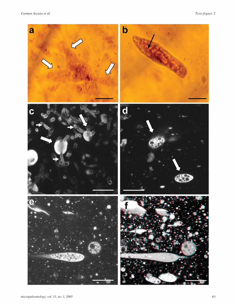
TEXT-FIGURE 2
a LM image of an amoeboid protozoa, probably of thegenus Amoeba. Arrows indicate pseudopodia; scalebar = 50�m.
b LM image of a ciliated, paramecium-like protozoa.Arrow indicates oral groove; scale bar = 50�m;
c Image of a single CLSM section showing a protozoa(arrows) and hyphae (arrowheads) cells; scale bar =50�m.
d Image of single CLSM section showing a protozoa(arrow); scale bar = 50�m.
e Image of single CLSM section showing a protozoa;scale bar = 50�m.
f 3D reconstruction (15 single CLSM sections sepa-rated by 1�m) of protozoa cells. Stereo image; scalebar = 50�m.
micropaleontology, vol. 51, no. 1, 2005 63
Carmen Ascaso et al. Text-figure 2
tures were closely associated with hyphae and resembled fungalspores.
Given the low resolution of LM and the refractive properties ofthe amber, a detailed description of the hyphae and spore-likestructures could not be made and it was not possible to deter-mine hyphal wall thicknesses.
Besides the many fungal structures observed in the amber sam-ple, a distinct dark fungal colony was detected (text-fig. 1c).This dark extensive mycelium appeared in the border zone ofthe amber piece, comprised of thick-walled vegetative hyphae1.5-3 µm in width (text-fig. 1d). Most of these hyphae had ir-regular surfaces and some appeared to have a septum but wecould not determine whether this was an artefact of fossilization(text-figs. 1d,e). However, numerous simple clamp connectionsresembling those of living basidiomycetes were observed(text-figs. 1d-f, arrows). The use of fluorescence microscopy onthe same specimen improved visualization of the mummifieddark mycelium structures, and globose, sometimes irregularlyconstricted cystidia-like structures were observed (text-fig. 1e,white arrow). In addition, long apical hyphae with irregular sur-faces and encrusted material resembling gloeocystidia shapescould be seen (text-fig. 1g). Few terminal bifurcated brancheswere detected (text-fig. 1f). Hyphal segments were produced byconstriction and the presence of refractive intercalary structureswas noted (text-fig. 1h, arrows). All these structures occur inpresent-day basidiomycetes.
Text-figure 2 shows both LM (text-figs. 2 a,b) and CLSM(text-figs. 2 c-f) images. Text-figure 2a is an LM image of anAmoeba showing several pseudopods (arrows indicatepseudopodia). Text-figure 2b shows a ciliate, probably of thegenus Paramecium. In text-figure 2c, the fungal cells (arrow-head) accompany the protozoan (arrows), as shown in a confo-cal simple section. Text-figures 2d-f only show protozoa:Text-figures 2d (protozoan indicated by arrows) and 2e are sim-ple sections and text-figure 2f is a 3D reconstruction of zone intext-figure 2e.
The SEM-BSE images reveal interesting ultrastructural detailsof the fungi (text-fig. 3) and protozoa (text-fig. 4). Text-figure3a shows a coenocytic, unbranched hypha with several buds re-sembling spores (arrowhead), similar to those observed by LMin text-figure 1b. Text-figure 3b shows a similar hypha, trans-versely cut to reveal radial filaments (arrowhead) and buds (ar-rows). In text-figure 3c, we can see the terminal branch apex ofa hypha, while text-figure 3d shows a hypha with constrictionstransverse to the main axis (arrows) similar to those observed in
text-figure 1h. The spatial relationship between hyphal apicesand protozoa is shown in text-figure 3e, in which sections of theprotozoan (arrows) can be seen in close contact with the hyphaeand hyphal apices (arrowhead). These hyphal apices presentbuds embedded in the fungal sheath (text-fig. 3f)
Text-figure 4a shows an amoeba with a temporary extension ofits body, or pseudopod, (upper arrow) in longitudinal section incontact with the fungal wall (lower arrow). Text-figure 4bshows a detail of the area described in text-figure 4a. The hyphain contact with the amoeba has radial filaments but is devoid ofbuds. In contrast, text-figure 4c shows a detail of the zone ofcontact between a protozoan and hypha (arrow) with spore-likebuds. When examining flagellates, SEM-BSE allows the de-tailed observation of recurrent flagellum joined to the flagellate(text-fig. 4d, arrow). Text-figures 4e and 4f show a fungalhypha penetrating a protozoan, or perhaps, a fungus captured bya protozoan. The protozoan is indicated with an lower arrowand the fungus is marked with an upper arrow. Text-figure 4 f isan image of a detail in text-figure 4e.
Lithic substrate
The images in text-figure 5 are SEM-BSE micrographs of sand-stone samples from Linnaeus Terrace. Text-figure 5a is a gen-eral view, showing live microorganisms (arrows) among quartzgrains (asterisks). Text-figure 5b shows a detail of the previousfigure in which protists (microalgae) can be seen alongsidefungi. Given the harsh conditions of the region, thesemicroalgae, possibly Trebouxia, show some degree ofplasmolysis (open arrow) and fungi show an alteredultrastructure (arrow). In text-figures 5 c-f, we provide imagesof a Linnaeus Terrace sandstone sample, but this time, contain-ing fossil microorganisms. Text-figure 5c is an overview inwhich quartz grains (asterisks) may be distinguished and amongthese, pores, indicating zones where live microorganisms previ-ously occurred and are now fossilized (open arrow). Emptypores could also be seen (black arrow) in which no microorgan-isms existed previously, or did exist but the permineralizationprocess was unsuccessful leading to their disappearance. Thehigh magnification image of microfossils in text-figure 5d al-lows the identification of presumptive diatoms (arrow) andTrebouxia-type microalgae (open arrow). Text-figure 5e alsoshows these algae (open arrow) along with the fossil diatoms(arrow). The high magnification image in text-figure 5f shows afossil microalga 6-8 ìm wide and 8-10 ìm long (arrow). Elementdistribution maps of these zones revealed high Al and K con-tents within and outside the fossil cells. The distribution map forFe was similar, but a higher concentration was noted at the mi-
64
Carmen Ascaso et al.: Fossil protists and fungi in amber and rock substrates
TEXT-FIGURE 3SEM-BSE images of amber inclusions
a Coenocytic, unbranched hypha with several buds re-sembling spores (arrowhead); scale bar = 10µm.
b Cross section of a coenocytic hypha revealing radialfilaments (arrowhead) and buds (arrows); scale bar =50�m.
c Terminal branch apex of a hypha; scale ba r =10�m.
d Hypha with constrictions (arrows); scale bar = 10�m.
e Protozoa (arrows) in close contact with hyphal apices(arrowhead); scale bar = 20 �m.
f Detailed image of hyphal apices with buds; scale bar =10 �m.

micropaleontology, vol. 51, no. 1, 2005 65
Carmen Ascaso et al. Text-figure 3

croorganism walls. Silica was weakly dispersed throughout thezones containing biological material and was obviously mostintense in the areas where quartz grains surrounded the pore.
Text-figure 6 shows fossil protists in sandstone from MountFleming. The pore in text-figure 6a was seen to contain severalcryptoendolithic permineralized microorganisms (arrows). Intext-figure 6b, several algae (arrows) surrounded by fungi ap-pear mineralized. The microalga indicated by an open arrowshows ultrastructural features that resemble thylakoids and lipidbodies. As it becomes mineralized, the alga marked with an as-terisk in text-figure 6b, like the alga indicated by a star intext-figure 6d, shows the marked presence of crystals occupy-ing the space in the centre of the cell that would have been thechloroplast. Text-figure 6c shows fossilized microalgae con-taining high amounts of Si inside, and high Fe levels in the areacorresponding to the chloroplast periphery, peripheric cyto-plasm and cell wall, observed as bright peripheral bands (ar-row). Some cells were completely filled with Ti and O(asterisk). EDS profiles for this zone indicated relative concen-tration changes in Si, Al, Na. Fe and Ti (data not shown).Text-figure 6d shows a Si rich crystal, as can be seen in text-fig-ure 6e, in the core of the cells and details of membranes be-tween this crystal and the cell wall. The distribution of Si can beseen in the map provided alongside the micrograph of the poreharbouring this alga. The element distribution maps for Al, Si,S, K, Ca and Fe highlight the particular presence of Fe and K,along with Al and some Si around the central silica crystal (ap-pearing as an intense bright spot in the Si map). This silica-richcrystal is located in what would be the pyrenoid and the centralpart of the chloroplast in the live alga. Thus, it seems that Fe, K,Al and some Si occupy the chloroplast periphery and the cyto-plasm. Silica was also weakly detected in these parts but moreso outside the cell. Ca was only found in the cell exterior.
The presence of S and Ca corresponds to gypsum, which envel-ops the microalgae Given there is some K and Fe outside thecells, jarosite may also be present.
DISCUSSION
All the protists found in the Álava amber were protozoa, includ-ing flagellate, amoeboid and ciliate forms, yet no protists suchas microscopic algae and slime molds were found. The proto-zoa observed were accompanied by fungi but no prokaryotes,bacteria nor cyanobacteria could be detected around these. Astext-figures 4a and b indicate, fungi seem to be closely associ-
ated with amoebae. We also observed this behaviour in aprevious investigation (Ascaso et al., 2003).
The microfossils of protists found in the Álava amber indicatethey belonged to the microcenosis of a semiaquatic habitat. Onthe other hand, the lack of algal and cyanobacterial fossils failsto suggest a permanently wet, semi-aquatic susbtratum(Rikkinen and Poinar 2001). The presence of the generaAmoeba and Paramecium was confirmed by LM. We were alsoable to identify Astasia (Euglena), Amebas limax and thecolpodid ciliate Pseudoplatyophrya nana. The SEM-BSE tech-nique applied revealed ultrastructural details of the flagellatesand details of flagellum insertion (text-fig. 4d). The first LM de-scription of fossil zooflagellates was made by Schönborn et al.(1999), while Ascaso et al. (2003) provided SEM-BSE imagesof flagellates from Álava amber. Both Schonbörn et al. (1999)and Ascaso et al. (2003) provided the first images ofpalaeo-flagellates.
Despite having observed ciliates by LM (such as the Parame-cium shown in text-fig. 2b), no ciliates could be easily identifiedusing SEM-BSE, probably due to the loss of their cilia oncetrapped in the resin that would eventually form the amber. Con-ventional SEM-SE has yielded useful data for the taxonomy ofciliated protozoa in aqueous media (Foissner 1991). Based onthis technique, it has been possible to 3-dimensionally recon-struct the oral apparatus of ciliated protozoa (Hofmann-Münz etal. 1990), but this invariably involved removing the organismsfrom the aqueous medium in which they lived. As mentionedabove, it is likely that when embedded in amber, ciliates losetheir cilia. Moreover, it is impossible to remove them from theamber without damaging their ultrastructural elements.Schombörn et al. (1999) identified Paramecium species in fossilamber whose length:width ratio, highly characteristic bodyshape and similarities to related species and to those of thepresent paper indicate they are congeneric organisms.
When cells were mineralized, SEM-BSE revealed ultra-structural details of the cytoplasm in the three protozoan groups(except the presence of cilia). Non-mineralized protozoa pre-serve their autofluorescence signal and this gives rise to inter-esting confocal microscopy images. Further, CLSM enables thedetection of partly- or non-mineralized fungi. Single confocalsections (text-figs. 2d,e) allow the observation of someultrastructural elements of cells, and serial confocal sectionsserve to obtain sufficient data for 3D reconstruction. The 3Dimages give some idea as to what extent fungal cells are spa-
66
Carmen Ascaso et al.: Fossil protists and fungi in amber and rock substrates
TEXT-FIGURE 4SEM-BSE images of amber inclusions
a Amoeba with pseudopods (upper arrow) in contactwith fungal wall (lower arrow); scale bar = 10�m.
b Detail of the area of contact between amoeba and thefungal wall in text-figure 4a (arrow); scale bar = 5�m.
c Contact (arrow) between protozoa (star) and hyphae(asterisk); scale bar = 10�m.
d Flagellate with recurrent flagellum (arrow); scale bar= 20�m.
e Fungal hypha (right arrow) penetrating a protozoan(left arrow); scale bar = 10�m.
f Detail of the image shown in text-figure 4e. Fungalhyphae (arrowhead) and protozoa (arrow); scale bar =5�m.

micropaleontology, vol. 51, no. 1, 2005 67
Carmen Ascaso et al. Text-figure 4

tially related to the protozoa in different zones of the amber. Insome areas, only protozoa appear after 3D reconstruction (thustext-fig. 2f shows no fungal cells). The autofluorescence re-sponse (signal intensity) provides significant information on thegreater or lesser degree of mineralization or, in other words, onthe state of preservation of natural fluorochromes. When exam-ining protozoan and fungal ultrastructure in amber, the CLSMand SEM-BSE techniques complement each other; the formerdetects the signal emitted by non-mineralised parts and the laterpicks up the signal from mineralised components.
In this study, the cytoplasm of fungi seemed to be fully miner-alized and therefore showed-up brightly in the SEM-BSE im-ages (text-figs. 3a-d and text-figs. 4a,b,e). In another zone ofthe same sample, the fungi were not mineralized and thus pre-served some autofluorescence clearly revealing wall structureby confocal microscopy (see text-fig. 2c). The qualitativeand/or quantitative microanalysis (EDS) of the chemical ele-ments in the inner structure of fossilized microorganisms em-bedded in amber indicated the presence of Fe and S, providinginformation on biomineralization and fossilization processes.
Some of the more abundant fungal hyphae inclusions observedby LM and SEM-BSE appear to be largely coenocytic, allowingtheir assignment to specific groups of fungi. It could even bethat, not having combined the techniques LM, FM, CLSM andSEM-BSE, to date, mycologists have not been able to explorefungi in amber in sufficient depth. Although current fungi areextremely diverse, so far, very few genera have been identifiedin fossil resins since it is often difficult to identify their repro-ductive structures using conventional techniques (Pirozynski1976; Rikkinen and Poinar 2001).
Hyphae were often endowed with radial structures that showedup brightly on SEM-BSE due to their high S and Fe contents. Itwas these fungi with numerous buds, resembling present-dayaquatic fungi that were found closest to the protozoa. Generallythese were amoebae, and it was these radial elements that madeintimate contact with the protozoan (text-figs. 4a,b). Although,we are not in a position to interpret interrelations between theprotozoa and fungi detected in amber from Álava, we are confi-dent that future surveys will shed some light on this issue.
The first descriptions of fungal inclusions in rock substrates re-fer to observations made in Antarctic sandstones and are veryrecent (Wierzchos and Ascaso 2002). Studies of fossilized
microbiota in Antarctic lithic substrates have mainly focussedon microalgae (Wierzchos and Ascaso 2001; Ascaso andWierzchos 2003).
In the present samples from Linnaeus Terrace, interesting com-parisons can be made between sandstones containing live mi-croorganisms and those harbouring fossil microorganisms,since both of these types of sample may be found. In LinneaeusTerrace sandstone containing live microorganisms, quartzgrains were loose, exposing fissures and pores among the grainsthat were occupied by living algae and fungi. However, giventheir ultrastructural appearance, the physiological state of themicrobiota was not optimal. Sandstones from this region withfossil microorganisms showed quartz grains that were tightlybound to each other (text-fig. 5c). In SEM-BSE images, poresshowed whitish patches as the result of the high atomic numberof permineralized microbial masses. In samples from MountFlemming, in which mineralized microorganisms were ob-served in all the samples examined, quartz grains were sotightly bound that they were often indistinguishable. However,the fossilization process in Mount Flemming often manages toperfectly preserve the ultrastructural features of microalgae.The Mount Fleming microalga fossil shown in text-figure 6d,has kept all its ultrastructural details including the innerchloroplast area normally occupied by the pyrenoid. This zoneis very rich in Si. These ultrastructural details and the presenceof a central area in the cell of high Si content, are similar charac-teristics to those shown by a fossil microalga, also from MountFleming, described in one of our previous studies (Wierzchosand Ascaso 2002). Mount Flemming harbours a further type ofalga, also with a well-preserved ultrastructure as shown intext-figure 6b. We are aware that given the complexity of thepermineralization process, only if we are really lucky will we beable to see membranes (chloroplast envelopes or thylakoidmembranes) in samples from Mount Fleming, yet these pro-cesses do occur. In contrast, in Linneaeus Terrace, where wemanaged to find habitats with live algae, the ultrastructuralpreservation of fossilized algae is less perfect. The high Fe con-tents of presumptive diatoms fossilized in Linnaeus Terrace(text-figs. 5 d,e) is striking. The deposition of silica in theMount Fleming algae principally occurs in the central area ofthe extant cell chloroplast, a central chloroplast that contained apyrenoid in which the cell stored rubisco. It is precisely thispyrenoid zone that, when fossilized, usually shows a crystal ap-pearance with crests in which silica builds up. Peripherally to
68
Carmen Ascaso et al.: Fossil protists and fungi in amber and rock substrates
TEXT-FIGURE 5SEM-BSE images of Linnaeus terrace sandstone inclusions.
a General view showing live microorganisms (arrows)among quartz grains (asterisks); scale bar = 200�m.
b Detailed view of an area in text-figure 5a showingmicroalgae (possibly Trebouxia), (open arrow) andfungi (arrow); scale bar = 10�m.
c Sandstone (asterisks) containing fossilized microor-ganisms in pores (open arrows). Black arrows point toempty pores; scale bar = 200�m.
d Algae (open arrows) and presumptive diatoms (whitearrow) fossilized in sandstone pores.; scale bar =20�m.
e Algae (open arrows) and presumptive diatoms (whitearrow) fossilized in sandstone pores; scale bar =20�m.
f Fossilized microalga (arrow) showing ultrastructuraldetails; scale bar = 5�m.

micropaleontology, vol. 51, no. 1, 2005 69
Carmen Ascaso et al. Text-figure 5

this crystal, areas corresponding to the rest of the chloroplastand surrounding cytoplasm were observed. These regions showa notable drop in Si and increased levels of Al, K and some-times Na. Iron was detected, its intensity increasing as the cellwall was approached. Oxygen always accompanied each of theelements detected. The elements Ca, S and O principally, and toa lesser extent K and Fe, appeared surrounding the microalgaeand their associated fungi in the Mount Fleming sandstone.These elements correspond to gypsum enveloping themicrobiota and may even reflect the presence of jarosite [KFe3(S04) 2 (OH)6]. Our research group previously detected jarositeby X-ray diffraction (Wierzchos and Ascaso 2002) in thematrix around fossilized cells.
In sandstone from Linnaeus Terrace, fossilized algae showedno central core containing Si, and the entire cell appeared to becompletely filled with Al, K and Fe, with Fe most intensely ob-served within the algal wall. The elements were also found out-side the cells, where no S or Ca were detected as in MountFleming. The weak presence of Si was noted in- and outsidecells.
When we compare the mineralization patterns observed for thetwo regions, Mount Fleming and Linnaeus Terrace, there aretwo outstanding features: the Si core does not seem to occur inthe Linnaeus Terrace fossilized cells, which could be explainedby the algae possibly not having pyrenoids, but elements suchas Al, K, and Fe are found in the cell cytoplasm and Fe is depos-ited at cell walls. This preference of Fe for the walls is such thatin other pores of Mount Fleming sandstone, ultrastructural de-tails were not detected in the algae (text-fig. 6c) but they couldbe identified through their morphology by comparing themwith those previously described for the same region, and aboveall through their mineralization pattern, in which the lo-calization of Fe provides essential information. The algae citedshow large amounts of Fe localized peripherally in the cell inte-rior, close to and at the wall. In their interior, the algae mainlyshow high Si levels (reminiscent of the Si core of themicroalgae in text-fig. 6b, asterisks, and 6d, star), but also con-tain Al, Na and K. In these pores, cells completely filled with ti-tanium were observed. These therefore had no Si, Al, Na or Kin their interior, but their periphery was still very rich in Fe.
In this report, we show how when the cell ultrastructure iswell-preserved (as in the images shown in text-figures 6b and6d) this is the best criterion of biogenicity. If this is not the case,
permineralization patterns complemented with a morphologicaldescription (since ultrastructure is unknown) along with achemical analysis on the nanoscale can, according to Conradand Nealson (2001), be an excellent approach to evaluating thefossil evidence. As observed here, sandstone rocks may showseveral possible mineralization patterns for the different groupsof microorganisms. In turn, these patterns -essential for under-standing whether what we are looking at is an entire microor-ganism or only a fragment of fossilized microorganism- coulddepend on the particular environment surrounding the microor-ganisms during their fossilization.
Previous studies on the live microbiota of granite have providedus with knowledge on combined geophysical/geochemical ac-tions occurring in the laminar minerals of granite (de los Ríos etal. 2002). In this substrate, the presence of cyanobacteria some-times gave rise to a low pH (de los Ríos et al. 2003) which, inturn, could induce the bioweathering of these laminar minerals.Once bioweathering and biomobilization of elements has com-menced, protists and fungi may gain access to elements which,in the appropriate conditions, might lead to cell permin-eralization, perhaps after their decay and/or death. In some in-stances, these biomineralization processes may result in theinorganic preservation of microorganism cells and give rise tomicrobial fossils.
ACKNOWLEDGMENTS
We sincerely thank E.I. Friedmann for antarctic samples, F.Pinto, S. Lapole and R. Santos for technical assistance, A.T.Martinez and M. J. Martínez for the discussion of results relatedto fungi, Ana Burton for translating the manuscript and CarmeloCorral. This study was funded by the projects: ProyectoÁmbar-2000 - Diputación Foral de Álava and ProyectoBOS2003-02418, REN2002-03542 and REN2003-07366-CO2-O2 of the Plan Nacional I+D.
REFERENCES
ALONSO, J., ARILLO, A., BARRÓN, E., CORRAL, J. C., GRIMALT,J., LÓPEZ, J. F., LÓPEZ, R., MARTÍNEZ-DELCLÒS, X.,ORTUÑO, V., PEÑALVER E. and TRINCAO, P. R., 2000. A newfossil resin with biological inclusions in lower Cretaceous depositsfrom Álava (Northern Spain, Basque-Cantabrian Basin). Journal ofPaleontology, 74(1):158-178.
70
Carmen Ascaso et al.: Fossil protists and fungi in amber and rock substrates
TEXT-FIGURE 6SEM-BSE images of Mount Fleming sandstone inclusions.
a Sandstone pores containing fossilized microorgan-isms (encircled by arrows); scale bar = 200�m.
b Fossilized algae (arrows). Asterisks highlight algaewith a homogeneously mineralized interior; scale bar= 5�m.
c Sandstone pores containing fossilized microorgan-isms. The asterisk marks an alga containing titanium.The open arrow points to an algal cell containing iron
in peripheral areas and silicon in the interior; scale bar= 20�m.
d Pore detail showing algae with ultrastructural detailssuch as chloroplast membranes and a silica crystal oc-cupying the pyrenoid zone (star); scale bar = 5�m.
e elemental distribution maps (EDS maps) of Al, Si, S,K, Fe and Ca corresponding to the SEM-BSE image intext-figure 6d.; scale bar= 5�m.

micropaleontology, vol. 51, no. 1, 2005 71
Carmen Ascaso et al. Text-figure 6

ASCASO, C. and WIERZCHOS, J., 2003. The search for biomarkersand microbial fossils in Antarctic rock microhabitats.Geomicrobiology Journal, 20:439-450.
ASCASO, C., WIERZCHOS, J., DE LOS RÍOS, A., 1998. In situ inves-tigations of lichens invading rock at cellular and enzymatic level.Symbiosis, 24:221-234.
ASCASO, C., WIERZCHOS, J., CORRAL, R., LÓPEZ, C. andALONSO, J., 2003. New application of light and electron micro-scopic techniques for the study of microbial inclusions in amber.Journal of Paleontology, 77:986-996.
CANO, R. J. and BORUKI, M., 1995. Revival and identification of bac-terial spores in 25 to 40 million year old Dominican amber. Science,268:1060-1064.
CONRAD, P. G. and NEALSON, K. H., 2001. A non-Earthcentric ap-proach to life detection. Astrobiology, 1:15-24.
CORRAL, J. C., LÓPEZ DEL VALLE, R. AND ALONSO, J., 1999. Elámbar cretácico de Álava (Cuenca Vasco-Cantábrica, Norte deEspaña). Su colecta y preparación. Estudios del Museo de CienciasNaturales de Álava, 14:7-21.
DE LOS RÍOS, A., WIERZCHOS, J. and ASCASO, C., 2002.Microhabitats and chemical microenvironments under saxicolous li-chens growing on granite. Microbial Ecology, 43:181-188.
DE LOS RÍOS, A., WIERZCHOS, J., SANCHO, L. and ASCASO, C.,2003. Acid microenvironments in microbial biofilms of Antarcticendolithic microsystems. Environmental Microbiology, 5:231-237.
FOISSNER, W., 1991. Basic light and scanning electron microscopicmethods for taxonomic studies of ciliated protozoa. European Jour-nal of Protistology, 27:313-330.
FRIEDMANN, E. I., DRUK, A. Y. and McKAY, C. P., 1994. Limits oflife and microbial extinction in the Antarctic desert. Antarctic Jour-nal of US, 29:176-179.
GRIMALDI, D. A., SHEDRINSKY, A. and WAMPLER, T. W., 2000a.A remarkable deposit of fossiliferous amber from the Upper Creta-ceous (Turonian) of New Jersey, In: Grimaldi, D. A., Ed. Studies offossils in amber, with particular reference to the Cretaceous of NewJersey. Leiden, The Netherlands: Backhuys Publishers 1-77.
GRIMALDI, D. A., LILLEGRAVEN, J. A., WAMPLER, T. W.,BOOKWALTER, D. and SHEDRINSKY, A., 2000b. Amber fromUpper Cretaceous through Paleocene strata of the Hanna Basin, Wy-oming, with evidence for source and taphonomy of fossil resins.Rocky Mountain Geology, 35:163-204.
HOFMANN-MÜNZ, A.H., SCHOPPMANN, H. and BARDELE, Ch.F., 1990. The oral apparatus of Colpoda variabilis (Ciliophora,Colpodidae), I, 3-D reconstruction by serial semi-thin sections andlow temperature scanning electron microscopy. European Journal ofProtistology, 26:81-96.
JOY, D. C., 1991. An introduction to Monte Carlo simulations. Scan-ning Microscopy, 5:329-337.
KOHRING, R., 1995. Fossile Bakterien und Pilzsporen aus denBaltischen Bernstein. Neues Jahrbuch für Paläontologie,Monatschefe, 6:321-335.
LAMBERT, L. H., COX, T., MITCHELL, K., ROSELLÓ-MORA, R.A., DEL CUETO, C., DODGE, D. E., ORKAND, P. and CANO, R.J., 1998. Staphylococcus succinus sp. nov. isolated from Dominicanamber. International Journal of Systematic bacteriology,48(2):511-518.
NÉRAUDEAU, D., PERRICHOT, V., DEJAX, J., MASURE, E., NEL,A., PHILIPPE, M., MOREAU, P., GUILLOCHEAU, F. andGUYOY, T., 2002. Un nouveau gisement à ambre insectifère et àvégétaux (Albien terminal probable): Archingeay (Charente-Mari-time, France). Geobios, 35:233-240.
PIROZYNSKI, K. A., 1976. Fossil fungi Annales Review ofPhytopahtology, 5:237-246.
POINAR, G. O. Jr., 1992. Life in Amber. Stanford, California: StanfordUniversity Press, 350 p.
POINAR, G. O. Jr., and THOMAS, G., 1982. An entomophtoralean fun-gus from Dominican amber. Mycologia, 74(2):332-351.
POINAR, G. O. Jr., and SINGER, R., 1990. Upper Eocene gilled mush-room form the Dominican Republic. Science, 248:1099-1101.
RAUTUREAU, M., COOKE, R.U. and BOYDE, A., 1993. The applica-tion of confocal microscopy to the study of stone weathering. EarthSurface Proceedings Landforms. 18:769-775.
RIKKINEN, J., and POINAR, G. O. Jr., 2001. Fossilized fungal myce-lium from Tertiary Dominican amber. Mycological Research,105:890-896.
SCHLEE, D. and DIETRICH, H. G., 1970. Insektenfürender Bernsteinaus der Unterkreide des Lebanon. Neues Jarbuch fur Geologie undPaläontologie Monatschefe, 1:40-50.
SCHÖNBORN, W., DÖRFELT, H., FOISSNER, W., KRIENITZ, L.and SCHÄFER, U., 1999. A fossilized microcenosis in Triassic am-ber. The Journal of Eukaryotic Microbiology, 46:571-584.
SMITH, A. B. and AUSTIN, J. J., 1997. Can geologically ancient DNAbe recovered from the fossil record? Geoscientist, 7(5):8-11.
STANKIEWICZ, B. A., POINAR, H. N., BRIGGS, D. E. G.,EVERSHED, R. P. and POINAR, G. O. Jr., 1998. Chemical preser-vation of plants and insects in natural resins. Proceedings of theRoyal Society of London. 256:641-647.
THOMAS, G. and POINAR, G. O. Jr., 1988. A fossil Aspergillus fromEocene Dominican amber. Journal of Paleontology, 62(1):141-143.
WAGGONER, B. M., 1994. An aquatic microfossil assemblage fromCenomanian amber of France. Lethaia, 27:77-84.
WIER, A., DOLAN, M., GRIMALDI, D. GUERRERO, R., WAGENS-BURG, J. and MARGULIS, L., 2002. Spirochete and protist symbi-onts of a termite (Mastotermes electrodominicus) in Miocene amber.Proceedings of the National Academy of Sciences, 99:1410-1413.
WIERZCHOS, J. and ASCASO, C., 1994. Application of backscatteredelectron imaging to the study of the lichen rock interface. Journal ofMicroscopy, 175:54-59.
———, 2001. Life, decay and fossilization of endolithic microorgan-isms from the Ross Desert, Antarctica: suggestions for in situ furtherresearch. Polar Biology, 24:863-868.
———, 2002. Microbial fossil record of rocks from the Ross Desert,Antarctica: implications in the search for past life on Mars. Interna-tional Journal of Astrobiology, 1:51-59.
WIERZCHOS, J., ASCASO, C., SANCHO, L. G. and GREEN, A.,2003. Iron-rich diagenetic minerals are biomarkers of microbial ac-tivity in Antarctic rocks. Geomicrobiology Journal, 20:15-24.
Manuscript received January 3, 2004Manuscript accepted March 11, 2005
72
Carmen Ascaso et al.: Fossil protists and fungi in amber and rock substrates
Related Documents