Focused ultrasound neuromodulation of cortical and subcortical brain structures using 1.9 MHz Hermes A. S. Kamimura, Shutao Wang, Hong Chen, Qi Wang, Christian Aurup, Camilo Acosta, Antonio A. O. Carneiro, and Elisa E. Konofagou Citation: Medical Physics 43, 5730 (2016); doi: 10.1118/1.4963208 View online: http://dx.doi.org/10.1118/1.4963208 View Table of Contents: http://scitation.aip.org/content/aapm/journal/medphys/43/10?ver=pdfcov Published by the American Association of Physicists in Medicine Articles you may be interested in Targeting accuracy and closing timeline of the microbubble-enhanced focused ultrasound blood-brain barrier opening in non-human primates AIP Conf. Proc. 1503, 35 (2012); 10.1063/1.4769913 Neuronavigation-guided focused ultrasound-induced blood-brain barrier opening: A preliminary study in swine AIP Conf. Proc. 1503, 29 (2012); 10.1063/1.4769912 Mechanical bioeffects of pulsed high intensity focused ultrasound on a simple neural model Med. Phys. 39, 4274 (2012); 10.1118/1.4729712 An anthropomorphic polyvinyl alcohol brain phantom based on Colin27 for use in multimodal imaging Med. Phys. 39, 554 (2012); 10.1118/1.3673069 The mechanism of interaction between focused ultrasound and microbubbles in blood-brain barrier opening in mice J. Acoust. Soc. Am. 130, 3059 (2011); 10.1121/1.3646905

Welcome message from author
This document is posted to help you gain knowledge. Please leave a comment to let me know what you think about it! Share it to your friends and learn new things together.
Transcript
Focused ultrasound neuromodulation of cortical and subcortical brain structuresusing 1.9 MHzHermes A. S. Kamimura, Shutao Wang, Hong Chen, Qi Wang, Christian Aurup, Camilo Acosta, Antonio A. O.Carneiro, and Elisa E. Konofagou Citation: Medical Physics 43, 5730 (2016); doi: 10.1118/1.4963208 View online: http://dx.doi.org/10.1118/1.4963208 View Table of Contents: http://scitation.aip.org/content/aapm/journal/medphys/43/10?ver=pdfcov Published by the American Association of Physicists in Medicine Articles you may be interested in Targeting accuracy and closing timeline of the microbubble-enhanced focused ultrasound blood-brain barrieropening in non-human primates AIP Conf. Proc. 1503, 35 (2012); 10.1063/1.4769913 Neuronavigation-guided focused ultrasound-induced blood-brain barrier opening: A preliminary study in swine AIP Conf. Proc. 1503, 29 (2012); 10.1063/1.4769912 Mechanical bioeffects of pulsed high intensity focused ultrasound on a simple neural model Med. Phys. 39, 4274 (2012); 10.1118/1.4729712 An anthropomorphic polyvinyl alcohol brain phantom based on Colin27 for use in multimodal imaging Med. Phys. 39, 554 (2012); 10.1118/1.3673069 The mechanism of interaction between focused ultrasound and microbubbles in blood-brain barrier opening inmice J. Acoust. Soc. Am. 130, 3059 (2011); 10.1121/1.3646905

Focused ultrasound neuromodulation of cortical and subcortical brainstructures using 1.9 MHz
Hermes A. S. Kamimuraa)
Department of Biomedical Engineering, Columbia University, New York, New York 10027 and Departmentof Physics, FFCLRP, University of São Paulo, Ribeirão Preto, SP 14040-901, Brazil
Shutao Wang, Hong Chen, Qi Wang, Christian Aurup, and Camilo AcostaDepartment of Biomedical Engineering, Columbia University, New York, New York 10027
Antonio A. O. CarneiroDepartment of Physics, FFCLRP, University of São Paulo, Ribeirão Preto, SP 14040-901, Brazil
Elisa E. Konofagoub)
Department of Biomedical Engineering, Columbia University, New York, New York 10027 and Departmentof Radiology, Columbia University, New York, New York 10032
(Received 9 April 2016; revised 2 September 2016; accepted for publication 5 September 2016;published 29 September 2016)
Purpose: Ultrasound neuromodulation is a promising noninvasive technique for controlling neuralactivity. Previous small animal studies suffered from low targeting specificity because of the low ultra-sound frequencies (<690 kHz) used. In this study, the authors demonstrated the capability of focusedultrasound (FUS) neuromodulation in the megahertz-range to achieve superior targeting specificity inthe murine brain as well as demonstrate modulation of both motor and sensory responses.Methods: FUS sonications were carried out at 1.9 MHz with 50% duty cycle, pulse repetitionfrequency of 1 kHz, and duration of 1 s. The robustness of the FUS neuromodulation was assessedfirst in sensorimotor cortex, where elicited motor activities were observed and recorded on videosand electromyography. Deeper brain regions were then targeted where pupillary dilation served as anindicative of successful modulation of subcortical brain structures.Results: Contralateral and ipsilateral movements of the hind limbs were repeatedly observed whenthe FUS was targeted at the sensorimotor cortex. Induced trunk and tail movements were alsoobserved at different coordinates inside the sensorimotor cortex. At deeper targeted-structures, FUSinduced eyeball movements (superior colliculus) and pupillary dilation (pretectal nucleus, locuscoeruleus, and hippocampus). Histological analysis revealed no tissue damage associated with theFUS sonications.Conclusions: The motor movements and pupillary dilation observed in this study demonstratethe capability of FUS to modulate cortical and subcortical brain structures without inducingany damage. The variety of responses observed here demonstrates the capability of FUS toperform functional brain mapping. C 2016 American Association of Physicists in Medicine.[http://dx.doi.org/10.1118/1.4963208]
Key words: brain, noninvasive neuromodulation, focused ultrasound, pupillary dilation
1. INTRODUCTION
Ultrasound neuromodulation has gained attention as a prom-ising technique to overcome limitations of current techniquessuch as the implantation of electrodes when using deep brainstimulation (DBS); the poor spatial resolution (≈1 cm), inad-equate depth of penetration, and short-lasting effects (milli-seconds) of transcranial magnetic stimulation (TMS); and thegene modification required by optogenetics.1,2 Focused ultra-sound (FUS) has been shown to be capable of modulating—suppressing or stimulating—specific parts of the brain such asthe motor, sensorimotor, and visual cortices.3–7 Tufail et al.2011 (Ref. 8) presented a general protocol for the stimu-lation of intact mouse brain and a review with the mostrecent findings in ultrasonic neuromodulation is presented inNaor et al., 2016.9 The ultrasound frequencies used in most
previous small animal neuromodulation studies were lowerthan 690 kHz. Such frequencies (in the kilohertz range) havebeen claimed to present not only superior transmission ratethrough the skull but also superior modulation efficiency.10
However, FUS with lower frequencies generally has largefocal spots generating problems of target specificity, especiallywith small animal models (rodents). A more confined focuscan be formed using higher frequencies, which allows spatial-selective modulation of the brain. Hypothetically, higher selec-tivity of ultrasound neuromodulation would allow stimulationof the specific groups of neurons, e.g., different brain regionsor brain structures, which in turn would help understandingprevious results with inconsistent lateralization of motor re-sponses.11,12 Previous studies have demonstrated the feasi-bility of utilizing megahertz frequency ultrasound to elicit mo-tor activations of limbs,11,12 tail,11 and whiskers11 of mice, but
5730 Med. Phys. 43 (10), October 2016 0094-2405/2016/43(10)/5730/6/$30.00 © 2016 Am. Assoc. Phys. Med. 5730

5731 Kamimura et al.: FUS neuromodulation of cortical/subcortical brain structures 5731
these studies failed to demonstrate consistent lateralization ofmuscle responses.
In addition to motor responses elicited by neuromodulation,the pupillary response can be used as an indicator of themodulation of subcortical structures of the brain associatedwith light reflex and cognition.13 The dilator and sphinctermuscles of the iris are directly innervated by brain regionsassociated with cognitive and emotional processing.14 Nev-ertheless, the relative deep location of subcortical structuresmakes eliciting pupil dilation a very challenging task for mostinvasive neuromodulation techniques. In this study, we aimedat demonstrating the high spatial resolution and superior tar-geting specificity of megahertz FUS in mice. FUS-inducedneuromodulation was performed at the cortex and subcorticalbrain structures to assess the potential of the technique to evokespecific functional brain activation.
2. MATERIAL AND METHODS
In accordance with the National Institutes of Health Guide-lines for animal research, all animal procedures for theseexperiments were reviewed and approved by the InstitutionalAnimal Care and Use Committee at Columbia University.The experiments were performed in wild type mice (strain:C57BL-6), which were anesthetized with intraperitoneal injec-tion of sodium pentobarbital (65 mg/kg). After injection, theanimals remained in the cage for a period of 20–30 min inorder for the anesthesia to take effect. The anesthesia levelwas assessed by the pedal reflex and vital sign recordings. Theanimals were depilated on the scalp and neck and positionedin a stereotaxic frame (David Kopf Instruments, Tujunga,CA, USA) using ear and bite bars. Oxygen was deliveredcontinuously at 0.8 l/min throughout the experiments. A vitalsign monitor with a collar format sensor (MouseOx Plus, StarrLife Sciences Corp., Torrington, CT, USA) was placed on thethroat of the animals to assess heart and breathing rates, whichwere recorded during sonication using an acquisition board(MP150, Biopac Systems, Inc., Santa Barbara, CA, USA).
A single-element FUS transducer (focus= 60 mm, aperture= 70 mm, inner hole diameter= 20 mm, f -number= 0.86, andfocus zone = 8.7 mm long with 1.0 mm diameter; calibrationdata presented in Fig. S1 of the supplementary material15) wasdriven by a function generator (33220A, Agilent Technolo-gies, Palo Alto, CA, USA) through a 50-dB power amplifier(ENI, Inc., Rochester, NY, USA). The transducer was attachedto an acrylic cone filled with degassed water. A recipientfilled with degassed water coupled acoustically the head ofthe animal to the transducer to allow the transducer to movefreely in space using a computer controlled 3D positioningsystem (VXM, Velmex, Inc., NY, USA) (see Fig. S2 of thesupplementary material15). The FUS sonications were carriedout at 1.9 MHz at a pulse repetition frequency of 1 kHz with50% duty cycle (950 pulses). The sonication was on for 1 sand off for 1 s and was repeated ten times at each targetedspot.
For the study of motor-evoked responses, the transducerwas moved randomly within a grid of 8 by 8 mm with aresolution of 1 mm and sonication was performed one time
per animal with the sequence of ten shots, unless otherwisenoticed. The center of the positioning grid was placed be-tween the bregma and lambda skull landmarks (landmarks areapproximately 4.2 mm apart from each other) covering thesensorimotor cortex and part of cerebellum. An ultrasound C-scan of a reference metallic grid placed on the top of lambda16
(see Fig. S2(b) of the supplementary material15) was used toplace the FUS focus at anteroposterior (AP) = −2 mm andmediolateral (ML) = 0 mm from lambda. Three mice wereused during motor response exploration.
The hind limb movements evoked by FUS excitation ofthe sensorimotor cortex were recorded on videos by a camera(EOS Rebel T3i, Canon, Melville, NY, USA) positioned atthe back of the animals. The random exploratory positioningof the transducer was performed only once per animal. How-ever, once a responsive region was detected, the ultrasoundfocus was symmetrically moved to the opposite brain hemi-sphere, seeking for ipsilateral and contralateral activation.Thus, spots in the opposite hemisphere of first detected respon-sive spots were sonicated twice due to the initially defined mapfor the random spatial exploration of lateralization of motorresponse. Muscle activities of the hind limbs were recordedat different acoustic pressure levels (1.12–1.79 MPa) usingan electromyography (EMG) system (BN-EMG2, BiopacSystems, Inc., Santa Barbara, CA, USA). The EMG signalswere acquired from 26-gauge electrodes placed 5-mm apartat the biceps femoris in both hind limbs with the groundelectrode placed on the tail (symmetrically apart from the otherelectrodes).
Once the robustness of modulating shallower (cortical)brain regions by FUS was confirmed, we evaluated its capa-bility of modulating deeper structures in the brain at DV(dorsal/ventral): −3 mm. The FUS was placed over the lo-cus coeruleus, hippocampus or superior colliculus. Locuscoeruleus and hippocampus are associated with stress andpanic, which modulatory responses can be indexed by pupildilation. Superior colliculus is associated with eye movements.To evaluate responses associated with the modulation of thesestructures, videos of the animal’s right eye were recordedduring sonications at different pressure levels (0.75–2.25 MPa,n = 3) (DMK 23U618, The Imaging Source, Bremen, Ger-many) (see Fig. S2 of the supplementary material15).
To evaluate the safety of the sonication in the megahertz-range, whole brain histological examinations using hematox-ylin and eosin (H&E) staining were performed in five micesonicated with 1.9 MPa at one hemisphere (AP = +2 mm, ML=+2 mm, and DV=−3 mm) and with 3.0 MPa (approximatelythe double of the threshold pressure observed for motor elicita-tion) at the opposite hemisphere (AP = +2 mm, ML = −2 mm,and DV = −3 mm). The reported pressures were derated forskull attenuation, calibrated using ex vivo skulls in a watertank. The higher pressure (3.0 MPa) was used to assess a saferange of pressure to account for variances in the skull atten-uation across animals and focus position at skull. A trainedobserver without knowledge of the location and parameters ofsonication performed the histological evaluation. The sampleswere evaluated for red blood cell extravasation into the brainparenchyma as well as cell and tissue loss.
Medical Physics, Vol. 43, No. 10, October 2016

5732 Kamimura et al.: FUS neuromodulation of cortical/subcortical brain structures 5732
3. RESULTS AND DISCUSSION
In this study, the animals remained nonresponsive to pedalpinches throughout the sonication period confirming deepanesthesia levels. The experiment was started when the heartand breathing rates were under 200 beats/min (bpm) and 70breaths/min (brpm), respectively. During sonication, the heartrate and breathing rate were under 400 bpm and 120 brpm,respectively. The anesthesia effect lasted approximately 90 min(assessed by pedal reflex) allowing sufficient experimentaltime.
Contralateral muscle activity recorded on EMG signals[Fig. 1(a)] was observed when ultrasound neuromodulationwas carried out at the sensorimotor cortex at AP (anterior/posterior): +2 mm, ML (medial/lateral): ±2 mm, and DV(dorsal/ventral): −1 mm for two out of three mice. The min-imum pressure to elicit hind limb movement was determinedto be 1.45 MPa between the two responsive mice. The averagesuccess rate, defined as the number of experiments duringwhich a mouse movement was observed divided by the totalnumber of experiments, increased with the increase of pressurefor both mice together. Higher pressures increased the successrate from 20% at the threshold pressure 1.45 MPa to 70%at 1.79 MPa. The peak of the EMG signal was recorded266±37 ms after the onset of the ultrasound pulse. Figure 1(b)shows pictures of the contralateral hind limb movementsrecorded on a video presented in the supplementary materials(see Fig. S3 of the supplementary material15). The videoshows contralateral paw movements elicited by sonications ofsymmetrical spots on the sensorimotor cortex (AP: +2 mmand ML: ±2 mm). The same video shows other sonicationswith elicitation of ipsilateral hind limb movements, the mostly
elicited movements observed in all three mice (AP: +1 mmand ML: ±3 mm), and tail movements observed in one mouse(AP: 0 mm and ML: −3 mm). Although, the motor responseswere not reproducible in all mice, once responsive spots weredetected repeated sonications elicited same motor responses.An electric stimulation study shows that the movement repre-sentation regions in mice vary among animals,17 which forultrasound neuromodulation may also occur due to differencesin skull attenuation, precision in positioning of focus, andvariances with the animal’s sensitivity to anesthesia. The lastpart of the video depicts the function generator used to drivethe transducer, which indicates the onset of sonication and therespective tail movement observed.
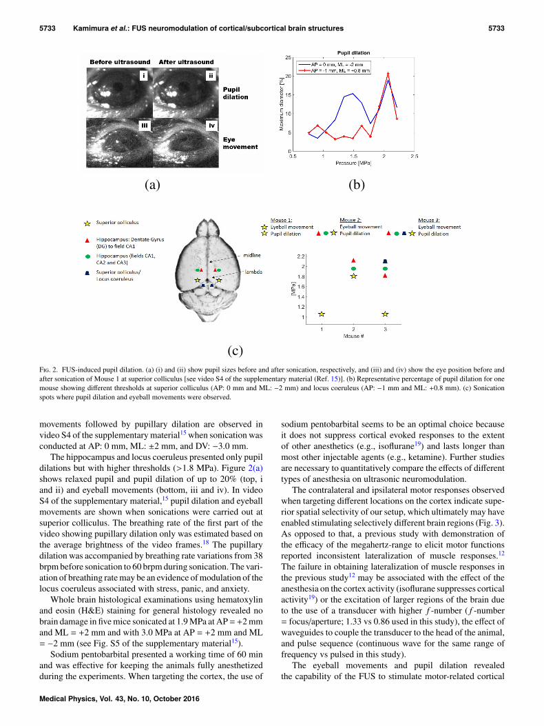
In a second set of experiments using the same transducer,we targeted subcortical brain structures where the ultrasoundfocus encompassed the superior colliculus (associated withmotor control of the eyes), the hippocampus or the locuscoeruleus (anxiety-related regions of the brain) as indicated inFig. 2. A lower threshold in eliciting pupil dilation (1.20 MPa)was observed when targeting the region of the superior collicu-lus. For the same region, eyeball movements were observedwhen sonications were conducted at higher pressures(>1.8 MPa). Given that the 1.9 MHz beam is 1 mm in diameter,it is possible that both superior colliculus and pretectal nucleuswere sonicated, since the superior colliculus reaches the pre-tectal nucleus near (∼0.5 mm) to the targeted region AP: 0 mm,ML:±2 mm, and DV:−3.0 mm (Fig. 2). The pupillary dilationobserved at this target may be associated with the modulationof the pretectal nucleus, which is directly involved with thepupillary light reflex. Input from retinal ganglion cells is sent tothe pretectal nucleus, which projects to the Edinger–Westphalnucleus that innervates the iris sphincter muscle. Eyeball
F. 1. FUS-induced motor responses. (a) EMG of the right hind limb during contralateral FUS stimulation at different acoustic pressure levels with success ratereferred to contralateral motor response elicitation and (b) contralateral paw movement elicited by FUS neuromodulation. Video frames recorded during the leftpaw movement when sonicating AP = +2 mm from lambda and ML = +2 mm (left) and during the right paw movement when sonicating AP = +2 mm fromlambda and ML = −2 mm (right) [see video S3 of the supplementary material (Ref. 15)].
Medical Physics, Vol. 43, No. 10, October 2016
5733 Kamimura et al.: FUS neuromodulation of cortical/subcortical brain structures 5733
F. 2. FUS-induced pupil dilation. (a) (i) and (ii) show pupil sizes before and after sonication, respectively, and (iii) and (iv) show the eye position before andafter sonication of Mouse 1 at superior colliculus [see video S4 of the supplementary material (Ref. 15)]. (b) Representative percentage of pupil dilation for onemouse showing different thresholds at superior colliculus (AP: 0 mm and ML: −2 mm) and locus coeruleus (AP: −1 mm and ML: +0.8 mm). (c) Sonicationspots where pupil dilation and eyeball movements were observed.
movements followed by pupillary dilation are observed invideo S4 of the supplementary material15 when sonication wasconducted at AP: 0 mm, ML: ±2 mm, and DV: −3.0 mm.
The hippocampus and locus coeruleus presented only pupildilations but with higher thresholds (>1.8 MPa). Figure 2(a)shows relaxed pupil and pupil dilation of up to 20% (top, iand ii) and eyeball movements (bottom, iii and iv). In videoS4 of the supplementary material,15 pupil dilation and eyeballmovements are shown when sonications were carried out atsuperior colliculus. The breathing rate of the first part of thevideo showing pupillary dilation only was estimated based onthe average brightness of the video frames.18 The pupillarydilation was accompanied by breathing rate variations from 38brpm before sonication to 60 brpm during sonication. The vari-ation of breathing rate may be an evidence of modulation of thelocus coeruleus associated with stress, panic, and anxiety.
Whole brain histological examinations using hematoxylinand eosin (H&E) staining for general histology revealed nobrain damage in five mice sonicated at 1.9 MPa at AP=+2 mmand ML = +2 mm and with 3.0 MPa at AP = +2 mm and ML= −2 mm (see Fig. S5 of the supplementary material15).
Sodium pentobarbital presented a working time of 60 minand was effective for keeping the animals fully anesthetizedduring the experiments. When targeting the cortex, the use of
sodium pentobarbital seems to be an optimal choice becauseit does not suppress cortical evoked responses to the extentof other anesthetics (e.g., isoflurane19) and lasts longer thanmost other injectable agents (e.g., ketamine). Further studiesare necessary to quantitatively compare the effects of differenttypes of anesthesia on ultrasonic neuromodulation.
The contralateral and ipsilateral motor responses observedwhen targeting different locations on the cortex indicate supe-rior spatial selectivity of our setup, which ultimately may haveenabled stimulating selectively different brain regions (Fig. 3).As opposed to that, a previous study with demonstration ofthe efficacy of the megahertz-range to elicit motor functionsreported inconsistent lateralization of muscle responses.12
The failure in obtaining lateralization of muscle responses inthe previous study12 may be associated with the effect of theanesthesia on the cortex activity (isoflurane suppresses corticalactivity19) or the excitation of larger regions of the brain dueto the use of a transducer with higher f -number ( f -number= focus/aperture; 1.33 vs 0.86 used in this study), the effect ofwaveguides to couple the transducer to the head of the animal,and pulse sequence (continuous wave for the same range offrequency vs pulsed in this study).
The eyeball movements and pupil dilation revealedthe capability of the FUS to stimulate motor-related cortical
Medical Physics, Vol. 43, No. 10, October 2016

5734 Kamimura et al.: FUS neuromodulation of cortical/subcortical brain structures 5734
F. 3. Map of most dominant responses observed during ultrasound neuromodulation of mice. Modulation of the cortex (D.V.: −1 mm) elicited tailand ipsilateral/contralateral hind limb movements. Modulation of subcortical structures of the brain (D.V.: −3 mm) elicited pupillary dilation and eyeballmovements.
structures in addition to anxiety-related and other subcorticalstructures of the brain (Fig. 3). Pupil dilation was observedwhen sonicating regions such as the limbic regions and thelocus coeruleus. The superior colliculus presented a lowerthreshold in evoking pupil dilation (1.2 vs 1.8 MPa). Thelocus coeruleus, associated with responses to stress and panic,projects to superior colliculus. Pupil dilation was observed witha higher threshold when sonicating the hippocampus (part ofthe limbic system), which supports functions such as adrena-line flow, emotion, and behavior. At this sonication spot (AP= +2 mm and ML = ±2 mm), the ultrasound focus could reachthe pretectal nucleus in the dorsal–ventral direction. Thus, thereis a possibility of modulation of the pretectal nucleus associatedwith light reflex. We show the feasibility of using higherfrequencies for modulating neuronal activity, demonstratingthat the resultant smaller acoustic focus can provide superiortarget specificity. Based on the results obtained from the cali-bration of the transducer (see Fig. S1 of the supplementarymaterial15), the transducer’s focal size can be highly improvedbyincreasing thedrivingfrequency(0.5MHz: lateral resolution= 3.4 mm and axial resolution= 17.7 mm vs 1.94 MHz: lateralresolution = 1.0 mm and axial resolution = 8.5 mm). Thus, theentireultrasoundparametric spacecanbeassessed inhumansorlarger animals seeking greater target specificity without beinglimited to submegahertz frequencies.
4. CONCLUSION
Reproducible contralateral and ipsilateral evoked motorresponses demonstrated the superior target specificity of themegahertz-range for brain modulation, since previous studiesfailed in demonstrating such consistent responses when usinglower frequencies. The sonication of deeper regions in thebrain translated to pupillary dilations is used as indications ofmodulation of subcortical structures associated with cognition
and light reflex responses. The variety of responses (motor andpupillary dilation) reported herein demonstrated the capabilityof FUS to perform functional brain mapping.
ACKNOWLEDGMENTS
This study was supported in part by NIH (Grant Nos.R01EB009041 and R01AG038961) and FAPESP (Grant Nos.2011/10809-6 and 2013/08116-8). The authors thank YangLiu, Ph.D., Edward Li, and Kathleen G. Fan for the technicalsupport.
a)Current Address: Molecular Imaging Research Center, Institut d’ImagerieBiomédicale, Commissariat à l’énergie atomique et aux énergies alterna-tives (CEA), Fontenay-aux-Roses 92265, France.
b)Author to whom correspondence should be addressed. Electronic mail:[email protected]; Telephone: 212-342-0863; Fax: 212-342-5773.
1S. Szobota and E. Y. Isacoff, “Optical control of neuronal activity,” Annu.Rev. Biophys. 39, 329–348 (2010).
2M. Häusser, “Optogenetics: The age of light,” Nat. Methods 11(10),1012–1014 (2014).
3Y. Tufail, A. Matyushov, N. Baldwin, M. L. Tauchmann, J. Georges,A. Yoshihiro, S. I. Helms Tillery, and W. J. Tyler, “Transcranial pulsedultrasound stimulates intact brain circuits,” Neuron 66(5), 681–694(2010).
4S.-S. Yoo, A. Bystritsky, J.-H. Lee, Y. Zhang, K. Fischer, B.-K. Min, N.J. McDannold, A. Pascual-Leone, and F. A. Jolesz, “Focused ultrasoundmodulates region-specific brain activity,” NeuroImage 56(3), 1267–1275(2011).
5R. L. King, J. R. Brown, W. T. Newsome, and K. B. Pauly, “Effectiveparameters for ultrasound-induced in vivo neurostimulation,” UltrasoundMed. Biol. 39(2), 312–331 (2013).
6Y. Younan, T. Deffieux, B. Larrat, M. Fink, M. Tanter, and J.-F. Aubry,“Influence of the pressure field distribution in transcranial ultrasonic neu-rostimulation,” Med. Phys. 40(8), 082902 (10pp.) (2013).
7R. L. King, J. R. Brown, and K. B. Pauly, “Localization of ultrasound-induced in vivo neurostimulation in the mouse model,” Ultrasound Med.Biol. 40(7), 1512–1522 (2014).
8Y. Tufail, A. Yoshihiro, S. Pati, M. M. Li, and W. J. Tyler, “Ultrasonicneuromodulation by brain stimulation with transcranial ultrasound,” Nat.Protoc. 6(9), 1453–1470 (2011).
Medical Physics, Vol. 43, No. 10, October 2016
5735 Kamimura et al.: FUS neuromodulation of cortical/subcortical brain structures 5735
9O. Naor, S. Krupa, and S. Shoham, “Ultrasonic neuromodulation,” J. NeuralEng. 13(3), 031003 (2016).
10H. Kim, A. Chiu, S. D. Lee, K. Fischer, and S.-S. Yoo, “Focused ultrasound-mediated non-invasive brain stimulation: Examination of sonication param-eters,” Brain Stimul. 5(5), 181–204 (2014).
11E. Mehic, J. M. Xu, C. J. Caler, N. K. Coulson, C. T. Moritz,and P. D. Mourad, “Increased Anatomical specificity of neuromod-ulation via modulated focused ultrasound,” PLoS One 9(2), e86939(2014).
12P. P. Ye, J. R. Brown, and K. B. Pauly, “Frequency dependence of ultra-sound neurostimulation in the mouse brain,” Ultrasound Med. Biol. 42(7),1512–1530 (2016).
13M. S. Gilzenrat, S. Nieuwenhuis, M. Jepma, and J. D. Cohen, “Pupil diam-eter tracks changes in control state predicted by the adaptive gain theoryof locus coeruleus function,” Cognit. Affective Behav. Neurosci. 10(2),252–269 (2010).
14S. Graur and G. Siegle, “Pupillary motility: Bringing neuroscience to thepsychiatry clinic of the future,” Curr. Neurol. Neurosci. Rep. 13: 365(2013).
15See supplementary material at http://dx.doi.org/10.1118/1.4963208 forcalibration of the transducer in water; more details about the experimentalsetup for motor response and pupil dilation observations; videos showingFUS-induced motor response elicitation; videos showing FUS-inducedpupil dilation and eyeball movements; and whole brain histologicexamination.
16J. J. Choi, M. Pernot, S. A. Small, and E. E. Konofagou, “Noninvasive,transcranial and localized opening of the blood–brain barrier using focusedultrasound in mice,” Ultrasound Med. Biol. 33(1), 95–104 (2007).
17K. A. Tennant, D. L. Adkins, N. A. Donlan, A. L. Asay, N. Thomas, J. A.Kleim, and T. A. Jones, “The organization of the forelimb representation ofthe C57BL/6 mouse motor cortex as defined by intracortical microstimula-tion and cytoarchitecture,” Cereb. Cortex 21(4), 865–876 (2011).
18F. Zhao, M. Li, Y. Qian, and J. Z. Tsien, “Remote measurements of heartand respiration rates for telemedicine,” PLoS One 8(10), e71384 (2013).
19M. Kawaguchi, K. Shimizu, H. Furuya, T. Sakamoto, H. Ohnishi, and J.Karasawa, “Effect of isoflurane on motor-evoked potentials induced byDirect electrical stimulation of the exposed motor cortex with single, double,and triple stimuli in rats,” Anesthesiology 85(5), 1176–1183 (1996).
Medical Physics, Vol. 43, No. 10, October 2016
Related Documents


![· Web viewNon-invasive low-intensity pulsed ultrasound with high spatial specificity and penetration depth has emerged as a novel neuromodulation technique []. The ultrasound waves](https://static.cupdf.com/doc/110x72/5e66f51fb3a78d49d059f889/web-view-non-invasive-low-intensity-pulsed-ultrasound-with-high-spatial-specificity.jpg)