Focused Cardiac Ultrasound Uncommon but Critical Diagnoses Made at the Point of Care lthough chest pain, shortness of breath, and syncope are among some of the most common conditions evaluated by emergency physicians, the differential diagnoses for these conditions are broad and contain some rare but serious diagnoses, such as pericardial tamponade, aortic dissection, and cardiomyopathies. Even more common diagnoses, such as acute myocardial infarction and pulmonary embolism, may present atypically or be unclear in the early stages of disease. With the use of focused cardiac ultrasound (FOCUS) at the point of care, a wider differential diagnosis can be explored, potentially streamlining the subsequent workup and ulti- mately improving diagnostic accuracy and clinical decision making. Here we report 8 cases in which FOCUS revealed an uncommon diagnosis, confirmed a suspected but unclear diagnosis, or suggested an alternate diagnosis not initially suspected. Our Institutional Review Board did not deem this study as human subject research; thus, approval was not required. Joseph Minardi, MD, Tom Marshall, MD, Greta Massey, MD, Erin Setzer, MD Received April 21, 2014, from the Department of Emergency Medicine, West Virginia University, Morgantown, West Virginia USA. Revision requested May 21, 2014. Revised manuscript accepted for publication July 15, 2014. Address correspondence to Joseph Minardi, MD, Department of Emergency Medicine, West Virginia University, 7413B HSS, 1 Medial Center Dr, Morgantown, WV 26506 USA. E-mail: [email protected] Abbreviations CT, computed tomographic; ECG, electrocardio- gram; FOCUS, focused cardiac ultrasound A ©2015 by the American Institute of Ultrasound in Medicine | J Ultrasound Med 2015; 34:727–736 | 0278-4297 | www.aium.org CASE SERIES Cardiovascular and respiratory conditions in acute care require rapid, critical decision making, often with limited clinical information. Focused cardiac ultrasound (FOCUS) can aid in diagnosis by providing information that may not be evident from a patient’s medical history, physical examination, and ancillary tests. Eight cases are presented in which FOCUS drastically altered the management of patient care, shortened the dif- ferential diagnosis, or allowed for the development of a definitive diagnosis. In 3 cases, diagnoses that were not initially suspected were identified by FOCUS. In the remain- ing cases, uncommon yet critical diagnoses were established at early stages along the patients’ courses of care. Key Words—acute care; echocardiography; emergency medicine; ultrasound Videos online at www.jultrasoundmed.org doi:10.7863/ultra.34.4.727

Welcome message from author
This document is posted to help you gain knowledge. Please leave a comment to let me know what you think about it! Share it to your friends and learn new things together.
Transcript

Focused Cardiac UltrasoundUncommon but Critical Diagnoses Made at the Point of Care
lthough chest pain, shortness of breath, and syncope areamong some of the most common conditions evaluated byemergency physicians, the differential diagnoses for these
conditions are broad and contain some rare but serious diagnoses, suchas pericardial tamponade, aortic dissection, and cardiomyopathies.Even more common diagnoses, such as acute myocardial infarctionand pulmonary embolism, may present atypically or be unclear inthe early stages of disease. With the use of focused cardiac ultrasound(FOCUS) at the point of care, a wider differential diagnosis can beexplored, potentially streamlining the subsequent workup and ulti-mately improving diagnostic accuracy and clinical decision making.Here we report 8 cases in which FOCUS revealed an uncommondiagnosis, confirmed a suspected but unclear diagnosis, or suggestedan alternate diagnosis not initially suspected. Our InstitutionalReview Board did not deem this study as human subject research;thus, approval was not required.
Joseph Minardi, MD, Tom Marshall, MD, Greta Massey, MD, Erin Setzer, MD
Received April 21, 2014, from the Department ofEmergency Medicine, West Virginia University,Morgantown, West Virginia USA. Revisionrequested May 21, 2014. Revised manuscriptaccepted for publication July 15, 2014.
Address correspondence to Joseph Minardi,MD, Department of Emergency Medicine, WestVirginia University, 7413B HSS, 1 Medial CenterDr, Morgantown, WV 26506 USA.
E-mail: [email protected]
AbbreviationsCT, computed tomographic; ECG, electrocardio -gram; FOCUS, focused cardiac ultrasound
A
©2015 by the American Institute of Ultrasound in Medicine | J Ultrasound Med 2015; 34:727–736 | 0278-4297 | www.aium.org
CASE SERIES
Cardiovascular and respiratory conditions in acute care require rapid, critical decisionmaking, often with limited clinical information. Focused cardiac ultrasound (FOCUS)can aid in diagnosis by providing information that may not be evident from a patient’smedical history, physical examination, and ancillary tests. Eight cases are presented inwhich FOCUS drastically altered the management of patient care, shortened the dif-ferential diagnosis, or allowed for the development of a definitive diagnosis. In 3 cases,diagnoses that were not initially suspected were identified by FOCUS. In the remain-ing cases, uncommon yet critical diagnoses were established at early stages along thepatients’ courses of care.
Key Words—acute care; echocardiography; emergency medicine; ultrasound
Videos online at www.jultrasoundmed.org
doi:10.7863/ultra.34.4.727
3404jum721-744 copy_Layout 1 3/17/15 10:06 AM Page 727
Minardi et al—Focused Cardiac Ultrasound at the Point of Care
J Ultrasound Med 2015; 34:727–736728
Case Descriptions
Case 1 An 87-year-old man with multiple comorbid conditionspresented to the emergency department with subjectivefevers, fatigue, shortness of breath, and confusion. His med-icalhistory included remote prosthetic mitral and aortic valveendocarditis among many other chronic medical problems.Physical examination revealed an alert elderly man withnormal vital signs and crackles in the right lower lung field.
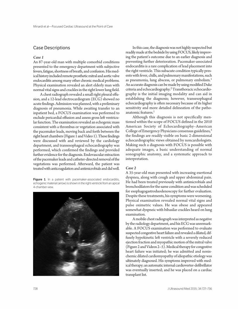
A chest radiograph revealed a small right pleural effu-sion, and a 12-lead electrocardiogram (ECG) showed noacute findings. Admission was planned, with a preliminarydiagnosis of pneumonia. While awaiting transfer to aninpatient bed, a FOCUS examination was performed toexclude pericardial effusion and assess gross left ventricu-lar function. The examination revealed an echogenic massconsistent with a thrombus or vegetation associated withthe pacemaker leads, moving back and forth between theright heart chambers (Figure 1 and Video 1). These findingswere discussed with and reviewed by the cardiologydepartment, and transesophageal echocardiography wasperformed, which confirmed the findings and providedfurther evidence for the diagnosis. Endovascular extractionof the pacemaker leads and catheter-directed removal of thevegetations was performed. Afterward, the patient wastreated with anticoagulation and antimicrobials and did well.
In this case, the diagnosis was not highly suspected butreadily made at the bedside by using FOCUS, likely improv-ing the patient’s outcome due to an earlier diagnosis andpreventing further deterioration. Pacemaker-associatedendocarditis is a rare complication of lead placement intothe right ventricle. This subacute condition typically pres-ents with fever, chills, and pulmonary manifestations, suchas pneumonia, lung abscess, or pulmonary embolism.1An accurate diagnosis can be made by using modified Dukecriteria and echocardiography.2 Transthoracic echocardio-graphy is the initial imaging modality and can aid inestablishing the diagnosis; however, transesophagealechocardiography is often necessary because of its highersensitivity and more detailed delineation of the patho -anatomic features.1
Although this diagnosis is not specifically men-tioned within the scope of FOCUS defined in the 2010American Society of Echocardiography–AmericanCollege of Emergency Physicians consensus guidelines,3the findings are readily visible on basic 2-dimensionalechocardiographic views obtained by noncardiologists.Making such a diagnosis with FOCUS is possible withadequate images, a basic understanding of normalsonographic anatomy, and a systematic approach tointerpretation.
Case 2 A 35-year-old man presented with increasing exertionaldyspnea, along with cough and upper abdominal pain.He had been treated previously with antimicrobials andbronchodilators for the same condition and was scheduledfor esophagogastroduodenoscopy for further evaluation.Despite these treatments, his symptoms were worsening.Physical examination revealed normal vital signs andpulse oximetric values. He was obese and appearedsomewhat dyspneic with bibasilar crackles heard on lungexamination.
A mobile chest radiograph was interpreted as negativeby the radiology department, and his ECG was unremark-able. A FOCUS examination was performed to evaluatesuspected congestive heart failure and revealed a dilated, dif-fusely hypokinetic left ventricle with a severely reducedejection fraction and myopathic motion of the mitral valve(Figure 2 and Videos 2–5). Medical therapy for congestiveheart failure was initiated; he was admitted and nonis-chemic dilated cardiomyopathy of idiopathic etiology wasultimately diagnosed. His symptoms improved with med-ical therapy; an automatic internal cardioverter-defibrillatorwas eventually inserted; and he was placed on a cardiactransplant list.
Figure 1. In a patient with pacemaker-associated endocarditis,echogenic material (arrow) is shown in the right ventricle from an apical4-chamber view.
3404jum721-744 copy_Layout 1 3/17/15 10:06 AM Page 728

In this case, congestive heart failure was included in thedifferential diagnosis but was able to be confirmed at thebedside, allowing a more focused downstream evaluation,likely decreasing subsequent investigations, and possiblypreventing further deterioration of the patient’s condition.It is possible that the diagnosis could have been establishedsooner had FOCUS been incorporated earlier in thecourse of his illness.
Case 3 A 5-year-old boy presented to the emergency departmentafter 2 episodes of unprovoked syncope without associateddyspnea, chest discomfort, or palpitations. His medicalhistory was unremarkable other than being small for his age,and there was no family history of sudden cardiac death.On physical examination, he appeared pale and was tachy-cardic, with a II/VI systolic murmur at the left sternal border.An ECG revealed a left axis and borderline increased leftventricular voltages, and portable chest radiography showedcardiomegaly. A FOCUS examination was performed toexclude pericardial effusion and assess gross left ventricularfunction. The examination revealed a hypokinetic leftventricle with symmetric left ventricular hypertrophy(Figure 3 and Videos 6 and 7). On admission, furtherevaluation confirmed a diagnosis of hypertrophic nonob-structive cardiomyopathy. Medical therapy was initiated,and he was advised to avoid strenuous athletic activities.
In this case, there were concerning yet nonspecificfindings, and FOCUS was able to establish an accurate,although preliminary, diagnosis early in the patient’scourse, leaving less uncertainty and likely decreasing sub-sequent ancillary testing. Hypertrophic cardiomyopathyis a genetic disorder with variable expression that cancause sudden cardiac death, especially among athletes,and should always be part of the differential diagnosisfor patients presenting with cardiovascular conditions.4
J Ultrasound Med 2015; 34:727–736 729
Minardi et al—Focused Cardiac Ultrasound at the Point of Care
Figure 2. In a patient with dilated cardiomyopathy, this image, capturedat end diastole in an apical 4-chamber view, shows that the internaldiameter of the left ventricle is estimated at 6.16 cm, which is dilated.
Figure 3. In this young male patient, a hypokinetic left ventricle with symmetric left ventricular hypertrophy was seen. A, At end diastole in a paraster-nal long-axis view, both the septal and free walls measure greater than 1.2 cm in thickness, which is consistent with left ventricular hypertrophy.B, At end-diastole in a parasternal short-axis view in the mid ventricle, both the septal and free walls measure greater than 1.2 cm in thickness, whichis consistent with left ventricular hypertrophy.
A B
3404jum721-744 copy_Layout 1 3/17/15 10:06 AM Page 729

Minardi et al—Focused Cardiac Ultrasound at the Point of Care
J Ultrasound Med 2015; 34:727–736730
A thorough history, physical examination, and ECG mayidentify most patients with a high risk for sudden death,but echocardiography is necessary for a specific diagnosis.5A limited 2-dimensional echocardiogram should be ade-quate to identify most high-risk patients. In small studies,physicians with limited training in echocardiography havedemonstrated their ability to acquire the proper views andmeasurements.6
Case 4 A 51-year-old man presented with acute chest aching thatbegan after running to a resuscitation in the hospital wherehe worked as a nurse. His medical history was unremarkable.Vital signs were normal, but he was pale and diaphoreticon examination. An ECG showed ST-segment elevationof less than 1 mm in leads V2 through V4 and no reciprocalchanges. His symptoms improved with nitroglycerin, andserial ECGs remained nondiagnostic. A FOCUS exami-nation was performed to further investigate his chest painand evaluate for further diagnostic evidence of suspectedmyocardial ischemia. The examination revealed hypoki-nesis of the left ventricular apex (Figure 4 and Video 8).With resolving symptoms, the patient was hesitant toundergo emergent cardiac catheterization but agreed
to the procedure after reviewing the sonographic find-ings with the emergency physician. He was found tohave complete occlusion of the left anterior descendingcoronary artery, which was successfully stented. Of note,initial troponin was undetectable and later peaked at29 ng/mL.
In this case, FOCUS added valuable diagnostic infor-mation to an already concerning clinical picture and helpedprovide vital information for the patient’s care, allowingprompt intervention and likely limiting the extent ofmyocardial injury, resulting in a better long-term func-tional outcome. Diagnosing regional wall motion abnor-malities with FOCUS can be challenging and should notbe used to exclude ischemia.7,8 Even among experiencedcardiologists, there is considerable inter-rater variabilityin diagnosing these abnormalities.9 However, in patientswith acute symptoms and initially inconclusive findings,FOCUS may offer additional diagnostic informationregarding ischemia or alternate diagnoses and facilitateprompt intervention if necessary.3 Higher-risk ischemiclesions involving larger myocardial territories should berecognizable by nontraditional users when adequateviews can be obtained.
Figure 4. These sequential apical 4-chamber views from a 51-year-old man with chest pain show hypokinesis in the left ventricular apex.A, This image from an apical 4-chamber view was captured at end diastole. When compared to B, which was captured during peak systole, distalhinge points and hypokinesis of the left ventricular apex can be seen. B, This image, captured at peak systole, displays hinge points (arrows) at thedistal septal and lateral walls of the left ventricle. Also, in comparison to A, the left ventricular apical walls have not thickened appropriately, and thearea at the left ventricular apex has not decreased substantially. These signs indicate a regional wall motion abnormality at the left ventricular apex,consistent with ischemia.
A B
3404jum721-744 copy_Layout 1 3/17/15 10:06 AM Page 730

Case 5 A 49-year-old man presented with dyspnea on exertion andchest pressure for several days. He reported a remote historyof deep venous thrombosis while working as a truck driver,but was no longer receiving anticoagulation therapy.He denied symptoms of deep venous thrombosis orhemoptysis, and he was a smoker. Vital signs, pulse oximet-ric values, and physical examination findings were normal.Chest radiographic and ECG findings were also normal.His troponin level was elevated into the diagnostic rangefor acute myocardial infarction.
Admission was planned for non–ST-elevationmyocardial infarction when a FOCUS examinationwas performed to assess gross left ventricular function.The examination revealed a massively dilated right ven-tricle with abnormal septal motion (Figure 5 and Videos9 and 10), prompting the physician to order a pulmonarycomputed tomographic (CT) angiogram, which revealedlarge central, bilateral pulmonary emboli. Treatment withanticoagulation was continued, and the patient was admit-ted to the hospital.
In this case, FOCUS suggested a diagnosis that wasinitially thought unlikely, prompting further evaluation,an accurate diagnosis, and a change in the care plan.Focused cardiac ultrasound is a useful diagnostic toolfor patients with suspected or confirmed cases of pul-monary embolism. In patients with suspected pulmonaryembolism without preexisting cardiopulmonary disease,right ventricular dilatation has been shown to be a specificyet insensitive finding for the diagnosis.10 When incorpo-rating this modality, it is important that clinicians be awareof other causes of right ventricular dilatation and considerthe entire clinical picture when interpreting sonographicfindings and making care decisions. Some findings that aremore suggestive of acute right ventricular dilatation includea right ventricular free wall thickness of less than 5 mm andthe McConnell sign, which is the presence of a hyperkineticright ventricular apex in the setting of a dilated and hypo-kinetic right ventricle. Focused cardiac ultrasound may pro-vide prognostic information in acute pulmonary embolismand can assist in therapeutic decision making, specificallyin selecting candidates for thrombolytic therapy.11
J Ultrasound Med 2015; 34:727–736 731
Minardi et al—Focused Cardiac Ultrasound at the Point of Care
Figure 5. Dilated right ventricle and abnormal septal motion in a 49-year-old man with pulmonary embolism. A, From a parasternal long-axis view,the left ventricle appears small in comparison to the more superficial, dilated right ventricle, and the right ventricular-to-left ventricular diameter ratiois greater than 1. B, From an apical 4-chamber view, the right ventricular-to-left ventricular diameter ratio is greater than 1. In addition, the proximalseptal wall can be seen bulging, paradoxically toward the left ventricle.
A B
3404jum721-744 copy_Layout 1 3/17/15 10:06 AM Page 731

Case 6 A 48-year-old man presented to the emergency depart-ment for left shoulder pain and dyspnea, which worsenedwhen lying flat. He had been seen 2 days previously forthe same symptoms, but the symptoms were worsening.His medical history was unremarkable. During a recentadmission for chest pain, a myocardial perfusion scan showeda region of ischemia, and a chest radiograph had shownsmall pleural effusions.
At the time of his visit to the emergency department, achest radiograph showed an increased yet small left pleuraleffusion, and the ECG was unremarkable. A FOCUS exam-ination was performed to further evaluate his symptoms andshowed a large pericardial effusion with right ventriculardiastolic collapse (Figure 6 and Video 11). Since he washemodynamically stable, he was taken to the cardiac proce-dures laboratory and underwent successful pericardiocen-tesis, where 640 mL of serous fluid was drained. Pathologicexamination revealed nonmalignant inflammatory cells, andhe was treated with indomethacin and did well.
In this case, the patient presented with atypical symp-toms and a nondiagnostic workup. Focused cardiac ultra-sound allowed an accurate diagnosis to be made when itwas not evident on the basis of the other available infor-mation. It is possible that earlier incorporation of FOCUSwould have resulted in a more timely diagnosis. Pericardi-tis is usually diagnosed on the basis of the patient’s history,
physical examination, and classic ECG findings, whichwere not present in this case.12 Pericardial effusions frometiologies other than acute pericarditis also do not typicallyhave classic ECG findings.13 The incidence of pericardialeffusion is quite variable, depending on the underlying dis-ease process, but may be as high as 20% in patients withrenal disease, up to 37% in some malignancies, and evenhigher in patients with human immunodeficiency virusinfection or AIDS. Thus, clinicians should consider andevaluate for pericardial effusion in symptomatic patients,especially those with known high-risk disease states.14–16
Case 7 A 60-year-old man presented to the emergency depart-ment after having a syncopal event preceded by “indiges-tion.” His only symptom on presentation was right legpain. His medical history was notable for remote coloncancer in remission and hypertension, and he was a for-mer smoker. During the physical examination, the patientwas bradycardic, with a heart rate of 40 beats per minute.Although he was not in any distress, his right leg was palewith diminished pulses and delayed capillary refill. A FOCUS examination was performed and showed adilated aortic root (Figure 7A and Video 12). Additionalviews of the abdominal aorta revealed a mobile flap, con-sistent with aortic dissection (Figure 7B and Video 13).A Stanford type A aortic dissection was suspected, and the
Minardi et al—Focused Cardiac Ultrasound at the Point of Care
J Ultrasound Med 2015; 34:727–736732
Figure 6. Large pericardial effusion with diastolic collapse of the right ventricle in a 48-year-old man. A, In this image from a subcostal view, a largepericardial effusion is shown, which is more prominent anteriorly but circumferential to the heart. The right ventricle (arrow) is shown at the mostdilated point to be filling poorly. B, From a subcostal view captured during diastole, a large pericardial effusion is shown, which is more prominentanteriorly but circumferential to the heart. The right ventricle (arrow) appears collapsed, consistent with tamponade.
A B
3404jum721-744 copy_Layout 1 3/17/15 10:06 AM Page 732
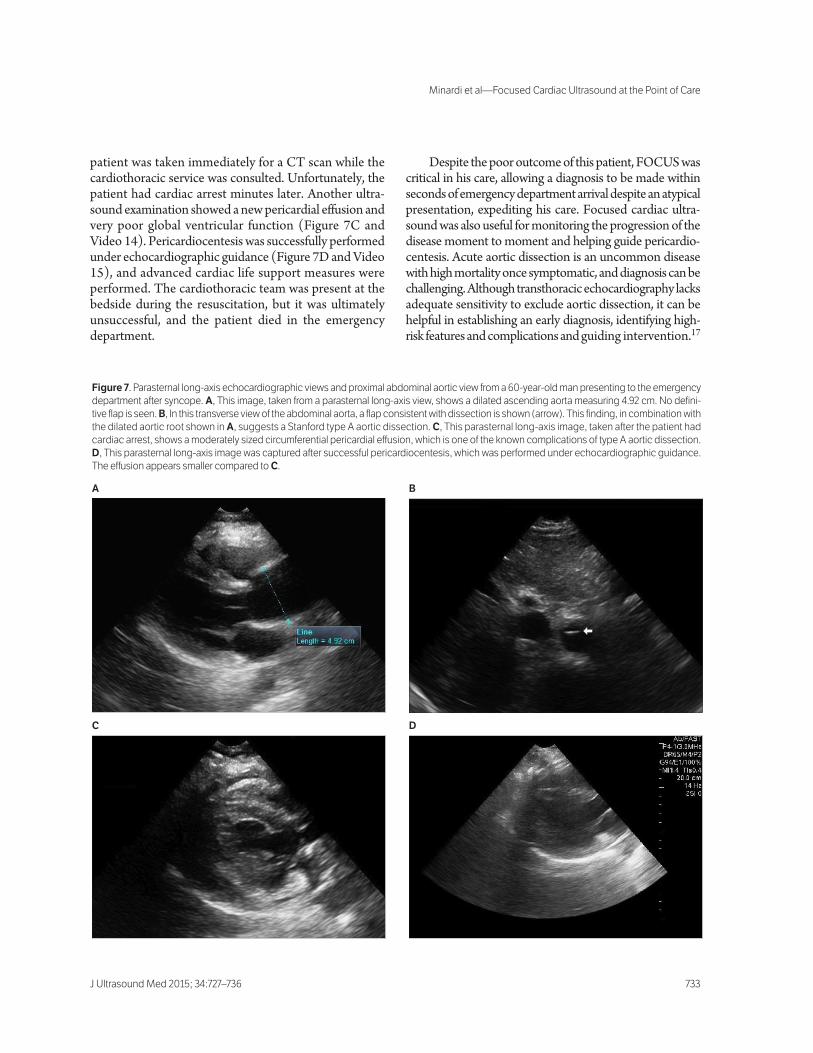
patient was taken immediately for a CT scan while thecardiothoracic service was consulted. Unfortunately, thepatient had cardiac arrest minutes later. Another ultra-sound examination showed a new pericardial effusion andvery poor global ventricular function (Figure 7C andVideo 14). Pericardiocentesis was successfully performedunder echocardiographic guidance (Figure 7D and Video15), and advanced cardiac life support measures wereperformed. The cardiothoracic team was present at thebedside during the resuscitation, but it was ultimatelyunsuccessful, and the patient died in the emergencydepartment.
Despite the poor outcome of this patient, FOCUS wascritical in his care, allowing a diagnosis to be made withinseconds of emergency department arrival despite an atypicalpresentation, expediting his care. Focused cardiac ultra-sound was also useful for monitoring the progression of thedisease moment to moment and helping guide pericardio-centesis. Acute aortic dissection is an uncommon diseasewith high mortality once symptomatic, and diagnosis can bechallenging. Although transthoracic echocardiography lacksadequate sensitivity to exclude aortic dissection, it can behelpful in establishing an early diagnosis, identifying high-risk features and complications and guiding intervention.17
J Ultrasound Med 2015; 34:727–736 733
Minardi et al—Focused Cardiac Ultrasound at the Point of Care
Figure 7. Parasternal long-axis echocardiographic views and proximal abdominal aortic view from a 60-year-old man presenting to the emergencydepartment after syncope. A, This image, taken from a parasternal long-axis view, shows a dilated ascending aorta measuring 4.92 cm. No defini-tive flap is seen. B, In this transverse view of the abdominal aorta, a flap consistent with dissection is shown (arrow). This finding, in combination withthe dilated aortic root shown in A, suggests a Stanford type A aortic dissection. C, This parasternal long-axis image, taken after the patient hadcardiac arrest, shows a moderately sized circumferential pericardial effusion, which is one of the known complications of type A aortic dissection.D, This parasternal long-axis image was captured after successful pericardiocentesis, which was performed under echocardiographic guidance.The effusion appears smaller compared to C.
A B
C D
3404jum721-744 copy_Layout 1 3/17/15 10:06 AM Page 733

Case 8 An 87-year-old woman who had recently undergone hipsurgery presented to the emergency department from anursing home with 1 day of dyspnea. Her medical historywas unremarkable. She was neither a smoker nor receivingestrogen therapy. Vital signs and physical examination find-ings were unremarkable while she was in the emergencydepartment, and there was no new leg swelling, redness, orpain. A FOCUS examination was performed to evaluategross left ventricular function and revealed a mobile mass inthe right atrium adherent to the free wall, which was thoughtto be a thrombus (Figure 8 and Video 16). There was noright ventricular dilatation, and the remainder of the exami-nation findings were negative. Pulmonary CT angiographicfindings were negative for pulmonary embolism. It wasbelieved that the patient may have had either multiple smallpulmonary emboli versus a pulmonary embolus that autol-ysed before performance of the CT scan. Anticoagulationwas initiated, and she was admitted to the hospital anddid well. Follow-up imaging revealed a decreasing size of themass, further solidifying the likelihood that a thrombus wasthe correct diagnosis.
In this case, FOCUS rapidly revealed an unusual diag-nosis that would have been difficult considering the negativeCT result. With the accurate diagnosis, proper therapy andfollow-up were provided, and the patient did well. A rightatrial thrombus is an uncommon problem, and optimal treat-ment has not been clearly defined.18,19 The diagnosis canusually be made by transthoracic echo cardiography, and thedifferential diagnosis should include other intracardiacmasses and vegetations. Transesophageal echocardiographyis likely more sensitive in making the diagnosis.20
Discussion
The cases above demonstrate how the addition of FOCUSto a standard clinical workup can improve patient care byidentifying both rare conditions as well as serious but unex-pected diagnoses in patients with atypical presentations.Such a strategy allows for early identification of importantclinical problems and life-threatening conditions while alsodirecting immediate management decisions and guidingcritical procedures.
Although it may appear that a liberal FOCUS strategycould increase medical costs, we argue that it could decreasethe need for subsequent ancillary testing and result inearlier, more accurate diagnoses, thus allowing better stew-ardship of limited resources. Additionally, this strategyshould also lead to earlier interventions with less compli-cated, more efficient clinical courses and, ultimately, betterpatient outcomes. This belief is demonstrated in cases 2and 6, in which 1 or more patient visits for the same con-ditions occurred before accurate diagnoses were made byincorporating FOCUS into the evaluation. For other appli-cations, the use of point-of-care ultrasound can decrease theuse of more expensive CT scans, in addition to decreasingthe length of stay as well as avoiding unnecessary ionizingradiation.21–23
Although it is true that echocardiography is an operator-dependent skill and that diagnostic accuracy improves withexperience, it is likely that as ultrasound training continuesto advance in medical school curricula and graduate med-ical education, a larger pool of experienced clinicians will becompetent in making less common and more challengingdiagnoses.24 The power and utility of FOCUS as a clinicaltool has been demonstrated in multiple studies in whichnontraditional users with limited training were able toobtain and accurately interpret limited echocardiograms,resulting in more accurate, earlier diagnoses and changes inthe patient care.25–27 Specifically, medical students withlimited echocardiographic training were able to make moreaccurate diagnoses than experienced cardiologists using astandard physical examination.28
Although we are enthusiastic about the growing incor-poration of FOCUS into standard clinical practice for non-cardiologists, it is important to recognize some of thelimitations of this modality. The first and likely mostimportant is a current heterogeneity in the level of trainingamong noncardiologists. Many practicing clinicians haveminimal training in FOCUS. Increased standardization inundergraduate and graduate medical education is neededto fully implement this modality into standard practice.The next limitation is equipment. Although portable ultra-
Minardi et al—Focused Cardiac Ultrasound at the Point of Care
J Ultrasound Med 2015; 34:727–736734
Figure 8. In this apical 4-chamber view from an elderly patient with dys-pnea, an echogenic clot is shown in the right atrium (arrow). This masswas mobile and appeared adherent to the right atrial free wall.
3404jum721-744 copy_Layout 1 3/17/15 10:06 AM Page 734

sound equipment has improved considerably in the pastdecade, there remains a narrowing gap in quality comparedto larger units used in echocardiography laboratories. Last,acutely ill patients and the time demands of acute carework environments present their own unique challengesthat contribute to decreased image quality and can con-tribute to interpretation errors. Again, improvements andstandardization in the training of noncardiologists shouldhelp in mitigating these limitations.
Larger controlled studies are needed to examinewhether a liberal FOCUS strategy would lead to wide-spread improvements in patient outcomes and be cost-effective. It would be premature to recommend performingFOCUS in every patient with these types of conditions.However, we believe that FOCUS should be consideredin every acutely ill patient with cardiovascular and respira-tory conditions and that training in clinical ultrasound fora wide range of clinicians should continue and expand.
References
1. Karchmer AW. Infections involving cardiac implantable electronicdevices. UpToDate website. http://www.uptodate.com/contents/infections-involving-cardiac-implantable-electronic-devices#H38.Accessed March 13, 2014.
2. Durack DT, Lukes AS, Bright DK. New criteria for diagnosis of infectiveendocarditis: utilization of specific echocardiographic findings. DukeEndocarditis Service. Am J Med 1994; 96:200–209.
3. Labovitz AJ, Noble VE, Bierig M, et al. Focused cardiac ultrasound in theemergent setting: a consensus statement of the American Society ofEchocardiography and American College of Emergency Physicians. J AmSoc Echocardiogr 2010; 23:1225–1230.
4. Shah SN. Hypertrophic cardiomyopathy. Medscape website.http://emedicine.medscape.com/article/152913-overview. AccessedMarch 13, 2014.
5. Asif IM, Drezner JA. Sudden cardiac death and preparticipation screen-ing: the debate continues—in support of electrocardiogram-inclusivepreparticipation screening. Prog Cardiovasc Dis 2012; 54:445–450.
6. Yim ES, Gillis EF, Ojala K, MacDonald J, Basilico FC, Corrado GD.Focused transthoracic echocardiography by sports medicine physicians:measurements relevant to hypertrophic cardiomyopathy. J UltrasoundMed 2013; 32:333–338.
7. Hoffmann R, von Bardeleben S, Barletta G, et al. Comparison of two- andthree-dimensional unenhanced and contrast-enhanced echocardiogra-phies versus cineventriculography versus cardiac magnetic resonance fordetermination of left ventricular function. Am J Cardiol 2014; 113:395–401.
8. Taylor RA, Moore CL. Accuracy of emergency physician–performedlimited echocardiography for right ventricular strain. Am J Emerg Med2014; 32:371–374.
9. Hoffmann R, von Bardeleben S, Kasprzak J, et al. Analysis of regional leftventricular function by cineventriculography, cardiac magnetic resonanceimaging, and unenhanced and contrast-enhanced echocardiography: amulticenter comparison of methods. J Am Coll Cardiol 2006; 47:121–128.
10. Dresden S, Mitchell P, Rahimi L, et al. Right ventricular dilation on bed-side echocardiography performed by emergency physicians aids in thediagnosis of pulmonary embolism. Ann Emerg Med 2014; 63:16–24.
11. Kline JA, Nordenholz KE, Courtney DM, et al. Treatment of submassivepulmonary embolism with tenecteplase or placebo: cardiopulmonaryoutcomes at 3 months (TOPCOAT)—multicenter double-blind,placebo-controlled randomized trial. J Thromb Haemost 2014; 12:459–468.
12. Spangler S. Acute pericarditis. Medscape website. http://emedicine.med-scape.com/article/156951-overview. Accessed March 4, 2014.
13. Meyers DG, Bagin RG, Levene JF. Electrocardiographic changes in peri-cardial effusion. Chest 1993; 104:1422–1426.
14. Bussani R, De-Giorgio F, Abbate A, Silvestri F. Cardiac metastases. J ClinPathol 2007; 60:27–34.
15. Meenakshisundaram R, Sweni S, Thirumalaikolundusubramanian P.Cardiac isoform of alpha 2 macroglobulin: a marker of cardiac involve-ment in pediatric HIV and AIDS. Pediatr Cardiol 2010; 31:203–207.
16. Lind A, Reinsch N, Neuhaus K, et al. Pericardial effusion of HIV-infectedpatients? Results of a prospective multicenter cohort study in the era ofantiretroviral therapy. Eur J Med Res 2011; 16:480–483.
17. Meredith EL, Masani ND. Echocardiography in the emergency assess-ment of acute aortic syndromes. Eur J Echocardiogr 2009; 10:i31–i39.
18. Lazar L, Dave R, Tabibiaszar R. Dilemma of right atrial thrombi, to dis-solve or to extract. Proc UCLA Healthcare 2012; 16:1–4.
19. Sobhy E, Alamaldine T, Sabti HA. Successful treatment of right-sidedheart thrombus with pulmonary embolism with thrombolytic therapy.J Cardiovasc Dis Diagn 2013; 1:121.
20. Chartier L, Bera J, Delomez M, et al. Free-floating thrombi in the rightheart: diagnosis, management, and prognostic indexes in 38 consecutivepatients. Circulation 1999; 99:2779–2783.
21. Peris A, Tutino L, Zagli G, et al. The use of point-of-care bedside lungultrasound significantly reduces the number of radiographs and com-puted tomography scans in critically ill patients. Anesth Analg 2010;111:687–692.
22. Jang T, Chauhan V, Cundiff C, Kaji AH. Assessment of emergency physi-cian–performed ultrasound in evaluating nonspecific abdominal pain.Am J Emerg Med 2014; 32:457–460.
23. Blaivas M, Harwood RA, Lambert MJ. Decreasing length of stay withemergency ultrasound examination of the gallbladder. Acad Emerg Med1999; 6:1020–1023.
24. Greenberg R. Making waves: ultrasound use increases in medicaleducation. AAMC Reporter December 2012. AAMC website. https://www.aamc.org/newsroom/reporter/dec2012/323592/ultrasound.html. Accessed March 5, 2014.
J Ultrasound Med 2015; 34:727–736 735
Minardi et al—Focused Cardiac Ultrasound at the Point of Care
3404jum721-744 copy_Layout 1 3/17/15 10:06 AM Page 735

J Ultrasound Med 2015; 34:727–736736
Minardi et al—Focused Cardiac Ultrasound at the Point of Care
25. Manasia AR, Nagaraj HM, Kodali RB, et al. Feasibility and potential clinical utility of goal-directed transthoracic echocardiography performedby noncardiologist intensivists using a small hand-carried device (SonoHeart) in critically ill patients. J Cardiothorac Vasc Anesth 2005;19:155–159.
26. Panoulas VF, Daigeler AL, Malaweera AS, et al. Pocket-size hand-heldcardiac ultrasound as an adjunct to clinical examination in the hands ofmedical students and junior doctors. Eur Heart J Cardiovasc Imaging 2013;14:323–330.
27. Decara JM, Kirkpatrick JN, Spencer KT, et al. Use of hand-carried ultra-sound devices to augment the accuracy of medical student bedside cardiacdiagnoses. J Am Soc Echocardiogr 2005; 18:257–263.
28. Kobal SL, Trento L, Baharami S, et al. Comparison of effectiveness ofhand-carried ultrasound to bedside cardiovascular physical examination.Am J Cardiol 2005; 96:1002–1006.
3404jum721-744 copy_Layout 1 3/17/15 10:06 AM Page 736
Related Documents