1 SECTION 20 Findings in Autism (ASD) Consistent with Electromagnetic Fields (EMF) and Radiofrequency Radiation (RFR) Martha Herbert, PhD, MD Pediatric Neurology TRANSCEND Research Program Massachusetts General Hospital Harvard Medical School Cindy Sage, MA Sage Associates Santa Barbara, CA USA For the BioInitiative Working Group December 2012

Welcome message from author
This document is posted to help you gain knowledge. Please leave a comment to let me know what you think about it! Share it to your friends and learn new things together.
Transcript

1
SECTION 20
Findings in Autism (ASD) Consistent with
Electromagnetic Fields (EMF) and
Radiofrequency Radiation (RFR)
Martha Herbert, PhD, MD
Pediatric Neurology
TRANSCEND Research Program
Massachusetts General Hospital
Harvard Medical School
Cindy Sage, MA
Sage Associates
Santa Barbara, CA USA
For the BioInitiative Working Group
December 2012
2
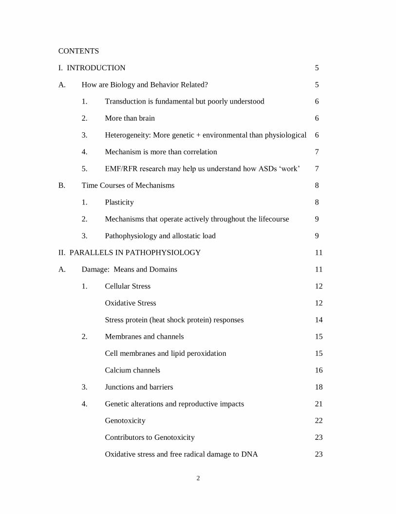
CONTENTS
I. INTRODUCTION 5
A. How are Biology and Behavior Related? 5
1. Transduction is fundamental but poorly understood 6
2. More than brain 6
3. Heterogeneity: More genetic + environmental than physiological 6
4. Mechanism is more than correlation 7
5. EMF/RFR research may help us understand how ASDs ‘work’ 7
B. Time Courses of Mechanisms 8
1. Plasticity 8
2. Mechanisms that operate actively throughout the lifecourse 9
3. Pathophysiology and allostatic load 9
II. PARALLELS IN PATHOPHYSIOLOGY 11
A. Damage: Means and Domains 11
1. Cellular Stress 12
Oxidative Stress 12
Stress protein (heat shock protein) responses 14
2. Membranes and channels 15
Cell membranes and lipid peroxidation 15
Calcium channels 16
3. Junctions and barriers 18
4. Genetic alterations and reproductive impacts 21
Genotoxicity 22
Contributors to Genotoxicity 23
Oxidative stress and free radical damage to DNA 23

3
Challenge to DNA repair mechanisms 24
Chromatin condensation 24
Gonadal and germline impacts 25
Implications of genotoxicity 26
5. Implications of Damage 26
B. Degradation of System Integrity 27
1. Mitochondrial dysfunction 27
2. Melatonin dysregulation 29
Melatonin, mitochondria, glutathione, oxidative stress 29
Melatonin can attenuate or prevent some EMF/RFR effects 30
Melatonin and autism 31
Autism and melatonin and glutathione 32
3. Disturbed immune function 32
Low-intensity exposures 33
Consequences of immune challenges during pregnancy 33
Potential immune contributions to reactivity and variability in ASDs 34
4. Alteration of and damage to cells in the brain 35
Brain cells 35
Neuroinflammation, glial activation and excitotoxicity 36
Altered development 39
Brain blood flow and metabolism 40
6. Electrophysiology perturbations 42
Seizures and epilepsy 42
Sleep 43
Quantitative electrophysiology 44

4
Sensory processing 45
Autonomic dysregulation 46
C. De-tuning of the Brain and Organism 48
1. Electromagnetic signaling, oscillation and synchrony are
fundamental, and vulnerable 48
2. Behavior as an “emergent property” 50
III. IMPLICATIONS 52
A. Summary 52
B. Exposures and Their Implications 53
1. Exposures have outpaced precautions 53
2. The population’s exposure has increased 54
3. Infants, children and childbearing families are highly exposed
and vulnerable 55
4. ASD risk and genomic damage to future generations 56
5. De-tuning the organism 56
C. Conclusions and Recommendations 58
1. Change our deployment of EMF/RFR 58
2. Encourage precautions right now based on present knowledge 58
3. Build an environmentally physiologically centered research
program in ASDs as a platform for investigating the
EMR/RFR-ASD linkage 59
4. Take the evidence as a call to action 60
IV. REFERENCES 61

5
I. INTRODUCTION
The premise of this review is that although scant attention has been paid to possible links
between electromagnetic fields and radiofrequency exposures (EMF/RFR) and Autism
Spectrum Disorders (ASDs), such links probably exist. The rationale for this premise is
that the physiological impacts of EMF/RFR and a host of increasingly well-documented
pathophysiological phenomena in ASDs have remarkable similarities. Additional support
may be found in the parallels between the rise in reported cases of ASDs and the
remarkable increases in EMF/RFR exposures over the past few decades. Reviewing
these similarities does not prove that these parallels imply causality – that kind of
research has not been done. Moreover, the physiological processes affected by
EMF/RFR are also impacted by other environmental factors. Yet EMF/RFR does not
need to be a unique contributor to ASDs to add significantly to system overload
(‘allostatic load’) and dysfunction. Even so these pathophysiological overlaps do suggest
that the potential for an EMF/RFR-ASD connection should be taken seriously, and that
their vulnerable biological features may make many with ASDs more likely to experience
adverse EMF/RFR impacts. This is a sufficient basis to recommend that precautionary
measures should be implemented and respected, that further research should be
prioritized, and that policy level interventions based on existing and emerging data
should be designed and pursued. Moreover, pursuing this link could help us understand
ASDs better and find more ways to improve the lives of people with ASDs and of so
many others.
A. How are Biology and Behavior Related?
Considering a potential link between ASDs and EMF/RFR (or indeed of any potential
contributor to incidence or pathogenesis) requires taking account of the evolution that has
been occurring in our understanding of the relationship between ASD’s behavioral and
biological features. ASDs were first labeled as ‘autism’ in 1943 by Leo Kanner, a child
psychiatrist who extracted several key behavioral features, related to communication and
social interaction challenges and a tendency toward restricted interests and repetitive
behaviors, characteristic of all 11 of the children in his first case series (Kanner 1943).
Although in the seven decades since this condition was first constructed as a category
there has been some modification of the way these behavioral features have been
characterized, ASDs are still defined behaviorally, although sensory issues such as hypo-
or hyper-reactivity have recently been included in the diagnostic criteria (Diagnostic and
Statistical Manual of Mental Disorders or DSM-V) (American Psychiatric Association
2000, 2013, May).

6
1. Transduction is fundamental but poorly understood
Yet in considering how an environmental factor such as EMF/RFR could lead to autism
and/or influence its severity or incidence, we need to think about how underlying biology
is transduced into changes in nervous system electrical activity, and how these in turn
generate the set of behaviors we have categorized as ‘autism.’ (Herbert 2005) This
means not taking behaviors as given, or as purely determined by genetics, but exploring
the full range of biology that generates these features and challenges.
2. More than brain
Although ‘autism’ has long been considered to be a psychiatric or neurological brain-
based disorder (Rapin and Katzman 1998; Polleux and Lauder 2004), it has become
undeniable that people diagnosed with ASDs often also have a multitude of biological
features – including systemic pathophysiological disturbances (such as oxidative stress,
mitochondrial dysfunction and metabolic and immune abnormalities) (Ming et al. 2012;
Tsaluchidu et al. 2008; Pieczenik and Neustadt 2007; Gonzalez et al. 2011) as well as
symptomatic medical comorbidities (such as gastrointestinal distress, recurrent infections,
epilepsy, autonomic dysregulation and sleep disruption) (Nikolov et al. 2009; Kotagal
and Broomall 2012; Kaartinen et al. 2012; Daluwatte et al. 2012; Tuchman and Cuccaro
2011; Canitano 2007; Malow 2004; Kang and Barnes 2013; Jyonouchi et al. 2011) – in
addition to the core defining behaviors – and many of these occur commonly (Kohane et
al. 2012). The problem has been that no one such biological feature has turned out to be
present in every single person carrying an ASD diagnosis – and they are not specific to
ASDs, either. Moreover there has been much variability in many of the features of
autism – not only between individuals but in many cases within individuals at different
points in time or under different circumstances. Because of this variability, the relevance
of many of these biological features has been dismissed as secondary and not intrinsically
related to the ‘autism.’ Instead, many have considered the behavioral features as
fundamental not only to how autism manifests and is definedbut also to the core intrinsic
nature of ASDs, even though the biological basis of these behaviors has by no means
been established.
3. Heterogeneity: More genetic and environmental than
physiological
It is not as if this variability is unique to the ‘environmental side.’ At the present time
over 800 genes have been associated with ASDs, and over 100 different rare genetic
syndromes are frequently accompanied by ASD, with no clear specific unifying
mechanism uniting this remarkable heterogeneity (Trikalinos et al. 2006; Ring et al.
2008; Pelphrey et al. 2011; Mandell 2011; Hall et al. 2012; Bill and Geschwind 2009).
Similarly a large number of potential environmental contributors are under investigation

7
ranging from toxicants and Vitamin D deficiency or failure to take prenatal vitamins to
air pollution and stress or infection in pregnancy (Whitehouse et al. 2012; Kocovska et al.
2012; Schmidt et al. 2011; Landrigan 2010; Roberts et al. 2007; Shelton, Hertz-Picciotto,
and Pessah 2012; Becerra et al. 2012; Volk et al. 2011). Yet at the physiological level a
smaller set of disturbances are showing up as common across substantial numbers of
people with ASDs – and in fact not uniquely to ASDs but also in myriad other chronic
conditions whose prevalence also appears to be increasing (Bilbo Jones, and Parker 2012;
Knox 2010). Prominent among these are immune disturbances including inflammation,
mitochondrial dysfunction, and oxidative stress, as well as toxic body burden.
Vulnerability to all of these can be increased mildly or substantially by a variety of often
common genetic mutations, but may remain latent without the overlay of environmental
triggers. Conversely, with substantial enough environmental input, genetic vulnerability
may not be necessary.
4. Mechanism is more than correlation
Just HOW biological features might be related to the behavioral features that have up
until now defined ASDs has not been clarified; until recently the main research effort
regarding pathophysiology in ASDs has been to establish the presence of these
phenomena in the first place. Even so, some correlations between biological and
behavioral features have been identified – e.g. a higher level of immune abnormalities
correlates with more aberrant behaviors (Wei et al. 2012; Careaga and Ashwood 2012;
Jyonouchi et al. 2011; Ashwood et al. 2011; Heuer et al. 2008; Zerrate et al. 2007; Curran
et al. 2007). Still, such correlations in themselves do not explain the mechanisms by
which the transduction of pathophysiology into behavior might actually occur. In order
to do that, an important component would be to study the relationship between systemic
pathophysiology and nervous system electrophysiology.
5. EMF/RFR research may help us understand how ASDs ‘work’
Assessing the potential contribution of EMF/RFR to ASDs puts this question of the
nature of the pathophysiology-behavior transduction into an interesting and provocative
light since the brain is simultaneously a tissue-based physical organ that can be
compromised by cellular pathophysiology as well as altered developmental processes,
and an information processing system that operates through networks of synchronized
electrical oscillations (brain waves) – and EMF/RFR impacts may occur directly at both
of these levels. To date the emphasis in ASD research has largely been on ‘structure-
function’ relationships that have been anatomy-centered. This research has generated
correlations between brain structures and behaviors, and has found some genetic
correlates as well, but it has made assumptions that these phenomena are rooted in
genetics and genetically perturbed molecular structures and substances. This leads to
targeting the molecular level with pharmaceuticals, but not to the broader agenda of

8
understanding environmental or physiological contributions or dynamic features of brain
and behavior. Thus, exploring how EMF/RFR impacts ASDs may help to force the
question of how these pathophysiological and electrophysiological/information
processing levels actually interact, and how anatomy may in many ways be a product
rather than a cause of physiology.
B. Time Courses of Mechanisms
For the most part, researchers have looked for causes of autism in mechanisms that occur
early and create permanent change or damage. This approach is logical if one assumes
that genetic influences are overwhelmingly predominant, and ‘autism’ is a fixed lifelong
trait. However evidence is emerging that ASDs may in many respects be more state-like
and variable than trait-like and fixed.
1. Plasticity
One of the remarkable shifts in conceptual thinking about ASDs is an appreciation of its
brain plasticity (Helt et al. 2008). Growing numbers of reports of improvement and loss
of diagnosis, reversal of neurological symptoms in a growing number of mouse models of
genetic syndromes that in humans prominently feature autism (Cobb, Guy and Bird 2010;
Ehninger et al. 2008; Goebel-Goody et al. 2012; Henderson et al. 2012; Kaphzan et al.
2012; Liu, huang, and Smith 2012; Mehta, Gandal, and Siegel 2011; Paylor et al. 2008;
Rotschafer et al. 2012; Sato et al. 2012; Suvrathan et al. 2010), short-term
pharmaceutically induced improvement in brain connectivity (Narayanan et al. 2010),
and transient reversal or abeyance of symptomatology under various circumstances
(including fever, fluid-only diet, and certain antibiotic treatments (Sandler et al. 2000;
Curran et al. 2007)) – all of these throw into question the long-standing assumption that
we are simply dealing with a ‘broken brain.’ Indeed, how could a ‘broken brain’ produce
markedly improved function with such a short turnaround time? (Herbert 2009) Such a
time frame cannot possibly be accounted for by remodeling of the brain’s anatomical
substrate. ‘Brain waves’ and their synchronization, on the other hand, could easily vary
over short time periods. Looking into physiological and environmental modulators not
only of brain development but also of everyday brain function becomes increasingly
imperative.
In addition, documentation of average to superior intelligence in most people with autism
(Edelson 2006; Dawson et al. 2007), as well as of domains of perceptual superiority
(Soulieres, Zeffiro, et al. 2011; Soulieres, Dawson et al. 2011; Samson et al. 2011;
Soulieres et al. 2010; Soulieres et al. 2009; Mottron et al. 2006; Mottron 2004; Bertone et
al. 2005; Perreault et al. 2011), call into question the long-standing assumption that ASDs
are intrinsically or for the most part associated with cognitive deficits – another strike
against the outdated ‘deficit’ or ‘broken brain’ model.

9
2. Mechanisms that operate actively throughout the lifecourse
One particularly valuable lesson about ASDs that can be learned from looking at how
EMF/RFR affects underlying biology is that these impacts are by no means confined to
early development. We already have clinical reports of ‘intermittent autism’ – for
example, some children with mitochondrial disease who have ups and downs of their
bioenergetics status ‘have autism’ on their bad days but don’t display autistic features on
their good days (Korson 2007). These children with their vulnerable, barely compensated
mitochondria seem to be teetering right at the brink of the interface of metabolic and
electrophysiological dysfunction, tipping back and forth on this knife edge. It makes one
wonder what everyday exposures – allergens, infection, pesticide on the school
playground, even perchance EMF/RFR – might contribute to the bad days (with their loss
of electrophysiological optimization, probably on account of insufficient energy to drive
fully integrated brain function), and conversely how many choices exist in everyday life
that could tilt things in the direction of more good days (by helping to stabilize more
optimal nervous system performance) (Herbert and Weintraub 2012).
The short time course needed for biologically effective EMF/RFR ‘doses’ to lead to
observable impacts reflects that these exposures can affect cells without obstruction
(unlike many chemical agents), and create impacts within minutes. This type of
mechanism may also give us fresh and important ways of understanding the short-term
variability – the good days and the bad days – that are so common in ASD even in those
who do not have a formal diagnosis of mitochondrial disease.
3. Pathophysiology and allostatic load
Based on these considerations, the strategy to be pursued in this examination of a
potential EMF/RFR - ASD link is to review the many parallels between underlying
biology, or pathophysiology, in ASDs and the impacts of EMF/RFR on living organisms.
EMF/RFR exposures have demonstrated impacts at just about every level at which
biology and physiology have been shown to be disrupted in ASDs. EMF/RFR has been
shown to potentiate the impact of various toxicants when both exposures occur together
(Juutilainen, Kumlin, and Naarala 2006); this may be additive or more than additive. This
suggests that EMF/RFR may synergize with other contributors and make things worse.
With many different environmental factors piling on to a much smaller number of
environmentally vulnerable physiological mechanisms (Herbert 2010), one must consider
that the model of ‘allostatic load’ – the sum total of stressors and burdens – may be
central to understanding how the many risk factors interact to create autism – and to
create a spectrum of levels of severity across so many of ASD’s associated features. A
cascade of exposures interacting with vulnerabilities can potentially lead to a tipping
point for an individual, such as the phenomenon of autistic regression experienced by a
substantial subset of people with ASDs. When exposures increase at the population

10
level, we are likely to see trends of increase in the number of people passing that tipping
point and getting diagnosed. EMF/RFR exposures have increased several thousand-fold
or more in the past two decades from wireless technology innovations that have
unplanned side effects from pulsed RFR, a newly classified human carcinogen (Baan et
al, 2011). Nearly six billion people globally own wireless phones, for example. Many
hundreds of thousands more are exposed to wireless whole-body transmissions from
wireless antenna facilities (Sage and Carpenter, BioInitiative 2012 Report, Section 24).
For this as well as for physiological reasons allostatic loading as a viable concept for the
study of ASDs should reasonably address EMF/RFR as one of the collection of exposures
of relevance to the overall stress load, since it is now a chronic and unremitting exposure
in daily life at environmentally relevant levels shown to cause bioeffects from
preconception and pregnancy through infancy, childhood and the whole lifecourse.
In an article entitled “Unrelenting Stress is Toxic,: The New Scientist (28 July 2012)
describes stress in an eloquent way:
“Unrelenting stress is toxic because it can turn the body’s defense system against
itself. Neuroendocrinologist Bruce McEwen at Rockefeller University in New
York says the stress response that evolved to protect us from harm can be
hijacked and actually cause harm when the stress level never abates. In a normal
situation, the introduction of stress causes the body to deliver a boost of energy –
by sending a surge of glucose to the muscles – and to increase heart rate, blood
pressure and breathing to get oxygen to the muscles in hurry. At the same time,
blood vessels constrict and clotting factors increase – ready to slow bleeding in
case you are wounded. These responses are a part of a fight-or-flight survival kit,
and once the stress has passed, these should subside. But for people under
unrelenting stress, this response never quite switches off – leaving sugar levels
unregulated, high blood pressure, increate risk of blood clots, depressed sex drive
and an immune system buckling under the strain. Prolonged exposure to stress
hormones can have other effects as well, including affecting the brain by altering
the structure of the neurons and their connections, which in turn can influence
behaviour and hormonal processes.”
This passage refers to effects on the hypothalamo-pituitary-adrenal axis (Aldad, 2012),
but as will be discussed in the Part II, equally important is cellular stress from stress
proteins (heat shock protein HSP) and from oxidative stress generated at very low-
intensity EMF and RFR levels as detailed in the BioInitiative 2012 Update, Section 7 by
Martin Blank, PhD; Blank, 2012). Both are significant kinds of stress that can add body-
burdens via allostatic loading.

11
II. PARALLELS IN PATHOPHYSIOLOGY
This section will review parallels in pathophysiology between ASDs and impacts of
EMF/RFR. It will begin with a review of mechanisms of direct impact at the level of
molecules, cells, tissues and genes. It will then move on to consider how these levels of
damage lead to degradation of the integrity of functional systems including mitochondrial
bioenergetics, melatonin, immune function and nervous system physiology. The review
of parallels will conclude with a discussion of electromagnetic signaling and
synchronized oscillation from membranes to nervous system, treating ‘aberrant’ neural
systems and somatic function and behaviors as consequences or ‘outputs’ of disturbed
underlying physiology to which EMF/RFR is a plausible contributor.
A. Damage: Means and Domains
ASDs have been conceptualized as ‘neurodevelopmental’ which has focused attention on
how genes and environment could alter brain development. This leads to the unstated
presumption that virtually everything important about the brain in ASDs has to do with
differences in the way it was formed. In genetics this has led to a hunt for
neurodevelopmental genes. There is no question that environmental impacts can alter
brain development, and impact brain function across the lifespan. This chapter begins the
work to systematically rectify the omission of EMF/RFR as one environmental
contributor in ASDs.
However the influence of the environment on neurodevelopmental conditions such as
ASDs does not stop there. Evidence is accumulating showing that increased expression
of genes associated with physiological dysregulation, as well as single-nucleotide
polymorphisms (SNPs) associated with these issues, may be if anything more
prominent than alterations of ‘neurodevelopmental’ genes (Lintas, Sacco, and Persico
2012). In a study of gene expression in ASDs, Down syndrome and Rett syndrome, these
authors state, “Our results surprisingly converge upon immune, and not
neurodevelopmental genes, as the most consistently shared abnormality in genome-wide
expression patterns. A dysregulated immune response, accompanied by enhanced
oxidative stress and abnormal mitochondrial metabolism seemingly represents the
common molecular underpinning of these neurodevelopmental disorders.” Others have
also found pathophysiology-related genes as figuring most prominently in alterations of
gene expression in ASD (Kong et al. 2012; Jung, Kohane, and Wall 2011; Voineagu et al.
2011; Waly et al. 2012). SNPs associated with methylation abnormalities, impaired

12
glutathione synthesis and mitochondrial dysfunction also have been identified as
significant risk factors.
Genetics may create risk, but the actual nervous system and health consequences
probably come from dysfunction at the physiological level. Evidence for
pathophysiological dysfunction in ASDs increasingly abounds. In particular, a growing
body of literature documents immune aberrations, low total and reduced glutathione
levels, lower activity of the anti-oxidative stress system and mitochondrial dysfunction.
These phenomena may be both genetically and environmentally modulated. As will be
discussed further below, they are certainly pertinent to the neurodevelopment of the
brain, which has been by far the dominant focus autism research, but it does not stop
there as they can significantly modulate brain function in real time, as well as shape the
function of the entire organism, including the autonomic system, the cardiovascular,
endocrine, immune, gastrointestinal and reproductive systems and more.
1. Cellular Stress
Oxidative Stress
Autism (ASD) research indicates that oxidative stress may be a common attribute
amongst many individuals with autism. In the past decade the literature on this has
moved from a trickle to a flood. Studies document reduced antioxidant capacity,
increased indicators of oxidative stress and free radical damage, alterations in nutritional
status consistent with oxidative stress, altered lipid profiles, and pertinent changes not
only in blood but also in brain tissue. Associations of ASDs with environmental
exposures such as air pollution and pesticides are indirectly supportive as well, since such
exposures are linked in other literature to oxidative stress (Kanthasamy et al. 2012;
Roberts et al. 2010; Knox 2010; Rose, Melnyk, Trusty, et al. 2012; Rose, Melnyk, Pavliv,
et al. 2012; Ghanizadeh et al. 2012; Frustaci et al. 2012; Rossignol and Frye 2011;
Adams et al. 2011, 2011; Mostafa et al. 2010; Zecavati and Spence 2009; Yao et al. 2006;
Naviaux 2012; Chauhan and Chauhan 2006; Chauhan, Chauhan, and Brown 2009).
Reactive oxygen species are produced as a normal consequence of mitochondrial
oxidative metabolism as well as other reactions, but when their number exceeds the cell’s
antioxidant capacity a situation of oxidative stress develops. It is certainly the case that
oxidative stress can be a consequence of exposures to chemical toxicants, or of the
interactive impacts of toxicants, nutritional insufficiencies and genetic vulnerabilities.
This set of risk factors has received considerable attention for the potential roles each
component and various possible combinations could play in causing or exacerbating
autism.
Less often mentioned in the ASD pathophysiology literature is that it is also well
established that EMF/RFR exposures can be associated with oxidative damage.
13
Published scientific papers that demonstrate the depth of EMF and RFR evidence
reporting oxidative damage in human and animal models are profiled in Section 6
(Genotoxicity) of this BioInitiative 2012 Report and in the BioInitiative Report (2007),
both by Henry Lai, PhD (Lai, 2012; Lai, 2007). These cellular effects can occur at low-
intensity, legal levels of exposure that are now ‘common environmental levels’ for
pregnant women, the fetus, the infant, the very young child, and the growing child as well
as for adults. Electromagnetic fields (EMF) can enhance free radical activity in cells (Lai
and Singh 2004; De Iuliis et al. 2009) particularly via the Fenton reaction, and
prolonging the effect causes a larger increase, indicating a cumulative effect. The Fenton
reaction is a catalytic process of iron to convert hydrogen peroxides, a product of
oxidative respiration in the mitochondria, into hydroxyl free radical, which is a very
potent and toxic free radical (Lai, in the BioInitiative Report 2007; Lai, 2007). Free
radicals damage and kill organelles and cells by damaging macromolecules, such as
DNA, protein and membrane components.
Further indications of a link to oxidative stress are findings that EMF and RFR at very
low intensities can modulate glutamate, glutathione and GABA, and affect mitochondrial
metabolism. Alterations in all these substances and processes have been documented in
ASDs (Bristot Silvestrin et al. 2012; Brown et al. 2012; Choudhury, Lahiri, and Rajamma
2012; Essa et al. 2012; Oberman 2012; Yang and Pan 2012; Chauhan, Audhya, and
Chauhan 2012; Frustaci et al. 2012; Main et al. 2012; Pecorelli et al. 2012; Rose, Melnyk,
Pavliv, et al. 2012; Rose, Melnyk, Trusty et al. 2012; Waly et al. 2012; Banerjee et al.
2012; Coghlan et al. 2012; Enticott et al. 2012; Kang and Barnes 2013; Mendez et al.
2012; Piton et al. 2012; Anitha, Nakamura, Thanseem, Matsuzaki, et al. 2012; Anitha,
Naamura, Thanseem, Yamada, et al. 2012; Gargus 2008; Giulivi et al. 2010;
Hadjixenofontos et al. 2013; Napolioni et al. 2011; Rossignol and Frye 2011). Campisi
et al (2010) report that increased glutamate levels from 900 MHz cell phone frequency
radiation on primary rat neocortical astroglial cell cultures induced a significant increase
in ROS levels and DNA fragmentation after only 20 min with pulsed RFR at non-thermal
levels (Campisi et al. 2010).
Fragopoulou et al (2012) conducted proteomics analysis of proteins involved in brain
regulation in mice as a consequence of prolonged exposure to EMF (Fragopoulou et al.
2012). They identified altered expression of 143 proteins, ranging from as low as 0.003
fold downregulation up to 114 fold overexpression with affected proteins including
neural function-related proteins including Glial Fibrillary Acidic Protein (GFAP), alpha-
synuclein, Glia Maturation Factor beta (GMF), apolipoprotein E (apoE)), heat shock
proteins, and cytoskeletal proteins (i.e., neurofilaments and tropomodulin), as well as
proteins of brain metabolism such as aspartate aminotransferase and glutamate
dehydrogenase. The authors pointed out that oxidative stress was consistent with some of
these changes.

14
Aberrations in glutathione metabolism and deficiencies in reserves of reduced glutathione
are increasingly associated with ASDs, both systemically and in the brain. The parallel
with EMF/RFR impacts here is strong, since glutathione reduction associated with
EMF/RFR is reported in at least twenty three relevant research studies in both human and
animal studies since 1998, including the following citations (Shapiro et al. 2012; Ozgur,
Guler, and Seyhan et al. 2010; Ozguner et al. 2005; Moustafa et al. 2001; Kesari, Kumar,
and Behari 2011; Jelodar, Akbari, and Nazifi 2012; Hoyto et al. 2008; Guney et al. 2007;
Esmekaya et al. 2011; Atasoy et al. 2012)Al-Demegh, 2012; Kumar, 2010; Meral, 2007;
Oktem et al. 2005; Ozguner et al. 2006). It is increasingly appreciated that glutathione is
a final common pathway, a critical piece of environmentally vulnerable physiology, as
glutathione reserves are compromised by an enormous number of environmental
stressors, so that the cumulative impact upon glutathione may be far greater than could be
predicted by the magnitude of any specific exposure (Lee, Jacobs, and Porta 2009), which
supports an allostatic loading model.
Also of note are studies showing that the effects of EMF/RFR can be reduced by
supplementation with antioxidants and radical scavengers. As an example, Vitamins E
and C reduced adverse impacts on rat endometrium from 900MHz EMR exposure
(Guney et al. 2007). Gingko biloba has also prevented mobile phone-induced increases
in malondialdehyde and nitric oxide levels in brain tissue as well as decreases in brain
superoxide dismutase and glutathione peroxidase activities and increases in brain xanthin
oxidase and adenosine deaminase activities, and treated rats were spared the
histopathological cell injury found in the untreated rats (Ilhan et al. 2004). Substantial
further literature on antioxidants and radical scavengers is reviewed in Section 15 in
Belyaev’s contribution to the Bioinitiative 2012 Report (Belyaev 2012).
Stress protein (heat shock protein) responses
Another well-documented effect of exposure to low- intensity ELF and RFR is the
creation of stress proteins (heat shock proteins) that signal a cell is being placed under
physiological stress) (Weisbrot et al. 2003; Velizarov, Raskmark, and Kwee 1999;
Leszczynski et al. 2004; Leszczynski et al. 2002; de Pomerai et al. 2000; Daniells et al.
1998; Blank and Goodman 2004). Heat shock proteins are in a family of inducible
proteins that are initiated when any increased need for protection from stray electrons
occurs (Padmini 2010; Bottoni, Giardina, and Scatena 2009). The HSP response is
generally associated with heat shock, exposure to toxic chemicals and heavy metals, and
other environmental insults. HSP is a signal of cells in distress. Plants, animals and
bacteria all produce stress proteins to survive environmental stressors like high
temperatures, lack of oxygen, heavy metal poisoning, and oxidative stress. It should also
be noted that the generation of HSP stress proteins can have constructive medical
applications, such as protection from reperfusion of the heart following ischemic injury
(George et al. 2008). Another concomitant impact of cellular stress can be protein
15
misfolding, which has been documented in association with exposure to EMF/RFR.
(Bohr and Bohr 2000; Mancinelli et al. 2004)
Although a number of papers have demonstrated increases in HSPs in people with ASDs
(El-Ansary and Al-Ayadhi 2012; Evers, Cunningham-Rundles, and Hollander 2002; El-
Ansary, Ben Bacha, and Kotb 2012; Walker, Segal, and Aschner 2006; Vojdani et al.
2004), it has been investigated far less often than oxidative stress. Part of the research
needed to study possible influences of EMF/RFR on ASDs would be to study this more
carefully.
2. Membranes and channels
Cell membranes and lipid peroxidation
Cell and organelle membranes play roles in partitioning cells from the extracellular
milieu as well as in sustaining boundaries and regulating flow of materials between
cellular compartments needing different metabolic parameters for their activities. They
also play critical roles in maintaining electrical differences and the flow of electricity.
Adey (2002) summarized studies that report cell membranes as the site of initial field
transductive coupling.
“Collective evidence points to cell membrane receptors as the probable site of
first tissue interactions with both ELF and microwave fields for many
neurotransmitters (Mironova et al. 1994), hormones (Liburdy 1995; Ishido, Nitta,
and Kabuto 2001), growth- regulating enzyme expression (Byus, Pieper, and
Adey 1987; Chen et al. 2000; Litovitz et al. 1993) (Penafiel et al. 1997), and
cancer-promoting chemicals (Cain, Thomas, and Adey 1993; Mevissen, Haussler,
and Loscher 1999). In none of these studies does tissue heating appear involved
causally in the responses. Physicists and engineers have continued to offer
microthermal, rather than athermal, models for these phenomena (Barnes 1996;
Astumian, Weaver, and Adair 1995), with views that exclude consideration of
cooperative organization and coherent charge states, but it is difficult to reconcile
experimental evidence for factors such as modulation frequency-dependence and
required duration of an amplitude-modulated signal to elicit a response
(coherence time) (Litovitz et al. 1993) with models based on the equilibrium
dynamics of tissue heating.” (Adey 2002)
Membranes are well-known targets of oxidative stress. Membrane damage is a major
route through which free radical damage proliferates through the cellular system. Lipid
peroxidation of membranes most often affects polyunsaturated fatty acids such as EPA
and DHA which are the most abundant and vulnerable lipids in the brain where the
damage they sustain can have serious impacts – DHA is 40% of brain tissue. Lipid
16
peroxidation of membranes has been identified as an effect of EMF/RFR in multiple
studies (Desai, Kesari, and Agarwal 2009; Phelan et al. 1992). A variety of other
mechanisms for membrane alteration related to EMF/RFR have been intimated in the
literature. Physicochemical properties of membranes such as phase transition of
phosphatidylcholine can be shifted by nonthermal effects of microwave radiation
(Beneduci et al. 2012). Membrane potential and currents may also be impacted by pulsed
radiofrequency fields (Linz et al. 1999). This has been observed graphically in altered
cellular movement in Paramecium caudatum, with these cells becoming broader, with a
broader-appearing cytopharynx, with their pulse vesicles having difficult in expelling
their content outside the cell, and with less efficient movement of cilia (Cammaerts et al.
(2011) which the authors suggested might be due to targeting of the cellular membrane.
The impacts on this unicellular organism may help us imagine what the impact of
EMF/RFR might be on cells with some structural similarities, such as columnar epithelial
cells and ciliated cells in mucosal surfaces in the respiratory system, digestive tract,
uterus and fallopian tubes and central spinal cord.
Indications of lipid peroxidation of membranes has been documented in ASDs, including
malonaldehyde and isoprostanes, as well as alteration of membrane phospholipids and
prostaglandins (Pecorelli et al. 2012; El-Ansary et al. 2010; El-Ansary, Ben Bacha, and
Kotb 2012; Zhang, Sun, et al. 2012; Yao et al. 2006; Al-Gadani et al. 2009; Chauhan and
Chauhan 2006; Ming, Stein, et al. 2005; Zoroglu et al. 2004) In one study the
iosoprostane levels showed a biomodal distribution with the majority of ASD subjects
showing moderate increase but a smaller group showing dramatic increases (Ming, Stein,
et al. 2005). Thromboxane, reflecting platelet activation, was also elevated in one study
(Yao et al. 2006). Given that this phenomenon has been identified in many people with
ASDs, it is plausible that such individuals will likely be more vulnerable to having such
cellular injuries caused, worsened or both by EMF/RFR exposures.
Calcium channels
Of particular prominence in the EMF/RFR physiological impact literature is the impact
on calcium channels and signaling. Calcium signaling is ubiquitous in biological systems
ranging from single-celled organisms to the most sophisticated functioning of our
nervous and immune systems. This signaling takes place through a myriad of
mechanisms within and between cells. The exquisite tuning of organisms is influenced
by the precision of functioning of these systems, with even subtle disturbances having the
potential to ramify in a nonlinear fashion through a system causing larger-scale
disturbances elsewhere. EMF/RFR exposures have been shown to create disturbances in
calcium signaling through a variety of mechanisms, including membrane leakage (Nesin
et al. 2012), alteration of calcium-binding proteins and GFAP reactivity (Maskey et al.
2012; Maskey et al. 2010), and altered ultrastructural distribution of calcium and
calcium-activated ATPases after exposure (Kittel et al. 1996). Adey (2002) provided an

17
overview of key studies on calcium efflux and the importance of calcium in cell
signalling. “Early studies described calcium efflux from brain tissue in response to ELF
exposures (Bawin and Adey 1976; Blackman et al. 1985), and to ELF-modulated RF
fields (Bawin and Adey 1976) (Blackman 1979) (Blackman et al. 1985; Dutta, Ghosh,
and Blackman 1989). Calcium efflux from isolated brain subcellular particles
(synaptosomes) with dimensions under 1.0 μm also exhibit an ELF modulation frequency-
dependence in calcium efflux, responding to 16 Hz sinusoidal modulation, but not to 50
Hz modulation, nor to an unmodulated RF carrier (Lin-Liu and Adey 1982). In the same
and different cell culture lines, the growth regulating and stress responsive enzyme
ornithine decarboxylase (ODC) responds to ELF fields (Byus et al. 1988; Litovitz et al.
1993) and to ELF-modulated RF fields (Byus, Pieper, and Adey 1987) (Litovitz et al.
1993) (Penafiel et al. 1997) .” (Adey 1994)
Dutta et al (1992) reported:
“Radio-frequency electromagnetic radiation (RFR) at 915 and 147 MHz, when
sinusoidally amplitude modulated (AM) at 16 Hz, has been shown to enhance
release of calcium ions from neuroblastoma cells in culture. The dose-response
relation is unusual, consisting of two power-density "windows" in which
enhanced efflux occurs, separated by power-density regions in which no effect is
observed. To explore the physiological importance of these findings, we have
examined the impact of RFR exposure on a membrane-bound enzyme,
acetylcholinesterase (AChE), which is intimately involved with the acetylcholine
(ACh) neurotransmitter system. Neuroblastoma cells (NG108), exposed for 30
min to 147-MHz radiation, AM at 16 Hz, demonstrated enhanced AChE activity,
as assayed by a procedure using 14C-labeled ACh. Enhanced activity was
observed within a time window between 7.0 and 7.5 h after the cells were plated
and only when the exposure occurred at power densities identified in a previous
report as being effective for altering the release of calcium ions. Thus RFR affects
both calcium-ion release and AChE activity in nervous system-derived cells in
culture in a common dose-dependent manner.” (Dutta et al. 1992)
The prominence of these calcium signaling impacts of EMF/RFR are striking when
considered in relation to ASD pathophysiology, where such alterations have been
proposed as of central importance. Calcium channels play an important role in regulating
neuronal excitability, whose disturbance during development has been thought by many
to be potentially contributory to the development of ASDs, as well as to the often
associated vulnerability to seizures. Gene alterations have been identified associated with
a number of voltage-gated calcium channels in ASDs (Smith, 2012; Krey and Dolmetsch
2007; Pasca et al. 2011; Gargus 2009; Lu et al. 2012). However, based on an
examination of patient laboratory and phenotype data it has been argued that aberrant
calcium signaling could be downstream: Palmieri and Persico (2010) suggest that “an
18
abnormal neuroimmune response as a relevant player in elevating intracellular Ca2+
levels, deranging neurodevelopment, driving oxidative stress, and ultimately affecting
synaptic function and neural connectivity especially in long-range neuronal pathways
physiologically responsible for integrated information processing.” (Palmieri and Persico
2010) Peng and Jou (2010) have in turn shown how increased intracellular calcium can
cause oxidative stress, and a vicious circle: “…mitochondrial ROS [reactive oxygen
species]rise can modulate Ca2+ dynamics and augment Ca2+ surge. The reciprocal
interactions between Ca2+ induced ROS increase and ROS modulated Ca2+ upsurge
may cause a feedforward, self-amplified loop creating cellular damage far beyond direct
Ca2+ induced damage.” (Peng and Jou 2010)
Environmental as well as genetic routes to calcium signaling dysfunction have been
identified (Pessah and Lein 2008) including chemicals such as the polyaromatic
hydrocarbons. PCB-95 in particular modulates the calcium-dependent signaling pathway
responsible for activity-dependent dendritic growth (Wayman, 2012; Wayman, 2012). In
fact, once a genetic mutation has been associated with altering a critical signaling
pathway and conferring risk for autism, chemicals or other environmental agents can be
identified that target the same pathways and also confer ASD risk. Stamou et al. (2012)
have reviewed this strategy of identifying multiple mechanisms converging on common
signaling pathways regarding Ca(2+)-dependent mechanisms as well as extracellular
signal-regulated kinases (ERK)/phosphatidylinositol-3-kinases (PI3K) and neuroligin-
neurexin-SHANK (Stamou et al. 2012). From this point of view, there may be no
particular reason to privilege genetic mutations in their contribution to a disturbance of
calcium signaling, since whether this function becomes derailed due to a genetic
mutation, from a chemical toxin or from EMF/RFR perturbation of calcium signaling, the
functional effect is comparable. Moreover if a person is subject to multiple triggers all of
which have calcium signaling impacts, the gene-environment interactions may lead to
impacts that could be less, the same as or more than any one contributor alone might
create.
3. Junctions and barriers
The damage discussed so far has been at the molecular and subcellular level. However
impacts from this level reverberate up to larger scales in the system. Where membranes
create boundaries between cells and subcellular compartments, barriers do this at a larger
scale. Cells become capable of forming barriers between each other through tight
junctions which block substances and cells from ‘slipping through the cracks,’ so to
speak, between the cells. Conversely, gap junctions are subcellular structures providing
openings that allow physical passage of materials between cells otherwise separated by
membranes.

19
It appears that such connections between cells can also be altered by electromagnetic
fields and radiofrequency exposures, at least under certain circumstances. High
frequency magnetic fields have been observed to be associated with a sharp decrease in
intercellular gap junction-like structures, in spite of increased gene expression for
pertinent proteins (Cervellati, 2009). Changes in tight junctions have been observed upon
exposure to microwave and x-ray irradiation {Palfia, 2001).
A number of papers in the ASD research field document problems pertinent to junctions.
Connexin abnormalities have been documented in neuropathological studies (Fatemi et
al. 2008). and MacFabe and colleagues identified lipid alterations associated with
oxidative stress, membrane fluidity and the modulation of gap junction coupling (Thomas
et al. 2012). Decrease in platelet endothelial cell adhesion molecule-1 were reduced and
this reduction correlated with repetitive behavior and abnormal brain growth; achesion
molecules modulate permeability and signaling at the blood-brain barrier as well as
leukocyte infiltration into the central nervous system (Onore et al. 2012).
EMF and RFR might also compromise biologically important barrier structures that
separate blood flow from organs like the brain (Salford et al, BioInitiative Report 2012,
Section 10) (Salford, 2012). This raises important questions regarding whether other
‘barriers’ that keep blood flow separate from the gut (gut-blood barrier), or the placenta
(blood-placenta barrier) or the eye (ocular-blood barrier) may also be rendered
pathologically leaky, and allow albumin, toxins, pro-inflammatory cytokines and
infectious agents to cross this barrier into the intestines (invoking immune responses) and
impacting the developing fetus (Somosy, 1993). While there are a fair number of
negative studies, there are also many studies showing and association between EMF/RFR
and pathological leakage of the blood-brain barrier (BBB), as well as evidence in animal
studies of damage to brain cells and damage to or death of neurons. Such leakage has
been shown to be potentiated by physiological factors such as diabetes and insulin
(Gulturk et al 2010) and has also potentiated viral lethality in a dose-dependent fashion
(Lange et al, 1991). Many of the positive findings were associated with non-thermal
exposures comparable to normal cell phone radiation exposure (Salford, 1994; Salford,
2003; Salford, 2007; Salford, 1992; Eberhardt, 2008; Nittby, 2009; Nittby, 2008). There
are scattered reports of increased permeability across other membranes and barriers, such
as the blood-testicle barrier in mice (Wang, 2008; Wang et al., 2010 and the rat liver
canalicular membrane (Lange, 1993). A 1992 study by Kues et al. reported that “studies
in our laboratory have established that pulsed microwaves at 2.45 GHz and 10 mW/cm2
are associated with production of corneal endothelial lesions and with disruption of the
blood-aqueous barrier in the non-human primate eye.” (Kues et al. 1992) A recent
study showing impact of high-frequency electromagnetic fields on trophoblastic
connexins (Cervellati et al. 2009) may indicate the vulnerability of the placenta and
placental barrier function to electromagnetic fields. A thorough review and
20
methodological discussion of literature regarding EMF/RFR impacts on the BBB is
provided by Salford in Section 10 of the BioIniative 2012 Report (Salford, 2012).
According to a review by Zlokovic, “BBB breakdown, due to disruption of the tight
junctions, altered transport of molecules between blood and brain and brain and blood,
aberrant angiogenesis, vessel regression, brain hypoperfusion, and inflammatory
responses, may initiate and/or contribute to a "vicious circle" of the disease process,
resulting in progressive synaptic and neuronal dysfunction and loss in disorders such as
Alzheimer's disease, Parkinson's disease, amyotrophic lateral sclerosis, multiple
sclerosis, and others.” (Zlokovic 2008). The integrity of the BBB can be compromised
by oxidative stress which can lead to increased permeability (Parathath, Parathath, and
Tsirka 2006). The resultant extravasation of albumin into brain parenchyma can be
excitotoxic and neurotoxic (Hassel, Iversen, and Fonnum 1994; Eimerl and Schramm
1991).
The evidence suggesting possible existence of barrier function compromise in people
with ASDs is largely indirect. The existence of brain neuroinflammation in ASDs has
been documented in a growing number of studies (Boso et al. 2006; El-Ansary and Al-
Ayadhi 2012; Young et al. 2011), and this is known to be associated with BBB
permeability (Erickson, Dohi, and Banks 2012; Janigro 2012; Takeshita and Ransohoff
2012). In a review of clinical MRI findings in ASDs 19/59 showed white matter signal
abnormalities (Boddaert et al. 2009), which in other settings have been associated with
cerebral hypoperfusion, though not necessarily in the same locations as the
hyperintensities (Vardi et al. 2011; Brickman, 2009). Blood flow abnormalities,
predominantly hypoperfusion, documented in a few dozen PET and SPECT studies,
could also be caused by and/or associated with physiological phenomena associated with
vascular permeability as will be revisited below. Increased intestinal permeability has
been documented (although its absence has also been documented) (de Magistris et al.
2010; Lucarelli et al. 1995; D'Eufemia et al. 1996; Horvath and Perman 2002; White
2003; Robertson et al. 2008; Souza et al. 2012) and discussed in the context of food
exposures, particularly gluten (Silva et al. 2012; Sapone et al. 2011; Visser et al. 2009;
Simpson et al. 2009; Fasano 2009; Lammers et al. 2008; De Angelis et al. 2006). The
reactivity to large numbers of different foods clinically observed in many children with
autism has been framed by some as a manifestation of indiscriminate exposure of the
immune system and the brain to food proteins on account of intestinal permeability as
well as BBB permeability (Theoharides and Doyle 2008). This reactivity could in turn
feed in to aberrant immune responsivity which in turn could further amplify barrier
vulnerability (Fasano, 2009).
A number of studies have made an association between an increased risk of having a
child with autism and maternal infection during pregnancy. This phenomenon looks like
it is a result of the maternal immune system response rather than being due to an impact

21
deriving from a specific infectious agent; but the potential for an accompanying
compromise of the placental barrier is also conceivable in this setting. Under these
circumstances the fetal risk of exposure to maternal blood toxins, cytokines and stress
proteins in-utero could potentially be increased if placenta barrier (BPB) function were
impaired. The integrity, or compromise thereto, of the maternal-fetal interface via the
placenta is an important modulator of brain development (Hsiao and Patterson 2012).
4. Genetic alterations and reproductive impacts
Because of the high heritability of autism that was calculated from the concordance rates
of monozygotic (identical) vs. dizygotic (fraternal) twins found in by a series of small
twin studies performed some decades ago, the overwhelming emphasis in recent decades
in autism research has been on genetics, and on finding linkages between genes, brain
and behavior. As mentioned earlier, this point of view also promotes more of a
structural/anatomical orientation than a bioelectric/physiological orientation. Along with
this emphasis it has seemed obvious to people just looking at the stubborn persistence of
symptoms in affected individuals that ASDs are inborn, lifelong brain defects. From this
vantage point there would be no reason to think about the transduction of
pathophysiology – whether acquired or genetic or some combination – to brain and hence
behavior (or, more broadly, neurocognitive function). Thus the research agenda of
looking for gene-brain-behavior correlations has seemed both self-evident and sufficient.
In recent years the genetic premises of this seemingly obvious framing of autism as
overwhelmingly genetic have been undermined at several levels. (The undermining of
the brain premises will be discussed beyond what was covered in Part I in later sections.)
First the number of reported cases is increasing, making it more difficult to maintain that
ASDs are purely genetic because these increases can only be partly explained away by
greater awareness or other data artifacts (King and Bearman 2009; Hertz-Picciotto and
Delwiche 2009). Second, the complexity of the ways we understand how genes might
relate to autism has grown, from an expectation a decade ago that a small number of
genes (even less than a dozen) would explain everything to an identification of close to a
thousand genes associated with autism, as well as ‘de novo’ mutations present in ASD
children but not their parents and even ‘boutique’ mutations not shared beyond an
individual family. Out of over a hundred genetic syndromes in which autism commonly
occurs, it is unclear what the pertinent genetic mutations and rearrangements have in
common to account for the shared association with ASDs (Anney et al. 2010; Betancur
2011). Moreover, a recent twin study that was much larger than any of the prior such
studies identified a modest genetic role but a substantial environmental role (Hallmayer et
al. 2011). Also of interest, a Swedish study of identical twins and schizophrenia grouped
into monochorionic (shared placenta) and dichorionic (each had its own placenta) showed
60% concordance for schizophrenia diagnosis for monochorionic twins but only 10.7%

22
concordance for dichorionic twins (Davis, Phelps, and Bracha 1995); though this work
has not yet been replicated in ASD twins, in principle it opens the door to non-genetic
interpretations of any concordance figures that have generally been assumed to be
indicators of heritable genetics. The authors of this study interpreted their findings as
consistent with data on viral infection as a contributor to schizophrenia risk (a possibility
also entertained in ASDs (Patterson 2012; Teixeira and Barichello 2012; Atladottir et al.
2012, 2012; Hornig et al. 1999), but one could also consider the possibility of differences
in the dichorionic cases in the integrity of the placental barrier.
All of this calls into question the idea that genetics can be presumed to be the ‘cause’ of
autism simply based upon heritability calculations, and upgrades the importance of
looking not only at the environment and environmentally vulnerable physiology, but also
at acquired mutations. There is certainly progress being made through genetic research to
the identification of networks of genes and mechanisms on which genes converge
(Voineagu et al. 2011), but environmental mechanisms converge on these mechanisms
too (Stamou et al. 2012), and the mechanisms are what drive the impacts.
Genotoxicity
One route through which environmental impacts may influence an organism’s status is by
changing genes through mutation – that is, by genotoxicity. This has been proposed as a
mechanism for the generation of ‘de novo’ mutations (found in children but not their
parents) being found in ASDs (Kinney et al. 2010) and increasingly in other settings as
well, making mutations something that needs to be accounted for rather than simply
assuming tey are associated with normal, stable variation. Reviews and published
scientific papers on genotoxicity and EMF report that both ELF-EMF and RFR exposures
can be considered genotoxic – i.e., damaging to DNA – under certain conditions of
exposure, including under conditions of intermittent and/or chronic ELF and RFR
exposure that are of low-intensity and below current world safety standards (Ruediger
2009; Ivancsits et al. 2005; Diem et al. 2005; Blank and Goodman 2011; Phillips, Singh,
and Lai 2009; REFLEX 31 May 2004; Sage and Carpenter 2009; Lai and Singh 2004).
Types of genetic damage reported have included DNA fragmentation and single- and
double-strand DNA breaks, micronucleation and chromosome aberrations, all of which
indicate genetic instability. Genotoxic impacts of EMF/RFR are further reviewed in the
BioInitiative Working Group 2007 contribution by Lai as well as in Section 6 of the
present Bioinitiative Report {Lai, 2007; Lai, 2012).
The European research program REFLEX (Risk Evaluation of Potential Environmental
Hazards From Low-Energy Electromagnetic Field Exposure Using Sensitive in vitro
Methods – a 5FP EU project) documented many changes in normal biological
functioning in tests on DNA at exposure levels below existing public safety
standards(REFLEX 31 May 2004). Some of the key findings included:
23
• Gene mutations, cell proliferation and apoptosis which are caused by or result in
altered gene and protein expression profiles. The convergence of these events is
required for the development of all chronic diseases.
• Genotoxic effects and a modified expression of numerous genes and proteins after
EMF exposure could be demonstrated with great certainty.
• Genotoxic effects produced by RF-EMF in fibroblasts, HL- 60 cells, granulosa
cells of rats and neural progenitor cells derived from mouse embryonic stem cells.
• Response of cells to RF exposure between SAR levels of 0.3 and 2 W/Kg with a
significant increase in single- and double-strand DNA breaks and in micronuclei
frequency.
• A clear demonstration of increase in intracellular generation of free radicals in
HL-60 cells accompanying RF-EMF exposure.
• The observation that the induced DNA damage was not based on thermal effects,
which raises concerns about the thermal-based environmental safety limits for
ELF-EMF exposure.
These impacts could be contributors to a role for genetics in ASDs that does not derive
from only inheritance but also from environmental and epigenetic influences. Moreover,
in the light of the great heterogeneity of genetic findings in ASD alongside the
documented impacts of EMF/RFR upon many other levels of pathophysiology than
simply genetics, it becomes worth reflecting whether genetics might not be the primary
problem but instead, in many cases at least, just one of many levels of collateral damage
from environmental impacts. Whatever genetic variants a person carries may bias their
system toward specific vulnerability, or may contribute more generically by increasing
entropy and molecular disorder; in either capacity they may aggravate the situation but
may not be part of the main cause.
Contributors to Genotoxicity
Oxidative stress and free radical damage to DNA
Oxidative stress and excessive free radical production are very well known to be
potentially genotoxic. They can be a consequence of myriad environmental factors,
including but by no means limited to EMF/RFR. The DNA damage that can result could
very well be one cause of ‘de novo’ mutations. Although there is not a consensus at this
time about the rates or causes of de novo mutations in ASDs, and using present methods
of detection are only found in a small percentage of individuals with ASDs, given the
potential contribution of environmentally triggered oxidative stress and free radical
damage that we know is present in at least large numbers of people with ASDs, a serious
investigation of the potential contribution of EMF and RFR to de novo mutations in ASD
seems warranted, given the large increase in exposure to these phenomena accompanying
the massively increased non-ionizing radiation exposures in daily life due to
24
electrification and the global saturation of RFR from wireless technologies (BioInitiative
2012 Report, Section 24, Public Health Implications, Sage and Carpenter, 2012).
Challenge to DNA repair mechanisms
Reduced DNA repair may contribute to increased risk of cancers, but it may also
contribute to a variety of other diseases and disturbances of growth and development.
When the rate of damage to DNA exceeds the rate at which DNA can be repaired, there is
the possibility of retaining mutations and initiating pathology. Failure to trigger DNA
damage repair mechanisms, or incomplete or failed repair, may be a consequence of a
variety of commonplace stressors, including EMF/RFR exposure. A decrease in DNA
repair efficiency has been reported to result from exposure to low-intensity RFR in
human stem cells, and other cells. Mobile phone frequency GSM exposure at the
frequency of 915 MHz consistently inhibited DNA repair foci in lymphocytes (Markova
et al. 2005; Belyaev et al. 2005; Belyaev, Markova, and Malmgren 2009). Belyaev,
Markova and colleagues (2005) and Markova et al. (2009) reported that very low-
intensity microwave radiation from mobile phones inhibits DNA repair processes in
human stem cells. A significant reduction in 53BP1 ((tumor suppressor p53 binding
protein 1) foci was found in cells exposed to microwave radiofrequency radiation within
one hour of exposure. Fibroblast cells were impacted in this fashion but adapted over
time, whereas stem cells were similarly affected (inhibited 53BP1 foci) but did not adapt
to microwave radiation during chronic exposure (Markova et al. 2005; Belyaev et al.
2005). Additional challenges to DNA repair mechanisms include not only toxicants and
other damaging inputs but also nutritional insufficiencies of substances important to the
proper functioning of DNA repair mechanisms, including Vitamin D, essential fatty
acids, and minerals such as selenium and molybdenum (Christophersen and Haug 2011).
The high possibility that various such contributors may combine supports an ‘allostatic
load’ model of environmental injury and genotoxicity. Also note the overlap between
nutritional risk factors for oxidative stress and for impaired DNA repair mechanisms.
This supports a vicious circle model where the more oxidative damage to the genome, the
less the cells will be prepared to deal with it successfully. It can also work the other way
around – nutrients can attenuate the degree of damage; instances of this will be discussed
in the Melatonin section below.
Chromatin condensation
Chromatin condensation is another hallmark of damage from EMF and RFR. Orderly
chromatin condensation is a normal part of cell division, but it can also be provoked
pathologically. The work of Markova, Belyaev and others has repeatedly shown that
RFR exposure can cause chromatin condensation. Belyaev (1997) reported that super-
low intensity RFR resulted in changes in genes, and chromatin condensation of DNA at
intensities comparable to exposures from cell towers (typically at RFR levels of 0.1 to 1.0
uW/cm2) (Belyaev, Alipov, and Harms-Ringdahl 1997). Significant microwave-induced

25
changes in chromatin conformation were observed when rat thymocytes were analyzed
in-between 30-60 min after exposure to MW (Belyaev and Kravchenko 1994). This effect
nearly disappeared if the cells were incubated more than 80 min between exposure and
analysis.
In recent studies, human lymphocytes from peripheral blood of healthy and
hypersensitive to EMF persons were exposed to non-thermal microwave radiation NT
MW) from the GSM mobile phones (Belyaev et al. 2005; Markova et al. 2005). NT MW
induced changes in chromatin conformation similar to those induced by heat shock,
which remained up to 24 h after exposure. The same group has reported that contrary to
human fibroblast cells, which were able to adapt during chronic exposure to GSM/UMTS
low intensity RFR exposure, human stem cells did not adapt (Belyaev, Markova, and
Malmgren 2009).
Researchers have recently identified large numbers of “spontaneous genetic glitches,” or
de novo mutations, more likely to be transmitted by fathers than by mothers to their
children (Neale et al. 2012; O'Roak et al. 2012; Sanders et al. 2012). These glitches are
widely distributed across the genome, with their location rather than their size conferring
risk. The Eichler team at the University of Washington found that 39% of the 126 most
severe or disruptive mutations map to a network associated with chromatin remodeling
that has already been ranked as significant amongst autism candidate genes (O'Roak et al.
2012). Whether the prominence of chromatin-related gene mutations can be related in
any meaningful way to the impacts of EMF/RFR on chromatin condensation is not
possible to say at this point in time and this apparent parallel between ASDs and
EMF/RFR may be a pure coincidence, though an intriguing one worth looking into
further, including regarding how these mutations and the chromatin-remodeling impacts
of EMF/RFR exposure may interact.
Gonadal and germline impacts
De novo mutations have been shown to be more of a problem related to paternal age
(O'Roak et al. 2012; Paul, Nagano, and Robaire 2011; Iossifov et al. 2012; Cantor et al.
2007; Alter et al. 2011), and this may be related to the impact of environmental factors
such as EMF/RFR on the stem cell genome, particularly in sperm which have no DNA
repair capacity. Vulnerability of testes and ova, and of sperm and egg cells, relates to the
tissue milieu in which damage to the germline can take place, as well as on the greater
vulnerability of stem cells. Several international laboratories have replicated studies
showing adverse effects on sperm quality, motility and pathology in men who use and
particularly those who wear a cell phone, PDA or pager on their belt or in a pocket
(Agarwal et al. 2008; Agarwal et al. 2009; Wdowiak, Wdowiak, and Wiktor 2007; De
Iuliis et al. 2009; Fejes et al. 2005; Aitken et al. 2005) Kumar, 2012). Other studies
conclude that usage of cell phones, exposure to cell phone radiation, or storage of a

26
mobile phone close to the testes of human males affect sperm counts, motility, viability
and structure (Aitken et al. 2004; Agarwal et al. 2007; Erogul et al. 2006). Animal
studies have demonstrated oxidative and DNA damage, pathological changes in the
testes of animals, decreased sperm mobility and viability, and other measures of
deleterious damage to the male germ line (Dasdag et al. 1999; Yan et al. 2007; Otitoloju
et al. 2010; Salama et al. 2009) Behari et al. 2006; Kumar et al. 2012). Of note, altered
fatty acids consistent with oxidative stress have been found in sperm cells in male
infertility (Zalata et al. 1998; Zalata, Hafez, and Comhaire 1995).
There are fewer animal studies that have studied effects of cell phone radiation on
female fertility parameters. Panagopoulous et al. 2012 report decreased ovarian
development and size of ovaries, and premature cell death of ovarian follicles and nurse
cells in Drosophila melanogaster (Panagopoulos 2012). Gul et al (2009) report rats
exposed to stand-by level RFR (phones on but not transmitting calls) caused decrease in
the number of ovarian follicles in pups born to these exposed dams (Gul, Celebi, and
Ugras 2009). Magras and Xenos (1997) reported irreversible infertility in mice after five
(5) generations of exposure to RFR at cell phone tower exposure levels of less than one
microwatt per centimeter squared (μW/cm2) (Magras and Xenos 1997).
Implications of genotoxicity
The issue of genotoxicity puts the contribution of genetic variation into a different light –
as something that needs to be accounted for, not necessarily assumed as the starting point.
In this regard it has been speculated that the apparent higher rates of autism in Silicon
Valley, discussed in the past as related to ‘geek genes’ (Silberman 2001), might be
conditioned by higher levels of exposure to EMF/RFR. The relationship between the
greater vulnerability of male sperm than of female eggs to adverse effects of EMF/RFR
exposure and the marked (4:1) predominance of paternal origin of de novo point
mutations (4:1 bias), also deserves further careful attention (O'Roak et al. 2012).
5. Implications of Damage
We have reviewed parallels between ASD and EMF/RFR in molecular, cellular and
tissue damage, including cellular stress (oxidative stress, the heat shock response and
protein misfolding), injury of membranes, aberrant calcium signaling, and compromise of
junctions and barriers. The genotoxicity of EMF/RFR was reviewed in relation to issues
of environmental contributions to autism and of the phenomenon of de novo mutations.
The compromise of the tissue substrate appears to have many commonalities in ASDs
and in EMF/RFR exposures. Also notable was the possibility of attenuating some of the
damage through increasing antioxidant status.
These commonalities come to mind in considering the implications of a recent study
documenting arrest of symptomatology in a mouse model of Rett syndrome through a
27
bone marrow transplant of wild-type microglia (Derecki et al. 2012; Derecki, Cronk, and
Kipnis 2012). The introduction of these competent microglia cells did not directly target
the neuronal defect associated with the MECP2 gene mutation; instead the benefits of the
transplant were diminished through inhibition of phagocytosis. Phagocytosis involves
removing debris. This suggests that while research has focused on how specific
molecular defects, particularly in the synapse, may contribute to Rett pathophysiology,
there may also be an important contribution from cellular debris, misfolded proteins and
other disordered cellular structure and function. Such disorder could be accumulating in
cells under the conditions of pathophysiological disarray reviewed above. This has
potentially broad implications for other genetic disorders, as well as for conditions like
ASDs which are for the most part idiopathic. Based on this study as well as on the levels
of damage just reviewed, problems in cells that are pertinent to ASDs most likely go
beyond any specific defect introduced by a mutation. Additionally it is conceivable that
many of the mutations may be not part of normal background variation but instead
collateral damage from the same environmental factors that are also driving the damage
to the pathophysiology. It is also encouraging that at least some of the damage and
dysfunction was reversible by a generic cellular mechanism (phagocytosis), and this
could have broad significance for idiopathic ASDs as well, along with other conditions
involving related pathophysiological challenges.
B. Degradation of System Integrity
In the setting of molecular, cellular and tissue damage, one would predict that the
organization and efficiency of a variety of organelles, organs and systems would also be
degraded. EMF/RFR exposures yield a stressful situation of chronically interrupted
homeostasis. Here we will review disturbances from EMF/RFR in systems (including
include oxidative and bioenergetics metabolism, immune function and
electrophysiological oscillations) that include molecular and cellular components subject
to the kinds of damage discussed in the previous section. We will review disturbances
that have been associated with EMF/RFR, and consider the parallel disturbances that
have been documented in ASDs.
1. Mitochondrial dysfunction

28
Mitochondria are broadly vulnerable, in part because the integrity of their membranes is
vital to their optimal functioning – including channels and electrical gradients, and their
membranes can be damaged by free radicals which can be generated in myriad ways.
Moreover, just about every step in their metabolic pathway can be targeted by
environmental agents, including toxicants and drugs, as well as mutations (Wallace and
Starkov 2000). This supports an allostatic load model for conditions in which
mitochondrial dysfunction is an issue, which includes ASDs as well as myriad other
chronic conditions.
Mitochondria are commonly discussed in terms of the biochemical pathways and
cascades of events by which they metabolize glucose and generate energy. But in
parallel with this level of function there also appears to be a dimension of
electromagnetic radiation that is part of the activity of these organelles. For example,
electromagnetic radiation can be propagated through the mitochondrial reticulum, which
along with the mitochondria has a higher refractive index than the surrounding cell and
can serve to propagate electromagnetic radiation within the network (Thar and Kuhl
2004). It is also the case that “The physiological domain is characterized by small-
amplitude oscillations in mitochondrial membrane potential (delta psi(m)) showing
correlated behavior over a wide range of frequencies…. Under metabolic stress, when
the balance between ROS [reactive oxygen species, or free radicals] generation and ROS
scavenging [as by antioxidants] is perturbed, the mitochondrial network throughout the
cell locks to one main low-frequency, high-amplitude oscillatory mode. This behavior has
major pathological implications because the energy dissipation and cellular redox
changes that occur during delta psi(m) depolarization result in suppression of electrical
excitability and Ca2+ handling…” (Aon, Cortassa, and O'Rourke 2008). These
electromagnetic aspects of mitochondrial physiology and pathophysiology could very
well be impacted by EMF/RFR.
There are also a variety of types of mitochondrial damage that have been documented in
at least some of the studies that have examined the impacts of EMF/RFR upon
mitochondria. These include reduced or absent mitochondrial cristae (Khaki et al. 2006;
Lahijani, Tehrani, and Sabouri 2009; Esmekaya et al. 2011), mitochondrial DNA damage
(Xu et al. 2010), swelling and crystallization (Lahijani, Tehrani, and Sabouri 2009),
alterations and decreases in various lipids suggesting an increase in their use in cellular
energetics (Chernysheva 1987), damage to mitochondrial DNA (Xu et al. 2010), and
altered mobility and lipid peroxidation after exposures (Wang et al. 2002). Also noted
has been enhancement of brain mitochondrial function in Alzheimer’s transgenic mice
and normal mice (Dragicevic et al. 2011). The existent of positive as well as negative
effects gives an indication of the high context dependence of exposure impacts, including
physical factors such as frequency, duration, and tissue characteristics; these are

29
intensively reviewed in Belyaev’s contribution to BioInitiative 2012 in Section 15
(Belyaev 2012).
The idea that mitochondrial dysfunction might be common in ASDs met with a fair bit of
consternation, and many professionals have preferred to limit their consideration to
mitochondrial disorders with proven genetic mutations. However the concept of
mitochondrial dysfunction is better established in other areas of medicine, with thousands
of papers and hundreds of reviews carrying “mitochondrial dysfunction” in their titles.
By now there is a large amount of evidence for biochemical and other abnormalities in a
large portion of children with autism that are consistent with mitochondrial dysfunction
(Giulivi et al. 2010; Palmieri et al. 2010; Pastural et al. 2009). Recently published
postmortem brain tissue studies that have added a new dimension of evidence for
mitochondrial abnormalities in ASDs will be reviewed in the section on alteration of
brain cells below.
Some have called the mitochondrial issues most commonly seen in ASDs ‘secondary
mitochondrial dysfunction’ (Zecavati and Spence 2009; Rossignol and Frye 2011) to
indicate that it results from environment insults and/or other pathophysiological
dysfunction rather than directly from genetics (Hadjixenofontos et al. 2012); the already
discussed potential for EMF/RFR to damage channels, membranes and mitochondria
themselves could contribute in a number of ways to degrading mitochondrial function
without a basis in genetic mutation, as could toxicant exposures and immune challenges.
In a meta-analysis of studies of children with ASD and mitochondrial disorder, the
spectrum of severity varied, and 79% of the cases were identified by laboratory not
associated with genetic abnormalities (Rossignol and Frye 2011). “Substantial
percentages of autistic patients display peripheral markers of mitochondrial energy
metabolism dysfunction, such as (a) elevated lactate, pyruvate, and alanine levels in
blood, urine and/or cerebrospinal fluid, (b) serum carnitine deficiency, and/or (c)
enhanced oxidative stress……In some patients, these abnormalities have been
successfully explained by the presence of specific mutations or rearrangements in their
mitochondrial or nuclear DNA. However, in the majority of cases, abnormal energy
metabolism cannot be immediately linked to specific genetic or genomic defects.”
(Palmieri and Persico 2010)
2. Melatonin dysregulation
Melatonin, mitochondria, glutathione, oxidative stress
Melatonin is well-known for its role in regulation of circadian rhythms, but it also plays
important metabolic and regulatory roles in relation to cellular protection, mitochondrial
malfunction and glutathione synthesis. (Leon et al. 2005; Luchetti et al. 2010; Limon-
Pacheco and Gonsebatt 2010) “It is known that melatonin scavenges oxygen and

30
nitrogen-based reactants generated in mitochondria. This limits the loss of the
intramitochondrial glutathione and lowers mitochondrial protein damage, improving
electron transport chain (ETC) activity and reducing mtDNA damage. Melatonin also
increases the activity of the complex I and complex IV of the ETC, thereby improving
mitochondrial respiration and increasing ATP synthesis under normal and stressful
conditions.” (Leon et al. 2005) It also helps prevent the breakdown of the mitochondrial
membrane potential, decrease electron leakage, and thereby reduce the formation of
superoxide anions. (Hardeland 2005) Pharmacological doses of melatonin not only
scavenge reactive oxygen and nitrogen species, but enhance levels of glutathione and the
expression and activities of some glutathione-related enzymes. (Limon-Pacheco and
Gonsebatt 2010; Gupta, Gupta, and Kohli 2003)
Melatonin can attenuate or prevent some EMF/RFR effects
Melatonin may have a protective effect in the setting of some EMF/RFR exposures,
apparently in relation to these functions just described. EMF/RFR can impact melatonin;
one example is exposure to 900-MHz microwave radiation promoted oxidation, which
reduced levels of melatonin and increased creatine kinase and caspase-3 in exposed as
compared to sham exposed rats (Kesari, Kumar, and Behari 2011).
Further types of adverse impacts can be seen in the next set of examples, but what is
interesting is that melatonin can attenuate or prevent them. In an experiment exposing
rats to MW from a GSM900 mobile phone with and without melatonin treatment to study
renal impacts (Oktem et al. 2005), the untreated exposed rats showed increases of lipid
peroxidation markers as reduction of the activities of superoxide dismutase, catalase and
glutathione peroxidase indicating decrement in antioxidant status. However these
negative effects were inhibited in the exposed rats treated with melatonin. Melatonin also
inhibited the emergence of preneoplastic liver lesions in rats exposed to EMFs (Imaida et
al. 2000). The development of DNA strand breaks was observed in RFR exposed rats;
this DNA damage was blocked by melatonin (Lai and Singh 1997). Exposure of cultured
cortical neurons to EMF led to an increase in 8-hydroxyguanine in neuronal
mitochondria, a common biomarker of DNA oxidative damage, along with a reduction in
the copy number of mitochondrial DNA and the levels of mitochondrial RNA transcripts;
but these effects could all be prevented by pretreatment with melatonin (Xu et al. 2010).
In a study of skin lesion induced by exposure to cell phone radiation, the skin changes in
the irradiated group (which included thicker stratum corneum, epidermal atrophy,
papillamatosis, basil cell proliferation, increased epidermal granular cell layer and
capillary proliferation, impaired collagen tissue distribution and separation of collagen
bundles in dermis) were prevented (except for hypergranulosis) by melatonin treatment
(Ozguner et al. 2004). Melatonin as well as caffeic acid phenyethyl ester (an antioxidant)
both protected against retinal oxidative stress in rates exposed long-term to mobile phone
irradiation (Ozguner, Bardak, and Comlekci 2006). Nitric oxide (NO) was increased in

31
nasal and sinus mucosa in rats after EMF exposure, with this NO possibly acting as a
defense mechanism suggesting tissue damage; but this was prevented by pretreatment
with melatonin (Yariktas et al. 2005). Melatonin treatment significantly prevented the
increase in the MDA (malondyaldehyde, a marker of lipid peroxidation) content and XO
(xanthine oxidase) activity in rat brain tissue after 40 days of exposure, but it was unable
to prevent the decrease of CAT activity and increase of carbonyl group contents
(Sokolovic et al. 2008).
Of note, the melatonin production of infants in isolettes in neonatal intensive care units
appears to be impacted by the high ELF-EMF environment, in that when infants were
removed from those exposures they showed an increase in melatonin levels (Bellieni, Tei,
et al. 2012). There is an increased prevalence of ASDs in children who were born
prematurely (Indredavik et al. 2010; Indredavik et al. 2008; Johnson et al. 2011; Johnson
et al. 2010; Johnson and Marlow 2011; Lampi et al. 2012; Limperopoulos 2009, 2010;
Limperopoulos et al. 2008; Matson, Matson, and Beighley 2011; Pinto-Martin et al.
2011). There are many potential prematurity-associated factors that could contribute to
increased risk for ASDs, but electromagnetic exposure might be one of them worthy of
further consideration, as it could be modified; conversely, such exposures in vulnerable
infants are likely to have much broader impacts beyond reducing melatonin synthesis.
Melatonin and autism
Based on the commonality of both sleep disorders and low melatonin levels, Bourgeron
(2007) proposed that synaptic and clock genes are important in ASDs, and that future
studies should investigate the circadian modulation of synaptic function (Bourgeron
2007). A number of melatonin-related genetic variants have been identified as associated
with ASDs. Polymorphisms, deletions and polymorphisms in the ASMT gene, which
encodes the last enzyme of melatonin synthesis, have been found (Pagan et al. 2011;
Jonsson et al. 2010; Melke et al. 2008), and variations have been found as well for
melatonin receptor genes (Chaste et al. 2010; Pagan et al. 2011; Jonsson et al. 2010).
CYP1A2 polymorphisms have been found in slow melatonin metabolisers, in whom
melatonin levels are aberrant and initial response to melatonin for sleep disappeared in a
few weeks (Braam et al. 2012).
Regarding melatonin status in people with ASDs, a recent meta-analysis summarized the
current findings as indicating that “1) Physiological levels of melatonin and/or melatonin
derivatives are commonly below average in ASD and correlate with autistic behavior, 2)
Abnormalities in melatonin-related genes may be a cause of low melatonin levels in ASD,
and 3) … treatment with melatonin significantly improves sleep duration and sleep onset
latency in ASD.” (Rossignol and Frye 2011) The meta-analysis also showed that
polymorphisms in melatonin-related genes in ASD could contribute to lower melatonin

32
concentrations or an altered response to melatonin, but only in a small percentage of
individuals, since pertinent genes were found in only a small minority of those screened.
Autism AND Melatonin AND Glutathione
Whereas PubMed searches for “autism AND melatonin” and “autism AND glutathione”
each coincidentally yielded 72 citations, and “melatonin AND glutathione” yielded 803
citations, the search for “autism AND melatonin AND glutathione” yielded zero
citations. This is interesting given the strong connection of melatonin and glutathione
metabolically, as discussed above, alongside of the strongly established interest in both
glutathione and melatonin in ASD research and increasingly in clinical practice.
Hopefully one contribution of an investigation of EMF/RFR links to ASDs will be to help
bring attention to this relationship, which may help identify potential environmental and
physiological causes for low melatonin in those without pertinent mutations. Of
pertinence, tryptophan hydroxylase (TPH2) – the rate limiting enzyme in the synthesis of
serotonin, from which melatonin is derived – is extremely vulnerable to oxidation, and
tends to misfold when its cysteine residues are oxidized, with the enzyme being
converted to a redox-cycling quinoprotein (Kuhn and Arthur 1999; Kuhn and Geddes
1999; Kuhn et al. 2011; Kuhn and Arthur 1997).
3. Disturbed immune function
There is by now a broad appreciation of the presence of immune disturbances in ASDs, to
the point where there is an emerging discussion of ASDs as neuroimmune disorders
(Bilbo, Jones, and Parker 2012; Persico, Van de Water, and Pardo 2012). Research
identifying immune features in ASDs spans from genetics where risk genes have been
identified to epigenetics where altered expression of immune genes is being reported as
prominent in ASD epigenetics (Kong et al. 2012; Waly et al. 2012; Lintas, Sacco, and
Persico 2012), and also includes prenatal infectious and immune disturbances as risk
factors for autism as well as other neurodevelopmental and neuropsychiatric diseases as
well as other conditions such as asthma (Patterson 2011; Smith et al. 2007; Fox, Amaral,
and Van de Water 2012). Immune disturbances in infants and children with ASD are
heterogeneous, with some but not all manifesting autoimmunity (Soumiya, Fukumitsu,
and Furukawa 2011; Martin et al. 2008). Anecdotally, recurrent infection is common
while on the other hand some get sick less often than their peers. It is common for people
with autism to have family members with immune or autoimmune diseases (Croen et al.
2005). The immune system is turning out to have an important role in brain development
(Bilbo and Schwarz 2012; Schwarz and Bilbo 2012; Boksa 2010). As mentioned, glial
activation associated with brain immune response has been identified in a growing
number of studies. Whether or not EMF/RFR contributes to these features of ASDs
causally, based on the evidence below regarding immune impacts of EMF/RFR exposure
(which is also reviewed much more thoroughly by Johansson in Section 8 of the present

33
Bioinitiative Report) (Blank 2012), it is certainly plausible that such exposures could
serve as aggravating factors.
Low-intensity exposures
It is clear that the body’s immune defense system responds to very low-intensity
exposures. Chronic exposure to factors that increase allergic and inflammatory responses
on a continuing basis is likely to be harmful to health, since the resultant chronic
inflammatory responses can lead to cellular, tissue and organ damage over time. We are
increasingly appreciating the extent to which many chronic diseases are related to chronic
immune system dysfunction. Disturbance of the immune system by very low-intensity
electromagnetic field exposure is discussed as a potential underlying cause for cellular
damage and impaired healing (tissue repair), which could lead to disease and
physiological impairment (Johansson 2009; Johannson 2007).
Both human and animal studies report that exposures to EMF and RFR at environmental
levels associated with new technologies can be associated with large immunohistological
changes in mast cells as well as other measures of immune dysfunction and
dysregulation. Mast cells not only can degranulate and release irritating chemicals
leading to allergic symptoms; they are also widely distributed in the body, including in
the brain and the heart, which might relate to some of the symptoms commonly reported
in relation to EMF/RFR exposure (such as headache, painful light sensitivity, and cardiac
rhythm and palpitation problems).
Consequences of immune challenges during pregnancy
As mentioned, infection in pregnancy can also increase the risk of autism and other
neurodevelopmental and neuropsychiatric disorders via maternal immune activation
(MIA). Viral, bacterial and parasitic infections during pregnancy are thought to
contribute to at least 30% of cases of schizophrenia (Brown and Derkits 2010). The
connection of maternal infection to autism is supported epidemiologically, including in a
Kaiser study where risk was associated with psoriasis and with asthma and allergy in the
second trimester (Croen et al. 2005), and in a large study of autism cases in the Danish
Medical registry (Atladottir et al. 2010) with infection at any point in pregnancy yielding
an adjusted hazard ration of 1.14 (CI: 0.96-1.34) and when infection occurred during
second trimester the odds ratio was 2.98 (CI: 1.29-7.15). In animal models, while there is
much variation in study design, mediators of the immune impact appear to include
oxidative stress, interleukin-6 and increased placental cytokines (Smith et al. 2007;
Patterson 2009; Boksa 2010). Garbett et al. (2012) commented on several mouse models
of the effects of MIA on the fetal brain that “The overall gene expression changes
suggest that the response to MIA is a neuroprotective attempt by the developing brain to
counteract environmental stress, but at a cost of disrupting typical neuronal
differentiation and axonal growth.” (Garbett et al. 2012). Maternal fetal brain-reactive

34
autoantibodies have also been identified in some cases (Braunschweig et al. 2012;
Braunschweig and Van de Water 2012; Fox, Amaral, and Van de Water 2012; Goines et
al. 2011; Wills et al. 2009; Wills et al. 2011; Zimmerman et al. 2007).
Although we have evidence of immune impacts of EMF/RFR, the impact of repeated or
chronic exposure to EMF and RFR during pregnancy is poorly studied; could this trigger
similar immune responses (cytokine production) and stress protein responses, which in
turn would have effects on the fetus? Although this has been poorly studied, we do have
data that very low cell phone radiation exposures during both human and mouse
pregnancies have resulted in altered fetal brain development leading to memory, learning,
and attention problems and behavioral problems (Aldad et al. 2012).
Potential immune contributions to reactivity and variability in ASDs
Immune changes in ASDs appear to be associated with behavioral change (Shi et al.
2003; Ashwood et al. 2008; Ashwood et al. 2011; Breece et al. 2012; Heuer et al. 2008),
but the mechanisms are complex and to date poorly understood (Careaga and Ashwood
2012) and likely will need to be elucidated through systems biology methods that capture
multisystem influences on the interactions across behavior, brain and immune regulation
(Broderick and Craddock 2012), including electrophysiology.
Two of the particularly difficult parts of ASDs are the intense reactivity and the
variability in assorted symptoms such as tantrums and other difficult behaviors. Children
with ASDs who also have gastrointestinal symptoms and marked fluctuation of
behavioral symptoms have been shown to exhibit distinct innate immune abnormalities
and transcriptional profiles of peripheral blood monocytes (Jyonouchi et al. 2011). It is
worth considering EMF/RFR exposures could be operating through related mechanisms
so as to add to allostatic loading in ways that exacerbate behavior. In Johansson 2006
and 2007 a foundation is provided for understanding how chronic EMF/RFR exposure
can compromise immune function and sensitize a person to even small exposures in the
future (Johannson 2007; Johansson et al. 2006). Johansson discusses alterations of
immune function at environmental levels resulting in loss of memory and concentration,
skin redness and inflammation, eczema, headache, and fatigue. Mast cells that
degranulate under EMF and RFR exposures and substances secreted by them (histamine,
heparin and serotonin) may contribute to features of this sensitivity to electromagnetic
fields (Johansson et al. 2006). Theoharides and colleagues have argued that
environmental and stress related triggers might activate mast cells, causing inflammatory
compromise and leading to gut-blood-brain barrier compromise, seizures and other ASD
symptoms (Theoharides et al. 2012, 2010), and that this cascade of immune response and
its consequences might also be triggered in the absence of infection by mitochondrial
fragments that can be released from cells in response to stimulation by IgE/anti-IgE or by
the proinflammatory peptide substance P (Zhang, Asadi, et al. 2012).

35
Seitz et al. (2005) reviewed an extensive literature on electromagnetic hypersensitivity
conditions reported to include sleep quality, dizziness, headache, skin rashes, memory
and concentration impairments related to EMF and RFR (Seitz, 2005). Some of these
symptoms are common in ASDs, whether or not they are due to EMF/RFR exposure, and
the experience of discomfort may be hard to document due to difficulties with self-
reporting in many people with ASDs.
Johansson (2007, 2009) also reports that benchmark indicators of immune system allergic
and inflammatory reactions occur under exposure conditions of low-intensity non-
ionizing radiation (immune cell alterations, mast cell degranulation histamine-positive
mast cells in biopsies and immunoreactive dendritic immune cells) (Johannson 2007;
Johansson 2009). In facial skin samples of electro- hypersensitive persons, the most
common finding is a profound increase in mast cells as monitored by various mast cell
markers, such as histamine, chymase and tryptase (Johansson et al. 2001). In ASDs,
infant and childhood rashes, eczema and psoriasis are common, and they are common in
family members as well (Bakkaloglu et al. 2008).
4. Alteration of and damage to cells in the brain
Brain cells have a variety of ways of reacting to environmental stressors, such as shape
changes, metabolic alterations, upregulation or downregulation of neurotransmitters and
receptors, other altered functionality, structural damage, production of un-metabolizable
misfolded proteins and other cellular debris, and apoptosis; these range along a spectrum
from adaptation to damage and cell death. These types of alterations can be looked at in
animals under controlled conditions, but in human beings direct cellular examination can
only be done on surgical biopsy tissue – which is hardly ever available in people with
ASDs – or after death, at which point there has been a whole lifetime of exposures that
are generally impossible to tease apart if there were even motivation to do so. This
complicates the comparison of brain cell and tissue-related pathophysiology between
what is seen in ASDs and what is associated with EMF/RFR exposures.
Brain cells
Impact of EMF/RFR on cells in the brain has been documented by some of the studies
that have examined brain tissue after exposure, although the interpretation of
inconsistencies across studies is complicated by sometimes major differences in impact
attributable to differences in frequencies and duration of exposure, as well as to
differences in resonance properties of tissues and other poorly understood constraints on
cellular response. These studies and methodological considerations have been reviewed
in depth in Belyaev, 2012 in section 15 of the 2012 BioInitiatve Report (Belyaev 2012),
as well as by Salford et al. (2012) in Section 10 (Salford, Nittby, and Persson 2012). A
few examples of observations after exposure have included dark neurons (an indicator of
neuronal damage), as well as alteration of neuronal firing rate (Bolshakov and Alekseev

36
1992), and upregulation of genes related to cell death pathways in both neurons and
astrocytes (Zhao, Zou, and Knapp 2007). Astrocytic changes included increased GFAP
and increased glial reactivity (Chan et al. 1999; Ammari et al. 2008; Ammari et al. 2010;
Brillaud, Piotrowski, and de Seze 2007), as well as astrocyte-pertinent protein expression
changes detected by Fragopoulou et al, 2012 as mentioned above. Also observed has
been a marked protein downregulation of the nerve growth factor glial maturation factor
beta (GMF) which is considered as an intracellular signal transduction regulator in
astrocytes, which could have significant impact on neuronal-glial interactions as well as
brain cell differentiation and tumor development. Diminution of Purkinje cell number
and density has also been observed, (Ragbetli et al. 2010) including in two studies of the
impacts of perinatal exposure (Albert, 1981; Albert, 1981). Promotion of pro-
inflammatory responses in EMF-stimulated microglial cells has also been documented
(Yang et al. 2010).
Neuropathology findings in ASDs have been varied and have been interpreted according
to various frameworks ranging from a regionalized approach oriented to identifying
potential brain relationships to ASD’s behavioral features (Amaral, Schumann, and
Nordahl 2008) to identifying receptor, neurotransmitter and interneuron abnormalities
that could account for an increased excitation/inhibition ratio (Levitt 2009; Geschwind
2007; Anney 2010; Casanova 2006; Rubenstein 2003). Studies have documented a range
of abnormalities in neurons, including altered cellular packing in the limbic system,
reduced dendritic arborization, and reductions in limbic GABAergic systems. Over the
past decade a shift has occurred from presuming that all pertinent brain changes occurred
prior to birth, to an acknowledgement that ongoing cellular processes appear to be
occurring not only after birth but well into adulthood (Bauman and Kemper 2005). One
of the reasons for this shift was the observation that head size (as well as brain weight
and size) was on average larger in children with autism, and the head sizes of children
who became diagnosed with autism increased in percentile after birth (Herbert 2005).
Neuroinflammation, glial activation and excitotoxicity
Although much attention has been paid in ASD brain literature to specific regions
manifesting differences in size and activity in comparison to those without ASDs, there
are other observations that are not strictly regional in nature, such as more widely
distributed scaling differences (e.g. larger brains, wider brains, increased white matter
volume, along with altered functional connectivity and coherence to be discussed below).
Recently more studies have appeared identifying pathophysiological abnormalities such
as neuroinflammation, mitochondrial dysfunction and glutathione depletion in brain
tissue. Neuroinflammation was first identified in a study of postmortem samples from
eleven individuals aged 5-44 who had died carrying an ASD diagnosis, in which
activated astrocytes and microglial cells as well as abnormal cytokines and chemokines
were found. Other research has identified further astrocyte abnormalities include, altered

37
expression of astrocyte markers GFAP abnormalities including elevation, antibodies, and
altered signaling (Laurence 2005; Singh 1997; Fatemi et al. 2008). Increased microglia
activation and density as well as increased myeloid dendritic cell frequencies have also
been documented. (Vargas et al. 2005; Breece et al. 2012; Tetreault et al. 2012), as has
abnormal microglial-neuronal interactions (Morgan et al. 2012). Recently through use of
the PET ligand PK11105 microglial activation was found to be significantly higher in
multiple brain regions in young adults with ASDs (Suzuki et al. 2013). Genes associated
with glial activation have been documented as upregulated. Garbett et al measured
increased transcript levels of many immune genes, as well as changes in transcripts
related to cell communication, differentiation, cell cycle regulation and chaperone
systems (Garbett et al. 2008). Voineaugu and colleagues performed transcriptomic
analysis of autistic brain and found a neuronal module of co-expressed genes which was
enriched with genetically associated variants, and an immune-glial module showing no
such enrichment for autism GWAS signals (Voineagu et al. 2011).
Neuroinflammation also does not appear to be strictly localized in a function-specific
fashion, and it may contribute both to more broadly distributed and more focal features
for tissue-based reasons. It may be that brain regions with particular prominence in
ASDs may have distinctive cellular characteristics – e.g. the amygdala (Baron-Cohen et
al. 2000; Dziobek et al. 2010; Hall et al. 2010; Mercadante et al. 2008; Nordahl et al.
2012; Otsuka et al. 1999; Schulkin 2007; Schumann and Amaral 2006; Schumann et al.
2009; Truitt et al. 2007; Zirlinger and Anderson 2003), which may have a larger or more
reactive population of astrocytes (Johnson, Breedlove, and Jordan 2010) or the basal
ganglia which may have greater sensitivity to even subtle hypoxia or perfusion
abnormalities. In this case it may be the histology of these areas that makes them
vulnerable to environmental irritants, and this may contribute to how environmental
factors such as EMF/RFR might trigger or aggravate some of ASD’s features. More
widely distributed brain tissue pathology be part of what leads to differences in ASDs in
brain connectivity. However these types of tissue-function relationships have been
poorly investigated. The contribution of tissue differences is one of the physical
considerations covered by Belyaev (2012) in Section 15 of the 2012 BioInitiative Report
(Belyaev, 2012).
Various signs of mitochondrial dysfunction and oxidative stress have also been identified
in the brain. Findings include downregulation of expression of mitochondrial electron
transport genes (Anitha, Nakamura, Thanseem, Matsuzaki, et al. 2012) or deficit of
mitochondrial electron transport chain complexes (Chauhan et al. 2011), brain region
specific glutathione redox imbalance (Chauhan, Audhya, and Chauhan 2012), and
evidence of oxidative damage and inflammation associated with low glutathione redox
status (Rose, Melnyk, Pavliv, et al. 2012). Oxidative stress markers were measured as
increased in cerebellum (Sajdel-Sulkowska, Xu, and Koibuchi 2009).

38
Additional support for the presence of tissue pathophysiology-based changes in brains of
people with ASDs comes from the various studies documenting reduction in Purkinje cell
numbers (Whitney et al. 2009; Whitney et al. 2008; Bauman and Kemper 2005; Shi et al.
2009; Blatt and Fatemi 2011; Fatemi et al. 2002; Fatemi et al. 2012), possibly due to
oxidative stress and an increased excitation/inhibition ratio that could potentially be
acquired (Fatemi et al. 2012). Also of note are changes in the glutamatergic and
GABAergic systems, which when imbalanced can disturb the excitation/inhibition ratio
and contribute to seizure disorders; reductions in GABA receptors as well as in GAD 65
and 67 proteins that catalyse the conversion of glutamate into GABA have been
measured. (Yip, Soghomonian, and Blatt 2007, 2008, 2009) A consensus statement on
the cerebellum in ASDs stated that, “Points of consensus include presence of abnormal
cerebellar anatomy, abnormal neurotransmitter systems, oxidative stress, cerebellar
motor and cognitive deficits, and neuroinflammation in subjects with autism.” (Fatemi et
al. 2012)
Some indirect corroboration for these findings has come from neuroimaging, where the
initial hypothesis regarding the tissue basis of the larger size of brains in so many people
with autism – that it was due to a higher density of neurons and more tightly packed
axons – came under question with the emergence of contradictory findings, well
reviewed a few years ago by Dager and colleagues (Dager et al. 2008). These include
reduced rather than increased density of NAA (n-acetylaspartate, a marker of neuronal
integrity and density that is produced in the mitochondria), reduced rather than increased
fractional anisotropy suggesting less tightly packed axonal bundles (Bode et al. 2011;
Cascio et al. 2012; Mak-Fan et al. 2012; Travers et al. 2012; Walker et al. 2012; Wolff et
al. 2012); Sundaram, 2008) and greater rather than lower diffusivity, all of which may be
more consistent with lower density of tissue and tissue metabolites and more fluid, which
could be consistent with neuroinflammation and/or oxidative stress. The early postnatal
development of such lower fractional anisotropy and increased diffusivity was measured
in the process of occurring recently, in the first large prospective longitudinal imaging
study of infants, who trended from 6 months to 2 years in the direction of these findings
becoming more pronounced – but still with substantial overlap with those infants who did
not develop autism (Wolff et al. 2012). This trend was consistent with prior studies
showing increase in head size after birth, and added some information about what was
happening in the brain to drive this size increase, although due to its methods it could
only indirectly address the possibilty that emergence during the first few years of life of
tissue pathophysiology disturbances such as neuroinflammation might be contributing to
these trends (Herbert 2012).
There is also substantial variability across many different types of brain findings. Of
interest is that a number of functional brain imaging and electrophysiology studies have
identified greater heterogeneity in response to stimuli between individuals in the ASD
39
group than individuals in the neurotypical control group (Muller et al. 2003; Dinstein et
al. 2012). This may make more sense from the point of view of non-linear response – i.e.
a disproportionality between output and input (as well as state and context sensitivity), in
a pathophysiologically perturbed brain system. Nonlinearity has also been a significant
methodological issue in EMF/RFR research because linear methods of study design and
data analysis have often been insensitive to effects, whereas nonlinear methods have been
argued to show greater sensitivity (Carrubba and Marino 2008; Marino, Wolcott,
Chervenak, Jourd'heuil, Nilsen, Frilot, et al. 2001; Marino and Frilot 2003; Carrubba et
al. 2006; Carrubba et al. 2012; Marino, Nilsen, and Frilot 2003; Marino, Wolcott, et al.
2001, 2001; Carrubba et al. 2007; Marino et al. 2000; Bachmann, 2005).
The presence of various types of tissue pathophysiology both in findings in postmortem
tissue from individuals with ASDs and in documented impacts of EMF/RFR exposure are
intriguing and suggest overlap in processes involved. But it is not really possible to infer
any specific agent of injury from cellular responses since for the most part these are not
specific but rather are stress or repair responses generic to a variety of triggers. It is
important to entertain how environmental agents could contribute to brain changes in
ASDs, and how these changes may develop over progress over time after the earliest
periods in brain development. EMF/RFR exposures could be preconceptional, prenatal
or postnatal – or all of the above; it is conceivable that this could be the case in ASDs as
well.
Altered development
There is some evidence for altered brain and organism development in relation to
EMF/RFR exposure. Aldad et al. 2012 exposed mice in utero to cellular telephone
radiation, with resultant aberrant miniature excitatory postsynaptic currents, dose-
responsive impaired glutamatergic synaptic transmission onto layer V pyramidal neurons
of the prefrontal cortex (Aldad et al. 2012). Lahijani exposed preincubated chicken
embryos to 50 Hz EMFs, and made the following morphological observations:
“exencephalic embryos, embryos with asymmetrical faces, crossed beak, shorter upper
beak, deformed hind limbs, gastroschesis, anophthalmia, and microphthalmia. H&E and
reticulin stainings, TEMS, and SEMs studies indicated EMFs would create hepatocytes
with fibrotic bands, severe steatohepatitis, vacuolizations, swollen and extremely
electron-dense mitochondria, reduced invisible cristae, crystalized mitochondria with
degenerated cristae, myelin-like figures, macrophages engulfing adjacent cells, dentated
nuclei, nuclei with irregular envelopes, degenerated hepatocytes, abnormal lipid
accumulations, lipid droplets pushing hepatocytes' nuclei to the corner of the cells,
abundant cellular infiltrations cellular infiltrations inside sinusoid and around central
veins, disrupted reticulin plexus, and release of chromatin into cytosol,, with partially
regular water layers,” and attributed cell damage to elevated free radical induced cell
membrane disruptions (Lahijani, Tehrani, and Sabouri 2009).

40
Although it is of great interest to characterize the changes in development associated with
ASDs, it is also difficult to do in human beings because at present diagnosis is not
possible until at least 2-3 years after birth. By now there have been a lot of prospective
studies of infants at high risk for autism, but the in vivo brain imaging and
electrophysiology data from these studies is only starting to be published, and so the for
now the main sources of information are still inference backwards from post-mortem or
imaging data, and animal models, both of which have clear limitations. Thus it is
impossible to seek precise parallels here between what we know about the development
of ASDs compared with the impacts of EMF/RFR exposures.
Nevertheless it is of real concern that such exposures have elicited some of the brain
tissue changes that have been documented, both in early development and subsequently.
Already noted above is the question of whether high exposures of neonates to monitoring
equipment may affect the melatonin levels of neonates (Bellieni, Tei, et al. 2012); these
exposures also impact heartrate variability . There are no studies yet on infants exposed
to baby surveillance monitors or DECT wireless phones. However there are good
laboratory testing studies yielding actual measurements of these devices that conclude:
“Maximum incident field exposures at 1m can significantly exceed those of base stations
(typically 0.1 - 1 V/m). At very close distances the derived or reference exposure limits
are violated” for baby surveillance monitors and DECT phones. Further, the authors
conclude that, based on very strictly controlled laboratory testing of everyday devices
like baby monitors and some cordless phones “(W)orse case peak spatial SAR values are
close to the limit for the public or uncontrolled environments, e.g., IEEE802.11b and
Bluetooth Class I“.(Kuhn et al. 2012) Even exposure of the fetus to laptop computer
wireless emissions through the pregnant mother’s use of them may on her lap involve
induction of strong intracorporeal electric current densities from the power supply
possibly even more than the device itself (Bellieni, Pinto, et al. 2012).
Brain blood flow and metabolism
Cerebral perfusion and metabolism abnormalities have been identified in close to 2 dozen
papers studying autistic cohorts. Cerebral perfusion refers to the quantity of blood flow
in the brain. Abnormal regulation of cerebral perfusion is found in a range of severe
medical conditions including tumors, vascular disease and epilepsy. Cerebral
hypoperfusion has also been found in a range of psychiatric disorders (Theberge 2008).
Neurocognitive hypotheses and conclusions, as well as localization of perfusion changes,
have been heterogeneous across these papers. Hypoperfusion or diminished metabolism
has been identified in frontal regions (George, 1992; Gupta, 2009; Degirmenci, 2008;
Wilcox, 2002; Galuska, 2002; Ohnishi, 2000; temporal lobes (Boddaert, 2002 ; Burroni,
2008 ; Degirmenci, 2008, Galuska, 2002, George, 1992 ; Hashimoto, 2000, Ohnishi,
2000, Ryu, 1999, Starkstein, 2000, Zilbovicius, 2000), as well as a variety of subcortical
regions including basal ganglia (Degirmenci, 2008; Ryu, 1999; Starkstein, 2000),

41
cerebellum (Ryu, 1999), limbic structures (Ito, 2005, Ohnishi, 2000) and thalamus (Ito,
2005, Ryu, 1999, Starkstein, 2000) – i.e., in a widely distributed set of brain regions. It is
interesting to note that even with this regional variation in localization, most of these
publications showed that cerebral perfusion was reduced; in the only one of those studies
reporting some areas of localized hyperfusion, these areas were found in the middle of
areas in the frontal pole and temporal lobe that were hypoperfused (McKelvey 1995),
Only one study showed no difference in perfusion between autistic and control subjects
(Herold 1988). Possibly because virtually all of these studies were oriented toward
testing neuropsychological rather than pathophysiological hypotheses, there were no
probes or tests reported to unearth the tissue level alterations that might be underlying
these reductions in blood flow in these brains.
While a large number of animal studies have documented BBB abnormalities from
EMF/RFR exposures, only a few PET studies have been performed evaluating EMF
exposure effects upon brain glucose metabolism. Volkow et al. performed PET scans
both with and without EMF exposure (50 min of GSM-900 with maximum SAR of 0.901
W/kg), and the participants were blinded to the exposure situation (Volkow et al. 2011).
A 7% increase in metabolism in the exposure situation compared to controls was
identified regionally on the same side of the head as where the mobile phone was placed,
in the right orbitofrontal cortex and in the lower part of the right superior temporal gyrus .
The strength of the E-field from the phones correlated positively with the brain
activation, which the authors hypothesized was from an increase in brain neuron
excitability. . A subsequent smaller study by Kwon et al. demonstrated not increased but
decreased brain 18
FDG uptake after GSM-900 exposure, this time in the temporoparietal
junction (Kwon et al. 2011).
Many possible mechanisms could be involved in the metabolic and perfusion
abnormalities identified, ranging from altered neuronal activity that was hypothesized in
the Volkow et al. (2011) 8FDG PET study to narrowing of vascular lumen in the setting
of reduced perfusion. Underlying tissue pathophysiology-based phenomena could
influence the measurable metabolism and perfusion abnormalities, via mechanisms such
as excitotoxicity, cell stress response, constriction of capillary lumen by activated
astrocytes, volume effects of vascular extravasation, subtle alterations in blood viscosity
due to immune or oxidative stress-associated blood chemical changes, with other
possibilities as well. Given the types of damage at the cellular level covered in this
pathophysiology section so far – including oxidative stress, membrane and barrier
function damage and poorly functioning channels, which occur both in ASDs as a
consequence of EMF/RFR exposure, and given the heterogeneity of localization of
abnormalities in the autism perfusion papers as well as considerations of nonlinearity, it
may not be so surprising that the results in the two PET studies of human impacts of
EMF exposure were not consistent.

42
6. Electrophysiology perturbations
At this stage the argument we hit a key pivot point, where we look at how the alterations
in molecular, cellular and systems physiological function, which occur in the brain as
well as in the body, impact the transduction into the electrical signaling activities of the
brain and nervous system. Certainly the cells and tissues whose physiological challenges
we have already discussed provide the material substrate for the electrical activity.
Although ASD behaviors are influenced by many factors, they must in principle be
mediated through nervous system electrophysiology.
If the cells responsible for generating synapses and oscillatory signaling are laboring
under cellular and oxidative stress, lipid peroxidation, impaired calcium and other
signaling system abnormalities, then mitochondrial metabolism will fall short, all the
more so because of the challenges from the immune system which in turn be triggered to
a major extent by environment. How well will synapses be generated? How well will
immune-activated and thereby distracted glial cells be able to modulate synaptic and
network activity? (Tasker et al. 2012; Eroglu and Barres 2010; Bilbo and Schwarz 2009;
Fields 2006)
At present we are in the early stages of being able to formulate these questions well
enough to address them. We do know that microglial activation can impact excitatory
neurotransmission mediated by astrocytes (Pascual et al. 2012). We do know that the
cortical innate immune response increases local neuronal excitability and can lead to
seizures (Rodgers et al. 2009; Gardoni et al. 2011). We do know that inflammation can
play an important role in epilepsy (Vezzani et al. 2011). We know less about lower
levels of chronic or acute pathophysiological dysfunction and how they may modulate
and alter the brain’s electrophysiology.
Seizures and Epilepsy
EEG signals in ASDs are abnormal on a variety of levels. At the most severe level,
EEGs show seizure activity. In addition to the association of some severe epilepsy
syndromes (e.g. Landau Kleffner, tuberous sclerosis) with autism, the risk of epilepsy is
substantially higher in people with ASDs than in the general population, with a large
subset of these individuals experiencing seizure onset around puberty, likely in relation to
aberrations in the dramatic and brain-impactful hormonal shifts of that phase of life.
Although less than 50% of people clearly have seizures or epilepsy, a much larger
number have indications of epileptiform activity, and an even larger percent have
subclinical features that can be noted by a clinical epileptologist though not necessarily
flagged as of clinical concern.
Epileptic seizures can be both caused by and cause oxidative stress and mitochondrial
dysfunction. Seizures can cause extravasation of plasma into brain parenchyma (Mihaly
43
and Bozoky 1984; Librizzi et al. 2012; Marchi et al. 2010; van Vliet et al. 2007; Yan et
al. 2005) which can trigger a vicious circle of tissue damage from albumin and greater
irritability, as discussed above. Evidence suggests that if a BBB is already disrupted,
there will be greater sensitivity to EMF/RFR exposure than if the BBB were intact (Tore
et al. 2002; Tore et al. 2001), suggesting that such exposures can further exacerbate
vicious circles already underway.
The combination of pathophysiological and electrophysiological vulnerabilities has been
explored in relation to the impact of EMF/RFR on people with epilepsy – which, as
discussed above, is a lot more common in ASDs than in the general population..
EMF/RFR exposures from mobile phone emissions have been shown to modulate brain
excitability and to increase interhemispheric functional coupling (Vecchio et al. 2012;
Tombini et al. 2012). In a rat model the combination of picrotoxin and microwave
exposure at mobile phone-like intensities led to a progressive increase in neuronal
activation and glial reactivity, with regional variability in the fall-off of these responses
three days after picrotoxin treatment (Carballo-Quintas et al. 2011), suggesting a
potential for interaction between a hyperexcitable brain and EMF/RFR exposure.
One critical issue here is nonlinearity and context and parameter sensitivity of impact. In
one study, rat brain slices exposed to EMF/RFR showed reduced synaptic activity and
diminution of amplitude of evoked potentials, while whole body exposure to rats led to
synaptic facilitation and increased seizure susceptibility in the subsequent analysis of
neocortical slices (Varro et al. 2009). Another study unexpectedly identified enhanced
rat pup post-seizure mortality after perinatal exposure to a specific frequency and
intensity of exposure, and concluded that apparently innocuous exposures during early
development might lead to vulnerability to stimuli presented later in development (St-
Pierre et al. 2007)
Sleep
Sleep involves a profound change in brain electrophysiological activity, and EEG
abnormalities including disrupted sleep architecture figure in sleep challenges in ASD.
Sleep symptoms include bedtime resistance, sleep onset delay, sleep duration and night
wakings, and sleep architecture can involve significantly less efficient sleep, less total
sleep time, prolonged sleep latency, and prolonged REM latency (Buckley et al. 2010;
Giannotti et al. 2011), with these sleep problems being worse in children with ASDs who
regressed than in those who did not regress into their autism (Giannotti, 2011). EEG
abnormalities have also been associated with EMF/RFR exposure, including disrupted
sleep architecture as well as changes in sleep spindles and in the coherence and
correlation across sleep stages and power bands during sleep (Borbely, 1999; Huber,
2003).
44
Sleep disturbance symptoms are also common in both situations. Insomnia is commonly
reported in people who are chronically exposed to low-level wireless antenna emissions.
Mann (1996) reported an 18% reduction in REM sleep, which is key to memory and
learning functions in humans. In ASDs sleep difficulties are highly pervasive and
disruptive not only to the affected individual but also to their whole family due to the
associated problems such as noise and the need for vigilance.
The multileveled interconnections involved in the modulation of sleep exemplify the
interconnectedness of the many levels of pathophysiology reviewed here: “Extracellular
ATP associated with neuro- and glio-transmission, acting via purine type 2 receptors,
e.g., the P2X7 receptor, has a role in glia release of IL1 and TNF. These substances in
turn act on neurons to change their intrinsic membrane properties and sensitivities to
neurotransmitters and neuromodulators such as adenosine, glutamate and GABA. These
actions change the network input-output properties, i.e., a state shift for the network.”
(Clinton et al. 2011) With disturbance simultaneously at so many of these levels, it is not
surprising that sleep dysregulation is nearly universal in ASDs, and common in the
setting of EMF/RFR exposures.
Quantitative electrophysiology
While clinical reading of EEG studies is done visually, a growing number of studies are
examining EEG and MEG data using digital signal processing analysis, and often using
data collected in controlled research settings with high density array equipment and
carefully designed stimuli paradigms. In these settings a variety of abnormalities have
been identified other than epileptic. These include abnormalities in the power spectrum,
i.e. the distribution of power over the different frequencies present, with some studies
showing impaired or reduced gamma-and activity (Sun et al. 2012; Rojas et al. 2008;
Rippon, 2007) and others 8) showing reduction of spectral power across all bands
(Tierney et al. 2012) while still others showed increased high-frequency oscillations.
(Orekhova et al. 2007) Abnormalities in coherence and synchronization between various
parts of the brain have been found (Muller 2008; Muller et al. 2011; Wass 2011),
comparable to abnormal functional connectivity measured by fMRI (Just et al. 2004) but
measurable using EEG or MEG with higher temporal resolution (Duffy, 2012; Isler,
2010; Murias, 2007; Murias, 2007; Coben, 2008). Several studies have identified
reduced complexity and increased randomness in EEGs of people with autism (Lai et al.
2010; Catarino et al. 2011), as well as an increase in power but a reduction in coherence
(Isler et al. 2010; Mathewson et al. 2012). Some electrophysiological metrics are
emerging as potential discriminators between brain signal from individuals with ASDs
and those who are neurotypical, such as a wavelet-chaos-neural network methodology
applied to EEG signal (Ahmadlou, Adeli, and Adeli 2010).

45
EMF/RFR also has impacts at levels of brain function measurable by these techniques. At
various frequencies and durations of exposure it has been noted to impact alpha and beta
rhythms (Hinrikus et al. 2008), to increase randomness (Marino, Nilsen, and Frilot 2003;
Marino and Carrubba 2009), to alter power, to modulate interhemispheric
synchronization (Vecchio et al. 2007), to alter electrical activity in brain slices (Tattersall
et al. 2001) and to alter the patterns of coordination (spectral power coherence) across the
major power bands (Hountala et al. 2008). Bachman et al. (2006) showed statistically
significant changes in EEG rhythms and dymanics occurred in between 12% and 20% of
healthy volunteers (Bachmann, 2006). In children, exposures to cell phone radiation
have resulted in changes in brain oscillatory activity during some memory tasks.
Sensory processing
At the symptomatic level issues with sensory processing are highly prevalent in ASDs.
Phenomenology can include hypersensitivity to external stimuli, hyposensitivity to
internal sensations and difficulty localizing sensation including pain, and difficulty
processing more than one sensory channel at one time. (Robledo, Donnellan, and
Strandt-Conroy 2012; Perry et al. 2007; Sacco et al. 2010) There is now
electrophysiological evidence of abnormalities at early (brainstem) stages of sensory
processing, as well as in later stages of processing that occur in the cortex. Some studies
have shown lower and some longer latencies of response to an auditory stimulus.
Domains of perception where the performance of people with ASDs is superior to that of
neurotypical individuals have been identified. (Marco et al. 2011) “It is obvious…that
sensory processing abnormalities in ASD are distributed rather than localized; sensory
abnormalities in ASD obviously span multiple dimensions of latency, adaptation,
magnitude and behavior abnormalities, with both enhanced and impaired behavior
associated with aberrant cortical responses. Given this diversity in findings, the
heterogeneity of ASD, and broad variability seen over and over again in the ASD groups
almost irrespective of the study, it is hard to imagine that one single theory could account
for all of these observations…. It is therefore probable that several mechanisms and
neuronal abnormalities, most likely at multiple levels (from single neurons through to
inter-area connections), all contribute to varying degrees to the abnormal sensory
processing observed in ASD. It is also likely that no single mechanism is unique to one
sensory modality, which is why we see such a widely distributed range of abnormalities
across modalities.” (Kenet 2011)
It is also possible that the mechanisms may not simply be neural – they may also be
modulated by glial, metabolic, immune, perfusional and other physiological processes
and physical properties as well. Yet although there is some consideration of the
pathophysiology-sensory function interaction (Kern et al. 2010), it has basically not been
fleshed out in studies of ASDs with experimental designs integrating pathophysiological
and electrophysiology.
46
Kenet et al. (2010) demonstrated environmental vulnerability of sensory processing in the
brain by the exposure of rat dams to noncoplanar polychlorinated biphenyls (PCBs),
during gestation and for three subsequent weeks of nursing (Kenet, 2011). “Although the
hearing sensitivity and brainstem auditory responses of pups were normal, exposure
resulted in the abnormal development of the primary auditory cortex (A1). A1 was
irregularly shaped and marked by internal nonresponsive zones, its topographic
organization was grossly abnormal or reversed in about half of the exposed pups, the
balance of neuronal inhibition to excitation for A1 neurons was disturbed, and the
critical period plasticity that underlies normal postnatal auditory system development
was significantly altered. These findings demonstrate that developmental exposure to
this class of environmental contaminant alters cortical development.” (Kenet et al. 2007).
This study may be particularly relevant for EMF/RFR exposures, as the noncoplanar
PCBs were discussed above as targeting calcium signaling as do EMF/RFR exposures –
i.e. they both converge upon a common cellular mechanism (Pessah and Lein 2008;
Stamou et al. 2012), justifying exploring the hypothesis that the outcomes one might
expect from EMF/RFR could be similar.
Autonomic dysregulation
Although there are a fair number of negative studies regarding the impact of EMF/RFR
exposure on the autonomic nervous system, increased HRV and autonomic disturbances
have been documented (Andrzejak et al. 2008; Szmigielski et al. 1998; Bortkiewicz et al.
2006; Graham et al. 2000; Saunders and Jefferys 2007). Buchner and Eger (2010), in a
study in rural Germany of the health impacts of exposures from a new base station
yielding novel exposure to EMF/RFR, saw a significant elevation of the stress hormones
adrenaline and noradrenaline during the first six months with a concomitant drop in
dopamine, with a failure to restore the prior levels after a year and a half. These impacts
were felt by the young, the old and the chronically ill, but not by healthy adults (Buchner
and Eger 2011).
Effects on the neonate are also evident. Bellieni et al (2008) found that heart rate
variability is adversely affected in infants hospitalized in isolettes or incubators where
ELF-EMF levels are in the 0.8 to 0.9 μT range (8 to 9 mG). Infants suffer adverse
changes in heart rate variability, similar to adults (Bellieni et al. 2008). This
electromagnetic stress may have lifelong developmental impacts, based on a study
showing that in utero beta 2 agonist exposure can potentially induce a permanent shift in
the balance of sympathetic-to-parasympathetic tone (Witter et al. 2009).
Meanwhile clinical observation and a growing body of literature support a major role for
stress in ASDs (Anderson and Colombo 2009; Anderson, Colombo, and Unruh 2012;
Daluwatte et al. 2012; Ming et al. 2011), with variability amongst individuals in the
severity of the stress response but a tendency to have high tonic sympathetic arousal at

47
baseline (Hirstein, Iversen, and Ramachandran 2001; Toichi and Kamio 2003; Ming,
Julu, et al. 2005; Mathewson et al. 2011; Cheshire 2012; Chang et al. 2012).
The impact of EMF/RFR exposure can also be greatly influenced by the stress system
status of the individual being exposed. Tore et al sympathecotomized some of his rats
before exposure to GSM, to simulate cell phone exposure (Tore et al. 2002; Tore et al.
2001). Salford et al. (2012) reviewed the results:
“Comparing the animals, which had been subjected to ganglionectomy, to the
other animals, Töre et al. made an interesting observation: as expected, albumin
extravasation was more prominent in the sympathectomised sham-exposed rats as
compared to normal exposed rats. This was due to the fact that the
sympathectomised rats were in a chronic inflammation-prone state with hyper-
development of pro-inflammatory structures, such as the parasympathetic and
sensory inputs as well as mast cells, and changes in the structure of the blood
vessels. Such an inflammation-prone state has a well-known effect on the BBB
leakage. However, when comparing sham-exposed sympathectomised rats to
GSM-exposed sympathectomised rats, a remarkable increase in albumin leakage
was present in the GSM exposed sympathectomised rats compared to the sham
rats. In the GSM-exposed sympathectomised rats, both brain areas and the dura
mater showed levels of albumin leakage resembling those observed in positive
controls after osmotic shock.[emphasis added] Indeed, more attention should be
paid to this finding, since it implicates that the sensitivity to EMF-induced BBB
permeability depends not only on power densities and exposure modulations, but
also on the initial state of health of the exposed subject.” (Salford, Nittby, and
Persson 2012)
This dramatically greater impact on an autonomically and immunologically vulnerable
set of animals raises concerns since the vulnerabilities of these animals bear some
resemblance to the pathophysiological challenges of individuals with ASDs.
The interconnection between stress and brain connectivity (or coherence) in ASDs is
brought out by Narayanan et al. (2010) n a pilot study testing the impact of the beta
blocker propranolol on brain functional connectivity measured using functional MRI
(Narayanan et al. 2010). A fairly immediate increase in functional connectivity was noted
from propranolol – but not from nadolol which has the same vascular effects but does not
cross the BBB. Propranolol decreases the burden of norepinephrine, thereby reducing the
impact of stress systems on brain processing, and the authors interpreted these effects as
creating an improvement of the brain’s signal-to-noise ratio (Hasselmo, 1997), allowing
it to utilize and coordinate more remote parts of its networks. This suggests that stressors
such as EMF/RFR, by adding non-biologically meaningful noise to the system, might
have the opposite effects, degrading coherent integration.

48
C. De-tuning of the Brain and Organism
1. Electromagnetic signaling, oscillation and synchrony are
fundamental, and vulnerable
While electrophysiological activity is an intrinsic property of the nervous system,
electromagnetic signaling are vital parts of every cell and of molecular structure.
“All life on earth has evolved in a sea of natural low-frequency electromagnetic
(EM) fields. They originate in terrestrial and extraterrestrial sources. The ever-
growing use of electric power over the last century has sharply modified this
natural environment in urban environments. Exposure to power-frequency fields
far stronger than the natural environment is now universal in civilized society.”
(Adey 1994)
Adey published some of the earliest scientific studies on the “cooperativity” actions of
cells in communication. Studies showing us that the flux of calcium in brain tissue and
immune cells is sensitive to ELF-modulated radiofrequency fields is actually telling us
that some of the most fundamental properties of cells and thus of our existence can be
modulated by EMF/RFR.
“…in first detection of environmental EM fields in tissues, there appears to be a
general consensus that the site of field action is at cell membranes. Strands of
protein are strategically located on the surface of cells in tissue, where they act as
detectors of electrical and chemical messages arriving at cell surfaces,
transducing them and transmitting them to the cell interior. The structural basis
for this transductive coupling by these protein strands is well known. Through
them, cell membranes perform a triple role, in signal detection, signal
amplification, and signal transduction to the cell interior.” (Adey 1994)
Communication between cells through gap junctions, which is a means of “metabolic
cooperation,” is also vulnerable to disruption, as discussed earlier.
Oscillation is also a universal phenomenon, and biological systems of the heart, brain and
gut are dependent on the cooperative actions of cells that function according to principles
of non-linear, coupled biological oscillations for their synchrony, and are dependent on
exquisitely timed cues from the environment at vanishingly small levels (Buzsaki 2006;
Strogatz 2003). The key to synchronization is the joint actions of cells that co-operate
electrically - linking populations of biological oscillators that couple together in large
arrays and synchronize spontaneously according to the mathematics described for
Josephson junctions (Brian Josephson, the 1993 Nobel prize winner for this concept).
This concept has been professionally presented in journal articles and also popularized in
a book by Prof. Steven Strogatz, a mathematician at Cornell University who has written

49
about ‘sync’ as a fundamental organizing principle for biological systems (Strogatz 2001)
(Strogatz 2003).
“Organisms are biochemically dynamic. They are continuously subjected to time-
varying conditions in the form of both extrinsic driving from the environment and
intrinsic rhythms generated by specialized cellular clocks within the organism
itself. Relevant examples of the latter are the cardiac pacemaker located at the
sinoatrial node in mammalian hearts and the circadian clock residing at the
suprachiasmatic nuclei in mammalian brains. These rhythm generators are
composed of thousands of clock cells that are intrinsically diverse but
nevertheless manage to function in a coherent oscillatory state. This is the case,
for instance, of the circadian oscillations exhibited by the suprachiasmatic nuclei,
the period of which is known to be determined by the mean period of the
individual neurons making up the circadian clock. The mechanisms by which this
collective behavior arises remain to be understood.” (Strogatz 2003)
The brain contains a population of oscillators with distributed natural frequencies, which
pull one another into synchrony (the circadian pacemaker cells). Strogatz has addressed
the unifying mathematics of biological cycles and external factors disrupt these cycles.
This also applies to mitochondria:
“Organisation of mitochondrial metabolism is a quintessential example of a
complex dissipative system which can display dynamic instabilities. Several
findings have indicated that the conditions inducing instabilities are within the
physiological range and that mild perturbations could elicit oscillations. Different
mathematical models have been put forth in order to explain the genesis of
oscillations in energy metabolism. One model considers mitochondria as an
organised network of oscillators and indicates that communication between
mitochondria involves mitochondrial reactive oxygen species (ROS) production
acting as synchronisers of the energy status of the whole population of
mitochondria. An alternative model proposes that extramitochondrial pH
variations could lead to mitochondrial oscillations.” (Iotti, Borsari, and Bendahan
2010)
The field of bioelectromagnetics has studied exposure to very low levels of
electromagnetic frequencies.
These exposures can alter critical properties of chemical reactions. “In a chemical
reaction, the bond breaks and each partner reclaims its electron from the bond, moving
away to encounter a new partner. It is now an unattached, highly reactive free radical.
Reforming a bond requires a meeting between two radicals with opposite electron spins,
the union producing a singlet pair. The lifetime of free radicals is typically short, in the

50
range of microseconds to nanoseconds. It is in this brief period that imposed magnetic
fields may alter the rate and amount of product of a chemical reaction. Since the effect is
only on the kinetics of chemical reactions, they are known as magnetokinetic effects
(Steiner and Ulrich, 1989). They occur only in nonthermal states of biomolecular
systems, defined as an insensitivity to random thermal interactions during the brief
period of their existence (Walleczek, 1994). They are a consequence of a coherent
quantum-mechanical step which accompanies free radical formation.” (Adey 1994)
Not just chemical reactions but synchronous biological oscillations in cells (pacemaker
cells) can be disturbed and disrupted by artificial, exogenous environmental signals,
whicn can lead to desynchronization of neural activity that regulates critical functions
(including metabolism) in the brain, gut and heart and circadian rhythms governing sleep
and hormone cycles (Strogatz, 1987). Buzsaki in his book Rhythms of the Brain (2006)
says “rhythms can be altered by a wide variety of agents and that these perturbations
must seriously alter brain performance.” (Buzsaki 2006)
Disturbance can get increasingly disruptive as more damage occurs and more systems are
thrown out of kilter and out of cooperativity. One can think of the kindling model in
which repeated induction of seizures leads to longer and more sever seizures and greater
behavioral involvement. The combination of disruptive and stimulatory effects of
biologically inappropriate EMF/RFR exposures could contribute to disruption of
synchronized oscillation and cooperativity at a myriad of levels but particularly in the
brain, and this may contribute to the loss of coherence and complexity in the brain in
autism, as well as dysregulation of multiple other bodily systems. Strogatz points out that
there are many more ways of being desynchronized than being synchronized (Strogatz,
2003). It has even been suggested that autism itself could be due to brain
desynchronization (Welsh, 2005).
2. Behavior as an “emergent property”
Although from a pathophysiological point of view one might hypothesize that a brain
with greater indications of oxidative stress along with immune activation and
mitochondrial dysfunction might generate different oscillatory activity than a brain in
which those pathophysiological features were absent, to date almost no attention has been
paid to testing this hypothesis in ASD or neurodevelopmental and neuropsychiatric
conditions more generally. From this vantage point it would make sense to propose that
the compromised whole body health status of at least many with ASDs would make it
harder for them to maintain the resilience of their brain cells and brain activities in the
face of potentially disruptive exposures. Yet the investigation of how this might occur
remains a largely unexplored frontier. But from the point of view of making sense of the

51
brain impact of environmental challenges – including but not limited to EMF-RFR – this
investigation is crucial.
The pathophysiological perspective that guides this review would suggest a move away
from considering the behavioral manifestations of ASDs as core ‘traits,’ Instead
behaviors may be better understood as ‘outputs’ or emergent properties – what the brain
and body produce – when their physiological attributes are altered in these fashions for
whatever reasons – be they genetic, environmental or many combinations of both
(Anderson 2009, 2008; Sieb 2004; Smith and Thelen 2003; Custodio et al. 2007; Herbert
2012). Sleep and consciousness have also been considered ‘emergent properties’
(Krueger et al. 2008; Krueger and Obal 2003). Brain oscillatory activity is critical for
organizing behavior, and it arises from cells and subcellular features that are shaped by
the environment and can act differently based on their functional status as well as on
account of external sensory or psychosocial stimuli.
In particular, a) brain oscillatory activity is intimately connected with underlying cellular,
metabolic and immune status, b) EMF/RFR is capable of perpetrating changes at each of
these levels, and c) problems at each of these levels can make other problems worse. And
as mentioned earlier, EMF/RFR and various toxicants can co-potentiate damage
(Juutilainen and Kumlin 2006; Juutilainen, Kumlin, and Naarala 2006; Verschaeve et al.
2006; Ahlbom et al. 2008; Hoyto et al. 2008; Juutilainen 2008; Luukkonen et al. 2009;
Markkanen, Juutilainen, and Naarala 2008), amplifying allostatic load.
Put together, all of this implies that the combination of these EMF/RFR impacts may
quite plausibly significantly contribute both to how ASDs happen in individuals and to
why there are more reported cases of ASDs than ever before (with studies showing that
not all of this increase can be written off as artifact (King and Bearman 2009; Hertz-
Picciotto and Delwiche 2009).
The hopeful side of this framing of the problem comes from moving beyond the
increasingly anachronistic idea that autism is determined overwhelmingly by genetic
code about which we can do little or nothing. An emerging model that explains much
more of what we now know frames ASDs as the dynamic, active outcomes of perturbed
physiological processes – again, more like a chronic but changeable ‘state’ than a ‘trait.’
In the latter model, one is empowered to strongly reduce exposures and to make
aggressive constructive environmental changes – particularly in diet and nutrition, given
their protective potency discussed above (Herbert and Weintraub 2012). In this way
allostatic load can be reduced, physiological damage can be repaired, homeostasis can be
restored and resilience and optimal function can be promoted.

52
III. IMPLICATIONS
A. Summary
In the above review, the case has been made that ASDs involve physiological challenges
at multiple levels, and that these challenges are paralleled in the physiological impacts of
EMF/RFR exposure. Evidence has also been presented to suggest that the many levels of
damage and degradation of physiological and functional integrity are profoundly related
to each other. Although autism spectrum disorders (ASDs) are defined by problems with
behavior, communication, social interaction and sensory processing, under the surface
they also involve a range of disturbances of underlying biology that find striking parallels
in the physiological impacts of electromagnetic frequency and radiofrequency exposures
(EMF/RFR). At the cellular and molecular level many studies of people with ASDs have
identified oxidative stress and evidence of free radical damage, evidence of cellular stress
proteins, as well as deficiencies of antioxidants such as glutathione. Elevated
intracellular calcium in ASDs can be associated with genetic mutations but more often
may be downstream of inflammation or chemical exposures. Cell membrane lipids may
be peroxidized, mitochondria may function poorly, and immune system disturbances of
various kinds are common. Brain oxidative stress and inflammation as well as measures
consistent with blood-brain barrier and brain perfusion compromise have been
documented. Changes in brain and autonomic nervous system electrophysiology can be
measured and seizures are far more common than in the population at large. Sleep
disruption and high levels of stress are close to universal. In parallel, all of these
phenomena have also been documented to result from or be modulated by EMF/RFR
exposure. Moreover, some people with ASDs have de novo mutations (that their parents
do not have), and EMF/RFR exposures could contribute to this due to their potential
genotoxicity. EMF/RFR exposure during pregnancy may send spurious signals to
developing brain cells during pregnancy, altering brain development during critical
periods, and may increase oxidative stress and immune reactivity that can increase risk
for later developmental impairments, with further disruption later in development
increasing risk, physiological dysregulation and severity of outcome.
All of this does not prove that EMF/RFR exposures cause autism, but it does raise
concerns that they could contribute by increasing risk, and by making challenging
biological problems and symptoms worse in these vulnerable individuals. Placed
alongside the dramatic rise in reported cases of ASDs, that parallels the dramatic rise in
deployment of wireless technologies, a strong case can be made for aggressively
investigating links between ASDs and EMR/RFR, and minimizing exposures for people
with autism as well as families preconceptionally, during pregnancy, and around infants
and children at home, at school, and in health care centers and hospitals.

53
These arguments have implications for how we understand what ASDs ‘are’ and how
they work. These implications call upon us to take the environmental contribution very
seriously, which involves on the one hand a sobering appreciation of the vast array of
exposures that can contribute to risk via perturbed development and physiological
degradation, and on the other hand a sense that there are powerful things we can do to
improve the situation.
B. Exposures and their Implications
Several thousand scientific studies over four decades point to serious biological effects
and health harm from EMF and RFR as are intensively reviewed in the many detailed
sections of this BioInitiative Report. These studies report genotoxicity, single-and
double-strand DNA damage, chromatin condensation, loss of DNA repair capacity in
human stem cells, reduction in free-radical scavengers (particularly melatonin), abnormal
gene transcription, neurotoxicity, carcinogenicity, damage to sperm morphology and
function, effects on behavior, and effects on brain development in the fetus of human
mothers that use cell phones during pregnancy. Cell phone exposure has been linked to
altered fetal brain development and ADHD-like behavior in the offspring of pregnant
mice.
1. Exposures have outpaced precautions
There is no question that huge new exposures to EMF/RFRs have occurred over the past
few decades. As discussed extensively in other parts of this Bioinitiative 2012 update
(Sage, 2012), there is much concern that regulations to date have been based on a very
limited sense of the pertinent biology, and particularly that limiting concern to thermal
impacts is no longer valid since there is a wealth of evidence by now that non-thermal
impacts can be biologically very powerful.
Only in the last two decades have exposures to RFR and wireless technologies become so
widespread as to affect virtually every living space, and affect every member of societies
around the world. Even as some disease patterns like brain tumors from cell phone use
have become ‘epidemiologially visible’, there are no comprehensive and systematic
global health surveillance programs that really keep up to report EMF/RFR health trends
(Fragopoulou et al. 2010).
“The deployment of new technologies is running ahead of any reasonable
estimation of possible health impacts and estimates of probabilities, let alone a
solid assessment of risk. However, what has been missing with regard to EMF has
been an acknowledgement of the risk that is demonstrated by the scientific
studies. There is clear evidence of risk, although the magnitude of the risk is

54
uncertain, and the magnitude of doing nothing on the health effects cost to society
is similarly uncertain. This situation is very similar to our history of dealing with
the hazards of smoking decades ago, where the power of the industry to influence
governments and even conflicts of interest within the public health community
delayed action for more than a generation, with consequent loss of life and
enormous extra health care costs to society.” (Sage and Carpenter 2009).
2. The population’s exposure has increased
Given the range of physiological impacts described in Part 2, the very rapid global
deployment of both old and new forms of emerging wireless technologies in the last two
decades needs aggressive evaluation from a public health perspective.
In the United States, the deployment of wireless infrastructure (cell tower sites) to
support cell phone use has accelerated greatly in the last decades. The Cellular Telephone
Institute of America (CTIA) estimated that in 1997 there were only 36,650 cell sites in
the US; but increased rapidly to 131,350 in June 2002; 210,350 in June 2007 and 265,561
in June 2012 (Roche, 2012; Cellular Telephone Industry of America (CTIA) 2012).
About 220,500 cell sites existed in 2008 (Reardon, 2007; Cellular Telephone Industry of
America (CTIA) 2012; Anonymous, May 2005). These wireless facilities for cellular
phone voice and data transmission produce RFR over broad areas in communities and are
an involuntary and unavoidable source of radiofrequency radiation exposure. Other new
RFR exposures that didn’t exist before are from WI-FI access points (hotspots) that
radiate 24/7 in cafes, stores, libraries, classrooms, on buses and trains, and from personal
WI-FI enabled devices (iPads, tablets, PDAs, etc).
Not surprisingly, the use of cell phones has a parallel growth trend. In the late 1980s and
early 1990’s, only a few percent of the US population were cell phone users. By 2008,
eighty-four percent (84%) of the population of the US owned cell phones [16]. CTIA
reports that wireless subscriber connections in the US increased from 49 million in June
1997 to 135 million in June 2002 to 243 million in June 2007 to 322 million in June 2012
(Roche, 2012; Cellular Telephone Industry of America (CTIA), June 2012) This
represents more than a 100% penetration rate in the US consumer market, up from just a
few percent in the early 1990’s. The number of wireless subscribers in June 1997 was
18%; in June 2002 it was 47%; in June 2007 it was 81% and in June 2012 it is 101%.
The annualized use of cell phones in the US was estimated to be 2.23 trillion minutes in
2008 and 2.296 trillion minutes in 2010 (CITA, 2012). There are 6 billion users of cell
phones world- wide in 2011 up from 2.2 billion in 2008 and many million more users of
cordless phones.
The number of US homes with only wireless cell phones has risen from 10.5% in 2007 to
31.6% in June of 2012 (Roche, 2012; Cellular Telephone Industry of America (CTIA),
55
June 2012). There are no statistics for June 1997 nor for June 2002, since landline (non-
wireless) phone use predominated. The shift to wireless communications, more minutes
of use, and reliance on cell and cordless phones rather than corded phones is an extremely
revealing measure of new EMF and RFR exposures for both adults and children.
3. Infants, children and childbearing families are highly exposed
and vulnerable
With regard to children, the spread of cell towers in communities, often placed on pre-
school, church day-care, and school campuses, means that young children may have
hundreds of thousands of times higher RF exposures in home and school environments
than existed even 20-25 years ago. In addition, the nearly universal switch to cordless
and cell phones, and away from corded landline phones, means close and repetitive
exposures to both EMF and RFR in the home. Wireless laptops and wireless internet in
schools, and home offices and for homework mean even more chronic exposures to RFR,
a designated IARC 2B Possible Human Carcinogen (International Agency for Research
on Cancer of the World Health Organization, May 2011; Baan, 2011) The great utility of
handheld devices as communication aids and sources of information and satisfaction for
people on the autism spectrum may come with a concerning underbelly.
Exposures prior to conception or during pregnancy and infancy are also important to
consider. These exposures can come from faulty wiring, proximity to power lines, or
high-frequency transients from a proximate transformer on a utility pole, or internal
sources of pulsed RFR in the home (examples include an electronic baby monitor in the
crib, a wireless router in the next room, a DECT phone that pulses high emissions of RFR
on a continuous basis 24/7, or conversion to all compact fluorescent bulbs that produce
significant 'dirty electricity' for occupants due to low-kilohertz frequency fields on
electrical wiring and in ambient space. Sick and vulnerable infants in neonatal intensive
care units are heavily exposed from being surrounded by equipment, with negative
metabolic and autonomic consequences documented and other likely consequences
needing further investigation (Bellieni et al. 2008; Bellieni, Tei, et al. 2012).
Wireless phones and laptops exposures produce extremely low frequency pulses from the
battery of the wireless device (Sage, 2007; Sage and Carpenter 2009) and the exposures
to pulsed radiofrequency microwave radiation when the wireless device is transmitting or
receiving calls and emails.
Especially since EMF/RFR exposures are already classified as IARC 2B Possible Human
Carcinogens, we should be actively investigating these sources of damage to DNA that
could reasonably result in 'de novo mutations' but also be aware that common
environmental exposures from EMF and RFR might play a role in the higher rates of
concordance for autism (ASD) among twins and siblings.

56
Researchers also should be aware that common environmental exposures from EMF and
RFR might play a role in the higher rates of autism (ASD) among twins and siblings, not
solely because of maternal use of wireless devices during pregnancy and paternal sperm
exposure to wireless devices peri-conception; but also because such oxidative damage to
DNA can occur at levels introduced into the world of the fetus, and young developing
infant and child via baby surveillance monitoring devices in the crib and wireless devices
in the home. The deployment of technologies poses risks to human fertility and
reproduction capacity, to the fetus, to children and adults (Sage and Carpenter 2009).
4. ASD risk and genomic damage to future generations
Barouki and Grandjean (2012) make a persuasive case that public health interventions are
critically needed in early childhood development to prevent adult diseases that appear
decades later (Barouki et al. 2012). Although they do not include EMF or RFR but only
chemicals, they do say that physiological stressors, which EMF and RFR certainly have
been established to be, should be reduced during critical development windows. They
say: “The current pandemic of non-communicable diseases and the increased prevalence
of important dysfunctions demand an open interrogation of why current interventions
appear insufficient. We now know that disease risk can be induced very early in the life
course and that it is modifiable by nutrients and environmental chemical exposures
(along with drugs. infections, and other types of stresses)”.
Part II of this chapter documents the detailed scientific basis for considering EMF/RFR
exposures to be of significance to the ASDs crisis. Public health interventions are
warranted now to protect the genetic heritage of humans, as well as the other stocks of
genetic material in wildlife and plants in the face of what appears to be on-going
impairment of these genomes. The risk of genomic damage for future generations is
sufficiently documented to warrant strong preventative action and new public safety
limits that observe EMF/RFR levels shown to cause adverse effects.
5. De-tuning the organism
Genetic mutations may lead to cancer and other diseases in the present and future
generations, but the exposures that are capable of creating genotoxic impacts also
compromise physiological function Even genotoxicity can have not only specific but
also non-specific effects due to inefficiencies, misfolded proteins, and cellular debris, as
discussed in the section “Implications of Damage” at the end of the first part of Part II,
regarding the rescue of a mouse model of Rett syndrome through enabling a probably
generic process of microglial phagocytosis, critical to debris removal, rather than through
correcting some specific molecular defect of the synapse (Derecki et al. 2012; Derecki,
Cronk, and Kipnis 2012).

57
In the present setting, where the argument about the pertinence of the cascade of
physiological and genotoxic compromises to autism includes the degradative impact on
oscillatory synchronization, it is also worth considering that oscillation is a property of
living and even physical systems much more generally, and not just of brain oscillatory
networks (Strogatz 2003). Under certain circumstances, phase transitions occur and
synchronization emerges, often rather quickly rather than gradually – more like a state
change than a gradual trend. On the other hand, as mentioned, synchronization can be
lost, and there are an enormous number of ways for a system to be de-synchronized,
which may relate to the heterogeneity amongst people with ASD that so vexes
researchers.
In the setting of autism, a baby gestated or developing as a neonate in a milieu with
excessively elevated EMF/RFR exposures is bound to have interference with the normal
development processes, including the organization of information and experience in the
brain. This baby’s environment also often nutritional insufficiencies (processed denatured
pesticide-laden food low in antioxidants, minerals and essential fatty acids essential to
cellular protection). The baby’s gestational period may have been complicated by the
mother’s own health issues such as conditions like obesity and diabetes (Krakowiak,
2012) which converge on inflammation, oxidative stress and other common forms of
physiological dysregulation associated with or even just eating nutrient-depleted,
pesticide-laden processed food. The exquisite ‘tuning up’ of the brain and body as it
develops will integrate and respond to the environmental inputs it receives, and is
particularly sensitive to environmental miscues (whether chemical like endocrine
disruptors, EMF/RFR, or other hostile environmental conditions whether hostile or
nurturing). To the extent that the baby is burdened with more disorganized or hostile
cues than nurturing and organizing cues, that baby may lose resiliency and become more
physiologically vulnerable –perhaps approaching a tipping point into decompensation.
From a systems point of view, the phenomenon of ‘autistic regression’ may occur after an
accumulation of multisystem signaling interference leading to a tipping point of loss of
some vital systems synchronization and increase in randomization. EMF/RFR exposures
could plausibly contribute both to this vulnerability and to the
decompensation/desynchronization process – as could other stressors such as infection,
toxicity, acute stress. The vulnerability, then, is the ‘allostatic load’ – the total burden of
stressors pressing toward disorganization. The tipping point may come in a variety of
ways but upon investigation one is likely to find that unless it is a severe stressor it is not
triggered simply by a single source of stress in an otherwise blissfully healthy child, but
rather is the “straw that breaks the camel’s back’ laid atop a prior accumulation of
‘allostatic load.’

58
C. Conclusions and Recommendations
1. Change our deployment of EMF/RFR
The deployment of RFR from wireless technologies has incredible momentum, and it has
made many things easier and many other things possible for the first time. On the other
hand this momentum can interfere with setting up the technology in a fashion truly
respectful of biological tolerances. Other sections in the Bioinitiative 2012 update will
address recommendations and guidelines for increasing the safety profile. This will
undoubtedly provoke controversy. The problems will not get settled immediately, and
transformation to healthier arrangements will take time.
“There is no question that global implementation of the safety standards proposed in
the Bioinitiative Report, if implemented abruptly and without careful planning, have
the potential to not only be very expensive but also disruptive of life and the
economy as we know it. Action must be a balance of risk to cost to benefit. The
major risk from maintaining the status quo is an increasing number of cancer cases,
especially in young people, as well as neurobehavioral problems at increasing
frequencies. The benefits of the status quo are expansion and continued development
of communication technologies. But we suspect that the true costs of even existing
technologies will only become much more apparent with time. Whether the costs of
remedial action are worth the societal benefits is a formula that should reward
precautionary behavior.” (Sage and Carpenter 2009)
2. Encourage precautions right now based on present knowledge
In the meantime many people have already started taking precautionary measures, and
more will wish to do so. Physicians and health care people should raise the visibility of
EMF/RFR as a plausible environmental factor in clinical evaluations and treatment
protocols. Reducing or removing EMF and wireless RFR stressors from the environment
is a reasonable precautionary action given the overall weight of evidence.
Children with existing neurological problems that include cognitive, learning,
attention, memory, or behavioral problems should as much as possible be
provided with wired (not wireless) learning, living and sleeping environments,
Special education classrooms should aim for 'no wireless' conditions to reduce
avoidable stressors that may impede social, academic and behavioral progress.
All children should reasonably be protected from the physiological stressor of
significantly elevated EMF/RFR (wireless in classrooms, or home environments).
School districts that are now considering all-wireless learning environments
should be strongly cautioned that wired environments are likely to provide better
learning and teaching environments, and prevent possible adverse health
consequences for both students and faculty in the long-term.

59
Monitoring of the impacts of wireless technology in learning and care
environments should be performed with sophisticated measurement and data
analysis techniques that are cognizant of the non-linear impacts of EMF/RFR and
of data techniques most appropriate for discerning these impacts.
There is sufficient scientific evidence to warrant the selection of wired internet,
wired classrooms and wired learning devices, rather than making an expensive
and potentially health-harming commitment to wireless devices that may have to
be substituted out later, and
Wired classrooms should reasonably be provided to all students who opt-out of
wireless environments.
Undoubtedly risks and the above recommendations will be dismissed by those poised to
benefit from the sale of these new systems. Many people also feel that new possibilities
have opened up to themselves and the world through wireless technologies. But the
public needs to know that these risks exist, that transition to wireless should not be
presumed safe, and that it is very much worth the effort to minimize exposures that still
provide the benefits of technology in learning, but without the threat of health risk and
development impairments to learning and behavior in the classroom.
Broader recommendations also apply, related to reducing the physiological vulnerability
to exposures, reduce allostatic load and build physiological resiliency through high
quality nutrition, reducing exposure to toxicants and infectious agents, and reducing
stress (Herbert and Weintraub 2012), all of which can be implemented safely based upon
presently available knowledge.
3. Build an environmentally physiologically centered research
program in ASDs as a platform for investigating the
EMR/RFR-ASD linkage
This review has been structured around the physiological parallels between ASDs and the
impacts of EMF/RFR. What is missing from the autism research agenda is some cross-
study of these two bodies of research evidence. To do this we will need both a
recognition of the importance of these risks, and a collaborative multi-site research
program centered around a “middle-out” physiological approach that can incorporate the
the gene-brain-behavior agenda that has dominated ASD research into a broader
framework (Herbert 2013). While the middle-out approach is an emerging framework in
systems biology that can incorporate complexity and nonlinear, multileveled modeling
(Cristofolini et al. 2008; de Graaf et al. 2009; Majumder and Mukherjee 2011; Vinga et
al. 2010; Walker and Southgate 2009), the gene-brain-behavior approach has been based
on an expectation of linear mapping across the levels on which it focuses, but instead the
systems involved appear to be much more complex, and the physiological levels largely

60
left out of this linear approach are critically important to helping people with ASDs
because they will help not only with understanding how environment impacts function
but also with identifying leverage points.
4. Take the evidence as a call to action
Both EMF and RFR exposures are already classified as IARC 2B Possible Human
Carcinogens. The substantial scientific literature on EMF and RFR effects on DNA, on
immune and blood-brain barrier disruption, on stress proteins, on circadian rhythms and
hormone disregulation, and on cognition, sleep, disruption of neural control and altered
brainwave activity all argue for reduction of exposures now, and better coordinated
research in these areas.
All relevant environmental conditions should be given weight in defining and
implementing prudent, precautionary actions to protect public health, including EMF and
RFR. Evidence is sufficient to add EMF/RFR prominently to the list of exposures that
can degrade the human genome, and impair normal development, health and quality of
our physiology. With the rising numbers people with ASDs and other childhood health
and developmental disorders, we cannot afford to ignore this component of risk to our
children and vulnerable populations. When the risk factors are largely avoidable or
preventable, ignoring clear evidence of large-scale health risks to global populations
poses unnecessary and unacceptable risks. Taking this evidence as a call to action will be
challenging and disruptive in the short term, but constructive in the longer term as we
learn to use EMF/RFR in healthier ways.
61
IV. REFERENCES
Adams, J. B., T. Audhya, S. McDonough-Means, R. A. Rubin, D. Quig, E. Geis, E.
Gehn, M. Loresto, J. Mitchell, S. Atwood, S. Barnhouse, and W. Lee. 2011. Effect of a
vitamin/mineral supplement on children and adults with autism. BMC Pediatr 11:111.
Adams, J. B., T. Audhya, S. McDonough-Means, R. A. Rubin, D. Quig, E. Geis, E.
Gehn, M. Loresto, J. Mitchell, S. Atwood, S. Barnhouse, and W. Lee. 2011. Nutritional
and metabolic status of children with autism vs. neurotypical children, and the association
with autism severity. Nutr Metab (Lond) 8 (1):34.
Adey, W. R. 2002. Evidence for Nonthermal Electromagnetic Bioeffects: Potential
Health Risks in Evolving Low-Frequency & Microwave Environments. Royal College of
Physicians, London May 16-17, 2002.
Adey, WR. 1994. A growing scientific consensus on the cell and molecular biology
mediating interactions with EM fields. In Symposium on Electromagnetic Transmissions,
Health Hazards, Scientific Evidence and Recent Steps in Mitigation.
Agarwal, A., F. Deepinder, R. K. Sharma, G. Ranga, and J. Li. 2008. Effect of cell phone
usage on semen analysis in men attending infertility clinic: an observational study. Fertil
Steril 89 (1):124-8.
Agarwal, A., N. R. Desai, K. Makker, A. Varghese, R. Mouradi, E. Sabanegh, and R.
Sharma. 2009. Effects of radiofrequency electromagnetic waves (RF-EMW) from
cellular phones on human ejaculated semen: an in vitro pilot study. Fertil Steril 92
(4):1318-25.
Ahlbom, A., J. Bridges, R. de Seze, L. Hillert, J. Juutilainen, M. O. Mattsson, G.
Neubauer, J. Schuz, M. Simko, and K. Bromen. 2008. Possible effects of electromagnetic
fields (EMF) on human health--opinion of the scientific committee on emerging and
newly identified health risks (SCENIHR). Toxicology 246 (2-3):248-50.
Ahmadlou, M., H. Adeli, and A. Adeli. 2010. Fractality and a wavelet-chaos-neural
network methodology for EEG-based diagnosis of autistic spectrum disorder. J Clin
Neurophysiol 27 (5):328-33.
Aitken, R. J., L. E. Bennetts, D. Sawyer, A. M. Wiklendt, and B. V. King. 2005. Impact
of radio frequency electromagnetic radiation on DNA integrity in the male germline. Int J
Androl 28 (3):171-9.
Al-Demegh, MA. 2012. Rat testicular impairment induced by electromagnetic radiation
from a conventional cellular telephone and the protective effects of the antioxidants
vitamins C and E. Clinics 67 (7):785-792.

62
Al-Gadani, Y., A. El-Ansary, O. Attas, and L. Al-Ayadhi. 2009. Metabolic biomarkers
related to oxidative stress and antioxidant status in Saudi autistic children. Clin Biochem
42 (10-11):1032-40.
Albert, E. N., M. F. Sherif, and N. J. Papadopoulos. 1981. Effect of nonionizing radiation
on the Purkinje cells of the uvula in squirrel monkey cerebellum. Bioelectromagnetics 2
(3):241-6.
Albert, E. N., M. F. Sherif, N. J. Papadopoulos, F. J. Slaby, and J. Monahan. 1981. Effect
of nonionizing radiation on the Purkinje cells of the rat cerebellum. Bioelectromagnetics
2 (3):247-57.
Aldad, T. S., G. Gan, X. B. Gao, and H. S. Taylor. 2012. Fetal radiofrequency radiation
exposure from 800-1900 mhz-rated cellular telephones affects neurodevelopment and
behavior in mice. Sci Rep 2:312.
Alter, M. D., R. Kharkar, K. E. Ramsey, D. W. Craig, R. D. Melmed, T. A. Grebe, R. C.
Bay, S. Ober-Reynolds, J. Kirwan, J. J. Jones, J. B. Turner, R. Hen, and D. A. Stephan.
2011. Autism and increased paternal age related changes in global levels of gene
expression regulation. PLoS One 6 (2):e16715.
Amaral, D. G., C. M. Schumann, and C. W. Nordahl. 2008. Neuroanatomy of autism.
Trends Neurosci 31 (3):137-45.
American Psychiatric Association. 2000. Diagnostic and Statistical Manual of Mental
Disorders DSM-IV-TR Fourth Edition (Text Revision). Arlington, VA: American
Psychiatric Publishing
American Psychiatric Association. 2013, May. Diagnostic and Statistical Manual of
Mental Disorders DSM-v. Arlington, VA: American Psychiatric Publishing
Ammari, M., E. Brillaud, C. Gamez, A. Lecomte, M. Sakly, H. Abdelmelek, and R. de
Seze. 2008. Effect of a chronic GSM 900 MHz exposure on glia in the rat brain. Biomed
Pharmacother 62 (4):273-81.
Ammari, M., C. Gamez, A. Lecomte, M. Sakly, H. Abdelmelek, and R. De Seze. 2010.
GFAP expression in the rat brain following sub-chronic exposure to a 900 MHz
electromagnetic field signal. Int J Radiat Biol 86 (5):367-75.
Anderson, C. J., and J. Colombo. 2009. Larger tonic pupil size in young children with
autism spectrum disorder. Dev Psychobiol 51 (2):207-11.
Anderson, C. J., J. Colombo, and K. E. Unruh. 2012. Pupil and salivary indicators of
autonomic dysfunction in autism spectrum disorder. Dev Psychobiol.
Anderson, G. M. 2008. The potential role for emergence in autism. Autism Res 1 (1):18-
30.
Anderson, G. M. 2009. Conceptualizing autism: the role for emergence. J Am Acad Child
Adolesc Psychiatry 48 (7):688-91.
63
Andrzejak, R., R. Poreba, M. Poreba, A. Derkacz, R. Skalik, P. Gac, B. Beck, A.
Steinmetz-Beck, and W. Pilecki. 2008. The influence of the call with a mobile phone on
heart rate variability parameters in healthy volunteers. Ind Health 46 (4):409-17.
Anitha, A., K. Nakamura, I. Thanseem, H. Matsuzaki, T. Miyachi, M. Tsujii, Y. Iwata, K.
Suzuki, T. Sugiyama, and N. Mori. 2012. Downregulation of the Expression of
Mitochondrial Electron Transport Complex Genes in Autism Brains. Brain Pathol.
Anitha, A., K. Nakamura, I. Thanseem, K. Yamada, Y. Iwayama, T. Toyota, H.
Matsuzaki, T. Miyachi, S. Yamada, M. Tsujii, K. J. Tsuchiya, K. Matsumoto, Y. Iwata,
K. Suzuki, H. Ichikawa, T. Sugiyama, T. Yoshikawa, and N. Mori. 2012. Brain region-
specific altered expression and association of mitochondria-related genes in autism. Mol
Autism 3 (1):12.
Anney, R., L. Klei, D. Pinto, R. Regan, J. Conroy, T. R. Magalhaes, C. Correia, B. S.
Abrahams, N. Sykes, A. T. Pagnamenta, J. Almeida, E. Bacchelli, A. J. Bailey, G. Baird,
A. Battaglia, T. Berney, N. Bolshakova, S. Bolte, P. F. Bolton, T. Bourgeron, S. Brennan,
J. Brian, A. R. Carson, G. Casallo, J. Casey, S. H. Chu, L. Cochrane, C. Corsello, E. L.
Crawford, A. Crossett, G. Dawson, M. de Jonge, R. Delorme, I. Drmic, E. Duketis, F.
Duque, A. Estes, P. Farrar, B. A. Fernandez, S. E. Folstein, E. Fombonne, C. M. Freitag,
J. Gilbert, C. Gillberg, J. T. Glessner, J. Goldberg, J. Green, S. J. Guter, H. Hakonarson,
E. A. Heron, M. Hill, R. Holt, J. L. Howe, G. Hughes, V. Hus, R. Igliozzi, C. Kim, S. M.
Klauck, A. Kolevzon, O. Korvatska, V. Kustanovich, C. M. Lajonchere, J. A. Lamb, M.
Laskawiec, M. Leboyer, A. Le Couteur, B. L. Leventhal, A. C. Lionel, X. Q. Liu, C.
Lord, L. Lotspeich, S. C. Lund, E. Maestrini, W. Mahoney, C. Mantoulan, C. R.
Marshall, H. McConachie, C. J. McDougle, J. McGrath, W. M. McMahon, N. M.
Melhem, A. Merikangas, O. Migita, N. J. Minshew, G. K. Mirza, J. Munson, S. F.
Nelson, C. Noakes, A. Noor, G. Nygren, G. Oliveira, K. Papanikolaou, J. R. Parr, B.
Parrini, T. Paton, A. Pickles, J. Piven, D. J. Posey, A. Poustka, F. Poustka, A. Prasad, J.
Ragoussis, K. Renshaw, J. Rickaby, W. Roberts, K. Roeder, B. Roge, M. L. Rutter, L. J.
Bierut, J. P. Rice, J. Salt, K. Sansom, D. Sato, R. Segurado, L. Senman, N. Shah, V. C.
Sheffield, L. Soorya, I. Sousa, V. Stoppioni, C. Strawbridge, R. Tancredi, K. Tansey, B.
Thiruvahindrapduram, A. P. Thompson, S. Thomson, A. Tryfon, J. Tsiantis, H. Van
Engeland, J. B. Vincent, F. Volkmar, S. Wallace, K. Wang, Z. Wang, T. H. Wassink, K.
Wing, K. Wittemeyer, S. Wood, B. L. Yaspan, D. Zurawiecki, L. Zwaigenbaum, C.
Betancur, J. D. Buxbaum, R. M. Cantor, E. H. Cook, H. Coon, M. L. Cuccaro, L.
Gallagher, D. H. Geschwind, M. Gill, J. L. Haines, J. Miller, A. P. Monaco, J. I.
Nurnberger, Jr., A. D. Paterson, M. A. Pericak-Vance, G. D. Schellenberg, S. W. Scherer,
J. S. Sutcliffe, P. Szatmari, A. M. Vicente, V. J. Vieland, E. M. Wijsman, B. Devlin, S.
Ennis, and J. Hallmayer. 2010. A genome-wide scan for common alleles affecting risk for
autism. Hum Mol Genet 19 (20):4072-82.
Anonymous. 2.14 billion cell phone subscribers in 2005 2005 May 20. Available from
http://news.softpedia.com/news/2-14-billion-cell-phone-subscribers-in-2005-2120.shtml.
Aon, M. A., S. Cortassa, and B. O'Rourke. 2008. Mitochondrial oscillations in
physiology and pathophysiology. Adv Exp Med Biol 641:98-117.

64
Ashwood, P., A. Enstrom, P. Krakowiak, I. Hertz-Picciotto, R. L. Hansen, L. A. Croen,
S. Ozonoff, I. N. Pessah, and J. Van de Water. 2008. Decreased transforming growth
factor beta1 in autism: a potential link between immune dysregulation and impairment in
clinical behavioral outcomes. J Neuroimmunol 204 (1-2):149-53.
Ashwood, P., P. Krakowiak, I. Hertz-Picciotto, R. Hansen, I. Pessah, and J. Van de
Water. 2011. Elevated plasma cytokines in autism spectrum disorders provide evidence
of immune dysfunction and are associated with impaired behavioral outcome. Brain
Behav Immun 25 (1):40-5.
Astumian, R. D., J. C. Weaver, and R. K. Adair. 1995. Rectification and signal averaging
of weak electric fields by biological cells. Proc Natl Acad Sci U S A 92 (9):3740-3.
Atasoy, H. I., M. Y. Gunal, P. Atasoy, S. Elgun, and G. Bugdayci. 2012.
Immunohistopathologic demonstration of deleterious effects on growing rat testes of
radiofrequency waves emitted from conventional Wi-Fi devices. J Pediatr Urol.
Atladottir, H. O., T. B. Henriksen, D. E. Schendel, and E. T. Parner. 2012. Autism after
infection, febrile episodes, and antibiotic use during pregnancy: an exploratory study.
Pediatrics 130 (6):e1447-54.
Atladottir, H. O., T. B. Henriksen, D. E. Schendel, and E. T. Parner. 2012. Using
maternally reported data to investigate the association between early childhood infection
and autism spectrum disorder: the importance of data source. Paediatr Perinat Epidemiol
26 (4):373-85.
Atladottir, H. O., P. Thorsen, L. Ostergaard, D. E. Schendel, S. Lemcke, M. Abdallah,
and E. T. Parner. 2010. Maternal infection requiring hospitalization during pregnancy and
autism spectrum disorders. J Autism Dev Disord 40 (12):1423-30.
Baan, R., Y. Grosse, B. Lauby-Secretan, F. El Ghissassi, V. Bouvard, L. Benbrahim-
Tallaa, N. Guha, F. Islami, L. Galichet, and K. Straif. 2011. Carcinogenicity of
radiofrequency electromagnetic fields. Lancet Oncol 12 (7):624-6.
Bachmann, M., J. Kalda, J. Lass, V. Tuulik, M. Sakki, and H. Hinrikus. 2005. Non-linear
analysis of the electroencephalogram for detecting effects of low-level electromagnetic
fields. Med Biol Eng Comput 43 (1):142-9.
Bachmann, M., J. Lass, J. Kalda, M. Sakki, R. Tomson, V. Tuulik, and H. Hinrikus.
2006. Integration of differences in EEG analysis reveals changes in human EEG caused
by microwave. Conf Proc IEEE Eng Med Biol Soc 1:1597-600.
Bakkaloglu, B., B. Anlar, F. Y. Anlar, F. Oktem, B. Pehlivanturk, F. Unal, C. Ozbesler,
and B. Gokler. 2008. Atopic features in early childhood autism. Eur J Paediatr Neurol 12
(6):476-9.
Banerjee, A., F. Garcia-Oscos, S. Roychowdhury, L. C. Galindo, S. Hall, M. P. Kilgard,
and M. Atzori. 2012. Impairment of cortical GABAergic synaptic transmission in an
environmental rat model of autism. Int J Neuropsychopharmacol:1-10.
65
Barnes, FS. 1996. The effects of ELF on chemical reaction rates in biological systems. In
Biological Effects of Magnetic and Electromagnetic Fields, edited by S. Ueno. New
York: Plenum Press.
Baron-Cohen, S., H. A. Ring, E. T. Bullmore, S. Wheelwright, C. Ashwin, and S. C.
Williams. 2000. The amygdala theory of autism. Neurosci Biobehav Rev 24 (3):355-64.
Barouki, R., P. D. Gluckman, P. Grandjean, M. Hanson, and J. J. Heindel. 2012.
Developmental origins of non-communicable disease: implications for research and
public health. Environ Health 11:42.
Bauman, M. L., and T. L. Kemper. 2005. Neuroanatomic observations of the brain in
autism: a review and future directions. Int J Dev Neurosci 23 (2-3):183-7.
Bawin, S. M., and W. R. Adey. 1976. Sensitivity of calcium binding in cerebral tissue to
weak environmental electric fields oscillating at low frequency. Proc Natl Acad Sci U S A
73 (6):1999-2003.
Becerra, T. A., M. Wilhelm, J. Olsen, M. Cockburn, and B. Ritz. 2012. Ambient Air
Pollution and Autism in Los Angeles County, California. Environ Health Perspect.
Bellieni, C. V., M. Acampa, M. Maffei, S. Maffei, S. Perrone, I. Pinto, N. Stacchini, and
G. Buonocore. 2008. Electromagnetic fields produced by incubators influence heart rate
variability in newborns. Arch Dis Child Fetal Neonatal Ed 93 (4):F298-301.
Bellieni, C. V., I. Pinto, A. Bogi, N. Zoppetti, D. Andreuccetti, and G. Buonocore. 2012.
Exposure to electromagnetic fields from laptop use of "laptop" computers. Arch Environ
Occup Health 67 (1):31-6.
Bellieni, C. V., M. Tei, F. Iacoponi, M. L. Tataranno, S. Negro, F. Proietti, M. Longini,
S. Perrone, and G. Buonocore. 2012. Is newborn melatonin production influenced by
magnetic fields produced by incubators? Early Hum Dev 88 (8):707-10.
Belyaev, I. 2012. Evidence for Disruption by Modulation: Role of Physical and
Biological Variables in Bioeffects of Non-Thermal Microwaves for Reproducibility,
Cancer Risk and Safety Standards. In BioInitiative 2012: A Rationale for a Biologically-
based Public Exposure Standard for Electromagnetic Fields (ELF and RF, edited by C.
Sage.
Belyaev, I., E. Markova, and L. Malmgren. 2009. Microwaves from Mobile Phones
Inhibit 53BP1 Focus Formation in Human Stem Cells Stronger than in Differentiated
Cells: Possible Mechanistic Link to Cancer Risk. Environ Health Perspect.
Belyaev, I. Y., L. Hillert, M. Protopopova, C. Tamm, L. O. Malmgren, B. R. Persson, G.
Selivanova, and M. Harms-Ringdahl. 2005. 915 MHz microwaves and 50 Hz magnetic
field affect chromatin conformation and 53BP1 foci in human lymphocytes from
hypersensitive and healthy persons. Bioelectromagnetics 26 (3):173-84.
66
Belyaev, IYa, Y. D. Alipov, and M. Harms-Ringdahl. 1997. Effects of zero magnetic
field on the conformation of chromatin in human cells. Biochim Biophys Acta 1336
(3):465-73.
Belyaev, SY, and VG Kravchenko. 1994. Resonance effect of low-intensity millimeter
waves on the chromatin conformational state of rat thymocytes. Zeitschrift für
Naturforschung [C] Journal of biosciences 49 (352-358 ).
Bertone, A., L. Mottron, P. Jelenic, and J. Faubert. 2005. Enhanced and diminished
visuo-spatial information processing in autism depends on stimulus complexity. Brain
128 (Pt 10):2430-41.
Betancur, C. 2011. Etiological heterogeneity in autism spectrum disorders: more than 100
genetic and genomic disorders and still counting. Brain Res 1380:42-77.
Bilbo, S. D., J. P. Jones, and W. Parker. 2012. Is autism a member of a family of diseases
resulting from genetic/cultural mismatches? Implications for treatment and prevention.
Autism Res Treat 2012:910946.
Bilbo, S. D., and J. M. Schwarz. 2009. Early-life programming of later-life brain and
behavior: a critical role for the immune system. Front Behav Neurosci 3:14.
Bilbo, S. D., and J. M. Schwarz. 2012. The immune system and developmental
programming of brain and behavior. Front Neuroendocrinol 33 (3):267-86.
Bill, B. R., and D. H. Geschwind. 2009. Genetic advances in autism: heterogeneity and
convergence on shared pathways. Curr Opin Genet Dev 19 (3):271-8.
Blackman, C. F., S. G. Benane, D. E. House, and W. T. Joines. 1985. Effects of ELF (1-
120 Hz) and modulated (50 Hz) RF fields on the efflux of calcium ions from brain tissue
in vitro. Bioelectromagnetics 6 (1):1-11.
Blackman, CF. 1979. Induction of calcium efflux from brain tissue by radio frequency
radiation. Radio Science 14:93-98.
Blank, M. 2012. Evidence for Stress Response (Stress Proteins) (Section 7). In The
BioInitiative Report 2012: A Rationale for a Biologically-based Public Exposure
Standard for Electromagnetic Fields (ELF and RF).
Blank, M., and R. Goodman. 2004. Comment: a biological guide for electromagnetic
safety: the stress response. Bioelectromagnetics 25 (8):642-6; discussion 647-8.
Blank, M., and R. Goodman. 2011. DNA is a fractal antenna in electromagnetic fields.
Int J Radiat Biol 87 (4):409-15.
Blatt, G. J., and S. H. Fatemi. 2011. Alterations in GABAergic biomarkers in the autism
brain: research findings and clinical implications. Anat Rec (Hoboken) 294 (10):1646-52.

67
Boddaert, N. , N. Chabane, C. Barthelemy, M. Bourgeois, J. B. Poline, F. Brunelle, Y.
Samson, and M. Zilbovicius. 2002. [Bitemporal lobe dysfonction in infantile autism:
positron emission tomography study]. J Radiol 83 (12 Pt 1):1829-33.
Boddaert, N., M. Zilbovicius, A. Philipe, L. Robel, M. Bourgeois, C. Barthelemy, D.
Seidenwurm, I. Meresse, L. Laurier, I. Desguerre, N. Bahi-Buisson, F. Brunelle, A.
Munnich, Y. Samson, M. C. Mouren, and N. Chabane. 2009. MRI findings in 77 children
with non-syndromic autistic disorder. PLoS One 4 (2):e4415.
Bode, M. K., M. L. Mattila, V. Kiviniemi, J. Rahko, I. Moilanen, H. Ebeling, O.
Tervonen, and J. Nikkinen. 2011. White matter in autism spectrum disorders - evidence
of impaired fiber formation. Acta Radiol 52 (10):1169-74.
Bohr, H., and J. Bohr. 2000. Microwave enhanced kinetics observed in ORD studies of a
protein. Bioelectromagnetics 21 (1):68-72.
Boksa, P. 2010. Effects of prenatal infection on brain development and behavior: a
review of findings from animal models. Brain Behav Immun 24 (6):881-97.
Bolshakov, MA, and SI Alekseev. 1992. Bursting responses of Lymnea neurons to
microwave radiation. Bioelectromagnetics 13:119-129.
Borbely, A. A., R. Huber, T. Graf, B. Fuchs, E. Gallmann, and P. Achermann. 1999.
Pulsed high-frequency electromagnetic field affects human sleep and sleep
electroencephalogram. Neurosci Lett 275 (3):207-10.
Bortkiewicz, A., E. Gadzicka, M. Zmyslony, and W. Szymczak. 2006. Neurovegetative
disturbances in workers exposed to 50 Hz electromagnetic fields. Int J Occup Med
Environ Health 19 (1):53-60.
Boso, M., E. Emanuele, P. Minoretti, M. Arra, P. Politi, S. Ucelli di Nemi, and F. Barale.
2006. Alterations of circulating endogenous secretory RAGE and S100A9 levels
indicating dysfunction of the AGE-RAGE axis in autism. Neurosci Lett 410 (3):169-73.
Bottoni, P., B. Giardina, and R. Scatena. 2009. Proteomic profiling of heat shock
proteins: An emerging molecular approach with direct pathophysiological and clinical
implications. Proteomics Clin Appl 3 (6):636-53.
Bourgeron, T. 2007. The possible interplay of synaptic and clock genes in autism
spectrum disorders. Cold Spring Harb Symp Quant Biol 72:645-54.
Braam, W., H. Keijzer, H. Struijker Boudier, R. Didden, M. Smits, and L. Curfs. 2012.
CYP1A2 polymorphisms in slow melatonin metabolisers: a possible relationship with
autism spectrum disorder? J Intellect Disabil Res.
Braunschweig, D., P. Duncanson, R. Boyce, R. Hansen, P. Ashwood, I. N. Pessah, I.
Hertz-Picciotto, and J. Van de Water. 2012. Behavioral correlates of maternal antibody
status among children with autism. J Autism Dev Disord 42 (7):1435-45.

68
Braunschweig, D., and J. Van de Water. 2012. Maternal autoantibodies in autism. Arch
Neurol 69 (6):693-9.
Breece, E., B. Paciotti, C. W. Nordahl, S. Ozonoff, J. A. Van de Water, S. J. Rogers, D.
Amaral, and P. Ashwood. 2012. Myeloid dendritic cells frequencies are increased in
children with autism spectrum disorder and associated with amygdala volume and
repetitive behaviors. Brain Behav Immun.
Brickman, A. M., J. Muraskin, and M. E. Zimmerman. 2009. Structural neuroimaging in
Altheimer's disease: do white matter hyperintensities matter? Dialogues Clin Neurosci 11
(2):181-90.
Brillaud, E., A. Piotrowski, and R. de Seze. 2007. Effect of an acute 900MHz GSM
exposure on glia in the rat brain: a time-dependent study. Toxicology 238 (1):23-33.
Bristot Silvestrin, R., V. Bambini-Junior, F. Galland, L. Daniele Bobermim, A.
Quincozes-Santos, R. Torres Abib, C. Zanotto, C. Batassini, G. Brolese, C. A. Goncalves,
R. Riesgo, and C. Gottfried. 2012. Animal model of autism induced by prenatal exposure
to valproate: Altered glutamate metabolism in the hippocampus. Brain Res.
Broderick, G., and T. J. Craddock. 2012. Systems biology of complex symptom profiles:
Capturing interactivity across behavior, brain and immune regulation. Brain Behav
Immun.
Brown, A. S., and E. J. Derkits. 2010. Prenatal infection and schizophrenia: a review of
epidemiologic and translational studies. Am J Psychiatry 167 (3):261-80.
Brown, M. S., D. Singel, S. Hepburn, and D. C. Rojas. 2012. Increased Glutamate
Concentration in the Auditory Cortex of Persons With Autism and First-Degree
Relatives: A (1) H-MRS Study. Autism Res.
Buchner, K, and H Eger. 2011. Changes of Clinically Important Neurotransmitters under
the Influence of Modulated RF Fields—A Long-term Study under Real-life Conditions
(translated; original study in German). Umwelt-Medizin-Gesellschaft 24 (1):44-57.
Buckley, A. W., A. J. Rodriguez, K. Jennison, J. Buckley, A. Thurm, S. Sato, and S.
Swedo. 2010. Rapid eye movement sleep percentage in children with autism compared
with children with developmental delay and typical development. Arch Pediatr Adolesc
Med 164 (11):1032-7.
Burroni, L. , A. Orsi, L. Monti, Y. Hayek, R. Rocchi, and A. G. Vattimo. 2008.
Regional cerebral blood flow in childhood autism: a SPET study with SPM evaluation.
Nucl Med Commun 29 (2):150-6.
Buzsaki, G. 2006. Rhythms of the Brain. New York: Oxford University Press.
Byus, C. V., K. Kartun, S. Pieper, and W. R. Adey. 1988. Increased ornithine
decarboxylase activity in cultured cells exposed to low energy modulated microwave
fields and phorbol ester tumor promoters. Cancer Res 48 (15):4222-6.
69
Byus, C. V., S. E. Pieper, and W. R. Adey. 1987. The effects of low-energy 60-Hz
environmental electromagnetic fields upon the growth-related enzyme ornithine
decarboxylase. Carcinogenesis 8 (10):1385-9.
Cain, C. D., D. L. Thomas, and W. R. Adey. 1993. 60 Hz magnetic field acts as co-
promoter in focus formation of C3H/10T1/2 cells. Carcinogenesis 14 (5):955-60.
Campisi, A., M. Gulino, R. Acquaviva, P. Bellia, G. Raciti, R. Grasso, F. Musumeci, A.
Vanella, and A. Triglia. 2010. Reactive oxygen species levels and DNA fragmentation on
astrocytes in primary culture after acute exposure to low intensity microwave
electromagnetic field. Neurosci Lett 473 (1):52-5.
Canitano, R. 2007. Epilepsy in autism spectrum disorders. Eur Child Adolesc Psychiatry
16 (1):61-6.
Cantor, R. M., J. L. Yoon, J. Furr, and C. M. Lajonchere. 2007. Paternal age and autism
are associated in a family-based sample. Mol Psychiatry 12 (5):419-21.
Carballo-Quintas, M., I. Martinez-Silva, C. Cadarso-Suarez, M. Alvarez-Figueiras, F. J.
Ares-Pena, and E. Lopez-Martin. 2011. A study of neurotoxic biomarkers, c-fos and
GFAP after acute exposure to GSM radiation at 900 MHz in the picrotoxin model of rat
brains. Neurotoxicology 32 (4):478-94.
Careaga, M., and P. Ashwood. 2012. Autism spectrum disorders: from immunity to
behavior. Methods Mol Biol 934:219-40.
Carrubba, S., C. Frilot, A. L. Chesson, and A. A. Marino. 2007. Nonlinear EEG
activation evoked by low-strength low-frequency magnetic fields. Neurosci Lett 417
(2):212-6.
Carrubba, S., C. Frilot, A. Chesson, and A. A. Marino. 2006. Detection of nonlinear
event-related potentials. J Neurosci Methods 157 (1):39-47.
Carrubba, S., and A. A. Marino. 2008. The effects of low-frequency environmental-
strength electromagnetic fields on brain electrical activity: a critical review of the
literature. Electromagn Biol Med 27 (2):83-101.
Carrubba, S., A. Minagar, A. L. Chesson, Jr., C. Frilot, 2nd, and A. A. Marino. 2012.
Increased determinism in brain electrical activity occurs in association with multiple
sclerosis. Neurol Res 34 (3):286-90.
Casanova, M. F. 2006. Neuropathological and genetic findings in autism: the significance
of a putative minicolumnopathy. Neuroscientist 12 (5):435-41.
Cascio, C., M. Gribbin, S. Gouttard, R. G. Smith, M. Jomier, S. Field, M. Graves, H. C.
Hazlett, K. Muller, G. Gerig, and J. Piven. 2012. Fractional anisotropy distributions in 2-
to 6-year-old children with autism. J Intellect Disabil Res.
70
Catarino, A., O. Churches, S. Baron-Cohen, A. Andrade, and H. Ring. 2011. Atypical
EEG complexity in autism spectrum conditions: a multiscale entropy analysis. Clin
Neurophysiol 122 (12):2375-83.
Cellular Telephone Industry of America (CTIA). Wireless Quick Facts: Midyear Figures
2012 June. Available from http://www.ctia.org/advocacy/research/index.cfm/aid/10323.
Cervellati, F., G. Franceschetti, L. Lunghi, S. Franzellitti, P. Valbonesi, E. Fabbri, C.
Biondi, and F. Vesce. 2009. Effect of high-frequency electromagnetic fields on
trophoblastic connexins. Reprod Toxicol 28 (1):59-65.
Chan, P., L. F. Eng, Y. L. Lee, and V. W. Lin. 1999. Effects of pulsed magnetic
stimulation of GFAP levels in cultured astrocytes. J Neurosci Res 55 (2):238-44.
Chang, M. C., L. D. Parham, E. I. Blanche, A. Schell, C. P. Chou, M. Dawson, and F.
Clark. 2012. Autonomic and behavioral responses of children with autism to auditory
stimuli. Am J Occup Ther 66 (5):567-76.
Chaste, P., N. Clement, O. Mercati, J. L. Guillaume, R. Delorme, H. G. Botros, C. Pagan,
S. Perivier, I. Scheid, G. Nygren, H. Anckarsater, M. Rastam, O. Stahlberg, C. Gillberg,
E. Serrano, N. Lemiere, J. M. Launay, M. C. Mouren-Simeoni, M. Leboyer, R. Jockers,
and T. Bourgeron. 2010. Identification of pathway-biased and deleterious melatonin
receptor mutants in autism spectrum disorders and in the general population. PLoS One 5
(7):e11495.
Chauhan, A, V Chauhan, and T. Brown, eds. 2009. Autism: Oxidative stress,
inflammation and immune abnormalities. Boca Raton, FL: Taylor & Francis / CRC Press.
Chauhan, A. , and V. Chauhan. 2006. Oxidative stress in autism. Pathophysiology 13
(3):171-181.
Chauhan, A., T. Audhya, and V. Chauhan. 2012. Brain region-specific glutathione redox
imbalance in autism. Neurochem Res 37 (8):1681-9.
Chauhan, A., F. Gu, M. M. Essa, J. Wegiel, K. Kaur, W. Ted Brown, and V. Chauhan.
2011. Brain region-specific deficit in mitochondrial electron transport chain complexes in
children with autism. J Neurochem.
Chen, G., B. L. Upham, W. Sun, C. C. Chang, E. J. Rothwell, K. M. Chen, H. Yamasaki,
and J. E. Trosko. 2000. Effect of electromagnetic field exposure on chemically induced
differentiation of friend erythroleukemia cells. Environ Health Perspect 108 (10):967-72.
Chernysheva, O. N. 1987. [Effect of an alternating magnetic field of industrial frequency
on the lipid composition of the rat liver]. Ukr Biokhim Zh 59 (3):91-4.
Cheshire, W. P. 2012. Highlights in clinical autonomic neuroscience: New insights into
autonomic dysfunction in autism. Auton Neurosci 171 (1-2):4-7.
Choudhury, P. R., S. Lahiri, and U. Rajamma. 2012. Glutamate mediated signaling in the
pathophysiology of autism spectrum disorders. Pharmacol Biochem Behav 100 (4):841-9.
71
Christophersen, O. A., and A. Haug. 2011. Animal products, diseases and drugs: a plea
for better integration between agricultural sciences, human nutrition and human
pharmacology. Lipids Health Dis 10:16.
Clinton, J. M., C. J. Davis, M. R. Zielinski, K. A. Jewett, and J. M. Krueger. 2011.
Biochemical regulation of sleep and sleep biomarkers. J Clin Sleep Med 7 (5 Suppl):S38-
42.
Cobb, S., J. Guy, and A. Bird. 2010. Reversibility of functional deficits in experimental
models of Rett syndrome. Biochem Soc Trans 38 (2):498-506.
Coben, R., A. R. Clarke, W. Hudspeth, and R. J. Barry. 2008. EEG power and coherence
in autistic spectrum disorder. Clin Neurophysiol 119 (5):1002-9.
Coghlan, S., J. Horder, B. Inkster, M. A. Mendez, D. G. Murphy, and D. J. Nutt. 2012.
GABA system dysfunction in autism and related disorders: from synapse to symptoms.
Neurosci Biobehav Rev 36 (9):2044-55.
Cristofolini, L., F. Taddei, M. Baleani, F. Baruffaldi, S. Stea, and M. Viceconti. 2008.
Multiscale investigation of the functional properties of the human femur. Philos Transact
A Math Phys Eng Sci 366 (1879):3319-41.
Croen, L. A., J. K. Grether, C. K. Yoshida, R. Odouli, and J. Van de Water. 2005.
Maternal autoimmune diseases, asthma and allergies, and childhood autism spectrum
disorders: a case-control study. Arch Pediatr Adolesc Med 159 (2):151-7.
Curran, L.K. , C.J. Newschaffer, L.C. Lee, S.O. Crawford, M.V. Johnston, and A.W.
Zimmerman. 2007. Behaviors associated with fever in children with autism spectrum
disorders. Pediatrics 120 (6):e1386-1392.
Custodio, R. J., C. E. Junior, S. L. Milani, A. L. Simoes, M. de Castro, and A. C.
Moreira. 2007. The emergence of the cortisol circadian rhythm in monozygotic and
dizygotic twin infants: the twin-pair synchrony. Clin Endocrinol (Oxf) 66 (2):192-7.
D'Eufemia, P., M. Celli, R. Finocchiaro, L. Pacifico, L. Viozzi, M. Zaccagnini, E. Cardi,
and O. Giardini. 1996. Abnormal intestinal permeability in children with autism. Acta
Paediatr 85 (9):1076-9.
Dager, S.R., S.D. Friedman, H. Petropoulos, and D.W.W. Shaw. 2008. Imaging evidence
for pathological brain development in Autism Spectrum Disorders. Edited by A.
Zimmerman, Autism: Current theories and evidence. Totowa, NJ: Humana Press.
Daluwatte, C., J. H. Miles, S. E. Christ, D. Q. Beversdorf, T. N. Takahashi, and G. Yao.
2012. Atypical Pupillary Light Reflex and Heart Rate Variability in Children with
Autism Spectrum Disorder. J Autism Dev Disord.
Daniells, C., I. Duce, D. Thomas, P. Sewell, J. Tattersall, and D. de Pomerai. 1998.
Transgenic nematodes as biomonitors of microwave-induced stress. Mutat Res 399
(1):55-64.

72
Dasdag, S., M. A. Ketani, Z. Akdag, A. R. Ersay, I. Sari, O. C. Demirtas, and M. S.
Celik. 1999. Whole-body microwave exposure emitted by cellular phones and testicular
function of rats. Urol Res 27 (3):219-23.
Davis, J. O., J. A. Phelps, and H. S. Bracha. 1995. Prenatal development of monozygotic
twins and concordance for schizophrenia. Schizophr Bull 21 (3):357-66.
Dawson, M., I. Soulieres, M. A. Gernsbacher, and L. Mottron. 2007. The level and nature
of autistic intelligence. Psychol Sci 18 (8):657-62.
De Angelis, M., C. G. Rizzello, A. Fasano, M. G. Clemente, C. De Simone, M. Silano,
M. De Vincenzi, I. Losito, and M. Gobbetti. 2006. VSL#3 probiotic preparation has the
capacity to hydrolyze gliadin polypeptides responsible for Celiac Sprue. Biochim Biophys
Acta 1762 (1):80-93.
de Graaf, A. A., A. P. Freidig, B. De Roos, N. Jamshidi, M. Heinemann, J. A. Rullmann,
K. D. Hall, M. Adiels, and B. van Ommen. 2009. Nutritional systems biology modeling:
from molecular mechanisms to physiology. PLoS Comput Biol 5 (11):e1000554.
De Iuliis, G. N., R. J. Newey, B. V. King, and R. J. Aitken. 2009. Mobile phone radiation
induces reactive oxygen species production and DNA damage in human spermatozoa in
vitro. PLoS One 4 (7):e6446.
de Magistris, L., V. Familiari, A. Pascotto, A. Sapone, A. Frolli, P. Iardino, M. Carteni,
M. De Rosa, R. Francavilla, G. Riegler, R. Militerni, and C. Bravaccio. 2010. Alterations
of the intestinal barrier in patients with autism spectrum disorders and in their first-degree
relatives. J Pediatr Gastroenterol Nutr 51 (4):418-24.
de Pomerai, D., C. Daniells, H. David, J. Allan, I. Duce, M. Mutwakil, D. Thomas, P.
Sewell, J. Tattersall, D. Jones, and P. Candido. 2000. Non-thermal heat-shock response to
microwaves. Nature 405 (6785):417-8.
Degirmenci, B. , S. Miral, G. C. Kaya, L. Iyilikci, G. Arslan, A. Baykara, I. Evren,
and H. Durak. 2008. Technetium-99m HMPAO brain SPECT in autistic children and
their families. Psychiatry Res 162 (3):236-43.
Derecki, N. C., J. C. Cronk, and J. Kipnis. 2012. The role of microglia in brain
maintenance: implications for Rett syndrome. Trends Immunol.
Derecki, N. C., J. C. Cronk, Z. Lu, E. Xu, S. B. Abbott, P. G. Guyenet, and J. Kipnis.
2012. Wild-type microglia arrest pathology in a mouse model of Rett syndrome. Nature
484 (7392):105-9.
Desai, N. R., K. K. Kesari, and A. Agarwal. 2009. Pathophysiology of cell phone
radiation: oxidative stress and carcinogenesis with focus on male reproductive system.
Reprod Biol Endocrinol 7:114.
Diem, E., C. Schwarz, F. Adlkofer, O. Jahn, and H. Rudiger. 2005. Non-thermal DNA
breakage by mobile-phone radiation (1800 MHz) in human fibroblasts and in transformed
GFSH-R17 rat granulosa cells in vitro. Mutat Res 583 (2):178-83.

73
Dinstein, I., D. J. Heeger, L. Lorenzi, N. J. Minshew, R. Malach, and M. Behrmann.
2012. Unreliable evoked responses in autism. Neuron 75 (6):981-91.
Dragicevic, N., P. C. Bradshaw, M. Mamcarz, X. Lin, L. Wang, C. Cao, and G. W.
Arendash. 2011. Long-term electromagnetic field treatment enhances brain mitochondrial
function of both Alzheimer's transgenic mice and normal mice: a mechanism for
electromagnetic field-induced cognitive benefit? Neuroscience 185:135-49.
Duffy, F. H., and H. Als. 2012. A stable pattern of EEG spectral coherence distinguishes
children with autism from neuro-typical controls - a large case control study. BMC Med
10:64.
Dutta, S. K., K. Das, B. Ghosh, and C. F. Blackman. 1992. Dose dependence of
acetylcholinesterase activity in neuroblastoma cells exposed to modulated radio-
frequency electromagnetic radiation. Bioelectromagnetics 13 (4):317-22.
Dutta, S. K., B. Ghosh, and C. F. Blackman. 1989. Radiofrequency radiation-induced
calcium ion efflux enhancement from human and other neuroblastoma cells in culture.
Bioelectromagnetics 10 (2):197-202.
Dziobek, I., M. Bahnemann, A. Convit, and H. R. Heekeren. 2010. The role of the
fusiform-amygdala system in the pathophysiology of autism. Arch Gen Psychiatry 67
(4):397-405.
Eberhardt, J. L., B. R. Persson, A. E. Brun, L. G. Salford, and L. O. Malmgren. 2008.
Blood-brain barrier permeability and nerve cell damage in rat brain 14 and 28 days after
exposure to microwaves from GSM mobile phones. Electromagn Biol Med 27 (3):215-
29.
Edelson, M.E. 2006. Are the majority of children with autism mentally retarded? A
systematic evaluation of the data. Focus on Autism and Other Developmental Disabilities
21 (2):66-82.
Ehninger, D., S. Han, C. Shilyansky, Y. Zhou, W. Li, D. J. Kwiatkowski, V. Ramesh, and
A. J. Silva. 2008. Reversal of learning deficits in a Tsc2+/- mouse model of tuberous
sclerosis. Nat Med 14 (8):843-8.
Eimerl, S., and M. Schramm. 1991. Acute glutamate toxicity and its potentiation by
serum albumin are determined by the Ca2+ concentration. Neurosci Lett 130 (1):125-7.
El-Ansary, A., and L. Al-Ayadhi. 2012. Neuroinflammation in autism spectrum
disorders. J Neuroinflammation 9 (1):265.
El-Ansary, A., S. Al-Daihan, A. Al-Dbass, and L. Al-Ayadhi. 2010. Measurement of
selected ions related to oxidative stress and energy metabolism in Saudi autistic children.
Clin Biochem 43 (1-2):63-70.
El-Ansary, A. K., A. Ben Bacha, and M. Kotb. 2012. Etiology of autistic features: the
persisting neurotoxic effects of propionic acid. J Neuroinflammation 9:74.

74
Enticott, P. G., H. A. Kennedy, N. J. Rinehart, B. J. Tonge, J. L. Bradshaw, and P. B.
Fitzgerald. 2012. GABAergic activity in autism spectrum disorders: An investigation of
cortical inhibition via transcranial magnetic stimulation. Neuropharmacology.
Erickson, M. A., K. Dohi, and W. A. Banks. 2012. Neuroinflammation: a common
pathway in CNS diseases as mediated at the blood-brain barrier.
Neuroimmunomodulation 19 (2):121-30.
Eroglu, C., and B. A. Barres. 2010. Regulation of synaptic connectivity by glia. Nature
468 (7321):223-31.
Esmekaya, M. A., E. Aytekin, E. Ozgur, G. Guler, M. A. Ergun, S. Omeroglu, and N.
Seyhan. 2011. Mutagenic and morphologic impacts of 1.8GHz radiofrequency radiation
on human peripheral blood lymphocytes (hPBLs) and possible protective role of pre-
treatment with Ginkgo biloba (EGb 761). Sci Total Environ 410-411:59-64.
Esmekaya, M. A., C. Ozer, and N. Seyhan. 2011. 900 MHz pulse-modulated
radiofrequency radiation induces oxidative stress on heart, lung, testis and liver tissues.
Gen Physiol Biophys 30 (1):84-9.
Essa, M. M., N. Braidy, K. R. Vijayan, S. Subash, and G. J. Guillemin. 2012.
Excitotoxicity in the Pathogenesis of Autism. Neurotox Res.
Evers, M., C. Cunningham-Rundles, and E. Hollander. 2002. Heat shock protein 90
antibodies in autism. Mol Psychiatry 7 Suppl 2:S26-8.
Fasano, A. 2009. Surprises from celiac disease. Sci Am 301 (2):54-61.
Fatemi, S. H., K. A. Aldinger, P. Ashwood, M. L. Bauman, C. D. Blaha, G. J. Blatt, A.
Chauhan, V. Chauhan, S. R. Dager, P. E. Dickson, A. M. Estes, D. Goldowitz, D. H.
Heck, T. L. Kemper, B. H. King, L. A. Martin, K. J. Millen, G. Mittleman, M. W.
Mosconi, A. M. Persico, J. A. Sweeney, S. J. Webb, and J. P. Welsh. 2012. Consensus
Paper: Pathological Role of the Cerebellum in Autism. Cerebellum.
Fatemi, S. H., T. D. Folsom, T. J. Reutiman, and S. Lee. 2008. Expression of astrocytic
markers aquaporin 4 and connexin 43 is altered in brains of subjects with autism. Synapse
62 (7):501-7.
Fatemi, S. H., A. R. Halt, G. Realmuto, J. Earle, D. A. Kist, P. Thuras, and A. Merz.
2002. Purkinje cell size is reduced in cerebellum of patients with autism. Cell Mol
Neurobiol 22 (2):171-5.
Fejes, I., Z. Zavaczki, J. Szollosi, S. Koloszar, J. Daru, L. Kovacs, and A. Pal. 2005. Is
there a relationship between cell phone use and semen quality? Arch Androl 51 (5):385-
93.
Fields, R. D. 2006. Advances in understanding neuron-glia interactions. Neuron Glia Biol
2 (1):23-6.

75
Fox, E., D. Amaral, and J. Van de Water. 2012. Maternal and fetal antibrain antibodies in
development and disease. Dev Neurobiol 72 (10):1327-34.
Fragopoulou, A. F., A. Samara, M. H. Antonelou, A. Xanthopoulou, A. Papadopoulou,
K. Vougas, E. Koutsogiannopoulou, E. Anastasiadou, D. J. Stravopodis, G. T. Tsangaris,
and L. H. Margaritis. 2012. Brain proteome response following whole body exposure of
mice to mobile phone or wireless DECT base radiation. Electromagn Biol Med 31
(4):250-74.
Fragopoulou, A., Y. Grigoriev, O. Johansson, L. H. Margaritis, L. Morgan, E. Richter,
and C. Sage. 2010. Scientific panel on electromagnetic field health risks: consensus
points, recommendations, and rationales. Rev Environ Health 25 (4):307-17.
Frustaci, A., M. Neri, A. Cesario, J. B. Adams, E. Domenici, B. Dalla Bernardina, and S.
Bonassi. 2012. Oxidative stress-related biomarkers in autism: Systematic review and
meta-analyses. Free Radic Biol Med 52 (10):2128-41.
Galuska, L. , S. Jr Szakall, M. Emri, R. Olah, J. Varga, I. Garai, J. Kollar, I. Pataki,
and L. Tron. 2002. [PET and SPECT scans in autistic children]. Orv Hetil 143 (21 Suppl
3):1302-4.
Garbett, K. A., E. Y. Hsiao, S. Kalman, P. H. Patterson, and K. Mirnics. 2012. Effects of
maternal immune activation on gene expression patterns in the fetal brain. Transl
Psychiatry 2:e98.
Garbett, K., P. J. Ebert, A. Mitchell, C. Lintas, B. Manzi, K. Mirnics, and A. M. Persico.
2008. Immune transcriptome alterations in the temporal cortex of subjects with autism.
Neurobiol Dis 30 (3):303-11.
Gardoni, F., M. Boraso, E. Zianni, E. Corsini, C. L. Galli, F. Cattabeni, M. Marinovich,
M. Di Luca, and B. Viviani. 2011. Distribution of interleukin-1 receptor complex at the
synaptic membrane driven by interleukin-1beta and NMDA stimulation. J
Neuroinflammation 8 (1):14.
Gargus, J.J. 2009. Mitochondrial component of calcium signaling abnormality in autism.
In Autism: Oxidative Stress, Inflammation, and Immune Abnormalities, edited by A.
Chauhan, V. Chauhan and T. Brown. Boca Raton, FL: CRC Press.
Gargus, JJ/Imtiaz, F aiqa. 2008. Mitochondrial Energy-Deficient Endophenotype in
Autism. American Journal of Biochemistry and Biotechnology 4 (2):198-207.
George, I., M. S. Geddis, Z. Lill, H. Lin, T. Gomez, M. Blank, M. C. Oz, and R.
Goodman. 2008. Myocardial function improved by electromagnetic field induction of
stress protein hsp70. J Cell Physiol 216 (3):816-23.
George, M. S. , D. C. Costa, K. Kouris, H. A. Ring, and P. J. Ell. 1992. Cerebral blood
flow abnormalities in adults with infantile autism. J Nerv Ment Dis 180 (7):413-7.
Geschwind, D. H., and P. Levitt. 2007. Autism spectrum disorders: developmental
disconnection syndromes. Curr Opin Neurobiol 17 (1):103-11.

76
Ghanizadeh, A., S. Akhondzadeh, Hormozi, A. Makarem, M. Abotorabi, and A.
Firoozabadi. 2012. Glutathione-related Factors and Oxidative Stress in Autism, a Review.
Curr Med Chem.
Giannotti, F., F. Cortesi, A. Cerquiglini, C. Vagnoni, and D. Valente. 2011. Sleep in
children with autism with and without autistic regression. J Sleep Res 20 (2):338-47.
Giulivi, C., Y. F. Zhang, A. Omanska-Klusek, C. Ross-Inta, S. Wong, I. Hertz-Picciotto,
F. Tassone, and I. N. Pessah. 2010. Mitochondrial dysfunction in autism. JAMA 304
(21):2389-96.
Goebel-Goody, S. M., E. D. Wilson-Wallis, S. Royston, S. M. Tagliatela, J. R. Naegele,
and P. J. Lombroso. 2012. Genetic manipulation of STEP reverses behavioral
abnormalities in a fragile X syndrome mouse model. Genes Brain Behav 11 (5):586-600.
Goines, P., L. Haapanen, R. Boyce, P. Duncanson, D. Braunschweig, L. Delwiche, R.
Hansen, I. Hertz-Picciotto, P. Ashwood, and J. Van de Water. 2011. Autoantibodies to
cerebellum in children with autism associate with behavior. Brain Behav Immun 25
(3):514-23.
Gonzalez, A., J. Stombaugh, C. Lozupone, P. J. Turnbaugh, J. I. Gordon, and R. Knight.
2011. The mind-body-microbial continuum. Dialogues Clin Neurosci 13 (1):55-62.
Graham, C., M. R. Cook, A. Sastre, M. M. Gerkovich, and R. Kavet. 2000. Cardiac
autonomic control mechanisms in power-frequency magnetic fields: a multistudy
analysis. Environ Health Perspect 108 (8):737-42.
Gul, A., H. Celebi, and S. Ugras. 2009. The effects of microwave emitted by cellular
phones on ovarian follicles in rats. Arch Gynecol Obstet 280 (5):729-33.
Guney, M., F. Ozguner, B. Oral, N. Karahan, and T. Mungan. 2007. 900 MHz
radiofrequency-induced histopathologic changes and oxidative stress in rat endometrium:
protection by vitamins E and C. Toxicol Ind Health 23 (7):411-20.
Gupta, SK, and BV Ratnam. 2009. Cerebral Perfusion Abnormalities in Children with
Autism and Mental Retardation A Segmental Quantitative SPECT Study. Indian Pediatr.
46 (2):161-4.
Gupta, Y. K., M. Gupta, and K. Kohli. 2003. Neuroprotective role of melatonin in
oxidative stress vulnerable brain. Indian J Physiol Pharmacol 47 (4):373-86.
Hadjixenofontos, A., M. A. Schmidt, P. L. Whitehead, I. Konidari, D. J. Hedges, H. H.
Wright, R. K. Abramson, R. Menon, S. M. Williams, M. L. Cuccaro, J. L. Haines, J. R.
Gilbert, M. A. Pericak-Vance, E. R. Martin, and J. L. McCauley. 2012. Evaluating
Mitochondrial DNA Variation in Autism Spectrum Disorders. Ann Hum Genet.
Hadjixenofontos, A., M. A. Schmidt, P. L. Whitehead, I. Konidari, D. J. Hedges, H. H.
Wright, R. K. Abramson, R. Menon, S. M. Williams, M. L. Cuccaro, J. L. Haines, J. R.
Gilbert, M. A. Pericak-Vance, E. R. Martin, and J. L. McCauley. 2013. Evaluating
mitochondrial DNA variation in autism spectrum disorders. Ann Hum Genet 77 (1):9-21.
77
Hall, D., M. F. Huerta, M. J. McAuliffe, and G. K. Farber. 2012. Sharing heterogeneous
data: the national database for autism research. Neuroinformatics 10 (4):331-9.
Hall, G. B., K. A. Doyle, J. Goldberg, D. West, and P. Szatmari. 2010. Amygdala
engagement in response to subthreshold presentations of anxious face stimuli in adults
with autism spectrum disorders: preliminary insights. PLoS One 5 (5):e10804.
Hallmayer, J., S. Cleveland, A. Torres, J. Phillips, B. Cohen, T. Torigoe, J. Miller, A.
Fedele, J. Collins, K. Smith, L. Lotspeich, L. A. Croen, S. Ozonoff, C. Lajonchere, J. K.
Grether, and N. Risch. 2011. Genetic heritability and shared environmental factors
among twin pairs with autism. Arch Gen Psychiatry 68 (11):1095-102.
Hardeland, R. 2005. Antioxidative protection by melatonin: multiplicity of mechanisms
from radical detoxification to radical avoidance. Endocrine 27 (2):119-30.
Hashimoto, T. , M. Sasaki, M. Fukumizu, S. Hanaoka, K. Sugai, and H. Matsuda.
2000. Single-photon emission computed tomography of the brain in autism: effect of the
developmental level. Pediatr Neurol 23 (5):416-20.
Hassel, B., E. G. Iversen, and F. Fonnum. 1994. Neurotoxicity of albumin in vivo.
Neurosci Lett 167 (1-2):29-32.
Hasselmo, M. E., C. Linster, M. Patil, D. Ma, and M. Cekic. 1997. Noradrenergic
suppression of synaptic transmission may influence cortical signal-to-noise ratio. J
Neurophysiol 77 (6):3326-39.
Helt, M., E. Kelley, M. Kinsbourne, J. Pandey, H. Boorstein, M. Herbert, and D. Fein.
2008. Can children with autism recover? If so, how? Neuropsychol Rev 18 (4):339-66.
Henderson, C., L. Wijetunge, M. N. Kinoshita, M. Shumway, R. S. Hammond, F. R.
Postma, C. Brynczka, R. Rush, A. Thomas, R. Paylor, S. T. Warren, P. W. Vanderklish,
P. C. Kind, R. L. Carpenter, M. F. Bear, and A. M. Healy. 2012. Reversal of disease-
related pathologies in the fragile X mouse model by selective activation of GABA(B)
receptors with arbaclofen. Sci Transl Med 4 (152):152ra128.
Herbert, M. R. 2005. Large brains in autism: the challenge of pervasive abnormality.
Neuroscientist 11 (5):417-40.
Herbert, M. R. 2010. Contributions of the environment and environmentally vulnerable
physiology to autism spectrum disorders. Curr Opin Neurol 23 (2):103-10.
Herbert, M.R. 2005. Autism: A Brain disorder or a disorder that affects the brain?
Clinical Neuropsychiatry 2 (6):354-379
http://www.marthaherbert.com/Herbert%20CN%20autism%20brain%20or%20affecting
%20brain%20final.pdf.
Herbert, M.R. 2009. Autism: The centrality of active pathophysiology and the shift from
static to chronic dynamic encephalopathy. Edited by A. Chauhan, V. Chauhan and T.
Brown, Autism: Oxidative stress, inflammation and immune abnormalities: Taylor &
Francis / CRC Press.

78
Herbert, M.R. 2012. Why aren't we there yet? Valuable but incomplete measures of brain
changes in babies with autism. In Autism Why and How.
Herbert, Martha R., and Karen Weintraub. 2012. The Autism Revolution: Whole Body
Strategies for Making Life All It Can Be, Harvard Health Publications. New York, NY:
Random House with Harvard Health Publications.
Herbert, MR. Emergent Systems Features 2012. Available from
http://www.autismwhyandhow.org/what-is-autism/emergent-systems-features/.
Herbert, MR. 2013. Autism: From Static Genetic Brain Defect to Dynamic
Gene‐Environment Modulated Pathophysiology. In Genetic Explanations: Sense and
Nonsense, edited by S. Krimsky and J. Gruber. Cambridge, MA: Harvard University
Press.
Herold, S. , R. S. Frackowiak, A. Le Couteur, M. Rutter, and P. Howlin. 1988. Cerebral
blood flow and metabolism of oxygen and glucose in young autistic adults. Psychol Med
18 (4):823-31.
Hertz-Picciotto, I., and L. Delwiche. 2009. The rise in autism and the role of age at
diagnosis. Epidemiology 20 (1):84-90.
Heuer, L., P. Ashwood, J. Schauer, P. Goines, P. Krakowiak, I. Hertz-Picciotto, R.
Hansen, L. A. Croen, I. N. Pessah, and J. Van de Water. 2008. Reduced levels of
immunoglobulin in children with autism correlates with behavioral symptoms. Autism
Res 1 (5):275-83.
Hinrikus, H., M. Bachmann, J. Lass, R. Tomson, and V. Tuulik. 2008. Effect of 7, 14 and
21 Hz modulated 450 MHz microwave radiation on human electroencephalographic
rhythms. Int J Radiat Biol 84 (1):69-79.
Hirstein, W., P. Iversen, and V. S. Ramachandran. 2001. Autonomic responses of autistic
children to people and objects. Proc Biol Sci 268 (1479):1883-8.
Hornig, M., H. Weissenbock, N. Horscroft, and W. I. Lipkin. 1999. An infection-based
model of neurodevelopmental damage. Proc Natl Acad Sci U S A 96 (21):12102-7.
Horvath, K., and J. A. Perman. 2002. Autism and gastrointestinal symptoms. Curr
Gastroenterol Rep 4 (3):251-8.
Hountala, C. D., A. E. Maganioti, C. C. Papageorgiou, E. D. Nanou, M. A. Kyprianou, V.
G. Tsiafakis, A. D. Rabavilas, and C. N. Capsalis. 2008. The spectral power coherence of
the EEG under different EMF conditions. Neurosci Lett 441 (2):188-92.
Hoyto, A., J. Luukkonen, J. Juutilainen, and J. Naarala. 2008. Proliferation, oxidative
stress and cell death in cells exposed to 872 MHz radiofrequency radiation and oxidants.
Radiat Res 170 (2):235-43.
Hsiao, E. Y., and P. H. Patterson. 2012. Placental regulation of maternal-fetal interactions
and brain development. Dev Neurobiol 72 (10):1317-26.

79
Huber, R., J. Schuderer, T. Graf, K. Jutz, A. A. Borbely, N. Kuster, and P. Achermann.
2003. Radio frequency electromagnetic field exposure in humans: Estimation of SAR
distribution in the brain, effects on sleep and heart rate. Bioelectromagnetics 24 (4):262-
76.
Ilhan, A., A. Gurel, F. Armutcu, S. Kamisli, M. Iraz, O. Akyol, and S. Ozen. 2004.
Ginkgo biloba prevents mobile phone-induced oxidative stress in rat brain. Clin Chim
Acta 340 (1-2):153-62.
Imaida, K., A. Hagiwara, H. Yoshino, S. Tamano, M. Sano, M. Futakuchi, K. Ogawa, M.
Asamoto, and T. Shirai. 2000. Inhibitory effects of low doses of melatonin on induction
of preneoplastic liver lesions in a medium-term liver bioassay in F344 rats: relation to the
influence of electromagnetic near field exposure. Cancer Lett 155 (1):105-14.
Indredavik, M. S., T. Vik, K. A. Evensen, J. Skranes, G. Taraldsen, and A. M. Brubakk.
2010. Perinatal risk and psychiatric outcome in adolescents born preterm with very low
birth weight or term small for gestational age. J Dev Behav Pediatr 31 (4):286-94.
Indredavik, M. S., T. Vik, J. Skranes, and A. M. Brubakk. 2008. Positive screening
results for autism in ex-preterm infants. Pediatrics 122 (1):222; author reply 222-3.
International Agency for Research on Cancer of the World Health Organization. 2011
May. IARC Classifies Radiofrequency Electromagnetic Fields as Possibly Carcinogenic
to Humans. Lyons, France: International Agency for Research on Cancer of the World
Health Organization.
Iossifov, I., M. Ronemus, D. Levy, Z. Wang, I. Hakker, J. Rosenbaum, B. Yamrom, Y.
H. Lee, G. Narzisi, A. Leotta, J. Kendall, E. Grabowska, B. Ma, S. Marks, L. Rodgers, A.
Stepansky, J. Troge, P. Andrews, M. Bekritsky, K. Pradhan, E. Ghiban, M. Kramer, J.
Parla, R. Demeter, L. L. Fulton, R. S. Fulton, V. J. Magrini, K. Ye, J. C. Darnell, R. B.
Darnell, E. R. Mardis, R. K. Wilson, M. C. Schatz, W. R. McCombie, and M. Wigler.
2012. De novo gene disruptions in children on the autistic spectrum. Neuron 74 (2):285-
99.
Iotti, S., M. Borsari, and D. Bendahan. 2010. Oscillations in energy metabolism. Biochim
Biophys Acta 1797 (8):1353-61.
Ishido, M., H. Nitta, and M. Kabuto. 2001. Magnetic fields (MF) of 50 Hz at 1.2 microT
as well as 100 microT cause uncoupling of inhibitory pathways of adenylyl cyclase
mediated by melatonin 1a receptor in MF-sensitive MCF-7 cells. Carcinogenesis 22
(7):1043-8.
Isler, J. R., K. M. Martien, P. G. Grieve, R. I. Stark, and M. R. Herbert. 2010. Reduced
functional connectivity in visual evoked potentials in children with autism spectrum
disorder. Clin Neurophysiol.
Ito, H. , K. Mori, T. Hashimoto, M. Miyazaki, A. Hori, S. Kagami, and Y. Kuroda.
2005. Findings of brain 99mTc-ECD SPECT in high-functioning autism--3-dimensional
stereotactic ROI template analysis of brain SPECT. J Med Invest 52 (1-2):49-56.

80
Ivancsits, S., A. Pilger, E. Diem, O. Jahn, and H. W. Rudiger. 2005. Cell type-specific
genotoxic effects of intermittent extremely low-frequency electromagnetic fields. Mutat
Res 583 (2):184-8.
Janigro, D. 2012. Are you in or out? Leukocyte, ion, and neurotransmitter permeability
across the epileptic blood-brain barrier. Epilepsia 53 Suppl 1:26-34.
Jelodar, G., A. Akbari, and S. Nazifi. 2012. The prophylactic effect of vitamin C on
oxidative stress indexes in rat eyes following exposure to radiofrequency wave generated
by a BTS antenna model. Int J Radiat Biol.
Johannson, O. 2007. Evidence for Effects on Immune Function. In BioInitiative Report:
A Rationale for a Biologically-based Public Exposure Standard for Electromagnetic
Fields (ELF and RF).
Johansson, M., M. Rastam, E. Billstedt, S. Danielsson, K. Stromland, M. Miller, and C.
Gillberg. 2006. Autism spectrum disorders and underlying brain pathology in CHARGE
association. Dev Med Child Neurol 48 (1):40-50.
Johansson, O. 2009. Disturbance of the immune system by electromagnetic fields-A
potentially underlying cause for cellular damage and tissue repair reduction which could
lead to disease and impairment. Pathophysiology 16 (2-3):157-77.
Johansson, O., S. Gangi, Y. Liang, K. Yoshimura, C. Jing, and P. Y. Liu. 2001.
Cutaneous mast cells are altered in normal healthy volunteers sitting in front of ordinary
TVs/PCs--results from open-field provocation experiments. J Cutan Pathol 28 (10):513-
9.
Johnson, R. T., S. M. Breedlove, and C. L. Jordan. 2010. Astrocytes in the amygdala.
Vitam Horm 82:23-45.
Johnson, S., C. Hollis, E. Hennessy, P. Kochhar, D. Wolke, and N. Marlow. 2011.
Screening for autism in preterm children: diagnostic utility of the Social Communication
Questionnaire. Arch Dis Child 96 (1):73-7.
Johnson, S., C. Hollis, P. Kochhar, E. Hennessy, D. Wolke, and N. Marlow. 2010.
Autism spectrum disorders in extremely preterm children. J Pediatr 156 (4):525-31 e2.
Johnson, S., and N. Marlow. 2011. Preterm birth and childhood psychiatric disorders.
Pediatr Res 69 (5 Pt 2):11R-8R.
Jonsson, L., E. Ljunggren, A. Bremer, C. Pedersen, M. Landen, K. Thuresson, M.
Giacobini, and J. Melke. 2010. Mutation screening of melatonin-related genes in patients
with autism spectrum disorders. BMC Med Genomics 3:10.
Jung, J. Y., I. S. Kohane, and D. P. Wall. 2011. Identification of autoimmune gene
signatures in autism. Transl Psychiatry 1:e63.
81
Just, M. A., V. L. Cherkassky, T. A. Keller, and N. J. Minshew. 2004. Cortical activation
and synchronization during sentence comprehension in high-functioning autism: evidence
of underconnectivity. Brain 127 (Pt 8):1811-21.
Juutilainen, J. 2008. Do electromagnetic fields enhance the effects of environmental
carcinogens? Radiat Prot Dosimetry 132 (2):228-31.
Juutilainen, J., and T. Kumlin. 2006. Occupational magnetic field exposure and
melatonin: interaction with light-at-night. Bioelectromagnetics 27 (5):423-6.
Juutilainen, J., T. Kumlin, and J. Naarala. 2006. Do extremely low frequency magnetic
fields enhance the effects of environmental carcinogens? A meta-analysis of experimental
studies. Int J Radiat Biol 82 (1):1-12.
Jyonouchi, H., L. Geng, D. L. Streck, and G. A. Toruner. 2011. Children with autism
spectrum disorders (ASD) who exhibit chronic gastrointestinal (GI) symptoms and
marked fluctuation of behavioral symptoms exhibit distinct innate immune abnormalities
and transcriptional profiles of peripheral blood (PB) monocytes. J Neuroimmunol.
Kaartinen, M., K. Puura, T. Makela, M. Rannisto, R. Lemponen, M. Helminen, R.
Salmelin, S. L. Himanen, and J. K. Hietanen. 2012. Autonomic arousal to direct gaze
correlates with social impairments among children with ASD. J Autism Dev Disord 42
(9):1917-27.
Kang, J. Q., and G. Barnes. 2013. A Common Susceptibility Factor of Both Autism and
Epilepsy: Functional Deficiency of GABA(A) Receptors. J Autism Dev Disord 43 (1):68-
79.
Kanner, L. 1943. Autistic disturbances of affective contact. Nerv Child (Reprint in Acta
Paedopsychiatr 1968b35(4):100-136 PMID 4880460 2:217-250.
Kanthasamy, A., H. Jin, V. Anantharam, G. Sondarva, V. Rangasamy, and A. Rana.
2012. Emerging neurotoxic mechanisms in environmental factors-induced
neurodegeneration. Neurotoxicology 33 (4):833-7.
Kaphzan, H., P. Hernandez, J. I. Jung, K. K. Cowansage, K. Deinhardt, M. V. Chao, T.
Abel, and E. Klann. 2012. Reversal of impaired hippocampal long-term potentiation and
contextual fear memory deficits in Angelman syndrome model mice by ErbB inhibitors.
Biol Psychiatry 72 (3):182-90.
Kenet, T. 2011. Sensory functions in ASD. In The Neuropsychology of Autism, edited by
D. Fein. New York: Oxford University Press.
Kenet, T., R. C. Froemke, C. E. Schreiner, I. N. Pessah, and M. M. Merzenich. 2007.
Perinatal exposure to a noncoplanar polychlorinated biphenyl alters tonotopy, receptive
fields, and plasticity in rat primary auditory cortex. Proc Natl Acad Sci U S A 104
(18):7646-51.
82
Kern, J. K., D. A. Geier, J. B. Adams, and M. R. Geier. 2010. A biomarker of mercury
body-burden correlated with diagnostic domain specific clinical symptoms of autism
spectrum disorder. Biometals 23 (6):1043-51.
Kesari, K. K., S. Kumar, and J. Behari. 2011. 900-MHz microwave radiation promotes
oxidation in rat brain. Electromagn Biol Med 30 (4):219-34.
Kesari, K. K., S. Kumar, and J. Behari. 2011. Effects of radiofrequency electromagnetic
wave exposure from cellular phones on the reproductive pattern in male Wistar rats. Appl
Biochem Biotechnol 164 (4):546-59.
Khaki, A. A., R. S. Tubbs, M. M. Shoja, J. S. Rad, A. Khaki, R. M. Farahani, S.
Zarrintan, and T. C. Nag. 2006. The effects of an electromagnetic field on the boundary
tissue of the seminiferous tubules of the rat: A light and transmission electron microscope
study. Folia Morphol (Warsz) 65 (3):188-94.
King, M., and P. Bearman. 2009. Diagnostic change and the increased prevalence of
autism. Int J Epidemiol 38 (5):1224-34.
Kinney, D. K., D. H. Barch, B. Chayka, S. Napoleon, and K. M. Munir. 2010.
Environmental risk factors for autism: do they help cause de novo genetic mutations that
contribute to the disorder? Med Hypotheses 74 (1):102-6.
Kittel, A., L. Siklos, G. Thuroczy, and Z. Somosy. 1996. Qualitative enzyme
histochemistry and microanalysis reveals changes in ultrastructural distribution of
calcium and calcium-activated ATPases after microwave irradiation of the medial
habenula. Acta Neuropathol 92 (4):362-8.
Knox, S. S. 2010. From 'omics' to complex disease: a systems biology approach to gene-
environment interactions in cancer. Cancer Cell Int 10:11.
Kocovska, E., E. Fernell, E. Billstedt, H. Minnis, and C. Gillberg. 2012. Vitamin D and
autism: clinical review. Res Dev Disabil 33 (5):1541-50.
Kohane, I. S., A. McMurry, G. Weber, D. Macfadden, L. Rappaport, L. Kunkel, J.
Bickel, N. Wattanasin, S. Spence, S. Murphy, and S. Churchill. 2012. The co-morbidity
burden of children and young adults with autism spectrum disorders. PLoS One 7
(4):e33224.
Kong, S. W., C. D. Collins, Y. Shimizu-Motohashi, I. A. Holm, M. G. Campbell, I. H.
Lee, S. J. Brewster, E. Hanson, H. K. Harris, K. R. Lowe, A. Saada, A. Mora, K.
Madison, R. Hundley, J. Egan, J. McCarthy, A. Eran, M. Galdzicki, L. Rappaport, L. M.
Kunkel, and I. S. Kohane. 2012. Characteristics and predictive value of blood
transcriptome signature in males with autism spectrum disorders. PLoS One 7
(12):e49475.
Korson, M. 2007. Intermittent autism in patients with mitochondrial disease. In Autism:
Genes, Brains, Babies and Beyond. Massachusetts General Hospital.

83
Kotagal, S., and E. Broomall. 2012. Sleep in children with autism spectrum disorder.
Pediatr Neurol 47 (4):242-51.
Krakowiak, P., C. K. Walker, A. A. Bremer, A. S. Baker, S. Ozonoff, R. L. Hansen, and
I. Hertz-Picciotto. 2012. Maternal metabolic conditions and risk for autism and other
neurodevelopmental disorders. Pediatrics 129 (5):e1121-8.
Krey, J. F., and R. E. Dolmetsch. 2007. Molecular mechanisms of autism: a possible role
for Ca2+ signaling. Curr Opin Neurobiol 17 (1):112-9.
Krueger, J. M., and F. Obal, Jr. 2003. Sleep function. Front Biosci 8:d511-9.
Krueger, J. M., D. M. Rector, S. Roy, H. P. Van Dongen, G. Belenky, and J. Panksepp.
2008. Sleep as a fundamental property of neuronal assemblies. Nat Rev Neurosci 9
(12):910-9.
Kues, H. A., J. C. Monahan, S. A. D'Anna, D. S. McLeod, G. A. Lutty, and S. Koslov.
1992. Increased sensitivity of the non-human primate eye to microwave radiation
following ophthalmic drug pretreatment. Bioelectromagnetics 13 (5):379-93.
Kuhn, D. M., and R. E. Arthur, Jr. 1999. L-DOPA-quinone inactivates tryptophan
hydroxylase and converts the enzyme to a redox-cycling quinoprotein. Brain Res Mol
Brain Res 73 (1-2):78-84.
Kuhn, D. M., and R. Arthur, Jr. 1997. Molecular mechanism of the inactivation of
tryptophan hydroxylase by nitric oxide: attack on critical sulfhydryls that spare the
enzyme iron center. J Neurosci 17 (19):7245-51.
Kuhn, D. M., and T. J. Geddes. 1999. Peroxynitrite inactivates tryptophan hydroxylase
via sulfhydryl oxidation. Coincident nitration of enzyme tyrosyl residues has minimal
impact on catalytic activity. J Biol Chem 274 (42):29726-32.
Kuhn, D. M., C. E. Sykes, T. J. Geddes, K. L. Jaunarajs, and C. Bishop. 2011.
Tryptophan hydroxylase 2 aggregates through disulfide cross-linking upon oxidation:
possible link to serotonin deficits and non-motor symptoms in Parkinson's disease. J
Neurochem 116 (3):426-37.
Kuhn, S, U Lott, A Kramer, and N. Kuster. 2012. Assessment of Human Exposure to
Electromagnetic Radiation from Wireless Devices in Home and Office
Environments.http://www.who.int/peh-emf/meetings/archive/bsw_kuster.pdf.
Kumaf, Girish. 2010 December. Report on Cell Tower Radiation Submitted to Secretary,
DOT, Delhi. Electrical Engineering Dept, IIT Bombay, Powai, Mumai – 400 076,
Kwon, M. S., V. Vorobyev, S. Kannala, M. Laine, J. O. Rinne, T. Toivonen, J.
Johansson, M. Teras, H. Lindholm, T. Alanko, and H. Hamalainen. 2011. GSM mobile
phone radiation suppresses brain glucose metabolism. J Cereb Blood Flow Metab 31
(12):2293-301.

84
Lahijani, M. S., D. M. Tehrani, and E. Sabouri. 2009. Histopathological and
ultrastructural studies on the effects of electromagnetic fields on the liver of preincubated
white Leghorn chicken embryo. Electromagn Biol Med 28 (4):391-413.
Lai, H. 2007. Evidence for Genotoxic Effects - RFR and ELF DNA Damage (Section 6).
In The BioInitiative Report 2012: A Rationale for a Biologically-based Public Exposure
Standard for Electromagnetic Fields (ELF and RF).
Lai, H. 2012. Evidence for Genotoxic Effects - RFR and ELF DNA Damage (Section 6).
In The BioInitiative Report 2012: A Rationale for a Biologically-based Public Exposure
Standard for Electromagnetic Fields (ELF and RF).
Lai, H., and N. P. Singh. 1997. Melatonin and a spin-trap compound block
radiofrequency electromagnetic radiation-induced DNA strand breaks in rat brain cells.
Bioelectromagnetics 18 (6):446-54.
Lai, H., and N. P. Singh. 2004. Magnetic-field-induced DNA strand breaks in brain cells
of the rat. Environ Health Perspect 112 (6):687-94.
Lai, M. C., M. V. Lombardo, B. Chakrabarti, S. A. Sadek, G. Pasco, S. J. Wheelwright,
E. T. Bullmore, S. Baron-Cohen, and J. Suckling. 2010. A shift to randomness of brain
oscillations in people with autism. Biol Psychiatry 68 (12):1092-9.
Lammers, K. M., R. Lu, J. Brownley, B. Lu, C. Gerard, K. Thomas, P. Rallabhandi, T.
Shea-Donohue, A. Tamiz, S. Alkan, S. Netzel-Arnett, T. Antalis, S. N. Vogel, and A.
Fasano. 2008. Gliadin induces an increase in intestinal permeability and zonulin release
by binding to the chemokine receptor CXCR3. Gastroenterology 135 (1):194-204 e3.
Lampi, K. M., L. Lehtonen, P. L. Tran, A. Suominen, V. Lehti, P. N. Banerjee, M.
Gissler, A. S. Brown, and A. Sourander. 2012. Risk of autism spectrum disorders in low
birth weight and small for gestational age infants. J Pediatr 161 (5):830-6.
Landrigan, P. J. 2010. What causes autism? Exploring the environmental contribution.
Curr Opin Pediatr 22 (2):219-25.
Lange, D. G., M. E. D'Antuono, R. R. Timm, T. K. Ishii, and J. M. Fujimoto. 1993.
Differential response of the permeability of the rat liver canalicular membrane to sucrose
and mannitol following in vivo acute single and multiple exposures to microwave
radiation (2.45 GHz) and radiant-energy thermal stress. Radiat Res 134 (1):54-62.
Laurence, J. A., and S. H. Fatemi. 2005. Glial fibrillary acidic protein is elevated in
superior frontal, parietal and cerebellar cortices of autistic subjects. Cerebellum 4
(3):206-10.
Lee, D. H., D. R. Jacobs, Jr., and M. Porta. 2009. Hypothesis: a unifying mechanism for
nutrition and chemicals as lifelong modulators of DNA hypomethylation. Environ Health
Perspect 117 (12):1799-802.
Leon, J., D. Acuna-Castroviejo, G. Escames, D. X. Tan, and R. J. Reiter. 2005. Melatonin
mitigates mitochondrial malfunction. J Pineal Res 38 (1):1-9.

85
Leszczynski, D., S. Joenvaara, J. Reivinen, and R. Kuokka. 2002. Non-thermal activation
of the hsp27/p38MAPK stress pathway by mobile phone radiation in human endothelial
cells: molecular mechanism for cancer- and blood-brain barrier-related effects.
Differentiation 70 (2-3):120-9.
Leszczynski, D., R. Nylund, S. Joenvaara, and J. Reivinen. 2004. Applicability of
discovery science approach to determine biological effects of mobile phone radiation.
Proteomics 4 (2):426-31.
Levitt, P., and D. B. Campbell. 2009. The genetic and neurobiologic compass points
toward common signaling dysfunctions in autism spectrum disorders. J Clin Invest 119
(4):747-54.
Librizzi, L., F. Noe, A. Vezzani, M. de Curtis, and T. Ravizza. 2012. Seizure-induced
brain-borne inflammation sustains seizure recurrence and blood-brain barrier damage.
Ann Neurol 72 (1):82-90.
Liburdy, RP. 1995. Cellular studies and interaction mechanisms of extremely low
frequency fields. Radio Science 20:179-203.
Limon-Pacheco, J. H., and M. E. Gonsebatt. 2010. The glutathione system and its
regulation by neurohormone melatonin in the central nervous system. Cent Nerv Syst
Agents Med Chem 10 (4):287-97.
Limperopoulos, C. 2009. Autism spectrum disorders in survivors of extreme prematurity.
Clin Perinatol 36 (4):791-805, vi.
Limperopoulos, C. 2010. Extreme prematurity, cerebellar injury, and autism. Semin
Pediatr Neurol 17 (1):25-9.
Limperopoulos, C., H. Bassan, N. R. Sullivan, J. S. Soul, R. L. Robertson, Jr., M. Moore,
S. A. Ringer, J. J. Volpe, and A. J. du Plessis. 2008. Positive screening for autism in ex-
preterm infants: prevalence and risk factors. Pediatrics 121 (4):758-65.
Lin-Liu, S., and W. R. Adey. 1982. Low frequency amplitude modulated microwave
fields change calcium efflux rates from synaptosomes. Bioelectromagnetics 3 (3):309-22.
Lintas, C., R. Sacco, and A. M. Persico. 2012. Genome-wide expression studies in autism
spectrum disorder, Rett syndrome, and Down syndrome. Neurobiol Dis 45 (1):57-68.
Litovitz, T. A., D. Krause, M. Penafiel, E. C. Elson, and J. M. Mullins. 1993. The role of
coherence time in the effect of microwaves on ornithine decarboxylase activity.
Bioelectromagnetics 14 (5):395-403.
Liu, Z. H., T. Huang, and C. B. Smith. 2012. Lithium reverses increased rates of cerebral
protein synthesis in a mouse model of fragile X syndrome. Neurobiol Dis 45 (3):1145-52.
Lu, A. T., X. Dai, J. A. Martinez-Agosto, and R. M. Cantor. 2012. Support for calcium
channel gene defects in autism spectrum disorders. Mol Autism 3 (1):18.

86
Lucarelli, S., T. Frediani, A. M. Zingoni, F. Ferruzzi, O. Giardini, F. Quintieri, M.
Barbato, P. D'Eufemia, and E. Cardi. 1995. Food allergy and infantile autism.
Panminerva Med 37 (3):137-41.
Luchetti, F., B. Canonico, M. Betti, M. Arcangeletti, F. Pilolli, M. Piroddi, L. Canesi, S.
Papa, and F. Galli. 2010. Melatonin signaling and cell protection function. FASEB J 24
(10):3603-24.
Luukkonen, J., P. Hakulinen, J. Maki-Paakkanen, J. Juutilainen, and J. Naarala. 2009.
Enhancement of chemically induced reactive oxygen species production and DNA
damage in human SH-SY5Y neuroblastoma cells by 872 MHz radiofrequency radiation.
Mutat Res 662 (1-2):54-8.
Magras, I. N., and T. D. Xenos. 1997. RF radiation-induced changes in the prenatal
development of mice. Bioelectromagnetics 18 (6):455-61.
Main, P. A., M. T. Angley, C. E. O'Doherty, P. Thomas, and M. Fenech. 2012. The
potential role of the antioxidant and detoxification properties of glutathione in autism
spectrum disorders: a systematic review and meta-analysis. Nutr Metab (Lond) 9:35.
Majumder, D., and A. Mukherjee. 2011. A passage through systems biology to systems
medicine: adoption of middle-out rational approaches towards the understanding of
therapeutic outcomes in cancer. Analyst 136 (4):663-78.
Mak-Fan, K. M., D. Morris, J. Vidal, E. Anagnostou, W. Roberts, and M. J. Taylor. 2012.
White matter and development in children with an autism spectrum disorder. Autism.
Malow, B. A. 2004. Sleep disorders, epilepsy, and autism. Ment Retard Dev Disabil Res
Rev 10 (2):122-5.
Mancinelli, F., M. Caraglia, A. Abbruzzese, G. d'Ambrosio, R. Massa, and E. Bismuto.
2004. Non-thermal effects of electromagnetic fields at mobile phone frequency on the
refolding of an intracellular protein: myoglobin. J Cell Biochem 93 (1):188-96.
Mandell, D. 2011. The heterogeneity in clinical presentation among individuals on the
autism spectrum is a remarkably puzzling facet of this set of disorders. Autism 15
(3):259-61.
Marchi, N., Q. Teng, C. Ghosh, Q. Fan, M. T. Nguyen, N. K. Desai, H. Bawa, P.
Rasmussen, T. K. Masaryk, and D. Janigro. 2010. Blood-brain barrier damage, but not
parenchymal white blood cells, is a hallmark of seizure activity. Brain Res 1353:176-86.
Marco, E. J., L. B. Hinkley, S. S. Hill, and S. S. Nagarajan. 2011. Sensory processing in
autism: a review of neurophysiologic findings. Pediatr Res 69 (5 Pt 2):48R-54R.
Marino, A. A., and S. Carrubba. 2009. The effects of mobile-phone electromagnetic
fields on brain electrical activity: a critical analysis of the literature. Electromagn Biol
Med 28 (3):250-74.

87
Marino, A. A., and C. Frilot, Jr. 2003. Comment on "proposed test for detection of
nonlinear responses in biological preparations exposed to RF energy".
Bioelectromagnetics 24 (1):70-2; discussion 73.
Marino, A. A., E. Nilsen, and C. Frilot. 2003. Nonlinear changes in brain electrical
activity due to cell phone radiation. Bioelectromagnetics 24 (5):339-46.
Marino, A. A., R. M. Wolcott, R. Chervenak, F. Jourd'Heuil, E. Nilsen, and C. Frilot,
2nd. 2000. Nonlinear response of the immune system to power-frequency magnetic
fields. Am J Physiol Regul Integr Comp Physiol 279 (3):R761-8.
Marino, A. A., R. M. Wolcott, R. Chervenak, F. Jourd'heuil, E. Nilsen, and C. Frilot, 2nd.
2001. Nonlinear determinism in the immune system. In vivo influence of electromagnetic
fields on different functions of murine lymphocyte subpopulations. Immunol Invest 30
(4):313-34.
Marino, A. A., R. M. Wolcott, R. Chervenak, F. Jourd'heuil, E. Nilsen, and C. Frilot, 2nd.
2001. Nonlinear dynamical law governs magnetic field induced changes in lymphoid
phenotype. Bioelectromagnetics 22 (8):529-46.
Marino, A. A., R. M. Wolcott, R. Chervenak, F. Jourd'heuil, E. Nilsen, C. Frilot, 2nd, and
S. B. Pruett. 2001. Coincident nonlinear changes in the endocrine and immune systems
due to low-frequency magnetic fields. Neuroimmunomodulation 9 (2):65-77.
Markkanen, A., J. Juutilainen, and J. Naarala. 2008. Pre-exposure to 50 Hz magnetic
fields modifies menadione-induced DNA damage response in murine L929 cells. Int J
Radiat Biol 84 (9):742-51.
Markova, E., L. Hillert, L. Malmgren, B. R. Persson, and I. Y. Belyaev. 2005.
Microwaves from GSM mobile telephones affect 53BP1 and gamma-H2AX foci in
human lymphocytes from hypersensitive and healthy persons. Environ Health Perspect
113 (9):1172-7.
Martin, L. A., P. Ashwood, D. Braunschweig, M. Cabanlit, J. Van de Water, and D. G.
Amaral. 2008. Stereotypies and hyperactivity in rhesus monkeys exposed to IgG from
mothers of children with autism. Brain Behav Immun 22 (6):806-16.
Maskey, D., H. J. Kim, H. G. Kim, and M. J. Kim. 2012. Calcium-binding proteins and
GFAP immunoreactivity alterations in murine hippocampus after 1 month of exposure to
835 MHz radiofrequency at SAR values of 1.6 and 4.0 W/kg. Neurosci Lett 506 (2):292-
6.
Maskey, D., M. Kim, B. Aryal, J. Pradhan, I. Y. Choi, K. S. Park, T. Son, S. Y. Hong, S.
B. Kim, H. G. Kim, and M. J. Kim. 2010. Effect of 835 MHz radiofrequency radiation
exposure on calcium binding proteins in the hippocampus of the mouse brain. Brain Res
1313:232-41.
Mathewson, K. J., I. E. Drmic, M. K. Jetha, S. E. Bryson, J. O. Goldberg, G. B. Hall, D.
L. Santesso, S. J. Segalowitz, and L. A. Schmidt. 2011. Behavioral and cardiac responses

88
to emotional stroop in adults with autism spectrum disorders: influence of medication.
Autism Res 4 (2):98-108.
Mathewson, K. J., M. K. Jetha, I. E. Drmic, S. E. Bryson, J. O. Goldberg, and L. A.
Schmidt. 2012. Regional EEG alpha power, coherence, and behavioral symptomatology
in autism spectrum disorder. Clin Neurophysiol 123 (9):1798-809.
Matson, M. L., J. L. Matson, and J. S. Beighley. 2011. Comorbidity of physical and
motor problems in children with autism. Res Dev Disabil 32 (6):2304-8.
McKelvey, J. R. , R. Lambert, L. Mottron, and M. I. Shevell. 1995. Right-hemisphere
dysfunction in Asperger's syndrome. J Child Neurol 10 (4):310-4.
Mehta, M. V., M. J. Gandal, and S. J. Siegel. 2011. mGluR5-antagonist mediated reversal
of elevated stereotyped, repetitive behaviors in the VPA model of autism. PLoS One 6
(10):e26077.
Melke, J., H. Goubran Botros, P. Chaste, C. Betancur, G. Nygren, H. Anckarsater, M.
Rastam, O. Stahlberg, I. C. Gillberg, R. Delorme, N. Chabane, M. C. Mouren-Simeoni, F.
Fauchereau, C. M. Durand, F. Chevalier, X. Drouot, C. Collet, J. M. Launay, M.
Leboyer, C. Gillberg, and T. Bourgeron. 2008. Abnormal melatonin synthesis in autism
spectrum disorders. Mol Psychiatry 13 (1):90-8.
Mendez, M. A., J. Horder, J. Myers, S. Coghlan, P. Stokes, D. Erritzoe, O. Howes, A.
Lingford-Hughes, D. Murphy, and D. Nutt. 2012. The brain GABA-benzodiazepine
receptor alpha-5 subtype in autism spectrum disorder: A pilot [(11)C]Ro15-4513 positron
emission tomography study. Neuropharmacology.
Meral, I., H. Mert, N. Mert, Y. Deger, I. Yoruk, A. Yetkin, and S. Keskin. 2007. Effects
of 900-MHz electromagnetic field emitted from cellular phone on brain oxidative stress
and some vitamin levels of guinea pigs. Brain Res 1169:120-4.
Mercadante, M. T., R. M. Cysneiros, J. S. Schwartzman, R. M. Arida, E. A. Cavalheiro,
and F. A. Scorza. 2008. Neurogenesis in the amygdala: a new etiologic hypothesis of
autism? Med Hypotheses 70 (2):352-7.
Mevissen, M., M. Haussler, and W. Loscher. 1999. Alterations in ornithine
decarboxylase activity in the rat mammary gland after different periods of 50 Hz
magnetic field exposure. Bioelectromagnetics 20 (6):338-46.
Mihaly, A., and B. Bozoky. 1984. Immunohistochemical localization of extravasated
serum albumin in the hippocampus of human subjects with partial and generalized
epilepsies and epileptiform convulsions. Acta Neuropathol 65 (1):25-34.
Ming, X. , T. P. Stein, M. Brimacombe, W. G. Johnson, G. H. Lambert, and G. C.
Wagner. 2005. Increased excretion of a lipid peroxidation biomarker in autism.
Prostaglandins Leukot Essent Fatty Acids 73 (5):379-384.

89
Ming, X., J. M. Bain, D. Smith, M. Brimacombe, G. Gold von-Simson, and F. B.
Axelrod. 2011. Assessing autonomic dysfunction symptoms in children: a pilot study. J
Child Neurol 26 (4):420-7.
Ming, X., P. O. Julu, M. Brimacombe, S. Connor, and M. L. Daniels. 2005. Reduced
cardiac parasympathetic activity in children with autism. Brain Dev 27 (7):509-16.
Ming, X., T. P. Stein, V. Barnes, N. Rhodes, and L. Guo. 2012. Metabolic perturbance in
autism spectrum disorders: a metabolomics study. J Proteome Res 11 (12):5856-62.
Mironova, G. D., M. Baumann, O. Kolomytkin, Z. Krasichkova, A. Berdimuratov, T.
Sirota, I. Virtanen, and N. E. Saris. 1994. Purification of the channel component of the
mitochondrial calcium uniporter and its reconstitution into planar lipid bilayers. J
Bioenerg Biomembr 26 (2):231-8.
Morgan, J. T., G. Chana, I. Abramson, K. Semendeferi, E. Courchesne, and I. P. Everall.
2012. Abnormal microglial-neuronal spatial organization in the dorsolateral prefrontal
cortex in autism. Brain Res 1456:72-81.
Mostafa, G. A., E. S. El-Hadidi, D. H. Hewedi, and M. M. Abdou. 2010. Oxidative stress
in Egyptian children with autism: relation to autoimmunity. J Neuroimmunol 219 (1-
2):114-8.
Mottron, L. 2004. Matching strategies in cognitive research with individuals with high-
functioning autism: current practices, instrument biases, and recommendations. J Autism
Dev Disord 34 (1):19-27.
Mottron, L., M. Dawson, I. Soulieres, B. Hubert, and J. Burack. 2006. Enhanced
perceptual functioning in autism: an update, and eight principles of autistic perception. J
Autism Dev Disord 36 (1):27-43.
Moustafa, Y. M., R. M. Moustafa, A. Belacy, S. H. Abou-El-Ela, and F. M. Ali. 2001.
Effects of acute exposure to the radiofrequency fields of cellular phones on plasma lipid
peroxide and antioxidase activities in human erythrocytes. J Pharm Biomed Anal 26
(4):605-8.
Muller, R. A. 2008. From loci to networks and back again: anomalies in the study of
autism. Ann N Y Acad Sci 1145:300-15.
Muller, R. A., N. Kleinhans, N. Kemmotsu, K. Pierce, and E. Courchesne. 2003.
Abnormal variability and distribution of functional maps in autism: an FMRI study of
visuomotor learning. Am J Psychiatry 160 (10):1847-62.
Muller, R. A., P. Shih, B. Keehn, J. R. Deyoe, K. M. Leyden, and D. K. Shukla. 2011.
Underconnected, but how? A survey of functional connectivity MRI studies in autism
spectrum disorders. Cereb Cortex 21 (10):2233-43.
Murias, M., J. M. Swanson, and R. Srinivasan. 2007. Functional connectivity of frontal
cortex in healthy and ADHD children reflected in EEG coherence. Cereb Cortex 17
(8):1788-99.

90
Murias, M., S. J. Webb, J. Greenson, and G. Dawson. 2007. Resting state cortical
connectivity reflected in EEG coherence in individuals with autism. Biol Psychiatry 62
(3):270-3.
Napolioni, V., A. M. Persico, V. Porcelli, and L. Palmieri. 2011. The mitochondrial
aspartate/glutamate carrier AGC1 and calcium homeostasis: physiological links and
abnormalities in autism. Mol Neurobiol 44 (1):83-92.
Narayanan, A., C. A. White, S. Saklayen, M. J. Scaduto, A. L. Carpenter, A. Abduljalil,
P. Schmalbrock, and D. Q. Beversdorf. 2010. Effect of propranolol on functional
connectivity in autism spectrum disorder--a pilot study. Brain Imaging Behav 4 (2):189-
97.
Naviaux, R. K. 2012. Oxidative shielding or oxidative stress? J Pharmacol Exp Ther 342
(3):608-18.
Neale, B. M., Y. Kou, L. Liu, A. Ma'ayan, K. E. Samocha, A. Sabo, C. F. Lin, C.
Stevens, L. S. Wang, V. Makarov, P. Polak, S. Yoon, J. Maguire, E. L. Crawford, N. G.
Campbell, E. T. Geller, O. Valladares, C. Schafer, H. Liu, T. Zhao, G. Cai, J. Lihm, R.
Dannenfelser, O. Jabado, Z. Peralta, U. Nagaswamy, D. Muzny, J. G. Reid, I. Newsham,
Y. Wu, L. Lewis, Y. Han, B. F. Voight, E. Lim, E. Rossin, A. Kirby, J. Flannick, M.
Fromer, K. Shakir, T. Fennell, K. Garimella, E. Banks, R. Poplin, S. Gabriel, M.
DePristo, J. R. Wimbish, B. E. Boone, S. E. Levy, C. Betancur, S. Sunyaev, E.
Boerwinkle, J. D. Buxbaum, E. H. Cook, Jr., B. Devlin, R. A. Gibbs, K. Roeder, G. D.
Schellenberg, J. S. Sutcliffe, and M. J. Daly. 2012. Patterns and rates of exonic de novo
mutations in autism spectrum disorders. Nature 485 (7397):242-5.
Nesin, V., A. M. Bowman, S. Xiao, and A. G. Pakhomov. 2012. Cell permeabilization
and inhibition of voltage-gated Ca(2+) and Na(+) channel currents by nanosecond pulsed
electric field. Bioelectromagnetics 33 (5):394-404.
Nikolov, R. N., K. E. Bearss, J. Lettinga, C. Erickson, M. Rodowski, M. G. Aman, J. T.
McCracken, C. J. McDougle, E. Tierney, B. Vitiello, L. E. Arnold, B. Shah, D. J. Posey,
L. Ritz, and L. Scahill. 2009. Gastrointestinal symptoms in a sample of children with
pervasive developmental disorders. J Autism Dev Disord 39 (3):405-13.
Nittby, H., A. Brun, J. Eberhardt, L. Malmgren, B. R. Persson, and L. G. Salford. 2009.
Increased blood-brain barrier permeability in mammalian brain 7 days after exposure to
the radiation from a GSM-900 mobile phone. Pathophysiology 16 (2-3):103-12.
Nittby, H., G. Grafstrom, J. L. Eberhardt, L. Malmgren, A. Brun, B. R. Persson, and L.
G. Salford. 2008. Radiofrequency and extremely low-frequency electromagnetic field
effects on the blood-brain barrier. Electromagn Biol Med 27 (2):103-26.
Nordahl, C. W., R. Scholz, X. Yang, M. H. Buonocore, T. Simon, S. Rogers, and D. G.
Amaral. 2012. Increased rate of amygdala growth in children aged 2 to 4 years with
autism spectrum disorders: a longitudinal study. Arch Gen Psychiatry 69 (1):53-61.
91
O'Roak, B. J., L. Vives, S. Girirajan, E. Karakoc, N. Krumm, B. P. Coe, R. Levy, A. Ko,
C. Lee, J. D. Smith, E. H. Turner, I. B. Stanaway, B. Vernot, M. Malig, C. Baker, B.
Reilly, J. M. Akey, E. Borenstein, M. J. Rieder, D. A. Nickerson, R. Bernier, J. Shendure,
and E. E. Eichler. 2012. Sporadic autism exomes reveal a highly interconnected protein
network of de novo mutations. Nature 485 (7397):246-50.
Oberman, L. M. 2012. mGluR antagonists and GABA agonists as novel pharmacological
agents for the treatment of autism spectrum disorders. Expert Opin Investig Drugs 21
(12):1819-25.
Ohnishi, T. , H. Matsuda, T. Hashimoto, T. Kunihiro, M. Nishikawa, T. Uema, and M.
Sasaki. 2000. Abnormal regional cerebral blood flow in childhood autism. Brain 123 ( Pt
9):1838-44.
Oktem, F., F. Ozguner, H. Mollaoglu, A. Koyu, and E. Uz. 2005. Oxidative damage in
the kidney induced by 900-MHz-emitted mobile phone: protection by melatonin. Arch
Med Res 36 (4):350-5.
Onore, C. E., C. W. Nordahl, G. S. Young, J. A. Van de Water, S. J. Rogers, and P.
Ashwood. 2012. Levels of soluble platelet endothelial cell adhesion molecule-1 and p-
selectin are decreased in children with autism spectrum disorder. Biol Psychiatry 72
(12):1020-5.
Orekhova, E. V., T. A. Stroganova, G. Nygren, M. M. Tsetlin, I. N. Posikera, C. Gillberg,
and M. Elam. 2007. Excess of high frequency electroencephalogram oscillations in boys
with autism. Biol Psychiatry 62 (9):1022-9.
Otitoloju, A. A., I. A. Obe, O. A. Adewale, O. A. Otubanjo, and V. O. Osunkalu. 2010.
Preliminary study on the induction of sperm head abnormalities in mice, Mus musculus,
exposed to radiofrequency radiations from global system for mobile communication base
stations. Bull Environ Contam Toxicol 84 (1):51-4.
Otsuka, H., M. Harada, K. Mori, S. Hisaoka, and H. Nishitani. 1999. Brain metabolites in
the hippocampus-amygdala region and cerebellum in autism: an 1H-MR spectroscopy
study. Neuroradiology 41 (7):517-9.
Ozguner, F. 2006. Protective effects of melatonin and caffeic acid phenethyl ester against
retinal oxidative stress in long-term use of mobile phone: A comparative study.
Molecular and Cellular Biochemistry 282:83-88.
Ozguner, F., A. Altinbas, M. Ozaydin, A. Dogan, H. Vural, A. N. Kisioglu, G. Cesur, and
N. G. Yildirim. 2005. Mobile phone-induced myocardial oxidative stress: protection by a
novel antioxidant agent caffeic acid phenethyl ester. Toxicol Ind Health 21 (9):223-30.
Ozguner, F., G. Aydin, H. Mollaoglu, O. Gokalp, A. Koyu, and G. Cesur. 2004.
Prevention of mobile phone induced skin tissue changes by melatonin in rat: an
experimental study. Toxicol Ind Health 20 (6-10):133-9.
92
Ozguner, F., Y. Bardak, and S. Comlekci. 2006. Protective effects of melatonin and
caffeic acid phenethyl ester against retinal oxidative stress in long-term use of mobile
phone: a comparative study. Mol Cell Biochem 282 (1-2):83-8.
Ozgur, E., G. Guler, and N. Seyhan. 2010. Mobile phone radiation-induced free radical
damage in the liver is inhibited by the antioxidants N-acetyl cysteine and
epigallocatechin-gallate. Int J Radiat Biol 86 (11):935-45.
Padmini, E. 2010. Physiological adaptations of stressed fish to polluted environments:
role of heat shock proteins. Rev Environ Contam Toxicol 206:1-27.
Pagan, C., H. G. Botros, K. Poirier, A. Dumaine, S. Jamain, S. Moreno, A. de Brouwer,
H. Van Esch, R. Delorme, J. M. Launay, A. Tzschach, V. Kalscheuer, D. Lacombe, S.
Briault, F. Laumonnier, M. Raynaud, B. W. van Bon, M. H. Willemsen, M. Leboyer, J.
Chelly, and T. Bourgeron. 2011. Mutation screening of ASMT, the last enzyme of the
melatonin pathway, in a large sample of patients with intellectual disability. BMC Med
Genet 12:17.
Palfia, Z., Z. Somosy, and G. Rez. 2001. Tight junctional changes upon microwave and
x-ray irradiation. Acta Biol Hung 52 (4):411-6.
Palmieri, L., V. Papaleo, V. Porcelli, P. Scarcia, L. Gaita, R. Sacco, J. Hager, F.
Rousseau, P. Curatolo, B. Manzi, R. Militerni, C. Bravaccio, S. Trillo, C. Schneider, R.
Melmed, M. Elia, C. Lenti, M. Saccani, T. Pascucci, S. Puglisi-Allegra, K. L. Reichelt,
and A. M. Persico. 2010. Altered calcium homeostasis in autism-spectrum disorders:
evidence from biochemical and genetic studies of the mitochondrial aspartate/glutamate
carrier AGC1. Mol Psychiatry 15 (1):38-52.
Palmieri, L., and A. M. Persico. 2010. Mitochondrial dysfunction in autism spectrum
disorders: cause or effect? Biochim Biophys Acta 1797 (6-7):1130-7.
Panagopoulos, D. J. 2012. Effect of microwave exposure on the ovarian development of
Drosophila melanogaster. Cell Biochem Biophys 63 (2):121-32.
Parathath, S. R., S. Parathath, and S. E. Tsirka. 2006. Nitric oxide mediates
neurodegeneration and breakdown of the blood-brain barrier in tPA-dependent
excitotoxic injury in mice. J Cell Sci 119 (Pt 2):339-49.
Pasca, S. P., T. Portmann, I. Voineagu, M. Yazawa, A. Shcheglovitov, A. M. Pasca, B.
Cord, T. D. Palmer, S. Chikahisa, S. Nishino, J. A. Bernstein, J. Hallmayer, D. H.
Geschwind, and R. E. Dolmetsch. 2011. Using iPSC-derived neurons to uncover cellular
phenotypes associated with Timothy syndrome. Nat Med 17 (12):1657-62.
Pascual, O., S. Ben Achour, P. Rostaing, A. Triller, and A. Bessis. 2012. Microglia
activation triggers astrocyte-mediated modulation of excitatory neurotransmission. Proc
Natl Acad Sci U S A 109 (4):E197-205.
Pastural, E., S. Ritchie, Y. Lu, W. Jin, A. Kavianpour, K. Khine Su-Myat, D. Heath, P. L.
Wood, M. Fisk, and D. B. Goodenowe. 2009. Novel plasma phospholipid biomarkers of

93
autism: mitochondrial dysfunction as a putative causative mechanism. Prostaglandins
Leukot Essent Fatty Acids 81 (4):253-64.
Patterson, P. H. 2009. Immune involvement in schizophrenia and autism: etiology,
pathology and animal models. Behav Brain Res 204 (2):313-21.
Patterson, P. H. 2011. Maternal infection and immune involvement in autism. Trends Mol
Med.
Patterson, P. H. 2012. Maternal infection and autism. Brain Behav Immun 26 (3):393.
Paul, C., M. Nagano, and B. Robaire. 2011. Aging results in differential regulation of
DNA repair pathways in pachytene spermatocytes in the Brown Norway rat. Biol Reprod
85 (6):1269-78.
Paylor, R., L. A. Yuva-Paylor, D. L. Nelson, and C. M. Spencer. 2008. Reversal of
sensorimotor gating abnormalities in Fmr1 knockout mice carrying a human Fmr1
transgene. Behav Neurosci 122 (6):1371-7.
Pecorelli, A., S. Leoncini, C. De Felice, C. Signorini, C. Cerrone, G. Valacchi, L. Ciccoli,
and J. Hayek. 2012. Non-protein-bound iron and 4-hydroxynonenal protein adducts in
classic autism. Brain Dev.
Pelphrey, K. A., S. Shultz, C. M. Hudac, and B. C. Vander Wyk. 2011. Research review:
Constraining heterogeneity: the social brain and its development in autism spectrum
disorder. J Child Psychol Psychiatry 52 (6):631-44.
Penafiel, L. M., T. Litovitz, D. Krause, A. Desta, and J. M. Mullins. 1997. Role of
modulation on the effect of microwaves on ornithine decarboxylase activity in L929
cells. Bioelectromagnetics 18 (2):132-41.
Peng, T. I., and M. J. Jou. 2010. Oxidative stress caused by mitochondrial calcium
overload. Ann N Y Acad Sci 1201:183-8.
Perreault, A., R. Gurnsey, M. Dawson, L. Mottron, and A. Bertone. 2011. Increased
sensitivity to mirror symmetry in autism. PLoS One 6 (4):e19519.
Perry, W., A. Minassian, B. Lopez, L. Maron, and A. Lincoln. 2007. Sensorimotor gating
deficits in adults with autism. Biol Psychiatry 61 (4):482-6.
Persico, A. M., J. Van de Water, and C. A. Pardo. 2012. Autism: where genetics meets
the immune system. Autism Res Treat 2012:486359.
Pessah, I.N., and P.J. Lein. 2008. Evidence for Environmental Susceptibility in Autism:
What We Need to Know About Gene x Environment Interactions. Edited by A.
Zimmerman, Autism: Current Theories and Models: Humana.
Phelan, A. M., D. G. Lange, H. A. Kues, and G. A. Lutty. 1992. Modification of
membrane fluidity in melanin-containing cells by low-level microwave radiation.
Bioelectromagnetics 13 (2):131-46.

94
Phillips, J. L., N. P. Singh, and H. Lai. 2009. Electromagnetic fields and DNA damage.
Pathophysiology 16 (2-3):79-88.
Pieczenik, S. R., and J. Neustadt. 2007. Mitochondrial dysfunction and molecular
pathways of disease. Exp Mol Pathol 83 (1):84-92.
Pinto-Martin, J. A., S. E. Levy, J. F. Feldman, J. M. Lorenz, N. Paneth, and A. H.
Whitaker. 2011. Prevalence of autism spectrum disorder in adolescents born weighing
<2000 grams. Pediatrics 128 (5):883-91.
Piton, A., L. Jouan, D. Rochefort, S. Dobrzeniecka, K. Lachapelle, P. A. Dion, J.
Gauthier, and G. A. Rouleau. 2012. Analysis of the effects of rare variants on splicing
identifies alterations in GABA(A) receptor genes in autism spectrum disorder
individuals. Eur J Hum Genet.
Polleux, F., and J. M. Lauder. 2004. Toward a developmental neurobiology of autism.
Ment Retard Dev Disabil Res Rev 10 (4):303-17.
Ragbetli, M. C., A. Aydinlioglu, N. Koyun, C. Ragbetli, S. Bektas, and S. Ozdemir.
2010. The effect of mobile phone on the number of Purkinje cells: a stereological study.
Int J Radiat Biol 86 (7):548-54.
Rapin, I., and R. Katzman. 1998. Neurobiology of autism. Ann Neurol 43 (1):7-14.
Reardon, M. Emerging markets fuel cell phone growth 2007. Available from
http://news.cnet.com/Emerging-markets-fuel-cell-phone-growth/2100-1039_3-
6159491.html.
REFLEX. 31 May 2004. Final Report. REFLEX (Risk Evaluation of Potential
Environmental Hazards From Low-Energy Electromagnetic Field Exposure Using
Sensitive in vitro Methods. Key Action 4 “Environment and Health”. Quality of Life and
Management of Living Resources. European Union.
Ring, H., M. Woodbury-Smith, P. Watson, S. Wheelwright, and S. Baron-Cohen. 2008.
Clinical heterogeneity among people with high functioning autism spectrum conditions:
evidence favouring a continuous severity gradient. Behav Brain Funct 4:11.
Rippon, G., J. Brock, C. Brown, and J. Boucher. 2007. Disordered connectivity in the
autistic brain: challenges for the "new psychophysiology". Int J Psychophysiol 63
(2):164-72.
Roberts, E. M., P. B. English, J. K. Grether, G. C. Windham, L. Somberg, and C. Wolff.
2007. Maternal residence near agricultural pesticide applications and autism spectrum
disorders among children in the California Central Valley. Environ Health Perspect.
2007 Oct;115(10):1482-9.
Roberts, R. A., R. A. Smith, S. Safe, C. Szabo, R. B. Tjalkens, and F. M. Robertson.
2010. Toxicological and pathophysiological roles of reactive oxygen and nitrogen
species. Toxicology 276 (2):85-94.

95
Robertson, M. A., D. L. Sigalet, J. J. Holst, J. B. Meddings, J. Wood, and K. A. Sharkey.
2008. Intestinal permeability and glucagon-like peptide-2 in children with autism: a
controlled pilot study. J Autism Dev Disord 38 (6):1066-71.
Robledo, J., A. M. Donnellan, and K. Strandt-Conroy. 2012. An exploration of sensory
and movement differences from the perspective of individuals with autism. Front Integr
Neurosci 6:107.
Roche, R. CTIA Wireless Industry Indices Report: Now Available 2012 Available from
http://blog.ctia.org/2012/05/17/indices-report/#comment-41703.
Rodgers, K. M., M. R. Hutchinson, A. Northcutt, S. F. Maier, L. R. Watkins, and D. S.
Barth. 2009. The cortical innate immune response increases local neuronal excitability
leading to seizures. Brain 132 (Pt 9):2478-86.
Rojas, D. C., K. Maharajh, P. Teale, and S. J. Rogers. 2008. Reduced neural
synchronization of gamma-band MEG oscillations in first-degree relatives of children
with autism. BMC Psychiatry 8:66.
Rose, S., S. Melnyk, O. Pavliv, S. Bai, T. G. Nick, R. E. Frye, and S. J. James. 2012.
Evidence of oxidative damage and inflammation associated with low glutathione redox
status in the autism brain. Transl Psychiatry 2:e134.
Rose, S., S. Melnyk, T. A. Trusty, O. Pavliv, L. Seidel, J. Li, T. Nick, and S. J. James.
2012. Intracellular and extracellular redox status and free radical generation in primary
immune cells from children with autism. Autism Res Treat 2012:986519.
Rossignol, D. A., and R. E. Frye. 2011. Melatonin in autism spectrum disorders: a
systematic review and meta-analysis. Dev Med Child Neurol 53 (9):783-92.
Rossignol, D. A., and R. E. Frye. 2011. Mitochondrial dysfunction in autism spectrum
disorders: a systematic review and meta-analysis. Mol Psychiatry.
Rossignol, D. A., and R. E. Frye. 2011. Mitochondrial dysfunction in autism spectrum
disorders: a systematic review and meta-analysis. Mol Psychiatry:1-25.
Rossignol, D. A., and R. E. Frye. 2011. A review of research trends in physiological
abnormalities in autism spectrum disorders: immune dysregulation, inflammation,
oxidative stress, mitochondrial dysfunction and environmental toxicant exposures. Mol
Psychiatry.
Rotschafer, S. E., M. S. Trujillo, L. E. Dansie, I. M. Ethell, and K. A. Razak. 2012.
Minocycline treatment reverses ultrasonic vocalization production deficit in a mouse
model of Fragile X Syndrome. Brain Res 1439:7-14.
Rubenstein, J. L., and M. M. Merzenich. 2003. Model of autism: increased ratio of
excitation/inhibition in key neural systems. Genes Brain Behav 2 (5):255-67.
Ruediger, H. W. 2009. Genotoxic effects of radiofrequency electromagnetic fields.
Pathophysiology 16 (2-3):89-102.

96
Ryu, Y. H. , J. D. Lee, P. H. Yoon, D. I. Kim, H. B. Lee, and Y. J. Shin. 1999.
Perfusion impairments in infantile autism on technetium-99m ethyl cysteinate dimer
brain single-photon emission tomography: comparison with findings on magnetic
resonance imaging. Eur J Nucl Med 26 (3):253-9.
Sacco, R., P. Curatolo, B. Manzi, R. Militerni, C. Bravaccio, A. Frolli, C. Lenti, M.
Saccani, M. Elia, K. L. Reichelt, T. Pascucci, S. Puglisi-Allegra, and A. M. Persico.
2010. Principal pathogenetic components and biological endophenotypes in autism
spectrum disorders. Autism Res 3 (5):237-52.
Sage, C., and D. O. Carpenter. 2009. Public health implications of wireless technologies.
Pathophysiology 16 (2-3):233-46.
Sage, C., D. O. Carpenter, and Eds. 2012. The BioInitiative Report 2012: A Rationale for
a Biologically-based Public Exposure Standard for Electromagnetic Fields (ELF and RF).
Sage, C., and DO Carpenter. 2012. Key Scientific Evidence and Public Health Policy
Recommendations. In The BioInitiative Report 2012: A Rationale for a Biologically-
based Public Exposure Standard for Electromagnetic Fields (ELF and RF).
Sage, C., O. Johansson, and S. A. Sage. 2007. Personal digital assistant (PDA) cell phone
units produce elevated extremely-low frequency electromagnetic field emissions.
Bioelectromagnetics 28 (5):386-92.
Sajdel-Sulkowska, E. M., M. Xu, and N. Koibuchi. 2009. Increase in cerebellar
neurotrophin-3 and oxidative stress markers in autism. Cerebellum 8 (3):366-72.
Salama, N., T. Kishimoto, H. O. Kanayama, and S. Kagawa. 2009. The mobile phone
decreases fructose but not citrate in rabbit semen: a longitudinal study. Syst Biol Reprod
Med 55 (5-6):181-7.
Salford, L. G., A. E. Brun, J. L. Eberhardt, L. Malmgren, and B. R. Persson. 2003. Nerve
cell damage in mammalian brain after exposure to microwaves from GSM mobile
phones. Environ Health Perspect 111 (7):881-3; discussion A408.
Salford, L. G., A. Brun, G. Grafstrom, J. Eberhardt, L. Malmgren, and BRR Persson.
2007. Non-thermal effects of EMF upon the mammalian brain: the Lund experience.
Environmentalist:493-500.
Salford, L. G., A. Brun, K. Sturesson, J. L. Eberhardt, and B. R. Persson. 1994.
Permeability of the blood-brain barrier induced by 915 MHz electromagnetic radiation,
continuous wave and modulated at 8, 16, 50, and 200 Hz. Microsc Res Tech 27 (6):535-
42.
Salford, L. G., J. Eberhardt, L. Malmgren, and BRR Perrson. 1992. Electromagnetic
field-induced permeability of the blood-brain barrier shown by immunohistochemical
methods. In Interaction mechanism of low-level electromagnetic fields, living systems.
Oxford: Oxford Univerfsity Press.

97
Salford, L. G., H. Nittby, and B. R. Persson. 2012. Effects of EMF from Wireless
Communication Upon the Blood-Brain Barrier. In BioInitiative 2012: A Rationale for a
Biologically-based Public Exposure Standard for Electromagnetic Fields (ELF and RF,
edited by C. Sage.
Samson, F., L. Mottron, I. Soulieres, and T. A. Zeffiro. 2011. Enhanced visual
functioning in autism: An ALE meta-analysis. Hum Brain Mapp.
Sanders, S. J., M. T. Murtha, A. R. Gupta, J. D. Murdoch, M. J. Raubeson, A. J. Willsey,
A. G. Ercan-Sencicek, N. M. DiLullo, N. N. Parikshak, J. L. Stein, M. F. Walker, G. T.
Ober, N. A. Teran, Y. Song, P. El-Fishawy, R. C. Murtha, M. Choi, J. D. Overton, R. D.
Bjornson, N. J. Carriero, K. A. Meyer, K. Bilguvar, S. M. Mane, N. Sestan, R. P. Lifton,
M. Gunel, K. Roeder, D. H. Geschwind, B. Devlin, and M. W. State. 2012. De novo
mutations revealed by whole-exome sequencing are strongly associated with autism.
Nature 485 (7397):237-41.
Sandler, R. H., S. M. Finegold, E. R. Bolte, C. P. Buchanan, A. P. Maxwell, M. L.
Vaisanen, M. N. Nelson, and H. M. Wexler. 2000. Short-term benefit from oral
vancomycin treatment of regressive-onset autism. J Child Neurol 15 (7):429-35.
Sapone, A., K. M. Lammers, V. Casolaro, M. Cammarota, M. T. Giuliano, M. De Rosa,
R. Stefanile, G. Mazzarella, C. Tolone, M. I. Russo, P. Esposito, F. Ferraraccio, M.
Carteni, G. Riegler, L. de Magistris, and A. Fasano. 2011. Divergence of gut permeability
and mucosal immune gene expression in two gluten-associated conditions: celiac disease
and gluten sensitivity. BMC Med 9:23.
Sato, A., S. Kasai, T. Kobayashi, Y. Takamatsu, O. Hino, K. Ikeda, and M. Mizuguchi.
2012. Rapamycin reverses impaired social interaction in mouse models of tuberous
sclerosis complex. Nat Commun 3:1292.
Saunders, R. D., and J. G. Jefferys. 2007. A neurobiological basis for ELF guidelines.
Health Phys 92 (6):596-603.
Schmidt, R. J., R. L. Hansen, J. Hartiala, H. Allayee, L. C. Schmidt, D. J. Tancredi, F.
Tassone, and I. Hertz-Picciotto. 2011. Prenatal Vitamins, One-carbon Metabolism Gene
Variants, and Risk for Autism. Epidemiology 22 (4):476-485.
Schulkin, J. 2007. Autism and the amygdala: an endocrine hypothesis. Brain Cogn 65
(1):87-99.
Schumann, C. M., and D. G. Amaral. 2006. Stereological analysis of amygdala neuron
number in autism. J Neurosci 26 (29):7674-9.
Schumann, C. M., C. C. Barnes, C. Lord, and E. Courchesne. 2009. Amygdala
enlargement in toddlers with autism related to severity of social and communication
impairments. Biol Psychiatry 66 (10):942-9.
Schwarz, J. M., and S. D. Bilbo. 2012. Sex, glia, and development: interactions in health
and disease. Horm Behav 62 (3):243-53.

98
Seitz, H., D. Stinner, T. Eikmann, C. Herr, and M. Roosli. 2005. Electromagnetic
hypersensitivity (EHS) and subjective health complaints associated with electromagnetic
fields of mobile phone communication--a literature review published between 2000 and
2004. Sci Total Environ 349 (1-3):45-55.
Shapiro, M., G. Akiri, C. Chin, J. P. Wisnivesky, M. B. Beasley, T. S. Weiser, S. J.
Swanson, and S. A. Aaronson. 2012. Wnt Pathway Activation Predicts Increased Risk of
Tumor Recurrence in Patients With Stage I Nonsmall Cell Lung Cancer. Ann Surg.
Shelton, J. F., I. Hertz-Picciotto, and I. N. Pessah. 2012. Tipping the balance of autism
risk: potential mechanisms linking pesticides and autism. Environ Health Perspect 120
(7):944-51.
Shi, L., S. H. Fatemi, R. W. Sidwell, and P. H. Patterson. 2003. Maternal influenza
infection causes marked behavioral and pharmacological changes in the offspring. J
Neurosci 23 (1):297-302.
Shi, L., S. E. Smith, N. Malkova, D. Tse, Y. Su, and P. H. Patterson. 2009. Activation of
the maternal immune system alters cerebellar development in the offspring. Brain Behav
Immun 23 (1):116-23.
Sieb, R. A. 2004. The emergence of consciousness. Med Hypotheses 63 (5):900-4.
Silberman, S. 2001. The Geek Syndrome. Wired, 2001 December.
Silva, M. A., J. Jury, Y. Sanz, M. Wiepjes, X. Huang, J. A. Murray, C. S. David, A.
Fasano, and E. F. Verdu. 2012. Increased bacterial translocation in gluten-sensitive mice
is independent of small intestinal paracellular permeability defect. Dig Dis Sci 57 (1):38-
47.
Simpson, M., M. Mojibian, K. Barriga, F. W. Scott, A. Fasano, M. Rewers, and J. M.
Norris. 2009. An exploration of Glo-3A antibody levels in children at increased risk for
type 1 diabetes mellitus. Pediatr Diabetes 10 (8):563-72.
Singh, V. K., R. Warren, R. Averett, and M. Ghaziuddin. 1997. Circulating
autoantibodies to neuronal and glial filament proteins in autism. Pediatr Neurol 17
(1):88-90.
Smith, L. B., and E. Thelen. 2003. Development as a dynamic system. Trends Cogn Sci 7
(8):343-348.
Smith, S. E., J. Li, K. Garbett, K. Mirnics, and P. H. Patterson. 2007. Maternal immune
activation alters fetal brain development through interleukin-6. J Neurosci 27 (40):10695-
702.
Sokolovic, D., B. Djindjic, J. Nikolic, G. Bjelakovic, D. Pavlovic, G. Kocic, D. Krstic, T.
Cvetkovic, and V. Pavlovic. 2008. Melatonin reduces oxidative stress induced by chronic
exposure of microwave radiation from mobile phones in rat brain. J Radiat Res 49
(6):579-86.

99
Somosy, Z., G. Thuroczy, and J. Kovacs. 1993. Effects of modulated and continuous
microwave irradiation on pyroantimonate precipitable calcium content in junctional
complex of mouse small intestine. Scanning Microsc 7 (4):1255-61.
Soulieres, I., M. Dawson, M. A. Gernsbacher, and L. Mottron. 2011. The level and nature
of autistic intelligence II: what about Asperger syndrome? PLoS One 6 (9):e25372.
Soulieres, I., M. Dawson, F. Samson, E. B. Barbeau, C. P. Sahyoun, G. E. Strangman, T.
A. Zeffiro, and L. Mottron. 2009. Enhanced visual processing contributes to matrix
reasoning in autism. Hum Brain Mapp 30 (12):4082-107.
Soulieres, I., B. Hubert, N. Rouleau, L. Gagnon, P. Tremblay, X. Seron, and L. Mottron.
2010. Superior estimation abilities in two autistic spectrum children. Cogn Neuropsychol
27 (3):261-76.
Soulieres, I., T. A. Zeffiro, M. L. Girard, and L. Mottron. 2011. Enhanced mental image
mapping in autism. Neuropsychologia 49 (5):848-57.
Soumiya, H., H. Fukumitsu, and S. Furukawa. 2011. Prenatal immune challenge
compromises the normal course of neurogenesis during development of the mouse
cerebral cortex. J Neurosci Res 89 (10):1575-85.
Souza, N. C., J. N. Mendonca, G. V. Portari, A. A. Jordao Junior, J. S. Marchini, and P.
G. Chiarello. 2012. Intestinal permeability and nutritional status in developmental
disorders. Altern Ther Health Med 18 (2):19-24.
St-Pierre, L. S., G. H. Parker, G. A. Bubenik, and M. A. Persinger. 2007. Enhanced
mortality of rat pups following inductions of epileptic seizures after perinatal exposures
to 5 nT, 7 Hz magnetic fields. Life Sci 81 (21-22):1496-500.
Stamou, M., K. M. Streifel, P. E. Goines, and P. J. Lein. 2012. Neuronal connectivity as a
convergent target of gene-environment interactions that confer risk for Autism Spectrum
Disorders. Neurotoxicol Teratol.
Starkstein, S. E. , S. Vazquez, D. Vrancic, V. Nanclares, F. Manes, J. Piven, and C.
Plebst. 2000. SPECT findings in mentally retarded autistic individuals. J Neuropsychiatry
Clin Neurosci 12 (3):370-5.
Strogatz, S. 2003. Sync: The Emerging Science of Spontaneous Order. New York:
Hyperion.
Strogatz, S. H. 2001. Exploring complex networks. Nature 410 (6825):268-76.
Sun, L., C. Grutzner, S. Bolte, M. Wibral, T. Tozman, S. Schlitt, F. Poustka, W. Singer,
C. M. Freitag, and P. J. Uhlhaas. 2012. Impaired gamma-band activity during perceptual
organization in adults with autism spectrum disorders: evidence for dysfunctional
network activity in frontal-posterior cortices. J Neurosci 32 (28):9563-73.
100
Sundaram, S. K., A. Kumar, M. I. Makki, M. E. Behen, H. T. Chugani, and D. C.
Chugani. 2008. Diffusion tensor imaging of frontal lobe in autism spectrum disorder.
Cereb Cortex 18 (11):2659-65.
Suvrathan, A., C. A. Hoeffer, H. Wong, E. Klann, and S. Chattarji. 2010.
Characterization and reversal of synaptic defects in the amygdala in a mouse model of
fragile X syndrome. Proc Natl Acad Sci U S A 107 (25):11591-6.
Suzuki, K., G Sugihara, Y Ouchi, K. Nakamura, and M Futatsubashi. 2013. Microglial
Activation in Young Adults With Autism Spectrum Disorder. JAMA Psychiatry 70
(1):49-58.
Szmigielski, S., A. Bortkiewicz, E. Gadzicka, M. Zmyslony, and R. Kubacki. 1998.
Alteration of diurnal rhythms of blood pressure and heart rate to workers exposed to
radiofrequency electromagnetic fields. Blood Press Monit 3 (6):323-30.
Takeshita, Y., and R. M. Ransohoff. 2012. Inflammatory cell trafficking across the
blood-brain barrier: chemokine regulation and in vitro models. Immunol Rev 248 (1):228-
39.
Tasker, J. G., S. H. Oliet, J. S. Bains, C. H. Brown, and J. E. Stern. 2012. Glial regulation
of neuronal function: from synapse to systems physiology. J Neuroendocrinol 24 (4):566-
76.
Tattersall, J. E., I. R. Scott, S. J. Wood, J. J. Nettell, M. K. Bevir, Z. Wang, N. P.
Somasiri, and X. Chen. 2001. Effects of low intensity radiofrequency electromagnetic
fields on electrical activity in rat hippocampal slices. Brain Res 904 (1):43-53.
Teixeira, A. L., and T. Barichello. 2012. Psychiatric syndromes secondary to central
nervous system infection. Rev Bras Psiquiatr 34 (2):221.
Tetreault, N. A., A. Y. Hakeem, S. Jiang, B. A. Williams, E. Allman, B. J. Wold, and J.
M. Allman. 2012. Microglia in the cerebral cortex in autism. J Autism Dev Disord 42
(12):2569-84.
Thar, R., and M. Kuhl. 2004. Propagation of electromagnetic radiation in mitochondria? J
Theor Biol 230 (2):261-70.
Theoharides, T. C., A. Angelidou, K. D. Alysandratos, B. Zhang, S. Asadi, K. Francis, E.
Toniato, and D. Kalogeromitros. 2010. Mast cell activation and autism. Biochim Biophys
Acta.
Theoharides, T. C., A. Angelidou, K. D. Alysandratos, B. Zhang, S. Asadi, K. Francis, E.
Toniato, and D. Kalogeromitros. 2012. Mast cell activation and autism. Biochim Biophys
Acta 1822 (1):34-41.
Theoharides, T. C., and R. Doyle. 2008. Autism, gut-blood-brain barrier, and mast cells.
J Clin Psychopharmacol 28 (5):479-83.

101
Thomas, R. H., M. M. Meeking, J. R. Mepham, L. Tichenoff, F. Possmayer, S. Liu, and
D. F. MacFabe. 2012. The enteric bacterial metabolite propionic acid alters brain and
plasma phospholipid molecular species: further development of a rodent model of autism
spectrum disorders. J Neuroinflammation 9:153.
Tierney, A. L., L. Gabard-Durnam, V. Vogel-Farley, H. Tager-Flusberg, and C. A.
Nelson. 2012. Developmental trajectories of resting EEG power: an endophenotype of
autism spectrum disorder. PLoS One 7 (6):e39127.
Toichi, M., and Y. Kamio. 2003. Paradoxical autonomic response to mental tasks in
autism. J Autism Dev Disord 33 (4):417-26.
Tombini, M., G. Pellegrino, P. Pasqualetti, G. Assenza, A. Benvenga, E. Fabrizio, and P.
M. Rossini. 2012. Mobile phone emissions modulate brain excitability in patients with
focal epilepsy. Brain Stimul.
Tore, F, PE dulou, E Haro, B Veyret, and P Aubineau. 2002. Effect of 2 h GSM-900
microwave exposures at 2.0, 0.5 and 0.12 W/kg on plasma protein extravasation in rat
brain and dura mater. Paper read at Proceedings of the 24th annual meeting of the
BEMS2002.
Tore, F, PE Dulou, E Hoaro, B Veyret, and P Aubineau. 2001. Two-hour exposure to 2-
W/kg, 900-MHZ GSM microwaves induces plasma protein extravasation in rat brain and
dura mater. Paper read at Proceedings of the 5th International congress of the EBEA, at
Helsinki, Finland.
Travers, B. G., N. Adluru, C. Ennis, P. M. Tromp do, D. Destiche, S. Doran, E. D. Bigler,
N. Lange, J. E. Lainhart, and A. L. Alexander. 2012. Diffusion tensor imaging in autism
spectrum disorder: a review. Autism Res 5 (5):289-313.
Trikalinos, T. A., A. Karvouni, E. Zintzaras, T. Ylisaukko-oja, L. Peltonen, I. Jarvela,
and J. P. Ioannidis. 2006. A heterogeneity-based genome search meta-analysis for
autism-spectrum disorders. Mol Psychiatry 11 (1):29-36.
Truitt, W. A., T. J. Sajdyk, A. D. Dietrich, B. Oberlin, C. J. McDougle, and A. Shekhar.
2007. From anxiety to autism: spectrum of abnormal social behaviors modeled by
progressive disruption of inhibitory neuronal function in the basolateral amygdala in
Wistar rats. Psychopharmacology (Berl) 191 (1):107-18.
Tsaluchidu, S., M. Cocchi, L. Tonello, and B. K. Puri. 2008. Fatty acids and oxidative
stress in psychiatric disorders. BMC Psychiatry 8 Suppl 1:S5.
Tuchman, R., and M. Cuccaro. 2011. Epilepsy and Autism: Neurodevelopmental
Perspective. Curr Neurol Neurosci Rep.
van Vliet, E. A., S. da Costa Araujo, S. Redeker, R. van Schaik, E. Aronica, and J. A.
Gorter. 2007. Blood-brain barrier leakage may lead to progression of temporal lobe
epilepsy. Brain 130 (Pt 2):521-34.
102
Vardi, N., N. Freedman, H. Lester, J. M. Gomori, R. Chisin, B. Lerer, and O. Bonne.
2011. Hyperintensities on T2-weighted images in the basal ganglia of patients with major
depression: cerebral perfusion and clinical implications. Psychiatry Res 192 (2):125-30.
Vargas, D.L. , C. Nascimbene, C. Krishnan, A.W. Zimmerman, and C.A. Pardo. 2005.
Neuroglial activation and neuroinflammation in the brain of patients with autism. Ann
Neurol 57 (1):67-81.
Varro, P., R. Szemerszky, G. Bardos, and I. Vilagi. 2009. Changes in synaptic efficacy
and seizure susceptibility in rat brain slices following extremely low-frequency
electromagnetic field exposure. Bioelectromagnetics 30 (8):631-40.
Vecchio, F., C. Babiloni, F. Ferreri, G. Curcio, R. Fini, C. Del Percio, and P. M. Rossini.
2007. Mobile phone emission modulates interhemispheric functional coupling of EEG
alpha rhythms. Eur J Neurosci 25 (6):1908-13.
Vecchio, F., M. Tombini, P. Buffo, G. Assenza, G. Pellegrino, A. Benvenga, C. Babiloni,
and P. M. Rossini. 2012. Mobile phone emission increases inter-hemispheric functional
coupling of electroencephalographic alpha rhythms in epileptic patients. Int J
Psychophysiol 84 (2):164-71.
Velizarov, S., P. Raskmark, and S. Kwee. 1999. The effects of radiofrequency fields on
cell proliferation are non-thermal. Bioelectrochem Bioenerg 48 (1):177-80.
Verschaeve, L., P. Heikkinen, G. Verheyen, U. Van Gorp, F. Boonen, F. Vander Plaetse,
A. Maes, T. Kumlin, J. Maki-Paakkanen, L. Puranen, and J. Juutilainen. 2006.
Investigation of co-genotoxic effects of radiofrequency electromagnetic fields in vivo.
Radiat Res 165 (5):598-607.
Vezzani, A., J. French, T. Bartfai, and T. Z. Baram. 2011. The role of inflammation in
epilepsy. Nat Rev Neurol 7 (1):31-40.
Vinga, S., A. R. Neves, H. Santos, B. W. Brandt, and S. A. Kooijman. 2010. Subcellular
metabolic organization in the context of dynamic energy budget and biochemical systems
theories. Philos Trans R Soc Lond B Biol Sci 365 (1557):3429-42.
Visser, J., J. Rozing, A. Sapone, K. Lammers, and A. Fasano. 2009. Tight junctions,
intestinal permeability, and autoimmunity: celiac disease and type 1 diabetes paradigms.
Ann N Y Acad Sci 1165:195-205.
Voineagu, I., X. Wang, P. Johnston, J. K. Lowe, Y. Tian, S. Horvath, J. Mill, R. M.
Cantor, B. J. Blencowe, and D. H. Geschwind. 2011. Transcriptomic analysis of autistic
brain reveals convergent molecular pathology. Nature 474 (7351):380-4.
Vojdani, A., M. Bazargan, E. Vojdani, J. Samadi, A. A. Nourian, N. Eghbalieh, and E. L.
Cooper. 2004. Heat shock protein and gliadin peptide promote development of peptidase
antibodies in children with autism and patients with autoimmune disease. Clin Diagn Lab
Immunol 11 (3):515-24.

103
Volk, H. E., I. Hertz-Picciotto, L. Delwiche, F. Lurmann, and R. McConnell. 2011.
Residential proximity to freeways and autism in the CHARGE study. Environ Health
Perspect 119 (6):873-7.
Volkow, N. D., D. Tomasi, G. J. Wang, P. Vaska, J. S. Fowler, F. Telang, D. Alexoff, J.
Logan, and C. Wong. 2011. Effects of cell phone radiofrequency signal exposure on
brain glucose metabolism. JAMA 305 (8):808-13.
Walker, D. C., and J. Southgate. 2009. The virtual cell--a candidate co-ordinator for
'middle-out' modelling of biological systems. Brief Bioinform 10 (4):450-61.
Walker, L., M. Gozzi, R. Lenroot, A. Thurm, B. Behseta, S. Swedo, and C. Pierpaoli.
2012. Diffusion tensor imaging in young children with autism: biological effects and
potential confounds. Biol Psychiatry 72 (12):1043-51.
Walker, S. J., J. Segal, and M. Aschner. 2006. Cultured lymphocytes from autistic
children and non-autistic siblings up-regulate heat shock protein RNA in response to
thimerosal challenge. Neurotoxicology 27 (5):685-92.
Wallace, K. B., and A. A. Starkov. 2000. Mitochondrial targets of drug toxicity. Annu
Rev Pharmacol Toxicol 40:353-88.
Waly, M. I., M. Hornig, M. Trivedi, N. Hodgson, R. Kini, A. Ohta, and R. Deth. 2012.
Prenatal and Postnatal Epigenetic Programming: Implications for GI, Immune, and
Neuronal Function in Autism. Autism Res Treat 2012:190930.
Wang, C., J. Cong, H. Xian, X. Cao, C. Sun, and K. Wu. 2002. [The effects of
electromagnetic pulse on fluidity and lipid peroxidation of mitochondrial membrane].
Zhonghua Lao Dong Wei Sheng Zhi Ye Bing Za Zhi 20 (4):266-8.
Wass, S. 2011. Distortions and disconnections: disrupted brain connectivity in autism.
Brain Cogn 75 (1):18-28.
Wayman, G. A., D. D. Bose, D. Yang, A. Lesiak, D. Bruun, S. Impey, V. Ledoux, I. N.
Pessah, and P. J. Lein. 2012. PCB-95 modulates the calcium-dependent signaling
pathway responsible for activity-dependent dendritic growth. Environ Health Perspect
120 (7):1003-9.
Wayman, G. A., D. Yang, D. D. Bose, A. Lesiak, V. Ledoux, D. Bruun, I. N. Pessah, and
P. J. Lein. 2012. PCB-95 promotes dendritic growth via ryanodine receptor-dependent
mechanisms. Environ Health Perspect 120 (7):997-1002.
Wdowiak, A., L. Wdowiak, and H. Wiktor. 2007. Evaluation of the effect of using
mobile phones on male fertility. Ann Agric Environ Med 14 (1):169-72.
Wei, H., K. K. Chadman, D. P. McCloskey, A. M. Sheikh, M. Malik, W. T. Brown, and
X. Li. 2012. Brain IL-6 elevation causes neuronal circuitry imbalances and mediates
autism-like behaviors. Biochim Biophys Acta 1822 (6):831-42.
104
Weisbrot, D., H. Lin, L. Ye, M. Blank, and R. Goodman. 2003. Effects of mobile phone
radiation on reproduction and development in Drosophila melanogaster. J Cell Biochem
89 (1):48-55.
Welsh, J. P., E. S. Ahn, and D. G. Placantonakis. 2005. Is autism due to brain
desynchronization? Int J Dev Neurosci 23 (2-3):253-63.
White, J. F. 2003. Intestinal pathophysiology in autism. Exp Biol Med (Maywood) 228
(6):639-49.
Whitehouse, A. J., B. J. Holt, M. Serralha, P. G. Holt, P. H. Hart, and M. M. Kusel. 2012.
Maternal Vitamin D Levels and the Autism Phenotype Among Offspring. J Autism Dev
Disord.
Whitney, E. R., T. L. Kemper, M. L. Bauman, D. L. Rosene, and G. J. Blatt. 2008.
Cerebellar Purkinje cells are reduced in a subpopulation of autistic brains: a stereological
experiment using calbindin-D28k. Cerebellum 7 (3):406-16.
Whitney, E. R., T. L. Kemper, D. L. Rosene, M. L. Bauman, and G. J. Blatt. 2009.
Density of cerebellar basket and stellate cells in autism: evidence for a late
developmental loss of Purkinje cells. J Neurosci Res 87 (10):2245-54.
Wilcox, J. , M. T. Tsuang, E. Ledger, J. Algeo, and T. Schnurr. 2002. Brain perfusion
in autism varies with age. Neuropsychobiology 46 (1):13-6.
Wills, S., M. Cabanlit, J. Bennett, P. Ashwood, D. G. Amaral, and J. Van de Water. 2009.
Detection of autoantibodies to neural cells of the cerebellum in the plasma of subjects
with autism spectrum disorders. Brain Behav Immun 23 (1):64-74.
Wills, S., C. C. Rossi, J. Bennett, V. Martinez Cerdeno, P. Ashwood, D. G. Amaral, and
J. Van de Water. 2011. Further characterization of autoantibodies to GABAergic neurons
in the central nervous system produced by a subset of children with autism. Mol Autism
2:5.
Witter, F. R., A. W. Zimmerman, J. P. Reichmann, and S. L. Connors. 2009. In utero beta
2 adrenergic agonist exposure and adverse neurophysiologic and behavioral outcomes.
Am J Obstet Gynecol 201 (6):553-9.
Wolff, J. J., H. Gu, G. Gerig, J. T. Elison, M. Styner, S. Gouttard, K. N. Botteron, S. R.
Dager, G. Dawson, A. M. Estes, A. C. Evans, H. C. Hazlett, P. Kostopoulos, R. C.
McKinstry, S. J. Paterson, R. T. Schultz, L. Zwaigenbaum, and J. Piven. 2012.
Differences in white matter fiber tract development present from 6 to 24 months in
infants with autism. Am J Psychiatry 169 (6):589-600.
Xu, S., Z. Zhou, L. Zhang, Z. Yu, W. Zhang, Y. Wang, X. Wang, M. Li, Y. Chen, C.
Chen, M. He, G. Zhang, and M. Zhong. 2010. Exposure to 1800 MHz radiofrequency
radiation induces oxidative damage to mitochondrial DNA in primary cultured neurons.
Brain Res 1311:189-96.

105
Yan, E., M. Castillo-Melendez, G. Smythe, and D. Walker. 2005. Quinolinic acid
promotes albumin deposition in Purkinje cell, astrocytic activation and lipid peroxidation
in fetal brain. Neuroscience 134 (3):867-75.
Yan, J. G., M. Agresti, T. Bruce, Y. H. Yan, A. Granlund, and H. S. Matloub. 2007.
Effects of cellular phone emissions on sperm motility in rats. Fertil Steril 88 (4):957-64.
Yang, X., G. He, Y. Hao, C. Chen, M. Li, Y. Wang, G. Zhang, and Z. Yu. 2010. The role
of the JAK2-STAT3 pathway in pro-inflammatory responses of EMF-stimulated N9
microglial cells. J Neuroinflammation 7:54.
Yang, Y., and C. Pan. 2012. Role of metabotropic glutamate receptor 7 in autism
spectrum disorders: A pilot study. Life Sci.
Yao, Y. , W.J. Walsh, W. R. McGinnis, and D. Pratico. 2006. Altered vascular
phenotype in autism: correlation with oxidative stress. Arch Neurol 63 (8):1161-1164.
Yariktas, M., F. Doner, F. Ozguner, O. Gokalp, H. Dogru, and N. Delibas. 2005. Nitric
oxide level in the nasal and sinus mucosa after exposure to electromagnetic field.
Otolaryngol Head Neck Surg 132 (5):713-6.
Yip, J., J. J. Soghomonian, and G. J. Blatt. 2007. Decreased GAD67 mRNA levels in
cerebellar Purkinje cells in autism: pathophysiological implications. Acta Neuropathol
113 (5):559-68.
Yip, J., J. J. Soghomonian, and G. J. Blatt. 2008. Increased GAD67 mRNA expression in
cerebellar interneurons in autism: implications for Purkinje cell dysfunction. J Neurosci
Res 86 (3):525-30.
Yip, J., J. J. Soghomonian, and G. J. Blatt. 2009. Decreased GAD65 mRNA levels in
select subpopulations of neurons in the cerebellar dentate nuclei in autism: an in situ
hybridization study. Autism Res 2 (1):50-9.
Young, A. M., E. Campbell, S. Lynch, J. Suckling, and S. J. Powis. 2011. Aberrant NF-
kappaB expression in autism spectrum condition: a mechanism for neuroinflammation.
Front Psychiatry 2:27.
Zalata, A. A., A. B. Christophe, C. E. Depuydt, F. Schoonjans, and F. H. Comhaire.
1998. The fatty acid composition of phospholipids of spermatozoa from infertile patients.
Mol Hum Reprod 4 (2):111-8.
Zalata, A., T. Hafez, and F. Comhaire. 1995. Evaluation of the role of reactive oxygen
species in male infertility. Hum Reprod 10 (6):1444-51.
Zecavati, N., and S. J. Spence. 2009. Neurometabolic disorders and dysfunction in autism
spectrum disorders. Curr Neurol Neurosci Rep 9 (2):129-36.
Zerrate, M. C., M. Pletnikov, S. L. Connors, D. L. Vargas, F. J. Seidler, A. W.
Zimmerman, T. A. Slotkin, and C. A. Pardo. 2007. Neuroinflammation and behavioral
106
abnormalities after neonatal terbutaline treatment in rats: implications for autism. J
Pharmacol Exp Ther 322 (1):16-22.
Zhang, B., S. Asadi, Z. Weng, N. Sismanopoulos, and T. C. Theoharides. 2012.
Stimulated human mast cells secrete mitochondrial components that have autocrine and
paracrine inflammatory actions. PLoS One 7 (12):e49767.
Zhang, Y., Y. Sun, F. Wang, Z. Wang, Y. Peng, and R. Li. 2012. Downregulating the
canonical Wnt/beta-catenin signaling pathway attenuates the susceptibility to autism-like
phenotypes by decreasing oxidative stress. Neurochem Res 37 (7):1409-19.
Zhao, T. Y., S. P. Zou, and P. E. Knapp. 2007. Exposure to cell phone radiation up-
regulates apoptosis genes in primary cultures of neurons and astrocytes. Neurosci Lett
412 (1):34-8.
Zilbovicius, M. , N. Boddaert, P. Belin, J. B. Poline, P. Remy, J. F. Mangin, L.
Thivard, C. Barthelemy, and Y. Samson. 2000. Temporal lobe dysfunction in childhood
autism: a PET study. Positron emission tomography. Am J Psychiatry 157 (12):1988-93.
Zimmerman, A. W., S. L. Connors, K. J. Matteson, L. C. Lee, H. S. Singer, J. A.
Castaneda, and D. A. Pearce. 2007. Maternal antibrain antibodies in autism. Brain Behav
Immun 21 (3):351-7.
Zirlinger, M., and D. Anderson. 2003. Molecular dissection of the amygdala and its
relevance to autism. Genes Brain Behav 2 (5):282-94.
Zlokovic, B. V. 2008. The blood-brain barrier in health and chronic neurodegenerative
disorders. Neuron 57 (2):178-201.
Zoroglu, S. S., F. Armutcu, S. Ozen, A. Gurel, E. Sivasli, O. Yetkin, and I. Meram. 2004.
Increased oxidative stress and altered activities of erythrocyte free radical scavenging
enzymes in autism. Eur Arch Psychiatry Clin Neurosci 254 (3):143-7.
Related Documents