Biogeosciences, 8, 2547–2560, 2011 www.biogeosciences.net/8/2547/2011/ doi:10.5194/bg-8-2547-2011 © Author(s) 2011. CC Attribution 3.0 License. Biogeosciences A laboratory experiment of intact polar lipid degradation in sandy sediments J. Logemann * , J. Graue * , J. K ¨ oster, B. Engelen, J. Rullk¨ otter, and H. Cypionka Institute for Chemistry and Biology of the Marine Environment (ICBM), Carl von Ossietzky University of Oldenburg, P.O. Box 2503, 26111 Oldenburg, Germany * These authors contributed equally to this work. Received: 7 March 2011 – Published in Biogeosciences Discuss.: 24 March 2011 Revised: 26 August 2011 – Accepted: 3 September 2011 – Published: 13 September 2011 Abstract. Intact polar lipids (IPLs) are considered biomark- ers for living biomass. Their degradation in marine sedi- ments, however, is poorly understood and complicates inter- pretation of their occurrence in geological samples. To in- vestigate the turnover of IPLs, a degradation experiment with anoxic sandy sediments from the North Sea was conducted. Intact cells of two organisms that do not naturally occur in North Sea sediments were chosen as IPL sources: (i) Saccha- romyces cerevisiae, representative for ester-bound acyl lipids that also occur in Bacteria, and (ii) the archaeon Haloferax volcanii, representative for ether-bound isoprenoid lipids. Surprisingly, IPLs with phosphoester-bound head groups showed approximately the same degradation rate as IPLs with glycosidic head groups. Furthermore, the results indi- cate a relatively fast degradation of S. cerevisiae IPLs with ester-bound moieties (analogs of bacterial membrane lipids) and no significant degradation of archaeal IPLs with ether- bound moieties. Pore water and 16S rRNA-based DGGE analysis showed only a minor influence of the IPL source on microbial metabolism and community profiles. Due to our results, the IPL-based quantification of Archaea and Bacte- ria should be interpreted with caution. 1 Introduction Intact polar lipids (IPLs) have widely been used as biomark- ers for living organisms in sediments and water columns for several years (e.g. Zink et al., 2003; Sturt et al., 2004; Bid- dle et al., 2006; Ertefai et al., 2008; Rossel et al., 2008; Schubotz et al., 2009; Van Mooy et al., 2009). Addition- Correspondence to: J. Logemann ([email protected]) ally, IPLs are applied as chemotaxonomic markers as some of these molecules are representative for specific microbial clades. The chemotaxonomic information of IPLs is based on the combination of various head groups with different side chains that are attached to a glycerol backbone by two different bonding types. In general, bacterial and eukaryal cytoplasma membranes contain intact polar lipids with non- isoprenoidal ester-bound fatty acid moieties. In contrast, ar- chaeal IPLs contain ether-bound isoprenoid moieties. This difference between bacterial and archaeal IPLs has been used to quantify Bacteria and Archaea in sediments and water samples (e.g. Rossel et al., 2008; Lipp et al., 2008; Schubotz et al., 2009; Van Mooy et al., 2009). Thus, IPL analy- sis is valuable as an alternative or complementary technique to standard microbiological methods. On a higher chemo- taxonomic level, ammonia-oxidizing bacteria can be iden- tified by the presence of ladderane lipids (Bouman et al., 2006; Jaeschke et al., 2009). Intact polar lipids with mixed ether/ester-bound moieties attached to the glycerol back- bone were found in some strains of sulfate-reducing bacteria (R¨ utters et al., 2001). Separate δ 13 C analysis of polar head groups or non-polar core lipids of IPLs can be used to gain information on the metabolism of their producers (Boschker et al., 1998; Lin et al., 2010; Takano et al., 2010). One fundamental assumption underlies most of these ap- plications: intact polar lipids are considered to degrade rapidly after cell death (White et al., 1979; Harvey et al., 1986). Harvey et al. (1986) examined the degradation of both, a glycosidic ether lipid and ester-bound phospholipids. Based on their findings, the authors assumed that intact po- lar lipids with glycosidic head groups show a higher stability against degradation than intact polar lipids with phosphoester head groups. Following the results of Harvey et al. (1986), Lipp and Hinrichs (2009) and Schouten et al. (2010) modeled the potential preservation of fossilized IPLs of planktonic Published by Copernicus Publications on behalf of the European Geosciences Union.

Welcome message from author
This document is posted to help you gain knowledge. Please leave a comment to let me know what you think about it! Share it to your friends and learn new things together.
Transcript
Biogeosciences, 8, 2547–2560, 2011www.biogeosciences.net/8/2547/2011/doi:10.5194/bg-8-2547-2011© Author(s) 2011. CC Attribution 3.0 License.
Biogeosciences
A laboratory experiment of intact polar lipid degradationin sandy sediments
J. Logemann∗, J. Graue∗, J. Koster, B. Engelen, J. Rullkotter, and H. Cypionka
Institute for Chemistry and Biology of the Marine Environment (ICBM), Carl von Ossietzky University of Oldenburg,P.O. Box 2503, 26111 Oldenburg, Germany∗ These authors contributed equally to this work.
Received: 7 March 2011 – Published in Biogeosciences Discuss.: 24 March 2011Revised: 26 August 2011 – Accepted: 3 September 2011 – Published: 13 September 2011
Abstract. Intact polar lipids (IPLs) are considered biomark-ers for living biomass. Their degradation in marine sedi-ments, however, is poorly understood and complicates inter-pretation of their occurrence in geological samples. To in-vestigate the turnover of IPLs, a degradation experiment withanoxic sandy sediments from the North Sea was conducted.Intact cells of two organisms that do not naturally occur inNorth Sea sediments were chosen as IPL sources: (i)Saccha-romyces cerevisiae, representative for ester-bound acyl lipidsthat also occur inBacteria, and (ii) the archaeonHaloferaxvolcanii, representative for ether-bound isoprenoid lipids.Surprisingly, IPLs with phosphoester-bound head groupsshowed approximately the same degradation rate as IPLswith glycosidic head groups. Furthermore, the results indi-cate a relatively fast degradation ofS. cerevisiaeIPLs withester-bound moieties (analogs of bacterial membrane lipids)and no significant degradation of archaeal IPLs with ether-bound moieties. Pore water and 16S rRNA-based DGGEanalysis showed only a minor influence of the IPL source onmicrobial metabolism and community profiles. Due to ourresults, the IPL-based quantification ofArchaeaandBacte-ria should be interpreted with caution.
1 Introduction
Intact polar lipids (IPLs) have widely been used as biomark-ers for living organisms in sediments and water columns forseveral years (e.g. Zink et al., 2003; Sturt et al., 2004; Bid-dle et al., 2006; Ertefai et al., 2008; Rossel et al., 2008;Schubotz et al., 2009; Van Mooy et al., 2009). Addition-
Correspondence to:J. Logemann([email protected])
ally, IPLs are applied as chemotaxonomic markers as someof these molecules are representative for specific microbialclades. The chemotaxonomic information of IPLs is basedon the combination of various head groups with differentside chains that are attached to a glycerol backbone by twodifferent bonding types. In general, bacterial and eukaryalcytoplasma membranes contain intact polar lipids with non-isoprenoidal ester-bound fatty acid moieties. In contrast, ar-chaeal IPLs contain ether-bound isoprenoid moieties. Thisdifference between bacterial and archaeal IPLs has been usedto quantify Bacteria and Archaea in sediments and watersamples (e.g. Rossel et al., 2008; Lipp et al., 2008; Schubotzet al., 2009; Van Mooy et al., 2009). Thus, IPL analy-sis is valuable as an alternative or complementary techniqueto standard microbiological methods. On a higher chemo-taxonomic level, ammonia-oxidizing bacteria can be iden-tified by the presence of ladderane lipids (Bouman et al.,2006; Jaeschke et al., 2009). Intact polar lipids with mixedether/ester-bound moieties attached to the glycerol back-bone were found in some strains of sulfate-reducing bacteria(Rutters et al., 2001). Separateδ13C analysis of polar headgroups or non-polar core lipids of IPLs can be used to gaininformation on the metabolism of their producers (Boschkeret al., 1998; Lin et al., 2010; Takano et al., 2010).
One fundamental assumption underlies most of these ap-plications: intact polar lipids are considered to degraderapidly after cell death (White et al., 1979; Harvey et al.,1986). Harvey et al. (1986) examined the degradation ofboth, a glycosidic ether lipid and ester-bound phospholipids.Based on their findings, the authors assumed that intact po-lar lipids with glycosidic head groups show a higher stabilityagainst degradation than intact polar lipids with phosphoesterhead groups. Following the results of Harvey et al. (1986),Lipp and Hinrichs (2009) and Schouten et al. (2010) modeledthe potential preservation of fossilized IPLs of planktonic
Published by Copernicus Publications on behalf of the European Geosciences Union.

2548 J. Logemann et al.: Laboratory experiment of intact polar lipid degradation
origin after sediment burial. Using different variables sig-nificantly influenced the results showing that modeling alonecannot resolve the preservation issue and that experimentaldata are needed.
The quantification ofBacteria and Archaeain the deepmarine biosphere by IPL analysis and fluorescence in situ hy-bridization (FISH) in comparison to catalyzed reporter depo-sition fluorescence in situ hybridization (CARD-FISH) andquantitative polymerase chain reaction (q-PCR) analysis hasbeen controversially discussed in several studies (e.g. Schip-pers et al., 2005; Biddle et al., 2006; Lipp et al., 2008).A reason for the contradictory abundances ofArchaeaandBacteria in these studies may be that ether-bound archaealIPLs are more stable than their ester-bound bacterial coun-terparts, which in turn may lead to an overestimation of ar-chaeal cell numbers. On the other hand, q-PCR-based inves-tigations may underestimate the yields of archaeal 16S rRNAgenes, as indicated by Lipp et al. (2008) and Teske andSørensen (2008). However, the lack of a broad study on thedegradation of intact polar lipids, which includes lipids withphosphoester and glycosidic head groups as well as ester- andether-bound moieties occurring inBacteriaandArchaea, re-spectively, makes it necessary to revisit the degradation ofIPLs to ensure the robustness of this proxy.
We designed a degradation experiment to answer threegeneral questions: what are the degradation rates of IPLs?Are there differences between ester- and ether-bound intactpolar lipids and what is the influence of the bonding type ofthe head group upon lipid degradation? The main degrada-tion experiment was accompanied by two controls: the firstcontrol was intended to assess any processes that are not me-diated by microorganisms but still lead to the degradation ofthe added IPLs. This control is subsequently named “abioticcontrol”. No cell material was added to the second control.It was used to investigate the development of the microbialcommunity without any further substrate addition under lab-oratory conditions. This control is subsequently named “un-treated control”.
2 Material and methods
2.1 Experimental setup
The incubation vessels for the degradation experiment andthe untreated control had a total volume of 2.5 l eachand were filled with 3 kg wet sediment (water content29 % wt, 2.13 kg dry wt). The sediment used in this ex-periment had been freshly collected in November 2009 onJanssand, a sandplate located approximately 3 km south ofSpiekeroog Island, North Sea, Germany (53◦44.178′ N and07◦41.974′ E). For sampling, the top centimeter of oxic sur-face sediment was removed until only black anoxic sedi-ment was visible. The underlying sediment was transferredinto plastic containers, which were sealed by a lid, trans-
ported to the laboratory and stored at 4◦C for one weekprior to further use. The total organic carbon (TOC) con-tent was 0.23 %. It was calculated as the difference betweentotal carbon (Vario EL Cube, Elementar AnalysensystemeGmbH, Germany) and inorganic carbon (UIC CO2 coulome-ter). The pore water concentration of sulfate was equal to thesea water concentration (28 mM). A total of 2.5 gSaccha-romyces cerevisiaebiomass (elemental composition, of drymass: 45.1 % C, 7.9 % H, 7.9 % N and 0.4 % S) as source forester-bound IPLs and 1.25 g ofHaloferax volcanii(18.9 % C,3.6 % H, 4.7 % N and 0.7 % S) as source for ether-bound IPLswere added to the sediment in the incubation vessels used tostudy IPL degradation.
As calculated from the equation of Adams et al. (1990),the dry weight of a singleSaccharomyces cerevisiaecellis approximately 60 pg. With a total sediment volume of1.5 l 2.8× 10⁷ cells ml−1 sediment were added. The cellsize of a singleHaloferax volcaniicell is 1–3× 2–3 µm and0.4–0.5 µm thick (Mullakhanbhai and Larsen, 1975). As-suming a water content of 80 %, this leads to a dry weightof 0.16–0.9 pg per single cell ofHaloferax volcanii. With1.25 g of dry Haloferax biomass, approximately 9.3× 10⁸–5.2× 10⁹ cells ml−1 were added. The elemental composi-tion of the dry biomass used in this experiment indicates acontamination with inorganic material. Thus, the calculatedcell number may be too high but still in the correct order ofmagnitude. After addition of cell material to the incubationvessel, the sediment was homogenized for 6 h on a mixingdevice by slow rotation (12 rpm). The experimental parame-ters for the untreated control were the same as for the degra-dation experiment but no inactive cell material was added.To prevent contamination with microorganisms due to sam-pling, several 100 ml bottles were used instead of a single2.5 l incubation vessel for the abiotic control. The bottlescontained 50 g of sediment, 50 mg dry inactive biomass ofS. cerevisiaeand 25 mg dry inactiveH. volcaniibiomass andwere closed with rubber stoppers. The incubation vessels ofthe abiotic control were autoclaved after addition of the intactpolar lipid-containing cell material.
2.2 Source material for intact polar lipids
As sources for intact polar lipids, two different organismswere used which do not occur in North Sea sediment. Ether-derived IPLs (diphytanylglycerols= DPGs) were originatedfrom pure culture ofHaloferax volcanii(DSM No. 16227)grown at 37◦C in “Haloferax sulfurifontis medium” (DSMZNo. 1018). This archaeon provided two kinds of structurallydifferent molecules – first, two IPLs with ether-bound iso-prenoid moieties and a phosphoester head group (PGP-Me-DPG, PG-DPG; Fig. 1) and second, an IPL with ether-boundisoprenoid moieties and a glycosidic head group (S-GL-1-DPG; Fig. 1). The cells were harvested at the end of the ex-ponential growth phase. None of theH. volcanii IPLs occurin the natural sediment and thus all of them could be used
Biogeosciences, 8, 2547–2560, 2011 www.biogeosciences.net/8/2547/2011/

J. Logemann et al.: Laboratory experiment of intact polar lipid degradation 2549
27
1
Figure 1: Chemical structures of IPLs characteristic for the two organisms used in this study: 2
A: diacylglycerol ester-bound core lipids (DAGs) of Saccharomyces cerevisiae bound to: 3
phosphatidylethanolamine (PE), phosphatidylcholine (PC), phosphatidylinositol (PI) and 4
phosphatydylserine (PS); B: diphytanylglycerol ether-bound core lipids (DPGs) of Haloferax 5
volcanii bound to: sulfono diglyco dialkylglycerol S-GL-1 (nomenclature according to Sprott 6
et al., 2003), phosphatidylglycerol (PG) and phosphatidylglycerol methylphosphate (PGP-7
Me). 8
Fig. 1. Chemical structures of IPLs characteristic for the two organisms used in this study:(A): diacylglycerol ester-bound core lipids(DAGs) of Saccharomyces cerevisiaebound to: phosphatidylethanolamine (PE), phosphatidylcholine (PC), phosphatidylinositol (PI) andphosphatidylserine (PS);(B): diphytanylglycerol ether-bound core lipids (DPGs) ofHaloferax volcaniibound to: sulfono diglyco dialkyl-glycerol S-GL-1 (nomenclature according to Sprott et al., 2003), phosphatidylglycerol (PG) and phosphatidylglycerol methylphosphate(PGP-Me).
as tracers to monitor the degradation of ether-bound IPLs.Commercially availableSaccharomyces cerevisiae(baker’syeast; Fala GmbH, Germany) was used as source for IPLswith ester-bound acylic moieties (diacylglycerols= DAGs)and phosphoester head groups.S. cerevisiaecells harbora broad variety of IPLs which were partly used as tracersbecause these IPLs are not synthetized by the natural sedi-ment microbial community.S. cerevisiaeand the harvestedarchaeal cells were freeze-dried and stored at−20◦C. Be-fore use in the experiment,H. volcaniiandS. cerevisiaecellswere pasteurized at 100◦C for 30 min. Thereafter, part ofthe cells was transferred to fresh medium (“Haloferax sul-furifontis medium” and yeast extract medium, respectively)to confirm that the cells were no longer alive and unable togrow.
Identification of S. cerevisiaelipids was achieved byHPLC-MS/MS experiments and comparison of mass spec-tral data to commercially available standards. The lipids ofH. volcaniiwere identified by MS/MS experiments and com-parison with published results of lipid structures (Sprott etal., 2003). In this experiment we monitored the contents of16 intact polar lipids, 13 of them were ester-bound and 3ether-bound.
2.3 Incubation parameters and sampling
After starting the experiment, the incubation vessels werestored at room temperature in the dark. Before sampling,
the sediment was homogenized for a minimum of 20 min ona mixing device. Headspace gas samples were taken witha syringe directly before sampling the sediment. For sedi-ment and pore water sampling, the incubation vessels weretransferred into a glove box with an oxygen-free nitrogen(99 %) and hydrogen (1 %) atmosphere. Samples were takenas triplicates, aliquots of 8–12 g sediment for IPL analysisand 3–5 g sediment for RNA extraction. Pore water (1–2 ml)was extracted with rhizones (Rhizon CSS 5 cm, RhizosphereResearch Products, Wageningen, The Netherlands) from thesamples that were collected for IPL analysis. Our previousinvestigations had ensured the absence of IPLs in the porewater samples (data not shown). Until further processing, allsamples were stored at−20◦C. The samples for RNA extrac-tion were kept at−80◦C. After sampling, the headspace ofthe incubation vessels was flushed with nitrogen to removetraces of hydrogen that was used in the glove box to sustainthe oxygen-free atmosphere. The constantly increasing pHwas adjusted at each sampling point to values between 7 and7.5 by addition of hydrochloric acid. After day 21, sulfateconcentrations were adjusted to approximately 28 mM by theaddition of 1 M Na2SO4 when concentrations dropped below10 mM. After day 76, when hydrogen sulfide reached toxicconcentrations, it was expelled by flushing the headspacewith CO2. After each CO2 flushing the headspace was re-placed with N2.
www.biogeosciences.net/8/2547/2011/ Biogeosciences, 8, 2547–2560, 2011
2550 J. Logemann et al.: Laboratory experiment of intact polar lipid degradation
2.4 Lipid extraction
Wet sediment was extracted using the Bligh & Dyer methodmodified according to Sturt et al. (2004). In the firstthree extraction steps, a single-phase mixture of methanol,dichloromethane and phosphate buffer (2:1:0.8,v/v/v) wasused. In the following three extraction steps, trichloroaceticacid replaced the phosphate buffer. The combined extractswere collected in a separatory funnel. Phase separation wasachieved by addition of dichloromethane and water to a fi-nal mixture of 1:1:0.9 (v/v/v, methanol, dichloromethane,aqueous phase). The organic phase containing the IPLs wasremoved and the aqueous phase washed three times withdichloromethane. The dry extracts were stored in a freezerat−20◦C until further use.
2.5 HPLC-MS
Intact polar lipids were analyzed by HPLC (2695 sepa-ration module, Waters, Milfort, USA) coupled to a time-of-flight mass spectrometer equipped with an electrospraysource (Micromass, Q-TOF micro, Waters, Milfort, USA).HPLC separation was achieved on a diol phase (Lichro-spher100 Diol 5 µm, CS – Chromatographie Service, Langer-wehe, Germany) using a 2× 125 mm column. A flow rateof 0.35 ml min−1 was employed with the following solventgradient: 1 min 100 % A, increasing over 20 min to 35 % A,65 % B using a concave curvature, followed by 40 min of re-conditioning (Rutters et al., 2001). Eluent A was a mixtureof n-hexane,i-propanol, formic acid and a solution of 25 %ammonia in water (79:20:1.2:0.04 by volume), eluent B wasi-propanol, water, formic acid and a solution of 25 % ammo-nia in water (88:10:1.2:0.04 by volume). After addition of aninjection standard (O-PE, phosphatidyl ethanolamine dietherwith two C16 alkyl moieties) to every sample, the extractswere dissolved in the starting eluent and directly analyzed.In this study, we exclusively report the change of those com-pounds that were added with the inactive cell material anddid not occur in the natural sediment. Due to the lack ofanalytical standards for the archaeal glycolipids used, it wasnot possible to determine the absolute concentrations of thesecompounds. Instead, ratios of peak areas of the monitoredcompounds to the peak area of the injection standard foreach sample were calculated. To compare the results of ester-bound and ether-bound IPLs the same procedure was appliedalso toSaccharomyces-derived IPLs. Peak areas were deter-mined by integration of mass traces. Since all samples hadthe same matrix background, this procedure should give rea-sonable results without any influence of changing ionization.The analytical error varied between 0.5 % and 7 % depend-ing on the investigated IPL and was determined by repeatedanalysis of the same samples taken at three different times.The limit of detection in general depends on the ionizationefficiency for every analyzed compound and typically lies
between 2 and 10 ng per injection and IPL for the mass spec-trometer used.
2.6 Chemical analyses
Sulfate concentrations were measured by an ion chro-matograph with an LCA A24 anion separation column(both Sykam, Furstenfeldbruck, Germany) at 60◦C fol-lowed by conductivity detection. The eluent consisted of0.64 g sodium carbonate, 0.2 g sodium hydroxide, 150 mlethanol and 2 ml modifier (0.1 g 4-hydroxybenzonitrile/10 mlmethanol) filled up to 1 l with distilled water. The flow ratewas set to 1.2 ml min−1. Prior to analysis the samples werediluted 1:100 in eluent without modifier.
The concentrations of gaseous compounds were deter-mined by an 8610C gas chromatograph (Schambeck SFDGmbH, Honnef, Germany). Analysis was carried out withargon (1 ml min−1) as carrier gas and at a column oven tem-perature of 40◦C. For analysis of molecular hydrogen andmethane a molecular sieve 13X packed column was used,whereas carbon dioxide was separated by a HayeSep Dpacked column. A thermal conductivity detector (256◦C)and a flame ionization detector (380◦C) were connected inseries for detection of the gases.
Sulfide concentrations were determined photometricallyas described by Cord-Ruwisch (1985).
2.7 Calculation of degradation rates
Degradation rates were calculated with the following equa-tion described by Schouten et al. (2010):
Ct = Ci ·e−k′t , (1)
with Ct = concentration at timet , Ci = initial concentrationandk′ being the kinetic degradation constant. This methodallows calculating degradation rates from degradation curveswithout curve fitting. The degradation constantsk′ were cal-culated for every time point separately using data of dayzero and the respective time point. Values for the calcula-tion of degradation rates of phosphatidylethanolamine dia-cylglycerol (PE-DAG) and glycol diphytanylglycerol (GL-DPG) were taken from Figs. 2 and 3 (beach sediments) inHarvey et al. (1986). For calculating degradation rates ofIPLs investigated in this study, we used mean values of ester-and ether-bound IPLs. Due to increasing contents of ester-and ether-bound IPLs, no degradation rates could be calcu-lated for the first 5 and first 9 days, respectively.
2.8 Determination of total cell numbers
Total cell counts were obtained after SYBR Green I stain-ing (Molecular Probes, Eugene, OR, USA) according to aprotocol of Lunau et al. (2005), which was adapted to sedi-ment samples. For sample fixation, 0.5 cm3 of sediment wastransferred to 4.5 ml of fixing solution (63 ml distilled water,
Biogeosciences, 8, 2547–2560, 2011 www.biogeosciences.net/8/2547/2011/

J. Logemann et al.: Laboratory experiment of intact polar lipid degradation 2551
30 ml methanol, 2 ml of 25 % aqueous glutardialdehyde so-lution, 5 ml Tween 80) and incubated at room temperatureovernight. For detaching cells from particles, the sedimentslurries were incubated for 15 min at 35◦C in an ultrasonicbath (35 kHz, 2× 320 W per period; Sonorex RK 103 H,Bandelin, Morfelden-Walldorf, Germany). Homogenizedaliquots of 20 µl were equally dispensed on a clean micro-scope slide in a square of 20× 20 mm. The slide was driedon a heating plate at 40◦C. A drop of 12 µl staining solution(190 µl Moviol, 5 µl SYBR Green I, 5 µl 1 M ascorbic acid inTAE buffer) was placed in the center of a 20× 20 mm cov-erslip, which was then placed on the sediment sample. After10 min of incubation, 20 randomly selected fields or at least400 cells were counted for each sediment sample by epifluo-rescence microscopy.
2.9 RNA extraction and quantification
Total RNA was extracted from 1 g sediment using the All-Prep DNA/RNA Mini Kit (Qiagen, Hilden, Germany) ac-cording to the manufacturer’s instructions. For cell disrup-tion, 1 g sediment and 1 ml RLT Buffer were added to 1 gglass beads (0.18 mm diameter, Sartorius, Gottingen, Ger-many). Samples were homogenized for 90 s using a MiniBeadbeater (Biospec Products, Bartlesville, USA).
For quantification, 100 µl of RiboGreen (Invitrogen, Eu-gene, USA) solution (diluted 1:200 in TE buffer; pH 7.5)were added to 100 µl of RNA extract (each sample diluted1:100 in TE buffer; pH 7.5) and transferred to a microtiterplate. Serial dilutions (200 to 1 ng µl−1) of E. coli 16S and23S ribosomal RNA (Roche, Grenzach-Wyhlen, Germany)were treated as described above and served as a calibrationstandard in each quantification assay. Fluorescence was mea-sured at an excitation of 485 nm and an emission of 520 nm.
2.10 Quantitative reverse transcription PCR(qRT-PCR)
Bacterial and archaeal 16S rRNA gene copy numbers weredetermined by quantitative reverse-transcription PCR usingthe OneStep RT-PCR Kit (Qiagen, Hilden, Germany). Theprimer pairs 519f/907r and s D Arch 0025-a-S-17/s-D-Arch-0344-a-S-20 were used to quantify bacterial and archaealRNA, respectively. Primer sequences of these two domainsare given in Wilms et al. (2007). Each 25 µl PCR reactioncontained 15.9 µl nuclease-free water, 5× RT-PCR Buffer(Qiagen, Hilden, Germany), 0.4 mM dNTP Mix (Qiagen,Hilden, Germany), 0.2 µM of each primer, 0.1 µl of a 1:500diluted SYBR Green I solution (Molecular Probes, Eugene,OR, USA), 1 µl One Step Enzyme Mix (Qiagen, Hilden, Ger-many) and 1 µl standard (109 to 102 gene copies per µl) orenvironmental target RNA. Thermal cycling comprised a re-verse transcription step for 30 min at 50◦C, followed by aninitial denaturation step for 15 min at 95◦C, 40 cycles ofamplification (10 s at 94◦C, 20 s at 54◦C for bacterial RNA
quantification or 48◦C for archaeal RNA quantification, 30 sat 72◦C and 20 s at 82◦C) and a terminal step (2 min at50◦C). After each run, a melting curve was recorded between50◦C and 99◦C to ensure that only specific amplification hadoccurred.
As standards for bacterial gene targets, 16S and 23S ribo-somal RNA ofE. coli (Roche Diagnostics GmbH, Grenzach-Wyhlen, Germany) were used. A PCR product was used asstandard for quantification ofArchaea. Archaeal primer se-quences and PCR conditions are given in Wilms et al. (2007).For each amplification the OneStep RT-PCR Kit was usedaccording to the manufacturer’s instructions. All PCRs con-tained a reverse transcription step (30 min, 50◦C) prior toamplification.
3 Results
The monitored microbial processes demonstrated similartrends in the degradation experiment and the untreated con-trol. In contrast, the abiotic control showed no sign of sulfatereduction, methanogenesis, IPL degradation, fermentation orany other microbial activity as demonstrated by the stabilityof all measured parameters (data not shown).
3.1 Sulfate and methane data
Sulfate reduction and methanogenesis are terminal anaero-bic mineralization processes. The concentrations of sulfateand methane were monitored to assess the current metabolicstatus of the experiment. Sulfate was completely consumedwithin the first 9 days (Fig. 2). Between day 9 and day 20, thesulfate concentration remained below the detection limit, un-til sulfate was refilled. Sulfate was replenished to mimic thenatural environment. At the end of the experiment, sulfatewas consumed more slowly than at the beginning, indicatingthe depletion of electron donors. The sulfate concentrationdecreased to 9 mM on day 97. Large amounts of methanewere only detected in the absence of sulfate. The slightlydecreasing values for sulfate consumption were in the rangeof the analytical error. The concentration of dissolved sul-fide in the pore water remained relatively low (6 mM) untilday 27. The maximum concentration of 38 mM was reachedon day 76. Oxygen was never detected in any incubationvessel.
3.2 Degradation of intact polar lipids
The HPLC-MS chromatograms of the initial cell materialof Saccharomyces cerevisiaeandHaloferax volcaniishowedcharacteristic signals for each organism (Fig. 3a and b). TheIPL composition of the microbial community in the untreatedcontrol is different from those found in the added cells andshowed a high variability (Fig. 3c). The characteristic sig-nals of the added cell material were easily detectable in the
www.biogeosciences.net/8/2547/2011/ Biogeosciences, 8, 2547–2560, 2011

2552 J. Logemann et al.: Laboratory experiment of intact polar lipid degradation
28
1
Figure 2: Methane production and cumulative sulfate consumption in the degradation 2
experiment and the untreated control. Sulfate consumption for each time point was calculated 3
by addition of measured day-to-day losses. Methane concentrations are given in percentage of 4
incubation vessel headspace. After day 20 sulfate was refilled when the concentration 5
decreased below 10 mM as indicated by the arrow. 6
7
8
Fig. 2. Methane production and cumulative sulfate consumption in the degradation experiment and the untreated control. Sulfate consumptionfor each time point was calculated by addition of measured day-to-day losses. Methane concentrations are given in percentage of incubationvessel headspace. After day 20 sulfate was refilled when the concentration decreased below 10 mM as indicated by the arrow.
degradation experiment (Fig. 3d). After an incubation pe-riod of 97 day substantial amounts of ester-bound IPLs ofS. cerevisiaewere degraded, whereas ether-bound IPLs ofH. volcaniiwere still present (Fig. 3e).
The amounts of ester-bound IPLs decreased in the courseof the experiment whereas those of ether-bound IPLs re-mained stable (Figs. 4–6). The head groups had no visibleinfluence on the observed degradation pattern. The amountsof ester-bound IPLs with PC and PI head groups did notshow any significant change in the first days of the degrada-tion experiment. Beginning at day 5, they decreased rapidlyover 5 days followed by a phase of moderate loss untilday 90. In case of phosphatidylethanolamine-diacylglycerol(PE-DAG) and phosphatidylserine-diacylglycerol (PS-DAG)highest amounts were found on day 5 (Fig. 4c and d). Subse-quently, the signal decreased over the rest of the experiment.
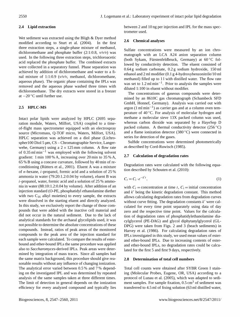
In contrast to this, the amounts of all ether-bound IPLs(Fig. 5) scattered but did not decrease significantly until theend of the degradation experiment. Ester- and ether-boundIPLs in the abiotic control showed behavior similar to eachother with slightly decreasing values in the course of the ex-periment (Fig. 6). The amounts of IPLs in the degradationexperiment increased within the first days while the abioticcontrol did not show this effect. The archaeal ether lipids inthe degradation experiment remained elevated by about 50 %relative to the abiotic control throughout the entire experi-ment.
Samples from the untreated control were taken in thesame sampling intervals as in the degradation experiment.Most of the IPLs added to monitor the degradation werenot detected in the untreated control. Exceptions werethe IPLs PE-DAG 34:2 and PE-DAG 36:2, but comparedto the corresponding PE-DAGs in the added biomass theirtotal amount was low (less than 3 %). However, otherIPLs such as sulfoquinovosyl diacylglycerol (SQ-DAG) andphosphatidylglycerol-diacylglcerol (PG-DAG) were identi-fied in the untreated control. Additionally, PE-DAGs withside chains different from those in the degradation experi-ment were found (31:0, 31:1, 33:1, 33:2, 35:2). SQ-DAG wasthe most prominent IPL in the untreated control. It showedincreasing abundance from day 0 to day 23 and then a de-crease to the starting value at a moderate rate after a ma-jor drop between days 23 and 27. PE-DAG and PG-DAGshowed an increase between day 0 and day 5 and returned tothe starting values in the course of the experiment.
3.3 IPL degradation rates
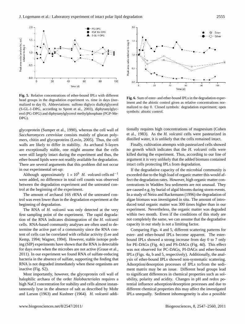
The calculation of degradation rates for ester-bound IPLs re-sulted in a linear relation between logk′ and log time (Fig. 7).Thek′-values of ester-bound IPLs were higher than those ofether-bound IPLs reflecting the faster decreasing concentra-tion of ester-bound IPLs (Fig. 6). Since thek′-values areplotted logarithmically, an increase of one unit represents tentimes faster degradation. Due to the scattered amounts of
Biogeosciences, 8, 2547–2560, 2011 www.biogeosciences.net/8/2547/2011/

J. Logemann et al.: Laboratory experiment of intact polar lipid degradation 2553
29
1
Figure 3: HPLC-ESI-MS base-peak intensity (BPI) chromatograms in negative ion-mode of 2
A: total extract of Saccharomyces cerevisiae; B: total extract of Haloferax volcanii; C: total 3
extract of the untreated control at day 0; D: total extract of the degradation experiment, 4
sample taken at day 0; E: total extract of the degradation experiment, sample taken at day 97. 5
For abbreviations see Figure 1. Please note that the observed peak intensities cannot be 6
directly transferred into concentrations. 7
8
Fig. 3. HPLC-ESI-MS base-peak intensity (BPI) chromatogramsin negative ion-mode of(A): total extract ofSaccharomyces cere-visiae; (B): total extract ofHaloferax volcanii; (C): total extract ofthe untreated control at day 0;(D): total extract of the degradationexperiment, sample taken at day 0;(E): total extract of the degra-dation experiment, sample taken at day 97. For abbreviations seeFig. 1. Please note that the observed peak intensities cannot be di-rectly transferred into concentrations.
ether-bound IPLs (Fig. 6), the relation for their kinetic degra-dation constants was less linear (Fig. 7).
3.4 Succession in microbial abundance, diversity andactivity
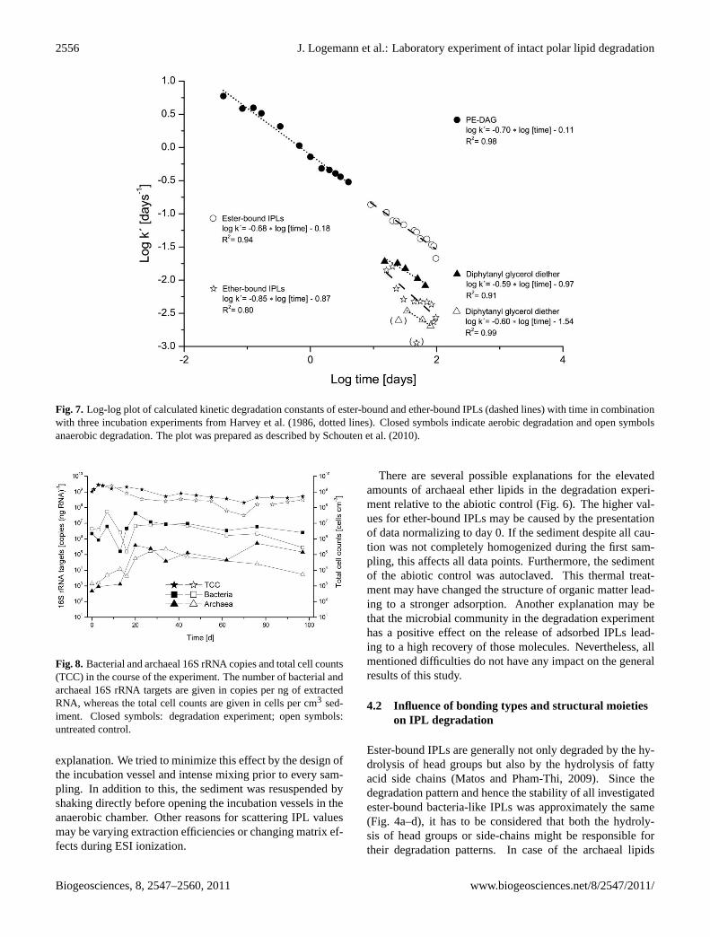
The development of the microbial communities was moni-tored to obtain background information for the degradationof IPLs. The total cell counts of the degradation exper-iment and the untreated sediment showed the same trendand decreased only slightly during the experiment (Fig. 8).The total cell numbers of the degradation experiment weremarginally higher than those of the untreated control. Thenumbers of bacterial 16S rRNA targets were one to twoorders of magnitude higher than those ofArchaea. Be-tween days 7 and 16, the bacterial 16S rRNA copy num-bers dropped significantly before returning to their previous
values. The number of archaeal 16S rRNA copies showeda generally increasing trend. After day 20, the numbers ofboth bacterial and archaeal 16S rRNA targets remained rel-atively constant. The RNA content of the sediment rangedfrom 80 to 4800 ng cm−3. Ravenschlag et al. (2000) de-termined an rRNA content of 0.9 to 1.4 fg rRNA per cellfor two sulfate-reducing bacteria from surface sediments.These values were used to assess the total cell numbers ofour study. The calculated values range between 8.9× 10⁷
and 3.4× 10⁹ cells cm−3, which fits nicely to our total cellcounts. The analysis of fermentation products showed no sig-nificant difference between the degradation experiment andthe untreated control (Fig. S1). The bacterial communityprofiles of the degradation experiment and the untreated sed-iment looked similar to each other (Fig. S2). Initially, thecommunity structure was highly diverse. This diversity de-creased in the course of the experiment probably due to adiminishing substrate spectrum as indicated in Fig. S1.
The archaeal community pattern showed minor differ-ences between the degradation experiment and the untreatedcontrol (Fig. S3).H. volcanii was only detected at the veryfirst sampling point in the degradation experiment. In thebeginning of the experiment, when sulfate was still present,no methanogenic archaea were detected (Fig. S3). Only af-ter depletion of sulfate the rRNA of methanogens was found.The presence and activity of these organisms were supportedby methane production observed in the absence of sulfate.In all samples, the content of eukaryotic RNA was too lowto obtain sufficient PCR products to prepare a DGGE witheukaryotic primers.
3.5 Total organic carbon
The carbon content was analyzed at five time points of thedegradation experiment and the untreated control. The differ-ence between the degradation experiment and the untreatedcontrol reflected the amount of organic matter that was addedto the degradation experiment with the inactive cell materialof S. cerevisiaeand H. volcanii. We added 1.36 g of cell-derived organic carbon which increased the TOC content ofthe natural sediment from 0.23 % Corg (4.9 g) to 0.29 % Corg(6.2 g). No pronounced difference was visible between thedegradation experiment and the untreated control for all otherparameters.
3.6 Effects of sediment-derived organic matter onmicrobial processes
The increasing amounts of fermentation products and to-tal cell counts within the first week demonstrate a stimula-tion of microbial activity. This was likely caused by a tem-perature increase from 4◦C (storage temperature) to 20◦Cin the laboratory. The addition of biomass caused a fasterincrease of methanogenesis and fermentation. The addedbiomass also resulted in slightly increased rates of sulfate
www.biogeosciences.net/8/2547/2011/ Biogeosciences, 8, 2547–2560, 2011

2554 J. Logemann et al.: Laboratory experiment of intact polar lipid degradation
30
1
Figure 4: Relative concentrations of ester-bound IPLs with different head groups in the 2
degradation experiment vs. time in days (normalized to day 0). Core lipid structures are given 3
as sum of fatty acids (e.g. 32:2) where 32 represents the number of carbon atoms and 2 4
represents the number of double bonds in the core lipid structure. See Figure 1 for 5
abbreviations. 6
7
8
9
Fig. 4. Relative concentrations of ester-bound IPLs with different head groups in the degradation experiment vs. time in days (normalized today 0). Core lipid structures are given as sum of fatty acids (e.g. 32:2) where 32 represents the number of carbon atoms and 2 represents thenumber of double bonds in the core lipid structure. See Fig. 1 for abbreviations.
consumption, methane production and fermentation (Figs. 2and S1). Roughly 10 % of the cellular dry weight consists oflipids (Stouthamer, 1979) and the IPL content is even lower.Therefore, the absolute amount of IPLs that were added withthe biomass ofH. volcaniiandS. cerevisiaeshould have beenlow compared to other organic compounds present in the sed-iment itself. Accordingly, the fermentation products do notonly reflect IPL degradation but mainly degradation of theorganic matter inherited from the natural sediment (Fig. S1).
4 Discussion
In this experiment, ester-bound bacteria-like IPLs were de-graded faster than ether-bound archaeal IPLs while an influ-ence of the structure and the bonding type of the head groupwas not detected.
4.1 Quality assessment of experimental design and data
Haloferax volcaniiandSaccharomyces cerevisiaedo not oc-cur naturally in tidal-flat sediments. Thus, it might be sus-pected that the observed degradation rates of IPLs turn outto be different from those of IPLs in the natural microbialcommunity. However, the source of ether- and ester-boundIPLs should have no influence on the degradation rate sincethe chemical structures and the bonding types of the addedmaterial also occur naturally in IPLs found in Wadden Seasediments (Rutters et al., 2001). Nevertheless, it is necessaryto use cell material that is not indigenous to the sedimentmatrix to monitor the degradation of individual IPLs withoutconfusing them with the inherited IPL inventory.
One could argue that the different degradation patterns ob-served for ether- and ester-bound IPLs are caused by a se-lective protection of ether-bound IPLs in intactH. volcaniicell material and on the other hand disrupted cells ofS. cere-visiae. Membrane lipids in living organisms are protected bytheir surrounding cell walls. The outer surface ofHaloferaxvolcanii consists of a hexagonally packed surface (S) layer
Biogeosciences, 8, 2547–2560, 2011 www.biogeosciences.net/8/2547/2011/
J. Logemann et al.: Laboratory experiment of intact polar lipid degradation 2555
31
1
Figure 5: Relative concentrations of ether-bound IPLs with different head groups in the 2
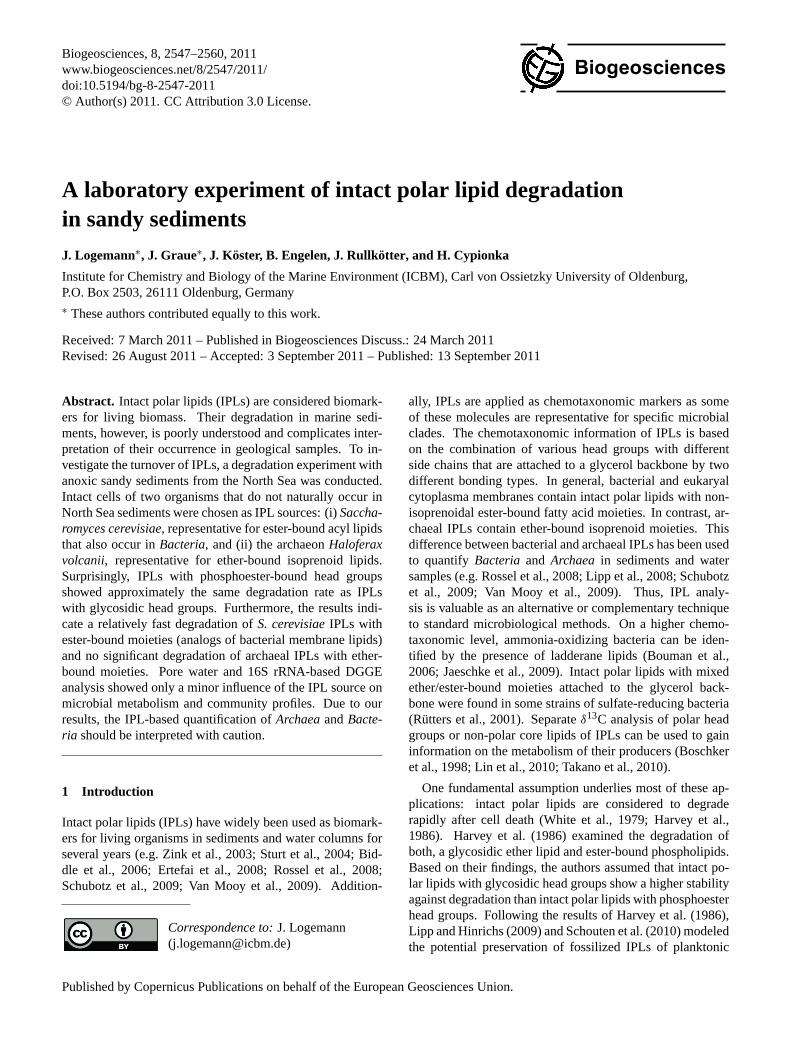
degradation experiment vs. time in days (normalized to day 0). Abbreviations: sulfono 3
diglyco dialkylglycerol (S-GL-1-DPG, according to Sprott et al., 2003), diphytanylglycerol 4
(PG-DPG) and diphytanylglycerol methylphosphate (PGP-Me-DPG). 5
6
Figure 6: Sum of ester- and ether-bound IPLs in the degradation experiment and the abiotic 7
control given as relative concentrations normalized to day 0. Closed symbols: degradation 8
experiment; open symbols: abiotic control. 9
10
Fig. 5. Relative concentrations of ether-bound IPLs with differenthead groups in the degradation experiment vs. time in days (nor-malized to day 0). Abbreviations: sulfono diglyco dialkylglycerol(S-GL-1-DPG, according to Sprott et al., 2003), diphytanylglyc-erol (PG-DPG) and diphytanylglycerol methylphosphate (PGP-Me-DPG).
glycoprotein (Sumper et al., 1990), whereas the cell wall ofSaccharomyces cerevisiaeconsists mainly of glucan poly-mers, chitin and glycoproteins (Levin, 2005). Thus, the cellwalls are likely to differ in stability. As archaeal S-layersare exceptionally stable, one might assume that the cellswere still largely intact during the experiment and thus, theether-bound lipids were not readily available for degradation.There are several arguments that this problem did not occurin our experimental set-up:
Although approximately 1× 10⁹ H. volcaniicells ml−1
were added, no difference in total cell counts was observedbetween the degradation experiment and the untreated con-trol at the beginning of the experiment.
The amount of archaeal 16S rRNA of the untreated con-trol was even lower than in the degradation experiment at thebeginning of degradation.
The RNA of H. volcanii was only detected at the veryfirst sampling point of the experiment. The rapid degrada-tion of the RNA indicates disintegration of theH. volcaniicells. RNA-based community analyses are often used to de-termine the active part of a community since the RNA con-tent of cells can be correlated with cellular activity (Lee andKemp, 1994; Wagner, 1994). However, stable isotope prob-ing (SIP) experiments have shown that the RNA is detectablefor days even when the microbes are not active (Graue et al.,2011). In our experiment we found RNA of sulfate-reducingbacteria in the absence of sulfate, supporting the finding thatRNA is not degraded immediately when these organisms areinactive (Fig. S2).
Most importantly, however, the glycoprotein cell wall ofhalophilic archaea of the orderHalobacterialesrequires ahigh NaCl concentration for stability and cells almost instan-taneously lyse in the absence of salt as described by Mohrand Larson (1963) and Kushner (1964).H. volcanii addi-
31
1
Figure 5: Relative concentrations of ether-bound IPLs with different head groups in the 2
degradation experiment vs. time in days (normalized to day 0). Abbreviations: sulfono 3
diglyco dialkylglycerol (S-GL-1-DPG, according to Sprott et al., 2003), diphytanylglycerol 4
(PG-DPG) and diphytanylglycerol methylphosphate (PGP-Me-DPG). 5
6
Figure 6: Sum of ester- and ether-bound IPLs in the degradation experiment and the abiotic 7
control given as relative concentrations normalized to day 0. Closed symbols: degradation 8
experiment; open symbols: abiotic control. 9
10
Fig. 6. Sum of ester- and ether-bound IPLs in the degradation exper-iment and the abiotic control given as relative concentrations nor-malized to day 0. Closed symbols: degradation experiment; opensymbols: abiotic control.
tionally requires high concentrations of magnesium (Cohenet al., 1983). As theH. volcanii cells were pasteurized indistilled water, it is unlikely that the cells remained intact.
Finally, cultivation attempts with pasteurized cells showedno growth which indicates that theH. volcanii cells werekilled during the experiment. Thus, according to our line ofargument it is very unlikely that the added biomass containedintact cells protecting IPLs from degradation.
If the degradative capacity of the microbial community isexceeded due to the high load of organic matter this would af-fect the degradation rates. However, high organic matter con-centrations in Wadden Sea sediments are not unusual. Theyare caused e.g. by burial of algal blooms during storm events.In a study of Neira and Rackemann (1996) the degradation ofalgae biomass was investigated in situ. The amount of intro-duced total organic matter was 300 times higher than in ourexperiment. Nevertheless, the organic matter was degradedwithin two month. Even if the conditions of this study arenot completely the same, we can assume that the degradativecapacity in our study is not a limiting factor.
Comparing Figs. 4 and 5, different scattering patterns forester- and ether-bound IPLs become apparent. The ester-bound IPLs showed a strong increase from day 0 to 7 onlyfor PE-DAGs (Fig. 4c) and PS-DAGs (Fig. 4d). This effectwas not observed for PC-DAGs, PI-DAGs and ether-boundIPLs (Figs. 4a, b and 5, respectively). Additionally, the anal-ysis of ether-bound IPLs showed non-systematic scattering.Adsorption/desorption processes of IPLs to/from the sedi-ment matrix may be an issue. Different head groups leadto significant differences in chemical properties such as sol-ubility, polarity and acidity. Changes in pH and redox po-tential influence adsorption/desorption processes and due todifferent chemical properties this may affect the investigatedIPLs unequally. Sediment inhomogeneity is also a possible
www.biogeosciences.net/8/2547/2011/ Biogeosciences, 8, 2547–2560, 2011
2556 J. Logemann et al.: Laboratory experiment of intact polar lipid degradation
32
1
Figure 7: Log-log plot of calculated kinetic degradation constants of ester-bound and ether-2
bound IPLs (dashed lines) with time in combination with three incubation experiments from 3
Harvey et al., 1986 (dotted lines). Closed symbols indicate aerobic degradation and open 4
symbols anaerobic degradation. The plot was prepared as described by Schouten et al. 5
(2010). 6
7
8
Figure 8: Bacterial and archaeal 16S rRNA copies and total cell counts (TCC) in the course of 9
the experiment. The number of bacterial and archaeal 16S rRNA targets are given in copies 10
Fig. 7. Log-log plot of calculated kinetic degradation constants of ester-bound and ether-bound IPLs (dashed lines) with time in combinationwith three incubation experiments from Harvey et al. (1986, dotted lines). Closed symbols indicate aerobic degradation and open symbolsanaerobic degradation. The plot was prepared as described by Schouten et al. (2010).
32
1
Figure 7: Log-log plot of calculated kinetic degradation constants of ester-bound and ether-2
bound IPLs (dashed lines) with time in combination with three incubation experiments from 3
Harvey et al., 1986 (dotted lines). Closed symbols indicate aerobic degradation and open 4
symbols anaerobic degradation. The plot was prepared as described by Schouten et al. 5
(2010). 6
7
8
Figure 8: Bacterial and archaeal 16S rRNA copies and total cell counts (TCC) in the course of 9
the experiment. The number of bacterial and archaeal 16S rRNA targets are given in copies 10
Fig. 8. Bacterial and archaeal 16S rRNA copies and total cell counts(TCC) in the course of the experiment. The number of bacterial andarchaeal 16S rRNA targets are given in copies per ng of extractedRNA, whereas the total cell counts are given in cells per cm3 sed-iment. Closed symbols: degradation experiment; open symbols:untreated control.
explanation. We tried to minimize this effect by the design ofthe incubation vessel and intense mixing prior to every sam-pling. In addition to this, the sediment was resuspended byshaking directly before opening the incubation vessels in theanaerobic chamber. Other reasons for scattering IPL valuesmay be varying extraction efficiencies or changing matrix ef-fects during ESI ionization.
There are several possible explanations for the elevatedamounts of archaeal ether lipids in the degradation experi-ment relative to the abiotic control (Fig. 6). The higher val-ues for ether-bound IPLs may be caused by the presentationof data normalizing to day 0. If the sediment despite all cau-tion was not completely homogenized during the first sam-pling, this affects all data points. Furthermore, the sedimentof the abiotic control was autoclaved. This thermal treat-ment may have changed the structure of organic matter lead-ing to a stronger adsorption. Another explanation may bethat the microbial community in the degradation experimenthas a positive effect on the release of adsorbed IPLs lead-ing to a high recovery of those molecules. Nevertheless, allmentioned difficulties do not have any impact on the generalresults of this study.
4.2 Influence of bonding types and structural moietieson IPL degradation
Ester-bound IPLs are generally not only degraded by the hy-drolysis of head groups but also by the hydrolysis of fattyacid side chains (Matos and Pham-Thi, 2009). Since thedegradation pattern and hence the stability of all investigatedester-bound bacteria-like IPLs was approximately the same(Fig. 4a–d), it has to be considered that both the hydroly-sis of head groups or side-chains might be responsible fortheir degradation patterns. In case of the archaeal lipids
Biogeosciences, 8, 2547–2560, 2011 www.biogeosciences.net/8/2547/2011/

J. Logemann et al.: Laboratory experiment of intact polar lipid degradation 2557
two structurally different bonding types of head groups werepresent – one glycosidic (S-GL-1) and two phosphoester(PG-DPG and PGP-Me-DPG; Fig. 1) head groups. However,the same degradation patterns were observed for both bind-ing types. In general, compared to glycosidic bonds, phos-phoester bonds are chemically more labile, since phospho-esters can be hydrolyzed by acids and bases whereas glyco-sidic bonds can usually only be hydrolyzed by acids (Beyerand Walter, 1991). Correspondingly, Harvey et al. (1986)found that glycosidic ether lipids were more stable than phos-pholipids with ester-bound moieties. However, they investi-gated the degradation of a glycosidic ether-bound lipid and aphosphoester ester-bound lipid. Thus, the study of Harvey etal. (1986) cannot be used to answer the question which partof an IPL-molecule (head group or moiety bonding type) isresponsible for the observed differential stability of bacterialand archaeal IPLs.
According to our results, we assume that the differencesin chemical stability of IPLs play only a minor role duringthe degradation of IPLs, at least in the investigated system.Since not only the head groups may have an influence on IPLdegradation, also the structurally different core lipids have tobe considered as possible factor for the observed degradationpatterns. In general, complex molecules like lipids and pro-teins are hydrolyzed by exo- or ectoenzymes which are re-leased by prokaryotic cells (Cypionka, 2010). Therefore, weconclude that microbiological enzymatic processes are thedriving force in IPL degradation as also suggested by Whiteet al. (1979) and Harvey et al. (1986) rather than expectedchemical stabilities alone.
4.3 IPL degradation rates
The kinetic degradation constants of ester-bound IPLs(Fig. 7) in the beginning of our experiment are in the samerange as those at the end of aerobic degradation of PE-DAG in beach sediments observed by Harvey et al. (1986).Measured signal intensities in the degradation experimenton day 7 were as high as expected from the amount ofadded biomass. Therefore, significant degradation did notoccur during the first 7 days. This means that the kineticdegradation constants of ester-bound IPLs in our experimentwere significantly lower than those determined by Harvey etal. (1986).
In case of ether-bound IPLs the kinetic degradation con-stants determined in our study lie between those calculatedfor the ether-bound IPL with a glycosidic head group underoxic and anoxic conditions. In all cases this is in accordanceto the common knowledge that the turnover of organic matteris aerobically faster than anaerobically.
4.4 Microbial activity
Originally, the experiment was also planned as an enrichmentculture for lipid-degrading bacteria. It turned out, however,
that the added cell material had only little influence on thecommunity structure and the metabolic activity. This had apositive side-effect, since the processes which originally oc-cur in tidal flat sediments were not disturbed and superposed.Therefore, the selected set-up is suitable to reflect the naturalIPL degradation in such sediments. The increasing total cellcounts in the beginning of the experiment were also visiblein the untreated control and thus were probably caused bystimulation due to mixing and the higher temperature (4◦Cstorage temperature and 20◦C during the experiment).
Approximately 10 % of theArchaeain Wadden Sea sed-iments are known to be methanogens (Wilms et al., 2007).They have a narrow substrate spectrum limited to simplemolecules and are not known to degrade lipids. Probably, themain lipid degraders areBacteriawhich are more abundantthanArchaeain intertidal surface sediments (e.g. Beck et al.,2011). Anyhow, the physiological roles of most sedimentaryarchaea are not known (e.g. Teske and Sørensen, 2008) andtherefore it cannot be excluded that they are also involved inthe degradation of lipids.
Some studies indicate thatArchaeause other enzymesthanBacteriato synthesize and degrade IPLs (Choquet et al.,1994; Daiyasu et al., 2005). The IPL degradation experimentwas conducted with anoxic surface sediment which is dom-inated by bacteria affiliated toProteobacteria, Actinobacte-ria, Bacteriodetes, Firmicutes, SpirochaetesandChloroflexi,and archaea affiliated toMethanosarcinales, Thermococ-cales, MethanomicrobialesandMethanobacteriales(Wilmset al., 2006). The community structure of the deep biosphere,however, consists mainly of bacteria related toChloroflexi,Gammaproteobacteriaand JS1 candidate group and of ar-chaea affiliated to the Miscellaneous Crenarchaeotic Group(MCG), Marine Benthic Group (MBG) and South AfricanGoldmine Euryarchaeotal Group (SAGMEG) (Inagaki et al.,2006; Webster et al., 2006; Teske and Sørensen, 2008). Asour results strongly suggest a microbial degradation of IPLsby enzymes, the community structure probably has a ma-jor influence on the degradation pattern. Thus, the observeddegradation rates might be different if the experiment is re-peated with sediment from the deep biosphere or in sedi-ments where sulfate-reducing bacteria are not stimulated.
The question whether ester-bound lipids are recycled andused for membrane synthesis byBacteria cannot be an-swered by our experimental-setup. Enzymes are describedfor EukaryaandProkarya that facilitate bidirectional ATP-independent flipping of polar lipids across cytoplasmic mem-branes (Sanyal and Menon, 2009). The incorporation of ex-trinsic cell building blocks is energetically useful for mi-croorganisms because it is more efficient to recycle exist-ing molecules than to break them down to smaller moleculesto synthesize them “de-novo”. Takano et al. (2010) added13C-labeled glucose to marine sediment and found that the13C was incorporated into the glycerol backbone of archaealmembrane lipids whereas the isoprenoid core lipids remainedunlabeled. This indicates a recycling of comparable large
www.biogeosciences.net/8/2547/2011/ Biogeosciences, 8, 2547–2560, 2011

2558 J. Logemann et al.: Laboratory experiment of intact polar lipid degradation
membrane building blocks. To answer the question if IPLscan be recycled as intact molecules, stable-isotope probing(SIP) or 14C-labeling would be suitable tools to follow thedegradation and incorporation of IPLs and their resultingproducts.
4.5 DNA, RNA and intact polar lipids as biomarkers forliving cells
DNA and IPLs are commonly used as biomarkers for livingcells (e.g. Biddle et al., 2006; Wilms et al., 2006). Addi-tionally, RNA is used to determine the active part of micro-bial communities (e.g. Griffith et al., 2000). However, thesuccessful application of these methods may be hamperedby several aspects like varying extraction efficiencies, matrixeffect and adsorption/desorption processes. The presence ofDNA does neither prove the activity nor even the presenceof the corresponding cells (Lorenz and Wackernagel, 1987;Josephson et al., 1993). In contrast, RNA is labile and theRNA content of cells can be correlated with cellular activ-ity (Lee and Kemp, 1994; Wagner, 1994). However, whencells are inactive or have a very slow metabolism, the RNAcontent might not be sufficient for detection. The stabilityof ether-bound IPLs in our study suggests that IPLs also donot reflect exclusively the living community. This shows thatnone of the mentioned tools can be used without caution buta combination of these tools may help to get a more accuratepicture.
5 Conclusions
The quantification ofBacteriaandArchaeain the deep ma-rine biosphere by IPL and FISH analysis led to the assump-tion that this habitat is dominated byArchaea (Biddle etal., 2006; Lipp et al., 2008). In contrast, catalyzed reporterdeposition fluorescence in situ hybridization (CARD-FISH)and quantitative polymerase chain reaction (q-PCR) suggesta predominance ofBacteria (Schippers et al., 2005; Ina-gaki et al., 2006). Our experiment shows that these inter-pretations have to be considered with caution, since ether-bound archaeal IPLs were clearly more stable than ester-bound bacteria-like IPLs over a period of 100 days. In na-ture, ether-bound IPLs may even be preserved for longer timeperiods since the enhanced temperature in the laboratory ac-celerated the degradation processes.
To give better insights into the degradation of IPLs in otherenvironments like the deep biosphere, it is desirable to con-duct degradation experiments reflecting the in situ conditionsof those habitats. For the deep biosphere this would meanthat the experiments must be conducted under high pressureand over a long period since microbial turnover of organicmatter is low. Furthermore, the addition of high substrateconcentration would lead to a distorted image of ongoing
processes as the in situ available organic matter is recalci-trant.
On the other hand, IPL-based quantification may lead toan overestimation of archaeal cell counts as indicated by ourstudy. Regarding the discussion above, molecular biologi-cal methods based on DNA or RNA can also be inaccurate.Thus, the abundances ofArchaeaandBacteriaobtained withboth methods have to be interpreted with caution.
Supplementary material related to thisarticle is available online at:http://www.biogeosciences.net/8/2547/2011/bg-8-2547-2011-supplement.pdf.
Acknowledgements.The authors are grateful to B. Kopke, B. Buch-mann, H. Haase and P. Neumann for their technical assistance.Many thanks to Michael Seidel for sharing his experience in IPLdegradation experiments that were conducted prior to this work. Wethank K.-U. Hinrichs, A. Schippers and two anonymous refereesfor their constructive comments and suggestions. This work wasfinancially supported by Deutsche Forschungsgemeinschaft (DFG)within the Research Group on “BioGeoChemistry of Tidal Flats”(FOR 432).
Edited by: K. Suzuki
References
Adams, A. E. M., Johnson, D. I., Longnecker, R. M., Sloat, B. F.,and Pringle, J. R.:CDC42 and CDC43, two additional genesinvolved in budding and establishment of cell polarity in the yeastSaccharomyces cerevisiae, J. Cell Biol., 111, 131–142, 1990.
Beck, M., Riedel, T., Graue, J., Koster, J., Kowalski, N., Wu, C.S., Wegener, G., Lipsewers, Y., Freund, H., Bottcher, M. E.,Brumsack, H.-J., Cypionka, H., Rullkotter, J., and Engelen, B.:Imprint of past and present environmental conditions on micro-biology and biogeochemistry of coastal Quaternary sediments,Biogeosciences, 8, 55–68,doi:10.5194/bg-8-55-2011, 2011.
Beyer, H. and Walter, W.: Lehrbuch der Organischen Chemie, 22.Auflage, S. Hirzel Verlag, Stuttgart, 1–1030, 1991.
Biddle, J. F., Lipp, J. S., Lever, M. A., Lloyd, K. G., Sørensen,K. B., Anderson, R., Fredricks, H. F., Elvert, M., Kelly, T. J.,Schrag, D. P., Sogin, M. L., Brenchley, J. E., Teske, A., House,C. H., and Hinrichs, K. U.: Heterotrophic archaea dominate sed-imentary subsurface ecosystems off Peru, P. Natl. Acad. Sci.USA, 103, 3846–3851, 2006.
Boschker, H. T. S., Nold, S. C., Wellsbury, P., Bos, D., de Graaf,W., Pel, R., Parkes, R. J., and Cappenberg, T. E.: Direct linkingof microbial populations to specific biogeochemical processes by13C-labelling of biomarkers, Nature, 392, 801–805, 1998.
Boumann, H. A., Hopmans, E. C., van de Leemput, I., Op denCamp, H. J. M., van de Vossenberg, J., Strous, M., Jetten, M.S. M., Damste, J. S. S., and Schouten, S.: Ladderane phospho-lipids in anammox bacteria comprise phosphocholine and phos-phoethanolamine headgroups, FEMS Microbiol. Lett., 258, 297–304, 2006.
Biogeosciences, 8, 2547–2560, 2011 www.biogeosciences.net/8/2547/2011/

J. Logemann et al.: Laboratory experiment of intact polar lipid degradation 2559
Choquet, C. G., Patel, G. B., Beveridge, T. J., and Sprott, G. D.: Sta-bility of pressure extruded liposomes made from archaebacterialether lipids, Appl. Microbiol. Biot., 42, 375–384, 1994.
Cohen, S., Oren, A., and Shilo, M.: The divalent cation requirementof Dead Sea halobacteria, Arch. Microbiol., 136, 184–190, 1983.
Cord-Ruwisch, R.: A quick method for the determination of dis-solved and precipitated sulfides in cultures of sulfate-reducingbacteria, J. Microbiol. Meth., 4, 33–36, 1985.
Cypionka, H.: Grundlagen der Mikrobiologie, Auflage 4, Springer,Heidelberg, 1–337, 2010.
Daiyasu, H., Kuma, K.-I., Yokoi, T., Morii, H., Koga, Y., and Toh,H.: A study of archaeal enzymes involved in polar lipid synthesislinking amino acid sequence information, genomic contexts andlipid composition, Archaea, 1, 399–410, 2005.
Ertefai, T. F., Fisher, M. C., Fredricks, H. F., Lipp, J. S., Pearson, A.,Birgel, D., Udert, K. M., Cavanaugh, C. M., Gschwend, P. M.,and Hinrichs, K. U.: Vertical distribution of microbial lipids andfunctional genes in chemically distinct layers of a highly pollutedmeromictic lake, Org. Geochem., 39, 1572–1588, 2008.
Graue, J., Engelen, B., and Cypionka, H.: Degradation of cyanobac-terial biomass in anoxic tidal-flat sediments: A microcosmstudy of metabolic processes and community changes, ISME J.,doi:10.1038/ismej.2011.120, in press, 2011.
Griffiths, R. I., Whiteley, A. S., O’Donnell, A. G., and Bailey, M.J.: Rapid method for coextraction of DNA and RNA from naturalenvironments for analysis of ribosomal DNA- and rRNA-basedmicrobial community composition, Appl. Environ. Microbiol.,66, 5488–5491, 2000.
Harvey, H. R., Fallon, R. D., and Patton, J. S.: The effect of organic-matter and oxygen on the degradation of bacterial membranelipids in marine sediments, Geochim. Cosmochim. Ac., 50, 795–804, 1986.
Inagaki, F., Nunoura, T., Nakagawa, S., Teske, A., Lever, M.,Lauer, A., Suzuki, M., Takai, K., Delwiche, M., Colwell, F.S., Nealson, K. H., Horikoshi, K., D’Hondt, S., and Jørgensen,B. B.: Biogeographical distribution and diversity of microbesin methane hydrate-bearing deep marine sediments on the Pa-cific Ocean Margin, P. Natl. Acad. Sci. USA, 103, 2815–2820,doi:10.1073/pnas.0511033103, 2006.
Jaeschke, A., Rooks, C., Trimmer, M., Nicholls, J. C., Hopmans, E.C., Schouten, S., and Damste, J. S. S.: Comparison of ladderanephospholipid and core lipids as indicators for anaerobic ammo-nium oxidation (anammox) in marine sediments, Geochim. Cos-mochim. Ac., 73, 2077–2088,doi:10.1016/j.gca.2009.01.013,2009.
Josephson, K. L., Gerba, C. P., and Pepper, I. L.: Polymerase chainreaction detection of nonviable bacterial pathogens, Appl. Envi-ron. Microbiol., 59, 3513–3515, 1993.
Kushner, D. J.: Lysis and dissolution of cells and envelopes ofan extremely halophilic bacterium, J. Bacteriol., 87, 1147–1156,1964.
Lee, S. H. and Kemp, P. F.: Single-cell RNA-content of naturalmarine planktonic bacteria measured by hybridization with mul-tiple 16S ribosomal-RNA-targeted fluorescent-probes, Limnol.Oceanogr., 39, 869–879, 1994.
Levin, D. E.: Cell wall integrity signaling inSaccharomyces cere-visiae, Microbiol. Mol. Biol. R., 69, 262–291, 2005.
Lin, Y. S., Lipp, J. S., Yoshinaga, M. Y., Lin, S. H., Elvert, M., andHinrichs, K. U.: Intramolecular stable carbon isotopic analysis
of archaeal glycosyl tetraether lipids, Rapid Commun. Mass Sp.,24, 2817–2826,doi:10.1002/rcm.4707, 2010.
Lipp, J. S. and Hinrichs, K. U.: Structural diversity and fate of intactpolar lipids in marine sediments, Geochim. Cosmochim. Ac., 73,6816–6833, 2009.
Lipp, J. S., Morono, Y., Inagaki, F., and Hinrichs, K.-U.: Signifi-cant contribution of Archaea to extant biomass in marine subsur-face sediments, Nature, 454, 991–994,doi:10.1038/nature07174,2008.
Lorenz, M. G. and Wackernagel, W.: Adsorption of DNA to sandand variable degradation rates of adsorbed DNA, Appl. Environ.Microbiol., 53, 2948–2952, 1987.
Lunau, M., Lemke, A., Walther, K., Martens-Habbena, W., and Si-mon, M.: An improved method for counting bacteria from sed-iments and turbid environments by epifluorescence microscopy,Environ. Microbiol., 7, 961–968, 2005.
Matos, A. R. and Pham-Thi, A. T.: Lipid deacylating enzymes inplants: Old activities, new genes, Plant Physiol. Bioch., 47, 491–503,doi:10.1016/j.plaphy.2009.02.011, 2009.
Mohr, V. and Larsen, H.: On the structural transformations and ly-sis ofHalobacterium salinariumin hypotonic and isotonic solu-tions, J. Gen. Microbiol., 31, 267–280, 1963.
Mullakhanbhai, M. F. and Larsen, H.:Halobacterium volcaniispec.nov., a Dead Sea halobacterium with a moderate salt require-ment, Arch. Microbiol., 104, 207–214, 1975.
Neira, C. and Rackemann, M.: Black spots produced byburied macroalgae in intertidal sandy sediments of the Wad-den Sea: Effects on the meiobenthos, J. Sea Res., 36, 153–170,doi:10.1016/s1385-1101(96)90786-8, 1996.
Ravenschlag, K., Sahm, K., Knoblauch, C., Jørgensen, B. B.,and Amann, R.: Community structure, cellular rRNA con-tent, and activity of sulfate-reducing bacteria in marine arc-tic sediments, Appl. Environ. Microbiol., 66, 3592–3602,doi:10.1128/aem.66.8.3592-3602.2000, 2000.
Rossel, P. E., Lipp, J. S., Fredricks, H. F., Arnds, J., Boetius, A.,Elvert, M., and Hinrichs, K. U.: Intact polar lipids of anaerobicmethanotrophic archaea and associated bacteria, Org. Geochem.,39, 992–999,doi:10.1016/j.orggeochem.2008.02.021, 2008.
Rutters, H., Sass, H., Cypionka, H., and Rullkotter, J.:Monoalkylether phosphoslipids in the sulfate-reducing bacteriaDesufosarcina variablisand Desulforhabus amnigenus, Arch.Microbiol., 176, 435–442, 2001.
Sanyal, S. and Menon, A. K.: Flipping Lipids: Why an’What’s the Reason for?, ACS Chem. Biol., 4, 895–909,doi:10.1021/cb900163d, 2009.
Schippers, A., Neretin, L. N., Kallmeyer, J., Ferdelman, T. G.,Cragg, B. A., Parkes, R. J., and Jørgensen, B. B.: Prokaryoticcells of the deep sub-seafloor biosphere identified as living bac-teria, Nature, 433, 861–864, 2005.
Schouten, S., Middelburg, J. J., Hopmans, E. C., and SinningheDamste, J. S.: Fossilization and degradation of intact polar lipidsin deep subsurface sediments: A theoretical approach, Geochim.Cosmochim. Ac., 74, 3806–3814, 2010.
Schubotz, F., Wakeham, S. G., Lipp, J. S., Fredricks, H. F., andHinrichs, K.-U.: Detection of microbial biomass by intact polarmembrane lipid analysis in the water column and surface sed-iments of the Black Sea, Environ. Microbiol., 11, 2720–2734,2009.
Sprott, G. D., Larocque, S., Cadotte, N., Dicaire, C. J., McGee,
www.biogeosciences.net/8/2547/2011/ Biogeosciences, 8, 2547–2560, 2011
2560 J. Logemann et al.: Laboratory experiment of intact polar lipid degradation
M., and Brisson, J. R.: Novel polar lipids of halophilic eubac-teriumPlanococcusH8 and archaeonHaloferax volcanii, BBA-Mol. Cell Biol. L., 1633, 179–188, 2003.
Stouthamer, A. H.: The search for correlation between theoreticaland experimental growth yields, Int. Rev. Biochem, 21, 1–15,1979.
Sturt, H. F., Summons, R. E., Smith, K., Elvert, M., and Hin-richs, K. U.: Intact polar membrane lipids in prokaryotes andsediments deciphered by high-performance liquid chromatogra-phy/electrospray ionization multistage mass spectrometry – newbiomarkers for biogeochemistry and microbial ecology, RapidCommun. Mass Sp., 18, 617–628, 2004.
Sumper, M., Berg, E., Mengele, R., and Strobel, I.: Primary struc-ture and glycosylation of the S-layer protein ofHaloferax vol-canii, J. Bacteriol., 172, 7111–7118, 1990.
Takano, Y., Chikaraishi, Y., Ogawa, N. O., Nomaki, H., Morono,Y., Inagaki, F., Kitazato, H., Hinrichs, K. U., and Ohkouchi, N.:Sedimentary membrane lipids recycled by deep-sea benthic ar-chaea, Nat. Geosci., 3, 858–861,doi:10.1038/ngeo983, 2010.
Teske, A. and Sørensen, K. B.: Uncultured archaea in deep marinesubsurface sediments: have we caught them all?, ISME J., 2, 3–18, 2008.
Van Mooy, B. A. S., Fredricks, H. F., Pedler, B. E., Dyhrman, S. T.,Karl, D. M., Koblizek, M., Lomas, M. W., Mincer, T. J., Moore,L. R., Moutin, T., Rappe, M. S., and Webb, E. A.: Phytoplanktonin the ocean use non-phosphorus lipids in response to phosphorusscarcity, Nature, 458, 69–72, 2009.
Wagner, R.: The regulation of ribosomal-RNA synthesis andbacterial-cell growth, Arch. Microbiol., 161, 100–109, 1994.
Webster, G., Parkes, R. J., Cragg, B. A., Newberry, C. J., Weight-man, A. J., and Fry, J. C.: Prokaryotic community compo-sition and biogeochemical processes in deep subseafloor sedi-ments from the Peru Margin, FEMS Microbiol. Ecol., 58, 65–85,doi:10.1111/j.1574-6941.2006.00147.x, 2006.
White, D. C., Davis, W. M., Nickels, J. S., King, J. D., and Bobbie,R. J.: Determination of the sedimentary microbial biomass byextractable lipid phosphate, Oecologia, 40, 51–62, 1979.
Wilms, R., Sass, H., Kopke, B., Koster, J., Cypionka, H., and En-gelen, B.: Specific Bacterial, Archaeal, and Eukaryotic Com-munities in Tidal-Flat Sediments along a Vertical Profile ofSeveral Meters, Appl. Environ. Microbiol., 72, 2756–2764,doi:10.1128/aem.72.4.2756-2764.2006, 2006.
Wilms, R., Sass, H., Kopke, B., Cypionka, H., and Engelen, B.:Methane and sulfate profiles within the subsurface of a tidal flatare reflected by the distribution of sulfate-reducing bacteria andmethanogenic archaea, FEMS Microbiol. Ecol., 59, 611–621,2007.
Zink, K. G., Wilkes, H., Disko, U., Elvert, M., and Horsfield, B.:Intact phospholipids – microbial “life markers” in marine deepsubsurface sediments, Org. Geochem., 34, 755–769, 2003.
Biogeosciences, 8, 2547–2560, 2011 www.biogeosciences.net/8/2547/2011/
Related Documents