Welcome message from author
This document is posted to help you gain knowledge. Please leave a comment to let me know what you think about it! Share it to your friends and learn new things together.
Transcript
ESTUDO SOBRE O COMPORTAMENTO BIOLÓGICOIN VITRO E IN VIVO DE UMA NOVA
HIDROXIAPATITE MODIFICADA
Porto 2006
Manuel António Pereira Gutierres

– III –
ESTUDO SOBRE O COMPORTAMENTO BIOLÓGICO IN VITRO E IN VIVO DE UMA NOVA HIDROXIAPATITE MODIFICADA
Dissertação de candidatura ao grau de Doutor apresentada à Faculdade de Medicina da Universidade do Porto
Orientador: Professor Doutor Luís de Almeida
Co-orientador: Professor Doutor José Domingos Santos
ESTUDO SOBRE O COMPORTAMENTO BIOLÓGICO IN VITRO E IN VIVO DE UMA NOVA HIDROXIAPATITE MODIFICADA
– IV –
Artigo 48º, Parágrafo 3: “A Faculdade não responde pelas doutrinas expendidas na dissertação”
(Regulamento da Faculdade de Medicina do Porto, 29 de Janeiro de 1931, Decreto nº19337)
– V –
ESTUDO SOBRE O COMPORTAMENTO BIOLÓGICO IN VITRO E IN VIVO DE UMA NOVA HIDROXIAPATITE MODIFICADA
CORPO CATEDRÁTICO DA FACULDADE DE MEDICINA
Professores Efectivos – Catedráticos
Doutor Alberto Manuel Barros da SilvaDoutor Altamiro Manuel Rodrigues Costa PereiraDoutor Álvaro Jerónimo Leal Machado de AguiarDoutor António Carlos Freitas Ribeiro SaraivaDoutor António José Pacheco PalhaDoutor Belmiro dos Santos PatrícioDoutor Cassiano Pena de Abreu e LimaDoutor Daniel Filipe Lima MouraDoutora Deolinda Maria Valente Alves Lima TeixeiraDoutor Eduardo Jorge da Cunha Rodrigues PereiraDoutor Fernando Manuel Mendes Falcão dos ReisDoutor Fernando Tavarela VelosoDoutora Isabel Maria Amorim Pereira RamosDoutor João Francisco Montenegro Andrade Lima BernardesDoutor Jorge Manuel Mergulhão Castro TavaresDoutor José Agostinho Marques LopesDoutor José Carlos Neves da Cunha AreiasDoutor José Henrique Dias Pinto de BarrosDoutor José Luís Medina VieiraDoutor José Manuel Lopes Teixeira AmaranteDoutor Luís Alberto Martins Gomes de AlmeidaDoutor Manuel Alberto Coimbra Sobrinho SimõesDoutor Manuel Augusto Cardoso de OliveiraDoutor Manuel Jesus Falcão Pestana VasconcelosDoutora Maria Amélia Duarte FerreiraDoutora Maria de Fátima Machado Henriques CarneiroDoutora Maria Dulce Cordeiro MadeiraDoutora Maria Isabel Amorim de AzevedoDoutora Maria Leonor Martins Soares DavidDoutor Manuel Maria Paula BarbosaDoutor Patrício Manuel Vieira Araújo Soares SilvaDoutor Rui Manuel Almeida Mota Cardoso
ESTUDO SOBRE O COMPORTAMENTO BIOLÓGICO IN VITRO E IN VIVO DE UMA NOVA HIDROXIAPATITE MODIFICADA
– VI –
Professores Jubilados ou Aposentados
Doutor Abel José Sampaio da Costa TavaresDoutor Alexandre Alberto Guerra Sousa PintoDoutor Amândio Gomes Sampaio Tavares Doutor António Augusto Lopes VazDoutor António Carvalho Almeida CoimbraDoutor António Fernandes da FonsecaDoutor António Fernandes Oliveira Barbosa Ribeiro BragaDoutor António Germano Pina Silva LealDoutor António Luís Tomé da Rocha RibeiroDoutor António Manuel Sampaio de Araújo TeixeiraDoutor Artur Manuel Giesteira de AlmeidaDoutor Cândido Alves Hipólito ReisDoutor Carlos Rodrigo Magalhães RamalhãoDoutor Daniel Santos Pinto SerrãoDoutor Fernando de Carvalho Cerqueira Magro FerreiraDoutor Francisco José Zarco Carneiro ChavesDoutor Francisco de Sousa LéDoutor Henrique José Ferreira Gonçalves Lecour de MenesesDoutor João Silva CarvalhoDoutor Joaquim Germano Pinto Machado Correia da SilvaDoutor Joaquim Oliveira Costa MaiaDoutor José Augusto Fleming TorrinhaDoutor José Carvalho de OliveiraDoutor José Fernando Barros Castro CorreiaDoutor José Manuel Costa Mesquita GuimarãesDoutor José Manuel Gonçalves Pina CabralDoutor José Pinto de BarrosDoutor Levi Eugénio Ribeiro GuerraDoutor Manuel Machado Rodrigues GomesDoutor Manuel Teixeira Amarante JúniorDoutora Maria da Conceição Fernandes Marques MagalhãesDoutor Mário José Cerqueira Gomes BragaDoutor Serafim Correia Pinto GuimarãesDoutor Valdemar Miguel Botelho dos Santos CardosoDoutor Walter Friedrich Alfred Osswald
– VII –
ESTUDO SOBRE O COMPORTAMENTO BIOLÓGICO IN VITRO E IN VIVO DE UMA NOVA HIDROXIAPATITE MODIFICADA
À Mariana, À InêsÀ minha Família
Aos meus Mestres
Aos meus Amigos

– IX –
ESTUDO SOBRE O COMPORTAMENTO BIOLÓGICO IN VITRO E IN VIVO DE UMA NOVA HIDROXIAPATITE MODIFICADA
PREFÁCIO
Elaborar um trabalho de doutoramento é, por várias razões, uma tarefa séria e difícil de cuja dimensão só nos percebemos quando nos propomos enfrentá-la.
A reputação e a tradição de rigor científico da Faculdade de Medicina da Universidade do Porto, o valor dos seus docentes e investigadores, a excelência dos trabalhos que precederam este criam-nos um profundo sentimento de humildade.
Se, por um lado, sabemos que no domínio da ciência, os avanços se dão através de pequenos passos, que se vão adicionando a outros que alguém anteriormente deu, por outro lado temos consciência de que, quando se faz investigação em áreas que lidam directamente com a saúde, o sentido de responsabilidade e a honestidade com que se trabalha têm que ser um primado.
Assim, desde que há seis anos tomámos esta decisão, fomos sendo surpreendidos por uma variedade de sentimentos, atitudes e emoções: a generosidade dos doentes em participar num ensaio clínico com o único objectivo de, desinteressadamente, ajudar o próximo; as reacções, em regra positivas, daqueles que nos rodeiam; a tristeza provocada pela recusa de publicação de um artigo, obrigando a acrescido trabalho e sacrifício para ser aprovado; a beleza das imagens recolhidas; o entusiasmo crescente pelos resultados que vão aparecendo; a alegria de cada pequena conquista e, por fim, o cerrar dos olhos e o concentrar das energias para melhor descortinar a luz que adivinhamos ao fundo do túnel.
Elaborar uma tese é, em suma, viver!
E viver é também agradecer.
Obrigado, pois, ao meu orientador, o Professor Doutor Luís de Almeida, reconhecido Catedrático desta Faculdade, cujas qualidades humanas revelam uma forma especial de entender as pessoas, que me guiou no caminho que permitiu que tivesse desenvolvido este trabalho. O seu apoio amigo e esclarecido esteve sempre presente ao longo destes anos e “obrigou-me” a não desistir nos momentos difíceis.
Ao Professor Doutor José Domingos Santos, meu co-orientador, o meu reconhecimento pela sua disponibilidade, ajuda e pela confiança transmitida, que me fez acreditar que poderíamos avançar com esta investigação.
Aos Professores Doutores Ascensão Lopes, Anabela Dias e Sooraj Hussain agradeço o esforçado trabalho de equipa que desenvolveram e me deram oportunidade de integrar, assim como a paciência e amizade que sempre demonstraram.
À Professora Doutora Helena Fernandes devo os ensinamentos transmitidos e toda a colaboração prestada na elaboração dos estudos in vitro.
ESTUDO SOBRE O COMPORTAMENTO BIOLÓGICO IN VITRO E IN VIVO DE UMA NOVA HIDROXIAPATITE MODIFICADA
– X –
Dos colegas do serviço, de quem recebi inúmeras demonstrações de solidariedade, tenho de salientar em especial o meu chefe de equipa, Dr. Sérgio Silva. Permanentemente a angariar doentes que satisfizessem as condições de integrarem o protocolo e, depois, sempre pronto a disponibilizar tempos operatórios que me permitissem operá-los, viabilizou, assim, a prossecução do ensaio clínico. Estou grato, mais do que pelas palavras, pelas provas diárias de amizade que sempre revelou.
Ao pessoal de enfermagem do bloco operatório, em particular à responsável de então, Sr.ª Enfermeira Ofélia, agradeço a permanente disponibilidade para a colaboração que lhes solicitei.
Aos amigos, custa-me nomeá-los por receio de ser menos justo com algum. Se pela idade com alguns já privo há mais de 20 anos, outros, mais recentes, conquistaram também o seu lugar na minha vida. Todos sabem, no entanto, o quanto apreciei o seu apoio.
Uma palavra final de gratidão para com a minha família: ao meu avô pelo brilhozinho nos olhos com que me incentivava a doutorar-me; aos meus pais pelo seu exemplo de vida e empenho na formação que, com muito amor, me transmitiram; ao meu irmão, incontestável e indefectível companheiro de sempre; aos meus tios e primos pelo seu apoio e frequentes incentivos.
Às minhas filhas, Mariana e Inês, agradeço a energia que me transmitem para enfrentar os desafios, sendo este que acabei de concluir, bem pequeno quando comparado com a enorme responsabilidade que representa para mim guiá-las e prepará-las para a vida...
– XI –
ESTUDO SOBRE O COMPORTAMENTO BIOLÓGICO IN VITRO E IN VIVO DE UMA NOVA HIDROXIAPATITE MODIFICADA
ÍNDICE
CAPÍTULO I – INTRODUÇÃO, OBJECTIVOS E ESTRUTURA DA TESE ............................... 1
CAPÍTULO II – CONCEITOS GERAIS SOBRE SUBSTITUTOS ÓSSEOS ................................. 9 1) “Substitutos ósseos – conceitos gerais e estado actual” M. Gutierres, M.A. Lopes, N. Sooraj Hussain, A.T. Cabral, L. Almeida, J.D. Santos Arquivos de Medicina (2006) vol 19(4): 153-162.............................................................. 11
CAPÍTULO III – ESTUDOS DE BIOCOMPATIBILIDADE IN VITRO .......................................23 2) “In vitro mineralisation of human bone marrow cells cultured on Bonelike®” M.A. Costa, M. Gutierres, L. Almeida, M.A. Lopes, J.D. Santos, M.H. Fernandes Key Engineering Materials (2004) vols 254-256: pp. 821-824............................................ 25
CAPÍTULO IV – ESTUDOS DE BIOCOMPATIBILIDADE IN VIVO ......................................... 31 3) “Biological behaviour of Bonelike® graft implanted in the tibia of humans” M. Gutierres, N. Sooraj Hussain, A. Afonso, L. Almeida, A.T. Cabral, M.A. Lopes, J.D. Santos Key Engineering Materials (2005) vols 284-286: pp. 1041-1044 ..................................33
4) “Histological and Scanning Electron Microscopy Analyses of Bone/Implant Interface Using the Novel Bonelike® Synthetic Bone Graft” M. Gutierres, N. Sooraj Hussain, M.A. Lopes, A. Afonso, A.T. Cabral, L. Almeida, J.D. Santos Journal of Orthopedic Research – aceite para publicação em Out/2005 ................39
5) “Bone ingrowth in macroporous Bonelike® for orthopedic applications” M. Gutierres, M.A. Lopes, N. Sooraj Hussain, A. Afonso, A.T. Cabral, L. Almeida, J.D. Santos Journal of Bone and Joint Surgery Br - submetido para publicação .........................47
CAPÍTULO V – APLICAÇÕES CLÍNICAS .....................................................................................65 6) “Opening wedge high tibial osteotomy using 3D biomodelling Bonelike® macroporous structures – case report” M. Gutierres, A. G. Dias, N. Sooraj Hussain, M.A. Lopes, A. Afonso, A.T. Cabral, L. Almeida, J.D. Santos Journal of Materials Science: Materials in Medicine – submetido para publicação ......67
CAPÍTULO VI – DISCUSSÃO E CONCLUSÕES .........................................................................79
CAPÍTULO VII – RESUMO/ABSTRACT .......................................................................................97
ESTUDO SOBRE O COMPORTAMENTO BIOLÓGICO IN VITRO E IN VIVO DE UMA NOVA HIDROXIAPATITE MODIFICADA
– XII –
CAPÍTULO VIII – ANEXOS ...........................................................................................................103 I. “Conceitos actuais da engenharia tecidular óssea – Substitutos ósseos” M. Béltran, M. Gutierres, N. Amorim, S. Silva Comunicação apresentada no XXV Congresso Nacional de Ortopedia e resumo publicado na Revista Portuguesa de Ortopedia e Traumatologia (2005) vol. 13 Separata 1: 20-21 ................................................................................................................105
II. “O papel dos substitutos ósseos na Ortopedia do futuro” M. Gutierres, M.A. Lopes, N. Sooraj Hussain, A.T. Cabral, L. Almeida, J.D. Santos Comunicação apresentada no XXV Congresso Nacional de Ortopedia e publicação de resumo na Revista Portuguesa de Ortopedia e Traumatologia (2005) vol. 13 Separata 1: 20 ......................................................................................................................109
III. “Resultados preliminares de um estudo sobre osteointegração de uma nova hidroxiapatite modificada reforçada com um vidro (Bonelike®)” M. Gutierres, M.A. Lopes, N. Sooraj Hussain, A.T. Cabral, L. Almeida, J.D. Santos Prémio da melhor comunicação apresentada no XXV Congresso Nacional de Ortopedia e resumo publicado na Revista Portuguesa de Ortopedia e Traumatologia (2005) vol. 13. Separata 1: 23-24 ........................................................... 113
– 1 –
CAPÍTULO I
INTRODUÇÃO, OBJECTIVOS E ESTRUTURA DA TESE

– 3 –
INTRODUÇÃO, OBJECTIVOS E ESTRUTURA DA TESE
INTRODUÇÃO E OBJECTIVOS
“Então, o Senhor Deus adormeceu profundamente o homem; e, enquanto ele dormia, tirou-lhe uma das suas costelas, cujo lugar preencheu de carne. Da costela que retirara do homem, o Senhor Deus fez a mulher e conduziu-a até ao homem.”
In “Livro do Génesis”
O conceito de transplante do tecido ósseo parece ter raízes bíblicas.Nos últimos anos, a evolução da ciência ortopédica nesta área tem sido muito rápida, em virtude da pressão que, sobre médicos e investigadores, o crescimento em número, diversidade e complexidade dos casos clínicos, tem colocado.O recurso a substitutos ósseos sintéticos tem-se generalizado, não só na Ortopedia, como também na Cirurgia Plástica e Medicina Dentária, devido a limitações decorrentes da utilização dos substitutos tradicionais. O autoenxerto, embora apresente as características ideais em termos de propriedades osteogénicas, apresenta o problema da morbilidade da zona da colheita, o de aumentar o tempo e gastos operatórios e a limitação em termos de quantidade e qualidade do osso recolhido, como sucede nos doentes osteoporóticos. O aloenxerto apresenta outro tipo de inconvenientes: a possibilidade de transmissão de doenças infecciosas, a sua imunogeneicidade, a variabilidade dos resultados da sua aplicação e, por fim, o seu elevado custo.Fruto de intensa investigação, surgem os cerâmicos de fosfato de cálcio que, por apresentarem uma composição semelhante à matriz óssea inorgânica, são bem tolerados e osteointegrados, dispensando a realização de uma cirurgia adicional para colheita de enxerto e, simultaneamente, não tendo limitações da quantidade disponível nem quaisquer riscos infecciosos. O fosfato tricálcico (TCP) nas suas formas alotrópicas, α e β-Ca3(PO4)2, apresenta uma óptima osteointegração, estimulando os osteoblastos e induzindo a diferenciação fenotípica das células osteogénicas. É ainda um biomaterial de rápida reabsorção. A hidroxiapatite (HA) sofreu uma generalização da sua utilização, fundamentalmente como revestimento de implantes, próteses e preenchimento de cavidades ósseas. Tem a desvantagem da sua lenta reabsorção e remodelação óssea, o que lhe confere baixa biofuncionalidade. Os materiais bifásicos de HA e TCP, geralmente numa proporção pode variar de 2/3 para 1/3, e com porosidades controladas, tentam ultrapassar as limitações que os seus constituintes apresentam quando utilizados separadamente, de forma a obter melhor qualidade de osteointegração, mantendo a resistência à compressão.Surge assim a ideia de sintetizar uma nova hidroxiapatite modificada pela adição, em percentagens controladas, de um vidro do sistema CaO-P2O5, o que permite introduzir
ESTUDO SOBRE O COMPORTAMENTO BIOLÓGICO IN VITRO E IN VIVO DE UMA NOVA HIDROXIAPATITE MODIFICADA
– 4 –
iões importantes na fisiologia do tecido ósseo (Ca, P, F). Este novo substituto ósseo foi patenteado e registado sob a designação de Bonelike®. Durante o processo de elaboração, os dois pós, depois de misturados e prensados, são aquecidos até 1300º C de forma a liquefazer o vidro (que tem o seu ponto de fusão por volta dos 800º C). Ocorre então uma série de reacções químicas à superfície da HA que levam à formação de α e β-TCP [Ca3(PO4)2]. Obtém-se assim um compósito que resulta não apenas da junção de HA e TCP, mas sim de um material com uma microestrutura com fases mais ligadas entre si e, por isso, com uma maior resistência mecânica. Isto é, não se trata apenas da mistura de dois pós, mas sim de duas fases quimicamente unidas.Após estudos prévios efectuados utilizando culturas com linhas celulares osteoblásticas e ensaios de experimentação em modelo animal (coelhos), o estudo que aqui apresentamos teve como objectivo caracterizar o comportamento biológico in vitro e in vivo deste novo biomaterial, nas suas formas granular e porosa, de modo a poder prever os resultados da sua aplicação clínica posterior.
ESTRUTURA DA TESE
Seguindo uma sequência pré-determinada, o trabalho foi planeado da seguinte forma:
No capítulo II é apresentada uma revisão bibliográfica relativa ao estado actual do conhecimento sobre substitutos ósseos, não só os auto e aloenxertos, cerâmicos de fosfato de cálcio, mas também de outras substâncias com características osteoindutoras, como as proteínas morfogenéticas ósseas (BMP s) e factores de crescimento. Os resultados desta pesquisa foram publicados sob a forma de artigo de revisão nos Arquivos de Medicina (publicação 1) e apresentados em duas comunicações no XXV Congresso Nacional de Ortopedia, cujos resumos foram também publicados numa separata da Revista Portuguesa de Ortopedia (anexos I e II).
No capítulo III são apresentados os resultados dos estudos in vitro, que avaliaram o comportamento de células de medula óssea cultivadas na superfície do Bonelike®, tendo como material controlo a Hidroxiapatite [Ca10(PO4)6(OH)2]. A caracterização das culturas foi efectuada relativamente a parâmetros de crescimento celular, pelo doseamento do conteúdo em proteína total e pelo ensaio do MTT (utilização de um corante, amarelo de tetrazolino, que é reduzido pelas células viáveis), e, também, a parâmetros de funcionalidade, designadamente a determinação da actividade da fosfatase alcalina e a capacidade de formação de matriz mineralizada. Complementarmente, foram
– 5 –
INTRODUÇÃO, OBJECTIVOS E ESTRUTURA DA TESE
também efectuados estudos com microscopia electrónica de varrimento (MEV) que permitiram observar as alterações topográficas ocorridas na superfície do Bonelike® e o padrão de comportamento das culturas celulares. Os resultados, que comprovaram a proliferação e diferenciação das células de medula óssea cultivadas na superfície deste material, com formação de matriz óssea mineralizada, foram publicados na revista Key Engineering Materials (publicação 2).
No capítulo IV são apresentados os resultados dos estudos de aplicação in vivo, que consistiram na realização de um ensaio clínico cujo projecto foi submetido e aprovado pela Comissão de Ética do Hospital de São João em Janeiro de 2000, com as seguintes fases:
1. Selecção de 26 doentes com indicação para realização de uma osteotomia de valgização tibial, isto é, aqueles com idade não avançada (< 65 anos) apresentando artrose do compartimento medial e varismo metafisário do joelho, mas mantendo ainda boa mobilidade da articulação.
2. Realização, em todos eles, de osteotomia de valgização tibial por subtracção de cunha externa (segundo a técnica de Coventry) e fixação com placa de Puddu.
3. Simultaneamente, foi aberta uma janela óssea de 1x1x1 cm, na cortical externa da tíbia, 3 cm distal ao ponto de entrada dos 2 parafusos, tendo sido aí colocados cerca de 2 gr de grânulos de Bonelike®, nos primeiros 13 doentes.
4. Seguimento dos doentes, segundo protocolos clínicos e radiológicos pré-determinados.
5. Colheitas de amostras do material implantado, simultaneamente à extracção do material de osteossíntese, aos 3 meses (1 doente controlo) e aos 6, 9 e 12 meses (4 doentes em cada data).
6. Na segunda série de 13 doentes, foram repetidos os procedimentos acima descritos, com a diferença do material implantado se encontrar sob a forma de cilindros porosos com 0,8 cm de diâmetro, perspectivando a sua aplicação clínica sob a forma de cunhas macroporosas para osteotomias de adição.
Após a colheita de todas as amostras, procedeu-se ao estudo dos cortes histológicos respectivos. Com a análise histológica, pretendeu-se fazer a determinação dos vários tipos de tecidos que rodeiam o implante, nomeadamente em termos de proliferação de vasos sanguíneos, formação de canais de Havers e aparecimento de osso novo.
ESTUDO SOBRE O COMPORTAMENTO BIOLÓGICO IN VITRO E IN VIVO DE UMA NOVA HIDROXIAPATITE MODIFICADA
– 6 –
Pretendeu-se também verificar a presença ou não de uma camada de tecido de fibrose interposta entre o biomaterial e o novo tecido ósseo.O recurso à Microscopia Electrónica de Varrimento (MEV) e à Microscopia Óptica sobre lâminas histológicas permitiu quantificar perímetros de contacto osso-grânulos através de métodos de histomorfometria com recurso a análise de imagem. Seguidamente efectuou-se uma análise estatística, da variação da percentagem de contacto em função do período de implantação respectivo, calculada a partir de uma série de cortes histológicos para cada um dos 13 doentes.Também por técnicas de microanálise de raios-X, foi possível fazer um estudo semiquantitativo da interface novo osso/implante, confirmando a sua grande similitude no que respeita aos teores de Cálcio e o Fósforo.Estes estudos efectuados com o Bonelike® na forma granular deram origem a duas publicações em revistas internacionais (Key Engineering Materials, Journal of Orthopaedic Research – publicações 3 e 4) e foram apresentados no XXV Congresso Nacional de Ortopedia, tendo ganho o prémio atribuído à melhor comunicação livre (cujo resumo foi posteriormente publicado numa separata da Revista Portuguesa de Ortopedia – anexo III).
O estudo MEV dos 13 doentes nos quais foi aplicada a forma macroporosa, sob a forma de cilindros, teve como objectivo estudar o crescimento ósseo pelo interior do biomaterial, quantificando a penetração de novo osso da periferia para o centro e calculando a percentagem de área coberta por este em função do tempo decorrido, através da utilização de um programa informático adequado. O artigo onde são apresentados estes resultados foi submetido para publicação na edição Britânica do Journal of Bone and Joint Surgery (publicação 5).
Finalmente no Capítulo V, descreve-se o início da aplicação clínica do Bonelike® sob a forma de cunhas de adição interna colocadas no foco de osteotomia de tíbias de doentes também com joelhos varus artrósicos. Assim é apresentado um caso clínico de um paciente operado aos 2 joelhos, no qual foi aplicado um material bifásico de HA/β-TCP já em comercialização e no outro joelho, após um intervalo de 8 meses, o Bonelike® preparado através de um processo de biomodelação 3D. A avaliação clínica e radiológica permitiu, mais uma vez, demonstrar a sua facilidade de osteointegração e, além disso, pôr à prova em situação de carga a sua resistência mecânica simultaneamente com um outro produto comercial. Este caso clínico foi recentemente enviado para publicação na revista Journal of Materials Science: Materials in Medicine (publicação 6).As características de biocompatibilidade com aposição directa de osso novo e facilidade de osteointegração com vascularização abundante, parecem confirmar o bom desempenho global do Bonelike® como substituto ósseo.
– 7 –
INTRODUÇÃO, OBJECTIVOS E ESTRUTURA DA TESE
Fica assim aberta uma linha de investigação com muito interesse para a comunidade ortopédica, que é a biomodelação 3D. Através desta, o material pode ser fabricado em diferentes formas adaptadas ao local de implantação através de moldes elaborados com o recurso a reconstruções tridimensionais por TAC dos defeitos ósseos.Existe também a possibilidade da utilização de outras formas de Bonelike®, nomeadamente em associação com géis reabsorvíveis, impregnadas com antibióticos ou com materiais com propriedades osteogénicas, o que abre um sem número de novas aplicações clínicas.

CAPÍTULO II
CONCEITOS GERAIS SOBRE SUBSTITUTOS ÓSSEOS
– 9 –

PUBLICAÇÃO I
“Substitutos ósseos – Conceitos gerais e estado actual”
ARQUIVOS DE MEDICINA (2006), VOL 19(4): 153-162
– 11 –

– 13 –
CONCEITOS GERAIS SOBRE SUBSTITUTOS ÓSSEOS
Gutierres M et al Substitutos Ósseos
153
REVISÃO ISSN 0871-3413 • ©ArquiMed, 2006
Substitutos ÓsseosConceitos Gerais e Estado Actual
Manuel Gutierres*, Maria Ascensão Lopes†‡, Nandyala Sooraj Hussain†‡, Abel Trigo Cabral*, Luís Almeida*,José Domingos Santos†‡*Faculdade de Medicina da Universidade do Porto; † Faculdade de Engenharia da Universidade do Porto; ‡Institutode Engenharia Biomédica, Porto
ARQUIVOS DE MEDICINA, 19(4): 153-162
BIOMATERIAIS: UM CONCEITO INOVADOR...
A aplicação de biomateriais remonta à pré-história,como o indicia a descoberta de crâneos com trepanaçõesnas quais foram utilizadas placas de ouro e prata (1).Estão também descritas, há milhares de anos, a aplicaçãode implantes dentários e a utilização de fios de sutura.Mais recentemente, a sua divulgação sofreu um forteimpulso com o aparecimento de lentes intraoculares,próteses articulares, implantes mamários, prótesesvalvulares e vasculares (2).
No entanto, a palavra “biomaterial” como a aplicamosactualmente, só há poucos anos foi introduzida nanomenclatura médica.
Na conferência de Chester, em 1991, definiu-se comoum “material destinado a contactar com sistemasbiológicos para avaliar, tratar, aumentar, ou substituirqualquer tecido, orgão ou função do organismo” (3).
Ao ser aplicado, deve manter as suas propriedades ecaracterísticas estruturais, mas simultaneamentesubstituir a função para a qual foi criado.
É também importante que permita uma boa adesãocelular à sua superfície, tenha uma resistência mecânicaadequada, não tenha características oncogénicas, sejahemostático, esterilizável e, por fim, que a sua produçãoem grandes quantidades seja fácil e com custos aceitáveis.
A IMPORTÂNCIA DOS SUBSTITUTOS ÓSSEOS
Tem sido notável o desenvolvimento dos Biomateriaisutilizados em cirurgia ortopédica, traumatológica e maxilo-facial, particularmente dos substitutos ósseos. Estes
Os substitutos ósseos são actualmente objecto de intensa investigação a nível mundial, com vista a ultrapassar aslimitações decorrentes da colheita de enxerto ou do recurso a bancos de osso, para preencher defeitos ósseos. Alémdisso, a utilização de substâncias com capacidade osteogénica abre novas perspectivas no tratamento de fracturas,pseudartroses e nas artrodeses vertebrais.Dada a grande quantidade de informação existente sobre esta matéria, este artigo pretende não só rever conceitosgerais como também sumarizar o que de mais recente foi publicado na área da aplicação de substitutos ósseos pararegeneração óssea.Palavras-chave: Enxerto ósseo, substituto ósseo, biomaterial, hidroxiapatite, factores de crescimento
podem ser definidos como “todo o material de origemhumana, animal, vegetal ou sintético, destinado àimplantação no homem com a perspectiva de umareconstituição do capital ósseo, para o reforço de umaestrutura óssea ou para o preenchimento de uma perdade substância óssea de origem traumática ou ortopédica”(4).
No âmbito da Década do Osso e da Articulação 2000-2010 aumentou significativamente o esforço deinvestigação a este nível, tendo sido criadas equipasmultidisciplinares com a finalidade de ultrapassar ascomplicações e limitações decorrentes da colheita deenxerto ósseo autólogo (5).
A utilização de aloenxertos apresenta sempre o riscopotencial de transmissão de doenças infecciosas (6).Além disso, nem sempre existe disponibilidade ouacessibilidade fácil para recorrer aos bancos de ossoexistentes, que se debatem habitualmente com problemasao nível do insuficiente volume de colheitas.
Anualmente, a nível mundial, são efectuadas mais de2 milhões de cirurgias ortopédicas nas quais se colheautoenxerto (7). Efectivamente, este é o que apresentaas melhores características de osteogénese,osteoindução e osteocondução (8). É difícil concentrarestas três propriedades num material sintético, mas épossível adicionar a uma matriz osteocondutora(cerâmicos como a hidroxiapatite ou o fosfato tricálcico),agentes bioactivos (como aspirado de medula e BMP‘s)que lhe forneçam as duas características restantes parasubstituírem com sucesso os auto e aloenxertos.
A aplicação de terapia genética continua também aser uma porta aberta de um universo de soluções para oproblema da reparação dos defeitos ósseos (9).
ESTUDO SOBRE O COMPORTAMENTO BIOLÓGICO IN VITRO E IN VIVO DE UMA NOVA HIDROXIAPATITE MODIFICADA
– 14 –
ARQUIVOS DE MEDICINA Vol. 19, Nº 4
154
RESPOSTA BIOLÓGICA AOS BIOMATERIAIS
O êxito da aplicação de um material no organismo,depende essencialmente de dois factores:
- a sua biofuncionalidade a qual está directamenterelacionada com a capacidade do biomaterialdesempenhar uma determinada função (ou partedesta) do organismo.- a sua biocompatibilidade que se baseia na análisedas reações ocorridas na superfície do implante, nãosó aquando da sua implantação, mas também aolongo do tempo, quando este sofre um processo dedegradação e desgaste (10).Assim, em termos de resposta biológica, após
implantação de um biomaterial ocorre a formação de umhematoma, com uma resposta de tipo inflamatória comchamada de água e glicoproteínas, que revestem eaderem ao implante.
Por quimiotactismo, numerosas células são recrutadaspara o local, nomeadamente neutrófilos, eosinófilos,monócitos e macrófagos (reacção de corpo estranho).Estas últimas além da sua actividade fagocítica, estimulama acção dos linfócitos, fibroblastos, osteoclastos e célulaspolimorfonucleares.
Seguidamente, inicia-se a angiogénese, com amigração e proliferação de células endoteliais que vãoformar um rede de capilares que constituirá o suportevascular da zona (11). Por fim, devido à acção decitoquinas (IL-1 e IL-2) e de diversos factores decrescimento (TGF-β, PDGF, IGF, BMP´s) vai ocorrer umprocesso de diferenciação das células mesenquimatosaspluripotenciais com a formação de matriz óssea e deosso imaturo.
A maturação e remodelação que encerram esteprocesso, salientam a semelhança que existe com afisiologia da formação do calo ósseo, subsequente a umafractura (12).
AS CLASSES DE BIOMATERIAIS
De uma forma geral, os Biomateriais podem classificar-se segundo duas vertentes: a sua composição química eo seu comportamento biológico.
A primeira subdivide os biomateriais em 4 classes:a) metais e ligas metálicasb) cerâmicosc) polímerosd) compósitosA classificação segundo o comportamento biológico é
baseada na resposta do tecido hospedeiro (13):1. Bioinertes - são aqueles que não provocam reacçãode corpo estranho no organismo, encontrando-se emligação directa ao tecido receptor. Exemplos: titânio,zircónia e alumina.2. Biotolerados - são moderadamente aceites pelotecido receptor, sendo geralmente envolvidos por umacápsula fibrosa. Exemplos: aço inoxidável, ligas decrómio-cobalto e o polimetilmetacrilato (PMMA).
3. Bioactivos - nestes existe a formação de umaligação directa aos tecidos vivos, pois geralmente têm,na sua composição, iões de cálcio e/ou fósforo (nocaso dos substitutos ósseos) que vão estabelecer umaponte química com o osso envolvente. Exemplos:hidroxiapatite, vidros bioactivos.4. Reabsorvíveis - aqueles que são lentamentedegradáveis e gradualmente substituídos pelos tecidosonde são implantados. Exemplos: fosfato tricálcico,vidros bioactivos.
Para o sucesso clínico do substituto é necessário quea uma boa osteointegração, esteja associada a resistênciamecânica necessária para o desempenho de funções desuporte. No sentido de potenciar as suas propriedadesmecânicas e físico-químicas, podem combinar-sediferentes tipos de materiais que se complementam entresi. Por esta razão, muitos ortopedistas utilizamactualmente materiais compósitos para reconstruçãoóssea (14), cujas propriedades são superiores às queresultariam da simples adição dos seus componentes.
Alguns princípios fundamentais da engenharia detecidos devem também ser respeitados (15). Por exemplo,para ocorrer regeneração tecidular é necessária apresença de células capazes de formar novo tecidoósseo (osteogénese); estas consigam aderir, crescer eatravessar todo o material (osteocondução) e estejampresentes factores que estimulem a sua diferenciaçãofenotípica em osteoblastos (osteoindução).
Dado o facto de esta revisão pretender ter um carácterprimordialmente clínico, serão estas propriedadesessenciais que vão ser utilizadas na esquematização eapresentação dos Biomateriais que passamos adescrever.
MATERIAIS OSTEOGÉNICOS
Neste grupo incluem-se todos aqueles biomateriaisque contém células vivas com capacidade para sediferenciarem em tecido ósseo. Estas encontram-se nãosó na medula óssea, mas também no periósteo e nostecidos moles peritrabeculares, pois derivam de célulasestaminais indiferenciadas do tecido connectivo (15).
1. Osso esponjoso autólogo
É colhido do próprio individuo (geralmente do íliaco outíbia próximal) pelo que não apresenta riscos de rejeiçãoou transmissão de doenças.
Além de uma matriz osteocondutora de minerais,cartilagem e proteínas, inclui também proteínasosteoindutoras além das células osteogénicas. Destastrês características resulta o sucesso da sua implantação,nomeadamente para obter fusões ao nível da colunavertebral, como o atesta a numerosa bibliografia publicadaa esse respeito (16-18).
Embora neste tipo de enxerto, não se levantem
– 15 –
CONCEITOS GERAIS SOBRE SUBSTITUTOS ÓSSEOS
Gutierres M et al Substitutos Ósseos
155
problemas de rejeição ou de transmissão de doenças, amorbilidade relacionada com a sua colheita pode, segundoalguns autores (5,19, 20), estár próxima dos 10%. Ascomplicações mais frequentes são a formação de hérnias,as perdas hemáticas, as lesões nervosas, os hematomase a infecção pós-operatória, além da possibilidade desofrer de dor crónica no local da colheita.
O tempo operatório, factor importante na rentabilidadedo bloco operatório, aumenta inevitavelmente, por sernecessária uma 2ª cirurgia.
Por fim a qualidade (diminuída com a osteoporose) equantidade do “stock “ ósseo podem ser insuficientespara o caso a tratar, levando, por exemplo, à não fusãode uma artrodese vertebral extensa (21,22).
2. Aspirado de medula óssea
Habitualmente é colhido por punção da crista ilíaca ecentrifugado de forma a concentrar as células estaminais(stem cells) , que existem na medula na proporção de 1por 50000 células nucleadas (23), aumentando assim asua eficácia. Foi inicialmente utilizado no tratamento deatrasos de consolidação, através da sua injecção no focode fractura (24).
Devido à sua capacidade osteogénica, a sua utilizaçãotem-se alargado a outras indicações, nomeadamente notratamento de quistos ósseos.
A fibrina que também a integra, forma uma matrizbiológica degradável, mas a falta de uma matriz inorgânicalimita as suas aplicações.
3. Osso cortical autólogo
Tem características de resistência mecânica quepermitem suportar carga imediata, pelo que tem algumasaplicações clínicas particulares, nomeadamente na colunavertebral (16).
No entanto, biologicamente as suas qualidades sãoinferiores às do osso esponjoso, por múltiplas razões: asua revascularização é demorada pois a sua porosidadeé reduzida; contém menos progenitores osteoblásticos ecélulas hematopoiéticas; a sua remodelação é tambémlenta pois a reabsorção, que habitualmente precede afase de osteogénese de novo osso, também o é (15).Estas desvantagens associadas à morbilidade da colheita,habitualmente não justificam a sua utilização emdetrimento do aloenxerto de osso cortical.
4. Osso autólogo vascularizado
Este tipo de enxertos é colhido com o seu pedículovascular, a fim de ser reanastomosado na zona onde estevai ser aplicado. Obriga, por isso, ao recurso amicrocirúrgia, pelo que a sua generalização enfrentaproblemas de ordem técnica (treino médico, tempo decirurgia) e de maior morbilidade na zona dadora (peróneo,
costela e ilíaco) (16).Dado que o suprimento vascular práticamente não é
interrompido, a viabilidade das células ósseas é muitomaior, a necrose do enxerto é quase nula e daí a suarápida incorporação.
A sua utilização reserva-se geralmente para defeitosósseos com extensão superior a 12cm, ou zonaspreviamente irradiadas ou a ser submetidas aquimioterapia pós-operatória.
MATERIAIS OSTEOCONDUTORES
1. Materiais Cerâmicos
A grande variedade de materiais que pertence a estaclasse é composto entre os elementos metálicos e não-metálicos, de origem natural ou sintética (25). É possívelproduzir materiais cerâmicos sintéticos com umacomposição semelhante à matriz óssea inorgânica. Estesmateriais não têm limitações em termos de quantidadedisponível, nem requerem qualquer procedimentocirúrgico adicional.
Como desvantagens à sua utilização generalizada noser humano salientam-se a sua inexistente actividadeosteogénica ou osteoindutiva e o seu fraco desempenhomecânico, em situações de tracção, para servirem deestrutura de suporte dada as inerentes fragilidade e aelevada rigidez destes materiais (25,26).
a) Sulfato de cálcioVulgarmente denominado como gesso (“plaster of
Paris”), utilizado para efectuar imobilizações em ortopedia,já existem descrições da sua aplicação datadas de 1892(27), no preenchimento de cavidades secundárias aosteomielite tuberculosa.
Mais recentemente encontram-se na literatura,referências à sua utilização, em Otorrinolaringologia,como implante de reconstrução pós mastoidectomia radi-cal (28), ou em Ortopedia para o tratamento de quistosósseos essenciais (29).
b) VidrosLarry Hench (30,31) foi o pioneiro da utilização de
vidros bioactivos para fins biomédicos. A principalcaracterística dos vidros bioactivos é a sua capacidadepara promover uma rápida e durável ligação química,através de uma interface apatítica, com o tecido ósseo(32,33).
O método de produção tradicional de um vidro, portratamento térmico de fusão seguido de arrefecimentoaté à solidificação sem que haja cristalização, tem-serevelado limitativo para determinadas gamas decomposições químicas, pelo que a preparação por viasol-gel tem vindo a ser introduzida. A constituição dosvidros é essencialmente à base de sílica (vidrossilicatados) ou de fósforo (vidros fosfatados), consoanteo formador de vidro usado. Estes últimos são mais
ESTUDO SOBRE O COMPORTAMENTO BIOLÓGICO IN VITRO E IN VIVO DE UMA NOVA HIDROXIAPATITE MODIFICADA
– 16 –
ARQUIVOS DE MEDICINA Vol. 19, Nº 4
156
fácilmente fundidos e quimicamente mais instáveis queos silicatados. A importância dos grupos fosfatos(PO43-) associados ao cálcio (Ca2+) na formação do tecidoósseo, é de salientar.
A capacidade de um vidro se ligar ao tecido ósseo,sofrer biodegradação e formar uma camada de apatitesuperficial, varia em função da sua composição e relaçãodos seus constituintes. Por exemplo, no caso dos vidrossilicatados, um alto conteúdo em Na2O e CaO, levam aque estes sejam muito reactivos quando colocados emmeio aquoso (13).
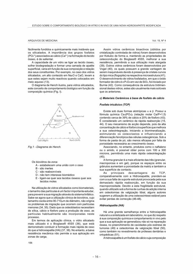
O diagrama de Hench ilustra, para vidros silicatados,este conceito de comportamento biológico em função dacomposição química (Fig. I).
Assim vidros cerâmicos bioactivos (obtidos porcristalização controlada de vidros) foram desenvolvidospor Kokubo de forma a, mantendo as propriedades deosteocondução do Bioglass® 45S5, melhorar a suaresistência, permitindo a sua utilização mais alargada(39). Outros vidros cerâmicos foram desenvolvidos porVogel (40), os quais possuem a grande vantagem deserem maquináveis devido à presença de cristais curvosdo tipo mica (flogopite) na respectiva microestrutura (41).O desenvolvimento de vidros fosfatados, em que o óxidoformador de vidro é o P2O5 em vez de SiO2, foi iniciado porBurnie (42). Como consequência da estrutura tridimen-sional destes vidros, estes são usualmente mais solúveisque os anteriores.
c) Materiais Cerâmicos à base de fosfato de cálcio
Fosfato tricálcico (TCP)
Existe sob duas formas alotrópicas α e β. Possui afórmula química Ca3(PO4)2 (relação molar Ca/P=1,5)contendo cerca de 39% de cálcio e 20% de fósforo (43).É considerado um cerâmico de rápida reabsorção (15,44). O seu mecanismo de acção depende, pois da altaconcentração de cálcio e fósforo à superfície que melhoraa sua osteointegração, iniciando a biomineralização,estimulando os osteoclastos e influenciando adiferenciação fenotípica das células osteogénicas. Sob aforma de pasta de pó, são menos eficazes por falta daporosidade necessária ao crescimento ósseo.
Associando, no entanto, produtos como o naftalenoou o amido, é possível obter poros com 100 a 300microns, permitindo uma mais rápida osteointegração(45).
A forma granular é a mais eficiente das três (granular,macroporosa e em gel), porque os espaços entre osgrânulos aumentam a porosidade da matriz e também asua superficíe de contacto.
As principais desvantagens do TCP,comparativamente com a Hidroxiapatite, prendem-secom a sua falta de suporte estrutural provocada pela suademasiado rápida reabsorção, em função da suamacroporosidade. Devido a esta fragilidade estrutural,quando utilizado sob a forma de cunhas de adição internaem osteotomias de valgização tibial, alguns autoressugerem a utilização de uma osteossíntese estável paraevitar perdas de correcção (46-48).
Hidroxiapatite (HA)
Há uma grande semelhança entre a hidroxiapatitenatural e a sintetizada em laboratório, no que diz respeitoà sua composição química e comportamento in vivo peloque a sua aplicação se generalizou não só na reparaçãoóssea, no preenchimento de cavidades pós exérese detumores (49) e osteotomias de valgização tibial (50),como também no revestimento de próteses dentárias eortopédicas (51).
A hidroxiapatite é um fosfato de cálcio cuja composição
Fig.1 - Diagrama de Hench.
Os biovidros da zona:A - estabelecem uma união com o ossoB - são inertesC - são reabsorvíveisD - não tem interesse biomédicoE - ligam-se quer aos tecidos ósseos quer aos
tecidos moles
Na utilização de vidros silicatados como biomateriais,o tamanho das partículas é um factor importante estudar,para prevenir a sua migração através do sistema linfático.Sabe-se agora que a utilização clínica de biovidros, cujotamanho oscila entre 90-710µm de diâmetro, não originaos problemas de migração que ocorrem com partículasmenores (34, 35). Dado que os osteoblastos necessitamde sílica, cálcio e fósforo para a produção de osso, aspartículas habitualmente são incorporadas nesteprocesso.
Em termos de aplicação clínica, o vidro silicatadomais utilizado é o Bioglass® 45S5 (36), que estádemonstrado conduzir à formação mais rápida de ossodo que a hidroxiapatite (HA) (37, 38). No entanto, a baixaresistência mecânica não permite a sua aplicação emzonas de carga.
– 17 –
CONCEITOS GERAIS SOBRE SUBSTITUTOS ÓSSEOS
Gutierres M et al Substitutos Ósseos
157
é Ca10 (PO4)6(OH)2 (relação molar Ca/P=1,67) pelo queapresenta, ao contrário do TCP, muito lenta reabsorçãoe remodelação óssea, o que leva a que se mantenha noorganismo durante anos (52).
Apresenta uma estrutura favorável à invasão vascu-lar, mas a lentidão de reabsorção e integração assimcomo a dificuldade em manter os grânulos no local dodefeito ósseo, levou à necessidade de criar compósitose pastas com características de biofuncionalidadesuperiores às dos seus constituintes individualmente.
Biocompósitos à base de fosfato de cálcio
Os fosfatos de cálcio bifásicos, são compósitos deHidroxiapatite e TCP. São utilizados geralmente naproporção de dois terços para um terço, com porosidadede 40 até 60%, favorecendo a velocidade e qualidade daosteointegração, a sua reabsorção, mas mantendo umaresistência à compressão que se aproxima, em algunsprodutos, dos 10 MPa (44).
Os autores deste artigo desenvolveram um compósitosintético, recentemente registado como Bonelike®, apartir de hidroxiapatite reforçada com um vidro de modoa introduzir iões importantes na fisiologia do tecido ósseo(53, 54). Este material, constituido por uma matriz dehidroxiapatite e fases α e β-TCP dispersas na suamicroestrutura, tem uma composição semelhante à parteinorgânica do tecido ósseo. O desenvolvimento destematerial teve como objectivo obter um biomaterial commelhor bioactividade (55) e propriedades mecânicas (56)do que os materiais actualmente disponíveiscomercialmente.
Estão em curso diversos trabalhos científicos,envolvendo o Serviço de Ortopedia da Faculdade deMedicina da Universidade do Porto e diversas entidadesde I&D nacionais e estrangeiras, para analisar o seucomportamento biológico quer in vitro (57) quer in vivo(58) não só com animais, como também, desde há 10anos para cá, com células humanas (59-61) e comdoentes (62) nos últimos 3 anos, nos quais este compósitotem sido aplicado. Os resultados têm sido bastantesuperiores em relação à HA convencional, em termos daavaliação da sua capacidade de formação de um novotecido ósseo.
Cimentos de cerâmica injectáveis
Recentemente, novos substitutos sintéticos de fosfatodicálcico-tricálcico foram desenvolvidos (63), de forma apoderem ser aplicados na forma líquida e, ao seremaquecidos à temperatura corporal, sofrerem um processode endurecimento através de uma reacção endotérmica.
São usados com sucesso no tratamento das fracturasdistais do rádio, com o objectivo de prevenir o colapsonos doentes com osteoporose (64).
2. Colagénio
Trata-se da proteína mais abundante da matriz ósseaextracelular. É considerado um bom veículo de transportepara outros enxertos sintéticos, assim como BMP´s ecélulas osteogénicas, porque a sua estrutura éconsiderada um meio adequado para a regeneraçãoóssea (45, 65).
A sua resistência estrutural é mínima e possui o riscopotencial de imunogenicidade.
Estudos efectuados têm no entanto atingido resultadoscontroversos. Por exemplo, em modelos caninos verificou-se que a adição de autoenxerto a um compósito de umcerâmico e colagéneo é biologicamente inferior do que autilização isolada de um volume menor de autoenxerto,na obtenção de fusões vertebrais (66).
Por outro lado, um estudo prospectivo randomizadoefectuado com compósito colagéneo-TCP vs medulaóssea no tratamento de fracturas em ossos longos reveloutaxas de consolidação sem diferenças estatisticamentesignificativas (67).
Um compósito deste tipo, de origem bovina, aprovadopela Food and Drug Administration(FDA) sob adenominação de Collagraft, revelou óptimos resultadosquando associado a células de medula, por exemplo nasartrodeses vertebrais ou preenchimento de cavidadesquísticas (68).
MATERIAIS OSTEOINDUTORES
1. Aloenxerto
É aquele que é proveniente de um dador da mesmaespécie. Actualmente já não é usado sem preparaçãoprévia (fresco), devido à resposta inflamatória quedesencadeia e aos riscos de transmissão de doenças.
Pode ser conservado de 2 formas (15):1. Congelado (“fresh-frozen”) => a -70ºC após umalavagem com antibiótico (a sua utilização pode ir de 1ano, se for mantido a -20ºC, até 5 anos, se a -70ºC)2. Liofilizado (“freeze-dried”) => lavagem dupla comantibiótico, congelação a -70ºC e secagem até ficarcom um conteúdo em água de cerca de 5% (duraçãoilimitada)A congelação reduz a imunogenicidade e consequente
rejeição crónica, mas não elimina o risco de transmissãode HIV (69). Pelo contrário, a liofilização reduz ainda maisa imunogenicidade e elimina o risco de HIV, mas diminuitambém a capacidade de osteoindução e as propriedadesmecânicas do enxerto.
Além dos problemas imunogénicos e infecciosos, osaloenxertos apresentam ainda outras desvantagens comoa sua inexistente capacidade osteogénica, a variabilidadedos resultados clínicos da sua aplicação e, por fim, o seuelevado custo.

ESTUDO SOBRE O COMPORTAMENTO BIOLÓGICO IN VITRO E IN VIVO DE UMA NOVA HIDROXIAPATITE MODIFICADA
– 18 –
ARQUIVOS DE MEDICINA Vol. 19, Nº 4
158
A quantidade disponível, comparativamente com oenxerto autólogo, é sem dúvida uma vantagem em relaçãoa este, assim como o é, a dispensabilidade de umprocedimento cirúrgico adicional no paciente, para a suacolheita.
Por fim, já é possível ter acesso a aloenxerto ósseocom uma grande variedade de formas físicas (gel, pó,fibras, pastas, etc).
Actualmente, está a ser desenvolvido um grandeesforço de investigação, no sentido de melhorar aintegração e remodelação deste tipo de enxerto atravésde modificações físico-químicas. As modificações físicaspassam por criar instrumentos de corte através de laser,por exmplo, de forma a obter formatos especiais, comfurações precisas, seguidos de desmineralização dasuperfície para melhorar a revascularização. Amodificação química dos aloenxertos pode passar aindapela adição de factores de crescimento, BMP-2 e BMP-7, que irão estimular o crescimento ósseo (6).
Em relação ao problema da rejeição crónica, diversaslinhas de investigação tem sido seguidas para melhorara tolerância aos aloenxertos. O estudo da compatibilidadegenética dos “loci” dos antigéneos de histocompatibilidade(HLA) ou de tolerância induzida por curtos períodos deimunossupressão seguidos de transplante de medula oumesmo com o recurso a anticorpos monoclonais são asque parecem mais promissoras (70).
2. Matriz óssea desmineralizada (DBM)
É produzida colocando aloenxerto em meio ácido, oque lhe retira a maior parte da componente mineral, masretém o colagéneo e as proteínas não colagéneas,incluindo factores de crescimento (44).
Se por um lado este processo destrói eventuais vírus,eliminando o risco de transmissão de doenças infecciosas,por outro diminui a sua resistência estrutural, limitando,assim, as suas possíveis aplicações em situações sujeitasa carga em Ortopedia (8).
O seu maior potencial de osteoinduçãocomparativamente ao aloenxerto (17), depende defactores como o tipo de soluções e o tempo usado nadesmineralização, o tamanho das suas partículas e astemperaturas e métodos de esterilização utilizados (71).Alguns estudos animais efectuados recentemente,comparam a eficácia das diferentes DBM comercializadas,em artrodeses de coluna onde são bastante utilizadascomo alternativa ou suplemento do aloenxerto (72, 73).
3. Xenoenxerto
São enxertos provenientes de outras espécies animais(ex: bovinos). Por esse motivo, necessitam de umtratamento antigénico, de deslipidização edesproteinização, que lhe reduz concomitantemente assuas capacidades osteoindutoras (74). Por isso, em
regra, previamente à sua aplicação, o xenoenxerto éimpregnado com células de medula óssea humana.
4. Factores de crescimento ósseo
Urist, em 1965, deu o primeiro passo na descobertados factores de crescimento ósseo. Ao implantarfragmentos ósseos, em posição subcutânea ou intra-muscular, verificou a formação de tecido ósseo,introduzindo então o termo “osteoindução” (75, 76, 77).
Além disso, ao estudarem-se os factoreshomeostáticos que, regulando a acção dos osteoblastose osteoclastos, contribuem para o equilíbrio da massaóssea, conclui-se que estes são de 2 tipos: sistémicos(através de hormonas como a paratormona e a calcitonina)ou locais (através de factores de crescimento). Estes sãopolipeptideos, específicos para determinados tecidos osquais se ligam a receptores de superfície, estimulando aactividade celular) (78). Há pois uma série de processosnos quais os factores de crescimento actuam: crescimentodo esqueleto, consolidação de fracturas, reparação dacartilagem articular e osteoporose.
A matriz óssea contém numerosos factores decrescimento (79), com diversas formas de actuação:
a) BMP (Bone Morphogenic Proteins)A matriz óssea desmineralizada foi o biomaterial de
eleição para isolar, purificar e fazer a clonagem moleculardas BMP´s.
Estas, após fixarem-se à célula menquimatosa com oauxílio da fibronectina, enviam sinais por proteínasespecíficas para o núcleo, levando à expressão de genesque induzem a síntese de macromoléculas envolvidas naformação de cartilagem e osso, originando um condrócitoou osteoblasto (79). Há pois aqui uma cascata daosteogénese com 3 passos: quimiotaxia e mitose,diferenciação em cartilagem e substituição por osso.
O processo de purificação, clonagem genética erecombinação (rh) é complexo pelo que muita investigaçãotem sido efectuada nesta área. Se por um lado estádemonstrada a sua ligação a receptores celularesespecíficos, o pleiotrofismo das BMP´s (capacidade deum simples gene ter uma multiplicidade de acçõesbiológicas) está comprovado inclusivamente no que dizrespeito à regulação da hematopoiese, síntese de matrizextracelular e até da apoptose (morte celular) (80).
Existem mais de 20 tipos de BMP´s, mas as maisactivas são a 2, 4 e 7 (mais conhecida por OP-1, osteo-genic protein-1, e já aprovada pela FDA) (81).
As aplicações clínicas das formas recombinantes,rhBMP-2 e da rhBMP-7, têm sido estudadasexaustivamente, nomeadamente no tratamento defracturas ou pseudartroses (82) e em artrodeses vertebrais(83, 18), muitas vezes com resultados superiores ao doenxerto autólogo. No entanto, esta elevada capacidadede osteoindução é, por alguns autores, consideradaexcessiva.
– 19 –
CONCEITOS GERAIS SOBRE SUBSTITUTOS ÓSSEOS
Gutierres M et al Substitutos Ósseos
159
A investigação dirige-se também para os veículos detransporte destas proteínas, no sentido da sua libertaçãoser controlada, por exemplo com a utilização de partículasde Hidroxiapatite associada a géis reabsorvíveis (84).
b) TGF-Beta (Transforming Growth Factor-Beta)Tem 3 isoformas, sendo a TGF-Beta 1 a mais
abundante no tecido ósseo (85). Trata-se de um factor decrescimento implicado na proliferação, migração,diferenciação e sobrevivência de muitos tipos de células,pelo que tem influência em processos tão diversos comoa embriogénese, angiogénese (86), inflamação (87) ecicatrização (88). Em contraste com as BMP‘s, é incapazde induzir a osteogénese a partir de célulasmesenquimatosas pluripotenciais, mas uma vezdesencadeado o processo, aumenta a pool de célulasosteoprogenitoras por indução da quimiotaxia eproliferação (89).
Diversas linhas de investigação, mostram que estátambém implicado na tumorigénese e invasão tumoral,nomeadamente através de metastização óssea, ao criarcondições locais favoráveis ao crescimento tumoral eangiogénese (90, 91).
O tratamento da osteoporose seria outra hipótese deaplicação clínica, mas que, por falta de um transportadoradequado, continua a ser de difícil implantação.
c) PDGF (Platelet Derived Growth Factor)É um mitogéneo de células mesenquimatosas,
produzido não só por plaquetas, mas também pormacrófagos, monócitos e células endoteliais (78). Actuaexclusivamente na replicação, mas não interfere nasfunções dos osteoblastos (92).
Estudos animais revelaram que uma simples injecçãode PDGF num foco de osteotomia, aumenta a densidadee volume de calo ósseo quando comparado com umgrupo controle (93). Verificou-se também que implantessintéticos tratados com plasma, apresentam umadensidade trabecular aos 6 meses que é superior aquelesnão tratados, sendo esta acção atribuída às PDGF (94)
d) IGF-I e II (Insulin-Like Growth Factor-I e II)Está há muito descrita a forma como o eixo hipotálamo-
hipofisário, actua libertando hormonas de crescimentoque se ligam a receptores específicos da placa decrescimento que libertam IGF-1. Há depois um feedbacknegativo sobre estas glândulas que frena a sua produção(95). Além deste efeito sobre o crescimento, actua tambémna reparação óssea, pelo que alguns investigadorescolocaram a hipótese de as hormonas de crescimentopoderem assim interferir na consolidação de fracturas(96), o que não se veio posteriormente a confirmar (97).
A IGF-2 é dos factores de crescimento maisabundantes do osso. Tem uma grande semelhançaquímica e biológica com a IGF-1, provavelmente porquea sua transdução para a célula alvo ocorre através domesmo receptor de superfície (95). É no entanto menos
potente. Uma linha de investigação interessante surgiucom a descoberta recente de que campos magnéticosexternos são capazes de estimular a produção de IGF-2em culturas de osteoblastos humanos (98).
e) FGF (Fibroblast Growth Factor)Originalmente foram descritos devido à sua actividade
mitogénica sobre os fibroblastos. No entanto, actualmente,são-lhes atribuídas funções de regulação de funçõescelulares tão diversas como mitogénese, diferenciação,produção de proteases e modulação de receptores (99).
Quer a FGF-1 (ácida) quer a FGF-2 (básica), actuamna regulação de células ósseas e cartilagíneas. A injecçãolocal no foco de fractura de um rato de FGF-1, produziuum alargamento significativo do calo ósseo, emborasimultaneamente inibisse a expressão do gene da matrizcartilagínea (98). Em relação à FGF-2, verificou-seimunhistoquimicamente que há um aumento da suaconcentração em redor de uma fractura em ratos sãos,ao contrário do que sucede em diabéticos, e que a suaaplicação nestes, sob a forma de gel de fibrina, restauravao processo de consolidação (100).
A sua acção é, no entanto, complexa pois pode terefeitos antagónicos em função da dose ministrada (101).
Estes problemas levaram a que diversosinvestigadores estudassem a aplicação da terapiagenética para o controle da formação óssea, permitindoassim obter níveis locais mais fisiológicos de factores decrescimento e a sua expressão celular mais prolongada,evitando assim o emprego de doses mais elevadasdestes sob a forma de bolus (102).
CONCLUSÕES
Os avanços científicos na área dos substitutos ósseossão uma realidade.
A nível de materiais osteocondutores, a evoluçãodeu-se com a introdução de técnicas inovadoras napreparação de compósitos HA/TCP com umcomportamento mecânico e biológico mais próximo doosso.
A engenharia de tecidos segue agora a linha da suaassociação com células ósseas com o propósito de lhesconferir o componente osteogénico que lhes falta.
As possibilidades que se abrem para a cirurgiareconstrutiva centram-se também nos avanços dabiomodelação, com a produção de implantes com aforma exacta do defeito ósseo, através do recurso atécnicas avançadas de reconstrução tridimensional.
Com a descoberta da importância da angiogénese edos factores de crescimento nos mecanismos de formaçãode osso, abre-se um universo de novas aplicações clínicasnão só no tratamento de pseudartroses e cirurgiareconstrutiva, mas inclusivamente na osteoporose eosteoartrose.
ESTUDO SOBRE O COMPORTAMENTO BIOLÓGICO IN VITRO E IN VIVO DE UMA NOVA HIDROXIAPATITE MODIFICADA
– 20 –
ARQUIVOS DE MEDICINA Vol. 19, Nº 4
160
REFERÊNCIAS
1 - Laurencin CT. Chapter I- Bone graft and bonegraft substi-tutes a brief history. In: Laurencin CT, editors. Bone graftsubstitute - ASTM International. 2004.
2 - Ratner BD. A history of Biomaterials. In: Ratner, edits.Biomaterials Science. 2nd edition. Elsevier Inc. 2004. pp.10-9.
3 - Williams et al. Conferência de consensos sobre definiçõesem Biomateriais. Chester 1991.
4 - Mainard. Les substituts osseox em 2001: MonographieEditée par GESTO (Association pour L‘etude des Greffes etsubstitutes Tissulaires en Orthopédie) sous la direction deDr. Mainard , Edition Romillat , Paris, 2001.
5 - Banwart JC. Iliac crest bone graft harvest donor sitemorbidity. Spine 1995;20:1055-60.
6 - Tomford WW. Bone allografts : Past, Present and future. Celland Tissue Banking 2000;1:105-9.
7 - Vaccaro AR. The Role of the Osteoconductive Scaffold inSynthetic Bone Graft. Orthopedics 2002; 25 (suppl 5): 571-8.
8 - Khan SN. Clinical applications of Bone Graft substitutes.Orthopedic Clinics of North America 2000;31:389-98.
9 - Nishida K, Gilbertson LG, Evans CH, Kang JD. Potentialapplications of gene therapy to the treatment of spinaldisorders. Spine 2000;25:1308-14.
10 - Proubasta J, Mur JG, Planell JA. Biocompatibilidad,materiales implantables, tipos de implante. In: Fundamentosde Biomecânica y Biomateriales, Ediciones Ergon. Madrid.1997. pp. 271-350.
11 - Davies JE. Histodynamics of endosseous wound healing.In: Davies JE, editors. Bone engineering. Canada: EM2.2000. pp. 1-11.
12 - Anderson. Biological responses to materials. Annu. RevMater Res 2001;31:81-110.
13 - Bauer TW, Muschler GF. Bone Graft Materials. Clin Orthop2000;37:10-27.
14 - Fleming JE Jr, Cornell CN, Muschler GF. Bone cells andmatrices in Orthopedic Tissue Engineering. Orthop ClinNorth Am 2000;31:357-74.
15 - Vaccaro AR, Chiba K, Heller JG. Bone grafting alternativesin spinal surgery. The Spine Journal 2002;2:206-15.
16 - Boden SD. Biology of lumbar spine fusion and use of bonegraft substitutes: Present, future and next generation. Tis-sue Engineering 2000;6:383-99.
17 - Burkus JK, Sandhu HS, Gornet MF, Longley MC. Use of rhBMP-2 in combination with structural control allografts;Clinical and radiographic outcomes in anterior lumbar spinalsurgery. J Bone Joint Surg 2005; 87-A (6); 1205-1212.
18 - Keler EE, Triflett WW. Iliac bone graft: Review of 160consecutive cases.J Oral Maxillofac Surg 1987;45:11-4.
19 - Younger E, Chapmann M. Morbidity at bone graft-donorsites. J Orthop Trauma 1989;3:192-5.
20 - Montgomery DM, Aronson DD, Lee CL, Lamont RL.Posterior spinal fusion: Allograft versus autografts bone. JSpine Disord 1990;3:370-5.
21 - Mc Carthy RE, Peek RD, Morrisy RT, Hough AJ Jr. Allograftbone in spinal fusion for paralytic scoliosis. J Bone JointSurg Am 1986;68: 70-5.
22 - Lane JM, Safdar NK. Bone Grafts of the 20th Century:Multiple purposes, materials and goals. Orthopedics Today.2000.
23 - Connolly J, Guse R, Lippiello L. Development of anosteogenic bone marrow preparation. J Bone Joint Surg Am1989;71:684-91.
24 - Peltier LF. The use of plaster of Paris to fill defects in bone.Clin Orthop 1961;21:1-31.
25 - Hogset O, Bredberg G. Plaster of Paris and hair cellmorphology. A scanning electron microscopic study of analternative implant materials for ear surgery. Acta Otolaryngol1988;106:331-8.
26 - Peltier LF, Jones RH. Treatment of unicameral bone cystsby curetage and packing with plaster of Paris pellets. ClinOrthop 2004;422:145-7.
27 - Hench LL, Best S. Ceramic, glasses and glass ceramics. In:Ratner BD, editors. Biomaterial sciences.2nd Edition. 2004.pp.155-70.
28 - Hench LL, Wilson J. Surface active biomaterials. Science1984;226:630-6.
29 - Boyan BD, Nasatzky E, Keller TA, Schartz Z. Substitutos delinjerto osseo. Current Opinion in Orthopedics 1998;III:17-23.
30 - Clark AE, Hench LL, Paschall HA. The influence of surfacechemistry on implant interface histology : A theorical basisfor implant materials selection. J Biomed Mater Res1976;110:161-71.
31 - Hench LL, Wilson J. Bioactive Glasses: Present andFuture. Bioceramics 1998;11:31-6.
32 - Gatti AM, Zaffe D. Bioactive glasses and chemical bond.Biomaterials 1992:97-106.
33 - Hamadouch M, Sedel L. Ceramics in Orthopaedics J BoneJoint Surg Br 2000; 82-B:1095-9.
34 - Oonishi H, Hench LL, Wilson I. Quantitative comparison ofbone growth behavior in ceramics of Bioglass ®, A-W glassceramics and HA. J Biomed Mater Res 2000;51:37-46.
35 - Oonishi H, Kushitani S, Yasukawa E, et al. Particulatebioglass compared with hydroxyapatite as a bone graftsubstitute. Clin Orthop Rel Research 1997;334:316-25.
36 - Kokubo T, Ito S, Huang Z. Ca-P - Rich Layer formed on highstrength bioactive glass ceramics A-W. J Biomed Mater Res1990;24:331-43.
37 - Vogel W, Holland H, Gummel J. Development of machine-able bioactive glass-ceramics for medical uses. J of Non-Crystaline Solids 1986;80:34-51.
38 - Holland W, Vogel W, Mortier WJ, Duvingneaud PH, NaessensG, Plumat E. A new type of crystal in machineable glass-ceramics. Glass Technology 1983;24:318-22.
39 - Burnie J. Controlled release glass (C. R. G.) - A newbiomaterial. PhD Thesis. 1988.
40 - Driessens FCM, Ramselaar MMA. Chemical reactions ofcalcium phosphate implants after implantation in vivo. JMater Sci 1992;3:413-7.
41 - Betz RR. Limitation of autograft and allograft new synthesissolutions. Orthopedics 2002 (suppl).
42 - Cornell CN. Osteoconductive materials and their role assubstitutes for autogenous bone grafts. Orthop Clin NorthAm 1999;30:599-8.
43 - Lascart T, Favard L, Burdin P, Traore O. Utilisation duphosphate tricalcique dans les osteotomies tibiales devalgisation par addition interne. Ann Orthopediques de L’Ouest 1998;30:137-41.
44 - Meynet J. Osteotomie tibiale de valgisation par ouvertureinterne: place des substituts osseux. Ann Orthopediques deL’Ouest 1998;30:171-3.
45 - Gaasbeek R, Toonen H, Heerwaarden R, Buma P. Mecha-nism of bone incorporation of β-TCP bone substitute in openwedge tibial osteotomy in patients. Biomat 2005;26;33:6713-9.
– 21 –
CONCEITOS GERAIS SOBRE SUBSTITUTOS ÓSSEOS
Gutierres M et al Substitutos Ósseos
161
46 - Yamamoto T, Onga T, Marui T, Mizuno K. Use of Hydroxya-patite to fill cavities after excision of begin bone tumors:clinical results. J Bone Joint Surg 2000;82-B:1117-20.
47 - Koshino T, Murase T, Saito T. Medial opening-wedge hightibial osteotomy with use of porous hydroxyapatite to treatmedial compartment osteoarthritis of the knee. J Bone JointSurg Am 2003; 85: 78-85.
48 - Regner L, Carlsson L, Karrholm J, Herbert P. Ceramiccoating improves tibial component fixation in total kneearthroplasty. J Arthroplasty 1998; 13: 882-889.
49 - Tadic D, Epple M. A thorough physicochemicalcharacterisation of 14 calcium phosphate-based bone sub-stitution materials in comparison to natural bone . Biomaterials2004; 25: 987-994.
50 - Santos JD, Hastings GW, Knowles JC. Sintered hydroxya-patite compositions and method for the preparation thereof,European Patent WO 0068164. 1999.
51 - Lopes MA. Glass reinforced hydroxyapatite composites:structure, physico chemical characterization and biologicalperformance. PhD Thesis. FEUP. 1999.
52 - Lopes MA, Santos JD, Monteiro FJ, Ohtsuki C, Osaka A ,Kaneko S, Inove H. Osteocompatibility and in vivo evalu-ation of glass reinforced hydroxyapatite composite.Bioceramics 1999;12:421-4.
53 - Lopes MA, Monteiro FJ, Santos JD. Glass reinforcedhydroxyapatite composites: secondary phase proportionsand densification effects on bioactive bending strength. JBiomed Mater Res 1999;48:734-40.
54 - Santos JD, Vasconcelos M, Reis RL, Afonso A, MonteiroFJ, Hastings GH. Glass reinforced hydroxyapatite compos-ites: physical properties and preliminary histological stud-ies in rabbits. Bioceramics 1994;7:243-8.
55 - Afonso A, Santos JD, Vasconcelos M, Branco R, CavalheiroJ. Granules of osteoapatite and glass-reinforced hydroxya-patite implanted in rabbit tibia. J Mat Scien: Materials inMedicine 1996;7:507-10.
56 - Costa MA, Gutierres M, Almeida L, Lopes MA, Santos JD,Fernandes MH. In vitro mineralization of human bone mar-row cells cultured on Bonelike® . Key Engineering Materials2004;254-256:821-4.
57 - Ferraz MP, Fernandes MH , Cabral T, Santos JD,Monteiro, FJ. In vitro growth and differentiation of osteo-blast-like human bone marrow cells on glass reinforcedhydroxyapatite plasma-sprayed coatings. J Mat Scien: Ma-terials in Medicine 1999;10:567-76.
58 - Ferraz MP, Knowles JC, Olsen I, Monteiro FJ, Santos JD.Flow cytometry analysis of the effects of pre-immersion onthe biocompatibility of glass-reinforced hydroxyapatiteplasma-sprayed coatings. Biomat 2000;21:813-20.
59 - Gutierres M, Hussain NS, Afonso A, et al. Biologicalbehaviour of bonelike® graft implanted in the tibia of hu-mans. Key Engineering Materials 2005;284-286:1041-4.
60 - Knnack D, Goad M, Rey C, Tofigni A, Chakravarthy P, LeeD. Resorbable calcium phosphate bone substitute. J BiomedMater Res 1998;43:399-409.
61 - Kopylou P, Jonsson K, Thorngren KG. Injectable calciumphosphate in the treatment of distal radius fractures. J HandSurg Br 1996;21:768-71.
62 - Schreiber RE, Blease K, Ambrosio A, Amburn E, SosnowskiB, Sampath TK. Bone induction by ADBMP-2/collagenimplants. J Bone Joint Surg Am 2005;87:1059-68.
63 - Muschler GF, Negami S, Hyodo A , Gaisser D, Gasley K ,Kambic H. Evaluation of collagen ceramic composite graftmaterials in a spinal fusion model. Clin Orthop 1996;328:250-60.
64 - Chapman MW, Bucholz R, Cornell C. Treatment of acutefractures with a collagen calcium phosphate graft material.A Randomized clinical trial. J Bone Joint Surg Am1997;79:495-502.
65 - Hollinger JO, Brekke J, Gruskin E , Lee DL. Role of bonesubstitutes. Clin Orthop 1996;324:55-65.
66 - Kelly EB. New frontiers in bone grafting. Orthop TechnReview 2000;2:1-5.
67 - Mathes DW, Randolph MA, Lee WP. Strategies for toler-ance induction to composite tissue allografts. Microsurgery2000;20:448-52.
68 - Boyce T, Edwards J, Scarborough N. Allograft bone: Theinfluence of processing on safety and performance. OrthopClin North Am 1999;30:571-81.
69 - Peterson B, Whang PG, Iglesias R , Wang JC, LiebermanJR.Osteoinductivity of commercially available demineral-ized bone matrix.J Bone Joint Surg Am 2004;86:2243-50.
70 -Morone MA, Boden SD. Experimental posterolateral lumbarspinal fusion with a demineralized bone matrix cell. Spine1998;23:159-67.
71 - Elves MW, Salama R. A study of the development ofcytotoxic antibodies produced in recipients of xenografts ofiliac bone. J Bone joint Surg Br 1974;56:331-9.
72 - Urist MR. Bone: formation by autoinduction. Science1965;150:893-9.
73 - Yuan H, Zou P, Yang Z, Zhang X, De Bruijn JD, Groot KD.Bone morphogenic protein and ceramic-induced osteogen-esis. J Mat Scien: Materials in Medicine 1998;9:717-21.
74 - Reddi AH, Marshall R. Urist: A renaissance scientist andorthopedic surgeon. J Bone Joint Surgery Am 2003;85:3-7.
75 - Khan SN, Bostrom MP, Lane J. Bone growth factors.Orthopedic Clinics of North America 2000;31:375-87.
76 - Reddi AH. Bone morphogenetic proteins: From basicscience to clinical applications. J Bone Joint Surg Am 2001;83-A (suppl1) (PT1): S1-6.
77 - Sugi Y, Yamamura H, Okagawa H, Markwald RR. Bonemorphogenetic protein-2 can mediate myocardial regula-tion atrioventricular cushion mesenchymal cell formation inmice. Developmental Biology 2004; 269:505-18.
78 - Asahina I, Sampath TK, Nishimura I, Hauschka PV. Humanosteogenic protein-1 induces both chondroblastic and os-teoblastic differentiation of osteoprogenitor cells derivedfrom newborn rat calvaria. J Cell Biol 1993;123:921-33.
79 - Friedlander GE, Perry CR, Cole JD, et al. Osteotomicprotein-1( BMP-7) in the treatment of tibial nonunions. JBone Joint Surg Am 2001;83-A (suppl.1) (PT2): S151-8.
80 - Boden SD, Zdeblick TA, Sandhu HS, Heim SE. The useof Rh BMP-2 in interbody fusion cases. Definitive evidanceof osteoinduction in humans: A preliminary report. Spine2000; 25: 376-86.
81 - Matsumoto T, Okazaki M, Inove M, et al. Hydroxyapatiteparticles as a controlled release carrier of protein. Biomat2004;25:3807-12.
82 - Janssens K, Dijke P, Janssens S, Hul WV. Transforminggrowth factor-beta1 to the bone. Endoc Rev 2005: 26:743-74.
83 - Pepper MS.Transforming growth factor-beta: vasculo-genesis, angiogensis and vessel wall integrity. CytokineGrowth Factor Rev 1997;8:21-43.
84 - Letterio JJ, Roberts AB. Regulation of immune responsesby TGF-beta. Annu Rev Immunol 1998;16:137-61.
85 - O´Kane S , Ferguson MW. Transforming growth factor betaand wound healing. Int J Biochem Cell Biol 1997;29:63-78.
ESTUDO SOBRE O COMPORTAMENTO BIOLÓGICO IN VITRO E IN VIVO DE UMA NOVA HIDROXIAPATITE MODIFICADA
– 22 –
ARQUIVOS DE MEDICINA Vol. 19, Nº 4
162
86 - Aufdemorte TB, Fox WC, Holt GR, McGuff HS, AmmannAJ, Beck LS. An intraosseous device for studies of bone-healing. The effect of transforming growth factor beta. JBone Joint Surg Am 74:1153-61.
87 - Siegel PM, Massague J. Cytostatic and apoptotic actionsof TGF-beta in homeostasis and cancer. Nat Rev Cancer2003;3:807-21.
88 - Derynck R, Akhurst RJ, Balmain A. TGF-beta signaling intumor suppression and cancer progression. Nat Genet2001;29:117-29.
89 - Hock JM, Canalis E. Platelet derived growth factorenhances bone cell replication, but not differentiate functionof osteoblasts. Endocrinol 1994;134:1423-8.
90 - Nash TJ, Howlett CR, Martin C, Steel J, Johnson KA,Hicklin DJ. Effect of platelet-derived growth factors on tibialosteotomies in rabbits. Bone 1994; 5:203-8.
91 - Marx RE, Carlson ER, Eichstaedt RM. Platelet-rich plasma:growth factor enhancement for bone grafts. Oral Surg1998;85:638-46.
92 - Trippel SB, Coutts RD, Einhorn TA, Mundy GR, RosenfeldRG.Instructional course lectures, the American Academy ofOrthopaedic Surgeons - growth factors as therapeuticagents. J Bone Joint Surg Am 1996;78:1272-86.
93 - Koskinen EV, Lindholm RV, Nieminen RA, Puranen JP,Attila U. Human growth hormone in delayed union and non-union of fractures. Internat Orthop 1978;1:317-22.
94 - Northmore-Ball MD, Wood MR. Meegitt BF. A biomechani-cal study of the effects of growth hormone in experimentalfracture healing. J Bone Joint Surg Br 1980;62:391-6.
95 - Ryaby JT, Fitzsimmons RJ, Khin NA, et al. A growth factordependent model for magnetic field regeneration of boneformation. Trans Orthop Res Soc 1994;19:518.
Correspondência:Dr. Manuel GutierresServiço de OrtopediaHospital de São JoãoAlameda Prof. Hernâni Monteiro4200-319 Porto
e-mail: [email protected]
96 - Jin Gushi S, Heydmann A, Kana SK, Macey LR, BolanderME. Acidic fibroblast growth factor (aFGF) injection stimu-lates cartilage enlargement and inhibits cartilage gene ex-pression in rat fracture healing. J Orthop Res 1990;8:364-71.
97 - Kawaguchi H, Kurokawa T, Hanada K, et al. Stimulationof fracture repair by recombinant human basic fibroblastgrowth factor in normal and streptozotocin - diabetic rats.Endocrinol 1994;135:774-81.
98 - Wang JS, Aspenberg P. Basic fibroblast growth factor andbone induction in rats. Acta Orthop Scandinavia 1993;64:551-69.
99 - Ludwig SC, Boden SD. Osteoinductive bone graft substi-tutes for spinal fusion. Orthop Clin North Am 1999;30:635-45.
100 - Hench LL, Wilson J, edits. An introduction to Bioceramics.Advanced series in Ceramics 1993;1.
101 - Kingery WD, Bowen HK, Uhlmann DR. Introduction toceramics. 2nd ed. New York: John Wiley & Sons. 1976.
102 - Schneider SJ, edits. Ceramic and Glasses. EngineeredMaterials Handbook. ASM International 1991;4.
CAPÍTULO III
ESTUDOS DE BIOCOMPATIBILIDADE IN VITRO
– 23 –

PUBLICAÇÃO II
“In vitro mineralisation of human bone marrow cells cultured on Bonelike®”
KEY ENGINEERING MATERIALS (2004), VOLS 254-256: 821-824
– 25 –

– 27 –
ESTUDOS DE BIOCOMPATIBILIDADE IN VITRO
In Vitro Mineralisation of Human Bone Marrow Cells Cultured on Bonelike
M. A. Costa1, M. Gutierres2, L. Almeida2, M. A. Lopes3,4, J. D. Santos3,4, M. H. Fernandes5
1ICBAS - Instituto de Ciências Biomédicas de Abel Salazar da Universidade do Porto, Largo Abel Salazar, 4000 Porto, Portugal
2Serviço de Ortopedia do Hospital de S. João, Largo Hernáni Monteiro, 4200 Porto, Portugal 3FEUP - Faculdade de Engenharia da Universidade do Porto, DEMM, Rua Dr Roberto Frias,
4200 Porto, Portugal 4INEB - Instituto de Engenharia Biomédica, Laboratório de Biomateriais, Rua do Campo Alegre
823, 4150 Porto, Portugal 5FMDUP - Faculdade de Medicina Dentária da Universidade do Porto, Rua Dr Manuel Pereira da
Silva, 4200 Porto, Portugal, [email protected]
Keywords: Bonelike, human bone marrow cells, in vitro mineralisation
Abstract. Bonelike is a CaO-P2O5 based glass-reinforced hydroxyapatite (HA) designed to mimic the inorganic composition of the bone tissue. This work evaluates the response of human bone marrow cells to Bonelike concerning cell proliferation and osteoblast differentiation. HA was used as control material. Results showed that Bonelike allowed the proliferation of bone marrow cells and their complete differentiation, as evidenced by the formation of cell-mediated mineralisation. In comparison with HA, Bonelike had a positive effect on the expression of alkaline phosphatase and also on the formation of a mineralised matrix, two osteoblast markers.
Introduction
Bonelike is a synthetic hydroxyapatite (HA) that is sintered in the presence of CaO-P2O5-based glasses using a patented process [1]. This synthetic bone graft was designed to improve the mechanical properties of calcium phosphate ceramics and mimic the inorganic composition of bone tissue. The physicochemical and mechanical behaviour of Bonelike have been extensively reported in literature [1-3].
Previous in vitro biological studies showed that glass-reinforced HA composites allow the proliferation of MG63 osteoblast-like cells and human bone marrow cells and the expression of osteoblast markers [4-6]. Also, in vivo studies performed in a rabbit model demonstrated that Bonelike composites induced earlier new bone formation around implants than HA [7].
Recently, a composite prepared by the addition (4 wt %) of a glass with the composition of 65P2O5-15CaO-10CaF2-10Na2O (in % mol) to HA was subject of clinical trials in implantology and maxillofacial surgery [8]. This study demonstrated extensive new bone formation around implanted granules and continuous replacement by new bone. Osteoblasts are the cells responsible for the formation of the bone tissue at the bone/material interface and the present work evaluates the response of human bone marrow cells to Bonelike composite, with the same chemical composition, concerning cell proliferation and osteoblast differentiation. HA was used as control material.
ESTUDO SOBRE O COMPORTAMENTO BIOLÓGICO IN VITRO E IN VIVO DE UMA NOVA HIDROXIAPATITE MODIFICADA
– 28 –
Materials and methods
For the preparation of Bonelike, a P2O5-based glass with the chemical composition of 65P2O5-15CaO-10CaF2-10Na2O, in mol %, was prepared from reagent grade chemicals by conventional techniques. The glass was then milled and added to HA powder in a proportion of 4.0 wt % using methanol as solvent medium. Disc samples were then prepared by uniaxial pressing at 288 MPa and sintered at 1300 ºC for 1h followed by natural cooling inside the furnace. Detailed description of Bonelike preparation was previously reported [1]. For cell culture studies, discs were polished down to 1 m, ultrasonically cleaned and sterilised before testing. HA samples were also prepared and used as control material. Human bone marrow cells (first subculture) were cultured on the surface of Bonelike and HA material samples for 28 days in -MEM, 10 % foetal bovine serum, 50 g/ml gentamicin and 2.5 g/ml fungizone, and supplemented with 50 g/ml ascorbic acid, 10 mM -glycerophosphate and 10 nM dexamethasone, experimental conditions that favour the complete expression of the osteoblast phenotype [9]. As reference control, cells were cultured in parallel on standard tissue culture plastic plates. Material samples were also incubated in the absence of cells in the same experimental conditions as those of the seeded materials. Control cultures and seeded materials were characterised at days 1, 3, 7, 14, 21 and 28 for cell viability/proliferation (MTT assay), total protein content and alkaline phosphatase (ALP) activity, using procedures previously described in detail [6,7,10,11]. Control and seeded materials were observed by scanning electron microscopy (SEM). The in vitro biological studies were performed at the Faculdade de Medicina Dentária da Universidade do Porto.
Results and discussion
Biochemical evaluation of the seeded Bonelike showed that human bone marrow cells proliferated during approximately three weeks and attained a stationary phase afterwards, as shown by the results concerning the MTT reduction and total protein content (Figs. 1 A and B). Cells expressed ALP, especially during the second and third weeks of culture (Fig. 1C). Seeded HA presented a similar behaviour concerning cell growth but lower (20 to 30 %) ALP activity than that measured on seeded Bonelike.
*
* *
0.0
0.3
0.6
0.9
1.2
0 7 14 21 28Days
A (
=600
nm
)
*
*
*
0
50
100
150
200
0 7 14 21 28
Days
Prot
ein
Con
tent
( g/
cm2 )
*'*' *
0.0
0.5
1.0
1.5
2.0
2.5
0 7 14 21 28
Days
AL
P (n
mol
/min
.cm
2 )
Fig.1. Cell viability/proliferation (A), total protein content (B) and alkaline phosphatase (ALP) activity (C) of human bone marrow cells grown on Bonelike () and HA ( ). *Significantly different from cultures performed on standard tissue culture plates (); Significantly different from cultures performed on seeded Bonelike.
A B C
Bioceramics 16822
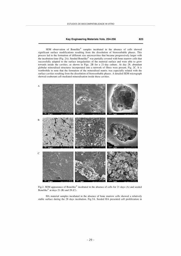
– 29 –
ESTUDOS DE BIOCOMPATIBILIDADE IN VITRO
SEM observation of Bonelike samples incubated in the absence of cells showed significant surface modifications resulting from the dissolution of bioresorbable phases. This process led to the formation of different size microcavities that became progressively larger with the incubation time (Fig. 2A). Seeded Bonelike was partially covered with bone marrow cells that successfully adapted to the surface irregularities of the material surface and were able to grow towards inside the cavities, as shown in Figs. 2B for a 21-day culture. At day 28, abundant globular mineralised structures incorporated into a network of fibres were present, Fig. 2C. It is worthwhile to note that the formation of the mineralised matrix was especially related with the surface cavities resulting from the dissolution of bioresorbable phases. A detailed SEM micrograph showed exuberant cell-mediated mineralisation inside these cavities.
Fig.2. SEM appearance of Bonelike incubated in the absence of cells for 21 days (A) and seeded Bonelike at days 21 (B) and 28 (C).
HA material samples incubated in the absence of bone marrow cells showed a relatively stable surface during the 28 days incubation, Fig.3A. Seeded HA presented cell proliferation in
A
B
C
60 m 2 m 1 m
3 m 1 m
60 m 3 m 1 m
60 m
Key Engineering Materials Vols. 254-256 823
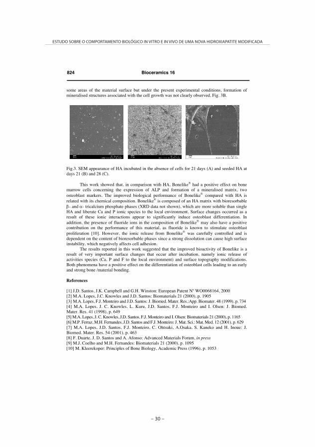
ESTUDO SOBRE O COMPORTAMENTO BIOLÓGICO IN VITRO E IN VIVO DE UMA NOVA HIDROXIAPATITE MODIFICADA
– 30 –
some areas of the material surface but under the present experimental conditions, formation of mineralised structures associated with the cell growth was not clearly observed, Fig. 3B.
Fig.3. SEM appearance of HA incubated in the absence of cells for 21 days (A) and seeded HA at days 21 (B) and 28 (C).
This work showed that, in comparison with HA, Bonelike had a positive effect on bone marrow cells concerning the expression of ALP and formation of a mineralised matrix, two osteoblast markers. The improved biological performance of Bonelike compared with HA is related with its chemical composition. Bonelike is composed of an HA matrix with bioresorbable - and - tricalcium phosphate phases (XRD data not shown), which are more soluble than single HA and liberate Ca and P ionic species to the local environment. Surface changes occurred as a result of these ionic interactions appear to significantly induce osteoblast differentiation. In addition, the presence of fluoride ions in the composition of Bonelike may also have a positive contribution on the performance of this material, as fluoride is known to stimulate osteoblast proliferation [10]. However, the ionic release from Bonelike was carefully controlled and is dependent on the content of bioresorbable phases since a strong dissolution can cause high surface instability, which negatively affects cell adhesion. The results reported in this work suggested that the improved bioactivity of Bonelike is a result of very important surface changes that occur after incubation, namely ionic release of activities species (Ca, P and F to the local environment) and surface topography modifications. Both phenomena have a positive effect on the differentiation of osteoblast cells leading to an early and strong bone /material bonding.
References
[1] J.D. Santos, J.K. Campbell and G.H. Winston: European Patent Nº WO0068164, 2000 [2] M.A. Lopes, J.C. Knowles and J.D. Santos: Biomaterials 21 (2000), p. 1905 [3] M.A. Lopes, F.J. Monteiro and J.D. Santos: J. Biomed. Mater. Res.:App. Biomater. 48 (1999), p. 734 [4] M.A. Lopes, J. C. Knowles, L. Kuru, J.D. Santos, F.J. Monteiro and I. Olsen: J. Biomed. Mater. Res. 41 (1998), p. 649 [5] M.A. Lopes, J. C. Knowles, J.D. Santos, F.J. Monteiro and I. Olsen: Biomaterials 21 (2000), p. 1165 [6] M.P. Ferraz, M.H. Fernandes, J.D. Santos and F.J. Monteiro: J. Mat. Sci.: Mat. Med. 12 (2001), p. 629 [7] M.A. Lopes, J.D. Santos, F.J. Monteiro, C. Ohtsuki, A.Osaka, S. Kaneko and H. Inoue: J. Biomed. Mater. Res. 54 (2001), p. 463 [8] F. Duarte, J. D. Santos and A. Afonso: Advanced Materials Forum, in press[9] M.J. Coelho and M.H. Fernandes: Biomaterials 21 (2000), p. 1095 [10] M. Kleerekoper: Principles of Bone Biology, Academic Press (1996), p. 1053
2 m
60 m
Bioceramics 16824
CAPÍTULO IV
ESTUDOS DE BIOCOMPATIBILIDADE IN VIVO
– 31 –

PUBLICAÇÃO III
“Biological behaviour of Bonelike® graft implanted in the tibia of humans”
KEY ENGINEERING MATERIALS (2005), VOLS 284-286: 1041-1044
– 33 –

– 35 –
ESTUDOS DE BIOCOMPATIBILIDADE IN VIVO
Biological Behaviour of Bonelike® Graft Implanted in the Tibia of Humans
M. Gutierres1,a, N. Sooraj Hussain2,3,b, A. Afonso4,c ,
L. Almeida1,d T. Cabral1,e, M.A. Lopes2,3,f and J.D. Santos2,3,g
1FMUP – Faculdade de Medicina da Universidade do Porto - Hospital de São João, Largo Hernâni Monteiro, 4200 Porto - Portugal
2DEMM, Faculty of Engg., University of Porto, Rua Dr.Roberto Frias, 4200-465 Porto, Portugal 3INEB-Instituto de Engenharia Biomédica, Laboratorio de Biomaterias, Rua Campo Alegre,823,
4150-180,Porto, Portugal. 4FMDUP, Faculdade de Medicina Dentária da Universidade do Porto, Rua Dr. Manuel Pereira da
Silva, 4200-393 Porto,Portugal
Keywords: Biological behavior, Bonelike®
graft, clinical trials
Abstract. This paper reports the ability of Bonelike®
to regenerate bone defected areas when
implanted in the tibia of 3 patients (average age of 59 years) during a high tibial osteotomy (HTO)
to treat medial compartment osteoarthritis of the knee. Bonelike®
is a synthetic bone graft designed
to mimic the inorganic composition of bone using a patent process that consists of liquid sintering
hydroxyapatite in the presence of CaO-P2O5 based glass. The preliminary clinical evaluation
performed showed that an intimate contact between new formed bone and Bonelike®
was
established (67±10 %), with no fibrous interface. Therefore, Bonelike®
is a single-handedly viable
osteoconductive synthetic grafting material as it has been demonstrated from the profiles of
radiological, histological and scanning electron microscopy analyses for the six months
implantation period.
Introduction
For decades, calcium phosphate ceramics have received a great attention in the clinical field such as
orthopaedic and dental materials because of their good ability to bond to living tissues. A wide
variety of bone graft substitutes have been reported for the repair and augmentation of bone areas
[1,2]. Among, the different bone defect regenerative techniques, when biomechanical strength is
not crucial, fresh autologuos cancellous graft is still considered the golden standard, when
evaluating bone graft materials. However, the morbidity rate and additional surgical time
associated with its collection, as well as the limited supply in each patient have been stimulated for
the search of substitutes. Literature [3] reports an overview of the basic concepts of bone grafting
and discusses the most commonly used bone-graft substitutes and their potential advantages and
disadvantages. To surpass these limitations, the use of allogeneic bone and biological processed
scaffolds such as demineralized bone matrix have been used. However, due to health and safety
issues concerns related to the effectiveness of processing on immunogenicity, disease transmission
and shortage of supply, the use of synthetic bone graft materials has been increasing steadily in the
last two decades. A wide variety of commercial synthetic forms of bone grafts are available in the
market made of hydroxyapatite (HA) and tricalcium phosphate (TCP). Over the past decade, the
authors have developed glass-reinforced hydroxyapatite (GR-HA) composites, recently registered
as Bonelike®
, whose chemical composition resembles that of inorganic bone tissue [4]. This novel
material, as result of its controlled chemical phase composition of HA, α- and β-TCP and its
microstructure showed to have better mechanical properties than the actual commercial bone grafts.
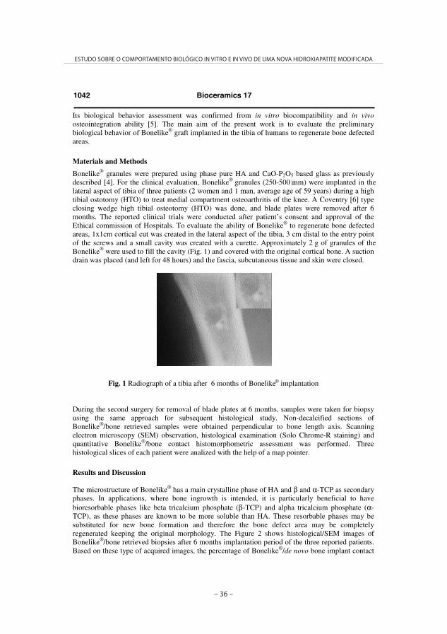
ESTUDO SOBRE O COMPORTAMENTO BIOLÓGICO IN VITRO E IN VIVO DE UMA NOVA HIDROXIAPATITE MODIFICADA
– 36 –
Its biological behavior assessment was confirmed from in vitro biocompatibility and in vivo
osteointegration ability [5]. The main aim of the present work is to evaluate the preliminary
biological behavior of Bonelike®
graft implanted in the tibia of humans to regenerate bone defected
areas.
Materials and Methods
Bonelike®
granules were prepared using phase pure HA and CaO-P2O5 based glass as previously
described [4]. For the clinical evaluation, Bonelike®
lateral aspect of tibia of three patients (2 women and 1 man, average age of 59 years) during a high
tibial ostotomy (HTO) to treat medial compartment osteoarthritis of the knee. A Coventry [6] type
closing wedge high tibial osteotomy (HTO) was done, and blade plates were removed after 6
months. The reported clinical trials were conducted after patient’s consent and approval of the
Ethical commission of Hospitals. To evaluate the ability of Bonelike®
to regenerate bone defected
areas, 1x1cm cortical cut was created in the lateral aspect of the tibia, 3 cm distal to the entry point
Bonelike®
were used to fill the cavity (Fig. 1) and covered with the original cortical bone. A suction
drain was placed (and left for 48 hours) and the fascia, subcutaneous tissue and skin were closed.
® implantation
During the second surgery for removal of blade plates at 6 months, samples were taken for biopsy
using the same approach for subsequent histological study. Non-decalcified sections of
Bonelike®
/bone retrieved samples were obtained perpendicular to bone length axis. Scanning
electron microscopy (SEM) observation, histological examination (Solo Chrome-R staining) and
quantitative Bonelike®
/bone contact histomorphometric assessment was performed. Three
histological slices of each patient were analized with the help of a map pointer.
Results and Discussion
The microstructure of Bonelike®
has a main crystalline phase of HA and β and α-TCP as secondary
phases. In applications, where bone ingrowth is intended, it is particularly beneficial to have
TCP), as these phases are known to be more soluble than HA. These resorbable phases may be
substituted for new bone formation and therefore the bone defect area may be completely
regenerated keeping the original morphology. The Figure 2 shows histological/SEM images of
Bonelike®
/bone retrieved biopsies after 6 months implantation period of the three reported patients.
Based on these type of acquired images, the percentage of Bonelike®
/de novo bone implant contact
Bioceramics 171042
����
Fig. 1 Radiograph of a tibia after 6 months of Bonelike
��
of the screws and a small cavity was created with a curette. Approximately 2 g of granules of the
bioresorbable phases like beta tricalcium phosphate (β-TCP) and alpha tricalcium phosphate (α-
granules (250-500 �mm) were implanted in the
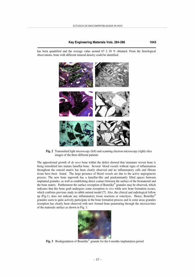
– 37 –
ESTUDOS DE BIOCOMPATIBILIDADE IN VIVO
has been quantified and the average value around 67 ± 10 % obtained. From the histological
observations, bone with different mineral density could be identified.
Fig. 2 Transmitted light microscopy (left) and scanning electron microscopy (right) slice
images of the three different patients
The appositional growth of de novo bone within the defect showed that immature woven bone is
being remodeled into mature lamellar bone. Several blood vessels without signs of inflammation
throughout the osteoid matrix has been clearly observed and no inflammatory cells and fibrous
tissue have been found. The large presence of blood vessels are due to the active angiogenesis
process. The new bone ingrowth has a lamellar-like and predominantly filled spaces between
implanted granules, as well as establishing direct contact between the surface of the biomaterial and
the bone matrix. Furthermore the surface resorption of Bonelike®
granules may be observed, which
indicates that this bone graft undergoes some resorption in vivo while new bone formation occurs,
which confirms previous study in rabbit animal model [7]. Also, the clinical and radiological follow
up (Fig.1), does not indicate any inflammatory tissue reactions or osteolysis. Hence, Bonelike®
granules seem to quite actively participate in the bone formation process and in some areas granules
resorption has clearly been observed with new formed bone penetrating through the microcavities
of the materials surface as shown in Fig. 3.
Fig. 3 Biodegradation of Bonelike
Key Engineering Materials Vols. 284-286 1043
granule for the 6 months implantation period
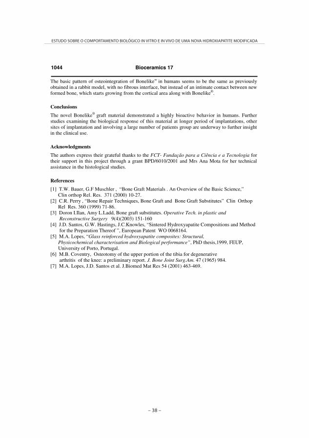
ESTUDO SOBRE O COMPORTAMENTO BIOLÓGICO IN VITRO E IN VIVO DE UMA NOVA HIDROXIAPATITE MODIFICADA
– 38 –
The basic pattern of osteointegration of Bonelike®
in humans seems to be the same as previously
obtained in a rabbit model, with no fibrous interface, but instead of an intimate contact between new
formed bone, which starts growing from the cortical area along with Bonelike®
.
Conclusions
The novel Bonelike®
graft material demonstrated a highly bioactive behavior in humans. Further
studies examining the biological response of this material at longer period of implantations, other
sites of implantation and involving a large number of patients group are underway to further insight
in the clinical use.
Acknowledgments
The authors express their grateful thanks to the FCT- Fundação para a Ciência e a Tecnologia for
their support in this project through a grant BPD/6010/2001 and Mrs Ana Mota for her technical
assistance in the histological studies.
References
[1] T.W. Bauer, G.F Muschler , “Bone Graft Materials . An Overview of the Basic Science,”
Clin orthop Rel. Res. 371 (2000) 10-27.
[2] C.R. Perry , “Bone Repair Techniques, Bone Graft and Bone Graft Substitutes” Clin Orthop
Rel Res. 360 (1999) 71-86.
[3] Doron I.Ilan, Amy L.Ladd, Bone graft substitutes. Operative Tech. in plastic and
Reconstructive Surgery 9(4)(2003) 151-160
[4] J.D. Santos, G.W. Hastings, J.C.Knowles, “Sintered Hydroxyapatite Compositions and Method
for the Preparation Thereof ”, European Patent WO 0068164.
[5] M.A. Lopes, “Glass reinforced hydroxyapatite composites: Structural,
Physicochemical characterisation and Biological performance”, PhD thesis,1999, FEUP,
University of Porto, Portugal.
[6] M.B. Coventry, Osteotomy of the upper portion of the tibia for degenerative
arthritis of the knee: a preliminary report. J. Bone Joint Surg.Am. 47 (1965) 984.
[7] M.A. Lopes, J.D. Santos et al. J.Biomed Mat Res 54 (2001) 463-469.
Bioceramics 171044
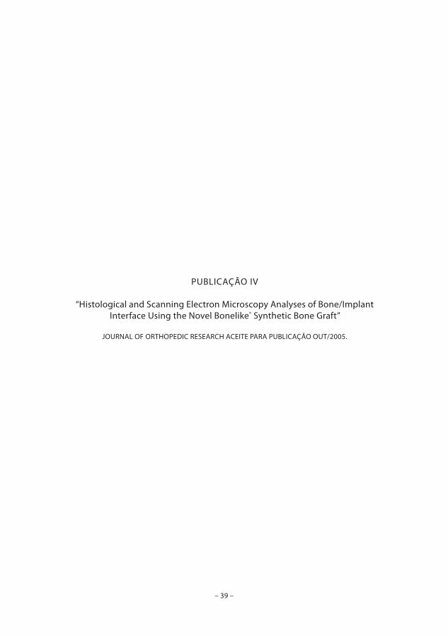
PUBLICAÇÃO IV
“Histological and Scanning Electron Microscopy Analyses of Bone/Implant Interface Using the Novel Bonelike® Synthetic Bone Graft”
JOURNAL OF ORTHOPEDIC RESEARCH ACEITE PARA PUBLICAÇÃO OUT/2005.
– 39 –

– 41 –
ESTUDOS DE BIOCOMPATIBILIDADE IN VIVO
Author Proof
AHistological and Scanning Electron Microscopy Analyses ofBone/Implant Interface Using the Novel Bonelike11
Synthetic Bone Graft
M. Gutierres,1 N. Sooraj Hussain,2,3 M.A. Lopes,2,3 A. Afonso,4 A.T. Cabral,1 L. Almeida,1 J.D. Santos1,2
1FMUP—Faculdade de Medicina da Universidade do Porto, Hospital de Sao Joo, Largo Hernani Monteiro,4200 Porto, Portugal
2DEMM, Faculty of Engg., University of Porto, Rua Dr.Roberto Frias, 4200-465 Porto, Portugal
3INEB—Instituto de Engenharia Biomedica, Laboratorio de Biomaterias, Rua Campo Alegre, 823, 4150-180, Porto, Portugal
4FMDUP, Faculdade de Medicina Dentaria da Universidade do Porto, Rua Dr. Manuel Pereira da Silva,4200-393 Porto, Portugal
Received 8 August 2005; accepted 31 October 2005
Published online ? ? ? ? in Wiley InterScience (www.interscience.wiley.com). DOI 10.1002/jor.20117
ABSTRACT: Synthetic bone grafts provide an alternative to autografts and allografts. Bonelike1 isa patented synthetic bone graft that mimics themineral composition of bone. The aim of the presentworkwas to assess the biological behavior of Bonelike1 graft in humans, beforeusing thematerial inorthopedic applications of bone regeneration, for example, in opening wedge high tibial osteotomiesfor medial knee osteoarthritis. Bonelike1 granules were implanted in cortical bone of 11 patientsundergoing osteotomies, and new bone formation, osteoconductive properties, and resorptioncharacteristics of the granules were analyzed. The granules ranged from 500 to 1000 mm and wereimplanted in the lateral aspect of the tibia. The patients’mean agewas 59 years (range 48 to 70 years);there were eight women and threemen, all suffering frommedial compartment osteoarthritis of theknee.At surgery, a1�1� 1-cmcortical defectwas created3 cmdistal to theentrypoint of the screws,in line with the long axis of tibia. The implanted Bonelike1 graft sample was extracted for analysisduring removal of the metallic prosthesis after implantation times of 6, 9, and 12 months.Radiological follow-up, scanning electron microscopy, histological analysis, and histomorphometricmeasurements were conducted on the retrieved samples to assess bone regeneration in the defectarea. Osteoconductive capacity was demonstrated by extensive mature bone formation around theimplanted granules and high levels of percentage bone-to-graft contact (from 67–84%). Bonelike1
acted as an excellent bioactive scaffold, allowing the migration, proliferation, and differentiation ofbone cells on its surface, and therefore regeneration of the defects was achieved in a rapid, controlledmanner. Our results suggest that Bonelike1 graft is an excellent candidate for orthopedicapplications where rapid new bone formation is a fundamental requirement. � 2006 Orthopaedic
Research Society. Published by Wiley Periodicals, Inc. J Orthop Res 00:1–7, 2006
Keywords: synthetic bone graft; Bonelike1; osteoconduction; clinical applications;histological analysis; orthopedics
INTRODUCTION
Bone is a connective tissue impregnated withcalcium and phosphate salts. The inorganic cal-cium salts, mainly calcium phosphate, provide thetissue with its ability to resist the compressiveforces of and impact 1
For decades, calcium phosphate ceramics havereceived a great deal of attention in clinical
applications as orthopedic and dental materialsbecause of their ability to bond with the livingtissues.2 To improve the properties of calciumphosphate materials for clinical use, hydroxyapa-tite (HA), a major inorganic component of bone,has been used extensively as bone graft filler andas a coating for dental and orthopedic implantsbecause of its osteoconductive properties.3–9
Synthetic bone grafts provide an alternative toautografts and allografts, but both have a numberof well-documented disadvantages.10 Autograftrequires a second surgical procedure that can leadto infection and chronic pain in the harvest site andrequires more hospital time than allograft or bone
JOURNAL OF ORTHOPAEDIC RESEARCH 2006 1
Correspondence to: J.D. Santos (Telephone: (351) 2250812419; Fax: (351) 22 50812419; E-mail: [email protected])
� 2006 Orthopaedic Research Society. Published by Wiley Periodicals,Inc.
forces .weight-bearing
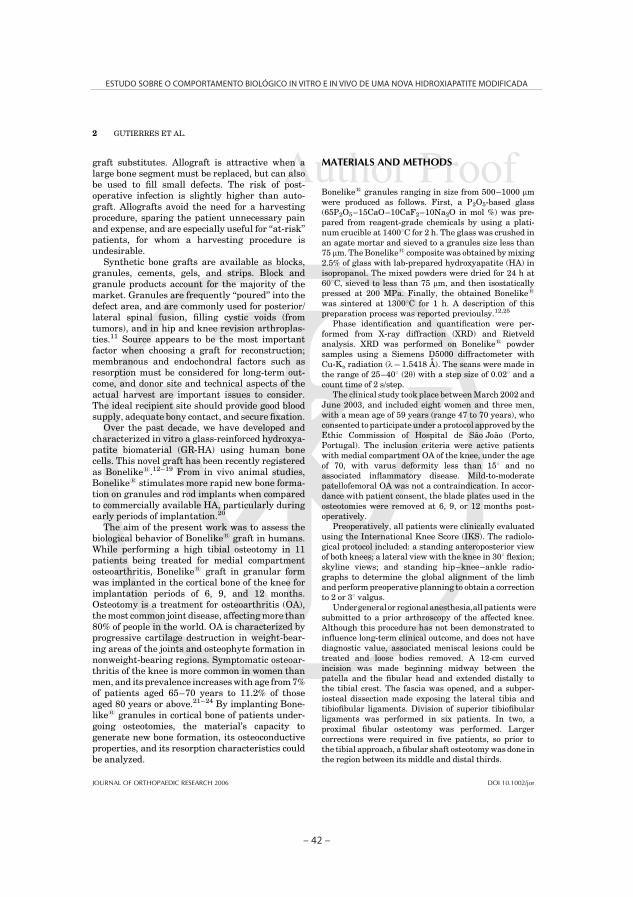
ESTUDO SOBRE O COMPORTAMENTO BIOLÓGICO IN VITRO E IN VIVO DE UMA NOVA HIDROXIAPATITE MODIFICADA
– 42 –
Author Proof
Agraft substitutes. Allograft is attractive when alarge bone segment must be replaced, but can alsobe used to fill small defects. The risk of post-operative infection is slightly higher than auto-graft. Allografts avoid the need for a harvestingprocedure, sparing the patient unnecessary painand expense, and are especially useful for ‘‘at-risk’’patients, for whom a harvesting procedure isundesirable.
Synthetic bone grafts are available as blocks,granules, cements, gels, and strips. Block andgranule products account for the majority of themarket. Granules are frequently ‘‘poured’’ into thedefect area, and are commonly used for posterior/lateral spinal fusion, filling cystic voids (fromtumors), and in hip and knee revision arthroplas-ties.11 Source appears to be the most importantfactor when choosing a graft for reconstruction;membranous and endochondral factors such asresorption must be considered for long-term out-come, and donor site and technical aspects of theactual harvest are important issues to consider.The ideal recipient site should provide good bloodsupply, adequate bony contact, and secure fixation.
Over the past decade, we have developed andcharacterized in vitro a glass-reinforced hydroxya-patite biomaterial (GR-HA) using human bonecells. This novel graft has been recently registeredas Bonelike1.12–19 From in vivo animal studies,Bonelike1 stimulates more rapid new bone forma-tion on granules and rod implants when comparedto commercially available HA, particularly duringearly periods of implantation.20
The aim of the present work was to assess thebiological behavior of Bonelike1 graft in humans.While performing a high tibial osteotomy in 11patients being treated for medial compartmentosteoarthritis, Bonelike1 graft in granular formwas implanted in the cortical bone of the knee forimplantation periods of 6, 9, and 12 months.Osteotomy is a treatment for osteoarthritis (OA),themost common joint disease, affectingmore than80% of people in the world. OA is characterized byprogressive cartilage destruction in weight-bear-ing areas of the joints and osteophyte formation innonweight-bearing regions. Symptomatic osteoar-thritis of the knee is more common in women thanmen, and its prevalence increaseswith age from7%of patients aged 65–70 years to 11.2% of thoseaged 80 years or above.21–24 By implanting Bone-like1 granules in cortical bone of patients under-going osteotomies, the material’s capacity togenerate new bone formation, its osteoconductiveproperties, and its resorption characteristics couldbe analyzed.
MATERIALS AND METHODS
Bonelike1 granules ranging in size from 500–1000 mmwere produced as follows. First, a P2O5-based glass(65P2O5–15CaO–10CaF2–10Na2O in mol %) was pre-pared from reagent-grade chemicals by using a plati-num crucible at 14008C for 2 h. The glass was crushed inan agate mortar and sieved to a granules size less than75 mm. The Bonelike1 composite was obtained bymixing2.5% of glass with lab-prepared hydroxyapatite (HA) inisopropanol. The mixed powders were dried for 24 h at608C, sieved to less than 75 mm, and then isostaticallypressed at 200 MPa. Finally, the obtained Bonelike1
was sintered at 13008C for 1 h. A description of thispreparation process was reported previoulsy.12,25
Phase identification and quantification were per-formed from X-ray diffraction (XRD) and Rietveldanalysis. XRD was performed on Bonelike1 powdersamples using a Siemens D5000 diffractometer withCu-Ka radiation (l¼ 1.5418 A). The scans were made inthe range of 25–408 (2y) with a step size of 0.028 and acount time of 2 s/step.
The clinical study took place betweenMarch 2002 andJune 2003, and included eight women and three men,with a mean age of 59 years (range 47 to 70 years), whoconsented to participate under a protocol approved by theEthic Commission of Hospital de ˜ ˜ (Porto,Portugal). The inclusion criteria were active patientswith medial compartment OA of the knee, under the ageof 70, with varus deformity less than 158 and noassociated inflammatory disease. Mild-to-moderatepatellofemoral OA was not a contraindication. In accor-dance with patient consent, the blade plates used in theosteotomies were removed at 6, 9, or 12 months post-operatively.
Preoperatively, all patients were clinically evaluatedusing the International Knee Score (IKS). The radiolo-gical protocol included: a standing anteroposterior viewof both knees; a lateral view with the knee in 308 flexion;skyline views; and standing hip–knee–ankle radio-graphs to determine the global alignment of the limband perform preoperative planning to obtain a correctionto 2 or 38 valgus.
Undersubmitted to a prior arthroscopy of the affected knee.Although this procedure has not been demonstrated toinfluence long-term clinical outcome, and does not havediagnostic value, associated meniscal lesions could betreated and loose bodies removed. A 12-cm curvedincision was made beginning midway between thepatella and the fibular head and extended distally tothe tibial crest. The fascia was opened, and a subper-iosteal dissection made exposing the lateral tibia andtibiofibular ligaments. Division of superior tibiofibularligaments was performed in six patients. In two, aproximal fibular osteotomy was performed. Largercorrections were required in five patients, so prior tothe tibial approach, afibular shaft osteotomywas done inthe region between its middle and distal thirds.
2 GUTIERRES ET AL.
JOURNAL OF ORTHOPAEDIC RESEARCH 2006 DOI 10.1002/jor
Sao Joao
generalor regionalanesthesia,all patients� were
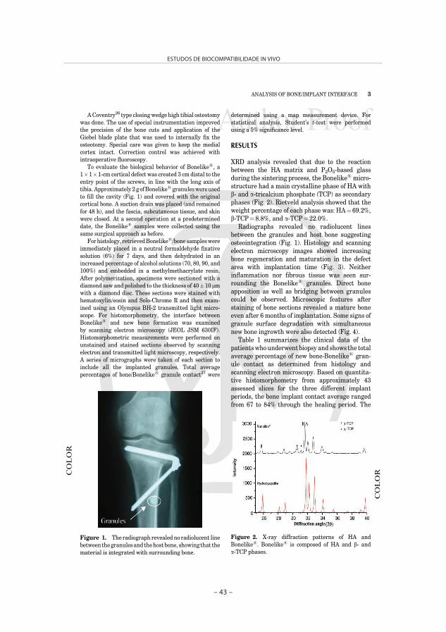
– 43 –
ESTUDOS DE BIOCOMPATIBILIDADE IN VIVO
Author Proof
AACoventry26 type closingwedge high tibial osteotomy
was done. The use of special instrumentation improvedthe precision of the bone cuts and application of theGiebel blade plate that was used to internally fix theosteotomy. Special care was given to keep the medialcortex intact. Correction control was achieved withintraoperative fluoroscopy.
To evaluate the biological behavior of Bonelike1, a1� 1� 1-cm cortical defect was created 3 cm distal to theentry point of the screws, in line with the long axis oftibia.Approximately 2 g ofBonelike1granuleswereusedto fill the cavity (Fig. 1) and covered with the originalcortical bone. A suction drain was placed (and remainedfor 48 h), and the fascia, subcutaneous tissue, and skinwere closed. At a second operation at a predetermineddate, the Bonelike1 samples were collected using thesame surgical approach as before.
For histology, retrievedBonelike1/bone sampleswereimmediately placed in a neutral formaldehyde fixativesolution (6%) for 7 days, and then dehydrated in anincreased percentage of alcohol solutions (70, 80, 90, and100%) and embedded in a methylmethacrylate resin.After polymerization, specimens were sectioned with adiamond saw and polished to the thickness of 40� 10 mmwith a diamond disc. These sections were stained withhematoxylin/eosin and Solo-Chrome R and then exam-ined using an Olympus BH-2 transmitted light micro-scope. For histomorphometry, the interface betweenBonelike1 and new bone formation was examinedby scanning electron microscopy (JEOL JSM 630IF).Histomorphometric measurements were performed onunstained and stained sections observed by scanningelectron and transmitted light microscopy, respectively.A series of micrographs were taken of each section toinclude all the implanted granules. Total averagepercentages of bone/Bonelike1 granule contact27 were
determined using a map measurement device. Forstatistical analysis, Student’s t-test were performedusing a 5% significance level.
RESULTS
XRD analysis revealed that due to the reactionbetween the HA matrix and P2O5-based glassduring the sintering process, the Bonelike1 micro-structure had a main crystalline phase of HA withb- and a-tricalcium phosphate (TCP) as secondaryphases (Fig. 2). Rietveld analysis showed that theweight percentage of each phase was: HA¼ 69.2%,b-TCP¼ 8.8%, and a-TCP¼ 22.0%.
Radiographs revealed no radiolucent linesbetween the granules and host bone suggestingosteointegration (Fig. 1). Histology and scanningelectron microscopy images showed increasingbone regeneration and maturation in the defectarea with implantation time (Fig. 3). Neitherinflammation nor fibrous tissue was seen sur-rounding the Bonelike1 granules. Direct boneapposition as well as bridging between granulescould be observed. Microscopic features afterstaining of bone sections revealed a mature boneeven after 6 months of implantation. Some signs ofgranule surface degradation with simultaneousnew bone ingrowth were also detected (Fig. 4).
Table 1 summarizes the clinical data of thepatientswho underwent biopsy and shows the totalaverage percentage of new bone-Bonelike1 gran-ule contact as determined from histology andscanning electron microscopy. Based on quantita-tive histomorphometry from approximately 43assessed slices for the three different implantperiods, the bone implant contact average rangedfrom 67 to 84% through the healing period. The
Figure 1. The radiograph revealed no radiolucent linebetween thegranules and thehost bone, showing that thematerial is integrated with surrounding bone.
Figure 2. X-ray diffraction patterns of HA andBonelike1. Bonelike1 is composed of HA and b- anda-TCP phases.
COLOR
COLOR
ANALYSIS OF BONE/IMPLANT INTERFACE 3
DOI 10.1002/jor JOURNAL OF ORTHOPAEDIC RESEARCH 2006
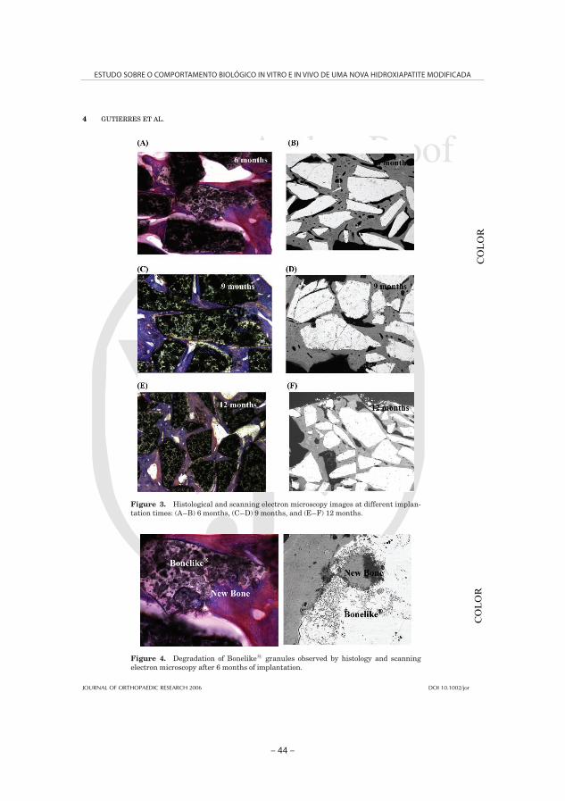
ESTUDO SOBRE O COMPORTAMENTO BIOLÓGICO IN VITRO E IN VIVO DE UMA NOVA HIDROXIAPATITE MODIFICADA
– 44 –
Author Proof
AFigure 3. Histological and scanning electron microscopy images at different implan-tation times: (A–B) 6 months, (C–D) 9 months, and (E–F) 12 months.
Figure 4. Degradation of Bonelike1 granules observed by histology and scanningelectron microscopy after 6 months of implantation.
COLOR
COLOR
4 GUTIERRES ET AL.
JOURNAL OF ORTHOPAEDIC RESEARCH 2006 DOI 10.1002/jor
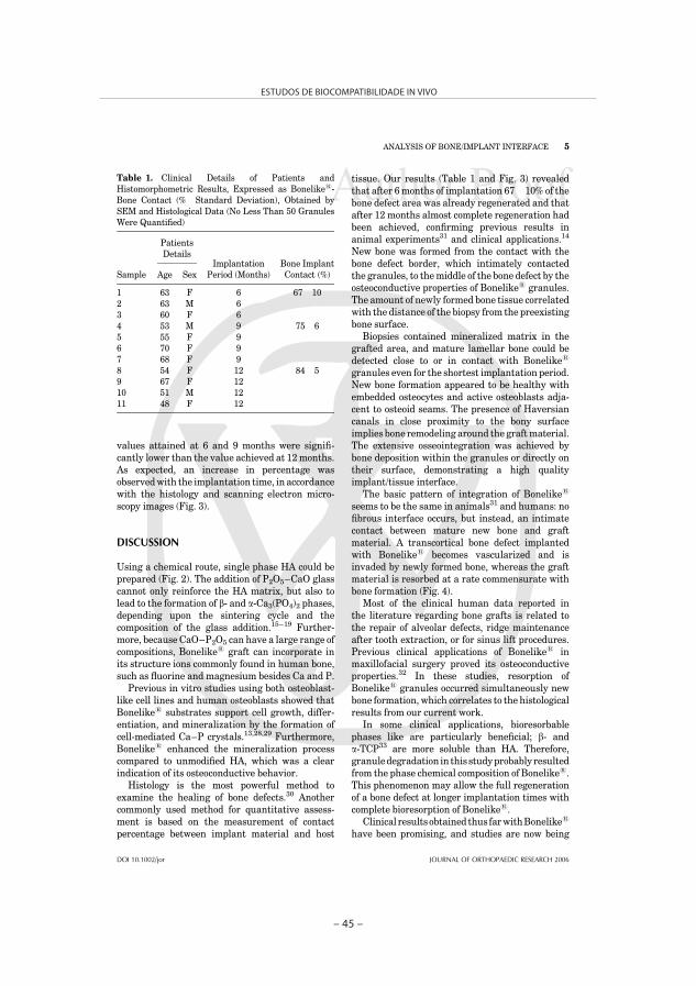
– 45 –
ESTUDOS DE BIOCOMPATIBILIDADE IN VIVO
Author Proof
Avalues attained at 6 and 9 months were signifi-cantly lower than the value achieved at 12months.As expected, an increase in percentage wasobservedwith the implantation time, in accordancewith the histology and scanning electron micro-scopy images (Fig. 3).
DISCUSSION
Using a chemical route, single phase HA could beprepared (Fig. 2). The addition of P2O5–CaO glasscannot only reinforce the HA matrix, but also tolead to the formation of b- and a-Ca3(PO4)2 phases,depending upon the sintering cycle and thecomposition of the glass addition.15–19 Further-more, because CaO–P2O5 can have a large range ofcompositions, Bonelike1 graft can incorporate inits structure ions commonly found in human bone,such as fluorine and magnesium besides Ca and P.
Previous in vitro studies using both osteoblast-like cell lines and human osteoblasts showed thatBonelike1 substrates support cell growth, differ-entiation, and mineralization by the formation ofcell-mediated Ca–P crystals.13,28,29 Furthermore,Bonelike1 enhanced the mineralization processcompared to unmodified HA, which was a clearindication of its osteoconductive behavior.
Histology is the most powerful method toexamine the healing of bone defects.30 Anothercommonly used method for quantitative assess-ment is based on the measurement of contactpercentage between implant material and host
tissue. Our results (Table 1 and Fig. 3) revealedthat after 6months of implantation 67� 10% of thebone defect area was already regenerated and thatafter 12 months almost complete regeneration hadbeen achieved, confirming previous results inanimal experiments31 and clinical applications.14
New bone was formed from the contact with thebone defect border, which intimately contactedthe granules, to themiddle of the bone defect by theosteoconductive properties of Bonelike1 granules.The amount of newly formed bone tissue correlatedwith the distance of the biopsy from the preexistingbone surface.
Biopsies contained mineralized matrix in thegrafted area, and mature lamellar bone could bedetected close to or in contact with Bonelike1
granules even for the shortest implantation period.New bone formation appeared to be healthy withembedded osteocytes and active osteoblasts adja-cent to osteoid seams. The presence of Haversiancanals in close proximity to the bony surfaceimplies bone remodeling around the graftmaterial.The extensive osseointegration was achieved bybone deposition within the granules or directly ontheir surface, demonstrating a high qualityimplant/tissue interface.
The basic pattern of integration of Bonelike1
seems to be the same in animals31 and humans: nofibrous interface occurs, but instead, an intimatecontact between mature new bone and graftmaterial. A transcortical bone defect implantedwith Bonelike1 becomes vascularized and isinvaded by newly formed bone, whereas the graftmaterial is resorbed at a rate commensurate withbone formation (Fig. 4).
Most of the clinical human data reported inthe literature regarding bone grafts is related tothe repair of alveolar defects, ridge maintenanceafter tooth extraction, or for sinus lift procedures.Previous clinical applications of Bonelike1 inmaxillofacial surgery proved its osteoconductiveproperties.32 In these studies, resorption ofBonelike1 granules occurred simultaneously newbone formation, which correlates to the histologicalresults from our current work.
In some clinical applications, bioresorbablephases like are particularly beneficial; b- anda-TCP33 are more soluble than HA. Therefore,granuledegradation in this studyprobably resultedfrom the phase chemical composition of Bonelike1.This phenomenon may allow the full regenerationof a bone defect at longer implantation times withcomplete bioresorption of Bonelike1.
Clinical results obtained thus farwithBonelike1
have been promising, and studies are now being
Table 1. Clinical Details of Patients andHistomorphometric Results, Expressed as Bonelike1-Bone Contact (%�Standard Deviation), Obtained bySEM and Histological Data (No Less Than 50 GranulesWere Quantified)
Sample
PatientsDetails
ImplantationPeriod (Months)
Bone ImplantContact (%)Age Sex
1 63 F 6 67� 102 63 M 63 60 F 64 53 M 9 75� 65 55 F 96 70 F 97 68 F 98 54 F 12 84� 59 67 F 1210 51 M 1211 48 F 12
ANALYSIS OF BONE/IMPLANT INTERFACE 5
DOI 10.1002/jor JOURNAL OF ORTHOPAEDIC RESEARCH 2006
ESTUDO SOBRE O COMPORTAMENTO BIOLÓGICO IN VITRO E IN VIVO DE UMA NOVA HIDROXIAPATITE MODIFICADA
– 46 –
Author Proof
Aconducted in other surgical applications, namely asmacroporous scaffolds for knee osteotomies.
In conclusion, Bonelike1 synthetic bone graftproved tobe osteoconductivewhen implanted in thetibiae of humans, and rapid regeneration of bonedefects was observed. The osteoconductive proper-ties of Bonelike1 result from its chemical composi-tion, which mimics that of human bone through aninnovative route of liquid phase sintering.
ACKNOWLEDGMENTS
The authors express their grateful thanks to the FCT(Fundacao para a Ciencia e a Tecnologia) for theirsupport in this project through Grant #BPD/6010/2001and Mrs. Ana Mota for her technical assistance in thehistological studies.
REFERENCES
1. Peter P, Heinrich R, Meinrad P. 2003. In: YH An, editor.Orthopaedic issues in osteoporosis. Boca Raton, FL: CRCPress.
2. Hench LL, Wilson J. 1999. In: An introduction tobioceramics. World Scientific Publishing Co. Pvt. Ltd.
3. Corpe RS, Steflik DE, Whitehead RY, et al. 2000.Correlative experimental animal and human clinicalretrieval evaluations. Crit Rev Biomed Eng 28:395–398.
4. Kitsugi T, Yamamuro T, Nakamura T, et al. 1993. Fourcalcium phosphate ceramics as bone substitutes for non-weight bearing. Biomaterials 14:216–224.
5. Koshino T, Murase T, Takagi T, et al. 2001. New boneformation around porous hydroxyapatite wedge implantedin opening wedge high tibial osteotomy in patients withosteoarthritis. Biomaterials 22:1579–1582.
6. LeGeros RZ. 1991. In: Meyers HM, editor. Calciumphosphates in oral biology and medicine. New York:Karger; pp. 4–45.
7. Macdonald DE, Betts F, Stranick M, et al. 2001. Physico-chemical study of plasma-sprayed hydroxyapatite-coatedimplants in humans. J Biomed Mater Res 54:480–490.
8. Oonishi H, Yamamoto M, Ishimaru H, et al. 1989. Theeffect of hydroxyapatite coating on bone growth into poroustitanium alloy implants. J Bone Surg Br 71:213–216.
9. Thomas KA. 1994. Hydroxyapatite coatings. Orthopedics17:267–278.
10. Doron Il, Amy LL. 2003. Bone graft substitutes. Oper TechPlastic Reconstruct Surg 9:151–160.
11. Write S. 1999. The bone-graft market in Europe. Commen-tary extracted from a report entitled ‘‘Emerging technol-ogies in orthopedics I.: Bone graft substitutes, bone growthstimulators and bone growth factors.’’ Datamonitor plc.;pp. 591–595.
12. Botelho CM, Brooks RA, Best SM, et al. 2004. Biologicaland physical-chemical characterization of phase pure HAand si-substituted hydroxyapatite by different microscopytechniques. Key Eng Mater 254:845.
13. Costa MA, Gutierres M, Almeida L, et al. 2004. In vitromineralisation of human bone marrow cells cultured onbonelike1. Key Eng Mater 254–256:821–824.
14. Gutierres M, Hussain NS, Afonso A, et al. 2005. Biologicalbehaviour of bonelike1 graft Implanted in the tibia ofhumans. Key Eng Mater 284–286:1041–1044.
15. Lopes MA, Santos JD, Monteiro FJ, et al. 1998. Glassreinforced hydroxyapatite: a comprehensive study of theeffect of glass composition on the crystallography of thecomposite. J Biomed Mater Res 39:244–251.
16. Lopes MA, Monteiro FJ, Santos JD. 1999. Glass-reinforcedhydroxyapatite composites: fracture toughness and hard-ness dependence on microstructural characteristics. Bio-materials 20:2085–2090.
17. Lopes MA, Silva RF, Monteiro FJ, et al. 2000. Micro-structural dependence of Young’s and shearmoduli of P2O5
glass reinforced hydroxyapatite for biomedical applica-tions. Biomaterials 21:749–754.
18. Lopes MA, Knowles JC, Santos JD. 2000. Structuralinsights of glass-reinforced hydroxyapatite composites byRietveld refinement. Biomaterials 21:1905–1910.
19. Santos JD, Knowles JC, Reis RL, et al. 1994. Microstruc-tural characterization of glass-reinforced hydroxyapatitecomposites. Biomaterials 15:5–10.
20. Lopes MA, Santos JD, Monteiro FJ, et al. 2001. Push-outtesting and histological evaluation of glass reinforcedhydroxyapatite composites implanted in the tibia ofrabbits. J Biomed Mater Res 54:463–469.
21. Felson DT, Naimark A, Anderson J, et al. 1987. Theprevalence of knee osteoarthritis in the elderly: theFramingham Study. Arthritis Rheum 30:914–918.
22. Guincamp C, Pap T, Schedel J, et al. 2000. Gene therapy inosteoarthritis. Joint Bone Spine 67:570–571.
23. Marc F, Serge P,MarioMLC, et al. 2003. Assessment of thetest–retest reliability and construct validity of a modifiedlequesne index in knee osteoarthritis. Joint Bone Spine70:520–525.
24. Pendleton A, Arden N, Dougados M, et al. 2000. EULARrecommendations for the management of knee osteoar-thritis: report of a task force of the Standing Committee forInternational Clinical Studies Including TherapeuticTrials (ESCISIT). Ann Rheum Dis 59:936–944.
25. Santos JD, Hastings GW, Knowles JC. 1999. Sinteredhydroxyapatite compositions and method for the prepara-tion thereof. Eur. Pat. WO 0068164.
26. Coventry MB. 1965. Osteotomy of the upper portion of thetibia for degenerative arthritis of the knee: a preliminaryreport. J Bone Joint Surg Am 47:984.
27. HingKA. 1996. Assessment of porous hydroxyapatite for bonereplacement (Ph D Thesis). London: University of London.
28. Lopes MA, Knowles JC, Santos JD, et al. 2000. Direct andindirect effects of P2O5-glass reinforced-hydroxyapatitecomposites and growth and function of osteoblast-like cells.Biomaterials 21:1165–1172.
29. Lopes MA, Knowles JC, Santos JD, et al. 1998. Flowcytometry for assessing biocompatibility. J Biomed MaterRes 41:649–656.
30. Ross MH, Romrell LJ, Kaye GI. 1995. Histology—a textand atlas. 3rd ed. Baltimore, MD: Williams & Wilkins.
31. Lobato JV, Hussain NS, Botelho CM, et al. 2005. Assess-ment of the potential of Bonelike1 graft for boneregeneration by using an animal model. Key Eng Mater284–286:877–880.
32. Duarte F, Lopes MA, Santos JD. 2004. Bonelike1
composites for the regeneration of bone lesions. Mater SciForum 455–456:370–373.
33. Laurencin CT. 2003. In: Bone graft substitutes. WestConshohocken, PA: ASTM International.
6 GUTIERRES ET AL.
JOURNAL OF ORTHOPAEDIC RESEARCH 2006 DOI 10.1002/jor
PUBLICAÇÃO V
“Bone ingrowth in macroporous Bonelike® for orthopedic applications”
JOURNAL OF BONE AND JOINT SURGERY – BRITISH (submetido para publicação)
– 47 –

– 49 –
ESTUDOS DE BIOCOMPATIBILIDADE IN VIVO
Bone ingrowth in macroporous Bonelike® for orthopaedic applications
M. Gutierres*, M.A. Lopes†+, N. Sooraj Hussain†+, A.F. Lemos¥, J.M.F. Ferreira¥, A. Afonso§, A.T.Cabral*, L. Almeida*, J.D. Santos†+
* FMUP – Faculdade de Medicina da Universidade do Porto - Hospital de São João, Largo Hernâni Monteiro, 4200 Porto. Portugal
† FEUP – Faculdade de Engenharia da Universidade do Porto, Rua Dr. Roberto Frias, 4200-465 Porto.Portugal
+ Instituto de Engenharia Biomédica (INEB), Laboratório de Biomateriais, Rua do Campo Alegre 823, 4150-180 Porto. Portugal
¥ Dep. De Eng. Cerâmica e do Vidro, UIMC, Universidade de Aveiro, Aveiro. Portugal§ FMDUP, Faculdade de Medicina Dentária da Universidade do Porto, Rua Dr. Manuel Pereira da Silva,
4200-393 Porto. Portugal
Corresponding Author:Prof. JD Santos, FEUP–DEMM, Rua Dr. Roberto Frias, 4200-465 Porto, Portugal Tel/Fax +351 22 5081419, Email: [email protected]
SummaryBonelike® is a synthetic bone graft recently developed to improve the mechanical properties of pure hydroxyapatite and to increase the similarity between synthetic bone grafts and bone tissue. This goal was achieved by the addition of P2O5 based glasses to a hydroxyapatite matrix, which promotes the liquid phase sintering, reduces porosity and increases interparticle bonding. Furthermore, this process enables the formation of a- and a- tricalcium phosphate phases, which have a higher degradation rate than pure hydroxyapatite and therefore controlled biodegradability of the material may be achieved.The osteoconductivity and biocompatibility of Bonelike® in the granular and dense forms have previously been reported. This study aimed at evaluating the biological behaviour of porous scaffold structures of Bonelike® suitable for either direct clinical use or tissue engineering applications by producing scaffolds with pore size and interconnectivity features suitable for supporting biological activities, such as new bone tissue growth and vascularization.Porous cylinder specimens of 8 mm diameter were implanted in the lateral aspect of the tibia of thirteen patients, with an mean age of 54 years old, during osteotomy surgeries for the treatment of medial compartment osteoarthritis of the knee. Implanted cylinders were retrieved at the same time of the removal of the blade plates at 3, 6, 9 and 12 months. Scanning electron microscopy and histological evaluations were performed to observe the biological responses of human bone tissue to porous Bonelike®. The penetration depth was determined for all implantation periods and after 6 months it was already possible to see new bone in the center of the implanted cylinders, which gives 100% of penetration depth for all implantations periods except for 3 months where bone could only be seen in the peripherical region. Regarding the percentage of the area covered by new bone calculated from 2D histological sections values of 53±15%, 76±12 and 88±9% was achieved for 6, 9 and 12 months respectively.
ESTUDO SOBRE O COMPORTAMENTO BIOLÓGICO IN VITRO E IN VIVO DE UMA NOVA HIDROXIAPATITE MODIFICADA
– 50 –
Porous Bonelike® due to its structural features permitted effective vascularization and bone ingrowth, and therefore was fully osteointegrated as shown in the histological surveys. A slowly biomaterial degradation with implantation time is envisaged since the material showed have surface degradation. Bonelike® scaffolds showed potential for complete ingrowth of osseous tissue and restoration of vascularity throughout the defected site.
Keywords: Bonelike®; porous materials; bone graft; orthopaedic.
Introduction
In the treatment of monocompartmental osteoarthritis of varus knees, high tibial osteotomy is considered as to be an effective procedure to reduce pain and control the progression of the disease, delaying more aggressive surgeries as total knee arthroplasties. This effect is based in the reduction of the stress and weight over the affected compartment, that is produced when the mechanical axis is transferred from the medial compartment to the junction of the lateral third with the two medial thirds. This corresponds to an alignment of 9-10º valgus. Some authors have demonstrated that one year after a high tibial osteotomy, radiologicaly is possible to detect a reduction of the bone density in medial compartment, thus reflecting a slower progression of the ostheoarthritis1. Others described some degree of histological cartilage regeneration after the joint surface has been unloaded2.The closing wedge osteotomy that was used in this study is the most common technique, but recently open wedge procedures have been described with
autografts3, allografts, cement4 and hydroxyapatite5. This second technique provides a more accurate correction of varus deformity when compared with the conventional lateral closing-wedge high tibial osteotomy, but in a time of great developments in knee arthroplasties, autologous chondrocyte implantation and growth factors there is still a place for both.A careful selection of the candidates is essential not only for the success of the surgery but also for the success of the clinical trial that we proposed to do with a bone graft substitute. Because bone is a porous tissue material, there is a physiological rationale use of porous materials in its replacement. Moreover, porous bone grafts is advantageous for the early incorporation of the graft into or apposed to the bone tissue surrounding it6-7. Several key aspects should be considered when designing porous materials for bone grafting6,8-9: (i) the size of pores at the surface of the graft, to actually allow for bone to “flow into” the graft and fill all its structure; (ii) the size of porous connecting the surface pores to allow areas of bony ingrowth to “meet up” within the porous graft; (iii)
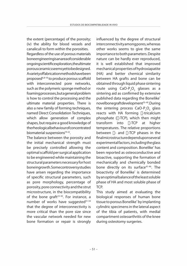
– 51 –
ESTUDOS DE BIOCOMPATIBILIDADE IN VIVO
the extent (percentage) of the porosity; (iv) the ability for blood vessels and canaliculi to form within the porosities.Regardless of the use of porous materials bone engineering is an area of considerable ongoing scientific exploration, the ultimate porous ceramics seems yet to be designed. A variety of fabrication methods have been proposed6,8-18 to produce porous scaffold with interconnected pore networks, such as the polymeric sponge method or foaming processes, but a general problem is how to control the processing and the ultimate material properties. There is also a new family of forming techniques, named Direct Consolidation Techniques, which allow generation of complex shapes, but require a good knowledge of the rheological behaviour of concentrated biomaterial suspensions19-22.The balance between the porosity and the initial mechanical strength must be precisely controlled allowing the optimal scaffold per surgical application to be engineered while maintaining the structural parameters necessary for host bone ingrowth. Some controversy studies have arisen regarding the importance of specific structural parameters, such as pore morphology, percentage of porosity, pore connectivity and the strut microstructure, in the biocompatibility of the bone graft23-30. For instance, a number of works have suggested31-34 that the degree of interconnectivity is more critical than the pore size since the vascular network needed for new bone formation or repair is strongly
influenced by the degree of structural interconnectivity among pores, whereas other works seems to give the same importance to both parameters. Despite nature can be hardly ever reproduced, it is well established that improved mechanical properties of hydroxiapatite (HA) and better chemical similarity between HA grafts and bone can be obtained through liquid phase sintering route using CaO-P2O5 glasses as a sintering aid as confirmed by extensive published data regarding the Bonelike® novel bone graft development35-40. During the sintering process CaO-P2O5 glass reacts with HA forming a-tricalcium phosphate (a-TCP), which then might transform into a-TCP at higher temperatures. The relative proportions between a- and a-TCP phases in the final microstructure depend upon several experimental factors, including the glass content and composition. Bonelike® has been reported as osteoconductive and bioactive, supporting the formation of mechanically and chemically bonded bone directly on its surface41-46. The bioactivity of Bonelike® is determined by an optimal balance of the least soluble phase of HA and most soluble phase of TCP. This study aimed at evaluating the biological responses of human bone tissue to porous Bonelike® by implanting cylindric specimens in the lateral aspect of the tibia of patients, with medial compartment osteoarthritis of the knee during osteotomy surgeries.
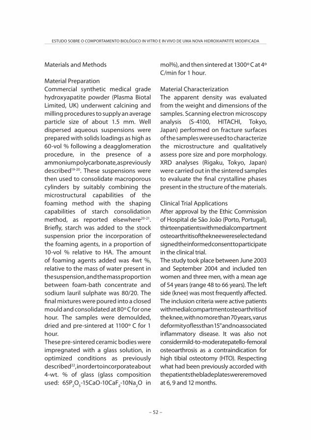
ESTUDO SOBRE O COMPORTAMENTO BIOLÓGICO IN VITRO E IN VIVO DE UMA NOVA HIDROXIAPATITE MODIFICADA
– 52 –
Materials and Methods
Material PreparationCommercial synthetic medical grade hydroxyapatite powder (Plasma Biotal Limited, UK) underwent calcining and milling procedures to supply an average particle size of about 1.5 mm. Well dispersed aqueous suspensions were prepared with solids loadings as high as 60-vol % following a deagglomeration procedure, in the presence of a ammonium polycarbonate, as previously described19-20. These suspensions were then used to consolidate macroporous cylinders by suitably combining the microstructural capabilities of the foaming method with the shaping capabilities of starch consolidation method, as reported elsewhere20-21. Briefly, starch was added to the stock suspension prior the incorporation of the foaming agents, in a proportion of 10-vol % relative to HA. The amount of foaming agents added was 4wt %, relative to the mass of water present in the suspension, and the mass proportion between foam-bath concentrate and sodium lauril sulphate was 80/20. The final mixtures were poured into a closed mould and consolidated at 80º C for one hour. The samples were demoulded, dried and pre-sintered at 1100º C for 1 hour. These pre-sintered ceramic bodies were impregnated with a glass solution, in optimized conditions as previously described22, in order to incorporate about 4-wt. % of glass (glass composition used: 65P2O5-15CaO-10CaF2-10Na2O in
mol%), and then sintered at 1300º C at 4º C/min for 1 hour.
Material CharacterizationThe apparent density was evaluated from the weight and dimensions of the samples. Scanning electron microscopy analysis (S-4100, HITACHI, Tokyo, Japan) performed on fracture surfaces of the samples were used to characterize the microstructure and qualitatively assess pore size and pore morphology. XRD analyses (Rigaku, Tokyo, Japan) were carried out in the sintered samples to evaluate the final crystalline phases present in the structure of the materials.
Clinical Trial Applications After approval by the Ethic Commission of Hospital de São João (Porto, Portugal), thirteen patients with medial compartment osteoarthritis of the knee were selected and signed the informed consent to participate in the clinical trial. The study took place between June 2003 and September 2004 and included ten women and three men, with a mean age of 54 years (range 48 to 66 years). The left side (knee) was most frequently affected. The inclusion criteria were active patients with medial compartment osteoarthritis of the knee, with no more than 70 years, varus deformity of less than 15° and no associated inflammatory disease. It was also not consider mild-to-moderate patello-femoral osteoarthrosis as a contraindication for high tibial osteotomy (HTO). Respecting what had been previously accorded with the patients the blade plates were removed at 6, 9 and 12 months.
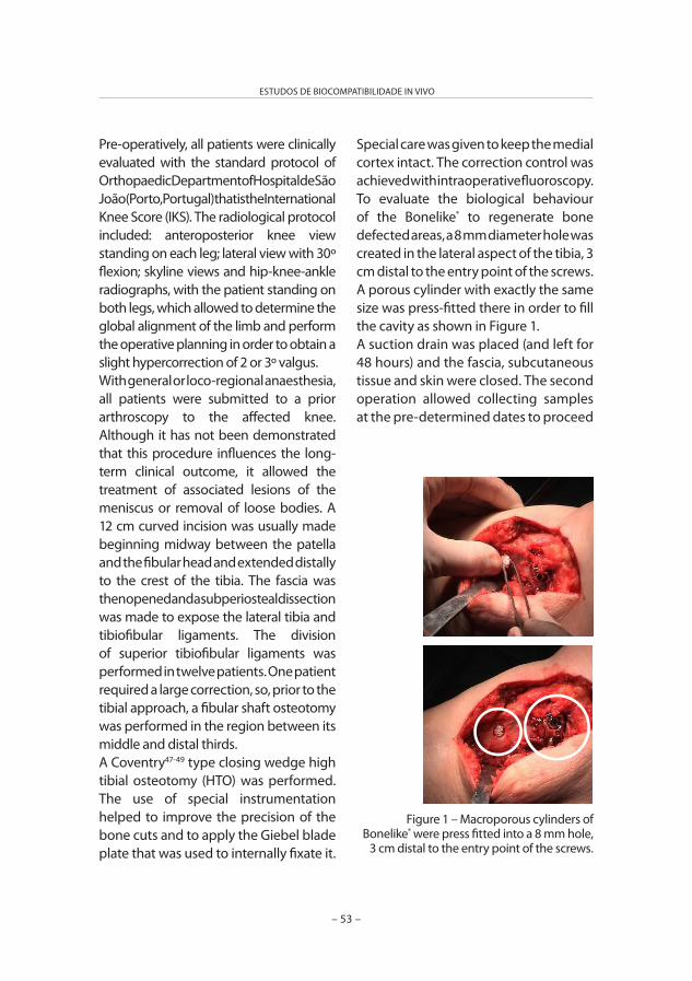
– 53 –
ESTUDOS DE BIOCOMPATIBILIDADE IN VIVO
Pre-operatively, all patients were clinically evaluated with the standard protocol of Orthopaedic Department of Hospital de São João (Porto, Portugal) that is the International Knee Score (IKS). The radiological protocol included: anteroposterior knee view standing on each leg; lateral view with 30º flexion; skyline views and hip-knee-ankle radiographs, with the patient standing on both legs, which allowed to determine the global alignment of the limb and perform the operative planning in order to obtain a slight hypercorrection of 2 or 3º valgus.With general or loco-regional anaesthesia, all patients were submitted to a prior arthroscopy to the affected knee. Although it has not been demonstrated that this procedure influences the long-term clinical outcome, it allowed the treatment of associated lesions of the meniscus or removal of loose bodies. A 12 cm curved incision was usually made beginning midway between the patella and the fibular head and extended distally to the crest of the tibia. The fascia was then opened and a subperiosteal dissection was made to expose the lateral tibia and tibiofibular ligaments. The division of superior tibiofibular ligaments was performed in twelve patients. One patient required a large correction, so, prior to the tibial approach, a fibular shaft osteotomy was performed in the region between its middle and distal thirds.A Coventry47-49 type closing wedge high tibial osteotomy (HTO) was performed. The use of special instrumentation helped to improve the precision of the bone cuts and to apply the Giebel blade plate that was used to internally fixate it.
Special care was given to keep the medial cortex intact. The correction control was achieved with intraoperative fluoroscopy. To evaluate the biological behaviour of the Bonelike® to regenerate bone defected areas, a 8 mm diameter hole was created in the lateral aspect of the tibia, 3 cm distal to the entry point of the screws. A porous cylinder with exactly the same size was press-fitted there in order to fill the cavity as shown in Figure 1.A suction drain was placed (and left for 48 hours) and the fascia, subcutaneous tissue and skin were closed. The second operation allowed collecting samples at the pre-determined dates to proceed
Figure 1 – Macroporous cylinders of Bonelike® were press fitted into a 8 mm hole,
3 cm distal to the entry point of the screws.
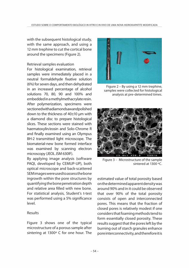
ESTUDO SOBRE O COMPORTAMENTO BIOLÓGICO IN VITRO E IN VIVO DE UMA NOVA HIDROXIAPATITE MODIFICADA
– 54 –
with the subsequent histological study, with the same approach, and using a 12 mm trephine to cut the cortical bone around the specimens (Figure 2).
Retrieval samples evaluationFor histological examination, retrieval samples were immediately placed in a neutral formaldehyde fixative solution (6%) for seven days, and then dehydrated in an increased percentage of alcohol solutions 70, 80, 90 and 100% and embedded in a methylmethacrylate resin. After polymerization, specimens were sectioned with a diamond saw and polished down to the thickness of 40±10 µm with a diamond disc to prepare histological slices. These sections were stained with haematoxylin/eosin and Solo-Chrome R and finally examined using an Olympus BH-2 transmitted light microscope. The biomaterial-new bone formed interface was examined by scanning electron microscopy (JEOL JSM 630IF). By applying image analysis (software PAQI, developed by CEMUP-UP), both optical microscope and back-scattered SEM images were used to assess the bone ingrowth within the pore structures by quantifying the bone penetration depth and relative area filled with new bone. For statistical analysis, Student’s t-test was performed using a 5% significance level.
Results
Figure 3 shows one of the typical microstructure of a porous sample after sintering at 1300º C for one hour. The
Figure 2 – By using a 12 mm trephine, samples were collected for histological
analysis at pre-determined times.
Figure 3 – Microstructure of the sample sintered at 1300 ºC.
estimated value of total porosity based on the determined apparent density was around 90% and in it could be observed that over 90% of the total porosity consists of open and interconnected pores. This means that the fraction of closed pores is relatively modest if one considers that foaming methods tend to form essentially closed porosity. These results suggest that the pores left by the burning out of starch granules enhance pore interconnectivity, and therefore it is
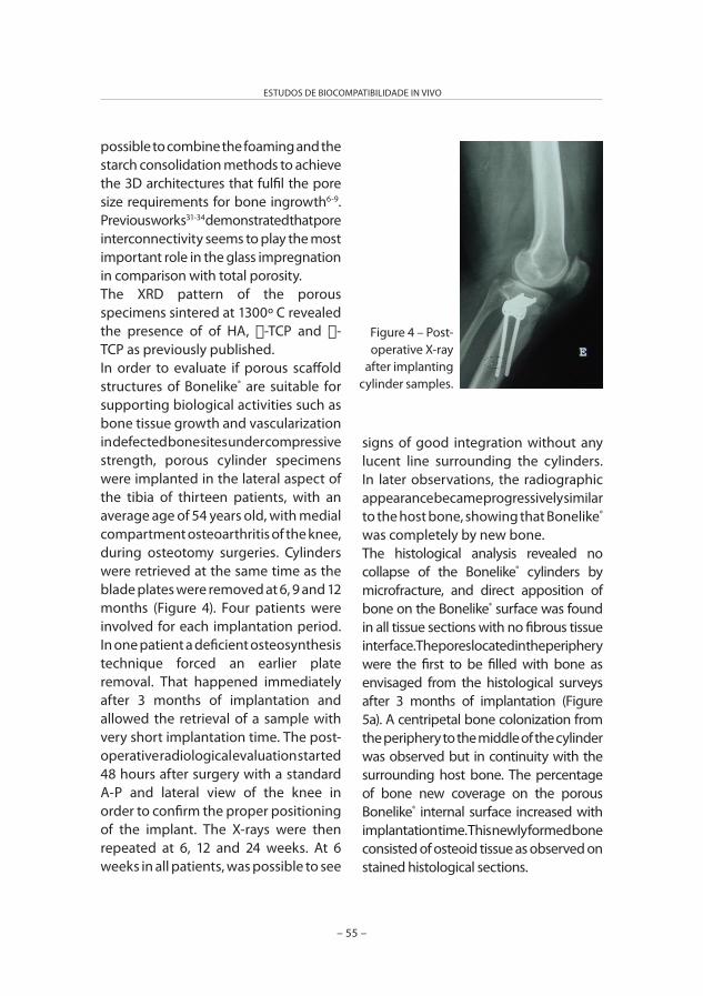
– 55 –
ESTUDOS DE BIOCOMPATIBILIDADE IN VIVO
possible to combine the foaming and the starch consolidation methods to achieve the 3D architectures that fulfil the pore size requirements for bone ingrowth6-9. Previous works31-34 demonstrated that pore interconnectivity seems to play the most important role in the glass impregnation in comparison with total porosity.The XRD pattern of the porous specimens sintered at 1300º C revealed the presence of of HA, a-TCP and a-TCP as previously published.In order to evaluate if porous scaffold structures of Bonelike® are suitable for supporting biological activities such as bone tissue growth and vascularization in defected bone sites under compressive strength, porous cylinder specimens were implanted in the lateral aspect of the tibia of thirteen patients, with an average age of 54 years old, with medial compartment osteoarthritis of the knee, during osteotomy surgeries. Cylinders were retrieved at the same time as the blade plates were removed at 6, 9 and 12 months (Figure 4). Four patients were involved for each implantation period. In one patient a deficient osteosynthesis technique forced an earlier plate removal. That happened immediately after 3 months of implantation and allowed the retrieval of a sample with very short implantation time. The post-operative radiological evaluation started 48 hours after surgery with a standard A-P and lateral view of the knee in order to confirm the proper positioning of the implant. The X-rays were then repeated at 6, 12 and 24 weeks. At 6 weeks in all patients, was possible to see
signs of good integration without any lucent line surrounding the cylinders. In later observations, the radiographic appearance became progressively similar to the host bone, showing that Bonelike® was completely by new bone.The histological analysis revealed no collapse of the Bonelike® cylinders by microfracture, and direct apposition of bone on the Bonelike® surface was found in all tissue sections with no fibrous tissue interface. The pores located in the periphery were the first to be filled with bone as envisaged from the histological surveys after 3 months of implantation (Figure 5a). A centripetal bone colonization from the periphery to the middle of the cylinder was observed but in continuity with the surrounding host bone. The percentage of bone new coverage on the porous Bonelike® internal surface increased with implantation time. This newly formed bone consisted of osteoid tissue as observed on stained histological sections.
Figure 4 – Post-operative X-ray
after implanting cylinder samples.
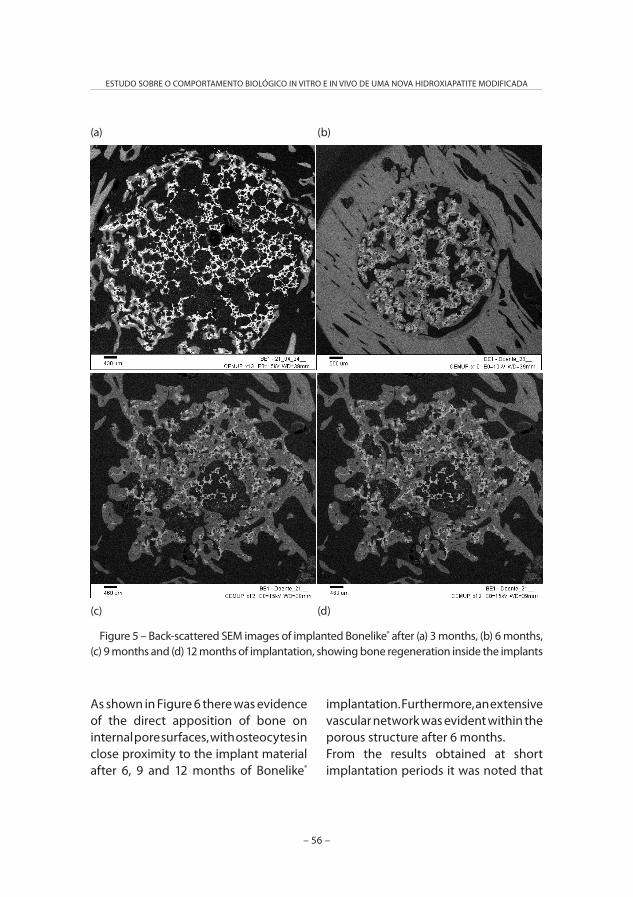
ESTUDO SOBRE O COMPORTAMENTO BIOLÓGICO IN VITRO E IN VIVO DE UMA NOVA HIDROXIAPATITE MODIFICADA
– 56 –
Figure 5 – Back-scattered SEM images of implanted Bonelike® after (a) 3 months, (b) 6 months, (c) 9 months and (d) 12 months of implantation, showing bone regeneration inside the implants
(a)
(c)
(b)
(d)
As shown in Figure 6 there was evidence of the direct apposition of bone on internal pore surfaces, with osteocytes in close proximity to the implant material after 6, 9 and 12 months of Bonelike®
implantation. Furthermore, an extensive vascular network was evident within the porous structure after 6 months. From the results obtained at short implantation periods it was noted that
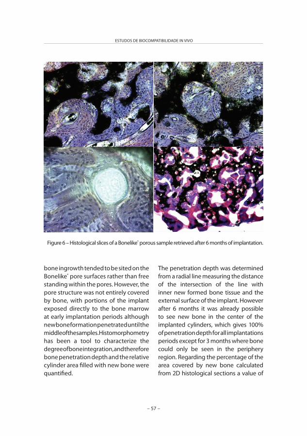
– 57 –
ESTUDOS DE BIOCOMPATIBILIDADE IN VIVO
bone ingrowth tended to be sited on the Bonelike® pore surfaces rather than free standing within the pores. However, the pore structure was not entirely covered by bone, with portions of the implant exposed directly to the bone marrow at early implantation periods although new bone formation penetrated until the middle of the samples. Histomorphometry has been a tool to characterize the degree of bone integration, and therefore bone penetration depth and the relative cylinder area filled with new bone were quantified.
Figure 6 – Histological slices of a Bonelike® porous sample retrieved after 6 months of implantation.
The penetration depth was determined from a radial line measuring the distance of the intersection of the line with inner new formed bone tissue and the external surface of the implant. However after 6 months it was already possible to see new bone in the center of the implanted cylinders, which gives 100% of penetration depth for all implantations periods except for 3 months where bone could only be seen in the periphery region. Regarding the percentage of the area covered by new bone calculated from 2D histological sections a value of
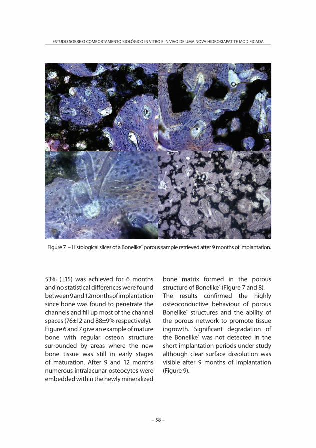
ESTUDO SOBRE O COMPORTAMENTO BIOLÓGICO IN VITRO E IN VIVO DE UMA NOVA HIDROXIAPATITE MODIFICADA
– 58 –
53% (±15) was achieved for 6 months and no statistical differences were found between 9 and 12months of implantation since bone was found to penetrate the channels and fill up most of the channel spaces (76±12 and 88±9% respectively).Figure 6 and 7 give an example of mature bone with regular osteon structure surrounded by areas where the new bone tissue was still in early stages of maturation. After 9 and 12 months numerous intralacunar osteocytes were embedded within the newly mineralized
Figure 7 – Histological slices of a Bonelike® porous sample retrieved after 9 months of implantation.
bone matrix formed in the porous structure of Bonelike® (Figure 7 and 8).The results confirmed the highly osteoconductive behaviour of porous Bonelike® structures and the ability of the porous network to promote tissue ingrowth. Significant degradation of the Bonelike® was not detected in the short implantation periods under study although clear surface dissolution was visible after 9 months of implantation (Figure 9).
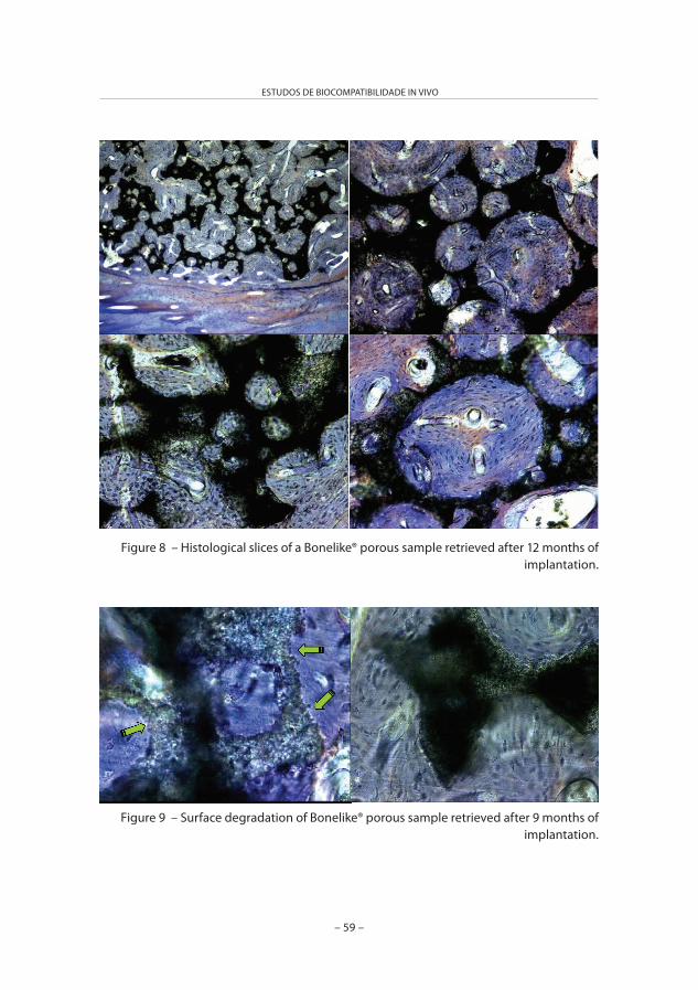
– 59 –
ESTUDOS DE BIOCOMPATIBILIDADE IN VIVO
Figure 8 – Histological slices of a Bonelike® porous sample retrieved after 12 months of implantation.
Figure 9 – Surface degradation of Bonelike® porous sample retrieved after 9 months of implantation.
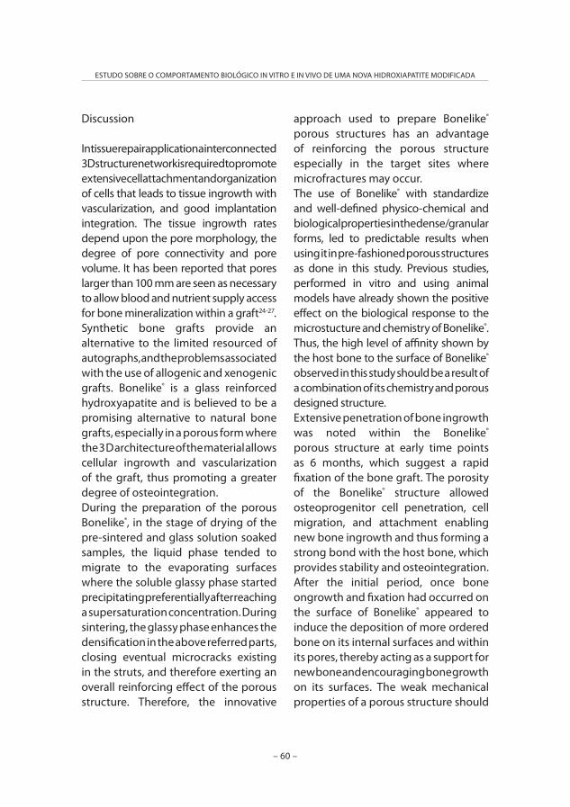
ESTUDO SOBRE O COMPORTAMENTO BIOLÓGICO IN VITRO E IN VIVO DE UMA NOVA HIDROXIAPATITE MODIFICADA
– 60 –
Discussion
In tissue repair application a interconnected 3D structure network is required to promote extensive cell attachment and organization of cells that leads to tissue ingrowth with vascularization, and good implantation integration. The tissue ingrowth rates depend upon the pore morphology, the degree of pore connectivity and pore volume. It has been reported that pores larger than 100 mm are seen as necessary to allow blood and nutrient supply access for bone mineralization within a graft24-27.Synthetic bone grafts provide an alternative to the limited resourced of autographs, and the problems associated with the use of allogenic and xenogenic grafts. Bonelike® is a glass reinforced hydroxyapatite and is believed to be a promising alternative to natural bone grafts, especially in a porous form where the 3 D architecture of the material allows cellular ingrowth and vascularization of the graft, thus promoting a greater degree of osteointegration.During the preparation of the porous Bonelike®, in the stage of drying of the pre-sintered and glass solution soaked samples, the liquid phase tended to migrate to the evaporating surfaces where the soluble glassy phase started precipitating preferentially after reaching a supersaturation concentration. During sintering, the glassy phase enhances the densification in the above referred parts, closing eventual microcracks existing in the struts, and therefore exerting an overall reinforcing effect of the porous structure. Therefore, the innovative
approach used to prepare Bonelike® porous structures has an advantage of reinforcing the porous structure especially in the target sites where microfractures may occur.The use of Bonelike® with standardize and well-defined physico-chemical and biological properties in the dense/granular forms, led to predictable results when using it in pre-fashioned porous structures as done in this study. Previous studies, performed in vitro and using animal models have already shown the positive effect on the biological response to the microstucture and chemistry of Bonelike®. Thus, the high level of affinity shown by the host bone to the surface of Bonelike® observed in this study should be a result of a combination of its chemistry and porous designed structure.Extensive penetration of bone ingrowth was noted within the Bonelike® porous structure at early time points as 6 months, which suggest a rapid fixation of the bone graft. The porosity of the Bonelike® structure allowed osteoprogenitor cell penetration, cell migration, and attachment enabling new bone ingrowth and thus forming a strong bond with the host bone, which provides stability and osteointegration. After the initial period, once bone ongrowth and fixation had occurred on the surface of Bonelike® appeared to induce the deposition of more ordered bone on its internal surfaces and within its pores, thereby acting as a support for new bone and encouraging bone growth on its surfaces. The weak mechanical properties of a porous structure should
– 61 –
ESTUDOS DE BIOCOMPATIBILIDADE IN VIVO
be eliminated once incorporation and bone ingrowth into the pores were achieved.The complete penetration of bone into the centre of Bonelike® cylinders must be consequence of the good interconnectivity among the pores within the structure of Bonelike®, which allowed for nutrient and oxygen availability leading to faster bone apposition and/or angiogenesis. Literature describes that in the short to medium term bone apposition was influenced in the first instance by the rate of cellular invasion, which is determined by the pore morphology, and in the medium to long term, by the rate of vascularization, which is determined by the connectivity network24-27.HA has been characterized as being much more stable compared to a- and a-TCP phases. Therefore porous Bonelike® has the ability to provide the scaffold for new bone growth due to the presence of the HA, as well as the ability to foster osteogenesis due to the presence of the degradable TCP phases and by the release of specific ions present in its composition. A series of dissolution-precipitation processes undergo in vivo and the release of ions, stimulated by the high surface area, will have a beneficial effect on cell biology activity, as it as also been demonstrated in several in vitro cellular studies and in vivo experiments.
Conclusions
This study demonstrates the porous Bonelike® is highly biocompatible, since direct apposition of bone to Bonelike® surface is the desired clinical outcome with full osteointegration and vascularization. Porous structures of Bonelike® can be applied in human adults for bone repair, even in weight-loaded bone, and bone ingrowth into the pores progressed with time of implantation. Porous structures of Bonelike® have potential to be used in tissue engineering.
ACKNOWLEDGMENTSThe authors express their grateful thanks to the FCT – Fundação para a Ciência e a Tecnologia for their support in this work through the project “New generation of bioactive and bioresorbable materials for bone regenerative surgery using 3D biomodelling” and also through Post-Doctoral grant SFRH/BPD/14969/2004, and to Mrs. Ana Mota for her technical assistance in the histological studies.
References
1. Akamatsu Y, Koshino T, Saito T, Wada J. Changes in osteosclerosis of the osteoarthritic knee after high tibial osteotomy. Clin. Orthop. 1997; 334: 207-214.
2. Coventry MB, Ilstrup DM, Wallrichs SL. Proximal tibial osteotomy. A critical long-term study of eighty-seven cases. J. Bone and Joint Surg. 1993; 75-A: 196-201.
ESTUDO SOBRE O COMPORTAMENTO BIOLÓGICO IN VITRO E IN VIVO DE UMA NOVA HIDROXIAPATITE MODIFICADA
– 62 –
3. Hernigou P, Medevielle D, Debeyre J, Goutallier D. Proximal tibial osteotomy for osteoarthritis with varus deformity. A ten to thirteen-year follow-up study. J Bone Joint Surg Am 1987; 69: 332-354.
4. Hernigou P, Ma W. Open wedge tibial osteotomy with acrylic bone cement as bone substitute. Knee 2001; 8: 103-110.
5. Koshino T, Murase T, Saito T. Medial opening–wedge high tibial osteotomy with use of porous hydroxyapatite to treat medial compartment osteoarthritis of the knee. J Bone Joint Surg Am 2003; 85: 78-85.
6. Porous Materials for Bone Engineered, Simske SJ, Ayers RA, Bateman TA in Materials Science Forum, Trans Tech Publications, Switzerland 1997; 250: 151-182.
7. Hing KA. Bone repair in the twenty-first century: biology, chemistry or engineering? Philos Transact A Math Phys Eng Sci. 2004;362(1825):2821-50.
8. Ben-Nissan, B. Natural bioceramics: from coral to bone and beyond. Current Opinion in Solid State & Materials Science 2003; 7(4/5): 283.
9. Shors EC, Holmes RE. Porous hydroxyapatite. In: Hench LL,. Wilson J, editors. Introduction to Biomaterials. Singapore: World Scientific 1993, p 181-198.
10. Prado da Silva MH, Lemos AF, Gibson IR, Ferreira JMF, Santos JD. Porous Glass reinforced hydroxyapatite Materials Produced With different Organic Additives. J. Non-Crystalline Solids 2002; 304: 286-292.
11. Prado da Silva MH, Lemos AF, Ferreira JMF, Lopes MA, Santos JD. Production of Porous Biomaterials based on Glass reinforced Hydroxyapatite composites.
Key Engineering Materials 2002; 230-232: 483-486.
12. Rocha J, Lemos A, Agathopoulos S, Valério P, Kannan S, Oktar F, Ferreira J. Scaffolds for bone restoration from cuttlefish. Bone 2005; 37(6): 850-857.
13. Sepulveda P, Binner JGP. Processing of cellular ceramics by foaming and in situ polymerization of organic monomers. Journal of the European Ceramic Society 1999; 19(12): 2059-2066.
14. Ozgur Engin N, Cuneyt Tas A. Manufacture of macroporous calcium hydroxyapatite bioceramics. Journal of the European Ceramic Society 1999; 19(13-14): 2569-2572.
15. Peng HX. Fan Z, Evans JRG, Busfield JJC. Microstructure of ceramic foams. Journal of the European Ceramic Society 2000; 20(7): 807-813.
16. Komlev Vladimir S, Barinov Serguei M, Koplik Elena V. A method to fabricate porous spherical hydroxyapatite granules intended for time-controlled drug release Biomaterials 2002; 23 (16): 3449-3454.
17. Milosevski M, Bossert J, Milosevski D, Gruevska N. Preparation and properties of dense and porous calcium phosphate. Ceramics International 1999; 25: 693-696.
18. Tadic D, Beckmann F, Schwarz K, Epple M. A novel method to produce hydroxyapatite objects with interconnecting porosity that avoids sintering. Biomaterials 2004; 25: 3335-3340.
19. Lemos AF, Santos JD, Ferreira JMF. Influence of characteristics of the starting hydroxyapatite powders and of deagglomeration procedure, on
– 63 –
ESTUDOS DE BIOCOMPATIBILIDADE IN VIVO
rheological behaviour of the suspensions. Materials Science Forum 2004; 455-456: 361-365.
20. Lemos AF, Ferreira JMF. Porous Bioactive Calcium Carbonate Implants Processed by Starch Consolidation. J. Mat. Sci. & Engineering C 2000; C11: 35-40.
21. Lemos AF, Ferreira JMF. Combining foaming and starch consolidation methods to develop macroporous HA implants. Key Engineering Materials 2004; 254-256: 1041-1044.
22. Lemos AF, Santos JD. Ferreira JMF. New method for the incorporation of soluble bioactive glasses to reinforce porous HA structures. Key Engineering Materials 2004; 254-256: 1033-1036.
23. Gauthiera O, Boulerb JM, Aguadoa E, Piletb P, Daculsi G. Macroporous biphasic calcium phosphate ceramics: Influence of macropore diameter and macroporosity percentage on bone ingrowth. Biomaterials 1998; 19: 133-139.
24. Hing AK, Best SM, Tanner KE, Bonfield W, Revell PA. Mediation of bone ingrowth in porous hydroxyapatite bone graft substitutes. J Biomed Mater Res 2004; 68A: 187-200.
25. Rubin PA, Popham JK, Bilyk JR, Shore JW. Comparison of fibrovascular ingrowth into hydroxyapatite and porous polyethylene orbital implants. Ophthal Plast Reconstr Surg. 1994; 10(2): 96-103.
26. Hing KA, Annaz B, Saeed S, Revell PA, Buckland T. Microporosity enhances bioactivity of synthetic bone graft substitutes. J Material Science: Materials in Medicine 2005; 16: 467-475.
27. Klawitter JJ, Bagwell JG, Weinstein AM, Sauer BW. An evaluation of bone growth
into porous high density polyethylene. J Biomed Mater Res. 1976 Mar; 10(2): 311-23.
28. Hing KA, Best SM, Tanner KE, Bonfield W, Revell PA. Quantification of bone ingrowth within bone-derived porous hydroxyapatite implants of varying density. J Materials Science: Materials in Medicine 1999; 10: 663-670.
29. Hing KA, Best SM, Tanner KE, Bonfield W, Revell PA. Biomechanical assessment of bone ingrowth in porous hydroxyapatite. J Materials Science: Mater Medicine 1997; 8: 731-736.
30. Ayers RA, Wolford LM, Bateman TA, Ferguson VL, Simske SJ. Quantification of bone ingrowth into porous block hydroxyapatite in humans. J Biomed Mater Res 1999; 47: 54-59.
31. Lu, JX, Flautre B, Anselme K, Hardouin P, Gallur A, Descamps M, Thierry B. Role of interconnections in porous bioceramics on bone recolonization in vitro and in vivo. Journal of Materials Science: Materials in Medicine 1999; 10(2): 111-120.
32. Eggli PS, Muller W, Schenk RK. Porous hydroxyapatite and tricalcium phosphate cylinders with two different pore size ranges implanted in the cancellous bone of rabbits. A comparative histomorphometric and histologic study of bony ingrowth and implant substitution. Clin Orthop Relat Res. 1988; 232: 127-38.
33. Kuhne JH, Bartl R, Frisch B, Hammer C, Jansson V, Zimmer M. Bone formation in coralline hydroxyapatite. Effects of pore size studied in rabbits. Acta Orthop Scand. 1994; 65(3): 246-52.
34. Holmes R, Mooney V, Bucholz R, Tencer A. A coralline hydroxyapatite bone graft
ESTUDO SOBRE O COMPORTAMENTO BIOLÓGICO IN VITRO E IN VIVO DE UMA NOVA HIDROXIAPATITE MODIFICADA
– 64 –
substitute. Preliminary report. Clin Orthop Relat Res. 1984; 188: 252-62.
35. Lopes MA, Knowles JC, Santos JD. Structural insights of glass-reinforced hydroxyapatite composites by Rietveld refinement. Biomaterials 2000; 21(18): 1905-1910.
36. Lopes MA, Santos JD, Monteiro FJ, Knowles JC. Glass-reinforced hydroxyapatite: A comprehensive study of the effect of glass composition on the crystallography of the composite. Journal of Biomedical Materials Research 1998; 39(2): 244-251.
37. Santos J, Hastings G, Knowles J; Sintered hydroxyapatite compositions and method for the preparation thereof. Worldwide Application (PCT), Patent N. 1 189 851, 1999.
38. Lopes MA, Monteiro FJ, Santos JD. Glass-reinforced hydroxyapatite composites: fracture toughness and hardness dependence on microstructural characteristics. Biomaterials 1999; 20(21): 2085-2090.
39. Lopes MA, Monteiro FJ, Santos JD. Glass-reinforced hydroxyapatite composites: Secondary phase proportions and densification effects on biaxial bending strength. Journal of Biomedical Materials Research 1999; 48(5): 734-740.
40. Lopes MA, Monteiro FJ, Santos JD, Serro AP, Saramago B. Hydrophobicity, Surface tension and Zeta potential measurements of glass reinforced hydroxyapatite composites. J. Biomed. Mater. Res. 1999; 45: 370-375.
41. Lopes MA, Knowles JC, Santos JD, Monteiro FJ, Olsen I. Direct and Indirect effects of P2O5-glass reinforced–hydroxyapatite composites and growth and function of osteoblast-like cells. Biomaterials 2000; 21: 1165-1172.
42. Lopes MA, Santos JD, Monteiro FJ, Ohtsuki C, Osaka A, Kaneko S, Inoue H. Push-out testing and histological evaluation of glass reinforced hydroxyapatite composites implanted in the tibiae of rabbits. J. Biomed. Mater. Res. 2001; 54: 463-469.
43. Ferraz MP, Fernandes MH, Santos JD, Monteiro FJ. HA and Double layer HA-P2O5/CaO Glass coatings: influence of chemical composition on human bone marrow cells osteoblastic behaviour. J Mater Sci: Mater in Med 2001; 12: 629-638.
44. Gutierres M, Hussain NS, Afonso A, Almeida L, Cabral T, Lopes MA, Santos JD. Biological behaviour of Bonelike® graft implanted in the tibia of humans. Bioceramics, 2005; 17: 1041-1044.
45. Duarte F, Santos JD, Afonso A. Medical applications of Bonelike® in maxillofacial surgery. Advanced Materials Forum 2004; 370-373.
46. Lobato JV, Sooraj Hussain N, Botelho CM, Mauricio AC, Lobato JM, Lopes MA, Afonso A, Ali N, Santos JD. Titanium dental implants coated with Bonelike®: clinical case report. Thin Solid Films, in press.
47. Coventry MB, Osteotomy of the upper portion of the tibia for degenerative arthritis of the knee: a preliminary report. J Bone Joint Surg Am 1965; 47: 984-990.
48. Coventry MB, Upper tibia osteotomy for gonarthrosis. The evolution of the operation in the last 18 years and long term results. Orthop Clin North Am 1979; 10: 191-210.
49. Coventry MB, Current concepts review. Upper tibia osteotomy for osteoarthritis. J Bone Joint Surg Am 1985; 67A: 1136-1140.
CAPÍTULO V
APLICAÇÕES CLÍNICAS
– 65 –

PUBLICAÇÃO VI
“Opening wedge high tibial osteotomy using 3D biomodelling Bonelike® macroporous structures – Case report”
JOURNAL OF MATERIALS SCIENCE: MATERIALS IN MEDICINE (submetido para publicação)
– 67 –

– 69 –
APLICAÇÕES CLÍNICAS
Opening wedge high tibial osteotomy using 3D biomodelling Bonelike® macroporous structures – case report
M. Gutierres*, A.G. Dias†+, M.A. Lopes†+, N. Sooraj Hussain†+, A.T.Cabral*, L. Almeida*, J.D. Santos†+
* FMUP - Faculdade de Medicina da Universidade do Porto - Hospital de São João, Largo Hernâni Monteiro, 4200 Porto, Portugal
† FEUP - Faculdade de Engenharia da Universidade do Porto, Rua Dr. Roberto Frias, 4200-465 Porto, Portugal
+ Instituto de Engenharia Biomédica (INEB), Laboratório de Biomateriais, Rua do Campo Alegre 823, 4150-180 Porto, Portugal
ABSTRACTTwo synthetic calcium phosphates in porous wedge shape, Bonelike® and a commercial HA/a-TCP biphasic material, were used as an alternative to bone autografts and allografts in the treatment of medial compartment osteoarthritis of varus knees. The structure of Bonelike® has a 3D architecture that is computer controlled, and a composition that mimics the mineral composition of natural bone. The HA/a-TCP biphasic material used as a control material in this study was prepared using conventional foaming based methods. No signs of inflammatory reactions were observed post-operatively for both materials. After 4 months of partial weight bearing, signs of fusion at the osteotomy site and good integration of the implanted wedges were observed, showing good mechanical resistance. Concerning the final correction attained, the left knee revealed a satisfactory valgus of 10º, but the right one only had a final value of 6º. The clinical evaluation using International Knee Score (IKS) showed good outcome in all parameters: complete range of motion in both knees, only slight pain on climbing stairs and no use of crutches.
Keywords: Bonelike®; opening wedge osteotomy; varus knee
1. INTRODUCTION
Dealing with bone defects is a permanent challenge in orthopaedic and trauma field. A valgus tibial osteotomy has been selected as an efficient method of treatment for patients with medial compartment osteoarthritis in varus knees1-5. Studies have shown that there is some degree of cartilage regeneration
after this procedure6-8, even without chondrocyte implantation.Lateral closing wedge high tibial osteotomy technique has been so far the most used surgery to solve this problem. Nevertheless, in order to avoid the peroneal osteotomy and obtain more accurate correction of varus deformity, a technique of medial opening wedge tibial osteotomy has been introduced.
ESTUDO SOBRE O COMPORTAMENTO BIOLÓGICO IN VITRO E IN VIVO DE UMA NOVA HIDROXIAPATITE MODIFICADA
– 70 –
The later technique has many advantages compare with the former one. Opening wedge tibial osteotomy is simple and less invasive technique, and the fixation is accomplished easily by means of metal plates and screws. Later, if conversion to a total knee arthroplasty is needed, the procedure is also simplified.A fashioned wedge with the exact angle for the opening required can be inserted in the osteotomy site, allowing not only coronal but also sagital corrections. However, opening wedge high tibial osteotomy has limitations such as the risk of collapsus, with loss of correction and non-union1,3,5,9-10.The gap created by the osteotomy may be filled with autograft or synthetic materials11. Autograft substitutes, most specifically iliac crest cortico-cancellous graft, is considered the most successful bone filling because of its osteoconductive, osteoinductive and osteogenic properties. However, autograft substitutes showed some drawbacks related to the donor site and require a second surgery. To overcome the disadvantages of the autografts, synthetic bone substitutes morphologically adapted to the host site are starting to be used in several orthopaedic applications. Pre-shaped wedges designed for specific applications simplified the surgery procedure as well as add some mechanical stability. The synthetic substitutes have been reported as bioactive and can support bone growth and remodelling, which may lead
to faster full weight-bearing after open wedge valgus high tibial osteotomy12-16.Three-dimensional porous scaffolds seem to be a good option for opening wedge high tibial osteotomy, as they offer a framework for new bone tissue formation and anchorage by providing surface and volume, that will allow cell ingrowth and an accurate cell distribution throughout the porous structure17-20.Recently, the authors reported the potential of a composite material, registered as Bonelike®, as a synthetic bone graft in animal models and in implantology and oral maxilofacial applications comparing with the commercially available pure HA. In the present study, two materials prepared in different ways, computer controlled 3D architecture fabrication and conventional based techniques, were used as a bone substitutes, Bonelike® and a HA/a-TCP biphasic material, respectively. The 3D architecture computer-controlled fabrication techniques, known as rapid prototyping (RP), have significant advantages over conventional based fabrication methods. Although conventional methods hold great success in preparing scaffolds for bone applications, the computer-controlled fabrication techniques promises rapid solutions in automating scaffold production as well provide several advantages such as easy processing, macro shape and structural design, which leads to good compressive strength15,21,22. The purpose of this work was to evaluate the performance of two porous wedges materials with different structures in the treatment of medial compartment osteoarthritis varus knee.
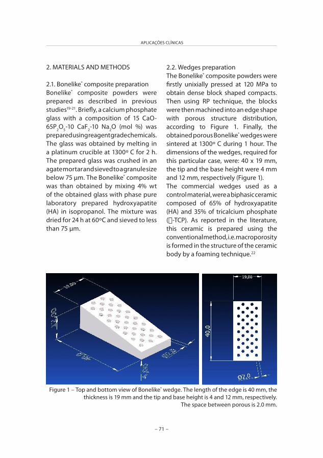
– 71 –
APLICAÇÕES CLÍNICAS
2. MATERIALS AND METHODS
2.1. Bonelike® composite preparationBonelike® composite powders were prepared as described in previous studies19-21. Briefly, a calcium phosphate glass with a composition of 15 CaO-65P2O5-10 CaF2-10 Na2O (mol %) was prepared using reagent grade chemicals. The glass was obtained by melting in a platinum crucible at 1300º C for 2 h. The prepared glass was crushed in an agate mortar and sieved to a granule size below 75 µm. The Bonelike® composite was than obtained by mixing 4% wt of the obtained glass with phase pure laboratory prepared hydroxyapatite (HA) in isopropanol. The mixture was dried for 24 h at 60ºC and sieved to less than 75 µm.
2.2. Wedges preparation The Bonelike® composite powders were firstly unixially pressed at 120 MPa to obtain dense block shaped compacts. Then using RP technique, the blocks were then machined into an edge shape with porous structure distribution, according to Figure 1. Finally, the obtained porous Bonelike® wedges were sintered at 1300º C during 1 hour. The dimensions of the wedges, required for this particular case, were: 40 x 19 mm, the tip and the base height were 4 mm and 12 mm, respectively (Figure 1).The commercial wedges used as a control material, were a biphasic ceramic composed of 65% of hydroxyapatite (HA) and 35% of tricalcium phosphate (a-TCP). As reported in the literature, this ceramic is prepared using the conventional method, i.e. macroporosity is formed in the structure of the ceramic body by a foaming technique.22
Figure 1 – Top and bottom view of Bonelike® wedge. The length of the edge is 40 mm, the thickness is 19 mm and the tip and base height is 4 and 12 mm, respectively.
The space between porous is 2.0 mm.
ESTUDO SOBRE O COMPORTAMENTO BIOLÓGICO IN VITRO E IN VIVO DE UMA NOVA HIDROXIAPATITE MODIFICADA
– 72 –
2.3. Materials characterizationFor X-ray diffraction analyses, samples were ground to fine powders of Bonelike®, and analysed using a Siemens D 5000 diffractometer with flat plate geometry and Cu Ka radiation. Data were collected from 10 to 80º (2a), with a step size of 0.02º and a counting time of 2 s per step. Quantitative phase analysis was performed by the Rietveld method using General Structure Analysis Software (GSAS, Los Alamos National Laboratory).
2.4. Clinical CaseIn June 2004, a 58 years old male patient (AMS) was evaluated in the Orthopaedic Department of Hospital de São João (Porto, Portugal), complaining of bilateral knee pain, specially in the left side, for the last 4 years. The character of the complain was mechanical, predominantly localized in the medial compartment, but also with some femuropatelar complains. The medication and Physiotherapy provided only partial relieve of the simptons. The patient referred that, when he was 40 years old, he was submitted to an open medial meniscectomy in the left knee.
2.4.1. Clinical and radiographic evaluationThe patient was preoperatively evaluated according the standard protocol of Orthopaedic Department of Hospital de São João (Porto, Portugal) that is the International Knee Score (IKS). The physical examination showed: bilateral genum varum, with quadriceps atrophy, a parapatelar scar from the
previous meniscectomy in the left knee, good range of motion, low volume hidarthrosis, + valgus stress test, but - Mc Murray and anterior drawer test.The radiological procedures included anteroposterior knee view standing on each leg; lateral view with 30º flexion; skyline views and hip-knee-ankle radiographs, with the patient standing on both legs, which allowed determining the global alignment of the limb and performing the operative planning in order to obtain an accurate correction. The radiographic exam showed a bilateral osteoarthrosis, mainly in the medial compartment (grade II-III – modified Ahlback) and a varus of 6º in the left and 10º in the right knee (Fig. 2).
2.4.2. Operative technique An opening wedge osteotomy was planned to obtain 2 or 3 º of hypercorrection in the alignment of both knees that is 9 or 10 º valgus. Firstly, the patient was submitted to a valgus osteotomy in the left knee with addition of a 12º medial wedge of a commercial HA/a-TCP biphasic material. Eight months later the same surgical procedure was applied to the right knee using the Bonelike® composite material. In both cases, the medial wedge opening side was stabilized by means of a Puddu plate and screws to avoid subsequent displacement of the wedge (Fig. 3). The post-operatory period was normal, with no signs of inflammatory reaction. Partial weight bearing was allowed until 4 months, when the use of crutches was discontinued.
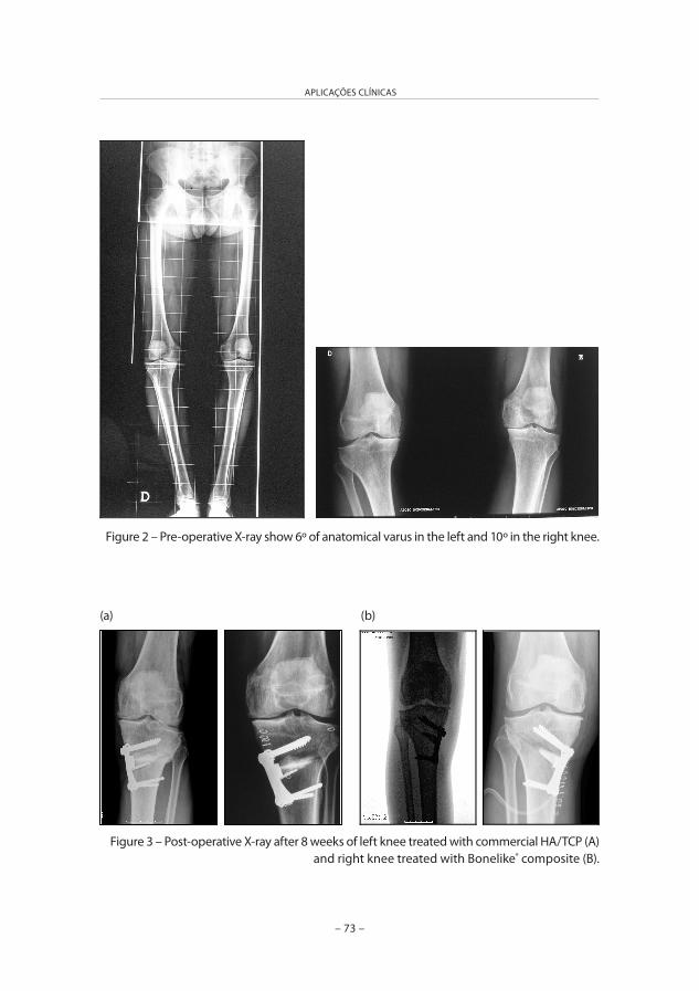
– 73 –
APLICAÇÕES CLÍNICAS
Figure 2 – Pre-operative X-ray show 6º of anatomical varus in the left and 10º in the right knee.
Figure 3 – Post-operative X-ray after 8 weeks of left knee treated with commercial HA/TCP (A)and right knee treated with Bonelike® composite (B).
(a) (b)
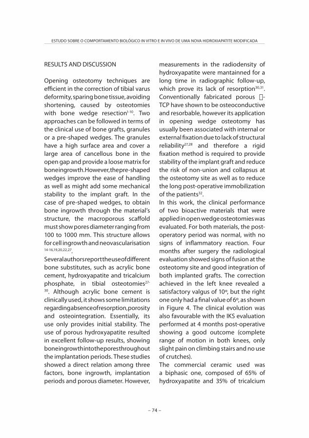
ESTUDO SOBRE O COMPORTAMENTO BIOLÓGICO IN VITRO E IN VIVO DE UMA NOVA HIDROXIAPATITE MODIFICADA
– 74 –
RESULTS AND DISCUSSION
Opening osteotomy techniques are efficient in the correction of tibial varus deformity, sparing bone tissue, avoiding shortening, caused by osteotomies with bone wedge resection1-10. Two approaches can be followed in terms of the clinical use of bone grafts, granules or a pre-shaped wedges. The granules have a high surface area and cover a large area of cancellous bone in the open gap and provide a loose matrix for bone ingrowth. However, the pre-shaped wedges improve the ease of handling as well as might add some mechanical stability to the implant graft. In the case of pre-shaped wedges, to obtain bone ingrowth through the material’s structure, the macroporous scaffold must show pores diameter ranging from 100 to 1000 mm. This structure allows for cell ingrowth and neovascularisation 14-16,19,20,22,27.Several authors report the use of different bone substitutes, such as acrylic bone cement, hydroxyapatite and tricalcium phosphate, in tibial osteotomies27-
30. Although acrylic bone cement is clinically used, it shows some limitations regarding absence of resorption, porosity and osteointegration. Essentially, its use only provides initial stability. The use of porous hydroxyapatite resulted in excellent follow-up results, showing bone ingrowth into the pores throughout the implantation periods. These studies showed a direct relation among three factors, bone ingrowth, implantation periods and porous diameter. However,
measurements in the radiodensity of hydroxyapatite were mantainned for a long time in radiographic follow-up, which prove its lack of resorption30,31. Conventionally fabricated porous a-TCP have shown to be osteoconductive and resorbable, however its application in opening wedge osteotomy has usually been associated with internal or external fixation due to lack of structural reliability27,28 and therefore a rigid fixation method is required to provide stability of the implant graft and reduce the risk of non-union and collapsus at the osteotomy site as well as to reduce the long post-operative immobilization of the patients32. In this work, the clinical performance of two bioactive materials that were applied in open wedge osteotomies was evaluated. For both materials, the post-operatory period was normal, with no signs of inflammatory reaction. Four months after surgery the radiological evaluation showed signs of fusion at the osteotomy site and good integration of both implanted grafts. The correction achieved in the left knee revealed a satisfactory valgus of 10º, but the right one only had a final value of 6º, as shown in Figure 4. The clinical evolution was also favourable with the IKS evaluation performed at 4 months post-operative showing a good outcome (complete range of motion in both knees, only slight pain on climbing stairs and no use of crutches). The commercial ceramic used was a biphasic one, composed of 65% of hydroxyapatite and 35% of tricalcium
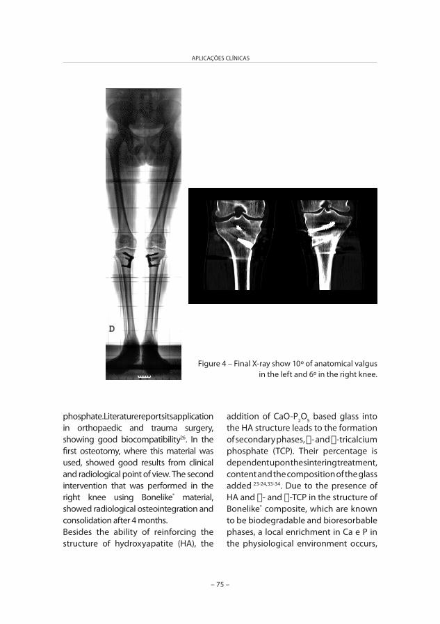
– 75 –
APLICAÇÕES CLÍNICAS
phosphate. Literature reports its application in orthopaedic and trauma surgery, showing good biocompatibility26. In the first osteotomy, where this material was used, showed good results from clinical and radiological point of view. The second intervention that was performed in the right knee using Bonelike® material, showed radiological osteointegration and consolidation after 4 months.Besides the ability of reinforcing the structure of hydroxyapatite (HA), the
addition of CaO-P2O5 based glass into the HA structure leads to the formation of secondary phases, a- and a-tricalcium phosphate (TCP). Their percentage is dependent upon the sintering treatment, content and the composition of the glass added 23-24,33-34. Due to the presence of HA and a- and a-TCP in the structure of Bonelike® composite, which are known to be biodegradable and bioresorbable phases, a local enrichment in Ca e P in the physiological environment occurs,
Figure 4 – Final X-ray show 10º of anatomical valgus in the left and 6º in the right knee.

ESTUDO SOBRE O COMPORTAMENTO BIOLÓGICO IN VITRO E IN VIVO DE UMA NOVA HIDROXIAPATITE MODIFICADA
– 76 –
which stimulates new bone formation helping in osteointegration of Bonelike® composite as well as improve the mechanical strength.Bonelike® wedge was prepared by 3D biomodelling, which is a technique that allows for complete structural control of the synthetic graft, automation of the manufacturing process and high precision of the pore size and pore distribution. All these parameters are known to play a fundamental role in the structural reliability of the synthetic graft and accounts for the higher compressive strength than those grafts that are prepared by conventional slip casting and foaming techniques. From radiological examination it was evident that no graft displacement, distortion or fracture occurred, which should be attributed to the high precision of the manufacturing process. This clinical case report confirms other previous studies that had shown that Bonelike® is rapidly osteointegrated after implantation in maxillofacial surgery and implantology36-37. In orthopaedic applications, radiological, histological histomorphometric measurements and scanning electron microscopy have previously demonstrated that Bonelike® is highly bioactive and it resorbs at a rate that is compatible with the new formation rate. The possibility of applying 3D Biomodelling techniques to manufactured high-precision macropores structures for opening wedge tibial osteotomy opens-up new areas of application of Bonelike® in the orthopaedic field.
ACKNOWLEDGMENTSThe authors express their grateful thanks to the FCT – Fundação para a Ciência e a Tecnologia for their support in this work through the project “New generation of bioactive and bioresorbable materials for bone regenerative surgery using 3D biomodelling” and also through Post-Doctoral grant SFRH/BPD/14969/2004.
REFERENCES
1. Gaasbeek RDA, Welsing RTC, Verdonschot N, Rijnberg WJ, van Loon CJM, van Kampen A. Accuracy and initial stability of open- and closed-wedge high tibial osteotomy: a cadaveric RSA study. Knee Surgery Sports Traumatology Arthroscopy 2005; 13(8): 689-694.
2. Naudie DDR, Amendola A, Fowler PJ. Opening wedge high tibial osteotomy for symptomatic hyperextension-varus thrust. American Journal of Sports Medicine 2004; 32(1): 60-70.
3. Scott WN, Clarke HD. The role of osteotomy 2003: Defining the niche. Orthopedics 2004; 27(9): 975-976.
4. Virolainen P, Aro HT. High tibial osteotomy for the treatment of osteoarthritis of the knee: a review of the literature and a meta-analysis of follow-up studies. Archives of Orthopaedic and Trauma Surgery 2004; 124(4): 258-261.
5. Wright JM, Crockett HC, Slawski DP, Madsen MW, Windsor RE. High tibial osteotomy. Journal of the American Academy of Orthopaedic Surgeons 2005; 13(4): 279-289.
– 77 –
APLICAÇÕES CLÍNICAS
6. Fujisawa Y, Masura K, Shiomi S. The effect of high tibial osteotomy on osteoarthritis of the knee. An arthroscopic study of 54 knee joints. Clin Orthop North Am 1979; 10: 585-608.
7. Odenbridge S, Egund N, Lindstrand A, Lohmander LS, Willen H. Cartilage regeneration after proximal tibial osteotomy for medial gonartrosis. An arthroscopic, roentgenographic and histologic study. Clin Orthop 1992; 277: 210-216.
8. Koshino T, Wada S, Ara Y, Saito T. Regeneration of degenerated articular cartilage after high tibial valgus osteotomy for medial compartmental osteoarthritis of the knee. Knee 2003; (10): 229-236.
9. Hernigou P. Open wedge tibial osteotomy: combined coronal and sagittal correction. Knee 2002; 9(1): 15-20.
10. Hoell S, Suttmoeller J, Stoll V, Fuchs S, Gosheger G. The high tibial osteotomy, open versus closed wedge, a comparison of methods in 108 patients. Archives of Orthopaedic and Trauma Surgery 2005;125(9): 638-643.
11. Betz RR. Limitations of autograft and allograft: New synthetic solutions. Orthopedics 2002; 25(5): S561-S570.
12. LeGeros RZ. Properties of osteoconductive biomaterials: Calcium phosphates. Clinical Orthopaedics and Related Research 2002; (395): 81-98.
13. Steffen T, Stoll T, Arvinte T, Schenk RK. Porous tricalcium phosphate and transforming growth factor used for anterior spine surgery. European Spine Journal 2001; 10: S132-S140.
14. Chu TMG, Orton DG, Hollister SJ, Feinberg SE, Halloran JW. Mechanical and in vivo performance of hydroxyapatite
implants with controlled architectures. Biomaterials 2002; 23(5): 1283-1293.
15. Hutmacher DW, Sittinger M, Risbud MV. Scaffold-based tissue engineering: rationale for computer-aided design and solid free-form fabrication systems. Trends in Biotechnology 2004; 22(7): 354-362.
16. Jones JR, Ehrenfried LM, Hench LL. Optimising bioactive glass scaffolds for bone tissue engineering. Biomaterials 2006; 27(7): 964-973.
17. Gauthier O, Bouler JM, Aguado E, Pilet P, Daculsi G. Macroporous biphasic calcium phosphate ceramics: influence of macropore diameter and macroporosity percentage on bone ingrowth. Biomaterials 1998; 19(1-3): 133-139.
18. Lu JX, Flautre B, Anselme K, Hardouin P, Gallur A, Descamps M, Thierry B. Role of interconnections in porous bioceramics on bone recolonization in vitro and in vivo. Journal of Materials Science-Materials in Medicine 1999; 10(2): 111-120.
19. Martina M, Subramanyam G, Weaver JC, Hutmacher DW, Morse DE, Valiyaveettil S. Developing macroporous bicontinuous materials as scaffolds for tissue engineering. Biomaterials 2005; 26(28): 5609-5616.
20. Moroni L, de Wijn JR, van Blitterswijk CA. 3D fiber-deposited scaffolds for tissue engineering: Influence of pores geometry and architecture on dynamic mechanical properties. Biomaterials 2006; 27(7): 974-985.
21. Leong KF, Cheah CM, Chua CK. Solid freeform fabrication of three-dimensional scaffolds for engineering replacement tissues and organs. Biomaterials 2003; 24(13): 2363-2378.
ESTUDO SOBRE O COMPORTAMENTO BIOLÓGICO IN VITRO E IN VIVO DE UMA NOVA HIDROXIAPATITE MODIFICADA
– 78 –
22. Hutmacher DW. Scaffold design and fabrication technologies for engineering tissues – state of the art and future perspectives. Journal of Biomaterials Science-Polymer Edition 2001; 12(1): 107-124.
23. Lopes MA, Knowles JC, Santos JD. Structural insights of glass-reinforced hydroxyapatite composites by Rietveld refinement. Biomaterials 2000; 21(18): 1905-1910.
24. Lopes MA, Santos JD, Monteiro FJ, Knowles JC. Glass-reinforced hydroxyapatite: A comprehensive study of the effect of glass composition on the crystallography of the composite. Journal of Biomedical Materials Research 1998; 39(2): 244-251.
25. Santos J, Hastings G, Knowles J; Sintered hydroxyapatite compositions and method for the preparation thereof. Worldwide Application (PCT), Patent N. 1 189 851, 1999.
26. Schwartz C, Liss P, Jacquemaire B, Lecestre P, Frayssinet P. Biphasic synthetic bone substitute use in orthopaedic and trauma surgery: clinical, radiological and histological results. Journal of Materials Science-Materials in Medicine 1999; 10(12): 821-825.
27. van Hemert WLW, Willems K, Anderson PG, van Heerwaarden RJ, Wymenga AB. Tricalcium phosphate granules or rigid wedge preforms in open wedge high tibial osteotomy: a radiological study with a new evaluation system. Knee 2004; 11(6): 451-456.
28. Gaasbeek RDA, Toonen HG, van Heerwaarden RJ, Buma P. Mechanism of bone incorporation of b-TCP bone substitute in open wedge tibial osteotomy in patients. Biomaterials 2005; 26(33): 6713-6719.
29. Hernigou P, Ma W. Open wedge tibial osteotomy with acrylic bone cement as bone substitute. Knee 2001; 8(2): 103-110.
30. Koshino T, Murase T, Saito T. Medial
opening-wedge high tibial osteotomy with use of porous hydroxyapatite to treat medial compartment osteoarthritis of the knee. Journal of Bone and Joint Surgery-American Volume 2003; 85A(1): 78-85.
31. Koshino T, Murase T, Takagi T, Saito T. New bone formation around porous hydroxyapatite wedge implanted in opening wedge high tibial osteotomy in patients with osteoarthritis. Biomaterials 2001; 22(12): 1579-1582.
32. Zhim F, Laflamme GY, Viens H, Saidane K, Yahia L. Biomechanical stability of high tibial opening wedge osteotomy: Internal fixation versus external fixation. Clinical Biomechanics 2005; 20(8): 871-876.
33. Lopes MA, Monteiro FJ, Santos JD. Glass-reinforced hydroxyapatite composites: fracture toughness and hardness dependence on microstructural characteristics. Biomaterials 1999; 20(21): 2085-2090.
34. Lopes MA, Monteiro FJ, Santos JD. Glass-reinforced hydroxyapatite composites: Secondary phase proportions and densification effects on biaxial bending strength. Journal of Biomedical Materials Research 1999; 48(5): 734-740.
35. Gutierres M, Hussain NS, Afonso A, Almeida L, Cabral T, Lopes MA, Santos JD. Biological behaviour of Bonelike® graft implanted in the tibia of humans. Bioceramics, Vol 17; 2005. p 1041-1044.
36. Duarte F, Santos JD, Afonso A. Medical applications of Bonelike® in maxillofacial surgery. Advanced Materials Forum Ii; 2004. p 370-373.
37. Lobato JV, Sooraj Hussain N, Botelho CM, Mauricio AC, Lobato JM, Lopes MA, Afonso A, Ali N, Santos JD. Titanium dental implants coated with Bonelike®: clinical case report. Thin Solid Films, in press.
CAPÍTULO VI
DISCUSSÃO E CONCLUSÕES
– 79 –

– 81 –
DISCUSSÃO E CONCLUSÕES
A crescente complexidade e exigência de soluções para resolver determinadas patologias do foro ortopédico e maxilofacial têm evidenciado o papel que os substitutos ósseos podem vir a desempenhar na Medicina do futuro, originando o aparecimento de imensa informação como resultado da investigação que tem sido desenvolvida nesta área, em especial desde a conferência de Chester em 1991(1).
No Capítulo II, pesquisando trabalhos como os publicados por Ratner(2) e Laurencin(3), e após uma breve resenha histórica, são abordados conceitos como os de osteocondução, osteoindução e osteogénese, essenciais para a compreensão do papel que as várias classes de biomateriais podem desempenhar na reparação do tecido ósseo. São ainda referidas as limitações decorrentes da utilização de autoenxertos ou do recurso a aloenxertos(4), que levaram a um interesse crescente pela utilização de materiais sintéticos com características osteocondutoras como os vidros bioactivos desenvolvidos por Hench(5) e Kokubo(6), com a possibilidade de os associar a substâncias bioactivas como aspirado de medula, proteínas morfogenéticas ósseas (BMP) e factores de crescimento, acrescentando-lhes qualidades osteoindutoras como descreveu Urist em 1965(7).
Devido à semelhança da sua composição com a matriz inorgânica do tecido ósseo, os cerâmicos de fosfato de cálcio têm sido cada vez mais utilizados. Com comprovada capacidade osteocondutora, alguns autores tentaram também demonstrar neste grupo de substitutos ósseos, eventuais propriedades osteoindutoras(8).
No âmbito desta dissertação foi avaliado o comportamento biológico de um novo cerâmico, denominado Bonelike®, desenvolvido e patenteado por Santos et al(9), e já testado preliminarmente, quer em laboratório quer através da sua aplicação em modelos animais. Pelos constituintes que o formam e o modo como estes se encontram quimicamente ligados, constitui um avanço em termos das suas características biológicas relativamente aos cerâmicos tradicionais, como o demonstram os estudos efectuados com culturas celulares descritos no Capítulo III.
Assim é conhecido que a implantação de um material no tecido ósseo resulta numa interface que é de extrema importância para o desempenho e sucesso do implante(10,11). O comportamento celular é significativamente afectado pelas características de superfície do biomaterial utilizado, nomeadamente morfologia, textura, composição química e tipos de pré-tratamento, e a caracterização dos processos biológicos que ocorrem nesta interface podem permitir desenvolver materiais para aplicações clínicas específicas(11,12). Contudo, a compreensão das interacções tecido ósseo/biomaterial é dificultada pela variedade e complexidade dos processos que ocorrem nesta interface. A utilização de culturas celulares
ESTUDO SOBRE O COMPORTAMENTO BIOLÓGICO IN VITRO E IN VIVO DE UMA NOVA HIDROXIAPATITE MODIFICADA
– 82 –
representativas e com um comportamento reprodutível constitui uma ferramenta de investigação de rotina para a avaliação da resposta celular a biomateriais(13,14). Estes ensaios permitem a caracterização dos eventos celulares e moleculares que ocorrem na interface tecido ósseo/biomaterial em condições experimentais definidas e controladas e a avaliação de interacções específicas, na ausência da complexidade sistémica que se observa in vivo. Actualmente, qualquer programa de avaliação de biomateriais deve incluir ensaios de biocompatibilidade in vitro realizados com recurso a culturas celulares(13).Após a implantação no tecido ósseo, os materiais que se utilizam como substitutos ósseos permanecem em contacto íntimo com a medula óssea. A adesão, proliferação e diferenciação de células precursoras dos osteoblastos em osteoblastos funcionalmente activos constituem processos essenciais para a formação de tecido ósseo na interface osso/material e, consequentemente, para a osteointegração do material. Deste modo, as culturas de células osteoblásticas provenientes de medula óssea humana constituem o modelo in vitro mais apropriado para avaliar o desempenho biológico de biomateriais candidatos a substitutos ósseos.
Neste trabalho, o desempenho biológico in vitro do substituto ósseo Bonelike® foi estudado pela avaliação do comportamento de proliferação e actividade funcional de células osteoblásticas de medula óssea humana cultivadas na sua superfície. A hidroxiapatite (HA) pura foi usada como controlo, uma vez que os substitutos ósseos existentes no mercado são na sua grande maioria de HA.
A cultura primária foi estabelecida através da cultura de uma suspensão celular de medula óssea humana obtida por lavagem de fragmentos ósseos descartados de cirurgias ortopédicas (após consentimento informado e aprovação pela Comissão de Ética Hospitalar). As células da 1ª subcultura (obtidas directamente da cultura primária) foram cultivadas na superfície de pastilhas densas de Bonelike® e de HA e, também, na ausência de materiais (placas de cultura standard – culturas controlo). Os materiais colonizados e as culturas controlo foram cultivados durante 28 dias em meio de cultura a-MEM suplementado com 10% de soro bovino fetal (SBF), 50 mg/ml de gentamicina, 2,5 ag/ml de anfotericina B, e na presença de 50 ag/ml de ácido ascórbico, 10 mM de a-glicerofosfato e 10 nM de dexametasona. As culturas celulares foram caracterizadas ao longo do período de cultura relativamente a parâmetros de proliferação/viabilidade celular – ensaio do MTT, conteúdo em proteína total, e de actividade funcional – actividade da fosfatase alcalina e formação de uma matriz extracelular mineralizada de fosfato de cálcio (observação por microscopia electrónica de varrimento e microanálise por raios-X).
– 83 –
DISCUSSÃO E CONCLUSÕES
As condições experimentais utilizadas, nomeadamente a suplementação do meio com ácido ascórbico, a-glicerofosfato e dexametasona, são frequentemente utilizadas na cultura de células provenientes de tecidos conjuntivos e favorecem a proliferação e o desenvolvimento de células osteoblásticas. O ácido ascórbico tem um papel essencial na síntese de colagénio, o componente fundamental da matriz óssea extracelular(15). O a-glicerofosfato é adicionado por rotina às culturas de células osteoblásticas para promover a formação de depósitos de fosfato de cálcio na matriz extracelular(16,17). Há uma variedade de estudos que mostra que a presença de a-glicerofosfato em sistemas de cultura de células osteoblásticas muito variados induz a formação de uma matriz extracelular mineralizada com propriedades e estrutura de tecido ósseo embrionário (“woven bone”)(16,17,18). A dexametasona é um glicocorticóide sintético frequentemente utilizado como agente osteoindutivo in vitro, devido ao seu efeito positivo na proliferação e/ou diferenciação de células osteoblásticas em vários sistemas de cultura(19-23).
Os resultados obtidos mostraram que as culturas celulares de medula óssea humana efectuadas em situação controlo – placa de cultura, ausência de material – apresentaram uma fase de crescimento exponencial durante 3 a 4 semanas, atingindo depois uma fase estacionária. O crescimento celular foi acompanhado pela produção de uma matriz extracelular abundante e síntese de níveis elevados de fosfatase alcalina. Esta enzima tem um papel essencial na hidrólise de fosfatos orgânicos (no caso das culturas celulares, o a-glicerofosfato), providenciando os iões fosfato necessários ao processo de mineralização da matriz(24). A partir da 3ª semana de cultura, verificou-se a formação de depósitos de fosfato de cálcio associados com a matriz extracelular, observados por microscopia electrónica de varrimento e analisados por microanálise de raios-X.
Os resultados observados sugerem que as culturas celulares de medula óssea humana efectuadas nas condições experimentais descritas apresentam uma relação proliferação/diferenciação característica do desenvolvimento do fenótipo osteoblástico: uma fase de proliferação celular activa que é acompanhada pela síntese de uma matriz extracelular abundante e produção de proteínas características (por exemplo, a fosfatase alcalina), maturação e organização da matriz extracelular e mineralização da matriz extracelular, processo que reflecte a expressão completa do fenótipo osteoblástico(25). Constituem assim um modelo apropriado para o estudo das interacções entre as células osteoblásticas e a superfície de substitutos ósseos.
A avaliação do desempenho biológico de amostras densas do substituto ósseo Bonelike® colonizadas com células de medula óssea humana mostrou uma proliferação celular que se manteve por três a quatro semanas e produção de níveis elevados de
ESTUDO SOBRE O COMPORTAMENTO BIOLÓGICO IN VITRO E IN VIVO DE UMA NOVA HIDROXIAPATITE MODIFICADA
– 84 –
fosfatase alcalina. As amostras apresentaram-se parcialmente cobertas com células de medula óssea que se adaptaram perfeitamente às reentrâncias e cavidades da superfície do material. Observou-se também um crescimento exuberante no interior das microcavidades que se desenvolveram no material durante o período de incubação e, na última semana de cultura, a formação de abundantes estruturas globulares mineralizadas incorporadas na matriz fibrilar. A microanálise por raios-X mostrou que estas estruturas tinham na sua composição compostos de Ca, P, C e O, evidenciando um processo de mineralização de mediação biológica. Nas mesmas condições experimentais, as amostras de HA apresentaram valores de proliferação celular e actividade de fosfatase alcalina inferiores aos observados no substituto ósseo Bonelike®. Observou-se crescimento celular em alguns locais da superfície, mas não houve evidência da formação de áreas mineralizadas.
O melhor desempenho biológico do substituto ósseo Bonelike® quando comparado com o da HA está intimamente relacionado com a sua composição química(26-
29). O compósito Bonelike® é composto por uma matriz de HA com duas fases bioreabsorvíveis: a- e a- fostato tricálcico, que são mais solúveis que a HA e que libertam localmente Ca e P(26-29). As alterações visíveis na sua superfície resultam destas interacções iónicas que aparentemente induzem de forma significativa a diferenciação das células osteoblásticas em osteoblastos funcionais. Ainda, a presença de iões de flúor (F) neste microambiente, libertados deste compósito, estimula a proliferação das células osteoblásticas. Conclui-se assim que o substituto ósseo Bonelike® apresenta maior bioactividade que a hidroxiapatite pura e que esta característica se deve a importantes alterações verificadas na sua superfície durante a incubação, nomeadamente a libertação de iões importantes na fisiologia do tecido ósseo (Ca, P, F), e modificações da topografia. Desta forma, verifica-se um efeito positivo sobre a proliferação e a diferenciação dos osteoblastos conduzindo a uma união bastante eficaz e bem mais rápida entre o biomaterial e o tecido ósseo.
No Capítulo IV são apresentados os resultados dos estudos in vivo observados após aplicação de Bonelike® sob a forma de grânulos e de cilindros macroporosos em tíbias de doentes com gonartrose do compartimento medial, submetidos a osteotomias de valgização.
A opção por esta intervenção cirúrgica para efectuar este estudo está relacionada com uma série de razões. Primeiro a segurança dos pacientes. Autores como Boden(30-35) e Burkus(36-37) têm estudado a aplicação de substitutos ósseos com capacidade de osteoindução, na obtenção de fusões vertebrais. A aplicação de biomateriais numa área delicada como a coluna vertebral, embora possível como linha de investigação futura, revestir-se-ia de maior risco em caso de deficiente
– 85 –
DISCUSSÃO E CONCLUSÕES
osteointegração. Além disso, o estudo histológico não teria sido possível realizar sem uma segunda intervenção destinada única e exclusivamente à colheita de uma amostra, o que não é éticamente aceitável. Por último, tratando-se a tíbia de um osso com uma localização subcutânea, torna-se simples a avaliação de qualquer reacção inflamatória local, que poderia eventualmente estar relacionada com fenómenos de rejeição do implante.
A reproductilidade das intervenções é também um factor importante para a padronização do estudo. A técnica cirúrgica e o cirurgião responsável foram sempre os mesmos e foi aplicado o biomaterial em cavidades produzidas sempre com dimensões iguais, localização precisa e em doentes pertencentes a um mesmo grupo etário e com um protocolo pré e pós-operatório definido para permitir obter assim resultados comparáveis. O preenchimento de defeitos ósseos unicamente em situações patológicas, embora tratando-se de uma situação clínica não provocada artificialmente, o que seria ideal em termos de avaliação do comportamento biológico, iria introduzir uma série de variáveis não controláveis, tornando difícil a avaliação e comparação dos resultados. A aplicação clínica do Bonelike® está já, no entanto, a ser progressivamente efectuada, não só em patologia Ortopédica como também em Maxilofacial.
A localização da cavidade cerca de 3 cm distal ao ponto de entrada dos parafusos, teve como objectivo colocar a zona de implantação o mais longe possível do foco de osteotomia onde indubitavelmente ocorrem alterações do metabolismo ósseo local, que poderiam modificar a resposta biológica ao material implantado.
Relativamente ao período de implantação e colheitas das amostras, seria interessante executá-la em fases mais precoces após a implantação, mas a necessidade de aguardar por uma completa e segura consolidação do foco de osteotomia limitou esta possibilidade.
Ao contrário do que sucedeu nos estudos in vitro, não foi utilizada nesta fase de estudo in vivo, qualquer grupo controlo. Para o fazer, idealmente seria necessário formar mais 3 grupos: um em que se deixariam cavidades por preencher; outro em que se utilizaria autoenxerto e, por fim, um terceiro grupo em que se utilizaria outro substituto ósseo, como a HA ou TCP. Em vez dos 26 casos operados, necessitar-se-ia assim de incluir muitos mais doentes, de forma a manter um número de aplicações de Bonelike® estatisticamente significativo. Este facto prolongaria no tempo este estudo de uma forma inaceitável, devido à dificuldade em seleccionar candidatos para cirurgia mantendo o mesmo rigor nas indicações. A realização de mais do que uma cavidade em cada tíbia aparentemente poderia ser uma solução, mas foi
ESTUDO SOBRE O COMPORTAMENTO BIOLÓGICO IN VITRO E IN VIVO DE UMA NOVA HIDROXIAPATITE MODIFICADA
– 86 –
hipótese abandonada pela fragilização que provocaria na cortical com o consequente aumentar do risco de fractura iatrogénica.O problema da não utilização de grupo controlo nesta fase pode ainda ser ultrapassado recorrendo a análise de ensaios comparativos em animais, utilizando este biomaterial como foi realizado por Santos JD e Lopes MA(38-42), ou outros materiais como descreve Oonishi(43-46). A extrapolação dos resultados destes ensaios para os humanos depara, no entanto, com alguns problemas como, por exemplo, o do limitado potencial osteogénico dos primatas comparativamente a outras espécies animais. Existe, no entanto, uma grande similitude no padrão de comportamento.
Como foi salientado anteriormente, o Bonelike® apresenta na sua constituição duas fases de rápida bioreabsorção, a e a-Ca3(PO4)2 (a e a-TCP), presentes na sua composição e que desempenham um papel fundamental na adesão, proliferação e diferenciação das células osteoblásticas, isto é, na sua bioactividade. A fase cristalina principal, a HA, sendo menos solúvel vai manter a morfologia original (47). Os estudos de microanálise de RX (método de Rietveld) efectuados, permitiram quantificar o peso relativa de cada um destes e são os seguintes: HA=69,2%; a-TCP=22,0% e a-TCP=8,8%.
Assim, nos doentes nos quais foram aplicados grânulos, a análise dos cortes histológicos em microscópio óptico (fixados em formol, desidratados por uma solução alcoólica em percentagens crescentes e após coloração com Hematoxilina-eosina e Solo-Chrome®), permitiu observar o íntimo contacto existente entre os grânulos e o osso, sem interposição de tecido de fibrose ou a presença de quaisquer células inflamatórias. Por outro lado, é bem evidente a presença de numerosos vasos sanguíneos que traduzem o processo de angiogénese activo que ocorre durante a osteogénese(48). É também possível identificar osso com diferentes densidades minerais e organização estrutural, traduzindo assim a presença de osso imaturo a sofrer um processo de remodelação para osso com a organização lamelar (com numerosos canais de Havers) característica do osso maduro. À superficíe de numerosos grânulos nota-se, logo após os 6 meses, a formação de microcavidades preenchidas por novo osso, o que revela já também um processo de biodegradação em curso.
Os estudos histomorfométricos foram efectuados a partir de imagens obtidas por microscopia electrónica de varrimento (MEV). Pretendendo-se demonstrar as características osteocondutoras do material, seria necessário medir a aposição do osso novo no implante, pelo que se optou por medir o perímetro dos grânulos que se encontrava coberto por aquele (em %). Realizados diversos cortes histológicos para cada um dos 3 diferentes períodos de implantação, verificou-se um aumento progressivo da percentagem do perímetro de contacto de 67±10%, para 75±6% e, posteriormente 84±5% (para 6, 9 e 12 meses, respectivamente). Estes resultados
– 87 –
DISCUSSÃO E CONCLUSÕES
demonstram que uma quase completa regeneração óssea ocorreu, a começar pelos grânulos localizados mais próximo dos bordos do defeito ósseo o que revela que um processo de osteocondução terá ocorrido com sucesso.
Com tempos de implantação mais longos, são de esperar não só percentagens de contacto osso-implante mais próximas ainda dos 100%, mas também que, após as fases mais solúveis (a e a-TCP) terem sofrido degradação, o mesmo sucederá com a HA, ainda que a um ritmo mais lento, o que poderá permitir uma completa regeneração de toda a área do defeito ósseo.
O estudo com cilindros macroporosos de Bonelike® veio salientar a importância do volume e morfologia dos poros, assim como da sua interconectividade, no grau de osteointegração. Valores de diâmetros acima de 100 µm são considerados como necessários para permitir o crescimento ósseo dentro dos poros, sendo a interconectividade destes essencial para a proliferação de uma rede vascular que assegure o necessário aporte de oxigénio e nutrientes. O modo como esta arquitectura tridimensional é conseguida, depende dos métodos utilizados na síntese das formas macroporosas, nomeadamente através da utilização de amido, espuma de poliuretano ou a combinação de ambos como sucedeu neste caso. A utilização de métodos de espuma origina preferencialmente poros fechados, pelo que a calcinação do amido foi responsável por se ter obtido 90% de porosidade interconectiva(49-53).
A histologia efectuada às amostras recolhidas aos 3, 6, 9 e 12 meses revelou ausência de colapso dos cilindros, um preenchimento progressivo com tecido ósseo da periferia para o interior, sem interposição de tecido de fibrose, além da presença de numerosos vasos sanguíneos.
A histomorfometria demonstrou que, também após este período, a penetração atingiu os 100%, visto que em todas as amostras foi possível observar a presença de osso no centro dos cilindros. Foi também crescendo a área coberta por este, até atingir valores de 88±9%, aos 12 meses. Fica assim demonstrada a boa osteoconductilidade do material assim como as vantagens em termos de osteointegração que a arquitectura 3D desenvolvida acrescenta.
O estudo da resistência mecânica de um material reveste-se de uma extrema importância para a sua completa caracterização como substituto ósseo, visto que será por vezes necessária a sua aplicação em situações em que a resistência estrutural será levada ao limite. Existem já estudos neste âmbito envolvendo o Bonelike® (54,55) que revelam uma tenacidade à fractura e dureza superiores aos dados publicados
ESTUDO SOBRE O COMPORTAMENTO BIOLÓGICO IN VITRO E IN VIVO DE UMA NOVA HIDROXIAPATITE MODIFICADA
– 88 –
na literatura para a HA. Este facto tem a ver essencialmente com a sua forma de preparação. Assim, a exposição do vidro a elevadas temperaturas vai liquefazê-lo de modo a preencher e encerrar pequenas fissuras da estrutura, reforçando-a e aumentando o seu grau de densificação. Por outro lado, o TCP (quer na fase a quer na a) apresenta uma resistência à flexão e tenacidade à fractura superiores à da HA pelo que a sua presença na constituição do Bonelike® vem reforçar estes dois parâmetros.
Além disso, há nos cerâmicos um compromisso entre a resistência e a porosidade que possuem. Por exemplo, para determinados compósitos com 65% de porosidade a resistência à flexão é de 0,2 MPa para uma taxa de sobrevivência de 100%, enquanto esse valor já chega aos 10 MPa se a porosidade descer para 40%. No entanto, para obter poros com um diâmetro de pelo menos 100 µm, só porosidades de 65% apresentam morfologias adequadas.
No Capítulo V, o caso clínico apresentado ilustra uma das situações em que a resistência do material, aplicado em situação de carga, se reveste de uma extrema importância. No entanto, a utilização de cunhas de cerâmicos em osteotomias de adição tibial é habitualmente efectuada acompanhada de um método rígido de fixação interna de forma a proporcionar estabilidade ao implante e prevenir a pseudoartrose ou o colapso da zona de osteotomia. Este facto reduz muito as forças compressivas que actuam sobre o implante colocado. Do ponto de vista clínico e imagiológico, as cunhas porosas implantadas (quer as constituídas pelo material bifásico HA/a-TCP comercial, quer o Bonelike®) apresentaram um comportamento promissor, sem quaisquer sinais de deformação ou fractura.
O Bonelike® tem já em fase de estudo animal a sua aplicação associada a geis reabsorvíveis(56), além da utilização sob a forma granular e macroporosa até aqui referidas. A possibilidade de utilização deste compósito associado a um gel reabsorvível que solidifica poderá não só facilitar a sua aplicação in situ, como servir de veículo de transporte para outros agentes com propriedades osteoinductivas, para assim promover a regeneração óssea. O veículo ideal deverá não só permitir uma libertação controlada de agentes como as BMP´s e factores de crescimento, mas também servir de matriz óssea tri-dimensional(57,58). A possibilidade de utilizar o Bonelike® como transportador de antibióticos, nomeadamente ampicilina, tem sido estudada de forma a optimizar a sua capacidade adsorptiva e de libertação através de modificações da porosidade e topografia de superfície de forma a aumentar a sua área, através de tratamento com Laser(59-61). Existem também pastas de fosfato de cálcio que, ao serem injectadas no osso, através de uma reacção endotérmica que ocorre aos 37º, endurecem, pelo que, se utilizadas em associação com substitutos
– 89 –
DISCUSSÃO E CONCLUSÕES
ósseos poderão, por exemplo, prevenir o colapso de ossos osteoporóticos(62). A sua degradação e perda das características mecânicas deverá coincidir com o crescimento ósseo até uma eventual substituição completa por matriz natural. Com o desenvolvimento actual das técnicas cirúrgicas minimamente invasivas, as perspectivas de aplicação deste tipo de materiais injectáveis que endurecem in situ, são particularmente promissoras. Por exemplo, na reconstrução de defeitos ósseos na região crâneo-facial, permitem minimizar a agressão cirúrgica, facilitando a cicatrização e a integração no tecido receptor.
A aplicação médica do Bonelike® para preencher defeitos ósseos em cirurgia Maxilofacial já foi iniciada(63), encontrando-se em fase de estudo a sua síntese com formatos específicos resultados de reconstruções 3D, com recurso a tomografia axial computorizada para tratar defeitos ósseos de maiores dimensões.
ESTUDO SOBRE O COMPORTAMENTO BIOLÓGICO IN VITRO E IN VIVO DE UMA NOVA HIDROXIAPATITE MODIFICADA
– 90 –
CONCLUSÕES FINAIS
1. Estudos celulares in vitro demonstraram que o substituto ósseo Bonelike® suporta a adesão, proliferação e diferenciação completa das células osteoblásticas humanas, incluindo a formação de uma matriz extracelular mineralizada, requisito essencial para materiais candidatos a substitutos ósseos.
2. Em virtude da composição química do Bonelike® incluir uma matriz de HA com duas fases bioreabsorvíveis a e a-TCP (mais solúveis que a HA e que libertam localmente iões que estão presentes na composição química do osso mineral), este substituto ósseo tem um efeito positivo nas células da medula óssea no que diz respeito à proliferação, expressão de fosfatase alcalina e formação de uma matriz extracelular mineralizada, quando comparado com a hidroxiapatite pura (HA).
3. O Bonelike® quando implantado na forma granular (500-1000 µm) em tíbias de humanos com idade média de 59 anos, por períodos de 6, 9 e 12 meses, apresentou uma elevada capacidade osteocondutora como o demonstra a extensa formação de osso maduro à volta dos grânulos e a elevada percentagem de contacto novo osso-biomaterial (67-84%) obtidos.
4. O Bonelike® quando implantado sob a forma de estruturas cilíndricas porosas com diâmetro de 8 mm, com adequado tamanho de poros e interconectividade, em tíbias de humanos com idade média de 54 anos, por períodos de 6, 9 e 12 meses permitiu a vascularização no interior das estruturas 3D e respectivo preenchimento com novo tecido ósseo, observando-se uma pequena biodegradação do biomaterial. Após 6 meses, já era visível novo osso no centro destas tendo o preenchimento dos poros atingido o valor de 88±9% aos 12 meses.
5. O caso clínico que reporta a utilização de uma cunha de Bonelike® no tratamento de artrose do compartimento medial em joelho varu mostrou aos 4 meses, sinais de fusão do foco de osteotomia e boa osteointegração do biomaterial, revelando uma boa resistência mecânica.
6. Desde que adequadamente produzido em termos de composição e estrutura 3D, o Bonelike® poderá ter variadas aplicações clínicas em situações de carga, como um substituto ósseo de primeira linha e apresentando ritmos de osteointegração e reabsorção adequados à formação de novo osso.
– 91 –
DISCUSSÃO E CONCLUSÕES
BIBLIOGRAFIA
1. Williams et al. Conferência de consensos sobre definições em Biomateriais. Chester 1991.
2. Ratner BD. Biomaterials Science. Ratner, editors. 2nd edition. Elsevier Inc; 2004.
3. Laurencin CT. Bone graft substitutes. Laurencin CT, editors – ASTM International; 2004.
4. Boyce T, Eduards J and Scarborough N. Allograft Bone: The influence of processing on safety and performance. Orthop Clin North Am 1999; 30: 571-581.
5. Hench LL, Wilson J. Bioactive Glasses: Present and Future. Bioceramics Nov 1998; 11: 31-36.
6. Kokubo T, Ito S, Huang Z. Ca-P - Rich Layer formed on high strength bioactive glass ceramics A-W. J Biomed Mater Res 1990; 24: 331-43.
7. Urist MR. Bone: formation by autoinduction. Science 1965; 150: 893-899.
8. Ripamonti U. Osteoinduction in porous hydroxyapatite implanted in heterotopic sites of different animal models. Biomaterials 1995; 17: 31-35.
9. Santos JD, Hastings GW, Knowles JC. Sintered hydroxyapatite compositions and method for the preparation thereof, European Patent WO 0068164, 1999.
10. Santos JD, Lakhan JJ, Monteiro FJ. Surface modifications of glass-reinforced hydroxyapatite composites. Biomaterials 1995; 16: 521-526.
11. Kumari TV, Vasudev U, Kumar A, Menon B. Cell surface interactions in the study of biocompatibility. Artif Organs 2002; 15: 37-41.
12. Puleo DA, Nanci A. Understanding and controlling the bone-implant interface. Biomaterials 1999; 20: 2311-2321.
13. Pizzoferrato A, Ciapetti G, Stea S, Cenni E, Arciola CR, Gran S, Savarino L. Cell culture methods for testing biocompatibility. Clinical Materials 1994; 15: 173-199.
14. Coelho MJ, Fernandes MH. Human bone cell cultures in biocompatibility testing. Part II: effect of ascorbic acid, a-glycerophosphate and dexamethasone on osteoblastic differentiation. Biomaterials 2000; 21: 1095-1102.
15. Lynch MP, Stein JL, Stein GS, Lian JB. The influence of type I collagen on the development and maintenance of the osteoblast phenotype in primary and passaged rat calvarial osteoblasts: modification of expression of genes supporting cell growth, adhesion and extracellular matrix mineralization. Exp Cell Res 1995; 216: 35-45.
16. Bellows CG, Aubin JE, Heersche JNM. Initiation and progression of mineralization of bone nodules formed in vitro. The role of alkaline phosphatase and organic phosphate. Bone Min 1991; 14: 27-40.
17. Bellows CG, Heersche JNM, Aubin JE. Inorganic phosphate aded exogenously or released from a-glycerophosphate initiates mineralization of osteoid nodules in vitro. Bone Min 1992; 17: 15-29.
ESTUDO SOBRE O COMPORTAMENTO BIOLÓGICO IN VITRO E IN VIVO DE UMA NOVA HIDROXIAPATITE MODIFICADA
– 92 –
18. Anderson HC, Morris DC. Mineralizatiom. In: Mundy JR, Martin TJ, editors. Handbook of experimental pharmacology. Berlin: Springer 1993; 267-298.
19. Bellows CG, Heersche JNM, Aubin JE. Determination of the capacity for proliferation and differentiation of osteoprogenitor cells in the presence and absence of dexamethasone. Dev Biol 1990; 140: 132-138.
20. Gundle R, Joyner CY, Triffit JT. Human bone tissue formation in diffusion chamber culture in vitro by bone derived cells and marrow stromal fibroblastic cells. Bone 1995; 16: 597-601.
21. Cheng SL, Yang JW, Riffins T. Differentiation of bone marrow osteogenic stromal cells in vitro. Induction of the osteoblast phenotype by dexamethasone. Endocrinology 1997; 134: 277-286.
22. Costa MA, Fernandes MH. Long-term effects of parathyroid hormone, 1,25-dihydroxyvitamin D3 and dexamethasone on the cell growth and functional activity of human osteogenic alveolar bone cell cultures. Pharmacological Research 2000; 42: 345-353.
23. Jørgensen NR, Henriksen Z, Sørensen OH, Civitelli R. Dexamethasone, BMP-2 and 1,25-dihydroxyvitamin D enhance a more differentiated osteoblast phenotype: validation of an in vitro model for human bone marrow-derived primary osteoblasts. Steroids 2004; 69: 219-226.
24. Chung CH, Golub EE, Forbes E, Tokuoka T, Shapiro IM. Mechanism of action of a-glycerophosphate in bone cell mineralization. Calcif tissue Int 1992; 51: 305-311.
25. Stein GS, Lian JB. Molecular mechanisms mediated proliferation-differentiation interrelationships during progressive development of the osteoblast phenotype: update. End Rev 1995; 4: 290-297.
26. Santos JD, Knowles JC, Reis RL, Monteiro FJ, Hastings G W. Microstructural characterization of glass-reinforced hydroxyapatite composites. Biomaterials 1994; 15: 5-10.
27. Lopes MA, Santos JD, Monteiro FJ, Knowles JC. Glass reinforced hydroxyapatite: a comprehensive study of the effect of glass composition on the crystallography of the composite. J Biomed Mater Res 1998; 39(2): 244-251.
28. Lopes MA, Monteiro FJ, Santos JD. Glass-reinforced hydroxyapatite composites: fracture toughness and hardness dependence on microstructural characteristics. Biomaterials 1999; 20: 2085-2090.
29. Lopes MA, Silva RF, Monteiro FJ, Santos JD. Microstructural dependence of Young’s and shear moduli of P2O5 glass reinforced hydroxyapatite for biomedical applications, Biomaterials 2000; 21: 749-754.
30. Boden SD. Biology of lumbar spine fusion and use of bone graft substitutes: Present, future and next generation. Tissue Engineering 2000; 6(4): 383-399.
31. Ludwig SC, Boden SD. Osteoinductive bone graft substitutes for spinal fusion. Orthop Clinics of North America 1999; 30(4): 635-645.
– 93 –
DISCUSSÃO E CONCLUSÕES
32. Boden SD, Zdeblick TA, Sandhu HS, Heim SE. The use of Rh BMP-2 in interbody fusion cases. Definitive evidance of osteoinduction in humans: A preliminary report. Spine 2000; 25: 376-86.
33. Boden SD, Martin GJ, Horton WC, et al. Laparoscopic anterior spinal arthrodesis with rhBMP-2 in a titanium interbody threaded cage. J Spinal Disord 1998, 11: 95-101.
34. Boden SD, Schimandle JH, Hutton WC. Lumbar intertransverse process spine arthrodesis using a bovine-derived osteoinductive bone protein. J Bone Joint Surg Am 1995; 77: 1404-17.
35. Boden SD, Schimandle JH, Hutton WC. An experimental lumbar intertransverse process spinal fusion model: Radiografic, histological, and biomechanical healing characteristics. Spine 1995; 20: 412-20.
36. Burkus JK, Sandhu HS, Gornet MF, Longley MC. Use of rh BMP-2 in combination with structural control allografts; Clinical and radiographic outcomes in anterior lumbar spinal surgery. J Bone Joint Surg 2005; 87-A (6); 1205-1212.
37. Burkus JK, Transfeldt EE, Kitchel SH, Watkins RG, Balderston RA. Clinical and radiographic outcomes of anterior lumbar interbody fusion using recombinant human bone morphogenetic protein-2. Spine. 2002; 27: 2396-408.
38. Santos JD, Vasconcelos M, Reis RL, Afonso A, Monteiro FJ, Hastings GH. Glass reinforced hydroxyapatite composites: physical properties and preliminary histological studies in rabbits. Bioceramics 1994; 7: 243-248.
39. Afonso A, Santos JD, Vasconcelos M, Branco R, Cavalheiro J. Granules of osteoapatite and glass-reinforced hydroxyapatite implanted in rabbit tibia. Journal of Materials Sciences: Materials in Medicine 1996; 7: 507-510.
40. Lopes MA, Santos JD, Monteiro FJ, Ohtsuki C, Osaka A, Kaneko S, Inoue H. Push out testing and histological evaluation of glass reinforced hydroxyapatite composites implanted in the tíbia of rabbits. J Biomed Mat Res 2000; 463-469.
41. Lopes MA, Santos JD, Monteiro FJ, Ohtsuki C, Osaka A, Kaneko S, Inoue H. Osteocompatibility and in vivo evaluation of glass reinforced hydroxyapatite composite. Bioceramics 1999; 12: 421-424.
42. Lobato JV, Hussain SJ, Botelho CM, Rodrigues JM, Luis AL, Mauricio AC, Lopes MA, Santos JD. Assessement of the potencial of Bonelike® graft for bone regeneration by using an animal model. Key Engineering Materials 2005, vols 284-286, pp 877-880.
43. Oonishi H, Hench LL, Wilson I. Quantitative comparison of bone growth behavior in ceramics of Bioglass®, A-W glass ceramics and HA. J Biomed Mater Res 2000; 51: 37-46.
44. Oonishi H, Kushitani S, Yasukawa E, Iwahi H, Hench L, Wilson J, Tusji E, Sugihara T. Particulate bioglass compared with hydroxyapatite as a bone graft substitute. Clin Orthop Rel Research 1997; 334: 316-325.
45. Oonishi H, Tusji E, Kushitani S, Aono M, Minami K. Bone ingrowth behavior differences into spaces between fine hydroxyapatite and alumina granules. In Yamamuro T, Kokubo T, Nakamura T (eds). Bioceramics 5. Kyoto. Kobonshi Kankokai 1992; 147-153.
ESTUDO SOBRE O COMPORTAMENTO BIOLÓGICO IN VITRO E IN VIVO DE UMA NOVA HIDROXIAPATITE MODIFICADA
– 94 –
46. Oonishi H, Tsuji E, Mizukooshi T, et al. The replacing behavior of tetra-calciumphosphate (TeCP) – Alteration to bone tissue in vivo. In Bonfield W, Hastings GW, Tanner KE (eds). Bioceramics 4. Guilford, Butterworth-Heinemann 1991; 191-197.
47. Queiroz AC, Santos JD, Monteiro FJ, Prado da Silva MH. Dissolution studies of hydroxyapatite and glass-reinforced hydroxyapatite ceramics. Materials Characterization 2003; 50: 197-202.
48. Deckers MM, Bezooijen RL, Horst G, Hoogendam J, Bent C, Papapoulos SE, Lowiik CW. Bone morphogenic proteins stimulate angiogenesis through osteoblast-derived vascular endothelial growth factor A. Endocrinology 2002; 143: 1545-1553.
49. Gauthier O, Bouler JM, Aguado E, Pilet P, Daculsi G. Macroporous biphasic calcium phosphate ceramics: influence of macropore diameter and macroporosity percentage on bone ingrowth. Biomaterials 1998; 19(1-3): 133-139.
50. Lu JX, Flautre B, Anselme K, Hardouin P, Gallur A, Descamps M, Thierry B. Role of interconnections in porous bioceramics on bone recolonization in vitro and in vivo. Journal of Materials Science-Materials in Medicine 1999; 10(2): 111-120.
51. Martina M, Subramanyam G, Weaver JC, Hutmacher DW, Morse DE, Valiyaveettil S. Developing macroporous bicontinuous materials as scaffolds for tissue engineering. Biomaterials 2005; 26(28): 5609-5616.
52. Moroni L, de Wijn JR, van Blitterswijk CA. 3D fiber-deposited scaffolds for tissue engineering: Influence of pores geometry and architecture on dynamic mechanical properties. Biomaterials 2006; 27(7): 974-985.
53. Hutmacher DW. Scaffold design and fabrication technologies for engineering tissues – state of the art and future perspectives. Journal of Biomaterials Science-Polymer Edition 2001; 12(1): 107-124.
54. Lopes MA, Monteiro FJ, Santos JD. Glass-reinforced hydroxyapatite composites: fracture toughness and hardness dependence on microstructural characteristics. Biomaterials 1999; 20: 2085-2090.
55. Prado da Silva MH, Lemos AF, Ferreira, Santos JD. Mechanical characterization of porous glass reinforced hydroxyapatite ceramics – Bonelike®. Materials Research 2003; 6(3).
56. Lobato JV, Hussain NS, Rodrigues JM, Luís AL, Cortez PP, Maurício AC, Lopes MA, Santos JD. Assessment of injectable Bonelike® graft using a rabbit model. Enviado para publicação.
57. Kokubo S et al. Bone regeneration by recombinant human bone morphogenic protein-2 and a novel biodegradable carrier in a rrabbit ulnar defect model. Biomaterials 2003; 24: 1643-51.
58. Uludag H, Gao T, Porter TJ, Friess W, Wozney JM. Delivery systems for BMP´s: factors contributing to protein retention at an application site. J Bone Joint Surg Am 2001; 83-A: S128-35.
– 95 –
DISCUSSÃO E CONCLUSÕES
59. Queiroz AC, Santos JD, Monteiro FJ, Gibson IR, Knowles JC. Adsorption and release studies of sodium ampicillin from hydroxyapatite and glass-reinforced hydroxyapatite composites. Biomaterials 2001; 22: 1393-1400.
60. Queiroz AC, Teixeira S, Santos JD, Monteiro FJ. Porous hydroxyapatite and glass reinforced hydroxyapatite for controlled release of sodium ampicillin. Key Engineering Materials 2004; 254-256: 997-1000.
61. Queiroz AC, Santos JD, Vilar R, Eugénio S, Monteiro FJ. Laser surface modification of hydroxyapatite and glass-reinforced hydroxyapatite. Biomaterials 2004; 25: 4607-4614.
62. Knnack D, Goad M, Rey C, Tofigni A, Chakravarthy P, Lee D. Resorbable calcium phosphate bone substitute. J Biomed Mater Res 1998; 43: 399-409.
63. Duarte F, Santos JD, Afonso A. Medical applications of Bonelike® in Maxilofacial surgery. Materials Science Forum 2004; 455-456: 370-373.

CAPÍTULO VII
RESUMO/ABSTRACT
– 97 –

– 99 –
RESUMO/ABSTRACT
RESUMO
O presente estudo teve como objectivo estudar o comportamento biológico de uma nova hidroxiapatite (HA), denominada Bonelike®, modificada pela adição de um vidro do sistema CaO-P2O5, de forma a introduzir, neste biomaterial, sintetizado a uma temperatura optimizada, iões importantes na fisiologia do tecido ósseo. Pretendeu-se, assim, obter um novo substituto ósseo com uma maior semelhança com a parte inorgânica deste tecido, pois é constituído por uma matriz de hidroxiapatite com a- e a- fosfato tricálcico (TCP) dispersos na sua microestrutura.
Os estudos in vitro efectuados avaliaram a resposta de células de medula óssea cultivadas na presença de Bonelike®, tendo como material controlo a HA pura. As culturas celulares foram caracterizadas relativamente a parâmetros de crescimento celular, pelo doseamento do conteúdo em proteína total e ensaio MTT, e de actividade funcional, pela determinação da actividade da fosfatase alcalina e capacidade de formação de uma matriz mineralizada. Os resultados relativos ao comportamento celular revelaram uma melhor resposta do Bonelike®, comparativamente à HA convencional, provavelmente devido às características de solubilidade das suas fases de TCP com a possibilidade de maior libertação de iões de Ca, P e F para o meio e assim indução significativa de diferenciação osteoblástica.
Complementarmente, foram também efectuados estudos com microscopia electrónica de varrimento (MEV) que permitiram avaliar as alterações topográficas ocorridas à superfície do Bonelike®, como a formação de cavidades resultantes da dissolução das suas fases bioreabsorvíveis, progressivamente preenchidas por células medulares e matriz óssea mineralizada.
Os estudos in vivo avaliaram a osteoconductividade deste material através da sua colocação em janelas corticais abertas em tíbias de 26 pacientes submetidos a osteotomias de valgização tibial para o tratamento de artrose do compartimento medial do joelho.
A aplicação, sob a forma de grânulos ou de cilindros macroporosos, foi seguida pela sua colheita em períodos de tempo predeterminados (6, 9 e 12 meses) no momento da extracção do material de osteossíntese utilizado nas osteotomias. As amostras foram submetidas a análise histológica e microscopia electrónica de varrimento para estudos histomorfométricos.
ESTUDO SOBRE O COMPORTAMENTO BIOLÓGICO IN VITRO E IN VIVO DE UMA NOVA HIDROXIAPATITE MODIFICADA
– 100 –
Ao redor dos grânulos, o exame histológico revelou um bom crescimento ósseo, sem qualquer reacção inflamatória ou interposição de tecido de fibrose. Consoante os diferentes tempos de implantação, verificou-se uma progressiva organização lamelar, típica do osso maduro. As medições do perímetro de contacto osso-grânulo por MEV, efectuadas em 43 cortes, mostraram um aumento gradual de 67 para 84%, sendo o último valor estatisticamente significativo (p<0,05).
O estudo histológico efectuado nas amostras sob a forma de cilindros revelou uma colonização óssea centrípeta através dos poros, desempenhando a interconectividade destes um papel importante na formação de uma rede vascular de suporte nutricional. Aos 6 meses foi possível verificar penetração óssea até ao centro, em 100% dos implantes, assim como uma área total coberta de 53%±15. Mais tarde, aos 9 e 12 meses, estes valores subiram para os 76%±12 e 88%±9, respectivamente, o que confirma o seu bom comportamento osteocondutor.
Por fim, num paciente submetido a osteotomias de valgização tibial bilateral, foi aplicado o Bonelike® sob a forma de cunha de adição interna, preparada através de um processo de biomodelação 3D, num dos joelhos, oito meses após ter sido efectuado o mesmo procedimento, no joelho contralateral, utilizando um outro material bifásico HA/a-TCP, já em comercialização. Esta aplicação revelou assim o seu bom comportamento clínico e radiológico quando utilizado em situações de carga, o que confirma a sua resistência estrutural e abre novas perspectivas de aplicação em diferentes campos da Ortopedia.
– 101 –
RESUMO/ABSTRACT
ABSTRACT
The present study had the purpose of studying de biological behaviour of a new hydroxyapatite (HA), called Bonelike®, modified by addition of a glass of the CaO-P2O5 system, produced at a controlled temperature, to introduce important ions for bone physiology. The intention was to obtain a material similar to the inorganic fraction of bone tissue, as it consists of a hydroxyapatite matrix with a- and a-tricalcium phosphate (TCP) in its microstructure.
In vitro studies evaluated the response of bone marrow cells cultured in the presence of glass-reinforced HA, and having HA as a control material.
Cell cultures were characterized regarding to cell growth, by quantifying the total proteins content and MTT trial, and to study the cellular activity, by determining alkaline phosphatase activity (APA) and the capacity of mineralized matrix formation. The results concerning to cellular behaviour demonstrated a better response with Bonelike®, in comparison to HA, probably because of the solubility characteristics of its TCP phases with the possibility of higher release of Ca, P and F ions to the middle and thus significant induction of osteoblastic differentiation.
Complementary, studies with scanning electron microscopy (SEM) were done, and verified the topographic changes that occurred on the surface of the modified HA, like the formation of cavities by the dissolution of the bioabsorvable phases, which were gradually filled by bone marrow cells and mineralized bone matrix.
In vivo studies evaluated the osteoconductivity of this material by its application in cortical windows opened in tibias of 26 patients submitted to high tibial osteotomies for the treatment of medial compartment gonarthrosis.
The application, in granular or macroporous cylindrical shapes, was followed by its harvesting after predetermined periods of time (6, 9 and 12 months) simultaneously to the extraction of the osteosynthesis material used in the osteotomies. The samples were submitted to histological analysis and scanning electron microscopy for histomorphometric studies.
Around the granules, histological examination, showed good bone growth, without any inflammatory reaction or fibrosis interposition. According to the different implantation times a gradual lamellar organization, typical of the mature bone, was observed. The SEM measurements of the bone-implant contact, performed in 43
ESTUDO SOBRE O COMPORTAMENTO BIOLÓGICO IN VITRO E IN VIVO DE UMA NOVA HIDROXIAPATITE MODIFICADA
– 102 –
cuts, showed an increase from 67 to 84%, the later being statistically significant (p<0,05).
The study with cylinders showed a pore centripetal bone colonization, with interconnected porosity playing a major role in the formation of a nutrient vascular network.
At 6 months it was possible to observe bone penetration into the center in 100% of the implants, as well as a total covered area of 53%±15. Later, at 9 and 12 months, this values increased to 76%±12 and 88%±9, respectively, which confirms the good osteoconductive behaviour of Bonelike®.
Finally, in a patient submitted to bilateral high tibial osteotomy, Bonelike® was used as a medial wedge in one knee, prepared by 3D biomodelling, 8 months after the same procedure had been done on the other knee, using a commercial HA/a-TCP biphasic material. This orthopaedic application showed the good clinical and radiological behaviour of Bonelike® when used in a weight bearing situation, and that confirms its structural resistance and opens-up new application possibilities in different Orthopedic fields.

CAPÍTULO VIII
ANEXOS
– 103 –

ANEXO I
“Conceitos actuais da engenharia tecidular óssea – Substitutos ósseos”
COMUNICAÇÃO APRESENTADA NO XXV CONGRESSO NACIONAL DE ORTOPEDIA E PUBLICAÇÃO DE RESUMO NA
REVISTA PORTUGUESA DE ORTOPEDIA E TRAUMATOLOGIA (2005). VOL 13SEPARATA 1: 20-21
– 105 –

– 107 –
ANEXOS
Conceitos actuais da engenharia tecidular óssea – Substitutos ósseos
M. Béltran, M. Gutierres, N. Amorim, S. Silva
Revista Portuguesa de Ortopedia e Traumatologia (2005). vol. 13. Separata 1: 20-21
A. INTRODUÇÃO
A capacidade de estimulação de células osteoprogenitoras indiferenciadas, para induzir a quimiotaxis, proliferação e diferenciação, permite a síntese de proteínas (matriz óssea) para posteriormente ser calcificada.
B. OBJECTIVO
Os autores fazem uma revisão sobre os materiais osteocondutores e sobre os actuais biomateriais osteoindutores utilizados no desenvolvimento de substituto do osso vivo – “osso híbrido”.
C. MÉTODOS
A Engenharia tecidular óssea tem vindo a demonstrar que um biomaterial ósseo com propriedades osteogénicas, osteoindutoras e osteocondutoras, bem como substâncias osteopromotoras estimulam a cascata natural da reparação óssea.
D. DISCUSSÃO
A revisão bibliográfica dos últimos 10 anos de estudos sobre a biomecânica e a biologia celular revelou que estes têm seguido linhas de investigação para uma melhor integração das próteses de osso, regeneração precoce de tecidos moles e aceleração na formação de calo ósseo de fracturas, minimizando o tempo de cicatrização.Neste contexto, nos últimos tempos são muitos os trabalhos direccionados ao estudo e utilização clínica de proteínas morfogénicas, BMP´s, e factores de crescimento, o que promete trazer grandes avanços num futuro próximo para o tratamento dos defeitos ósseos.

ANEXO II
“O papel dos substitutos ósseos na Ortopedia do futuro”
COMUNICAÇÃO APRESENTADA NO XXV CONGRESSO NACIONAL DE ORTOPEDIA E PUBLICAÇÃO DE RESUMO NA
REVISTA PORTUGUESA DE ORTOPEDIA E TRAUMATOLOGIA (2005). VOL 13SEPARATA 1: 20
– 109 –

– 111 –
ANEXOS
O papel dos substitutos ósseos na Ortopedia do futuro
M. Gutierres, M.A. Lopes, N. Sooraj Hussain, A.T. Cabral, L. Almeida, J.D. Santos
Revista Portuguesa de Ortopedia e Traumatologia (2005). vol. 13. Separata 1: 20
Em Ortopedia têm sido constantes os progressos na área das artroplastias, traumatologia, oncologia e cirurgia minimamente invasiva. No entanto, paralelamente a estes, novos desafios se colocam para preencher os defeitos ósseos que cada vez mais frequentemente surgem na prática clínica do ortopedista.O auto-enxerto tem sido considerado o enxerto de eleição, pela sua capacidade osteogénica e características biológicas, mas apresenta o problema da morbilidade da colheita e das limitações da quantidade disponível.O entusiasmo do recurso a bancos de osso decresceu com a possibilidade de transmissão de HIV. Diversas linhas de investigação foram seguidas no sentido de fazer melhorar a tolerância aos aloenxertos, de forma a ultrapassar o problema da rejeição crónica. Abrem-se assim as portas para uma infinidade de novas soluções alternativas em termos de substitutos ósseos.Em primeiro lugar os cerâmicos à base de fosfato de cálcio que, tratando-se de materiais sintéticos com muitas semelhanças com a matriz óssea, são bem tolerados e integrados, mas limitam-se a desempenhar funções osteocondutoras. A possibilidade de sintetizar compósitos de fosfato tricálcico e hidroxiapatite, veio permitir aliar as capacidades de rápida osteointegração do primeiro, com a melhoria da resistência estrutural do segundo.A engenharia tecidular de tecidos tem evoluído de forma a adicionar a estes materiais osteocondutores, células resultantes de, por exemplo, aspirado de medula óssea, de forma a obter a capacidade osteogénica que lhes falta.A biomodelação permite através da construção de modelos elaborados por técnicas de reconstrução tridimensional, preencher defeitos ósseos com um formato pré-determinado.Por fim, os factores de crescimento (como as BMP´s, TGF-B, PDGF, IGF e FGF) devido ao seu poder de osteoindução poderão ter um papel fundamental no tratamento futuro de patologias como: fracturas, pseudartroses, patologia da coluna vertebral e, quem sabe, na osteoartrose e osteoporose.

ANEXO III
“Resultados preliminares de um estudo sobre osteointegração de uma nova hidroxiapatite modificada reforçada com um vidro (Bonelike®)”
PRÉMIO PARA A MELHOR COMUNICAÇÃO APRESENTADA NO XXV CONGRESSO NACIONAL DE ORTOPEDIA E PUBLICAÇÃO DE RESUMO NA
REVISTA PORTUGUESA DE ORTOPEDIA E TRAUMATOLOGIA (2005). VOL 13SEPARATA 1: 23-24
– 113 –

– 115 –
ANEXOS
Resultados preliminares de um estudo sobre osteointegração de uma nova hidroxiapatite modificada reforçada com um vidro (Bonelike®)
M. Gutierres, M.A. Lopes, N. Sooraj Hussain, A.T. Cabral, L. Almeida, J.D. Santos
Revista Portuguesa de Ortopedia e Traumatologia (2005). vol. 13. Separata 1: 23-24
A. INTRODUÇÃO
- Os cerâmicos à base de fosfato de cálcio têm sido, desde há alguns anos, aplicados como substituto ósseo no tratamento de diversas patologias do foro ortopédico e maxilofacial. No entanto, tem-se levantado diversos problemas respeitantes à resistência mecânica, assim como tempos de integração e reabsorção inadequados, nos múltiplos materiais actualmente em comercialização. O objectivo do trabalho foi testar, pela primeira vez em doentes, o comportamento biológico de uma nova hidroxiapatite reforçada com vidro no preenchimento de defeitos ósseos.
B. MATERIAL
- O novo material resulta da adição de um vidro do sistema CaO-P2O5 à hidroxiapatite. Foi aplicado sob a forma de grânulos na tíbia de 11 doentes que apresentavam artrose do compartimento medial do joelho, motivo pelo qual foram submetidos a osteotomias de valgização tibial.
C. MÉTODOS
- Foi efectuada em tôdos os doentes, a abertura de uma pequena janela óssea com 1x1 cm, cerca de 3 cm abaixo do ponto de entrada dos parafusos da placa de Giebel, na face externa da tíbia. A cavidade resultante foi preenchida com cerca de 2 gr de grânulos e coberta com a cortical previamente retirada.
Os doentes foram divididos em 3 grupos, que foram reoperados aos 6, 9 e 12 meses para ser efectuada a extracção do material de osteossíntese e simultaneamente serem colhidas as amostras.
Estas foram submetidas a análise histológica e microscopia electrónica de varrimento (MEV) para estudo histomorfométrico.
ESTUDO SOBRE O COMPORTAMENTO BIOLÓGICO IN VITRO E IN VIVO DE UMA NOVA HIDROXIAPATITE MODIFICADA
– 116 –
D. RESULTADOS
- O exame histológico revelou um bom crescimento ósseo ao redor dos grânulos sem qualquer reacção inflamatória nem a interposição de qualquer tecido de fibrose. Consoante os diferentes tempos de implantação encontrou-se uma progressiva organização lamelar, típica do osso maduro.
As medições da superfície de contacto osso-grânulo por MEV, efectuadas em 43 cortes, mostraram um aumento de 67 para 84% (aos 6 e 12 meses), sendo este último o valor estatisticamente mais significativo (P < 0,05).
E. DISCUSSÃO
- O bom desempenho biológico da hidroxiapatite reforçada com vidro está intimamente relacionado com a sua composição química. Este material é composto por uma matriz de HA com duas fases bioreabsorvíveis: alfa- e beta- fosfato tricálcico, que são mais solúveis que a HA e que libertam iões importantes na fisiologia do tecido ósseo (Ca, F, P).
- Os autores referem as vantagens da associação de cerâmicos com diferentes fases de solubilidade e reabsorção, comentando diversas imagens obtidas nos cortes histológicos e de microscopia electrónica. Abordam ainda outros trabalhos recentemente publicados sobre compósitos.
F. CONCLUSÃO
- Este estudo parece revelar que este novo substituto ósseo apresenta uma excelente osteointegração em humanos. No entanto é necessário implantá-lo em mais pacientes, em diferentes pontos e durante períodos mais longos, para obter uma informação mais completa do seu comportamento biológico.

Related Documents

![arXiv:1602.07723v1 [gr-qc] 24 Feb 2016 · Chairperson: Professor Doutor José Pizarro de Sande e Lemos Supervisor: Professor Doutor Vítor Manuel dos Santos Cardoso Member of the](https://static.cupdf.com/doc/110x72/5f51ca3e18aa995b42387005/arxiv160207723v1-gr-qc-24-feb-2016-chairperson-professor-doutor-jos-pizarro.jpg)