Endometrial polyps Jagdish prasad ola

Endometrialpolyps jagdish prasad ola
Aug 07, 2015
Welcome message from author
This document is posted to help you gain knowledge. Please leave a comment to let me know what you think about it! Share it to your friends and learn new things together.
Transcript
Endometrial polyps
Jagdish prasad ola
Definition
• Benign localised overgrowth of endometrial glands and stroma, covered by epithelium, projecting above the adjacent epithelium
• Clonal lesions – chromosome 6
Clinical features
• Prevalence ~ 24%
• More common in women > 40
• Present with – intermenstrual or post-menopausal bleeding– Infertility– Persistent bleeding following curettage
• Common association with Tamoxifen use
Pathological findings
• Sessile or pedunculated
• Size: 1mm and beyond – may fill the endometrial cavity and project through the cervical os
• May be multiple
• May originate anywhere, but most commonly fundus

polyp
www.freelivedoctor.com
Histopathology • Irregularly outlined glands that may be out of phase with
endometrium• Fibrovascular stalk or fibrous stroma with numerous thick
walled vessels• Metaplastic epithelium particularly squamous may be
present• Those in the lower uterine segment may contain
endocervical glands• Mesenchymal component contains endometrial stroma,
fibrous tissue or smooth muscle. • Absence of cytological atypia • hyperplasia, carcinoma (any type) and carcinosarcoma
may involve or be entirely confined to a polyp• endometrial intraepithelial carcinoma may be identified in
an atrophic polyp
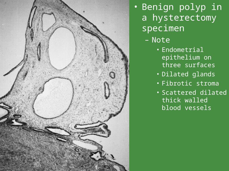
• Benign polyp in a hysterectomy specimen– Note
• Endometrial epithelium on three surfaces
• Dilated glands
• Fibrotic stroma
• Scattered dilated thick walled blood vessels
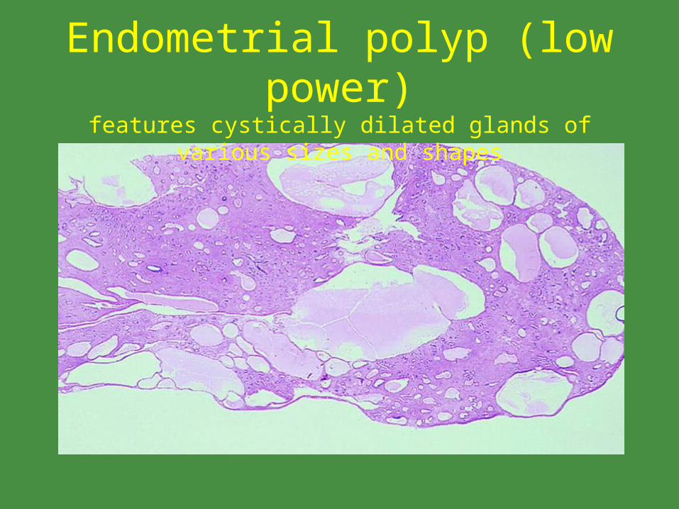
Endometrial polyp (low power)features cystically dilated glands of various sizes and shapes
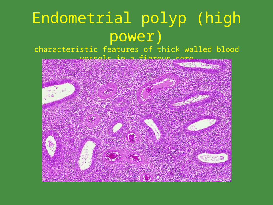
Endometrial polyp (high power)characteristic features of thick walled blood vessels in a fibrous core
Classification• Morphologically diverse lesions that are difficult to
subclassify.• Most are either hyperplastic, atrophic or functional.
– Hyperplastic• resemble diffuse non polypoid endometrial hyperplasia
• no evidence that these have the same significance as diffuse hyperplasia, so best to avoid the term hyperplastic in the diagnosis
– Atrophic• low columnar or cuboidal cells lining cystically dilated glands
• typically in post-menopausal patients
– Functional• resemble normal cycling endometrium
• relatively uncommon
Tamoxifen related polyps
• Larger, sessile with a honeycomb appearance
• bizarre stellate shape of glands and frequent epithelial and stromal metaplasias
• often periglandular stromal condensation
• malignant transformation in up to 3%
• interestingly the cytogenetic profile is similar to non-iatrogenic lesions
Differential Diagnosis• Endometrial hyperplasia
– diffuse process, majority of fragments in curettage, absence of thick walled vessels
• polypoid endometrial carcinoma– malignant epithelial cells
• adenofibroma• adenosarcoma
– stromal cells cytologically atypical and mitotically active– stromal cells packed tightly around non malignant glands– leaf like pattern
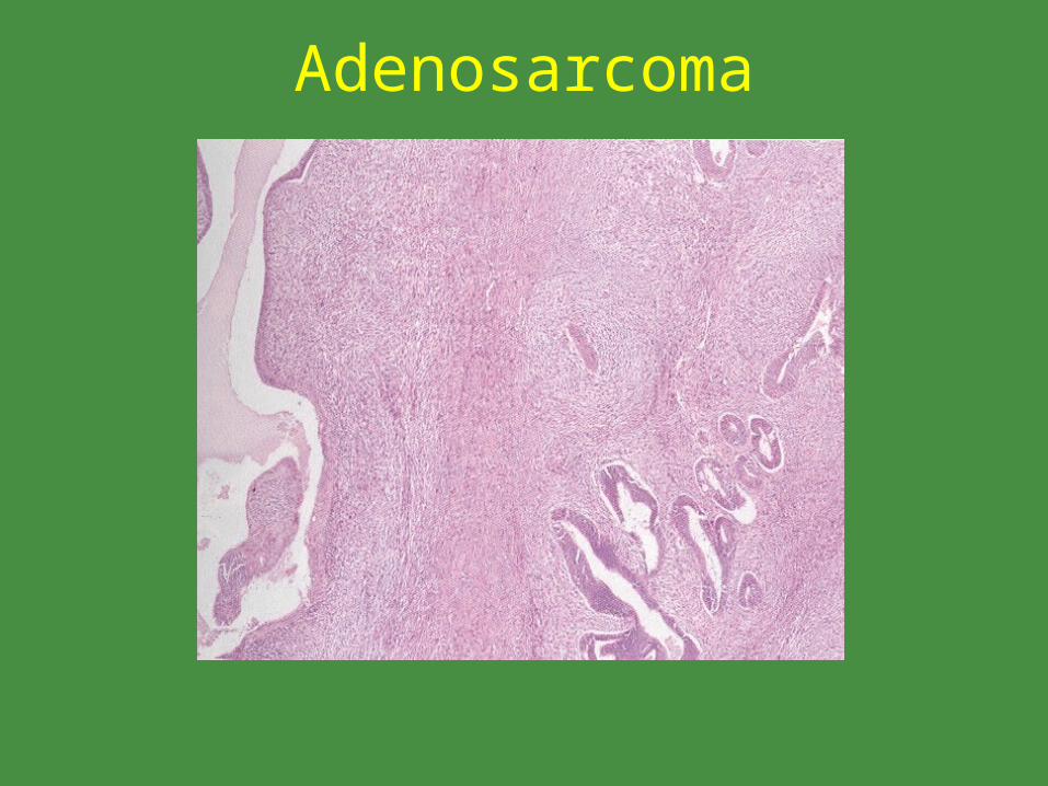
Adenosarcoma
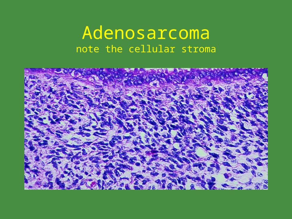
Adenosarcomanote the cellular stroma
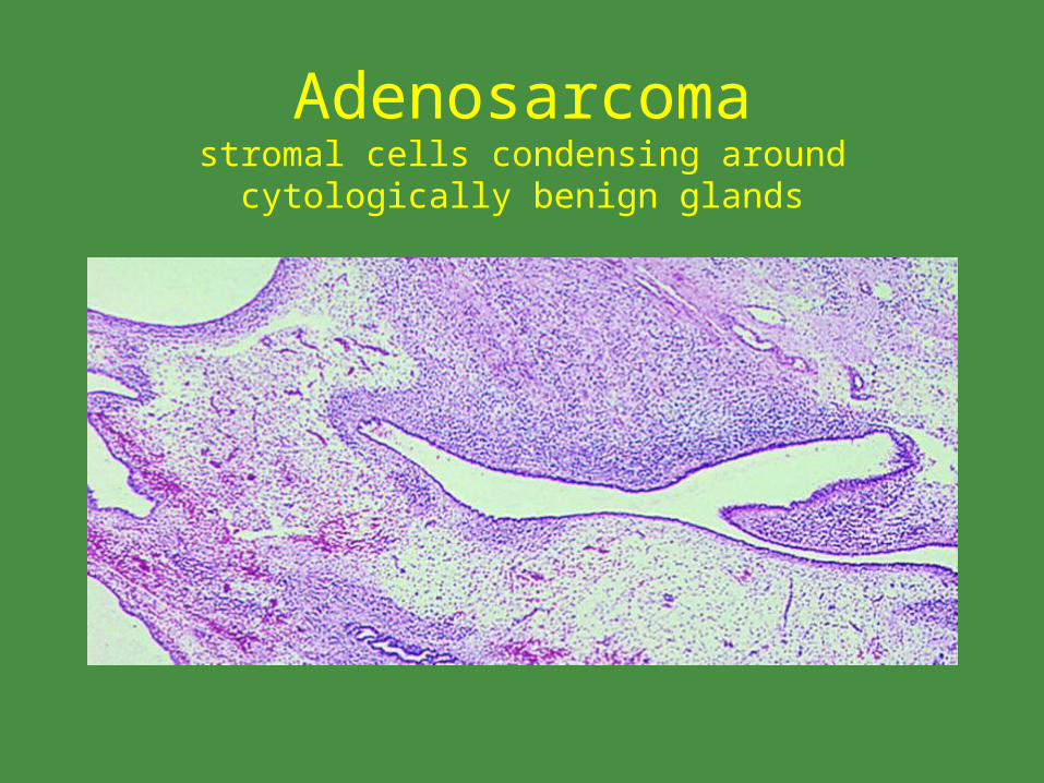
Adenosarcomastromal cells condensing around cytologically benign glands
Clinical behavior and treatment
• At most 5% of polyps contain carcinoma
• polyps may represent a marker of increased cancer risk, but no evidence suggests they are more likely to become cancer than the adjacent endometium
• those containing atypical hyperplasia or carcinoma should be treated as per similar flat lesions
Related Documents