Endocannabinoid system acts as a regulator of immune homeostasis in the gut Nandini Acharya a , Sasi Penukonda b , Tatiana Shcheglova a , Adam T. Hagymasi a , Sreyashi Basu a,1 , and Pramod K. Srivastava a,1 a Department of Immunology and Carole and Ray Neag Comprehensive Cancer Center, University of Connecticut School of Medicine, Farmington, CT 06030; and b Division of Diabetes and Endocrinology, Connecticut Children’s Medical Center, Farmington, CT 06032 Edited by David Artis, Weill Cornell Medical College, New York, NY, and accepted by Editorial Board Member Carl F. Nathan March 27, 2017 (received for review July 23, 2016) Endogenous cannabinoids (endocannabinoids) are small molecules biosynthesized from membrane glycerophospholipid. Ananda- mide (AEA) is an endogenous intestinal cannabinoid that controls appetite and energy balance by engagement of the enteric nervous system through cannabinoid receptors. Here, we uncover a role for AEA and its receptor, cannabinoid receptor 2 (CB2), in the regulation of immune tolerance in the gut and the pancreas. This work demonstrates a major immunological role for an endocanna- binoid. The pungent molecule capsaicin (CP) has a similar effect as AEA; however, CP acts by engagement of the vanilloid receptor TRPV1, causing local production of AEA, which acts through CB2. We show that the engagement of the cannabinoid/vanilloid recep- tors augments the number and immune suppressive function of the regulatory CX3CR1 hi macrophages (Mϕ), which express the highest levels of such receptors among the gut immune cells. Ad- ditionally, TRPV1 -/- or CB2 -/- mice have fewer CX3CR1 hi Mϕ in the gut. Treatment of mice with CP also leads to differentiation of a regulatory subset of CD4 + cells, the Tr1 cells, in an IL-27–depen- dent manner in vitro and in vivo. In a functional demonstration, tolerance elicited by engagement of TRPV1 can be transferred to naïve nonobese diabetic (NOD) mice [model of type 1 diabetes (T1D)] by transfer of CD4 + T cells. Further, oral administration of AEA to NOD mice provides protection from T1D. Our study unveils a role for the endocannabinoid system in maintaining immune homeostasis in the gut/pancreas and reveals a conversation be- tween the nervous and immune systems using distinct receptors. mucosal immunity | T-regulatory cells | cannabis | diabetes | CX3CR1 macrophage T he endocannabinoid system (ECS) is highly conserved in evo- lution dating back to at least 600 million years (1). It consists of (i ) lipid endocannabinoids; (ii ) their receptors such as the G protein-coupled receptors, cannabinoid receptor 1 (CB1), canna- binoid receptor 2 (CB2), and a ligand-gated cation channel vanilloid receptor 1 (i.e., TRPV1); and (iii ) the enzymes such as fatty acid amide hydrolyase (FAAH) that regulate the levels of endocanna- binoids in vivo (2). The ECS impacts several aspects of mammalian physiology, particularly in the gut. Endogenous cannabinoids such as anandamide (AEA) belong to the N-acylethanolamine family and are synthesized from membrane glycerophospholipids (3, 4). AEA is an intestinal endocannabinoid, which engages its cognate receptors on the enteric nervous system and contributes to con- trol of appetite and energy balance (5). Here, we have unraveled a role of the ECS in regulating immune homeostasis in the gut– pancreas axis. The intestinal immune system is continuously exposed to a va- riety of antigens. An effective immune response must be launched against pathogenic insults; however, it must maintain tolerance to the vast amount of antigens such as those in commensal flora, endogenous metabolites, and food components. Mononuclear phagocytes (MNPs) such as CX3CR1 hi macrophage (Mϕ) and CD103 + dendritic cells (DCs), which are present abundantly in the small intestinal lamina propria (siLP), play an instrumental role in maintaining this exquisite balance between inflammation and tolerance (6). On the basis of expression level of the chemokine receptor CX3CR1, two functionally and phenotypically distinct populations of MNPs in the gut have been identified. Although both pop- ulations differentiate from blood derived monocytes, CX3CR1 hi Mϕ are regulatory in nature, whereas CX3CR1 lo cells are per- petuators of inflammation (7). Adoptive transfer of CX3CR1 hi Mϕ provide protection from inflammatory diseases like colitis in mouse models (8). However, the microenvironment signal that “educates” the monocytes and promotes differentiation into CX3CR1 hi (as opposed to CX3CR1 lo )Mϕ still remains elusive. It is increasingly appreciated that the MNPs play a crucial role in gut equilibrium. Identifying key elements that condition the siLP MNPs to become tolerogenic will enable exploration of strategies that can be helpful in providing protection from inflammatory diseases. The studies described here connect the nervous system (re- sponsible for sensing exogenous cannabinoids such as marijuana, as well as endogenous ones) to one of the most fundamental properties of the immune system, i.e., maintenance of an immune tolerant environment in the gut. These studies have a bearing on marijuana use and abuse, particularly with respect to the formu- lations meant for ingestion, and very significantly, for new avenues of understanding and treating human diseases. Significance Exogenous cannabinoids such as marijuana exert their influ- ence through cannabinoid receptors. Endogenous cannabi- noids such as anandamide (AEA) function through the same receptors, and their physiological roles are a subject of intense study. Here, we show that AEA plays a pivotal role in main- taining immunological health in the gut. The immune system in the gut actively tolerates the foreign antigens present in the gut through mechanisms that are only partially understood. We show that AEA contributes to this critical process by pro- moting the presence of CX3CR1 hi macrophages, which are im- munosuppressive. These results uncover a major conversation between the immune and nervous systems. In addition, with the increasing prevalence of ingestion of exogenous mari- juana, our study has significant implications for public health. Author contributions: N.A., S.B., and P.K.S. designed research; N.A., S.P., T.S., A.T.H., and S.B. performed research; N.A., S.B., and P.K.S. contributed new reagents/analytic tools; N.A., S.B., and P.K.S. analyzed data; and N.A. and P.K.S. wrote the paper. The authors declare no conflict of interest. This article is a PNAS Direct Submission. D.A. is a Guest Editor invited by the Editorial Board. Freely available online through the PNAS open access option. 1 To whom correspondence may be addressed. Email: [email protected] or SBasu1@ mdanderson.org. This article contains supporting information online at www.pnas.org/lookup/suppl/doi:10. 1073/pnas.1612177114/-/DCSupplemental. www.pnas.org/cgi/doi/10.1073/pnas.1612177114 PNAS | May 9, 2017 | vol. 114 | no. 19 | 5005–5010 IMMUNOLOGY AND INFLAMMATION

Welcome message from author
This document is posted to help you gain knowledge. Please leave a comment to let me know what you think about it! Share it to your friends and learn new things together.
Transcript
Endocannabinoid system acts as a regulator of immunehomeostasis in the gutNandini Acharyaa, Sasi Penukondab, Tatiana Shcheglovaa, Adam T. Hagymasia, Sreyashi Basua,1,and Pramod K. Srivastavaa,1
aDepartment of Immunology and Carole and Ray Neag Comprehensive Cancer Center, University of Connecticut School of Medicine, Farmington, CT 06030;and bDivision of Diabetes and Endocrinology, Connecticut Children’s Medical Center, Farmington, CT 06032
Edited by David Artis, Weill Cornell Medical College, New York, NY, and accepted by Editorial Board Member Carl F. Nathan March 27, 2017 (received forreview July 23, 2016)
Endogenous cannabinoids (endocannabinoids) are small moleculesbiosynthesized from membrane glycerophospholipid. Ananda-mide (AEA) is an endogenous intestinal cannabinoid that controlsappetite and energy balance by engagement of the entericnervous system through cannabinoid receptors. Here, we uncovera role for AEA and its receptor, cannabinoid receptor 2 (CB2), in theregulation of immune tolerance in the gut and the pancreas. Thiswork demonstrates a major immunological role for an endocanna-binoid. The pungent molecule capsaicin (CP) has a similar effect asAEA; however, CP acts by engagement of the vanilloid receptorTRPV1, causing local production of AEA, which acts through CB2.We show that the engagement of the cannabinoid/vanilloid recep-tors augments the number and immune suppressive function ofthe regulatory CX3CR1hi macrophages (Mϕ), which express thehighest levels of such receptors among the gut immune cells. Ad-ditionally, TRPV1−/− or CB2−/− mice have fewer CX3CR1hi Mϕ inthe gut. Treatment of mice with CP also leads to differentiation ofa regulatory subset of CD4+ cells, the Tr1 cells, in an IL-27–depen-dent manner in vitro and in vivo. In a functional demonstration,tolerance elicited by engagement of TRPV1 can be transferred tonaïve nonobese diabetic (NOD) mice [model of type 1 diabetes(T1D)] by transfer of CD4+ T cells. Further, oral administration ofAEA to NOD mice provides protection from T1D. Our study unveilsa role for the endocannabinoid system in maintaining immunehomeostasis in the gut/pancreas and reveals a conversation be-tween the nervous and immune systems using distinct receptors.
mucosal immunity | T-regulatory cells | cannabis | diabetes |CX3CR1 macrophage
The endocannabinoid system (ECS) is highly conserved in evo-lution dating back to at least 600 million years (1). It consists of
(i) lipid endocannabinoids; (ii) their receptors such as the Gprotein-coupled receptors, cannabinoid receptor 1 (CB1), canna-binoid receptor 2 (CB2), and a ligand-gated cation channel vanilloidreceptor 1 (i.e., TRPV1); and (iii) the enzymes such as fatty acidamide hydrolyase (FAAH) that regulate the levels of endocanna-binoids in vivo (2). The ECS impacts several aspects of mammalianphysiology, particularly in the gut. Endogenous cannabinoids suchas anandamide (AEA) belong to the N-acylethanolamine familyand are synthesized from membrane glycerophospholipids (3, 4).AEA is an intestinal endocannabinoid, which engages its cognatereceptors on the enteric nervous system and contributes to con-trol of appetite and energy balance (5). Here, we have unraveleda role of the ECS in regulating immune homeostasis in the gut–pancreas axis.The intestinal immune system is continuously exposed to a va-
riety of antigens. An effective immune response must be launchedagainst pathogenic insults; however, it must maintain tolerance tothe vast amount of antigens such as those in commensal flora,endogenous metabolites, and food components. Mononuclearphagocytes (MNPs) such as CX3CR1hi macrophage (Mϕ) andCD103+ dendritic cells (DCs), which are present abundantly in thesmall intestinal lamina propria (siLP), play an instrumental role in
maintaining this exquisite balance between inflammation andtolerance (6).On the basis of expression level of the chemokine receptor
CX3CR1, two functionally and phenotypically distinct populationsof MNPs in the gut have been identified. Although both pop-ulations differentiate from blood derived monocytes, CX3CR1hi
Mϕ are regulatory in nature, whereas CX3CR1lo cells are per-petuators of inflammation (7). Adoptive transfer of CX3CR1hi
Mϕ provide protection from inflammatory diseases like colitis inmouse models (8). However, the microenvironment signal that“educates” the monocytes and promotes differentiation intoCX3CR1hi (as opposed to CX3CR1lo) Mϕ still remains elusive.It is increasingly appreciated that the MNPs play a crucial role ingut equilibrium. Identifying key elements that condition the siLPMNPs to become tolerogenic will enable exploration of strategiesthat can be helpful in providing protection from inflammatorydiseases.The studies described here connect the nervous system (re-
sponsible for sensing exogenous cannabinoids such as marijuana,as well as endogenous ones) to one of the most fundamentalproperties of the immune system, i.e., maintenance of an immunetolerant environment in the gut. These studies have a bearing onmarijuana use and abuse, particularly with respect to the formu-lations meant for ingestion, and very significantly, for new avenuesof understanding and treating human diseases.
Significance
Exogenous cannabinoids such as marijuana exert their influ-ence through cannabinoid receptors. Endogenous cannabi-noids such as anandamide (AEA) function through the samereceptors, and their physiological roles are a subject of intensestudy. Here, we show that AEA plays a pivotal role in main-taining immunological health in the gut. The immune system inthe gut actively tolerates the foreign antigens present in thegut through mechanisms that are only partially understood.We show that AEA contributes to this critical process by pro-moting the presence of CX3CR1hi macrophages, which are im-munosuppressive. These results uncover a major conversationbetween the immune and nervous systems. In addition, withthe increasing prevalence of ingestion of exogenous mari-juana, our study has significant implications for public health.
Author contributions: N.A., S.B., and P.K.S. designed research; N.A., S.P., T.S., A.T.H., andS.B. performed research; N.A., S.B., and P.K.S. contributed new reagents/analytic tools;N.A., S.B., and P.K.S. analyzed data; and N.A. and P.K.S. wrote the paper.
The authors declare no conflict of interest.
This article is a PNAS Direct Submission. D.A. is a Guest Editor invited by the EditorialBoard.
Freely available online through the PNAS open access option.1To whom correspondence may be addressed. Email: [email protected] or [email protected].
This article contains supporting information online at www.pnas.org/lookup/suppl/doi:10.1073/pnas.1612177114/-/DCSupplemental.
www.pnas.org/cgi/doi/10.1073/pnas.1612177114 PNAS | May 9, 2017 | vol. 114 | no. 19 | 5005–5010
IMMUNOLO
GYAND
INFLAMMATION
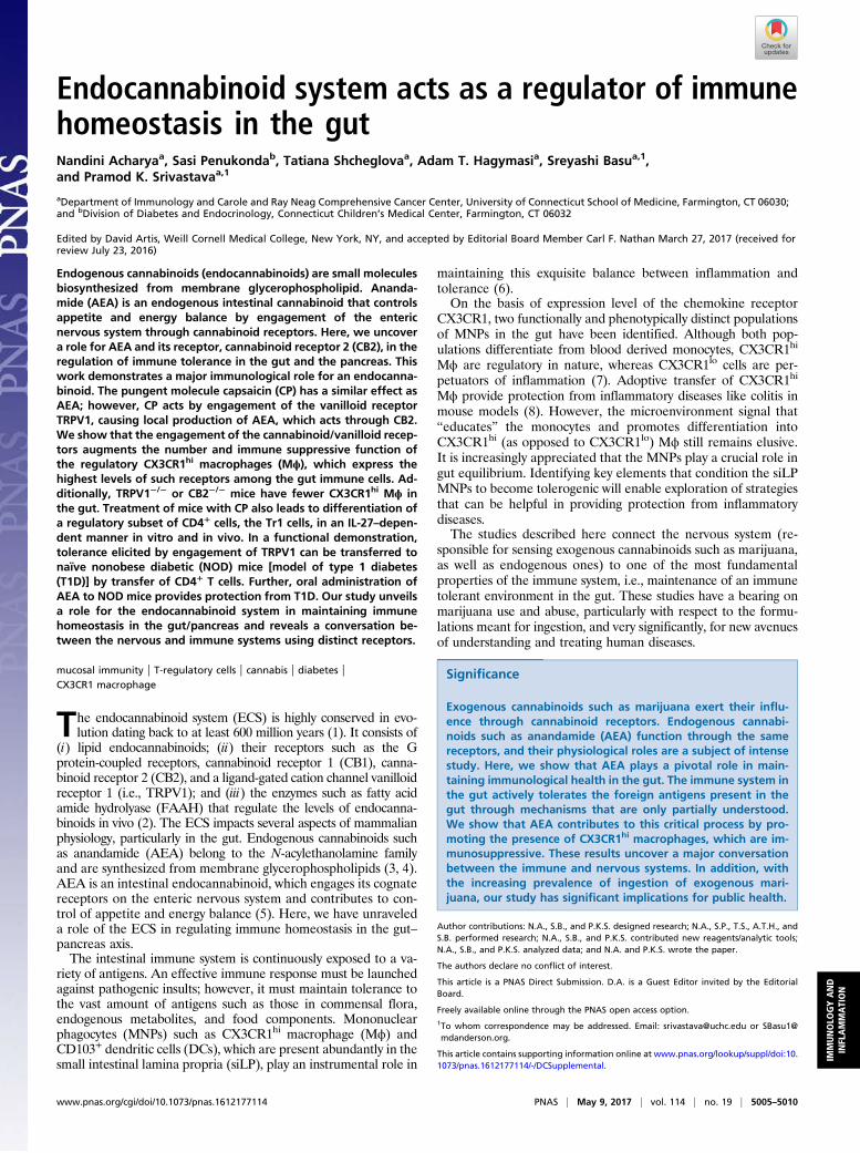
ResultsCX3CR1hi Mϕ Express the Highest Levels of ECS Receptors CB2 andTRPV1. siLP cells were isolated after removing the Peyer’s patches(PPs), and cell suspensions obtained were analyzed. Fig. S1Adepicts the gating strategy to identify the different cell pop-ulations. The SSChi cells were gated for CD11b+SiglecF+ cells(i.e., eosinophils) and the SSClo cells for lymphocytes (B220+ andCD3+) and the MNPs. Phenotypic characterization of the MNPswas based on the expression of integrins CD11b (αM), CD11c(αX), CD103 (αE), and the chemokine receptor CX3CR1.Special attention was paid to the CX3CR1 phenotype becauseCX3CR1hi Mϕ but not the CX3CR1lo cells play a significant rolein immune homeostasis (7). To facilitate the identification ofCX3CR1hi and CX3CR1lo cells, we used the CX3CR1+/gfp reportermice in which GFP expression is a marker for CX3CR1 ex-pression (9). As a quality-control measure, cell suspensions de-rived from siLP of these mice were stained with antibody againstCX3CR1 and also checked for GFP by flow cytometry. The twopatterns were consistent (Fig. S1B). We classified the MNPs as R1(CD11b+CD11c+) and R2 (CD11b−CD11c+). R1 was further sub-classified into CD11b+CD11c+CX3CR1hi, CD11b+CD11c+CX3CR1lo,and CD11b+CD11c+CD103+, and, in the R2, we looked forCD11b−CD11chiCD103+ (Fig. S1A, Bottom). Selected pop-ulations were analyzed for the expression level of TRPV1 (Fig.S1C) and CB2 (Fig. S1D). Of note, the MNPs expressed higherlevels of TRPV1 and CB2 compared with the lymphocytes andeosinophils. Strikingly, CX3CR1hi cells expressed higher levels ofTRPV1 and CB2 compared with the other cells analyzed (Fig.1A). This finding was corroborated by quantitative PCR (qPCR)analysis of mRNA levels of TRPV1 and Cnr2 (gene encodingCB2) on the various gut immune populations (Fig. S1E). Be-cause of the substantial overlap in surface phenotype of in-testinal DCs and Mϕ, designation of CX3CR1hi cells as DCs orMϕ is debatable. However, Tamoutounour et al. have shown thatexpression of CD64 (the high-affinity FcγR1) and absence ofCD103 can be used in combination with CD11c and MHCII todefinitively define functionally distinct Mϕ in intestine of mice(10). The population predominantly expressing TRPV1 andCB2 was found to meet these criteria (Fig. 1B, Top). Consistentwith Tamoutounour et al., we show that CX3CR1lo cells are aheterogeneous population comprising of Mϕ (CD64+) and DCs(CD64−; Fig. S2A). Previous reports demonstrate that CX3CR1hi
Mϕ make IL-10 ex vivo without stimulation (7). The IL-10–producing capacity of the CX3CR1hi Mϕ was tested ex vivo with-out any stimulation by using the IL-10–GFP reporter mice (11); itwas observed that CX3CR1hi Mϕ express high levels of IL-10 (Fig.1B, Bottom). Thus, TRPV1 and CB2, which are integral compo-nents of the ECS, are preferentially expressed by the CX3CR1hi
regulatory Mϕ.
ECS Receptors Regulate CX3CR1hi Mϕ Homeostasis in the Gut. Giventhe high expression of TRPV1 and CB2 on CX3CR1hi Mϕ, weanalyzed the role of each receptor. For TRPV1, we back-crossedTRPV1−/− mice (12) with CX3CR1gfp/gfp reporter mice to obtainCX3CR1gfp/+ TRPV1−/− mice. We validated the expression offunctional (i.e., WT) allele and GFP allele for CX3CR1 and theabsence of TRPV1 by PCR (Fig. S2B). Introduction of GFPdisrupts the expression of endogenous CX3CR1 such that ahomozygous mice does not express functional CX3CR1 proteinwhereas heterozygous mice do, and the expression of CX3CR1in them can be followed through GFP. By analyzing the siLP cellsof the CX3CR1gfp/+TRPV1−/− mice, we observed that the absenceof TRPV1 influences the frequency and number of CX3CR1+
cells differently: the frequency and number of CX3CR1hi Mϕ arereduced significantly (Fig. 1C, Middle), whereas, in case of theCX3CR1lo cells, there is a trend of decreasing number and fre-quency but the difference is not statistically significant (Fig. 1C,
Bottom). These results in CX3CR1gfp/+TRPV1−/− mice were alsoreproduced in TRPV1−/− mice in which CX3CR1 was identifiedwith antibody staining (Fig. S2C). The frequency and number ofthe other cells such as CD103+ DCs and CD3+ and B220+ cellsremain unaffected in the absence of TRPV1 (Fig. S2 D and E).Next, we tested the importance of CB2 on the homeostasis of
CX3CR1hi Mϕ by using mice genetically lacking CB2: the fre-quency and absolute number of CX3CR1hi Mϕ was significantlyreduced in CB2-deficient mice (Fig. 1D, Middle). As in TRPV1−/−
mice, there is a trend of decreasing number and frequencyamong the CX3CR1lo cells in CB2−/− mice as well, but the dif-ference is not statistically significant (Fig. 1D, Bottom).
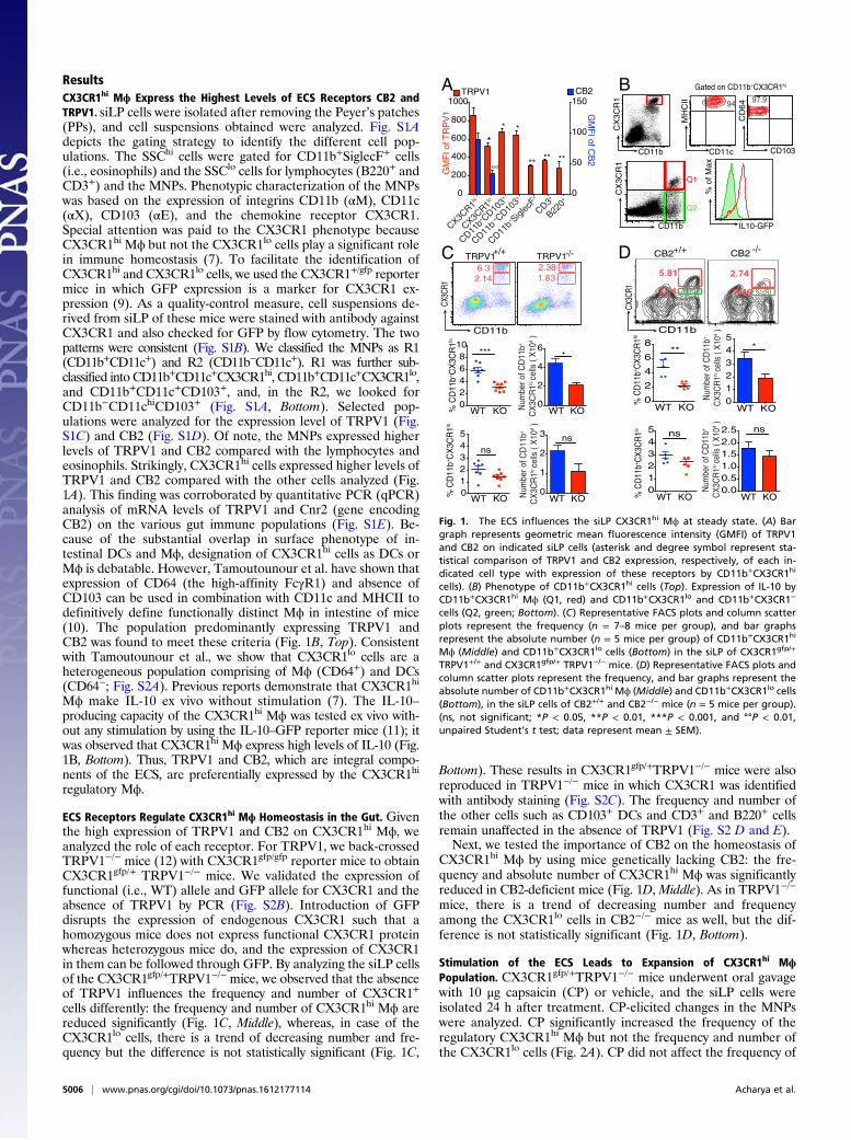
Stimulation of the ECS Leads to Expansion of CX3CR1hi MϕPopulation. CX3CR1gfp/+TRPV1−/− mice underwent oral gavagewith 10 μg capsaicin (CP) or vehicle, and the siLP cells wereisolated 24 h after treatment. CP-elicited changes in the MNPswere analyzed. CP significantly increased the frequency of theregulatory CX3CR1hi Mϕ but not the frequency and number ofthe CX3CR1lo cells (Fig. 2A). CP did not affect the frequency of
D +/+ -/-
% C
D11
b+ CX
3CR
1hi
Num
ber o
f CD
11b+
CX
3CR
1hi c
ells
( X
104 )
20
468
10***
0
2
4
6*
WT KO WT KO
KO012345
ns
0
1
2
3 ns
Num
ber o
f CD
11b+
CX
3CR
1lo c
ells
( X
104 )
% C
D11
b+ CX
3CR
1lo
WT WT KO
CD11b
CX3C
R1
TRPV1+/+ TRPV1-/-2.381.83
6.32.14
TRPV1+/+
TRPV1-/-
% C
D11
b+ CX3
CR
1hiCX
3CR1
2.74
2.46
5.81
3.01
0
2
4
6
8**
012345
*
Num
ber o
f CD
11b+
CX3
CR
1hi ce
lls (
X104 )
CB2 CB2
CD11b
WT KO WT KO
% C
D11
b+ CX3
CR
1lo
012345
0.00.51.01.52.02.5 nsns
Num
ber o
f CD
11b+
CX3
CR
1lo ce
lls (
X104 )
Q1
Q2
CD11b
CX
3CR
1
IL10-GFP
% o
f Max
B
CD11b
CX
3CR
1
CD103
CD
64
97.9
CD11c
MH
CII 94
Gated on CD11b+CX3CR1hi
C
0
200
400
600
800
1000
0
50
100
150
GM
FI o
f TR
PV
1
TRPV1 CB2
**
* *
**** **
°°
GM
FI of C
B2
B220+
CD3+
CX3CR1
hi
CX3CR1
lo
CD11b+ CD10
3+
CD11b-+ CD10
3+
CD11b+ Sigl
ecF+
A
WT KO WT KO
Fig. 1. The ECS influences the siLP CX3CR1hi Mϕ at steady state. (A) Bargraph represents geometric mean fluorescence intensity (GMFI) of TRPV1and CB2 on indicated siLP cells (asterisk and degree symbol represent sta-tistical comparison of TRPV1 and CB2 expression, respectively, of each in-dicated cell type with expression of these receptors by CD11b+CX3CR1hi
cells). (B) Phenotype of CD11b+CX3CR1hi cells (Top). Expression of IL-10 byCD11b+CX3CR1hi Mϕ (Q1, red) and CD11b+CX3CR1lo and CD11b+CX3CR1−
cells (Q2, green; Bottom). (C) Representative FACS plots and column scatterplots represent the frequency (n = 7–8 mice per group), and bar graphsrepresent the absolute number (n = 5 mice per group) of CD11b+CX3CR1hi
Mϕ (Middle) and CD11b+CX3CR1lo cells (Bottom) in the siLP of CX3CR1gfp/+
TRPV1+/+ and CX3CR1gfp/+ TRPV1−/− mice. (D) Representative FACS plots andcolumn scatter plots represent the frequency, and bar graphs represent theabsolute number of CD11b+CX3CR1hi Mϕ (Middle) and CD11b+CX3CR1lo cells(Bottom), in the siLP cells of CB2+/+ and CB2−/− mice (n = 5 mice per group).(ns, not significant; *P < 0.05, **P < 0.01, ***P < 0.001, and °°P < 0.01,unpaired Student’s t test; data represent mean ± SEM).
5006 | www.pnas.org/cgi/doi/10.1073/pnas.1612177114 Acharya et al.
CD11c+CD103+ DCs (Fig. S3A). To test if CP-mediated in-crease in CX3CR1hi Mϕ frequency was caused by enhancedproliferation, an in vivo BrdU incorporation assay was per-formed in mice that had undergone oral gavage with CP: feedingof CP did not result in incorporation of BrdU in CX3CR1hi Mϕ(Fig. S3B). We performed RNA sequencing and IsoEM/IsoDEanalysis (13, 14) (as described in SI Materials and Methods) onthe total MNPs of siLP to study the changes mediated bytreatment with CP; the heat map for hierarchical clustering ofthe top 1,200 differentially expressed genes (DEGs) shows aclear presence of genes that are significantly up- or down-regulated by CP treatment (Fig. S3C). We used Database forAnnotation, Visualization and Integrated Discovery (DAVID)Bioinformatic Resource, version 6.8, to study the functional-related gene groups and pathways. We found that CP treat-ment significantly enhances the cytokine/chemokine-mediatedsignaling pathways such as CCL2, CXCL12, CSF1, and IL-10(Table S1). Thus, the increase in the frequency of CX3CR1hi
Mϕ must derive from augmented migration or differentiation ora combination of both processes.To distinguish between the contribution of TRPV1-expressing
neurons vs. immune cells to the phenomenon shown in Fig. 2A,TRPV1−/− mice reconstituted with bone marrow (BM) fromTRPV1+/+ mice were treated with CP. The siLP CX3CR1hi Mϕpopulations in all groups were monitored: CP-fed BM chimeric(BMC) mice behaved like the WT mice, indicating that TRPV1
expression on the hematopoietic cells was sufficient for activity.There was a notable, albeit statistically insignificant, differencebetween the expansion observed in the WT mice compared withthe BMC mice, suggesting a possible, though not definitive,contribution from the neuronal TRPV1 in the process (Fig. 2B).The effect of oral administration of AEA, an endogenous
compound capable of activating vanilloid and cannabinoid re-ceptors, on CX3CR1hi Mϕ of siLP was examined. Oral adminis-tration of AEA led to a significant increase in the frequency ofCX3CR1hi Mϕ (Fig. 2C, Bottom Left). As expected, we did notobserve a difference in the frequency of CX3CR1lo cells aftertreatment with AEA (Fig. 2C, Bottom Right). We reasoned that, ifadministering exogenous AEA can affect the CX3CR1hi Mϕ, in-creasing the endogenous levels of AEA by inhibiting the enzymethat catabolizes it should yield similar results. Endogenous levelsof AEA are controlled by the enzyme FAAH, which catabolizesAEA into arachidonic acid and ethanolamine (Fig. 2D, Top).Inhibition of FAAH, the enzyme that catabolizes AEA, increasesthe levels of AEA in the brain and peripheral tissues (15). Massaet al. demonstrated that mice genetically deficient in FAAH areprotected from 2,4-dinitrobenzene sulfonic acid-induced colitis,highlighting the antiinflammatory role of AEA in the gut (16).Mice underwent gavage with an orally viable, irreversible
FAAH inhibitor, PF3845, and the effect of this treatment onCX3CR1hi Mϕ in LP was examined 24 h after feeding. Treat-ment of mice with PF3845 increased the frequency of the regu-latory Mϕ demonstrating that endogenous AEA can profoundlyinfluence the homeostasis of CX3CR1hi Mϕ (Fig. 2D, Bottom).
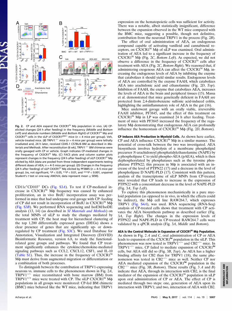
CP Induces AEA Production in Myeloid Cells. As shown here earlier,CP and AEA influence CX3CR1hi Mϕ in a similar manner. Thepotential of cross-talk between the two was investigated. AEAbiosynthesis involves hydrolysis of a membrane phospholipidprecursor N-arachidonoyl phosphatidylethanolamine (NAPE) bya phospholipase C to yield phospho-AEA (pAEA), which is thendephosphorylated by phosphatases such as the tyrosine phos-phatase PTPN22; this process in Mϕ is associated with an in-crease of the level of PTPN22 (17) and a decrease in the level ofphospholipase D NAPE-PLD (17). Consistent with this pattern,analysis of the transcriptome of siLP MNPs from CP-treatedmice revealed that CP leads to increase in the expression ofPTPN22 with a concomitant decrease in the level of NAPE-PLD(Fig. 3A, Top Left).To explore this phenomenon mechanistically in a pure mye-
loid population (as opposed to in vivo, in which the effects couldbe indirect), the Mϕ cell line RAW264.7, which expressesTRPV1 (Fig. S4A), was used. RNA sequencing (RNA-Seq)analysis of CP-treated cells shows that treatment with CP acti-vates the AEA biosynthesis pathway as described earlier (Fig.3A, Top Right). The changes in the expression levels ofPTPN22 and NAPE-PLD in CP-treated RAW264.7 cells werevalidated by a qPCR analysis (Fig. 3A, Middle and Bottom).
AEA Is the Central Molecule in Expansion of CX3CR1hi Mϕ Population.As shown in Fig. 2 A and C, oral administration of CP or AEAleads to expansion of the CX3CR1hi population in the siLP. Thisphenomenon was now tested in TRPV1−/− and CB2−/− mice. InTRPV1−/− mice, CP failed to mediate expansion of CX3CR1hi
cells, but AEA still did so (Fig. 3B, Top). As AEA has a higherbinding affinity for CB2 than for TRPV1 (18), the same phe-nomenon was tested in CB2−/− mice as well. Neither CP norAEA mediated expansion of the CX3CR1hi population in theCB2−/− mice (Fig. 3B, Bottom). These results (Fig. 3 A and B)indicate that AEA, through its interaction with CB2, is the finalmediator of the expansion of the CX3CR1hi population in siLPupon oral administration of CP or AEA. The effect of CP ismediated through two steps: one, generation of AEA upon itsinteraction with TRPV1; and two, interaction of AEA with CB2.
8.381.74
3.971.81
Veh CP
CD11b
CX
3CR
1
0.0
0.5
1.0
1.5
**
Num
ber
of C
D11
b+
CX
3CR
1hi c
ells
( X
104
)
02468
10 **
% C
D11
b+C
X3C
R1hi
Veh VehCP CP
0
1
2
3 ns
Num
ber
of C
D11
b+
CX
3CR
1lo c
ells
( X
103
)
012345
ns
% C
D11
b+C
X3C
R1lo
Veh VehCP CP
DEthanolamine
AEA FAAHInhibited by
PF3845
Arachidoinc Acid+
% C
D11
b+C
X3C
R1hi
0
2
4
6
8 *
Veh
PF3845
8.34
1.88
3.41
1.64
3.66
1.74
6.54
2.02
Veh AEA-250 AEA-400 AEA-500
CD11b
CX
3CR
1C
B
Veh CP Veh CP0
2
4
6
8
10 ns**
*
WT WT WT TRPV1-/-BMC
% C
D11
b+C
X3C
R1hi
Veh25
0 40
0 50
00
1
2
3
nsns
ns
Veh 250
400
500
0
2
4
6
8
10
***
ns
**
% C
D11
b+C
X3C
R1hi
% C
D11
b+C
X3C
R1lo
AEA AEA
A
Fig. 2. CP and AEA expand the CX3CR1hi Mϕ population in vivo. (A) CP-elicited changes (24 h after feeding) in the frequency (Middle and BottomLeft) and absolute numbers (Middle and Bottom Right) of CX3CR1hi Mϕ andCX3CR1lo cells in the siLP of CX3CR1gfp/+ mice (n = 4 mice per group). Veh,vehicle-treated mice. (B) TRPV1−/− mice (n = 4 mice per group) were lethallyirradiated and, 24 h later, received CD45.1 C57BL/6 BM as described in Ma-terials and Methods. After reconstitution (6 wk), TRPV1−/− BM chimeras wereorally gavaged with CP or vehicle. Graph indicates CP-mediated changes inthe frequency of CX3CR1hi Mϕ. (C) FACS plots and column scatter plotsrepresent changes in the frequency (24 h after feeding) of siLP CX3CR1hi Mϕelicited by AEA (data are pooled from three independent experiments testingdifferent doses of AEA; n = 4–5 mice per group). (D) Changes in the frequency(24 h after feeding) of siLP CX3CR1hi Mϕ elicited by PF3845 (n = 4–5 mice pergroup). (ns, not significant; *P < 0.05, **P < 0.01, and ***P < 0.001, unpairedStudent’s t test or one-way ANOVA; data represent mean ± SEM).
Acharya et al. PNAS | May 9, 2017 | vol. 114 | no. 19 | 5007
IMMUNOLO
GYAND
INFLAMMATION
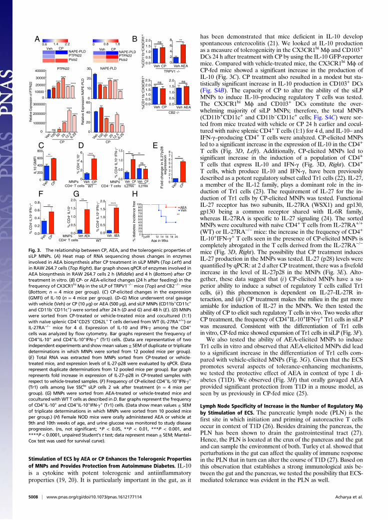
Stimulation of ECS by AEA or CP Enhances the Tolerogenic Propertiesof MNPs and Provides Protection from Autoimmune Diabetes. IL-10is a cytokine with potent tolerogenic and antiinflammatoryproperties (19, 20). It is particularly important in the gut, as it
has been demonstrated that mice deficient in IL-10 developspontaneous enterocolitis (21). We looked at IL-10 productionas a measure of tolerogenicity in the CX3CR1hi Mϕ and CD103+
DCs 24 h after treatment with CP by using the IL-10 GFP-reportermice. Compared with vehicle-treated mice, the CX3CR1hi Mϕ ofCP-fed mice showed a significant increase in the production ofIL-10 (Fig. 3C). CP treatment also resulted in a modest but sta-tistically significant increase in IL-10 production in CD103+ DCs(Fig. S4B). The capacity of CP to alter the ability of the siLPMNPs to induce IL-10–producing regulatory T cells was tested.The CX3CR1hi Mϕ and CD103+ DCs constitute the over-whelming majority of siLP MNPs; therefore, the total MNPs(CD11b+CD11c+ and CD11b−CD11c+ cells; Fig. S4C) were sor-ted from mice treated with vehicle or CP 24 h earlier and cocul-tured with naïve splenic CD4+ T cells (1:1) for 4 d, and IL-10– andIFN-γ–producing CD4+ T cells were analyzed. CP-elicited MNPsled to a significant increase in the expression of IL-10 in the CD4+
T cells (Fig. 3D, Left). Additionally, CP-elicited MNPs led tosignificant increase in the induction of a population of CD4+
T cells that express IL-10 and IFN-γ (Fig. 3D, Right). CD4+
T cells, which produce IL-10 and IFN-γ, have been previouslydescribed as a potent regulatory subset called Tr1 cells (22). IL-27,a member of the IL-12 family, plays a dominant role in the in-duction of Tr1 cells (23). The requirement of IL-27 for the in-duction of Tr1 cells by CP-elicited MNPs was tested. FunctionalIL-27 receptor has two subunits, IL-27RA (WSX1) and gp130,gp130 being a common receptor shared with IL-6R family,whereas IL-27RA is specific to IL-27 signaling (24). The sortedMNPs were cocultured with naïve CD4+ T cells from IL-27RA+/+
(WT) or IL-27RA−/− mice: the increase in the frequency of CD4+
IL-10+IFN-γ+ T cells seen in the presence of CP-elicited MNPs iscompletely abrogated in the T cells derived from the IL-27RA−/−
mice (Fig. 3D, Right). The possibility that CP treatment inducesIL-27 production in the MNPs was tested. IL-27 (p28) levels werequantified by qPCR: at 2 d after CP treatment, there was a fivefoldincrease in the level of IL-27p28 in the MNPs (Fig. 3E). Alto-gether, these data suggest that (i) CP-elicited MNPs have a su-perior ability to induce a subset of regulatory T cells called Tr1cells, (ii) this phenomenon is dependent on IL-27–IL-27R in-teraction, and (iii) CP treatment makes the milieu in the gut moreamiable for induction of IL-27 in the MNPs. We then tested theability of CP to elicit such regulatory T cells in vivo. Two weeks afterCP treatment, the frequency of CD4+IL-10+IFN-γ+ Tr1 cells in siLPwas measured. Consistent with the differentiation of Tr1 cellsin vitro, CP-fed mice showed expansion of Tr1 cells in siLP (Fig. 3F).We also tested the ability of AEA-elicited MNPs to induce
Tr1 cells in vitro and observed that AEA-elicited MNPs did leadto a significant increase in the differentiation of Tr1 cells com-pared with vehicle-elicited MNPs (Fig. 3G). Given that the ECSpromotes several aspects of tolerance-enhancing mechanisms,we tested the protective effect of AEA in context of type 1 di-abetes (T1D). We observed (Fig. 3H) that orally gavaged AEAprovided significant protection from T1D in a mouse model, asseen by us previously in CP-fed mice (25).
Lymph Node Specificity of Increase in the Number of Regulatory Mϕby Stimulation of ECS. The pancreatic lymph node (PLN) is thefirst site in which initiation and priming of autoreactive T cellsoccur in context of T1D (26). Besides draining the pancreas, thePLN has been shown to drain the gastrointestinal tract (27).Hence, the PLN is located at the crux of the pancreas and the gutand can sample the environment of both. Turley et al. showed thatperturbations in the gut can affect the quality of immune responsein the PLN that in turn can alter the course of T1D (27). Based onthis observation that establishes a strong immunological axis be-tween the gut and the pancreas, we tested the possibility that ECS-mediated tolerance was evident in the PLN as well.
BA
E
G H
012345678
expre
ssio
n **
Veh CP
F
02468 12 14 16 18 200
102030405060708090
100
Veh AEA
NAPE-PLDPTPN22Plcb2
Veh CP Veh
0.6 1.4 2.2 0 0.8 1.6
NAPE-PLDPTPN22Plcb2
CP
D
0.0
0.2
0.4
0.6
0.8**
CPVeh
% C
D4+
IL10
+ IF
N-γ
+
0
10000
20000
30000
40000
***
********
********
0
10
20
30
********
**** ****
****
Veh 100 50 25 12
.56.2
50
100000
200000
300000
ns *
****
****
****
Veh0
20
40
60**
****
****
*
****
PTPN22 NAPE-PLD
Rel
ativ
e Ex
pres
sion
of P
TPN
22
Veh CP0.0
0.5
1.0
1.5
2.0
ns
Veh AEA0
2
4
6
8 **
TRPV1 -/-
%C
D11b
+C
X3C
R1
hi
Veh CP0.00.51.01.52.02.5
ns
Veh AEA0.0
0.5
1.0
1.5
2.0
ns
CB2 -/-
%C
D11b
+C
X3C
R1
hi
IL27RA IL27RA-/-
MNPsCD4+ T cells
Veh CP Veh CP0.0
0.5
1.0
1.5 **
ns
**
Veh CPWT
0
2
4
6 ***
% C
D4+
IL10
+
MNPsCD4+ T cells +/+
**
100 50 25 12
.56.2
5
Rel
ativ
e E
xpre
ssio
n N
AP
E-P
LD
PCPC
C
Veh AEA Veh AEA
% C
D4
+ IL
10
0.0
0.5
1.0
1.5
2.0 **
0.0
0.5
1.0
1.5
2.0 ****
WT MNPsCD4+ T cells %
dia
bete
s in
cidence
free
0
200
400
600
800
IL10 G
MF
I **
Veh CP
Age in Wks
Fold
change in IL27p28
% C
D4+
IL10
+ IF
N- γ
% C
D4+
IL10
+ IF
N-γ+
+
+
+
Fig. 3. The relationship between CP, AEA, and the tolerogenic properties ofsiLP MNPs. (A) Heat map of RNA sequencing shows changes in enzymesinvolved in AEA biosynthesis after CP treatment in siLP MNPs (Top Left) andin RAW 264.7 cells (Top Right). Bar graph shows qPCR of enzymes involved inAEA biosynthesis in RAW 264.7 cells 2 h (Middle) and 4 h (Bottom) after CPtreatment in vitro. (B) CP- or AEA-elicited changes (24 h after feeding) in thefrequency of CX3CR1hi Mϕ in the siLP of TRPV1−/− mice (Top) and CB2−/− mice(Bottom; n = 4 mice per group). (C) CP-elicited changes in the expression(GMFI) of IL-10 (n = 4 mice per group). (D–G) Mice underwent oral gavagewith vehicle (Veh) or CP (10 μg) or AEA (500 μg), and siLP MNPs (CD11b+CD11c+
and CD11b−CD11c+) were sorted after 24 h (D and G) and 48 h (E). (D) MNPswere sorted from CP-treated or vehicle-treated mice and cocultured (1:1)with naïve splenic CD4+CD25−CD62L+ T cells derived from WT (IL-27RA+/+) orIL-27RA−/− mice for 4 d. Expression of IL-10 and IFN-γ among the CD4+
cells was analyzed by flow cytometry. Bar graphs represent the frequency ofCD4+IL-10+ and CD4+IL-10+IFN-γ+ (Tr1) cells. (Data are representative of twoindependent experiments and showmean values ± SEM of duplicate or triplicatedeterminations in which MNPs were sorted from 12 pooled mice per group).(E) Total RNA was extracted from MNPs sorted from CP-treated or vehicle-treated mice, and expression levels of IL-27-p28 were evaluated by qPCR. (Datarepresent duplicate determinations from 12 pooled mice per group). Bar graphrepresents fold increase in expression of IL-27–p28 in CP-treated samples withrespect to vehicle-treated samples. (F) Frequency of CP-elicited CD4+IL-10+IFN-γ+
(Tr1) cells among live SSClo siLP cells 2 wk after treatment (n = 4 mice pergroup). (G) MNPs were sorted from AEA-treated or vehicle-treated mice andcocultured withWT T cells as described inD. Bar graphs represent the frequencyof CD4+IL-10+ and CD4+IL-10+IFN-γ+ (Tr1) cells. (Data show mean values ± SEMof triplicate determinations in which MNPs were sorted from 10 pooled miceper group.) (H) Female NOD mice were orally administered AEA or vehicle at9th and 10th weeks of age, and urine glucose was monitored to study diseaseprogression. (ns, not significant; *P < 0.05, **P < 0.01, ***P < 0.001, and****P < 0.0001, unpaired Student’s t test; data represent mean ± SEM; Mantel–Cox test was used for survival curve).
5008 | www.pnas.org/cgi/doi/10.1073/pnas.1612177114 Acharya et al.
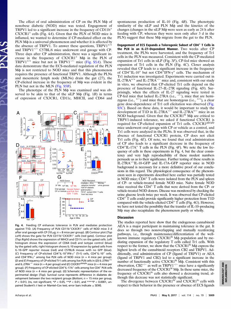
The effect of oral administration of CP on the PLN Mϕ ofnonobese diabetic (NOD) mice was tested. Engagement ofTRPV1 led to a significant increase in the frequency of CD11b+
CX3CR1+ cells (Fig. 4A). Given that the PLN of NOD mice isinflamed, we wanted to determine if CP-mediated effect on thePLNMϕ is a universal phenomenon and whether it is affected bythe absence of TRPV1. To answer these questions, TRPV1+/+
and TRPV1−/− C57BL/6 mice underwent oral gavage with CP.Three days after CP administration, there was a significant in-crease in the frequency of CX3CR1+ Mϕ in the PLN ofTRPV1+/+ mice but not in TRPV1−/− mice (Fig. S5A). Thesedata demonstrate that the ECS-mediated regulation of the PLNMϕ is not restricted to NOD mice and that this phenomenonrequires the presence of functional TRPV1. Although the PLNsand mesenteric lymph node (MLNs) drain the gut (27), theCP-elicited increase in the frequency of Mϕ was evident in thePLN but not in the MLN (Fig. S5B).The phenotype of the PLN Mϕ was examined and was ob-
served to be akin to that of the siLP Mϕ (Fig. 1B) in termsof expression of CX3CR1, CD11c, MHCII, and CD64 and
spontaneous production of IL-10 (Fig. 4B). The phenotypicsimilarity of the siLP and PLN Mϕ and the kinetics of thechanges (changes in the siLP Mϕ were seen as early as 24 h afterfeeding with CP, whereas they were seen only after 3 d in thePLN) suggest that these Mϕ migrate from the gut to the PLN.
Engagement of ECS Expands a Tolerogenic Subset of CD4+ T Cells inthe PLN in an IL-27–Dependent Manner. Two weeks after CPtreatment, the PLNs were harvested, and the frequency of Tr1cells in the PLN was measured. Consistent with the CP-mediatedexpansion of Tr1 cells in siLP (Fig. 3F), CP-fed mice showed anexpansion of Tr1 cells in the PLN (Fig. 4C). Closer analysisrevealed that CP leads to a significant increase in the frequencyof CD4+IL-10+ but not CD4+IFN-γ+ cells. The mechanism ofTr1 induction was investigated. Experiments were carried out inIL-27RA+/+ and IL-27RA−/− mice and, consistent with our studyin vitro, we observed that CP-elicited Tr1 cells depend on thepresence of functional IL-27–IL-27R signaling (Fig. 4D). Sur-prisingly, when the effects of IL-27 signaling were tested inCP-fed mice that lacked IL-27RA (i.e., −/−), mice that are hemi-zygous (i.e., +/−) and mice that are homozygous (i.e., +/+), a cleargene dose-dependence of Tr1 cell elicitation was observed (Fig.S5C). Based on these data, it would be important to study thedevelopment of T1D in IL-27RA−/− and IL-27RA+/− mice in anNOD background. Given that the CX3CR1hi Mϕ are critical toTRPV1-induced tolerance, we asked if functional CX3CR1 isrequired for CP-elicited expansion of Tr1 cells. CX3CR1gfp/gfp
mice underwent oral gavage with CP or vehicle as a control, andTr1 cells were analyzed in the PLNs. It was observed that, in theabsence of functional CX3CR1 protein, CP does not elicitTr1 cells (Fig. 4E). Of note, we found that oral administrationof CP also leads to a significant decrease in the frequency ofCD4+IL-17A+ T cells in the PLN (Fig. 4F). We note the low fre-quency of cells in these experiments in Fig. 4, but the clear consis-tency and very high reproducibility of these modest numberspersuade us as to their significance. Further testing of these results inIL-27RA−/−IL-10–GFP and IL-17A–GFP reporter mice in NODbackground is necessary for a more definitive proof of our conclu-sions in this regard. The physiological consequence of the phenom-enon seen in experiments described here earlier was partially testedin NOD mice; CD4+ T cells were isolated from the PLN and spleenof CP- or vehicle-treated female NOD mice. Naïve female NODmice received the CD4+ T cells that were derived from the CP- orvehicle-treated NOD donors. Disease was monitored by checking theurine glucose levels twice per week. It was observed that CP-elicitedCD4+ T cells could provide significantly higher protection from T1Dcompared with the vehicle-elicited CD4+ T cells (Fig. 4G). However,we have not tested the possibility that the transfer of IL-10–producingMϕ may also recapitulate the phenomenon partly or wholly.
DiscussionThe studies reported here show that the endogenous cannabinoidAEA is a major participant in maintaining tolerance in the gut. Itdoes so through two nonoverlapping and mutually reenforcingpathways, i.e., through maintenance/differentiation of the well-known immune regulatory CX3CR1hi Mϕ population and by me-diating expansion of the regulatory T cells called Tr1 cells. Withrespect to the former, we show that the CX3CR1hi Mϕ express thehighest levels of the cannabinoid receptors CB2 and TRPV1. Ad-ditionally, oral administration of CP (ligand of TRPV1) or AEA(ligand of TRPV1 and CB2) led to a significant increase in thenumber of functionally active CX3CR1hi Mϕ. Consistent with thisobservation, CB2−/− as well as TRPV1−/− mice have a significantlydecreased frequency of the CX3CR1hi Mϕ. In these same mice, thefrequency of CX3CR1lo cells also showed a decreasing trend, al-though this decrease was not statistically significant.The divergence between CX3CR1hi and CX3CR1lo cells with
respect to their behavior in the presence or absence of ECS ligands
A B
0
2
4
6****
Veh CP
% C
D11
b+C
X3C
R1+
D % C
D4
+ IL
10
+ IF
N-γ
+
0.0
0.1
0.2
0.3 **
Veh CP 0.0
0.2
0.4
0.6
% C
D4
+ IL10 + *
Veh CP0
1
2
3 ns
Veh CP
0.31398.60.091199.2
IL10
IFN
-γ
CD64
% o
f Max
Gated on CD11b+CX3CR1+
IL10-GFP
% o
f Max
CD11c
MH
CII
CX
3CR
1
CD11b
Veh CP Veh CP0.00
0.05
0.10
0.15
0.20
0.25 **
ns
IL27RA+/+ IL27RA-/-
% C
D4+
IL10
+ IF
N-γ
+
C
% C
D4+
IL10
+ IF
N-γ
+
Veh CP0.0
0.1
0.2
0.3
ns
CX3CR1gfp/gfp
0.612 0.219
IL17
A
CD4
Veh CP
Veh CP0.0
0.1
0.2
0.3
%C
D4
+ IL1
7A+
*
9th Wk 10th Wk 12th Wk
NOD Donor
Oral gavage CP or Vehicle
Harvest Spleenand PLN. Sort CD4+ T cells.
Irradiate + 4hrs adoptive transfer 10X106 diabeticsplenocytes +7X10⁶ sorted CD4+ T cells from fed mice
Moniter urine glucose
NOD Recipient
10 12 14 16 18 20 220
20
40
60
80
100 CP Veh
CP
IL27RA-/-
IL27RA+/+Veh CP1.96 0.208
0.54797.3
1.17 0.0768
0.38698.4
0.901 0.0373
0.52898.5
0.719 0.0329
0.48498.8
Veh CP
IL10
IFN-γ
% C
D4
+ IF
N-γ
+
Veh CP
0.68 0.0456 0.945 0.134
E
F G
% d
iabete
s in
cidence
free
Age in Wks
Fig. 4. Feeding CP enhances tolerance in PLN and mediates protectionagainst T1D. (A) Frequency of PLN CD11b+CX3CR1+ cells of NOD mice 3 dafter oral gavage with CP (10 μg; n = 8 mice per group). (B) Contour plot (TopLeft) shows the gate for PLN CD11b+CX3CR1+ cells (red gate). Contour plot(Top Right) shows the expression of MHCII and CD11c on the gated cells. Lefthistogram shows the expression of CD64 (red) and isotype control (blue)by the gated cells; right histogram shows IL-10 expression by gated cells fromIL-10–GFP reporter mouse (red) and C57BL/6 mouse with no GFP (blue).(C) Frequency of CP-elicited CD4+IL-10+IFN-γ+ (Tr1) cells, CD4+IL-10+ cells,and CD4+IFN-γ+ among live PLN cells of NOD mice (n = 4 mice per group).(D and E) Frequency of CP-elicited Tr1 cells among live PLN cells in (D) IL-27RA+/+
and IL-27RA−/−mice (n = 4 per group) and (E) CX3CR1gfp/gfp mice (n = 4 mice pergroup). (F) Frequency of CP-elicited CD4+IL-17A+ cells among live CD4+ PLN cellsof NOD mice (n = 4 mice per group). (G) Schematic representation of the ex-perimental design (Top). Survival curve represents difference in diabetes de-velopment between the two recipient groups (Bottom; n = 15 mice per group,P < 0.01). (ns, not significant; *P < 0.05, **P < 0.01, and ****P < 0.0001, un-paired Student’s t test or Mantel–Cox test; error bars indicate ± SEM).
Acharya et al. PNAS | May 9, 2017 | vol. 114 | no. 19 | 5009
IMMUNOLO
GYAND
INFLAMMATION
or receptors suggests two interesting ideas. One, this observation isimportant because CX3CR1hi and CX3CR1lo cells are ontogeni-cally related and arise from Ly6Chi blood-derived monocytes.Hence, the decrease in the frequency of the CX3CR1+ cells inthe absence of CB2 or TRPV1 suggests that the ECS may beplaying a role in the recruitment of the monocyte precursors.Second, the observation that the absence of TRPV1 or CB2powerfully influences the CX3CR1hi Mϕ in particular suggeststhat some mechanism other than recruitment governs the ho-meostasis of these cells. It is conceivable that the ECS may be theerstwhile esoteric microenvironmental signal that plays a criticalrole in the conditioning and maturation of CX3CR1hi Mϕ.Our results clarify the relationship between the roles of CP, AEA,
TRPV1, and CB2. Both CP and AEA, administered orally, lead toan expansion of the CX3CR1hi Mϕ population, and protection fromT1D in NOD mice. However, AEA, through its engagement withCB2, is the actual and final mediator of expansion of CX3CR1hi Mϕ.CP simply mediates release of endogenous AEA through its in-teraction with TRPV1, and the AEA in turn acts through CB2.Engagement of ECS also leads to a significant increase in the
frequency of the regulatory Tr1 cells in the siLP and PLN, al-though it does not, as reported earlier, increase the number and/or function of Foxp3+Tregs (25). Induction of Tr1 cells in vivooccurs through the established IL-27–IL-27R axis (28, 29). In ourstudy as well, the requirement of IL-27 for TRPV1-mediatedTr1 expansion is seen in IL-27RA−/− mice. Importantly, CP-mediated Tr1 expansion in mice that are IL-27RA(−/−), hemi-zygous (+/−), or homozygous (+/+) demonstrated a gene dose-dependence of Tr1 cells on IL-27RA. Such dose-dependency ofIL-27 signaling is an observation that suggests that the quantity ofIL-27RA is the limiting factor in physiological conditions. Addi-tionally, engagement of TRPV1 causes a modest but significantdecrease in the frequency of IL-17A CD4+ T cells in the PLN.Thus, CP can skew the CD4+ T-cell compartment toward toler-ance promotion by inhibiting Th17 cells and augmenting theTr1 cells. Although modest, the cytokine changes in the CD4+
T-cell compartment were significant and consistent. However, theuse of IL-27RA−/−IL-10–GFP reporter mice and IL-17A–GFP
reporter mice in NOD background would be necessary for a finalconfirmation of our conclusion. Functional relevance of thismodulation in the cytokine production is exemplified by the studythat transfer of CP-elicited CD4+ T cells could transfer toleranceto naïve NOD mice. Further, as IL-27RA−/− mice are not avail-able in the NOD background, the involvement of IL-27 in ECS-mediated protection from diabetes has not been directly proven,even though it is supported by several key observations. Study ofthe course of T1D development in IL-27RA−/− mice in an NODbackground is required for a firm delineation of the role ofIL-27 in this autoimmune disease.Our studies report an example of a neurologically active endo-
cannabinoid playing a substantial immunological role, and suggestsinteresting possibilities of concordant regulation of immune tol-erance and energy balance through one ligand and two receptors.
Materials and MethodsMice. C57BL/6, NOD/Lt, TRPV1−/−, CB2−/−, IL-27RA−/− (also known as WSX-1−/−),CX3CR1gfp/gfp, and IL-10–GFP reporter mice were obtained from theJackson Laboratory. All animals except NOD/Lt were on a C57BL/6 back-ground. CX3CR1gfp/gfp were crossed with C57BL/6 mice to obtain CX3CR1gfp/+
mice. CX3CR1gfp/gfp mice were backcrossed with TRPV1−/− to obtainCX3CR1gfp/+TRPV1−/− mice.
Generation of BMCs. Eight-week-old TRPV1−/− mice received 1,000 rads in asingle dose of γ-irradiation. Two hours later, via retroorbital injection, irra-diated hosts were adoptively transferred with 1.5 × 10⁶ BM cells harvestedfrom femurs and tibias of CD45.1+C57BL/6 WT mice. After 7 wk, BM chimericmice were bled via tail bleeding to check for BM reconstitution by FACS.
Statistical Analyses. Data were analyzed by Student’s t test (unpaired, one-tailed) or one-way ANOVA except for the survival curves, which were ana-lyzed by Mantel–Cox test (GraphPad). P < 0.05 was considered significant.
ACKNOWLEDGMENTS. The authors acknowledge Anupinder Kaur andJoshua Tagore for technical assistance, Dr. Kepeng Wang (University ofConnecticut) for assistance with qPCR analysis, and Drs. Robert Clark and VijayRathinam (University of Connecticut) for critically reading the manuscript. Thiswork was supported by aMucosal Immunology Study Team Junior Faculty award(to S.B.) and the Neag Cancer Immunology Translational Program (P.K.S.).
1. McPartland JM, Matias I, Di Marzo V, Glass M (2006) Evolutionary origins of the en-docannabinoid system. Gene 370:64–74.
2. Di Marzo V, Bifulco M, De Petrocellis L (2004) The endocannabinoid system and itstherapeutic exploitation. Nat Rev Drug Discov 3:771–784.
3. Schmid HH, Schmid PC, Natarajan V (1990) N-acylated glycerophospholipids and theirderivatives. Prog Lipid Res 29:1–43.
4. Schmid HH (2000) Pathways and mechanisms of N-acylethanolamine biosynthesis: Cananandamide be generated selectively? Chem Phys Lipids 108:71–87.
5. Di Marzo V, Matias I (2005) Endocannabinoid control of food intake and energybalance. Nat Neurosci 8:585–589.
6. Varol C, Zigmond E, Jung S (2010) Securing the immune tightrope: Mononuclearphagocytes in the intestinal lamina propria. Nat Rev Immunol 10:415–426.
7. Bain CC, et al. (2013) Resident and pro-inflammatory macrophages in the colonrepresent alternative context-dependent fates of the same Ly6Chi monocyte pre-cursors. Mucosal Immunol 6:498–510.
8. Kayama H, et al. (2012) Intestinal CX3C chemokine receptor 1(high) (CX3CR1(high))myeloid cells prevent T-cell-dependent colitis. Proc Natl Acad Sci USA 109:5010–5015.
9. Jung S, et al. (2000) Analysis of fractalkine receptor CX(3)CR1 function by targeted de-letion and green fluorescent protein reporter gene insertion. Mol Cell Biol 20:4106–4114.
10. Tamoutounour S, et al. (2012) CD64 distinguishes macrophages from dendritic cells inthe gut and reveals the Th1-inducing role of mesenteric lymph node macrophagesduring colitis. Eur J Immunol 42:3150–3166.
11. Madan R, et al. (2009) Nonredundant roles for B cell-derived IL-10 in immune counter-regulation. J Immunol 183:2312–2320.
12. Davis JB, et al. (2000) Vanilloid receptor-1 is essential for inflammatory thermal hy-peralgesia. Nature 405:183–187.
13. Nicolae M, Mangul S, Mandoiu II, Zelikovsky A (2011) Estimation of alternativesplicing isoform frequencies from RNA-Seq data. Algorithms Mol Bio l 6:9.
14. Al Seesi S, Tiagueu YT, Zelikovsky A,Mandoiu II (2014) Bootstrap-based differential geneexpression analysis for RNA-Seq data with and without replicates. BMC Genomics 15:S2.
15. Cravatt BF, Lichtman AH (2004) The endogenous cannabinoid system and its role innociceptive behavior. J Neurobiol 61:149–160.
16. Massa F, et al. (2004) The endogenous cannabinoid system protects against colonicinflammation. J Clin Invest 113:1202–1209.
17. Liu J, et al. (2006) A biosynthetic pathway for anandamide. Proc Natl Acad Sci USA103:13345–13350.
18. Ross RA (2003) Anandamide and vanilloid TRPV1 receptors. Br J Pharmacol 140:790–801.19. Bogdan C, Vodovotz Y, Nathan C (1991) Macrophage deactivation by interleukin 10.
J Exp Med 174:1549–1555.20. Moore KW, de Waal Malefyt R, Coffman RL, O’Garra A (2001) Interleukin-10 and the
interleukin-10 receptor. Annu Rev Immunol 19:683–765.21. Kühn R, Löhler J, Rennick D, Rajewsky K, Müller W (1993) Interleukin-10-deficient
mice develop chronic enterocolitis. Cell 75:263–274.22. Groux H, et al. (1997) A CD4+ T-cell subset inhibits antigen-specific T-cell responses
and prevents colitis. Nature 389:737–742.23. Awasthi A, et al. (2007) A dominant function for interleukin 27 in generating in-
terleukin 10-producing anti-inflammatory T cells. Nat Immunol 8:1380–1389.24. Pflanz S, et al. (2004) WSX-1 and glycoprotein 130 constitute a signal-transducing
receptor for IL-27. J Immunol 172:2225–2231.25. Nevius E, Srivastava PK, Basu S (2012) Oral ingestion of capsaicin, the pungent com-
ponent of chili pepper, enhances a discreet population of macrophages and confersprotection from autoimmune diabetes. Mucosal Immunol 5:76–86.
26. Höglund P, et al. (1999) Initiation of autoimmune diabetes by developmentally regulatedpresentation of islet cell antigens in the pancreatic lymph nodes. J Exp Med 189:331–339.
27. Turley SJ, Lee JW, Dutton-Swain N, Mathis D, Benoist C (2005) Endocrine self and gut non-self intersect in the pancreatic lymph nodes. Proc Natl Acad Sci USA 102:17729–17733.
28. Apetoh L, et al. (2010) The aryl hydrocarbon receptor interacts with c-Maf to promote thedifferentiation of type 1 regulatory T cells induced by IL-27. Nat Immunol 11:854–861.
29. Pot C, et al. (2009) Cutting edge: IL-27 induces the transcription factor c-Maf, cytokineIL-21, and the costimulatory receptor ICOS that coordinately act together to promotedifferentiation of IL-10-producing Tr1 cells. J Immunol 183:797–801.
30. Medina-Contreras O, et al. (2011) CX3CR1 regulates intestinal macrophage homeo-stasis, bacterial translocation, and colitogenic Th17 responses in mice. J Clin Invest121:4787–4795.
31. Huang da W, Sherman BT, Lempicki RA (2009) Systematic and integrative analysis oflarge gene lists using DAVID bioinformatics resources. Nat Protoc 4:44–57.
32. Huang da W, Sherman BT, Lempicki RA (2009) Bioinformatics enrichment tools: Pathstoward the comprehensive functional analysis of large gene lists.Nucleic Acids Res 37:1–13.
5010 | www.pnas.org/cgi/doi/10.1073/pnas.1612177114 Acharya et al.
Related Documents