www.indiandentalacademy.com INDIAN DENTAL ACADEMY Leader in continuing dental education www.indiandentalacademy.com EARLY TREATMENT OF CLASS II MALOCCLUSIONS IN ORTHODONTICS

Welcome message from author
This document is posted to help you gain knowledge. Please leave a comment to let me know what you think about it! Share it to your friends and learn new things together.
Transcript
www.indiandentalacademy.com
INDIAN DENTAL ACADEMYLeader in continuing dental education
www.indiandentalacademy.com
EARLY TREATMENT OF CLASS II MALOCCLUSIONS IN ORTHODONTICS
TREATMENT PLANNING IN CLASS-II MALOCCLUSION
Angles class-II malocclusion forms a major part of an orthodontic practice. More than half of all the treated malocclusions in US are the class-II malocclusions. There had been a dramatic change in trend in the management of these malocclusions.
In the early 80's class-II treatment comprised of functional appliance like the activator and guide planes and / or camouflage treatment with the usual upper 4's, lower 5's extraction and fixed appliances. But the results obtained were not satisfactory in all cases. Then came an array of functional appliances from Balter's bionator (1956) to the present day Twin Block (1977, Clark). Functional jaw orthopaedics became the mainstay in orthodontic practice in India in the early 90's. Now the present trend is a two-phase treatment- with the first phase comprising of functional jaw orthopaedics to correct the skeletal abnormality followed by a second phase of fixed appliances in which non-extraction treatment forms an important entity.
www.indiandentalacademy.com
Recent technological advances like the digital photography, computerized diagnostic aids, morphing and warping to evaluate the treatment outcome along with an array of adjuncts like molar distalizing appliances, Niti palatal expanders, new generation wires, sophisticated mandibular protraction devices and magnets have enabled the orthodontist to choose the right prescription for his case.
Treatment planning in orthodontics starts with an ethical understanding of the Doctor / patient relationship. Previously the orthodontists chose and planned the preferred treatment and patient either accepted or rejected it. The present concept is to outline the patients problems use inputs to establish priorities in dealing with the problem, present reasonable alternatives and explain risk / benefits.
www.indiandentalacademy.com
The orthodontist must:
1. Recognize the various characteristics of malocclusion and Dentofacial deformity.
2. Define the nature of problem including the etiology if possible.
3. Design a treatment strategy based on specific needs and desires of the individual.
4. Present the treatment strategy to the patient. Computers have
facilitated communication with the patients through graphic imaging and to visualize facial effects of treatment before final treatment decisions are made. This allows the orthodontist to design the face first and measure what is needed to get there.
www.indiandentalacademy.com
TREATMENT MODALITIES-
These include:
1. Redirection of facial growth through functional alteration of dentoalveolar eruption pattern or jaw growth (functional appliance treatment).
2. Dentofacial orthopaedics in which dentofacial growth is altered through the application of the forces sufficient in magnitude to retard / redirect maxillary or maxillo
mandibular growth.3. Repositioning the teeth through orthodontic tooth movement.4. Surgical orthodontic treatment. The treatment modality
depends on the:1. Nature of the problem2. Severity of the problem.
The range of correction that can be accomplished through each of these modalities can be assessed from the envelope of discrepancy.
www.indiandentalacademy.com
Class II Treatment –Historical
Finger pressure
Wire frame work extending down from the Lingual Arch Advancing and Stimulating Mandibular growth Retraction of Max Teeth also affects Max Growth
Restraining Max Growth-with Head gear- succeeds in producing Differential in favor of Mandible- only if mandible grows
Functional Appliances:-1930
Idea of forcing the lower jaw forward that would stimulate Mandibular growth and Correct Class II
The Incisal capping of the appliance gave better resistance to forward displacement and controlling eruption of lower incisors Moves the lower teeth forward and upper teeth back- like Class II Elastics
www.indiandentalacademy.com
Diagnosis: A database, which comprises of
1. Patient questioning
The diagnosis of the problem is arrived by formulating a–
a. Elicit the chief problem (functional/esthetics). b. Brief medical history c. Family history d. Social and behavioral history
2. Clinical evaluation - I-Extra oral, II-Intra oral I. Extra oral: a. Profile, frontal b. Functional
II. Intraoral
www.indiandentalacademy.com
3. Evaluation of diagnostic recordsa. Study castsb. Radiographsc. Facial and intra oral images.
In treating a potential class-II patient functional examination is very crucial but commonly over looked.
www.indiandentalacademy.com
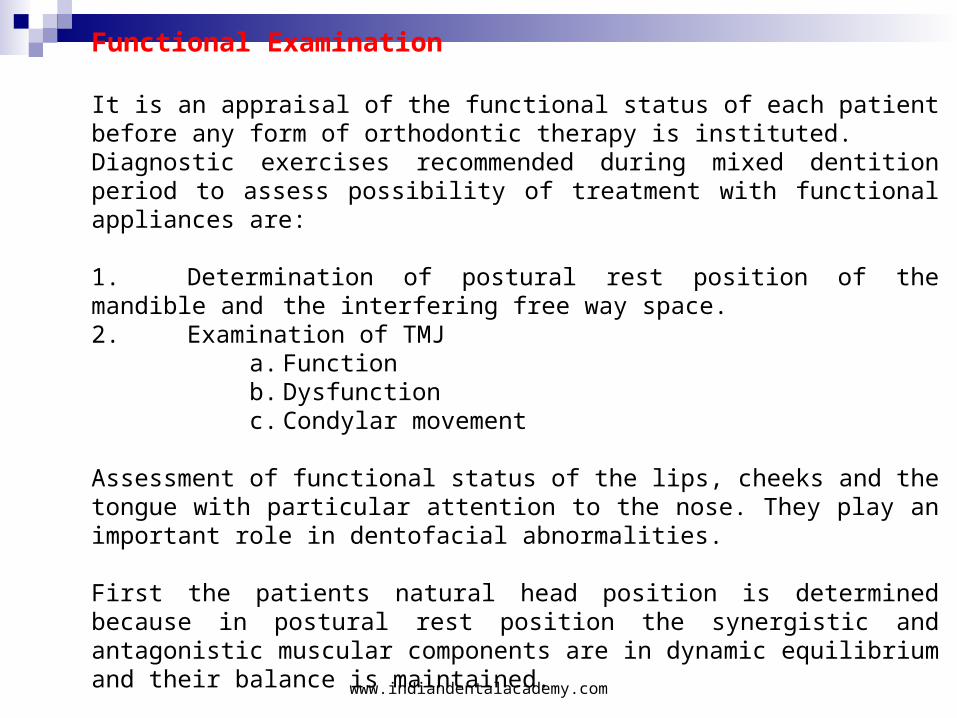
Functional Examination
It is an appraisal of the functional status of each patient before any form of orthodontic therapy is instituted.Diagnostic exercises recommended during mixed dentition period to assess possibility of treatment with functional appliances are:
1. Determination of postural rest position of the mandible and the interfering free way space.
2. Examination of TMJ a. Function b. Dysfunction c. Condylar movement
Assessment of functional status of the lips, cheeks and the tongue with particular attention to the nose. They play an important role in dentofacial abnormalities.
First the patients natural head position is determined because in postural rest position the synergistic and antagonistic muscular components are in dynamic equilibrium and their balance is maintained.
www.indiandentalacademy.com
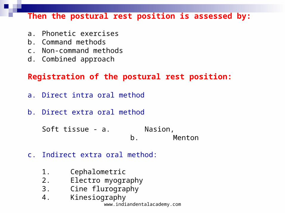
Then the postural rest position is assessed by:
a. Phonetic exercises b. Command methodsc. Non-command methods d. Combined approach
Registration of the postural rest position:
a. Direct intra oral method
b. Direct extra oral method
Soft tissue - a. Nasion, b. Menton
c. Indirect extra oral method:
1. Cephalometric2. Electro myography3. Cine flurography4. Kinesiography
www.indiandentalacademy.com
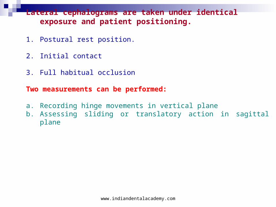
Lateral cephalograms are taken under identical exposure and patient positioning.
1. Postural rest position.
2. Initial contact
3. Full habitual occlusion
Two measurements can be performed:
a. Recording hinge movements in vertical plane b. Assessing sliding or translatory action in sagittal plane
www.indiandentalacademy.com
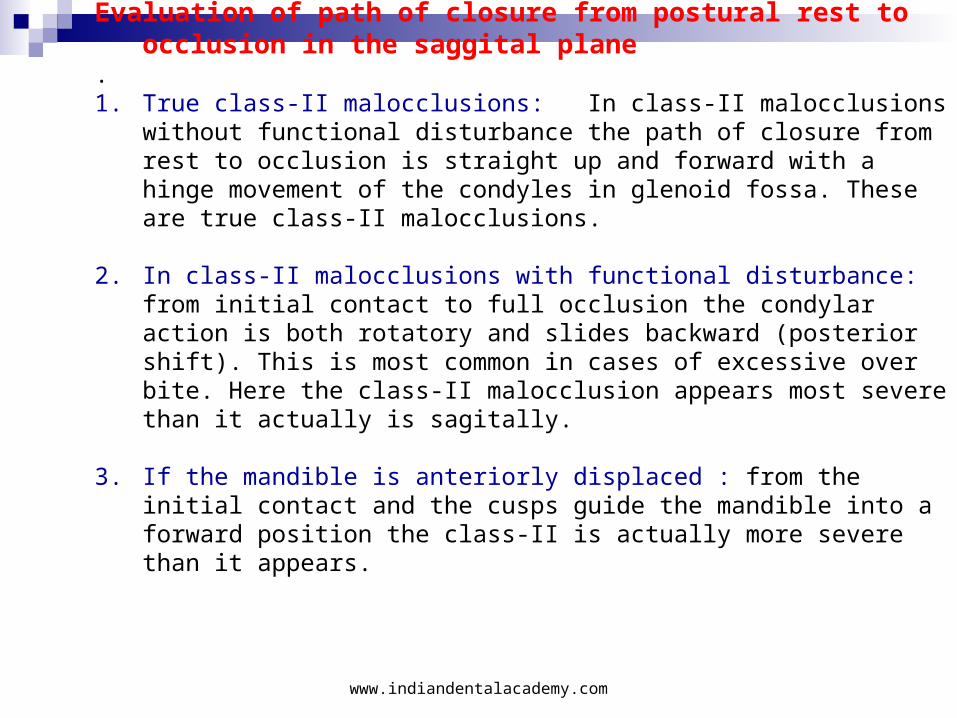
Evaluation of path of closure from postural rest to occlusion in the saggital plane
.1. True class-II malocclusions: In class-II malocclusions without
functional disturbance the path of closure from rest to occlusion is straight up and forward with a hinge movement of the condyles in glenoid fossa. These are true class-II malocclusions.
2. In class-II malocclusions with functional disturbance: from initial contact to full occlusion the condylar action is both rotatory and slides backward (posterior shift). This is most common in cases of excessive over bite. Here the class-II malocclusion appears most severe than it actually is sagitally.
3. If the mandible is anteriorly displaced : from the initial contact and the cusps guide the mandible into a forward position the class-II is actually more severe than it appears.
www.indiandentalacademy.com
Vertical evaluation
A. True deep bite - Large free way space
B. Pseudo deep bite.
In true deep bite cases a large inter occlusal clearance is caused by infra occlusion of posterior segments resulting from lateral tongue posture or tongue thrust habit. Elimination of environmental factors and functional therapy gives good results. The prognosis is good in a true deep bite case if vertical growth pattern is present.
In pseudo deep bite cases with horizontal growth pattern the possibility of the correction with functional appliances is limited,
hence the prognosis is poor.
Combination of- I. true deep bite and horizontal growth pattern, II. Pseudo deep bite and vertical growth pattern- limited prognosis can be expected.
www.indiandentalacademy.com
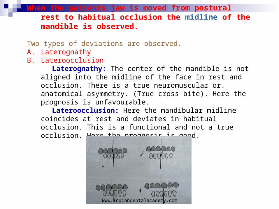
When the patients jaw is moved from postural rest to habitual occlusion the midline of the mandible is observed.
Two types of deviations are observed.A. LaterognathyB. Lateroocclusion Laterognathy: The center of the mandible is not aligned
into the midline of the face in rest and occlusion. There is a true neuromuscular or. anatomical asymmetry. (True cross bite). Here the prognosis is unfavourable.
Lateroocclusion: Here the mandibular midline coincides at rest and deviates in habitual occlusion. This is a functional and not a true occlusion. Here the prognosis is good.
www.indiandentalacademy.com
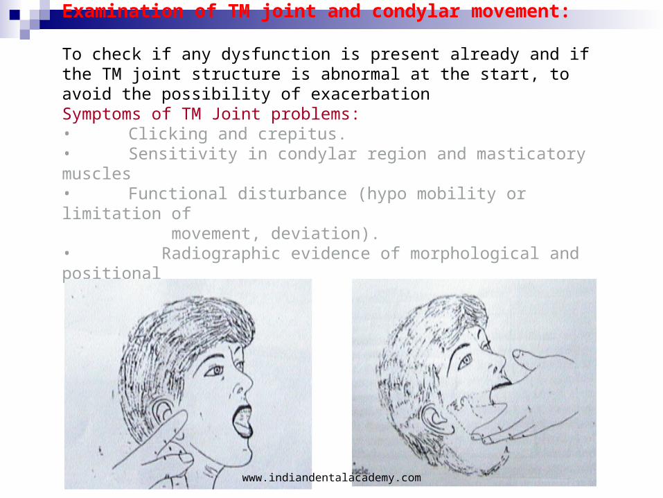
Examination of TM joint and condylar movement:
To check if any dysfunction is present already and if the TM joint structure is abnormal at the start, to avoid the possibility of exacerbationSymptoms of TM Joint problems:• Clicking and crepitus.• Sensitivity in condylar region and masticatory muscles• Functional disturbance (hypo mobility or limitation of movement, deviation).• Radiographic evidence of morphological and positional abnormalities.
www.indiandentalacademy.com
Assessment of Stomatognathic dysfunction:
• Mouth breathing• Bruxism• Thumb and finger sucking• Tongue thrusting• Lip biting• Abnormal deglutition
Examination of Lips:
A. ClinicallyB. Lateral cephalogram
Examination of respirationMouth breathing
www.indiandentalacademy.com

ASSESSING GROWTH DIRECTIONS
The bones that constitute the face descend down and at the same time move forwards. This means there is a vertical growth and anteroposterior growth. When both are balanced there is smooth downward and forward movement of the facial skeleton. The mandible rotates both clockwise and anti-clockwise during this process. Increase in clockwise growth results in excessive vertical growth. Counter clockwise growth results in deficiency of vertical growth.
www.indiandentalacademy.com
Vertical elements of growth
The frontonasal and frontomaxillary sutures which produce an increase in the distance from nasion to anterior nasal spine makes the maxillary molar and posterior nasal spine to move away from the base of the skull. The growth of the posterior alveolar process causes the molar teeth to move away from the palatal plane. Growing of the mandibular posterior alveolar process caused the molars to move away occlusally.If the vertical element of this growth is more than the condylar growth the chin moves downwards and backwards. If the sum of all the vertical components are less than the vertical growth of the condyle the chin grows forwards and upwards.With this above knowledge we proceed to treatment planning.
www.indiandentalacademy.com
APPLIANCES MODULES OF THE EARLY SYNERGISTIC TREATMENT CONCEPT
The concepts of treatment are ultimately more important than the actual appliances utilized. The appliances described are recommended as the result of personal experience and highly satisfactory results related to
(1) ease of use; (2) patient comfort; (3) minimum demand for patient cooperation; (4) minimum chair time; (5) cost effectiveness; (6) resistance to breakage; (7) minimum iatrogenic side effects; (8) minimum non-scheduled appointments or patient problems; (9) hygiene; (10) ease of fabrication; (11) predictability and stability of results and (12) minimum risks.
www.indiandentalacademy.com
Class II Treatment.- When?---Early Treatment- Deciduous Dentition - 2 to 7 years of age
Mixed Dentition - 8 to 11 years, when the dentition is mixed in nature
Transition Dentition - 11 to 12 years, just before the eruption of all
second bicuspids- all the second deciduous standing
Late Treatment- Adolescent/ Early Perm Dent 12 to 17 years
Very Late Treatment- Adult Therapy
Characteristics can be detected to predict certain Un /and Favorable
patterns of Growth
But Accurate Growth Predictions are simply not possible to for the
children who need it the most!
www.indiandentalacademy.com
A. Treatment planning for orthodontic problems in primary dentition.
B. Treatment planning for orthodontic problems in early mixed dentition period.
i. Moderateii. Severe
C. Treatment planning for orthodontic problems in adolescence (Late mixed and early permanent dentitiion)
1. Alignment problem2. Transverse problem3. Anteroposterior problem4. Vertical problem
D. Treatment planning for orthodontic problems in adults
www.indiandentalacademy.com
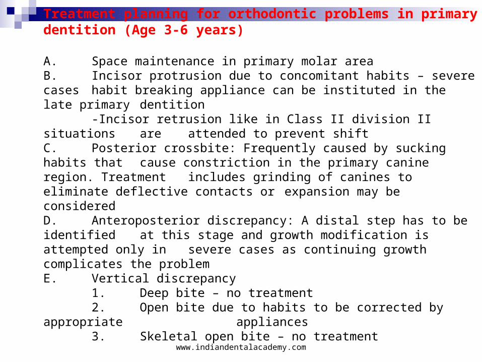
Treatment planning for orthodontic problems in primary dentition (Age 3-6 years)
A. Space maintenance in primary molar areaB. Incisor protrusion due to concomitant habits – severe cases habit breaking appliance can be instituted in the late primary dentition
-Incisor retrusion like in Class II division II situations are attended to prevent shiftC. Posterior crossbite: Frequently caused by sucking habits that cause constriction in the primary canine region. Treatment includes grinding of canines to eliminate deflective contacts or expansion may be consideredD. Anteroposterior discrepancy: A distal step has to be identified at this stage and growth modification is attempted only in severe cases as continuing growth complicates the problemE. Vertical discrepancy
1. Deep bite – no treatment2. Open bite due to habits to be corrected by appropriate
appliances3. Skeletal open bite – no treatment
www.indiandentalacademy.com
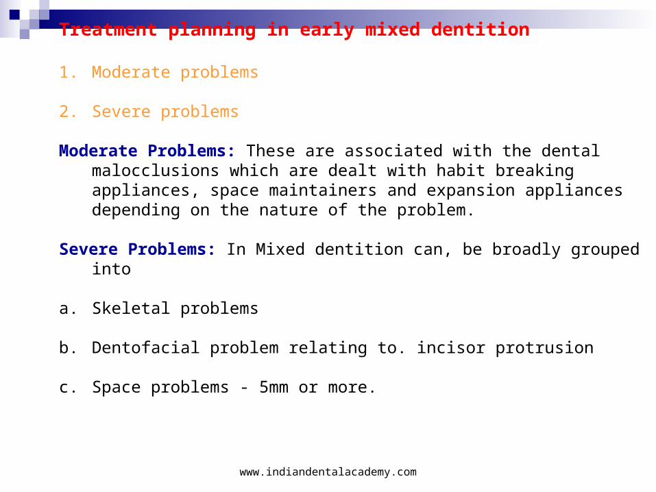
Treatment planning in early mixed dentition
1. Moderate problems
2. Severe problems
Moderate Problems: These are associated with the dental malocclusions which are dealt with habit breaking appliances, space maintainers and expansion appliances depending on the nature of the problem.
Severe Problems: In Mixed dentition can, be broadly grouped into
a. Skeletal problems
b. Dentofacial problem relating to. incisor protrusion
c. Space problems - 5mm or more.
www.indiandentalacademy.com
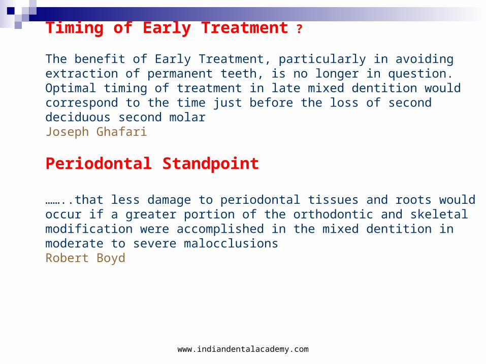
Timing of Early Treatment ? The benefit of Early Treatment, particularly in avoiding extraction of permanent teeth, is no longer in question. Optimal timing of treatment in late mixed dentition would correspond to the time just before the loss of second deciduous second molar Joseph Ghafari
Periodontal Standpoint
……..that less damage to periodontal tissues and roots would occur if a greater portion of the orthodontic and skeletal modification were accomplished in the mixed dentition in moderate to severe malocclusionsRobert Boyd
www.indiandentalacademy.com
Timing of Early Treatment ?
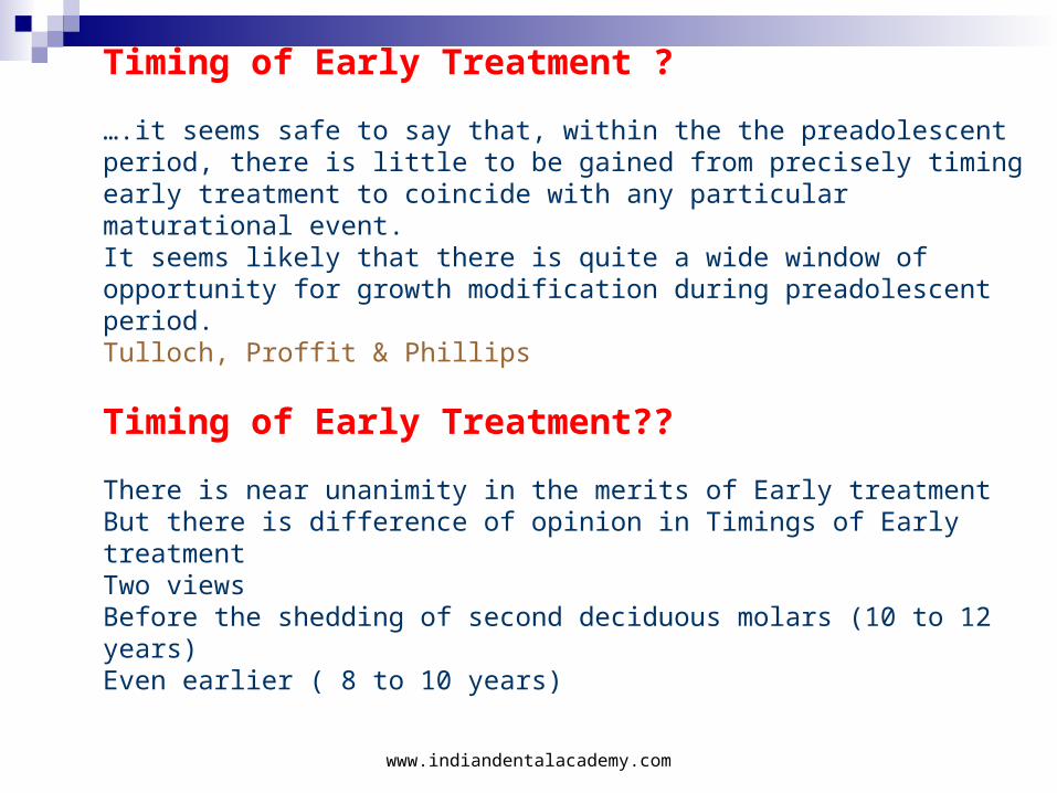
….it seems safe to say that, within the the preadolescent period, there is little to be gained from precisely timing early treatment to coincide with any particular maturational event.It seems likely that there is quite a wide window of opportunity for growth modification during preadolescent period.Tulloch, Proffit & Phillips Timing of Early Treatment??
There is near unanimity in the merits of Early treatmentBut there is difference of opinion in Timings of Early treatmentTwo viewsBefore the shedding of second deciduous molars (10 to 12 years)Even earlier ( 8 to 10 years)
www.indiandentalacademy.com
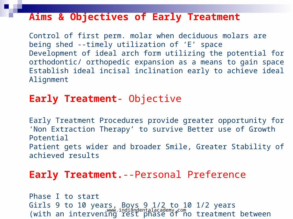
Aims & Objectives of Early Treatment
Control of first perm. molar when deciduous molars are being shed --timely utilization of ‘E’ spaceDevelopment of ideal arch form utilizing the potential for orthodontic/ orthopedic expansion as a means to gain spaceEstablish ideal incisal inclination early to achieve ideal Alignment
Early Treatment- Objective
Early Treatment Procedures provide greater opportunity for ‘Non Extraction Therapy’ to survive Better use of Growth PotentialPatient gets wider and broader Smile, Greater Stability of achieved results
Early Treatment.--Personal Preference
Phase I to startGirls 9 to 10 years, Boys 9 1/2 to 10 1/2 years(with an intervening rest phase of no treatment between Phase I and IIof 0 months to 24 months)
www.indiandentalacademy.com
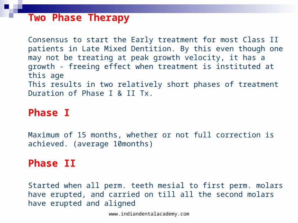
Two Phase Therapy
Consensus to start the Early treatment for most Class II patients in Late Mixed Dentition. By this even though one may not be treating at peak growth velocity, it has a growth - freeing effect when treatment is instituted at this ageThis results in two relatively short phases of treatmentDuration of Phase I & II Tx.
Phase I
Maximum of 15 months, whether or not full correction is achieved. (average 10months)
Phase II
Started when all perm. teeth mesial to first perm. molars have erupted, and carried on till all the second molars have erupted and aligned
www.indiandentalacademy.com
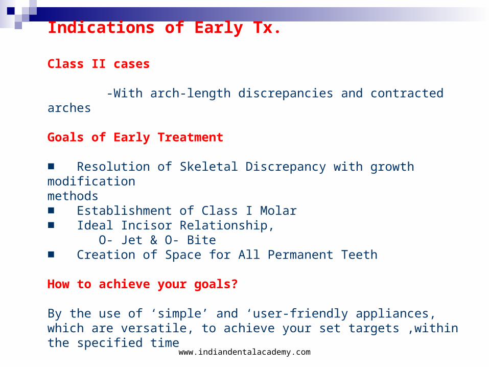
Indications of Early Tx.
Class II cases
-With arch-length discrepancies and contracted arches Goals of Early Treatment
■ Resolution of Skeletal Discrepancy with growth modification methods■ Establishment of Class I Molar■ Ideal Incisor Relationship, O- Jet & O- Bite■ Creation of Space for All Permanent Teeth How to achieve your goals?
By the use of ‘simple’ and ‘user-friendly appliances, which are versatile, to achieve your set targets ,within the specified time
www.indiandentalacademy.com
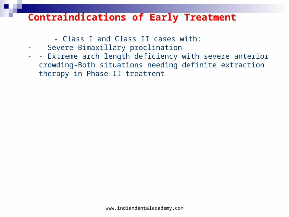
Contraindications of Early Treatment
- Class I and Class II cases with:- - Severe Bimaxillary proclination- - Extreme arch length deficiency with severe anterior crowding-Both
situations needing definite extraction therapy in Phase II treatment
www.indiandentalacademy.com
The appliances of choice most commonly used in our practice have been divided into those considered to be principally
(1) Orthopedic;
(2) Functional;
(3) Orthodontic or tooth moving and
(4) Retentive.
www.indiandentalacademy.com
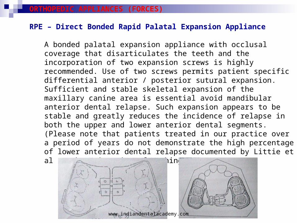
ORTHOPEDIC APPLIANCES (FORCES)
RPE – Direct Bonded Rapid Palatal Expansion Appliance
A bonded palatal expansion appliance with occlusal coverage that disarticulates the teeth and the incorporation of two expansion screws is highly recommended. Use of two screws permits patient specific differential anterior / posterior sutural expansion. Sufficient and stable skeletal expansion of the maxillary canine area is essential avoid mandibular anterior dental relapse. Such expansion appears to be stable and greatly reduces the incidence of relapse in both the upper and lower anterior dental segments. (Please note that patients treated in our practice over a period of years do not demonstrate the high percentage of lower anterior dental relapse documented by Littie et al at the University of Washington)
www.indiandentalacademy.com
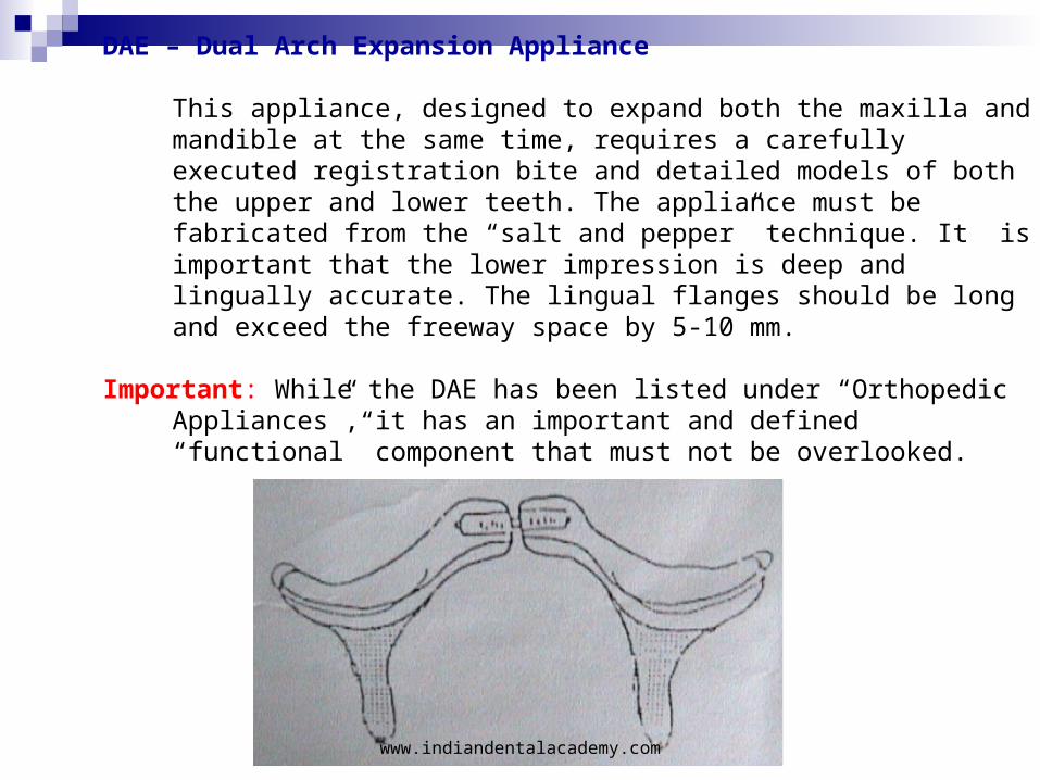
DAE – Dual Arch Expansion Appliance
This appliance, designed to expand both the maxilla and mandible at the same time, requires a carefully executed registration bite and detailed models of both the upper and lower teeth. The appliance must be fabricated from the “salt and pepper” technique. It is important that the lower impression is deep and lingually accurate. The lingual flanges should be long and exceed the freeway space by 5-10 mm.
Important: While the DAE has been listed under “Orthopedic Appliances”, it has an important and defined “functional” component that must not be overlooked.
www.indiandentalacademy.com
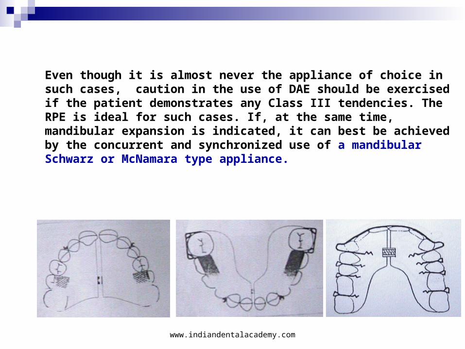
Even though it is almost never the appliance of choice in such cases, caution in the use of DAE should be exercised if the patient demonstrates any Class III tendencies. The RPE is ideal for such cases. If, at the same time, mandibular expansion is indicated, it can best be achieved by the concurrent and synchronized use of a mandibular Schwarz or McNamara type appliance.
www.indiandentalacademy.com
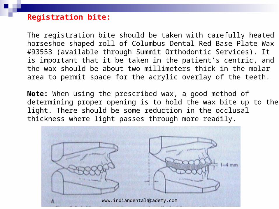
Registration bite:
The registration bite should be taken with carefully heated horseshoe shaped roll of Columbus Dental Red Base Plate Wax #93553 (available through Summit Orthodontic Services). It is important that it be taken in the patient’s centric, and the wax should be about two millimeters thick in the molar area to permit space for the acrylic overlay of the teeth.
Note: When using the prescribed wax, a good method of determining proper opening is to hold the wax bite up to the light. There should be some reduction in the occlusal thickness where light passes through more readily.
www.indiandentalacademy.com
Bite Planes (BP) With or Without Expansion
The use of removable bite planes either with or without expansion is imperative to keep the maxillary and mandibular teeth disarticulated following the use of an RPE or DAE, if a sagittal discrepancy still prevails. The appliances, if utilized with expansion, have an orthopedic effect. As with the DAE, they also have a functional effect, the result of disarticulation of the teeth, eruption of the buccal quadrants, and release of the mandible permitting natural reposturing.
Note: In “high angle” cases, both upper and lower posterior teeth must be indexed (flat plane) to prevent additional eruption of teeth.
www.indiandentalacademy.com
MANAGEMENT OF MANDIBULAR DEFICIENCY
Functional appliances form the main stay for treatment of mandibular deficiency in growing children. The following criteria can be used for case selection:
1. Individuals with growth potential2. Retrognathic mandible3. Deep bite4. Low mandibular plane angle5. Favourable VTO
The functional concept employs carefully designed removable appliance in an effort to achieve harmonious development of dento facial structures by eliminating unfavourable myofunctional and occlusal factors and improving the functional environment of the developing dentition. This concept flourished and became the basis of functional therapy for over a century resulting in the development of wide range of functional appliances.
www.indiandentalacademy.com
Removal Functional Appliances1. Bite plate2. Monobloc3. Activator4. Bionator5. Modified activator6. Functional regulator7. Palate free activator8. Twinblock9. Functional Magnetic System10. Mandibular Growth Advancer
www.indiandentalacademy.com
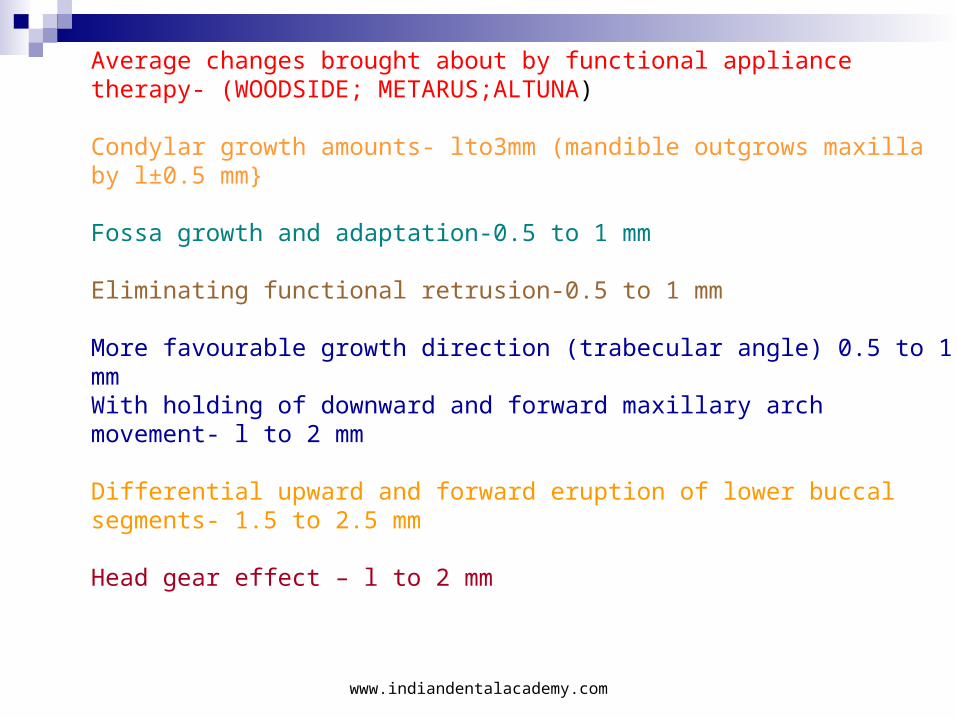
Average changes brought about by functional appliance therapy- (WOODSIDE; METARUS;ALTUNA)
Condylar growth amounts- lto3mm (mandible outgrows maxilla by l±0.5 mm}
Fossa growth and adaptation-0.5 to 1 mm
Eliminating functional retrusion-0.5 to 1 mm
More favourable growth direction (trabecular angle) 0.5 to 1 mmWith holding of downward and forward maxillary arch movement- l to 2 mm
Differential upward and forward eruption of lower buccal segments- 1.5 to 2.5 mm
Head gear effect – l to 2 mm
www.indiandentalacademy.com
Similar studies by Vangerall & Harvold (1985) on activator, frankell appliance by FRANKELL (1987) have shown favourable changes with functional appliances.
FRANK WEILAND & BERGT INGERVALL compared the effects of herren activator, activator-headgear & jasper jumper and demonstrated higher percentage of success with jasper jumper.
Recently Twin block appliance has become popular due to its versatility and patient compliance.
The cephalometric changes brought about by this appliance include an increase in mandibular unit length (condylion to gnathion) by 6.5mm with increase in ramal height (2/3) and body length (1/3) ( MILLS & MECULLOCH.)
www.indiandentalacademy.com
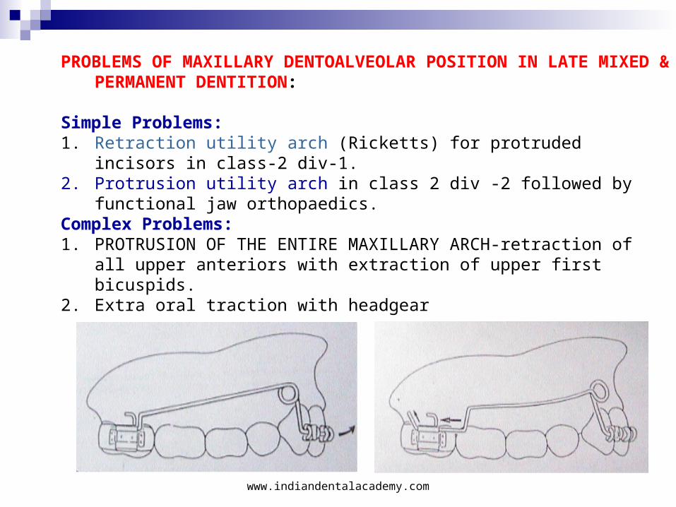
PROBLEMS OF MAXILLARY DENTOALVEOLAR POSITION IN LATE MIXED & PERMANENT DENTITION:
Simple Problems:1. Retraction utility arch (Ricketts) for protruded incisors in class-2
div-1. 2. Protrusion utility arch in class 2 div -2 followed by functional jaw
orthopaedics.Complex Problems:1. PROTRUSION OF THE ENTIRE MAXILLARY ARCH-retraction of all
upper anteriors with extraction of upper first bicuspids.2. Extra oral traction with headgear
www.indiandentalacademy.com
In class II mesially migrated molars with straight profile molar distallization is attempted. This is especially successful before the eruption of II molars.
The distallization devices are1. Pendulum appliance (Hilgers 1992)2. Distal Jet (Carano et al 1996)3. Modified Nance arch with niti coils or wire (Gianelly 1991)4. Magnetic appliance (Gianelly 1989)5. Jones Jig (Jones and White 1992)6. Lokar distallizing appliance7. Molar distallizing bow (Jerkel and Rakosi 1991).
Fixed functional appliances1. Herbst2. Herbst with High pull headgear3. MARS appliance4. Jasper Jumper5. Mandibular Protraction application6. Universal bite jumper7. Churrojumper
www.indiandentalacademy.com
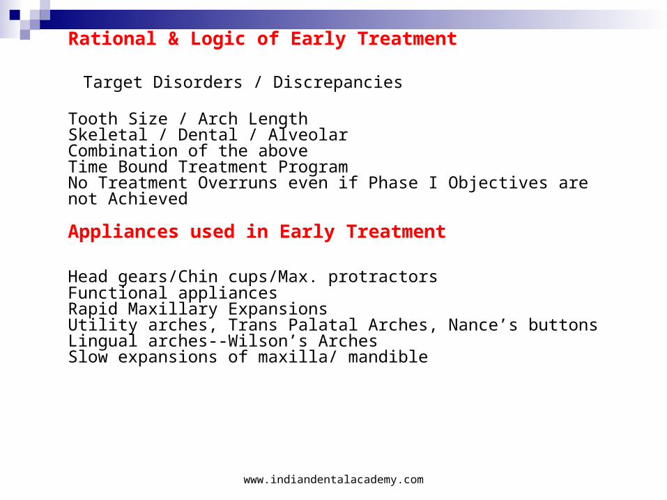
Rational & Logic of Early Treatment
Target Disorders / Discrepancies
Tooth Size / Arch LengthSkeletal / Dental / AlveolarCombination of the aboveTime Bound Treatment ProgramNo Treatment Overruns even if Phase I Objectives are not Achieved
Appliances used in Early Treatment
Head gears/Chin cups/Max. protractorsFunctional appliancesRapid Maxillary ExpansionsUtility arches, Trans Palatal Arches, Nance’s buttonsLingual arches--Wilson’s ArchesSlow expansions of maxilla/ mandible
www.indiandentalacademy.com
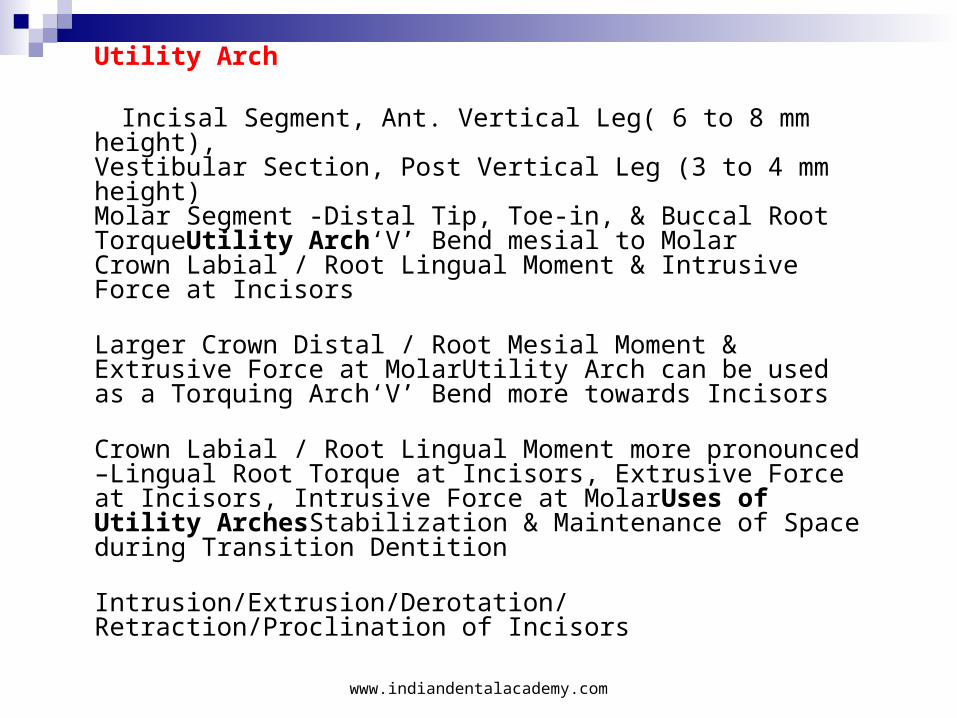
Utility Arch
Incisal Segment, Ant. Vertical Leg( 6 to 8 mm height),Vestibular Section, Post Vertical Leg (3 to 4 mm height)Molar Segment -Distal Tip, Toe-in, & Buccal Root TorqueUtility Arch‘V’ Bend mesial to MolarCrown Labial / Root Lingual Moment & Intrusive Force at Incisors
Larger Crown Distal / Root Mesial Moment & Extrusive Force at MolarUtility Arch can be used as a Torquing Arch‘V’ Bend more towards Incisors
Crown Labial / Root Lingual Moment more pronounced –Lingual Root Torque at Incisors, Extrusive Force at Incisors, Intrusive Force at MolarUses of Utility ArchesStabilization & Maintenance of Space during Transition Dentition
Intrusion/Extrusion/Derotation/ Retraction/Proclination of Incisors
www.indiandentalacademy.com
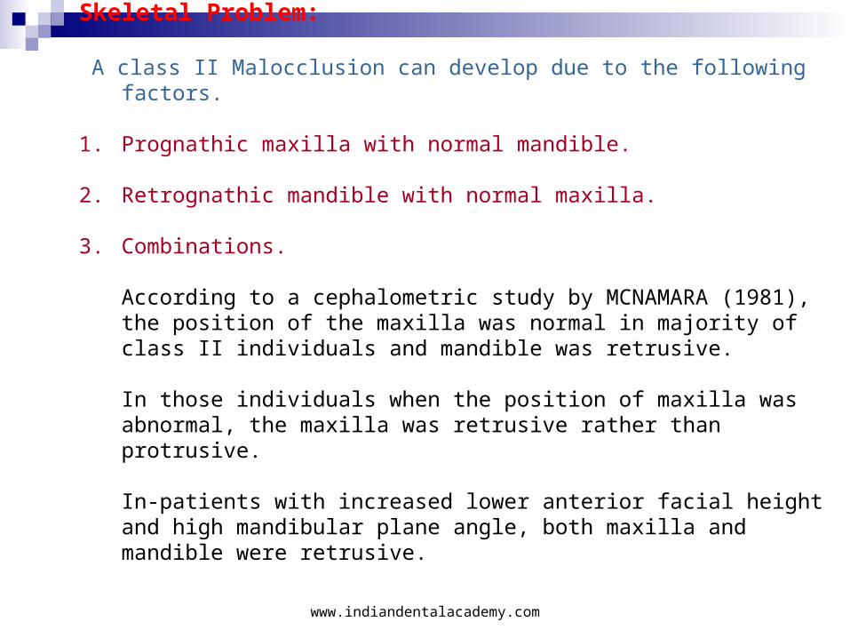
Skeletal Problem:
A class II Malocclusion can develop due to the following factors.
1. Prognathic maxilla with normal mandible.
2. Retrognathic mandible with normal maxilla.
3. Combinations.
According to a cephalometric study by MCNAMARA (1981), the position of the maxilla was normal in majority of class II individuals and mandible was retrusive.
In those individuals when the position of maxilla was abnormal, the maxilla was retrusive rather than protrusive.
In-patients with increased lower anterior facial height and high mandibular plane angle, both maxilla and mandible were retrusive.
www.indiandentalacademy.com
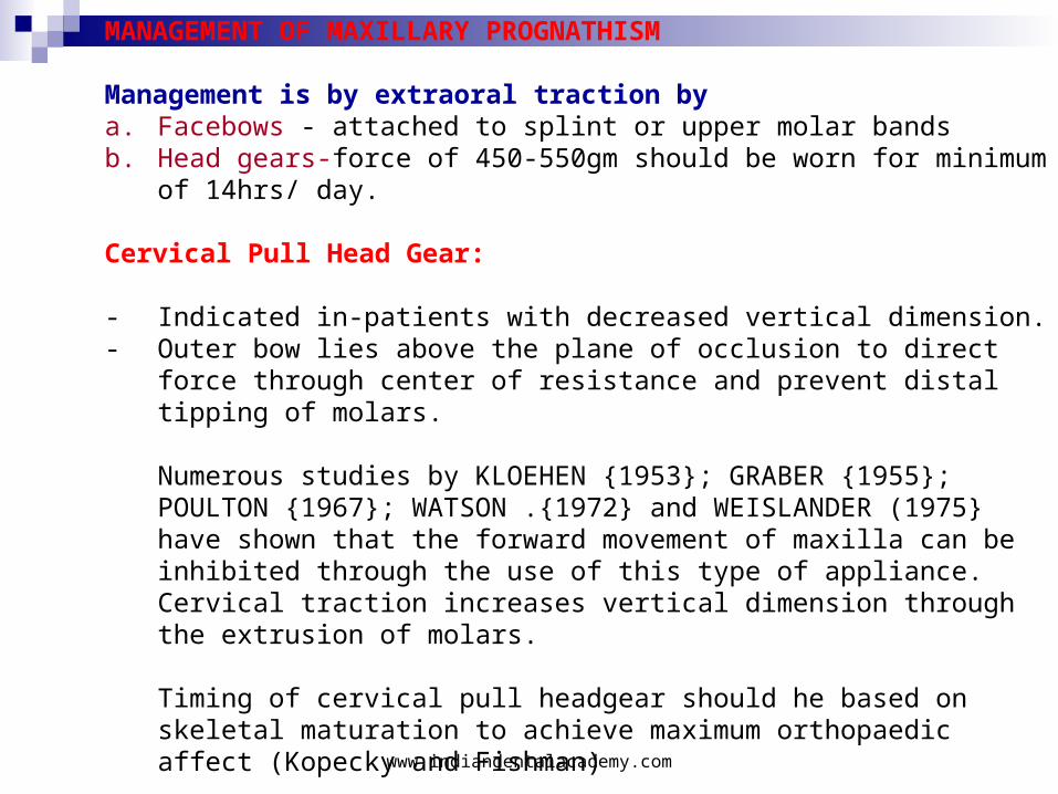
MANAGEMENT OF MAXILLARY PROGNATHISM
Management is by extraoral traction bya. Facebows - attached to splint or upper molar bandsb. Head gears-force of 450-550gm should be worn for minimum of
14hrs/ day.
Cervical Pull Head Gear:
- Indicated in-patients with decreased vertical dimension.- Outer bow lies above the plane of occlusion to direct force through
center of resistance and prevent distal tipping of molars.
Numerous studies by KLOEHEN {1953}; GRABER {1955}; POULTON {1967}; WATSON .{1972} and WEISLANDER (1975} have shown that the forward movement of maxilla can be inhibited through the use of this type of appliance. Cervical traction increases vertical dimension through the extrusion of molars.
Timing of cervical pull headgear should he based on skeletal maturation to achieve maximum orthopaedic affect (Kopecky and Fishman)
www.indiandentalacademy.com
High Pull Head Gear
- Indicated in patients with increased vertical dimension- Face bow is anchored to an occipital anchoring unit to produce vertically directed force.- High pull headgear can decrease vertical growth of maxilla thus allowing autorotation of the mandible.- Mauric Firouz and Ravindra Nanda Demonstrated that when the force was directed at the levels of Trifurcation of maxillary molars significant distal movement and intrusion of molars was achieved along with restoration of vertical and horizontal maxillary growth.
Medium Pull Head Gear
- Indicated in average angle cases
www.indiandentalacademy.com
Management in adolescent period:
Camouflage treatment: Beyond the adolescent growth spurt to correct a skeletal problem teeth should be displaced relative to their supporting bone to compensate for the underlying jaw discrepancy. This is termed as camouflage treatment.
Indications for class-II camouflage treatment:
1. Too old for successful growth modification.2. Mild to moderate skeletal class-II.3. Reasonably good alignment of teeth.4. Good vertical facial proportions - neither very short nor very long
face.
www.indiandentalacademy.com
Camouflage - Contra-indications
1. Severe Class II with Vertical Displacial 2. Extreme & Severe Crowding with Protrusion -Where Ext. space
used up for Alignment 3. With Remaining Good Growth potential 4. Non- Growing with more than Moderate Dis.placia- Surgical
Camouflage vs Surgery5. Classic case of mature 14 yr old with full cusp Class II with 10
mm OJ & Mandibular Deficiency- Max Premolar Ext. & Ret of Ants6. Surgical Mandibular Advancement Past The Growth/
Careful Tx plan/ Too severe for Camouflage > 10 mm Overjet7. Mandible short, Lower teeth protrusive8. Deficient Chin, and/or Long Face9. Lower Incisor Position/Protrusion
www.indiandentalacademy.com
Extractions in Class-II
1. Extraction of upper first bicuspids only with lower non-extraction2. Extraction of upper first bicuspids and lower second bicuspids3. Extraction of upper and lower first bicuspids in cases with severe lower crowding.4. Distalization of molars with extraction of second molar. Upto 4mm of distal movement of first molar is possible by extraction of second molars. Thus an ideal case for second molar extraction is a patient with less than a full cusp class-II. This concept was put forward by WITZIG AND SPHAL.
www.indiandentalacademy.com
Second molar extraction has the following advantages:
A. Simplified mechanical treatmentB. Less need for patient co-operation in some casesC. Reduced incidence of late lower incisor crowdingD. Avoidance of third molar impactionE. Easier first molar distalizationF. Increased long term stability of the result. (Richardson)
Disadvantages of the second molar extractions are:
1. Inadequate space is created to deal with significant anterior crowding or protrusion.
2. Delay while awaiting third molar eruption.3. Possible need to upright lower third molars in some cases at a
difficult time for the patient.4. Difficulty in keeping contact with the patient to achieve third molar
uprighting.The present trend is to incorporate fixed functional appliance or twin
block along with fixed appliances.
www.indiandentalacademy.com
Combined surgical and orthodontic treatment-When Is Surgery Indicated?
- When severe skeletal or very severe Dentoalveolar problems are too severe to correct by Orthodontics alone
When Camouflage - dental compensations to mask skeletal dicrepancy- may produce poor facial estheticsSurgical Tx.-
Indications
-Adult Patients , With little remaining growth
Younger patients with Extremely severe or progressive Deformity
Good General health statusSurgical - Treatment PlanningDental CompensationsMost Skeletal problems are with some Dental CompensationsCamouflage & Surgical Preparation would need opposite Tooth movements
Camouflage in Definite Surgical cases to be avoided unless a Good Outcome
www.indiandentalacademy.com
Pre-surgical orthodontics - Goals:
1. Alignment of arch segments and make them compatible
2. To establish the anteroposterior and vertical position of incisors - Extraction pattern:Dental compensations are removed. Extractions may be needed in the lower arch i.e., lower 4's and non-extraction in the upper or upper 5's.
3. Stabilizing arch wires are placed.Surgical movements are simulated on the recent cephalogram and the functional and esthetic balance is evaluated and if satisfactory results are achieved these surgical movements are duplicated on models and inter occlusal surgical splint is fabricated.
www.indiandentalacademy.com
Surgical Procedures:
1. Maxillary excess - Le Fort-I Osteotomy2. Mandibular deficiency - Saggital split ramus Osteotomy3. Deficient chin - advancement genioplasty
Post-surgical Orthodontics
Timing: Post-surgical orthodontics can be initiated 3 to 4 weeks after the release of immobilization Stabilizing arch wires are removed and replaced by working arch wires with light vertical forces till a good stable occlusion is achieved.
www.indiandentalacademy.com
Distraction Osteogenesis - Deficient Mandible
Distraction Osteogenesis is a biological process of new bone formation between surfaces of bone segments gradually separated by incremental traction. Though this technique was practiced earlier it was revolutionized by Ilizarov in 1989, with his technique for limb lengthening. In 1992, McCarthy was the first to clinically apply an external device to the mandible and in 1994, he developed and intra oral mandibular distraction device.In congenital mandibular deficiency, a multiplanar distraction is required. Following a single or double osteotomy, it is possible to distract both vertically and horizontally using Bi-Directional distractor. This in very severe mandibular hypoplasia where ramus and body are affected, this procedure results in rapid distraction as well as development of mandibular angle.
Conclusion: With such a wide range of treatment modalities and appliances listed out for class-II malocculsion it is at the hands of a skilled orthodontist to appropriately time and choose the right treatment according to individual patient needs.
www.indiandentalacademy.com
www.indiandentalacademy.com
www.indiandentalacademy.comLeader in continuing dental education
Related Documents