Dynamic reconfiguration of human brain functional networks through neurofeedback Sven Haller a, ⁎, Rotem Kopel b, c , Permi Jhooti g, h , Tanja Haas d , Frank Scharnowski b, c , Karl-Olof Lovblad a , Klaus Scheffler e, f , Dimitri Van De Ville b, c a Service neuro-diagnostique et neuro-interventionnel DISIM, University Hospitals of Geneva, Switzerland b Department of Radiology and Medical Informatics, University of Geneva, Switzerland c Institute of Bioengineering, Ecole Polytechnique Fédérale de Lausanne, Switzerland d Institute of Radiology, University Hospital Basel, Switzerland e Max Planck Institute for Biological Cybernetic, Tübingen, Germany f Department of Biomedical Magnetic Resonance, University of Tübingen, Tübingen, Germany g Institute of Research in Art and Design (IDK), Switzerland h Academy of Art and Design, University of Applied Sciences and Arts Northwestern Switzerland (FHNW), Switzerland abstract article info Article history: Accepted 5 May 2013 Available online 16 May 2013 Keywords: Neurofeedback Self-regulation Functional connectivity Real-time fMRI Recent fMRI studies demonstrated that functional connectivity is altered following cognitive tasks (e.g., learning) or due to various neurological disorders. We tested whether real-time fMRI-based neurofeedback can be a tool to voluntarily reconfigure brain network interactions. To disentangle learning-related from regulation-related effects, we first trained participants to voluntarily regulate activity in the auditory cortex (training phase) and subsequently asked participants to exert learned voluntary self-regulation in the absence of feedback (transfer phase without learning). Using independent component analysis (ICA), we found network reconfigurations (increases in functional network connectivity) during the neurofeedback training phase between the auditory target region and (1) the auditory pathway; (2) visual regions related to visual feedback processing; (3) insula related to intro- spection and self-regulation and (4) working memory and high-level visual attention areas related to cogni- tive effort. Interestingly, the auditory target region was identified as the hub of the reconfigured functional networks without a-priori assumptions. During the transfer phase, we again found specific functional connectiv- ity reconfiguration between auditory and attention network confirming the specific effect of self-regulation on functional connectivity. Functional connectivity to working memory related networks was no longer altered con- sistent with the absent demand on working memory. We demonstrate that neurofeedback learning is mediated by widespread changes in functional connectivity. In contrast, applying learned self-regulation involves more limited and specific network changes in an audi- tory setup intended as a model for tinnitus. Hence, neurofeedback training might be used to promote recov- ery from neurological disorders that are linked to abnormal patterns of brain connectivity. © 2013 Elsevier Inc. All rights reserved. Introduction Studying how different brain areas interact may hold the key to un- derstand how information is processed in the human brain. Recent de- velopments in data analysis techniques have opened up exciting opportunities to investigate such functional connectivity with functional magnetic resonance imaging (fMRI). The techniques to study large-scale networks using fMRI can be divided into two main approaches. According to the first approach, functional connectivity is measured by interregional temporal correlations of the fMRI blood oxygenation level dependent (BOLD) signal (Biswal et al., 1995). This approach requires the choice of a seed region, for which correlation maps can be built. Among other findings, seed-region based approaches lead to the discov- ery of resting-state functional networks (Fox and Raichle, 2007). The second approach relies on multivariate and data-driven techniques such as independent component analysis (ICA) (Calhoun et al., 2001b; McKeown et al., 1998a, 1998b). ICA can be used to decompose the data into a set of spatial maps and associated time-courses without using pre-defined seed regions (Daubechies et al., 2009). Group-level ICA is a powerful technique to investigate distinct functional networks (Beckmann et al., 2005; Damoiseaux et al., 2006; Greicius et al., 2003). Many fMRI studies exploring functional connectivity intrinsically assume a static organization. However, recent evidence suggests that NeuroImage 81 (2013) 243–252 ⁎ Corresponding author at: Service neuro-diagnostique et neuro-interventionnel DISIM, Hôpitaux Universitaires de Genève, Rue Gabrielle Perret-Gentil 4, 1211 Genève 14, Switzerland. Fax: +41 22 37 27072. E-mail address: [email protected] (S. Haller). 1053-8119/$ – see front matter © 2013 Elsevier Inc. All rights reserved. http://dx.doi.org/10.1016/j.neuroimage.2013.05.019 Contents lists available at SciVerse ScienceDirect NeuroImage journal homepage: www.elsevier.com/locate/ynimg

Welcome message from author
This document is posted to help you gain knowledge. Please leave a comment to let me know what you think about it! Share it to your friends and learn new things together.
Transcript
NeuroImage 81 (2013) 243–252
Contents lists available at SciVerse ScienceDirect
NeuroImage
j ourna l homepage: www.e lsev ie r .com/ locate /yn img
Dynamic reconfiguration of human brain functional networksthrough neurofeedback
Sven Haller a,⁎, Rotem Kopel b,c, Permi Jhooti g,h, Tanja Haas d, Frank Scharnowski b,c, Karl-Olof Lovblad a,Klaus Scheffler e,f, Dimitri Van De Ville b,c
a Service neuro-diagnostique et neuro-interventionnel DISIM, University Hospitals of Geneva, Switzerlandb Department of Radiology and Medical Informatics, University of Geneva, Switzerlandc Institute of Bioengineering, Ecole Polytechnique Fédérale de Lausanne, Switzerlandd Institute of Radiology, University Hospital Basel, Switzerlande Max Planck Institute for Biological Cybernetic, Tübingen, Germanyf Department of Biomedical Magnetic Resonance, University of Tübingen, Tübingen, Germanyg Institute of Research in Art and Design (IDK), Switzerlandh Academy of Art and Design, University of Applied Sciences and Arts Northwestern Switzerland (FHNW), Switzerland
⁎ Corresponding author at: Service neuro-diagnostiqueHôpitaux Universitaires de Genève, Rue Gabrielle PerrSwitzerland. Fax: +41 22 37 27072.
E-mail address: [email protected] (S. Haller).
1053-8119/$ – see front matter © 2013 Elsevier Inc. Allhttp://dx.doi.org/10.1016/j.neuroimage.2013.05.019
a b s t r a c t
a r t i c l e i n f oArticle history:Accepted 5 May 2013Available online 16 May 2013
Keywords:NeurofeedbackSelf-regulationFunctional connectivityReal-time fMRI
Recent fMRI studies demonstrated that functional connectivity is altered following cognitive tasks (e.g., learning)or due to various neurological disorders.We testedwhether real-time fMRI-based neurofeedback can be a tool tovoluntarily reconfigure brain network interactions. To disentangle learning-related from regulation-relatedeffects, we first trained participants to voluntarily regulate activity in the auditory cortex (training phase) andsubsequently asked participants to exert learned voluntary self-regulation in the absence of feedback (transferphase without learning).Using independent component analysis (ICA), we found network reconfigurations (increases in functionalnetwork connectivity) during the neurofeedback training phase between the auditory target region and(1) the auditory pathway; (2) visual regions related to visual feedback processing; (3) insula related to intro-spection and self-regulation and (4) working memory and high-level visual attention areas related to cogni-tive effort. Interestingly, the auditory target region was identified as the hub of the reconfigured functionalnetworks without a-priori assumptions. During the transfer phase, we again found specific functional connectiv-ity reconfiguration between auditory and attention network confirming the specific effect of self-regulation onfunctional connectivity. Functional connectivity toworkingmemory relatednetworkswas no longer altered con-sistent with the absent demand on working memory.We demonstrate that neurofeedback learning is mediated by widespread changes in functional connectivity.In contrast, applying learned self-regulation involves more limited and specific network changes in an audi-tory setup intended as a model for tinnitus. Hence, neurofeedback training might be used to promote recov-ery from neurological disorders that are linked to abnormal patterns of brain connectivity.
© 2013 Elsevier Inc. All rights reserved.
Introduction
Studying how different brain areas interact may hold the key to un-derstand how information is processed in the human brain. Recent de-velopments in data analysis techniques have opened up excitingopportunities to investigate such functional connectivity with functionalmagnetic resonance imaging (fMRI). The techniques to study large-scalenetworks using fMRI can be divided into two main approaches.According to the first approach, functional connectivity is measured by
et neuro-interventionnel DISIM,et-Gentil 4, 1211 Genève 14,
rights reserved.
interregional temporal correlations of the fMRI blood oxygenation leveldependent (BOLD) signal (Biswal et al., 1995). This approach requiresthe choice of a seed region, for which correlation maps can be built.Among other findings, seed-region based approaches lead to the discov-ery of resting-state functional networks (Fox and Raichle, 2007). Thesecond approach relies on multivariate and data-driven techniquessuch as independent component analysis (ICA) (Calhoun et al., 2001b;McKeown et al., 1998a, 1998b). ICA can be used to decompose the datainto a set of spatial maps and associated time-courses without usingpre-defined seed regions (Daubechies et al., 2009). Group-level ICA isa powerful technique to investigate distinct functional networks(Beckmann et al., 2005; Damoiseaux et al., 2006; Greicius et al., 2003).
Many fMRI studies exploring functional connectivity intrinsicallyassume a static organization. However, recent evidence suggests that
244 S. Haller et al. / NeuroImage 81 (2013) 243–252
functional connectivity can be modulated spontaneously (Raichle,2010), by exogenous stimulation (Buchel et al., 1999), and by learning(Bassett et al., 2011; Lewis et al., 2009). Importantly, changes infunctional connectivity have also been linked with the course of a vari-ety of neurological diseases (Fox and Greicius, 2010) as well as the re-covery from certain neurological diseases (Wang et al., 2010). Suchobservations raise the possibility that learning-related changes in func-tional connectivity can help to accelerate the recovery. This is especiallythe case if the learning-related changes in functional connectivity canbe targeted at the networks involved in the recovery.
A new and promising approach that allows targeting specific regionsand networks directly is real-time fMRI (rt-fMRI) neurofeedback(deCharms, 2008; Weiskopf et al., 2004b). The basic principle of rt-fMRIneurofeedback is to present a biofeedback signal extracted online fromfMRI BOLD measurements. With the help of such a signal, participantscan learn self-regulation of BOLD activity by means of operant condition-ing. Several studies have demonstrated the feasibility of self-regulatingactivity in specific brain areas using rt-fMRI neurofeedback (e.g.,deCharms et al., 2004; Posse et al., 2003; Weiskopf et al., 2003,2004a; Yoo and Jolesz, 2002). Some studies have even shown thatself-regulation results in clinical benefits for specific neurological condi-tions such as chronic pain (deCharms et al., 2005), tinnitus (Haller et al.,2010), and Parkinson's disease (Subramanian et al., 2011). Further,there is preliminary evidence that learning self-regulation of brain activitycan lead to changes in functional connectivity (Horovitz et al., 2010; Lee etal., 2011; Rota et al., 2011). However, the studies looking into changes infunctional connectivity are limited for two reasons. Firstly, they appliedseed-region approaches that limit the investigation of connectivitychanges to pre-defined region of interests (ROIs). Secondly, they only in-vestigated connectivity changes during the neurofeedback training phasebut theydid not look into such changeswhenparticipants applied learnedself-regulation; i.e., when participants performed previously learnedself-regulationwithout feedback. Especially with respect to clinical appli-cations the transfer condition is more important than the training phasebecause learned self-regulation along with the accompanying changesin functional connectivity can be voluntarily applied by the patient.
Here we significantly extend the previous investigations of changesin functional connectivity due to neurofeedback by using data-driventechniques that do not require defining a seed region a priori. Becausechanges in functional connectivity during the neurofeedback trainingphase might be related to the neurofeedback per se, to learningmecha-nisms, or both, we included a transfer phase during which participantsapplied the previously learned strategy in the absence of feedbackand hence absence of learning. We hypothesize that our data-drivenapproach—i.e., independent component analysis (ICA)—can identifychanges in functional networks that are related to the neurofeedbacktarget region, in particular, the auditory cortex. Further, we hypothesizethat the functional connectivity changes during neurofeedback learningwill differ from the changes during applied self-regulation; e.g., only theformer will include changes in networks related to feedback processingand reinforcement learning while the latter will demonstrate changesin functional connectivity related to self-regulation.
Materials and methods
The setup and the experimental procedure were similar to a previ-ously published study (Haller et al., 2010). For readability, the mainpoints are repeated here. For further details, please see Haller et al.(2010). The data used in this study were collected for a previous exper-iment examining the impact of rt-fMRI on the default-mode network(Van De Ville et al., 2012).
Participants
Twelve healthy, right-handed individuals (mean age 28.4 years;range 24–33) with normal audition gave written informed consent
to participate in the experiment, which was approved by the localethics committee. Before the experiment, they received written in-structions describing that they will learn to regulate their auditorycortex activity with the help of neurofeedback. The instructions in-cluded an explanation of the neurofeedback display and recom-mended as potential regulation strategies to direct attention awayfrom the auditory perception. Further, we explained to the participantsthat the feedback was delayed by approximately 8 s (the hemodynamicdelay plus the real-time analysis processing time).
fMRI data acquisition
All experiments were performed on a 3 T Magnetom Veriowhole-body MR scanner, using a standard 12-channel receive headcoil (Siemens Healthcare, Erlangen, Germany). Functional data wereacquired with a single-shot gradient echo planar imaging sequence(matrix size: 64 × 64; isotropic resolution: 3 × 3 × 3 mm; echo timeTE: 40 ms, repetition time TR: 2000 mswith 130 repetitions for the au-ditory localizer runs, 195 repetitions for the training runs and 210repetitions for the transfer runs). Additionally, we acquired an anatom-ical T1-weighted structural scan of the whole brain (MPRAGE; 1 mmisotropic resolution; matrix size 256 × 256; 176 sagittal partitions, TE:3.4 ms, repetition time TR: 2000 ms, TI: 1000 ms).
The neurofeedback setup used Turbo BrainVoyager (Brain Innova-tions, Maastricht, The Netherlands) and custom scripts running onMATLAB (MathWorks Inc., Natick MA, USA). It allowed participants toobserve BOLD signal changes in specific brain regions with a delay ofless than 2 s from the acquisition of the image. Head motion wascorrected in real-time using Turbo-BrainVoyager.
Experimental procedure
In the first scanning session, a standard fMRI auditory block-designparadigmwas performed to identify each participant's primary auditorycortices. For this, we presented participants with 5 repetitions of 20 s bi-lateral auditory stimulation interleaved with 20 s resting baseline. Theauditory stimulus was a sine tone of 1000 Hz and pulsating at 10 Hz,which is known to induce a strong and long-lasting BOLD response(Haller et al., 2006; Seifritz et al., 2002).
Next, participants took part in 4 rt-fMRI neurofeedback training runsper day repeated over 4 days (with approximately 1 week intervals be-tween training sessions). The training runs were composed of a 30 sbaseline block, followed by 4 repetitions of alternating blocks of 60 sdown-regulation and 30 s baseline blocks. During the down-regulationblocks, the same pulsating sine tone of 1000 Hz as in the localizer runswas presented. Participants were presented feedback about their suc-cess, which indicated the percentage of signal change compared to theprevious baseline block. The visual feedback display was continuouslypresented during the entire run.
After the neurofeedback training sessions, each participantperformed a single self-regulation in the absence of feedback (transferphase). While changes in connectivity during the training phase mightconflate regulation and learning effects, the transfer runs allowassessing the effect of regulation without feedback and thus no furtherlearning-related effects. In the transfer phase, we also included acounting-backwards condition; i.e., the participantswere asked tomen-tally count backwards from 100 in steps of−7. The purpose of this taskwas to ascertain a control task with cognitive and working memoryload, without the specific application of the previously learnedself-regulation strategy. The transfer runs were composed of five 20 sdown-regulation (D) blocks interleaved with five counting (C) back-wards blocks and eleven rest (R) blocks of the same duration in aRDRCR… design. The block length during the transfer runs was 20 s ascompared to 60 s during the training runs. During the training runs,participants were asked to try out different down-regulation strategiesin the presence of neurofeedback. Therefore, we opted for regulation
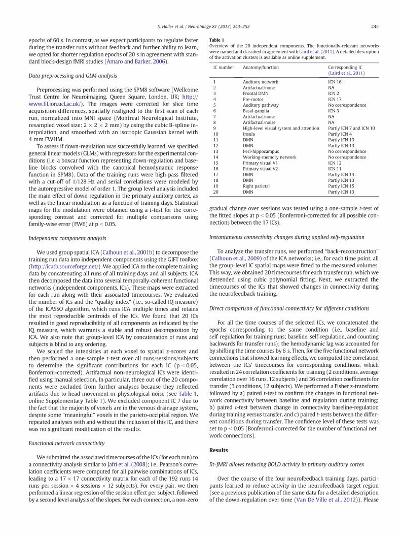
Table 1Overview of the 20 independent components. The functionally-relevant networkswere named and classified in agreement with Laird et al. (2011). A detailed descriptionof the activation clusters is available as online supplement.
IC number Anatomy/function Corresponding IC(Laird et al., 2011)
1 Auditory network ICN 162 Artifactual/noise NA3 Frontal DMN ICN 24 Pre-motor ICN 175 Auditory pathway No correspondence6 Basal-ganglia ICN 37 Artifactual/noise NA8 Artifactual/noise NA9 High-level visual system and attention Partly ICN 7 and ICN 1010 Insula Partly ICN 411 DMN Partly ICN 1312 DMN Partly ICN 1313 Peri-hippocampus No correspondence14 Working-memory network No correspondence15 Primary visual V1 ICN 1216 Primary visual V2 ICN 1117 DMN Partly ICN 1318 DMN Partly ICN 1319 Right parietal Partly ICN 1520 DMN Partly ICN 13
245S. Haller et al. / NeuroImage 81 (2013) 243–252
epochs of 60 s. In contrast, as we expect participants to regulate fasterduring the transfer runs without feedback and further ability to learn,we opted for shorter regulation epochs of 20 s in agreement with stan-dard block-design fMRI studies (Amaro and Barker, 2006).
Data preprocessing and GLM analysis
Preprocessing was performed using the SPM8 software (WellcomeTrust Centre for Neuroimaging, Queen Square, London, UK; http://www.fil.ion.ucl.ac.uk/). The images were corrected for slice timeacquisition differences, spatially realigned to the first scan of eachrun, normalized into MNI space (Montreal Neurological Institute,resampled voxel size: 2 × 2 × 2 mm) by using the cubic B-spline in-terpolation, and smoothed with an isotropic Gaussian kernel with4 mm FWHM.
To assess if down-regulation was successfully learned, we specifiedgeneral linearmodels (GLMs)with regressors for the experimental con-ditions (i.e. a boxcar function representing down-regulation and base-line blocks convolved with the canonical hemodynamic responsefunction in SPM8). Data of the training runs were high-pass filteredwith a cut-off of 1/128 Hz and serial correlations were modeled bythe autoregressive model of order 1. The group level analysis includedthe main effect of down regulation in the primary auditory cortex, aswell as the linear modulation as a function of training days. Statisticalmaps for the modulation were obtained using a t-test for the corre-sponding contrast and corrected for multiple comparisons usingfamily-wise error (FWE) at p b 0.05.
Independent component analysis
We used group spatial ICA (Calhoun et al., 2001b) to decompose thetraining run data into independent components using the GIFT toolbox(http://icatb.sourceforge.net/). We applied ICA to the complete trainingdata by concatenating all runs of all training days and all subjects. ICAthen decomposed the data into several temporally-coherent functionalnetworks (independent components, ICs). These maps were extractedfor each run along with their associated timecourses. We evaluatedthe number of ICs and the “quality index” (i.e., so-called IQ measure)of the ICASSO algorithm, which runs ICA multiple times and retainsthe most reproducible centroids of the ICs. We found that 20 ICsresulted in good reproducibility of all components as indicated by theIQ measure, which warrants a stable and robust decomposition byICA. We also note that group-level ICA by concatenation of runs andsubjects is blind to any ordering.
We scaled the intensities at each voxel to spatial z-scores andthen performed a one-sample t-test over all runs/sessions/subjectsto determine the significant contributions for each IC (p b 0.05,Bonferroni-corrected). Artifactual non-neurological ICs were identi-fied using manual selection. In particular, three out of the 20 compo-nents were excluded from further analyses because they reflectedartifacts due to head movement or physiological noise (see Table 1,online Supplementary Table 1). We excluded component IC 7 due tothe fact that the majority of voxels are in the venous drainage system,despite some “meaningful” voxels in the parieto-occipital region. Werepeated analyses with and without the inclusion of this IC, and therewas no significant modification of the results.
Functional network connectivity
We submitted the associated timecourses of the ICs (for each run) toa connectivity analysis similar to Jafri et al. (2008); i.e., Pearson's corre-lation coefficients were computed for all pairwise combinations of ICs,leading to a 17 × 17 connectivity matrix for each of the 192 runs (4runs per session × 4 sessions × 12 subjects). For every pair, we thenperformed a linear regression of the session effect per subject, followedby a second level analysis of the slopes. For each connection, a non-zero
gradual change over sessions was tested using a one-sample t-test ofthe fitted slopes at p b 0.05 (Bonferroni-corrected for all possible con-nections between the 17 ICs).
Instantaneous connectivity changes during applied self-regulation
To analyze the transfer runs, we performed “back-reconstruction”(Calhoun et al., 2009) of the ICA networks; i.e., for each time point, allthe group-level IC spatial maps were fitted to the measured volumes.This way, we obtained 20 timecourses for each transfer run, which wedetrended using cubic polynomial fitting. Next, we extracted thetimecourses of the ICs that showed changes in connectivity duringthe neurofeedback training.
Direct comparison of functional connectivity for different conditions
For all the time courses of the selected ICs, we concatenated theepochs corresponding to the same condition (i.e., baseline andself-regulation for training runs; baseline, self-regulation, and countingbackwards for transfer runs); the hemodynamic lag was accounted forby shifting the time courses by 6 s. Then, for the five functional networkconnections that showed learning effects, we computed the correlationbetween the ICs' timecourses for corresponding conditions, whichresulted in 24 correlation coefficients for training (2 conditions, averagecorrelation over 16 runs, 12 subjects) and 36 correlation coefficients fortransfer (3 conditions, 12 subjects). We performed a Fisher z-transformfollowed by a) paired t-test to confirm the changes in functional net-work connectivity between baseline and regulation during training;b) paired t-test between change in connectivity baseline-regulationduring training versus transfer, and c) paired t-tests between the differ-ent conditions during transfer. The confidence level of these tests wasset to p b 0.05 (Bonferroni-corrected for the number of functional net-work connections).
Results
Rt-fMRI allows reducing BOLD activity in primary auditory cortex
Over the course of the four neurofeedback training days, partici-pants learned to reduce activity in the neurofeedback target region(see a previous publication of the same data for a detailed descriptionof the down-regulation over time (Van De Ville et al., 2012)). Please
246 S. Haller et al. / NeuroImage 81 (2013) 243–252
note that in the neurofeedback training runs, we actually presentedan auditory stimulus during the down-regulation blocks, which wasnot present during the baseline blocks. Hence, participants are not ac-tually down-regulating spontaneous activity in their auditory cortex,but they are learning to reduce stimulus-induced activity. These re-sults confirm our earlier report that voluntarily reducing auditorycortex activity can be learned with the help of neurofeedback(Haller et al., 2010).
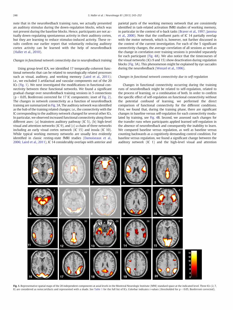
Changes in functional network connectivity due to neurofeedback training
Using group-level ICA, we identified 17 temporally-coherent func-tional networks that can be related to neurologically related processessuch as visual, auditory, and working memory (Laird et al., 2011);i.e., we excluded 3 artifactual and vascular components out of the 20ICs (Fig. 1). We next investigated the modifications in functional con-nectivity between these functional networks. We found a significantgradual change over neurofeedback training sessions in 5 connections(p b 0.05, Bonferroni corrected for 17 IC components; inset of Fig. 2).The changes in network connectivity as a function of neurofeedbacktraining are summarized in Fig. 3A. The auditory networkwas identifiedas the hub of the training related changes; i.e., the connectivity with theIC corresponding to the auditory network changed for several other ICs.In particular, we observed increased functional connectivity along threedifferent axes: (a) brainstem auditory pathway (IC 5), (b) high-levelvisual and attention networks (IC 9), and (c) a chain of three networksincluding an early visual cortex network (IC 15) and insula (IC 10).While typical working memory networks are usually less evidentlyidentified in classic resting-state fMRI studies (Damoiseaux et al.,2006; Laird et al., 2011), IC 14 considerably overlaps with anterior and
1 2 3
7 8 6
16 18 17
12 11 13
10 24 8
4 -22 1
32 30 -2
6 44 28
Fig. 1. Representative spatial maps of the 20 independent components at axial levels in the M8) are considered as noise/artifacts and represented with a shade. See Table 1 for the full li
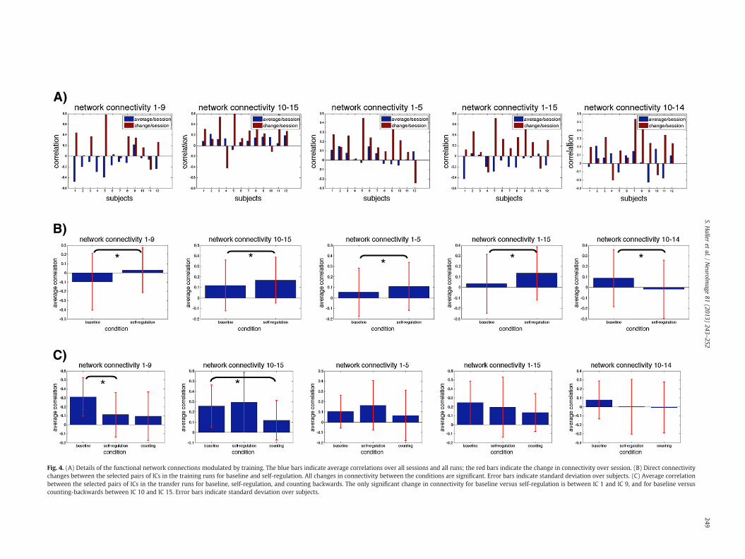
parietal parts of the working memory network that are consistentlyidentified in task-related activation fMRI studies of working memory,in particular in the context of n-back tasks (Braver et al., 1997; Jansmaet al., 2000). Note that the confluent parts of IC 14 partially overlapwith the motor network, which is, however, not further discussed inthe context of the current investigation. For each of the 5 significantconnectivity changes, the average correlation of all sessions as well asthe change in correlation over training sessions is provided separatelyfor each participant (Fig. 4A). We also notice that the timecourses ofthe visual networks (ICs 9 and 15) show deactivation during regulationblocks (Fig. 3A). This phenomenon might be explained by eye saccadesduring the neurofeedback (Wenzel et al., 1996).
Changes in functional network connectivity due to self-regulation
Changes in functional connectivity occurring during the trainingruns of neurofeedback might be related to self-regulation, related tothe process of learning, or a combination of both. In order to confirmthe specific effect of self-regulation on functional connectivity withoutthe potential confound of learning, we performed the directcomparison of functional connectivity for the different conditions.First, we found that, during the training phase, there are significantchanges in baseline versus self-regulation for each connectivity modu-lated by training, see Fig. 4B. Second, we assessed such changes forthe transfer runs when participants applied learned self-regulation inthe absence of neurofeedback and consequently the inability to learn.We compared baseline versus regulation, as well as baseline versuscounting backwards as a cognitively demanding control condition. Forbaseline versus regulation, we found a significant change between theauditory network (IC 1) and the high-level visual and attention
4 5
9 10
14 15
20 19
24 -10
0 28 -8
6 36 6
32 32
ontreal Neurologic Institute (MNI) standard space at the indicated level. Three ICs (2, 7,st of ICs. Colorbar indicates t-values (thresholded for p b 0.05, Bonferroni corrected).
**
**
increase / session
decr
ease
/ se
ssio
n
*
Fig. 2. Linear modulation over training days of functional connectivity between each pair of ICs. Colorbar indicates t-values, connections indicated with * are significant (p b 0.05,Bonferroni corrected for all possible connections between the 17 selected ICs).
247S. Haller et al. / NeuroImage 81 (2013) 243–252
networks (IC 9), see Fig. 4C. Interestingly, connectivity decreased duringtransfer, while it increased during training. In addition, the connectivitybetween the auditory network (IC 1) and the auditory pathway (IC 5),as well as with the low level visual (IC 15), working memory (IC 14)and the insular (IC 10) networks did not change significantly duringthe transfer phase. For the counting backwards condition, we onlyfound one significant change (decrease) for IC 10–IC 15, confirming aspecific effect of self-regulation on functional connectivity. While thechange in connectivity between insula and low level visual networksduring learning might be unexpected at first glance, it is worthwhilementioning that the analysis of the current investigation is targeted toassess of self-regulation related effects. The analysis of the transferphase was done using back-reconstruction of ICs defined during theneurofeedback training runs. The analysis is thus biased and nottargeted for the specific assessment of counting backwards versus base-line. Interestingly, meta-analysis of task-related fMRI studies includesinsula for “counting”, “numbers” and basic visual areas for “numbers”(http://neurosynth.org) indicating an involvement of these two regionsduring the (mental) manipulating of numbers and counting. Thecounting backwards condition was aimed to confirm the specific effectof self-regulation during the transfer phase and to exclude a global ef-fect on functional connectivity due to the application of a cognitivelydemanding task, which was indeed the case.
Direct comparison of functional connectivity for different conditions
We confirmed that all functional network interactions that showlearning effects during training (Fig. 3A) have aswell a significant changewhen comparing correlation in baseline versus regulation (Fig. 4B). Thedifferences in connectivity during transfer were significant for IC 1–IC 9(baseline-regulation) and IC 10–IC 15 (baseline-counting). In addition,
we found a significant change in baseline-regulation when comparingtraining and transfer for two connections; i.e., IC 1–IC 15 and IC 1–IC 9.
Discussion
We deployed ICA (data-driven exploratory method without a-priorispecification of regions of interest) and successfully identified changesin functional network connectivity as a function of neurofeedback train-ing as well as to applying learned self-regulation. Interestingly, theauditory cortex, whichwas the target area for neurofeedback was iden-tified as the hub of these network changes even though this was notspecified a priori. These results show (a) that functional brain networkscan change as a function of learning, (b) that rt-fMRI-basedneurofeedback training causes network changes that are specific tothe neurofeedback target region, and (c) that network changes relatedto applying learned self-regulation are different from those of the train-ing phase. We will also discuss potential clinical applications of ourfindings.
Learning related changes of functional connectivity
Despite the large number of fMRI studies that used functional con-nectivitymeasures, only a limited number of them specifically assesseddynamic changes in functional connectivity (Bassett et al., 2011; Lewiset al., 2009). Depending on the complexity of the task, they founddiffer-ences in complex functional networks when comparing them beforeand after behavioral training. The neurofeedback training approachthat we used significantly advances these earlier findings. Rather thanmeasuring functional-connectivity changes related to a behavioraltask, our approach allows to directly target specific brain regions. Also,rather than comparing fMRI scans before and after the behavioral

A) Training phase with feedback
Low-level visual
insula working memory
brainstem
auditory network
2
4
6
8
10
12
14
B) Transfer phase without feedback
9 5
10 15 14
1
2
4
6
8
10
12
14
high-level visual and attention
Low-level visual
insula working memory
brainstem auditory network 9 5
10 15 14
1
high-level visual and attention
Fig. 3. (A) Functional network connectivity between the auditory network and various networks is modulated by neurofeedback training. The auditory network (IC 1) was iden-tified without prior assumptions as the hub of altered functional connectivity. Functional connectivity was dynamically reconfigured between this auditory network along threeaxes: (1) auditory pathway (IC 5); (2) high-level visual and attention network (IC 9); and (3) several networks related to visual processing of the feedback (IC 15) andhigher-level cognition, notably insula related to introspection and self-regulation (IC 10) and working memory (IC 14). White crosses indicate significant increase of functional con-nectivity as a function of training. (B) Connectivity between baseline and self-regulation during transfer phase (without neurofeedback) is modified between the auditory network(IC 1) and high-level visual and attention network (IC 9). Consistent with the absence of feedback and, consequently, the inability to further learn, functional connectivity of IC 1with the visual as well as the memory networks was not modified. Colorbar indicates t-values (thresholded for p b 0.05, Bonferroni corrected).
248 S. Haller et al. / NeuroImage 81 (2013) 243–252
training, the neurofeedback learning takes place during scanning. Thisallows examining progressive learning-related changes in functionalconnectivity. It also allows distinguishing between learning-relatedchanges and those related to applying learned self-regulation.
Neurofeedback training gradually changes functional connectivity withthe auditory target region
The process of learning to self-regulate the auditory target region ismediated by gradual changes in connectivity. Using data-driven analysis,we found that the auditory target region was the hub of these changes(Fig. 3). Note that this network includes bilateral auditory area despitethe unilateral ROI in the neurofeedback training, which is consistentwith the close anatomic connection of both auditory areas and e.g. bilat-eral (about 2/3 dominant) contralateral auditory activation to unilateralauditory stimulation (Haller et al., 2006). Our data indicate that theneurofeedback target region does not only change in terms of activity,but in addition alters its connectivity with other networks. During
neurofeedback training the functional connectivity of the auditory targetregion changed along three axes. Firstly, connectivitywith the brainstemnetwork (IC 5) increased. This brainstem network (IC 5) includes manypathways, yet in the context of the current investigation the most rele-vant pathway is the auditory pathway, including the cochlear nuclei, su-perior olivary complex and inferior colliculi of the tectum and themedialgeniculate ganglion of the thalamus. Secondly, connectivity between theauditory target area and higher-level visual and attention networks (IC9) showed linear increase in connectivity. The self-regulation requireshigh attentional demands. Moreover, the higher-level visual associationarea is associated with visual tracking (Laird et al., 2011) required forthe tracking of the feedback. Thirdly, connectivity with a widespreadnetwork (ICs 15–10–14) including low-level visual as well as insularand working memory areas changed with neurofeedback training.While the early visual network is consistent with the visual feedback,the insular andworkingmemory areas might be attributed to introspec-tive awareness induced by feedback from one's own brain activity andworking memory demands when learning to self-regulate. Note that
*
A)
B)
C)
** **
Fig. 4. (A) Details of the functional network connections modulated by training. The blue bars indicate average correlations over all sessions and all runs; the red bars indicate the change in connectivity over session. (B) Direct connectivitychanges between the selected pairs of ICs in the training runs for baseline and self-regulation. All changes in connectivity between the conditions are significant. Error bars indicate standard deviation over subjects. (C) Average correlationbetween the selected pairs of ICs in the transfer runs for baseline, self-regulation, and counting backwards. The only significant change in connectivity for baseline versus self-regulation is between IC 1 and IC 9, and for baseline versuscounting-backwards between IC 10 and IC 15. Error bars indicate standard deviation over subjects.
249S.H
alleret
al./NeuroIm
age81
(2013)243
–252
250 S. Haller et al. / NeuroImage 81 (2013) 243–252
several previous rt-fMRI neurofeedback investigations found insular ac-tivation (for example, see Haller et al., 2010; Subramanian et al., 2011)indicating that this region might be involved in the process ofself-regulation per se, while this has not yet been systematicallyassessed.
While the functional interpretation of each IC that exhibits changes inits connectivity remains somehow speculative, the overall pattern of re-sults suggests that task-relevant functional connections are reinforced.This confirms previous findings of connectivity changes due toneurofeedback learning. For example, Rota et al. (2011) studied linguisticprosody by training participants to self-regulate the right inferior frontalgyrus (rIFG). Using a ROI seed-based functional connectivity analysis,these authors showed that the initially widespread connectivity of therIFG to frontal and temporal areas decreased over four training sessions.They also showed that the connectivity became more lateralized to theright hemisphere. In another recent study, Horovitz et al. (2010) usedsimilar analysis methods to show that neurofeedback training of themotor area led to increased basal ganglia involvement and bilateralmotor cortex connectivity. Finally, Lee et al. (2011) used a multivariateGranger causality analysis to investigate neurofeedback training relatedchanges in the insular cortex. Similar to the studies discussed above,the authors showed that neurofeedback training leads to a reduction ofpresumably redundant connections and to a strengthening of relevantconnections. However, these studies rely on the a priori choice of seed re-gions and, therefore, do not allow investigating changes in functionalconnectivity between networks.
Application of previously learned self-regulation causes changes infunctional connectivity
Another limitation of the above-mentioned studies is that they inves-tigated functional connectivity changes only during the neurofeedbacktraining phase. The neurofeedback training, however, is different fromap-plying learned self-regulation after the training. The training involves pro-cessing and interpreting the neurofeedback display, testing differentstrategies, and evaluating the training success. All these components arereflected in the connectivity changes thatwe foundduringneurofeedbacktraining (Fig. 3A). Once participants learned self-regulation of theauditory target area, participants can do so even in the absence ofneurofeedback. We also confirmed direct changes between baseline andself-regulation for all connections modulated by training (Fig. 4B).
In order to disentangle the confounding effect of learning and toconfirm changes in functional connectivity related to self-regulation,participants performed an additional transfer run without feedbackand thus without the ability to further learn. In particular, participantswere asked to apply the previously learned self-regulation strategy inthe absence of feedback. This self-regulation task was contrasted to acognitively demanding control task (counting backwards). When par-ticipants apply their newly acquired self-regulation skill during thetransfer run, only the connectivity between the auditory network andattention and high-level visual network changed when comparingbaseline against self-regulation, consistent with high demands on at-tention during self-regulation, which was acquired during the trainingphase (Figs. 3B and 4C). Learning-related connectivity changes relatedto introspection, memory demands, or to reinforcement learning wereno longer present. Note that the connectivity between the auditory net-work (IC 1) and the brainstem auditory pathway (IC 5) showed a trend(but non-significant; p = 0.15) also during self-regulation. In contrast,we observed no effect of the counting backwards condition on this func-tional connectivity. This confirms that the observed changes in func-tional connectivity are specific and related to self-regulation learnedin rt-fMRI neurofeedback and not simply an effect of performing acognitively demanding (control) task.
The analysis of the transfer runs was based on back-reconstructionusing the 20 ICA maps determined from the training phase data. Wemention that alternative approaches of ICA for multiple-condition
data have been proposed and could be considered for future work(Beckmann et al., 2006; Calhoun et al., 2001a; Long et al., 2009).
Direct comparison of functional connectivity between training and transferruns
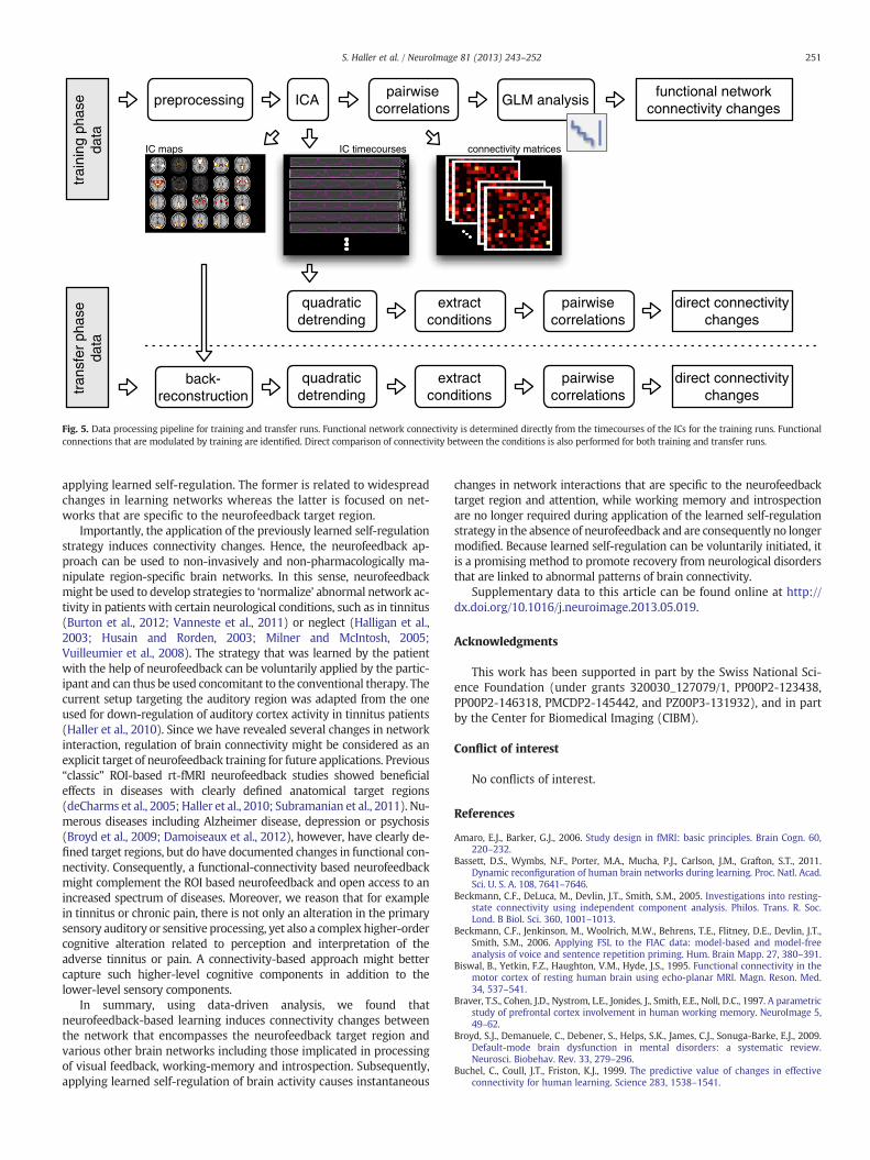
The direct comparison between training and transfer runs is compli-cated by the different nature of the training and transfer runs. First,concerning duration, participants were instructed to test severalself-regulation strategies during the training runs. In order to have suf-ficient time to do so, the block lengthwas 60 s. In contrast, during trans-fer runs, the application of the previously learned self-regulationstrategy is faster and thus we opted for a block length of 20 s in agree-mentwith standard block-design fMRI experiments (Amaro andBarker,2006). Second, concerning conditions, the training runs had only 2 con-ditions (baseline and self-regulation). In contrast, the transfer runs had3 conditions (baseline, self-regulation, and counting backwards) withthe latter condition intended as cognitively demanding control condi-tion. Third, concerning repetitions, training runs were repeated 4times per day in order to give participants sufficient time to learnself-regulation, while the transfer run was performed only once afterall learning runs. Consequently, differences in block length andnumber of runs between training and transfer runs might potentiallyconfound the analysis, and both experimental paradigms are notidentical (Fig. 5).
For all functional network connections that showed learningeffects during training (Fig. 3A), we confirmed a significant changein correlation when performing a direct comparison between base-line and regulation (Fig. 4B). In addition, the change in connectivitybetween baseline and regulation was compared between trainingand transfer. Two connections survived this test. The first one (IC 1–IC15: auditory-low level visual) confirms our previous result (i.e., signifi-cant change in connectivity regulation-baseline within transfer runs).The second one (IC 1–IC 9: auditory-high level visual) seems contradic-tory because we had before a significant change in connectivity ofregulation-baseline both within training and within transfer. However,a closer inspection reveals that the change within training is positive(increase in connectivity), while the change within transfer is negative(decrease in connectivity). In sum, the direct comparison confirms ourprevious findings and even refines the functional connectivity betweenauditory and high level visual for regulation applied during transfer.
Limitations
In this study, all training runs were pooled together for the ICA de-composition, which is used as an unsupervised dimensionality reduc-tion tool prior to further analysis. Similar to PCA, spatial ICA iscompletely blind to temporal relationships of the data (i.e., temporalpermutation does not influence the result), neither is ICA informedabout the paradigm. The optimization criterion of ICA (i.e., a surrogatefor spatial independence) will be driven by the spatial property‘averaged over time’. However, the fact that post-ICA analysis of thetimecourses shows statistically significant changes over time is meth-odologically valid and does not originate from circularity in the analysis.We previously found learning effects on activity in the target regionusing a two-level GLM analysis (Van De Ville et al., 2012). Nevertheless,it is worthwhile mentioning that pooling the data might decrease thesensitivity of our data analysis approach. Finally, we mention that theICASSO algorithm was used to determine the number of componentsand monitor the robustness of the decomposition.
Outlook and conclusions
Our approach of combining data-driven analysis tools with aneurofeedback training and transfer phase allowed us to disentanglethe connectivity changes when learning to self-regulate and when
preprocessing ICApairwise
correlationsGLM analysis
functional networkconnectivity changes
trai
ning
pha
seda
ta
back-reconstruction
quadraticdetrending
extract conditions
pairwisecorrelations
direct connectivitychanges
tran
sfer
pha
seda
ta
quadraticdetrending
extract conditions
pairwisecorrelations
direct connectivitychanges
IC maps IC timecourses connectivity matrices
Fig. 5. Data processing pipeline for training and transfer runs. Functional network connectivity is determined directly from the timecourses of the ICs for the training runs. Functionalconnections that are modulated by training are identified. Direct comparison of connectivity between the conditions is also performed for both training and transfer runs.
251S. Haller et al. / NeuroImage 81 (2013) 243–252
applying learned self-regulation. The former is related to widespreadchanges in learning networks whereas the latter is focused on net-works that are specific to the neurofeedback target region.
Importantly, the application of the previously learned self-regulationstrategy induces connectivity changes. Hence, the neurofeedback ap-proach can be used to non-invasively and non-pharmacologically ma-nipulate region-specific brain networks. In this sense, neurofeedbackmight be used to develop strategies to ‘normalize’ abnormal network ac-tivity in patients with certain neurological conditions, such as in tinnitus(Burton et al., 2012; Vanneste et al., 2011) or neglect (Halligan et al.,2003; Husain and Rorden, 2003; Milner and McIntosh, 2005;Vuilleumier et al., 2008). The strategy that was learned by the patientwith the help of neurofeedback can be voluntarily applied by the partic-ipant and can thus be used concomitant to the conventional therapy. Thecurrent setup targeting the auditory region was adapted from the oneused for down-regulation of auditory cortex activity in tinnitus patients(Haller et al., 2010). Since we have revealed several changes in networkinteraction, regulation of brain connectivity might be considered as anexplicit target of neurofeedback training for future applications. Previous“classic” ROI-based rt-fMRI neurofeedback studies showed beneficialeffects in diseases with clearly defined anatomical target regions(deCharms et al., 2005; Haller et al., 2010; Subramanian et al., 2011). Nu-merous diseases including Alzheimer disease, depression or psychosis(Broyd et al., 2009; Damoiseaux et al., 2012), however, have clearly de-fined target regions, but do have documented changes in functional con-nectivity. Consequently, a functional-connectivity based neurofeedbackmight complement the ROI based neurofeedback and open access to anincreased spectrum of diseases. Moreover, we reason that for examplein tinnitus or chronic pain, there is not only an alteration in the primarysensory auditory or sensitive processing, yet also a complex higher-ordercognitive alteration related to perception and interpretation of theadverse tinnitus or pain. A connectivity-based approach might bettercapture such higher-level cognitive components in addition to thelower-level sensory components.
In summary, using data-driven analysis, we found thatneurofeedback-based learning induces connectivity changes betweenthe network that encompasses the neurofeedback target region andvarious other brain networks including those implicated in processingof visual feedback, working-memory and introspection. Subsequently,applying learned self-regulation of brain activity causes instantaneous
changes in network interactions that are specific to the neurofeedbacktarget region and attention, while working memory and introspectionare no longer required during application of the learned self-regulationstrategy in the absence of neurofeedback and are consequently no longermodified. Because learned self-regulation can be voluntarily initiated, itis a promising method to promote recovery from neurological disordersthat are linked to abnormal patterns of brain connectivity.
Supplementary data to this article can be found online at http://dx.doi.org/10.1016/j.neuroimage.2013.05.019.
Acknowledgments
This work has been supported in part by the Swiss National Sci-ence Foundation (under grants 320030_127079/1, PP00P2-123438,PP00P2-146318, PMCDP2-145442, and PZ00P3-131932), and in partby the Center for Biomedical Imaging (CIBM).
Conflict of interest
No conflicts of interest.
References
Amaro, E.J., Barker, G.J., 2006. Study design in fMRI: basic principles. Brain Cogn. 60,220–232.
Bassett, D.S., Wymbs, N.F., Porter, M.A., Mucha, P.J., Carlson, J.M., Grafton, S.T., 2011.Dynamic reconfiguration of human brain networks during learning. Proc. Natl. Acad.Sci. U. S. A. 108, 7641–7646.
Beckmann, C.F., DeLuca, M., Devlin, J.T., Smith, S.M., 2005. Investigations into resting-state connectivity using independent component analysis. Philos. Trans. R. Soc.Lond. B Biol. Sci. 360, 1001–1013.
Beckmann, C.F., Jenkinson, M., Woolrich, M.W., Behrens, T.E., Flitney, D.E., Devlin, J.T.,Smith, S.M., 2006. Applying FSL to the FIAC data: model-based and model-freeanalysis of voice and sentence repetition priming. Hum. Brain Mapp. 27, 380–391.
Biswal, B., Yetkin, F.Z., Haughton, V.M., Hyde, J.S., 1995. Functional connectivity in themotor cortex of resting human brain using echo-planar MRI. Magn. Reson. Med.34, 537–541.
Braver, T.S., Cohen, J.D., Nystrom, L.E., Jonides, J., Smith, E.E., Noll, D.C., 1997. A parametricstudy of prefrontal cortex involvement in human working memory. NeuroImage 5,49–62.
Broyd, S.J., Demanuele, C., Debener, S., Helps, S.K., James, C.J., Sonuga-Barke, E.J., 2009.Default-mode brain dysfunction in mental disorders: a systematic review.Neurosci. Biobehav. Rev. 33, 279–296.
Buchel, C., Coull, J.T., Friston, K.J., 1999. The predictive value of changes in effectiveconnectivity for human learning. Science 283, 1538–1541.
252 S. Haller et al. / NeuroImage 81 (2013) 243–252
Burton, H., Wineland, A., Bhattacharya, M., Nicklaus, J., Garcia, K.S., Piccirillo, J.F., 2012.Altered networks in bothersome tinnitus: a functional connectivity study. BMCNeurosci. 13, 3.
Calhoun, V.D., Adali, T., Pearlson, G.D., Pekar, J.J., 2001. Spatial and temporal independentcomponent analysis of functional MRI data containing a pair of task-related wave-forms. Hum. Brain Mapp. 13, 43–53.
Calhoun, V.D., Adali, T., Pearlson, G.D., Pekar, J.J., 2001. A method for making group in-ferences from functional MRI data using independent component analysis. Hum.Brain Mapp. 14, 140–151.
Calhoun, V.D., Liu, J., Adali, T., 2009. A review of group ICA for fMRI data and ICA forjoint inference of imaging, genetic, and ERP data. NeuroImage 45, S163–S172.
Damoiseaux, J.S., Rombouts, S.A., Barkhof, F., Scheltens, P., Stam, C.J., Smith, S.M.,Beckmann, C.F., 2006. Consistent resting-state networks across healthy subjects.Proc. Natl. Acad. Sci. U. S. A. 103, 13848–13853.
Damoiseaux, J.S., Prater, K.E., Miller, B.L., Greicius, M.D., 2012. Functional connectivity tracksclinical deterioration in Alzheimer's disease. Neurobiol. Aging 33, 828.e19–828.e30.
Daubechies, I., Roussos, E., Takerkart, S., Benharrosh, M., Golden, C., D'Ardenne, K., Richter,W., Cohen, J.D., Haxby, J., 2009. Independent component analysis for brain fMRI doesnot select for independence. Proc. Natl. Acad. Sci. U. S. A. 106, 10415–10422.
deCharms, R.C., 2008. Applications of real-time fMRI. Nat. Rev. Neurosci. 9, 720–729.deCharms, R.C., Christoff, K., Glover, G.H., Pauly, J.M., Whitfield, S., Gabrieli, J.D., 2004.
Learned regulation of spatially localized brain activation using real-time fMRI.NeuroImage 21, 436–443.
deCharms, R.C., Maeda, F., Glover, G.H., Ludlow, D., Pauly, J.M., Soneji, D., Gabrieli, J.D.,Mackey, S.C., 2005. Control over brain activation and pain learned by using real-time functional MRI. Proc. Natl. Acad. Sci. U. S. A. 102, 18626–18631.
Fox, M.D., Greicius, M., 2010. Clinical applications of resting state functional connectiv-ity. Front. Syst. Neurosci. 4, 19.
Fox, M.D., Raichle, M.E., 2007. Spontaneous fluctuations in brain activity observed withfunctional magnetic resonance imaging. Nat. Rev. Neurosci. 8, 700–711.
Greicius, M.D., Krasnow, B., Reiss, A.L., Menon, V., 2003. Functional connectivity in theresting brain: a network analysis of the default mode hypothesis. Proc. Natl. Acad.Sci. U. S. A. 100, 253–258.
Haller, S., Wetzel, S.G., Radue, E.W., Bilecen, D., 2006. Mapping continuous neuronal ac-tivation without an ON–OFF paradigm: initial results of BOLD ceiling fMRI. Eur. J.Neurosci. 24, 2672–2678.
Haller, S., Birbaumer, N., Veit, R., 2010. Real-time fMRI feedback training may improvechronic tinnitus. Eur. Radiol. 20, 696–703.
Halligan, P.W., Fink, G.R., Marshall, J.C., Vallar, G., 2003. Spatial cognition: evidencefrom visual neglect. Trends Cogn. Sci. 7, 125–133.
Horovitz, S.G., Berman, B.D., Hallett, M., 2010. Real time BOLD functional MRI neuro-feedback affects functional connectivity. Conf. Proc. IEEE Eng. Med. Biol. Soc.2010, 4270–4273.
Husain, M., Rorden, C., 2003. Non-spatially lateralized mechanisms in hemispatialneglect. Nat. Rev. Neurosci. 4, 26–36.
Jafri, M.J., Pearlson, G.D., Stevens, M., Calhoun, V.D., 2008. A method for functional net-work connectivity among spatially independent resting-state components inschizophrenia. NeuroImage 39, 1666–1681.
Jansma, J.M., Ramsey, N.F., Coppola, R., Kahn, R.S., 2000. Specific versus nonspecificbrain activity in a parametric N-back task. NeuroImage 12, 688–697.
Laird, A.R., Fox, P.M., Eickhoff, S.B., Turner, J.A., Ray, K.L., McKay, D.R., Glahn, D.C.,Beckmann, C.F., Smith, S.M., Fox, P.T., 2011. Behavioral interpretations of intrinsicconnectivity networks. J. Cogn. Neurosci. 23, 4022–4040.
Lee, S., Ruiz, S., Caria, A., Veit, R., Birbaumer, N., Sitaram, R., 2011. Detection of cerebralreorganization induced by real-time fMRI feedback training of insula activation: amultivariate investigation. Neurorehabil. Neural Repair 25, 259–267.
Lewis, C.M., Baldassarre, A., Committeri, G., Romani, G.L., Corbetta, M., 2009. Learningsculpts the spontaneous activity of the resting human brain. Proc. Natl. Acad. Sci.U. S. A. 106, 17558–17563.
Long, Z., Chen, K., Wu, X., Reiman, E., Peng, D., Yao, L., 2009. Improved application of in-dependent component analysis to functional magnetic resonance imaging studyvia linear projection techniques. Hum. Brain Mapp. 30, 417–431.
McKeown, M.J., Jung, T.P., Makeig, S., Brown, G., Kindermann, S.S., Lee, T.W., Sejnowski,T.J., 1998. Spatially independent activity patterns in functional MRI data during theStroop color-naming task. Proc. Natl. Acad. Sci. U. S. A. 95, 803–810.
McKeown, M.J., Makeig, S., Brown, G.G., Jung, T.P., Kindermann, S.S., Bell, A.J.,Sejnowski, T.J., 1998. Analysis of fMRI data by blind separation into independentspatial components. Hum. Brain Mapp. 6, 160–188.
Milner, A.D., McIntosh, R.D., 2005. The neurological basis of visual neglect. Curr. Opin.Neurol. 18, 748–753.
Posse, S., Fitzgerald, D., Gao, K., Habel, U., Rosenberg, D., Moore, G.J., Schneider, F., 2003.Real-time fMRI of temporolimbic regions detects amygdala activation duringsingle-trial self-induced sadness. NeuroImage 18, 760–768.
Raichle, M.E., 2010. Two views of brain function. Trends Cogn. Sci. 14, 180–190.Rota, G., Handjaras, G., Sitaram, R., Birbaumer, N., Dogil, G., 2011. Reorganization of
functional and effective connectivity during real-time fMRI-BCI modulation ofprosody processing. Brain Lang. 117, 123–132.
Seifritz, E., Esposito, F., Hennel, F., Mustovic, H., Neuhoff, J.G., Bilecen, D., Tedeschi, G.,Scheffler, K., Di Salle, F., 2002. Spatiotemporal pattern of neural processing in thehuman auditory cortex. Science 297, 1706–1708.
Subramanian, L., Hindle, J.V., Johnston, S., Roberts, M.V., Husain, M., Goebel, R., Linden,D., 2011. Real-time functional magnetic resonance imaging neurofeedback fortreatment of Parkinson's disease. J. Neurosci. 31, 16309–16317.
Van De Ville, D., Jhooti, P., Haas, T., Kopel, R., Lovblad, K.O., Scheffler, K., Haller, S.,2012. Recovery of the default mode network after demanding neurofeedbacktraining occurs in spatio-temporally segregated subnetworks. NeuroImage 63,1775–1781.
Vanneste, S., Focquaert, F., Van de Heyning, P., De Ridder, D., 2011. Different restingstate brain activity and functional connectivity in patients who respond and not re-spond to bifrontal tDCS for tinnitus suppression. Exp. Brain Res. 210, 217–227.
Vuilleumier, P., Schwartz, S., Verdon, V., Maravita, A., Hutton, C., Husain, M., Driver, J.,2008. Abnormal attentional modulation of retinotopic cortex in parietal patientswith spatial neglect. Curr. Biol. 18, 1525–1529.
Wang, L., Yu, C., Chen, H., Qin, W., He, Y., Fan, F., Zhang, Y., Wang, M., Li, K., Zang, Y.,Woodward, T.S., Zhu, C., 2010. Dynamic functional reorganization of the motor ex-ecution network after stroke. Brain 133, 1224–1238.
Weiskopf, N., Veit, R., Erb, M., Mathiak, K., Grodd, W., Goebel, R., Birbaumer, N., 2003.Physiological self-regulation of regional brain activity using real-time functionalmagnetic resonance imaging (fMRI): methodology and exemplary data. NeuroImage19, 577–586.
Weiskopf, N., Mathiak, K., Bock, S.W., Scharnowski, F., Veit, R., Grodd, W., Goebel, R.,Birbaumer, N., 2004. Principles of a brain-computer interface (BCI) based on real-time functional magnetic resonance imaging (fMRI). IEEE Trans. Biomed. Eng. 51,966–970.
Weiskopf, N., Scharnowski, F., Veit, R., Goebel, R., Birbaumer, N., Mathiak, K., 2004. Self-regulation of local brain activity using real-time functional magnetic resonance im-aging (fMRI). J. Physiol. Paris 98, 357–373.
Wenzel, R., Bartenstein, P., Dieterich, M., Danek, A., Weindl, A., Minoshima, S., Ziegler,S., Schwaiger, M., Brandt, T., 1996. Deactivation of human visual cortex during in-voluntary ocular oscillations. A PET activation study. Brain 119, 101–110.
Yoo, S.S., Jolesz, F.A., 2002. Functional MRI for neurofeedback: feasibility study on ahand motor task. Neuroreport 13, 1377–1381.
Related Documents