Division Handbook of the Division of Pediatric Radiology Russell H. Morgan Department of Radiology and Radiological Science and the Johns Hopkins Hospital 2010-2011

Welcome message from author
This document is posted to help you gain knowledge. Please leave a comment to let me know what you think about it! Share it to your friends and learn new things together.
Transcript
Division Handbook
of the
Division of Pediatric Radiology
Russell H. Morgan Department of Radiology and Radiological
Science and the
Johns Hopkins Hospital
2010-2011
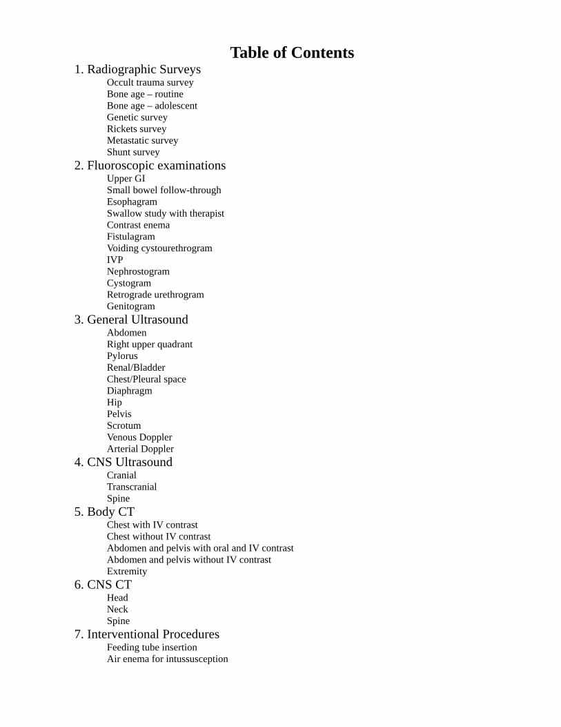
Table of Contents 1. Radiographic Surveys Occult trauma survey Bone age – routine Bone age – adolescent Genetic survey Rickets survey Metastatic survey Shunt survey
2. Fluoroscopic examinations Upper GI Small bowel follow-through Esophagram Swallow study with therapist Contrast enema Fistulagram Voiding cystourethrogram IVP Nephrostogram Cystogram Retrograde urethrogram Genitogram
3. General Ultrasound Abdomen Right upper quadrant Pylorus Renal/Bladder Chest/Pleural space Diaphragm Hip Pelvis Scrotum Venous Doppler Arterial Doppler
4. CNS Ultrasound Cranial Transcranial Spine
5. Body CT Chest with IV contrast Chest without IV contrast Abdomen and pelvis with oral and IV contrast Abdomen and pelvis without IV contrast Extremity
6. CNS CT Head Neck Spine
7. Interventional Procedures Feeding tube insertion Air enema for intussusception
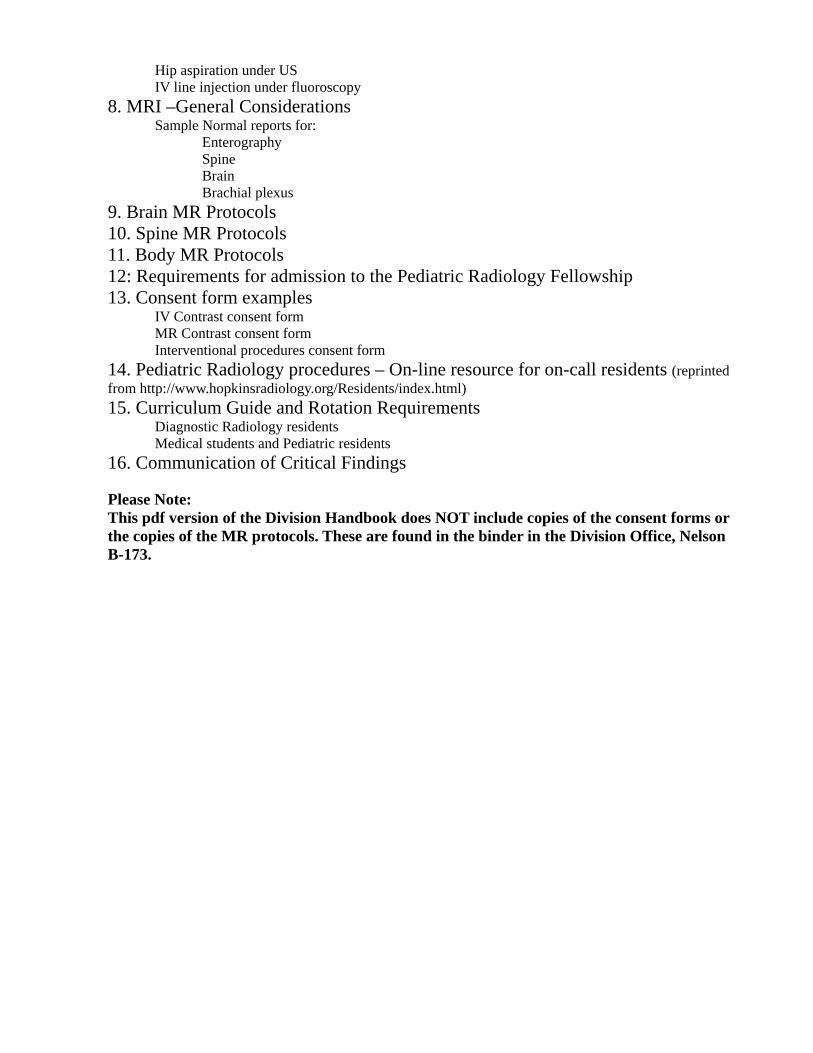
Hip aspiration under US IV line injection under fluoroscopy
8. MRI –General Considerations Sample Normal reports for: Enterography Spine Brain Brachial plexus
9. Brain MR Protocols 10. Spine MR Protocols 11. Body MR Protocols 12: Requirements for admission to the Pediatric Radiology Fellowship 13. Consent form examples IV Contrast consent form MR Contrast consent form Interventional procedures consent form
14. Pediatric Radiology procedures – On-line resource for on-call residents (reprinted from http://www.hopkinsradiology.org/Residents/index.html) 15. Curriculum Guide and Rotation Requirements Diagnostic Radiology residents Medical students and Pediatric residents
16. Communication of Critical Findings Please Note: This pdf version of the Division Handbook does NOT include copies of the consent forms or the copies of the MR protocols. These are found in the binder in the Division Office, Nelson B-173.
Plain Radiographic Surveys
Usual indications: Evaluation for specific entities where certain constellation of bone findings will be diagnostic. Series are ordered to save time and patient charges Basic method: AP views alone are usually sufficient; limbs may be combined to decrease the number of exposures Usual images: as below Sample normal reports : as below Unusual indications: “Completion series” --patient has outside studies and a few exams are needed to make a complete survey. (This usually comes as a request from the Genetics Clinic.) Modifications to method/images for these indications: A few views are done as ordered; reporting is done in consultation with the ordering physician, preferably along with the outside exams **************** Indication: Question of non- accidental trauma. Occult trauma survey, 18 views: The survey consists of AP views of the chest, pelvis, humeri, forearms, hands, feet, tibias and fibulas, and femurs with lateral views of the skull, spine, and chest and oblique views of the ribs. The skeleton appears normal in mineralization throughout. The degree of skeletal maturation is appropriate for age. There is no sign of bony dysplasia. No acute or healing fracture is found. IMPRESSION: Normal skeletal survey. ********************* Indication: Short stature. Bone Age: Single AP view of the left hand was obtained. The patient's chronological age is ---------years and-------- months. The bone age by the standards of Greulich and Pyle is most consistent with a (male)( female )of------ years and---------- months of age. The standard deviation of the bone age at this chronologic age is -------months. IMPRESSION: (Advanced)( Delayed)( Normal) bone age. (Bone age is within two standard deviations of the chronologic age.) ******************* Indication: Skeletal Dysplasia; need to assess skeletal maturity Chronologic age: -----------years and----------- months. Bone age: View of the left hand and wrist is compared with the standards of Greulich and Pyle. The bone age is most consistent with a( male)( female) of -----------years and ------------months of age. The standard deviation of the bone age at this chronologic age is months. Views of the knee are compared with standards of Pyle and Hoerr. They most resemble that of a( male)( female) of -------years and----------- months of age. Views of the ankle and foot are compared with standards of Hoerr, Pyle and Francis. They most resemble that of a( male)( female) of------ years and----------- months of age. IMPRESSION: (Advanced )(Delayed)( Normal bone age.)( The bone age is within 2 standard
deviations of the chronologic age), according to the hand and wrist and (advanced)( delayed)( normal) by other standards. *********************
Genetic Survey:
Indication:
Findings: The survey consists of AP views of the upper extremities including the hands, the lower extremities with the pelvis, the feet, the skull and the chest, with lateral views of the skull and spine.
The bones show normal mineralization throughout. The bony maturity is appropriate for chronologic age. There is normal proportionality between the proximal, middle and distal limb segments. No structural abnormality of any diaphysis, metaphysis or epiphysis is seen.
Impression: Normal genetic survey
*************
Rickets Survey
Indications: question of renal osteodystrophy
Findings: AP views of the left hand and left knee were obtained. Bony mineralization appears normal with no coarsening of trabeculae and no subperiosteal resorption. No soft tissue calcification is seen.
Impression: No evidence of renal osteodystrophy.
*********************
Metastatic Survey**[Note: this is rarely performed now, having been superseded by bone scan and PET scan. Plain films are sometime used to further assess areas highlighted by these other methods.]
Indication: Question of bony metastasis.
Findings: The survey consists of AP views of the arms, legs, pelvis, chest, and skull, with lateral view of the skull and spine and oblique views of the ribs.
The bony contours are normal throughout with no focus of sclerosis or lysis. No periosteal reaction or soft tissue mass or calcification is seen.
Impression: No evidence of metastatic lesion.
********************
Shunt Survey **[Note: ideally, it should be known if the patient's “VP Shunt” is a
ventriculoperitoneal, ventriculoatrial, or ventriculopleural shunt, and the imaging should be confined to those areas.]
Indication: question of shunt malfunction.
Findings: The survey consists of AP views of the skull, chest and abdomen, with additional lateral view of the skull. The right parietal shunt catheter is seen terminating centrally in the calvarium. The catheter passes through a radiolucent shunt valve. Shunt tubing then passes without interruption over the right side of the neck and chest and loops in the abdomen, terminating in the left flank. The tip of the shunt has moved since the prior exam. There is no soft tissue mass associated with the tip of the shunt.
The lungs are clear and the mediastinum is unremarkable. The abdomen shows a normal bowel gas pattern and no organomegaly.
Impression: No evidence of shunt fixation or disruption.
Upper GI Usual indications: vomiting, feeding intolerance, r/o malrotation Basic method: barium by bottle, cup, syringe Usual images: AP/lateral full esophagus Profile duodenal bulb/pylorus AP stomach and duodenum to show DJ junction position Observe GEJ and esophagus intermittently for 5 minutes to look for reflux Sample Normal report: Indication: Feeding intolerance. Upper GI: Scout view of the abdomen showed no soft tissue or bony abnormality. The patient drank barium well from a bottle, showing normal oral and pharyngeal swallowing. The esophagus and stomach showed normal anatomy, motility and mucosal detail. There was unobstructed progress of the contrast from the stomach through a normal pylorus, duodenal bulb and duodenum. The duodeno-jejunal junction is in normal position. Early loops of jejunum show no abnormal distension. Intermittent fluoroscopic observation of the stomach and esophagus for 5 minutes during quiet respiration revealed no gastroesophageal reflux. Impression: Normal UGI. Unusual indications and Modifications to method/images for these indications: Nissen fundoplication evaluation: inject GT first and get stomach, duodenum images. Give patient enough PO contrast (1-2cc may be enough) to establish that esophagus drains and GEJ is below diaphragm. G-tube placement evaluation: Use water soluble contrast. For low-suspicion tubes, consider injection then AP and XTL plain films. When insertion was complicated, inject under fluoro, rotating patient so that the length of the tube is imaged, then establish that the pylorus and bulb are unobstructed. ALARA: keep exposure to xR minimal during fluoroscopy by - collimating to area of interest - placing screen reasonably close to the patient as possible but still comfortable - using small image size – no magnification - using brief intermittent pulsed fluoroscopy to focus as well as for documentation of abnormality
Small Bowel Follow Through Usual indications: Inflammatory bowel disease, feeding intolerance after small bowel surgery Basic method: Contrast (usually barium) followed from the stomach to the ileocecal valve. Encourage the patient to drink ½ the contrast before the first film, then sip the remainder over 30-60 min. Sitting up and walking around speed the transit time. Usual images: AP abdomen 10 minutes after contrast ingestion, then another AP abdomen 30 minutes later. You want your films to show a nice contrast column in the jejunum and ileum, so plan additional films sparingly. Compression views of the ICV finish the exam. Sample normal report: Indication: S/p ileal resection form NEC perforation, now with feeding intolerance; ?stricture Small Bowel Follow-through: The duodenum, jejunum and ileum show normal caliber and motility, with no areas of focal narrowing or distension. Transit time to the colon was 45 minutes, within normal limits. Spot views of the ileocecal junction were normal. Impression: Normal small bowel follow-through Unusual indications: following Sitz markers Modifications to method/images for these indications: Sitz markers are radio-opaque pellets swallowed in capsule form by patients in whom small bowel or colon dysmotility is suspected. Daily abdominal exams are ordered until all markers are cleared. ALARA: keep exposure to xR minimal during fluoroscopy by - collimating to area of interest - placing screen reasonably close to the patient as possible but still comfortable - using small image size – no magnification - using brief intermittent pulsed fluoroscopy to focus as well as for documentation of abnormality
Esophagram Usual indications: Question of esophageal obstruction (e.g.food impaction) in older child, assess treatment response (dilation for stricture, botox for achalasia), look for leak (after atresia repair). **Should not be used for vomiting, feeding difficulties, question of reflux – these indications deserve an UGI. Basic method: As for the UGI: Patient should swallow volitionally (for best airway protection and physiological assessment). Recumbent positioning challenges the motility the most. When assessing for leak, watch the chest tube (if present) to determine if contrast is draining through it. Otherwise try to assess entire circumference. Usual images: True AP and lateral views are key. Oblique views are need to document for leak. Sample normal report: Indication: history of repaired esophageal atresia, now with possible food impaction. Esophagram: The patient swallowed barium in AP, lateral and oblique projections. Narrowing of the lumen at the junction of the upper 1/3 and lower 2/3 corresponds to the patient's history of reanastamosis. Motility is impaired in the lower 2/3 of the esophagus, but clearance was adequate. Upright positioning aided clearance. No obstruction or retained food residue was present. Impression: No acute obstruction. Unusual indications: foreign body in esophagus Modifications to method/images for these indications: patient may not want to lie down. Standing may help contrast outline the obstruction. ALARA: keep exposure to xR minimal during fluoroscopy by - collimating to area of interest - placing screen reasonably close to the patient as possible but still comfortable - using small image size – no magnification - using brief intermittent pulsed fluoroscopy to focus as well as for documentation of
Swallow Study with Therapist (aka modified barium swallow, feeding study, video swallow)
Usual indications: feeding difficulties (refusal, apnea, cyanosis, coughing/choking), pneumonia or asthma raising suspicion for dysphagia and/or aspiration Basic method: The patient's usual feeding posture is replicated using a swallow chair or other adaptive device. The patient is accompanied by a feeding therapist (an Occupational Therapist or Speech-Language Pathologist) who positions the patient and mixes the textures to be offered. The therapist or the child's caregiver will feed the child. Because of space constraints and because it offers the best spatial separation of air-way and food-way, only the lateral view is used. The field of view should be coned to the mouth, pharynx and cervical esophagus. The radiologist is positioned to see the child and the fluoro screen. Continuous fluoro is used, and the study is entirely recorded on DVD—no spot images are made. The fluoro recording should begin when the contrast has been delivered to the child's mouth and stop when the bolus passes beyond the upper esophageal sphincter. Where there is continuous sucking/swallowing, a run of 4-6 swallows should be recorded, attempting particularly to capture the first swallow of the contrast. The therapist may have several textures to test. Be mindful of the accumulating dose and help the therapist triage the substances. Note the texture of the offering (thin liquid, puree) and how it is given (bottle, cup, spoon). Fatigue plays an important part in dysphagia; allowing the child to feed for awhile with intermittent observation/recording can help uncover pathology without overdosing. Magnification should not be used except, for example, where there is question of aspiration in a tiny infant and a momentary assessment of the enlarged airway is needed. Usual images: Several video clips comprising 2 minutes or less of fluoro time Sample normal report: Indication:Coughing and food refusal. Video swallow: The patient was placed upright in the swallow chair. Puree consistency was offered by spoon. The patient took 3 boluses and showed normal oral and pharyngeal phases of swallow, with no post swallow retention. The patient was then given thin liquid by bottle. He exhibited normal continuous suckle and swallow with normal bolus formation, bolus transit, and timing of pharyngeal swallow onset. No penetration of contrast occurred during any swallow, nor was there any aspiration. The patient did not cough during the exam. Impression: Normal swallow study. Unusual indications: Physiologic monitoring may be done during the exam, to look for subclinical signs of distress. Modifications to method/images for these indications: none ALARA: keep exposure to xR minimal during fluoroscopy by - collimating to area of interest - placing screen reasonably close to the patient as possible but still comfortable - using small image size – no magnification - using brief intermittent pulsed fluoroscopy to focus as well as for documentation of abnormality
Contrast Enema Usual Indications-Newborn bowel obstruction, constipation (r/o Hirschprungs disease [HD]), history of NEC—question of stricture. History of pull-through for Hirschprung disease. Basic Method- Choice of contrast: water soluble (cystoconray) or barium. In general, the water soluble is used in the neonate and maybe useful in the treatment of constipation. Barium is used in the diagnosis of HD. Enema tip varies by age and size of patient. Foley catheter or pediatric blue tip is typically used. Tape across the buttocks is used to help hold the tube in place and maintain the anal seal. Contrast may be instilled by gravity but the rate must be controlled and monitored carefully. It can injected by syringe with caution under the supervision of the attending pediatric radiologist. Progress is monitored by fluoroscopy. Usual Images-Document prn with last image hold or fluoroscopic exposures. In the case of HD, AP and lateral view of the full rectum is necessary. Sample normal report-Indication: Chronic constipation in a three-month old child. Normal birth history. Barium enema: The rectum was catheterized with a 16F foley catheter and taped in place. Initial filling of the rectum showed normal caliber and distensibilty. To aid further filling, the foley balloon was inflated with 5cc of air. Filling proceeded normally, encountering no stricture or mass. The cecum and appendix filled, but no reflux could be achieved into the terminal ileum, a common occurrence in this age group. The position of the colon in the abdomen is normal. Impression: Normal barium enema. Unusual indications- 1)History of diverting colostomy or ileostomy—need to visualize mucous fistula. 2) Patient with CF and DIOS – need for therapeutic enema. 3) newborn with obstruction: meconium ileus vs meconium plug Modification to method/images for these indications-1)Use water soluble contrast. May fill from the rectum, if patent, or antegrade—find the opening usually at the base of the proximal limb. Distal limb may be short or long. 2&3) Use cystoconray; try to reflux contrast into the distal ileum. (+/- mucomist or other surfactant for larger patient); may require multiple fill/evacuate cycles. ALARA may become problematic! ALARA: keep exposure to xR minimal during fluoroscopy by - collimating to area of interest - placing screen reasonably close to the patient as possible but still comfortable - using small image size – no magnification - using brief intermittent pulsed fluoroscopy to focus as well as for documentation of abnormality
Fistulagram Usual indications: draining sinus with suspicion of origin from an internal viscus (patent urachus, enterocutaneous fistula) Basic method: Use omnipaque 300 or 350 in a small syringe. Number 5 or 8 feeding tubes have blunt ends and single sideholes, making them good to try to thread into the sinus. Wear gloves to protect yourself; the procedure is clean but not sterile. Clean the skin with water and gauze, then pat dry. Gently push the catheter into the sinus as far as you can. Tape in place. Position the patient under the fluoroscope and very slowly inject. Roll the patient as needed to display the tract across your image. Usual images: Initial images may be wide field to locate landmarks, then cone down. Document tract length. Sample normal report: Indication: Draining sinus. Fistulagram: A number 8 catheter was gently inserted about 2cm into the sinus in the patient's right lower quadrant. Gentle hand-injection of about 5cc of omnipaque opacified a blind-ending tract that extended another 3 cm beyond the tip pf the catheter. Impression: No enteral communication of draining sinus. Unusual indications: Question of ostomy stricture Modifications to method/images for these indications: A foley catheter might work better to fill the bowel retrograde from the ostomy: inflate the balloon and hold it against the opening to occlude it. Once you've seen the pre-ostomy bowel, inflate the balloon just inside the opening, just enough to achieve filling ALARA: keep exposure to xR minimal during fluoroscopy by - collimating to area of interest - placing screen reasonably close to the patient as possible but still comfortable - using small image size – no magnification - using brief intermittent pulsed fluoroscopy to focus as well as for documentation of abnormality
Voiding Cystourethrography (VCUG) Usual indications: Urinary tract infection (UTI) Evaluation of fetal GU tract abnormality Follow-up vescicoureteral reflux (VUR) Follow-up post ureteral reimplantation or other uretero-vesical surgery Basic method: Catheterization of urinary bladder by attending radiologist/fellow or nurse. Retrograde filling of bladder with CystoConray 17% by controlled gravity. Monitoring bladder filling and voiding by repeat pulsed, short intermittent fluoroscopy. Bladder filling to capacity, i.e. until spontaneous voiding, with catheter in place. Usual images: (last image holds, no routine exposures): Bladder: at early filling, end of voiding, postvoid. Minimal VUR may occur at end of voiding. Urethra at voiding: in male: oblique view (urethra parallel to lower side femur); in female: AP view. Renal area: (combined R+L) at late filling, voiding, postvoid. At first appearance of and maximum VUR. Delayed postvoid renal images in significant VUR: at 1min; if necessary also at 10min postvoid to demonstrate adequate drainage, while catheter remains in place for continuous bladder drainage and to prevent ongoing or repeat VUR. Special views: oblique views for R and L ureters to exclude or demonstrate ureteral ectopy and for anatomic abnormality, e.g. ureterocele, ureterocele eversion, diverticulum, urachal remnant, etc. Occasional exposures: for small details/structures e.g. in uro-genital malformations, male urethra, etc. Sample normal report: Indication: UTI in 1 year old girl. VCUG: The bladder was catheterized with an 8F urethral catheter and a few cc of residual urine was removed. The bladder was filled with 50cc of cysticonray. No vescicoureteral reflux was seen in the filling or voiding phases. Voiding revealed a normal urethra and emptied the bladder completely. Impression: Normal VCUG. Unusual indications: child less than 6 months old for VCUG Modifications to usual method: The “tri-cyclic VCUG”: The infant bladder may not completely relax and fill sufficiently to exclude reflux. Let the infant void with the catheter in place, then let the bladder refill. Do this a third time. You should see progressively larger bladder volumes and better, more forceful voiding. *ALARA: keep exposure to xR minimal by - coning to area of interest - placing screen close to the patient - using small image size - using brief intermittent pulsed fluoroscopy to focus as well as for detection of abnormality
Excretion Urography/ Intravenous Pyleogram (EU/IVP) Usual indications: No routine indications anymore; replaced by US+MAG3 scintigraphy or MR urography Occasional indications: Early post-op. Question renal function in trauma patient: single KUB after abdominal CT with IV contrast instead of a delayed CT scan.
Basic method: Patient fasting for about three hours prior to examination. Prepared for exam by a “child life” person. Informed consent must be obtained for contrast injection. (Use the contrast form [Part 13]; talk about wheezing, hives, airway swelling.) Empty bladder is necessary for the exam! Patient is sent to the bathroom for complete voiding. Depending on the situation, an infant or young child might require catheterization for drainage and to keep the bladder empty *. Intravenous access/line installed by nurse. IV contrast dose is 2cc/kg.
Scout view: AP supine. This is limited to the medial aspect of right and left hemiabdomen and pelvis, making sure that the symphysis be included. A boy’s scrotum should be protected by an appropriate lead shield. The upper and mid point of the selected field of exposure might be marked on the patient’s skin allowing for potential correction of the field of view upon review of the “KUB” in order to optimize the technique.
3-Minute view: Use a stopwatch. Complete intravenous (iv) contrast injection within two minutes. One minute then remains for fine tuning the patient’s position and field of view so that a first focused view of the renal area includes both entire kidneys and excludes lungs and iliac crests. This is reviewed to determine if another view is needed sooner than the…
15-Minute view: KUB is performed demonstrating the entire urinary tract If renal anatomy, contrast excretion and drainage to the bladder is satisfactory and the overall result allows for calling the result “within normal/physiological range”, or showing an adequate follow-up result, the procedure is completed. The iv access will be removed.
Sample normal report: Indication: Two week s/p bilateral ureteral reimplantation. Question of adequate drainage. IVP: After informed consent was obtained from the patient’s parents and IV access was achieved, 60cc omnipaque was injected without incident. 3-minute exam showed simultaneous appearance of nephrograms and pyelograms. At 15 minutes, contrast filled the normal ureters and opacified the floor of the bladder, delineating the reimplantation sites. Impression: Normal postoperative IVP.
Modifications for special circumstances: Additional views may be necessary in the presence of proximal (pyelo-ureteral) or distal (uretero-vesical) urinary tract obstruction. One delayed exposure (e.g.60 minutes) is usually sufficient. Delaying this view to 90 or 120 minutes may be arbitrary and can usually be decided by the experienced pediatric radiologist on the basis of the 3- and 15-minute views. In other circumstances, fewer views may be required.
* Any condition of actual or former uretero-vesical abnormality (obstruction and/or vesicoureteral reflux), including a status post ureteral reimplantation requires an empty bladder to exclude true obstruction. A full bladder may mimic obstruction by functional (re)distention of the upper urinary tract. Also, in order to avoid misleading results (VUR may mimic function as well as loss of function) known vesicoureteric reflux must be prevented in EU. This is achieved by catheterization and keeping the catheter under open drainage while excretion urography is performed.
Be aware of the the ALARA principle! A routine EU with satisfactory results doesn’t need more than a total of three exposures (scout, 3-minutes, 15-minutes). Relevant obstruction is typically demonstrated by a total of four exposures.
Nephrostogram Usual indications: Evaluate nephrostomy placement. Evaluate antegrade drainage post pyeloplasty (Anderson-Hynes). Evaluate antegrade drainage post uretero-vesical surgery / ureteral reimplantation, etc. Basic method: Patient in prone or supine position. Contrast bottle hanging at table level. If patient has a bladder catheter, the bladder should be drained, then the catheter clamped. If nephrostomy tube is closed, evaluate pressure before proceeding with nephrostogram. If volume of collecting system is significant, aspirate the estimated volume before instillation of similar amount of contrast material. Fill renal collecting system to capacity and watch carefully for drainage. Use CystoConray 17% and graded hydrostatic pressure, starting at around 20cm H2O and gradually rising to 40, 50cm H2O by slowly elevating the bottle. Hand injection should not be used. Use frequent brief intermittent pulsed fluoroscopy for observation of occurring drainage. Take last image captures consistently to pick up prompt or small volumes of draining contrast material. Watch for pyeolo-ureteral and uretero-vesical drainage. Usual images: (last image holds, no routine exposures!) Pyelo-ureteral junction, usually post pyeloplasty . Ureteral anastomosis at any level: post surgery or dilatation of stricture, post transuretero-ureterostomy. Uretero-vesical junction: Primary obstructive megaureter, post uretero-vesical surgery /ureteral reimplantation, ureteral tapering, ureterocele excision and junction plasty, etc. Delayed captures in apparent obstruction: if no drainage of contrast material is observed (not unusual in early post-op condition), clamp nephrostomy tube for 30-60 min and then take an abdominal plain view (KUB) or fluoro again.(Having the patient upright during the time will also aid drainage; keeping the bladder catheter closed will also help capture any small amount of drainage. )After the exam, leave all clamps open to allow for drainage. Sample normal report: Indication: Two weeks s/p UPJ repair; check drainage. Right nephrostogram: Using aseptic technique, the right nephrostomy tube was connected to gravity-fed contrast which slowly filled the collecting system. There was prompt drainage through the UPJ to the distal ureter and bladder. Mild residual caliber change at the surgery site was seen. Impression: Patent right UPJ. *ALARA: keep exposure to xR minimal during fluoroscopy by - coning to area of interest - placing screen close to the patient - customizing adequate vertical image size to include the entire upper urinary tract of the involved side - using brief intermittent pulsed fluoroscopy to focus as well as for image captures
Cystogram Usual indications: question of bladder perforation (traumatic, post-op); assess volume and +/- VUR (intraop exstrophy repair) Basic method: use urethral or suprapubic catheter (usually placed by clinical service). Instill cystoconray by gravity drip, watching under fluoro, until there is patient discomfort or leak is seen. Usual images: AP, lateral, obliques Sample normal report: ()cc of contrast was instilled by gravity, demonstrating normal bladder contour. No vescicoureteral reflux was seen. No extravasation from the bladder was visible before or after drainage. The contrast drained completely through the catheter. Unusual indications: CT cystogram for complex post-op or post trauma patient Modifications to method/images for these indications: Instead of cystoconray, use 4% omni solution: saline with 4cc omni350/100cc (e.g.10cc added to 250cc bag) ALARA: keep exposure to xR minimal during fluoroscopy by - collimating to area of interest - placing screen reasonably close to the patient as possible but still comfortable - using small image size – no magnification - using brief intermittent pulsed fluoroscopy to focus as well as for documentation of abnormality
Retrograde Male Urethrography Usual indications: Stricture, typically posttraumatic. Complementary to VCUG, usually post urethral surgery / urethra plasty. Status post traumatic urethral rupture. Basic method: Placement of 6-French Foley catheter into the fossa navicularis. Use air to inflate balloon and check balloon by preliminary inflation and deflation prior to placement by means of a small caliber syringe (5cc). An older patient may be able to hold the balloon pinched in the fossa himself. Place patient in slightly right oblique position (toward radiologist), with lower leg bent, as to have the urethral course parallel to the femur for proper projection of urethra in profile. Gently inject CystoConray 17% contrast material using preferably a 10cc or maximum 20cc syringe for controlled pressure. Typically, the external sphincter will be closed by reflex which will allow for better estimating the length of a stricture. Alternatively, synchronous voiding of contrast material from the bladder (as in VCUG) spontaneously or, in a cooperative patient, upon request, will also allow for proper demonstration of the length of a given stricture. After adequate documentation of the urethra, deflate the Foley balloon and remove the catheter from the urethra. Promptly use pulsed fluoroscopy for observation of urethra. Take repeat quick last image captures or a radiographic spot view for detail which might be desirable. Usual images: (last image holds, possibly spot exposure) Urethra must be imaged in its entire length. Possibly by more than one capture or exposure. Sample normal report: Indication: History of urethral tear. Retrograde urethrogram: Using a 6 F foley catheter, occlusion of the distal urethra was achieved. Gentle hand injection of contrast filled and distended the urethra, which appeared normal with no stricture or wall irregularity. Impression: normal urethrogram. Unusual indications: status post posterior urethral valves ablation with question of residual obstruction—this is best documented by VCUG. ALARA: keep exposure to xR minimal during fluoroscopy by - collimating to area of interest - placing screen reasonably close to the patient, as to be comfortable for injection of contrast material through urethral catheter - using small image size – no magnification - using brief intermittent pulsed fluoroscopy to focus as well as for documentation of abnormality
Genitogram Usual indications: infant with ambiguous genitalia, suspicion for urogenital sinus or persistent cloaca. Please Note: The CPT code for this study has the official name “Vaginogram”. We use the more generic term “genitogram”, which is less stressful and more acceptable to parents of a baby whose sex is uncertain, especially if they believe the baby is male. Basic method: Begin as for a VCUG: 1.Inspect the perineum, determine which orifices produce stool or urine and see if there is a third orifice. Using sterile technique, catheterize the orifice which produces urine, utilizing a straight catheter (a feeding tube or urethral catheter). 2. With the child in lateral projection, gently hand-inject CystoConray to determine if you are in the bladder, vagina, utricle, etc. If you are in the bladder, slowly withdraw the catheter while injecting to try to flush contrast into another channel. 3. If there is only one orifice, be prepared to try to insert 2-3 catheters in an effort to delineate the different tracts. Usual images: Lateral views are key to sort out the tracts from each other. Try to demonstrate the length/configuration of the urethra, especially where a second tract joins it—the length of the common channel above the perineum may influence surgical reconstructive management. If the rectum also joins, then the length of the fistula should be worked out, if possible. Sample normal report: Indication: Ambiguous genitalia Genitogram: The perineum showed two orifices: stool was seen emerging from the more posterior orifice and clear urine came from the more anterior orifice, seen at the base of unfused labioscrotal folds. The more anterior orifice was sterilely catheterized and clear urine was retrieved. Hand injection of contrast outlined a normal bladder located anteriorly on lateral view. Injection during catheter withdrawal opacified a channel projecting from the posterior aspect of the urethra. The catheter could not be directed into the channel, but sufficient opacification was achieved to show an oblong space with a button-like filling defect superiorly. The gas-filled rectum was seen posterior to this space and no contrast was seen to enter it. The common channel distal to the joining of the urethra to the second space was about 1 cm in length. Impression: Findings consistent with a urogenital sinus, with no connection to the rectum. The presence of the filling defect superiorly in the vaginal vault implies the presence of the cervix and uterus. This should be verified with pelvis ultrasound. Unusual indications: Midline mass in a newborn female—suspicion for hydrometroculpos Modifications to method/images for these indications: none, but the catheter may preferentially go into the vagina which can be very capacious and appear to be the bladder; careful imaging in the lateral view to see the more anterior urethra is key. ALARA: keep exposure to xR minimal during fluoroscopy by - collimating to area of interest - placing screen reasonably close to the patient as possible but still comfortable - using small image size – no magnification - using brief intermittent pulsed fluoroscopy to focus as well as for documentation of abnormality
Abdominal Ultrasound Usual indications: abdominal pain Basic method: Use a scanner and transducer with pediatric abdominal settings; adjust gain and depth for best penetration without making the images too bright Usual images: Longitudinal and transverse images of liver (with gall bladder, common bile duct), spleen, pancreas, kidneys. Sagittal view of the bladder to show ascites. Spectral Doppler images of main portal vein, both renal hila, IVC (intra and extrahepatic – to show continuity) Sample normal report Indication: abdominal pain Abdomen US: The liver is normal in size and echogenicity, measuring ()cm in right lobe sagittal span. There is no intra- or extra hepatic biliary duct dilatation. The common bile duct measures ()mm. The gall bladder is well-distended and contains no stones or sludge, nor is there any wall thickening. The IVC is normal. The pancreas is normal in appearance throughout head, body and tail with no ductal prominence or calcification. The spleen measures ()cm in diameter and has normal echogenicity throughout. The right kidney measures () cm in length and ()cc in volume. The left kidney measures () cm in length and ()cc in volume. Both appear normal in size and location, with normal corticomedullary differentiation. There is no hydronephrosis or hydroureter. No ascites is found. Impression: Normal abdomen ultrasound. Unusual indications 1) portal hypertension, biliary atresia, liver transplant 2) r/o cholelithiasis 3) r/o appendicitis 4) r/o intussusception 5) looking for CSF-oma Modifications to method/images for these indications 1) duplex hepatic US: spectral Doppler imaging of hepatic arteries and veins, portal veins, surgical shunts 2) may do a RUQ only IF patient is older and/or has had a complete abdomen at JHH previously 3) compression views of the RLQ looking for non-compressible blind-ending bowel structure, local fluid collection; Doppler views looking for hyperemia 4) limited views of the bowel, looking for “target” mass; use Doppler to confirm mesentery as the intussusceptum and assess viability of involved bowel loop. Air-filled or fluid-filled loops can lie anterior to the intussusception complex: be sure and scan the flanks from the back. 5) look on recent plain film to see where to look for the shunt tip; look for parallel lines that show the shunt tubing, look for cystic fluid collection that contains the shunt
Right Upper Quadrant US Usual indications: RUQ pain with jaundice or other symptoms related to the biliary system, especially with predisposing conditions such as prolonged TPN, CF or sickle cell disease. Basic method: grayscale and Doppler imaging of liver, pancreas and right kidney; patient should be fasted 3(infants) to 8 (teens) hours Usual images: transverse and sagittal images with organ dimensions; common bile duct width; gall bladder images with pressure from transducer (Murphy sign); showing liver texture and its echogenicity compared with kidney; Doppler view of main portal vein. May need linear transducer to show subtle periportal echogenicity; showing the edges of the liver is helpful to detect early fibrosis Sample normal report: Indication: post prandial pain, history of sickle cell disease RUQ US: The liver measures 13 cm in sagittal span (normal for age) and is normal and homogeneous in in echotexture. There is no dilatation of the biliary tree; the common bile duct measures 2mm in width. The gall bladder is well-distended and contains no stones or sludge. There is no wall thickening. The patient was not tender to palpation oft the gallbladder with the transducer. There is normal antegrade flow in the main portal vein. The pancreas is normal in the visualized head and body. The right kidney measures 7cm in length and 60cc in volume. It appears normal with no hydronephrosis or stone. Impression: Normal RUQ US. Unusual indications: 1) In a patient who has never had an abdomen US, this should be done instead of a RUQ 2) liver transplant, portal vein thrombosis, portal hypertension
Modifications to method/images for these indications: 1)See abdomen US 2) hepatic Doppler should be performed – see abdomen US page
Pylorus US Usual indications: vomiting infant, usually 4-6 weeks old, non-bilious projectile emesis after feeding Basic method: linear transducer, child supine or right decubitus position, look in the RUQ just to the right of midline. Rotate transducer to align with the long axis of the channel. If the stomach is filled with gas (due to crying) burp the infant, then give clear fluid by bottle (e.g.glucose water—no milk or formula). Usual images: longitudinal view along the channel: measure the channel length and the muscle wall thickness. In real time, watch for fluid passage through the channel. Sample normal report: The pylorus was found in the expected location. The channel length is 10mm (normal<13mm). The thickness of the muscular wall is 1.8mm (normal<2mm). During sonographic observation, fluid was seen passing through the pylorus. Unusual indications: none Modifications to method/images for these indications: none
Renal/Bladder US Usual indications: R/o anomalies (as a cause of UTI; as part of a syndrome of abnormalities) such as duplication, ectopia, fusion; looking for source of funguria/fungemia; spina bifida: look for neurogenic bladder and its effects; r/o hydronephrosis (stones, posterior urethral valves, reflux) Basic method: Patient is examined from the flanks and in the midline pelvis in supine position, then prone for a dorsal approach. Usual images: sagittal and transverse images of the kidneys and bladder; renal length, diameter and volume; bladder dimensions and volume in pre and post-void; Doppler images at main renal hila with waveforms Sample normal report: Indication: First UTI Renal and bladder US: The bladder contained 120cc at the time of scanning. There was no intraluminal debris or stones. The wall was normal in thickness, without trabeculation. After voiding, there was no residual. The right kidney measured 8cm in length and 110 cc in volume. The left kidney measured 7.8 cm in length and 112 cc in volume. Both appeared normal in echogenicity, with no hydronephrosis, hydroureter or stone. Doppler insonation of the main renal hila found normal arterial and venous waveforms. Impression: Normal renal and bladder US. Unusual indications: assess renal transplant
Modifications to method/images for these indications Trace the renal artery to the iliac artery, sampling along the length for RI and peak velocity; sample the iliac artery for comparison; sample in the kidney (arcuate and interlobar arteries in upper, middle and lower poles) for RI's to look for upward trend that might signal rejection
Chest/Pleural space US Usual indications: quantify/ characterize pleural effusion; mark spot for thoracentesis Basic method: short-focus sector transducer for best visualization between ribs Usual images: Patient sitting up if possible. Coronal and sagittal views to include diaphragm/liver/spleen when fluid is small. Observe debris, septations. Quantify by measuring from skin surface to pleural surface, then skin surface to surface of lung. Sample report: Left pleural effusion Left Chest ultrasound: The patient was examined in seated position, leaning forward on a pillow. A moderate effusion is found localizing mainly in the subpulmonic and posterior pleural spaces, where it measures between 2 and 4.5 cm in thickness. Circumferential scanning finds fluid anteriorly and laterally as well, thinning to a few millimeters superiorly. No debris or septation is seen. A spot was marked for thoracentesis where the chest wall measured 12mm in thickness and the lung surface was 6cm from the skin surface. The needle should be inserted perpendicular to the skin surface to a depth of 3cm to be in the center of the fluid. Impression: Pleural effusion as described. Unusual indications: Evaluate lung mass—question of sequestration or CCAM
Modification to basic method: Image from all sides to see the mass without intervening air-filled lung. Scan up from the diaphragm, using the liver and spleen as windows. Use Doppler imaging to try to see a supplying vessel from the aorta; venous drainage may be harder to trace—may go to pulmonary vein, azygous, hemiazygous, or IVC.
Diaphragm US Usual indications:Question of diaphragm paralysis Basic method: The study must be done with the patient off of any positive pressure ventilation. To exclude paradoxical motion, both diaphragms should be viewed simultaneously, unless patient is cooperative enough to clearly inhale and exhale Usual images: In small babies,a linear transducer can be used in coronal plane to image both posterior diaphragms using the liver as a window, scanning across the cartilaginous vertebral bodies. In larger patients, the sub-xiphoid approach with a sector transducer can work. The excursion distance should be measured. Sample normal report: Persistently elevated hemidiaphragm on chest xray. Diaphragm ultrasound: Both diaphragms were visualized through the liver in coronal plane. There was equal excursion of both diaphragms during several spontaneous breathing cycles. Unusual indications: looking for sub-pulmonic or subdiaphragmatic fluid collections, question of eventration or herniation
Modifications to method/images for these indications: More attention paid to seeing the continuity of the diaphragms and less to their motion.
Hip US Usual indications: Suspicion of hip dysplasia by history (breech birth, female gender, family history) or clinical exam. Basic method: Patient supine, rotated so that the examined hip is elevated, sonographer steadies the leg with one hand and holds the transducer with the other; use linear transducer that penetrates the femoral head and sees the depths of the acetabulum. Imaging in transverse and coronal planes. Images are done with the leg extended and flexed. Dynamic imaging in coronal plane with internal and external rotation in flexion and “piston maneuver” (posterior pressure on flexed knee) in transverse plane. Usual images: coronal images that include a straight iliac border, the femoral head, the tri-radiate cartilage; alpha and beta angles and femoral head diameter are measured. Transverse images are done in dynamic phase to document non-subluxing femoral head. Sample normal report: Indication: breech birth. Bilateral hip US: The right femoral head is found within the acetabulum and measures 15mm in diameter. The alpha angle is 50 degrees and the beta angle is 59 degrees. The left femoral head is found within the acetabulum and measures 15mm in diameter. The alpha angle is 47 degrees and the beta angle is 62 degrees. Dynamic imaging found no abnormal movement of the femoral heads. Impression: Normal Hip US Unusual indications: 1)Follow-up hips in harness: assess alignment and acetabular deepening. 2)Suspicion of hip effusion Modifications to method/images for these indications: 1) the harness is not removed; no dynamic imaging is done. Sonographic window might be limited, but assess angles as best you can. 2) Method is VERY different: the patient is supine with extended legs. Longitudinal scanning from the anterior approach only is done. The non-painful side is imaged first. The image should include the inferior aspect of the femoral head and the metaphysis of the femoral neck – the area where the joint capsule inserts. The painful side is then imaged, attempting to reproduce the plane obtained on the normal side. Look for distension of the capsule over the femoral metaphysis.
Pelvis Ultrasound Usual Indications- Pelvic pain, aberrant menses, precocious puberty, palpable mass. Basic Method- Transabdominal scanning through full bladder. Transvaginal approach in older female who is sexually active. Usual Images- Images of the uterus and ovaries; include color Doppler of the ovaries. If mass found, show its relationship to normal structures. Sample normal report-Indications: Precocious puberty. Pelvis US: The uterus has normal pre-pubertal shape and appearance. It measures __mm in fundal height, ___mm in AP diameter and ___mm in transverse diameter. Both ovaries are normal in appearance with a few tiny follicular cysts. They measure ___ x ___ x ___mm on the right and ___ x ___ x ___ mm on the left.. Normal blood flow pattern is seen. There is no free fluid in the cul-de-sac. Impression: Normal pelvis US Unusual indications-1)Perineal infection or mass. 2)Anal atresia: determine distance of rectal stump from perineum.
Modification to method/images for these indications- 1)Transperineal approach with linear transducer for more superficial abnormalities.2) Linear or small sector transducer on the perineum in sagittal or coronal view—look for echogenic or fluid-filled blunt-ended viscus. Measure the distance from the transducer face.
Scrotal US Usual indications: Trauma, pain, question of torsion; question of tumor; non-palpable testes Basic method: Doppler and grayscale images; may need linear transducer in babies Usual images: testicular and epididymal dimensions, echotexture, scrotal wall thickness, presence of fluid or bowel loops in scrotum. Documentation of Doppler arterial flow in testes must be done with equivalent flow parameters: both testes in one Doppler box is best Sample normal report: Indication: sudden onset of scrotal pain; question of torsion. Scrotal US: The right testis measures 1.3 x 0.8 x 0.8 cm and the left testis measures 1.2 x 0.9 x 0.8 cm. Both are normal and homogeneous in echogenicity. Both testes are found in the scrotum, which contains no fluid or debris. The epidiymis appears normal bilaterally. The scrotal wall shows no thickening or increased flow. Doppler insonation of the testes shows normal and symmetric arterial flow. Impression: Normal scrotal US. Unusual indications: 1) follow up microlithiasis 2) non-palpable testes
Modifications to method/images for these indications:1) try to find the same imaging planes to determine if calcifications are increasing in number; 2) if the testes are not in the canal, abdominal US should be done. However, the dysplastic gonads are often hard to find and MRI scan is usually necessary.
Venous Ultrasound Usual indications: Limb swelling; question of venous patency for line placement Basic method: Grayscale imaging of veins, with and without compression; Doppler imaging with waveforms; augmentation Usual images: Lower extremity: iliac, common femoral, greater saphenous confluence, femoral (deep and superficial), popliteal, peroneal/posterior tibial (if visible). Upper extremity: brachial, basilic, cephalic, axillary, subclavian, internal jugular, external jugular and SVC if visible. Sample normal report: Indication:Left leg swelling. Left lower extremity duplex ultrasound: Grayscale images of the iliac and common femoral veins, greater saphenous confluence, femoral (deep and superficial) and popliteal veins showed normal caliber, compression, and augmentation. Color-flow Doppler insonation showed normal waveforms throughout with no turbulence . The peroneal and posterior tibial veins were seen to be patent, though on color Doppler imaging only. Impression: No evidence of deep venous thrombosis above the knee. Unusual indications: question of vascular malformation Modifications to method/images for these indications: Usually a subcutaneous mass; use a linear transducer, look for very slow flow; palpate with the transducer to see if flow can be effaced; estimate the ratio of stroma to blood vessels.
Arterial US Usual indications: cold extremity/can’t feel pulse (e.g. after catheterization or trauma); hypertension (e.g. FMH in renal arteries); stroke/post-ECMO (carotid pathology); assess AV fistula Basic method: Linear transducer Usual images: grayscale images in longitudinal and transverse planes through vessels of interest. Color-doppler views to look for turbulence. Spectral Doppler to document waveforms. RI, systolic velocity if there is question of narrowing or obstruction – get aorta velocity for reference. Sample normal report: Indication: cold extremity after catheterization. Right lower extremity arterial US: Grayscale and Doppler images of the aorta, right iliac, common femoral, deep femoral, popliteal, posterior tibial and peroneal arteries were obtained. Waveform tracings showed normal upstroke, diastolic flow, and no turbulence. Resistive indices were normal. Systolic velocities at each level were appropriate. Impression: Normal exam Unusual indications: 1) looking for pulmonary sequestration; 2) renal artery stenosis
Modifications to method/images for these indications: 1)use the liver and spleen (or the clear-fluid-filled stomach) as a window to look up through the diaphragm at the base of the lungs, searching for a solid mass adjacent to air-filled lung. If you find it, use Doppler to try to find a feeding arterial branch from the aorta. You might be able to trace venous drainage to the IVC (extralobar). 2)trace the renal arteries from the aorta to the hilum, sampling at intervals to get the velocity and the RI; sample the aorta (RA velocity should be 3x the aorta to be stenotic); sample within the kidney to look for tardis parvus pulses.
Cranial US Usual indications: diagnosis or follow-up of intracranial hemorrhage, hydrocephalus, congenital anomalies Basic method: 8.5 mHz transducer, use anterior fontanelle in coronal and sagittal planes; occasionally posterior fontanelle or trans-temporal; Doppler: anterior callosal artery (waveform and RI, with and without transducer pressure), superior sagittal sinus waveform. Usual images: Sagittal: midline, lateral ventricle, caudothalamic notch; coronal: frontal lobes, frontal horns, foramen of Monroe, ventricular atria, vertex white matter. Doppler flow images of pericallosal artery (either side) and the superior sagittal sinus Sample normal report Indication: Prematurity. Cranial Ultrasound: The ventricles are normal in caliber and configuration. There is no evidence of subependymal or parenchymal hemorrhage. The white matter appears normal. No midline anomaly is seen. Doppler insonation of a pericallosal artery and the superior sagittal sinus shows normal waveforms and flow direction. Pericallosal artery resistive index is_____ . IMPRESSION: Normal conventional and color-coded duplex ultrasound of the brain.
Unusual indications: large head in an older child Modifications to method/images for these indications: Imaging with US will be very limited, but possible through the remnants of the fontanelles, through the temoporal bone, or sometimes along the sutures. The dim outline of the ventricular system might be visible, and the extra-axial space can be estimated: this can help the clinician determine the urgency of the next exam, CT vs MRI
Trans-Cranial Doppler US Usual indications: Patients with sickle cell disease are monitored for increased intracranial arterial velocities, which can herald a stroke. These patients can be transfused to bring down their velocities. Basic method: This takes special training for the sonographer to learn the right patient positioning and to recognize the vascular landmarks. Trans temporal and foramen magnum windows are used. Usual images: The right and left branches of the Circle of Willis are traced and representative waveforms are obtained. Machine-generated parameters are recorded on a worksheet which is scanned into the patient record. We use the Timed Average Mean Velocities in our reports (abbreviated TAP on the images) This is NOT the same as the peak systolic or Tmax. Sample normal report: (fill in the blanks and remove/use bracketed material as appropriate) Indication: Sickle cell disease. Transcranial Doppler Ultrasound: Ultrasound examination of the main vessels of the circle of Willis including the MCA, ACA, PCA, and distal ICA were interrogated via bilateral transtemporal approach. In addition, the basilar and vertebral arteries were evaluated through the foramen magnum. Maximum timed average mean velocities were encountered in the (vessel) and measured (number) cm/sec. These and the remaining timed average mean velocities in all vessels were normal by STOP criteria, with all velocities below 170 cm/sec. Application of a stricter standard of 155 cm/sec for possibly abnormal values, to account for non-angle corrected technique, still finds all values are normal. Study worksheets were reviewed back through (date). No upward trend in velocities is observed. [<OR Rising velocity trends are noted in (vessels) on the (side) but values are still less than reportable levels.>] IMPRESSION: Normal transcranial Doppler examination. Unusual indications: This methodology might be requested for an ICU patient in whom stroke or other brain circulatory disturbance is suspected where CT or MR is not possible
Modifications to method/images for these indications: none
Spine Ultrasound Usual indications: sacral dimple, skin-covered spinal dysraphic defect Basic method: Linear transducer sagittal on the back and transverse over the dimple Usual images: Sagittal covering sacrum and lower lumbar region, then mid lumbar to lower thoracic region; double-screen image to join the two images. Document level of conus; annotate lumbar levels. Transverse over the spine and dimple to look for mass or fistula. Sample normal report: Indication: Sacral dimple. Ultrasound of the spine: The tip of the conus of the spinal cord is found between L1 and L2. There is normal motion of the spinal roots. The filum terminale is not enlarged. A normal number of sacral segments is present. Scanning directly over the patient's sacral dimple finds no soft tissue mass, fluid collection, or evidence of fistula. IMPRESSION: Normal spine ultrasound. Unusual indications: none
Modifications to method/images for these indications: none
Chest CT with IV contrast Usual indications: mediastinal mass (diagnosis or follow-up), complicated pneumonia (empyema vs lung abscess) Basic method: Contrast consent from parent/guardian (see Part 13). Contrast consent is usually obtained by the Radiology nurses. The radiologist may be called to talk to the family if they have questions. Otherwise, the radiologist should monitor the injection and imaging to insure completeness and to be on hand to deal with any reactions. Single-phase helical acquisition on single breath-hold at or near the end of contrast injection (2cc/kg). Usual images: transverse images with sagittal and coronal reformatted images Sample normal report: Indication: suspect mediastinal mass from CXR. CT scan of the chest: Technique: Spiral acquisition through the chest from thoracic inlet through the diaphragms was reconstructed in thin sections following IV contrast administration. Findings: The lungs are clear and normally inflated. There is no pleural thickening or effusion. The mediastinum is normal in contour with no adenopathy or mass. Normal thymic tissue is present, appropriate for age. The heart size is normal. No vascular anomaly is present. There is no bony abnormality. IMPRESSION: Normal CT scan of the chest. Unusual indications: 1) pulmonary embolus 2) cardiac anomaly, coarct, post cardiac surgery 3) pulmonary sequestration Modifications to method/images for these indications: 1) need large-bore IV for fast machine injection – no hand injection. Single phase scan starting before the end of the injection. 2) One of the few reasons for dual-phase scanning. Use bolus method. Start scanning in the middle of the injection, then again at the end of the injection. Ideally, you get pulmonary arterial/venous phase followed by pulmonary venous/systemic arterial phase. Usually, however, in small children the phase separation is too short for scanning, unless you can use the Flash scanner. 3) Try for systemic arterial phase to see the abnormal supply. The path of the venous drainage is nice if you can see it, but don’t do an extra scan for it. ALARA: keep radiation dose from CT as low as possible! CT scan parameters should be adjusted for patient weight. Restriction of scan area to the area of interest should be attempted. Requests for multi-phase scanning should be reviewed with the requesting service for appropriateness, possible move to the Flash scanner, or possible substitution of other modalities.
Chest CT without contrast Usual indications: Question of parenchymal disease, pneumonia. Usual patients have CF or are post BMT Basic method: helical scan, 3 mm re-formats OR 1mm axial scans every 5-10mm Usual images: transverse images with sagittal and coronal reformatted images Sample normal report: Indication: chronic cough CT scan of the chest Technique: Axial scans through the chest were obtained without contrast. Findings: The lungs are normally inflated and clear. Interstitial markings are normal and there is no bronchial wall thickening. The airway has normal branching and caliber. There is no pleural thickening or fluid. There is limited visualization of the mediastinum, but it is normal in contour. There is no bony abnormality. The soft tissues of the chest wall are grossly normal. Impression: normal exam. Unusual indications 1) congential lung malformation— cystic adenomatoid malformation or congenital lobar emphysema (Note: question of sequestration should have IV contrast or, better, US) 2) pleural effusion, empyema. 3) airway abnormality--stricture, aplasia Modifications to method/images for these indications 1) none 2) should have IV contrast to better delineate collapsed lung from fluid and outline an inflammatory rind. 3) fast scanning in a single breath hold is best (e.g. Flash scanner) MPR’s may be needed. ALARA: keep radiation dose from CT as low as possible! CT scan parameters should be adjusted for patient weight. Restriction of scan area to the area of interest should be attempted. Requests for multi-phase scanning should be reviewed with the requesting service for appropriateness, possible move to the Flash scanner, or possible substitution of other modalities.
Abdomen and Pelvis CT with Oral and IV contrast Usual indications: abdominal pain (esp question of appendicitis), Bowel obstruction, abdominal mass, trauma Basic method: Patient drinks (or is administered by tube) GI contrast over 45 minutes (infant) – 90 minutes(teenager); total 50cc + 10cc/kg. Contrast consent from parent/guardian (see Part 13). Contrast consent is usually obtained by the Radiology nurses. The radiologist may be called to talk to the family if they have questions. Otherwise, the radiologist should monitor the injection and imaging to insure completeness and to be on hand to deal with any reactions. Single-phase helical acquisition on single breath-hold at or near the end of contrast injection (2cc/kg). Helical scan lung bases through pubis after IV contrast injection. Scan delay vs slow contrast injection: aim to image in midvenous phase. Usual images: transverse images with coronal and sagittal reformatted images Sample normal report Indication: abdominal pain; suspect appendicitis. CT scan of the abdomen and pelvis: Technique: Spiral acquisition through the abdomen and pelvis was reconstructed in thin sections following IV and oral contrast administration. Findings: The liver, spleen, pancreas, kidneys and adrenal glands appear normal. The bowel appears normal throughout with normal transit of contrast through non-distended stomach, small bowel and colon. The appendix appears normal. The bladder is unremarkable and no free fluid is seen around it. [The uterus and ovaries appear normal for age.] No vascular or bony anomaly is present. IMPRESSION: Normal CT scan of the abdomen and pelvis. Unusual indications: blunt trauma (e.g.MVA) Modifications to method/images for these indications: study is done without oral contrast, on the expectation that the patient may need emergent surgery. ALARA: keep radiation dose from CT as low as possible! CT scan parameters should be adjusted for patient weight. Restriction of scan area to the area of interest should be attempted. Requests for multi-phase scanning should be reviewed with the requesting service for appropriateness, possible move to the Flash scanner, or possible substitution of other modalities.
Abdomen/Pelvic CT without IV contrast Usual Indications- R/O urinary calculus Basic Method- Spiral CT, appropriate parameters for patient size Usual Images- 3mm axial reconstructions through the whole abdomen and pelvis. Coronal and sagittal reconstructions as needed. Sample normal report- Indication: Hematuria Abdomen and pelvis CT: There is no evidence of calculus in the urinary system. In this study without contrast, no abnormality of the solid or hollow organs is seen. Lung bases are normal. The bony structures are normal. Impression: Normal exam, limited as described. Unusual indications- Other common indications in a patient that for some reason cannot have IV contrast, for example to look for abscess. In general, thorough ultrasound examination would be preferable in these cases. Modification to method/images for these indications- Intestinal contrast should be administered. ALARA: keep radiation dose from CT as low as possible! CT scan parameters should be adjusted for patient weight. Restriction of scan area to the area of interest should be attempted. Requests for multi-phase scanning should be reviewed with the requesting service for appropriateness, possible move to the Flash scanner, or possible substitution of other modalities.
Extremity CT Usual indications: complex fracture, bone or soft tissue tumor/infection Basic method: affected limb is isolated within the scanner where possible (e.g. ankle/foot, hand-wrist). Upper arm/shoulder or thigh/hip will usually have to include the opposite limb and/or the torso. (Occasionally, inclusion of the opposite side is desirable, when the pathology is subtle.)Spiral scan with thin cuts and multiplane reconstructions. For fractures, no contrast is usually necessary, unless there is question of vascular damage. Tumors and infections need contrast. Usual images: transverse, sagittal, coronal recons Sample normal report: Indication: question of osteomyelitis in the distal tibia. CT scan of the left leg: Spiral acquisition through the distal left lower extremity from mid-tibia to mid-foot was reconstructed in thin sections following intravenous contrast administration. Findings: The bones are homogeneously mineralized throughout with no focal area of sclerosis or erosion. There is no periosteal elevation or soft tissue enhancement. The ankle joint is included and shows normal articulation with no joint space effusion. Bony maturation is appropriate for age. IMPRESSION: Normal scan. Unusual indications: foreign body localization Modifications to method/images for these indications: none. **Questions of tumor localization are better addressed with MRI, except perhaps in the case of osteoid osteoma. Contrast should be used. ALARA: keep radiation dose from CT as low as possible! CT scan parameters should be adjusted for patient weight. Restriction of scan area to the area of interest should be attempted. Requests for multi-phase scanning should be reviewed with the requesting service for appropriateness, possible move to the Flash scanner, or possible substitution of other modalities.
Head CT Usual indications: Trauma, seizures, headache (**Tumors should be evaluated by MRI) Basic method: Helical Scan from foramen magnum to vertex ;position head to exclude orbits and face, non-contrast Usual images: 3mm reformats in transverse plane Sample normal report TECHNIQUE: Axial CT scan images were performed from the foramen magnum to the vertex without administration of intravenous contrast. FINDINGS: The brain was imaged reaching from the skull base to the vertex. Normal densities of the supra- and infratentorial brain parenchyma. Normal cortico-medullary differentiation. Age appropriate myelination and gyration. No focal postraumatic lesions. No focal hemorrhages, contusion, lacerations or extraaxial hematomas. Normal sized midline ventricles. Basal cisterns age appropriate. Normal sized subarachnoid spaces. There is no evidence of shift of midline structures. Skull intact without signs of fractures. Skull base including mastoids and paranasal sinuses also without acute posttraumatic lesions. Normal aeration of the paranasal sinuses. No extracranial hematomas. Visualized portions of the bones are unremarkable. IMPRESSION: Unremarkable unenhanced CT of the brain. No acute posttraumatic lesions visible.
Unusual indications: 1)f/u hydrocephalus 2) immediately post surgery 3) infection: meningitis, abscess 4) question of arterial dissection or dural sinus thrombosis Modifications to method/images for these indications: 1)axial imaging q 5mm 2)&3) &4) contrast needed. Contrast consent from parent/guardian (see Part 13). Contrast consent is usually obtained by the Radiology nurses. The radiologist may be called to talk to the family if they have questions. Otherwise, the radiologist should monitor the injection and imaging to insure completeness and to be on hand to deal with any reactions. Single-phase helical acquisition on single breath-hold at or near the end of contrast injection (2cc/kg). ALARA: keep radiation dose from CT as low as possible! CT scan parameters should be adjusted for patient weight. Restriction of scan area to the area of interest should be attempted. Requests for multi-phase scanning should be reviewed with the requesting service for appropriateness, possible move to the Flash scanner, or possible substitution of other modalities.
Neck CT Usual indications: infection, trauma, evaluation of neck swelling, especially lymphocele or branchial cleft anomaly where the airway may be compromised, making sedation for MRI inadvisable. (However, where pathology is less acute or life-threatening, MRI should be used.) (For thyroid imaging, or evaluation of superficial masses, US should be the first-line exam.) Basic method: Helical scan from foramen magnum to upper mediastinum, with contrast. Contrast consent from parent/guardian (see Part 13). Contrast consent is usually obtained by the Radiology nurses. The radiologist may be called to talk to the family if they have questions. Otherwise, the radiologist should monitor the injection and imaging to insure completeness and to be on hand to deal with any reactions. Single-phase helical acquisition on single breath-hold at or near the end of contrast injection (2cc/kg). The patient should be encouraged not to swallow. Usual images: axial images with coronal and sagittal reconstructions. Sample normal report Indication: Question of retropharyngeal abscess TECHNIQUE: Axial CT images of the neck were obtained after uneventful administration of intravenous contrast. FINDINGS: No masses or pathologic cervical lymphadenopathy are seen. The pharyngeal and laryngeal structures are unremarkable with no evidence of asymmetry. No enhancing masses are seen in the neck. The deep vessels are normal with no sign of thrombosis. IMPRESSION: Normal examination of the neck . Unusual indications: none Modifications to method/images for these indications: none ALARA: keep radiation dose from CT as low as possible! CT scan parameters should be adjusted for patient weight. Restriction of scan area to the area of interest should be attempted. Requests for multi-phase scanning should be reviewed with the requesting service for appropriateness, possible move to the Flash scanner, or possible substitution of other modalities.
Spine CT Usual indications: Trauma (** Questions of infection, tumor, disc pathology should have MRI) Basic method: Helical scan through area of interest, non-contrast Usual images: Reformatted images in transverse, sagittal and coronal planes Sample normal report CT CERVICAL SPINE WITHOUT CONTRAST INDICATION: Trauma TECHNIQUE: Contiguous axial images of the cervical spine were obtained from the skull base through T1. This data was reconstructed into thinner slices and 2D multiplanar reformations obtained. FINDINGS: The craniocervical junction has an unremarkable appearance. The cervical vertebrae are normal in height and alignment. There is no evidence of fracture or subluxation. The intervertebral disk spaces are well preserved. There is no significant degenerative change present. The central canal and neural foramina are patent. The facet joint alignment is anatomic bilaterally. C2-3 level: [<Findings>] C3-4 level: [<Findings>] C4-5 level: [<Findings>] C5-6 level: [<Findings>] C6-7 level: [<Findings>] C7-T1 level: [<Findings>]
Impression: Normal CT scan of the cervical spine Unusual indications: hardware assessment Modifications to method/images for these indications: reconstructions need special windowing to decrease metallic artifacts. ALARA: keep radiation dose from CT as low as possible! CT scan parameters should be adjusted for patient weight. Restriction of scan area to the area of interest should be attempted. Requests for multi-phase scanning should be reviewed with the requesting service for appropriateness, possible move to the Flash scanner, or possible substitution of other modalities.
Feeding Tube Insertion Usual Indications- Need for enteral feeds. Unsuccessful attempt to pass tube blindly. Patient without gag reflex and unprotected airway precludes blind placement. Basic Method- With patient and parents in the room, perform a time-out: right patient, right exam. Informed consent. Use two-part form (see Part 13). Talk about trauma to esophagus, bleeding from the nose because of friction. Use 6 or 8 F metal tipped feeding tube with guide wire. Flush tube with guide in place to lubricate wire to ease removal. Use lidocaine jelly, if available. Advance from the nose to the stomach or possibly duodenum. Use fluoroscopy after initial placement of tube into stomach to help direct the tube to the duodenum. Useful tricks: Bentson guidewire; second NG tube to decompress and “tubularize” the stomach. Inject a small amount of contrast through tube to visualize pylorus and make the tract more slippery. (Flush tube with some water to avoid wire stickiness.) Mark the point with a Sharpie where the tube enters the nose. Use duoderm and plastic tape fix the tube to the face. Usual Images-Document final position with last image hold. Sample normal report-Indication: vomits NG feeds. Feeding tube insertion: Informed consent was obtained from the patient’s parents. With the patient in the room, a “time out” was held, confirming right patient, right exam. Under fluoroscopic guidance, 6 or 8 F feeding tube was advance from the nose to the duodenum. The patient tolerated the procedure well. Impression: Successful placement of nasoduodenal tube. Unusual indications-None Modification to method/images for these indications-None ALARA: keep exposure to xR minimal during fluoroscopy by - collimating to area of interest - placing screen reasonably close to the patient as possible but still comfortable - using small image size – no magnification - using brief intermittent pulsed fluoroscopy to focus as well as for documentation of abnormality
Air Enema for Intussusception Usual Indications: Ileocolic intussusception documented by recent ultrasound exam or other study. Basic Method: If the ultrasound was more than a few hours ago, or if the patient has experienced abatement of pain, re-doing the US may be advisable to make sure the intussusception is still there. Preliminary Radiographs (within the preceeding few hours)-AP and erect or decub to look for signs of obstruction or free air. With the patient and parents in the room, do a time out to confirm right patient, right exam. Obtain Informed Consent using the standard 2-part form (see Part 13). Assemble air administering device: tubing, connector with pressure valve control (though some faculty prefer not to use it), insufflator bulb-pump. Insert 18 (infant) - 26 (older child) F Foley catheter into rectum and inflate balloon under fluoroscopy. Instill air by hand. Observe the progress of the air and document with images. When the intussusception is reduced, air will appear in ileal loops. Obtain KUB and dependent image if necessary (e.g. if there was a pre-exisitng obstruction and the small bowel is inflated already). If reduction is not achieved, try again after administering glucagon ½ adult dose IV (reconstitute just prior to administration). You may give the other ½ dose after 30min-1hour. Patient should be getting some IV fluid during that time. Sometimes sedation (e.g.Versed) will let the bowel relax so reduction can occur-- ask the ED staff to give it. Usual Images- documentation of reduction Sample report-Indication: Intussusception seen on US. Air enema: Following time out and informed consent procedures, the rectum was catheterized with a 26 F foley catheter. Air enema performed with successful reduction of intussusception. Patient tolerated the procedure well. Impression: Reduced intussusception Unusual indications- Enteroenteric intussusception. It may be difficult to determine this before the enema; the procedure is unlikely to be successful in these cases. Modification to method/images for these indications-None ALARA: keep exposure to xR minimal during fluoroscopy by - collimating to area of interest - placing screen reasonably close to the patient as possible but still comfortable - using small image size – no magnification - using brief intermittent pulsed fluoroscopy to focus as well as for documentation of abnormality
Hip Aspiration Under US Usual indications: Patient with fever, increased WBC count, increased CRP, joint pain/refuses to bear weight. Fluid present on diagnostic US (see method under Hip Ultrasound). Basic method: Discuss the indications for the aspiration with referring physician. Determine US approach (decide on biopsy guide Y/N), mark site to puncture and place Emla cream. Arrange sedation plan depending on where the patient currently is: with Peds ED attending, with floor team, or with Radiology nursing. With patient, parents, clinical personnel, sonographer all in the room, perform a Time Out: right patient, right site. Obtain consent using standard 2-page form (see Part 13). Have biopsy cart with sterile kit labeled Biopsy PK which includes back table cover, 8 gauze 4”X4”, bag, needle 2.5 gX5/8”, drape, towel, 4 gauze 2X2”, 2 syringe, 10ml, 2 medicine cup, 10 microscopic slides, bandage,adhesive, 2swabstick, needle 22GX1.5”, tray. In addition, provide betadyne, spinal needle (s), 20G x1.5” or 3.5” or longer if necessary, Extension tubing, Lidocaine. Prep skin with betadyne. Local and deep anesthesia with lidocaine Advance the needle into the fluid space, either blindly based on previously determine position, or with the biopsy guide. Document needle position Remove stylet and aspirate using extension tubing or by attaching the syringe to the needle hub. Hand off fluid, making sure it is labeled with the patient's name and number. Remove needle, apply pressure Rescan to document decreased or eliminated fluid and lack of hematoma Apply bandaid Talk with referring physician and family Usual images: Only for documentation. Sample normal report: Indication: Suspicion of septic hip. Hip aspiration under ultrasound: After time out was performed and consent was acquired, the hip effusion was localized by ultrasound. The hip was prepped and draped in a sterile fashion and sterile precautions were observed throughout. A 20g spinal needle was advanced into the joint capsule and 2cc of fluid were removed and given to the referring clinical service. Impression: Successful hip aspiration Unusual indications: Little or no fluid, but suspicion is high. Modifications to method/images for these indications: Access space, inject 2-3cc saline, aspirate
IV Line Injection under Fluoroscopy Usual Indications-Central catheter or port malfunction. Basic Method- With the patient and parents in the room, do a time-out to document right patient, right exam. Informed consent using the 2-part IV contrast form (see Part 13). Use aseptic technique to access the line. Inject nonionic contrast under fluoroscopy. Observe flow of contrast along entire catheter to look for leakage and/or patency of catheter. Usual Images-If the catheter is intact, record a short video clip of the contrast as leaves the catheter. If the catheter is leaking, document with fluoroscopic spot images. If there are more than one port, inject the one that is malfunctioning or each of them. Sample normal report-Indication: Port cannot be flushed. Line injection: The patient arrived in the fluoroscopy suite with the port accessed. Following time out and informed consent procedures, 10cc of Omnipaque was injected into the central line using aseptic technique. There is no evidence of extravasation and the line is patent ending in the distal superior vena cava. Impression: No line obstruction. Unusual indications-None. Modification to method/images for these indications- None. ALARA: keep exposure to xR minimal during fluoroscopy by - collimating to area of interest - placing screen reasonably close to the patient as possible but still comfortable - using small image size – no magnification - using brief intermittent pulsed fluoroscopy to focus as well as for documentation of abnormality
Magnetic Resonance Imaging – General Considerations Usual indications: Head, Spine: tumor, infection Pelvis: Tumor, congenital GU malformation; post reduction surgery for hip dysplasia Abdomen: Inflammatory bowel disease, tumor, liver disease Extremities: tumor, vascular malformation Basic method/Usual images: Varies with indication; see accompanying protocol sheets (Parts 9-11) Sample normal reports: see below MRI scanning for pediatric patients is complicated by their requirements for sedation or natural sleep to get through the exam. The radiologist and fellow must be familiar with the departmental systems that facilitate this and know their role in them to ensure smooth work flow. Physicians send their requisitions to peds GA MRI scheduling office (phone number: 5-8353, fax: 410 614 1837) MRI RN Patient Care Coordinators (PCC’s) review the requisitions, pt’s medical history, length of MRI ordered, and decide on GA vs. Sedation. They have some clinical guidelines. If there are any clinical questions or risks for general anesthesia, the RN may call the parent or referring/PCP for clarification, and may review the pt in advance with an attending anesthesiologist. After nurses complete their evaluation, they send the order in RIS. Pediatric neuroradiologists access the system everyday, and protocol the case choosing from our standard protocols, and estimate the length (time) of the exam. If additional sequences are necessary, then it’s edited in the system. If the pediatric neuroradiologist thinks that additional exam or different exam than ordered is necessary, then the ordering physician is contacted and this is discussed. Consent for contrast administration is necessary in children that are 2 years of age or younger. Consent will be obtained from the parent/guardian. Can be obtained via phone if they are not present. (See Part 13 for form). ** Appropriate treatment is done if contrast reaction is observed during the scan or during the recovery monitoring. Monitoring the child after completion of sedation/GA MRI: Upon completion or close to completion of an exam, MRI technologist calls the pediatric neuroradiology resident/fellow or attending. The study must be cleared by the radiologist for appropriateness, adherence to the protocol, quality of imaging etc. If additional exam is felt to be necessary while the child is under GA, then the ordering physician is contacted and per his/her order additional exam is done. If additional sequence less than 5 min. or so is required, then that is done per the order of the clearing pediatric neuroradiologist. Post sedation/GA recovery: The patient stays in the MRI recovery area, and post exam monitoring can take from 30 min, to as much as needed. There are 2 PACU RN’s that work in both Scheduling and PACU; there are RN’s who work strictly in MRI PACU, and some RN’s float throughout Radiology. The last case of the day must start by 3:30pm, usually finishes around 5pm, and will then recover in the Peds PACU. In the NCB, all MRI Peds GA and sedation patients will recover in the Peds PACU on the 4th floor. Discharge instructions are given by the MRI GA/sedation nurse or by the PACU RN, wherever the child recovers after exam.
Once the study is done, it gets reported in the system before the end of the day, as we do in other areas of radiology. Ordering physicians access the reports like they do in any other radiology exam. Critical findings are reported to the ordering physician, or physician taking care of the child per departmental guidelines. We do not discuss imaging findings directly with the parents upon completion of each exam on a routine basis. If requested we always talk with the parents, answer their questions.
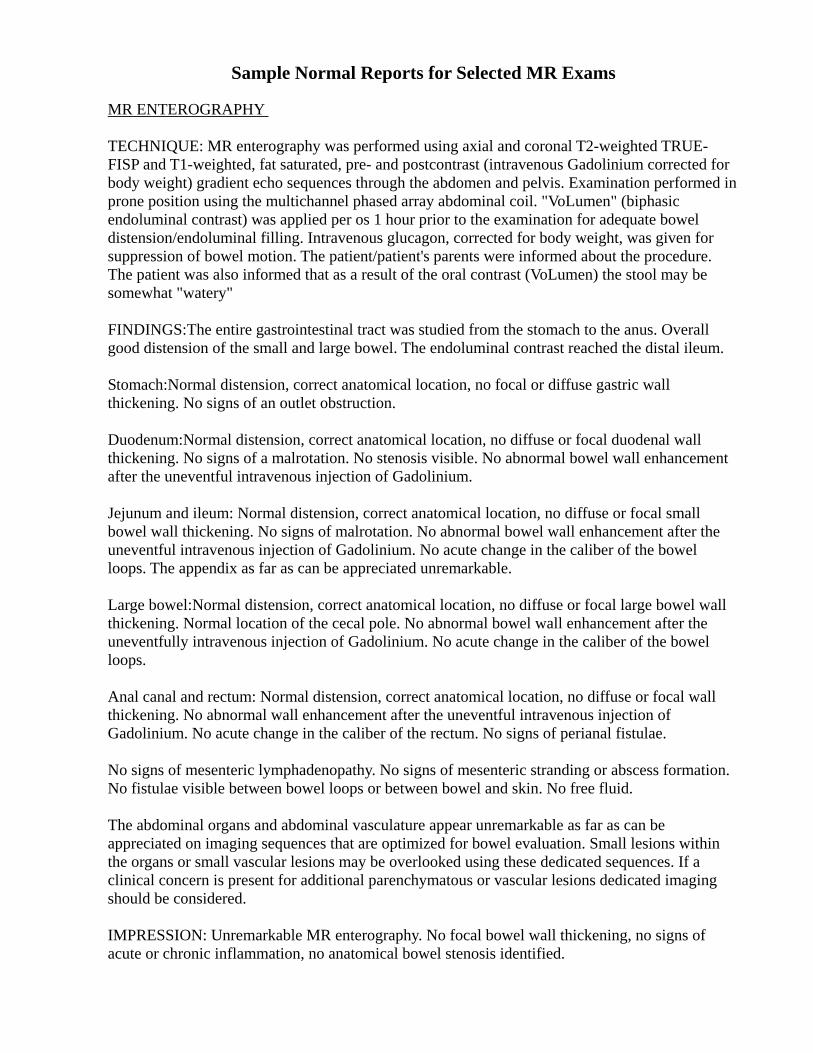
Sample Normal Reports for Selected MR Exams MR ENTEROGRAPHY TECHNIQUE: MR enterography was performed using axial and coronal T2-weighted TRUE-FISP and T1-weighted, fat saturated, pre- and postcontrast (intravenous Gadolinium corrected for body weight) gradient echo sequences through the abdomen and pelvis. Examination performed in prone position using the multichannel phased array abdominal coil. "VoLumen" (biphasic endoluminal contrast) was applied per os 1 hour prior to the examination for adequate bowel distension/endoluminal filling. Intravenous glucagon, corrected for body weight, was given for suppression of bowel motion. The patient/patient's parents were informed about the procedure. The patient was also informed that as a result of the oral contrast (VoLumen) the stool may be somewhat "watery" FINDINGS:The entire gastrointestinal tract was studied from the stomach to the anus. Overall good distension of the small and large bowel. The endoluminal contrast reached the distal ileum. Stomach:Normal distension, correct anatomical location, no focal or diffuse gastric wall thickening. No signs of an outlet obstruction. Duodenum:Normal distension, correct anatomical location, no diffuse or focal duodenal wall thickening. No signs of a malrotation. No stenosis visible. No abnormal bowel wall enhancement after the uneventful intravenous injection of Gadolinium. Jejunum and ileum: Normal distension, correct anatomical location, no diffuse or focal small bowel wall thickening. No signs of malrotation. No abnormal bowel wall enhancement after the uneventful intravenous injection of Gadolinium. No acute change in the caliber of the bowel loops. The appendix as far as can be appreciated unremarkable. Large bowel:Normal distension, correct anatomical location, no diffuse or focal large bowel wall thickening. Normal location of the cecal pole. No abnormal bowel wall enhancement after the uneventfully intravenous injection of Gadolinium. No acute change in the caliber of the bowel loops. Anal canal and rectum: Normal distension, correct anatomical location, no diffuse or focal wall thickening. No abnormal wall enhancement after the uneventful intravenous injection of Gadolinium. No acute change in the caliber of the rectum. No signs of perianal fistulae. No signs of mesenteric lymphadenopathy. No signs of mesenteric stranding or abscess formation. No fistulae visible between bowel loops or between bowel and skin. No free fluid. The abdominal organs and abdominal vasculature appear unremarkable as far as can be appreciated on imaging sequences that are optimized for bowel evaluation. Small lesions within the organs or small vascular lesions may be overlooked using these dedicated sequences. If a clinical concern is present for additional parenchymatous or vascular lesions dedicated imaging should be considered. IMPRESSION: Unremarkable MR enterography. No focal bowel wall thickening, no signs of acute or chronic inflammation, no anatomical bowel stenosis identified.
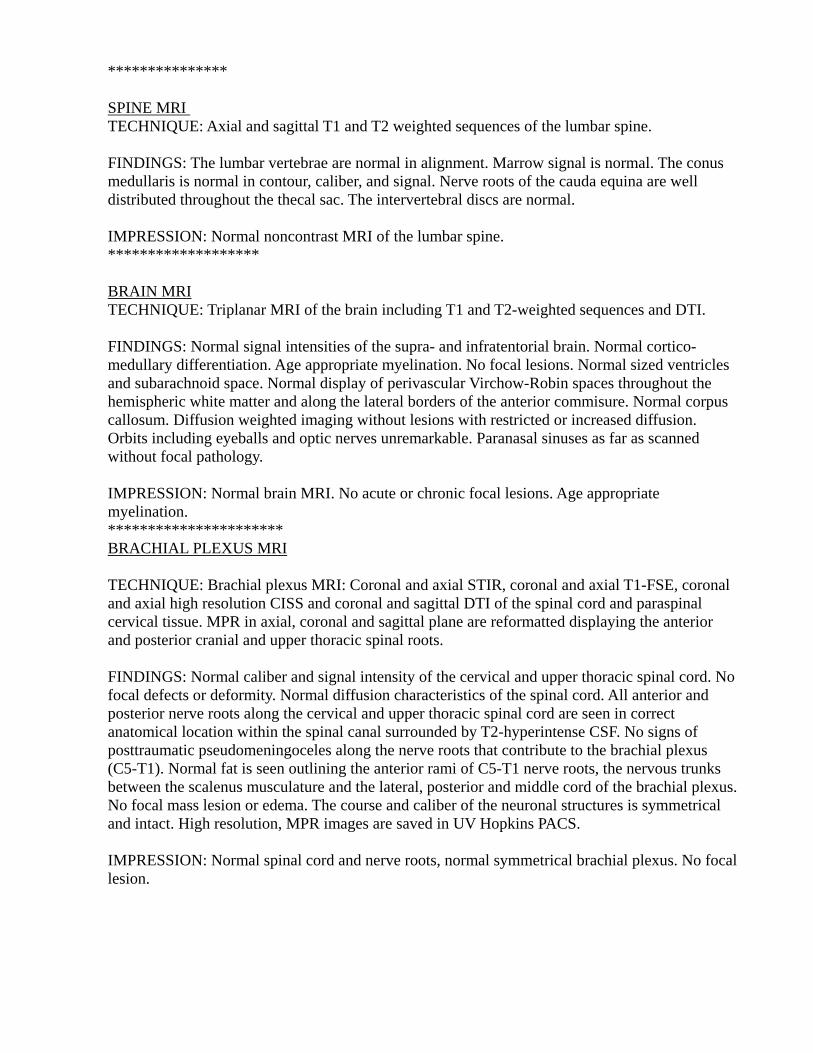
*************** SPINE MRI TECHNIQUE: Axial and sagittal T1 and T2 weighted sequences of the lumbar spine. FINDINGS: The lumbar vertebrae are normal in alignment. Marrow signal is normal. The conus medullaris is normal in contour, caliber, and signal. Nerve roots of the cauda equina are well distributed throughout the thecal sac. The intervertebral discs are normal. IMPRESSION: Normal noncontrast MRI of the lumbar spine. ******************* BRAIN MRI TECHNIQUE: Triplanar MRI of the brain including T1 and T2-weighted sequences and DTI. FINDINGS: Normal signal intensities of the supra- and infratentorial brain. Normal cortico-medullary differentiation. Age appropriate myelination. No focal lesions. Normal sized ventricles and subarachnoid space. Normal display of perivascular Virchow-Robin spaces throughout the hemispheric white matter and along the lateral borders of the anterior commisure. Normal corpus callosum. Diffusion weighted imaging without lesions with restricted or increased diffusion. Orbits including eyeballs and optic nerves unremarkable. Paranasal sinuses as far as scanned without focal pathology. IMPRESSION: Normal brain MRI. No acute or chronic focal lesions. Age appropriate myelination. ********************** BRACHIAL PLEXUS MRI TECHNIQUE: Brachial plexus MRI: Coronal and axial STIR, coronal and axial T1-FSE, coronal and axial high resolution CISS and coronal and sagittal DTI of the spinal cord and paraspinal cervical tissue. MPR in axial, coronal and sagittal plane are reformatted displaying the anterior and posterior cranial and upper thoracic spinal roots. FINDINGS: Normal caliber and signal intensity of the cervical and upper thoracic spinal cord. No focal defects or deformity. Normal diffusion characteristics of the spinal cord. All anterior and posterior nerve roots along the cervical and upper thoracic spinal cord are seen in correct anatomical location within the spinal canal surrounded by T2-hyperintense CSF. No signs of posttraumatic pseudomeningoceles along the nerve roots that contribute to the brachial plexus (C5-T1). Normal fat is seen outlining the anterior rami of C5-T1 nerve roots, the nervous trunks between the scalenus musculature and the lateral, posterior and middle cord of the brachial plexus. No focal mass lesion or edema. The course and caliber of the neuronal structures is symmetrical and intact. High resolution, MPR images are saved in UV Hopkins PACS. IMPRESSION: Normal spinal cord and nerve roots, normal symmetrical brachial plexus. No focal lesion.
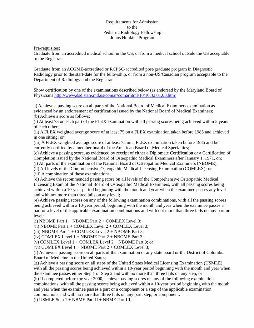
Requirements for Admission to the
Pediatric Radiology Fellowship Johns Hopkins Program
Pre-requisites: Graduate from an accredited medical school in the US, or from a medical school outside the US acceptable to the Registrar. Graduate from an ACGME-accredited or RCPSC-accredited post-graduate program in Diagnostic Radiology prior to the start-date for the fellowship, or from a non-US/Canadian program acceptable to the Department of Radiology and the Registrar. Show certification by one of the examinations described below (as endorsed by the Maryland Board of Physicians http://www.dsd.state.md.us/comar/comarhtml/10/10.32.01.03.htm) a) Achieve a passing score on all parts of the National Board of Medical Examiners examination as evidenced by an endorsement of certification issued by the National Board of Medical Examiners; (b) Achieve a score as follows: (i) At least 75 on each part of the FLEX examination with all passing scores being achieved within 5 years of each other; (ii) A FLEX weighted average score of at least 75 on a FLEX examination taken before 1985 and achieved in one sitting; or (iii) A FLEX weighted average score of at least 75 on a FLEX examination taken before 1985 and be currently certified by a member board of the American Board of Medical Specialties; (c) Achieve a passing score, as evidenced by receipt of either a Diplomate Certification or a Certification of Completion issued by the National Board of Osteopathic Medical Examiners after January 1, 1971, on: (i) All parts of the examination of the National Board of Osteopathic Medical Examiners (NBOME); (ii) All levels of the Comprehensive Osteopathic Medical Licensing Examination (COMLEX); or (iii) A combination of these examinations; (d) Achieve the recommended passing score on all levels of the Comprehensive Osteopathic Medical Licensing Exam of the National Board of Osteopathic Medical Examiners, with all passing scores being achieved within a 10-year period beginning with the month and year when the examinee passes any level and with not more than three fails on any level; (e) Achieve passing scores on any of the following examination combinations, with all the passing scores being achieved within a 10-year period, beginning with the month and year when the examinee passes a part or a level of the applicable examination combinations and with not more than three fails on any part or level: (i) NBOME Part 1 + NBOME Part 2 + COMLEX Level 3; (ii) NBOME Part 1 + COMLEX Level 2 + COMLEX Level 3; (iii) NBOME Part 1 + COMLEX Level 2 + NBOME Part 3; (iv) COMLEX Level 1 + NBOME Part 2 + NBOME Part 3; (v) COMLEX Level 1 + COMLEX Level 2 + NBOME Part 3; or (vi) COMLEX Level 1 + NBOME Part 2 + COMLEX Level 3; (f) Achieve a passing score on all parts of the examination of any state board or the District of Columbia Board of Medicine in the United States; (g) Achieve a passing score on all steps of the United States Medical Licensing Examination (USMLE) with all the passing scores being achieved within a 10-year period beginning with the month and year when the examinee passes either Step 1 or Step 2 and with no more than three fails on any step; or (h) If completed before the year 2000, achieve passing scores on any of the following examination combinations, with all the passing scores being achieved within a 10-year period beginning with the month and year when the examinee passes a part or a component or a step of the applicable examination combinations and with no more than three fails on any part, step, or component: (i) USMLE Step 1 + NBME Part II + NBME Part III;
(ii) USMLE Step 1 + USMLE Step 2 + NBME Part III; (iii) USMLE Step 1 + NBME Part II + USMLE Step 3; (iv) NBME Part I + USMLE Step 2 + USMLE Step 3; (v) NBME Part I + USMLE Step 2 + NBME Part III; (vi) NBME Part I + NBME Part II + USMLE Step 3; (vii) FLEX Component 1 + USMLE 3; (viii) FLEX Component 2 + USMLE Step 1 + NBME Part II; (ix) FLEX Component 2 + USMLE Step 1 + USMLE Step 2; (x) FLEX Component 2 + NBME Part I + USMLE Step 2; or (xi) FLEX Component 2 + NBME Part I + NBME Part II. Where applicable, show ECFMG certification. Non-US citizens: show eligibility for J-1 visa status. Please Note: the Department of Radiology no longer sponsors H1B visas for clinical fellows. Application: Complete the Johns Hopkins Clinical Fellowship Application Form and return it with CV and personal statement to the program director. Arrange for 3 letters of recommendation to be sent to the program director. If invited, come for a personal interview with division faculty. The application is also reviewed by the Office of the Registrar, to determine if the academic credentials are acceptable. Acceptance: If the application is successful, the applicant will be notified by the program director by email; a letter of intent will be sent that the acceptee should return to accept the position. The acceptee will then be sent an offer letter from the office of the Chairman of the Department of Radiology a few months before the appointment begins. The acceptee must sign and return the letter to complete the acceptance of the position. The position may be taken up only after successfully passing the University-sponsored criminal background check and completing the orientation curriculum designated by the Office of Graduate Medical Education for the year the fellowship begins.
Pediatric Radiology On Call September, 2010 -- JBensonMD
Getting in touch: Peds ER docs’ area 5-7330 ; Peds Radiology Reading Room 5-6454 PICU 5-5260 NICU 5-5255 Pediatric medical inpatients are covered by two teams at night and on weekends– Senior intern services covered Neill 3-9543 3-5431 General, GI, Cardiology Zinkham 3-9733 3-5733 General, Pulmonary, Renal, Heme GPS intern 3-6660 Check on-line schedule for the pediatric radiologist or fellow on call. However, late schedule changes may be known only to the hospital operator, so if the listed beeper doesn’t answer, call 5-5020 and ask who is on call. PLEASE CALL THE ATTENDING IF YOU HAVE ANY QUESTIONS! There is attending coverage 24/7, in house usually until 5 including weekends, by phone otherwise. Preliminary reports: It is EXTREMELY helpful if you dictate a preliminary report whenever you have a consultation with a pediatric clinician about a study, or if you do one of the studies outlined below. Put your dictation under “No Attending”, unless you have talked about it with one of the attendings already. Contrast: Oral contrast for Fluoro (UGI, G-tube checks): When contrast for an emergent GI study is needed for a sick patient or where perforation is suspected, you can use diluted water-soluble contrast. Most contrasts are hyperosmolar (greater than the normal serum osmolality [NSO]of 285mOsm/l) and the trick is to make them just dense enough to see but not enough to cause much damage to the GI mucosa or to lungs if aspirated. Do not use Gastrografin without diluting it! Also, keeping the volume small cuts down on reflux. Make the concentration and the amount fit the patient. Suggestions: Patient weight: <3kg 3-10kg 10-30kg >30kg Conc (x NSO) 1 1.5 2 2 Approx amount (cc) 5 20 40 60 Non-ionics give you more density per osm, but are more expensive, especially since you use only a few cc’s from a bottle. Be sure to dilute with WATER, not saline! See chart in the peds fluoro suite for contrast/water proportions to reach ideal osmolality levels. Barium is still a good choice for less debilitated, non-perforated patients, since it’s inert, tastes better, and is dense enough that you can dilute the standard preps for smaller patients where constipation could be an issue.
Oral contrast for CT: Use the standard mix for peds (4cc Omnipaque 350/100 cc water or non-carbonated drink), and encourage the consumption of a total of 50cc + 10cc/kg prior to the scan (range: 30-45 min for babies and toddlers, 45-75 minutes for small kids, 60-90 minutes for bigger kids and adolescents).
IV contrast for CT: consent is needed, except in dire emergency. Use 1cc/pound or 2cc/kg. Large central lines, mediports, or peripheral angiocaths can be used, but not PICC lines (at this point). There is almost NO reason to get a non-contrast CT of the abdomen; US will tell you as much without the radiation.
Typical situations where radiology input is requested: Call the on-call Peds Radiology attending before doing any of these procedures! 1.Bilious vomiting in newborn:
A true emergency. UGI for malrotation is needed, because of the high association with midgut volvulus. The attending needs to at least see your images, since decision to go to OR rests on it. Fluoro is best, to monitor where the contrast is and get the best picture of the ligament of Treitz, but they may ask for a portable if the baby is too fragile. (argument against: may miss the ligament, then the contrast complicates f/u exam). If they insist, and the peds rad attending agrees, 1) get plain film (PF) confirmation of OG tube in stomach 2) Make non-ionic contrast as above and send it with tech. 3)Have tech set up to take KUB then inject through OG tube with patient in right lateral decub position 4) put the patient supine again for KUB after 30 second-1minute, **making sure the patient is absolutely straight and flat**.
2.Abdominal distension, question of colon obstruction from meconium plugs or stool: NOT usually an emergency. Can be done with the attending next day. Sometimes associated in newborn with bilious emesis; may be the next step if UGI is neg for malro. Do not do unsupervised enemas! Unlike other exams, if complications occur they are usually dire.
3.Feeding intolerance/vomiting in older infant: Usually less emergent, might wait til next day, especially if plain film (PF) is non-obstructive Ddx: malrotation, pyloric stenosis, obstruction . Barium is OK for UGI. But SBFT with PF only may be all they need if pt previously studied. Suggest US for HPS (upper limit nl: muscle thickness 3mm, channel length 13mm)
4.Gtube replaced; check position: Find out if the tube went easily into an established tract (probably PF study OK), or if there was ANY difficulty (probably needs fluoro). First-time changes usually should get fluoro. If there is a post-placement PF, check for free air. If it is there, discuss with surgeons if study is warranted. PF study: Have tech set up for KUB. Inject Gtube (see contrast discussion, above) with patient in right lateral decub position. Place supine again for film. Then get XTL abdomen. Look for contrast only in stomach and bowel (not in peritoneum), make sure there is emptying of stomach into duodenum, check for free air. Fluoro: Start with patient turned so that the abdominal wall is in profile and tube is seen throughout its length (usually RAO or left lateral decub). Inject under fluoro, looking for any extravasation from the tract. If all goes into the stomach, turn to right lateral decub to insure that the balloon does not occlude the pylorus and that the stomach empties. Remove all the contrast you can at the end.
5.Newborn candidate for ECMO, need HUS: HUS ideally should be nl, though Gr I’s won’t hold them back, since baby will otherwise die.
6.HUS f/u, baby on ECMO: Not looking for subtleties: usually takes an intracranial explosion to remove pt from machine.
7.Irritability, lethargy, abdominal pain in toddler, r/o/intussusception: Get KUB with left lateral decubitus – see if cecum fills with air, look for evidence of SBO US is very good at looking for the mass: not subtle; clinicians will usually accept a negative exam. Attending needed if air enema is to be done for reduction.
8.Limp, fever, r/o septic hip: Get US to look for effusion (linear transducer, longitudinally over hip joints anteriorly; compare two sides). Call attending if fluid is present on affected side.
9.Foreign body: Aspirated or swallowed: AP CXR look for FB itself or asymmetric inflation; lat CXR should
include neck and nasopharynx to look for unsuspected impaction (esp in infants and mentally-challenged pts) Aspirated: Decub CXR’s can work (dependent side should deflate) but can be non-helpful if pt takes a big breath. Fluoro of the chest is quick and decisive (less radiation than repeated decubs). Swallowed: If it is in the cervical or thoracic esophagus, look at AP and lat CXR for signs of pneumomediastinum. If below the diaphragm on KUB, position less important unless object is sharp. XTL can help define position (eg in gastric fundus) and look for free air.
Radiology Resident Rotations in Pediatric Radiology: Curriculum Guide and Rotation Requirements
(revised 2/21/11)
Pediatric Radiology is one of the few places left in academic practice where you can still be a general radiologist, one who can perform and interpret a variety of examinations and correlate them to help the clinicians come to a diagnosis for the patient. This may be daunting at first, but later becomes exciting and enjoyable. During your rotations in our division in particular we will do our best to familiarize you with the spectrum of clinical pathology in pediatrics and its radiologic appearance through the cases you look at and report, guided reading, didactic lectures, and case conferences. Scope of services The center of the division is the Dorst Reading Room (Nelson B-172) named for John Phillips Dorst, the first director of Pediatric Radiology at Johns Hopkins. The division of Pediatric Radiology is in charge of plain films, fluoroscopy (e.g. GI and GU studies), body CT and Ultrasound for any patient with a pediatric attending physician. Generally, the patients are under the age of 19, but can be older. Three weeks out of each month we cover neuro MR and CT M-F during the day. We are currently in charge of certain body MRI exams in children (whole body and fetal MRI; MRI for inflammatory bowel disease, vascular malformations and DDH repair assessment; MR urography); all others are in the purview of the adult MR or MSK sections. Pelvis US on pregnant patients and all nuclear medicine/PET are the responsibility of the adult Radiology services. Also, skeletal images performed in the JHOC are usually read by the MSK service. The division is open M-F 8:30am -5:00pm and an attending is on call every night (7 days a week) after 5pm, providing back-up for the evening and night censors. The pediatric radiologist on call comes in on Saturdays, Sundays and holidays 8:30am -5pm to read exams and perform emergency studies. Educational Goals and Objectives The ACGME mandates that all training programs provide professional education leading to competency in 6 areas. To that end, this program has formulated the following educational goals, with training objectives designed to help attain these goals:
GOALS By the end of the rotations the resident should have competency in the following areas, as indicated by the ability to: Medical Knowledge (MK): Use a knowledge base of pediatric medical/surgical problems and related anatomy to recognize abnormal radiologic findings and compose an age-appropriate differential diagnosis. Patient Care (PC): Plan and perform radiologic procedures skillfully, with attention to patient well-being while minimizing radiation dose Professionalism(PRO): Project a competent, concerned, compassionate professional image Interpersonal and Communication Skills (ICS): Communicate effectively with mentors, colleagues, co-workers, patients and families to insure understanding of all aspects of the resident’s radiology practice Practice-based Learning and Improvement (PBLI): Critically evaluate current methods and published research to improve radiology practice Systems-based Practice (SBP): Utilize other resources and ancillary professionals (such as nurse, radiographer, sonographer, information technologist and radiation physicist) to enhance patient care and safety
Our TRAINING OBJECTIVES for you in these rotations will aid you in achieving these 6 goals. These are listed below, with the goals to which each pertains:
1. Learn to recognize common clinical presentations and typical radiologic pathologies in Pediatric Radiology and distinguish them from the wide range of normal appearances. (MK, PC) 2. Learn to perform radiologic procedures in children and appropriate interaction with patients and their families through observation of and instruction by attending pediatric radiologists and the gradual assumption of graded responsibility. (MK, PC, PRO) 3. Learn the importance of clinical data, radiographic technique and positioning, and comparison studies in coming to an accurate and helpful interpretation of the exam. (PC, PRO)
a. Attend clinical conferences where images are discussed in the context of clinical care. The pediatric radiologists go to the Pediatric ED, NICU and PICU on weekday mornings to do xray rounds with the residents. Also, we are often asked to present patients at Pediatric M&M and clinical conferences with the GI, Renal, Pediatric Surgery, Oncology and Pulmonary services. b. Use RadAssistant to find out the clinical and imaging histories of your patients and integrate this data in your interpretations.
4. Use online and print-based resources to expand your knowledge base for pediatric radiology (PubMed, textbooks, ACR Appropriateness Criteria) and progressively take on the role of a radiologic consultant. (PRO, MK, SBP) 5. Gain an appreciation for balancing the risks and benefits of radiation. Demonstrate decreasing fluoro times. (PC, ICS, PBLI)
a. Coning and shielding in radiography, the use of pulsed fluoroscopy and last-image “grabs” instead of exposures, judicious patient restraint, decreasing mAs in CT, consulting with clinicians to convert CT requests to US and MRI where possible are all ways in which we try to reduce radiation burden on our patients.
6. Develop a reporting style that summarizes the clinical problem for which the study is done, enumerates image findings, and offers an interpretation of those findings with recommendations for further imaging as needed. (PRO, ICS) 7. Participate in M-W-F divisional noon film conferences that review current cases. (MK, PBLI) 8. Attend Thursday didactic conference (variable start time, noon or later; lasts 1 hour). This may be an invited speaker or faculty speaker lecturing on a topic, journal club with a faculty or fellow presenter, or case follow-up conference where we discuss the outcome of an imaging unknown: what we did right and how we might improve. (MK, PBLI) 9. Utilize learning resources in the division for self-study during “down-time” and to complete required assessments (SBP, MK):
a. Required modules in the on-line curriculum: Under the aegis of the Society for Pediatric Radiology, a modular curriculum covering a wide range of topics in pediatric radiology has been placed on the web (https://www.cchs.net/pediatricradiology/ ). Each resident should complete the modules listed below for their rotation, print out the certificates and give them to Iris Bellamy or Dr. Benson. Residents are encouraged to complete assigned modules from other rotations as well as other modules as their interest leads them. A total of 26 modules is required over the four years. Print the certificates and hand them in. These will form a part of your evaluation for each rotation. b. Film teaching modules: a collection of green Teaching File jackets containing manila envelopes of films are found in a caddy under the light boxes in the reading room.
These are good examples of common things. An explanation of the case is found on its manila envelope. Please check the medical record number on the films against the number on the envelope to be sure you have a match before putting them away! c. On-line Teaching files: the website Pediatric Radiology.Com is a compendium of links to on-line texts and files. Go to www.pediatricradiology.com and scroll down the page to find links to teaching file websites at other institutions or to meta-textbooks. Several residents recommend the Cincinnati Children’s website that has a very good module for subtle fractures: http://www.tellviewnow.com/teachmod_launch_public/index.htm d. Hardcover textbooks and journals in the bookshelves in the reading room and office can be signed out or read here.
First rotation
Fluoroscopy: Observe all fluoroscopy (PC, PRO) Perform UGI and VCUG exams on children older than 1 year with the
pediatric imaging attending in the room (PC, PRO) Perform fluoro on teenagers without the attending if the resident has done an adult fluoro rotation (PC, PRO) Learn to recognize malrotation, GE reflux, vescicoureteral reflux, pyloric stenosis (MK, PC)
Plain Film: Learn to recognize normal bone, chest and abdomen films at various ages, Salter-type fractures, non-accidental trauma, pneumonia, reactive airways disease, bronchiolitis,sickle cell changes, croup, pneumothorax, free air in the abdomen, bone age using Greulich and Pyle atlas (MK, PC)
CT: Review cases with the pediatric imaging CT attending of the day (MK,PC) All sections: Preliminarily report all studies, then review reports with assigned attending,
making the directed changes. (MK, ICS, PRO) Contact clinicians concerning critical findings, demonstrating knowledge of
institutional systems, and document contacts (ICS, PRO, SBP) Self-study: Complete these modules from the SPR on-line curriculum, print out the certificates
and give them to Dr. Benson or Iris Bellamy: Fluoroscopy, Lines and Catheters, Radiation Safety, Pneumonia, Malrotation, GU Reflux, Childhood Fracture, Posterior Urethral Valves (MK, PC)
Second rotation
Fluoroscopy: Perform familiar fluoro exams and review films and technique with the Attending (PC, PRO) Watch, then perform enema exams and other exams on patients less than one year with the attending in the room (MK, PC, PRO) Learn to recognize colonic aganglionosis, inflammatory bowel disease, meconium ileus, intussusception, malrotation, swallowing dysfunction (MK, PC) Plain film: Learn to recognize ICU pathology: RDS, chronic lung disease, edema, meconium aspiration, tubes and catheters and their proper positions (MK, PC) CT: Obtain consent for and then cover injections and sedations in CT area (PC, ICS) Learn to recognize and characterize abdominal and mediastinal masses, diagnose trauma pathology (MK, PC) Ultrasound: Perform selected sonograms with the sonographer. (SBP, PC, PRO)
Review cases with the pediatric ultrasound attending of the day (MK, PC) All sections: Preliminarily report all studies, then review reports with assigned attending,
making the directed changes. (MK, ICS, PRO) Contact clinicians concerning critical findings, demonstrating knowledge of
institutional systems, and document contacts (ICS, PRO, SBP) Self-study: Complete these modules from the on-line curriculum, print out the certificates and
give them to Dr. Benson or Iris Bellamy: Patient Care, Neonatal Chest, Intussusception, Ureteropelvic Junction Obstruction, Neonatal Low Intestinal Obstruction, Child Abuse, Cranial Ultrasound, Spine Dimple (MK, PC)
Third and subsequent rotations
Fluoroscopy: Perform all exams with or without attending in the room, depending on degree of familiarity (PC, PRO) Learn to recognize microcolon, tracheobrochomalacia, diaphragm
Paralysis (MK, PC) Plain Film: Learn to recognize congenital cardiac disease, dwarfing syndromes, post- operative complications (MK, PC) CT: Learn to recognize inflammatory and obstructive bowel pathology, lung infiltration,
abnormal fluid collections, bronchiectasis (MK, PC) Ultrasound: Perform selected sonograms with the sonographer (SBP, PC, PRO)
Review images with the attending. (MK, PC) Learn to recognize intracranial pathology, abnormal bowel, congenital
renal abnormalities, normal and abnormal appearance of the maturing pelvis, testicular torsion, hypertrophic pyloric stenosis (MK, PC) All sections: Preliminarily report all studies, then review reports with assigned attending,
making the directed changes. (MK, ICS, PRO) Contact clinicians concerning critical findings, demonstrating knowledge of
institutional systems, and document contacts (ICS, PRO, SBP) Self-study: Complete these modules from the on-line curriculum, print out the certificates and
give them to Dr. Benson or Iris Bellamy: Professionalism, Hip Dysplasia, Hypertrophic Pyloric Stenosis, Childhood Stroke, and six other modules of your choice. (MK, PRO, PC)
All Rotations
1. Residents are expected to come promptly to the Reading Room immediately after morning resident conference. The weekly schedule is posted on the bulletin board; please consult it to find where you have been assigned for the day and who your attending is. Report to that person to get a patient schedule and workflow updates. If there is no morning conference, or you do not choose to attend it, then check the schedule and join your attending in the appropriate clinical area: PICU 8:00 am CMSC 7; ED 8:15 am Park 1; reading room 8:30am Dorst Reading Room. If you have to be late for any reason, contact Ms. Bellamy in the office or your attending of the morning. 2. Portal accepts entries by age, so all patients 18yo and younger come to PEDS lists, regardless of where they were done. The workstations in the Reading Room automatically open PEDS lists only. The studies appear in multiple lists simultaneously (e.g. ED patients are in the Emergency room list, the Morning or Afternoon Plain Film list, the All Plain Films list, and the All Pediatric Exams list). This allows the workflow to proceed in different ways, depending on the day. It also insures a tight net to catch all the exams. So, after reading everything in the assigned list, you
should review one of the more general lists to make sure nothing gets missed. Duty station Portal Worklist to read from Fluoro/IP/ED Emergency Room, Morning Plain Films; Fluoroscopy 8:30-12:30 Fluoro/PF Afternoon Plain Films; Fluoroscopy 12:30-5:00 NICU/PICU NICU/PICU CT/US/MR CT, Ultrasound, MRI (see under “Scope of Services” to see which exams we read);
in weeks when Adult Neuro covers Peds Neuro, we read the TCD’s and cranial US’s which will be found in the Neuro Ultrasound list
3. Informal Case Conference is held in front of the big screen in the Reading Room at noon on Mondays, Wednesdays and Fridays. Residents are expected to attend and take cases. Residents are also encouraged to present interesting recent cases. 4. Thursdays at 11:15am the attending staff has a meeting and residents are asked to be in the Reading Room during that time to help clinicians, answer technologists’ questions, etc. 5. Didactic Conference is held each Thursday from approximately 12:00 to 1:00 pm. This alternates between lecture, journal club, and follow-up case conference. The residents are expected to attend. 6. Other clinical conference are held in the Reading Room at various times during the month—check the weekly schedule for day and time – and the residents are encouraged to sit in as the work schedule permits. 7. Any studies done in the division before 5pm are read out the same day; the work-day in the division ends when the final pre-5pm study is reported. 8. At the end of each day, HIPAA and good citizenship practices require everyone to: Log out of Portal, Outlook, EPR, RadAssistant and UV
Put all paper requisitions and schedules in the shredder box, along with any scraps of paper that have patient information on them Turn off reading lamps Throw out trash Empty drinks in the sink in the tech lounge and dispose of cups and bottles (Please do not put cups of liquid—even covered ones—in the trash. They spill as the trash cans are emptied.) Please recycle! Empty cans and plastic bottles can be put in the blue lined bins in the tech lounge.
Evaluations A single E-valu* form is submitted for each resident for each rotation. It reflects collated input from ALL of the pediatric radiology faculty. Progress in reporting style, development of knowledge base, performance in fluoroscopy, completion of on-line modules, participation in noon conference, and level of interest and engagement all provide basis for evaluation and comment.
Staff Radiologists: Thierry Huisman MD, division chair , Jane Benson MD, fellow and resident education director, Melissa Spevak MD, Renee Flax-Goldenberg MD, Aylin Tekes MD, Ulrich Willi MD Fellow: Madhan Bosemani MD(7/1/10-6/30/11) Iris Bellamy, Administrative Assistant and Fellowship Coordinator; Chanel Britt, Administrative Aide Nurses: Ron Wardrope RN, Kristina Hoerl RN
Technologists: Elyce Wolfgang RT, Chief Technologist; Debra Wolinski RT, Senior Technologist; Mindy Arias RT, Laurie Jones RT, Alison Brooks RT, Danielle Davis RT, Kim Francis RT, Elizabeth Kase RT, Reggie Poon RT, Monica Stevens RT, Keisha Bethea RT ,Keisha Blake RT, Heather Hodges RT, Angie Meaney RT
Pediatric Radiology Elective for Medical Students and Pediatric Residents
(revised 11/30/10)
Scope of services The division of Pediatric Radiology at Johns Hopkins is in charge of plain films, fluoroscopy (e.g. GI and GU studies), body CT and Ultrasound for any patient with a pediatric attending physician. Generally, the patients are under the age of 19, but can be older. A pediatric radiologist reads head/spine MRI and head/spine CT 3 weeks out of each month; the other week is the purview of the adult Neuro service. We are also in charge of certain body MRI exams. We do not do pelvis US on pregnant patients, or any nuclear medicine: these are the responsibility of the adult Radiology services. Also, skeletal images performed in the JHOC are usually read by the Musculoskeletal service. The division is open M-F 8:30-5:00 and an attending is on call every night after 5pm, providing back-up for the evening and night censors. The pediatric radiologist on call comes in on Saturdays, Sundays and holidays to read films, perform emergency studies and do CT and US. After 5:00 pm every evening the radiology resident censor (3-RADS) is available to field pediatric radiology questions (5-2174), with the peds radiology attending as backup. Staff Radiologists: Thierry Huisman MD, division chair Jane Benson MD, Melissa Spevak MD, Renee Flax-Goldenberg MD, Aylin Tekes MD , Ulrich Willi MD Fellow: Thangamadhan Bosemani (7/1/10-6/30/11) Nurses: Ron Wardrope RN, Kristina Hoerl RN Technologists: Elyce Wolfgang RT, Chief Technologist Debra Wolinski RT, Laurie Jones RT, Mindy Arias RT, Alison Brooks RT, Danielle Davis-Sloan RT, Kim Francis RT, Elizabeth Kase RT, Reggie Poon RT, Laura Reynolds RT, Keisha Bethea RT, Heather Hodges RT, Keisha Blake RT Administrative Assistant and Fellowship Coordinator: Iris Bellamy Locations Reading Room: Nelson B172 – 5-6454 Administrative Office: Nelson B173 – 5-6141 Fluoroscopic and Radiographic Procedure Rooms 1&3: adjacent to the Reading Room CT scans are performed in Adult Body CT, Nelson Basement US exams are performed in Adult Ultrasound, Halsted Basement, and in Peds Room 2 Our goals for you in this rotation and the ways that you can accomplish them (correlated with the Six Competencies):
1. Observe how things are done in Pediatric Radiology so that you can better explain them to your patients (Interpersonal and Communication Skills, Patient Care).
a. See at least one of each type of fluoroscopy procedure that we regularly do (VCUG, UGI, Contrast enema, video swallow study, enteral tube placement)
b. Watch some plain films being done c. Shadow the Radiology Nurses as they explain procedures to parents and patients
and prep patients for fluoro and CT.
2. Learn the importance of clinical data, radiographic technique and positioning, and comparison studies in coming to an accurate and helpful interpretation of the exam (Medical Knowledge, Practice-based Learning and Improvement, Professionalism).
a. Sit with the radiologists, fellow, and radiology residents and look at images as they are read.
b. Attend M-W-F noon conference in the reading room when current studies are discussed by the radiology attendings with the radiology residents.
c. Attend clinical conferences where images are discussed in the context of clinical care. The pediatric radiologists go to the ED, PICU and NICU on weekday mornings to do xray rounds with the residents. Also, we are often asked to present patients at Pediatric M&M and clinical conferences with the GI, Renal, Pediatric Surgery, Oncology and Pulmonary services.
3. Deepen your knowledge of your clinical area of interest (such as future fellowship plans) (Medical knowledge, Practice-based Learning).
a. Let us know, so we can be sure that you see relevant cases. 4. Learn to recognize common pathologies in Pediatric Radiology by utilizing learning
resources in the division for self-study during “down-time” to complete required assessments (Systems-based Practice, Medical Knowledge):
a. Required modules in the on-line curriculum: Under the aegis of the Society for Pediatric Radiology, a modular curriculum covering a wide range of topics in pediatric radiology has been placed on the web (https://www.cchs.net/pediatricradiology/). Each student should complete these four modules, print out the certificates and give them to Iris Bellamy or Dr. Benson: Radiation Safety, Pneumonia, GU Reflux, and Childhood Fracture. Students are encouraged to complete other modules as their interest leads them. Print the certificates and hand them in.
b. Film teaching modules: a collection of green Teaching File jackets containing manila envelopes of films are found in a caddy against the back wall of the reading room. These are good examples of common things. An explanation of the case is found on its manila envelope. Please check the medical record number on the films against the number on the envelope to be sure you have a match!
c. On-line Teaching files: the website Pediatric Radiology.com is a compendium links to on-line texts and files. Go to www.pediatricradiology.com and scroll down the page to find links to teaching file websites at other institutions or to meta-textbooks. Several residents recommend the Cincinnati Children’s website that has a very good module for subtle fractures: http://www.tellviewnow.com/teachmod_launch_public/index.htm
d. Hardcover textbooks, and journals in the bookshelves in the reading room can be signed out or read here. “Practical Pediatric Radiology” by S. Hilton is a particularly useful text for you because it is organized by clinical problem, and lets you see the scope of imaging available to work-up a diagnostic algorithm.
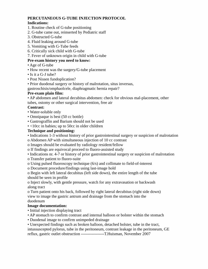
PERCUTANEOUS G-TUBE INJECTION PROTOCOL Indications: 1. Routine check of G-tube positioning 2. G-tube came out, reinserted by Pediatric staff 3. Obstructed G-tube 4. Fluid leaking around G-tube 5. Vomiting with G-Tube feeds 6. Critically sick child with G-tube 7. Fever of unknown origin in child with G-tube Pre-exam history you need to know: • Age of G-tube • How recent was the surgery/G-tube placement • Is it a G-J tube? • Post Nissen fundoplication? • Prior duodenal surgery or history of malrotation, situs inversus, gastroschisis/omphaolcele, diaphragmatic hernia repair? Pre-exam plain film: • AP abdomen and lateral decubitus abdomen: check for obvious mal-placement, other tubes, ostomy or other surgical intervention, free air Contrast: • Water-soluble only • Omnipaque is best (50 cc bottle) • Gastrograffin and Barium should not be used • <10cc in babies; up to 50cc in older children Technique and positioning: • Indications 1-3 without history of prior gastrointestinal surgery or suspicion of malrotation o Abdomen AP with simultaneous injection of 10 cc contrast o Images should be evaluated by radiology resident/fellow o If findings are equivocal proceed to fluoro-assisted study • Indications nr. 4-7 or history of prior gastrointestinal surgery or suspicion of malrotation o Transfer patient to fluoro-suite o Using pulsed fluoroscopy technique (6/s) and collimate to field-of-interest o Document procedure/findings using last-image hold o Begin with left lateral decubitus (left side down), the entire length of the tube should be seen in profile o Inject slowly, with gentle pressure, watch for any extravasation or backwash along tract o Turn patient onto his back, followed by right lateral decubitus (right side down) view to image the gastric antrum and drainage from the stomach into the duodenum Image documentation: • Initial injection displaying tract • AP stomach to confirm contrast and internal balloon or bolster within the stomach • Duodenal image to confirm unimpeded drainage • Unexpected findings such as broken balloon, detached bolster, tube in the tract, intussuscepted pylorus, tube in the peritoneum, contrast leakage in the peritoneum, GE reflux, gastric outlet obstruction -----------------T.Huisman, November 2007
Related Documents


![THE The JOHNS HOPKINS CLUB Events JOHNS HOPKINS … [4].pdf · Club Herald July / August 2015 Events THE The JOHNS HOPKINS CLUB JOHNS HOPKINS UNIVERSITY 3400 North Charles Street,](https://static.cupdf.com/doc/110x72/5fae1ad08ad8816d2e1aaabe/the-the-johns-hopkins-club-events-johns-hopkins-4pdf-club-herald-july-august.jpg)