DISEASES OF WILD AND CULTURED FISHES IN ALASKA Theodore Meyers, Tamara Burton, Collette Bentz, Jayde Ferguson, Davis Stewart, and Norman Starkey July 2019 Alaska Department of Fish and Game Fish Pathology Laboratories

Welcome message from author
This document is posted to help you gain knowledge. Please leave a comment to let me know what you think about it! Share it to your friends and learn new things together.
Transcript
DISEASES OF WILD AND CULTURED FISHES
IN ALASKA
Theodore Meyers, Tamara Burton, Collette Bentz, Jayde Ferguson, Davis Stewart, and Norman Starkey
July 2019
Alaska Department of Fish and Game Fish Pathology Laboratories

The Alaska Department of Fish and Game printed this publication at a cost of $9.83 in Anchorage, Alaska, USA.

3
About This Field Guide
This field guide is a product of the Ichthyophonus Diagnostics, Educational and Outreach Program which was initiated and funded by the Yukon River Panel’s Restoration and Enhancement fund and facilitated by the Yukon River Drainage Fisheries Association in conjunction with the Alaska Department of Fish and Game. The original impetus driving the production of this guide was from a concern that Yukon River fishers were discarding Canadian-origin Chinook
salmon believed to be infected by Ichthyophonus. It was decided to develop an educational program that included the creation of a field guide containing
photographs and descriptions of frequently encountered parasites within Yukon River fish.
This field guide is to serve as a brief illustrated reference that lists many of the
common (and not so common) parasitic, infectious, and noninfectious diseases of wild and cultured fishes encountered in Alaska. The content is directed
towards lay users, as well as fish culturists at aquaculture facilities and field
biologists and is not a comprehensive treatise nor should it be considered a scientific document. Interested users of this guide are directed to the listed fish
disease references for additional information.
Information contained within this field guide is published from the laboratory
records of the Alaska Department of Fish and Game, Fish Pathology Section that has regulatory oversight of finfish health in the State of Alaska. This third
printing includes several new entries, some new photographs and updated information on previous diseases and parasites. This version may be downloaded as a PDF from the ADF&G website at the following web address: http://www.adfg.alaska.gov/static/species/disease/pdfs/fish_disease_book.pdf
Text written and provided by: Theodore Meyers, Tamara Burton, Collette Bentz, and
Jayde Ferguson, Alaska Department of Fish and Game, Fish Pathology Laboratories, 333 Raspberry Road, Anchorage, Alaska 99518; P.O. Box 115526 (physical 3333 Glacier Highway), Juneau, Alaska 99811-5526.
Manuscript Photographs: Alaska Department of Fish and Game, Fish Pathology Laboratory photo archives except where indicated.
Publication design by Southfork Graphic Services.
Cover Photograph: Ichthyophonus growing in laboratory culture.
©2019 Alaska Department of Fish and Game, third printing
The original printing of this publication was produced with funding and support from the Yukon River Panel and its members and the Yukon River Drainage Fisheries Association. Current funding is provided by the ADF&G, Commercial Fisheries and
Sport Fish Divisions.
The Alaska Department of Fish and Game (ADF&G) administers all programs and activities free from discrimination based on race, color, national origin, age, sex, religion, marital status, pregnancy, parenthood, or disability. The department administers all programs and activities in compliance with Title VI of the Civil Rights
Act of 1964, Section 504 of the Rehabilitation Act of 1973, Title II of the Americans with Disabilities Act of 1990, the Age Discrimination Act of 1975, and Title IX of the Education Amendments of 1972. If you believe you have been discriminated against in any program, activity, or facility please write ADF&G ADA Coordinator, P.O. Box
115526, Juneau, AK 99811-5526; U.S. Fish and Wildlife Service, 4401 N. Fairfax
Drive, MS 2042, Arlington, VA 22203; or Office of Equal Opportunity, U.S. Department of the Interior, 1849 C Street NW MS 5230, Washington DC 20240.
For information on alternative formats and questions on this publication, please contact the department’s ADA Coordinator at (VOICE) 907-465-6077, (Statewide Telecommunication Device for the Deaf) 1-800-478-3648, (Juneau TDD) 907-465-3646, or (FAX) 907-465-6078.
10
20
30
40
50
60
70
80
90
100
110
120
Viruses Aquareovirus .........................................................................2
Erythrocytic Inclusion Body Syndrome (EIBS) ....................4
Erythrocytic Necrosis Virus (VENV) .....................................6
Infectious Hematopoietic Necrosis Virus (IHNV) ................8
North American Viral Hemorrhagic Septicemia Virus (NA-VHSV)..............................................
Pacifc Salmon Paramyxovirus ...........................................12
Piscine Orthoreovirus (PRV) ...............................................14
Bacteria Bacterial Coldwater Disease (BCWD).................................16
Bacterial Gill Disease ...........................................................18
Bacterial Kidney Disease (BKD) .........................................
Enteric Redmouth Disease (ERM) ..................................... 22
Furunculosis.........................................................................24
Fusobacteria-like Agent .................................................... 26
Marine Tenacibaculosis..................................................... 28
Motile Aeromonas and Pseudomonas Septicemia ...........
Mycobacteriosis of Fish.......................................................32
Vibriosis............................................................................... 34
Fungi Phaeohyphomycosis of Safron Cod ................................. 36
Phoma herbarum ............................................................... 38
Protozoa Epistylis (Heteropolaria) ....................................................
Hexamita..............................................................................42
Ichthyobodiasis (Costiasis) ................................................ 44
Ichthyophonus.................................................................... 46
Saprolegniasis – Cotton Wool Disease............................. 48
Trichodiniasis ......................................................................
Trichophrya (Capriniana) .................................................. 52
White Spot Disease ............................................................ 54
X-Cell Tumors ..................................................................... 56
Cnidaria Ceratonova (Ceratomyxa) shasta........................................ 58
Henneguya ..........................................................................
Table of Contents
Kudoa .................................................................................. 62
Myxobolus neurotropus ...................................................... 64
Myxobolus squamalis ......................................................... 66
Tetracapsuloides bryosalmonae (PKD) .............................. 68
Helminths (Worms) Acanthocephalans (spiny headed worms)........................
Anisakid Larvae (nematode) ............................................. 72
Philometra (nematode).......................................................74
Philonema (nematode) .......................................................76
Black Spot Disease (trematode) .........................................78
Encysted Digenean Metacercariae (trematode) ..............
Larval Diplostomulum of the Eye (trematode) ............... 82
Gyrodactylus and Dactylogyrus (monogene) ................. 84
Piscicola (annelid).............................................................. 86
Diphyllobothrium (cestode) ............................................... 88
Schistocephalus (cestode).................................................
Triaenophorus (cestode) ....................................................92
Arthropods
External Parasitic Copepods .............................................. 94
Salmincola (copepod) ....................................................... 96
Sarcotaces (copepod) ........................................................ 98
Non-infectious Diseases
Bloat (Water Belly)...........................................................
Blue Sac Disease of Fry..................................................... 102
Coagulated Yolk Disease (White Spot Disease).............. 104
Drop-out Disease ............................................................. 106
Gas Bubble Disease (GBD)................................................ 108
Mushy Halibut Syndrome .................................................
Neoplasia (Tumors)...........................................................112
Organ and Tissue Anomalies ............................................116
Pigment Aberrations in Fish .............................................118
Sunburn (Back-Peel)........................................................
Reference Glossary of Terms ............................................................. 122
Fish Disease References ....................................................127
2
VIRUSES
Aquareovirus
I. Causative Agent and Disease Aquareovirus is a genus in the virus
family Reoviridae. These icosahedral (60-80 nm) 11 segmented double- stranded RNA viruses (over 50) have been isolated from a variety of marine and freshwater aquatic animals world-wide including finfish, and bivalve mol-luscs. Genetic analyses have identified 7 different genotypes or species (A-G) of aquareoviruses. Most of these viruses produce self-limiting infections of low pathogenicity and are not associated with extensive disease or mortality. Exceptions include isolates from 7 fish species that have been associated with fish mortality, most notably the grass carp aquareovirus (G). The viral agents are most often isolated from asymp-tomatic adult carrier fish during routine screening examinations.
II. Host Species In the Pacific Northwest states of
Washington, Oregon and California, adult Chinook salmon appear to be the most frequent species infected with aquareovirus A or B. The virus has also been isolated from adult coho and chum salmon and steelhead. Rainbow trout have been experimentally infected with the virus resulting in mild hepatitis with no overt disease or mortality. In Alaska, aquareoviruses have been isolated from Chinook salmon (species B) and geoduck clams (species A).
III. Clinical Signs Fish naturally infected with aquareo-
viruses generally do not exhibit clinical signs of disease. Experimental infections can produce focal necrotic lesions in the livers of rainbow trout, chum salmon and bluegill fry. Other pathogenic exceptions include the grass carp species G that is
associated with epizootic fish mortal-ity producing severe hemorrhaging in fingerlings and yearlings resulting in up to 80% mortality.
IV. Transmission Transmission is horizontal via water
or from fish to fish. Isolates from bivalve mollusks likely represent virus that has been shed into the water column from a fish host and then bioaccumulated into shellfish tissues by filter feeding.
V. Diagnosis Detection of Aquareovirus is by
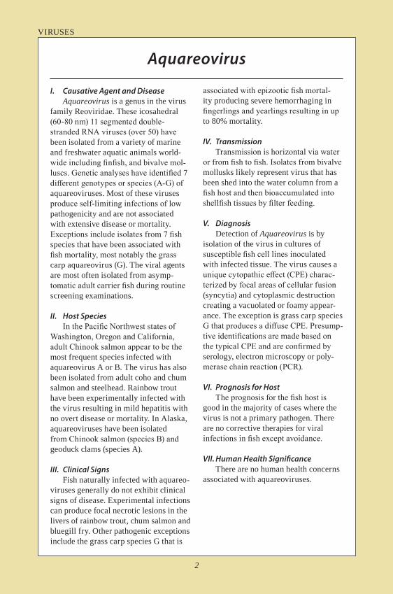
isolation of the virus in cultures of susceptible fish cell lines inoculated with infected tissue. The virus causes a unique cytopathic effect (CPE) charac-terized by focal areas of cellular fusion (syncytia) and cytoplasmic destruction creating a vacuolated or foamy appear-ance. The exception is grass carp species G that produces a diffuse CPE. Presump-tive identifications are made based on the typical CPE and are confirmed by serology, electron microscopy or poly-merase chain reaction (PCR).
VI. Prognosis for Host The prognosis for the fish host is
good in the majority of cases where the virus is not a primary pathogen. There are no corrective therapies for viral infections in fish except avoidance.
VII. Human Health Signifcance There are no human health concerns
associated with aquareoviruses.
3
VIRUSES
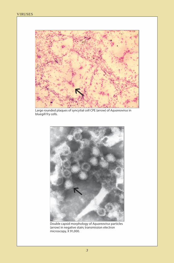
Large rounded plaques of syncytial cell CPE (arrow) of Aquareovirus in bluegill fry cells.
Double capsid morphology of Aquareovirus particles (arrow) in negative stain; transmission electron microscopy, X 91,000.

4
VIRUSES
Erythrocytic Inclusion Body Syndrome (EIBS)
I. Causative Agent and Disease Erythrocytic inclusion body syn-
drome (EIBS) is caused by an unclassi-fied icosahedral virus (70-80 nm) that infects erythrocytes of several salmonid fishes in fresh and seawater. Typically, EIBS presents with single or multiple pale, basophilic inclusions (0.4-1.6 um) in the cytoplasm of erythrocytes in stained peripheral blood smears. Affected fish may be asymptomatic, but more often have varying degrees of anemia and secondary bacterial and fungal infections. In severe cases of uncomplicated anemia, cumulative fish mortality over 20% has been reported with hematocrits less than 20%.
II. Host Species EIBS has been found in Chinook,
coho and Atlantic salmon in the Pa-cific Northwest, Japan, Norway and the British Isles. Other salmonid species showing variable susceptibilities by ex-perimental injection with infected blood homogenates include cutthroat trout, masou salmon and chum salmon.
III. Clinical Signs Fish are lethargic, anorexic and
anemic with chronic mortality often associated with secondary infections by other pathogens. Five stages of EIBS have been described: preinclusion, inclu-sion body formation, cell lysis with low hematocrits, recovery with increasing hematocrits and full recovery.
IV. Transmission The disease can be transmitted
horizontally while surviving fish gener-ally recover and develop an acquired immunity against reinfection that is transferable by passive immunization.
V. Diagnosis Isolation and replication of the
virus in available fish cell lines has been unsuccessful. Thus, diagnosis is by observation of the small pale blue inclusion bodies in the cytoplasm of infected erythrocytes with confirmation by transmission electron microscopy (TEM). The virus is found free in the cytoplasm or more commonly occurs in membrane bound cytoplasmic inclusion bodies within erythrocytes.
VI. Prognosis for Host Severe fish losses caused directly
by EIBS are rare. However, fish become weakened from the anemia and mortal-ity from other associated environmental stressors or secondary pathogens can be significant. The disease generally is self-limiting with recovery and immunity in survivors.
VII. Human Health Signifcance There are no human health concerns
with the EIBS virus.
NOTE: In Alaska, only one case of EIBS has been reported in 2004 affecting juvenile Chinook salmon in seawa-ter netpens. Subsequent studies have shown that EIBS in Japanese farmed coho salmon may be caused by a strain of Piscine Orthoreovirus (PRV-2). Molecular studies have determined that PRV is present in Alaskan coho and Chinook salmon. See PRV chapter for more detail.
5
VIRUSES
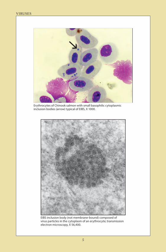
Erythrocytes of Chinook salmon with small basophilic cytoplasmic inclusion bodies (arrow) typical of EIBS, X 1000.
EIBS inclusion body (not membrane-bound) composed of virus particles in the cytoplasm of an erythrocyte; transmission electron microscopy, X 56,400.

6
VIRUSES
Erythrocytic Necrosis Virus (VENV)
I. Causative Agent and Disease Erythrocytic necrosis or viral
erythrocytic necrosis (VEN) is caused by several similar iridoviruses having double-stranded DNA and a hexagonal shape ranging in size from 130-330 nm. The viruses infect erythrocytes causing a hemolytic disease often resulting in anemia and secondary infections by other pathogens including VHSV Type IVa
II. Host Species There are likely several different
strains of the virus worldwide in the marine environment infecting a large variety of more than 20 anadromous and marine fish species. In Alaska, VENV has been detected in Pacific herring from several locations but has not yet been observed in salmonids. Results from experimental infections and occur-rence of epizootics in young-of-the-year Pacific herring indicate that juveniles are more susceptible than older fish.
III. Clinical Signs Adult herring generally show no
clinical signs of disease. In juvenile Pacific herring, fish are anemic exhibit-ing nearly white gills and pale visceral organs. Liver color may be green due to breakdown of blood hemoglobin releasing excess biliverdin. Hemotocrits may be as low as 2 to 10%, erythrocytes are fragile causing hemolysis of blood samples, and immature erythrocytes predominate in peripheral blood. High mortality with dead fish on the shoreline accompanied by extensive congregations of predator birds may occur in areas where juvenile herring are weakened by the disease.
IV. Transmission Transmission of this virus is likely
horizontal from fish to fish based on the few experimental studies using water-borne exposure. Adult carrier fish of susceptible species are likely reservoirs of the virus that is transmitted to juve-nile fish. Anadromous fish likely become infected during their marine phase of life. There is some suggestion that the virus is vector-borne and one instance of infection in juvenile salmonids in freshwater suggesting vertical transmis-sion from adult anadromous parent fish.
V. Diagnosis Diagnosis is made with blood smears
showing characteristic eosinophilic inclusion bodies (1-4 um) present in the cytoplasm of erythrocytes when stained with Giemsa or Wright stains. Impres-sion smears of hematopoietic head kidney can be substituted for blood. The virus is confirmed by the observation of iridovirus particles associated with inclusion bodies using electron micros-copy (TEM). VEN viruses have not been successfully cultured in available fish cell lines, however an unvalidated PCR is available for the virus in Pacific her-ring.
VI. Prognosis for Host The virus in Alaskan juvenile Pacific
herring caused one of the first natural epizootics reportedly associated with mass fish mortality. Chronic to subacute mortality in juvenile Pacific herring can also occur, especially when stressed.
VII. Human Health Signifcance No human health concerns are as-
sociated with VEN virus.
7
VIRUSES
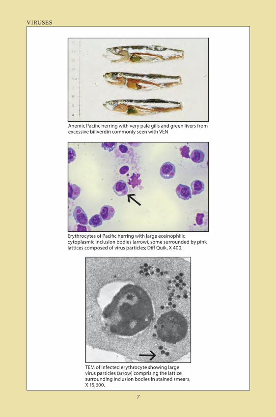
Anemic Pacifc herring with very pale gills and green livers from excessive biliverdin commonly seen with VEN
Erythrocytes of Pacifc herring with large eosinophilic cytoplasmic inclusion bodies (arrow), some surrounded by pink lattices composed of virus particles; Dif Quik, X 400.
TEM of infected erythrocyte showing large virus particles (arrow) comprising the lattice surrounding inclusion bodies in stained smears, X 15,600.
8
VIRUSES
Infectious Hematopoietic Necrosis Virus (IHNV)
I. Causative Agent and Disease III. Clinical Signs Infectious hematopoietic necro-
sis virus (IHNV) is a bullet-shaped novirhabdovirus that is enzootic to the North American Pacific Northwest but was inadvertently established in the US Snake River Valley in Idaho and in sev-eral countries of Asia and Europe. The three genetic clades of IHNV (U,M,L) can infect several salmonid species and have had severe economic impacts on intensively cultured salmon and trout. IHNV in Alaska (U clade) has been limited primarily to sockeye salmon and rarely Chinook and chum salmon when infected sockeye are present in their wa-ter supplies. Culture of sockeye salmon in Alaska by avoidance of IHNV has been successful through the rigorous use of the ADF&G sockeye salmon culture policy. The disease, infectious hemato-poietic necrosis (IHN), is an acute, sys-temic infection causing necrosis of the kidney tissues and other visceral organs resulting in extensive mortality in hatch-ery reared sockeye salmon juveniles as well as in wild stocks of out-migrating sockeye salmon smolts.
II. Host Species Fish species susceptible to infection
and disease by IHNV include: sockeye, Chinook, chum, amago, yamame and Atlantic salmon; cutthroat trout and rain-bow/steelhead trout. Brook and brown trout are experimentally susceptible to infection and mortality while lake trout are intermediate in susceptibility. Arctic char and grayling are resistant while coho salmon are also resistant but can carry the virus when in the presence of other susceptible virus-infected fish spe-cies. Mortality is highest in young fish and resistance to infection and disease increases with age.
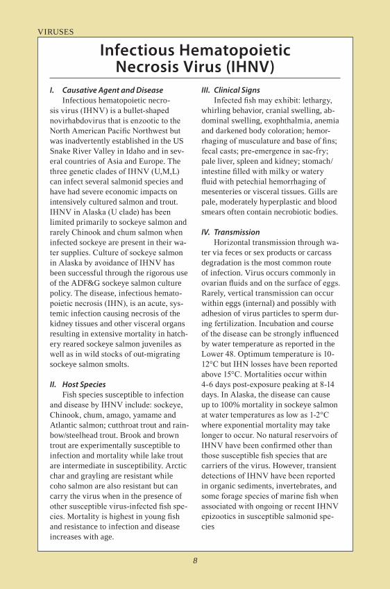
Infected fish may exhibit: lethargy,whirling behavior, cranial swelling, ab-dominal swelling, exophthalmia, anemia and darkened body coloration; hemor-rhaging of musculature and base of fins; fecal casts; pre-emergence in sac-fry; pale liver, spleen and kidney; stomach/ intestine filled with milky or watery fluid with petechial hemorrhaging of mesenteries or visceral tissues. Gills are pale, moderately hyperplastic and blood smears often contain necrobiotic bodies.
IV. Transmission Horizontal transmission through wa-
ter via feces or sex products or carcass degradation is the most common route of infection. Virus occurs commonly in ovarian fluids and on the surface of eggs. Rarely, vertical transmission can occur within eggs (internal) and possibly with adhesion of virus particles to sperm dur-ing fertilization. Incubation and course of the disease can be strongly influenced by water temperature as reported in the Lower 48. Optimum temperature is 10-12°C but IHN losses have been reported above 15°C. Mortalities occur within 4-6 days post-exposure peaking at 8-14days. In Alaska, the disease can causeup to 100% mortality in sockeye salmonat water temperatures as low as 1-2°Cwhere exponential mortality may takelonger to occur. No natural reservoirs ofIHNV have been confirmed other thanthose susceptible fish species that arecarriers of the virus. However, transientdetections of IHNV have been reportedin organic sediments, invertebrates, andsome forage species of marine fish whenassociated with ongoing or recent IHNVepizootics in susceptible salmonid spe-cies
9
VIRUSES
V. Diagnosis Susceptible fish cell cultures are in-
oculated with kidney and spleen tissues (whole fry if small) or ovarian fluids from fish suspected of having IHNV. Presumptive diagnosis results from dif-fuse or plaqued lysis of inoculated cell monolayers (cytopathic effect). Virus is definitively identified by PCR.
VI. Prognosis for Host Prognosis for infected fish is poor.
Survivors of epizootics and subclinical infections become lifelong carriers of the virus. There is no known therapy for fish that have been infected with IHNV.
In Alaskan hatcheries, all infected lots of fish are destroyed. The occurrence of the disease is avoided through preventative measures including a virus-free water supply, rigorous disinfection, isolation of egg and fish lots and containment of diseased fish. There is an effective DNA vaccine used in Canada that is also li-censed in the US but has been restricted commercially due to unlikely safety concerns regarding GMO products.
VII. Human Health Signifcance There are no human health concerns
associated with IHN virus.
Exaggerated (top, middle) cephalic bumps on sockeye salmon fry commonly occur with IHN disease.
Scoliosis in sockeye salmon smolt surviving IHN.
Necrotic macrophages or kidney cells (necrobiotic bodies-arrows) with debris in peripheral blood, X 1000.
Hemorrhaging at the base of the fns is sometimes observed in IHN disease.
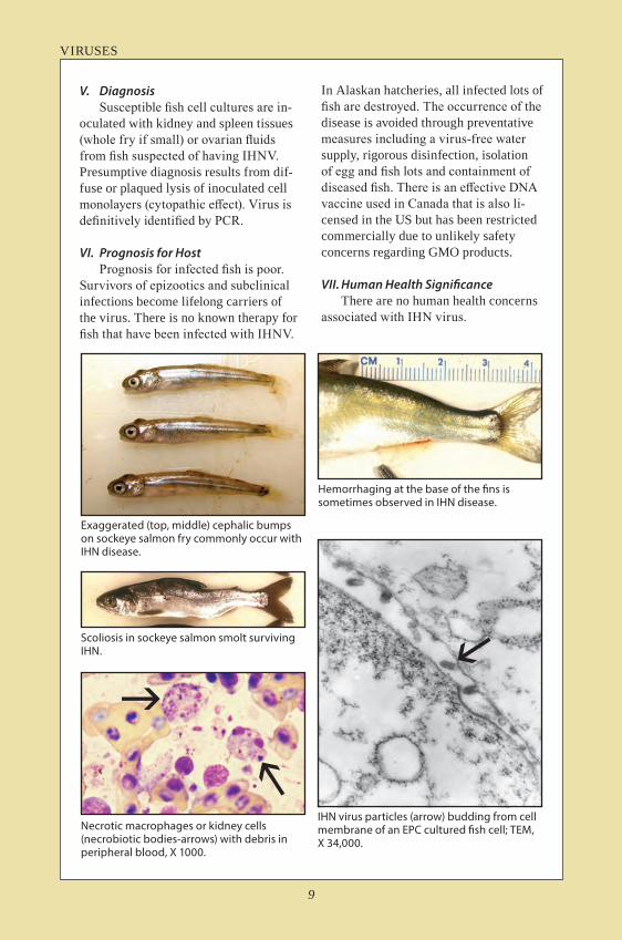
IHN virus particles (arrow) budding from cell membrane of an EPC cultured fsh cell; TEM, X 34,000.

10
VIRUSES
North American Viral Hemorrhagic Septicemia Virus (NA-VHSV)
I. Causative Agent and Disease North American viral hemorrhagic
septicemia virus (NA-VHSV) Type IVa is a bullet-shaped RNA novirhabdovirus. It is molecularly distinct from the similar Type IVb in the Great Lakes, USA that is pathogenic for a large number of nonsalmonid species. It is also different from Type IVc in marine/freshwater spe-cies of Atlantic Canada and most VHSV strains in Asia (IVa occurs in Japan/ Korea) and Europe (Types I, II, III) that are pathogenic for some marine species and rainbow trout.
II. Host Species NA-VHSV Type IVa infects many
marine fish species in the northern Pacific Ocean including anadromous coho and Chinook salmon. In Alaska, the virus is reported from Pacific herring, Pacific cod, Pacific hake and walleye pollock. Two isolates from pink salmon have been the only occurrences of VHSV from a free ranging Alaskan salmonid. The virus is enzootic in populations of Northern Pacific herring and sardines causing epizootic mortal-ity. Experimental studies indicate that juvenile Alaskan Chinook, coho, pink and sockeye salmon are refractory to the virus by waterborne exposure.
III. Clinical Signs Detection of NA-VHSV from ana-
dromous salmonids in Washington and Oregon has generally been at very low levels and prevalences and not associ-ated with clinical disease. In Pacific cod, secondary VHSV infection can be de-tected at low levels in skin erosions and ulcers caused by other primary patho-gens. Septicemia with skin hemorrhages may also occur. In Pacific herring, the virus can be acutely lethal for up to
100% of exposed juvenile fish with lower chronic mortality occurring in adults. Infected juvenile herring develop hemor-rhages of the skin around the mouth and isthmus and/or at the base of fins while occasional hemorrhages occur in adult fish along the flanks that may progress to ulcers. Fin erosion and lethargic swimming behavior may also be present. Experimentally infected juvenile rain-bow trout exhibited darkened body color and hemorrhaging at the base of fins and vent associated with low mortality.
IV. Transmission Transmission of VHSV is horizontal
through ambient seawater from fish to fish and likely by ingestion of infected fish. Individual infected juvenile Pacific herring can shed up to 106.5 plaque form-ing units (PFU) of virus per ml. Primary virus infection is through the epidermis and possibly gill tissues followed by systemic infection (viremia). Because VHSV in the Pacific Northwest is indig-enous to Pacific herring and other forage species utilized by salmon, these prey are a likely source of VHSV periodi-cally detected in adult coho and Chinook salmon in Washington State.
V. Diagnosis Cultures of susceptible fish cell
lines are inoculated with kidney, spleen, liver, ovarian fluids or epidermal lesions from suspect fish. Presumptive diagno-sis is made when characteristic cyto-pathic effect (CPE) or lysis occurs in cell monolayers from virus infection. Virus identification is confirmed by PCR.
VI. Prognosis for Host Susceptible juvenile herring sustain
up to 100% mortality which may not occur in adult fish or is lower and more

11
VIRUSES
chronic. Herring that survive virus in-fection develop apparent immunity to re-infection. Noteworthy, is that low levels of VHSV can occasionally be detected in a small percentage of apparently healthy herring from most populations. Clinical disease and mortality from the virus is
variable but generally lower in other for-age species.
VII. Human Health Signifcance There are no human health concerns
associated with NA-VHS virus.
Skin hemorrhaging in infected Pacifc herring
Left: Pacifc herring with typical VHS hemorrhage; Right: Skin hemorrhaging in infected Pacifc cod (photo: NMFS staf)
Electron micrograph of VHSV particles (arrow) in a cultured EPC fsh cell, X 56,500

12
VIRUSES
Pacific Salmon Paramyxovirus
I. Causative Agent and Disease Pacific salmon paramyxovirus
(PSPV) is a large, irregular shaped, enveloped, single-stranded RNA virus about 125-250 nm in diameter belonging in the family Paramyxoviridae and the genus Aquaparamyxovirus. The virus is of low virulence and not associated with disease or mortality. The viral agent is generally isolated from asymptomatic carrier fish during routine viral screen-ing.
II. Host Species The most common host in North
America is adult Chinook salmon from Alaska, Oregon and Washington. Un-confirmed isolates have been reported from other salmonids. In Norway, At-lantic salmon paramyxovirus (ASPV) has been isolated from seawater reared Atlantic salmon.
III. Clinical Signs No clinical signs of disease are
associated with fish infected by PSPV. The ASPV paramyxovirus in Norway is reportedly associated with the disease syndrome, proliferative gill inflamma-tion (PGI).
IV. Transmission The mode of transmission is hori-
zontal by water or fish to fish. A marine reservoir for the virus is suspected.
V. Diagnosis Detection of paramyxovirus is by
isolation in cultures of susceptible fish cell lines inoculated with infected tissue. The virus causes a cytopathic effect (CPE) characterized by retracted and rounded cells after an extensive incuba-tion period. Presumptive identifications are made by observing the typical CPE.
This virus has the unique characteristic among fish viruses of hemagglutinating erythrocytes from fish, some mammals (human, rabbit, horse, guinea pig and swine), and birds. Hemagglutination allows viral placement in the Paramyxo-viridae and confirmation of a para-myxovirus along with other procedures including ultrastructural morphology by electron microscopy, fluorescent an-tibody testing (FAT), PCR and sequenc-ing.
VI. Prognosis for Host The prognosis for the host is good
regarding the non-pathogenic nature of the North American isolates of PSPV. The role of Norwegian ASPV in causing PGI is questionable since other agents have been present confounding the true etiology of fish mortality. In this case perhaps corrective therapy would include optimizing the environment and avoidance. Further study is warranted.
VII. Human Health Signifcance There are no human health concerns
associated with paramyxoviruses in fish.
13
VIRUSES
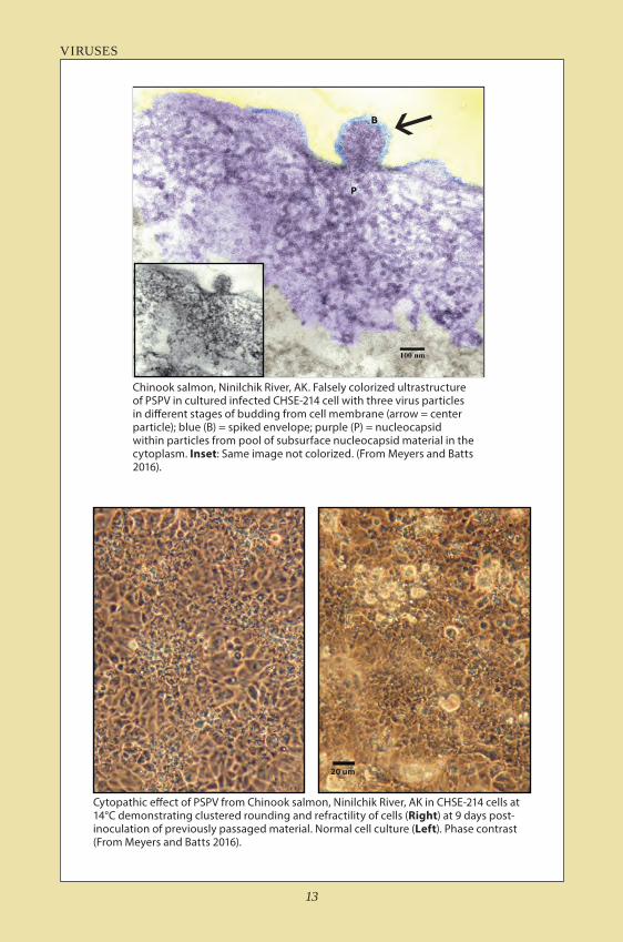
Chinook salmon, Ninilchik River, AK. Falsely colorized ultrastructure of PSPV in cultured infected CHSE-214 cell with three virus particles in diferent stages of budding from cell membrane (arrow = center particle); blue (B) = spiked envelope; purple (P) = nucleocapsid within particles from pool of subsurface nucleocapsid material in the cytoplasm. Inset: Same image not colorized. (From Meyers and Batts 2016).
20 um
Cytopathic efect of PSPV from Chinook salmon, Ninilchik River, AK in CHSE-214 cells at 14°C demonstrating clustered rounding and refractility of cells (Right) at 9 days post-inoculation of previously passaged material. Normal cell culture (Left). Phase contrast (From Meyers and Batts 2016).

14
VIRUSES
Piscine Orthoreovirus (PRV)
I. Causative Agent and Disease Piscine orthoreovirus (PRV), also
known as Atlantic salmon reovirus, was identified in 2010 by next genera-tion sequencing of tissues from farmed Atlantic salmon in Norway dying from the disease “heart and skeletal muscle inflammation” (HSMI). The virus has double-stranded RNA with 10 segments and is 72 nm in diameter. There are three strains of the virus (PRV1, 2, 3) possibly influencing disease outcome in different host species under different environmental conditions.
II. Host Species PRV is reported from Norway,
Denmark, Ireland, Chile, Japan and the Pacific Northwest (WA, AK, BC, Can-ada) infecting Atlantic salmon, Pacific salmon and trout (cutthroat, steelhead, sea-run brown). In Alaska, PRV was sequenced from three stocks of coho and one stock of Chinook and unconfirmed in one stock of chum salmon.
III. Clinical Signs HSMI, described from Norway in
1999, causes anorexia, lethargy, and ascites with inflammatory lesions of the heart and skeletal muscle. It is a disease of farmed Atlantic salmon (PRV-1) and rainbow trout (PRV-3) in both freshwater and seawater. PRV-1 has been associated with jaundice, anemia and degenerative/ necrotic lesions of the liver/kidney in healthy farmed Chinook salmon and PRV-2 is associated with EIBS and jaundice/anemia in farmed coho salmon in Japan. Stress may precipitate clinical disease.
IV. Transmission PRV can be transmitted by injection
and horizontally. Marine forage fish
species may be possible reservoirs.
V. Diagnosis HSMI disease is diagnosed by
histological changes of mononuclear inflammation and necrosis of the heart and red skeletal muscle with absence of pancreatic lesions. PRV replicates in the cytoplasm of red blood cells producing inclusion bodies similar to EIBS with or without anemia. This finding suggests a relationship of PRV with EIBS as well as HSMI. PRV does not replicate well in available fish cell lines, requiring molecular detection and sequencing for confirmation of the virus and strain.
VI. Prognosis for Host Rarely, 20% mortality from HSMI
has occurred in Atlantic salmon smolts 5-9 months after transfer to seawater.However, high levels of PRV genetic ma-terial are detected in asymptomatic wildand cultured salmonids with no evidenceof HSMI disease. In one experiment,PRV was infectious for Chinook andsockeye salmon and persisted but did notcause fish mortality or HSMI, or otherapparent disease. Testing of archived tis-sues from BC indicated PRV was presentin asymptomatic wild and farmed Pacificsalmon since 1987, possibly as earlyas 1977 before Atlantic salmon wereimported for aquaculture. The ubiquityof PRV, apparent historic presence inwild Pacific salmon stocks in the PNWand lack of clear association with diseasesuggest the virus is of low risk to wildspecies of Pacific salmon.
VII. Human Health Signifcance There are no human health concerns
regarding infection of fish with PRV.
15
VIRUSES
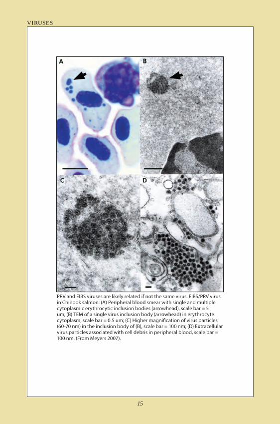
PRV and EIBS viruses are likely related if not the same virus. EIBS/PRV virus in Chinook salmon: (A) Peripheral blood smear with single and multiple cytoplasmic erythrocytic inclusion bodies (arrowhead), scale bar = 5 um; (B) TEM of a single virus inclusion body (arrowhead) in erythrocyte cytoplasm, scale bar = 0.5 um; (C) Higher magnifcation of virus particles (60-70 nm) in the inclusion body of (B), scale bar = 100 nm; (D) Extracellular virus particles associated with cell debris in peripheral blood, scale bar = 100 nm. (From Meyers 2007).

16
BACTERIA
Bacterial Coldwater Disease (BCWD) and other freshwater flavobacteria
I. Causative Agent and Disease The causative agent of BCWD, Fla-
vobacterium psychrophilum, is a Gram-negative proteolytic bacterium causing systemic disease in colder waters. Clini-cal signs usually occur below 12°C and in Alaska often occur at extremely cold water temperatures of 1°C. The bacte-rium, originally classified to the genus Cytophaga, was changed to Flexibacter and later to Flavobacterium. The species name means “cold loving”. BCWD is characterized by tissue necrosis of the fins that progresses to complete destruc-tion of the caudal peduncle exposing the vertebrae. Other common names for this condition are peduncle disease or low temperature disease. Infections by other freshwater species of flavobacteria are generally non-systemic causing similar erosive skin lesions that may occur any-where on the surface of the fish host.
II. Host Species BCWD is found in temperate sal-
monid producing regions worldwide. Juvenile coho and Chinook salmon are particularly susceptible. External infec-tions by other freshwater flavobacteria also occur worldwide affecting several species of fish.
III. Clinical Signs BCWD begins with darkening of the
peduncle region when water tempera-tures are between 4-12°C with up to 50% mortality prior to the occurrence of more chronic peduncle erosion. When acute, lesions appear in the areas of increased pigmentation on the peduncle region, or elsewhere. Ulcers are deep and the caudal fin may erode completely expos-ing the vertebral column. When present, internal lesions may only consist of mild petechial hemorrhages within the adipose
tissues surrounding the pyloric caeca. Chronic BCWD can result in lordosis and scoliosis (“crinkle-back”) and an abnormal swimming posture from the destruction of muscle bundles adjacent to the vertebral column. Another sequella is bacterial invasion of the brainstem caus-ing erratic swimming, darkened posterior body and sudden death from damage to nervous tissues, vertebral cartilage and bone. Other flavobacteria cause erosive skin lesions that may penetrate deeply into the underlying skeletal muscle.
IV. Transmission Transmission of BCWD is horizontal
through the water column and vertically through the eggs of infected adult sal-monids. The bacteria have been isolated from internal organs and gonadal fluids of returning adult salmon suggesting they are carriers of the infection during their seawater phase but reinfection upon entering freshwater is also possible. All other Flavobacterium species are com-mon inhabitants of aquatic ecosystems allowing for horizontal transmission.
V. Diagnosis Presumptive diagnosis includes isola-
tion of long, filamentous, Gram-negative bacteria that are non-motile or have glid-ing motility from kidney tissues or typi-cal skin lesions of fish. The bacteria grow well on Cytophaga and TYES agars, with optimum growth at 15-16°C. Colonies are bright yellow, entirely convex or with convex centers and a spreading periphery resembling a “fried egg”. Colonies turn orange-red when KOH is added indicat-ing flexirubin pigment. Growth of F. psychrophilum is inhibited by Congo red added to TYES agar or diffusion discs allowing rapid differentiation from other Flavobacterium species. Confirmatory
17
BACTERIA
diagnosis is done using PCR or mono-clonal direct FAT.
VI. Prognosis for Host BCWD begins as an external infec-
tion that becomes systemic. External flavobacteria generally remain as such but either type of infection can result in fish mortality. Hatchery fish can be treated for the infections with external
1 hr treatments of formalin and for BCWD antibiotic therapy may be necessary as well.
VII. Human Health Signifcance There are no human health concerns
associated with Flavobacterium psy-chrophilum or other flavobacteria.
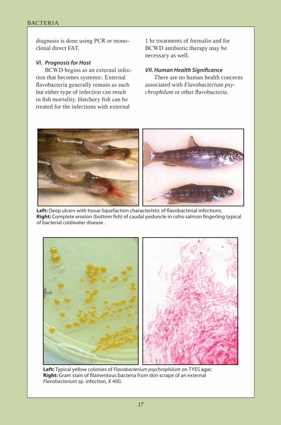
Left: Deep ulcers with tissue liquefaction characteristic of favobacterial infections; Right: Complete erosion (bottom fsh) of caudal peduncle in coho salmon fngerling typical of bacterial coldwater disease .
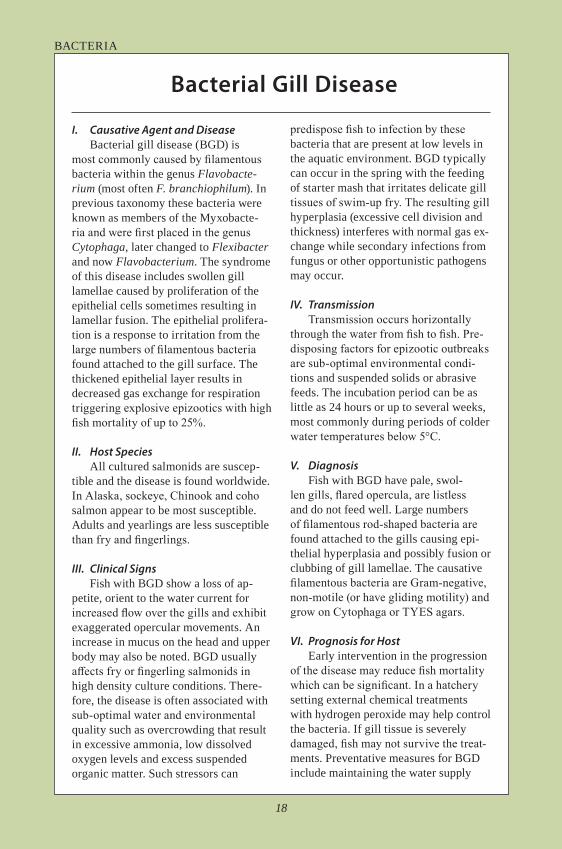
Left: Typical yellow colonies of Flavobacterium psychrophilum on TYES agar; Right: Gram stain of flamentous bacteria from skin scrape of an external Flavobacterium sp. infection, X 400.
18
BACTERIA
Bacterial Gill Disease
I. Causative Agent and Disease Bacterial gill disease (BGD) is
most commonly caused by filamentous bacteria within the genus Flavobacte-rium (most often F. branchiophilum). In previous taxonomy these bacteria were known as members of the Myxobacte-ria and were first placed in the genus Cytophaga, later changed to Flexibacter and now Flavobacterium. The syndrome of this disease includes swollen gill lamellae caused by proliferation of the epithelial cells sometimes resulting in lamellar fusion. The epithelial prolifera-tion is a response to irritation from the large numbers of filamentous bacteria found attached to the gill surface. The thickened epithelial layer results in decreased gas exchange for respiration triggering explosive epizootics with high fish mortality of up to 25%.
II. Host Species All cultured salmonids are suscep-
tible and the disease is found worldwide. In Alaska, sockeye, Chinook and coho salmon appear to be most susceptible. Adults and yearlings are less susceptible than fry and fingerlings.
III. Clinical Signs Fish with BGD show a loss of ap-
petite, orient to the water current for increased flow over the gills and exhibit exaggerated opercular movements. An increase in mucus on the head and upper body may also be noted. BGD usually affects fry or fingerling salmonids in high density culture conditions. There-fore, the disease is often associated with sub-optimal water and environmental quality such as overcrowding that result in excessive ammonia, low dissolved oxygen levels and excess suspended organic matter. Such stressors can
predispose fish to infection by these bacteria that are present at low levels in the aquatic environment. BGD typically can occur in the spring with the feeding of starter mash that irritates delicate gill tissues of swim-up fry. The resulting gill hyperplasia (excessive cell division and thickness) interferes with normal gas ex-change while secondary infections from fungus or other opportunistic pathogens may occur.
IV. Transmission Transmission occurs horizontally
through the water from fish to fish. Pre-disposing factors for epizootic outbreaks are sub-optimal environmental condi-tions and suspended solids or abrasive feeds. The incubation period can be as little as 24 hours or up to several weeks, most commonly during periods of colder water temperatures below 5°C.
V. Diagnosis Fish with BGD have pale, swol-
len gills, flared opercula, are listless and do not feed well. Large numbers of filamentous rod-shaped bacteria are found attached to the gills causing epi-thelial hyperplasia and possibly fusion or clubbing of gill lamellae. The causative filamentous bacteria are Gram-negative, non-motile (or have gliding motility) and grow on Cytophaga or TYES agars.
VI. Prognosis for Host Early intervention in the progression
of the disease may reduce fish mortality which can be significant. In a hatchery setting external chemical treatments with hydrogen peroxide may help control the bacteria. If gill tissue is severely damaged, fish may not survive the treat-ments. Preventative measures for BGD include maintaining the water supply
19
BACTERIA
free of fish (especially adults), mud and VII. Human Health Signifcance silt, reducing stress such as overcrowd- The causative bacteria of BGD are ofing, avoiding low dissolved oxygen or no human health concern high ammonia levels and avoiding exces-sive fish handling.
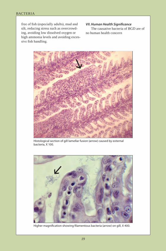
Histological section of gill lamellar fusion (arrow) caused by external bacteria, X 100.
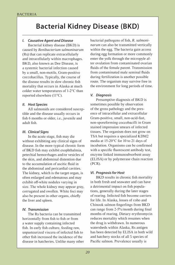
Higher magnifcation showing flamentous bacteria (arrow) on gill, X 400.
20
BACTERIA
Bacterial Kidney Disease (BKD)
I. Causative Agent and Disease Bacterial kidney disease (BKD) is
caused by Renibacterium salmoninarum (Rs) that can replicate extracellularly and intracellularly within macrophages. BKD, also known as Dee Disease, is a systemic bacterial infection caused by a small, non-motile, Gram-positive coccobacillus. Typically, the course of the disease results in slow chronic fish mortality that occurs in Alaska at much colder water temperatures of 1-2°C than reported elsewhere (11°C).
II. Host Species All salmonids are considered suscep-
tible and the disease usually occurs in fish 6 months or older, i.e., juvenile and adult fish.
III. Clinical Signs In the acute stage, fish may die
without exhibiting any clinical signs of disease. In the more typical chronic form of BKD fish may exhibit exophthalmia, petechial hemorrhages and/or vesicles of the skin, and abdominal distention due to the accumulation of ascitic fluid in the abdominal and pericardial cavities. The kidney, which is the target organ, is often enlarged and edematous and may exhibit off-white nodules varying in size. The whole kidney may appear gray, corrugated and swollen. White foci may also be present in other organs, chiefly the liver and spleen.
IV. Transmission The Rs bacteria can be transmitted
horizontally from fish to fish or from a water supply containing infected fish. In early fish culture, feeding raw, unpasteurized viscera of infected fish to other fish increased the incidence of the disease in hatcheries. Unlike many other
bacterial pathogens of fish, R. salmoni-narum can also be transmitted vertically within the egg. The bacteria gain access during egg formation or more commonly enter the yolk through the micropyle af-ter ovulation from contaminated ovarian fluids of the female parent. Transmission from contaminated male seminal fluids during fertilization is another possible route. The organism may survive free in the environment for long periods of time.
V. Diagnosis Presumptive diagnosis of BKD is
sometimes possible by observation of the gross pathology and the pres-ence of intracellular and extracellular Gram-positive, small, non-acid-fast, non-sporeforming coccobacilli in Gram stained impression smears of infected tissues. The organism does not grow on TSA but requires a specialized KDM2 media at 15-20°C for 10 to 21 days of incubation. Organisms can be confirmed with a specific fluorescent antibody test, enzyme linked immunoabsorbent assay (ELISA) or by polymerase chain reaction (PCR).
VI. Prognosis for Host BKD results in chronic fish mortality
in both fresh and seawater and can have a detrimental impact on fish popula-tions, generally during the later stages of rearing. Infected fish become carriers for life. In Alaska, losses of coho and Chinook salmon fingerlings from BKD can range from 2-5%/month during final months of rearing. Dietary erythromycin reduces mortality which resumes when the drug is withdrawn. In numerous watersheds within Alaska, Rs antigen has been detected by ELISA in both wild and hatchery stocks of all 5 species of Pacific salmon. Prevalence usually is

21
BACTERIA
less than 10%, but some systems have VII. Human Health Signifcance carrier rates up to 90%. Wild trout, char There are no human health concernsand grayling are reservoirs for Rs, often associated with R. salmoninarum. showing prevalences of up to 100%.
Left: White pustules (arrows) in the posterior kidney and liver of a juvenile coho salmon typical of BKD; Inset: Infected fsh with exophthalmia; Right: White kidney pustules (arrow) in adult Bear Lake sockeye salmon with BKD (photo: CIAA staf).
Left: Stained kidney smear with small Gram-positive coccobacilli typical of R. salmoninarum, X 1000; Right: BKD bacteria, Renibacterium salmoninarum, stained with a fuorescene dye (green), fuorescent antibody test, X 1000.
22
BACTERIA
Enteric Redmouth Disease (ERM)
I. Causative Agent and Disease Enteric redmouth disease (ERM) or
yersiniosis is caused by Gram-negative, motile bacteria known as Yersinia ruckeri. The name ERM is derived from the inflammation and petechial hemorrhages of the lower hind gut and inside and around the mouth of infected fish that are not unique signs of infection by this bacterium. ERM is an acute septicemia in salmonids with bacterial foci, necrosis and inflammation in all tissues. In Alaska, two serotypes of the bacteria, known as O1 and O2, can cause the disease. The two serotypes are differentiated from each other based on biochemical and/or serological tests. The virulence varies considerably within each serogroup but Y. ruckeri O1 has been more pathogenic in Alaskan salmonids and elsewhere. The bacteria are found worldwide where salmonids are cultured and there are a total of 6 serotypes with several subgroups.
II. Host Species Rainbow trout are the most sensitive
host, but all salmonids and several other fish species are susceptible to infection. ERM is a major concern in the Norwegian Atlantic salmon industry causing fish mortality in both fresh and seawater pens.
III. Clinical Signs Externally, clinical signs can be
similar to other bacterial septicemias. Infected fish are often lethargic and dark in color. Inflammation and petechiation are prominent in and around the mouth, the isthmus and on the opercula. Petechial hemorrhages are commonly at the base of the fins. Fish often exhibit exophthalmia and a distended abdomen. Internally, the stomach is often filled
with watery fluid and petechiation may be present in the musculature and visceral organs, most notably in the hind gut and liver.
IV. Transmission The bacterium is horizontally
transmitted from fish to fish via the fecal oral route and often becomes localized in the lower intestine of fish surviving a disease outbreak. Bacteria can remain viable for a limited time in ambient water to infect susceptible fish. Other reservoirs of the bacteria include fish-eating birds reported near aquaculture facilities.
V. Diagnosis Presumptive diagnosis is made by
the cultivation of a Gram-negative, oxidase negative, motile bacterial rod from blood, kidney, or lesions when inoculated onto bacteriological media. Diagnosis is confirmed with biochemical tests or fluorescent antibody tests specific for Yersinia ruckeri Types O1 and O2. Type O1 includes subgroups a and b while Type O2 is composed of 3 subgroups (a,b,c). The remaining serotypes are O3, O5, O7 and O8. The majority of epizootics in salmonids are caused by motile serotype O1a. That said, the current serotyping scheme is inconsistent and not as helpful as genetic sequencing in determining relatedness in clonal clustering that may influence epidemiological differences.
VI. Prognosis for Host Under aquaculture conditions,
diseased fish generally die if there is no antibiotic intervention. Prognosis for the population is good if the condition is recognized early so that antibiotic therapy can be initiated.

23
BACTERIA
VII. Human Health Signifcance There are no human health concerns
associated with Yersinia ruckeri.
Petechial hemorrhages of the liver present in enteric redmouth disease.
Difuse internal petechial hemorrhaging (arrow) typically present with bacterial septicemias like enteric redmouth disease.

24
BACTERIA
Furunculosis
I. Causative Agent and Disease IV. Transmission Furunculosis is caused by a Gram-
negative bacterium known as Aeromo-nas salmonicida and is probably the most commonly encountered bacterial pathogen in cultured salmonids. The disease occurs worldwide in freshwater and has also been reported in the marine environment. It is known to occur in North America, Europe, Asia, and Africa. Furunculosis is characterized by a generalized bacteremia with focal necrotic swellings in the muscle tissue called furuncles.
II. Host Species All salmonid species are susceptible.
Rainbow trout show some resistance. Young fish are the most susceptible, especially when the water temperatures are > 8º C. In hatcheries, pink and chum salmon are less likely to develop furun-culosis since they are not reared long before being released to seawater. Many non-salmonid species of fish in both ma-rine and freshwater are also susceptible to infection by A. salmonicida, some strains of which are atypical.
III. Clinical Signs In acute septicemia where rapid
death may occur, gross clinical signs may not develop. In subacute and chronic infections, body darkening, lethargy and loss of appetite are associ-ated with the typical focal necrosis in the muscle, often visible as a swelling under the skin. These lesions eventually ulcer-ate producing deep craters. Erythema, petechiation and exophthalmia may be present and the abdomen of the fish may be distended with internal ascitic fluid. Bloody fluid may be discharged from the anal vent and the kidney, liver and/or spleen may be enlarged.
Horizontal transmission to suscepti-ble fish is via the water column or by the fecal-oral route. Diseased or carrier fish are point sources of infection. Increasing water temperature exacerbates the inci-dence and intensity of infection. Vertical transmission of the bacteria has not been demonstrated.
V. Diagnosis Presumptive diagnosis is made by
culture of a Gram-negative, oxidase positive (an oxidase negative isolate has been described), non-motile bacterial rod from blood, kidney, or lesions on TSA or furunculosis agar with the pro-duction of a brown diffusible pigment. Some strains of bacteria may not pro-duce pigment. Diagnosis is confirmed by biochemical tests, slide agglutination and fluorescent antibody tests specific for A. salmonicida.
VI. Prognosis for Host In nature, the disease usually results
in mortality. In a hatchery, prognosis for the fish population is good if the condition is caught early and antibiotic therapy is initiated.
VII. Human Health Signifcance There are no human health concerns
associated with A. salmonicida.
25
BACTERIA
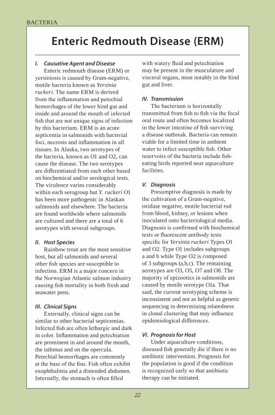
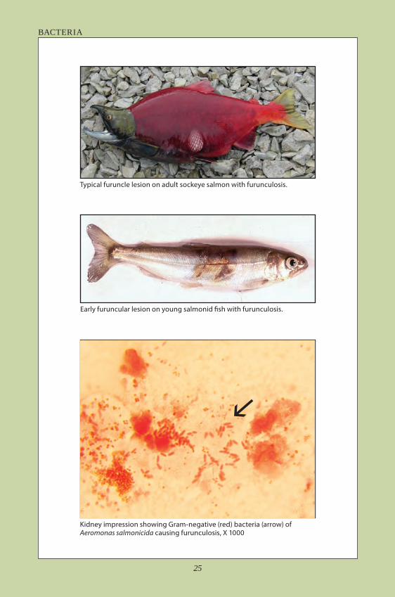
Typical furuncle lesion on adult sockeye salmon with furunculosis.
Early furuncular lesion on young salmonid fsh with furunculosis.
Kidney impression showing Gram-negative (red) bacteria (arrow) of Aeromonas salmonicida causing furunculosis, X 1000

26
BACTERIA
Fusobacteria-like Agent
I. Causative Agent and Disease VI. Prognosis for Host An external skin and/or gill infection
is caused by long, non-motile, Gram-negative bacterial rods that are sharply pointed at both ends. The bacteria, commonly referred to as fusobacteria, infect cultured salmonid fish in fresh water during periods of very cold water temperatures, usually less than 5°C. Infection produces excessive mucus externally and on the gills causing respi-ratory distress.
II. Host Species This organism has been detected on
cultured salmonid fishes at various life stages from alevin to pre-smolt in the Pacific Northwest and Alaska. Chinook and coho salmon have been the most commonly affected species.
III. Clinical Signs The skin of infected fish has exces-
sive mucus production and gill infec-tions result in lamellar hyperplasia and increased respiration.
IV. Transmission These bacteria are probably trans-
mitted horizontally through the water from fish to fish.
V. Diagnosis Diagnosis includes observation of
Gram-negative, non-motile, bacterial rods with a beaded appearance having characteristic attenuated ends on the skin and/or gills of infected fish. The biomass of bacteria is often extensive. This bacterial organism has not been cultured successfully on conventional bacterial media but minor temporary success has been achieved on swabs aerobically incubated in 50% MEM and lake water at low pH (4.5).
External infection by these bacteriaresults in high fish mortality if there is no intervening therapy. One or two external applications of formalin or hydrogen peroxide have been successful treatments.
VII. Human Health Signifcance There are no known human health
concerns associated with this fusobacte-ria-like agent.

27
BACTERIA
Fusobacteria stained with Giemsa showing typical fusiform shape with pointed ends and beaded appearance, X 1000.
Gram stain of skin scrape showing high biomass of fusobacteria, X 1000.
28
BACTERIA
Marine Tenacibaculosis
I. Causative Agent and Disease Tenacibaculum maritimum is a fila-
mentous, Gram-negative bacterium that moves by gliding motility. These marine bacteria are opportunistic pathogens of fish producing external (sometimes systemic) infections such as bacterial gill disease, fin rot, skin ulcers or eroded mouth disease. Infections are often initiated by physical trauma, pinheading and adverse environmental conditions. Resulting fish mortality can be signifi-cant.
II. Host Species All marine fish worldwide are
potentially susceptible to infection by Tenacibaculum maritimum which has been isolated from a variety of salmo-nid fishes, Dover sole, sea bass, turbot, bream, halibut and sardines. In Alaska, this bacterial pathogen has caused mortality of juvenile Pacific salmon in seawater netpens during the winter and early spring.
III. Clinical Signs Diseased fish have ulcerated skin
lesions, frayed or eroded fins and tail. Moderate to severe erosions of the head and mouth may also occur. Infected epidermal tissue may appear pale yellow to white due to the presence of large numbers of bacteria. Infected gills of fish may produce excessive mucus, have pale color and exhibit lamellar hyper-plasia. Secondary systemic infections by other bacteria commonly occur through open lesions.
IV. Transmission Tenacibaculum is a naturally occur-
ring marine bacterium and is transmit-ted horizontally through the water from fish to fish, generally requiring minor
external trauma or other environmental stressors.
V. Diagnosis Diagnosis of Tenacibaculum
infections is made by observing large numbers of filamentous bacteria in wet mounts of lesion material. The bacteria can be cultured on seawater Cytophaga or TYES agars with added 1.5% NaCl or 30 ppt to full strength seawater and incubated at 15°C. The colonies, often yellow in color, are catalase and oxidase positive with no flexirubin pigment in cell walls. Identity confirmation of T. maritimum is by PCR and there are three O serotypes.
VI. Prognosis for Host This external bacterial infection can
cause significant mortality, especially if fish are stressed. Treatment has been successful with oral antibiotic therapy.
VII. Human Health Signifcance There are no human health concerns
associated with Tenacibaculum mariti-mum.

29
BACTERIA
Caudal fn lesion on a halibut caused by infection with a marine Tenacibaculum.
Severely eroded head and upper jaw of a coho salmon smolt due to marine Tenacibaculum infection.

30
BACTERIA
Motile Aeromonas and Pseudomonas Septicemia
I. Causative Agent and Disease Motile bacterial septicemias are
caused by Gram-negative bacteria including Aeromonas and Pseudomonas with the Aeromonas hydrophila-complex and Pseudomonas fluorescens being the most common species. The A. hy-drophila (liquefaciens) complex contains numerous biotypes and serotypes of A. hydrophila as well as A. sobria , A.caviae, A. shuberti and A. veronii. Thesebacteria are ubiquitous in the aquaticenvironment and are found around theworld in both fresh and brackish water,but more commonly in freshwater. Thesebacteria generally cause a systemic,hemorrhagic disease in fish. Most ofthese bacteria are considered opportu-nistic pathogens causing disease in fishcompromised by stress or other patho-gens. Some species, most commonly P. fluorescens and A. hydrophila, have beenreported as primary fish pathogens insystems of high intensity fish culture.
II. Host Species When less than optimum condi-
tions prevail, all freshwater fish species are likely susceptible to these bacteria. Among salmonids, rainbow trout and Chinook salmon are probably the most susceptible to the A. hydrophila com-plex. Both Aeromonas and Pseudomonas are pathogenic for other cold-blooded vertebrates including frogs and reptiles and will infect mammals including man through wounds or when they are im-munocompromised.
III. Clinical Signs Lethargy, low-level mortality and
occasional cutaneous lesions on the body surface may occur. Inflammation and erosion in and around the mouth with
hemorrhaging and necrosis of the fins is common. Exophthalmia and abdominal distention with ascitic fluid may also be present. Internally, the kidney may be soft and swollen and the spleen enlarged. Petechial hemorrhages may be present internally in many tissues and the intes-tines may be inflamed and filled with yellow mucus or bloody fluid.
IV. Transmission These bacteria are among the normal
flora of healthy fish and are ubiquitous in the aquatic environment. They are particularly abundant in organically polluted waters while infected carrier fish and other aquatic animals can serve as reservoirs. Transmission is horizontal from fish to fish or from contaminated water. Water temperatures 10°C or above favor these opportunistic pathogens.
V. Diagnosis A presumptive diagnosis is made
when fish exhibit characteristic clinical signs with tissue imprints, squashes or blood smears containing Gram-negative, motile rod-shaped bacteria. A definitive diagnosis is made by isolation of the organism on appropriate bacteriological media followed by identification from biochemical tests.
VI. Prognosis for Host Severely affected fish will die.
However, these bacteria are generally weak pathogens. Poor environmental conditions predispose fish to disease out-breaks which are self-resolving without intervention by antibiotic therapy when the source of stress is removed. When necessary, antibiotic therapy can be ef-fective, except some pseudomonads are resistant to available aquaculture drugs.

31
BACTERIA
VII. Human Health Signifcance
Some bacteria in these generacan cause disease in humans through wounds or when immunocompromised.
Petechial hemorrhages on ventral surface of a salmonid fsh with bacterial septicemia.
Petechial hemorrhages of liver, pyloric caeca, gut and visceral fat (arrow) of a juvenile salmonid fsh with bacterial septicemia.
32
BACTERIA
Mycobacteriosis of Fish
I. Causative Agent and Disease Mycobacteriosis is caused by a
pleomorphic, Gram-variable, acid-fast, aerobic, non-motile group of bacteria in the family Mycobacteriaceae, genus Mycobacterium, that includes the human pathogen M. tuberculosis. There are over 190 species and 11 subspecies that are rods 0.2–0.6 um in diameter and 1–10 um long. The most common species in fish are M. marinum, fortuitum, chelonae and shottsii that cause a chronic sys-temic granulomatous disease affecting any or all tissues and organs. Most mycobacteria can survive within host macrophages.
II. Host Species Distribution is worldwide in amphib-
ians, reptiles and 151 species of wild and cultured fish representing 40 families including many freshwater aquarium species, salmonids and marine fishes. The disease causes serious problems in cultured species, especially aquarium fish. In Alaska, the disease has occurred in sablefish, pollock and saffron cod and is likely more widespread in other fish species.
III. Clinical Signs Gross clinical signs of mycobact-
eriosis may be non-specific scale loss, dermal ulceration, emaciation, spinal defects and ascites. Internal signs include enlarged spleen, kidney and/ or liver and characteristic gray or white nodules in internal organs.
IV. Transmission The epizootiology in fish includes
horizontal transmission by contact and ingestion as established experimentally in zebrafish and early salmonid culture when raw fish were used as feed. Verti-
cal (transovarian) transmission in live bearing fishes has also been reported. Other aquatic organisms can be infected and act as vectors.
V. Diagnosis Presumptive diagnosis is made by
histologic examination of tissues show-ing typical inflammatory granulomas with concentric layers of noncompressed and compressed (spindle cell layer) epithelioid cells forming discrete spheri-cal lesions. The centers are necrotic, often containing mycobacteria visible by acid-fast stains. Fast and slow growing mycobacteria can be cultured on selec-tive liquid and agar media providing phenotypic characteristics with defini-tive identification by PCR.
VI. Prognosis for Host Mycobacteriosis in fish is a chronic
disease often precipitated by stress. Acute mortality in wild fish is uncom-mon. Long-term population declines attributed to mycobacteria are possible as suspected for Chesapeake Bay striped bass, likely exacerbated by environ-mental decline. There is no effective treatment for infected fish which should be culled if in a culture environment.
VII. Human Health Signifcance Aquatic mycobacteria pose signifi-
cant zoonotic concerns, especially in immunocompromised people. M. mari-num, fortuitum and chelonae are known human pathogens producing granuloma-tous skin lesions and sometimes deeper infections of peripheral tissues (with marinum). Infections are treatable by antibiotic therapy.

33
BACTERIA
C
Left: Large round white granulomas (pointer) in the kidney and peritoneum of a sablefsh (photo: Eric Forrer) caused by Mycobacterium sp.; Right: Histology of mycobacteria nodules in a Siamese fghting fsh showing concentric layers of epithelioid cells with necrotic centers (C), X 400.
B
Left: Histology of sablefsh granuloma in post-mortem kidney with mycobacterial colony (B) in necrotic center, X 200; Right: Histological acid-fast stain of mycobacteria (red) in sablefsh granuloma, X 1000.
34
BACTERIA
Vibriosis
I. Causative Agent and Disease The genus Vibrio contains signifi-
cant bacterial pathogens of marine fish that cause vibriosis, an acute bacterial septicemia. The primary pathogens include V. (Listonella) anguillarum, V. ordalii and V. salmonicida. In addi-tion, Vibrio alginolyticus may occur as a secondary invader and V. vulnificus is generally restricted to European and Japanese waters. Vibrio salmonicida is reported from Atlantic Canada and Maine in North America and in Norway, Shetland Islands and Faroe Islands in Europe causing cold water vibriosis or Hitra disease mostly in Atlantic salmon. These bacteria are ubiquitous in the marine environment causing typical Gram-negative acute septicemias with bacterial foci, necrosis, hemorrhaging and inflammation in most fish tissues.
II. Host Species Because vibriosis has occurred in an
extensive number of fish species world-wide, most marine fish species are likely to be susceptible. All species of Pacific salmon and trout are susceptible to vib-riosis that often involves V. anguillarum. Coho salmon seem to be more resistant while chum and Chinook salmon are very susceptible. V. ordalii and V. sal-monicida are principally associated with Pacific and Atlantic salmon, respective-ly, while V. vulnificus most often infects eels causing red pest disease.
III. Clinical Signs Characteristic clinical signs of vib-
riosis include inflammation and redden-ing along the ventral and lateral areas of the fish with petechial hemorrhaging that develops at the base of fins, vent and within the mouth. Acute cases exhibit a darkened body with swollen, cutaneous
lesions that ulcerate, releasing blood. There may also be corneal opacity followed by evulsion of the orbital contents. Internally, the intestine may be distended with a clear, viscous fluid. Hemorrhaging is common in the viscera and around the intestines, with swelling and necrosis of the kidney and spleen.
IV. Transmission Horizontal transmission occurs from
organisms in the water or contact be-tween fish. Outbreaks have occurred in freshwater fish fed carcasses of marine fish. In Alaska, disease does not usually occur until seawater temperatures reach 8°C.
V. Diagnosis Presumptive diagnosis is made by
observing motile, curved Gram-negative bacterial rods in spleen squashes or peripheral blood smears of marine or anadromous fish. Bacteria can be isolated on tryptic soy agar, sometimes requiring 1.5% NaCl. Confirmatory diagnosis is made using biochemical or slide agglutination tests.
VI. Prognosis for Host Epizootics of vibriosis in wild fish
populations are rare but result in signifi-cant fish mortality. When cultured sal-monids are reared in seawater net pens the disease is common resulting in high mortality if not treated with antibiotics. Several licensed vaccine preparations for aquaculture have been effective in the control of vibriosis.
VII. Human Health Signifcance The Vibrio species associated with
most fish diseases such as V. anguil-larum, V. ordalii and V. salmonicida are not considered to be human pathogens.
35
BACTERIA
However, several other vibrios are of human health concern including V. chol-erae, V. vulnificus, V. parahaemolyticus and occasionally V. alginolyticus.
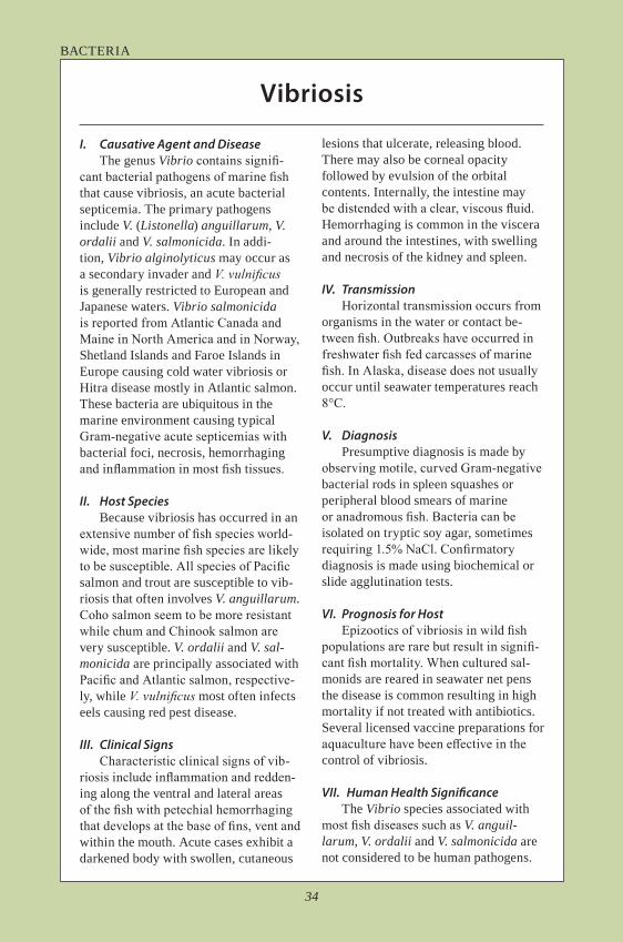
Bloody ascites (arrow) in abdominal cavity typically present in fsh with vibriosis.
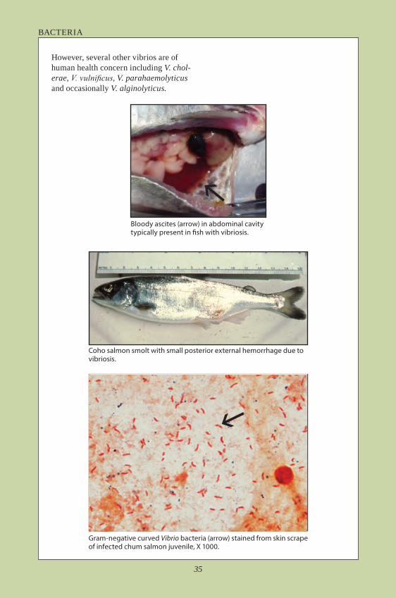
Coho salmon smolt with small posterior external hemorrhage due to vibriosis.
Gram-negative curved Vibrio bacteria (arrow) stained from skin scrape of infected chum salmon juvenile, X 1000.

36
FUNGI
Phaeohyphomycosis of Saffron Cod and Other Fish Species
I. Causative Agent and Disease Fungal infections caused by a
variety of dark-colored (dematiaceous) fungi define phaeohyphomycosis. Large black, oval, external lesions of the skin and smaller foci on the gills have been reported by subsistence users in saffron cod and other fish species from the Norton Sound area of Alaska since 2005. The lesions are caused by at least 8 different opportunistic ascomycete fungi including; Alternaria sp., Cladosporium herbarum, Chaetomium globosum, Cadophora luteo-olivacea, Penicillium sp., Phoma herbarum, Pseudophacidium ledi and Valsa sordida. These fungi typically infect the surface of the skin with invasion of the underlying skeletal muscle. Mortality has not been reported but estimated prevalence is 1 in 200 fish. These brown to black pigmented fungi are filamentous and ubiquitous in the soil. They occur as plant pathogens, on paper products, wood, natural fiber textiles, in the air and on plant debris.
II. Host Species The black external lesions have been
confirmed from saffron cod and also photographed in rainbow smelt from Norton Sound including estuarine wa-ters of the Unalakleet, Nome and Snake Rivers.
III. Clinical Signs Skin lesions caused by these fungi
are typically large (1 X 2 cm), black, slightly raised circumscribed plaques that are firm with rugose textured centers. Gill infection results in smaller black foci within the soft tissues of the filaments. Rarely are internal tissues invaded and often there is food in the gut indicating that infected fish are feeding normally.
IV. Transmission The external nature of the fungal
infections suggests that transmission is by ascospores contained in ambient sea-water or sediments, possibly increased by rain, flooding and stress that require previous mechanical tissue injury as a portal of entry into the host. However, the actual mode of transmission is unknown. Reports of these black lesions on fish most commonly occur during the late fall and early winter months of October through December.
V. Diagnosis Diagnosis is based on typical clinical
signs of shallow, circular, black rugose plaques or foci on the skin and/or gills with hyphae present in wet mounts. This is followed by isolation and characteriza-tion of the fungus on artificial media and confirmation of fungal genus and species by PCR.
VI. Prognosis for Host The prognosis for infected fish is
unknown but the large skin plaques and/ or involvement of the gill tissues suggest a chronic debilitating mycosis that may result in mortality.
VII. Human Health Signifcance Several of these fungi are opportu-
nistic human pathogens in immunocom-promised hosts. These fungi have caused fatal deep mycoses as well as brain abscess, sinusitis, peritonitis, cutaneous lesions, pneumonia and onychomycosis (nail infections). Chaetomium globosum produces mycotoxins such as chaeto-min and chaetoglobosin and commonly grows inside homes on water damaged roofs, ceilings, walls and carpets, pos-sibly representing an allergenic threat to human health.
37
FUNGI
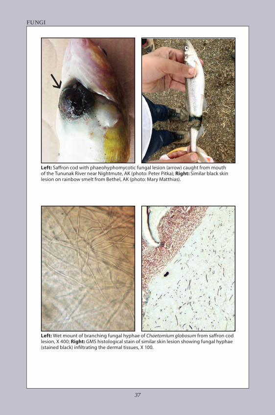
Left: Safron cod with phaeohyphomycotic fungal lesion (arrow) caught from mouth of the Tununak River near Nightmute, AK (photo: Peter Pitka); Right: Similar black skin lesion on rainbow smelt from Bethel, AK (photo: Mary Matthias).
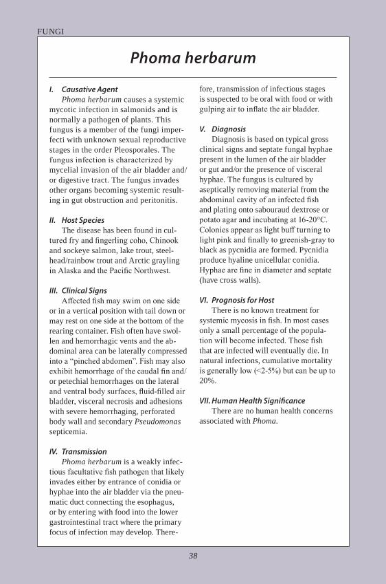
Left: Wet mount of branching fungal hyphae of Chaetomium globosum from safron cod lesion, X 400; Right: GMS histological stain of similar skin lesion showing fungal hyphae (stained black) infltrating the dermal tissues, X 100.
38
FUNGI
Phoma herbarum
I. Causative Agent Phoma herbarum causes a systemic
mycotic infection in salmonids and is normally a pathogen of plants. This fungus is a member of the fungi imper-fecti with unknown sexual reproductive stages in the order Pleosporales. The fungus infection is characterized by mycelial invasion of the air bladder and/ or digestive tract. The fungus invades other organs becoming systemic result-ing in gut obstruction and peritonitis.
II. Host Species The disease has been found in cul-
tured fry and fingerling coho, Chinook and sockeye salmon, lake trout, steel-head/rainbow trout and Arctic grayling in Alaska and the Pacific Northwest.
III. Clinical Signs Affected fish may swim on one side
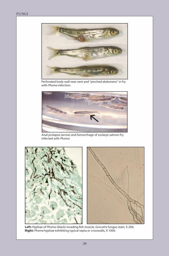
or in a vertical position with tail down or may rest on one side at the bottom of the rearing container. Fish often have swol-len and hemorrhagic vents and the ab-dominal area can be laterally compressed into a “pinched abdomen”. Fish may also exhibit hemorrhage of the caudal fin and/ or petechial hemorrhages on the lateral and ventral body surfaces, fluid-filled air bladder, visceral necrosis and adhesions with severe hemorrhaging, perforated body wall and secondary Pseudomonas septicemia.
IV. Transmission Phoma herbarum is a weakly infec-
tious facultative fish pathogen that likely invades either by entrance of conidia or hyphae into the air bladder via the pneu-matic duct connecting the esophagus, or by entering with food into the lower gastrointestinal tract where the primary focus of infection may develop. There-
fore, transmission of infectious stages is suspected to be oral with food or with gulping air to inflate the air bladder.
V. Diagnosis Diagnosis is based on typical gross
clinical signs and septate fungal hyphae present in the lumen of the air bladder or gut and/or the presence of visceral hyphae. The fungus is cultured by aseptically removing material from the abdominal cavity of an infected fish and plating onto sabouraud dextrose or potato agar and incubating at 16-20°C. Colonies appear as light buff turning to light pink and finally to greenish-gray to black as pycnidia are formed. Pycnidia produce hyaline unicellular conidia. Hyphae are fine in diameter and septate (have cross walls).
VI. Prognosis for Host There is no known treatment for
systemic mycosis in fish. In most cases only a small percentage of the popula-tion will become infected. Those fish that are infected will eventually die. In natural infections, cumulative mortality is generally low (<2-5%) but can be up to 20%.
VII. Human Health Signifcance There are no human health concerns
associated with Phoma.
39
FUNGI
Perforated body wall near vent and “pinched abdomens” in fry with Phoma infection.
Anal prolapse (arrow) and hemorrhage of sockeye salmon fry infected with Phoma.
Left: Hyphae of Phoma (black) invading fsh muscle, Grocotts fungus stain, X 200; Right: Phoma hyphae exhibiting typical septa or crosswalls, X 1000.

40
PROTOZOA
Epistylis (Heteropolaria)
I. Causative Agent and Disease VI. Prognosis for Host Epistylis is a sessile, ciliated
freshwater protozoan that propagates as colonies at the ends of non-contractile stalks on the skin and sometimes the gills of fish. This organism is not a true parasite but an epibiont utilizing fish as a substrate for attachment that will cause tissue necrosis from secreted proteolytic enzymes. This biofouling and tissue damage results in osmoregulatory stress and secondary invasion by opportunistic bacteria and water molds. This proto-zoan exists worldwide.
II. Host Species All species of salmonids are suscep-
tible, but infestations are more common in catfish and other warmwater fish spe-cies including their egg masses.
III. Clinical Signs Flashing is a nonspecific sign of
external attachment by any parasite or epibiont. Infested fish may also produce excessive external mucus and exhibit white or hemorrhagic lesions.
IV. Transmission This organism reproduces by binary
fission and is horizontally transmitted from fish to fish by transformation of the zooid (bell shaped body) into a disc-shaped ciliated telotroch. Slow water flows with high organic loads and abun-dant bacteria on which it feeds favor the colonization of this protozoan.
V. Diagnosis Diagnosis is made by observation
of the live protozoan in wet mounts of skin scrapes. The colonies appear like a cluster of bluebells growing on stalks at-tached to the fish by a disc. Epistylis has branched non-contractile stalks.
The prognosis for an infested fish isgood if organism numbers are low and fish are not stressed. Heavy colonial growth in a hatchery setting must be treated with salt or chemicals (formalin or hydrogen peroxide) to reduce numbers of protozoa and prevent secondary infections by bacteria and water molds. Infestation is a sign of poor water quality that should be improved.
VII. Human Health Signifcance There are no human health concerns
associated with Epistylis.

41
PROTOZOA
Stalked ciliates of the genus Epistylis, X 1000.
Skin smear from a juvenile sockeye salmon with Epistylis ciliates (arrow) among host epithelial cells, X 400.

42
PROTOZOA
Hexamita
I. Causative Agent and Disease VI. Prognosis for Host Hexamita is a pyriform-shaped
protozoan (6-12 um long by 3-5 um wide) with eight (6 anterior and 2 posterior) flagella. This is largely an intestinal parasite of salmonids which can cause fatal tissue and systemic visceral infesta-tions (hexamitosis) in other fish species.
II. Host Species Members of the genus Hexamita
parasitize wild, farmed and aquarium freshwater fish and amphibians world-wide. In cold and temperate waters many fish families are potential hosts. H. salmonis most commonly parasitizessalmon species.
III. Clinical Signs Fish parasitized with Hexamita may
not have any clinical signs. However, when parasites are numerous fish may show signs of anorexia, emaciation, weakness, listlessness, pale gills, ab-dominal distention, fecal casts, a hemor-rhagic vent, exophthalmia and/or dark body coloration.
IV. Transmission Transmission occurs horizontally in
the water by the fecal-oral route where ingestion of cysts or vegetative stages (trophozoites) occurs by a fish host.
V. Diagnosis Diagnosis is made by observation
of the protozoan in fecal contents of the gastrointestinal tract or, if systemic, from visceral smears of parasitized fish. Confirmation is by morphological identification of the parasite based on body shape, size, number and location of flagella using phase contrast or bright field microscopy.
Prognosis for fish host is dependentupon tissue affected and degree of infes-tation. Mortalities are associated with heavy, systemic infestations of Hexam-ita. Damage to the intestinal epithe-lium, intestinal obstruction and anemia contribute to pathological changes in the fish host. Dietary administration of 3% magnesium sulfate has been an effective treatment for purging the organism from the intestine in salmonids.
VII. Human Health Signifcance Hexamita is not known to be a hu-
man health concern.

43
PROTOZOA
Single Hexamita stained with Giemsa, X 400.
Two Hexamita stained with iodine showing posterior and anterior fagella, X 400.

44
PROTOZOA
Ichthyobodiasis (Costiasis)
I. Causative Agent and Disease Ichthyobodiasis is caused by a
flagellated protozoan of the genus Ichthyobodo, formally known as Costia. These parasites are very small (5-10 um) with both free swimming and attached stages that can easily be overlooked in an examination. I. necator is an obligate parasite infesting the skin and/or gills of fishes including salmonids. When present on gills, Ichthyobodo seriously reduces the ability of young salmon to adapt to seawater.
II. Host Species This organism lacks host specificity
and parasitizes a wide variety of warm and cold water fish species and amphib-ians worldwide. Although primarily in freshwater, there have been reports of marine or euryhaline strains/species. Fingerlings and fry are especially sus-ceptible, although older fish also become parasitized.
III. Clinical SignsFish infested with Ichthyobodo are
often anorexic and listless and will typically exhibit flashing behavior. In advanced cases a blue-gray film ap-pears on the surface of fish caused by increased mucus production and general hyperplasia of epidermal epithelium. Gill hyperplasia and lamellar fusion (clubbing) can occur if gills are infested. Secondary bacterial and fungal infec-tions are common.
IV. Transmission This organism is horizontally trans-
mitted from fish to fish. Subclinically parasitized fish are the reservoirs for the parasite in the environment. Ichthyo-bodo reproduces by asexual longitudinal fission where one cell produces 2 motile
daughter cells, each with 2 flagella, that parasitize the same or different host. Mo-tile forms attach by means of a flat disc with two small microtubules extending into the host cell but retain flagella. In-festation of a host must occur within one hour after division or the parasite dies.
V. Diagnosis Definitive diagnosis is made by wet
mount preparations of skin and/or gills. The organisms exhibit a characteristic asymmetrical, oval, flat-bodied attached form with a smaller number of free-swimming forms that are more ellip-soidal in outline. Two unequal flagella are occasionally visible arising from the anterior end and lie along a funnel-shaped groove on the organism’s ventral side. The parasites can also be observed as attached forms in stained histological sections.
VI. Prognosis for Host Ichthyobodo is considered one of the
most pathogenic flagellate protozoans of salmonid culture causing significant mortality, especially in smaller juvenile fish. In the hatchery environment, Ich-thyobodo must be removed by chemical treatment, generally formalin. Seawater does not have any effect on the parasite and the severity of the disease may in-crease among lightly parasitized fish that survive seawater transition but are held for further rearing.
VII. Human Health Signifcance There are no human health concerns
associated with Ichthyobodo.

45
PROTOZOA
Wet mount of salmonid gills showing numerous Ichthyobodo (arrow) attached along the periphery of lamellae, X 200.
Higher magnifcation of attached Ichthyobodo (arrow), X 1000.
Ichthyobodo attached to gill lamellae (arrow), histological section, X 100.
46
PROTOZOA
Ichthyophonus
I. Causative Agent and Disease Ichthyophonus hoferi, the causative
agent reported for the disease ichthyo-phoniasis, may comprise several dif-ferent populations or strains. Although once considered a member of the fungi, Ichthyophonus was recently reclassi-fied as a protozoan member of the class Mesomycetozoea, a highly diverse group of organisms having characteristics of both animals and fungi. The severity of disease is dependent on the general stress and health of the fish host. Disease can be acute with mortality, chronic with reduced performance or subclinical. Ich-thyophonus is a systemic pathogen local-izing in major organ systems including the heart. Infected fish are carriers for life.
II. Host Species Ichthyophonus has infected a wide
range of freshwater, marine and anadro-mous fish (80 species), amphibians and reptiles worldwide.
III. Clinical Signs The gross clinical signs of Ich-
thyophonus can be confused with other similar conditions. A strong inflamma-tory response against the parasite often results in visible granulomas encapsulat-ing the macrospores of the organism. These granulomas contain host lym-phocytes, macrophages, neutrophils and fibrous connective tissue that appear as white, yellow or brown foci in infected tissues such as the spleen, liver, kidney, skeletal muscle and especially the heart.
IV. Transmission Ichthyophonus is an obligate patho-
gen likely of marine origin. Piscivorous fishes are infected through consumption of infected prey while rainbow trout can
be infected horizontally by cohabitation. Similar transmission studies with plank-tivorous species, such as Pacific herring, have failed by both routes.
V. Diagnosis Microscopic diagnosis is made by
wet mounts of infected tissues, usu-ally lesions of the heart or muscle. Tissue explant cultures using a liquid Ichthyophonus medium can increase detection in lightly infected fish that are not clinically diseased. Microscopic or histological examination of infected tissues demonstrate the characteristic variously sized macrospores (schizonts) and hyphae (germination tubes) of the organism. PCR is useful to confirm cultures, diagnose severe infections of Ichthyophonus or when the organisms are no longer viable for culture.
VI. Prognosis for Host Some species, such as Atlantic her-
ring, are more susceptible to Ichthyopho-nus infections and have sustained mass mortality from the disease. Other species and some stocks within a species have more resistance to exposure and may be-come infected with the parasite without serious consequences. In experimental studies with juvenile herring, death from injection of Ichthyophonus macrospores can occur in 80% of the fish within 60 days. Other field studies of adult Pacific herring have suggested the pathogen can persist for long periods without initiating rapid disease or mortality.
VII. Human Health Signifcance This parasite is a pathogen only for
poikilothermic animals. Therefore, there are no human health concerns associated with Ichthyophonus.

47
PROTOZOA
Left: Difuse white coalescing granulomas containing Ichthyophonus in a salmon fllet; Right: Various sized schizonts in a wet smear of a granuloma, X 200.
Left: White granulomas of Ichthyophonus in the kidney of a coho salmon; Right: Culture of round various-sized schizonts (largest lower left) surrounded by elaborate branched germination tubes (hyphae), X 400.
48
PROTOZOA
Saprolegniasis – Cotton Wool Disease
I. Causative Agent and Disease The disease saprolegniasis is caused
by water molds (oomycetes) mostly in the genus Saprolegnia. Genetic sequenc-ing places oomycetes in the class Oomy-cota, phylum Heterokontophyta, related to photosynthetic brown algae, diatoms and apicomplexan protozoa. They are filamentous protists based on production of oospores, diploid chromosomes, cell walls of beta glucans and cellulose, two types of zoospore flagella and tubular cristae in the mitochondria. Saproleg-niasis describes any cotton-like growth adherent to skin or gills that include sev-eral genera of molds. Water molds occur in fresh and brackish water less than 2.8 ppt salinity. Most are saprophytes occurring naturally in the environment and are opportunistic pathogens, usu-ally requiring prior injury of external tissues from mechanical abrasion or other primary pathogens. Some species of Saprolegnia (parasitica) are primary pathogens producing a systemic disease.
II. Host Species All freshwater fish species, incu-
bating eggs and other lower aquatic vertebrates/invertebrates worldwide are susceptible to saprolegniasis.
III. Clinical Signs The mold produces white/brown
cotton-like foci on the surface of the skin and/or gills. Early foci are pale with peripheral areas of erythema and central zones of lifted scales that frequently ulcerate, exposing underlying muscu-lature. Systemic infections produce mycelial masses in the gut and viscera causing peritonitis, extensive hemor-rhage, necrosis and adhesions. In smaller juvenile fish, external signs of bloating caused by gut obstruction may progress
to perforation of the abdominal wall.
IV. Transmission External mold infections are
transmitted through ambient water by infectious biflagellated zoospores re-leased from hyphal sporangia. Systemic infections in cultured fish occur by ingestion of uneaten food that has been colonized by mold hyphae. Environ-mental stress plays an important role in the etiology of the external disease. Outbreaks occur primarily after minor injury from handling or during crowded conditions when environmental quality is suboptimal. Adult salmon migrating to spawning areas have weakened im-mune systems and often have external infections of Saprolegnia. Also, cold water temperatures predispose fish to mold disease because development of zoospores and sexual stages are favored while host tissue repair and the inflam-matory response are slowed by the lower host metabolism.
V. Diagnosis Diagnosis is based on typical gross
clinical signs of white, cottony tufts of hyphae on the skin, gills and other surfaces of infected fish or eggs. Wet mounts of mycelium from lesions show large, branching, non-septate hyphae. Terminal ends of older hyphae form club-shaped sporangia containing biflagellated zoospores. The mold can be isolated on cornmeal or potato agar.
VI. Prognosis for Host When external infections are exten-
sive and/or involve the gills, death of the host is likely from fluid imbalance and peripheral circulatory failure (shock). In the hatchery environment external fungus infections can be treated success-
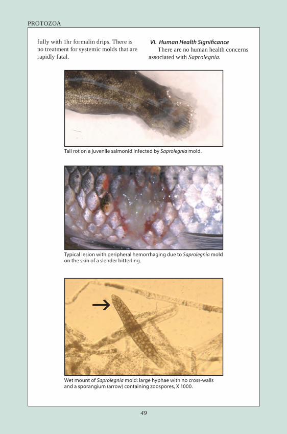
49
PROTOZOA
fully with 1hr formalin drips. There is VI. Human Health Signifcance no treatment for systemic molds that are There are no human health concernsrapidly fatal. associated with Saprolegnia.
Tail rot on a juvenile salmonid infected by Saprolegnia mold.
Typical lesion with peripheral hemorrhaging due to Saprolegnia mold on the skin of a slender bitterling.
Wet mount of Saprolegnia mold: large hyphae with no cross-walls and a sporangium (arrow) containing zoospores, X 1000.

50
PROTOZOA
Trichodiniasis
I. Causative Agent and Disease Trichodiniasis is caused by ciliated
protozoans of the family Trichodinidae in which the most common of 6 genera is Trichodina represented by over 30 species. This protozoan is probably the most frequently encountered external obligate parasite in cultured freshwater fishes worldwide. Some species in this family also parasitize fish and shellfish in the marine environment. Trichodina (40-60 um in diameter) is saucer-shaped and moves along the surface of the skin, fins and gills of fish by means of its cilia. It feeds on the detritus and other debris found on the surface of the fish using tooth-like structures called denticles. These denticles scrape the debris from the surface of the fish to the mouth of the parasite. When abundant, the scraping and movement of these organ-isms irritate the skin and gill surfaces causing hyperplasia of the epithelium. Extreme cases of hyperplasia can result in reduced gas exchange or reduced osmoregulation in the fish host. When environmental conditions are suboptimal or when fish tissues are mechanically damaged, more severe infestations may occur.
II. Host Species Protozoa of this family are found
parasitizing freshwater and marine fish species worldwide. Rainbow and steel-head trout, coho and Chinook salmon appear more susceptible than other spe-cies of salmonids. Young fish (yearlings or younger) are most susceptible. The parasite has also been reported from amphibian tadpoles.
III. Clinical Signs Fish parasitized by Trichodina often
have white patches and/or mottling of
the skin and fins. Excessive mucus is produced causing a white to bluish sheen of the skin. Fins are generally frayed and fish exhibit flashing behavior by scrap-ing their bodies against hard surfaces. If the gills are heavily infested opercular movements may be labored.
IV. Transmission Fish are infested with Trichodina
through direct transmission from fish to fish or from organisms in the water originating from a subclinically infested reservoir host. The organisms reproduce by binary fission whereby daughter organisms either attach immediately to the original host or seek a new host in the water column.
V. Diagnosis Diagnosis is easily made by micro-
scopic observation of the highly motile spinning protozoan in a wet mount prep-aration of skin scrapes or gill tissues. When abundant, the organisms may be visible gliding on the skin surface with the naked eye. Genus and species iden-tification require microscopic examina-tion of the shape and arrangement of the denticles on the chitin disc surrounding the mouth of the parasite.
VI. Prognosis for Host Trichodinid protozoa are relatively
weak pathogens when compared to other external protozoans infesting fish. The prognosis for parasitized fish is good when parasite numbers are low, and fish are not stressed. However, some of these protozoa are serious pathogens causing high fish mortality, especially in hatchery cultured species. Under these conditions external chemical treatment with formalin is necessary and effective in controlling the parasite.
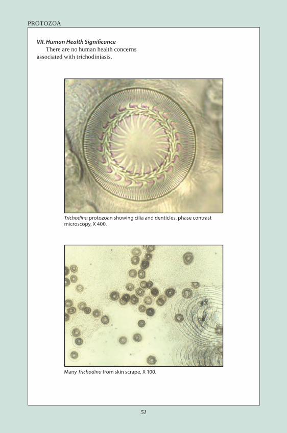
51
PROTOZOA
VII. Human Health Signifcance There are no human health concerns
associated with trichodiniasis.
Trichodina protozoan showing cilia and denticles, phase contrast microscopy, X 400.
Many Trichodina from skin scrape, X 100.

52
PROTOZOA
Trichophrya (Capriniana)
I. Causative Agent and Disease Trichophrya is a protozoan (30-40
um) in the subclass Suctoria that attach-es to the gills, skin or fins of a fish host. The protozoan has suctorial tentacles, which are used to feed on plankton and other ciliates in the water and on fish mucus and epithelial cells. When present in very large numbers, the ciliates can cause pathological changes in the gills including hyperplasia and necrosis that interfere with respiration.
II. Host Species This protozoan is commonly found
on the gills of many freshwater teleosts in North America and Eurasia.
III. Clinical Signs Nonspecific gill hyperplasia is the
principal clinical sign of infestation often accompanied by flashing behavior typical of any external parasite infesta-tion. The parasite may be observed on the gill lamellae by microscopic exami-nation. In Alaska, the occurrence of this parasite is generally incidental to other more significant etiologies.
IV. Transmission This ciliate is horizontally transmit-
ted from fish to fish. Water with high organic loads, as occurs in lake rearing, favors growth of this organism.
V. Diagnosis Diagnosis is made by observing wet
mounts of skin scrapes or gill tissues. The organism has an oval or irregularly elongated body which adheres to the gill lamella with a flattened broad at-tachment surface and the upper surface exhibits tentacles. The body of the parasite appears orange to brown. The dorsal tentacles are prominent and can
retract into the cell if disturbed. Overall, Trichophrya resembles a pincushion.
VI. Prognosis for Host Prognosis for the host is good when
infestations are light, and the fish are not otherwise stressed. When present in large numbers gill hyperplasia can interfere with respiration and predispose fish to infections by bacteria and fungi. Trichophrya in Alaska has been resistant to formalin treatments while exposure to seawater when fish are transferred to marine net pens has been more effective in eliminating the parasite.
VII. Human Health Signifcance There are no human health concerns
associated with Trichophrya.
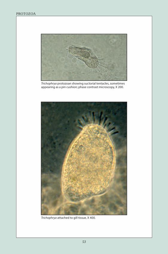
53
PROTOZOA
Trichophrya protozoan showing suctorial tentacles, sometimes appearing as a pin cushion; phase contrast microscopy, X 200.
Trichophrya attached to gill tissue, X 400.

54
PROTOZOA
White Spot Disease
I. Causative Agent and Disease White Spot Disease in marine fish
is caused by a large ciliated protozoan, Cryptocaryon irritans. The disease also occurs in freshwater fish but is caused by a distantly related larger ciliate, Ich-thyophthirius multifiliis. Infections by both parasites cause significant disease problems for marine and freshwater aquarists and commercial aquaculture worldwide. Both protozoa are obligate parasites infecting mostly the epidermis of the skin, fins and gills causing rapid mortality. Major differences between the parasites exist regarding salinity toler-ance and duration of life cycle.
II. Host Species Both parasites are distributed world-
wide and infect a broad spectrum of wild and cultured marine and freshwater fish species. In Alaska, Cryptocaryon has been reported at two large aquarium facilities parasitizing captive greenling, sculpins, wolf eels and rockfish. These infections occurred at colder seawater temperatures of 8°C suggesting a strain of the parasite better adapted to lower temperatures. In the late 1970s, Ichthy-ophthirius caused pre-spawning mortal-ity in Alaskan sockeye salmon and in 3-spine sticklebacks. More recently, Ichhas been reported in captive goldfish,pike and in feral Arctic grayling.
III. Clinical Signs Both protozoa produce coalesc-
ing white spots, nodules or patches on the surface of the skin, gills and fins. Excessive mucus production or changes in skin color may be accompanied by flashing behavior and increasing fish mortality.
IV. Transmission The parasites divide by simple
fission, are highly contagious and are transmitted horizontally fish to fish. There are three stages of the parasites: encysted trophont (feeding stage) that drops from the fish to become a tomont cyst (reproductive stage) producing tomites; tomites escape the cyst becom-ing theronts (free-swimming infective stage). Depending on the fish host, strain of parasite and water temperature, the life cycle may take from days to weeks. The freshwater life cycle is completed in 5 to 7 d at 21°C and 30 to 40 d at 10°C. Water temperatures of 32°C kill the Ich theront. About 10% of the Ich trophonts on a fish may reproduce within the host epithelium adding to the infection inten-sity. Marine white spot occurs between 15 and 30°C with an average life cycle of 1 to 2 wks, but may range from 6 d to 11 wks.
V. Diagnosis External white spots on the skin or
gills are examined by wet mounts that demonstrate the large ciliated trophont stage (48-452 um for marine form; 500-1000 um for freshwater form) with a lobed macronucleus for Cryptocaryon and one that is horseshoe-shaped for Ichthyophthirius.
VI. Prognosis for Host Wild fish generally have light
infections with negligible mortality, acquire at least partial resistance and are reservoirs for the parasites. Culture conditions allow explosive parasite re-production causing extensive and rapid fish mortality. External treatment with copper sulfate pentahydrate is the most effective therapy against the theronts but must be repeated for 3-6 wks to prevent

55
PROTOZOA
reinfection by theronts still to come VII. Human Health Signifcance from the remaining resistant trophonts There are no human health concernsembedded in surviving fish tissues and with white spot disease in fish. tomont cysts in the substrate.
Marine white spot on the dorsal skin and pectoral fns of a captive copper rockfsh (photo: Lynn Wilbur).
C
C
Left: Wet mount of rockfsh white spot lesion with two Cryptocaryon trophonts (C), X 400; Center: Histology section of parasitized rockfsh epidermis with encysted Cryptocaryon trophonts (C), X 400; Right: Wet mount of freshwater white spot lesion from pike with Ichthyophthirius trophont showing horseshoe-shaped macronucleus, X 1000.
56
PROTOZOA
X-Cell Tumors
I. Causative Agent and Disease X-cell tumors are xenomas com-
posed of enlarged host cells containing a replicating protozoan parasite belong-ing to the superphylum Alveolata and phylum Perkinsozoa as determined by molecular studies. There are at least two highly distinct X-cell clades of protozoa in the Family Xcellidae comprising the genera Gadixcellia and Xcellia. The former genus includes pseudobranchial parasites of Gadiformes (cod and blue whiting), and the other is composed of gill and epidermal X-cells from Perciformes and flatfish Pleuronecti-formes. Both genera together are sister to Perkinsus spp., pathogens of bivalve mollusks, and are the first such perkin-sids known to cause pathology in fish.
II. Host Species X-cell disease is reported worldwide
in one freshwater fish species (hardhead catfish) and various marine demersal fish including: dab, cod, rockfish, walleye pollock, flatfishes, gobiids, and icefish in the Antarctic.
III. Clinical Signs Papillomatous or fibroma-like
growths on the fins, skin or as tissue masses involving the pseudobranchs or gill filaments and lamellae. Ulceration may occur but generally all forms are limited to the dermal/epidermal layers of the skin. Molecular probes have occa-sionally shown a few X-cells in visceral organs including liver or kidney where they do not appear to proliferate.
IV. Transmission The current mode of transmission is
unknown. Infection by co-habitation and transplantation of xenomas have been unsuccessful.
V. Diagnosis The presence of apparent tissue
masses on the skin, fins or in the bran-chial cavities of demersal fish species is suggestive of X-cell tumors. Three types of X-cell xenomas are described indicating tissue and host specificities: Type 1- pseudotumors on the branchial lamellae in the common dab from the Atlantic; Type 2- lesions primarily on the pseudobranchs of cod and related species in the Atlantic and Pacific; Type 3- lesions on the skin reported for vari-ous species of flatfishes, a gobiid fishin the Pacific and in a hardhead catfish(LA, USA). Histologic/ultrastructuralfeatures of X-cells are uniform consist-ing of large rounded to polygonal cellshaving faint eosinophilic cytoplasm, acentrally located enlarged nucleus witha prominent nucleolus. Host granula-tion tissue may infiltrate the X-cell masswhere the cells may be irregular in sizeand multinucleated plasmodial X-cellshave been observed in the xenomas ofAtlantic and Pacific cod.
VI. Prognosis for Host X-cell xenomas are not reported to
be fatal, however, ulceration can lead to secondary infection by other pathogens and involvement of gill and pseudo-branch tissues seriously debilitates res-piration in fish hosts. There is evidence that host cell response may cause regres-sion of X-cell xenomas in some infected fish.
VII. Human Health Signifcance This parasite infects only fish. There
are no human health concerns associated with X-cell tumors.

57
PROTOZOA
Left: X-cell xenomas forming white plaques in and around the branchial chamber of a black rockfsh; Right: close-up view of xenomas causing white thickened gill tissues of a dusky rockfsh.
E
X
Left: Ulcerated X-cell xenomas on skin of Pacifc cod (photo: Ruth Fairell); Center: Histologic section of cod lesion with epidermis (E) and focus of degenerative X-cells (X) in dermis beneath, X 200; Right: Multinucleated plasmodial X-cells (P), X 1000.
P

58
CNIDARIA
Ceratonova (Ceratomyxa) shasta
I. Causative Agent and Disease The parasite Ceratonova shasta is
not a protozoan but a metazoan in the class Myxosporea in the phylum Cni-daria (anemones, jellyfish, corals) based on molecular studies and the feature of discharging cells (cnidocytes) known as polar capsules. The parasite produces crescent shaped spores 14-23 um long by 6-8 um wide at the suture line. A single spore contains two refractile polar capsules, each with an extensible coiled filament. Vegetative trophozoites and spores produce necrotic lesions within various tissues of salmonid fishes but the parasite has a tropism for the gastroin-testinal tract, especially the intestine. High mortality may occur in susceptible juvenile fish and the parasite contributes to significant pre-spawning mortality of infected adult salmon. Depending on the host species and stock, natural exposure to the parasite may allow some fish populations to develop resistance to infection and severity of the disease. Ceratonova occurs seasonally (May to November) becoming more intense when water temperatures reach or exceed 10°C.
II. Host Species This organism parasitizes several
different species of freshwater and ana-dromous salmonids and is restricted to the Pacific Northwest (PNW) and British Columbia. Ceratonova shasta is present in wild adult chum, coho and sockeye salmon, rainbow trout and Dolly Varden in Alaska within several south central and interior drainages including; Yetna, Yukon, Naknek, Wood, King Salmon, Togiak and Sushana Rivers and Lower Talarik, Mortenson and Russell Creeks. Clinical signs of disease, except for white gill nodules in sockeye, have been
absent in parasitized wild fish nor has the parasite been found in any hatchery stocks of Alaskan salmonids.
III. Clinical Signs Parasitized fish typically appear
darkened in color with swollen or hem-orrhaged vents and abdomens distended by ascites. Although lesions are variable by age and fish species, the entire diges-tive tract may be affected with granu-lomas and abscesses (boils) causing tissue necrosis that may spread to major visceral organs and skeletal muscula-ture. These lesions contain developing multicellular trophozoites and spores. Each trophozoite forms a pansporoblast usually containing two spores.
IV. Transmission Ceratonova shasta is transmitted
to fish through the gills by infectious actinosporean tetractinomyxon stages in the water that are shed by parasitized freshwater polychaete worms of the species Manayunkia speciosa that serve as the alternate host. The worms become parasitized by ingestion of mature spores released by parasitized live or decom-posing fish hosts. However, unlike other myxozoans, the parasite develops within the alternate host epidermis rather than within the intestinal epithelium.
V. Diagnosis Tissue lesions or intestinal scrapes
are examined for spores having the typi-cal size and morphology of this parasite. Identity is confirmed with fluorescein or enzyme conjugated antibody tests and by PCR specific for Ceratonova shasta.
VI. Prognosis for Host Depending on the fish species, stock
and water temperature, prognosis may
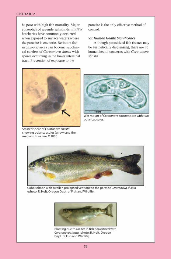
59
CNIDARIA
be poor with high fish mortality. Major epizootics of juvenile salmonids in PNW hatcheries have commonly occurred when exposed to surface waters where the parasite is enzootic. Resistant fish in enzootic areas can become subclini-cal carriers of Ceratonova shasta with spores occurring in the lower intestinal tract. Prevention of exposure to the
parasite is the only effective method of control.
VII. Human Health Signifcance Although parasitized fish tissues may
be aesthetically displeasing, there are no human health concerns with Ceratonova shasta.
Stained spore of Ceratonova shasta showing polar capsules (arrow) and the medial suture line, X 1000.
10 um
Wet mount of Ceratonova shasta spore with two polar capsules.
Coho salmon with swollen prolapsed vent due to the parasite Ceratonova shasta (photo: R. Holt, Oregon Dept. of Fish and Wildlife).
Bloating due to ascites in fsh parasitized with Ceratonova shasta (photo: R. Holt, Oregon Dept. of Fish and Wildlife).

60
CNIDARIA
Henneguya
I. Causative Agent and Disease Henneguya is not a protozoan but a
metazoan in the class Myxosporea in the phylum Cnidaria (anemones, jellyfish, corals) based on molecular studies and the feature of discharging cells (cnido-cytes) known as polar capsules. The ge-nus has about 120 different species, some are very host and tissue specific. The parasite is found in fish as an ovoid spore (11 x 9 um) with two anterior polar cap-sules and two long caudal appendages (26-40 um). The most common species in Alaska is H. salminicola. The spores of this parasite occur in the muscle and under the skin of Pacific salmon causing a condition known as “milky flesh” dis-ease because of the creamy white fluid containing spores that oozes from the cysts (pansporoblasts) during filleting. It is also known as “tapioca” disease from the many small round spore containing cysts in the flesh.
II. Host Species Many species of anadromous, marine
and freshwater fishes are susceptible to the several different species of Henne-guya distributed worldwide.
III. Clinical Signs Fish infected with Henneguya have
numerous white pansporoblasts (cysts) in the target tissues that may contain thousands of spores.
IV. Transmission Henneguya salminicola is transmit-
ted by an infectious stage in freshwa-ter. Pacific salmon become infected as juveniles and the parasites reach the muscle via the circulatory system passing through several developmental stages that eventually become spores. The spores are enclosed in a visible pan-
sporoblast or cyst formed of host tissue. When post-spawned salmon decompose, the cysts rupture and release spores into the water where they likely are ingested by an invertebrate alternate host such as a tubificid worm. Infectious stages (ac-tinospores) for juvenile salmon develop in the invertebrate host and are released into the water column.
V. Diagnosis White cysts in the flesh are examined
microscopically for the typical 2-tailed spores characteristic of Henneguya. The condition can also be diagnosed by histological examination of tissues to verify presence of the parasite. Species is determined by further spore morphol-ogy.
VI. Prognosis for Host Many fish parasites are well tolerated
depending on the target tissue affected. Fish mortality from Henneguya target-ing the flesh has not been reported.
VII. Human Health Signifcance Although the cysts in the flesh are
unsightly when present in large num-bers, there are no human health concerns associated with Henneguya.
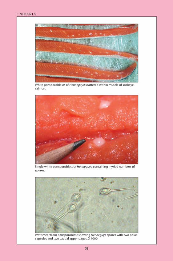
61
CNIDARIA
White pansporoblasts of Henneguya scattered within muscle of sockeye salmon.
Single white pansporoblast of Henneguya containing myriad numbers of spores.
Wet smear from pansporoblast showing Henneguya spores with two polar capsules and two caudal appendages, X 1000.

62
CNIDARIA
Kudoa
I. Causative Agent and Disease Kudoa species are metazoans in
the class Myxosporea in the phylum Cnidaria (anemones, jellyfish, corals) based on molecular studies and the feature of discharging cells (cnidocytes) known as polar capsules. The genus has over 90 different species among which K. thyrsites is most commonly reportedin over 30 marine and estuarine fishspecies worldwide. Many Kudoa speciesinfect skeletal muscle tissues producingpseudocysts containing stellate-shapedspores with 4 or more valves and polarcapsules. Infections produce inflam-mation but no significant disease. Highintensities of Kudoa cause unfit productquality from post-mortem myoliquefac-tion of fish flesh (soft flesh syndrome)due to release of proteolytic enzymesfrom the parasite.
II. Host Species Many species of marine and
estuarine fishes from several families are reported hosts of Kudoa species worldwide. In Alaska, Kudoa has rarely occurred in coho, Chinook and pink salmon, and in Pacific halibut. Sexually mature fish appear to be more suscep-tible to infection.
III. Clinical Signs Fish infected with Kudoa have white
pseudocysts in the skeletal muscle fibers containing hundreds of spores. Muscle tissue may exhibit liquefaction with varying intensities of the parasite that may go visually unnoticed. Heavily infected fish exhibit soft flesh when held on ice for 3-6 d or if cold smoked.
IV. Transmission Kudoa is likely transmitted by an
infectious stage from a marine inver-
tebrate as described for other myxo-sporeans. After infection, the parasites reach the skeletal muscle by the circula-tory system passing through several developmental stages that eventually become spores enclosed in a visible pseudocyst within the host skeletal muscle fiber. When the fish host dies and decomposes, the pseudocysts rupture re-leasing spores into the water where they likely are ingested by the invertebrate alternate host, probably an oligochaete or polychaete worm. Infectious stages (actinospores) for fish develop in the invertebrate host and are released into the water column.
V. Diagnosis White pseudocysts or inflamma-
tory foci in the flesh are examined in wet mounts for spores having 4 or more valves with polar capsules character-istic of Kudoa. Species is determined by further spore morphology and PCR. Due to its very wide geographic and host distribution, the reports of K. thyrsites from around the world may represent several indistinguishable species. Also confounding a diagnosis; post-mortem liquefaction may occur from developing parasite stages, prior to the visual pres-ence of mature pseudocysts and spores.
VI. Prognosis for Host Kudoa parasitism is well tolerated
and fish mortality has not been reported. However, there are species parasitizing the heart muscle which may negatively affect cardiac performance.
VII. Human Health Signifcance Although the pseudocysts and/
or the myoliquefaction of the flesh are unacceptable for consumption, there are no human health concerns reported for
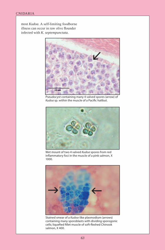
63
CNIDARIA
most Kudoa. A self-limiting foodborne illness can occur in raw olive flounder infected with K. septempunctata.
25 um
Pseudocyst containing many 4 valved spores (arrow) of Kudoa sp. within the muscle of a Pacifc halibut.
Wet mount of two 4 valved Kudoa spores from red infammatory foci in the muscle of a pink salmon, X 1000.
Stained smear of a Kudoa-like plasmodium (arrows) containing many sporoblasts with dividing sporogonic cells; liquefed fllet muscle of soft-feshed Chinook salmon, X 400.
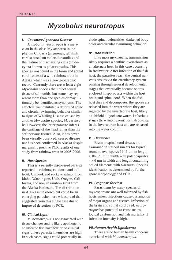
64
CNIDARIA
Myxobolus neurotropus
I. Causative Agent and Disease Myxobolus neurotropus is a meta-
zoan in the class Myxosporea in the phylum Cnidaria (anemones, jellyfish, corals) based on molecular studies and the feature of discharging cells (cnido-cytes) known as polar capsules. This species was found in the brain and spinal cord tissues of a wild rainbow trout in Alaska which was a new geographic record. Currently there are at least eight Myxobolus species that infect neural tissue of salmonids, but some may rep-resent more than one species or may ul-timately be identified as synonyms. The affected trout exhibited a deformed spine and circular swimming behavior similar to signs of Whirling Disease caused by another Myxobolus species, M. cerebra-lis. However, the latter parasite infects the cartilage of the head rather than the soft nervous tissues. Also, it has never been visually observed, caused disease nor has been confirmed in Alaska despite marginally positive PCR results of one study from rainbow trout in 2005-2006.
II. Host Species This is a recently discovered parasite
reported in rainbow, cutthroat and bull trout, Chinook and sockeye salmon from Idaho, Washington, Utah, Oregon, Cali-fornia, and now in rainbow trout from the Alaska Peninsula. The distribution in Alaska is unknown but could be an emerging parasite more widespread than suggested from this single case due to improved detection by PCR.
III. Clinical Signs M. neurotropus is not associated with
tissue changes and is likely apathogenic so infected fish have few or no clinical signs unless parasite intensities are high. In such cases, signs could potentially in-
clude spinal deformities, darkened body color and circular swimming behavior.
IV. Transmission Like most myxozoans, transmission
likely requires a benthic invertebrate as an alternate host, in this case occurring in freshwater. After infection of the fish host, the parasites reach the central ner-vous tissues via the circulatory system passing through several developmental stages that eventually become spores enclosed in sporocysts within the host brain and spinal cord. When the fish host dies and decomposes, the spores are released into the water where they are ingested by the invertebrate host, likely a tubificid oligochaete worm. Infectious stages (triactinomyxons) for fish develop in the invertebrate host and are released into the water column.
V. Diagnosis Brain or spinal cord tissues are
examined in stained smears for typical round to oval spores 11-13 um in length x 10-12 um in width with polar capsules 4 x 6 um in width and length containing coiled filaments with 6-8 turns. Species identification is determined by further spore morphology and PCR.
VI. Prognosis for Host Parasitisms by many species of
myxosporeans are well tolerated by fish hosts unless infections cause dysfunction of major organs and tissues. Infection of the brain and spinal cord by M. neuro-tropus has potential to cause neuro-logical dysfunction and fish mortality if infection intensity is high.
VII. Human Health Signifcance There are no human health concerns
associated with M. neurotropus.

65
CNIDARIA
Rainbow trout infected with Myxobolus neurotropus from Margo Creek on the Alaska Peninsula - darkened body coloration and spinal deformity.
Wet mount of M. neurotropus unstained spores from the spinal cord of the rainbow trout above, X 200. Inset scale bar = 10 um (from Bentz et al. 2012).
Malachite green-stained smear of M. neurotropus spores with oviform polar capsules of nearly equal size, X 1000.
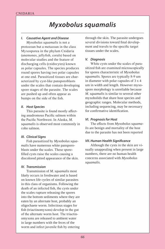
66
CNIDARIA
Myxobolus squamalis
I. Causative Agent and Disease Myxobolus squamalis is not a
protozoan but a metazoan in the class Myxosporea in the phylum Cnidaria (anemones, jellyfish, corals) based on molecular studies and the feature of discharging cells (cnidocytes) known as polar capsules. The species produces round spores having two polar capsules at one end. Parasitized tissues are char-acterized by cyst-like pansporoblasts under the scales that contain developing spore stages of the parasite. The scales are pushed up and often appear as bumps on the side of the fish.
II. Host Species This parasite is found mostly affect-
ing anadromous Pacific salmon within the Pacific Northwest. In Alaska, M. squamalis is observed most commonly in coho salmon.
III. Clinical Signs Fish parasitized by Myxobolus squa-
malis have numerous white pansporo-blasts under the scales. These spore-filled cysts raise the scales causing a discolored pitted appearance of the skin.
IV. Transmission Transmission of M. squamalis most
likely occurs in freshwater and is based on known life cycles of similar parasites in this class of organisms. Following the death of an infected fish, the cysts under the scales rupture releasing the spores into the bottom sediments where they are eaten by an alternate host, probably an oligochaete worm. Infectious stages for fish (triactinomyxons) develop in the gut of the alternate worm host. The triactin-omyxons are released to ambient water in large numbers with the feces of the worm and infect juvenile fish by entering
through the skin. The parasite undergoes several divisions toward final develop-ment and travels to the specific target tissues under the scales.
V. Diagnosis White cysts under the scales of para-
sitized fish are examined microscopically for spores characteristic of Myxobolus squamalis. Spores are typically 8-9 um in diameter with polar capsules of 3 x 4 um in width and length. However myxo-spore morphology is unreliable because M. squamalis is similar to several othermyxobolids that share host species andgeographic ranges. Molecular methods,including sequencing, may be necessaryfor confirmative identification.
VI. Prognosis for Host The effects from Myxobolus squama-
lis are benign and mortality of the host due to the parasite has not been reported
VII. Human Health Signifcance Although the cysts in the skin are vi-
sually unappealing when present in large numbers, there are no human health concerns associated with Myxobolus squamalis.

67
CNIDARIA
Skin lesions typical of infection by Myxobolus squamalis in a coho salmon.
Wet mount (Left) and stained smear (Right) of Myxobolus spores with polar flaments discharged from the polar capsules (cnidocytes), X 1000.
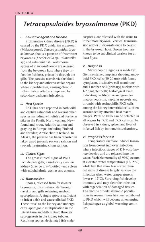
68
CNIDARIA
Tetracapsuloides bryosalmonae (PKD)
I. Causative Agent and Disease Proliferative kidney disease (PKD) is
caused by the PKX cnidarian myxozoan (Malacosporea), Tetracapsuloides bryo-salmonae, that is a parasite of freshwater bryozoans (Fredericella sp., Plumatella sp.) and salmonid fish. Waterborne spores of T. bryosalmonae are released from the bryozoan host where they in-fect the fish host, primarily through the gills. The parasite travels via the blood to the kidney and other vascular organs where it proliferates, causing chronic inflammation often accompanied by secondary pathogen infections.
II. Host Species PKD has been reported in both wild
and captive salmonids and several other species including whitefish and northern pike in the Pacific Northwest and New-foundland; trout, Atlantic salmon and grayling in Europe, including Finland and Sweden; Arctic char in Iceland. In Alaska, the parasite has been reported in lake-reared juvenile sockeye salmon and two adult returning chum salmon.
III. Clinical Signs The gross clinical signs of PKD
include pale gills, a uniformly swollen kidney (may be gray/mottled) and spleen with exophthalmia, ascites and anemia.
IV. Transmission Spores, released from freshwater
bryozoans, infect salmonids through the skin and gills releasing ameboid sporoplasms. A single spore is sufficient to infect a fish and cause clinical PKD. These travel to the kidney and undergo extra-sporogonic multiplication in the interstitium and differentiate through sporogenesis in the kidney tubules. Resulting spores, designated fish mala-
cospores, are released with the urine to infect more bryozoa. Vertical transmis-sion allows T. bryosalmonae to persist in the bryozoan host. Brown trout are known to be subclinical carriers for at least 5 yrs.
V. Diagnosis Microscopic diagnosis is made by:
Giemsa-stained imprints showing amoe-boid PKX cells (10-20 um) with foamy cytoplasm, distinctive cell membrane and 1 mother cell (primary) nucleus with 1-7 daughter cells; histological exam indicating proliferative and granulo-matous nephritis, vascular necrosis and thrombi with eosinophilic PKX cells among the kidney interstitial cells, often surrounded by attached host macro-phages. Parasite DNA can be detected in all organs by PCR and PKX cells can be observed in kidney, spleen and liver of infected fish by immunohistochemistry.
VI. Prognosis for Host Temperature increase induces transi-
tion from covert into overt infection where infectious stages of T. bryosalmo-nae develop and are released into the water. Variable mortality (5-90%) occurs at elevated water temperatures (12-15°C) while fish that show less severe clini-cal signs of disease largely survive the infection when water temperature is lower (< 12°C). Surviving fish develop immunity and may clear the infection with regeneration of damaged tissues. The decline of wild salmonid popula-tions in several rivers has been attributed to PKD which will become an emerging fish pathogen as global warming contin-ues.
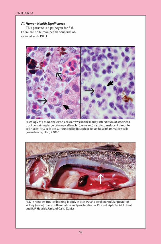
69
CNIDARIA
VII. Human Health Signifcance
This parasite is a pathogen for fish.There are no human health concerns as-
Histology of eosinophilic PKX cells (arrows) in the kidney interstitium of steelhead
sociated with PKD.
trout containing large primary cell nuclei (dense red) next to translucent daughter cell nuclei. PKX cells are surrounded by basophilic (blue) host infammatory cells (arrowheads); H&E, X 1000.
A
PKD in rainbow trout exhibiting bloody ascites (A) and swollen nodular posterior kidney (arrow) due to infammation and proliferation of PKX cells (photo: M. L. Kent and R. P. Hedrick, Univ. of Calif., Davis).
70
HELMINTHS
Acanthocephalans (Spiny-Headed Worms)
I. Causative Agent and Disease Acanthocephalans are endoparasitic
worms characterized by a retractable proboscis armed with rows of hooks used to attach to the intestines of fish. Many genera have been described as adults in the intestines of fish while some larval forms have also been identified in the viscera. Genera commonly found in Alaskan fishes are Neoechinorhynchus, Acanthocephalus and Corynosoma. Gut infestation by numerous acanthocepha-lans can cause fibrotic nodules on the surface of the intestine. The intestine may become inflamed with the destruc-tion of intestinal villi and resulting necrotic and degenerative changes in mucosal epithelium. Intestinal absorptive efficiency may be compromised leading to decreased growth and emaciation. Acanthocephalans occasionally perfo-rate the intestinal wall which can lead to peritonitis and death of the host.
II. Host Species Acanthocephalans have been found
in both marine and freshwater fishes worldwide.
III. Clinical Signs Parasitized fish may be emaciated
with inflamed intestinal tracts and tissue necrosis in areas where worms are at-tached to the intestinal wall.
IV. Transmission Acanthocephalans require a ver-
tebrate animal as a definitive host and arthropods as an intermediate host. Fish usually are the final host for aquatic acanthocephalans and microcrustaceans (amphipod, copepod, isopod or ostracod) are generally the intermediate host. Inter-mediate hosts are infected by eating eggs eliminated in the feces of parasitized fish.
An egg will hatch in the intermediate host releasing an acanthor that penetrates the gut and develops into an acanthella/ cystocanth. The life cycle is complete when a fish eats a parasitized microcrus-tacean and the adult worm develops in the alimentary tract of the fish host. In some cases, fish are the second interme-diate host as well as the final host.
V. Diagnosis Diagnosis is made by the visual de-
tection of adult acanthocephalans in the intestine or invasive larvae in the body cavity of a parasitized fish. The shape of the proboscis, the arrangement and the number of proboscis hooks are important characteristics used to definitively iden-tify the species of acanthocephalan. PCR has been useful in confirming species that has resulted in changing taxonomy.
VI. Prognosis for Host The principal effects on the final host
can include mechanical damage to the intestinal wall and emaciation. Signifi-cant fish mortality or emaciation due to infestation by acanthocephalans are rare unless the worms are present in large numbers.
VII. Human Health Signifcance There are no human health concerns
associated with these parasites.

71
HELMINTHS
Acanthocephalan worm of the genus Pomphorhynchus.
Pomphorhynchus: a higher magnifcation of the spiny head (proboscis) showing numerous hooks, X 200.
Highly armed proboscis of Echinorhynchus.
72
HELMINTHS
I
Anisakid Larvae
Causative Agent and Disease The larval form (third stage juvenile)
of several nematode species within the subfamily Anisakinae are found coiled in the flesh and viscera of parasitized fish. Common genera include Anisakis, Paranisakis, Porrocaecum, Pseudoter-ranova and Contracaecum. The larvae are relatively non-pathogenic to the fish host, although visceral adhesions from migrating larval worms do occur.
II. Host Species Anisakid larvae are common in
marine and anadromous fishes worldwide and have also been reported in squid and cuttlefish. In Alaska, among other fish species, these worms are commonly found in Pacific salmon and cod, walleye pollock, Pacific halibut and Pacific her-ring (herring worm).
III. Clinical Signs Parasitized fish contain reddish or
white, tightly coiled larval worms found most commonly in skeletal muscle and visceral organs. These areas may exhibit mild inflammation, encapsulation and/or granuloma formation. Visceral adhesion may occur in fish when many juvenile worms are present in the visceral cavity. This condition causes production of fibrous connective tissue by the fish host in response to irritation from migrating worms.
IV. Transmission Anisakid worms have a complex
life cycle involving several hosts. Eggs eliminated in the feces from the final host hatch in the sea where the larvae are consumed by crustaceans (usually Euphausids), which in turn are eaten by fish. The larva burrows into the gut or flesh of the fish and encysts until its life
cycle is completed when ingested by the final host, usually a marine mammal but sometimes a bird or fish. Incidental para-sitism of a human host usually results in re-encystment of the juvenile worm. The nematode matures in the gut of the marine mammal host and releases eggs into the sea to continue the cycle. Some anisakid larvae can also be transmitted from fish to fish through predation.
V. Diagnosis Presumptive diagnosis is made by
visual examination of the body cavity, organs and flesh of the fish for typi-cal coiled worms. Examination under a dissecting microscope can verify the identity of the larval nematodes based on morphological characteristics. PCR has been useful in confirming species that has resulted in changing taxonomy.
VI. Prognosis for Host Prognosis for the fish host is good. In
most cases the worms are well tolerated and there have been few reported cases of fish mortality due to juvenile anisakid parasitism.
VII. Human Health Signifcance Anisakiasis in humans can be ac-
quired by eating viable worms in raw or partially cooked fish. The Food and Drug Administration recommends cooking fish at 67ºC for 5 minutes or freezing at -20ºC for at least 7 d before eating to killjuvenile anisakid worms.

73
HELMINTHS
Anisakis Life Cycle
Humans contract by consumption of raw or undercooked Marine mammal
fish (abnormal host) final host for the adult nematode Anisakis
Eggs and second
Fish and Squid
stage larva
Free swimming larva ingested by crustaceans (Euphausids)
Anisakis third stage juvenile worms tightly coiled in liver.
Anisakis third stage juvenile worm being pulled from salmonid muscle.

74
HELMINTHS
Philometra
I. Causative Agent and Disease Philometra is a nematode parasi-
tizing the body cavities or tissues of fish. Larval stages of this worm migrate to the final resting sites in the subcutaneous tissues (fins, head, and body) or body cavities of predatory fish. The migration of the parasite within the host can result in inflammation of visceral organs, mechanical damage of blood vessels with hemorrhaging and destruction of skeletal joints resulting in poor growth and emaciation.
II. Host Species Many species of marine and fresh-
water fish, including salmonids, are susceptible to this parasite that is found worldwide.
III. Clinical Signs Nodules under the flesh containing
juvenile or adult worms cause raised scales or are visible between the fin rays of the fish host. Larger nodules contain gravid females that eventually extrude through the skin and disintegrate to release live larvae. This is followed by complete healing of the host flesh leav-ing little sign of previous infestation.
IV. Transmission Philometra has a two-host life cycle.
Larval worms are transmitted through an intermediate host (copepods) to the fi-nal fish host. Predatory fish may acquire the parasite by eating infested cope-pods or forage fish that have preyed on infested copepods. In skin infestations the much larger female parasites excyst and burst releasing larvae into the water to be ingested by copepods where the larvae undergo a series of molts. When parasitizing a body cavity, larvae are released through the gut with the feces
of the host or the female migrates to the skin surface to release larvae.
V. Diagnosis Diagnosis is made by observation of
typical Philometra worms in fish host body cavities or subcutaneous tissues, particularly the fins, snout, and head or areas of raised scales. Dissection of nodules expose the long, smooth, fili-form worms characteristic of the genera. Worms are usually red in color and the immensely larger females contain live larvae and burst easily when placed in water.
VI. Prognosis for Host Prognosis for the host is dependent
on the degree of infestation and other environmental stressors that may be present. Generally, Philometra is well tolerated causing no significant harm to fish hosts.
VII. Human Health Signifcance Philometra is not of human health
concern.
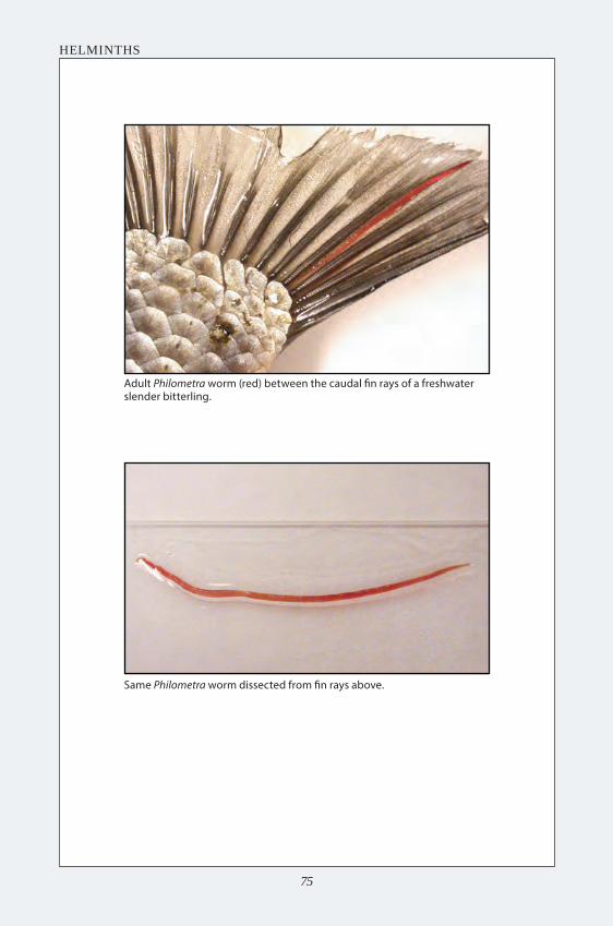
75
HELMINTHS
Adult Philometra worm (red) between the caudal fn rays of a freshwater slender bitterling.
Same Philometra worm dissected from fn rays above.
76
HELMINTHS
Philonema
I. Causative Agent and Disease Philonema (oncorhynchi and agu-
bernaculum) is a nematode (roundworm) found in the visceral cavity of fish that rarely migrates to the musculature. Larval, sub-adult and adult worms (17 mm to 86 mm) can be present. The worms generally do not cause significant pathology in the fish host but a condition known as visceral adhesion occasion-ally occurs in severely parasitized fish. Visceral adhesion is characterized by the production of fibrous connective tissue by the fish host in response to tissue ir-ritation from migrating worms. In severe cases, internal organs are bound together by the scar tissue.
II. Host Species The parasite occurs in all anadro-
mous Pacific salmon, resident trout and char in upper North America and the Northern Pacific including Russia and Japan.
III. Clinical Signs Usually there are no clinical signs of
nematode infestation. Highly parasitized fish may have extensive visceral adhe-sion discovered only by necropsy.
IV. Transmission Juvenile fish acquire the parasites
in freshwater but the adult worms may develop while the fish are at sea. The life cycle includes live larvae released from gravid female worms extruded with fish eggs from adult spawning fish. The larval worms infest a freshwater cope-pod where they develop into third stage larvae that are infectious for juvenile salmonids. Fish are infested by eating the parasitized copepods and the larvae
migrate into the body cavity where worms molt into sub-adults and eventu-ally adults that produce more larvae.
V. Diagnosis Diagnosis is made by necropsy of
diseased fish and the visual identification of the nematode. Philonema is a filiform worm having a rounded anterior end and a posterior tail tapering into a sharp point. These and other morphological features are used to identify the worm.
VI. Prognosis for Host Prognosis for the host is good unless
infestation is severe or other stressors further debilitate the fish. Severe parasitism can cause visceral adhesions that interfere with spawning ability and may cause serious organ dysfunction. However, the literature indicates this condition is probably transitory and does not cause significant fish mortality.
VII. Human Health Signifcance Philonema is not infectious for hu-
mans.

77
HELMINTHS
Many juvenile Philonema found in the visceral cavity of an adult coho salmon, X 200.
Juvenile Philonema nematode with posterior tail tapering into a fne point, X 400.
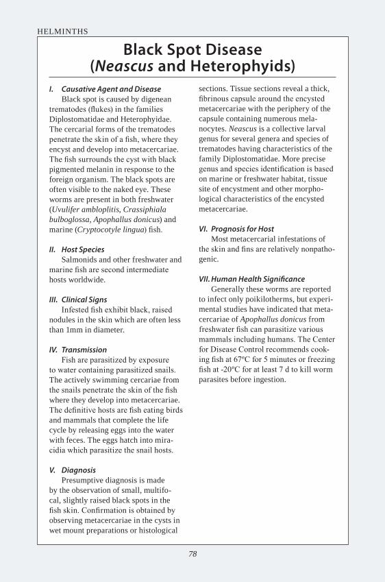
78
HELMINTHS
Black Spot Disease (Neascus and Heterophyids)
I. Causative Agent and Disease Black spot is caused by digenean
trematodes (flukes) in the families Diplostomatidae and Heterophyidae. The cercarial forms of the trematodes penetrate the skin of a fish, where they encyst and develop into metacercariae. The fish surrounds the cyst with black pigmented melanin in response to the foreign organism. The black spots are often visible to the naked eye. These worms are present in both freshwater (Uvulifer ambloplitis, Crassiphiala bulboglossa, Apophallus donicus) and marine (Cryptocotyle lingua) fish.
II. Host Species Salmonids and other freshwater and
marine fish are second intermediate hosts worldwide.
III. Clinical Signs Infested fish exhibit black, raised
nodules in the skin which are often less than 1mm in diameter.
IV. Transmission Fish are parasitized by exposure
to water containing parasitized snails. The actively swimming cercariae from the snails penetrate the skin of the fish where they develop into metacercariae. The definitive hosts are fish eating birds and mammals that complete the life cycle by releasing eggs into the water with feces. The eggs hatch into mira-cidia which parasitize the snail hosts.
V. Diagnosis Presumptive diagnosis is made
by the observation of small, multifo-cal, slightly raised black spots in the fish skin. Confirmation is obtained by observing metacercariae in the cysts in wet mount preparations or histological
sections. Tissue sections reveal a thick, fibrinous capsule around the encysted metacercariae with the periphery of the capsule containing numerous mela-nocytes. Neascus is a collective larval genus for several genera and species of trematodes having characteristics of the family Diplostomatidae. More precise genus and species identification is based on marine or freshwater habitat, tissue site of encystment and other morpho-logical characteristics of the encysted metacercariae.
VI. Prognosis for Host Most metacercarial infestations of
the skin and fins are relatively nonpatho-genic.
VII. Human Health Signifcance Generally these worms are reported
to infect only poikilotherms, but experi-mental studies have indicated that meta-cercariae of Apophallus donicus from freshwater fish can parasitize various mammals including humans. The Center for Disease Control recommends cook-ing fish at 67ºC for 5 minutes or freezing fish at -20ºC for at least 7 d to kill worm parasites before ingestion.

79
HELMINTHS
Typical black spots composed of melanin that surround encysted metacercariae of the larval genus Neascus in an Arctic grayling.
Marine form of black-spot heterophyid circumscribed by blue-green pigment on the caudal fn of a Bering cisco (photo: B. Collyard, ADF&G).
Encysted metacercaria of Neascus, X 1000.

80
HELMINTHS
Encysted Digenean Metacercariae (white and yellow grubs)
I. Causative Agent and Disease White and yellow grubs, caused by
several species of digenean trematodes (flukes) have cercarial forms that pene-trate the skin of fish, where they encyst and develop into metacercariae, appear-ing as white or yellow nodules in the flesh. The most common worms present in freshwater are Clinostomum spp. (yellow grub) and Posthodiplostomum minimum (white grub). In the marine ecosystem there are many species of trematodes that produce white and yel-low cysts in fish.
II. Host Species Many North American fish species
are second intermediate hosts for the freshwater white and yellow grubs. Sim-ilar or the same species occur in Europe, South America and Asia. Also, numer-ous species of marine fish worldwide can be parasitized with metacercariae that produce white and yellow cysts.
III. Clinical Signs Infested fish exhibit white or yellow
nodules in the flesh that can vary greatly in size.
IV. Transmission Fish are infested by exposure to
fresh or seawater containing parasit-ized snails or other invertebrates. The actively swimming cercariae shed by the invertebrate penetrate the skin of the fish and/or travel to other target tissues where they develop into metacercariae. The definitive hosts for freshwater white and yellow grubs are fish eating birds where adult worms occur in the mouth, throat or intestine. The life cycle is com-pleted by release of eggs into the water with saliva or feces. The eggs hatch into miracidia which parasitize the inver-
tebrate hosts. The inclusion of many other trematode species, especially in the marine environment, involves more hosts including birds and mammals as final hosts while fish can be a second intermediate host, paratenic host or the final host.
V. Diagnosis Presumptive diagnosis is by the
observation of white or yellow cysts containing metacercariae in the skin, fins or viscera of fish. Freshwater white grubs are 1-1.5 mm and yellow grubs are 3-8 mm. The smaller white grub encystsin visceral organs, especially kidney,liver and heart. The yellow grub is morecommon in skin, fins and musculature.In marine fish the nodules may varyconsiderably in location and size due tothe many different trematode species.Confirmation is by observing metacer-cariae in the cysts of wet mount prepara-tions or histological sections. Tetracotyleis a collective larval genus for severalgenera and species of trematodes havingcharacteristics of the family Strigeidae.More precise genus and species identifi-cation is based on tissue site of encyst-ment and morphological characteristicsof the metacercariae. DNA sequencinghas also become an important methodfor identifying these parasites
VI. Prognosis for Host Most metacercarial infestations are
relatively non-pathogenic. Encystment in visceral organs may cause significant pathology and fish mortality when infes-tation intensity is high. Grub-infested flesh also produces inferior product quality in commercial and farmed fish.
VII. Human Health Signifcance Generally, these worms are consid-

81
HELMINTHS
ered to infect only poikilotherms, but there have been cases in Japan, Korea and SE Asia where metacercariae of C. complanatum have parasitized humans from eating affected fish. The Center for
Disease Control recommends cooking fish at 67ºC for 5 minutes or freezing fish at -20ºC for at least 7 d to kill worm parasites before ingestion.
Left: Yellow grub-like encysted metacercariae (arrow) in the pericardial cavity of a sheefsh; Right: Strigeid-type trematode larva teased from a cyst on the left, resembles a Tetracotyle of an Ichthyocotylurus sp., X 10.
Left: Yellow grub-like metacercariae encysted in the peritoneal lining and musculature of a rainbow trout; Right: Metacercariae encysted in the skin of a starry founder causing numerous raised nodules.

82
HELMINTHS
Larval Diplostomulum of the Eye
I. Causative Agent and Disease Eye fluke is caused by digenean
larval trematodes of the genus Diplos-tomulum that parasitize the eye of many fish species. A common trematode found in the lens is D. spathaceum while other species are found in the vitreous cham-ber of the eye. The parasites can remain in the eye for a long time often resulting in cataracts and blindness in the fish host.
II. Host Species Many salmonids and other fresh
water fish are susceptible throughout North America and Europe.
III. Clinical Signs The fish may have cataracts and the
eye appears opaque.
IV. Transmission As with other digenean trematodes,
the fish becomes parasitized horizontally through the water from infested snails. The invasive cercariae from a snail (first intermediate host) penetrate the fish (second intermediate host), usually through the skin, and migrate to the eye where they develop into the metacer-carial form. The life cycle is completed when the fish host is eaten by a piscivo-rous bird where the adult fluke develops in the gut and releases eggs into the water.
V. Diagnosis This condition is diagnosed by wet
mount observation of metacercariae in the lens or vitreous humor of the eye in a parasitized fish. Typical metacercariae can also be identified using histological methods.
VI. Prognosis for Host If parasitized bilaterally, complete
blindness may result, and the fish host will probably die from predation or in-ability to find food. When only one eye is parasitized, the host fish may survive for an indefinite period of time.
VII. Human Health Signifcance There are no human health concerns
associated with this parasite.
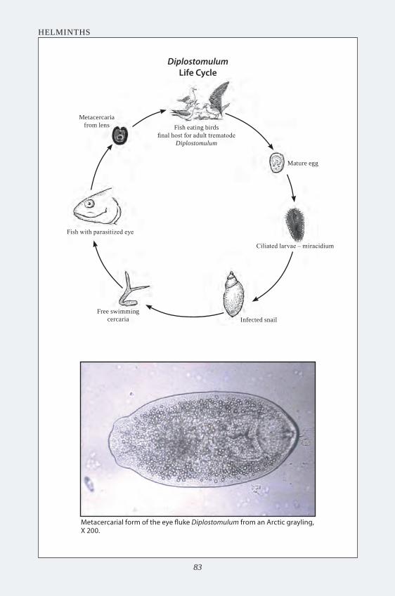
83
HELMINTHS
Diplostomulum Life Cycle
Metacercaria from lens Fish eating birds
final host for adult trematode Diplostomulum
Mature egg
Fish with parasitized eye
Infected snail Free swimming
cercaria
Ciliated larvae – miracidium
Metacercarial form of the eye fuke Diplostomulum from an Arctic grayling, X 200.

84
HELMINTHS
Gyrodactylus and Dactylogyrus
I. Causative Agent and Disease The genus Gyrodactylus contains
many species but G. salmonis is a com-mon parasite of salmonids in North America. This small (0.2 mm) monoge-nean fluke attaches to gills, fins and skin epithelium using an attachment organ or opisthaptor armed with a pair of large hooks and 16 marginal hooklets. The head of the worm is bi-lobed, lacks eye spots and the worm produces live young. Heavy infestations by the parasite can result in destruction of the gills or skin epithelium due to mechanical damage caused by the attachment organ. The genus Dactylogyrus is found on the gills of mostly cyprinid fishes and is also very small (0.3 mm). Dactylogyrus is recognized by a four-lobed head with four eye spots and produces eggs. The opisthaptor consists of one conspicuous pair of large hooks and up to 12 smaller hooklets. When the worm is present in large numbers, gill hyperplasia and necrosis may result.
II. Host Species The genus Gyrodactylus has many
species in Eurasia and North America that parasitize both marine and freshwa-ter fish. In Alaska, the worm is common-ly observed as an external parasite of wild and hatchery salmonids. The genus Dactylogyrus is found worldwide para-sitizing mostly cyprinids in freshwater.
III. Clinical Signs The skin of fish infested with Gyro-
dactylus may become mottled, necrotic and dark with excess mucus production. Infestation of the gills often results in lamellar hyperplasia, also accompanied by excessive mucus production and rapid respiratory movements. Heavy body in-festations cause fin erosion with flashing
behavior and lethargy. Gill infestations of Dactylogyrus produce clinical signs very similar to Gyrodactylus.
IV. Transmission Horizontal transmission occurs
between fish by physical contact in crowded environments or when the flukes are present in the water seeking a fish host. Both genera are hermaphro-ditic. Gyrodactylus produces live young that attach to the same or different host. Dactylogyrus releases fertilized eggs that hatch in the water column producing juveniles that likely attach to a different host.
V. Diagnosis Diagnosis is made by observing the
parasites in wet mounts of skin scrapes or gill tissues. Gyrodactylus has no eye-spots and is viviparous having embryos with well-developed hooks visible inside the body of the adults. Dactylogyrid flukes have 4 eye spots and contain vis-ible eggs.
VI. Prognosis for Host Prognosis for the host is good if in-
festations are not excessive. If extensive mechanical damage occurs to the fins, skin and/or gills the fish become very susceptible to secondary infections with opportunistic pathogens. Formalin treat-ments are used in the hatchery environ-ment to eliminate these external flukes from fish.
VII. Human Health Signifcance There are no human health concerns
associated with either Gyrodactylus or Dactylogyrus.
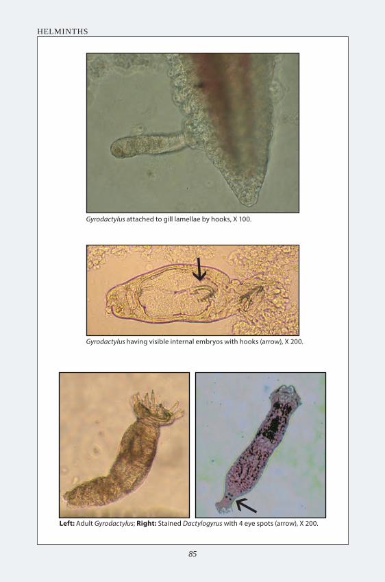
85
HELMINTHS
Gyrodactylus attached to gill lamellae by hooks, X 100.
Gyrodactylus having visible internal embryos with hooks (arrow), X 200.
Left: Adult Gyrodactylus; Right: Stained Dactylogyrus with 4 eye spots (arrow), X 200.
86
HELMINTHS
Piscicola
I. Causative Agent and Disease Piscicola is a freshwater leech
belonging to the phylum Annelida (segmented worms) that can be abundant in some freshwater lakes, ponds and streams. Piscicola attaches to the skin of freshwater fish and is nourished by suck-ing blood and other tissue fluids from the host. Members of the genus Piscicola usually remain attached to a fish for sev-eral days while feeding and then drop off and sink to the bottom where the food is digested. Piscicola has well developed oral and caudal suckers with a subcylin-drical and elongate body. Leeches usu-ally do not cause serious harm to their hosts since most tissue damage is local-ized at the sites of attachment. However, when present in large numbers parasitic leeches can cause extensive tissue dam-age to fishes including epidermal erosion and ulceration, hemorrhaging, necrosis and anemia. External epidermal erosions may serve as portals of entry for second-ary bacterial or fungal pathogens.
II. Host Species The parasite occurs on many species
of freshwater fishes in Europe and North America. Salmonids are most commonly parasitized by P. salmositica in Alaska.
III. Clinical Signs Piscicola leeches are visible with the
naked eye. Attachment of leeches may occur anywhere on the host body and are often found on or under the opercula, in the mouth, along the jaw and at the bases of fins.
IV. Transmission The life cycle of leeches is relatively
simple, consisting of an egg, a juvenile stage and a mature hermaphroditic adult that produces eggs. After digestion of
a blood meal, a leech either attaches to a fish for another feeding cycle or it produces eggs. Eggs are encased in oval “cocoons” that are attached to the substrate at the bottom of the lake or river. Juvenile leeches hatch from the eggs and enter the water column to find a fish host. Parasitic juvenile leeches usu-ally require several blood meals before becoming mature adults. Leeches of this genus have been implicated as possible vectors of the fish virus, IHNV.
V. Diagnosis Leeches are obvious by visual
examination of the host. Observation of the worm under a dissecting microscope for various morphological character-istics including color and pattern of pigmentation, number and arrangement of eye spots on the oral sucker and other external features help identify the genus Piscicola.
VI. Prognosis for Host Leeches usually do not cause signifi-
cant harm to their hosts unless present in large numbers. Prognosis for a host is good when infestations are low to mod-erate, but host inflammation may occur locally at the site of attachment.
VII. Human Health Signifcance There are no human health concerns
associated with Piscicola.

87
HELMINTHS
Piscicola Life Cycle
Adult Piscicola leeches attach to fish host
Hatched, leeches seek fish host
Engorged leech falls off host producing eggs
Eggs encased in cocoons Eggs attach to vegetation or rocks in the substrate
Adult and juvenile Piscicola on rainbow trout producing typical epidermal attachment lesions (visible as round depressions).
Freshwater leech of the genus Piscicola (measurement in cm).

88
HELMINTHS
Diphyllobothrium
I. Causative Agent and Disease Six species of diphyllobothrid ces-
todes (tapeworm) occur in Alaska, all of which use fish as a second intermediate and/or as a paratenic host. Three species of larval Diphyllobothrium that most commonly occur in Alaskan salmonid fishes include D. ditremum, dendriticum and nihonkaiense. The cestode larvae can be found free in the visceral cavity or encysted in the viscera or muscle tis-sues.
II. Host Species Planktivorous and carnivorous fresh-
water fishes are potential hosts in North/ South America and Eurasia including salmonids, whitefish, perch, northern pike, sticklebacks, burbot, and blackfish.
III. Clinical Signs The larval Diphyllobothrium can
be found (sometimes encysted) in the muscles, viscera and connective tis-sues of the fish host causing adhesions, hemorrhaging (particularly the liver) and ascitic fluid resulting in abdominal dis-tension. Severe infestations in juvenile fish can cause mortality.
IV. Transmission Infestation of the fish host is part of
a 3-host life cycle for this parasite. Adult worms are found in the small intestine of definitive hosts that are fish eating birds or mammals (including humans). Eggs from adult worms are released into the water with feces where they develop into a free swimming coracidium larval stage that is ingested by copepods, the first intermediate host. The procercoid develops in the copepod and, when eaten by the fish second intermediate host, develops into the plerocercoid stage. Plerocercoids re-encyst near the gut of
predatory fish that become paratenic hosts when other infested fish are eaten. The life cycle is complete when the fish host is eaten by a mammal or bird defini-tive host where the worm becomes an egg-producing adult.
V. Diagnosis Diagnosis is made by visual identi-
fication of the cestode during necropsy of a parasitized fish. Plerocercoid stages of Diphyllobothrium have a compressed scolex with characteristic bothria or grooves. The body is usually slightly wrinkled, suggesting segmentation. PCR has been useful in confirming species that has resulted in changing taxonomy.
VI. Prognosis for Host Prognosis for the host is good pro-
vided the infestation is low and there are not other stressors involved. Juvenile fish are more adversely affected than older fish and can die from severe plerocercoid infestations.
VII. Human Health Signifcance Species of this cestode group can
successfully parasitize humans. Most human infestations are accidental since the natural hosts are fish eating birds and mammals. Infestation in man occurs by ingestion of raw or lightly smoked fish that contain viable plerocercoid larvae. The Center for Disease Control recommends cooking fish at 67ºC for 5 minutes or freezing fish at -20ºC for at least 7 d to kill worm parasites before ingestion.

89
HELMINTHS
Diphyllobothrium Life Cycle
Crustacean ingested by second
Piscivorous fish (paratenic host)
Humans parasitized by consumption of raw or
undercooked fish Fish eating birds and mamals are final
hosts for adult cestode Diphyllobothrium
Procercoid larvae in first intermediate host,
a copepod crustacean
Eggs
Ciliated larvae (coracidium)
intermediate host (fish) where larvae develop into plerocercoids
Left: Subsurface white cysts (center) in brook trout liver with plerocercoids of Diphyllobothrium sp.; Right: Diphyllobothrium sp. plerocercoid with wrinkled body suggesting segmentation.
Encysted plerocercoids in viscera of rainbow trout (black arrows).
Bothria (grooves) in plerocercoid scolex characteristic of Diphyllobothrium sp.
90
HELMINTHS
Schistocephalus
I. Causative Agent and Disease Schistocephalus is a cestode (tape-
worm) within the family Diphylloboth-riidae parasitizing fish hosts as plerocer-coid larvae transmitted by ingestion of parasitized copepods. The worm in the fish host occurs in the body cavity often causing abdominal distention due to multiple infestations and the large size of the plerocercoids.
II. Host Species Several freshwater fish species are
susceptible to this parasite in North America and Eurasia. In Alaska, this cestode is most often found in stickle-backs.
III. Clinical Signs Fish with heavy infestations of this
parasite are often bloated and mis-shapen since the parasite is quite large. Normal fish swimming behavior may be impaired.
IV. Transmission Transmission occurs through a com-
plex life cycle utilizing two intermediate hosts. The first intermediate host is a co-pepod that is parasitized by a coracidium hatched from a cestode egg deposited in the water column. A procercoid stage develops in the gut of the copepod that is eaten by the second intermediate host, a freshwater fish. The plerocercoid devel-ops in the second intermediate fish host that is eaten by the final bird host where the adult worm develops and produces eggs in the intestinal tract.
V. Diagnosis Diagnosis is made by internal obser-
vation of the plerocercoid larvae. The white larvae are 2-7 cm long, broader at one end and segmented with shallow
bothria (grooves) on the scolex. Fish will often contain multiple plerocercoids.
VI. Prognosis for Host Prognosis for the host is dependent
on the degree of infestation. Pathology caused by the plerocercoids includes growth retardation, abdominal disten-sion, and physiological dysfunction of internal organs. The debilitation caused by the parasite increases the vulnerabil-ity of the fish host to predation by the final host.
VII. Human Health Signifcance There are no known human health
concerns associated with Schistocepha-lus.

91
HELMINTHS
Plerocercoid stage of Schistocephalus from abdominal cavity of parasitized stickleback
Two plerocercoids of Schistocephalus removed from the body cavity of a stickleback.
92
HELMINTHS
Triaenophorus
I. Causative Agent and Disease Triaenophorus crassus is a cestode
(tapeworm) belonging to the family Triaenophoridae that parasitizes fish as plerocercoid larvae (1 mm X 30 cm) found encysted in the musculature. These cestodes can also be found as adults living in the guts of predatory fish.
II. Host Species There are many fish intermediate
hosts for the plerocercoid (larval) stage of Triaenophorus in North America and Europe. The definitive hosts are piscivo-rous fishes such as northern pike.
III. Clinical Signs Triaenophorus often stimulates
formation of yellow to white cysts of host connective tissue that surround the plerocercoids in the muscle. Encysted or unencysted larvae can cause local-ized muscle discoloration and necrosis. Liver dysfunction and blood loss can occur from larval migration through the viscera and may be associated with hemorrhaging, necrosis, fibrosis, edema and tissue discoloration. Severe adult tapeworm infestations in the gut can cause perforations, mechanical block-age with distension and prevent nutrient uptake causing emaciation and anemia.
IV. Transmission The life cycle of this tapeworm oc-
curs in freshwater where eggs are shed from adult worms living in the intestinal tracts of the final hosts (usually preda-tory fish). The cestode eggs, containing a coracidium, are eaten by copepods and develop into procercoids. The copepods are eaten by the second intermediate fish host where the procercoids migrate from the intestinal tract to the muscle
and develop into the plerocercoid stage. The life cycle of the worm is completed when the parasitized fish is eaten by the final fish host, commonly a northern pike. Eggs are produced after the worm develops into an adult in the intestinal tract of the final fish host.
V. Diagnosis Diagnosis is made by observation of
encysted or unencysted white plerocer-coids in the skeletal musculature of a parasitized fish. Identifying microscopic features of the plerocercoid scolex (head) include dorsal and ventral pairs of trident shaped hooks on an apical disc. Adult worms are larger and found in the intestinal tract.
VI. Prognosis for Host Prognosis for the fish host depends
on the degree of cestode infestation, the age and size of the fish and expo-sure to other stressors. Generally, these parasites have caused health problems with juvenile cultured fish but are well tolerated when occurring as natural infestations of larger healthy fish.
VII. Human Health Signifcance Although this tapeworm is not
known to occur in man or other warm-blooded animals, infested fish flesh is unsightly.
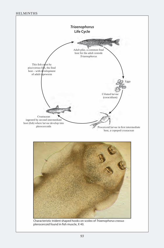
93
HELMINTHS
Triaenophorus Life Cycle
Adult pike, a common final host for the adult cestode
Triaenophorus
Ciliated larvae (coracidium)
Procercoid larvae in first intermediate host, a copepod crustacean
Crustacean ingested by second intermediate
host (fish) where larvae develop into plerocercoids
This fish eaten by piscivorous fish, the final host – with development
of adult tapeworm
Eggs
Characteristic trident shaped hooks on scolex of Triaenophorus crassus plerocercoid found in fsh muscle, X 40.

94
ARTHROPODS
External Parasitic Copepods
I. Causative Agent and Disease A variety of different parasitic
copepods can cause external infesta-tions of freshwater and marine fish. Some members of the group are com-monly referred to as fish lice. They are frequently found on the body, around the mouth, and on the gills. Members of the order Copepoda commonly found in Alaska include the genera Lernaea (anchor worm) in both fresh and marine waters, Salmincola (discussed in next section) in freshwater and Lepeoph-theirus (sealice) in marine waters. Fish infested with external parasitic copepods are often lethargic and may flash or rub against substrate. In heavy infestations, the skin may appear opaque due to the production of mucus and the fins may be frayed. Epidermal and gill tissues may become necrotic with secondary infections by fungi and bacteria. These parasites are found worldwide.
II. Host Species A variety of different freshwater and
marine fishes are susceptible to infesta-tions from these copepods.
III. Clinical Signs Parasitized fish may act listless and
lethargic. Mechanical abrasion due to the attachment and/or feeding by the cope-pods is common resulting in frayed fins, gill hyperplasia, and patchy epidermal damage and necrosis. Infections with secondary pathogens often occur.
IV. Transmission Most of these organisms have a
direct life cycle involving a number of free-living and larval stages. Transmis-sion is through contact with an infective free-swimming stage of the organism in the water column. The infective stage at-
taches to the fish where it goes through a number of larval stages before becoming an adult.
V. Diagnosis The larger parasites can be seen with
the naked eye. Definitive identification is based on microscopic morphologies of body parts and structures.
VI. Prognosis for Host The prognosis for the host depends
on the specific parasite, tissue loca-tion and numbers present. If parasite numbers are small, fish normally survive with little adverse effects unless the copepods are anchor worms that destroy one or both eyes. This can result in blindness and death of the host by starvation or predation. When present in large numbers, such as Lepeophthei-rus in seawater netpens, significant fish losses may result. SLICE is a drug used in Canada and Europe for eliminating fish lice in seawater netpens and is ap-proved for use in the US under an INAD permit.
VII. Human Health Signifcance There are no human health concerns
associated with these organisms.

95
ARTHROPODS
Lepeophtheirus salmonis copepod from the surface of a salmonid fsh.
Left: Lepeophtheirus on coho salmon; Right: Phrixocephalus cincinnatus (anchor worm) embedded in the eye of an arrowtooth founder.
96
ARTHROPODS
Salmincola
I. Causative Agent and Disease Parasitic copepods of the genus
Salmincola are most often found at-tached to gill filaments, opercula, tissues within the mouth cavity, and fins of salmonid fishes. The parasites feed on blood and epithelial tissues of their hosts. Salmincola species are restricted largely to freshwater but may survive on salmonids while at sea. The adult female copepods are larger than the males and attach permanently to the fish host with a modified mouth part known as a bulla that is inserted into the host tissues. Host damage by parasitic copepods depends on the location of the attachment site, the species of parasite, and the size and type of bulla. Gill attachment by Salmincola can damage delicate epidermal tissues resulting in necrosis and loss of surface area for respiration. Attachment may also provide portals of entry for second-ary invaders such as bacteria and fungi.
II. Host Species Salmincola has been reported more
commonly from salmonid species in North America and Europe.
III. Clinical Signs Salmincola copepods are visible to
the naked eye when attached to fins, bases of fins, skin, opercula, gills and branchial chamber. Gill damage caused by displacement from Salmincola can be extensive resulting in retarded filament growth and tissue necrosis. Gill hyper-plasia and hypertrophy may also lead to fusion of the filaments, thus reducing surface area for necessary gas exchange and respiration.
IV. Transmission Salmincola have a direct, but com-
plicated life cycle. Females produce two
clusters of eggs twice during a 3-month life span. Eggs hatch into a larval form that can survive free-swimming for several days. The larvae attach to gills or fins of a fish host and molt into 4 suc-cessive larval stages and degenerate into grub-like parasites. Males then detach and copulate with the females, after which the males die. Females molt into the adult stage and produce two pairs of egg clusters. The female Salmincola dies shortly after the second group of eggs hatch.
V. Diagnosis Salmincola are large enough to ob-
serve grossly. Visual examination of fish skin, fins, gills and mouth can reveal the extent of copepod infestation. Micro-scopic examination of various morpho-logical characteristics aid in identifying the parasite to the genus and species.
VI. Prognosis for Host Prognosis for the host is good when
infestations are not severe and damage to gill tissue is minimal. Generally, in-festations with this parasite do not cause significant fish mortality.
VII. Human Health Signifcance There are no human health concerns
associated with Salmincola.

97
ARTHROPODS
Salmincola Life Cycle
Adult Salmincola attaches to fish host
male ← Egg sacs
Adult female copepod with button-shaped bulla
and male attached
Copepodids are released from egg sacs
Free swimming copepodids Copepodids molt several
times while attached to fish host
Severe Salmincola infestation of rainbow trout gills; note necrotic areas at tips of gill lamellae.
98
ARTHROPODS
Sarcotaces
I. Causative Agent and Disease V. Diagnosis Sarcotaces arcticus is an endopara-
sitic copepod several centimeters long found encysted under the skin and in the muscle tissue of marine fish. The juvenile copepod inserts its head into the flesh and is eventually covered by the host skin except for the last pointed body segment that maintains connection with the outside seawater. When the parasite dies, after growing to an enormous size, the tissue forms a closed cyst. In Alaska, this parasite is commonly found in rockfish (Sebastes spp.) encysted near the anus where surrounding intestinal tissue forms a sac-like process. When fish are filleted the ruptured cysts release black fluid, a breakdown product of blood that the parasite has engorged.
II. Host Species This parasitic copepod is found most
commonly in species of Sebastes spp. in the northern Pacific Ocean and in other genera of teleosts in European waters.
III. Clinical Signs Sarcotaces parasites are difficult to
detect externally because of the subder-mal location of the cyst. If the cyst lies over a bony surface, swelling of the skin may occur. In parasitized rockfish, the anal cysts are generally obvious.
IV. Transmission The life cycle is direct by horizontal
transmission. Larval copepodids are released into ambient seawater from the encysted adult female after fertilization by a much smaller male that has entered through the open portal of the cyst. The juveniles seek out a new host to continue the life cycle.
Diagnosis is made by internal exami-nation of the fish for characteristic pear-shaped cysts exuding black fluid and morphological identification of the larger female parasite. The body is oval with ill-defined transverse bands correspond-ing to segments and a double rosette is often visible around the mouthparts.
VI. Prognosis for Host Prognosis for the host is good if in-
festation is minimal and there are no sig-nificant environmental stressors present. Infestations by Sarcotaces are associated with lower fecundity in rockfish and fish flesh becomes unappealing when fillets are tainted with the fluid leaking from cysts.
VII. Human Health Signifcance There are no human health concerns
with Sarcotaces.
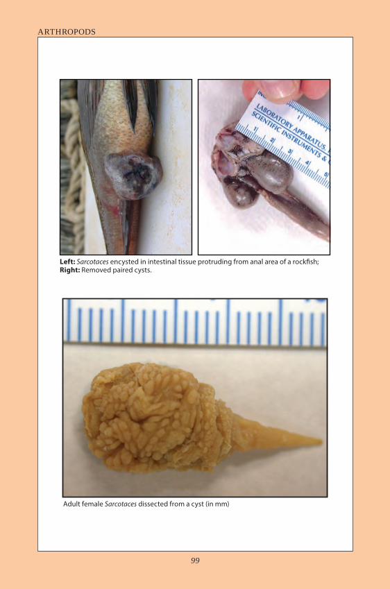
99
ARTHROPODS
Left: Sarcotaces encysted in intestinal tissue protruding from anal area of a rockfsh; Right: Removed paired cysts.
Adult female Sarcotaces dissected from a cyst (in mm)

100
NON-INFECTIOUS DISEASES
Bloat (Water Belly)
I. Causative Agent and Disease This is a non-infectious condition
where the abdomen of salmonids is abnormally distended by an enlarged, water-filled stomach. The condition is most often seen in salmonids reared in seawater. The cause of this condition is not well understood, but potential vari-ables may include: a combined failure of osmotic regulation, increased lipids, temperature and stress; increased drink-ing of seawater and nutrient overloading due to excessive feeding. In other words, ingestion of nutrient-rich food may result in slowed stomach emptying leading to decreased absorption of water, thirst and increased drinking that further exacer-bates stomach distention.
II. Host Species This condition is reported frequently
in Chinook, coho, chum and Atlantic salmon and also in rainbow trout. Bloat in Alaska is most common in juvenile coho, but also observed in chum, pink and Chinook salmon.
III. Clinical Signs Fish with bloat exhibit severe disten-
tion of the abdominal wall. Necropsy reveals a massively enlarged stomach with a very thin wall. The stomach is filled with a clear, watery fluid mixed with feed.
IV. Transmission The disease is non-infectious and
cannot be transmitted from fish to fish.
V. Diagnosis Bloat is usually diagnosed by the
presence of excessive amounts of clear, watery fluid in the stomach. The stomach wall is thinned from distension, but other significant histological changes are not present.
VI. Prognosis for Host Although this condition can cause
mortality, affected fish often survive for weeks. A reduced feeding regime after fish have been starved for several days or changing the composition of the food will reduce the problem in captive fish.
VII. Human Health Signifcance There are no human health concerns
associated with this condition.

101
NON-INFECTIOUS DISEASES
Chum salmon fry with swollen abdomen typical of bloat.
Chinook salmon smolt with swollen abdomen typical of bloat.
102
NON-INFECTIOUS DISEASES
Blue Sac Disease of Fry
I. Causative Agent and Disease V. Diagnosis Blue sac disease of fry is considered
to be a non-infectious disease caused by the accumulation of metabolic wastes and reduced dissolved oxygen result-ing in excessive buildup of ammonia nitrogen. The disease was reproduced in several species of salmonid alevins by allowing metabolic wastes to accumu-late or by adding ammonia to the water. However, the condition has also been reproduced experimentally in rainbow and brook trout by infection of eyed eggs with the bacteria Aeromonas hydrophila.
II. Host Species This condition has been reported
primarily in salmonid fishes, especially brook trout and other char that tend to be the most susceptible species.
III. Clinical Signs The alevin/fry exhibit an abnormal
accumulation of fluid, often bluish in color, at the posterior of the yolk sac often progressing to surround the entire yolk. Due to the increased fluid, fry cannot swim normally. Fry may have ex-ophthalmia, coagulated yolk, and appear smaller and pale. Petechial hemorrhages of the head, thoracic and vitelline blood vessels can occur in severe cases with hemorrhaging into the blue sac fluid and severe anemia.
IV. Transmission In most cases, the disease is due to
suboptimal environmental conditions and is not transmissible. However, when poor environmental conditions can be ruled out, transmission of blue sac disease by A. hydrophila becomes a pos-sibility.
Diagnosis is based on the observationof typical clinical signs of the condition.
VI. Prognosis for Host Blue sac disease is usually fatal due
to incomplete organogenesis and body development.
VII. Human Health Signifcance There are no human health concerns
associated with this condition.

103
NON-INFECTIOUS DISEASES
Swollen yolk sacs of cultured lake trout caused by Blue Sac Disease.
104
NON-INFECTIOUS DISEASES
Coagulated Yolk Disease (White Spot Disease)
I. Causative Agent and Disease Coagulated yolk disease is a
noninfectious condition resulting from unsatisfactory environmental conditions during incubation. A wide variety of factors probably contribute towards the disease including gas supersaturation, unfavorably high water temperatures, heavy metals in the water supply (Cu, Al, Zn), soft water, low water flows, low dissolved oxygen, exposure to chemicals or contaminants, excessive handling and otherwise inadequate or stressful incu-bation conditions. Yolk proteins become denatured and coagulate as manifested by the appearance of white spots in incu-bating eggs and the yolk sacs of hatched alevins. Yolk resorption is disrupted, resulting in defective development of vital organs. This causes physiological alterations in organ functions resulting in death of eggs during incubation or af-terwards in alevins and larger juveniles.
II. Host Species All fish eggs and alevins are suscep-
tible.
III. Clinical Signs White spots or flecks in eggs are
typically at the surface of the yolk and randomly distributed. In alevins, the coagulated yolk appears a few days after hatching and may enlarge and coalesce with time. In fry that have completely absorbed the egg sac the coagulated yolk appears as a white mass in the visceral cavity, sometimes associated with clot-ted blood. Pinheading, anemic gills, and white or frayed fins are sometimes observed in affected fry. Non-infectious tail and fin erosion (especially pecto-ral fins) can be caused by unabsorbed coagulated yolk that remains in the body cavity that interferes with the ability of
the fish to maintain the replacement of epithelium covering the extremities; the fins are the first to be affected but is self-limiting, commonly observed in young Chinook salmon shortly after transfer from incubators and troughs to rearing ponds.
IV. Transmission This disease is not infectious and
cannot be transmitted from fish to fish.
V. Diagnosis Diagnosis is made by observing
the abnormal white flecks or masses of coagulated yolk in eggs, alevins or fry.
VI. Prognosis for Host Most fish with coagulated yolk will
eventually die before reaching 1 gram in size due to improper organ development. Juveniles appear normal then suddenly drop-out.
VII. Human Health Signifcance There are no human health concerns
associated with this condition.
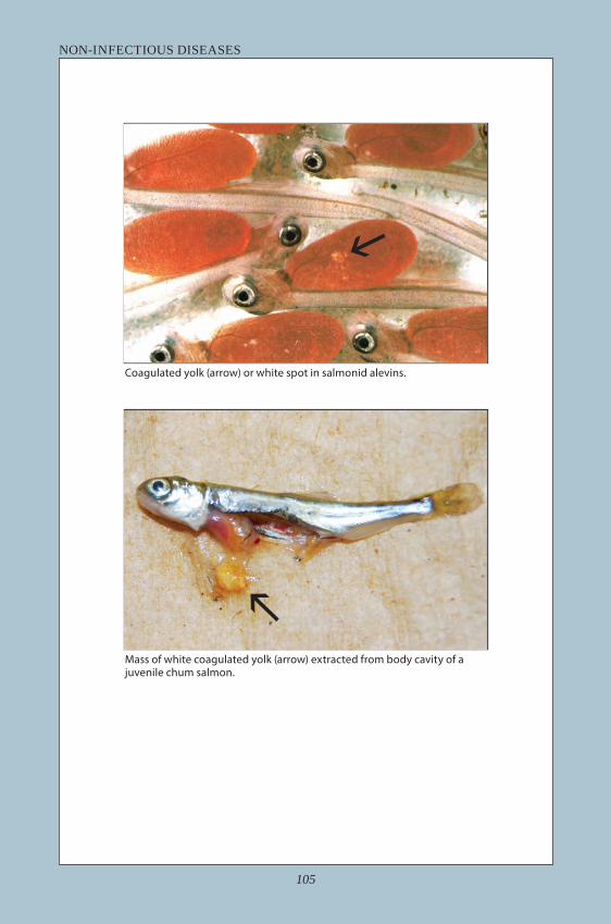
105
NON-INFECTIOUS DISEASES
Coagulated yolk (arrow) or white spot in salmonid alevins.
Mass of white coagulated yolk (arrow) extracted from body cavity of a juvenile chum salmon.
106
NON-INFECTIOUS DISEASES
Drop-out Disease
I. Causative Agent and Disease II. Host Species Drop-out disease is commonly
observed in hatchery reared juvenile salmonids but is not caused by an infec-tious agent or a deficiency in the diet. Affected fish may exhibit gill hyperpla-sia and severely clubbed gills, usually stop feeding and become emaciated or pinheaded. Other forms of drop-out are not associated with gill hyperplasia. Sec-ondary bacterial, fungal and protozoan infections often develop in affected fish.
Drop-out associated gill hyperplasia causes include: 1. The fine particles in starter feeds
irritate delicate gill epithelium.2. Diatom blooms of Chaetoceros con-
volutus can cause severe gill abrasionin fish that are held in seawaternetpens.
3. Repeated therapeutic chemical treat-ments for external parasites andbacteria can irritate gill epithelium.
Drop-out NOT associated with gill hyperplasia - causes include: 1. An increase in feed pellet size may
prevent a proportion of the smallerfish from eating enough to maintaingood body weight and they becomepinheaded.
2. White spot or coagulated yolk causingincomplete organ development canresult in mortality of seeminglyhealthy fish during early or laterjuvenile stages. Fish are notpinheaded.
3. Not enough yolk (minimum 3-5%body weight) remaining when alevinsemerge from incubators. Mechanicalremoval may be necessary, especiallywith chum salmon.
All salmonids are susceptible, butthe condition is seen most frequently in Chinook, coho and chum salmon in Alaska.
III. Clinical Signs Fish may stop feeding and become
emaciated resulting in thin bodies and large heads referred to as pinheading. Gill hyperplasia, sometimes very severe, occurs in many instances but not all as indicated previously. Secondary infec-tions commonly occur adding confusion to the primary diagnosis.
IV. Transmission Since this is not an infectious dis-
ease, transmission between fish does not occur.
V. Diagnosis Diagnosis of drop-out and its
cause depends on whether the fish are pinheaded and have or do not have gill hyperplasia and have been exposed to one or more of the conditions listed.
VI. Prognosis for Host Mortality can be up to 20-30% of the
population or higher in the case of an al-gal bloom. Drop-out from gill hyperpla-sia can be corrected by removal or avoid-ance of the gill irritant(s). Drop-out from coagulated yolk or not enough yolk are sequellae from preexisting conditions that cannot be changed in the current cohort of fish but could be prevented in the next production cycle by improving incubator conditions or removing fish earlier.
VII. Human Health Signifcance There are no human health concerns
associated with this condition.

107
NON-INFECTIOUS DISEASES
Severe gill hyperplasia with fusion and clubbing commonly seen in drop-out disease due to irritation caused by feeding starter diets, X 100.
Wet mount of Chaetoceros convolutus diatoms that can cause gill irritation and abrasion, X 400.

108
NON-INFECTIOUS DISEASES
Gas Bubble Disease (GBD)
I. Causative Agent and Disease Gas bubble disease is not infectious
and is caused by supersaturated levels of total dissolved gas in the water. Lesions in the fish are caused by the accumula-tion of gas bubbles in blood vasculature and tissues. Either supersaturation of oxygen or nitrogen can result in the disease, however, the total dissolved gas (TDG) is more important than individual gases or varying combined gas ratios. Supersaturation occurs when water contains more dissolved gas than it can normally hold in solution at a given temperature and atmospheric pressure. Under high pressure or at low tempera-tures water can contain more gas. Gas supersaturation in water can occur from both natural and artificial causes. In nature, supersaturation occurs in plunge pools at the base of waterfalls, in natural springs and wells where water is under pressure at depth, and in water that has melted from glaciers or snow. During the photosynthetic process water bodies containing heavy aquatic plant growth can be saturated with oxygen and can become supersaturated upon warming. Artificially supersaturated water occurs in plunge pools from dams, when water is heated such as power plant effluent, and if air is entrained in pipes or pumps where pump pressure or gravity head forces gas into solution.
II. Host Species The disease can affect any fish or
invertebrate anywhere when in supersat-urated waters. Levels of gas supersatu-ration causing pathological changes or mortality vary for different fish species and age of fish. Fry become susceptible post-hatch when they begin swimming up for food (at about 16 days). Steelhead trout are most sensitive (average thresh
old of 102-103% TDG before chronic problems develop) while coho salmon are least sensitive (average threshold 115.7% TDG)
III. Clinical Signs Fish with GBD often exhibit loss
of equilibrium, abnormal buoyancy and may float at the water surface. Fish may also exhibit violent head shaking, convulsions, flared opercula, release of excessive gas from buccal cavity, blindness and may die with the mouth open. Alevins may show hemorrhage of vitelline vessels, rupture of yolk-sac membranes, and coagulated yolk. Subcutaneous bubbles can accumulate in tissues of the head, mouth, fin rays, and gill arches. Air bubbles are often visible in gill lamellar capillaries. Hemorrhage of gills, fins, skin, muscle, gonads, and intestinal epithelium can also occur. Fins may be eroded with whitened fin tips and exophthalmia may occur with blood present in the anterior chamber of eye.
TDG 100-106% Embolic lesions will appear
with hemostasis
≥ 103% Certain species of salmonid fry are stressed and may later develop conditions leading to death (i.e., coagulated yolk, fin erosion, tail erosion, etc.)
> 120% Acute levels, fry willdie before signs or lesions indicate a problem
IV. Transmission Due to the environmental nature of
this disease, transmission between fish does not occur.
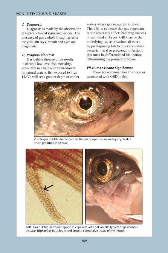
109
NON-INFECTIOUS DISEASES
V. Diagnosis Diagnosis is made by the observation
of typical clinical signs and lesions. The presence of gas emboli in capillaries of the gills, fin rays, mouth and eyes are diagnostic.
VI. Prognosis for Host Gas bubble disease often results
in chronic low-level fish mortality, especially in a hatchery environment. In natural waters, fish exposed to high TDG’s will seek greater depth or cooler
waters where gas saturation is lower. There is no evidence that gas supersatu-ration adversely affects hatching success of salmonid embryos. GBD can be the underlying cause of various diseases by predisposing fish to other secondary bacterial, viral or protozoan infections that must be differentiated first before determining the primary problem.
VII. Human Health Signifcance There are no human health concerns
associated with GBD in fish.
Visible gas bubbles in connective tissues of operculum and eye typical of acute gas bubble disease.
Left: Gas bubbles (arrow) trapped in capillaries of a gill lamella typical of gas bubble disease; Right: Gas bubbles in and around connective tissue of the mouth.

110
NON-INFECTIOUS DISEASES
Mushy Halibut Syndrome
I. Causative Agent and Disease VI. Prognosis for Host Smaller halibut of 15-20 lbs caught
by sportfishing charters near Homer and Soldotna, AK have had a condi-tion locally known as “mushy halibut”. Typically, this condition consists of fish having large areas of body muscle that is abnormally translucent and flaccid or jellylike. The overall body condition of these fish is usually poor and often they are released because of the potential for inferior meat quality.
II. Host Species Smaller Pacific halibut, mostly in
the Cook Inlet and Homer/Seward areas but affected fish in other areas of Alaska have been reported.
III. Clinical Signs Fish are asymptomatic except for
poor body condition. Large areas of the fillets are abnormally translucent and flaccid in texture.
IV. Transmission No infectious agents or parasites
have been detected in affected fish, therefore, transmission from fish to fish is not likely. A nutritional deficiency is suspected.
V. Diagnosis Diagnosis is by gross observation
of flaccid, translucent musculature with confirmation of a non-infectious degenerative myopathy by histological examination. There is severe muscle fiber atrophy, fragmentation and necrosis with loss of muscle mass. There may be accompanying inflammatory cells, fibro-sis and calcification of atrophied fibers. There is some attempt at regeneration as evidenced by internal nuclear chains within myofibers.
Reportedly, the Cook Inlet and Hom-er/Seward areas are nursery grounds for large numbers of young halibut that feed primarily on forage fish that have recently declined in numbers. Stomach contents of smaller halibut now contain mostly small crab species. Whether this forage is deficient, either in quantity or in essential nutrients is not known. How-ever, mushy halibut syndrome is similar to that described for higher animals with nutritional deficiencies in vitamin E and selenium. This muscle atrophy would further limit the ability of halibut to capture prey possibly leading to further malnutrition and increased severity of the primary nutritional deficiency.
VII. Human Health Signifcance Although aesthetically displeasing,
there are no known human health con-cerns with mushy halibut syndrome.
NOTE: mushy halibut is not to be confused with chalky halibut, a condi-tion of opaque flesh with a cooked-like texture. Chalkiness is another flesh quality problem caused by the buildup of excessive lactic acid from overexertion of fish fighting capture for several hours on longline fishing gear.
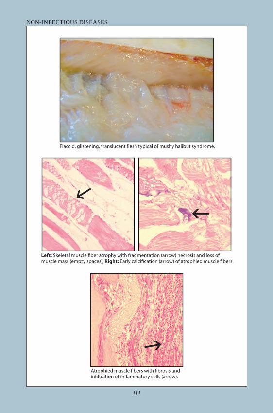
111
NON-INFECTIOUS DISEASES
Flaccid, glistening, translucent fesh typical of mushy halibut syndrome.
Left: Skeletal muscle fber atrophy with fragmentation (arrow) necrosis and loss of muscle mass (empty spaces); Right: Early calcifcation (arrow) of atrophied muscle fbers.
Atrophied muscle fbers with fbrosis and infltration of infammatory cells (arrow).
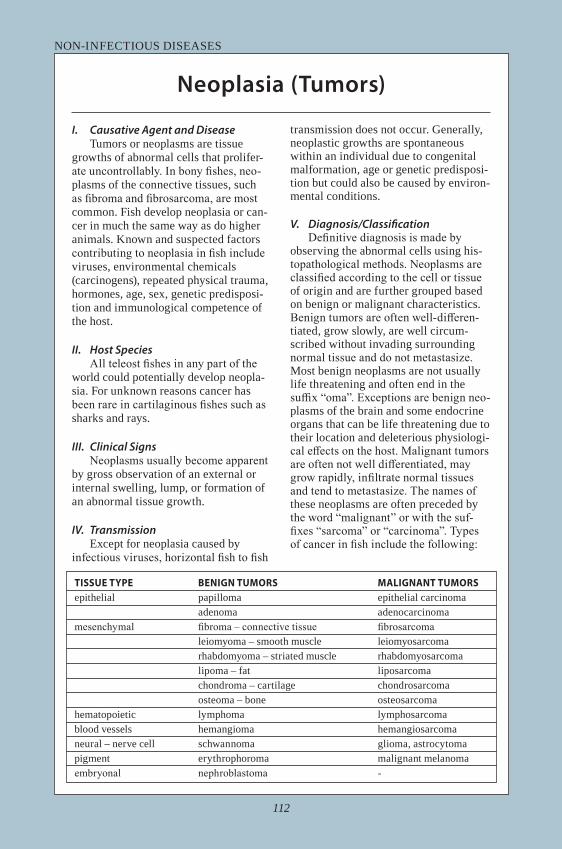
112
NON-INFECTIOUS DISEASES
Neoplasia (Tumors)
I. Causative Agent and Disease Tumors or neoplasms are tissue
growths of abnormal cells that prolifer-ate uncontrollably. In bony fishes, neo-plasms of the connective tissues, such as fibroma and fibrosarcoma, are most common. Fish develop neoplasia or can-cer in much the same way as do higher animals. Known and suspected factors contributing to neoplasia in fish include viruses, environmental chemicals (carcinogens), repeated physical trauma, hormones, age, sex, genetic predisposi-tion and immunological competence of the host.
II. Host Species All teleost fishes in any part of the
world could potentially develop neopla-sia. For unknown reasons cancer has been rare in cartilaginous fishes such as sharks and rays.
III. Clinical Signs Neoplasms usually become apparent
by gross observation of an external or internal swelling, lump, or formation of an abnormal tissue growth.
IV. Transmission Except for neoplasia caused by
infectious viruses, horizontal fish to fish
transmission does not occur. Generally, neoplastic growths are spontaneous within an individual due to congenital malformation, age or genetic predisposi-tion but could also be caused by environ-mental conditions.
V. Diagnosis/Classifcation Definitive diagnosis is made by
observing the abnormal cells using his-topathological methods. Neoplasms are classified according to the cell or tissue of origin and are further grouped based on benign or malignant characteristics. Benign tumors are often well-differen-tiated, grow slowly, are well circum-scribed without invading surrounding normal tissue and do not metastasize. Most benign neoplasms are not usually life threatening and often end in the suffix “oma”. Exceptions are benign neo-plasms of the brain and some endocrine organs that can be life threatening due to their location and deleterious physiologi-cal effects on the host. Malignant tumors are often not well differentiated, may grow rapidly, infiltrate normal tissues and tend to metastasize. The names of these neoplasms are often preceded by the word “malignant” or with the suf-fixes “sarcoma” or “carcinoma”. Types of cancer in fish include the following:
TISSUE TYPE BENIGN TUMORS MALIGNANT TUMORS epithelial papilloma epithelial carcinoma
adenoma adenocarcinoma mesenchymal fibroma – connective tissue fibrosarcoma
leiomyoma – smooth muscle leiomyosarcoma rhabdomyoma – striated muscle rhabdomyosarcoma lipoma – fat liposarcoma chondroma – cartilage chondrosarcoma osteoma – bone osteosarcoma
hematopoietic lymphoma lymphosarcoma blood vessels hemangioma hemangiosarcoma neural – nerve cell schwannoma glioma, astrocytoma pigment erythrophoroma malignant melanoma embryonal nephroblastoma -
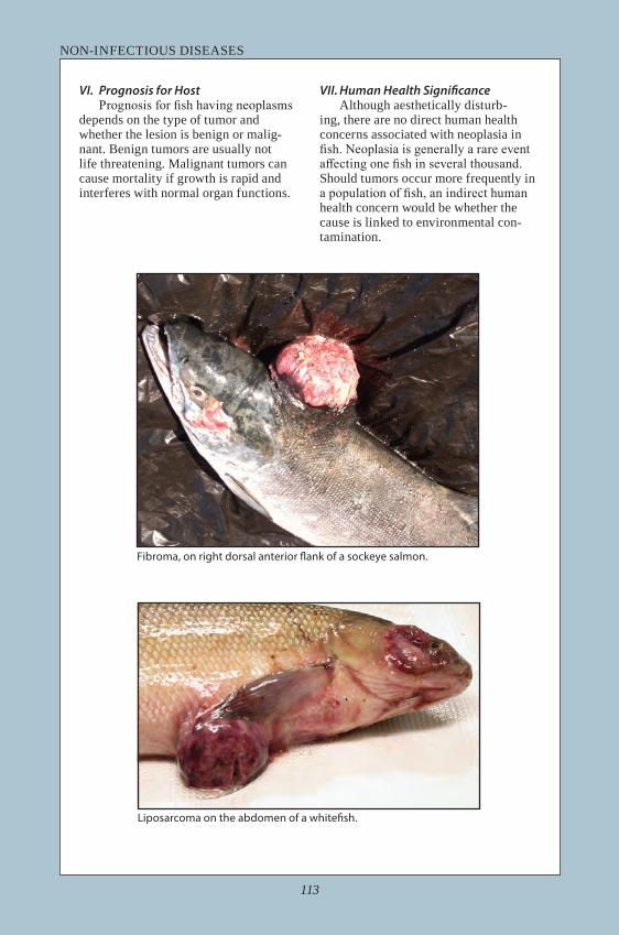
113
NON-INFECTIOUS DISEASES
VI. Prognosis for Host Prognosis for fish having neoplasms
depends on the type of tumor and whether the lesion is benign or malig-nant. Benign tumors are usually not life threatening. Malignant tumors can cause mortality if growth is rapid and interferes with normal organ functions.
VII. Human Health Signifcance Although aesthetically disturb-
ing, there are no direct human health concerns associated with neoplasia in fish. Neoplasia is generally a rare event affecting one fish in several thousand. Should tumors occur more frequently in a population of fish, an indirect human health concern would be whether the cause is linked to environmental con-tamination.
Fibroma, on right dorsal anterior fank of a sockeye salmon.
Liposarcoma on the abdomen of a whitefsh.

114
NON-INFECTIOUS DISEASES
Left: Cut surface of a liposarcoma on the back of a quillback rockfsh; Right: Thymic lymphosarcoma (arrow) in branchial cavity of a sockeye salmon.
Left: Fibrosarcoma (arrow) infltrating the muscle tissue from the body wall in a salmon; Right: Pedunculated papilloma on the back of a coho salmon.
Left: Rhabdomyosarcoma (green) in the musculature of a Pacifc halibut; Right: Black melanoma on the head of a chum salmon; Inset: Sockeye: when cut, melanomas can exude a black tarry fuid containing melanin pigment that stains surfaces, (photo: Scott Albert).

115
NON-INFECTIOUS DISEASES
Left: Ameloblastoma from odontogenic epithelium of teeth in king salmon causing thickened maxillaries (arrow); Right: Fibrosarcoma in left posterior body wall of sockeye salmon.
Left: Multifocal dermal fbrosarcoma in skin of pink salmon; Right: Prickle cell carcinoma of the epidermis in a sablefsh.
Left: Fibrosarcoma of the skin in a sheefsh; Right: Unidentifed reddish neoplasm in anal area of a northern pike heavily infested with leeches.
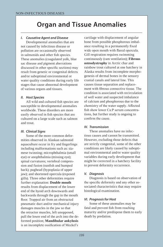
116
NON-INFECTIOUS DISEASES
Organ and Tissue Anomalies
I. Causative Agent and Disease Developmental anomalies that are
not caused by infectious disease or pollution are occasionally observed in salmonids and other fish species. These anomalies (coagulated yolk, blue sac disease and pigment aberrations discussed in other specific sections) may result from genetic or congenital defects and/or suboptimal environmental or water quality conditions during early life stages that cause abnormal development of various organs and tissues.
II. Host Species All wild and cultured fish species are
susceptible to developmental anomalies worldwide. These disorders are more easily observed in fish species that are cultured on a large scale such as salmon and trout.
III. Clinical Signs Some of the more common defor-
mities observed in Alaskan salmonid aquaculture occur in fry and fingerlings including malformations such as: sia-mese twinning; microphthalmia (small eye) or anophthalmia (missing eye); spinal curvatures; vertebral compres-sion and fusion (sunfish and humped back); pughead (hypoplasia of upper jaw); and shortened opercula (exposed gills). Three other deformities require further explanation: Double mouth results from displacement of the lower end of the hyoid arch downwards and backwards through the gap in the mouth floor. Trapped air from an obstructed pneumatic duct and/or mechanical injury damages muscles in the jaw so that the retractor muscles, left unopposed, pull the lower end of the arch into the de-formed position; Mandibular ankylosis is an incomplete ossification of Meckel’s
cartilage with displacement of angular bone from possible phosphorous imbal-ance resulting in a permanently fixed wide open mouth with flared opercula. Gill respiration requires swimming continuously (ram ventilation); Fibrous osteodystrophy in Arctic char and rainbow trout cultured at one hatchery in Alaska results from incomplete morpho-genesis of dermal bones in the sensory cranial canals and lateral line. This causes tissue separation and replace-ment with fibrous connective tissue. The condition is associated with recirculation of well water and suspected imbalance of calcium and phosphorous due to the chemistry of the water supply. Affected fish show lower Ca:P serum concentra-tions, but further study is ongoing to confirm the cause.
IV. Transmission These anomalies have no infec-
tious causes and cannot be transmitted. However, excluding those defects that are strictly congenital, some of the other conditions are likely caused by subopti-mal environmental and/or water quality variables during early development that might be corrected in a hatchery facility to prevent deformity occurrence.
V. Diagnosis Diagnosis is based on observation of
the specific deformity and any other as-sociated characteristics that may require histological examination.
VI. Prognosis for Host Some of these anomalies may be
fatal and prevent fish from reaching maturity and/or predispose them to early death by predation.
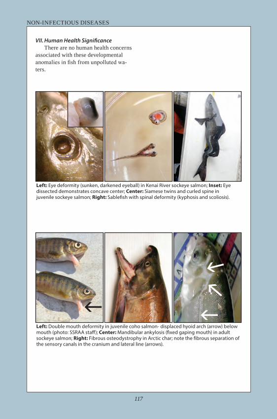
117
NON-INFECTIOUS DISEASES
VII. Human Health Signifcance There are no human health concerns
associated with these developmental anomalies in fish from unpolluted wa-ters.
Left: Eye deformity (sunken, darkened eyeball) in Kenai River sockeye salmon; Inset: Eye dissected demonstrates concave center; Center: Siamese twins and curled spine in juvenile sockeye salmon; Right: Sablefsh with spinal deformity (kyphosis and scoliosis).
Left: Double mouth deformity in juvenile coho salmon- displaced hyoid arch (arrow) below mouth (photo: SSRAA staf); Center: Mandibular ankylosis (fxed gaping mouth) in adult sockeye salmon; Right: Fibrous osteodystrophy in Arctic char; note the fbrous separation of the sensory canals in the cranium and lateral line (arrows).
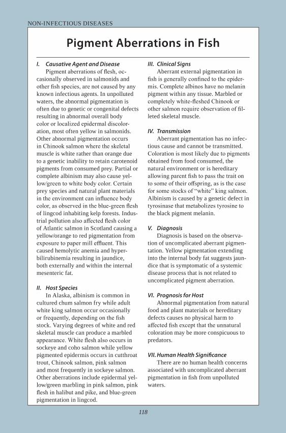
118
NON-INFECTIOUS DISEASES
Pigment Aberrations in Fish
I. Causative Agent and Disease III. Clinical Signs Pigment aberrations of flesh, oc-
casionally observed in salmonids and other fish species, are not caused by any known infectious agents. In unpolluted waters, the abnormal pigmentation is often due to genetic or congenital defects resulting in abnormal overall body color or localized epidermal discolor-ation, most often yellow in salmonids. Other abnormal pigmentation occurs in Chinook salmon where the skeletal muscle is white rather than orange due to a genetic inability to retain carotenoid pigments from consumed prey. Partial or complete albinism may also cause yel-low/green to white body color. Certain prey species and natural plant materials in the environment can influence body color, as observed in the blue-green flesh of lingcod inhabiting kelp forests. Indus-trial pollution also affected flesh color of Atlantic salmon in Scotland causing a yellow/orange to red pigmentation from exposure to paper mill effluent. This caused hemolytic anemia and hyper-bilirubinemia resulting in jaundice, both externally and within the internal mesenteric fat.
II. Host Species In Alaska, albinism is common in
cultured chum salmon fry while adult white king salmon occur occasionally or frequently, depending on the fish stock. Varying degrees of white and red skeletal muscle can produce a marbled appearance. White flesh also occurs in sockeye and coho salmon while yellow pigmented epidermis occurs in cutthroat trout, Chinook salmon, pink salmon and most frequently in sockeye salmon. Other aberrations include epidermal yel-low/green marbling in pink salmon, pink flesh in halibut and pike, and blue-green pigmentation in lingcod.
Aberrant external pigmentation infish is generally confined to the epider-mis. Complete albinos have no melanin pigment within any tissue. Marbled or completely white-fleshed Chinook or other salmon require observation of fil-leted skeletal muscle.
IV. Transmission Aberrant pigmentation has no infec-
tious cause and cannot be transmitted. Coloration is most likely due to pigments obtained from food consumed, the natural environment or is hereditary allowing parent fish to pass the trait on to some of their offspring, as is the case for some stocks of “white” king salmon. Albinism is caused by a genetic defect in tyrosinase that metabolizes tyrosine to the black pigment melanin.
V. Diagnosis Diagnosis is based on the observa-
tion of uncomplicated aberrant pigmen-tation. Yellow pigmentation extending into the internal body fat suggests jaun-dice that is symptomatic of a systemic disease process that is not related to uncomplicated pigment aberration.
VI. Prognosis for Host Abnormal pigmentation from natural
food and plant materials or hereditary defects causes no physical harm to affected fish except that the unnatural coloration may be more conspicuous to predators.
VII. Human Health Signifcance There are no human health concerns
associated with uncomplicated aberrant pigmentation in fish from unpolluted waters.
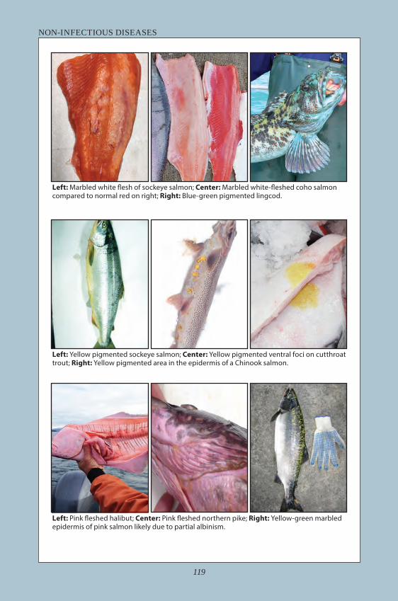
119
NON-INFECTIOUS DISEASES
Left: Marbled white fesh of sockeye salmon; Center: Marbled white-feshed coho salmon compared to normal red on right; Right: Blue-green pigmented lingcod.
Left: Yellow pigmented sockeye salmon; Center: Yellow pigmented ventral foci on cutthroat trout; Right: Yellow pigmented area in the epidermis of a Chinook salmon.
Left: Pink feshed halibut; Center: Pink feshed northern pike; Right: Yellow-green marbled epidermis of pink salmon likely due to partial albinism.

120
NON-INFECTIOUS DISEASES
Sunburn (Back-Peel)
I. Causative Agent and Disease V. Diagnosis Sunburn is a non-infectious disease
in cultured fish caused by overexposure to ultraviolet radiation (UV) from sun-light. Certain diet ingredients causing photosensitization can be predisposing factors. Sunburn is most commonly ob-served during the summer months in the northern latitudes when hatchery fish are moved from an indoor rearing container to shallow outside units with very clear water. Wild fish in shallow lakes and rivers could be potentially susceptible except they rarely remain in direct sun-light long enough for overexposure.
II. Host Species Sunburn is observed almost exclu-
sively in cultured salmonids exposed for long periods to direct sunlight. Other fish species with small delicate scales, partial scaling or no scales at all would also be particularly susceptible.
III. Clinical Signs Lesions from sunburn are first
recognized by a darkening of the skin between the head and the dorsal fin. The epidermal layer turns white and eventually sloughs off. The underlying dermal layer of skin becomes exposed and eventually a white, craterous lesion forms. This lesion can begin with the dorsal fin that first becomes whitened and then erodes to the body surface. Any lesion from sunburn is very likely to become infected with opportunistic bacteria, water molds or fungi.
IV. Transmission Since sunburn is an environmentally
mediated disease, transmission between fish does not occur.
Sunburn is diagnosed by the obser-vation of typical lesions with a history of lengthy exposure to sunlight.
VI. Prognosis for Host When the lesions are uncomplicated
by secondary infections of bacteria or fungi, the mortality is generally quite low. If shade is provided, healing of the lesions is rapid with complete recovery.
VII. Human Health Signifcance There are no human health concerns
associated with sunburn in fish.
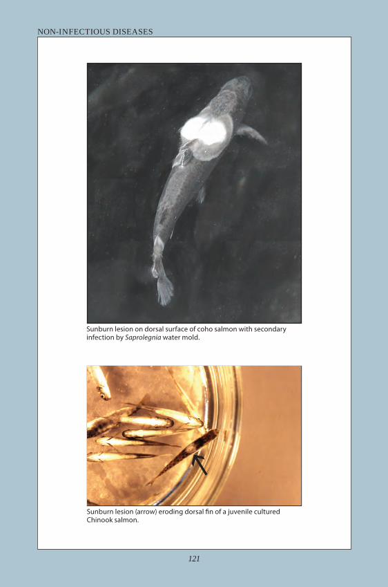
121
NON-INFECTIOUS DISEASES
Sunburn lesion on dorsal surface of coho salmon with secondary infection by Saprolegnia water mold.
Sunburn lesion (arrow) eroding dorsal fn of a juvenile cultured Chinook salmon.
REFERENCE
Glossary of Terms
Acanthocephalans – a phylum of spiny headed worms, these parasites require two hosts for completion of the life cycle and are most commonly found in the in-testines of fish.
Acid-fast – a physical property of some bacteria that are resistant to decoloriza-tion by acids during the staining proce-dure.
Alevin – a newly hatched fish still with yolk sac attached.
Anadromous – relating to fish, such as salmon, that migrate up rivers from the sea to spawn in freshwater.
Anemia – deficiency of red blood cells and/or hemoglobin.
Arthropod – belonging to the phylum Arthropoda, an insect or crustacean that has a cuticle made of chitin forming an exoskeleton with segments and jointed appendages.
Ascites – the presence of fluid in the ab-dominal cavity.
Bacteria – any of a large group of uni-cellular prokaryotic organisms that lack a cell nucleus, reproduce by fission or by forming spores, and in some cases cause disease.
Basophilic – tissue components having an affinity for dye under basic pH condi-tions that stain blue (as in histology).
Buccal cavity – cavity inside the mouth anterior to the gill arches.
Caudal peduncle – the region of the fish body between the end of the anal fin and
the base of the caudal fin.
Cercariae – infectious larvae of digene-ans usually released from the gastropod first intermediate host.
Cestode – a tapeworm with a modified head segment or scolex used for attach-ment, usually in the gut. Tapeworms generally require three hosts for develop-ment.
Copepod – small planktonic crustaceans which are an important part of the aquatic food chain. Some are parasitic.
Coracidium – the ciliated first stage of an aquatic cestode hatched from an egg that, when ingested, infects the first intermedi-ate host, typically a crustacean.
CPE (Cytopathic Efect) – damage to cul-tured cells caused by virus infection.
Crustacean – an arthropod having a segmented body and jointed appendages with two pairs of antennae at some stage in their life cycle.
Cyprinid – a fish of the Cyprinidae family consisting of carps, shiners and minnows.
Cyst – a capsule of connective tissue formed by the host around a foreign body, such as a parasite, that acts as an irritant.
Cytoplasm – the fluid-like substance that fills the cell, consisting of cytosol and or-ganelles excluding the nucleus.
Diplobacilli – paired rod-shaped bacte-rial cells.
DNA – deoxyribonucleic acid containing the genetic information for the reproduc-
122
REFERENCE
tion, development and function of living organisms including some viruses.
Electron microscopy – use of an electron microscope that generates an electron beam focused through a series of mag-netic lenses to create an enlarged image of ultrastructural details at thousands of times higher magnification than a stan-dard light microscope.
ELISA – enzyme-linked immunosorbent assay is an antigen/antibody reaction coupled with an enzyme substrate that produces a color change measured in a spectrophotometer. The test is used to detect the presence of a target organism antigen or an antibody directed towards a target organism.
Encyst – to enclose in a cyst.
Endoparasitic – a parasite that lives within the body of another organism rath-er than on the surface.
Eosinophilic – a red color of cells or tis-sues in histological sections or smears that have been stained with eosin dye.
Epibiont – an organism that uses the body surface of another as a substrate but takes no nourishment or other benefit.
Epithelium – one or more layers of spe-cialized cells forming the covering of most internal and external surfaces of the body and its organs.
Epizootics – an outbreak of a disease in an animal population or an unusually large increase in prevalence and/or inten-sity of a parasite.
Erythema – an abnormal red color of the skin or other tissues caused by capillary congestion.
Erythrocyte – red blood cell.
Exophthalmia (popeye) – abnormal pro-trusion of the eyeball, often from fluid imbalance.
Explant culture – tissue that is placed in a culture medium for either growth of the tissue cells or growth of an organism con-tained in the tissues.
Extracellular – outside the cell.
Fibrotic nodules – focal areas of excess fibrous tissue formed as a reparative pro-cess or as a reaction to a foreign body.
Final host (defnitive host) – the host in which a parasite develops into an adult form to reproduce.
Fluorescent antibody test (FAT) – a test using antibody against a specific patho-gen or primary antibody that is conjugat-ed with a fluorescein dye. The conjugated antibody sticks to the target organism causing fluorescence when viewed with a fluorescent microscope.
Fungi – heterotrophic organisms (with chitinous cell walls) that form spores pro-ducing hyphae which penetrate dead or living material to obtain nutrients.
Furuncle – boil like lesion in the muscu-lature.
Gill Lamellae – gill filaments bear many branches known as lamellae covered by a single layer of epithelium and each containing a blood capillary. Lamellae increase the surface area of the gill fila-ments to enhance respiration and gas ex-change from ambient water.
Gram-negative rod – a rod-shaped bac-terium that does not retain the crystal vio-let in the Gram stain, but is stained by the pink safranin counter stain.
Gram-positive rod – a rod-shaped bac-
123
REFERENCE
terium that retains the crystal violet in the Gram stain and is dark purple in color.
Granuloma – a chronic focal inflamma-tory lesion that walls off a foreign body and usually consists of different types of host inflammatory cells and fibroblastic connective tissue.
Hemagglutinate – the clumping together of red blood cells.
Hemorrhage – occurrence of blood with-in the tissues outside the normal vascular channels.
Hemostasis – the ability of an organism or cell to maintain internal equilibrium by adjusting its physiological processes.
Hemotocrits – the packed cell volume of erythrocytes in whole blood expressed as a percentage.
Histological (histology) – the micro-scopic anatomy of cells and tissues as viewed in thin stained sections on glass slides.
Hyperplasia – an increase in the growth of cell numbers of a tissue or organ that may or may not increase its overall size; usually stimulated by an irritant.
Hyphae – long branching vegetative fila-ments of a fungus or certain molds.
Infammation – a host response to tissue damage or irritation comprised of swell-ing, redness, heat (in warm blooded ani-mals), pain, sometimes causing dysfunc-tion of the tissues and organs involved.
Intermediate host – a host in which there is development of the asexual or imma-ture stage of a parasite.
Intracellular – inside the cell.
Lethargy – a state of sluggishness or in-activity.
Macrophage – a large host white blood cell occurring in tissues and in peripheral blood that ingests foreign particles and infectious microorganisms by phagocy-tosis.
Melanocytes – an epidermal cell of neu-ral crest origin capable of synthesizing the black pigment melanin.
Metacercariae – a developmental stage from encysted cercariae of digenean trematodes generally occurring in a sec-ond intermediate host.
Micropyle – the tiny opening in an egg through which a spermatozoon can enter for fertilization.
Miracidium – the ciliated larval stage of a digenean trematode hatching from the egg which infests the first intermediate host, usually a snail.
Mycosis – fungal infection.
Myopathy – a degenerative disease of muscle.
Necropsy – a postmortem examination of an animal.
Nematode – unsegmented worm of the phylum Nematoda, having an elongated, cylindrical body; a roundworm.
Neoplasms/neoplasia – cancer caused by uncontrolled abnormal growth of tis-sue cells.
Operculum – the flap on either side cov-ering the gill chamber in bony fishes.
Opisthaptor – a posterior attachment or-gan in monogenean flukes.
124

REFERENCE
Organogenesis – the process where the embryonal ectoderm, mesoderm and en-doderm differentiate and develop into the internal organs of the juvenile and adult fish.
Parasite – an organism that lives on or within another organism at whose ex-pense it obtains some advantage.
Paratenic host – an additional or option-al intermediate host in which no develop-ment of the parasite occurs but is a host which may serve as an essential link in the completion of the parasite’s life cycle.
Pathogen – an infectious agent that can cause disease.
PCR (Polymerase Chain Reaction) – amplification of targeted lengths of RNA or DNA by repeated thermal cycling reac-tions using polymerase enzyme.
Pericardium – the membrane surround-ing the heart.
Peritonitis – inflammation of the lining of the abdominal cavity (peritoneum).
Petechial hemorrhage – a small focal or pinpoint hemorrhage.
Pinheading – young fish that exhibit an emaciated body from poor feeding re-sponse producing the appearance of an enlarged head.
Piscivorous – fish eating.
Plaque Forming Unit (PFU) – number of infectious virus particles per unit volume based on the number of holes or plaques in the monolayer of the infected cell cul-ture.
Plerocercoid – the third larval stage of cestodes parasitizing fish that have an ob-vious scolex. Generally found in the sec-
ond intermediate fish host.
Poikilothermic – animals with internal body temperatures that cannot be self-regulated, often determined by the ambi-ent temperature of the environment; cold-blooded.
Proboscis – any of various elongate feed-ing, defensive, attachment or sensory or-gans of the oral region, found in certain leeches and worms.
Procercoid – the solid first parasitic larva of some tapeworms that develops usually in the body cavity of a copepod.
Prognosis – a prediction of how a disease will progress, and the chance for recov-ery.
Protozoan – any of a large group of sin-gle-celled, usually microscopic, eukary-otic organisms, such as amoebas, ciliates, flagellates, and some molds.
Pycnidia – an asexual structure contain-ing conidia, found in certain fungi.
RNA – ribonucleic acid; the nucleic acid that is used in key metabolic processes for all steps of protein synthesis in all living cells and carries the genetic information for many viruses.
Salmonid – belonging or pertaining to the family Salmonidae including salmon, trout, char, and whitefishes.
Scolex – the head segment of a cestode that attaches to its host.
Scoliosis – lateral deviation in the nor-mally straight line of the spine.
Septate – divided by crosswalls or septa.
Septicemia – presence of bacteria in the blood.
125
REFERENCE
Sequencing – techniques used to deter- Trematodes – worms with a characteris-mine the nucleotide sequence of RNA or tic flattened oval to elongated shape hav-DNA in the genome of an organism or ing oral and ventral suckers and a com-virus to help obtain a taxonomic identi- plex (digenetic) life cycle involving two or fication. three hosts.
Serotype – a unique antigenic property of Virulence – the pathogenicity or ability a bacterial cell or virus identified by sero- of an infectious agent to produce disease. logical methods.
Virus – a very small infectious agent Sporangia – a single or many celled composed of a nucleic acid core (RNA or structure from which spores or zoospores DNA) surrounded by a protein coat that are produced in a fungus or mold. replicates only within living host cells.
Spore – a reproductive structure that is Viscera – internal organs of an animal. adapted for dispersion and survival for extended periods of time in unfavorable Vitelline – relating to or associated with conditions. the yolk of an egg.
Teleost –the group of fishes with a bony skeleton.
126
REFERENCE
Fish Disease References
Beakes GW, Glockling SL, Sekimoto S. 2012. The evolutionary phylogeny of the oomycete “fungi”. Protoplasma 249: 3-19.
Bentz CR, Hogge CI, Meyers TR, Ferguson JA. 2012. Myxobolus neurotropus infecting rainbow trout in Alaska, a new geographic record. Journal of Aquatic Animal Health 24:225-231.
Deiterich, Robert A. 1981. Alaskan Wildlife Diseases. University of Alaska. Fairbanks, Alaska.
Di Cicco E, Ferguson HW, Kaukinen KH, Schulze AD, Li S, Tabata A, Gunther OP, Mordecai G, Suttle CA, Miller KM. 2018. The same strain of Piscine orthoreovirus (PRV-1) is involved with the development of different, but related, diseases in Atlantic and Pacific salmon in British Columbia. FACETS https://doi.org/10.1139/facets-2018-0008.
Falk K, Aamelfot M, Bendik DO, Meyers TR, Iversen SA, White WR, Bollinger L, Jahrling PB, Kuhn JH, Lewis CE, Loiacono CM, White D. 2015. Viral Hemorrhagic Fevers of Animals Caused by Negative-Strand RNA Viruses. Chapter 11 pages 247-317 In (Shapshak P, Sinnott JT, Somboonwit C, Kuhn JH (Eds). Global Virology I, Springer.
Fish Health Section/AFS. 2016. Fish Health Section Bluebook: Suggested Procedures for the Detection and Identification of Certain Finfish and Shellfish Pathogens. https://units. fisheries.org/fhs/fish-health-section-blue-book-2016/section-1-diagnostic/
Foox J, Siddall ME. 2015. The road to Cnidaria: History of Phylogeny of the Myxozoa. Journal of Parasitology 101: 269-274.
Freeman MA, Fuss J, Kristmundsson A, Keeling PJ, Shalchian-Tabrizi K, Bass D. 2017. Divergent fish parasites related to perkinsids and dinoflagellates. Current Biology 27:1645–1651.
Garver K, Marty GD, Cockburn SN, Richard J, Hawley LM, Müller A, Thompson RL, Purcell MK, Saksida S. 2016. Piscine reovirus, but not Jaundice Syndrome, was transmissible to Chinook Salmon, Oncorhynchus tshawytscha (Walbaum), Sockeye Salmon, Oncorhynchus nerka (Walbaum), and Atlantic Salmon, Salmo salar L. Journal of Fish Diseases 39: 117-128.
Gauthier DT, Rhodes MW. 2008. Mycobacteriosis in fishes: A review. Veterinary Journal 180: 33-47.
Hoffman, GL. 1999. Parasites of North American Freshwater Fishes. 2nd ed. Cornell University Press. Ithaca, New York.
Kent, ML. 1992. Diseases of Seawater Netpen-Reared Salmonid Fishes of the Pacific Northwest. Department of Fisheries and Oceans. Nanaimo, B.C., Canada.
Kibenge FSB, Godoy MG (Eds.). 2016. Aquaculture Virology. Academic Press.
Lom, J, Dykova I 1992. Protozoan Parasites of Fishes. Elsevier Science Publishers. Amsterdam, The Netherlands.
Margolis L, Kabata Z. 1988. Guide to the Parasites of Fishes of Canada, Part II Crustacea Department of Fisheries and Oceans. Ottawa, Canada.
Meyers TR. 2007. First report of erythrocytic inclusion body syndrome (EIBS) in Chinook salmon Oncorhynchus tshawytscha in Alaska, USA. Diseases of Aquatic Organisms 76: 169-172.
Meyers TR. 2017. Piscine orthoreovirus (PRV) in the Pacific Northwest appears to be of low risk to wild Pacific salmonids. Pacific Northwest Fish Health Protection Committee, Infor. Report No. 10.
127
128
Fish Disease References
Meyers TR, Batts WN. 2016. Paramyxoviruses of Fish. Pages. 259-265 In Kibenge F, Godoy M. (Eds) Aquaculture Virology, Elsevier.
Mitchum DL. 1995. Parasites of Fishes in Wyoming. Wyoming Game and Fish Department. Cheyenne,Wyoming.
Moulton JE. 1990. Tumors in Domestic Animals, California Press. Berkeley, California.
Post G. 1987. Textbook of Fish Health. T. F. H. Publications. Neptune City, New Jersey.
Purcell MK, Powers RL, Evered J, Kerwin J, Meyers TR, Stewart B, Winton JR. 2018. Molecular testing of adult Pacific salmon and trout (Oncorhynchus spp.) for several RNA viruses demonstrates widespread distribution of piscine orthoreovirus in Alaska and Washington. Journal Fish Diseases 41: 347-355.
Roberts RJ. 1989. Fish Pathology, 2nd edit. Bailliere Tindall. London, England.
Roberts RJ, Shepherd CJ. 1986. Handbook of Trout and Salmon Diseases, 2nd edit. Fishing News Books. Oxford, Great Britain.
Schaperclaus W. 1991. Fish Diseases Volume I and II. Amerind Publishing, New Delhi.
Sindermann CJ. 1990. Principal Diseases of Marine Fish and Shellfish Volume 1, 2nd edit. Academic Press. San Diego, California.
Stoskpof MK. 1993. Fish Medicine. W. B. Saunders Company, Pennsylvania.
Untergasser D. 1989. Handbook of Fish Diseases. T. F. H. Publications. Neptune City, New Jersey.
Warren JW. 1991. Diseases of Hatchery Fish, sixth edition. USFWS, Pacific Region.
Wessel O, Braaen S, Alarcon M, Haatveit H, Roos N, Markussen T, Tengs T, Dahle MK, Rimstad E. 2017. Infection with purified Piscine orthoreovirus demonstrates a causal relationship with heart and skeletal muscle inflammation in Atlantic salmon. PLoS ONE 12(8): e0183781.
Wolf, Ken. 1988. Fish Viruses and Fish Viral Diseases. Cornell University Press. Ithaca.
World Organization for Animal Health (OIE). 2018. Manual of Diagnostic Tests for Aquatic Animals. OIE, Paris, France.
128

128128

Y UKON R I V ER DR A INAGE FISHER IES A SSOCI ATION
Related Documents