Diseases of the Adrenal Glands. Adrenal Insufficiency. Adrenal Hyperfunction. Hormonally Active Tumors. Essentials of Diagnosis, Treatment and Prevention of Major Endocrine Diseases LECTURE IN INTERNAL MEDICINE FOR IV COURSE STUDENTS M. Yabluchansky, L. Bogun, L. Martymianova, O. Bychkova, N. Lysenko, m. bRYNZA V.N. Karazin National University Medical School’ Internal Medicine Dept.

Welcome message from author
This document is posted to help you gain knowledge. Please leave a comment to let me know what you think about it! Share it to your friends and learn new things together.
Transcript
Diseases of the Adrenal Glands. Adrenal Insufficiency. Adrenal Hyperfunction.
Hormonally Active Tumors.
Essentials of Diagnosis, Treatment and Prevention of Major Endocrine Diseases
LECTURE IN INTERNAL MEDICINE FOR IV COURSE STUDENTS
M. Yabluchansky, L. Bogun, L. Martymianova, O. Bychkova, N. Lysenko, m. bRYNZA V.N. Karazin National University Medical School’ Internal Medicine Dept.
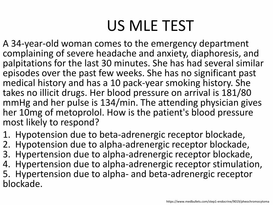
US MLE TEST A 34-year-old woman comes to the emergency department complaining of severe headache and anxiety, diaphoresis, and palpitations for the last 30 minutes. She has had several similar episodes over the past few weeks. She has no significant past medical history and has a 10 pack-year smoking history. She takes no illicit drugs. Her blood pressure on arrival is 181/80 mmHg and her pulse is 134/min. The attending physician gives her 10mg of metoprolol. How is the patient's blood pressure most likely to respond? 1. Hypotension due to beta-adrenergic receptor blockade, 2. Hypotension due to alpha-adrenergic receptor blockade, 3. Hypertension due to alpha-adrenergic receptor blockade, 4. Hypertension due to alpha-adrenergic receptor stimulation, 5. Hypertension due to alpha- and beta-adrenergic receptor blockade.
https://www.medbullets.com/step1-endocrine/9019/pheochromocytoma
US MLE TEST
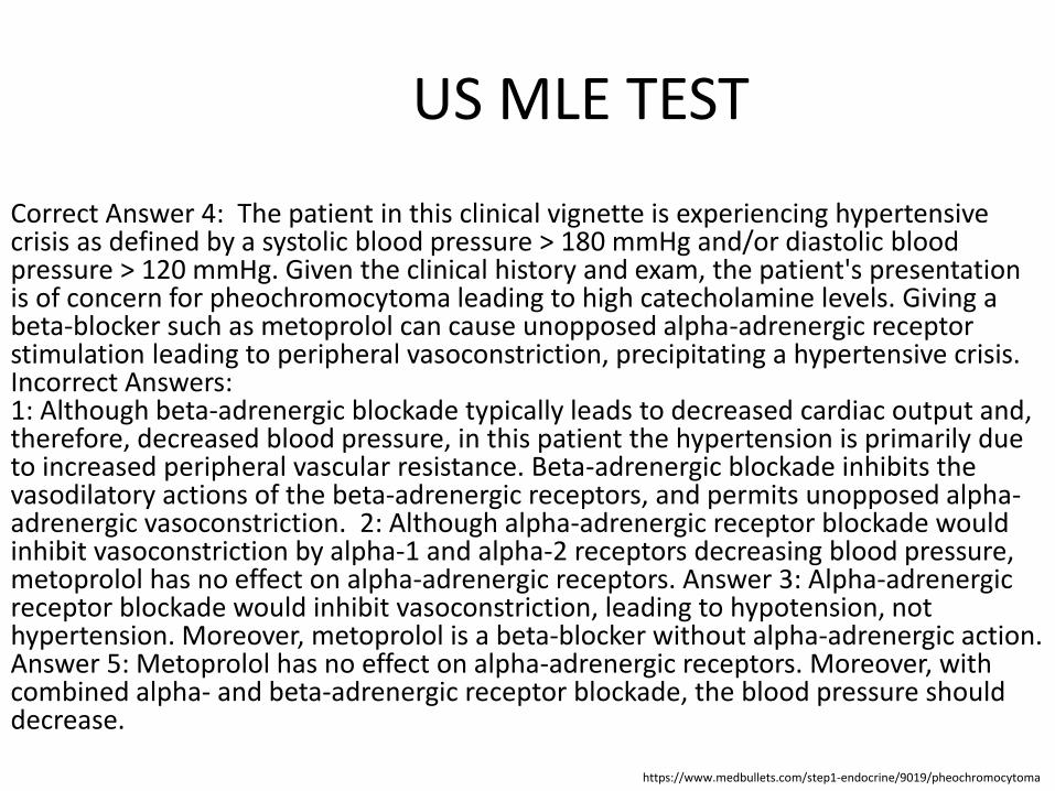
Correct Answer 4: The patient in this clinical vignette is experiencing hypertensive crisis as defined by a systolic blood pressure > 180 mmHg and/or diastolic blood pressure > 120 mmHg. Given the clinical history and exam, the patient's presentation is of concern for pheochromocytoma leading to high catecholamine levels. Giving a beta-blocker such as metoprolol can cause unopposed alpha-adrenergic receptor stimulation leading to peripheral vasoconstriction, precipitating a hypertensive crisis. Incorrect Answers: 1: Although beta-adrenergic blockade typically leads to decreased cardiac output and, therefore, decreased blood pressure, in this patient the hypertension is primarily due to increased peripheral vascular resistance. Beta-adrenergic blockade inhibits the vasodilatory actions of the beta-adrenergic receptors, and permits unopposed alpha-adrenergic vasoconstriction. 2: Although alpha-adrenergic receptor blockade would inhibit vasoconstriction by alpha-1 and alpha-2 receptors decreasing blood pressure, metoprolol has no effect on alpha-adrenergic receptors. Answer 3: Alpha-adrenergic receptor blockade would inhibit vasoconstriction, leading to hypotension, not hypertension. Moreover, metoprolol is a beta-blocker without alpha-adrenergic action. Answer 5: Metoprolol has no effect on alpha-adrenergic receptors. Moreover, with combined alpha- and beta-adrenergic receptor blockade, the blood pressure should decrease.
https://www.medbullets.com/step1-endocrine/9019/pheochromocytoma
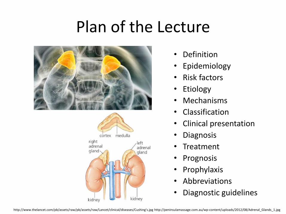
Plan of the Lecture
• Definition
• Epidemiology
• Risk factors
• Etiology
• Mechanisms
• Classification
• Clinical presentation
• Diagnosis
• Treatment
• Prognosis
• Prophylaxis
• Abbreviations
• Diagnostic guidelines
http://www.thelancet.com/pb/assets/raw/pb/assets/raw/Lancet/clinical/diseases/Cushing's.jpg http://peninsulamassage.com.au/wp-content/uploads/2012/08/Adrenal_Glands_1.jpg
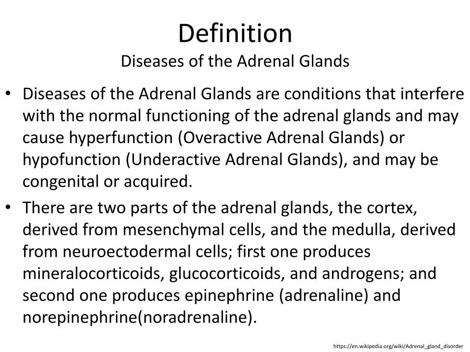
Definition Diseases of the Adrenal Glands
• Diseases of the Adrenal Glands are conditions that interfere with the normal functioning of the adrenal glands and may cause hyperfunction (Overactive Adrenal Glands) or hypofunction (Underactive Adrenal Glands), and may be congenital or acquired.
• There are two parts of the adrenal glands, the cortex, derived from mesenchymal cells, and the medulla, derived from neuroectodermal cells; first one produces mineralocorticoids, glucocorticoids, and androgens; and second one produces epinephrine (adrenaline) and norepinephrine(noradrenaline).
https://en.wikipedia.org/wiki/Adrenal_gland_disorder

Epidemiology Epidemiologic study of adrenal gland disorders in Japan
• The total numbers of patients in Japan in 1997 were estimated as 1,450 for primary aldosteronism, 1,250 for Cushing's syndrome, 290 for adrenal preclinical Cushing's syndrome, 660 for Addison's disease, and 1,030 for pheochromocytoma
• For the first time, a reliable national estimation of the prevalence of disorders of adrenal hormones was conducted in this study
http://www.eurorad.org/eurorad/view_figure.php?pubid=11230&figid=34889&nr=1&lang=en http://www.ncbi.nlm.nih.gov/pubmed/10915016
Abdominal ultrasonography revealed a 5cm heterogeneous nodule in the right adrenal gland

Risk Factors • There does not appear to be a particular group who is most at risk for
adrenal gland disorders
• While there are certain risk factors for adrenal gland disorders, most of them occur without apparent cause, where risk factors are absent
• The risk of development adrenal diseases may be increased by genetic predisposition; by a problem in another gland, such as the pituitary gland, or when a disease or infection affects one or both of the adrenal glands; and certain lifestyle behaviors and environmental factors, such as smoking and exposure to carcinogens
• Adrenal gland disorders appear to affect men, women, and children equally and may develop at any age.
http://www.cancercenter.com/adrenal-cancer/risk-factors/ sharecare.com/health/adrenal-gland/who-risk-adrenal-gland-disorders
Etiology
• Congenital anomalies (genetic mutations)
• Immunopathology
• Infections (e.g., tuberculosis, acquired immune deficiency syndrome (AIDS))
• A problem in another glands, such as the pituitary, which regulate the adrenal gland
• Tumors including pheochromocytomas
• Bilateral adrenal hemorrhage or infarction
• Bilateral adrenalectomy
• Certain medicines https://medlineplus.gov/adrenalglanddisorders.html

Mechanisms
• The major disorders of the adrenal cortex are characterized by excessive or deficient secretion of each type of adrenocortical hormone accordingly to interference of etiologic and pathogenetic factors that include distress, injury, vascular reactions, thrombosis, necrosis, acute and chronic inflammation, dystrophy, hypertrophy, neoplastic processes, etc.
• The most common disorder of the adrenal medulla is pheochromocytoma, that originates from chromaffin cells and excretes catecholamines, but pheochromocytoma may be referred to as secreting paragangliomas when found in extra-adrenal chromaffin cells; neoplasms, such as neuroblastomas and ganglioneuromas, may also be of neuronal lineage.
http://accessmedicine.mhmedical.com/content.aspx?bookid=339§ionid=42811321 ncbi.nlm.nih.gov/pmc/articles/PMC2576282/

International Classification of Diseases
http://apps.who.int/classifications/icd10/browse/2016/en#/E00-E07
Chapter IV (E00-E90) Endocrine, nutritional and metabolic diseases
(E20-E35) Disorders of other endocrine glands
E26 Hyperaldosteronism: E26.0 Primary hyperaldosteronism, E26.1 Secondary hyperaldosteronism, E26.8 Other hyperaldosteronism (Bartter syndrome), E26.9 Hyperaldosteronism, unspecified
E27 Other disorders of adrenal gland: E27.0 Other adrenocortical overactivity, E27.1 Primary adrenocortical insufficiency, E27.2 Addisonian crisis, E27.3 Drug-induced adrenocortical insufficiency, E27.4 Other and unspecified adrenocortical insufficiency, E27.5 Adrenomedullary hyperfunction, E27.8 Other specified disorders of adrenal gland, E27.9 Disorder of adrenal gland, unspecified
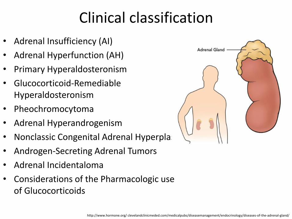
Clinical classification • Adrenal Insufficiency (AI)
• Adrenal Hyperfunction (AH)
• Primary Hyperaldosteronism
• Glucocorticoid-Remediable Hyperaldosteronism
• Pheochromocytoma
• Adrenal Hyperandrogenism
• Nonclassic Congenital Adrenal Hyperplasia
• Androgen-Secreting Adrenal Tumors
• Adrenal Incidentaloma
• Considerations of the Pharmacologic use of Glucocorticoids
http://www.hormone.org/ clevelandclinicmeded.com/medicalpubs/diseasemanagement/endocrinology/diseases-of-the-adrenal-gland/

Adrenal Insufficiency Etiology of Primary Adrenal Insufficiency
• The most common cause of primary adrenal insufficiency (AI) is autoimmune destruction of the adrenal glands (Addison’s disease - 70%-90% of all causes
• Some other causes of primary AI include bilateral adrenal hemorrhage, drugs (e.g., mitotane, etomidate, ketoconazole), infectious diseases (e.g., tuberculosis, HIV infection, disseminated histoplasmosis, paracoccidiomycosis)) affects glucocorticoid and mineralocorticoid secretion and may be fatal if untreated
• Addison’s disease may coexist with other autoimmune conditions, such as type 1 diabetes, hypothyroidism, or hypoparathyroidism.
clevelandclinicmeded.com/medicalpubs/diseasemanagement/endocrinology/diseases-of-the-adrenal-gland/

Adrenal Insufficiency Etiology of Secondary and Tertiary Adrenal Insufficiency
• Secondary and tertiary AI occur commonly after the discontinuation of glucocorticoids
• Less frequently, adrenocorticotropic hormone (ACTH) deficiency may be caused by pituitary macroadenomas, pituitary surgery or radiation, and parasellar diseases
• Megestrol acetate, an appetite stimulator used in some patients with advanced cancer or cachexia related to AIDS may be associated with secondary AI
• Tertiary AI results from the inadequate secretion of corticotropin-releasing hormone (CRH0
• Secondary and tertiary AI only affect cortisol secretion, because ACTH has only a minor role in regulation of aldosterone secretion.
clevelandclinicmeded.com/medicalpubs/diseasemanagement/endocrinology/diseases-of-the-adrenal-gland/
Adrenal Insufficiency Manifestations
• All patients with primary AI complain of fatigue, anorexia, and weight loss; skin hyperpigmentation, initially on the extensor surfaces, palmar creases, and buccal mucosa, results from increased levels of ACTH and other pro-opiomelanocortin–related peptides, including melanocyte-stimulating hormone
• Secondary AI manifests more insidiously with lack of skin hyperpigmentation, salt craving, metabolic acidosis, and hyperkalemia, because mineralocorticoid secretion is intact; fatigue, hyponatremia, and hypoglycemia are some of the clinical manifestations in secondary adrenal insufficiency.
clevelandclinicmeded.com/medicalpubs/diseasemanagement/endocrinology/diseases-of-the-adrenal-gland/
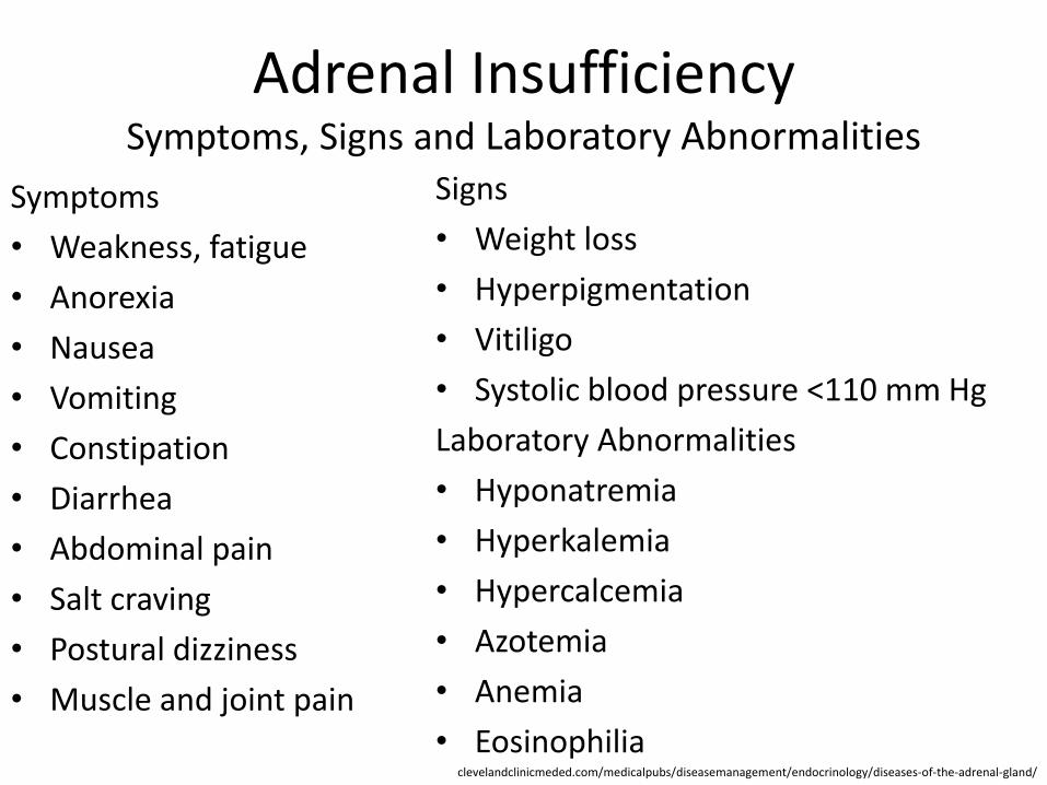
Adrenal Insufficiency Symptoms, Signs and Laboratory Abnormalities
Symptoms
• Weakness, fatigue
• Anorexia
• Nausea
• Vomiting
• Constipation
• Diarrhea
• Abdominal pain
• Salt craving
• Postural dizziness
• Muscle and joint pain
clevelandclinicmeded.com/medicalpubs/diseasemanagement/endocrinology/diseases-of-the-adrenal-gland/
Signs
• Weight loss
• Hyperpigmentation
• Vitiligo
• Systolic blood pressure <110 mm Hg
Laboratory Abnormalities
• Hyponatremia
• Hyperkalemia
• Hypercalcemia
• Azotemia
• Anemia
• Eosinophilia

Adrenal Insufficiency Symptoms, Signs and Laboratory Abnormalities
https://healedbybacon.files.wordpress.com/2015/11/addisons.png?w=607&h=661
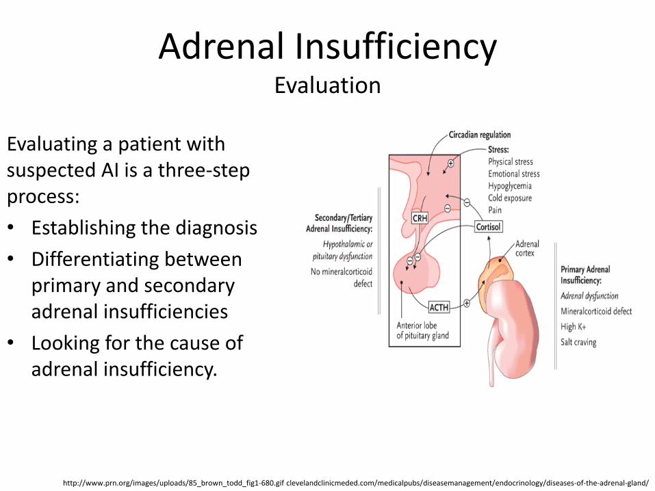
Adrenal Insufficiency Evaluation
Evaluating a patient with suspected AI is a three-step process:
• Establishing the diagnosis
• Differentiating between primary and secondary adrenal insufficiencies
• Looking for the cause of adrenal insufficiency.
http://www.prn.org/images/uploads/85_brown_todd_fig1-680.gif clevelandclinicmeded.com/medicalpubs/diseasemanagement/endocrinology/diseases-of-the-adrenal-gland/

Adrenal Insufficiency Diagnosis
• An early morning (8 am) plasma cortisol level lower than 3 μg/dL confirms AI, whereas a value higher than 15 μg/dL makes the diagnosis highly unlikely
• Cortisol levels in the range of 3 to 15 μg/dL may be seen in patients with primary, secondary, or tertiary adrenal insufficiency
• These patients should be evaluated by the cosyntropin (Cortrosyn) stimulation test (CST) at any time during the day.
clevelandclinicmeded.com/medicalpubs/diseasemanagement/endocrinology/diseases-of-the-adrenal-gland/

Adrenal Insufficiency Differentiation Primary and Secondary Adrenal Insufficiencies
• Differentiation Between Primary and Secondary AI is done through the measurement of the basal plasma ACTH level
• An elevated ACTH level is consistent with primary AI
• A low or normal-range ACTH level, with a low cortisol level, confirms the diagnosis of secondary or tertiary AI.
https://courses.washington.edu/conj/bess/feedback/cortisol/cortisoldisorders.html clevelandclinicmeded.com/medicalpubs/diseasemanagement/endocrinology/diseases-of-the-adrenal-gland/

Adrenal Insufficiency Determining the Cause of Adrenal Insufficiency
• When the biochemical workup is consistent with primary AI, computed tomography (CT) scanning of the adrenal glands may help with the differential diagnosis
• Enlarged adrenal glands or calcifications suggest an infectious, hemorrhagic, or metastatic cause
• In rare circumstances, CT-guided percutaneous fine-needle aspiration of enlarged adrenal glands may help establish the diagnosis
• Patients with tuberculous AI usually have evidence of active systemic disease
• Magnetic resonance imaging (MRI) of the pituitary gland is indicated if glucocorticoid therapy as the cause of the secondary AI has been ruled out.
https://clevelandclinicmeded.com/medicalpubs/diseasemanagement/endocrinology/diseases-of-the-adrenal-gland/

US MLE TEST A 53-year-old nurse presents to the emergency department after experiencing a syncopal episode at work. She reports a 6-month history of worsening fatigue, 20-pound weight loss, and dizziness when standing up from a sitting position. Her PMH is significant for hypothyroidism, for which she takes levothyroxine. Her blood pressure while supine is 105/60, which falls to 75/50 when standing. On exam, she appears fatigued, and you note hyperpigmentation of the gums as shown in Figure A. Labs are notable for sodium of 128 mmol/L, and potassium of 5.6 mEq/L. Which of the following is the most likely pathology underlying this presentation? 1. Autoimmune adrenalitis, 2. Infectious adrenalitis, 3. Hemorrhagic infarction of the adrenal glands, 4. Metastatic disease involving the adrenal glands, 5. Medication-induced suppression of the adrenal glands
https://www.medbullets.com/step1-endocrine/9019/pheochromocytoma

US MLE TEST
Correct Answer 1: This clinical presentation is consistent with Addison’s disease with associated hyponatremia and hyperkalemia; the most common cause of Addison’s disease in the United States is autoimmune adrenalitis.
Incorrect Answers: Answers 2-4: While all of these are potential causes of Addison’s disease (primary adrenal insufficiency), autoimmune adrenalitis is the most common cause of Addison’s disease in the United States.
https://www.medbullets.com/step1-endocrine/9019/pheochromocytoma
Adrenal Insufficiency Treatment
• Patients with Addison’s disease require lifelong replacement with glucocorticoids and mineralocorticoids; the dose is adjusted based on clinical status, including the presence or absence of orthostatic hypotension, hypertension, and electrolyte imbalance
• Patients with secondary or tertiary adrenal insufficiency do not need mineralocorticoid replacement
• Most patients can be educated to self administer hydrocortisone, and reduce the risk of an emergency room visit
• All patients should wear some form of identification indicating their adrenal insufficiency status.
https://clevelandclinicmeded.com/medicalpubs/diseasemanagement/endocrinology/diseases-of-the-adrenal-gland/

Adrenal Insufficiency Alert Medical ID
For patients with AI, medical identification (ID) necklaces or bracelets are absolutely essential with information of their diagnosis and
treatment regimen.
s-media-cache-ak0.pinimg.com/736x/ca/e6/1f/cae61f17ec45a8c94cac00ef6be4af62.jpg s-media-cache-ak0.pinimg.com/236x/28/d8/d5/28d8d55e4765ff4a30ed0473ddbc8 .laurenshope.com/stainless-red-oval-medical-id-tag? clevelandclinicmeded.com/medicalpubs/diseasemanagement/endocrinology/diseases-of-the-adrenal-gland/

Adrenal Insufficiency Adrenal Crisis
• Adrenal crisis (acute AI) is a life-threatening emergency, which usually manifests with nausea, vomiting, abdominal pain, abdominal tenderness, fever, acute abdomen and shock
• Patients may be previously undiagnosed or have chronic primary AI, with no or inadequate glucocorticoid replacement
• Treatment of adrenal crisis should not be delayed
• Diagnostic workup in a patient with no history of AI should include a plasma sample for cortisol and ACTH level determination, immediately followed by an intravenous (IV) bolus of hydrocortisone, 100 mg, and adequate fluid replacement (normal saline)
• Hydrocortisone should be continued, 50 mg every 8 hours, while awaiting laboratory results.
https://clevelandclinicmeded.com/medicalpubs/diseasemanagement/endocrinology/diseases-of-the-adrenal-gland/

US MLE TEST A 46-year-old female with a history of type I diabetes mellitus presents with complaints of chronic fatigue, muscle weakness, and headaches. These symptoms have been present for the past year. Her vital signs today are as follows: T 36.9 C, HR 82 bpm, BP 88/52 mmHg, RR 14 rpm, O2 sat98% on room air. Physical exam does not demonstrate any specific abnormalities. After inquiry, the patient reports that her blood pressure has always "run low." Initial work-up reveals an 8AM cortisol level of 3.6 ug/dL. Which of the following is the best next step in management of this patient? 1. Low dose dexamethasone suppression test 2. High dose dexamethasone suppression test 3. Urgent 5mg hydrocortisone injection 4. Cosyntropin stimulation test 5. Pituitary MRI
https://www.medbullets.com/step1-endocrine/9019/pheochromocytoma

US MLE TEST
Correct Answer 4: This patient's presentation of fatigue, weakness, and headaches in the setting of low blood pressures and a decreased 8 AM cortisol level is suggestive of adrenal insufficiency. A cosyntropin stimulation test is the next diagnostic step in differentiating primary from secondary adrenal insufficiency and determining the etiology of this patient's low cortisol level. Incorrect Answers: 1 & 2: A dexamethasone suppression test is appropriate for work-up of elevated cortisol levels, not depressed levels as seen in this vignette. 3: Work-up to identify the cause of this patient's adrenal insufficiency should be pursued prior to initiating adrenal replacement therapy. This patient's presentation is not consistent with an acute adrenal crisis.5: Secondary adrenal insufficiency should be confirmed prior to pursuit of this expensive diagnostic test.
https://www.medbullets.com/step1-endocrine/9019/pheochromocytoma
Adrenal Insufficiency Adrenal Insufficiency in the Critically Ill Patient
• The overall incidence of AI in critically ill patients is less than 10%, but an incidence as high as 50% in a patient with septic shock has been reported
• Intensive care unit (ICU) patients with hemodynamic instability, despite fluid resuscitation (especially in the presence of shock), should be tested for AI
• Hydrocortisone, 50 mg IV every 6 to 8 hours is an adequate replacement dose for critically ill patients with suspected AI
• Treatment with this dose should be continued for 2 or 3 days
• After hemodynamic improvement, a gradual taper of hydrocortisone, depending on the patient’s condition, should be instituted.
https://clevelandclinicmeded.com/medicalpubs/diseasemanagement/endocrinology/diseases-of-the-adrenal-gland/
Adrenal Hyperfunction Selected List of Related Disorders
• Disorders related to adrenal hyperfunction (AH) are relatively rare, but they have significant mortality and morbidity if untreated
• When the adrenal glands produce too many hormones, the symptoms vary depending on the disorder
• Disorders related to AH include:
• Cushing’s syndrome
• Primary hyperaldosteronism
• Pheochromocytoma/Paraganglioma
• Hormonally Active Tumors.
https://clevelandclinicmeded.com/medicalpubs/diseasemanagement/endocrinology/diseases-of-the-adrenal-gland/

Adrenal Hyperfunction Cushing’s Syndrome: Definition and Classification
• Cushing’s syndrome (CS) is composed of symptoms and signs associated with prolonged exposure to inappropriately high levels of plasma glucocorticoids
• Exogenous glucocorticoid intake is the most common cause of CS
• The endogenous causes are divided into ACTH-dependent and ACTH-independent CS.
https://csrf.net/wp-content/uploads/2013/06/symptoms.gif clevelandclinicmeded.com/medicalpubs/diseasemanagement/endocrinology/diseases-of-the-adrenal-gland/

Adrenal Hyperfunction Cushing’s Syndrome: Etiology of Endogenous Form
clevelandclinicmeded.com/medicalpubs/diseasemanagement/endocrinology/diseases-of-the-adrenal-gland
ACTH-Dependent
Cushing's syndrome (67%)
Ectopic ACTH secretion (12%)
Ectopic CRH secretion (<1%)
ACTH-Independent
Adrenal adenoma (10%)
Adrenal carcinoma (8%)
Micro-and macronodular adrenal hyperplasias (1%)

Adrenal Hyperfunction Cushing’s Syndrome: Clinical Features
http://img.medscape.com/fullsize/migrated/474/901/nf474901.tab1.gif

Adrenal Hyperfunction Symptoms, Signs and Laboratory Abnormalities
https://thumbs.dreamstime.com/z/cushings-disease-syndrome-signs-symptoms-cushing-s-labeled-human-silhouette-internal-organs-vector-illustration-55259613.jpg
Adrenal Hyperfunction Cushing’s Syndrome: Establishing the Diagnosis
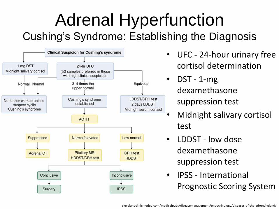
• UFC - 24-hour urinary free cortisol determination
• DST - 1-mg dexamethasone suppression test
• Midnight salivary cortisol test
• LDDST - low dose dexamethasone suppression test
• IPSS - International Prognostic Scoring System
clevelandclinicmeded.com/medicalpubs/diseasemanagement/endocrinology/diseases-of-the-adrenal-gland/
Adrenal Hyperfunction Cushing’s Syndrome: Differential Diagnosis 1
• Once CS is biochemically confirmed, the plasma ACTH level should be measured, preferably in the morning
• A suppressed or low ACTH level (<10 pg/mL) is consistent with ACTH-independent CS and should be followed by adrenal CT scanning
• ACTH values of 10 to 20 pg/mL may be seen in patients with both adrenal and pituitary causes for CS; these patients should undergo a CRH stimulation test
• A flat response of ACTH to CRH during the test suggests an adrenal cause, but a more than 50% increase in the ACTH level during the test is consistent with Cushing’s disease
• ACTH levels higher than 20 pg/mL suggest ACTH-dependent CS.
clevelandclinicmeded.com/medicalpubs/diseasemanagement/endocrinology/diseases-of-the-adrenal-gland/

Adrenal Hyperfunction Cushing’s Syndrome: Differential Diagnosis 2
• About 90% of patients with ACTH-dependent CS have a pituitary cause and the rest are ectopic in origin
• ACTH levels tend to be higher in ectopic CS compared with Cushing’s disease, but there is significant overlap
• A more than 50% increase in the ACTH level after the CRH test and more than an 80% reduction in the morning cortisol level (8-9 am) after taking 8 mg dexamethasone at 11 pm during a high-dose DST is consistent with a pituitary source, and in the presence of a pituitary adenoma, almost establishes the definitive diagnosis of CS.
clevelandclinicmeded.com/medicalpubs/diseasemanagement/endocrinology/diseases-of-the-adrenal-gland/

Adrenal Hyperfunction Cushing’s Syndrome: Differential Diagnosis 3
• . If the CRH stimulation test and DST results are not concordant and MRI does not show a pituitary adenoma, then inferior petrosal sinus sampling to distinguish ectopic from Cushing’s disease is indicated
• Localizing tumors that produce ectopic ACTH is accomplished by chest and abdominal CT studies, followed by neck CT if no source is found
• An octreotide scan may be of some value in patients with ectopic CS and negative imaging studies.
clevelandclinicmeded.com/medicalpubs/diseasemanagement/endocrinology/diseases-of-the-adrenal-gland/

US MLE TEST
A 35-year-old Caucasian female presents to the hospital alarmed by her recent truncal weight gain, facial hair growth, and thinning skin. During the physical exam, the physician finds that the patient is hypertensive. Serum analysis reveals hyperglycemia. The physician suspects a pituitary adenoma. Which dexamethasone test result would help confirm the physician's suspicions?
1. Low-dose, increased ACTH; high-dose, increased ACTH 2. Low-dose, increased ACTH; high-dose, decreased ACTH 3. Low-dose, decrease in ACTH; high-dose, no change in ACTH 4. Low-dose, no change in ACTH; high-dose, no change in ACTH 5. Low-dose, no change in ACTH; high-dose, decreased ACTH
https://www.medbullets.com/step1-endocrine/9019/pheochromocytoma
US MLE TEST
Correct Answer 5: A dexamethasone suppression test in a patient with a pituitary ACTH-secreting adenoma yields no change in ACTH level with a low-dose and a decrease in ACTH level with a high-dose. Incorrect Answers: 1, 2, and 3 are not typical results of a dexamethasone test. 4: No change in ACTH levels with both a low and high-dose dexamethasone administration is consistent with an ectopic ACTH source or with an adrenal tumor.
https://www.medbullets.com/step1-endocrine/9019/pheochromocytoma

Adrenal Hyperfunction Cushing’s Syndrome: Treatment
• Surgical (trans-sphenoidal) removal of the ACTH-secreting pituitary tumor is the treatment of choice
• CS caused by an adrenal adenoma is usually cured by laparoscopic unilateral adrenalectomy
• Adrenal carcinoma is an aggressive tumor with a poor prognosis; surgical resection at an early stage, along with lifelong mitotane therapy soon after surgery, offers the only chance for cure or long-term remission
• Surgical removal of an ectopic ACTH-producing tumor results in cure
• Medical therapy with ketoconazole, metyrapone, aminoglutethimide, or mitotane may be considered for patients with a limited life expectancy or for alleviation of hypercortisolemic symptoms before surgery.
clevelandclinicmeded.com/medicalpubs/diseasemanagement/endocrinology/diseases-of-the-adrenal-gland/

US MLE TEST
A 50-year-old female is evaluated by her physician for recent weight gain. Physical examination is notable for truncal obesity, wasting of her distal musculature and moon facies. In addition she complains of abnormal stretch marks that surround her abdomen. The physician suspects pituitary adenoma. Which of the following high-dose dexamethasone suppression test findings and baseline ACTH findings would support his view? 1. Cortisol suppression, normal baseline ACTH 2. Cortisol suppression, high baseline ACTH 3. No cortisol suppression, high baseline ACTH 4. No cortisol suppression, low baseline ACTH 5. Elevation of cortisol above pre-test levels, high baseline ACTH
https://www.medbullets.com/step1-endocrine/9019/pheochromocytoma
US MLE TEST
Correct Answer 2: The patient in this scenario has symptoms of Cushing’s syndrome. In the setting of high baseline ACTH, suppression of cortisol with a high dose of dexamethasone indicates pituitary adenoma as the cause of disease. Incorrect Answers: 1: Cortisol suppression after dexamethasone administration amid normal baseline ACTH levels is a normal finding in patients without Cushing’s syndrome. 3: A lack of cortisol suppression after dexamethasone administration amid high baseline ACTH levels indicates an ectopic ACTH-producing tumor. Examples include small cell lung cancer and bronchial carcinoids. 4: A lack of cortisol suppression after dexamethasone administration amid low baseline ACTH levels is diagnostic of a cortisol-producing tumor, such as an adrenal adenoma. 5: Dexamethasone administration does not increase serum cortisol above pre-test values.
https://www.medbullets.com/step1-endocrine/9019/pheochromocytoma
Primary Hyperaldosteronism Prevalence
• Conn first described primary hyperaldosteronism (PH) in 1955 in a patient with an adrenal adenoma
• A prevalence as high as 10% in hypertensive patients, and women in their fourth to sixth decade of life are affected more often than men
• A solitary aldosterone-producing adenoma (65%) and bilateral idiopathic hyperplasia (30%) are the most common subtypes of PH
• The adenomas are usually benign and smaller than 2 cm in diameter
• Idiopathic adrenal hyperplasia may be accompanied by adrenocortical nodules and is associated with lower aldosterone levels and less severe hypertension, compared with adenomas.
clevelandclinicmeded.com/medicalpubs/diseasemanagement/endocrinology/diseases-of-the-adrenal-gland/

Primary Hyperaldosteronism Causes
http://images.slideplayer.com/26/8443752/slides/slide_15.jpg

Primary Hyperaldosteronism Forms of Familial Hyperaldosteronism
Two forms of familial primary hyperaldosteronism (FH) have been described:
• FH type I, or glucocorticoid-remediable hyperaldosteronism (GRH), is an autosomal dominant disease characterized by a chimeric gene between the 11β-hydroxylase and aldosterone synthase, with varying degrees of hyperaldosteronism, which responds to exogenous glucocorticoids
• FH type II is an autosomal dominant disorder of both the aldosterone-producing adenoma (APA) and idiopathic hyperaldosteronism (IHA).
clevelandclinicmeded.com/medicalpubs/diseasemanagement/endocrinology/diseases-of-the-adrenal-gland/
Primary Hyperaldosteronism Clinical Presentation
• The clinical picture varies from asymptomatic to symptoms related to hypertension, hypokalemia, or both
• Patients may have headaches, polyuria, nocturia, polydipsia, parasthesias, weakness, and muscle cramps
• There are no specific physical findings
• The degree of hypertension is usually moderate to severe, and may be refractory to conventional antihypertensive agents
• Malignant hypertension and leg edema are rare
• The left ventricular hypertrophy is disproportionate to the level of blood pressure and improves after treatment of PH, even if hypertension persists.
clevelandclinicmeded.com/medicalpubs/diseasemanagement/endocrinology/diseases-of-the-adrenal-gland/

Primary Hyperaldosteronism Signs and Symptoms
http://www.cell.com/cms/attachment/2007953169/2030530657/gr2.jpg

Primary Hyperaldosteronism laboratory Tests
• Routine laboratory tests may show slightly high serum sodium levels (143-147 mEq/L), hyperglycemia, hypokalemia, metabolic alkalosis, and hypomagnesemia
• Although most patients with hyperaldosteronism are not hypokalemic, a low serum potassium level may be noted, either spontaneously or after thiazide or loop diuretic use
• Hypokalemia may be severe and difficult to correct
• Hypokalemia presence reduces the secretion of aldosterone and thus should be corrected before the laboratory evaluation of hyperaldosteronism.
clevelandclinicmeded.com/medicalpubs/diseasemanagement/endocrinology/diseases-of-the-adrenal-gland/

Primary Hyperaldosteronism Diagnosis
• The workup of a patient for PH involves the following steps: screening tests for PH, establishing the autonomy of aldosterone secretion, and determination of the source of hyperaldosteronism
• The categories of patients:
– hypertensive with spontaneous or thiazide-induced hypokalemia
– hypertensive resistant to therapy
– hypertensive with adrenal incidentalomas.
clevelandclinicmeded.com/medicalpubs/diseasemanagement/endocrinology/diseases-of-the-adrenal-gland/

Primary Hyperaldosteronism Diagnosis
Adrenal adenoma on Computer Tomography http://columbiasurgery.org/conditions-and-treatments/primary-hyperaldosteronism-conns-syndrome

Primary Hyperaldosteronism Screening Tests
• Although hypokalemia in a hypertensive patient is suggestive of hyperaldosteronism, normokalemia does not exclude the diagnosis
• Measurement of the 24-hour urinary potassium level can be useful in assessing the cause of a low potassium level
• Urinary potassium excretion >30 mEq/24 hours in a patient with hypokalemia suggests PH, if plasma renin activity (PRA) is low
• The ratio of the plasma aldosterone concentration (PAC) to PRA (PAC/PRA) is the best screening test for PH
• Spironolactone and eplerenone should be discontinued for 6 weeks before testing
• Low or suppressed PRA during therapy with angiotensin-converting enzyme (ACE) inhibitors or angiotensin receptor blockers (ARBs) suggests hyperaldosteronism.
clevelandclinicmeded.com/medicalpubs/diseasemanagement/endocrinology/diseases-of-the-adrenal-gland/

Primary Hyperaldosteronism Differentiating Aldosterone-Producing Adenoma From
Idiopathic Hyperaldosteronism
clevelandclinicmeded.com/medicalpubs/diseasemanagement/endocrinology/diseases-of-the-adrenal-gland/
Parameter APA IHA
HTN, potassium concentration [K+]
HTN more severe, higher likelihood of hypokalemia
HTN less severe, less likelihood of hypokalemia
Upright posture test Decrease or <30% increase of serum aldosterone level
Increase by >30% of serum aldosterone level
18-Hydroxycorticosterone >100 ng/mL <100 ng/mL
Computed tomography scan
>1-cm adrenal tumor, with normal contralateral adrenal
No adrenal tumor, bilateral thickening of adrenals
Bilateral adrenal venous sampling Lateralization No lateralization
APA, aldosterone-producing adenoma; HTN, hypertension; IHA, idiopathic hyperaldosteronism
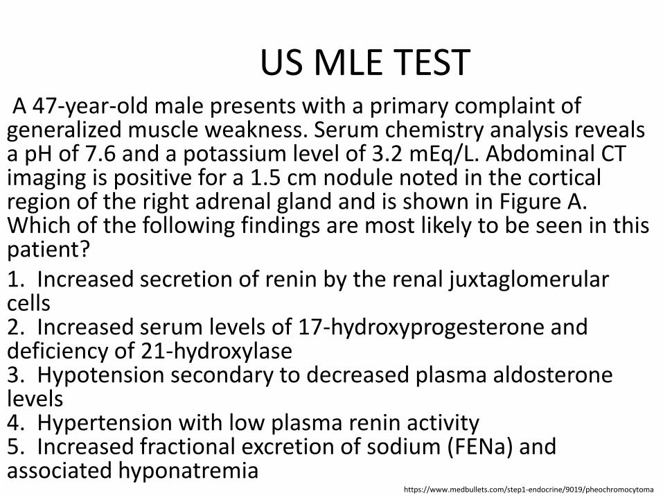
US MLE TEST A 47-year-old male presents with a primary complaint of generalized muscle weakness. Serum chemistry analysis reveals a pH of 7.6 and a potassium level of 3.2 mEq/L. Abdominal CT imaging is positive for a 1.5 cm nodule noted in the cortical region of the right adrenal gland and is shown in Figure A. Which of the following findings are most likely to be seen in this patient? 1. Increased secretion of renin by the renal juxtaglomerular cells 2. Increased serum levels of 17-hydroxyprogesterone and deficiency of 21-hydroxylase 3. Hypotension secondary to decreased plasma aldosterone levels 4. Hypertension with low plasma renin activity 5. Increased fractional excretion of sodium (FENa) and associated hyponatremia
https://www.medbullets.com/step1-endocrine/9019/pheochromocytoma

US MLE TEST
Correct Answer 4: This patient’s presentation is consistent with primary hyperaldosteronism (Conn’s syndrome) due to a benign adenoma in the zona glomerulosa layer of the adrenal cortex. Hypertension and low plasma renin activity are characteristic findings of this condition. Incorrect Answers: 1: Primary hyperaldosteronism is characterized by low plasma renin activity. 2: Deficiency of the 21-hydroxylase enzyme would reduce adrenal production of mineralocorticoids (aldosterone) and glucocorticoids, shunting precursors instead towards increased androgen production. 3: Hypertension, not hypotension, would be expected in a state of aldosterone excess. 5: Decreased excretion of sodium would be anticipated in the setting of increased aldosterone; hypernatremia is a rare finding but is consistent with hyperaldosteronism.
https://www.medbullets.com/step1-endocrine/9019/pheochromocytoma
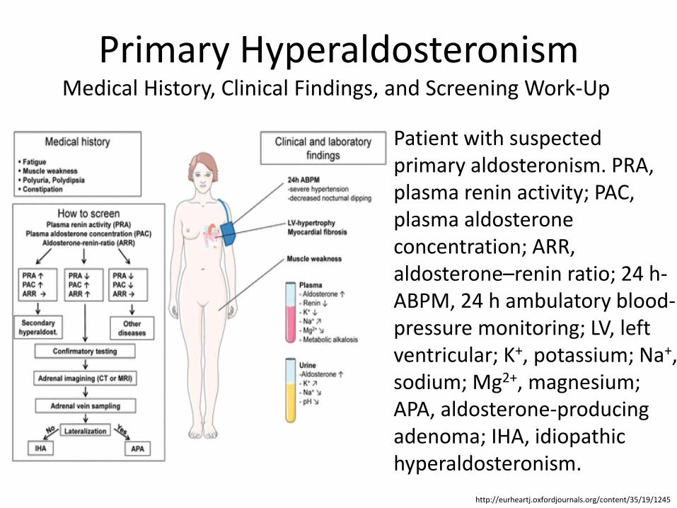
Primary Hyperaldosteronism Medical History, Clinical Findings, and Screening Work-Up
Patient with suspected primary aldosteronism. PRA, plasma renin activity; PAC, plasma aldosterone concentration; ARR, aldosterone–renin ratio; 24 h-ABPM, 24 h ambulatory blood-pressure monitoring; LV, left ventricular; K+, potassium; Na+, sodium; Mg2+, magnesium; APA, aldosterone-producing adenoma; IHA, idiopathic hyperaldosteronism.
http://eurheartj.oxfordjournals.org/content/35/19/1245

Primary Hyperaldosteronism Treatment
• The treatment goals are to reduce the morbidity and mortality
• Unilateral adrenalectomy results in normalization of hypokalemia and improvement in hypertension in all patients
• Medical treatment is reserved for patients with IHA or those with APAs who are poor surgical candidates
• Spironolactone or Eplerenone is the treatment of choice
• Side effects include painful gynecomastia, nausea, headaches, impotence, and irregular menstruation; Eplerenone that blocks the aldosterone receptor selectively has a better side effect profile
• The use of a mineralocorticoid is usually not necessary.
clevelandclinicmeded.com/medicalpubs/diseasemanagement/endocrinology/diseases-of-the-adrenal-gland/
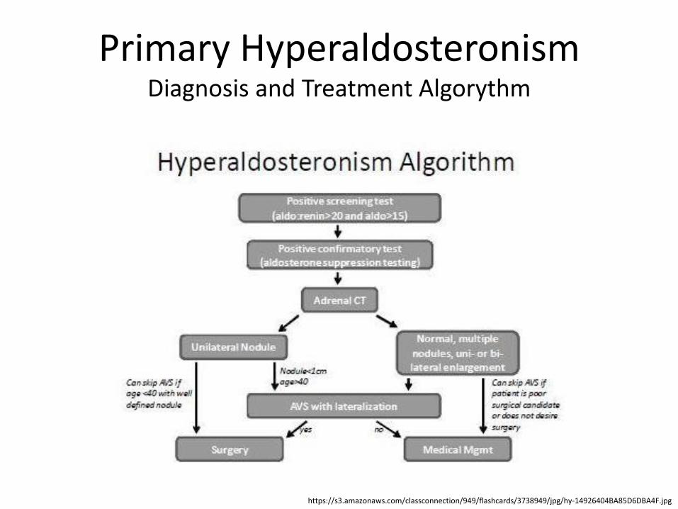
Primary Hyperaldosteronism Diagnosis and Treatment Algorythm
https://s3.amazonaws.com/classconnection/949/flashcards/3738949/jpg/hy-14926404BA85D6DBA4F.jpg

Primary Hyperaldosteronism Glucocorticoid-Remediable Hyperaldosteronism
• Familial hyperaldosteronism type I, or glucocorticoid-remediable hyperaldosteronism (GRH) individuals are younger 40 years, exhibit hypertension resistant to standard therapy, and have a family history of primary hyperaldosteronism or a cerebrovascular accident at a young age
• Most have a normal serum potassium level
• A very high urinary 18-hydroxycortisol level (>3000 nmol/24 hr) or genetic testing to detect a chimeric gene establishes the diagnosis
• Affected individuals should have neurovascular screening for cerebral aneurysms
• Treatment with glucocorticoids, eplerenone, spironolactone, amiloride, and triamterene is effective.
clevelandclinicmeded.com/medicalpubs/diseasemanagement/endocrinology/diseases-of-the-adrenal-gland/

US MLE TEST A 33-year-old male presents to his primary care physician with complaints of headaches and muscle weakness. His physical exam is entirely within normal limits except for a blood pressure of 150/95. Subsequent routine blood lab work showed a sodium level of 146 and potassium level of 3.0. What is the best pharmacological therapy for this patient?
1. Hydrochlorthiazide 2. Spironolactone 3. Propanolol 4. Lisinopril 5. Fludrocortisone
https://www.medbullets.com/step1-endocrine/9019/pheochromocytoma

US MLE TEST
Correct Answer 2: The patient in this vignette most likely has hyperaldosteronism given his hypernatremic, hypokalemic hypertension. The best treatment would be an aldosterone antagonist like spironolactone. Incorrect Answers: 1: Hydrochlorothiazide would worsen this patient's hypokalemia, and is therefore not the best choice. 3: Propanolol is not a first line drug for hypertension and is therefore not the best choice. 4: Lisinopril wound not be treating the most likely underlying cause of this patient's hypertension and electrolyte disorder, and is therefore not the best choice. 5: Fludrocortisone is used in the treatment of Addison's disease in which a patient has insufficient production of aldosterone.
https://www.medbullets.com/step1-endocrine/9019/pheochromocytoma
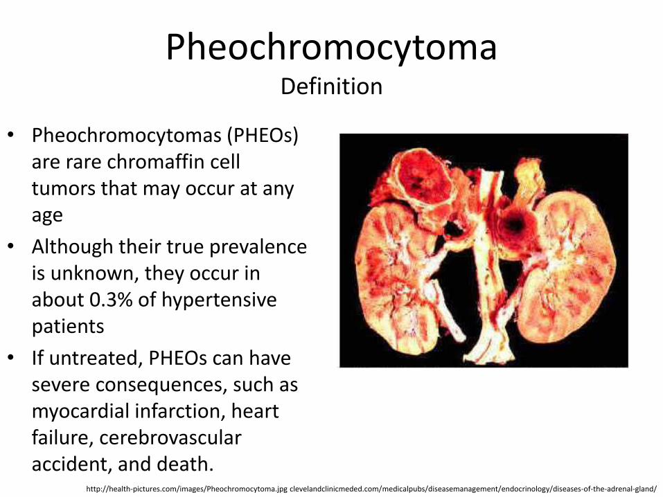
Pheochromocytoma Definition
• Pheochromocytomas (PHEOs) are rare chromaffin cell tumors that may occur at any age
• Although their true prevalence is unknown, they occur in about 0.3% of hypertensive patients
• If untreated, PHEOs can have severe consequences, such as myocardial infarction, heart failure, cerebrovascular accident, and death.
http://health-pictures.com/images/Pheochromocytoma.jpg clevelandclinicmeded.com/medicalpubs/diseasemanagement/endocrinology/diseases-of-the-adrenal-gland/

Pheochromocytoma Anatomic Considerations and Etiology
• Most PHEOs are benign, sporadic, unilateral, and located within the adrenal gland
• Extra-adrenal PHEOs (paragangliomas) occur in about 15% of cases in the superior and inferior para-aortic areas, including the Zuckerkandl organ, bladder, thorax, and head, neck, and pelvis
• Paragangliomas tend to occur in younger patients <20 years); they are multifocal in about 15% to 30% of cases
• Bilateral PHEOs are usually seen as part of familial syndromes
• Malignant PHEOs have a higher prevalence in ectopic PHEOs and lower prevalence in familial PHEOs
• About 10% to 15% of PHEOs are hereditary in nature.
clevelandclinicmeded.com/medicalpubs/diseasemanagement/endocrinology/diseases-of-the-adrenal-gland/

Pheochromocytoma Clinical Manifestations
• Patients with PHEO may have paroxysmal hypertension (48%) or persistent hypertension (29%), or be normotensive (13%).
• Wide fluctuations in blood pressure and resistance to antihypertensive medications are typical of those with PHEO
• The triad of headaches, palpitations, and diaphoresis suggests the diagnosis of PHEO, but absence of these symptoms does not exclude the disease
• Attacks are usually precipitated by emotional stress, exercise, anesthesia, abdominal pressure, or ingestion of tyramine- containing foods
• Symptoms include pallor, flushing, orthostatic hypotension, weight loss, dyspnea, polyuria, polydipsia, visual blurring, focal neurologic symptoms, and change in mental status.
http://health-pictures.com/images/Pheochromocytoma.jpg clevelandclinicmeded.com/medicalpubs/diseasemanagement/endocrinology/diseases-of-the-adrenal-gland/

Pheochromocytoma Diagnosis
• The diagnosis can be established by measuring catecholamines and metanephrines in plasma (blood) or through a 24-hour urine collection
• Care should be taken to rule out other causes of adrenergic (adrenalin-like) excess like hypoglycemia, stress, exercise, and drugs affecting the catecholamines like stimulants, methyldopa, dopamine agonists, or ganglion blocking antihypertensives
• Various foodstuffs (e.g. coffee, tea, bananas, chocolate, cocoa, citrus fruits, and vanilla) can also affect the levels of urinary metanephrine and VMA (vanillylmandelic acid)
• Imaging studies of the head, neck, and chest, and abdomen can help localize the tumor.
https://en.wikipedia.org/wiki/Pheochromocytoma#Diagnosis

US MLE TEST
A 49 year-old-male presents with a primary complaint of several recent episodes of severe headache, sudden anxiety, and a "racing heart". The patient originally attributed these symptoms to stress at work; however, these episodes are becoming more frequent and severe. Laboratory evaluation during such an episode reveals elevated plasma free metanephrines. Which of the following additional findings in this patient is most likely? 1. Decreased 24 hour urine vanillylmandelic acid (VMA) levels, 2. Episodic hypertension, 3. Anhidrosis, 4. Diarrhea, 5. Hypoglycemia
https://www.medbullets.com/step1-endocrine/9019/pheochromocytoma

US MLE TEST
Correct Answer 2: This patient’s presentation is consistent with a pheochromocytoma producing excessive levels of catecholamines (epinephrine and norepinephrine). Common symptoms of pheochromocytoma include: episodic hypertension, palpitations, tachycardia, diaphoresis, tremors, anxiety, and headache. Incorrect Answers: 1: VMA levels would be expected to increase in the setting of catecholamine excess, as VMA is a degradation product of both norepinephrine and epinephrine. 3: Hyperhidrosis (excessive sweating), not anhidrosis, is a common manifestation of pheochromocytoma. 4: Increased levels of catecholamines inhibit peristalsis, leading to ileus and decreased intestinal motility, not diarrhea. 5: Hyperglycemia is more common in pheochromocytoma due to increased glycogenolysis and gluconeogenesis.
https://www.medbullets.com/step1-endocrine/9019/pheochromocytoma

Pheochromocytoma Imaging Studies
Computer tomography showing
a 2 cm left adrenal mass
http://www.scielo.br/scielo.php?script=sci_arttext&pid=S1677-55382009000100005
Magnetic resonance imaging showing large
right adrenal mass (arrows)
Magnetic resonance imaging
showing large adrenal mass
Pheochromocytoma Treatment
• Surgical resection of the tumor is the treatment of first choice
• Either surgical option requires prior treatment with the non-specific and irreversible alpha adrenoceptor blocker phenoxybenzamine or a short acting alpha antagonist (e.g. prazosin, terazosin, or doxazosin)
• Once the pheochromocytoma has been resected, thereby removing the major source of circulating catecholamines, a situation arises where there is both very low sympathetic activity and volume depletion; this can result in profound hypotension
• Therefore, it is usually advised to "salt load" pheochromocytoma patients before their surgery.
https://en.wikipedia.org/wiki/Pheochromocytoma#Diagnosis

US MLE TEST
A 34-year-old female presents to the emergency room with headache and palpitations. She is sweating profusely and appears tremulous on exam. Vital signs are as follows: HR 120, BP 190/110, RR 18, O2 99% on room air, and Temp 37C. Urinary metanephrines and catechols are positive. Which of the following medical regimens is contraindicated as a first-line therapy in this patient?
1. Phenoxybenzamine, 2. Nitroprusside, 3. Propranolol 4. Labetalol, 5. Lisinopril
https://www.medbullets.com/step1-endocrine/9019/pheochromocytoma

US MLE TEST
Correct Answer 3: In a patient with pheochromocytoma, NEVER give pure beta-blockade. This would result in unopposed alpha activity from circulating catecholamines and could result in malignant hypertension.
Incorrect Answers: 1: Use of an alpha blocker in conjunction with a beta blocker, like Propranolol, is also not contraindicated. 2: Nitrates, like nitroprusside, are not contraindicated. 4: Use of an agent with combined alpha and beta blockade, such as labetalol is not contraindicated. 5: Use of an ACE-inhibitor like lisinopril is not contraindicated.
https://www.medbullets.com/step1-endocrine/9019/pheochromocytoma
Adrenal Hyperandrogenism The Adrenal Glands as the Source of Androgens
• The adrenal glands are an important source of androgens, especially in children and women
• The primary adrenal androgens, dehydroepiandrosterone (DHEA) and dehydroepiandrosterone sulfate (DHEAS sulfate), are under ACTH control and have little intrinsic androgenic activity
• However, they are converted to androstenedione and then to testosterone (and estrogen) in both the adrenal gland and peripheral tissues
• DHEA and DHEA sulfate are responsible for adrenarche (pubic hair development)
• Peak levels occur in the third decade of life and decline progressively to 25% of peak levels around the age of 80 years.
clevelandclinicmeded.com/medicalpubs/diseasemanagement/endocrinology/diseases-of-the-adrenal-gland
Adrenal Hyperandrogenism Clinical Manifestations in Childhood
• In female neonates, androgen excess causes female pseudohermaphroditism (ambiguous genitalia)
• Male infants exhibit penile enlargement
• At prepuberty androgen excess manifests as increased rate of growth in height and skeletal maturation with premature epiphyseal fusion and short adult height; boys exhibit penile enlargement, hair growth in androgen-dependent areas, deepening of the voice, and other secondary sexual characteristics; girls have hirsutism, acne, and clitoromegaly
• At puberty androgen excess causes premature skeletal maturation and short adult height in boys, and amenorrhea, different degree of virilization, and increased skeletal maturation, resulting in short adult height In girls.
clevelandclinicmeded.com/medicalpubs/diseasemanagement/endocrinology/diseases-of-the-adrenal-gland

Adrenal Hyperandrogenism Clinical Manifestations in Adults
• In men, a decrease in size of the testicles, testosterone secretion, and spermatogenesis caused by inhibition of gonadotropin secretion may occur
• In women, hirsutism, acne, menstrual irregularities, male pattern baldness, infertility, decreased breast tissue, increased muscle mass, android body habitus, and clitoromegaly may occur.
clevelandclinicmeded.com/medicalpubs/diseasemanagement/endocrinology/diseases-of-the-adrenal-gland
Adrenal Hyperandrogenism Management
• Management of hyperandrogenism symptoms
• The use of antiandrogens such as cyproterone acetate, spironolactone, and flutamide
• Cyproterone acetate is a synthetic steroidal antiandrogen, progestin, and antigonadotropin
• Spironolactone is aldosterone antagonists
• Flutamide is a synthetic, non-steroidal antiandrogen
en.wikipedia.org/wiki/Hyperandrogenism clevelandclinicmeded.com/medicalpubs/diseasemanagement/endocrinology/diseases-of-the-adrenal-gland

Nonclassic Congenital Adrenal Hyperplasia
• Nonclassic congenital adrenal hyperplasia is characterized by milder enzyme dysfunction and manifests commonly in adolescence or adulthood
• Although some patients with NCAH may be asymptomatic, hirsutism is the most common symptom
• The ACTH stimulation test is the best screening test for evaluating adrenal gland functions
• Asymptomatic patients with NCAH do not need glucocorticoid treatment
• If the main concern of the patient is infertility, ovulation induction is the treatment of choice
• If the patient seeks medical advice because of hirsutism, anti-androgens may be used.
http://www.ncbi.nlm.nih.gov/pubmed/17551465

Androgen-Secreting Adrenal Tumors
• Primary adrenocortical carcinoma may be associated with excess androgen secretion, and sometimes cortisol may also occur
• Primary adrenocortical carcinoma is a rare disease, with an incidence of 1 per 600,000 to 1,600,000 and a prevalence of 4 to 12 per 1,000,000
• Female patients may exhibit virilization with very high levels of DHEA sulfate (500 μg/dL or higher), testosterone, and urinary 17-ketosteroids
• Primary adrenocortical carcinomas are highly malignant, with a poor prognosis
• Tumors are usually larger than 6 cm, invade the capsule, metastasize early, and typically recur after surgery.
clevelandclinicmeded.com/medicalpubs/diseasemanagement/endocrinology/diseases-of-the-adrenal-gland
Adrenal Incidentaloma
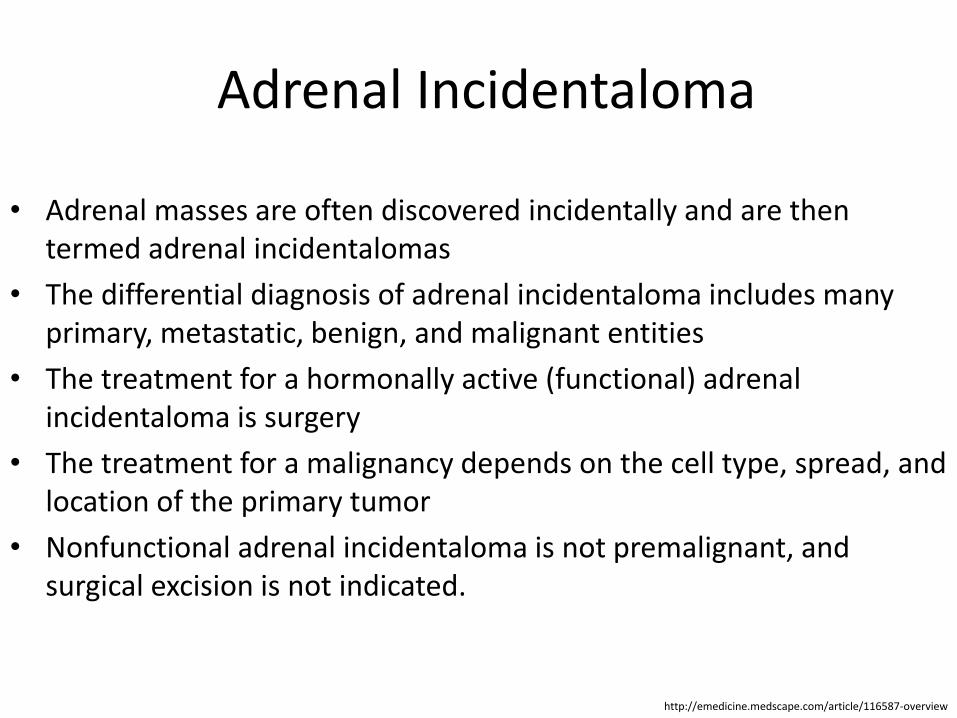
• Adrenal masses are often discovered incidentally and are then termed adrenal incidentalomas
• The differential diagnosis of adrenal incidentaloma includes many primary, metastatic, benign, and malignant entities
• The treatment for a hormonally active (functional) adrenal incidentaloma is surgery
• The treatment for a malignancy depends on the cell type, spread, and location of the primary tumor
• Nonfunctional adrenal incidentaloma is not premalignant, and surgical excision is not indicated.
http://emedicine.medscape.com/article/116587-overview

Considerations of the Pharmacologic use of Glucocorticoids
• Pharmacologic doses of synthetic glucocorticoids are used in a wide variety of diseases for their anti-inflammatory and immunosuppressive effects
• Two main clinical problems arise with the chronic use of pharmacologic doses of glucocorticoids, iatrogenically induced Cushing’s syndrome and adrenal insufficiency on abrupt cessation of therapy
• Alternate-day therapy and avoidance of nighttime doses are associated with less suppression of the hypothalamic–pituitary–adrenal axis after discontinuation of glucocorticoid therapy.
clevelandclinicmeded.com/medicalpubs/diseasemanagement/endocrinology/diseases-of-the-adrenal-gland

Prognosis and Prophylaxis
• Prognosis depends strictly from the diseases nature, their clinical manifestations, quality of treatment and compliance of all its prescriptions
• There are no known lifestyle changes to lower the risk of developing essential adrenal gland disorders
• The following dietary and lifestyle considerations may support healthy adrenal function: avoiding simple carbohydrates, calorie restriction, limiting stimulants, exercising, maintaining a positive outlook and good self esteem, adequate sleep at regular intervals, etc.
http://www.lifeextension.com/Protocols/Metabolic-Health/Adrenal-Disorders/Page-07
Abbreviations
ACTH – adrenocorticotropic hormone
AI - adrenal insufficiency
AIDS - acquired immune deficiency syndrome
AH - adrenal hyperfunction
APA - aldosterone-producing adenoma
DST - 1-mg dexamethasone suppression test
CRH - corticotropin-releasing hormone
CS - Cushing’s syndrome
CST - cosyntropin (Cortrosyn) stimulation test
CT - computed tomography
DHEA - dehydroepiandrosterone
DHEAS - dehydroepiandrosterone sulfate
ICU -intensive care unit
ID – identification
IHA - idiopathic hyperaldosteronism
IV – intravenous
IPSS - International Prognostic Scoring System
FH - familial hyperaldosteronism
GRH - glucocorticoid-remediable hyperaldosteronism
LDDST - low dose dexamethasone suppression test
MRI - magnetic resonance imaging
NCAH - nonclassic congenital adrenal hyperplasia
PH – primary hyperaldosteronism
UFC - 24-hour urinary free cortisol determination

Diagnostic and Treatment Guidelines
• Adrenal Insufficiency and Addison's Disease
• Clinical Guidelines for the Diagnosis and Treatment of Cushing's Disease in Korea
• Addison's Disease: New Guideline Details Diagnosis and Treatment
• Addison Disease: Early Detection and Treatment Principles
• Diseases of the Adrenal Gland
Related Documents