Dipterocarpaceae : Mycorrhizae and Regeneration W.T.M. Smits CENTRALE LAN DBOU WC ATALOG US 0000 0572 5169

Welcome message from author
This document is posted to help you gain knowledge. Please leave a comment to let me know what you think about it! Share it to your friends and learn new things together.
Transcript
Dipterocarpaceae :
Mycorrhizae and Regeneration
W.T.M. Smits
CENTRALE LAN DBOU WC ATALOG US
0000 0572 5169
BlöLlOTaiEKM CKNDBOUWUNIVER S TTlCTi,
WACiF.NINCEN
Promotoren : dr. ir. R.A.A. Oldeman, hoogleraar in de bosteelt en bosoecologie
dr. ir. J. Dekker, emeritus hoogleraar in de fytopathologie
fJ>J0k'2ö't , ß r j
2 1 OKT. f994 W.T.M. Smits i . r-» ^ „
UB-CARDEX
Dipterocarpaceae :
Mycorrhizae and Regeneration
Proefschrift ter verkrijging van de graad van doctor in de landbouw- en milieuwetenschappen op gezag van de rector magnificus, dr. C.M. Karssen, in het openbaar te verdedigen op woensdag 12 oktober 1994 des namiddags te vier uur in de Aula van de Landbouwuniversiteit te Wageningen.
Uitgegeven als boek door Stichting Tropenbos
,':> n bb'n}û
Abstract
Smits, W.T.M. (1994). Dipterocarpaceae : Mycorrhizae and Regeneration. PhD Thesis, Wageningen Agricultural University, The Netherlands, 242 pp., 76 figs., 27 tables, 9 boxes, 8 plates with colour pictures, 242 references, 16 terms in glossary, English, Dutch and Indonesian summaries.
Research on mycorrhizae of Dipterocarpaceae is described, involving inventories of both mycorrhizae and sporocarps in natural forest and experimental work in nurseries, green houses, laboratories and gnotobiotic systems. An assessment is made of dipterocarp mycorrhizal specificity and a discussion is presented on how mycorrhizal specificity may have contributed to speciation in Dipterocarpaceae. Other aspects touched upon include work on a non-ectomycorrhizal association of a fungus with dipterocarp roots, proposed to be called amphymycorrhizae. Also discussed are the effects of physical influences upon dipterocarp ectomycorrhizae, demonstrating the negative impact of high topsoil temperatures and lack of oxygen upon functioning and survival of dipterocarp ectomycorrhizae. Furthermore how dipterocarp ectomycorrhizae influence regeneration of Dipterocarpaceae through enhanced survival near the mother trees. At the end of the book practical recommendations are given for optimalization of management of mixed dipterocarp forests based upon the conclusions reached in the research, including the use of correct fungus-dipterocarp combinations for different sites.
CIP-DATA KONINKLIJKE BIBLIOTHEEK, DEN HAAG
Smits, W.T.M.
Dipterocarpaceae : Mycorrhizae and Regeneration. Thesis Wageningen, - With réf. - With summaries in Dutch, English and Indonesian. ISBN 90-5485-331-X. Subject headings: Dipterocarpaceae/ tropical forestry/ mycorrhizae/ root research ; forests
Tevens uitgegeven als boek door Stichting Tropenbos
;.AJO??0 I> )8ZCI
1) Direct photosynthetic carbohydrate production and translocation from leaves to roots is a precondition for formation of ectomycorrhizal sporocarps associated with Dipterocarps. (This book).
2) The so-called delicate balance of species rich climax tropical rain forest persists because of the robustness of the forest to overcome disturbances.
3) Conservation of primary rain forests through a boycott of tropical timber cannot be reached through individual country actions and will hurt the poor people living near these forests most.
4) Verdere studie van zwammen kan veel toekomstig gezwam over soortvorming voorkomen.
5) Orangutans are great botanists.
6) Crown shyness, without thigmotropy, must be explained through the presence of so far unknown sensitory mechanisms in plants.
7) Specialization in science mostly leads to regular but modest progress, while a more generalistic approach, combining the specialisms and insight in the functioning of the practical world, will bring greatest progress for all.
8) With increasing welfare, biodiversity becomes an increasingly valuable commodity and the present downward trend in number of species surviving will turn upward as a result.
9) There will never be an end to the chain of smaller particles making up matter nor to the space and time matter occupies.
10) To become a democratic political leader one will always face the dilemma that one is expected to have strong principles but nevertheless be ready to abandon ones own principles for the sake of the majority; a good leader must therefore long more for power than anything else and can never be truly trustful to himself or others.
W.T.M. Smits, "Dipterocarpaceae : Mycorrhizae and Regeneration" 12 Oktober 1994, Wageningen, Nederland.
Dipterocarpaceae :
Mycorrhizae and Regeneration
Table of contents
Contents 5
List of figures 3
List of boxes 12
List of tables 13
Preface 15
Acknowledgements 17
Chapter 1 : Introduction 19
1.1 Dipterocarpaceae 19 1.1.1 General 19 1.1.2 History of utilization 22 1.2 Mycorrhizae 25 1.2.1 General 25 1.2.2 Mycorrhizae associated with Dipterocarps 27 1.3 Purpose and outline of the research 27 1.3.1 General setting of the research 28 1.3.2 General description of the research area 30
Chapter 2 : Inventory of mushrooms found near Dipterocarps 33 2.1 Introduction 33 2.2 Methods 33 2.2.1 Layout of the plot 33 2.2.2 Collection and treatment of sporocarps 37 2.2.3 Assessment of the mycorrhizal status and/or specificity of the associations 42 2.3 Results 45 2.3.1 Tree species composition of the plot 45 2.3.2 The mushrooms: keys and descriptions 60 2.3.3 Combinations of Dipterocarps and ectomycorrhizal sporocarps 83 2.3.4 Factors affecting mushroom development 86
Contents - 5 -
a. Influence of rainfall upon sporocarp appearance 86 b. Substrate preferences of several suspected ectomycorrhizal fungi 87 C. The influence of light and temperature upon sporocarp formation 88 d. Relation between physiological condition of phytobionts
and sporocarp formation 88 e. Rate of appearance and deterioration of sporocarps 92
2.4 Discussion 92 2.4.1 Reliability of the survey data with respect to assessing host-specificity 92
a. Omission of small and hypogeous sporocarps 93 b. The limitation to the crown projection areas 93 C. The possibility of other tree species being associated with
the encountered sporocarps 94 d. The limitation to dipterocarp canopy trees 96 e. The differences in vitality of the sampled trees 97 f. The differences in numbers of sampled trees per species 97 g. The possible correlation between topography, site of the
sampled trees and differences in encountered sporocarps 98 h. The possibility of incomplete sampling 98 i. Possible interactions between physical influences and
appearance of sporocarps 101 j . The limitation to one sample plot 101
2.4.2 Specificity 103
Chapter 3 : Inventories of ectomycorrhizae 107 3.1 Introduction 107 3.2 Methods (description and evaluation) 107 3.2.1 Method 1, direct systematic root sampling 107 3.2.2 Method 2, direct root sampling of individual trees 109 3.2.3 Method 3, root sampling on spots of mycobiont sporocarp appearances I l l 3.2.4 Method 4, top soil washing and plotting of ectomycorrhizal types I l l 3.2.5 Method 5, collecting of roots of dipterocarp seedlings resulting
from natural regeneration 113 3.2.6 Method 6, sampling of seedlings resulting from natural
regeneration and transferred to perforons 113 3.2.7 Method 7, planting of non-mycorrhizal dipterocarp seedlings under seed trees
followed by evaluation of mycorrhizal types established 114 3.3 Results 114 3.3.1 Results of direct systematic root sampling 114 3.3.2 Results of top soil washing and plotting of ectomycorrhizal types 117 3.3.3 Results from collecting roots of natural regeneration of Dipterocarps 118 3.4 Discussion 120
Chapter 4 : Inoculation experiments 123 4.1 Introduction 123 4.2 Methods 123 4.2.1 Infection of non-mycorrhizal dipterocarp seedlings related to distance
from large dipterocarp trees 124
4.2.2 Influence of seed collecting method upon mycorrhizal infection after transplanting to medium without inoculum 124
4.2.3 Inoculation of non-mycorrhizal dipterocarp seedlings with chopped sporocarps of potential ectomycorrhizal fungi 125
4.3 Results 125 4.3.1 Correlation between mycorrhizal infection and distance from large dipterocarp trees. 125 4.3.2 Occurrence of first ectomycorrhizal infection 126 4.3.3 Compatibility of phytobiont-mycobiont combinations under greenhouse conditions . 127 4.4 Discussion 128
Chapter 5 : Perforon studies 131 5.1 Introduction 131 5.2 Methods 138 5.2.1 Ectomycorrhizal development in soil and in perforations 138 5.2.2 Process of mycorrhizal infection in perforons and amount of inoculum needed
for growth enhancement of previously non-mycorrhizal plants 138 5.2.3 Rate of root growth and spread of ectomycorrhizae studied in
perforons with Shorea stenoptera and Anisoptera marginata 139 5.2.4 Inter-species exchange of ectomycorrhizal fungi amongst dipterocarp seedlings . . . 139 5.3 Results 140 5.3.1 Influence of perforations upon roots and ectomycorrhizae 140 5.3.2 Infection process, start of growth enhancement and amount of inoculum needed . . . 141 5.3.3 Growth of roots and ectomycorrhizal hyphae 143 5.3.4 Selective mycorrhizal establishment in presence of various ectomycorrhizal fungi . . 143 5.4 Discussion and conclusions 144 5.4.1 Methodology 144 5.4.2 The obligate nature of dipterocarp mycorrhizae 146 5.4.3 Specificity of dipterocarp ectomycorrhizae 147
Chapter 6 : Physical influences upon ectomycorrhizae 149 6.1 Introduction 149 6.2 Methods 149 6.2.1 Experiment 1 (controlled heating of mycorrhizal root systems) 149 6.2.2 Experiment 2 (planting under three different field conditions with varying
light intensities and soil temperatures) 151 6.2.3 Experiment 3 (separation of light effects upon the parts above ground
and soil temperature upon ectomycorrhizae) 152 6.2.4 Experiment 4 (effect of soil compaction) 152 6.2.5 Experiment 5 (effect of soil exposure on older trees) 153 6.3 Results 153 6.3.1 Results of experiment 1 153 6.3.2 Results of experiment 2 153 6.3.3 Results of experiment 3 155 6.3.4 Results of experiment 4 156 6.3.5 Results of experiment 5 156 6.4 Discussion 156
- 7 -
Chapter 7 : A new type of dipterocarp root-fungus association 161 7.1 Introduction 161 7.2 Materials and methods 161 7.3 Results 162 7.4 Discussion 165
Chapter 8 : General discussion 171 8.1 Introduction 171 8.2 Review of the hypotheses (enhanced niche adaptation in
Dipterocarpaceae by mycorrhizal symbiosis and specificity of dipterocarp ectomycorrhizae) 171
8.3 Suitability of the approach followed 175 8.3.1 The location: why Borneo? 175 8.3.2 Methods and approach used 176 8.4 Evidences provided by the current research 176 8.4.1 The obligate character of dipterocarp ectomycorrhizae 177 8.4.2 Specificity of dipterocarp mycorrhizae 179 8.4.3 Spatial isolation of dipterocarp clumps enhanced by mycorrhizae 186 8.4.4 Comparison of the research results with literature 188 8.5 Practical application of the results 191 8.5.1 Implications for natural stand management 191 8.5.2 Consequences for planting of Dipterocarps 193
Summary (English) 196
Samenvatting (Dutch summary) 198
Ringkasan (Indonesian summary) 200
Glossary 202
References 203
Colour Plates 217
Appendices 227 Appendix 1 : Map of the Wanariset I Research Forest Appendix 2 : Map of the Wartono Kadri trail Appendix 3 : Listing of database linking files Appendix 4 : Schematic representation of research set-up Appendix 5 : Crown projections of Shorea laevis in clumps Appendix 6 : Mycorrhizal status of tree species in the Wanariset forest Appendix 7 : Ectomycorrhizal types of Shorea laevis Appendix 8 : List of ectomycorrhizal mushroom species Appendix 9 : Map of Borneo with locations cited
Curriculum Vitae 243
8 -
List of figures
1. Distribution of Dipterocarpoideae in Malesia. Numbers above the hyphen represent the endemics and the number under the hyphen non-endemics.
2. Germination of Vatica chartacea and Dipterocarpus sp. as influenced by the wing-like projections.
3. Early flowering of a one year old Dipterocarpus hasseltii seedling.
4. Map of the island Borneo.
5. Transverse section of an ectomycorrhizal root (schematic). Hartig net between the cortical cells; the mantle covering the root surface.
6. Yellow non-mycorrhizal Shorea laevis Ridl. seedlings. Note the group of mycorrhizal healthy plants.
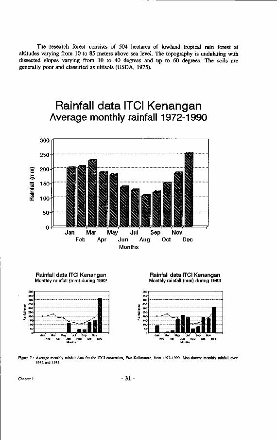
7. Rainfall data for the ITCI concession in East-Kalimantan over a period of 18 years and during the years 1982 and 1983.
8. Arrangement of ironwood markers in the plot.
9. Graph showing the relation between stem diameter and crown diameter for five light red meranti species.
10. Design of the new mushroom drying equipment. Air is circulated freely between the layers of screen on which mushrooms are placed and the container for the water attracting crystals.
11. Graphic presentation of weight loss in the mushroom dryer of Heimiella retispora, Amanita elata and Russuta cf. pectinatoides.
12. Method of making incisions between phytobionts and suspected mycobiont. Length of incisions depended upon distance from stem base.
13. Graphic presentation of the number of species against the number of individuals per tree species.
14. Distribution of number of individuals over three diameter classes divided for three mycorrhizal status groups.
15. Percentages of individuals in three diameter classes divided over ectomycorrhizal, endomycorrhizal and potential ectomycorrhizal trees.
16. Percentage of Dipterocarps of total number of ectomycorrhizal trees over three diameter classes.
17. Drawings of Boletaceae ; a. Boletus ferruginosporus; b. Boletelhts emodensis; c. Strobilomyces polypyramis; d. Tylopihts alboater; e. Pulveroboletus ravenelii; f. Heimiella retispora; g. Boletus off. olivaceirubens; h. Austroboletus dictyotus; i. Tylopilus ballouii; j . Boletus spinifer.
18. Drawings of Amanitae: a. Amanita tjibodensis Boedijn; b. Amanita sychnopyramis Corner &Bas; c. Amanita borneensis; d. Amanita longistriata Imai; e. Amanita elata (Mass.) Comer & Bas; f. Amanita xanthogala Bas; g. Amanita cf. avellaneosquamosa Imai; h. Amanita centunculus Corner & Bas; i. Amanita fritillaria (Berk.) Sacc; j . Amanita similis Boedijn; k. Amanita cf. duplex Comer & Bas.
19. Drawings of Russulaceae: a. Russula eburneoareolata Hongo; b. Russula sp. indet. 2.; c. Russula spec. nov. ?; d Russula cf. pectinatoides Peck; e. Russula senecis Imai; f. Russula cf. metachroa Hongo; g. Russula sp. indet. 7.; h. Russula lilacea Quel. i. Russula japonica Hongo; j . Lactarius subpiperatus Hongo; k Lactarius cf. austrovolemus Hongo.
20. Drawings of other mushrooms: a. Hebeloma vinosophyllum Hongo; b. Cortinarius anomalous (Fr.: Fr.) Fr.; c. Laccaria laccata (Scop.: Fr) B. & Br.; d. Phylloporus bogoriensis Höhn.; e. Phylloporus aff. infundibuliformis (Cleland) Sing.; f. Craterellus verrucosus Mass.; g. Aphelaria dendroides (Jungh.) Corner; h. Scleroderma dictyosporum Pat.; i. Scleroderma cf. columnare.
- 9 -
21. Plotting of ectomycorrhizal trees with crowns in illumination classes 1 and 2, occurring between markers 10.10, 20.10, 20.17 and 10.17, in combination with the ectomycorrhizal sporocarps encountered during six month inventory.
22. Weekly rainfall data (mm) and numbers of Amanitae, Russulaceae and Boletaceae plotted during a three month period between markers 10.10, 10.15, 15.15 and 15.10.
23. A collection of ectomycorrhizal sporocarps collected during a mast flowering season within a period of three hours.
24. Identifications of some of the mushrooms depicted in figure 23.
25. Correlation between flushing of Anisoptera marginata plants and appearance of Inocybe mangayi mushrooms.
26. Mushroom and flush appearance in Anisoptera marginata over a three month period.
27. Number of mushroom appearances per individual plant.
28. Time difference (in days) between the instant of flushing and moment of mushroom appearance.
29. Root system extent of Shorea laevis and Hopea nervosa trees.
30. Appearance of ectomycorrhizal sporocarps in and around ditched plot number 2.
31. Rainfall data over 1990 at the Wanariset location.
32. Shorea laevis actual tree crown projections, calculated projections and presence of Shorea laevis roots.
33. Graphic representation of the correlation of numbers of ectomycorrhizal roots of three types with soil depth compared to total root length (I) with soil depth. I: see table 16. Description of mycorrhizal types in appendix 8.
34. Relation between crown diameter, root extension and appearance of ectomycorrhizal types on the washed roots of Shorea laevis.
35. Distribution of seedlings of Hopea rudiformis in a 2 meter wide strip, 25 meters long, starting from the stem base of a mother tree.
36. Relation between root system architecture and distribution of larger Hopea rudiformis seedlings connected to the roots of the mother tree by ectomycorrhizal connections.
37 Photograph showing the clumped distribution of larger seedlings of Hopea rudiformis along the roots of the mother tree shown within the square of figure 35 on the right.
38. Appearance of ectomycorrhizal sporocarps and distribution of ectomycorrhizal types encountered under Shorea laevis (tree number 224 in plot Wartono).
39. Correlation between distance from large dipterocarp trees and survival of non-mycorrhizal Shorea assamica seedlings.
40. Spread of mycorrhizal infections in a plantbed of non-mycorrhizal S. assamica seedlings.
41. Four types of boxes ("perforons"), suitable for use with the horizontally perforated soil system.
42. An intrascope (non-flexible endoscope) with flexible glass fibre connector to guide the light beam and medical biopsy forceps.
43. Biopsy forceps as used in Wageningen to remove selected parts of the fine roots that grow through the perforations.
44. Picture of Anisoptera marginata roots crossing a perforation. No mycorrhizae present on the roots.
45. Simple way of recording the roots and root growth in the perforations on video. The light that has passed the glass fibre light connector is "cold".
-10
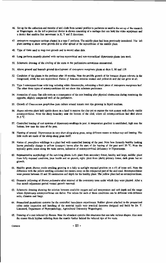
46. Set up for the collection and transfer of soil clods from natural profiles to perforons as used in the set-up of the research at Wageningen. At the left a practical device is shown consisting of a carriage that can hold the video equipment and a device that enables free movement in X, Y and Z directions.
47. Anisoptera marginata cuttings planted in a type C perforon. The middle plant had been previously inoculated. The left plant starting to show some growth did so after spread of the mycorrhizae of the middle plant.
48. Type of form used to map root growth and to record other data.
49. Long perforon recently planted with various mycorrhizal and non-mycorrhizal dipterocarp plants (see text).
50. Schematic drawing of the circling of the roots in the perforations sometimes encountered.
51. Above ground and beneath ground development of Anisoptera marginata plants at days 0, 90 and 119.
52. Condition of the plants in the perforon after 10 months. Note the prolific growth of for instance Hopea odorata in the foreground, while the non-mycorrhizal Vatica cf. bancana remains stunted and yellowish and did not grow at all.
53. Type 3 ectomycorrhizae with long radiating white rhizomorphs, colonizing a fresh piece of Anisoptera marginata leaf. The other three types of ectomycorrhizae did not show this substrate preference.
54. Formation of many fine side roots as a consequence of the root bending after physical obstruction during reentering the opposite, slightly compacted wall of the perforation.
55. Growth of Cenococcum geophylum pure culture around tomato root tips growing in liquid medium.
56. Hopea odorata plant held upside down on a hand to remove the clay pot to expose the root system with clearly visible ectomycorrhizae. Note the sharp boundary near the bottom of the clod, where all ectomycorrhizae had died above 31.5 °C.
57. Controlled heating of root systems of dipterocarp seedlings in peat. A temperature gradient is established, high near the bottom, low near the rims of the pots.
58. Planting of several Dipterocarps in very short alang-alang grass, using different means to reduce top soil heating. The little roofs are made of the alang-alang grass itself.
59. Vatica cf. pauciflora Wildlings in a plant bed with controlled heating of the peat. Note how formerly healthy looking leaves gradually change to yellow (younger) leaves after the start of the heating of the peat until 35 °C. Note the typically green zones along the main nerves, indicative of ectomycorrhizal deficiency in Dipterocarps.
60. Representative morphology of the surviving plants. Left: plant from secondary forest, healthy and large, middle: plant from fully exposed condition, poor health and no growth, right: plant from (dark) primary forest, dark green but no growth.
61. Healthy green Shorea ovalis seedling growing in a fully to sunlight exposed position on a rill of loose soil. Note the difference with the yellow seedling collected two meters away on the compacted part of the skid road. Ectomycorrhizae were present between 15 and 30 centimetres soil depth for the healthy plant. The yellow plant had no ectomycorrhizae.
62. Dramatic yellowing of Shorea polyandra after removal of the overstorey trees under which they were planted. After a four month adjustment period normal growth resumed.
63. Schematic drawing showing the relation between available oxygen and soil temperature and soil depth and the range where dipterocarp ectomycorrhizae can thrive. The values for each of these conditions can be different with different soils, climates and fungi.
64. Pressurized gnotobiotic systems for the controlled inoculation experiments. Rubber gloves attached to the pressurized cabin make inspection and handling of the material inside very practical (systems designed and build by Dr. T. Limonard, Department of Phytopathology, Agricultural University Wageningen).
65. Drawing of a root infected by Riessia. Note the abundant cystidia-like structures that can take various shapes. Also note the coarse black hyphae radiating from the mantle further behind the infected tips of the roots.
Contents - 1 1 -

66. Hopea odorata root system. Note complete blackening of the roots except for the fast growing white root tips.
67. Cystidia like projections (called conidiophores by Jülich, 1985) as they appear on the mantle surface of the new fungus-root association.
68. Transverse section of a Hopea odorata root infected by Riessia radicicola. Note the thick mantle, absence of a Hartig net, intracellular hyphae bearing clamp connections and thick hyphae radiating from the mantle.
69. Haustoria-like intracellular structures resulting from the Riessia radicicola infection.
70. Scanning electron microscope images of Riessia infected roots. Note the many cystidia-like projections, the thick mantle of coarse hyphae, the unchanged form of the cortex cells and intracellular hyphae.
71. Root system of Hopea odorata. Note the clear layering of mycorrhizal presence from the warm underside to the cool soil near the rim. From top to bottom : top 5 cm only non-mycorrhizal roots, from 5-11 cm amphimycorrhizae, from 11-19 light brown pyramidal ectomycorrhizae, from 19-26 cm amphimycorrhizae, from 26-30 non-mycorrhizal roots.
72. Options of speciation. In this figure x,y and z can represent various kind of niches, e.g. different soil types. X may for example represent the original soil type and Y another one, while z at the same time can be the original one or the other one.
73. Anisoptera marginata explant on MS medium. Only after addition of thiamine in the medium new leaves turned green instead of yellow and did not fall of as fast.
74. Anisoptera marginata plants partly sprayed with vitamin Bl (thiamin) applying watery solution as foliar spray. Note the dramatic change in leaf colour of the plant that was sprayed. Internodal leaf elongation still remained suppressed after new green leaves produced.
75. Air-layering of Dipterocarpaceae. Yellow leaves formed after removal of the bark for air-layering.
76. A transect in virgin dipterocarp forest in north western Borneo.
List of boxes
Box 1 : Early flowering in Dipterocarps
Box 2 : Ectomycorrhizal mushroom records
Box 3 : Drying of mushroom specimens
Box 4 : The use of ditches
Box 5 : Rainfall data and their correlation with mushroom appearances
Box 6 : Difficulties studying root distribution in a mixed tropical rain forest
Box 7: The perforon technology
Box 8 : Amphimycorrhizae
Box 9 : Mycorrhizae, whafs in a name
- 12 -
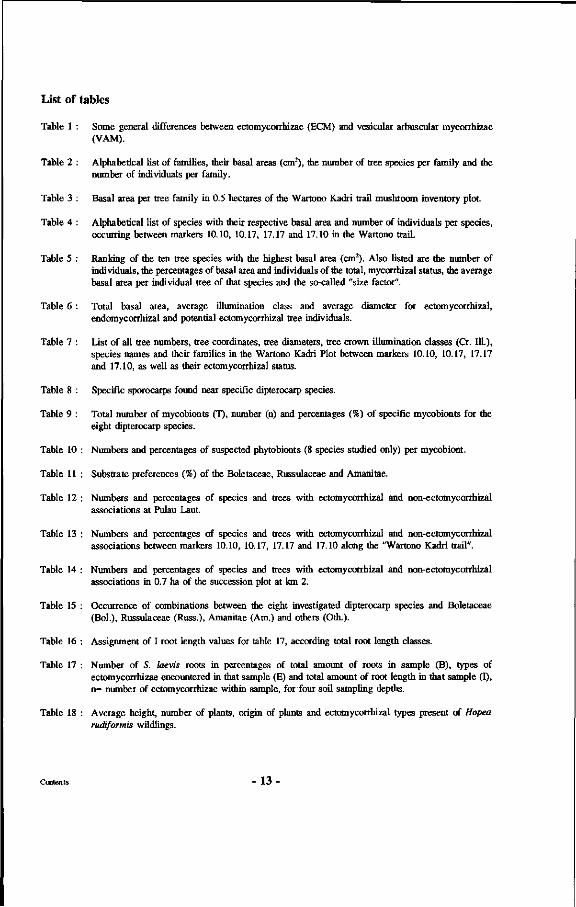
List of tables
Table 1 : Some general differences between ectomycorrhizae (ECM) and vesicular arbuscular mycorrhizae (VAM).
Table 2 : Alphabetical list of families, their basal areas (cm2), the number of tree species per family and the number of individuals per family.
Table 3 : Basal area per tree family in 0.5 hectares of the Wartono Kadri trail mushroom inventory plot.
Table 4 : Alphabetical list of species with their respective basal area and number of individuals per species, occurring between markers 10.10, 10.17, 17.17 and 17.10 in the Wartono trail.
Table 5 : Ranking of the ten tree species with the highest basal area (cm2). Also listed are the number of individuals, the percentages of basal area and individuals of the total, mycorrhizal status, the average basal area per individual tree of that species and the so-called "size factor".
Table 6 : Total basal area, average illumination class and average diameter for ectomycorrhizal, endomycorrhizal and potential ectomycorrhizal tree individuals.
Table 7 : List of all tree numbers, tree coordinates, tree diameters, tree crown illumination classes (Cr. 111.), species names and their families in the Wartono Kadri Plot between markers 10.10, 10.17, 17.17 and 17.10, as well as their ectomycorrhizal status.
Table 8 : Specific sporocarps found near specific dipterocarp species.
Table 9 : Total number of mycobionts (T), number (n) and percentages (%) of specific mycobionts for the eight dipterocarp species.
Table 10 : Numbers and percentages of suspected phytobionts (8 species studied only) per mycobiont.
Table 11 : Substrate preferences (%) of the Boletaceae, Russulaceae and Amanitae.
Table 12 : Numbers and percentages of species and trees with ectomycorrhizal and non-ectomycorrhizal associations at Pulau Laut.
Table 13 : Numbers and percentages of species and trees with ectomycorrhizal and non-ectomycorrhizal associations between markers 10.10, 10.17, 17.17 and 17.10 along the "Wartono Kadri trail".
Table 14 : Numbers and percentages of species and trees with ectomycorrhizal and non-ectomycorrhizal associations in 0.7 ha of the succession plot at km 2.
Table 15 : Occurrence of combinations between the eight investigated dipterocarp species and Boletaceae (Bol.), Russulaceae (Russ.), Amanitae (Am.) and others (Oth.).
Table 16 : Assignment of I root length values for table 17, according total root length classes.
Table 17 : Number of S. laevis roots in percentages of total amount of roots in sample (B), types of ectomycorrhizae encountered in that sample (E) and total amount of root length in that sample (I), n- number of ectomycorrhizae within sample, for four soil sampling depths.
Table 18 : Average height, number of plants, origin of plants and ectomycorrhizal types present of Hopea rudiformis Wildlings.
-13
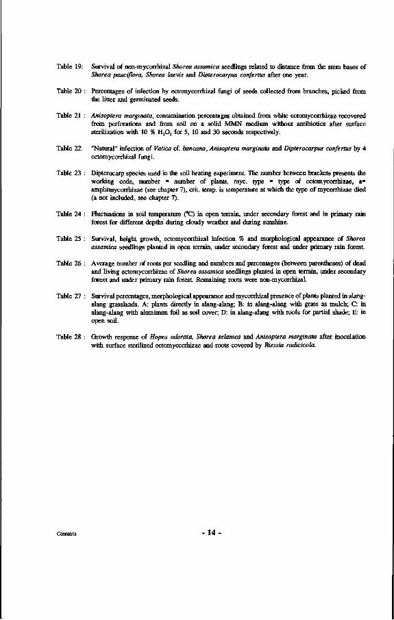
Table 19: Survival of non-mycorrhizal Shorea assamica seedlings related to distance from the stem bases of Shorea pauciflora, Shorea laevis and Divterocarpus confertus after one year.
Table 20 : Percentages of infection by ectomycorrhizal fungi of seeds collected from branches, picked from the litter and germinated seeds.
Table 21 : Anisoptera marginata, contamination percentages obtained from white ectomycorrhizae recovered from perforations and from soil on a solid MMN medium without antibiotics after surface sterilization with 10 % H202 for 5, 10 and 30 seconds respectively.
Table 22: "Natural" infection of Vatica cf. bancana, Anisoptera marginala and Dipterocarpus confertus by 4 ectomycorrhizal fungi.
Table 23 : Dipterocarp species used in the soil heating experiment The number between brackets presents the working code, number - number of plants, myc. type = type of ectomycorrhizae, a= amphimycorrhizae (see chapter 7), crit. temp, is temperature at which the type of mycorrhizae died (a not included, see chapter 7).
Table 24 : Fluctuations in soil temperature (°C) in open terrain, under secondary forest and in primary rain forest for different depths during cloudy weather and during sunshine.
Table 25 : Survival, height growth, ectomycorrhizal infection % and morphological appearance of Shorea assamica seedlings planted in open terrain, under secondary forest and under primary rain forest
Table 26 : Average number of roots per seedling and numbers and percentages (between parentheses) of dead and living ectomycorrhizae of Shorea assamica seedlings planted in open terrain, under secondary forest and under primary rain forest Remaining roots were non-mycorrhizal.
Table 27 : Survival percentages, morphological appearance and mycorrhizal presence of plants planted in alang-alang grasslands. A: plants directly in alang-alang; B: in alang-alang with grass as mulch; C: in alang-alang with aluminum foil as soil cover; D: in alang-alang with roofs for partial shade; E: in open soil.
Table 28 : Growth response of Hopea odorata, Shorea selanica and Anisoptera marginata after inoculation with surface sterilized ectomycorrhizae and roots covered by ttiessia radicicola.
-14
Preface
In March 1980, as part of my M.Sc. program, I had the opportunity to spend seven months in the tropical rain forest of East-Kalimantan, a period that became decisive for my further work During this period I was involved in various kinds of research and activities related to Dipterocarps. I noticed the great difficulties in producing young Dipterocarps from seed. The plants in the nurseries I visited all turned yellow and would eventually die. I also noticed that many of the seedlings of the natural regeneration, abundant at that time in logged-over forest, became yellow as well. Some initial work seemed to indicate that light was a decisive factor. Before returning to my university I collected some seeds of a number of dipterocarp species. These were germinated in the greenhouse and showed favourable initial development. After a few months these seedlings also started developing yellowish leaves and stunted growth. After a wide variety of treatments, involving light intensity, air humidity, temperature, day length and media, did not result in any improvement of the morphological appearance of the seedlings I started looking for presence of some symbiotic association. From literature it was learned that Dipterocarps had been found to be ectomycorrhizal. My plants proved to be non-mycorrhizal. Inoculation with various ectomycorrhizal mushrooms occurring in The Netherlands did not result in any improvement. Only after soil with ectomycorrhizal roots from the mother tree was obtained from Indonesia, my seedlings started showing vigorous growth. After a small accident in the greenhouse in which the temperature of the soil rose too high I noticed that the formerly healthy plants became yellow again. This was later shown to be due to the fact that their ectomycorrhizae had disappeared as a result of the high soil temperature.
It was this series of small events that arose my interest in continuing to study this obviously very important relationship between the Dipterocarps and certain symbiotic fungi. The fact that these forest giants were completely depending upon these almost invisible fungi on their root system and that it clearly was not good enough to just study trees as a forester, but that one also for practical reasons had to take other components of the ecosystem in account, has ever since fascinated me.
This publication presents some of the results that were obtained during a cooperative project between Indonesia and The Netherlands. This project called "Mycorrhizae in the tropical rain forest (Dipterocarpaceae)" was based upon results obtained from experiments carried out at the departments of Silviculture and Forest Ecology and Phytopathology, Wageningen Agricultural University in The Netherlands.
The hypotheses formulated during this previous period were tested in the field during the course of the cooperative project mentioned before. From the side of The Netherlands the coordination and supervision were in the hands of Prof.Dr.Ir. R.A.A. Oldeman of the Department of Silviculture and Prof.Dr. J. Dekker of the Department of Phytopathology. The financing from The Netherlands was borne by The Foundation for Advancement of Research in the Tropics (WOTRO), The Directorate-General for Science Policy of the Ministry of Education and Science, and the Wageningen Agricultural University, later also by the institute for Forestry and Nature Research, IBN-DLO.
15-
In Indonesia the coordination and supervision of the project was in the hands of the Director-General for Forestry Research and Development of the Indonesian Ministry of Forestry, Dr.Ir. Setyono Sastrosumarto. The supporting parties were the Agency for Forestry Research and Development, The Directorate-General for Reforestation and Land Rehabilitation, both from the Ministry of Forestry, and the state forestry enterprise P.T. INHUTANI I.
The field research was carried out at the field station Wanariset I Samboja, located at 38 km from Balikpapan along the main Balikpapan-Samarinda road. This station resorts under the responsibility of the Forest Research Institute Samarinda, a subdivision of the Agency for Forestry Research and Development. To this station belong some 3500 hectares of research forest, in which a few hundred hectares of relatively undisturbed primary rain forest are present. This is where most of the field experiments were executed. At the station itself some plant beds and a greenhouse were available. During the research the facilities of the station have been much extended.
Most of the research was carried out between September 1985 and October 1987. Since the end of 1987 the project was continued and further enlarged through a new cooperation called the TROPENBOS-Kalimantan programme, now called "the International MOF-Tropenbos Kalimantan Project", which is being executed by the Institute for Forestry and Nature Research IBN-DLO from The Netherlands, the Indonesian Agency for Forestry Research and Development of the Ministry of Forestry, and the state forestry enterprises P.T. INHUTANI I and P.T. INHUTANI II.
It is hoped that the results presented in this book may stimulate more researchers to turn their attention to tropical ectomycorrhizae, which so far have been given relatively little attention. It is also hoped that the approach followed here, basic research with continuous practical spin-off, which represents one of the basic philosophies of the TROPENBOS-programme, will be pursued by more researchers in the tropics. There is not much time left to study the tropical rain forest and provide blueprints for sustainable management of this precious resource.
Balikpapan, June 1994.
16 -
Acknowledgements
First of all I want to thank my promotors Prof.Dr.Ir. R.A.A. Oldeman and Prof.Dr. J. Dekker. It was Prof. Oldeman who first stimulated me to get involved in the fascinating world of tropical rain forests and their components through his very first lectures at the Wageningen Agricultural University. He has been my constant guide during the years of my research in the tropics, until the completion of this document. Prof. Dekker, who, besides providing the logical set-up as now presented in this publication, enabled me to combine the mycological aspects with forestry. Dr.Ir. T. Limonard has contributed much to the research itself and the critical evaluation of this manuscript. His very precise criticism and constructive advice have been the perfect balance and symbiosis for my youthful enthusiasm.
During the period previous to my moving to Indonesia, two dear friends, Joop Hildebrand and the late Bob Schalk, have given me much support. Without the green fingers of Bob many of the percentages of successful rooting of dipterocarp cuttings would have been significantly lower as would probably have been the success with mycorrhizal inoculations performed in his greenhouse. I hope this book will help to keep the memory of this special man vivid. Without all the advice of Joop on Indonesia and the help of all his friends there, I am sure that I would not have been successful in realizing the goals of the project. In the Netherlands I have been stimulated and helped much by all my colleagues at the Department of Silviculture and the Department of Phytopathology of the Wageningen Agricultural University. The technical staff of these departments and the Biotechnion service department were always ready and willing to build the numerous strange instruments I designed for research purposes.
Jan van den Bos and Paul Hillegers of IBN-DLO were very patient in giving me the opportunity to write up my research in the years I have been working for JJ3N-DLO. I want to thank Joost Foppes for offering me the first clue to take a look at mycorrhizae. I thank Prof.Dr. Peter Ashton and the late Dr. Marius Jacobs for their stimulating discussions and Peter especially for the extensive comments on and corrections in this manuscript. I have had several valuable discussions with Prof.Dr. F. Halle and Prof.Dr. E.F. Briinig on ecological matters concerning Dipterocarpaceae, for which I thank them.
I am most grateful to His Excellency the former Indonesian Minister of Forestry, Dr. Soedjarwo, his advisor, Ir. Soenaryo, and the former Director-General for Reforestation and Rehabilitation, Ir. Wartono Kadri, for their confidence in the proposed research and inviting me to come to Indonesia. I thank Prof. Sukiman for introducing my research to them. Some time after my arrival the Director-General of the Agency for Forestry Research and Development, Dr.Ir. Setyono Sastrosumarto, became Indonesian supervisor of all the project activities in cooperation with Prof. Oldeman, and it was through his agency that assistance for the research was obtained. In the province Dr.Ir. Soetarso Priasukmana provided counterparts and facilities.
Acknowledgements - 1 7 -
Much help was obtained from the state forestry enterprise P.T. INHUTANI I, who actively got involved in the project. I want especially to thank the former President-Director Ir. Wahyudi, former director of production, Ir. Hendro Prastowo and Ir. Muhandis from the Jakarta office, and my close personal friend Drs.Ing. Soedjono Hardjosantoso former head of the INHUTANI unit in Balikpapan.
I also want to thank the person who at that time was President-Director of P.T. INHUTANI n, and who is at present Minister of Forestry of Indonesia, His Excellency, Ir. Djamaludin Suryohadikusumo, who in various ways, from sending coffee to moral support during difficult times, provided invaluable help. To all my fellow workers who joined me in the fieldwork, I express my deepest gratitude for their help and motivation. I want to mention especially Ir. Zulian Hanafi, Ir. Irsyal Yasman and Ir. Massofian Noor who sacrificed much for the success of the project, especially during the early years when much hardship was suffered at the field location.
Being no taxonomist I was very happy to receive criticism of Dr. Annelies Jansen for the part on mushroom descriptions. I received help as well with producing the extended morphological descriptions of the collected mushrooms, so as to include microscopic characteristics, from Dr. Tom Kuyper. The linguist Drs. J.L. Moerbeek helped me with the analysis of the Greek and Latin origin of the word mycorrhizae.
Wim Middelplaats of the Wageningen Agricultural University made several of the fine figures in Chapter 5 and 7 for which I am very grateful. Junus Tahitu and Arie Stolk of the IBN-DLO institute helped me with preparing some other figures for publication, while Gerrit Seigers and Joke Mahulete put a lot of effort in finalizing the manuscript for the printer.
While starting to live and work in the tropics my parents had to, and will continue to miss their grandchildren, for many years.
And, last but not least, I want to thank my wife for her understanding and patience every time I spend so much time in the forest and with my books.
Besides the people already mentioned, there were many more that contributed to the realization of this work. To all of them I express my deepest gratitude. The project was financed by all the Indonesian partners mentioned above and from the Netherlands it was co-sponsored by the Wageningen Agricultural University, the Foundation for Advancement of Scientific Research in the Tropics (WOTRO) and the Directorate-General for Science Policy and in the latest phase through the TROPENBOS-Kalimantan project executed by the Forest Research Institute "De Dorschkamp" from the Netherlands, now known as the Institute for Forest and Nature Research, IBN-DLO. The Tropenbos Foundation financed the publication of this book, for which I am most grateful.
Acknowledgements - 18 -
Chapter 1 : Introduction
1.1 Dipterocarpaceae
1.1.1 General
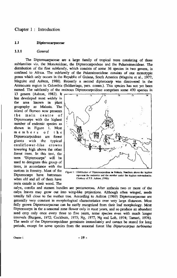
The Dipterocarpaceae are a large family of tropical trees consisting of three subfamilies viz. the Monotoideae, the Dipterocarpoideae and the Pakaraimeoideae. The distribution of the first subfamily, which consists of some 36 species in two genera, is confined to Africa. The subfamily of the Pakaraimeoideae consists of one monotypic genus which only occurs in the Republic of Guiana, South America (Maguire et al., 1977; Maguire and Ashton, 1980). Recently a second dipterocarp was discovered in the Araracuara region in Colombia (Saldarriaga, pers. comm.). This species has not yet been named. The subfamily of the resinous Dipterocarpoideae comprises some 470 species in 13 genera (Ashton, 1982). It has developed most widely in the area known in plant geography as Malesia. The island of Borneo now presents t h e m a i n c e n t r e of Dipterocarps with the highest number of endemic species as shown in Figure 1. Most m e m b e r s o f t h e Dipterocarpoideae are forest giants with the typical c au l i f lower - l ike crowns towering high above the other forest trees. In this text, the term "Dipterocarps" will be used to designate this group of trees, in accordance with the custom in forestry. Most of the Dipterocarps have buttresses when old and all of them have resin canals in their wood. The
calyx, corolla and stamen bundles are pentamerous. After anthesis two or more of the calyx leaves may grow out into wing-like projections. Although often winged, seeds usually fall close to the mother tree. According to Ashton (1969) Dipterocarpaceae are generally very constant in morphological characteristics over very large distances. Most fully grown Dipterocarpaceae can be easily recognized from their leaf morphology. Most Dipterocarps in the a-seasonal zone flower only in mast years, and so produce an abundant seed crop only once every three to five years, some species even with much longer intervals (Burgess, 1972; Cockburn, 1975; Ng, 1977; Ng and Loh, 1974; Tamari, 1976). The seeds of the Dipterocarpoideae germinate immediately and cannot be stored for long periods, except for some species from the seasonal forest like Dipterocarpus turbinatus
Figure 1 Distribution of Dipterocarpoideae in Malesia. Numbers above the hyphen represent the endemics and the number under the hyphen non-endemics. Courtesy of P.S. Ashton, (1983)
Chapter 1 - 19 -

(Tamari, 1976; Tompsett, 1985;1987). Sometimes the seeds already germinate while still hanging on the tree. Many seeds are destroyed by insects. The seedlings establish dense carpets under the mother trees during a mass flowering year. The foresters' term "mother trees" is used here in the sense of trees yielding the fruits and seedlings mentioned and will be used in this sense throughout the rest of the document. A drought spell can significantly reduce the amount of seedlings. Especially during germination the seedlings are very susceptible to drought since their emerging hypocotyl has to make a curve of almost half a circle before reaching the soil. This is due to the projecting wings that cause the seed to land on the surface with the germination spot upward, away from the soil. Some species like Vatica chartacea Ashton seem to have overcome this problem through another configuration of the wings (see Figure 2). The feature of the projecting wings seems to be some remnant from the period in which the Dipterocarpaceae were still a family of trees with wind-spread seeds, before in the Tertiary they started migrating from Africa to South-East Asia (Jacobs, 1981) although some authors (e.g. Ashton, 1969) suggest that in some species the wings still do have a selective value. The number of surviving seedlings is reduced very quickly (Ashton, 1982) and few seedlings grow up to become big trees. Some seedlings can survive for very long periods in heavy shade almost without growing. Browne and Mathews (1914, p. 474) concluded that "...according to the available figures, the average dipterocarp is 116 years old when 5 centimetres in diameter.", although Ashton (1993, pers. comm.) considers this conclusion incorrect because it is based upon extrapolation of growth rates. Most Dipterocarps grow according to the architectural model of Roux with continuous and sometimes diffuse branching (Halle et al., 1978). Halle (1979) also mentions the occurrence of the model of Massart and of Rauh. A few Dipterocarps, like Cotylelobium spp. and Vatica chartacea, sometimes show transitions between the models of Troll and Roux when they are still young, like some Mediterranean species described earlier by Roux (1968), and a Guianese Melastomataceae analysed by Oldeman (1974). The others produce straight monopodial stems and normally show early self-pruning characteristics. Their very long branchless boles with only a slight taper towards their top make the logs very suitable for use in wood industries.
Figure 2: Germination of Vatica chartacea and Dipterocarpus sp. as influenced by the wing-like projections.
When the trees start reaching the upper canopy a process starts called architectural metamorphosis (Edelin, 1984). The crowns become more open and crown shyness becomes apparent (Halle and Ng, 1981). In the natural forest these emergent species do not start flowering until their crowns have reached fully exposed light conditions and this
Chapter 1 - 2 0 -
rarely occurs before the age of 30 years and after reaching a diameter of at least 30 centimetres. In plantations, especially those established outside their area of natural occurrence, flowering can start much earlier. This was noted in plantations of Dipterocarps that had been established to produce, amongst others, valuable resins (Schuitemaker, 1933; Ardikoesoema and Noerkamal, 1955; Torquebiau, 1984; Messer, 1985) and illipe nuts.
Box 1. : Early flowering in Dipterocarps
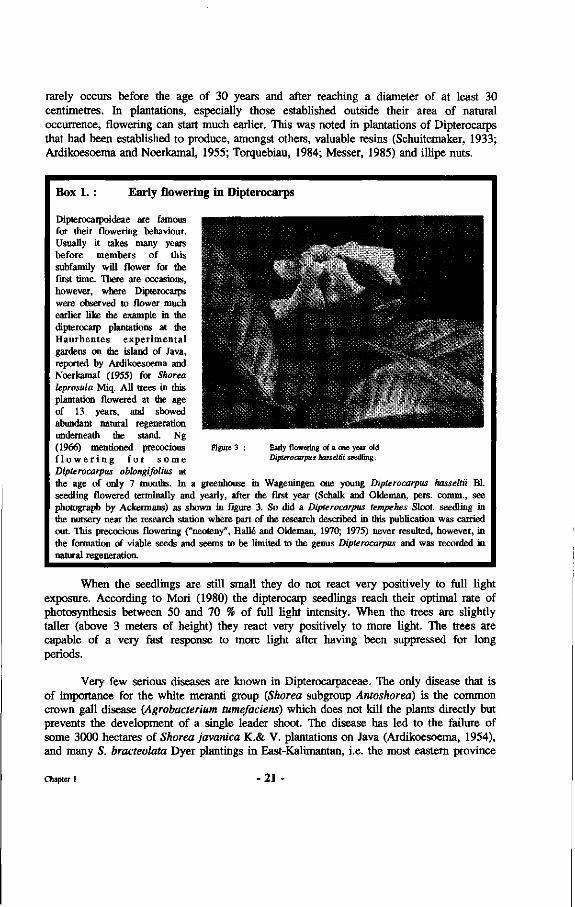
Dipterocaipoideae are famous for their flowering behaviour. Usually it takes many years before members of this subfamily will flower for the first time. There are occasions, however, where Dipterocarps were observed to flower much earlier like the example in the dipterocaip plantations at the Haurbentes experimental gardens on the island of Java, reported by Ardikoesoema and Noerkamal (1955) for Shorea leprosula Miq. All trees in this plantation flowered at the age of 13 years, and showed abundant natural regeneration underneath the stand. Ng (1966) mentioned precocious f l o w e r i n g for s o m e Dipterocarpus oblongifolius at the age of only 7 months. In a greenhouse in Wageningen one young Dipterocarpus hasseltii Bl. seedling flowered terminally and yearly, after the first year (Schalk and Oldeman, pers. comm., see photograph by Ackermans) as shown in figure 3. So did a Dipterocarpus tempehes Sloot, seedling in the nursery near the research station where part of the research described in this publication was carried out. This precocious flowering ("neoteny", Halle and Oldeman, 1970; 1975) never resulted, however, in the formation of viable seeds and seems to be limited to the genus Dipterocarpus and was recorded in natural regeneration.
Figure 3 Early flowering of a one year old Dipterocarpus hasseltii seedling.
When the seedlings are still small they do not react very positively to full light exposure. According to Mori (1980) the dipterocarp seedlings reach their optimal rate of photosynthesis between 50 and 70 % of full light intensity. When the trees are slightly taller (above 3 meters of height) they react very positively to more light. The trees are capable of a very fast response to more light after having been suppressed for long periods.
Very few serious diseases are known in Dipterocarpaceae. The only disease that is of importance for the white meranti group {Shorea subgroup Antoshorea) is the common crown gall disease (Agrobacterium tumefaciens) which does not kill the plants directly but prevents the development of a single leader shoot. The disease has led to the failure of some 3000 hectares of Shorea javanica K.& V. plantations on Java (Ardikoesoema, 1954), and many S. bracteolata Dyer plantings in East-Kalimantan, i.e. the most eastern province
Chapter 1 -21 -
of the Indonesian part of Borneo. A few species like Dipterocarpus confertus Sloot., D. cornutus Dyer (pers. observ.) and Hopea mengerawan Miq. show some top borer damage but the number of damaged seedlings is low whereas the seedlings almost always recover from the repeated attacks (Voogd, 1933; Kalshoven, 1934). Smits et al. (1990a) have described dipterocarp seedling pests and diseases in more detail.
Dipterocarpaceae can grow on very poor soils. They are mainly confined to the lowland zones where the typical mixed dipterocarp lowland forest type can be found. In Peninsular Malaysia, above a height of 300 meters above sea level a distinct dipterocarp hill forest type commences. In Borneo this difference is not so clear. Above 800 meters above sea level very few dipterocarp species can be found. The Dipterocarpaceae are the main constituents of the lowland rain forest of Malesia. They can make up more than 80% of all the upper canopy trees. Endert (1933a) reports on an inventory in Sangkulirang. He found an average volume of 280 m3 per hectare, with variations from 205-527 m3 per hectare. He also reports on some very rich forest in which a white meranti species made up 673 m3 of the total of 946 m3 per hectare of commercial sized timber!
The distribution of dipterocarp trees is typically clumped. Some authors thought this distribution to be the result of their limited means of seed dispersal (e.g. Burgess, 1972); others like Smits (1982) and Ashton (1982) thought that this might be related to the availability of mycorrhizal inoculum. Sometimes pure stands of one Dipterocarpaceae species occur like Shorea albida Sym. occurring in swamp areas or Shorea selanica Bl. on some islands in the eastern part of the Indonesian archipelago.
Dipterocarps as a group show many unique features not shown by other tropical tree groups on other continents with tropical rain forest. However, there is at least one family of trees of South America, the Vochysiaceae, that show many similarities in behaviour as well as morphology with the Dipterocarps (Oldeman and Fundter, 1989), but this family has never reached the dominance shown by the Dipterocarps in the forests in South-East Asia.
1.1.2 History of utilization
In Indonesia the history of dipterocarp exploitation on a larger scale is of very recent date. The management of these forests is even more so. In 1849 the first Dutch foresters set foot on Java and have since 1880 set up very intensive and well defined management systems for teak (Altona, 1926). Historically, distinction was made between the forests on the island of Java and the forest at the so-called outer islands. Nieuwenhuis (1900), who crossed Borneo from Pontianak to Samarinda, recorded only traditional shifting cultivation by Dayaks. It was not until far in the 1920s that the first outside interest for Dipterocarpaceae in Indonesia came to expression in the panglongs. These were Chinese wood exploitation companies on islands near the coast of Sumatra and on Sumatra itself, not far from Singapore, that started their activities around 1880. They worked exclusively with Chinese labourers who often had to work under very bad conditions. Most of these forests were located in peat swamp areas with on the average 70 cubic meters of marketable wood per hectare (Sewandono, 1937). The trees felled belonged to the Dipterocarpaceae, Apocynaceae, Annonaceae and other families. The trees
Chapter 1 - 22 -
were felled by hand and transported on sledges over "knuppel roads", covered with roundwood "knuppels" that were kept slippery with mud or pig fat. A similar system was used on Kalimantan where it was called "kuda-kuda" logging. The wood was transported on sailing ships to Singapore. During the last years of these panglongs (till the beginning of the World War II) some railways were also built for log transportation. Before 1900, practically no interest existed for the dipterocarp forests in the other parts of the so-called "outer provinces". After the "pacification" of these areas some interest appeared for the enormous wood reserves available there (e.g. Kerbert, 1909). Exploration activities began in Palembang where some large, private forest exploitation started for example on the island of Simalur (West coast of Sumatra), Palembang, and shortly after that in Palau, West-Kalimantan and Telok Seliman in East-Kalimantan. Because of many difficulties such as bad planning and problems with log extraction from the felling site to the loading platforms, all of these companies suffered large losses (Kools, 1949). Around 1925 the Forest Service started systematic exploration of the so-called outer provinces. Surveys were made of the standing volume with a line sampling method. Numerous herbarium specimens and wood samples were collected and identified. The forest research institute made lists of scientific and local tree names (Hildebrand, 1949-1954).
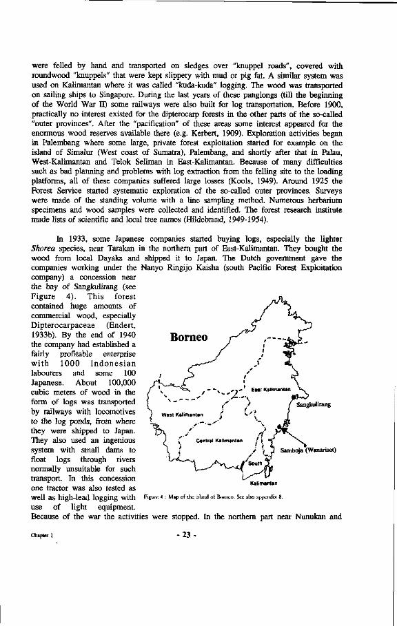
Borneo
In 1933, some Japanese companies started buying logs, especially the lighter Shorea species, near Tarakan in the northern part of East-Kalimantan. They bought the wood from local Dayaks and shipped it to Japan. The Dutch government gave the companies working under the Nanyo Ringijo Kaisha (south Pacific Forest Exploitation company) a concession near the bay of Sangkulirang (see Figure 4). This forest contained huge amounts of commercial wood, especially Dipterocarpaceae (Endert, 1933b). By the end of 1940 the company had established a fairly profitable enterprise with 1000 Indones ian labourers and some 100 Japanese. About 100,000 cubic meters of wood in the form of logs was transported by railways with locomotives to the log ponds, from where they were shipped to Japan. They also used an ingenious system with small dams to float logs through rivers normally unsuitable for such transport. In this concession one tractor was also tested as well as high-lead logging with use of light equipment. Because of the war the activities were stopped. In the northern part near Nunukan and
Sangkulirang
Samboja (Wanariset)
Figure 4 : Map of the island of Borneo. See also appendix 8.
Chapter 1 - 2 3 -
Sebatik a large concession, called "Oost Borneo", was granted to a company, with K.P.M. (Koninklijke Pakketvaart Maatschappij), N.I.S.H.M. (Nederlandsch Indische Steenkolen en Hout Maatschappij) and the local government of Bulungan as shareholders. Because of the great financial losses this operation was stopped by the Dutch government in 1941 (Kools, 1949).
Other important wood companies in East-Kalimantan during that period were the Borneo Busan Kaisha in Samarinda, the firm H. Yukimoto in Balikpapan and the BPM, an oil company (Boer, 1937). The latter felled large quantities of wood mainly for its own supply. These companies and several other, smaller ones obtained the wood from their concession through intermediary of Chinese traders, not through organized logging activities of their own.
Plans for exploitation of the Batu Licin forests in South-Kalimantan for the abundant Dipterocarpaceae and the Bornean Ironwood, did not become operational because of World War II.
The great need for wood during this war caused many mechanical sawmills to be installed. In East-Kalimantan this happened among others in Balikpapan, Samarinda and Nunukan. After the war these were taken over by the Forest Service.
Until the 1950s the situation did not change much. The great change in forest exploitation came with the approval of the law on foreign investment in 1967 (Manning, 1971; Wiersum, 1978), which made it very profitable to start logging operations. With the possibility of using modem heavy equipment, exploitation now no longer was limited to the exiguous zones along the rivers, i.e. mostly slopes running towards the water.
The first experiments with mechanical logging in East-Kalimantan were conducted by Soepono and Ardiwinata in 1958 in Mentawir near Balikpapan (Zuid-Ooster afdeling, unpublished document). Here a rail system was used to transport the logs to the log pond. Modem logging operations usually build road systems that allow them to do year-round logging and transportation of the logs under all weather conditions. In the Philippines and Peninsular Malaysia mechanical logging in the mixed dipterocarp forests had been introduced earlier. The depletion of their forests, especially in the Philippines, was consequently much faster. After the law on foreign investment came into force, large scale exploitation of the forest so far undisturbed was commenced vigorously and led to the timber boom of the seventies (Manning, 1971). Presently Indonesia is the main exporter of dipterocarp wood and wood products, followed by Malaysia. Dipterocarp wood makes up more than 25% of the world trade in tropical hardwood timber and products (Ashton, 1980). Wood from the genus Shorea accounts for 80% of this volume. Large exporters of dipterocarp timber like The Philippines and Thailand have now banned logging because their natural supplies have been exhausted. Indonesia still has a large area of mixed dipterocarp forests which is managed in accordance with the Indonesian selective cutting and planting system known as TPTI (Tebang Pilih Tanam Indonesia). In practice the application of this system was considered inadequate for various reasons, such as difficult regeneration of commercial species and slow diameter increments. The Malayan Uniform System was once considered to be fairly successful but, due to the conversion of the lowland rain forest into rubber and oil palm plantations in Peninsular Malaysia, very few
Chapter 1 - 2 4 -
forests managed under this system remain. In view of the great pressure upon them and their importance as a source of income, provision of job opportunities, watershed protection and as a treasure house of genes there is an urgent need to look into better ways of managing and thus preserving these mixed dipterocarp forests. In recent years Indonesia has taken many firm actions to improve its forest management. This is being implemented through disciplining of the concession holders, more investments in research, the establishment of plantations of faster growing species, multipurpose species to relieve some of the pressure on the natural forests, etc. It is hoped that these approaches, and others like planting of trees yielding other products besides wood (e.g. resins, illipe nuts (Burck, 1886; 1887)) in more intensive land use systems than shifting cultivation with slash and burn agriculture, may further improve the present situation.
1.2 Mycorrhizae
1.2.1 General
The word mycorrhizae consists of two words originating from the Greek language, being pîÇa (root) and |iÔK(pç (mushroom, fungus). Box 10 in Chapter 8 discusses the origin and correct Latin spelling in detail. The word mycorrhizae was first proposed by Frank (1885) who saw fungal structures in roots of trees belonging to the family of the Fagaceae (Fagus, Quercus, Carpinus) he was investigating. Other researchers like Kamienski (1882) had seen the structures before, but Frank was the first to suggest that the observed fungi might be involved in taking up nutrients and possibly other compounds from the soil to the advantage of the higher plant. Later research proved that Frank had been right.
Some form of mycorrhizal presence can be found on roots of almost all plants except for a few families like Cruciferae, Juncaceae and Cyperaceae (Harley and Smith, 1983). Several types of mycorrhizae are known like Vesicular Arbuscular Mycorrhizae, usually known as VAM, Ectomycorrhizae, Ectendomycorrhizae, Arbutoid mycorrhizae, Monotropoid mycorrhizae, Ericoid mycorrhizae and Orchid mycorrhizae. Most land plants possess VAM. Ectomycorrhizae, hereafter mostly referred to as ECM, are mainly confined to the roots of some forest tree species. There are very few herbaceous plants like some species in the genera Lactuca (Leguminosae) and Galium (Rubiaceae) that posses ECM. Table 1 lists some of the differences between the two most important mycorrhizal symbioses for tree species.
The fungus in the mutualistic symbiosis, which will be called mycobiont, takes up nutrients and water from the soil and transfers these to the roots of the associated plant or phytobiont (Bowen, 1973). Possibly, ectomycorrhizal fungi are also capable of taking up nutrients directly from organic material (Went and Stark, 1968; Stark, 1971) as also supported by the findings of Abuzinadah and Read (1989) that showed ectomycorrhizal fungi to be capable of taking up nitrogen from organic material and making it available to the higher plant. Zak (1964), Marx (1969a,b; 1970) and Marx and Davey (1967) also mentioned the capability of certain ectomycorrhizal fungi to prevent root diseases in their phytobionts. Certain ectomycorrhizal fungi have even been described as capable of strangling nematodes (Tamas, 1985).
Chapter 1 - 2 5 -
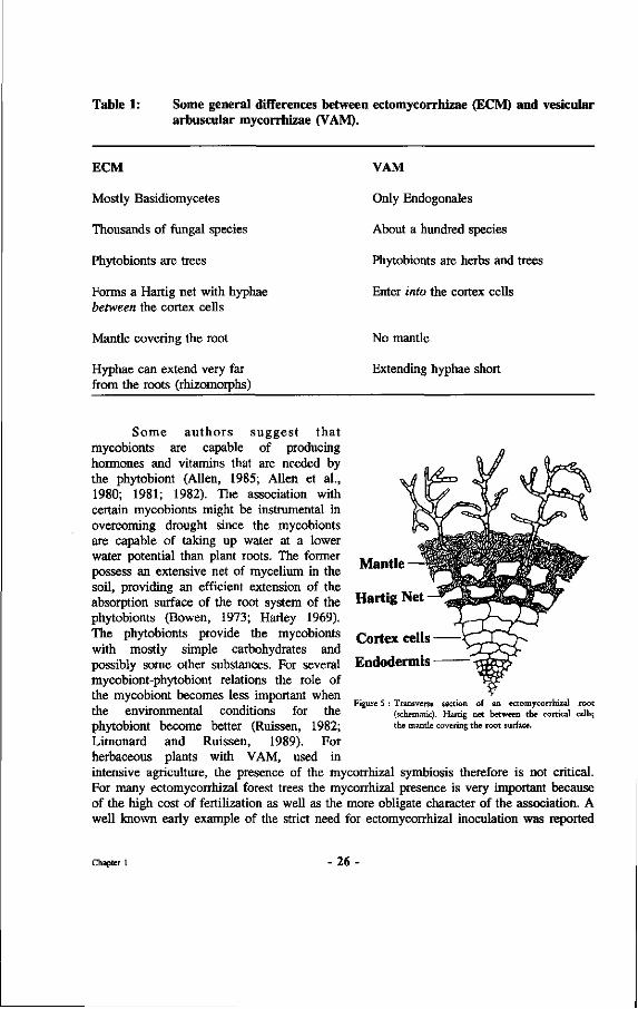
Table 1: Some general differences between ectomycorrhizae (ECM) and vesicular arbuscular mycorrhizae (VAM).
ECM
Mostly Basidiomycetes
Thousands of fungal species
Phytobionts are trees
Forms a Hartig net with hyphae between the cortex cells
Mantle covering the root
Hyphae can extend very far from the roots (rhizomorphs)
VAM
Only Endogonales
About a hundred species
Phytobionts are herbs and trees
Enter into the cortex cells
No mantle
Extending hyphae short
Some authors suggest that mycobionts are capable of producing hormones and vitamins that are needed by the phytobiont (Allen, 1985; Allen et al., 1980; 1981; 1982). The association with certain mycobionts might be instrumental in overcoming drought since the mycobionts are capable of taking up water at a lower water potential than plant roots. The former possess an extensive net of mycelium in the soil, providing an efficient extension of the absorption surface of the root system of the phytobionts (Bowen, 1973; Harley 1969). The phytobionts provide the mycobionts with mostly simple carbohydrates and possibly some other substances. For several mycobiont-phytobiont relations the role of the mycobiont becomes less important when the environmental conditions for the phytobiont become better (Ruissen, 1982; Limonard and Ruissen, 1989). For herbaceous plants with VAM, used in
intensive agriculture, the presence of the mycorrhizal symbiosis therefore is not critical. For many ectomycorrhizal forest trees the mycorrhizal presence is very important because of the high cost of fertilization as well as the more obligate character of the association. A well known early example of the strict need for ectomycorrhizal inoculation was reported
Mantle —
Hartig Net
Cortex cells
Endodermis
Figure 5 : Transverse section of an ectomycorrhizal root (schematic). Hartig net between the cortical cells; the mantle covering the root surface.
Chapter 1 26
by Roelofs (1930) for the production of Pinus merkusii seedlings in Indonesia. It was not until the introduction of an ectomycorrhizal fungus belonging to the genus Suillus that the pines could be produced in large quantities. Other examples concern the worldwide introduction of certain pines from Central America. Briscoe (1959) and Hacskaylo (1967, 1971) mention the inoculations needed for pines in Puerto Rico.
1.2.2 Mycorrhizae associated with Dipterocarps
Singh (1966) mentioned that all Dipterocarpaceae investigated by him proved to possess an ectomycorrhizal symbiosis. Louis and Scott (1987) mention that Singh was the first to report the presence of ectomycorrhizae on roots of Dipterocarps. Actually their presence on roots of seedlings of Hopea mengerawan had already been mentioned by Roosendael and Thorenaar (1924, p. 530), and de Voogd (1933, p. 707) on roots of Shorea platyclados Sloot, seedlings. It is only of recent date that more publications start appearing on dipterocarp mycorrhizae e.g. Bakshi (1974), Hong (1979), Shamsuddin (1979), de Alwis and Abeynayake (1980), Khemnark (1980), Iskandar (1983), Becker (1983), Smits (1983a, b, c), Nuhamara et al. (1985), Chalermpongse (1987), Hadi (1987), Smits et al. (1987), Louis and Scott (1987), Lee (1988).
1.3 Purpose and outline of the research
The main purpose of this research was to support management of the mixed dipterocarp forests in South-East Asia. As mentioned above many problems have been encountered with the management of dipterocarp forests, and especially with their dominant forests trees belonging to the family of the Dipterocarpaceae. As referred to in the preface, the work presented in this book is based upon initial findings with the dipterocarp seed material brought from Indonesia in 1980. Most of the work reported concerns field work in East-Kalimantan. The work involved various aspects like phenological studies, experiments for induction of flowering, monitoring of pests and diseases, the development of techniques for vegetative propagation, techniques for hedge orchards yielding large quantities of vertical shoots suitable for stem cutting production, Wildling collection systems and pilot scale planting trials of Dipterocarps. This book only discusses those parts of the work that relate to the importance of mycorrhizae for the Dipterocarpaceae. The practical goal for the total project was to support production of good quality dipterocarp planting stock. To reach this goal it is necessary to understand the role of the dipterocarp mycorrhizae in the natural situation and what factors contribute to the optimal functioning of these mycorrhizae.
The approach followed was that, first, the mycorrhizal situation in an undisturbed natural forest vegetation was studied with no or few destructive actions. This especially concerned the inventories of ectomycorrhizal sporocarps in permanent inventory plots. This work is described in Chapter 2. Then, the below-ground situation was looked at in greater detail through the inventarisation of ectomycorrhizal roots; this involved some disturbance of the natural situation. This work is important to evaluate the relevance of sporocarp inventories as a reflection of the below ground situation. These inventories are presented in Chapter 3. Advancing further away from the natural situation, inoculation
Chapter 1 - 2 7 -
experiments were conducted involving both infection in the natural vegetation as well as under controlled conditions in greenhouses. It was hoped that differences in the mycobiont-phytobiont combinations, as compared to the natural situation, could provide insight in the selection processes taking place under natural conditions. Chapter 4 deals with the results of this type of work. Next, in Chapter 5 the more qualitative aspects of the dipterocarp mycorrhizae, e.g. compatibility, were studied in artificial in vivo systems called perforons. In Chapter 6 the influence of physical disturbances upon the functioning of the dipterocarp mycorrhizae was studied in detail. This work has particular reference to the situation resulting from large disturbances brought about by harvesting operations in the mixed dipterocarp forests. In Chapter 7 a new type of dipterocarp fungus association is discussed. This association was discovered in 1983 and some material was provided to the Rijksherbarium, Leiden for identification (Jülich, 1985). In Chapter 8 the results of the previous chapters are discussed. An analysis is presented of the importance of the findings for explaining species diversity in tropical rain forest with special reference to the Dipterocarpaceae. If mycorrhizae play an important role in the process of speciation it is of great practical importance to understand their functioning so as to manage the forest in such a way that species diversity can be kept high. This is necessary in view of the great importance of the mixed dipterocarp forests as a valuable gene pool. Finally, at the end of Chapter 8 the practical importance of the results is summarized and recommendations for adjusted management practices are given, as well as some recommendations for future research.
1.3.1 General setting of the research
As mentioned above many problems related to the management of the mixed dipterocarp forests still exist. These involve practical problems like difficult seed storage, and therefore difficult production of dipterocarp planting stock supply and, most importantly, difficult practical application of guidelines for management on a large scale under field conditions. Problems encountered in the use of the Indonesian Selective Cutting and Planting System, for example, reside in different responses to light of the different dipterocarp species, the problems involved with their seedling recognition, and consequently their proper maintenance in terms of release to light and removal of competing plants.
Especially after logging or other disturbances like fire or shifting cultivation, natural regeneration of Dipterocarpaceae can be problematic. Artificial regeneration has been practised only on a small scale. Some older dipterocarp plantations exist in Sumatra, for example in Purbatongga, province of North Sumatra (Butar-Butar and Supriana, 1987). Some forests of Shorea javanica planted by local people can be found near Krui in the province of West Sumatra (Torquebiau, 1984). Many of the Tengkawang forests near Sanggau in the Indonesian province of West-Kalimantan were planted (Smits et al., 1990b). Near Tidung Palung and Melak in East-Kalimantan some 20 year old tengkawang plantations have been established with very high annual diameter increments recorded. Some people believe that the sacred Dipterocarpus hasseltii forest of Sangeh on Bali was planted. The dipterocarp plantations on Java in Haurbentes and Darmaga are well known as are the dipterocarp plantings at Kepong, the Forest Research Institute of Malaysia, which were established on former mining sites. In East Kalimantan Smits et al. (1990b)
Chapter 1 - 2 8 -
cite several more examples of dipterocarp planting by Indonesian concessionaires in recent years. At the time of writing of this report approximately 100 million new dipterocarp plants have been produced by concession holders as part of their enrichment planting schemes. The production of dipterocarp planting stock has been enforced by law in Indonesia. Appendix 1 provides a listing of many hundreds of dipterocarp trial plots in experimental forests in Indonesia.
These examples show that dipterocarp planting is possible. Most of these plantations were started with Wildlings collected from natural forest and first planted in light shade. The Wildlings needed to be collected with a large soil clod adhering to their root system. Many of the plants collected in this way died after transplanting. The intensive methods used for most of the dipterocarp plantations mentioned above therefore do not support large scale planting of the Dipterocarpaceae or at least show the planting to be very problematic.
The irregular seed supply due to the mast flowering habit of the Dipterocarpaceae, the impossibility of storing their seeds for prolonged periods and the failure of many seedlings to survive either in the nursery after germination or in the field after transplanting have, until recently, been reasons for the absence of large scale artificial regeneration of Dipterocarps. The reasons for the failure of the seedlings at that time were not very clear. Many researchers and foresters mention that Dipterocarpaceae do not withstand high light intensities and that many seedlings die after direct exposure. Seedlings in nurseries, grown from seeds collected in the forest very often soon became yellowish and finally would die as can be seen in Figure 6 (Plate 1), showing a large nursery of Dipterocarpaceae with yellow, stunted seedlings. Experiments with direct seeding in secondary forest also yielded very negative results, many of the seeds being destroyed by seed predators and the ones that germinated producing yellow, stunted plants that eventually would die.
Smits (1982, 1983a) showed that some Dipterocarpaceae, like Anisoptera marginata Korth. and Vatica pauciflora Miq., are obligately ectomycorrhizal and hypothesized that failure of dipterocarp planting may well have been related to mycorrhizal problems. Non-mycorrhizal seedlings all become yellowish and stunted and eventually die. These non-mycorrhizal plants looked very much like the yellowish plants in the dipterocarp nurseries and the young seedlings along the exposed skid roads in logged-over forest in East-Kalimantan. Only after inoculation with a suitable mycobiont, collected under the mother tree of the seedlings, would the plants form normal looking green leaves.
To solve the other problem, namely the irregular and unpredictable planting stock supply due to mast flowering, several authors conducted vegetative propagation experiments. Earlier work on vegetative propagation of Dipterocarpaceae was published by Halle and Kamil (1981), Srivastava and Manggil (1981), Smits (1982, 1983b), Chouffot-Struycken (1986). These experiments were only small scale trials and did not support a mass production system for dipterocarp planting stock.
Chapter 1 - 2 9
Chim and On (1973) found that only a few dipterocarp seedlings, all of them looking very healthy before commencement of exploitation, survived logging operations. The number of seedlings surviving the logging operation was much lower than might be expected based upon physical damage by the actual logging operation. Smits (1983a) hypothesized that the problematic natural regeneration of Dipterocarpaceae after logging might be related to the influence of some physical factors upon the performance of the dipterocarp mycorrhizae, and that several aspects of dipterocarp ecology might be related to specificity of dipterocarp mycorrhizae.
It is hoped that the results of the present study can contribute to a better understanding of how the Dipterocarpoideae could have evolved into so many species on the island of Borneo. The hypotheses of Smits (1983a) suggest that the dipterocarp ectomycorrhizae may have been, and may still be involved in this process of species differentiation through a process of enhanced niche specialization. This type of speciation, as described by Ashton (1969) might be enhanced through spatial isolation between clumps of trees because of the limited occurrence of dipterocarp ectomycorrhizal fungi outside their rooting zone. These aspects are discussed in detail in Chapter 8.
1.3.2 General description of the research area
Most of the work was executed in the neighbourhood of the Wanariset Samboja research station, which is located near the village of Samboja at kilometre 38 along the road Balikpapan-Samarinda, in the province of East-Kalimantan, Indonesia (see Figure 4 and Appendix 8). The forest is located just south of the equator at 1 degree South Latitude and 116 degree 56 minutes East Longitude.
The climate is classified as type A under the classification by Schmidt and Ferguson (1951). There is no clear month without rain although generally less rain falls during the period from May to September. Occasional longer dry periods do occur, notably as the result of El Nino Southern Oscillation, a phenomenon in the Pacific ocean co-determining the climates around it. Figure 7 provides the average rainfall records of the nearby ITCI concession over a period of 18 years and over the years 1982 and 1983, during which the most severe drought period in written history of East-Kalimantan was recorded.
As can be seen from the figures below for 1982 and 1983 there was a period of about 12 months when there was virtually no rain. This extreme drought led to extensive fires that resulted in the destruction of 3.6 million hectares in East-Kalimantan and 1 million hectares in Sabah (Malaysia).
The map of the Wanariset forest is presented in Appendix 2. The research forest Wanariset Samboja forms part of the Wanariset research station, which is a field station of the Forest Research Institute Samarinda under the Agency for Forestry Research and Development of the Indonesian Ministry of Forestry. The legal status of the forest is that of "research forest", which means that it is one of the best protected forests in Indonesia.
Chapter 1 - 3 0 -
The research forest consists of 504 hectares of lowland tropical rain forest at altitudes varying from 10 to 85 meters above sea level. The topography is undulating with dissected slopes varying from 10 to 40 degrees and up to 60 degrees. The soils are generally poor and classified as ultisols (USDA, 1975).
Rainfall data ITCI Kenangan Average monthly rainfall 1972-1990
Jan Mar May Jul Sep Nov Feb Apr Jun Aug Oct Dec
Months
Rainfall data ITCI Kenangan Monthly rainfall (mm) during 1982
Rainfall data ITCI Kenangan Monthly rainfall (mm) during 1983
Jan Mar May Jul Sep Nov Feb Apr Jun Aug Oct Dec
Months
Jan Mar May Jul Sep Nov Feb Apr Jun Aug Oct Dec
Months
Figure 7 : Average monthly rainfall data for the ITCI concession. East-Kalimantan, from 1972-1990. Also shown: monthly rainfall over 1982 and 1983.
Chapter 1 31
The Wanariset I research forest is very rich in tree and animal species. Two French ornithologists (Eve and Guigue, 1988) recorded more than 150 bird species within a five day period in the research forest. Weghorst (1991, unpublished report) and Susilo (1994, unpublished record) encountered many more species.
The forest is dominated by Dipterocarpaceae as will be shown in Chapter 2 (2.3.1 and 2.4). The two most typical tree species in the forest are Shorea laevis Ridl. and Eusideroxylon zwageri T. et B., being widespread over the research area and surroundings and always occurring side by side. A checklist of the tree species of the Balikpapan-Samarinda area was published by Kessler et al. (1992). A flora describing the 280 most important tree species was published by Kessler and Sidiyasa (1994).
Chapter 1 - 32
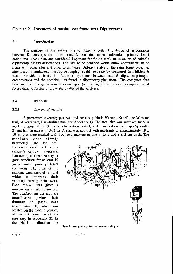
Chapter 2 : Inventory of mushrooms found near Dipterocarps
2.1 Introduction
The purpose of this survey was to obtain a better knowledge of associations between Dipterocarps and fungi normally occurring under undisturbed primary forest conditions. These data are considered important for future work on selection of suitable dipterocarp fungus associations. The data to be obtained would allow comparisons to be made with other sites and other forest types. Different states of the same forest type, i.e. after heavy disturbances like fire or logging, could then also be compared. In addition, it would provide a basis for future comparisons between natural dipterocarp-fungus combinations and the combinations found in dipterocarp plantations. The computer data base and the linking programmes developed (see below) allow for easy incorporation of future data, to further improve the quality of the analyses.
2.2 Methods
2.2.1 Lay-out of the plot
A permanent inventory plot was laid out along "rintis Wartono Kadri", the Wartono trail, at Wanariset, East-Kalimantan (see Appendix 1). The area, that was surveyed twice a week for most of the 60 weeks observation period, is demarcated on the map (Appendix 2) and had an extent of 3.02 ha. A grid was laid out with quadrants of approximately 10 x 10 m, that were marked with ironwood markers of two m long and 3 x 3 cm thick. The m a r k e r s we re f i rmly hammered into the soil. I r o n w o o d s t i c k s (Eusideroxylon zwageri, Lauraceae) of this size stay in good condition for at least 10 years under primary forest conditions. The ends of the markers were painted red and white to improve their visibility during field work. Each marker was given a number on an aluminum tag. The numbers on the tags are coordinates giving their distance to point zero (coordinates 0.0), which was located on the road to Sepaku, at km 3.8 from the station (see map in Appendix 2). In the Northern direction the
Figure 8 : Arrangement of ironwood markers in the plot
Chapter 2 33
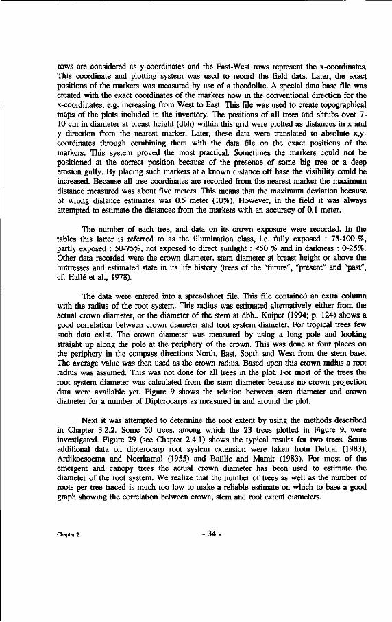
rows are considered as y-coordinates and the East-West rows represent the x-coordinates. This coordinate and plotting system was used to record the field data. Later, the exact positions of the markers was measured by use of a theodolite. A special data base file was created with the exact coordinates of the markers now in the conventional direction for the x-coordinates, e.g. increasing from West to East. This file was used to create topographical maps of the plots included in the inventory. The positions of all trees and shrubs over 7-10 cm in diameter at breast height (dbh) within this grid were plotted as distances in x and y direction from the nearest marker. Later, these data were translated to absolute x,y-coordinates through combining them with the data file on the exact positions of the markers. This system proved the most practical. Sometimes the markers could not be positioned at the correct position because of the presence of some big tree or a deep erosion gully. By placing such markers at a known distance off base the visibility could be increased. Because all tree coordinates are recorded from the nearest marker the maximum distance measured was about five meters. This means that the maximum deviation because of wrong distance estimates was 0.5 meter (10%). However, in the field it was always attempted to estimate the distances from the markers with an accuracy of 0.1 meter.
The number of each tree, and data on its crown exposure were recorded. In the tables this latter is referred to as the illumination class, i.e. fully exposed : 75-100 %, partly exposed : 50-75%, not exposed to direct sunlight : <50 % and in darkness : 0-25%. Other data recorded were the crown diameter, stem diameter at breast height or above the buttresses and estimated state in its life history (trees of the "future", "present" and "past", cf. Halle et al., 1978).
The data were entered into a spreadsheet file. This file contained an extra column with the radius of the root system. This radius was estimated alternatively either from the actual crown diameter, or the diameter of the stem at dbh.. Kuiper (1994; p. 124) shows a good correlation between crown diameter and root system diameter. For tropical trees few such data exist. The crown diameter was measured by using a long pole and looking straight up along the pole at the periphery of the crown. This was done at four places on the periphery in the compass directions North, East, South and West from the stem base. The average value was then used as the crown radius. Based upon this crown radius a root radius was assumed. This was not done for all trees in the plot. For most of the trees the root system diameter was calculated from the stem diameter because no crown projection data were available yet. Figure 9 shows the relation between stem diameter and crown diameter for a number of Dipterocarps as measured in and around the plot.
Next it was attempted to determine the root extent by using the methods described in Chapter 3.2.2. Some 50 trees, among which the 23 trees plotted in Figure 9, were investigated. Figure 29 (see Chapter 2.4.1) shows the typical results for two trees. Some additional data on dipterocarp root system extension were taken from Dabral (1983), Ardikoesoema and Noerkamal (1955) and Baillie and Mamit (1983). For most of the emergent and canopy trees the actual crown diameter has been used to estimate the diameter of the root system. We realize that the number of trees as well as the number of roots per tree traced is much too low to make a reliable estimate on which to base a good graph showing the correlation between crown, stem and root extent diameters.
Chapter 2 - 3 4
Also we have no clear insight in the symmetry of the root systems investigated. Kuiper (1994) reports considerable asymmetry of the root systems even for Douglas-fir with symmetrical crowns. However, since the construction of such a graph, showing exact correlations between crown and root diameters, would need an additional extensive research project, too large to fit within the scope of this project, we have based our root extent estimates on the limited data available.
Although there seems to be a trend to relatively larger root diameters in the 30-50 cm diameter class we have, for the analyses, assumed that the root system diameter reaches 2.5 meters further than the crown projection diameter, being slightly lower than the overall average of 2.8 meters that the roots traced extended from the average crown diameter.
Relation between stem diameter and crown radius for light red merantis
•
ShorQa leprosula
+
Shorea
* Shorea
•
parvifolia
smithiana
Shorea OVHNG
x
Shorea pauciflora
20 40 60 80 100 120 140 Stem diameter (cm)
Figure 9 : Graph showing the relation between stem diameter and crown diameter for five light red meranti species.
Leaf vouchers of all trees were collected and stored in the Wanariset herbarium. As far as possible, trees were identified to species level. The identifications were done in the Wanariset herbarium, by checking of descriptions from literature and by comparisons with herbarium specimens present in the herbarium and previously identified by both Dr. M.J.M. van Balgooy from the Rijksherbarium Leiden and the Malaysian senior botanist Dr. Kochummen. Dipterocarp identifications were checked by Dr. P.S. Ashton. For species other than dipterocarps we always attempted to identify the family to which trees belonged in order to assess the possibility of those trees being ectomycorrhizal.
Chapter 2 - 3 5 -
The list of ectomycorrhizal tree families and genera presented by Harley and Smith (1983) was consulted to assess this, as well as other publications like Lodge (1987) and Azizah et al. (1987). In addition, use was made of the mycorrhizal file based upon analyses of root samples of trees included in the herbarium collection at Wanariset (see Chapter 3 and Appendix 7). Palms were present in large numbers and sizes but have not been included in the list because none are known to be ectomycorrhizal.
The method used to analyze the mycobiont-phytobiont correlations has been as follows. The data were stored into spreadsheets. Two separate files were extracted, from the tree spreadsheet and the mushroom spreadsheet, one containing the number of mushrooms recorded and the coordinates of those mushrooms, the other one containing the tree number and the coordinates of that tree and the radius of the root system. The radius of the root system was related to the crown diameter and stem diameter as described above. Both files were written in the ASCII format, and extracted from the larger files containing all other data besides the record number and coordinates as presented for the area between markers 10.10, 10.17, 17.17 and 17.10, in table 7 (Chapter 2.3.1) for the trees. This area is frequently used as example because in this part of the plot all inventories and identifications are complete as compared to the rest of the plot where the most important dipterocarp species are identified but not all other tree species. During these analyses various parameters could be used to select the list of trees to include, for instance, only the ones known to be ectomycorrhizal and the ones potentially ectomycorrhizal. Potentially ectomycorrhizal refers to the fact that the species belongs to a family or genus in which there are species known to be ectomycorrhizal, but that the mycorrhizal status for this particular species is not yet known with certainty. Then a program was then written in BASIC programming language to compare the coordinates of each mushroom record with those of each tree. This program is provided in Appendix 3 together with some other BASIC programs that were used in the analyses mentioned below. The distance between the stem base and the mushroom was calculated from the differences in X and Y coordinates. This distance was then compared with the root radius. If this distance was smaller than the root radius, the mushroom record number and tree number were entered into another file. This new file was linked up with the mycobiont and phytobiont identifications. Finally, the frequencies of all combinations encountered were determined and analysed.
The methods for analysis of the computer files, and the combining of the various data bases are presented in the schemes of Appendix 4, which provides both the flow chart for the execution of the research as well as the analyses of the data by means of combined analyses of the various data bases. Appendix 3 provides some of the programs written in BASIC to perform the linking.
As mentioned in the introduction (1.3), this approach allowed for updating of the analyses at any time when more data in any of the subdata bases became available. For instance, if a certain tree species proved to be ectomycorrhizal through the root analyses (see Chapter 3) the trees of that species could be included in the (extracted) list and the analyses rerun without delay.
Chapter 2 - 3 6
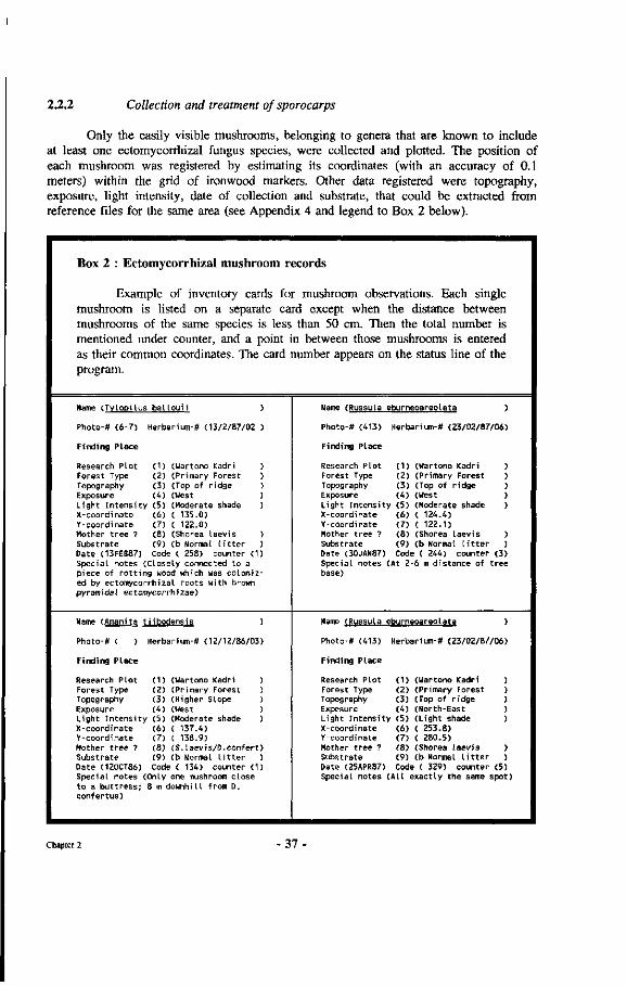
2.2.2 Collection and treatment of sporocarps
Only the easily visible mushrooms, belonging to genera that are known to include at least one ectomycorrhizal fungus species, were collected and plotted. The position of each mushroom was registered by estimating its coordinates (with an accuracy of 0.1 meters) within the grid of ironwood markers. Other data registered were topography, exposure, light intensity, date of collection and substrate, that could be extracted from reference files for the same area (see Appendix 4 and legend to Box 2 below).
Box 2 : Ectomycorrhizal mushroom records
Example of inventory cards for mushroom observations. Each single mushroom is listed on a separate card except when the distance between mushrooms of the same species is less than 50 cm. Then the total number is mentioned under counter, and a point in between those mushrooms is entered as their common coordinates. The card number appears on the status line of the program.
Name (TvlooUus ba l l ou i i )
Photo-# (6-7) Herbarium-* (13/2/87/02 )
Finding Place
Research Plot (1) (Wartono Kadri ) Forest Type (2) (Primary Forest ) Topography (3) (Top of r idge ) Exposure (4) (West ) Light In tens i ty (5) (Moderate shade ) X-coordinate (6) ( 135.0) Y-coordinate (7) ( 122.0) Mother t ree ? (8) (Shorea laevis ) Substrate (9) (b Normal L i t t e r ) Date (13FEB87) Code ( 258) counter (1) Special notes (Closely connected to a piece of r o t t i ng wood which was colonized by ectomycorrhizal roots with brown pyramidal ectomycorrhizae)
Name (Amanita t i ibodensis )
Photo-# ( ) Herbarium-« (12/12/86/03)
Finding Place
Research Plot (1) (Wartono Kadri ) Forest Type (2) (Primary Forest ) Topography (3) (Higher Slope ) Exposure (4) (West ) Light In tens i ty (5) (Moderate shade ) X-coordinate (6) ( 137.4) Y-coordinate (7) ( 138.9) Mother t ree ? (8) (S. laevis /D.confer t ) Substrate (9) (b Normal l i t t e r ) Date (120CT86) Code ( 134) counter (1) Special notes (Only one mushroom close to a but t ress; 8 m downhill from D. confertus)
Name (Russula eburneoareolata )
Photo-# (413) Herbarium-* (23/02/87/06)
Finding Place
Research Plot (1) (Wartono Kadri ) Forest Type (2) (Primary Forest ) Topography (3) (Top of r idge ) Exposure (4) (West ) Light In tensi ty (5) (Moderate shade ) X-coordinate (6) ( 124.4) Y-coordinate (7) ( 122.1) Mother t ree ? (8) (Shorea laevis ) Substrate (9) (b Normal l i t t e r ) Date (30JAN87) Code ( 244) counter (3) Special notes (At 2-6 m distance of t ree base)
Name (Russula eburneoareolata )
Photo-* (413) Herbarium-* (23/02/87/06)
Finding Place
Research Plot (1) (Wartono Kadri ) Forest Type (2) (Primary Forest ) Topography (3) (Top of r idge ) Exposure (4) (North-East ) Light In tens i ty (5) (L ight shade ) X-coordinate (6) ( 253.8) Y-coordinate (7) ( 280.5) Mother t ree ? (8) (Shorea laevis ) Substrate (9) (b Normal l i t t e r ) Date (25APR87) Code ( 329) counter (5) Special notes (A l l exactly the same spot)
Chapter 2 - 3 7 -
All these data were entered into a database which allowed correlations to be tested. An example of the form in which the data were stored in the database is shown in Box 2. The program used was a commercially available computer data base system called Super Base for Commodore computers. Later, the data were transferred to spreadsheet files of other brands.
a) Legend for the Wartono Kadri Mushroom Inventory Database (Mushroom, wkl)
The entry "name" on the cards, or "Mushroom species" in the spreadsheets, is used to enter the names, or code thereof, as presented in the identification keys (see 2.3.1).
The numbers of photographs and herbarium specimens are only entered when morphological data were collected from that sample. If these numbers are present it means that the actual mushroom specimens depicted have been included in the herbarium voucher collection at Wanariset. Part of the collection was transferred to the Rijksherbarium, Leiden.
The categories "research plot" and "forest type" refer to the plot and forest type where the collection of the sporocarp was made. The forest type is only used to indicate the general condition of the forest at the location of the research plot, for instance, primary forest versus logged-over, burned or secondary forest after shifting cultivation. Variations like large gaps may occur in such a plot classified as primary forest.
Topography is classified in four categories :
a) top of ridge b) higher slope c) lower slope d) (wet) valley bottom
"Exposure" refers to South (S), North (N), East (E) and West (W), or the intermediate compass direction e.g., NE = North East.
Light intensity. The values presented are based upon the plotting of light intensities during cloudy conditions and based upon the Minolta spot range finder measurements in each 5x5 meter quadrant. In addition,as a cross check the light intensity was also entered in the mushroom cards as shown above. In this field classification four classes were used, in the absence of light measuring equipment, based upon a subjective classification by the author :
a) heavy shade b) moderate shade c) light shade d) open terrain.
Chapter 2 - 38
Coordinates are given in X,Y values. The values are all given in meters. The smallest unit of measurement/estimation is 0.1 meters with assumed accuracy of +/- 0.5 meter. The value of the coordinates is calculated by reference to the coordinates of the markers, that are placed on approximately 10x10 meter spacing, and whose exact location was calculated as mentioned in Chapter 2.2.1. (For plotting the mushrooms, coordinates are adjusted to the corrections that have to be made for the coordinates of the markers. In the above cards the first two digits represent the marker number which should be at approximately the right distance).
The category "Mother tree" was entered for use in preliminary analyses and only represented an intuitive entry, based upon the distance from the nearest large Dipterocarp, the direction in which the butresses of this dipterocarp extended, and experience. This category was not used in the analyses.
The date was entered in the following format : xx/yyy/zz. In this format xx represents the day of the month, yyy the month of the year and zz the year of inventory. The "Code" represented the calculated value of the date starting from date zero, and the numbers were generated by the program itself.
Substrate stands for the categories of litter classes. The following field criteria were used for the classification :
a) mineral soil (more than 50% of the soil surface not covered with whole leaves;
b) thin litter (more than 50% of the soil surface covered with fallen leaves but less than two layers of whole leaves);
c) normal to thick litter (two to five layers of dead leaves, matting of the litter by fine roots and fungal hyphae obvious, some small branches present);
d) small accumulations of Utter and dead wood (more than five layers of whole leaves and layer mostly thicker than five centimeters).;
e) large accumulations of litter and dead wood (termite nests, accumulation of branches and litter between butresses, between leaf sheaths of palm stems, etc.).
The heading "Special notes" or "Remarks" in the later spreadsheets, concerns special observations like reference to certain mycorrhizal types, or correlation with a certain tree or a flowering event.
b) Collection of sporocarps
Collecting of sporocarps was done by five different people. Sometimes a German shepherd dog was also used. All material of doubtful identity was checked by the author.
During the first three months, it was attempted to check every square meter of the plot. This was done by simply following the rows of ironwood markers. It soon became clear that very few sporocarps were found in the valleys and wetter places (for instance small plateaus). In those first three months only 5 specimens were collected from these
Chapter 2 - 39 -
biotopes, on two occasions. In the same period some 300 sporocarps were collected on the ridges and slopes. For the purpose of efficiency in collecting sufficiently large numbers of mushrooms for the analyses the method was adjusted and valleys and wetter places were excluded from further inventories. Such places were not present in the part of the Wartono Kadri plot between markers 10.10, 10.17, 17.17 and 17.10. Therefore the main habitat studied is primary forest dominated by dipterocarps, located on well draining slopes.
It was also observed that there were "good" mushroom spots, and locations where mushrooms were rarely found. Many mushrooms were found around marker 13.13 (see Figure 21 in 2.3.3). To facilitate the inventory work the surveys were limited to rather small zones along the main trails that had been laid out along "good" mushroom spots.
The area most intensively surveyed along this trail had an extent of 2 ha. During more than 40 visits to the forest, no mushrooms could be found at other places in the forest at times when mushrooms were absent near marker 13.13. For practical reasons, therefore, the consecutive surveys were limited to days when mushrooms could be found near this marker. This marker was located conveniently close to the road, so that checking could be done without much loss of time. When accidental observations of mushrooms were made during activities on other locations outside the plot, the "Wartono Kadri" trail was always revisited too.
Descriptions of the mushrooms collected were made from fresh material, following the methods and terminology described by Largent (1977). With the help of Dr. A. Jansen, Agricultural University Wageningen, some microscopical investigations were made of the spore morphology of the species included in the keys. These investigations only served to doublecheck the correct identification of the genera to which the mushrooms belonged and are not mentioned in this publication. Spore prints (sporees) were made of most mushroom species. Later most specimens or mushroom names referred to in this publication were identified by Dr. T. Kuyper under a consultancy with IBN-DLO (see Chapter 2.3.2).
The mushrooms were dried in a small air-tight cabinet. This cabinet was designed during the research and performed very well. The design is described in more detail in Box 3. The weight loss of the drying sporocarps differs for each species, and some examples of the weight loss over time are presented in Figure 11.
Herbarium material of the specimens collected and described as well as the determined specimens have been deposited at the Wanariset herbarium and duplicates were deposited at the Rijksherbarium Leiden in The Netherlands.
Chapter 2 - 4 0
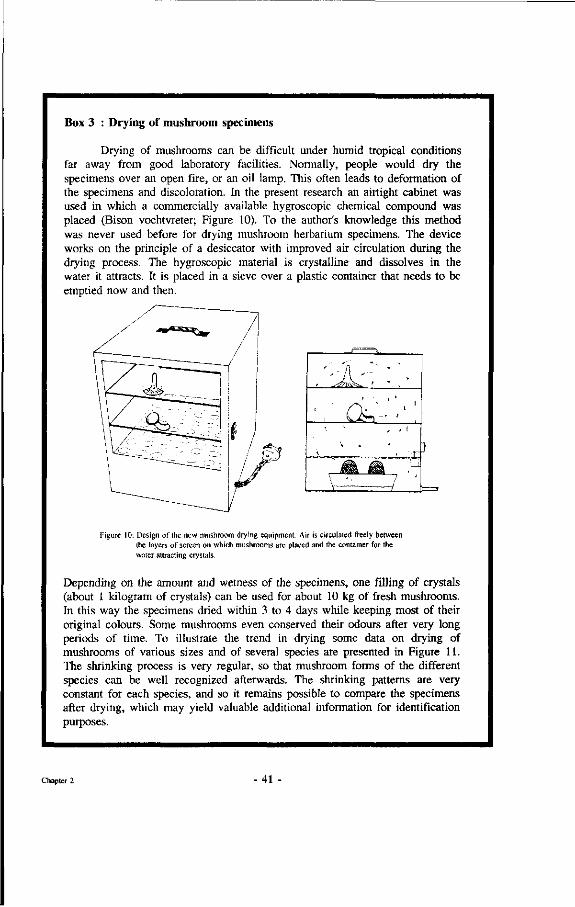
Box 3 : Drying of mushroom specimens
Drying of mushrooms can be difficult under humid tropical conditions far away from good laboratory facilities. Normally, people would dry the specimens over an open fire, or an oil lamp. This often leads to deformation of the specimens and discoloration. In the present research an airtight cabinet was used in which a commercially available hygroscopic chemical compound was placed (Bison vochtvreter; Figure 10). To the author's knowledge this method was never used before for drying mushroom herbarium specimens. The device works on the principle of a desiccator with improved air circulation during the drying process. The hygroscopic material is crystalline and dissolves in the water it attracts. It is placed in a sieve over a plastic container that needs to be emptied now and then.
Cfrr
—4C
Figure 10: Design of the new mushroom drying equipment. Air is circulated freely between the layers of screen on which mushrooms are placed and the container for the water attracting crystals.
Depending on the amount and wetness of the specimens, one filling of crystals (about 1 kilogram of crystals) can be used for about 10 kg of fresh mushrooms. In this way the specimens dried within 3 to 4 days while keeping most of their original colours. Some mushrooms even conserved their odours after very long periods of time. To illustrate the trend in drying some data on drying of mushrooms of various sizes and of several species are presented in Figure 11. The shrinking process is very regular, so that mushroom forms of the different species can be well recognized afterwards. The shrinking patterns are very constant for each species, and so it remains possible to compare the specimens after drying, which may yield valuable additional information for identification purposes.
Chapter 2 41
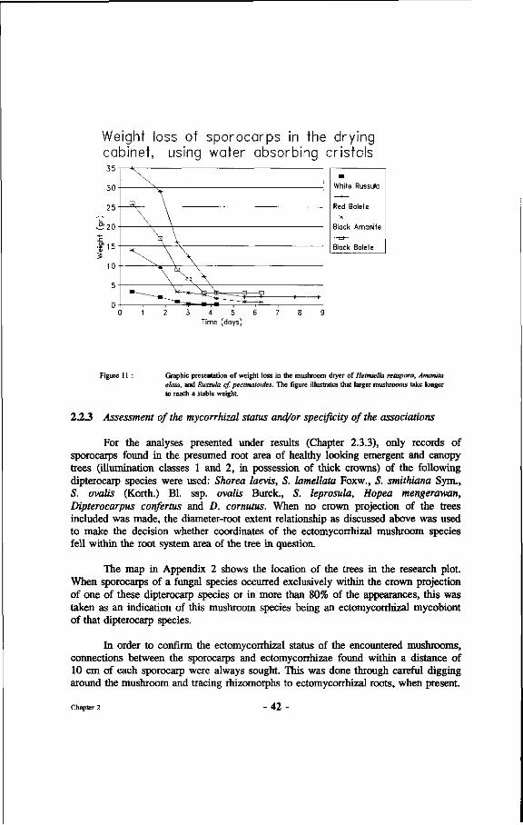
Weight loss of sporocarps in the drying cabinet, using water absorbing cristals 3 5 -
-m-White Russula
Red Bolete
Black Amanite - - S -
Black Bolete
3 4 5 6 Time (days)
Figure 11 : Graphic presentation of weight loss in the mushroom dryer of Heimiella retispora, Amanita elata, and Russula cf. pectinatoides. The figure illustrates that larger mushrooms take longer to reach a stable weight.
2.2.3 Assessment of the mycorrhizal status and/or specificity of the associations
For the analyses presented under results (Chapter 2.3.3), only records of sporocarps found in the presumed root area of healthy looking emergent and canopy trees (illumination classes 1 and 2, in possession of thick crowns) of the following dipterocarp species were used: Shorea laevis, S. lamellata Foxw., S. smithiana Sym., S. ovalis (Korth.) Bl. ssp. ovalis Buret, S. leprosula, Hopea mengerawan, Dipterocarpus confertus and D. cornutus. When no crown projection of the trees included was made, the diameter-root extent relationship as discussed above was used to make the decision whether coordinates of the ectomycorrhizal mushroom species fell within the root system area of the tree in question.
The map in Appendix 2 shows the location of the trees in the research plot. When sporocarps of a fungal species occurred exclusively within the crown projection of one of these dipterocarp species or in more than 80% of the appearances, this was taken as an indication of this mushroom species being an ectomycorrhizal mycobiont of that dipterocarp species.
In order to confirm the ectomycorrhizal status of the encountered mushrooms, connections between the sporocarps and ectomycorrhizae found within a distance of 10 cm of each sporocarp were always sought. This was done through careful digging around the mushroom and tracing rhizomorphs to ectomycorrhizal roots, when present.
Chapter 2 42
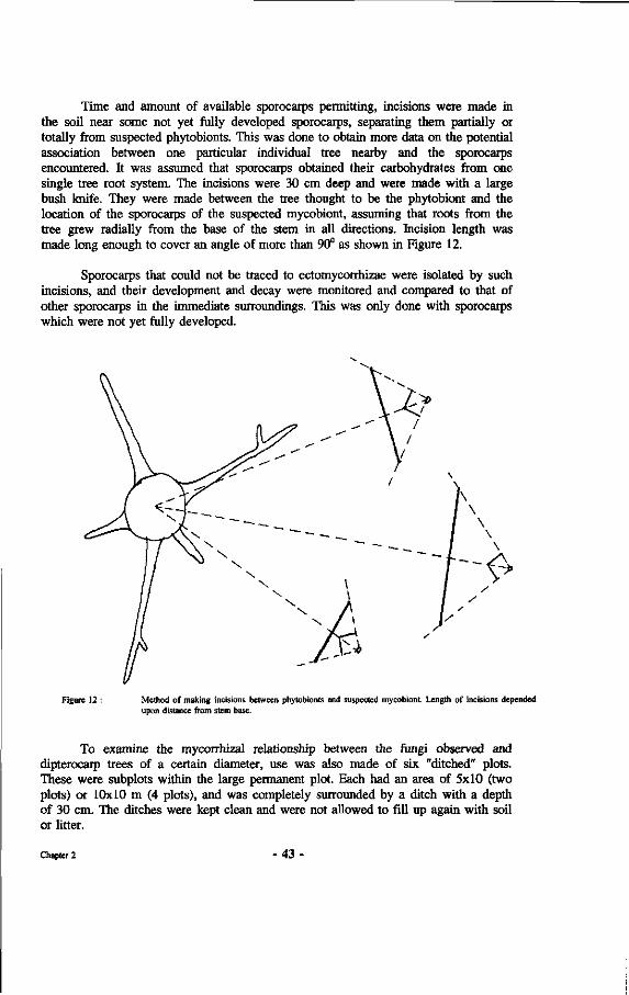
Time and amount of available sporocarps permitting, incisions were made in the soil near some not yet fully developed sporocarps, separating them partially or totally from suspected phytobionts. This was done to obtain more data on the potential association between one particular individual tree nearby and the sporocarps encountered. It was assumed that sporocarps obtained their carbohydrates from one single tree root system. The incisions were 30 cm deep and were made with a large bush knife. They were made between the tree thought to be the phytobiont and the location of the sporocarps of the suspected mycobiont, assuming that roots from the tree grew radially from the base of the stem in all directions. Incision length was made long enough to cover an angle of more than 90° as shown in Figure 12.
Sporocarps that could not be traced to ectomycorrhizae were isolated by such incisions, and their development and decay were monitored and compared to that of other sporocarps in the immediate surroundings. This was only done with sporocarps which were not yet fully developed.
Figure 12 : Method of making incisions between phytobionts and suspected mycobiont. Length of incisions depended upon distance from stem base.
To examine the mycorrhizal relationship between the fungi observed and dipterocarp trees of a certain diameter, use was also made of six "ditched" plots. These were subplots within the large permanent plot. Each had an area of 5x10 (two plots) or 10x10 m (4 plots), and was completely surrounded by a ditch with a depth of 30 cm. The ditches were kept clean and were not allowed to fill up again with soil or litter.
Chapter 2 - 43
These plots were laid out in such a manner that, within their limits, only small trees were growing having a diameter not exceeding 25 cm at breast height. All these trees were growing in the shadow of taller trees and were presumed not to produce enough carbohydrate to enable their mycobiont to produce sporocarps (Hacskaylo, 1973). At least five dipterocarp trees were present in each plot. The development of sporocarps of suspectedly ectomycorrhizal fungi within and around these plots was monitored very carefully at least twice a week over a period of 30 to 60 weeks.
Box 4 : The use of ditches
The intention of this ditching experiment was to investigate
a) whether growth of small Dipterocarps thus released from root competition would increase ;
b) whether, after 3 months following planting, non-mycorrhizal seedlings inside and outside the plot would form different mycorrhizae indicating that older trees might have other mycobionts than the younger ones of the same species; and
c) to see whether mycobionts associated with smaller trees that did not have their crowns in abundant light and thus might be expected to have a lower carbohydrate surplus, still were capable of producing sporocarps.
Such trees grow in the shade of taller trees and presumably do not produce enough carbohydrates to enable their mycobiont(s) to produce sporocarps. Hacskaylo (1973) e.g. showed the strong correlation between the development of sporocarps and direct photosynthetic production by the phytobiont in the symbiotic association with pine. The use of ditches surrounding a plot of forest has the additional advantage over the incisions described above that all roots of larger trees must have been severed.
Chapter 2 - 44
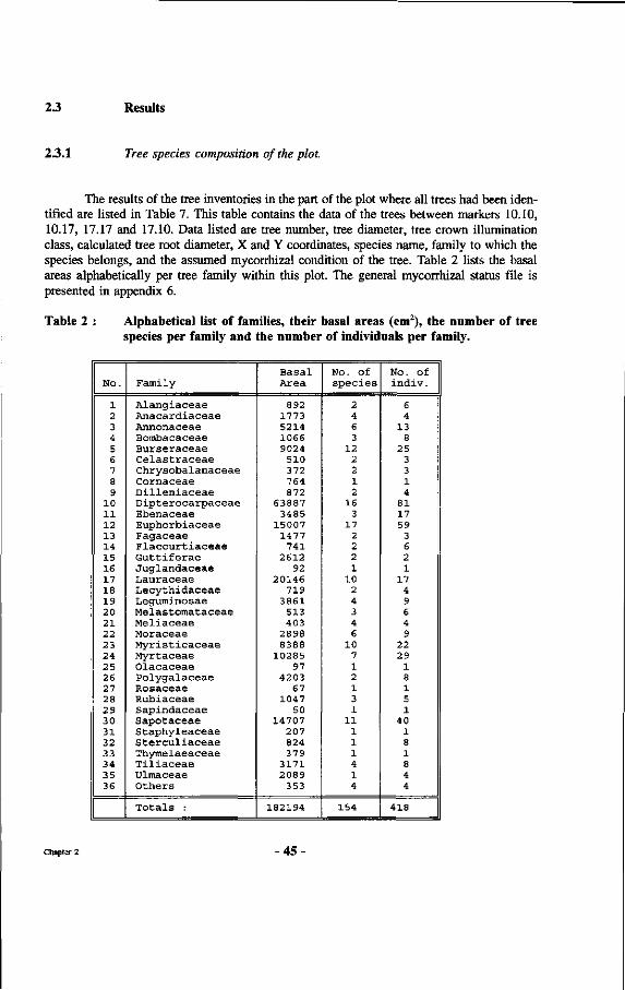
2.3 Results
2.3.1 Tree species composition of the plot.
The results of the tree inventories in the part of the plot where all trees had been identified are listed in Table 7. This table contains the data of the trees between markers 10.10, 10.17, 17.17 and 17.10. Data listed are tree number, tree diameter, tree crown illumination class, calculated tree root diameter, X and Y coordinates, species name, family to which the species belongs, and the assumed mycorrhizal condition of the tree. Table 2 lists the basal areas alphabetically per tree family within this plot. The general mycorrhizal status file is presented in appendix 6.
Table 2 : Alphabetical list of families, their basal areas (cm2), the number of tree species per family and the number of individuals per family.
No.
1 2 3 4 5 6 7 8 9
10 11 12 13 14 15 16 17 18 19 20 21 22 23 24 25 26 27 28 29 30 31 32 33 34 35 36
Family
Alangiaceae Anacardiaceae Annonaceae Bombacaceae Burseraceae Celastraceae Chrysobalanaceae Cornaceae Dilleniaceae Dipterocarpaceae Ebenaceae Euphorbiaceae Fagaceae Flacourtiaceae Guttiferae Juglandaceae Lauraceae Lecythidaceae Leguminosae Melastomataceae Meliaceae Moraceae Myristicaceae Myrtaceae Olacaceae Polygalaceae Rosaceae Rubiaceae Sapindaceae Sapotaceae Staphyleaceae Sterculiaceae Thymelaeaceae Tiliaceae Ulraaceae Others
Totals :
Basal Area
892 1773 5214 1066 9024
510 372 764 872
63887 3485
15007 1477
741 2612
92 20146
719 3861
513 403
2898 8388
10285 97
4203 67
1047 50
14707 207 824 379
3171 2089
353
182194
No. of species
2 4 6 3
12 2 2 1 2
16 3
17 2 2 2 1
10 2 4 3 4 6
10 7 1 2 1 3 1
11 1 1 1 4 1 4
154
No. Of indiv.
6 4
13 8
25 3 3 1 4
81 17 59
3 6 2 1
17 4 9 6 4 9
22 29
1 8 1 5 1
40 1 8 1 8 4 4
418
Chapter 2 •45-
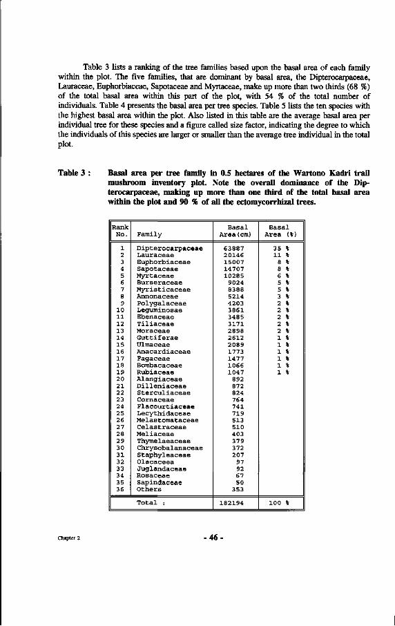
Table 3 lists a ranking of the tree families based upon the basal area of each family within the plot. The five families, that are dominant by basal area, the Dipterocarpaceae, Lauraceae, Euphorbiaceae, Sapotaceae and Myrtaceae, make up more than two thirds (68 %) of the total basal area within this part of the plot, with 54 % of the total number of individuals. Table 4 presents the basal area per tree species. Table 5 lists the ten species with the highest basal area within the plot. Also listed in this table are the average basal area per individual tree for these species and a figure called size factor, indicating the degree to which the individuals of this species are larger or smaller than the average tree individual in the total plot.
Table 3 : Basal area per tree family in 0.5 hectares of the Wartono Kadri trail mushroom inventory plot Note the overall dominance of the Dipterocarpaceae, making up more than one third of the total basal area within the plot and 90 % of all the ectomycorrhizal trees.
Rank No.
1 2 3 4 5 6 7 S 9
10 11 12 13 14 15 16 17 18 19 20 21 22 23 24 25 26 27 28 29 30 31 32 33 34 35 36
Family
Dipterocarpaceae Lauraceae Euphorbiaceae Sapotaceae Myrtaceae Burseraceae Myristicaceae Annonaceae Polygalaceae Leguminosae Ebenaceae Tiliaceae Moraceae Guttiferae Ulmaceae Anacardiaceae Fagaceae Bombacaceae Rubiaceae Alangiaceae Dilleniaceae Sterculiaceae Cornaceae Flacourtiaceae Lecythidaceae Melas tomataceae Celastraceae Meliaceae Thymelaeaceae Chrysobalanaceae Staphyleaceae Olacaceea Juglandaceae Rosaceae Sapindaceae Others
Total :
Basal Area(cm)
63887 20146 15007 14707 10285
9024 8388 5214 4203 3861 3485 3171 2898 2612 2089 1773 1477 1066 1047
892 872 824 764 741 719 513 510 403 379 372 207
97 92 67 50
353
182194
Basal Area (%)
35 % 11 %
8 % 8 % 6 % 5 % 5 % 3 % 2 % 2 % 2 % 2 % 2 % 1 % 1 % 1 % 1 % 1 % 1 %
100 %
Chapter 2 -46
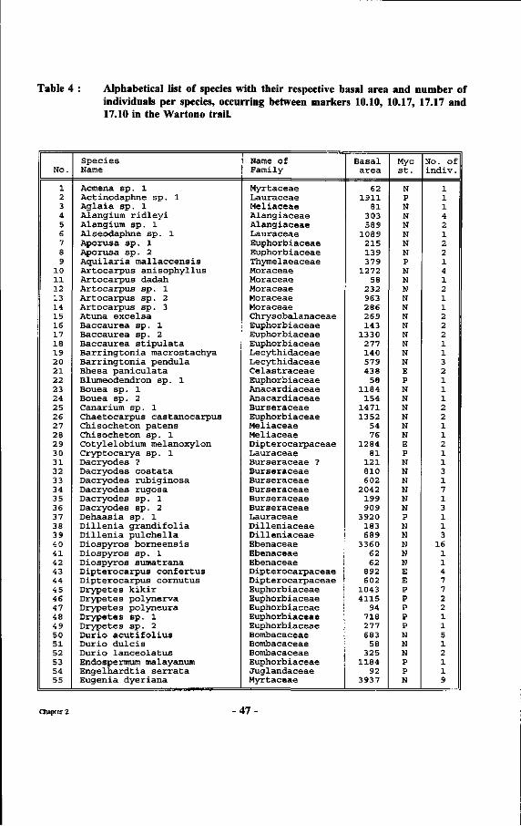
Table 4 : Alphabetical list of species with their respective basal area and number of individuals per species, occurring between markers 10.10, 10.17, 17.17 and 17.10 in the Wartono trail.
No.
1 2 3 4 5 6 7 8 9
10 11 12 13 14 15 16 17 18 19 20 21 22 23 24 25 26 27 28 29 30 31 32 33 34 35 36 37 38 39 40 41 42 43 44 45 46 47 48 49 50 51 52 53 54 55
Species Name
Acmena sp. 1 Actinodaphne sp. 1 Aglaia sp. 1 Alangium ridleyi Alangium sp. 1 Alseodaphne sp. 1 Aporusa sp. 1 Aporusa sp. 2 Aquilaria mallaccensis Artocarpus anisophyllus Artocarpus dadah Artocarpus sp. 1 Artocarpus sp. 2 Artocarpus sp. 3 Atuna excelsa Baccaurea sp. 1 Baccaurea sp. 2 Baccaurea stipulata Barringtonia macrostachya Barringtonia pendula Bhesa paniculata Blumeodendron sp. 1 Bouea sp. 1 Bouea sp. 2 Canarium sp. 1 Chaetocarpus castanocarpus Chisocheton patens Chisocheton sp. 1 Cotylelobiura melanoxylon Cryptocarya sp. 1 Dacryodes ? Dacryodes costata Dacryodes rubiginosa Dacryodes rugosa Dacryodes sp. 1 Dacryodes sp. 2 Dehaasia sp. 1 Dillenia grandifolia Dillenia pulchella Diospyros borneensis Diospyros sp. 1 Diospyros sumatrana Dipterocarpus confertus Dipterocarpus cornutus Drypetes kikir Drypetes polynerva Drypetes polyneura Drypetes sp. 1 Drypetes sp. 2 Durio acut i fol ius Durio dulcis Durio lanceolatus Endospermura malayanum Engelhardtia serrata Eugenia dyeriana
Name of Family
Myrtaceae Lauraceae Meliaceae Alangiaceae Alangiaceae Lauraceae Euphorbiaceae Euphorbiaceae Thymelaeaceae Moraceae Moraceae Moraceae Moraceae Moraceae Chrysobalanaceae Euphorbiaceae Euphorbiaceae Euphorbiaceae Lecythidaceae Lecythidaceae Celastraceae Euphorbiaceae Anacardiaceae Anacardiaceae Burseraceae Euphorbiaceae Meliaceae Meliaceae Dipterocarpaceae Lauraceae Burseraceae ? Burseraceae Burseraceae Burseraceae Burseraceae Burseraceae Lauraceae Dilleniaceae Dilleniaceae Ebenaceae Ebenaceae Ebenaceae Dipterocarpaceae Dipterocarpaceae Euphorbiaceae Euphorbiaceae Euphorbiaceae Euphorbiaceae Euphorbiaceae Bombacaceae Bombacaceae Bombacaceae Euphorbiaceae Juglandaceae Myrtaceae
Basal area
62 1911
81 303 589
1089 215 139 379
1272 58
232 963 286 269 143
1330 277 140 579 438
58 1184
154 1471 1352
54 76
1284 81
121 810 602
2042 199 909
3920 183 689
3360 62 62
892 602
1043 4115
94 718 277 683
58 325
1184 92
3937
Myc St.
N P N N N N N N P N N N N N N N N N N N E P N N N N N N E P N N N N N N P N N N N N E E P P P P P N N N P P N
No. Of indiv.
1 1 1 4 2 1 2 2 1 4 1 2 1 1 2 2 2 1 1 3 2 1 1 1 2 2 1 1 2 1 1 3 1 7 1 3 1 1 3
16 1 1 4 7 7 2 2 1 1 5 1 2 1 1 9
Chapter 2 •47-
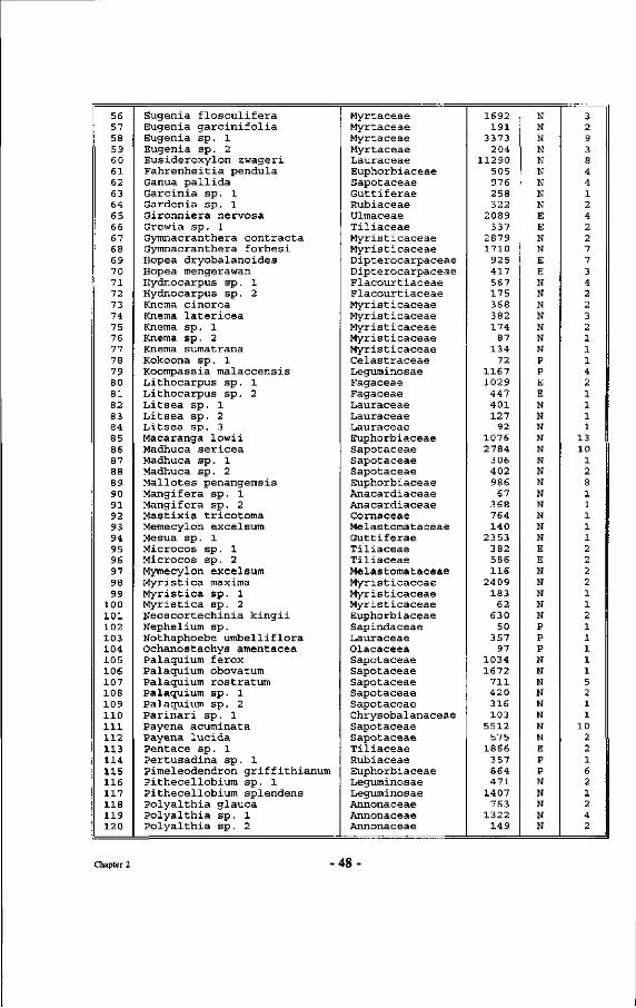
56 57 58 59 60 61 62 63 64 65 66 67 68 69 70 71 72 73 74 75 76 77 78 79 80 81 82 83 84 85 86 87 88 89 90 91 92 93 94 95 96 97 98 99
100 101 102 103 104 105 106 107 108 109 110 111 112 113 114 115 116 117 118 119 120
Eugenia flosculifera Eugenia garcinifolia Eugenia sp. 1 Eugenia sp. 2 Eusideroxylon zwageri Fahrenheitia pendula Ganua pallida Garcinia sp. 1 Gardenia sp. 1 Gironniera nervosa Grewia sp. 1 Gymnacranthera contracta Gymnacranthera forbesi Hopea dryobalanoides Hopea mengerawan Hydnocarpus sp. 1 Hydnocarpus sp. 2 Kneraa cinerea Knema latericea Knema sp. 1 Knema sp. 2 Knema sumatrana Kokoona sp. 1 Koompassia malaccensis Lithocarpus sp. 1 Lithocarpus sp. 2 Litsea sp. 1 Litsea sp. 2 Litsea sp. 3 Macaranga lowii Madhuca sericea Madhuca sp. 1 Madhuca sp. 2 Mallotes penangensis Mangifera sp. 1 Mangifera sp. 2 Mastixia tricotoma Memecylon excelsum Mesua sp. 1 Microcos sp. 1 Microcos sp. 2 Mymecylon excelsum Myristica maxima Myristica sp. 1 Myristica sp. 2 Neoscortechinia kingii Nephelium sp. Nothaphoebe umbelliflora Ochanostachys amentacea Palaquium ferox Palaquium obovatum Palaquium rostratum Palaquium sp. 1 Palaquium sp. 2 Parinari sp. 1 Payena acuminata Payena lucida Pentace sp. 1 Pertusadina sp. 1 Pimeleodendron griffithianum Pithecellobium sp. 1 Pithecellobium splendens Polyalthia glauca Polyalthia sp. 1 Polyalthia sp. 2
Myrtaceae Myrtaceae Myrtaceae Myrtaceae Lauraceae Euphorbiaceae Sapotaceae Guttiferae Rubiaceae Ulmaceae Tiliaceae Myristicaceae Myristicaceae Dipterocarpaceae Dipterocarpaceae Flacourtiaceae Flacourtiaceae Myristicaceae Myristicaceae Myristicaceae Myristicaceae Myristicaceae Celastraceae Leguminosae Fagaceae Fagaceae Lauraceae Lauraceae Lauraceae Euphorbiaceae Sapotaceae Sapotaceae Sapotaceae Euphorbiaceae Anacardiaceae Anacardiaceae Cornaceae Melastomataceae Guttiferae Tiliaceae Tiliaceae Melastomataceae Myristicaceae Myristicaceae Myristicaceae Euphorbiaceae Sapindaceae Lauraceae Olacaceea Sapotaceae Sapotaceae Sapotaceae Sapotaceae Sapotaceae Chrysobalanaceae Sapotaceae Sapotaceae Tiliaceae Rubiaceae Euphorbiaceae Leguminosae Leguminosae Annonaceae Annonaceae Annonaceae
1692 191
3373 204
11290 505 976 258 322
2089 337
2879 1710
925 417 567 175 368 382 174
87 134
72 1167 1029
447 401 127
92 1076 2784
306 402 986
67 368 764 140
2353 382 586 116
2409 183
62 630
50 357
97 1034 1672
711 420 316 103
5512 575
1866 357 864 471
1407 763
1322 149
N N N N N N N N N E E N N E E N N N N N N N P P E E N N N N N N N N N N N N N E E N N N N N P P P N N N N N N N N E P P N N N N N
3 2 9 3 8 4 4 1 2 4 2 2 7 7 3 4 2 2 3 2 1 1 1 4 2 1 1 1 1
13 10
1 2 8 1 1 1 1 1 2 2 2 2 1 1 2 1 1 1 1 1 5 2 1 1
10 2 2 1 6 2 1 2 4 2
Chapter 2 -48
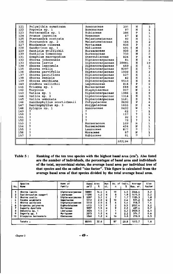
121 122 123 124 125 126 127 128 129 130 1 31 132 133 134 135 136 137 138 139 140 1 41 142 143 144 145 146 147 148 149 150 1 51 152 153 154 155 156 157
P o l y a l t h i a sumatrana Popowia s p . 1 P o r t e r a n d i a s p . 1 Prunus j a pon i ca P t e rnand ra r o s t r a t a P t e rnand ra s p . 2 Rhodamnia c i n e r e a Sandoricum s p . 1 S a n t i r i a g r i f f i t h i i S a n t i r i a tomentosa Scaphium macropodium Shorea j o h o r e n s i s Shorea l a e v i s Shorea l e p r o s u l a Shorea o v a l i s Shorea p a r v i f o l i a Shorea p a u c i f l o r a Shorea s emin is Shorea smi th i ana S indora w a l l i c h i i Triomma s p . 1 Tu rp i n i a Va t i c a s p . 1 Va t i c a s p . 2 Va t i c a umbonata Xanthophyllum s c o r t i c h e n i i Xanthophyllum s p . 1 Xylopia s p . 1 •? 7 ? ? ? ? ? •p
?
Annonaceae Annonaceae Rubiaceae Rosaceae Melastomataceae Melastomataceae Myrtaceae Mel iaceae Burseraceae Burse raceae S t e r c u l i a c e a e D ip t e roca rpaceae D ip t e roca rpaceae D ip t e roca rpaceae D ip t e roca rpaceae D ip t e roca rpaceae D ip t e roca rpaceae D ip t e roca rpaceae D ip t e roca rpaceae Leguminosae Burseraceae S taphy leaceae D ip t e roca rpaceae D ip t e roca rpaceae D ip t e roca rpaceae Po lyga laceae Po lyga laceae Annonaceae ? ? ? •?
Burseraceae Burse raceae Lauraceae Moraceae Rubiaceae
391 97
286 67 92
165 826 191 920 730 824
81 38881
459 8297 1909
127 42
5510 816 688 207
1986 1314 1161 2600 1603 2492
62 127
92 72
109 424 877
87 81
182194
N N P E N N P N N N P E E E E E E E E P N N E E E P N N ? ? •?
? N N P N P
3 1 1 1 1 2 2 1 2 2 8 1
19 1 7 3 1 1 6 2 1 1 9 5 5 4 4 1 1 1 1 1 1 1 1 1 1
Table 5 : Ranking of the ten tree species with the highest basal area (cm2). Also listed are the number of individuals, the percentages of basal area and individuals of the total, mycorrhizal status, the average basal area per individual tree of that species and the so called "size factor". This figure is calculated from the average basal area of that species divided by the total average basal area.
No.
1 2 3 4 5 6
/ 8 9
10
Species Name
Shorea Laevis Eusideroxylon zwageri Shorea oval i s Payena acuminata Shorea smithiana Drypetes polynerva Eugenia dyeriana Dehaasia sp. 1 Eugenia sp. 1 Diospyros borneensis
Name of Fami ly
Dipterocarpaceae Lauraceae Dipterocarpaceae Sapotaceae D i pterocarpaceae Euphorbiaceae Myrtaceae Lauraceae Myrtaceae Ebenaceae
Totals :
Basal Area cmA2
38881 11290 8297 5512 5510 4115 3937 3920 3373 3360
88195
% 14.4 4.2 3.1 2.0 2.0 1.5 1.5 1.4 1.2 1.2
32.6
Myc S t .
E N E N E P N P N N
No. of n
19 8 7
10 6 2 9 1 9
16
87
i nd iv .
% 4.5 1.9 1.7 2.4 1.4 0.5 2.2 0.2 2.2 3.8
20.8
Average Bas. ar .
2046.4 1411.3 1185.3 551.2 918.3
2057.4 437.4
3920.3 374.7 210.0
1013.7
Size factor
3.2 2.2 1.8 0.9 1.4 3.2 0.7 6.1 0.6 0.3
1.6
Chapter 2 -49
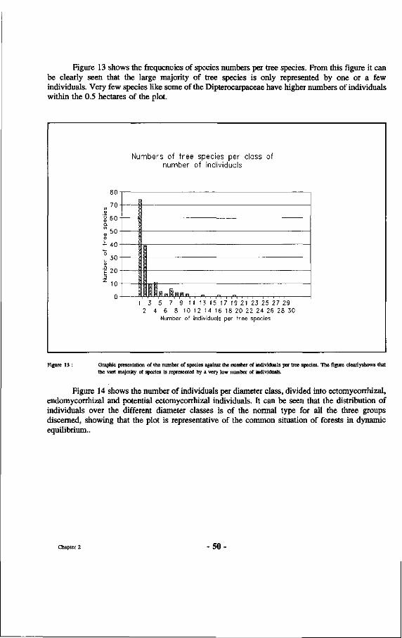
Figure 13 shows the frequencies of species numbers per tree species. From this figure it can be clearly seen that the large majority of tree species is only represented by one or a few individuals. Very few species like some of the Dipterocarpaceae have higher numbers of individuals within the 0.5 hectares of the plot.
70 I
: 60
| 5 0
40
30
! 20
: 10
Numbers of tree species per class of number of individuals
I 3 5 7 9 11 13 15 17 19 21 23 25 27 29 2 4 6 8 10 12 14 16 18 20 22 24 26 28 30
Number of individuals per tree species
Figure 13 : Graphic presentation of the number of species against the number of individuals per tree species. The figure clearlyshows that the vast majority of species is represented by a very low number of individuals.
Figure 14 shows the number of individuals per diameter class, divided into ectomycorrhizal, endomycorrhizal and potential ectomycorrhizal individuals. It can be seen that the distribution of individuals over the different diameter classes is of the normal type for all the three groups discerned, showing that the plot is representative of the common situation of forests in dynamic equilibrium..
Chapter 2 50
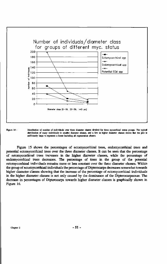
Number of individuals/diameter class for groups of different myc. status
200
180
160
! 140
' 120 j
: loo
; 80
' 60
40
20
0
Ectomycorrhizal spp
Endomycorrhizal spp
Potential ECM spp
Diameter class (0-19, 20-39, >40 cm)
Figure 14 : Distribution of number of individuals over three diameter classes divided for three myconhizal status groups. The typical distribution of many individuals in smaller diameter classes, and a few in higher diameter classes shows that the plot is sufficiently large to represent a forest including all regeneration phases.
Figure 15 shows the percentages of ectomycorrhizal trees, endomycorrhizal trees and potential ectomycorrhizal trees over the three diameter classes. It can be seen that the percentage of ectomycorrhizal trees increases in the higher diameter classes, while the percentage of endomycorrhizal trees decreases. The percentage of trees in the group of the potential ectomycorrhizal individuals remains more or less constant over the three diameter classes. Within the group of ectomycorrhizal individuals the percentage of Dipterocarps decreases somewhat towards higher diameter classes showing that the increase of the percentage of ectomycorrhizal individuals in the higher diameter classes is not only caused by the dominance of the Dipterocarpaceae. The decrease in percentages of Dipterocarps towards higher diameter classes is graphically shown in Figure 16.
Chapter 2 - 5 1
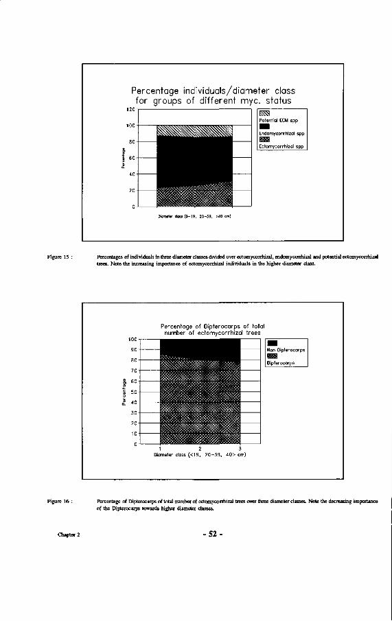
Percentage individuals/diameter class for groups of different myc. status
120
100
80
\ 60
40
Potential ECM spp
Endomycorrhizal spp
n Ectomycorrhizal spp
Diameler class (0-19, 20-39, >40 c
Figure 15 : Percentages of individuals in three diameter classes divided over ectomycorrhizal, endomycorrhizal and potential ectomycorrhizal trees. Note the increasing importance of ectomycorrhizal individuals in the higher diameter class.
Percentage of Dipterocarps of total number of ectomycorrhizal trees
8 0 -
7 0 i
§> 60-1 o
Î 50
£ 40
30
20-
10-
o -
^^^^^^^^^H \
• Dipterocarps
Diameter class (<19, 2 0 - 3 9 , 40> cm)
Figure 16 : Percentage of Dipterocarps of total number of ectomycorrhizal trees over three diameter classes. Note the decreasing importance of the Dipterocarps towards higher diameter classes.
Chapter 2 -52
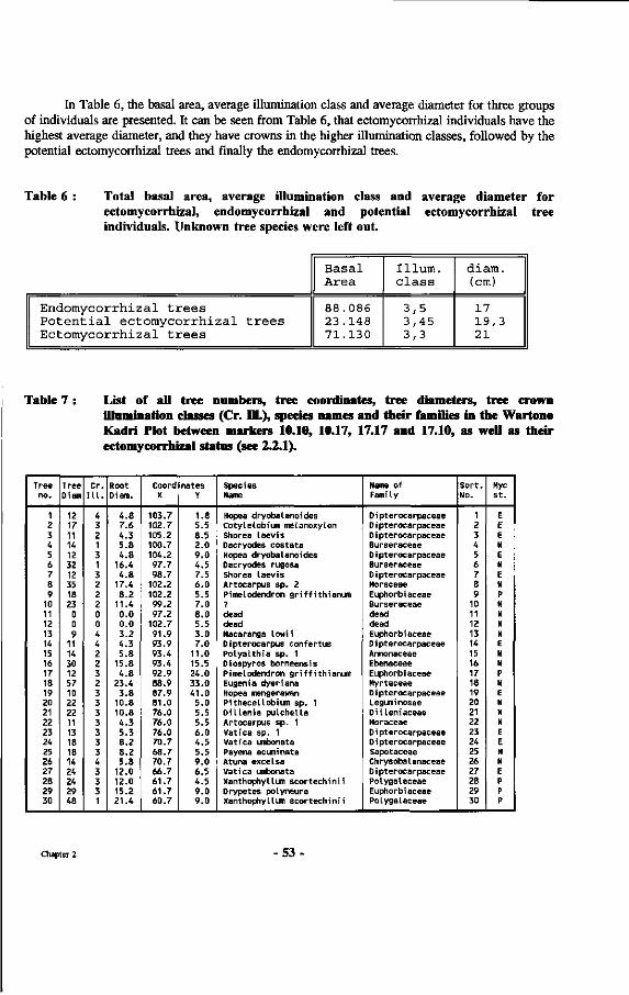
In Table 6, the basal area, average illumination class and average diameter for three groups of individuals are presented. It can be seen from Table 6, that ectomycorrhizal individuals have the highest average diameter, and they have crowns in the higher illumination classes, followed by the potential ectomycorrhizal trees and finally the endomycorrhizal trees.
Table 6 : Total basal area, average illumination class and average diameter for ectomycorrhizal, endomycorrhizal and potential ectomycorrhizal tree individuals. Unknown tree species were left out.
Endomycorrhizal trees Potential ectomycorrhizal trees Ectomycorrhizal trees
Basal Area
88.086 23.148 71.130
Ilium, class
3,5 3,45 3,3
diam. (cm)
17 19,3 21
Table 7 : List of all tree numbers, tree coordinates, tree diameters, tree crown illumination classes (Cr. DL), species names and their families in the Wartono Kadri Plot between markers 10.10, 10.17, 17.17 and 17.10, as well as their ectomycorrhizal status (see 2.2.1).
Tree no.
1 2 3 4 5 6 7 8 9
10 11 12 13 14 15 16 17 18 19 20 21 22 23 24 25 26 27 28 29 30
Tree Diam
12 17 11 14 12 32 12 35 18 23 0 0 9
11 14 30 12 57 10 22 22 11 13 18 18 14 24 24 29 48
Cr. ILL.
4 3 2 1 3 1 3 2 2 2 0 0 4 4 2 2 3 2 3 3 3 3 3 3 3 4 3 3 3 1
Root Diam.
4.8 7.6 4.3 5.8 4.8
16.4 4.8
17.4 8.2
11.4 0.0 0.0 3.2 4.3 5.8
15.8 4.8
23.4 3.8
10.8 10.8 4.3 5.3 8.2 8.2 5.8
12.0 12.0 15.2 21.4
Coordinates X
103.7 102.7 105.2 100.7 104.2 97.7 98.7
102.2 102.2 99.2 97.2
102.7 91.9 93.9 93.4 93.4 92.9 88.9 87.9 81.0 76.0 76.0 76.0 70.7 68.7 70.7 66.7 61.7 61.7 60.7
Y
1.8 5.5 8.5 2.0 9.0 4.5 7.5 6.0 5.5 7.0 8.0 5.5 3.0 7.0
11.0 15.5 24.0 33.0 41.0 5.0 5.5 5.5 6.0 4.5 5.5 9.0 6.5 4.5 9.0 9.0
Species Name
Hopea dryobalanoides Cotylelobium meLanoxyLon Shorea laevis Dacryodes costata Hopea dryobalanoides Dacryodes rugosa Shorea laevis Artocarpus sp. 2 Pimelodendron griffithianum ? dead dead Macaranga lowii Dipterocarpus confertus Polyalthia sp. 1 Diospyros borneensis PimeLodendron griffithianum Eugenia dyeriana Hopea mengerawan Pithecellobium sp. 1 Dillenia pulchella Artocarpus sp. 1 Vatica sp. 1 vatica umbonata Payena acuminata Atuna excelsa Vatica umbonata Xanthophyllum scortechinii Drypetes polyneura Xanthophyllum scortechinii
Name of Fami ly
D i pterocarpaceae D i pterocarpaceae D i pterocarpaceae Burseraceae D i pterocarpaceae Burseraceae D i pterocarpaceae Moraceae Euphorbiaceae Burseraceae dead dead Euphorbiaceae Dipterocarpaceae Annonaceae Ebenaceae Euphorbiaceae Myrtaceae D i pterocarpaceae Leguminosae Dilleniaceae Moraceae D i pterocarpaceae D i pterocarpaceae Sapotaceae Chrysobalanaceae Dipterocarpaceae Polygalaceae Euphorbiaceae Polygalaceae
Sort. No.
1 2 3 4 5 6 7 8 9
10 11 12 13 14 15 16 17 18 19 20 21 22 23 24 25 26 27 28 29 30
Myc St.
E E E N E N E N P N N N N E N N P N E N N N E E N N E P P P
Chapter 2 5 3 -
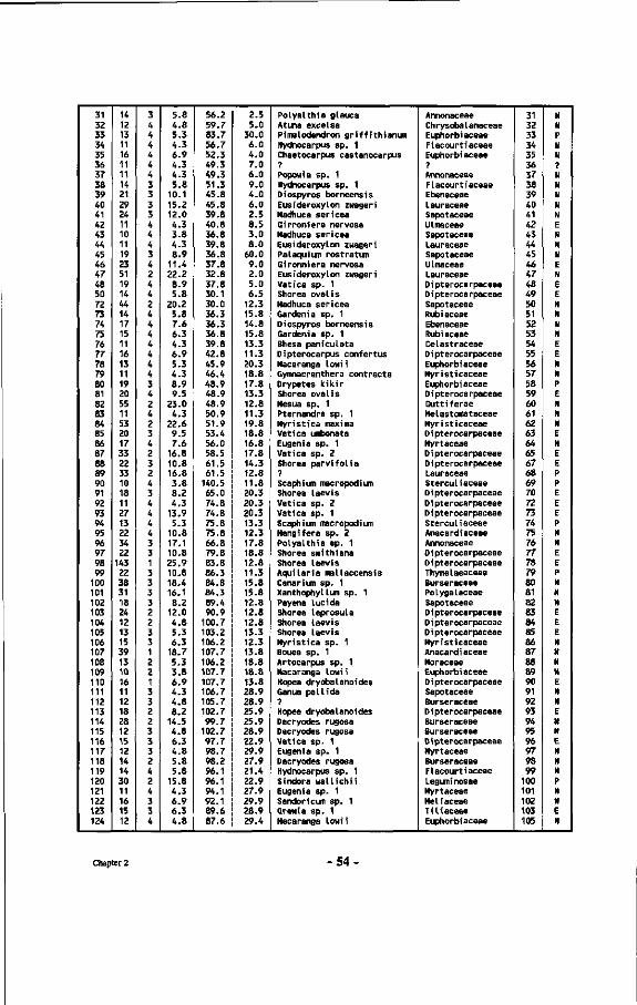
31 32 33 34 35 36 37 38 39 40 41 42 43 44 45 46 47 48 50 72 73 74 75 76 77 78 79 80 81 82 83 84 85 86 87 88 89 90 91 92 93 94 95 96 97 98 99
100 101 102 103 104 105 106 107 108 109 110 111 112 113 114 115 116 117 118 119 120 121 122 123 124
14 12 13 11 16 11 11 14 21 29 24 11 10 11 19 23 51 19 14 44 14 17 15 11 16 13 11 19 20 55 11 53 20 17 33 22 33 10 18 11 27 13 22 34 22
143 22 38 31 18 24 12 13 15 39 13 10 16 11 12 18 28 12 15 12 14 14 30 11 16 15 12
3 4 4 4 4 4 4 3 3 3 3 4 4 4 3 4 2 4 4 2 4 4 4 4 4 4 4 3 4 2 4 2 3 4 2 3 2 4 3 4 4 4 4 3 3 1 3 3 3 3 2 2 3 3 1 2 2 1 3 3 2 2 3 3 3 2 4 2 4 3 3 4
5.8 4.8 5.3 4.3 6.9 4.3 4.3 5.8
10.1 15.2 12.0 4.3 3.8 4.3 8.9
11.4 22.2 8.9 5.8
20.2 5.8 7.6 6.3 4.3 6.9 5.3 4.3 8.9 9.5
23.0 4.3
22.6 9.5 7.6
16.8 10.8 16.8 3.8 8.2 4.3
13.9 5.3
10.8 17.1 10.8 25.9 10.8 18.4 16.1 8.2
12.0 4.8 5.3 6.3
18.7 5.3 3.8 6.9 4.3 4.8 8.2
14.5 4.8 6.3 4.8 5.8 5.8
15.8 4.3 6.9 6.3 4.8
56.2 59.7 83.7 56.7 52.3 49.3 49.3 51.3 45.8 45.8 39.8 40.8 36.8 39.8 36.8 37.8 32.8 37.8 30.1 30.0 36.3 36.3 36.8 39.8 42.8 45.9 46.4 48.9 48.9 48.9 50.9 51.9 53.4 56.0 58.5 61.5 61.5
140.5 65.0 74.8 74.8 75.8 75.8 66.8 79.8 83.8 86.3 84.8 84.3 89.4 90.9
100.7 103.2 106.2 107.7 106.2 107.7 107.7 106.7 105.7 102.7 99.7
102.7 97.7 98.7 98.2 96.1 96.1 94.1 92.1 89.6 87.6
2.5 5.0
30.0 6.0 4.0 7.0 6.0 9.0 4.0 6.0 2.5 8.5 3.0 8.0
60.0 9.0 2.0 5.0 6.5
12.3 15.8 14.8 15.8 13.3 11.3 20.3 18.8 17.8 13.3 12.8 11.3 19.8 18.8 16.8 17.8 14.3 12.8 11.8 20.3 20.3 20.3 13.3 12.3 17.8 18.8 12.8 11.3 15.8 15.8 12.8 12.8 12.8 13.3 12.3 13.8 18.8 18.8 13.8 28.9 28.9 25.9 25.9 28.9 22.9 29.9 27.9 21.4 22.9 27.9 29.9 28.9 29.4
Polyalthia glauca Atuna excelsa Pimei «tendron griffithianun Hydnocarpus sp. 1 Chaetocarpus castanocarpus ? Popowia sp. 1 Hydnocarpus sp. 1 Diospyros borneensis Eusideroxylon zwageri Madhuca sericea Girroniera nervosa Madhuca sericea Eusideroxylon zwageri Palaquium rostratum Gironniera nervosa Eusideroxylon zwageri Vatica sp. 1 Shorea oval is Madhuca sericea Gardenia sp. 1 Diospyros borneensis Gardenia sp. 1 Bhesa paniculata Dipterocarpus confertus Macaranga lowii Gymnacranthera contracta Drypetes kikir Shorea oval is Mesua sp. 1 Pternandra sp. 1 Myristica maxima Vatica umbonata Eugenia sp. 1 Vatica sp. 2 Shorea parvifolia ? Scaphium macropodium Shorea laevis Vatica sp. 2 Vatica sp. 1 Scaphium macropodium Mangifera sp. 2 Polyalthia sp. 1 Shorea smithiana Shorea laevis Aquilaria mallaccensis Canarium sp. 1 Xanthophyllum sp. 1 Payena lucida Shorea leprosula Shorea laevis Shorea laevis Myristica sp. 1 Bouea sp. 1 Artocarpus sp. 1 Macaranga lowi i Hopea dryobalanoides Ganua pallida ? Hopea dryobalanoides Dacryodes rugosa Dacryodes rugosa Vatica sp. 1 Eugenia sp. 1 Dacryodes rugosa Hydnocarpus sp. 1 Sindora wallichi.i Eugenia sp. 1 Sandoricum sp. 1 Grewia sp. 1 Macaranga lowii
Annonaceae Ch rysoba L anaceae Euphorbiaceae Flacourtiaceae Euphorbiaceae ? Annonaceae Flacourtiaceae Ebenaceae Lauraceae Sapotaceae Ulmaceae Sapotaceae Lauraceae Sapotaceae Ulmaceae Lauraceae Dipterocarpaceae Dipterocarpaceae Sapotaceae Rubiaceae Ebenaceae Rubiaceae Celastraceae Dipterocarpaceae Euphorbiaceae Myristicaceae Euphorbiaceae D i pterocarpaceae Guttiferae Melastomataceae Myristicaceae D i pterocarpaceae Myrtaceae Dipterocarpaceae Dipterocarpaceae Lauraceae Sterculiaceae D i pterocarpaceae D i pterocarpaceae Dipterocarpaceae Sterculiaceae Anacardiaceae Annonaceae Dipterocarpaceae D i pterocarpaceae Thymelaeaceae Burseraceae Polygalaceae Sapotaceae D i pterocarpaceae D i pterocarpaceae Dipterocarpaceae Myristicaceae Anacardiaceae Moraceae Euphorbiaceae Dipterocarpaceae Sapotaceae Burseraceae Dipterocarpaceae Burseraceae Burseraceae Dipterocarpaceae Myrtaceae Burseraceae Flacourtiaceae Leguminosae Myrtaceae Meliaceae Tiliaceae Euphorbiaceae
31 32 33 34 35 36 37 38 39 40 41 42 43 44 45 46 47 48 49 50 51 52 53 54 55 56 57 58 59 60 61 62 63 64 65 67 68 69 70 72 73 74 75 76 77 78 79 80 81 82 83 84 85 86 87 88 89 90 91 92 93 94 95 96 97 98 99
100 101 102 103 105
N N P N N ? N N N N N E N N N E N E E N N N N E E N N P E N N N E N E E P P E E E P N N E E P N N N E E E N N N N E N N E N N E N N N P N N E N
Chapter 2 • 5 4 .
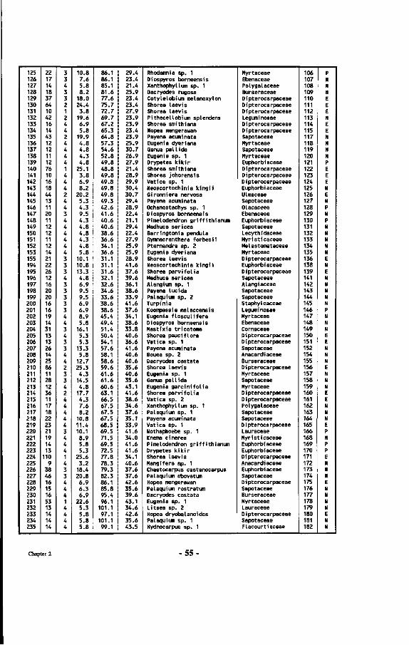
125 126 127 128 129 130 131 132 133 134 135 136 137 138 139 140 141 142 143 144 145 146 147 148 149 150 151 152 153 155 194 195 196 197 198 199 200 201 202 203 204 205 206 207 208 209 210 211 212 213 214 215 216 217 218 219 220 221 222 223 224 225 226 227 228 229 230 231 232 233 234 235
22 17 14 18 37 64 10 42 16 14 43 12 12 11 12 76 10 16 18 44 13 11 20 11 12 12 11 12 14 21 22 26 12 16 20 20 16 16 19 14 31 13 13 26 14 25 86 11 28 12 36 11 17 18 22 23 21 19 14 13
110 9
38 46 16 15 16 53 13 14 14 14
3 3 4 3 3 2 1 2 4 4 2 4 4 4 4 1 4 4 4 2 4 4 3 4 4 4 4 4 4 3 3 3 4 3 3 3 3 3 4 4 3 4 3 3 4 4 2 3 3 4 2 4 4 4 4 4 3 4 4 4 1 4 3 3 4 4 4 1 4 4 4 4
10.8 7.6 5.8 8.2
18.0 24.4 3.8
19.6 6.9 5.8
19.9 4.8 4.8 4.3 4.8
25.1 3.8 6.9 8.2
20.2 5.3 4.3 9.5 4.3 4.8 4.8 4.3 4.8 5.8
10.1 10.8 13.3 4.8 6.9 9.5 9.5 6.9 6.9 8.9 5.8
16.1 5.3 5.3
13.3 5.8
12.7 25.3 4.3
14.5 4.8
17.7 4.3 7.6 8.2
10.8 11.4 10.1 8.9 5.8 5.3
25.6 3.2
18.4 20.8 6.9 6.3 6.9
22.6 5.3 5.8 5.8 5.8
86.1 86.1 85.1 81.6 77.6 75.7 72.7 69.7 67.2 65.3 64.8 57.3 54.6 52.8 49.8 48.8 49.8 49.8 49.8 49.8 49.3 42.6 41.6 40.6 40.6 38.6 36.6 34.1 36.6 31.1 31.1 31.6 32.1 32.6 34.6 33.6 38.6 38.6 45.4 49.4 51.4 50.4 54.1 57.6 58.1 58.6 59.6 61.6 61.6 60.6 63.1 66.5 67.5 67.5 67.5 68.5 69.5 71.5 69.5 72.5 77.8 78.3 79.3 82.3 86.1 85.8 95.4 96.1
101.1 97.1
101.1 99.1
29.4 23.4 21.4 25.9 23.4 23.4 27.9 23.9 23.9 23.4 23.9 25.9 30.7 26.9 27.9 21.4 28.9 29.9 30.4 30.7 29.4 28.9 22.4 21.1 29.4 22.4 27.9 25.9 25.9 28.9 41.6 37.6 39.6 36.1 38.6 33.9 41.6 37.6 34.1 38.6 33.8 40.6 36.6 41.6 40.6 40.6 35.6 40.6 35.6 43.1 41.6 38.6 34.6 37.6 35.1 33.9 41.6 34.0 41.6 41.6 34.1 40.6 37.6 37.6 42.6 35.6 39.6 43.1 34.6 42.6 35.6 43.5
Rhodamnia sp. 1 Diospyros borneensis Xanthophyllum sp. 1 Dacryodes rugosa Cotylelobium melanoxylon Shorea Laevis Shorea Laevis PitheceUobium splendens Shorea smithiana Hopea mengerawan Payena acuminata Eugenia dyeriana Ganua pallida Eugenia sp. 1 Drypetes kikir Shorea smithiana Shorea johorensis Vatica sp. 1 Neoscortechinia kingii Gironniera nervosa Payena acuminata Ochanostachys sp. 1 Diospyros borneensis Pimelodendron griffithianum Madhuca sericea Barringtonia pendula Gymnacranthera forbesi i Pternandra sp. 2 Eugenia dyeriana Shorea laevis Neoscortechinia kingii Shorea parvifolia Madhuca sericea Alangium sp. 1 Payena lucida Palaquium sp. 2 Turpinia Koompassia malaccensis Eugenia flosculifera Diospyros borneensis Mastixia tricotoma Shorea pauciflora Vatica sp. 1 Payena acuminata Bouea sp. 2 Dacryodes costata Shorea laevis Eugenia sp. 1 Ganua pallida Eugenia garcinifolia Shorea parvifolia Vatica sp. 2 Xanthophyllum sp. 1 Palaquium sp. 1 Payena acuminata Vatica sp. 1 Nothaphoebe sp. 1 Knema cinerea Pimelodendron griffithianum Drypetes kikir Shorea laevis Mangifera sp. 1 Chaetocarpus castanocarpus Palaquium obovatum Hopea mengerawan Palaquium rostratum Dacryodes costata Eugenia sp. 1 Litsea sp. 2 Hopea dryobalanoides Palaquium sp. 1 Hydnocarpus sp. 1
Myrtaceae Ebenaceae Polygalaceae Burseraceae D i pterocarpaceae D i pterocarpaceae D i pterocarpaceae Leguminosae D i pterocarpaceae Dipterocarpaceae Sapotaceae Myrtaceae Sapotaceae Myrtaceae Euphorbiaceae Dipterocarpaceae Dipterocarpaceae Dipterocarpaceae Euphorbi aceae ULmaceae Sapotaceae Olacaceea Ebenaceae Euphorbiaceae Sapotaceae Lecythidaceae Myristicaceae Melastomataceae Myrtaceae Dipterocarpaceae Euphorbiaceae Dipterocarpaceae Sapotaceae Alangiaceae Sapotaceae Sapotaceae Staphyleaceae Leguminosae Myrtaceae Ebenaceae Cornaceae Dipterocarpaceae D i pterocarpaceae Sapotaceae Anacardiaceae Burseraceae Dipterocarpaceae Myrtaceae Sapotaceae Myrtaceae Dipterocarpaceae Dipterocarpaceae Polygalaceae Sapotaceae Sapotaceae Dipterocarpaceae Lauraceae Myristicaceae Euphorbiaceae Euphorbiaceae D i pterocarpaceae Anacardi aceae Euphorbiaceae Sapotaceae D i pterocarpaceae Sapotaceae Burseraceae Myrtaceae Lauraceae D i pterocarpaceae Sapotaceae Flacourtiaceae
106 107 108 109 110 111 112 113 114 115 117 118 119 120 121 122 123 124 125 126 127 128 129 130 131 132 133 134 135 136 138 139 141 142 143 144 145 146 147 148 149 150 151 152 154 155 156 157 158 159 160 161 162 163 164 165 166 168 169 170 171 172 173 174 175 176 177 178 179 180 181 182
P N N N E E E N E E N N N N P E E E N E N P N P N N N N N E N E N N N N N P N N N E E N N N E N N N E E N N N E P N P P E N N N E N N N H E N N
Chapter 2 -55-
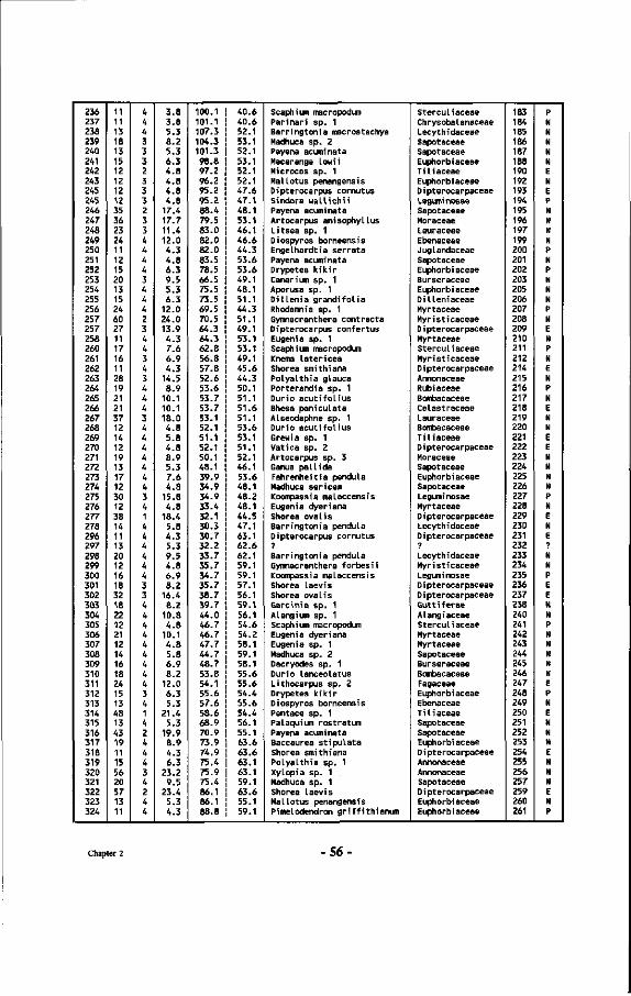
236 237 238 239 240 241 242 243 245 245 246 247 248 249 250 251 252 253 254 255 256 257 257 258 260 261 262 263 264 265 266 267 268 269 270 271 272 273 274 275 276 277 278 296 297 298 299 300 301 302 303 304 305 306 307 308 309 310 311 312 313 314 315 316 317 318 319 320 321 322 323 324
11 11 13 18 13 15 12 12 12 12 35 36 23 24 11 12 15 20 13 15 24 60 27 11 17 16 11 28 19 21 21 37 12 14 12 19 13 17 12 30 12 38 14 11 13 20 12 16 18 32 18 22 12 21 12 14 16 18 24 15 13 48 13 43 19 11 15 56 20 57 13 11
4 4 4 3 3 3 2 3 3 3 2 3 3 4 4 4 4 3 4 4 4 2 3 4 4 3 4 3 4 4 4 3 4 4 4 4 4 4 4 3 4 1 4 4 4 4 4 4 3 3 4 4 4 4 4 4 4 4 4 3 4 1 4 2 4 4 4 3 4 2 4 4
3.8 3.8 5.3 8.2 5.3 6.3 4.8 4.8 4.8 4.8
17.4 17.7 11.4 12.0 4.3 4.8 6.3 9.5 5.3 6.3
12.0 24.0 13.9 4.3 7.6 6.9 4.3
14.5 8.9
10.1 10.1 18.0 4.8 5.8 4.8 8.9 5.3 7.6 4.8
15.8 4.8
18.4 5.8 4.3 5.3 9.5 4.8 6.9 8.2
16.4 8.2
10.8 4.8
10.1 4.8 5.8 6.9 8.2
12.0 6.3 5.3
21.4 5.3
19.9 8.9 4.3 6.3
23.2 9.5
23.4 5.3 4.3
100.1 101.1 107.3 104.3 101.3 98.8 97.2 96.2 95.2 95.2 88.4 79.5 83.0 82.0 82.0 83.5 78.5 66.5 75.5 73.5 69.5 70.5 64.3 64.3 62.8 56.8 57.8 52.6 53.6 53.7 53.7 53.1 52.1 51.1 52.1 50.1 48.1 39.9 34.9 34.9 33.4 32.1 30.3 30.7 32.2 33.7 35.7 34.7 35.7 38.7 39.7 44.0 46.7 46.7 47.7 44.7 48.7 53.8 54.1 55.6 57.6 58.6 68.9 70.9 73.9 74.9 75.4 75.9 75.4 86.1 86.1 88.8
40.6 40.6 52.1 53.1 52.1 53.1 52.1 52.1 47.6 47.1 48.1 53.1 46.1 46.6 44.3 53.6 53.6 49.1 48.1 51.1 44.3 51.1 49.1 53.1 53.1 49.1 45.6 44.3 50.1 51.1 51.6 51.1 53.6 53.1 51.1 52.1 46.1 53.6 48.1 48.2 48.1 44.5 47.1 63.1 62.6 62.1 59.1 59.1 57.1 56.1 59.1 56.1 54.6 54.2 58.1 59.1 58.1 55.6 55.6 54.4 55.6 54.4 56.1 55.1 63.6 63.6 63.1 63.1 59.1 63.6 55.1 59.1
Scaphium macropodum Parinari sp. 1 Barringtonia macrostachya Madhuca sp. 2 Payena acuminata Macaranga Lowii Microcos sp. 1 Mal Lotus penangensis Dipterocarpus cornutus Sindora wallichii Payena acuminata Artocarpus anisophyLlus Litsea sp. 1 Diospyros borneensis Engelhardtia serrata Payena acuminata Drypetes kikir Canarium sp. 1 Aporusa sp. 1 Dillenia grandifolia Rhodamnia sp. 1 Gymnacranthera contracta Dipterocarpus confertus Eugenia sp. 1 Scaphium macropodum Knema latericea Shorea smithiana Polyalthia glauca Porterandia sp. 1 Durio acutifolius Bhesa paniculata Alseodaphne sp. 1 Durio acutifolius Grewia sp. 1 Vatica sp. 2 Artocarpus sp. 3 Ganua pallida Fahrenheitia pendula Madhuca sericea Koompassia malaccensis Eugenia dyeriana Shorea oval is Barringtonia pendula Dipterocarpus cornutus ? Barringtonia pendula Gymnacranthera forbesii Koompassia malaccensis Shorea laevis Shorea oval is Garcinia sp. 1 Alangium sp. 1 Scaphium macropodum Eugenia dyeriana Eugenia sp. 1 Madhuca sp. 2 Dacryodes sp. 1 Durio lanceolatus Lithocarpus sp. 2 Drypetes kikir Diospyros borneensis Pentace sp. 1 Palaquium rostratum Payena acuminata Baccaurea stipulata Shorea smithiana Polyalthia sp. 1 Xylopia sp. 1 Madhuca sp. 1 Shorea laevis Mal lotus penangensis Pimelodendron griffithianum
Sterculiaceae Ch rysoba l anaceae Lecythidaceae Sapotaceae Sapotaceae Euphorbiaceae T ili aceae Euphorbiaceae D i pterocarpaceae Leguminosae Sapotaceae Moraceae Lauraceae Ebenaceae Juglandaceae Sapotaceae Euphorbiaceae Burseraceae Euphorbiaceae Dilleniaceae Myrtaceae Myristicaceae Dipterocarpaceae Myrtaceae Sterculiaceae Myristicaceae Dipterocarpaceae Annonaceae Rubiaceae Bombacaceae Celastraceae Lauraceae Bombacaceae Tiliaceae D i pterocarpaceae Moraceae Sapotaceae Euphorbiaceae Sapotaceae Leguminosae Myrtaceae Dipterocarpaceae Lecythidaceae D i pterocarpaceae ?
Lecythidaceae Myristicaceae Leguminosae D i pterocarpaceae D i pterocarpaceae Guttiferae Alangiaceae Sterculiaceae Myrtaceae Myrtaceae Sapotaceae Burseraceae Bombacaceae Fagaceae Euphorbiaceae Ebenaceae Tiliaceae Sapotaceae Sapotaceae Euphorbiaceae Dipterocarpaceae Annonaceae Annonaceae Sapotaceae D i pterocarpaceae Euphorbi aceae Euphorbi aceae
183 184 185 186 187 188 190 192 193 194 195 196 197 199 200 201 202 203 205 206 207 208 209 210 211 212 214 215 216 217 218 219 220 221 222 223 224 225 226 227 228 229 230 231 232 233 234 235 236 237 238 240 241 242 243 244 245 246 247 248 249 250 251 252 253 254 255 256 257 259 260 261
P N N N N N E N E P N N N N P N P N N N P N E N P N E N P N E N N E E N N N N P N E N E ? N N P E E N N P N N N K N E P N E N N N E N N N E N P
Chapter 2 56-
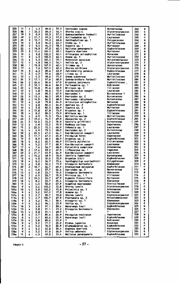
325 326 327 328 329 330 331 332 333 334 335 336 337 338 339 340 341 342 343 344 345 346 347 348 349 350 351 352 353 354 355 356 357 358 359 360 361 362 363 364 365 366 367 368 369 370 371 372 373 374 375 376 377 378 379 380
104a 105a 106a 107a 111a 119a 120a 121a 122a 123a 125a 126a 127a 130a 131a 137a 138a 139a
11 86 19 49 25 10 20 22 23 10 11 13 12 30 17 15 11 13 34 66 21 22 12 12 26 28 12 10 10 30 18 16 40 24 12 71 16 90 36 19 18 13 17 35 49 29 12 18 10 39 14 12 16 41 18 11 9
10 9 8 8 9 9
10 10 10 8 8
10 9 9 9 9
11
4 1 3 2 3 4 3 4 3 4 3 4 4 1 4 3 3 4 2 2 2 4 3 4 3 4 4 4 4 4 4 4 3 3 4 2 4 2 3 3 4 4 4 2 2 4 4 4 4 2 4 4 3 3 4 4 4 4 4 3 3 3 4 3 4 4 4 4 4 4 4 4 4 4
4.3 25.3 8.9
21.7 12.7 3.8 9.5
10.8 11.4 3.8 4.3 5.3 4.8
15.8 7.6 6.3 4.3 5.3
17.1 24.6 10.1 10.8 4.8 4.8
13.3 14.5 4.8 3.8 3.8
15.8 8.2 6.9
19.0 12.0 4.8
25.0 6.9
25.3 17.7 8.9 8.2 5.3 7.6
17.4 21.7 15.2 4.8 8.2 3.8
18.7 5.8 4.8 6.9
19.3 8.2 4.3 3.2 3.8 3.2 2.7 2.7 3.2 3.2 3.8 3.8 3.8 2.7 2.7 3.8 3.2 3.2 3.2 3.2 4.3
89.8 89.3 91.3 91.3 91.3 94.3 94.3 97.3
100.5 100.0 103.5 105.5 106.5 107.5 107.5 89.6 99.6 98.6 97.6 96.6 96.6 95.6 96.6 89.3 86.3 84.3 79.8 80.3 80.3 87.8 85.3 74.3 74.3 72.3 73.3 72.3 73.3 67.3 67.3 68.3 65.5 57.7 56.7 53.2 49.9 48.9 30.0 36.7 30.2 36.7 37.7 33.7 33.7 33.7 33.7 30.0
103.2 102.2 107.2 99.7
105.7 95.1 96.1 91.1 91.1 91.1 85.6 82.6 80.6 71.7 76.3 52.8 51.8 49.8
59.1 56.1 57.1 57.2 63.6 63.1 58.1 60.1 63.1 60.1 62.1 59.1 56.1 62.1 62.2 63.9 68.9 71.9 68.9 67.9 66.9 68.9 72.9 67.9 69.9 68.9 64.9 64.9 68.9 73.9 73.9 73.4 69.9 70.9 70.9 68.9 68.9 64.1 65.9 68.9 68.9 68.9 68.9 67.9 64.9 70.9 73.9 73.4 71.9 69.9 64.4 70.9 70.9 67.9 65.4 67.9 12.8 15.8 17.8 19.8 27.9 30.4 23.9 30.4 21.2 20.9 26.9 25.9 23.9 25.9 30.9 30.4 26.9 21.4
Dacryodes rugosa Shorea oval is Gymnacranthera forbesi i Actinodaphne sp. 1 Xanthophyllum sp. 1 Knema sp. 1 Eugenia sp. 1 Mal lotus penangensis Eugenia dyeriana Artocarpus anisophyllus Knema sp. 1 Memecylon excelsum Vatica sp. 2 Triomjiia sp. 1 Shorea smithiana Fahrenheit ia pendula Litsea sp. 3 Knema sumatrana Gymnacranthera forbesii Drypetes polyneura Pertusadina sp. 1 Microcos sp. 2 Eusideroxylon zuageri Dacryodes ? Dacryodes sp. 2 Dacryodes rubiginosa Artocarpus anisophyllus Aporusa sp. 2 Eugenia sp. 2 Drypetes sp. 1 Microcos sp. 1 Myristica maxima Baccaurea sp. 2 Santiria griffithii Madhuca sericea Dehaasia sp. 1 Dacryodes sp. 2 Eusideroxylon zwageri Palaquium ferox Drypetes sp. 2 Dillenia pulchella Eusideroxylon zwageri Polyalthia sumatrana Lithocarpus sp. 1 Eusideroxylon zwageri Santiria tomentosa Drypetes kikir Xanthophylluïi scortechinii Diospyros borneensis Endospermum malayanum Dacryodes sp. 2 Diospyros borneensis Microcos sp. 1 ? Eugenia flosculifera Diospyros borneensis Scaphium macropodum Shorea laevis Polyalthia sp. 1 Acmena sp. 1 Shorea laevis Pternandra sp. 2 Diospyros sp. 1 Vatica sp. 1 Macaranga lowi i Diospyros borneensis ? Palaquium rostratum Macaranga lowii ? Prunus japonica Blumeodendron sp. 1 Eugenia dyeriana Vatica umbonata Mal lotus penangensis
Burseraceae D i pterocarpaceae Myristicaceae Lauraceae Polygalaceae Myristicaceae Myrtaceae Euphorbiaceae Myrtaceae Moraceae Myristicaceae Melastomataceae Dipterocarpaceae Burseraceae Dipterocarpaceae Euphorbiaceae Lauraceae Myristicaceae Myristicaceae Euphorbiaceae Rubiaceae Tili aceae Lauraceae Burseraceae ? Burseraceae Burseraceae Moraceae Euphorbi aceae Myrtaceae Euphorbi aceae Tiliaceae Myristicaceae Euphorbiaceae Burseraceae Sapotaceae Lauraceae Burseraceae Lauraceae Sapotaceae Euphorbiaceae Dilleniaceae Lauraceae Annonaceae Fagaceae Lauraceae Burseraceae Euphorbiaceae Polygalaceae Ebenaceae Euphorbiaceae Burseraceae Ebenaceae T ili aceae Myrtaceae Ebenaceae Sterculiaceae D i pterocarpaceae Annonaceae Myrtaceae Dipterocarpaceae Melastomataceae Ebenaceae D i pterocarpaceae Euphorbiaceae Ebenaceae ? Sapotaceae Euphorbi aceae Rubi aceae Rosaceae Euphorbiaceae Myrtaceae D i pterocarpaceae Euphorbiaceae
262 263 264 265 266 267 268 269 270 271 272 273 274 275 276 277 278 279 280 281 282 283 284 285 286 287 288 289 290 291 292 293 294 295 296 297 298 299 300 301 302 303 304 305 306 307 308 309 310 311 312 313 314 315 316 317 318 319 320 321 322 323 324 325 326 327 328 329 330 331 332 333 334 335
N E N P N N N N N N N N E N E N N N N P P E N ? N N N N N P E N N N N P N N N P N N N E N N P P N P N N E N N P E N N E N N E N N ? N N P E P N E N
Chapter 2 57-
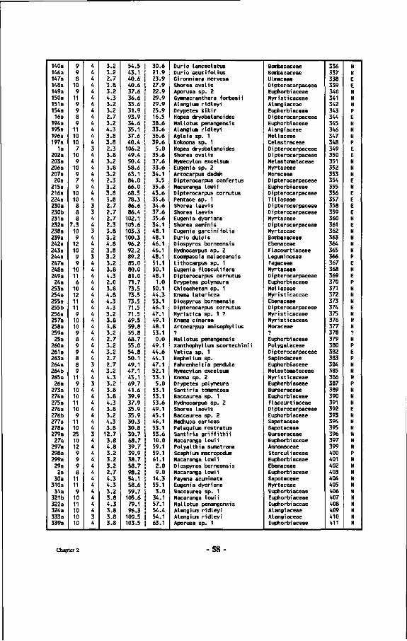
140a 146a 147a 148a 149a 150a 151a 154a 16a
194a 195a 196a 197a
1a 202a 203a 206a 207a 20a
215a 216a 224a 230a 230b 231a 232a 238a 239a 242a 243a 244a 247a 248a 249a 24a
253a 254a 255a 255b 256a 257a 258a 259a 25a
260a 261a 263a 264a 264b 265a 26a
273a 274a 275a 276a 276b 277a 278a 279a 27a
297a 298a 299a 29a 2a
30a 310a 31a
321b 322a 324a 333a 339a
9 9 8
10 9
11 9 9 8 9
11 10 10 7
10 9
10 9 7 9
10 10 8 8 8
7.3 10 9
12 10 9 9
10 11 6
10 12 11 11 9
10 10 9 8 9 9 8 8 9
11 9
10 10 11 10 9
11 10 25 10 12 9 9 9 8
11 11 9
10 11 10 10 10
4 4 4 4 4 4 4 4 4 4 4 4 4 3 4 4 4 4 4 4 4 4 3 3 4 4 3 4 4 2 3 4 4 4 4 4 4 4 4 4 4 4 4 4 4 4 4 3 4 4 3 4 4 4 4 4 4 4 3 4 4 4 4 4 4 4 4 4 4 4 4 3 4
3.2 3.2 2.7 3.8 3.2 4.3 3.2 3.2 2.7 3.2 4.3 3.8 3.8 2.3 3.8 3.2 3.8 3.2 2.3 3.2 3.8 3.8 2.7 2.7 2.7 2.3 3.8 3.2 4.8 3.8 3.2 3.2 3.8 4.3 2.0 3.8 4.8 4.3 4.3 3.2 3.8 3.8 3.2 2.7 3.2 3.2 2.7 2.7 3.2 4.3 3.2 3.8 3.8 4.3 3.8 3.2 4.3 3.8
12.7 3.8 4.8 3.2 3.2 3.2 2.7 4.3 4.3 3.2 3.8 4.3 3.8 3.8 3.8
54.5 43.1 40.6 40.6 37.6 36.6 35.6 31.9 93.9 34.6 35.1 37.6 40.4
106.2 49.4 50.4 58.6 63.1 84.0 66.0 68.5 78.3 86.6 86.4
102.1 105.6 103.3 100.3 96.2 92.2 89.2 85.0 80.0 81.0 71.7 73.5 73.5 73.5 71.5 71.5 69.5 59.8 55.8 68.7 55.0 54.8 50.1 49.1 47.1 45.1 69.7 41.6 39.9 37.9 35.9 35.9 30.3 30.8 30.7 68.7 39.7 39.9 38.7 58.7 98.2 54.1 58.6 59.7
105.6 79.1 96.3
100.5 103.5
30.6 21.9 23.9 27.9 22.9 29.9 29.9 25.9 16.5 38.6 33.6 36.6 39.6 5.0
35.6 37.6 33.6 34.1 3.5
35.6 43.6 35.6 34.6 37.6 35.6 34.1 48.1 48.1 46.1 46.1 48.1 51.1 50.1 48.1 1.0
50.1 44.3 53.1 46.1 47.1 49.1 48.1 53.1 0.0
49.1 44.6 44.1 47.1 52.1 53.1 5.0
53.1 53.1 53.6 49.1 45.1 46.1 53.1 53.6 10.0 59.1 59.1 61.1 2.0 9.0
14.3 55.1 3.0
34.1 57.1 54.4 54.1 63.1
Durio lanceolatus Durio acutifolius Gironniera nervosa Shorea oval is Aporusa sp. 2 Gymnacranthera forbesii Alangium ridleyi Drypetes kikir Hopea dryobalanoides Mal lotus penangensis Alangium ridleyi Aglaia sp. 1 Kokoona sp. 1 Hopea dryobalanoides Shorea oval is Mymecylon excel sum Eugenia sp. 2 Artocarpus dadah Dipterocarpus confertus Macaranga lowii Dipterocarpus cornutus Pentace sp. 1 Shorea Laevis Shorea laevis Eugenia dyeriana Shorea seminis Eugenia garcinifolia Durio dulcis Diospyros borneensis Hydnocarpus sp. 2 Koompassia malaccensis Lithocarpus sp. 1 Eugenia flosculifera Dipterocarpus cornutus Drypetes polyneura Chisocheton sp. 1 Knema latericea Diospyros borneensis Dipterocarpus cornutus Myristica sp. 1 ? Knema cinerea Artocarpus anisophyllus ? Mal lotus penangensis Xanthophyllum scortechinii Vatica sp. 1 Nephelium sp. Fahrenheitia pendula Mymecylon excelsum Knema sp. 2 Drypetes polyneura Sant ir ia tomentosa Baccaurea sp. 1 Hydnocarpus sp. 2 Shorea laevis Baccaurea sp. 2 Madhuca sericea Palaquium rostratum Santiria griffithii Macaranga Lowii Polyalthia sumatrana Scaphium macropodum Macaranga lowii Diospyros borneensis Macaranga lowi i Payena acuminata Eugenia dyeriana Baccaurea sp. 1 Macaranga lowii Mal lotus penangensis Alangium ridleyi Alangium ridleyi Aporusa sp. 1
Bombacaceae Bombacaceae ULmaceae D i pterocarpaceae Euphorbiaceae Myristicaceae Alangiaceae Euphorbiaceae Dipterocarpaceae Euphorbiaceae Alangiaceae Meliaceae Celastraceae D i pterocarpaceae D i pterocarpaceae Melastomataceae Myrtaceae Moraceae Dipterocarpaceae Euphorbiaceae D i pterocarpaceae T ili aceae D i pterocarpaceae Dipterocarpaceae Myrtaceae Dipterocarpaceae Myrtaceae Bombacaceae Ebenaceae Flacourtiaceae Leguminosae Fagaceae Myrtaceae Dipterocarpaceae Euphorbiaceae Meliaceae Myristicaceae Ebenaceae D i pterocarpaceae Myristicaceae Myristicaceae Moraceae ?
Euphorbiaceae Polygalaceae Dipterocarpaceae Sapindaceae Euphorbiaceae Melastomataceae Myristicaceae Euphorbiaceae Burseraceae Euphorbiaceae Flacourtiaceae Dipterocarpaceae Euphorbiaceae Sapotaceae Sapotaceae Burseraceae Euphorbiaceae Annonaceae Sterculiaceae Euphorbiaceae Ebenaceae Euphorbiaceae Sapotaceae Myrtaceae Euphorbiaceae Euphorbiaceae Euphorbiaceae Alangiaceae Alangiaceae Euphorbiaceae
336 337 338 339 340 341 342 343 344 345 346 347 348 349 350 351 352 353 354 355 356 357 358 359 360 361 362 363 364 365 366 367 368 369 370 371 372 373 374 375 376 377 378 379 380 382 383 384 385 386 387 389 390 391 392 393 394 395 396 397 399 400 401 402 403 404 405 406 407 408 409 410 411
N H E E N N N P E N N N P E E N N N E N E E E E N E N N N N P E N E P N N N E N N N ?
N P E P N N N P N N N E N N N N N N P N N N N N N N N N N N
Chapter 2 -58
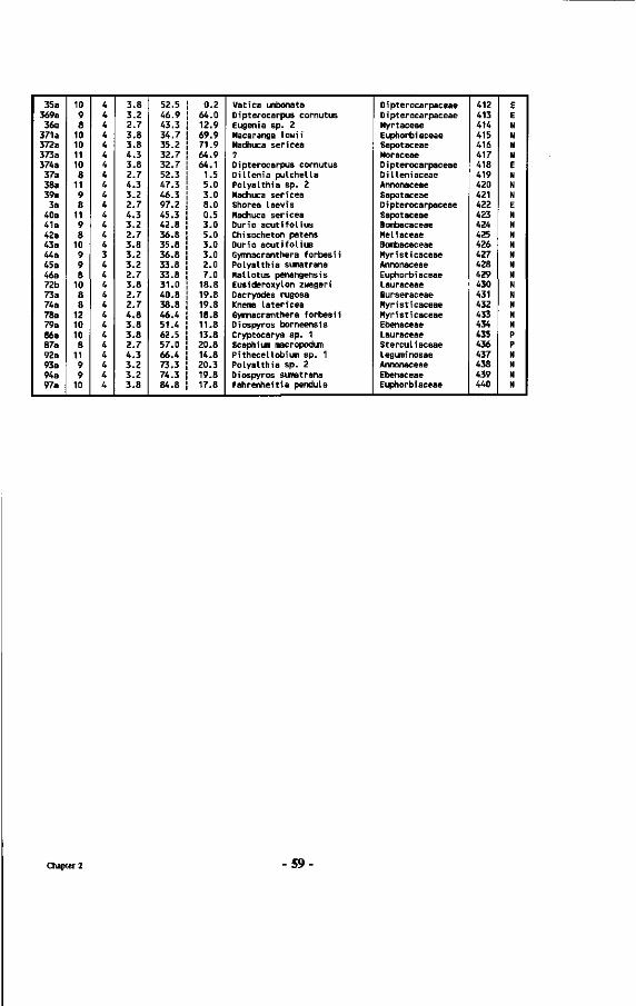
35a 369a 36a
371a 372a 373a 374a 37a 38a 39a 3a
40a 41a 42a 43a 44a 45a 46a 72b 73a 74a 78a 79a 86a 87a 92a 93a 94a 97a
10 9 8
10 10 11 10 8
11 9 8
11 9 8
10 9 9 8
10 8 8
12 10 10 8
11 9 9
10
4 4 4 4 4 4 4 4 4 4 4 4 4 4 4 3 4 4 4 4 4 4 4 4 4 4 4 4 4
3.8 3.2 2.7 3.8 3.8 4.3 3.8 2.7 4.3 3.2 2.7 4.3 3.2 2.7 3.8 3.2 3.2 2.7 3.8 2.7 2.7 4.8 3.8 3.8 2.7 4.3 3.2 3.2 3.8
52.5 46.9 43.3 34.7 35.2 32.7 32.7 52.3 47.3 46.3 97.2 45.3 42.8 36.8 35.8 36.8 33.8 33.8 31.0 40.8 38.8 46.4 51.4 62.5 57.0 66.4 73.3 74.3 84.8
0.2 64.0 12.9 69.9 71.9 64.9 64.1
1.5 5.0 3.0 8.0 0.5 3.0 5.0 3.0 3.0 2.0 7.0
18.8 19.8 19.8 18.8 11.8 13.8 20.8 14.8 20.3 19.8 17.8
Vatica umbonata Dipterocarpus cornutus Eugenia sp. 2 Macaranga lowi i Madhuca sericea ? Dipterocarpus cornutus Diüenia pulchella Polyalthia sp. 2 Madhuca sericea Shorea laevis Madhuca sericea Durio acutifolius Chisocheton patens Durio acutifolius Gymnacranthera forbesii Polyalthia sumatrana Mal lotus penangensis Eusideroxylon zwageri Dacryodes rugosa Knema latericea Gymnacranthera forbesii Diospyros borneensis Cryptocarya sp. 1 Scaphium macropodum Pithecellobium sp. 1 Polyalthia sp. 2 Diospyros sumatrana Fahrenheitia pendula
D i pterocarpaceae Dipterocarpaceae Myrtaceae Euphorbi aceae Sapotaceae Moraceae D i pterocarpaceae Dilleniaceae Annonaceae Sapotaceae D i pterocarpaceae Sapotaceae Boinbacaceae Meliaceae Boinbacaceae Myristicaceae Annonaceae Euphorbiaceae Lauraceae Burseraceae Myristicaceae Myristicaceae Ebenaceae Lauraceae Sterculiaceae Leguminosae Annonaceae Ebenaceae Euphorbiaceae
412 413 414 415 416 417 418 419 420 421 422 423 424 425 426 427 428 429 430 431 432 433 434 435 436 437 438 439 440
E E N N N N E N N N E N N N N N N N N N N N U P P N N N N
Chapter 2 -59
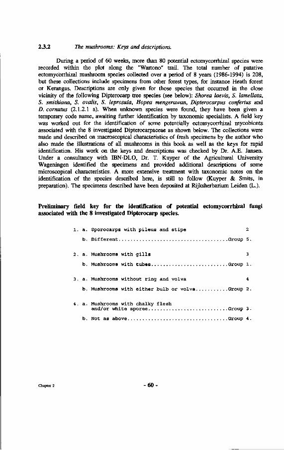
2.3.2 The mushrooms: Keys and descriptions.
During a period of 60 weeks, more than 80 potential ectomycorrhizal species were recorded within the plot along the "Wartono" trail. The total number of putative ectomycorrhizal mushroom species collected over a period of 8 years (1986-1994) is 208, but these collections include specimens from other forest types, for instance Heath forest or Kerangas. Descriptions are only given for those species that occurred in the close vicinity of the following Dipterocarp tree species (see below): Shorea laevis, S. lamellata, S. smithiana, S. ovalis, S. leprosula, Hopea mengerawan, Dipterocarpus confertus and D. cornutus (2.1.2.1 a). When unknown species were found, they have been given a temporary code name, awaiting further identification by taxonomie specialists. A field key was worked out for the identification of some potentially ectomycorrhizal mycobionts associated with the 8 investigated Dipterocarpaceae as shown below. The collections were made and described on macroscopical characteristics of fresh specimens by the author who also made the illustrations of all mushrooms in this book as well as the keys for rapid identification. His work on the keys and descriptions was checked by Dr. A.E. Jansen. Under a consultancy with IBN-DLO, Dr. T. Kuyper of the Agricultural University Wageningen identified the specimens and provided additional descriptions of some microscopical characteristics. A more extensive treatment with taxonomie notes on the identification of the species described here, is still to follow (Kuyper & Smits, in preparation). The specimens described have been deposited at Rijksherbarium Leiden (L.).
Preliminary field key for the identification of potential ectomycorrhizal fungi associated with the 8 investigated Dipterocarp species.
1. a. Sporocarps with pileus and stipe 2
b. Different Group 5.
2. a. Mushrooms with gills 3
b. Mushrooms with tubes Group 1.
3. a. Mushrooms without ring and volva 4
b. Mushrooms with either bulb or volva Group 2 .
4. a. Mushrooms with chalky flesh and/or white sporee Group 3 .
b. Not as above Group 4 .
Chapter 2 - 60 •
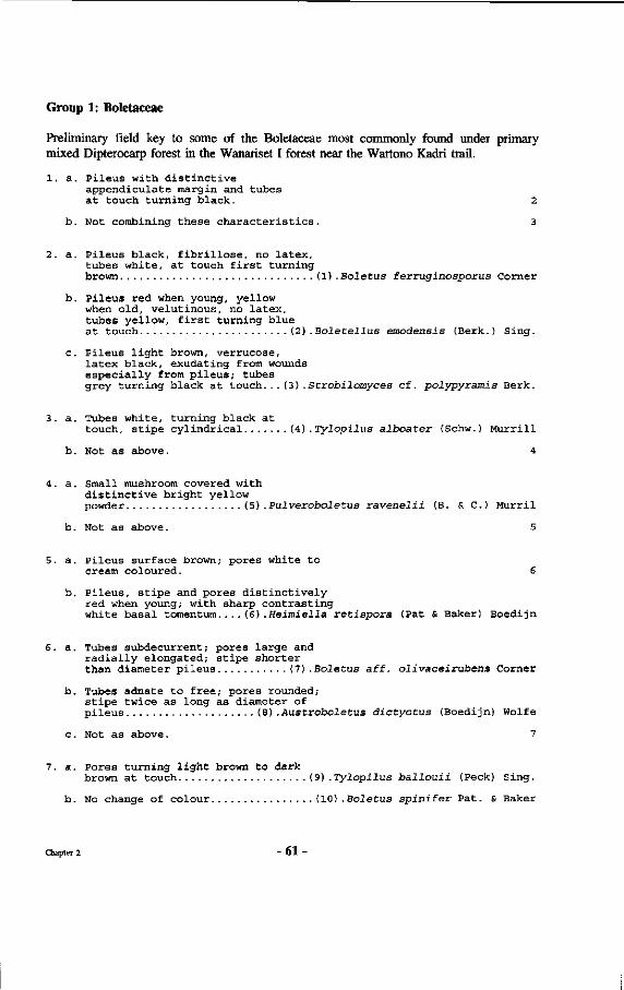
Group 1: Boletaceae
Preliminary field key to some of the Boletaceae most commonly found under primary mixed Dipterocarp forest in the Wanariset I forest near the Wartono Kadri trail.
1. a. Pileus with distinctive appendiculate margin and tubes at touch turning black. 2
b. Not combining these characteristics. 3
2. a. Pileus black, fibrinöse, no latex, tubes white, at touch first turning brown (1) .Boletus ferruginosporus Corner
b. Pileus red when young, yellow when old, velutinous, no latex, tubes yellow, first turning blue at touch (2) .Boletellus emodensis (Berk.) Sing.
c. Pileus light brown, verrucose, latex black, exudating from wounds especially from pileus; tubes grey turning black at touch...(3).Strobilomyces cf. polypyramis Berk.
3. a. Tubes white, turning black at touch, stipe cylindrical (4) . Tylopilus alboater (Schw.) Murrill
b. Not as above. 4
4. a. Small mushroom covered with distinctive bright yellow powder (5) .Pulverobo le tus ravenelii (B. & C. ) Murril
b. Not as above. 5
5. a. Pileus surface brown; pores white to cream coloured. 6
b. Pileus, stipe and pores distinctively red when young; with sharp contrasting white basal tomentum....(6).Heimiella retispora (Pat & Baker) Boedijn
6. a. Tubes subdecurrent; pores large and radially elongated; stipe shorter than diameter pileus (7) .Boletus aff. olivaceirubens Corner
b. Tubes adnate to free; pores rounded; stipe twice as long as diameter of pileus (8) .Austroboletus dictyotus (Boedijn) Wolfe
c. Not as above. 7
7. a. Pores turning light brown to dark brown at touch (9) .Tylopilus ballouii (Peck) Sing.
b. No change of colour (10) .Boletus spinifer Pat. & Baker
Chapter 2 - 6 1
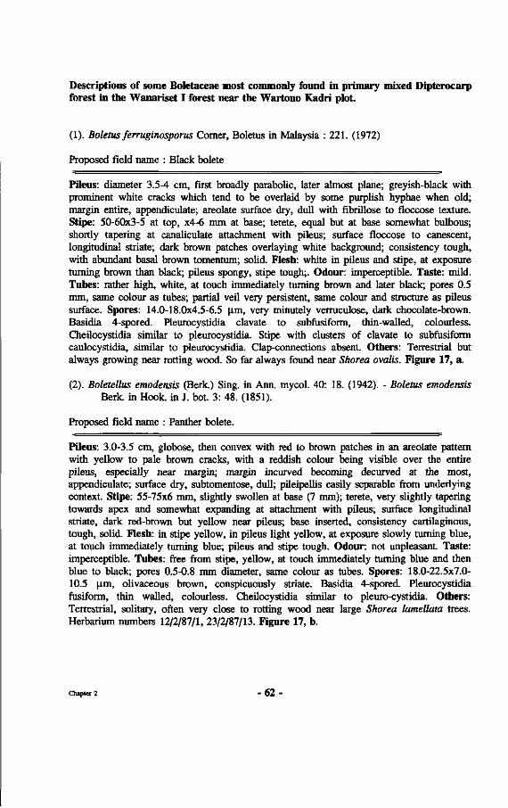
Descriptions of some Boletaceae most commonly found in primary mixed Dipterocarp forest in the Wanariset I forest near the Wartono Kadri plot.
(1). Boletus ferruginosporus Corner, Boletus in Malaysia : 221. (1972)
Proposed field name : Black bolete
Pileus: diameter 3.5-4 cm, first broadly parabolic, later almost plane; greyish-black with prominent white cracks which tend to be overlaid by some purplish hyphae when old; margin entire, appendiculate; areolate surface dry, dull with fibrillose to floccose texture. Stipe: 50-60x3-5 at top, x4-6 mm at base; terete, equal but at base somewhat bulbous; shortly tapering at canaliculate attachment with pileus; surface floccose to canescent, longitudinal striate; dark brown patches overlaying white background; consistency tough, with abundant basal brown tomentum; solid. Flesh: white in pileus and stipe, at exposure turning brown than black; pileus spongy, stipe tough;. Odour: imperceptible. Taste: mild. Tubes: rather high, white, at touch immediately turning brown and later black; pores 0.5 mm, same colour as tubes; partial veil very persistent, same colour and structure as pileus surface. Spores: 14.0-18.0x4.5-6.5 Jim, very minutely verruculose, dark chocolate-brown. Basidia 4-spored. Pleurocystidia clavate to subfusiform, thin-walled, colourless. Cheilocystidia similar to pleurocystidia. Stipe with clusters of clavate to subfusiform caulocystidia, similar to pleurocystidia. Clap-connections absent. Others: Terrestrial but always growing near rotting wood. So far always found near Shorea ovalis. Figure 17, a.
(2). Boletellus emodensis (Berk.) Sing, in Ann. mycol. 40: 18. (1942). - Boletus emodensis Berk, in Hook, in J. bot. 3: 48. (1851).
Proposed field name : Panther bolete.
Pileus: 3.0-3.5 cm, globose, then convex with red to brown patches in an areolate pattern with yellow to pale brown cracks, with a reddish colour being visible over the entire pileus, especially near margin; margin incurved becoming decurved at the most, appendiculate; surface dry, subtomentose, dull; pileipellis easily separable from underlying context. Stipe: 55-75x6 mm, slightly swollen at base (7 mm); terete, very slightly tapering towards apex and somewhat expanding at attachment with pileus; surface longitudinal striate, dark red-brown but yellow near pileus; base inserted, consistency cartilaginous, tough, solid. Flesh: in stipe yellow, in pileus light yellow, at exposure slowly turning blue, at touch immediately turning blue; pileus and stipe tough. Odour: not unpleasant. Taste: imperceptible. Tubes: free from stipe, yellow, at touch immediately turning blue and then blue to black; pores 0.5-0.8 mm diameter, same colour as tubes. Spores: 18.0-22.5x7.0-10.5 um, olivaceous brown, conspicuously striate. Basidia 4-spored. Pleurocystidia fusiform, thin walled, colourless. Cheilocystidia similar to pleuro-cystidia. Others: Terrestrial, solitary, often very close to rotting wood near large Shorea lamellata trees. Herbarium numbers 12/2/87/1, 23/2/87/13. Figure 17, b.
Chapter 2 - 6 2
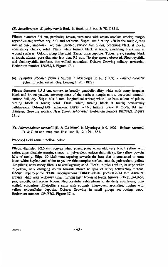
(3). Strobilomyces cf. polypyramis Berk. in Hook, in J. bot. 3: 78. (1851).
Pileus: diameter 3.5 cm, parabolic; brown, verrucose with cream areolate cracks; margin appendiculate; surface dry, dull and scabrous. Stipe: 60x15 at top x28 in the middle, x20 mm at base, amphora- like; base inserted, surface like pileus, becoming black at touch; consistency chalky, solid. Flesh: white turning black at touch, exudating black sap at wound surfaces. Odour: sharp like acid. Taste: imperceptible. Tubes: grey, turning black at touch, adnate; pore diameter less than 0.2 mm. No ripe spores observed. Pleurocystidia and cheilocystidia fusiform, thin-walled, colourless. Others: Growing solitary, terrestrial. Herbarium number 12/2/87/3. Figure 17, c.
(4). Tylopilus alboater (Schw.) Murrill in Mycologia 1: 16. (1909). - Boletus alboater Schw. in Sehr, naturf. Ges. Leipzig 1: 95. (1822).
Pileus: diameter 4.5-5 cm, convex to broadly parabolic; dirty white with many irregular black and brown patches covering most of the surface; margin entire, decurved, smooth; surface dull, dry. Stipe: 60x13 mm, longitudinal striate; white like base colour of pileus, turning black at touch; solid. Flesh: white, turning black at touch; consistency cartilageous. Odour/taste: unknown. Pores: white, turning black at touch, 0.4 mm diameter. Growing solitary. Near Shorea johorensis. Herbarium number 18/2/87/2. Figure 17, d.
(5). Pulveroboletus ravenelii (B. & C.) Murril in Mycologia 1: 9. 1909. -Boletus ravenelii B. & C. in ann. mag. nat. Hist., ser. II, 12: 429. 1853.
Proposed field name : Yellow bolete.
Pileus: diameter 1-2.5 cm, convex when young plane when old; very bright yellow with entire, appendiculate margin; smooth to pulverulent surface dull, sticky; the yellow powder falls of easily. Stipe: 30-42x3 mm; tapering towards the base that is connected to some loose white hyphae and white to yellow rhizomorphs; surface smooth, pulverulent, yellow like pileus; consistency fibrous to cartilageous; solid. Flesh: in pileus white, in stipe white to yellow, only changing colour towards brown at apex of stipe; consistency fibrous. Odour: imperceptible. Taste: inconspicuous. Tubes: adnate, pores 0.2-0.4 mm diameter, greyish white with yellowish tinge, turning light brown at touch. Spores: 9.0-11.0x4.0-5.0 \im, smooth, ochraceous brown. Pleurocystidia subfusiform to slenderly subclavate, thin-walled, colourless. Pileipellis a cutis with strongly interwoven ascending hyphae with yellow extracellular deposits. Others: Growing in small groups on rotting wood. Herbarium number 13/4/87/2. Figure 17, e.
Chapter 2 - 6 3 *
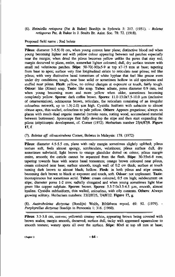
(6). Heimiella retispora (Pat & Baker) Boedijn in Sydowia 5: 217. (1951). - Boletus retisporus Pat. & Baker in J. Straits Br. Asiat. Soc. 78: 72. (1918).
Proposed field name : Red bolete
Pileus: diameter 3-5.5(-9) cm, when young convex later plane; distinctive blood red when young becoming lighter red with yellow colour appearing between red patches especially near the margin; when dried the pileus becomes yellow unlike the pores that stay red; margin decurved to plane, entire, somewhat lighter coloured; dull, dry surface texture with small red velutinous patches. Stipe: 50-70(-90)x5-9 at top x7-15 mm at base; tapering from base to apex; surface very fine longitudinal striate to reticulate near pileus; red like pileus; with very distinctive basal tomentum of white hyphae that feel like grease even under dry conditions; tough, near base solid or sometimes hollow in old specimens and stuffed near pileus. Flesh: yellow, no colour changes at exposure or touch, fairly tough. Odour: like (Rinso) soap. Taste: like soap. Tubes: adnate, pores diameter 0.9 mm, red when young becoming more and more yellow when older, sometimes becoming completely yellow. Sporee: dark coffee brown. Spores: 11.0-13.0x7.5-10.0 |im (inclusive of ornamentation), ochraceous brown, reticulate, the reticulum consisting of an irregular colourless network, up to 1.5(-2.0) |im high. Cystidia fusiform with subacute to almost obtuse apex, thin-walled, colourless to pale yellow. Others: Appears gregarious; always on places with much dead organic material (termite nests, rotting wood, accumulated material between buttresses). Sporocarps first fully develop the stipe and then start expanding the pileus (stipiticarpic development, cf. Comer (1972). Herbarium number 25/4/87/8. Figure 17, f.
(7). Boletus off. olivaceirubens Comer, Boletus in Malaysia: 178. (1972)
Pileus: diameter 4.5-5.5 cm, plane with only margin sometimes slightly uplifted; pileus texture soft, feels almost spongy, scrobiculate, velutinous; pileus surface dull, dry sometimes subviscid, light brown to orange glandular dotted on crème; pileus margin entire, smooth; the cuticle cannot be separated from the flesh. Stipe: 30-35x6-8 mm; tapering towards base with scarce basal tomentum; orange brown coloured near pileus, cream coloured near base; surface smooth, tough wall of 0.2 cm thick; surface at touch turning dark brown to almost black; hollow. Flesh: in both pileus and stipe cream, becoming dark brown to black at exposure and touch, soft. Odour: not unpleasant. Taste: inconspicuous but sometimes acrid. Tubes: cream coloured; 0.5 cm high; subdecurrent on stipe; diameter pores 1-2 mm; radially elongated and when young sometimes light blue green like copper sulphate. Sporee: brown. Spores: 5.5-7.0x3.5-4.5 \im, smooth, almost hyaline. Cystidia subfusiform, thin walled, colourless, with oily contents. Others: Always growing solitary. Herbarium numbers: 23/2/87/5, 7/4/87/2. Figure 17, g.
(8). Austroboletus dictyotus (Boedijn) Wolfe, Biblitheca mycol. 69: 92. (1979). -Porphyrellus dictyotus Boedijn in Persoonia 1: 316. (1960).
Pileus: 3.5-3.8 cm, convex; yellowish creamy white, appearing brown being covered with brown scales; margin smooth, decurved; surface dull, tacky with appressed squamulose to smooth texture; watery spots all over the surface. Stipe: 80x6 at top x8 mm at base;
Chapter 2 - 6 4 -
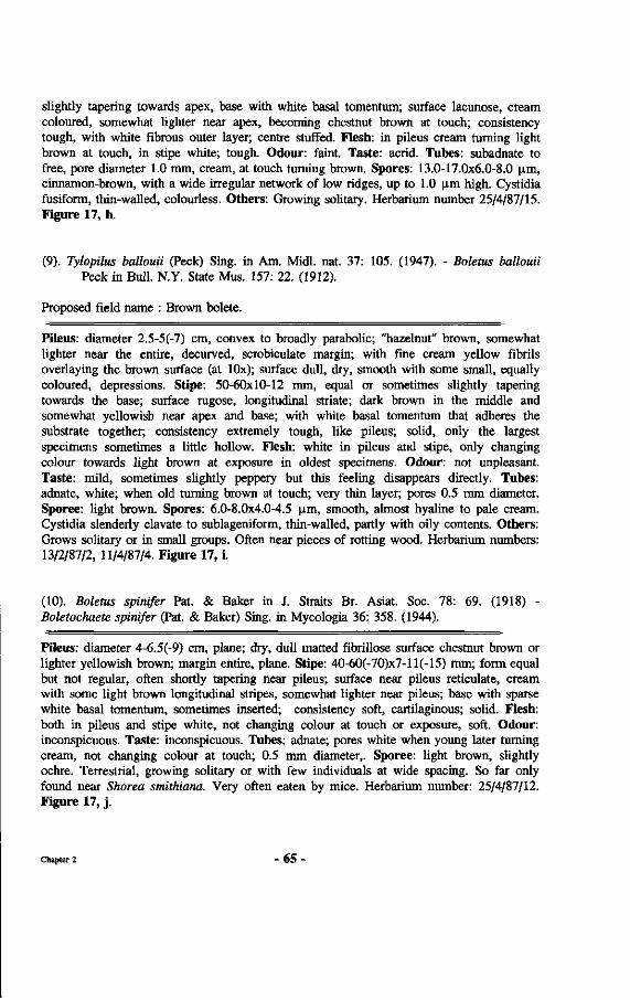
slightly tapering towards apex, base with white basal tomentum; surface lacunose, cream coloured, somewhat lighter near apex, becoming chestnut brown at touch; consistency tough, with white fibrous outer layer; centre stuffed. Flesh: in pileus cream turning light brown at touch, in stipe white; tough. Odour: faint. Taste: acrid. Tubes: subadnate to free, pore diameter 1.0 mm, cream, at touch turning brown. Spores: 13.0-17.0x6.0-8.0 p.m, cinnamon-brown, with a wide irregular network of low ridges, up to 1.0 |im high. Cystidia fusiform, thin-walled, colourless. Others: Growing solitary. Herbarium number 25/4/87/15. Figure 17, h.
(9). Tylopilus ballouii (Peck) Sing, in Am. Midi. nat. 37: 105. (1947). - Boletus ballouii Peck in Bull. N.Y. State Mus. 157: 22. (1912).
Proposed field name : Brown bolete.
Pileus: diameter 2.5-5(-7) cm, convex to broadly parabolic; "hazelnut" brown, somewhat lighter near the entire, decurved, scrobiculate margin; with fine cream yellow fibrils overlaying the brown surface (at lOx); surface dull, dry, smooth with some small, equally coloured, depressions. Stipe: 50-60x10-12 mm, equal or sometimes slightly tapering towards the base; surface rugose, longitudinal striate; dark brown in the middle and somewhat yellowish near apex and base; with white basal tomentum that adheres the substrate together; consistency extremely tough, like pileus; solid, only the largest specimens sometimes a little hollow. Flesh: white in pileus and stipe, only changing colour towards light brown at exposure in oldest specimens. Odour: not unpleasant. Taste: mild, sometimes slightly peppery but this feeling disappears directly. Tubes: adnate, white; when old turning brown at touch; very thin layer; pores 0.5 mm diameter. Sporee: light brown. Spores: 6.0-8.0x4.0-4.5 ^m, smooth, almost hyaline to pale cream. Cystidia slenderly clavate to sublageniform, thin-walled, partly with oily contents. Others: Grows solitary or in small groups. Often near pieces of rotting wood. Herbarium numbers: 13/2/87/2, 11/4/87/4. Figure 17, i.
(10). Boletus spinifer Pat. & Baker in J. Straits Br. Asiat. Soc. 78: 69. (1918) -Boletochaete spinifer (Pat. & Baker) Sing, in Mycologia 36: 358. (1944).
Pileus: diameter 4-6.5(-9) cm, plane; dry, dull matted fibrillose surface chestnut brown or lighter yellowish brown; margin entire, plane. Stipe: 40-60(-70)x7-ll(-15) mm; form equal but not regular, often shortly tapering near pileus; surface near pileus reticulate, cream with some light brown longitudinal stripes, somewhat lighter near pileus; base with sparse white basal tomentum, sometimes inserted; consistency soft, cartilaginous; solid. Flesh: both in pileus and stipe white, not changing colour at touch or exposure, soft. Odour: inconspicuous. Taste: inconspicuous. Tubes: adnate; pores white when young later turning cream, not changing colour at touch; 0.5 mm diameter,. Sporee: light brown, slightly ochre. Terrestrial, growing solitary or with few individuals at wide spacing. So far only found near Shorea smithiana. Very often eaten by mice. Herbarium number: 25/4/87/12. Figure 17, j .
Chapter 2 - 6 5 •
N n * m
n vi j s *
- a
S s
i l il «o 3
à 3
£ a, l a 1 1 sp-s
3 E 05 S;
Chapter 2 66

Group 2: Amanitae
Preliminary field key and descriptions for the Amanitae encountered close to one or one or more of the 8 investigated Dipterocarp species within the primary mixed Dipterocarp forest of Wanariset I.
1. a. Pileus margin smooth 2
b. Pileus margin striate 3
2. a. Pileus white, without scales, with volva (1) .Amanita tjibodensis Boedijn
b. Pileus light yellow, with brown bread crumb-like scales, without volva (6) .Amanita xanthogala Bas
c. Pileus dark brown, with small scales, with volva (11) .Amanita cf. duplex Corner & Bas
3. a. Pileus diameter more than 7 cm 4
b. Pileus diameter smaller than 7 cm 5
4. a. Pileus with scales, volva absent, stipe white (3) .Amanita borneensis
b. Pileus without scales, volva present, stipe cream coloured (4) .Amanita longistriata Imai
c. Pileus without scales, volva present, stipe yellow (10) .Amanita similis Boedijn
5. a. Volva present 6
b. Volva absent 7
6. a. Pileus with scales, annulus absent (7) .Amanita cf. avellaneosquamosa Imai
b. Pileus smooth, annulus present (2) .Amanita sychnopyramis Corner & Bas
7. a. Annulus absent 8
b. Annulus present (9) .Amanita fritillaria (Berk.) Sacc.
8. a. Pileus with large waxy scales..(5).Amanita elata (Mass.) Corner & Bas
b. Pileus without scales (8) .Amanita centunculus Corner & Bas
Chapter 2 - 67 •
Descriptions of some Amanitae most commonly found in primary mixed Dipterocarp forest in the Wanariset I forest near the Wartono Kadri plot.
(1). Amanita tjibodensis Boedijn in Sydowia 5: 325. (1951)
Proposed field name : White Amanita.
Pileus: diameter 6-9(-14) cm, plane, white; surface dry to tacky, smooth, sericeous, with slightly appendiculate margin. Stipe: 100-130x8-11 at top xl0-15 at base, slightly tapering towards apex; with sparse loose tomentum at bulbous base; arachnoid from base to top, annulus absent; surface smooth, white, consistency fragile; hollow; volva saccate, flaring, covering bulb. Flesh: rather fragile, without colour changes, white. Odour: sharp. Taste: salty. Gills: free to adnexed, close to crowded; many lamellulae present, no branching of gills; white but at touch turning slightly brown at the entire, smooth edges. Sporee: white. Growing terrestrial, solitary or gregarious. Herbarium numbers: 12/12/86/3, 25/4/87/5. Figure 18, a.
(2). Amanita sychnopyramis Corner & Bas in Persoonia 2: 291. (1962)
Proposed field name : Black Amanita.
Pileus: diameter 4.5-6.0 cm, convex becoming plane then with deep slits in surface and margin; grey to black, in the centre homogeneous coloured, at 1/3 of the pileus diameter with a lighter coloured grey zone, then dark grey and striated towards margin; pileus surface lucid, gelatinous when wet and subviscid when dry, texture rimose, glabrous; margin decurved, when old crenate; margin surface pellucid. Stipe: 80-110x4-5 at top x7-ll mm at inserted base; slightly tapering from base to apex, with smooth, slightly lucid surface; white to grey and apex near pileus covered with fine powdery lines exactly under the gills; hollow; annulus grey and smooth, superior, moveable, seldom encountered; volva circumsessile, saccate, 2.0-3.0 cm high. Flesh: white without colour changes, fragile, fibrous; splits very easy in radial direction in pileus. Odour: imperceptible. Taste: not unpleasant. GUIs: free, spacing close to crowded; white to grey white with smooth margin; many lamellulae present at 1/4, 1/2 and 3/4 of the stipe-margin distance; some bifurcating gills. Sporee: white. Spores: 7.5-9.5x7.5-9.0 \im, globose to subglobose, colourless, slightly thick-walled, inamyloid. Others: Pileipellis a slightly gelatinized pellis. Always growing solitary and in heavy shade. Herbarium number 23/2/87/10. Figure 18, b.
(3). Amanita borneensis in Sydowia 5: 324. (1951)
Pileus: diameter 8.0-14.0 cm, plane; light mustard brown, somewhat darker in the centre, covered with white conical scales in rows; pileus surface squarrose, slightly sericeous, tacky when dry; margin entire, smooth, striate. Stipe: 80-140x8-13 at top xl3-20 mm at bulbous scaly base, tapering towards apex; base with sparse loose tomentum; surface smooth, white near pileus, somewhat cream further down; consistency cartilageous,
Chapter 2 - 6 8 -
hollow; annulus absent. Flesh: white, cartilageous; no colour changes at touch. Odour/taste: unknown. Gills: free to adnexed, close to subdistant; white to cream; lamellulae present, no branching of gills; margin entire. Sporee: white. Spores: 7.5-9.0x8.5-9.5 |im, globose to subglobose, thin-walled, amyloid. Others: Remnants of volva on pileus consisting of ellipsoid to pear-shaped cells, 50-80x30-50 [i.m on narrow hyphae, 4-10 |i.m wide. Pileipellis a slightly gelatinized cutis. Growing solitary on lighter spots in the forest. Herbarium number 25/2/87/2. Figure 18, c.
(4). Amanita longistriata Imai in J. Fac. Agric. Hokkaido Imp. Uni. 43: 11. (1938).
Pileus: diameter 8.0-9.0 cm, parabolic when young later plane; pileus centre umbonate, in the middle dark brown, becoming lighter brown near the crenate margin; pileus surface lucid when young sericeous when old, viscid when young, tacky when old; texture rimose, glabrous; fragile; striae till 1.5 cm from margin, some bifurcating, margin surface sulcate. Stipe: 90-170x7 at top x9 mm at base; equal or very slightly tapering towards apex, base inserted; surface smooth, white to cream; stipe hollow with few loose hyphae inside, wall of stipe 0.2 cm thick, moveable white annulus, very thin with striate serrate margin, often not encountered; volva saccate, 2.5-4.6 cm high, 1.5-2.0 cm diameter, thick, spongy, turgid. Flesh: white, fragile, cartilageous. Gills: free, close, white; few lamellulae, gills branching near margin; with smooth entire edges. Odour: faint. Taste: faint. Sporee: white. Spores: 10.0-12.5x10.0-12.0 ^m, globose to subglobose, slightly thick-walled, inamyloid. Others: Pileipellis a slightly gelatinized cutis of somewhat interwoven hyphae with brown intracellular pigment. Always appearing gregarious, especially near Shorea laevis, on open places with thick litter and some dead wood. Herbarium number: 25/4/87/2. Figure 18, d.
(5). Amanita elata (Mass.) Corner & Bas in Persoonia 2: 286. (1962)
Pileus: diameter 3.5 cm, plane; light yellow to slightly ochre; pileus surface slightly lucid, subviscid, smooth, glabrous; surface covered with light yellow waxy scales, large in the middle, smaller towards the margin; margin plane, entire to crenate, margin surface striate till 1/4 diameter pileus. Stipe: 65x4 at top xl2 at bulbous inserted base; bulb scaly, velutinous, 12x12 mm diameter; surface smooth, white to cream; cartilaginous, hollow; no annulus present. Flesh: white yellowish, at touch sometimes turning slightly red, fragile. Odour: faint. Taste: faint. Gills: abruptly adnexed to adnate, close; white yellowish becoming slightly red at touch; many lamellulae present, some gills bifurcating; edges entire, smooth. Sporee: white. Spores: 7.0-9.0x6.5-7.5 um, subglobose to broadly ellipsoid, slightly thick-walled, inamyloid. Others: Pileipellis a thin ixocutis of interwoven, 2-5 |im thick, colourless hyphae. Growing solitary in heavy shade. So far only found near Dipterocarpus cornutus. Herbarium number: 11/4/87/3. Figure 18, e.
Chapter 2 - 6 9 •
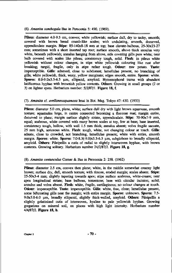
(6). Amanita xanthogala Bas in Persoonia 5: 490. (1969).
Pileus: diameter 4.0-9.5 cm, convex; white yellowish; surface dull, dry to tacky, smooth; covered with brown bread crumb-like scales; with undulating, smooth, decurved, appendiculate margin. Stipe: 85-140x8-18 mm at top; base clavate-bulbous, 25-30x23-27 mm; sometimes with a short inserted tap root; surface smooth, above thick annulus very white, beneath yellowish; annulus hanging from above, side covering gills pure white; near bulb covered with scales like pileus; consistency tough, solid. Flesh: in pileus white yellowish without colour changes, in stipe white yellowish colouring like rust after breaking; turgid, fragile, only in stipe rather tough. Odour: raw potato. Taste: imperceptible. Gills: adnexed, close to subdistant; lamellulae present, no branching of gills; white yellowish, thick, waxy, yellow marginate; edges smooth, entire. Sporee: white. Spores: 8.0-9.0x5.5-6.5 |im, ellipsoid, amyloid. Hymenophoral trama with abundant latificerous hyphae with brownish yellow contents. Others: Growing in small groups (2 or 3) on lighter spots. Herbarium number: 5/2/87/1. Figure 18, f.
(7). Amanita cf. avellaneosquamosa Imai in Bot. Mag. Tokyo 47: 430. (1933)
Pileus: diameter 5.0 cm, plane, white; surface dull dry with light brown squamose, smooth texture; squamules large, in centre connected becoming a floccose mat; margin entire, decurved to plane; margin surface slightly striate, appendiculate. Stipe: 70-90x7-9 mm, equal; scabrous, white covered with many brown scales at top, few at base; base inserted, consistency tough, hollow, with wall 1.5 mm thick; annulus absent; volva fragile saccate, 25 mm high, sericeous white. Flesh: tough, white, not changing colour at touch. Gills: adnate, close to crowded, not branching, lamellulae present; white with entire, smooth margin. Sporee: white. Spores: 7.0-8.5(-9.0)x5.5-6.5 p.m, subglobose to broadly ellipsoid, amyloid. Others: Pileipellis a cutis of radial to slightly interwoven hyphae, with brown contents. Growing solitary. Herbarium number 24/2/87/2. Figure 18, g.
(8). Amanita centunculus Comer & Bas in Persoonia 2: 258. (1962)
Pileus: diameter 2.5 cm, convex then plane; white, in the middle somewhat creamy light brown; surface dry, dull, smooth texture; with rimose, eroded margin; scales absent. Stipe: 25-50x3-4 mm; slightly tapering towards apex; stipe surface scabrous, white-cream; near apex longitudinal striate; base bulbous, tomentose; base with circular incision; solid; annulus and volva absent. Flesh: white, fragile, cartilaginous; no colour changes at touch. Odour: imperceptible. Taste: imperceptible. Gills: white, free, close; lamellulae present, some bifurcating gills near the margin; with entire margin. Sporee: unknown. Spores: 7.5-9.0x5.0-6.0 Jim, broadly ellipsoid, slightly thick-walled, amyloid. Others: Pileipellis a slightly gelatinized cutis of interwoven, hyaline to pale yellowish hyphae. Growing gregarious on mineral soil, on places with high light intensity. Herbarium number 4/4/87/2. Figure 18, h.
Chapter 2 - 70 •
(9). Amanita fritillaria (Berk.) Sacc. in Syll. Fung. 9:2. (1981)
Pileus: diameter 6.0-7.0 cm; convex to broadly parabolic when young, later almost plane; scales dark brown on coffee brown cuticle; surface dry, dull, with slightly translucent-striate margin. Stipe: 80x9 at top x l l mm near base, tapering towards apex; lucid white to grey with grey spots above annuhis, below superior annulus light brown with superficial pulverulent brown powder; stuffed; annulus present, very thin hanging from above, grey white; some brown scales present near base of same colour as scales on pileus. Flesh: white, soft, fragile. Odour: very faint. Taste: unknown. Gills: white, close to crowded; sometimes bifurcating, lamellulae present; margin smooth; brown pileus cuticle visible between gills. Sporee: white. Spores: 6.5-8.0x5.5-7.0 |im, subglobose, sometimes broadly ellipsoid, slightly thick-walled, amyloid. Others: Pileipellis a slightly gelatinized cutis of hyphae with brown intracellular contents. Growing solitary. Herbarium number 9/4/87/2. Figure 18, i.
(10). Amanita similis Boedijn in Sydowia 5: 322. (1951)
Pileus: diameter 11.0-11.5 cm, narrowly parabolic when young, later becoming plane; chestnut brown when young later light brown, with remaining dark brown spot in the centre; surface very lucid when young, later slightly lucid, smooth, tacky when old; margin white to cream, translucent-striate (pellucid), entire; cuticle can be loosened till the centre. Stipe: 135-150x10-14 mm, equal; bright yellow with yellow-brown patches; lucid, smooth, fragile, cartilaginous, hollow; movable, light yellow annulus present hanging from the superior part of the stipe; membranous saccate, white volva present. Flesh: yellowish, cartilaginous, not changing colour at touch. Odour: distinctive, difficult to circumscribe. Taste: unknown. Gills: free, yellow marginate, close to crowded, few lamellulae present; with entire smooth edge. Sporee: unknown. Spores: 8.0-10.0x6.5-8.5 Jim, subglobose to broadly ellipsoid, slightly thick-walled, inamyloid. Others: Pileipellis a dry cutis of radial to slightly interwoven, yellowish hyphae. Growing in scattered caespitose to gregarious small groups. Herbarium number 18/2/87/2. Figure 18, j .
(11). Amanita cf. duplex Comer & Bas in Persoonia 2: 275. (1962)
Pileus: diameter 7.5-9.0 cm, first convex later plane; very dark brown in the centre becoming lighter near the margin; with grey brown superficial scales that become less in number near the margin; surface looking sericeous, tacky to dry; smooth margin lacunate when old. Stipe: 90-100x7 near apex x20 near bulbous base; white with superficial light brown scales and powder like pileus surface; hollow with a wall of 2 mm thick; annulus absent; fragile, saccate to constricted volva present; height 4.5 cm, light brown. Flesh: white, tough; not changing colour upon exposure or touch. Odour: not unpleasant. Taste: unknown. Gills: free, white, close to crowded, often bifurcating, without lamellulae, fragile; with smooth, entire edges. Sporee: white. Spores: 7.0-8.0x5.0-6.0 \im, broadly ellipsoid, sometimes subglobose or ellipsoid, slightly thick-walled, amyloid. Pileipellis a somewhat gelatinized cutis of almost colourless hyphae. Others: Growing scattered. Herbarium number 3/3/87/1. Figure 18, k.
Chapter 2 - 7 1 -
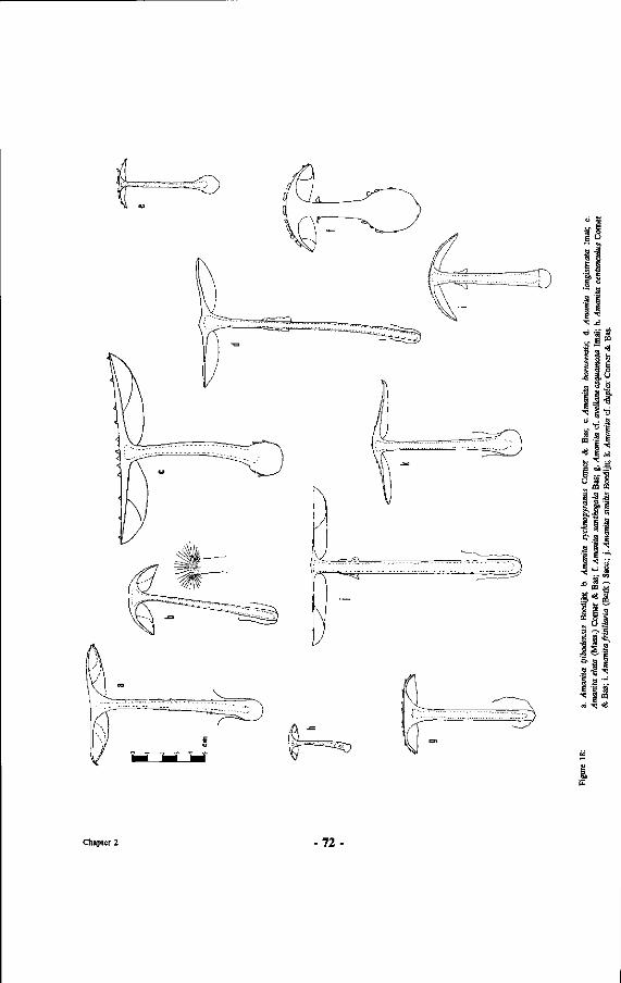
a <->
f i ~ a s 'I
g S h ig
1 i t a -a -s
'Ë -a a
fel'« &a a 1 .
1 CQ
'Eî
f a
1 E
1 S U
i 1 1 a ë
-S
1 « '8
I --s CQ
•3
Chapter 2 -72
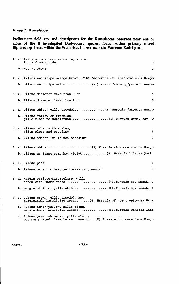
Group 3: Russulaceae
Preliminary field key and descriptions for the Russulaceae observed near one or more of the 8 investigated Dipterocarp species, found within primary mixed Dipterocarp forest within the Wanariset I forest near the Wartono Kadri plot.
1. a. Parts of mushroom exudating white latex from wounds 2
b. Not as above 3
2. a. Pileus and stipe orange-brown..(10).Lactarius cf. austrovolemus Hongo
b. Pileus and stipe white (11) .Lactarius subpiperatus Hongo
3. a. Pileus diameter more than 9 cm 4
b. Pileus diameter less than 9 cm 5
4. a. Pileus white, gills crowded (9) .Russula japonica Hongo
b. Pileus yellow or greenish, gills close to subdistant (3) .Russula spec. nov. ?
5. a. Pileus often with scales, gills close and seceding 6
b. Pileus smooth, gills not seceding 7
6. a. Pileus white (1) .Russula eburneoareolata Hongo
b. Pileus at least somewhat violet (8) .Russula lilacea Quel.
7. a. Pileus pink 8
b. Pileus brown, ochre, yellowish or greenish 9
8. a. Margin striate-tuberculate, gills often with rusty spots (7) .Russula sp. indet. 7
b. Margin striate, gills white (2) .Russula sp. indet. 2
9. a. Pileus brown, gills crowded, not marginated, lamellulae absent (4).Russula cf. pectinatoides Peck
b. Pileus ochre/yellow, gills close, marginated, lamellulae absent (5) .Russula senecis Imai
c. Pileus greenish brown, gills close, not marginated, lamellulae present.... (6) .Russula cf. metachroa Hongo
Chapter 2 - 73
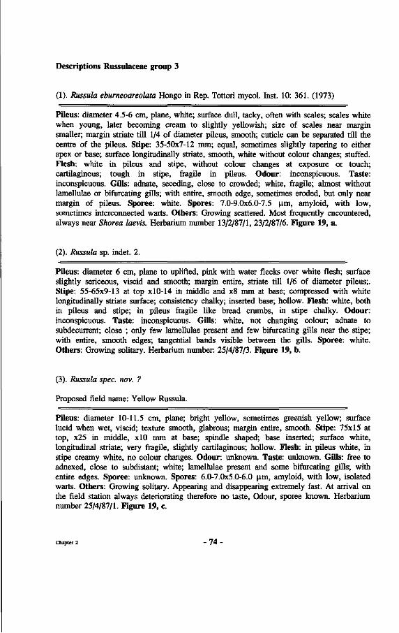
Descriptions Russulaceae group 3
(1). Russuia eburneoareolata Hongo in Rep. Tottori mycol. Inst. 10: 361. (1973)
Pileus: diameter 4.5-6 cm, plane, white; surface dull, tacky, often with scales; scales white when young, later becoming cream to slightly yellowish; size of scales near margin smaller; margin striate till 1/4 of diameter pileus, smooth; cuticle can be separated till the centre of the pileus. Stipe: 35-50x7-12 mm; equal, sometimes slightly tapering to either apex or base; surface longitudinally striate, smooth, white without colour changes; stuffed. Flesh: white in pileus and stipe, without colour changes at exposure or touch; cartilaginous; tough in stipe, fragile in pileus. Odour: inconspicuous. Taste: inconspicuous. Gills: adnate, seceding, close to crowded; white, fragile; almost without lamellulae or bifurcating gills; with entire, smooth edge, sometimes eroded, but only near margin of pileus. Sporee: white. Spores: 7.0-9.0x6.0-7.5 |im, amyloid, with low, sometimes interconnected warts. Others: Growing scattered. Most frequently encountered, always near Shorea laevis. Herbarium number 13/2/87/1, 23/2/87/6. Figure 19, a.
(2). Russula sp. indet. 2.
Pileus: diameter 6 cm, plane to uplifted, pink with water flecks over white flesh; surface slightly sericeous, viscid and smooth; margin entire, striate till 1/6 of diameter pileus;. Stipe: 55-65x9-13 at top xl0-14 in middle and x8 mm at base; compressed with white longitudinally striate surface; consistency chalky; inserted base; hollow. Flesh: white, both in pileus and stipe; in pileus fragile like bread crumbs, in stipe chalky. Odour: inconspicuous. Taste: inconspicuous. Gills: white, not changing colour; adnate to subdecurrent; close ; only few lamellulae present and few bifurcating gills near the stipe; with entire, smooth edges; tangential bands visible between the gills. Sporee: white. Others: Growing solitary. Herbarium number: 25/4/87/3. Figure 19, b.
(3). Russula spec. nov. ?
Proposed field name: Yellow Russula.
Pileus: diameter 10-11.5 cm, plane; bright yellow, sometimes greenish yellow; surface lucid when wet, viscid; texture smooth, glabrous; margin entire, smooth. Stipe: 75x15 at top, x25 in middle, xlO mm at base; spindle shaped; base inserted; surface white, longitudinal striate; very fragile, slightly cartilaginous; hollow. Flesh: in pileus white, in stipe creamy white, no colour changes. Odour: unknown. Taste: unknown. Gills: free to adnexed, close to subdistant; white; lamellulae present and some bifurcating gills; with entire edges. Sporee: unknown. Spores: 6.0-7.0x5.0-6.0 \im, amyloid, with low, isolated warts. Others: Growing solitary. Appearing and disappearing extremely fast. At arrival on the field station always deteriorating therefore no taste, Odour, sporee known. Herbarium number 25/4/87/1. Figure 19, c.
Chapter 2 - 7 4 •
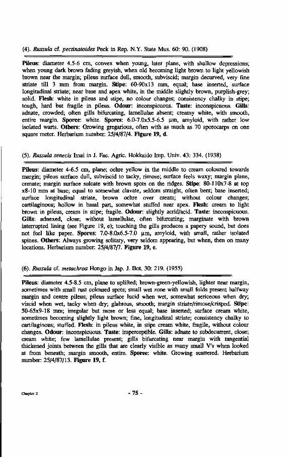
(4). Russula cf. pectinatoides Peck in Rep. N.Y. State Mus. 60: 90. (1908)
Pileus: diameter 4.5-6 cm, convex when young, later plane, with shallow depressions; when young dark brown fading greyish, when old becoming light brown to light yellowish brown near the margin; pileus surface dull, smooth, subviscid; margin decurved, very fine striate till 3 mm from margin. Stipe: 60-90x13 mm, equal; base inserted, surface longitudinal striate; near base and apex white, in the middle slightly brown, purplish-grey; solid. Flesh: white in pileus and stipe, no colour changes; consistency chalky in stipe; tough, hard but fragile in pileus. Odour: inconspicuous. Taste: inconspicuous. Gills: adnate, crowded; often gills bifurcating, lamellulae absent; creamy white, with smooth, entire margin. Sporee: white. Spores: 6.0-7.0x5.5-6.5 urn, amyloid, with rather low isolated warts. Others: Growing gregarious, often with as much as 70 sporocarps on one square meter. Herbarium number: 25/4/87/4. Figure 19, d.
(5). Russula senecis Imai in J. Fac. Agric. Hokkaido Imp. Univ. 43: 334. (1938)
Pileus: diameter 4-6.5 cm, plane; ochre yellow in the middle to cream coloured towards margin; pileus surface dull, subviscid to tacky, rimose; surface feels waxy; margin plane, crenate; margin surface sulcate with brown spots on the ridges. Stipe: 80-110x7-8 at top x8-10 mm at base; equal to somewhat clavate, seldom straight, often bent; base inserted; surface longitudinal striate, brown ochre over cream; without colour changes; cartilaginous; hollow in basal part, somewhat stuffed near apex. Flesh: cream to light brown in pileus, cream in stipe; fragile. Odour: slightly acrid/acid. Taste: inconspicuous. Gills: adnexed, close; without lamellulae, often bifurcating; marginate with brown interrupted lining (see Figure 19, e); touching the gills produces a papery sound, but does not feel like paper. Spores: 7.0-8.0x6.5-7.0 |im, amyloid, with small, rather isolated spines. Others: Always growing solitary, very seldom appearing, but when, then on many locations. Herbarium number: 25/4/87/7. Figure 19, e.
(6). Russula cf. metachroa Hongo in Jap. J. Bot. 30: 219. (1955)
Pileus: diameter 4.5-8.5 cm, plane to uplifted; brown-green-yellowish, lighter near margin, sometimes with small rust coloured spots; small wet zone with small folds present halfway margin and centre pileus; pileus surface lucid when wet, somewhat sericeous when dry; viscid when wet, tacky when dry; glabrous, smooth; margin striate/rimose/crisped. Stipe: 50-65x9-18 mm; irregular but more or less equal; base inserted; surface cream white, sometimes becoming slightly light brown; fine, longitudinal striate; consistency chalky to cartilaginous; stuffed. Flesh: in pileus white, in stipe cream white, fragile, without colour changes. Odour: inconspicuous. Taste: imperceptible. Gills: adnate to subdecurrent, close; cream white; few lamellulae present; gills bifurcating near margin with tangential thickened joints between the gills that are clearly visible as many small V's when looked at from beneath; margin smooth, entire. Sporee: white. Growing scattered. Herbarium number: 25/4/87/13. Figure 19, f.
Chapter 2 - 7 5 •
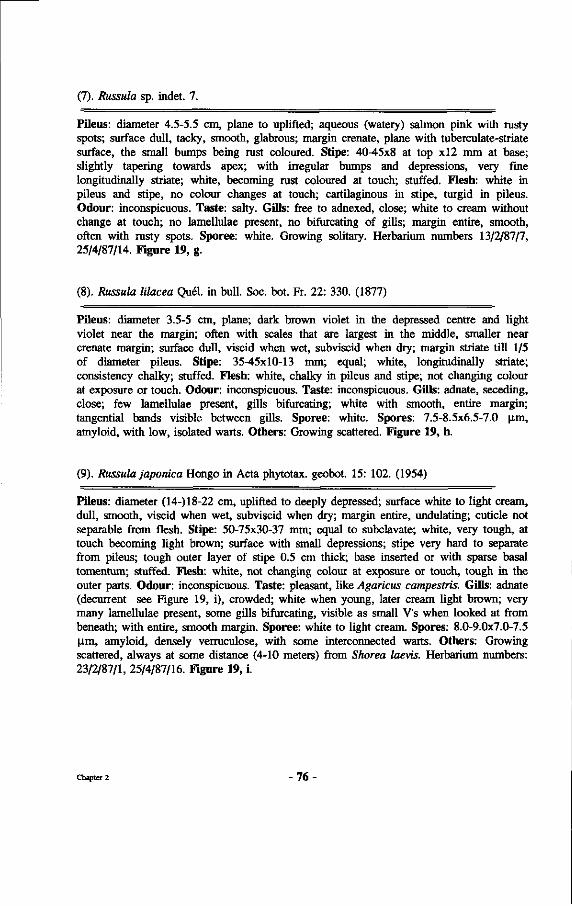
(7). Russuia sp. indet. 7.
Pileus: diameter 4.5-5.5 cm, plane to uplifted; aqueous (watery) salmon pink with rusty spots; surface dull, tacky, smooth, glabrous; margin crenate, plane with tuberculate-striate surface, the small bumps being rust coloured. Stipe: 40-45x8 at top xl2 mm at base; slightly tapering towards apex; with irregular bumps and depressions, very fine longitudinally striate; white, becoming rust coloured at touch; stuffed. Flesh: white in pileus and stipe, no colour changes at touch; cartilaginous in stipe, turgid in pileus. Odour: inconspicuous. Taste: salty. Gills: free to adnexed, close; white to cream without change at touch; no lamellulae present, no bifurcating of gills; margin entire, smooth, often with rusty spots. Sporee: white. Growing solitary. Herbarium numbers 13/2/87/7, 25/4/87/14. Figure 19, g.
(8). Russula lilacea Quel, in bull. Soc. bot. Fr. 22: 330. (1877)
Pileus: diameter 3.5-5 cm, plane; dark brown violet in the depressed centre and light violet near the margin; often with scales that are largest in the middle, smaller near crenate margin; surface dull, viscid when wet, subviscid when dry; margin striate till 1/5 of diameter pileus. Stipe: 35-45x10-13 mm; equal; white, longitudinally striate; consistency chalky; stuffed. Flesh: white, chalky in pileus and stipe; not changing colour at exposure or touch. Odour: inconspicuous. Taste: inconspicuous. Gills: adnate, seceding, close; few lamellulae present, gills bifurcating; white with smooth, entire margin; tangential bands visible between gills. Sporee: white. Spores: 7.5-8.5x6.5-7.0 |im, amyloid, with low, isolated warts. Others: Growing scattered. Figure 19, h.
(9). Russula japonica Hongo in Acta phytotax. geobot. 15: 102. (1954)
Pileus: diameter (14-) 18-22 cm, uplifted to deeply depressed; surface white to light cream, dull, smooth, viscid when wet, subviscid when dry; margin entire, undulating; cuticle not separable from flesh. Stipe: 50-75x30-37 mm; equal to subclavate; white, very tough, at touch tecoming light brown; surface with small depressions; stipe very hard to separate from pileus; tough outer layer of stipe 0.5 cm thick; base inserted or with sparse basal tomentum; stuffed. Flesh: white, not changing colour at exposure or touch, tough in the outer parts. Odour: inconspicuous. Taste: pleasant, like Agaricus campestris. Gills: adnate (decurrent see Figure 19, i), crowded; white when young, later cream light brown; very many lamellulae present, some gills bifurcating, visible as small V's when looked at from beneath; with entire, smooth margin. Sporee: white to light cream. Spores: 8.0-9.0x7.0-7.5 |im, amyloid, densely verruculose, with some interconnected warts. Others: Growing scattered, always at some distance (4-10 meters) from Shorea laevis. Herbarium numbers: 23/2/87/1, 25/4/87/16. Figure 19, i.
Chapter 2 - 7 6 -
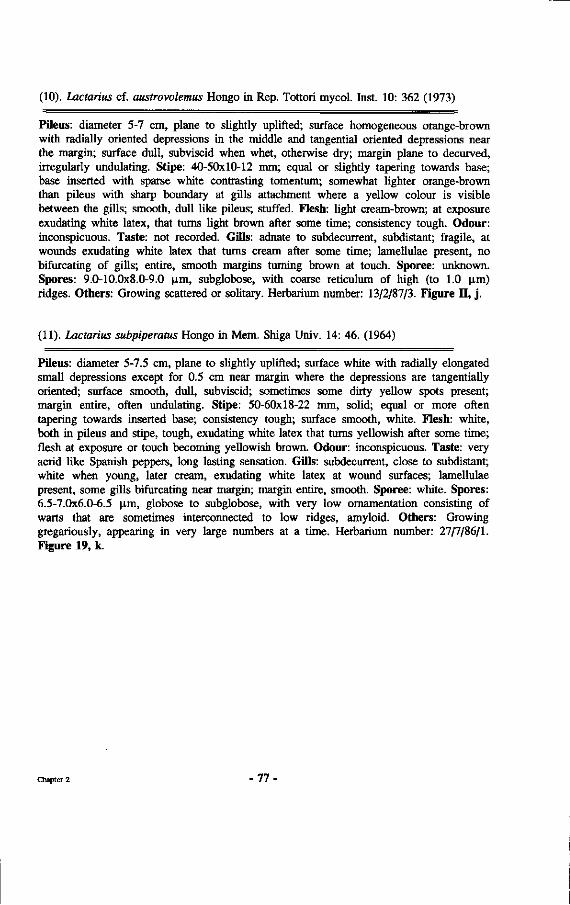
(10). Lactarius cf. austrovolemus Hongo in Rep. Tottori mycol. Inst. 10: 362 (1973)
Pileus: diameter 5-7 cm, plane to slightly uplifted; surface homogeneous orange-brown with radially oriented depressions in the middle and tangential oriented depressions near the margin; surface dull, subviscid when whet, otherwise dry; margin plane to decurved, irregularly undulating. Stipe: 40-50x10-12 mm; equal or slightly tapering towards base; base inserted with sparse white contrasting tomentum; somewhat lighter orange-brown than pileus with sharp boundary at gills attachment where a yellow colour is visible between the gills; smooth, dull like pileus; stuffed. Flesh: light cream-brown; at exposure exudating white latex, that turns light brown after some time; consistency tough. Odour: inconspicuous. Taste: not recorded. Gills: adnate to subdecurrent, subdistant; fragile, at wounds exudating white latex that turns cream after some time; lamellulae present, no bifurcating of gills; entire, smooth margins turning brown at touch. Sporee: unknown. Spores: 9.0-10.0x8.0-9.0 ^m, subglobose, with coarse reticulum of high (to 1.0 |im) ridges. Others: Growing scattered or solitary. Herbarium number: 13/2/87/3. Figure II, j .
(11). Lactarius subpiperatus Hongo in Mem. Shiga Univ. 14: 46. (1964)
Pileus: diameter 5-7.5 cm, plane to slightly uplifted; surface white with radially elongated small depressions except for 0.5 cm near margin where the depressions are tangentially oriented; surface smooth, dull, subviscid; sometimes some dirty yellow spots present; margin entire, often undulating. Stipe: 50-60x18-22 mm, solid; equal or more often tapering towards inserted base; consistency tough; surface smooth, white. Flesh: white, both in pileus and stipe, tough, exudating white latex that turns yellowish after some time; flesh at exposure or touch becoming yellowish brown. Odour: inconspicuous. Taste: very acrid like Spanish peppers, long lasting sensation. Gills: subdecurrent, close to subdistant; white when young, later cream, exudating white latex at wound surfaces; lamellulae present, some gills bifurcating near margin; margin entire, smooth. Sporee: white. Spores: 6.5-7.0x6.0-6.5 |im, globose to subglobose, with very low ornamentation consisting of warts that are sometimes interconnected to low ridges, amyloid. Others: Growing gregariously, appearing in very large numbers at a time. Herbarium number: 27/7/86/1. Figure 19, k.
Chapter 2 - 7 7 •
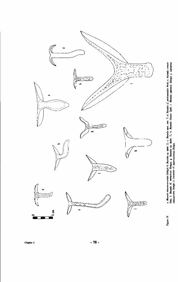
s g
.M 3 -a
| l |
lia S g M
i l
Chapter 2 -78
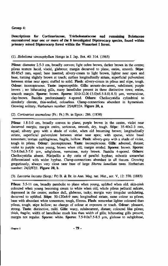
Group 4:
Descriptions for Cortinariaceae, Tricholomataceae and remaining Boletaceae encountered near one or more of the 8 investigated Dipterocarp species, found within primary mixed Dipterocarp forest within the Wanariset I forest.
(1). Hebeloma vinosophyllum Hongo in J. Jap. Bot. 40: 314. (1965)
Pileus: diameter 2.5-3 cm, broadly convex; light ochre brown, darker brown in the centre; pileus texture lucid, viscid, glabrous; margin decurved to plane, entire, smooth. Stipe: 80-85x5 mm, equal; base inserted; silvery-cream to light brown, lighter near apex and base, turning slightly brown at touch; surface longitudinally striate, superficial pulverulent between striae near apex; stuffed to solid. Flesh: silvery-cream in pileus and stipe, tough. Odour: inconspicuous. Taste: imperceptible. Gills: arcuate-decurrent, subdistant; purple brown ; no bifurcating gills, many lamellulae present in three distinctive rows; entire, smooth margin. Sporee: brown. Spores: 10.0-12.0(-13.0)x6.5-8.0(-8.5) p.m, verruculose, clay-brown. Basidia predominantly 4-spored. Others: Cheilocystidia cylindrical to slenderly clavate, thin-walled, colourless. Clamp-connections abundant in hymenium. Growing solitary. Herbarium number: 25/4/87/24. Figure 20, a.
(2). Cortinarius anomalous (Fr.: Fr.) Fr. in Epicr.: 286. (1838)
Pileus: 1.8-3.0 cm, broadly convex to plane; purple brown in the centre, violet near decurved margin; margin entire, sericeous, smooth, dry, tough. Stipe: 35-40x2-3 mm, equal; silvery grey with a shade of violet, when old becoming brown; longitudinally striate, superficial pulverulent between striae near apex; with sparse, white basal tomentum; texture cartilaginous, fragile, hollow. Flesh: silvery-grey with a shade of violet, tough in pileus. Odour: inconspicuous. Taste: inconspicuous. Gills: adnexed, distant; violet to purple when young, brown when old; margin eroded. Sporee: brown. Spores: 7.0-8.0x6.5-7.0 jim, subglobose, verrucose, rusty brown. Basidia 4-spored. Others: Cheilocystidia absent. Pileipellis a dry cutis of parallel hyphae; subcutis somewhat differentiated with wider hyphae. Clamp-connections abundant in all tissues. Growing gregariously, always very close near base of large Shorea lamellata trees. Herbarium number: 24/2/87/1. Figure 20, b.
(3). Laccaria laccata (Scop.: Fr) B. & Br. in Ann. Mag. nat. Hist., ser. V, 12: 370. (1883)
Pileus: 3.5-11 cm, broadly parabolic to plane when young, uplifted when old; skin-pink coloured when young becoming cream to white when old; whole pileus pellucid sulcate, depressed in the centre; surface dull, glabrous, tacky; margin very irregular undulating, decurved to uplifted. Stipe: 50-120x6-9 mm; longitudinal striate, same colour as pileus; base with abundant white tomentum; tough, fibrous. Flesh: somewhat lighter coloured than pileus, tough; stipe hollow; no change of colour at exposure or touch. Odour: pleasant, strong. Taste: distinctive, mild. Gills: waxy, subdecurrent, distant; coloured like pileus; thick, fragile; width of lamellulae much less than width of gills; bifurcating gills present; margin not regular. Sporee: white. Spores: 7.5-9.0x7.5-8.5 jim, globose to subglobose,
Chapter 2 - 7 9 •
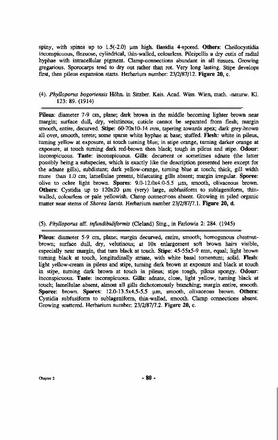
spiny, with spines up to 1.5(-2.0) urn high. Basidia 4-spored. Others: Cheilocystidia inconspicuous, flezuose, cylindrical, thin-walled, colourless. Pileipellis a dry cutis of radial hyphae with intracellular pigment. Clamp-connections abundant in all tissues. Growing gregarious. Sporocarps tend to dry out rather than rot. Very long lasting. Stipe develops first, then pileus expansion starts. Herbarium number: 23/2/87/12. Figure 20, c.
(4). Phylloporus bogoriensis Höhn, in Sitzber. Kais. Acad. Wiss. Wien, math, -naturw. Kl. 123: 89. (1914)
Pileus: diameter 7-9 cm, plane; dark brown in the middle becoming lighter brown near margin; surface dull, dry, velutinous; cuticle cannot be separated from flesh; margin smooth, entire, decurved. Stipe: 60-70x10-14 mm, tapering towards apex; dark grey-brown all over, smooth, terete; some sparse white hyphae at base; stuffed. Flesh: white in pileus, turning yellow at exposure, at touch turning blue; in stipe orange, turning darker orange at exposure, at touch turning dark red-brown then black; tough in pileus and stipe. Odour: inconspicuous. Taste: inconspicuous. Gills: decurrent or sometimes adnate (the latter possibly being a subspecies, which is exactly like the description presented here except for the adnate gills), subdistant; dark yellow-orange, turning blue at touch; thick, gill width more than 1.0 cm; lamellulae present, bifurcating gills absent; margin irregular. Sporee: olive to ochre light brown. Spores: 9.0-12.0x4.0-5.5 |im, smooth, olivaceous brown. Others: Cystidia up to 120x20 \im (very) large, subfusiform to sublageniform, thin-walled, colourless or pale yellowish. Clamp connectons absent. Growing in piled organic matter near stems of Shorea laevis. Herbarium number 23/2/87/7.1. Figure 20, d.
(5). Phylloporus aff. infundibuliformis (Cleland) Sing., in Farlowia 2: 284. (1945)
Pileus: diameter 5-9 cm, plane; margin decurved, entire, smooth; homogenous chestnut-brown; surface dull, dry, velutinous; at lOx enlargement soft brown hairs visible, especially near margin, that turn black at touch. Stipe: 45-55x5-9 mm, equal; light brown turning black at touch, longitudinally striate, with white basal tomentum; solid. Flesh: light yellow-cream in pileus and stipe, turning dark brown at exposure and black at touch in stipe, turning dark brown at touch in pileus; stipe tough, pileus spongy. Odour: inconspicuous. Taste: inconspicuous. Gills: adnate, close, light yellow, turning black at touch; lamellulae absent, almost all gills dichotomously branching; margin entire, smooth. Sporee: brown. Spores: 12.0-13.5x4.5-5.5 p.m, smooth, olivaceous brown. Others: Cystidia subfusiform to sublageniform, thin-walled, smooth. Clamp connections absent. Growing scattered. Herbarium number: 23/2/87/7.2. Figure 20, e.
Chapter 2 - 8 0 •
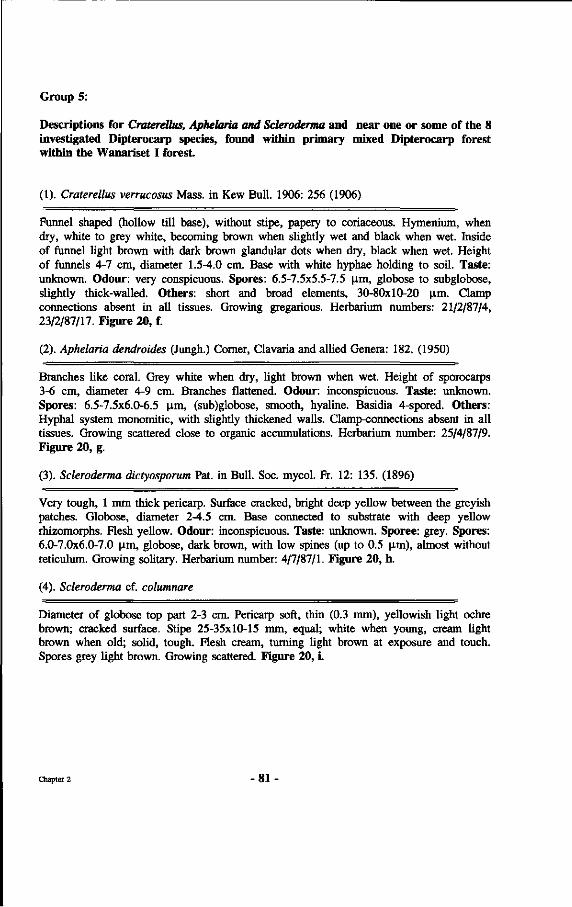
Group 5:
Descriptions for Craterellus, Aphelaria and Scleroderma and near one or some of the 8 investigated Dipterocarp species, found within primary mixed Dipterocarp forest within the Wanariset I forest.
(1). Craterellus verrucosus Mass. in Kew Bull. 1906: 256 (1906)
Funnel shaped (hollow till base), without stipe, papery to coriaceous. Hymenium, when dry, white to grey white, becoming brown when slightly wet and black when wet. Inside of funnel light brown with dark brown glandular dots when dry, black when wet. Height of funnels 4-7 cm, diameter 1.5-4.0 cm. Base with white hyphae holding to soil. Taste: unknown. Odour: very conspicuous. Spores: 6.5-7.5x5.5-7.5 |im, globose to subglobose, slightly thick-walled. Others: short and broad elements, 30-80x10-20 p.m. Clamp connections absent in all tissues. Growing gregarious. Herbarium numbers: 21/2/87/4, 23/2/87/17. Figure 20, f.
(2). Aphelaria dendroides (Jungh.) Corner, Clavaria and allied Genera: 182. (1950)
Branches like coral. Grey white when dry, light brown when wet. Height of sporocarps 3-6 cm, diameter 4-9 cm. Branches flattened. Odour: inconspicuous. Taste: unknown. Spores: 6.5-7.5x6.0-6.5 |im, (sub)globose, smooth, hyaline. Basidia 4-spored. Others: Hyphal system monomitic, with slightly thickened walls. Clamp-connections absent in all tissues. Growing scattered close to organic accumulations. Herbarium number: 25/4/87/9. Figure 20, g.
(3). Scleroderma dictyosporum Pat. in Bull. Soc. mycol. Fr. 12: 135. (1896)
Very tough, 1 mm thick pericarp. Surface cracked, bright deep yellow between the greyish patches. Globose, diameter 2-4.5 cm. Base connected to substrate with deep yellow rhizomorphs. Flesh yellow. Odour: inconspicuous. Taste: unknown. Sporee: grey. Spores: 6.0-7.0x6.0-7.0 urn, globose, dark brown, with low spines (up to 0.5 |im), almost without reticulum. Growing solitary. Herbarium number: 4/7/87/1. Figure 20, h.
(4). Scleroderma cf. columnar e
Diameter of globose top part 2-3 cm. Pericarp soft, thin (0.3 mm), yellowish light ochre brown; cracked surface. Stipe 25-35x10-15 mm, equal; white when young, cream light brown when old; solid, tough. Flesh cream, turning light brown at exposure and touch. Spores grey light brown. Growing scattered. Figure 20, i.
Chapter 2 - 8 1
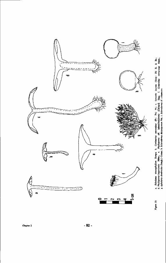
Un
ë 3
•8 E
a 'E
1 „& <J
JD
O on
g
'Ë
•g :§
J ~ 'S
1 1 j * .
a.
s
•J
S I o
1 % | 1 <u
3 Lf
E ö
a -a
s •§
Chapter 2 82-
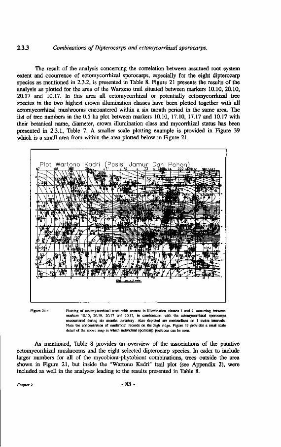
2.3.3 Combinations of Dipterocarps and ectomycorrhizal sporocarps.
The result of the analysis concerning the correlation between assumed root system extent and occurrence of ectomycorrhizal sporocarps, especially for the eight dipterocarp species as mentioned in 2.3.2, is presented in Table 8. Figure 21 presents the results of the analysis as plotted for the area of the Wartono trail situated between markers 10.10, 20.10, 20.17 and 10.17. In this area all ectomycorrhizal or potentially ectomycorrhizal tree species in the two highest crown illumination classes have been plotted together with all ectomycorrhizal mushrooms encountered within a six month period in the same area. The list of tree numbers in the 0.5 ha plot between markers 10.10, 17.10, 17.17 and 10.17 with their botanical name, diameter, crown illumination class and mycorrhizal status has been presented in 2.3.1, Table 7. A smaller scale plotting example is provided in Figure 39 which is a small area from within the area plotted below in Figure 21.
Plot Wartono K 6D7 «8
adri (Posisi Jamur Dan Pohon)
Figure 21 : Plotting of ectomycorrhizal trees with crowns in illumination classes 1 and 2, occurring between marken; 10.10, 20.10, 20.17 and 10.17, in combination with the ectomyconhizat sporocarps encountered during six months inventory. Also depicted are contouriines on 1 metre intervals. Note the concentration of mushroom records on the high ridge. Figure 39 provides a smal scale detail of the above map in which individual sporocarp positions can be seen.
As mentioned, Table 8 provides an overview of the associations of the putative ectomycorrhizal mushrooms and the eight selected dipterocarp species. In order to include larger numbers for all of the mycobiont-phytobiont combinations, trees outside the area shown in Figure 21, but inside the "Wartono Kadri" trail plot (see Appendix 2), were included as well in the analyses leading to the results presented in Table 8.
Chapter 2 83
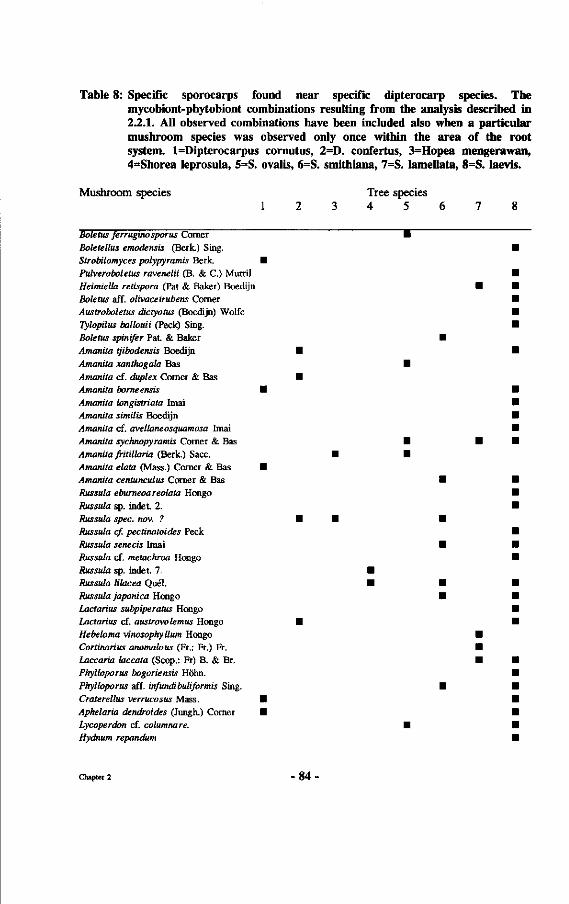
Table 8: Specific sporocarps found near specific dipterocarp species. The mycobiont-phytobiont combinations resulting from the analysis described in 2.2.1. All observed combinations have been included also when a particular mushroom species was observed only once within the area of the root system. l=Dipterocarpus cornutus, 2=D. confertus, 3=Hopea mengerawan, 4=Shorea leprosula, 5=S. ovalis, 6=S. smithiana, 7=S. lamellata, 8=S. laevis.
Mushroom species Tree species 1 2 3 4 5 6 7 8
Boletus ferruginosporus Comer I Boletellus emodensis (Berk.) Sing. • Strobilomyces polypyramis Berk. •
Pulveroboletus ravenelii (B. & C.) Murril Heimiella retispora (Pat & Baker) Boedijn Boletus aff. olivaceirubens Corner
Austroboletus dictyotus (Boedijn) Wolfe Tylopilus ballouii (Peck) Sing. Boletus spinifer Pa t & Baker
Amanita tjibodensis Boedijn •
Amanita xanthogala Bas • Amanita cf. duplex Comer & Bas • Amanita borne ensis •
Amanita longistriata Imai Amanita similis Boedijn Amanita cf. avellaneosquamosa Imai
Amanita sychnopyramis Comer & Bas •
Amanita fritillaria (Berk.) Sacc. • • Amanita elata (Mass.) Comer & Bas • Amanita centunculus Comer & Bas Russula eburneoareolata Hongo
Russula sp. indet. 2. Russula spec. nov. ? • • Russula cf. pectinatoides Peck
Russula senecis Imai Russula cf. metachroa Hongo
Russula sp. indet. 7. • Russula lilacea Quel. • Russula japonica Hongo Lactarius subpiperatus Hongo Lactarius cf. austrovolemus Hongo • Hebeloma vinosophy Hum Hongo Cortinarius anomalous (Fr.: Fr.) Fr. Laccaria laccata (Scop.: Fr) B. & Br.
Phylloporus bogoriensis Höhn.
Phylloporus aff. infundibuliformis Sing.
Craterellus verrucosus Mass. • Aphelaria dendroides (Jungh.) Comer • Lycoperdon cf. columnare. • Hydnum repandum
Chapter 2 - 8 4 •
Table 9 shows the number of mycobionts associated with each of the eight investigated dipterocarp species. Furthermore, it shows the number of mycobionts which is specific for each of these dipterocarp species as well as the percentage of the mycorrhizae which is specific for these Dipterocarps, as result of the analyses described above.
Table 9 : Total number of mycobionts (T), number (n) and percentages (%) of specific mycobionts for the eight dipterocarp species. Number of trees of each species included between brackets.
Shorea l a e v i s (18) Sho rea l a m e l l a t a (8) Sho r ea s m i t h i a n a (4) Sho rea o v a l i s (3) Sho rea l e p r o s u l a (2) Hopea mengerawan (3) D i p t e r o c a r p u s c o n f e r t u s (7) D i p t e r o c a r p u s c o r n u t u s (2)
T
29 5 7 5 2 2 4 5
n
15 2 1 2 1 0 1 2
%
52 40 14 40 50
0 25 40
The number of phytobionts associated with certain mycobionts is presented in Table 10. The contents of Table 10 are based upon the forementioned analyses on the correlation between pytobiont positions and mycobiont positions. As can be seen from this table the number of mycobionts that only associates with one phytobiont is 60%, indicating a high degree of specificity for the conditions investigated here.
Table 10 : Numbers and percentages of suspected phytobionts (8 species studied only) per mycobiont.
Number of mycobionts
Pe rcen tage of mycobionts(%)
Number of p o t e n t i a l a s s o c i a t e d p hy t ob i on t s
1
24
GO
2
13
33
3
3
7
Chapter 2 8 5 -
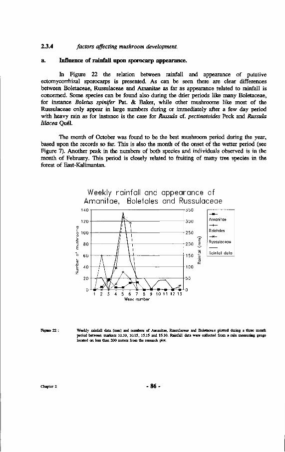
2.3.4 factors affecting mushroom development.
a. Influence of rainfall upon sporocarp appearance.
In Figure 22 the relation between rainfall and appearance of putative ectomycorrhizal sporocarps is presented. As can be seen there are clear differences between Boletaceae, Russulaceae and Amanitae as far as appearance related to rainfall is concerned. Some species can be found also during the drier periods like many Boletaceae, for instance Boletus spinifer Pat. & Baker, while other mushrooms like most of the Russulaceae only appear in large numbers during or immediately after a few day period with heavy rain as for instance is the case for Russula cf. pectinatoides Peck and Russula lilacea Quel.
The month of October was found to be the best mushroom period during the year, based upon the records so far. This is also the month of the onset of the wetter period (see Figure 7). Another peak in the numbers of both species and individuals observed is in the month of February. This period is closely related to fruiting of many tree species in the forest of East-Kalimantan.
Weekly rainfall and appearance of Amanitae, Boletales and Russulaceae
i b U
300
250
200
100
50
0
E E
ô
'o
_^ Amanitae
Boletales
Russulaceae
Rainfall data
1 2 3 4 5 6 7 8 9 10 1 1 1 2 13 Week number
Figure 22 : Weekly rainfall data (mm) and numbers of Amanitae, Russulaceae and Boletaceae plotted during a three month period between markers 10.10, 10.15, 15.15 and 15.10. Rainfall data were collected from a rain measuring gauge located on less than 200 meters from the research plot.
Chapter 2 -86
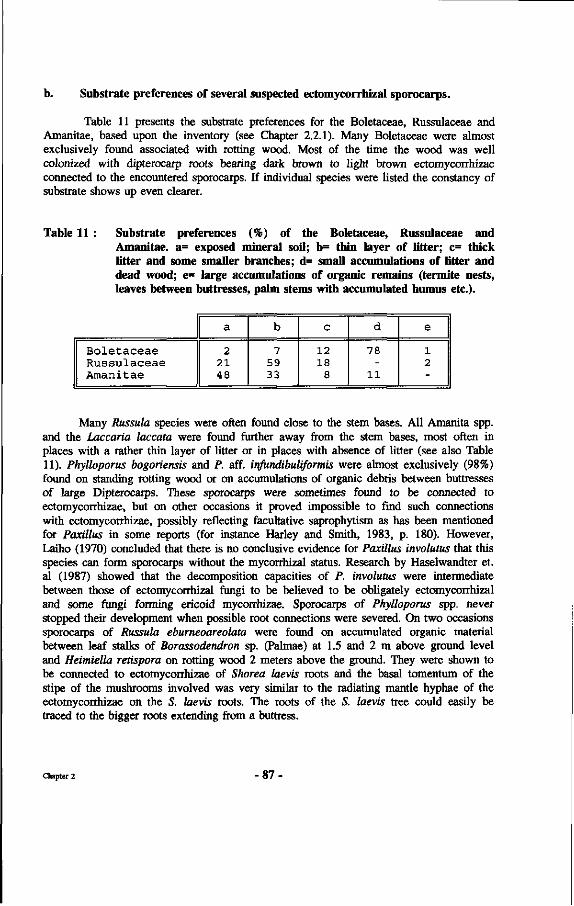
b. Substrate preferences of several suspected ectomycorrhizal sporocarps.
Table 11 presents the substrate preferences for the Boletaceae, Russulaceae and Amanitae, based upon the inventory (see Chapter 2.2.1). Many Boletaceae were almost exclusively found associated with rotting wood. Most of the time the wood was well colonized with dipterocarp roots bearing dark brown to light brown ectomycorrhizae connected to the encountered sporocarps. If individual species were listed the constancy of substrate shows up even clearer.
Table 11 : Substrate preferences (%) of the Boletaceae, Russulaceae and Amanitae. a= exposed mineral soil; b= thin layer of litter; c= thick Utter and some smaller branches; d= small accumulations of litter and dead wood; e= large accumulations of organic remains (termite nests, leaves between buttresses, palm stems with accumulated humus etc.).
Boletaceae Russulaceae Amanitae
a
2 21 48
b
7 59 33
c
12 18
8
d
78
11
e
1 2
Many Russula species were often found close to the stem bases. All Amanita spp. and the Laccaria laccata were found further away from the stem bases, most often in places with a rather thin layer of litter or in places with absence of litter (see also Table 11). Phylloporus bogoriensis and P. aff. infundibuliformis were almost exclusively (98%) found on standing rotting wood or on accumulations of organic debris between buttresses of large Dipterocarps. These sporocarps were sometimes found to be connected to ectomycorrhizae, but on other occasions it proved impossible to find such connections with ectomycorrhizae, possibly reflecting facultative saprophytism as has been mentioned for Paxillus in some reports (for instance Harley and Smith, 1983, p. 180). However, Laiho (1970) concluded that there is no conclusive evidence for Paxillus involutus that this species can form sporocarps without the mycorrhizal status. Research by Haselwandter et. al (1987) showed that the decomposition capacities of P. involutus were intermediate between those of ectomycorrhizal fungi to be believed to be obligately ectomycorrhizal and some fungi forming ericoid mycorrhizae. Sporocarps of Phylloporus spp. never stopped their development when possible root connections were severed. On two occasions sporocarps of Russula eburneoareolata were found on accumulated organic material between leaf stalks of Borassodendron sp. (Palmae) at 1.5 and 2 m above ground level and Heimiella retispora on rotting wood 2 meters above the ground. They were shown to be connected to ectomycorrhizae of Shorea laevis roots and the basal tomentum of the stipe of the mushrooms involved was very similar to the radiating mantle hyphae of the ectomycorrhizae on the S. laevis roots. The roots of the S. laevis tree could easily be traced to the bigger roots extending from a buttress.
Chapter 2 - 87 -
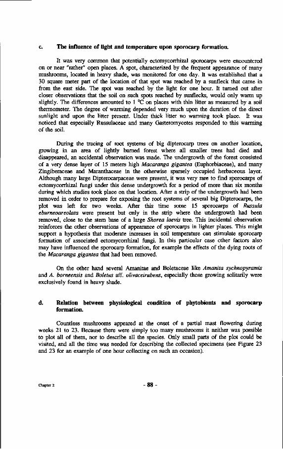
c. The influence of light and temperature upon sporocarp formation.
It was very common that potentially ectomycorrhizal sporocarps were encountered on or near "rather" open places. A spot, characterized by the frequent appearance of many mushrooms, located in heavy shade, was monitored for one day. It was established that a 30 square meter part of the location of that spot was reached by a sunfleck that came in from the east side. The spot was reached by the light for one hour. It turned out after closer observations that the soil on such spots reached by sunflecks, would only warm up slightly. The differences amounted to 1 °C on places with thin litter as measured by a soil thermometer. The degree of warming depended very much upon the duration of the direct sunlight and upon the litter present. Under thick litter no warming took place. It was noticed that especially Russulaceae and many Gasteromycetes responded to this warming of the soil.
During the tracing of root systems of big dipterocarp trees on another location, growing in an area of lightly burned forest where all smaller trees had died and disappeared, an accidental observation was made. The undergrowth of the forest consisted of a very dense layer of 15 meters high Macaranga gigantea (Euphorbiaceae), and many Zingiberaceae and Maranthaceae in the otherwise sparsely occupied herbaceous layer. Although many large Dipterocarpaceae were present, it was very rare to find sporocarps of ectomycorrhizal fungi under this dense undergrowth for a period of more than six months during which studies took place on that location. After a strip of the undergrowth had been removed in order to prepare for exposing the root systems of several big Dipterocarps, the plot was left for two weeks. After this time some 15 sporocarps of Russula eburneoareolata were present but only in the strip where the undergrowth had been removed, close to the stem base of a large Shorea laevis tree. This incidental observation reinforces the other observations of appearance of sporocarps in lighter places. This might support a hypothesis that moderate increases in soil temperature can stimulate sporocarp formation of associated ectomycorrhizal fungi. In this particular case other factors also may have influenced the sporocarp formation, for example the effects of the dying roots of the Macaranga gigantea that had been removed.
On the other hand several Amanitae and Boletaceae like Amanita sychnopyramis and A. borneensis and Boletus aff. olivaceirubens, especially those growing solitarily were exclusively found in heavy shade.
d. Relation between physiological condition of phytobionts and sporocarp formation.
Countless mushrooms appeared at the onset of a partial mast flowering during weeks 21 to 23. Because there were simply too many mushrooms it neither was possible to plot all of them, nor to describe all the species. Only small parts of the plot could be visited, and all the time was needed for describing the collected specimens (see Figure 23 and 23 for an example of one hour collecting on such an occasion).
Chapter 2 - 8 8 •
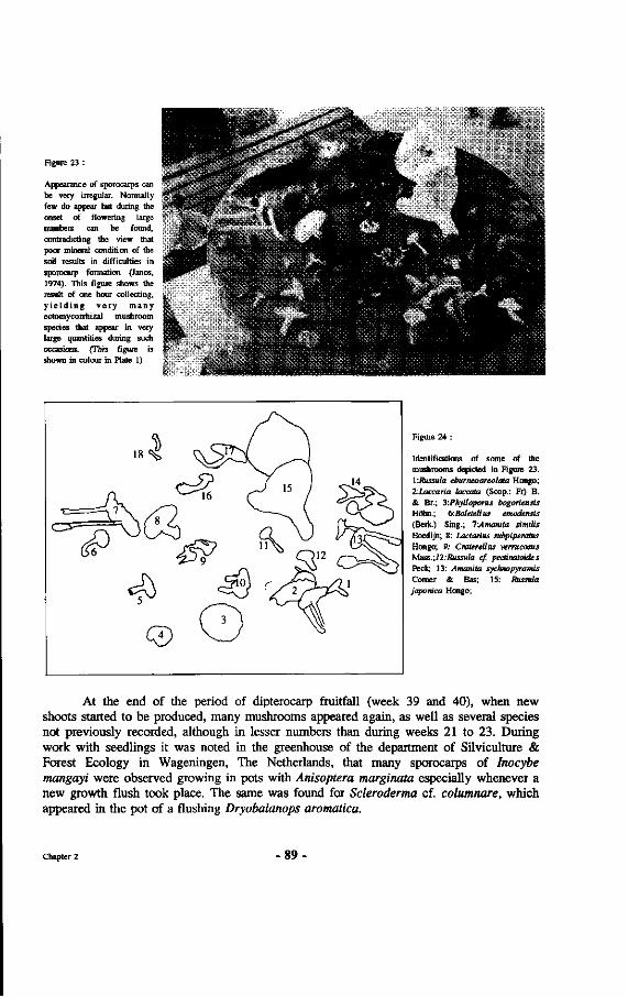
Figure 23 :
Appearance of sporocarps can be very irregular. Normally few do appear but during the onset of flowering large numbers can be found, contradicting the view that poor mineral condition of the soil results in difficulties in sporocarp formation (Janos, 1974). This figure shows the result of one hour collecting, y i e l d i n g v e r y many ectomycorrhizal mushroom species that appear in very large quantities during such occasions. (This figure is shown in colour in Plate 1)
Figure 24 :
Identifications of some of the mushrooms depicted in Figure 23. V.RussuIa eburneoareotata Hongo; 2:Laccaria toccata (Scop.: Fr) B. & Br.; y.Phylloporus bogoriensis Höhn.; 6:Boleteltus emodensis (Berk.) Sing.; 7'Amanita simiüs Boedijn; 8: Lactarius subpiperatus Hongo; 9: CratereUus verrucosus Mass.;12:Russuta cf. pectinatoides Peck; 13: Amanita sycfaiopyramis Comer & Bas; 15: Russula japonica Hongo;
At the end of the period of dipterocarp fruitfall (week 39 and 40), when new shoots started to be produced, many mushrooms appeared again, as well as several species not previously recorded, although in lesser numbers than during weeks 21 to 23. During work with seedlings it was noted in the greenhouse of the department of Silviculture & Forest Ecology in Wageningen, The Netherlands, that many sporocarps of lnocybe mangayi were observed growing in pots with Anisoptera marginata especially whenever a new growth flush took place. The same was found for Scleroderma cf. columnare, which appeared in the pot of a flushing Dryobalanops aromatica.
Chapter 2 89
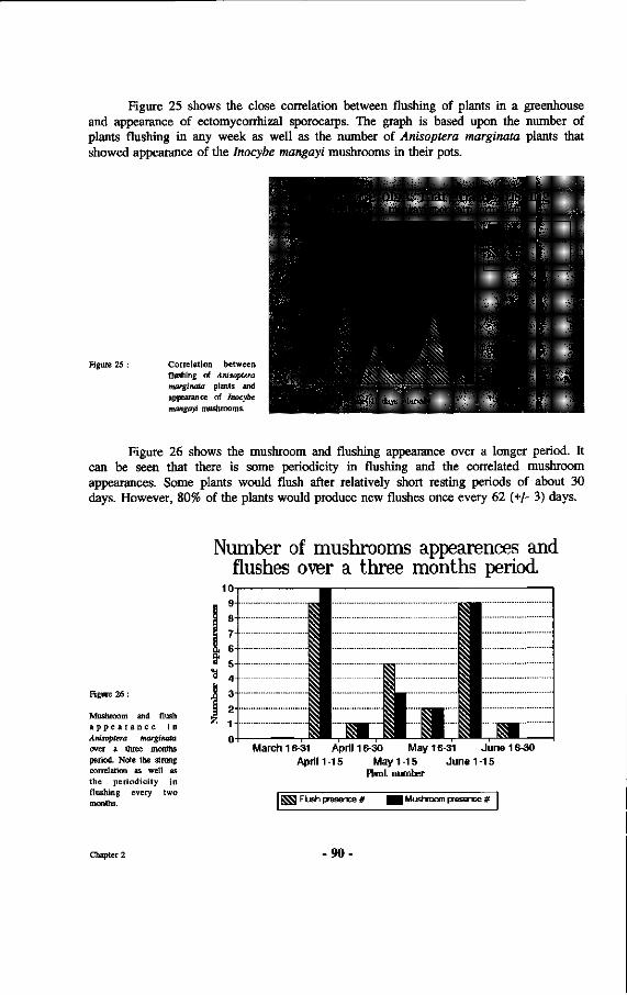
Figure 25 shows the close correlation between flushing of plants in a greenhouse and appearance of ectomycorrhizal sporocarps. The graph is based upon the number of plants flushing in any week as well as the number of Anisoptera marginata plants that showed appearance of the Inocybe mangayi mushrooms in their pots.
Figure 25 : Correlation between flushing of Anisoptera marginata plants and appearance of Inocybe mangayi mushrooms.
j^ïnartinata flushing *-•'—^BpjawMif rnmmlirn
Sporoearp Tcmidfli)!i I
Flush appmirüiïco - i •
Time il'i ±**K hibinahT
[•:-*••-. • • # # ' •?' [Hj...; ' ^ ï - i^V ^
',,'j~'^»"S-*?:'-US-'.'1 '§&•£•"'• "
Figure 26 shows the mushroom and flushing appearance over a longer period. It can be seen that there is some periodicity in flushing and the correlated mushroom appearances. Some plants would flush after relatively short resting periods of about 30 days. However, 80% of the plants would produce new flushes once every 62 (+/- 3) days.
Figure 26 :
Mushroom and flush a p p e a r a n c e i n Anisoptera marginata over a three months period. Note the strong correlation as well as the periodicity in flushing every two months.
Number of mushrooms appearences and flushes over a three months period.
1 March 16-31 April 16-30 May 16-31 June 16-30
April 1 -15 May 1 -15 June 1 -15 Plant number
SjjS Flush presence # | Mushroom presence #
Chapter 2 -90
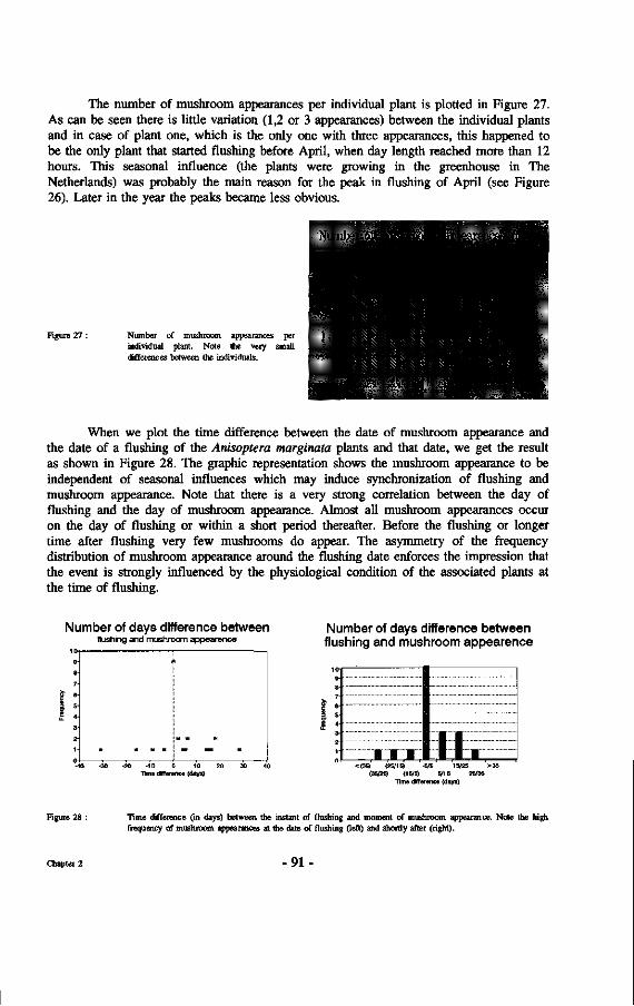
The number of mushroom appearances per individual plant is plotted in Figure 27. As can be seen there is little variation (1,2 or 3 appearances) between the individual plants and in case of plant one, which is the only one with three appearances, this happened to be the only plant that started flushing before April, when day length reached more than 12 hours. This seasonal influence (the plants were growing in the greenhouse in The Netherlands) was probably the main reason for the peak in flushing of April (see Figure 26). Later in the year the peaks became less obvious.
Number of rnùSËSofflUsiàp̂ over a 200 dajsij
Figure 27 : Number of mushroom appearances per individual plant. Note the very small differences between the individuals.
When we plot the time difference between the date of mushroom appearance and the date of a flushing of the Anisoptera marginata plants and that date, we get the result as shown in Figure 28. The graphic representation shows the mushroom appearance to be independent of seasonal influences which may induce synchronization of flushing and mushroom appearance. Note that there is a very strong correlation between the day of flushing and the day of mushroom appearance. Almost all mushroom appearances occur on the day of flushing or within a short period thereafter. Before the flushing or longer time after flushing very few mushrooms do appear. The asymmetry of the frequency distribution of mushroom appearance around the flushing date enforces the impression that the event is strongly influenced by the physiological condition of the associated plants at the time of flushing.
Number of days difference between flushing and mushroom appearance
This dHTerence (days)
Number of days difference between flushing and mushroom appearence
(25/15) -5/5 15/25 >35 (35/25) (15/5) 5/15 25/35
Time différence (days)
Figure 28 : Time difference (in days) between the instant of flushing and moment of mushroom appearance. Note the high frequency of mushroom appearances at the date of flushing (teft) and shortly after (right).
Chapter 2 -91
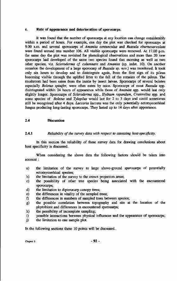
e. Rate of appearance and deterioration of sporocarps.
It was found that the number of sporocarps at any location can change considerably within a period of hours. For example, one day the plot was checked for sporocarps at 9.00 a.m. and several sporocarps of Amanita centunculus and Russula eburneoareolata were found around tree number 106. All visible sporocarps were removed. At 17.00 p.m. the same day the plot was revisited for phenological observations and more than 20 new sporocarps had developed of the same two species found that morning as well as two other species, viz. Scleroderma cf. columnare and Amanita (sp. indet. 10). On another occasion the development of a large sporocarp of Russula sp. nov.) was monitored. It took only six hours to develop and to disintegrate again, from the first sign of its pileus becoming visible through the uplifted litter to the fall of the remains of the pileus. The mushroom had been eaten from the inside by insect larvae. Sporocarps of several boletes especially Boletus spinifer, were often eaten by mice. Sporocarps of most Russula spp. disintegrated within 24 hours of appearance while those of Amanita spp. would last only slightly longer. Sporocarps of Scleroderma spp., Hydnum repandum, Craterellus spp. and some species of Boletus and Tylopilus would last for 2 to 3 days and could sometimes still be recognized after 4 days. Laccaria laccata was the only potentially ectomycorrhizal fungus producing long-lasting sporocarps. They lasted up to 14 days after appearance.
2.4 Discussion
2.4.1 Reliability of the survey data with respect to assessing host-specificity.
In this section the reliability of these survey data for drawing conclusions about host specificity is discussed.
When considering the above data the following factors should be taken into account :
a) the limitation of the survey to large above-ground sporocarps of potentially ectomycorrhizal species;
b) the limitation of the survey to the crown projection areas; c) the possibility of other tree species being associated with the encountered
sporocarps; d) the limitation to dipterocarp canopy trees; e) the differences in vitality of the sampled trees; f) the differences in numbers of sampled trees between species; g) the possible correlation between topography and site at the location of the
phytobiont and differences in encountered sporocarps; h) the possibility of incomplete sampling; i) possible interactions between physical influences and the appearance of sporocarps; j) the limitation to one sample plot.
In the following sections these 10 points will be discussed.
Chapter 2 - 9 2 -
a. Omission of small and hypogeous sporocarps
If published lists with names of ectomycorrhizal mushroom species are considered (e.g. Trappe, 1962; 1977) it can be noted that leaving out very small mushrooms and subterranean species does not severely affect the results. Almost all sporocarps of ectomycorrhizal fungi are so large that they are easily discerned with the unaided eye. Athough many attempts were made to find subterranean sporocarps, the author has only found specimens of Rhizopogon on three occasions and three specimens of Tuber. A trained dog also did not locate more specimens. Subterranean sporocarps of ectomycorrhizal fungi are known from Java (Trappe, pers. comm.). Their abundance seems to be far less in the primary mixed dipterocarp forest of East-Kalimantan than in temperate forests. In view of the forementioned the omission of very small mushrooms and hypogeous fungi it is assumed it did not have a large influence upon the results presented here. The number of potential mycobionts will thus not increase much and the analyses of the number of mycobiont species occurring with the phytobiont species is not expected to change much if all hypogeous fungi were located. The degree of actual specificity probably should not be much different from the findings presented in this publication.
The limitation to the crown projection areas
Examinations of the root systems of some nine big dipterocarp trees showed that the diameter of the root systems generally extended not more than a few meters beyond the crown projection of those trees (see 2.2.1). Figure 29 shows two examples, respectively of Shorea laevis and Hopea rudiformis Ashton. In the downhill direction the roots were washed out of the soil with the help of a very strong jet of water as described in 3.2.4. Many uncertainties remain concerning the assumpt ion that crown projection is congruent with root system extent. For example, it was not possible to investigate the form and extent of the root systems of big trees growing on steep slopes. Only the superficial roots (< 20 cm deep) were recorded. Crowns are very often asymmetrical while a circular spread of the root system was assumed. An example of the crown projections of a group (clump) of Shorea laevis trees along the Wartono trail is presented in Appendix 6. Overhanging trees will probably have a Figure 29 : Root system extent of Shorea laevis and Hopea nervosa trees.
The dotted lines mark the projection of the crown periphery.
Chapter 2 93
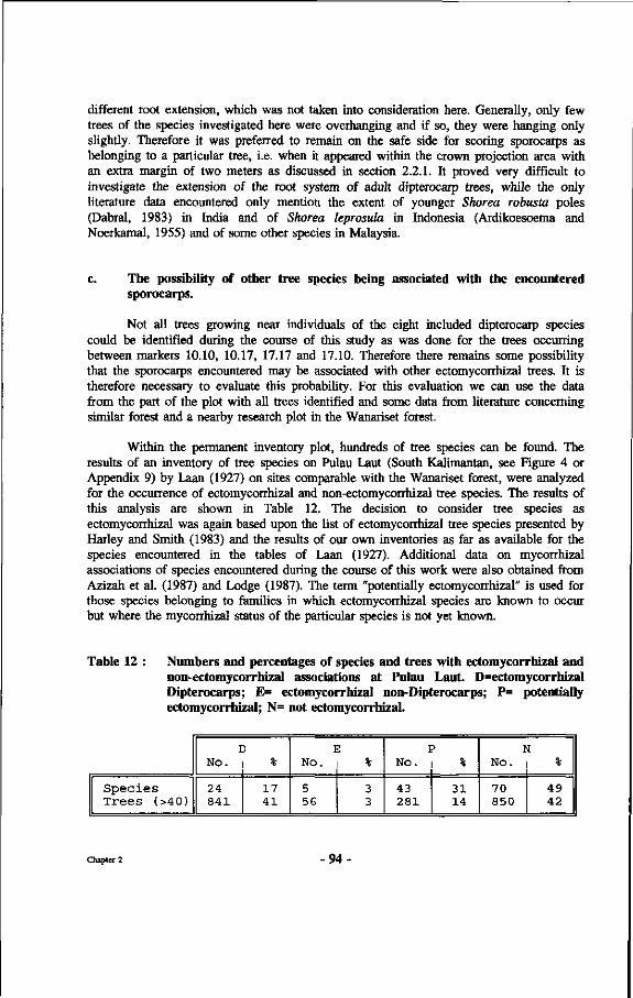
different root extension, which was not taken into consideration here. Generally, only few trees of the species investigated here were overhanging and if so, they were hanging only slightly. Therefore it was preferred to remain on the safe side for scoring sporocarps as belonging to a particular tree, i.e. when it appeared within the crown projection area with an extra margin of two meters as discussed in section 2.2.1. It proved very difficult to investigate the extension of the root system of adult dipterocarp trees, while the only literature data encountered only mention the extent of younger Shorea robusta poles (Dabral, 1983) in India and of Shorea leprosula in Indonesia (Ardikoesoema and Noerkamal, 1955) and of some other species in Malaysia.
c. The possibility of other tree species being associated with the encountered sporocarps.
Not all trees growing near individuals of the eight included dipterocarp species could be identified during the course of this study as was done for the trees occurring between markers 10.10, 10.17, 17.17 and 17.10. Therefore there remains some possibility that the sporocarps encountered may be associated with other ectomycorrhizal trees. It is therefore necessary to evaluate this probability. For this evaluation we can use the data from the part of the plot with all trees identified and some data from literature concerning similar forest and a nearby research plot in the Wanariset forest.
Within the permanent inventory plot, hundreds of tree species can be found. The results of an inventory of tree species on Pulau Laut (South Kalimantan, see Figure 4 or Appendix 9) by Laan (1927) on sites comparable with the Wanariset forest, were analyzed for the occurrence of ectomycorrhizal and non-ectomycorrhizal tree species. The results of this analysis are shown in Table 12. The decision to consider tree species as ectomycorrhizal was again based upon the list of ectomycorrhizal tree species presented by Harley and Smith (1983) and the results of our own inventories as far as available for the species encountered in the tables of Laan (1927). Additional data on mycorrhizal associations of species encountered during the course of this work were also obtained from Azizah et al. (1987) and Lodge (1987). The term "potentially ectomycorrhizal" is used for those species belonging to families in which ectomycorrhizal species are known to occur but where the mycorrhizal status of the particular species is not yet known.
Table 12 : Numbers and percentages of species and trees with ectomycorrhizal and non-ectomycorrhizal associations at Pulau Laut. D=ectomycorrhizal Dipterocarps; E= ectomycorrhizal non-Dipterocarps; P= potentially ectomycorrhizal; N= not ectomycorrhizal.
Species Trees (>40)
I No.
24 841
5 %
17 41
I No.
5 56
: %
3 3
I No.
43 281
3
31 14
Î No.
70 850
49 42
Chapter 2 -94
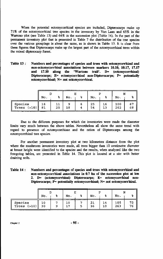
When the potential ectomycorrhizal species are included, Dipterocarps make up 71% of the ectomycorrhizal tree species in the inventory by Van Laan and 65% in the Wartono plot (see Table 13) and 64% in the succession plot (Table 14). In the part of the permanent inventory plot that is presented in Table 7 the distribution of the tree species over the various groupings is about the same, as is shown in Table 13. It is clear from these figures that Dipterocarps make up the largest part of the ectomycorrhizal trees within the mixed dipterocarp forest.
Table 13 : Numbers and percentages of species and trees with ectomycorrhizal and non-ectomycorrhizal associations between markers 10.10, 10.17, 17.17 and 17.10 along the "Wartono trail". D= (ectomycorrhizal) Dipterocarps; E= ectomycorrhizal non-Dipterocarps; P= potentially ectomycorrhizal; N= not ectomycorrhizal.
Species Trees (>10)
I No.
16 81
%
11 20
I No.
9 18
6 4
P No. %
25 54
IG 13
N No. | %
100 262
67 68
Due to the different purposes for which the inventories were made the diameter limits vary much between the above tables. Nevertheless all show the same trend with regard to presence of ectomycorrhizae and the ration of Dipterocarps among the ectomycorrhizal tree species.
For another permanent inventory plot at two kilometres distance from the plot where the mushroom inventories were made, all trees bigger than 10 centimetre diameter at breast height were identified to the species and the results, when analyzed like the two foregoing tables, are presented in Table 14. This plot is located at a site with better draining soils.
Table 14 : Numbers and percentages of species and trees with ectomycorrhizal and non-ectomycorrhizal associations in 0.7 ha of the succession plot at km 2. D= (ectomycorrhizal) Dipterocarps; E= ectomycorrhizal non-Dipterocarps; P= potentially ectomycorrhizal; N= not ectomycorrhizal.
No. D l No. No. No.
Species Trees (>10)
10 30
10 17
21 36
14 10
105 263
72 76
Chapter 2 9 5 -
During the survey it was not possible to identify all trees growing within the total permanent inventory plot. The more intensively investigated parts of the plot are almost complete as to identification as mentioned above. Since the identification of some herbarium material of other parts of the plot had not yet been completed, these preliminary analyses were limited to the best known and easily recognizable dipterocarp species encountered. All Dipterocarps were identified with certainty within the plot as well as the species not included in the analyses. The type of data base established allows for addition of data later and rerunning the analyses. With the continuation of the research the results of the analyses become increasingly reliable. Nevertheless from the tables above it can be seen that there is a substantial number of ectomycorrhizal non-dipterocarp trees in the plots. From Figure 16 in Chapter 2.3.1 it can be seen that these trees also tend to reach large sizes. There is therefore a possibility that several of the combinations listed in Table 8 may not occur. The degree of specificity, based upon presence of ectomycorrhizal sporocarps, may therefore be larger than indicated by the results in Table 8. The typical clumping of many Dipterocarps (see Appendix 6) limits this possibility somewhat depending upon the radius of space occupied by the crowns of the Dipterocarps in the clumps.
d. The limitation to dipterocarp canopy trees
Only the largest trees with their crowns exposed to full light in the upper forest canopy, e.g. illumination classes 1 and 2, see 2.2.1, were considered in these analyses. If smaller trees might be capable of supporting ectomycorrhizal sporocarp formation this would probably mean that several combinations mentioned in Table 8 may not occur. However, this limitation is thought to be justifiable because of the following three indications:
1) during the 60 weeks survey not a single sporocarp of a potential ectomycorrhizal fungus was encountered within the plots that were separated from the surrounding forest by ditches (see 2.2.3). Within these plots only small sized Dipterocarps were present, all of them with their crowns in a suppressed position. Around these plots many sporocarps of ectomycorrhizal fungi developed, as shown in Figure 30 for ditched plot 2.
2) Only one positive record was obtained of an Amanita borneensis sporocarp associated with a planted Shorea laevis sapling, with a diameter of 5 cm, growing in secondary forest vegetation under approximately 50% light intensity. In the many other planting trials with Dipterocarps, among others in several plots within the Wanariset forest, within the ITCI concession and on Pulau Laut, no ectomycorrhizal sporocarps were found by the author, or the other researchers at the Wanariset research station, to be associated with smaller diameter dipterocarp trees, the crowns of which were not yet fully exposed.
3) Not a single incision made between small dipterocarp trees and immature ectomycorrhizal sporocarps (see 2.2.3) succeeded in halting of sporocarp development. It is therefore safe to consider only the largest overstory dipterocarp trees, for listing possible fungus-dipterocarp combinations.
Chapter 2 - 9 6 •
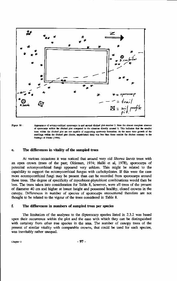
** -K
« * *
*
* •
*
S.|«i)o < V
** *
» - •
•*
#
-*U» —•»
* *
* *•
^ -
#•
— ._
*
*
•»«•••f / * "
* *
- ~" = Tlra.il
g[ - 50-'/ /"^f'/e
Figure 30 : Appearance of ectomycorrhizat sporocarps in and around ditched plot number 2. Note the almost complete absence of sporocarps within the ditched plot compared to the situation directly around it. This indicates that the smaller trees within the ditched plot are not capable of supporting sporocarp formation. At the same time growth of the seedlings within the ditched plot (Smits, unpublished data) was less than those outside the ditches contrary to the findings of Fricke (1904).
e. The differences in vitality of the sampled trees
At various occasions it was noticed that around very old Shorea laevis trees with an open crown (trees of the past; Oldeman, 1974; Halle et al, 1978), sporocarps of potential ectomycorrhizal fungi appeared very seldom. This might be related to the capability to support the ectomycorrhizal fungus with carbohydrates. If this were the case more ectomycorrhizal fungi may be present than can be recorded from sporocarps around these trees. The degree of specificity of mycobiont-phytobiont combinations would then be less. The trees taken into consideration for Table 8, however, were all trees of the present of diameter 40 cm and higher at breast height and possessed healthy, closed crowns in the canopy. Differences in number of species of sporocarps encountered therefore are not thought to be related to the vigour of the trees considered in Table 8.
f. The differences in numbers of sampled trees per species
The limitation of the analyses to the dipterocarp species listed in 2.3.2 was based upon their occurrence within the plot and the ease with which they can be distinguished with certainty from other tree species in the area. The number of canopy trees of the present of similar vitality with comparable crowns, that could be used for each species, was inevitably rather unequal.
Chapter 2 97
One of the first things to be noted in Table 8 is the large number of symbionts encountered with Shorea laevis, as compared to that found with the other dipterocarp species. The number of S. laevis trees within the plot is the largest by far among the species examined. However, this is of little influence upon the number of associated ectomycorrhizal fungal species. For instance even the single Shorea laevis tree (number 224, Table 7), at the "good mushroom spot", between the (14.13), (13.13), (14.12) and (13.12) markers, possessed 20 different suspected ectomycorrhizal fungi. Shorea laevis is the slowest growing species among the Dipterocarps included in the study based upon the performance of this species in growth and yield studies in the nearby ITCI concession over a period of 18 years (van Eijk-Bos, in preparation). The average age may have been around 500 years. It is unclear how this speed of growth may be related to the high number of mycobionts. In Chapter 8 this will be discussed further. The amount of mycobionts near tree number 226 is almost the same number as the total of all other possible ectomycorrhizal fungal species of the other seven examined dipterocarp species together (23). The appearance of sporocarps for most species is very well synchronized. Whenever one particular ectomycorrhizal mushroom species appeared, it was generally found near almost all large individuals of the concerned dipterocarp species growing on similar sites. It is therefore improbable that the results presented in Table 8 would have changed much if more trees of each tree species had been included in the survey.
g. The possible correlation between topography, site of the sampled trees and differences in encountered sporocarps
Shorea laevis is a tree typically found on forested ridges, which are, as mentioned, the richest mushroom spots. These are also the best draining spots. The combination of many Shorea laevis and Eusideroxylon zwageri trees and their dominance in terms of basal area is very clear in the plot (see Table 5). This association is typically indicative of better draining sites. Shorea lamellata, normally growing on slopes, was growing in comparable sites within this plot, but having much fewer possibly associated sporocarps. As a matter of fact, out of the 8 species treated here only Dipterocarpus confertus was always located on the slopes, or at the foot of the slopes within the investigated plot. Therefore, the influence of topography on the appearance of ectomycorrhizal sporocarps linked to any of the (other) species discussed here can be excluded in the present context.
h. The possibility of incomplete sampling
The list of ectomycorrhizal sporocarps presented is likely to be incomplete. This can be expected because of: 1) terrain conditions, 2) high rate of deterioration of sporocarps, 3) seasonal and climatological influences, 4) physiological condition of the phytobionts.
1) Sometimes it is difficult to discern sporocarps on lighter spots where abundant ground vegetation occurs. Here the more dense and higher herb vegetation makes it much harder to see the sporocarps between the leaves. The visibility under old growth primary forest, where undergrowth plants do not possess many leaves and where very few herbs can grow or survive, is quite good.
Chapter 2 - 98 -
2) Many sporocarps must have been missed because they appeared and disappeared within the three days period that separated consecutive visits to the plot (cf. 2.3.3.d). In the Netherlands it is sufficient to visit a plot once every two weeks during the autumn (Termorshuizen, 1990). The sporocarps encountered are picked and their pilei are left on the ground so as to prevent a second registration during the next visit of the plot. The picked pilei can still be found and recognized after two weeks. It even happens that in spring the picked pilei are still intact because of the low winter temperatures. As has been mentioned in 2.3.3.e, the situation is very different in East-Kalimantan. Deterioration of most sporocarps is much faster.
3) In the temperate zones mycorrhizal mushrooms appear mainly in autumn and to a lesser extent in spring. They are most abundant after wet periods. Mushroom inventories are made over many consecutive years. It is thought that after three consecutive years or mushroom seasons, probably 70-80% of all macrofungi present have been found. There are known cases of mushrooms of the same fungus, which did only appear again after more than 10 years. An example is the appearance of Inocybe godeyi near Betula pubescens in a plot near Oostvoorne (the Netherlands) that was regularly checked. The species was recorded only three times over a period of 30 years (Kuiper, 1986). Arnolds (1985) provides more such examples.
Very little is known about the times of appearance of mushrooms in tropical rain forests. The climate near the Wanariset forest is classified as a tropical rain forest climate (cf. Introduction 1.4). However, the weather is very unpredictable, and pronounced dry periods do occur. From the data collected so far it is difficult to find correlations between the appearance of sporocarps and climatological events. Sometimes large quantities of sporocarps are found right in the middle of some very dry weeks, e.g. Lactarius subpiperatus in week 9 of the observation period. Most of the time, mushrooms appear after two to three very rainy days. Very often, however, wet periods occur without a single mushroom appearing. Sometimes the wet months turn out to be the driest (for instance see figure 31, Box 5. The months February and October normally the wettest months and the months in which many mushrooms appear were the driest months in the year 1990) and if flowering does occur most of the mushroom species occur, albeit in smaller numbers.
The observation of mushroom appearance in the research plot after two or three rainy days is based on visual observations in the forest because the weather in the forest does not correspond closely to that at the station when considered over short periods. For the occasion presented in Figure 22 data were used of the rainfall measuring equipment positioned in a large open spot a mere 200 meters from the edge of the Wartono Kadri plot.
Chapter 2 - 9 9 •
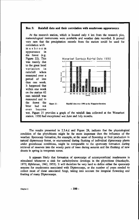
Box 5: Rainfall data and their correlation with mushroom appearances
At the research station, which is located only 4 km from the research plot, meteorological instruments were available and weather data recorded. It proved very rare that the precipitation records from the station could be used for correlation with m u s h r o o m appearance in the forest (e.g. Figure 22). This Wanariset Samboja Rainfall Data 1990
was mainly due to the great local 500
va r i a t i o n in rainfall when ~400
measured over a -500
period of less | than one week. 20° It happened that within one week on the station 62 »J mm rainfall was measured and in the forest the F igure31:
litter had not even become wet. Figure 31 provides a graph station. 1990 had exceptional wet
1 1 1
• i j i
•n ! 1 I I I 1
jan feb mar apr may jun [ul aug Sep oct nov dec Months
Rainfall data over 1990 at the Wanariset location.
of the rainfall data collected at the Wanariset June and July months.
The results presented in 2.3.4.d and Figure 28, indicate that the physiological condition of the phytobionts might be far more important than the influence of the weather. Sporocarp formation, for example, at the onset of flowering or fruit production in natural dipterocarp forest, or encountered during flushing of individual dipterocarp plants under greenhouse conditions, might be comparable to the sporocarp formation during retrieval of reserves into the woody parts of trees during autumn and the flushing of new shoots in spring in temperate zones.
It appears likely that formation of sporocarps of ectomycorrhizal mushrooms is stimulated whenever a sink for carbohydrates develops in the phytobiont (Hacskaylo, 1973; Björkman, 1942, 1953). It will therefore be very hard to define either the sporocarp seasons for mushrooms associated with Dipterocarps, or the number of years needed to collect most of these associated fungi, taking into account the irregular flowering and fruiting of many Dipterocarps.
Chapter 2 100
It is therefore probable that mushrooms of more species will be found to be associated with one or some of the 8 dipterocarp species from Table 8 during later surveys. The degree of phytobiont specificity might then become less. Long term monitoring is therefore very important for more critical evaluation of the degree of specificity of dipterocarp mycorrhizae.
i. Possible interactions between physical influences and appearance of sporocarps
A factor that was observed to be of particular importance was the soil temperature. As mentioned in 2.3.4.C, sporocarps generally would develop in somewhat open spots, that were reached by direct sunlight. Since the tops of the ridges in a lowland tropical rain forest are always the lightest spots it would tally with the above observations, that the high number of potential ectomycorrhizal sporocarps encountered here is related to the greater influence of sunflecks, direct solar radiation, and slightly warming patches of forest soil.
Agerer (1985) also suspects a relation between the temperature and the production of sporocarps in some temperate forests in Germany. The tops of the ridges are, however, also the dryer places and might have better aeration of the soil. This might be related also to ectomycorrhizal sporocarp production. All trees considered in this analysis were compared for the light intensities around their bases but no significant differences were noticed. In general, members of the genus Dipterocarpus have flat, rather open crowns, that let through more light, but this is then compensated by a denser undergrowth.
Therefore differences in light intensity, number and duration of sunflecks cannot account for the large variations in number of sporocarps found around the bases of the trees in the survey and especially the difference between the number of sporocarps occurring around Shorea laevis trees compared to the other species investigated.
j . The limitation to one sample plot
During field trips in natural forest within the ITCI concession, (some 60 km from the research plot), no other ectomycorrhizal sporocarps were found near the same dipterocarp species than those present in the Wanariset I forest, at least when they were growing on comparable sites.
Iskandar (Mulawarman University Samarinda), collected mushrooms in slightly disturbed dipterocarp forest at some 25 km from the research plot at irregular intervals in the same period. It appeared that the mushrooms he found on other locations are the same as in the plot at Wanariset for those places where soil and topographical conditions as well as species composition were comparable (undulating hills from 0 to 200 m above sea level; nutrient-poor ultisols; forest dominated by Shorea laevis).
Zulkifli (1992, pers. comm., Forest Research Institute Pematang Siantar) collected ectomycorrhizal mushroom species in a Shorea platyclados plantation at more than 1.200 m altitude on well draining soil in North Sumatra.
Chapter 2 - 101 -
Some three species he found (Russuia eburneoareolata, Russula sp. indet. 2, and Lactarius subpiperatus) also occurred in East-Kalimantan. Scleroderma dictyosporum in this study encountered in natural mixed dipterocarp forest, was found to occur in Eucalyptus urophylla plantations in North Sumatra by Zulkifli (1992, pers. comm., Forest Research Institute Pematang Siantar).
From discussions with mycorrhizae researchers from South East Asia during the BIO-REFOR workshop (Suhardi, 1993) it was learned that several mushroom species encountered in this study also occurred in the Philippines and in Thailand.
Too few observations of sporocarps potentially associated with Dipterocarps were made in other places with different terrain conditions to draw firmer conclusions. Ashton (1982) gives many data on distribution of dipterocarp species related to altitude, soil type and topography. He also noted a correlation between the phosphorus content of the soil and the occurrence of dipterocarp species. Few species are found on more than one soil type, although it is not yet possible to tell when larger areas, for instance South East Asia, are considered. It is therefore hard to make comparisons between the occurrence of different potential ectomycorrhizal sporocarps near one dipterocarp species on more than one soil type. As a matter of fact, these data rather suggest that there are well-defined combinations of soil types, dipterocarp species and symbiotic fungus species, and that such comparisons cannot be made at all save for rare exceptions on heterogeneous sites.
On one occasion, however, the author noticed that around a Shorea leprosula tree growing in an Agathis borneensis stand (Km 43, ITCI concession) on a sandy podzol with a peat like humus layer (Heath forest) close to sea level, fruit bodies of a Phylloporus sp. indet. and a Boletellus longicollus Ces. occurred. None of these was ever found in the permanent inventory plot. Neither were any of the potentially ectomycorrhizal sporocarps encountered in the permanent inventory plot present in the above mentioned Agathis stand. Therefore differences may exist related to soil types. This will be discussed further in Chapter 8.
Considering these 10 points (a-j), it follows that the differences in numbers of potentially associated ectomycorrhizal fungi between Shorea laevis and the other dipterocarp species cannot be clearly related to environmental conditions and therefore, for this moment, must be linked to the species involved.
As to the total number of ectomycorrhizal mushroom species listed in Table 8, there remains a possibility that some of the mentioned potential mycobionts of the eight dipterocarp species studied, belong to other ectomycorrhizal trees, that have not been identified as yet. In this case the degree of specificity would increase.
Another remarkable factor seems to be the substrate preferences of the ectomycorrhizal fungi associated with Dipterocarps as well as the preferences with differing site conditions. This indicates that there may be some adaptation to very small ecological niches. If one could define these ecological niches with more certainty it may very well be that the appearance of the sporocarps may show even clearer patterns. When the total system of phytobiont-mycobiont-environment would be considered, it can be assumed that a higher degree of ecological specificity may be found.
Chapter 2 - 102 -
Nevertheless, taking into account the incomplete sampling, the irregular and so far unpredictable appearance of sporocarps of ectomycorrhizal fungi associated with Dipterocarps, the still increasing number of species of mycobionts encountered, in combination with the short observation period and the limitation to only one plot on one location for the study of potential mycobionts, it can be safely claimed that many more potential mycobionts will be found to be associated with the eight dipterocarp species investigated here. Effects, possibly leading to a higher degree of specificity, need to be investigated further before more solid conclusions can be reached.
2.4.2 Specificity.
The concept of specificity is sometimes defined as the unique association of one species of phytobiont with one species of mycobiont (Alexander, 1987). This type of specificity is known from many plant parasite relationships, for instance Hemileia vastatrix on Coffea. For these plant parasite relationships the specificity may even be at the level of races of the parasite. In this precise sense, based upon the sporocarp collections and the assumptions discussed in this chapter, the association between dipterocarp trees and ectomycorrhizal fungi cannot be considered to be specific, and certainly not when the whole family is considered. As Harley and Smith (1983) conclude, not even the widely quoted Larix spp. Suillus grevillii (Boletus elegans) relationship fulfils this strict definition.
To avoid misunderstandings it is necessary to distinguish between various levels of specificity. There are differences in phytobiont specificity and mycobiont specificity, e.g. one host having several mycobionts or one mycobiont being capable of forming ectomycorrhizae with a variety of phytobionts. Molina et al. (1992a) describe the various degrees of specificity in more detail. This will also be treated in Chapter 8. Molina also discusses "linkage potential", meaning that less specificity of phytobionts results in higher linkage potential and therefore better long term prospects of survival. As mentioned these aspects will be discussed extensively in Chapter 8.
Another important point of importance is the condition under which specificity is investigated. Some phytobiont-mycobiont combinations may prove possible in axenic culture but may be absent in the natural ecosystem (Kelley, 1950; p. 136). It is clear that, for the dipterocarp situation, where their occurrence almost is completely limited to natural forests, only the ecological specificity as occurring under the natural situation of competition is biologically relevant. Since this study aims at the use of such insights for application in practical forestry, in this book the term specificity will apply to ecological specificity, meaning "the degree to which phytobiont-mycobiont combinations occur in undisturbed natural ecosystems".
Harley and Smith (1983, p. 378) mention two important reasons why it would be improbable for mycorrhizal symbioses to be specific. The first one is that an unspecific fungus will not be restricted in its distribution, and that it would be unfavourable for the tree to develop mutations that would result in resistance to infection. The second reason they mention for the rareness of specificity in mycorrhizal associations, is "that many root-inhabiting fungi, including a large number of mycorrhizal fungi, depend upon the spread of perennial mycelium and persistence as resting propagules much more than upon
Chapter 2 - 103 -
widespread dissemination of spores. Chances of survival are clearly improved by the existence of a wide range of susceptible species of host plants, so there appear to be selection pressures on the fungi against the evolution of close host/fungus specificity".
Both reasonings are somewhat related. When survival of the species is the goal, indeed these reasons are plausible. Under the stable conditions in non-seasonal tropical rain forest, especially the forests on the Sunda shelf, the need for changing to other phytobionts and for survival apart from the phytobiont may not be that important. Under the closed canopy the conditions in the top soil are very constant indeed (see also Chapter 6) and living roots of the phytobiont are present at any time. Especially for the Dipterocarps the typical clumping, seedlings mainly occurring within the root radius of the mother tree, makes it less necessary to survive on roots of other phytobionts, a possibility for migrating within the clump always being available. Janzen (1974) mentions as reason for clumping the avoidance of seed prédation through mass flowering and fruiting in combination with heavy seeds falling near the tree and not the limitation to this area because of availability of mycorrhizal inoculum.
Differences in growth performance between various phytobiont-mycobiont-site combinations are known to occur (see Chapters 4, 5 and 7). Selection towards more specific mycobiont-phytobiont relationships under stable conditions may therefore be possible.
Much more work is needed on comparisons between regions before we can define certain mycobiont-phytobiont-site associations. Above we already mentioned the occurrence of some of the encountered mushroom species in Sumatra, associated with a Dipterocarp and a eucalypt species. As mentioned, there seem to be many mushroom species associated with Dipterocarps in East-Kalimantan that occur widespread in the region (Indonesia, Malaysia, Thailand, Philippines).
For instance in Thailand the following species from East-Kalimantan were found to occur in deciduous dipterocarp forest (Anivat Chalermpongse, pers. comm.): Amanita borneensis, Strobilomyces polypyramis, Russuia eburneoareolata, Russula nigricans (Thailand, Philippines, Sumatra, Kalimantan), Laccaria laccata, Russula cf. metachroa, Lactarius cf. austrovolemus. From the Philippines Laccaria laccata, Russula eburneoareolata and Lactarius subpiperatus were found to occur in common (DelaCruz, pers. comm.). Bakshi (1974) also lists more than 20 ectomycorrhizal fungi associated with Sal (Shorea robusta), some of which are in common with East-Kalimantan. Undoubtedly further cooperation between researchers in the region will reveal that many more mushroom species have a wider distribution. Comer (1970) and Hongo (1978) discuss in more detail the mycofloristic ties in the region of South East Asia.
The work in this chapter has involved the plotting of mycobiont sporocarps. It is therefore possible that what is recorded here is "sporocarp specificity" which may differ from the actual below-ground situation. The difference between these two situations has been found to occur (Harley and Smith, 1983; p. 369). In Chapter 3 the below soil situation with regard to mycorrhizal types encountered is evaluated. Here we will assess the degree of "sporocarp specificity" for a small subplot.
Chapter 2 - 104 •
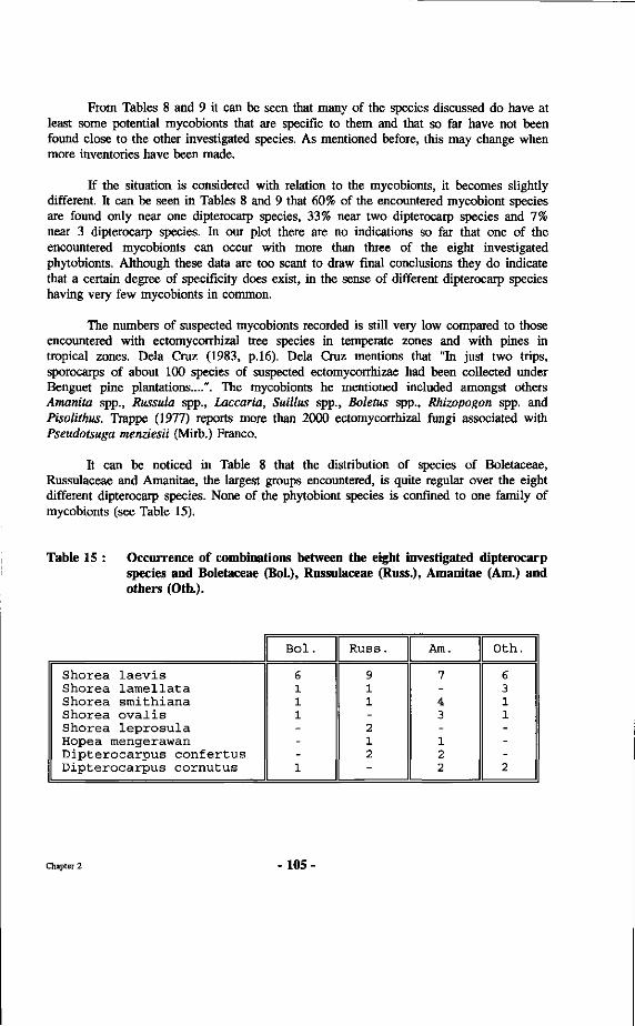
From Tables 8 and 9 it can be seen that many of the species discussed do have at least some potential mycobionts that are specific to them and that so far have not been found close to the other investigated species. As mentioned before, this may change when more inventories have been made.
If the situation is considered with relation to the mycobionts, it becomes slightly different. It can be seen in Tables 8 and 9 that 60% of the encountered mycobiont species are found only near one dipterocarp species, 33% near two dipterocarp species and 7% near 3 dipterocarp species. In our plot there are no indications so far that one of the encountered mycobionts can occur with more than three of the eight investigated phytobionts. Although these data are too scant to draw final conclusions they do indicate that a certain degree of specificity does exist, in the sense of different dipterocarp species having very few mycobionts in common.
The numbers of suspected mycobionts recorded is still very low compared to those encountered with ectomycorrhizal tree species in temperate zones and with pines in tropical zones. Delà Cruz (1983, p.16). Delà Cruz mentions that "In just two trips, sporocarps of about 100 species of suspected ectomycorrhizae had been collected under Benguet pine plantations....". The mycobionts he mentioned included amongst others Amanita spp., Russula spp., Laccaria, Suillus spp., Boletus spp., Rhizopogon spp. and Pisolithus. Trappe (1977) reports more than 2000 ectomycorrhizal fungi associated with Pseudotsuga menziesii (Mirb.) Franco.
It can be noticed in Table 8 that the distribution of species of Boletaceae, Russulaceae and Amanitae, the largest groups encountered, is quite regular over the eight different dipterocarp species. None of the phytobiont species is confined to one family of mycobionts (see Table 15).
Table 15 : Occurrence of combinations between the eight investigated dipterocarp species and Boletaceae (Bol.), Russulaceae (Russ.), Amanitae (Am.) and others (Oth.).
Shorea laevis Shorea lamellata Shorea smithiana Shorea ovalis Shorea leprosula Hopea mengerawan Dipterocarpus confertus Dipterocarpus cornutus
Bol.
6 1 1 1 ---1
Russ.
9 1 1 -2 1 2
-
Am.
7 -4 3 -1 2 2
Oth.
6 3 1 1 ---2
Chapter 2 - 1 0 5 -
If a piece of root could be identified as belonging to one of the investigated dipterocarp species, cross-sections were made of the ectomycorrhizae and these were studied in detail. For the comparison of colours, when feasible, use was made of the mycological colour chart (Rayner, 1970).
The equipment available did not allow for greater enlargements than 400 X. This excluded the observation of certain hyphal characteristics.
Quantitative data as presented in table 17, were collected using the "grid intersect method" (Giovanetti and Mosse, 1980). By using the formula :
2 H
and an intersect distance of 0.5 inch, causing R = n (cm) (Marsh, 1971). In this formula R stands for total root length, A the area in which the roots are distributed, n the number of intersections between roots or mycorrhizae and the grid lines and H the total length of the grid lines.
If too many roots were present in one sample the roots were cut in small pieces of about 2 cm length and the sample was thoroughly mixed in water and divided into two equal parts. When necessary this procedure was repeated more often. The resulting root length was then multiplied with the inverse fraction of the total root sample counted and investigated.
The results obtained with this method are described under paragraph 3.3.1, results of direct systematic root sampling. This method is very laborious for inventories of ectomycorrhizae in a primary mixed dipterocarp forest. Problems encountered were the following.
a) The very dense root mat under a primary rain forest makes the analyses of these kind of samples very time-consuming. This problem, however, is encountered in most methods of root research.
b) One finds an enormous diversity of roots in each sample, also in those from the area within the crown projection, among which many ectomycorrhizal roots of several different plant species. This consequently leads to difficulties in recognizing the species to which the roots belong. However it is possible to recognize many roots directly as being non-dipterocarp and sometimes belonging to families with distinctive root characteristics like Ebenaceae with black roots, Myristicaceae with roots of a slimy appearance and Sapotaceae having roots with a translucent cortex.
c) Even when working very carefully, ectomycorrhizae tend to break off very easy during the separation of the different roots, introducing the risks that certain mycorrhizal roots are missed during the microscopical investigation.
Chapter 3 - 108 -
d) The organic layer, where most of the ectomycorrhizae are located, contains organic debris so closely adhering to ectomycorrhizae that it is very difficult to make adequate descriptions of the morphological types. Only the use of the method with perforons (see Chapter 6) can cope with this problem.
e) The original idea of comparing central stele structures to identify the tree species forming the ectomycorrhizae may be feasible, but only after very extensive inventories and many detailed anatomical studies which was impossible to do during the course of this work. It was found that thin roots show secondary thickening and that there is considerable variation between cross-sections made through different roots of the same diameter originating from the same plant. This may depend on the age, the previous development and the extension of the root beyond the cross-sections examined. Some success was obtained in recognizing Shorea laevis roots from different samples. The identification, however, was based upon prior selection of roots bearing white pyramidal ectomycorrhizae. Most of these roots bearing the same ectomycorrhizae proved to have a more or less comparable central stele structure belonging to S. laevis. So the result could only be obtained by circular reasoning.
f) Even 240 samples per tree in 10 directions may not be enough to correctly asses the ectomycorrhizal colonization of the roots. On several occasions, the occurrence of ectomycorrhizal mushrooms was limited to small patches (see Chapter 2.1: "Mushroom inventories"), which might indicate the limited extent of a certain type of ectomycorrhiza at one place, and these small patches might be easily missed with the line method used here, as was the case for a patch of Hydnum repandum occurrence (see Figure 33).
g) At some places, where rotten roots left holes in the soil, perhaps enhancing aeration, ectomycorrhizae also occur deeper in the soil. These deeper ectomycorrhizae might well belong to other fungal species. These spots are seldom or never included in sampling. Dan Nepstad (pers. comm. P.S. Ashton) has found ectomycorrhizal roots up to 3 meters down in soil in Amazonia.
3.2.2 Method 2, direct root sampling of individual trees
The second method used consisted of digging along buttresses of large Dipterocarps and following the thin roots connected to these buttresses or their extensions until side roots with ectomycorrhizae were encountered. Later a pricking method was developed using a sharp pointed iron bar of 10 mm diameter with a cross bar for handling welded on the top. By pricking the roots every 10-20 cm, starting from the buttresses it proved very practical to locate them quick and accurately without the need for much digging. This method is also much less destructive than digging along the roots. Staining and microscopic examination were performed in the same way as described under method 1). This method was used to collect the roots for the mycorrhizal status file of trees in the Wanariset forest as presented in Appendix 7.
Chapter 3 - 109 -
Problems related to this second method used are partially the same as those mentioned above. Sometimes it proved to be difficult to sample the roots of the correct tree, even when digging along their main roots, when collecting was done by local personnel. Again, the very dense root mat was an important handicap, together with the high degree of soil compaction directly around the buttresses, leading to much broken roots when digging them out.
A problem of another nature is the difference in soil conditions between the direct surroundings of the tree base and buttresses, and the soil at several meters distance. Some dipterocarp species like Shorea laevis and Dryobalanops spp. form mature crowns built by branches under a comparably steep angle, as a result of which a large amount of stem flow reaches the soil near the stem base. Other species, like S. stenoptera Burck. and many Dipterocarpus spp., possess more horizontally oriented branches in their mature crowns (compare the architectural diagrams of several Dipterocarps by Edelin (1984)) and so stem flow is less. If a person is caught in the field by a tropical rain shower, the former trees provide quite a good shelter since the amount of throughfall is much lower compared to the throughfall under Dipterocarps with more horizontally spread branches, preventing one from becoming completely soaked.
Besides the variation in the amount of water received in the area directly around the base of stems one often observes much organic debris around the stem bases. This feature again is particularly clear under S. laevis. According to Arsad Anom (staff ITCI, personal communication), termites have a preference for making nests at the base of big S. leprosula and S. parvifolia Dyer stems. At the stem bases of many other old Dipterocarps, the presence of termites often indicates that the stem may be hollow. In paragraph 2.3.4.a it has already been mentioned that differences were encountered in mushroom appearances close to and farther away from the stem bases. The collection of root samples with ectomycorrhizae close to the buttresses therefore may not yield a correct insight in the presence of other important ectomycorrhizal fungi upon other parts of the root system of the same tree.
The situation can be compared with the distance-related differences in amounts of accumulated organic matter with many palms that grow in the understorey of the forest. These palms collect many falling leaves and twigs with their spread leaves. The captured debris is transported down the leaf stalks. These stalks have canals located at their upper part. Under the crowns of these palms the soil is almost bare without presence of the common amounts of herbs and seedlings. Near the stem a large accumulation of organic soil is found where only the roots of bigger trees surrounding these palms are encountered in considerable quantities.
The results obtained with this method were so inconsistent (often personnel brought back roots of other species than the one investigated) that they are not discussed here. Appendix 6 lists results from double checks only. Only when all mycorrhizal types encountered from all the experiments and inventories can be compared, these results will become more valuable through faster discrimination of non-intended roots in the collected samples.
Chapter 3 - 110 •
32.3 Method 3, root sampling on spots of mycobiont sporocarp appearances
The third method rested upon the digging up of roots at places where ectomycorrhizal mushroom species had been observed within the crown projection of a large dipterocarp tree belonging to one of the species listed in 2.1. Then it was tried to find morphological resemblances with roots collected near the buttresses. The root samples were further treated as mentioned under the first method.
This third method proved to be useful at times. Viz. the sampling of S. laevis roots from locations more than 300 meters apart on places where the same ectomycorrhizal sporocarp {Russula japonica) had been observed during several visits, yielded roots with the same morphological type of ectomycorrhizae. This method of digging up roots with ectomycorrhizae at places where sporocarps of ectomycorrhizal fungi have appeared was also used by Jansen (1986), who was working on a key to ectomycorrhizal fungi based upon characters of the individual ectomycorrhizae as seen through a dissecting microscope at 6-50 X enlargement. One problem is, that the number of observations on sporocarps has to be sufficiendy large and that numerous ectomycorrhizae need to be collected. Using this approach Jansen (1991) obtained a highly significant correlation between numbers of mycorrhizae in soil and the cumulative abundance of fruit bodies of mycorrhizal fungi.
By the use of mushroom inventories, the chance is still high that many ectomycorrhizal types are missed, because the sporocarps formed by the particular fungus concerned did not yet appear or may not appear at all. They also may not have been seen because of the rapid decay of the sporocarps in the primary rain forest or because of their small size or hypogeous nature (cf. Chapter 2.3.3.d). In temperate zones mycorrhizal research is mostly conducted in monocultures, where the conditions are more homogeneous. For tropical rain forests, with their huge species-richness, this work needs to be combined with comparisons between roots bearing the ectomycorrhizae and roots of possible ectomycorrhizal trees in the close surroundings of the collection site. This method can therefore be promising when a longer period of time can be spent on it.
3.2.4. Method 4, top soil washing and plotting of ectomycorrhizal types
In case of some dipterocarp trees that were growing on moderate slopes in the vicinity of some water supply in the form of a small water-course, parts of the root systems could be cleaned from soil material by applying large amounts of water. In Box 6 an overview is provided of the difficulties involved using this method, varying from logistical problems to technical results that could be obtained.
Chapter 3 - 1 1 1 -
Box 6 : Difficulties studying root distribution in a mixed tropical rain forest.
Root research in species rich forest is complicated (Kahn, 1983). To determine the root extension of tree species involved in the research, root systems had to be washed out with water. This involved many practical problems. First the use of a water pump was tried out while using a high water pressure to remove the soil from between the roots, but later it was preferred to wash out the roots carefully by rinsing each small part of the root system at one time. Hands and fine brushes were used. When ectomycorrhizae were encountered, they were treated as reported under the method described in Chapter 3.1.1. Another plot, located not far from the road could be washed out with water brought to the forest in a tank on a truck, which enabled the survey of roots in some places where water supply was a serious problem.
The main purpose of this work was to determine the horizontal extension of root systems of several Dipterocarps of different diameters. Examples of this work are presented in Chapter 2.4. Lb (Figure 29). During this work, however, some important observations were also made on the horizontal distribution of ectomycorrhizae.
This fourth method, using a water-pump, was only partly feasible for the survey of ectomycorrhizae. The pressure applied to wash away the soil particles, ruined many ectomycorrhizae. This method is especially useful for obtaining information on the extent and structure of the main frame of root systems. And then only for the superficial roots, not the sinkers under the buttresses. Not only many ectomycorrhizae disappear but also many fine roots are lost with this method. The same was found by Schmidt (Department of Forestry, Wageningen Agricultural University, personal communication) in Surinam. Furthermore a lot of damage is caused to the larger trees and the soil that eroded downhill brings about large changes there as well.
Gently rinsing the roots in situ gave the same problems as the cleaning of roots from the core samples in method 1. Many of the ectomycorrhizae entering the organic litter are easily lost during the preparation of the terrain. The work must be done very carefully and is rather time-consuming.
For practical reasons, the location should be close to a sufficiently large source of water, which almost automatically excludes ridges. Root systems and the presence of mycorrhizae may well be different on wetter places. This method has therefore been used only few times in the research reported here. The results of the use of this method are presented in Chapter 3.3.2.
Chapter 3 - 112 •
3.2.5 Method 5, collecting of roots of dipterocarp seedlings resulting from natural regeneration
The collection of seedlings around seed trees. These seedlings were dug out very carefully, so as not to disturb or lose any part of their root system. The ectomycorrhizal types encountered on these root systems were classified on the basis of several morphological characteristics, such as colour, type of branching, presence of rhizomorphs etc.. Microscopical investigations were conducted as described sub 1). Seedlings were collected from plot km 1.5 (Hopea rudiformis, Shorea laevis and S. pauciflora King), plot km 2.1 (H. rudiformis), plot km 2.4 (H. rudiformis) plot km 3.0 (fl. rudiformis), the Wartono trail (S. pauciflora) (see Appendix 2) and the ITCI concession {H. rudiformis). The results of these inventories are presented in Table 18.
The fifth method is considered to be the most practical one. Root systems of dipterocarp seedlings can be recovered rather easily if the seedlings are still small (Becker 1983). Roots still adhering to the seedlings suffer less during transport than other root samples, separated from the rest of the root system. One has to be very careful, otherwise many types can be missed as shown by Marx and Hatchell (1986). They found that as much as 75% of the lateral roots and ectomycorrhizae were stripped from pine seedlings when lifting was not done cautiously enough.
Disadvantages are the uncertain availability of dipterocarp seedlings, cf. Chapter 1.1.1, and the uncertainty whether or not ectomycorrhizal fungi on roots of dipterocarp seedlings are representative for those on roots of the seed trees. Last & al. (1984) and Mason & al. (1986) observed a sequence of ectomycorrhizal fungi, living with Betula pubescens in Scotland. Malajczuk (1987) observed the same type of succession for Eucalypts and Pines in Australia. This sequence is related to the age of the phytobiont. Dighton and Mason (1985), Dighton & al. (1986) and Last et al. (1992) also provide such examples.
The chance that one deals with an ectomycorrhizal fungus of the mother tree is higher if the ectomycorrhizal type is encountered on roots of seedlings from many different localities with the same species of mother tree. Results obtained with this method are presented in under results in Chapter 3.3.3.
3.2.6 Method 6, sampling of seedlings resulting from natural regeneration and transferred to perforons
In this method seedlings are collected from underneath the seed tree and planted in perforons. The perforons were filled with pasteurized soil to prevent infection of other ectomycorrhizal fungi like the common Telephora terrestris. Because of the limited amount of seedlings that could be studied in this way, the results of these experiments as well as a more detailed discussion on the possibilities of using this method for mycorrhizal research are not presented in this Chapter but in Chapter 5, "Perforon studies".
Chapter 3 - 113 -
Perforons consist of sturdy containers filled with soil, the soil can be perforated horizontally after which the growth of the plant roots and associated symbioses can be studied by means of additional equipment when the newly developed roots cross the perforations. Their use is extensively described and discussed in Chapter 6. In the perforations new ectomycorrhizae are easy to observe, without adhering soil particles.
3.2.7 Method 7, planting of non-mycorrhizal dipterocarp seedlings under seed trees followed by evaluation of mycorrhizal types established
This method consists of planting of non-mycorrhizal seedlings under seed trees and collecting them after 3 to 6 months following the procedure of method five for the evaluation of the root systems and their possible infections, e.g. with ectomycorrhizae.
The seventh method is considered as in situ inoculation and is therefore discussed in Chapter 4, "Inoculation experiments".
3.3 Results
3.3.1 Results of direct systematic root sampling
In table 17 the results are presented of the analyses of 240 cores sampled around Shorea laevis (tree number 224). Table 16 shows how the values I for the root length were assigned. Figure 32 shows a combination of the crown projections of Shorea laevis trees in the plot and the positions of the cores containing S. laevis roots. Over this map the circular projections that were calculated by the computer program are shown. It is clear that the presence of 5. laevis roots in these soil cores does not fit very well with the distribution of the roots of tree number 224 based upon the calculated circle around it. Also note that sometimes the asymmetry of the tree crown can lead to much variation in the calculated radius for the circular sphere of the crown projection and therefore the assumed root distribution. However, most roots of tree number 224 are approximately confined to a circular area around the tree with a diameter of 12 meters.
47% of the total of the 240 sampled soil cores, contained ectomycorrhizae. Only 75% of the samples containing roots of Shorea laevis were taken from inside the assumed root distribution of large ectomycorrhizal trees. The ectomycorrhizal types recognized from these samples are shown in Appendix 7 and their distribution over the different cores in Table 17 and Figure 38. Concerning the basal area within the plot 39% of the trees were ectomycorrhizal, while another 13% were potentially ectomycorrhizal (cf. Table 6). These two values for basal area and presence of roots with ectomycorrhizae (52 and 47%) therefore correspond well with each other.
Chapter 3 - 114 •
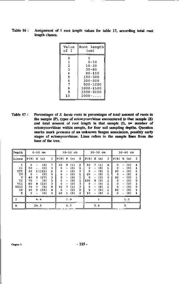
Table 16 : Assignment of I root length values for table 17, according total root length classes.
Value of I
0 1 2 3 4 5 6 7 8 9
10
Root l e n g t h (cm)
0 0-10
10-30 30-80 80-150
150-300 300-500 500-1000
1000-1500 1500-2000 2000- . . . .
Table 17 : Percentages of S. Iaevis roots in percentages of total amount of roots in the sample (P), types of ectomycorrhizae encountered in that sample (£) and total amount of root length in that sample (I), n= number of ectomycorrhizae within sample, for four soil sampling depths. Question marks mark presence of an unknown fungus association, possibly early stages of ectomycorrhizae. Lines refers to the sample lines from the base of the tree.
D e p t h
L i n e s
I I I
I I I I V
V V I
V I I V I I I
I X X
I
n
0
P ( % )
0 5 0 3 0
0 4 0 7 0 6 0 7 0 3 0
0
- 1 0 cm
E ( n )
- ( 0 ) - ( 0 ) 1 ( 1 5 2 ) - ( 0 ) 5 ( 2 7 ) - ( 0 ) 8 ( 2 3 ) ? ( 5 ) 5 ( 3 6 ) - ( 0 )
I
7 3 6 5 3 2 3 8 4 3
4 . 4
2 4 . 3
1 0 - 2 0 cm
P ( % )
3 0 0 0 0 0 0 0
3 0 0
2 0
E ( n )
5 ( 1 ) - ( 0 ) - ( 0 ) - ( 0 ) - ( 0 ) - ( 0 ) - ( 0 ) ? ( 1 ) - ( 0 ) 1 ( 5 )
I
2 1 3 1 1 1 3 3 2 2
1 . 9
0 . 7
2 0 -
P ( % )
5 0 0 0
2 0 0
1 0 0 0 0 0
1 0
E
?
----8
----
30 cm
( n )
( 1 ) ( 0 ) ( 0 ) ( 0 ) ( 0 ) ( 3 ) ( 0 ) ( 0 ) ( 0 ( ( 0 )
I
2 1 1 1 0 1 1 1 1 1
1
0 4
3 0 - 4 0 cm
P ( % )
0 0
9 0 0
9 0 0 0 0
9 0 0
E ( n )
- ( 0 ) - ( 0 ) - ( 0 ) - ( 0 ) - ( 0 ) - ( 0 ) - ( 0 ) - ( 0 ) - ( 0 ) - ( 0 )
I
4 1 0 2 1 0 1 0 1 1
1 . 1
0
Chapter 3 115
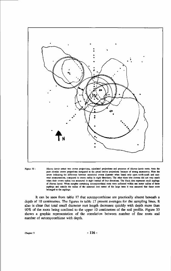
Figure 32 : Shorea laevis actual tree crown projections, calculated projections and presence of Shorea laevis roots. Note the poor circular crown projections compared to the actual crown projections because of strong asymmetry. Note the arrow indicating the difference between calculated crown diameter when based only upon north-south and east-west measurements, compared to crown radius in eight directions. The other three tree crowns did not very much when their crown radius was measured in eight instead of four directions. The black dots represent small saplings of Shorea laevis. When samples containing ectomycorrhizal roots were collected within one meter radius of these saplings and outside the radius of the assumed root extent of the large trees it was assumed that these roots belonged to the saplings.
It can be seen from table 17 that ectomycorrhizae are practically absent beneath a depth of 10 centimetres. The figures in table 17 present averages for the sampling lines. It also is clear that total small diameter root length decreases quickly with depth more than 90% of the roots being confined to the upper 10 centimetres of the soil profile. Figure 33 shows a graphic representation of the correlation between number of fine roots and number of ectomycorrhizae with depth.
Chapter 3 - 1 1 6 -
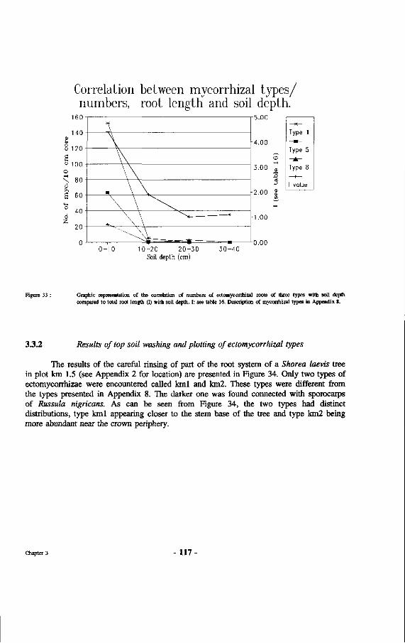
Correlation between mycorrhizal types/ numbers, root length and soil depth.
0 -10 10 -20 2 0 - 30 3 0 - 4 0 Soil depth (cm)
5.00
4.00
Type 1
Type 5
Type 8
I value
Figure 33 : Graphic representation of the correlation of numbers of ectomycorrhizal roots of three types with soil depth compared to total root length (1) with soil depth. I: see table 16. Description of mycorrhizal types in Appendix 8.
3.3.2 Results of top soil washing and plotting of ectomycorrhizal types
The results of the careful rinsing of part of the root system of a Shorea laevis tree in plot km 1.5 (see Appendix 2 for location) are presented in Figure 34. Only two types of ectomycorrhizae were encountered called kml and km2. These types were different from the types presented in Appendix 8. The darker one was found connected with sporocarps of Russula nigricans. As can be seen from Figure 34, the two types had distinct distributions, type kml appearing closer to the stem base of the tree and type km2 being more abundant near the crown periphery.
Chapter 3 - 1 1 7 -
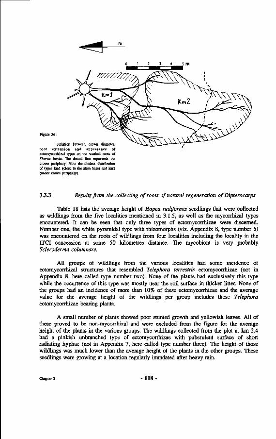
0 1 2 3 4 5
Figure 34 :
Relation between crown diameter, root extension and appearance of ectomyconhizal types on the washed roots of Shorea laevis. The dotted line represents the crown periphety. Note the distinct distribution of types kml (close to the stem base) and km2 (under crown periphery).
3.3.3 Results from the collecting of roots of natural regeneration of Dipterocarps
Table 18 lists the average height of Hopea rudiformis seedlings that were collected as Wildlings from the five localities mentioned in 3.1.5, as well as the mycorrhizal types encountered. It can be seen that only three types of ectomycorrhizae were discerned. Number one, the white pyramidal type with rhizomorphs (viz. Appendix 8, type number 5) was encountered on the roots of Wildlings from four localities including the locality in the ITCI concession at some 50 kilometres distance. The mycobiont is very probably Scleroderma columnare.
All groups of Wildlings from the various localities had some incidence of ectomyconhizal structures that resembled Telephora terrestris ectomycorrhizae (not in Appendix 8, here called type number two). None of the plants had exclusively this type while the occurrence of this type was mostly near the soil surface in thicker litter. None of the groups had an incidence of more than 10% of these ectomycorrhizae and the average value for the average height of the Wildlings per group includes these Telephora ectomycorrhizae bearing plants.
A small number of plants showed poor stunted growth and yellowish leaves. All of these proved to be non-mycorrhizal and were excluded from the figure for the average height of the plants in the various groups. The Wildlings collected from the plot at km 2.4 had a pinkish unbranched type of ectomycorrhizae with puberulent surface of short radiating hyphae (not in Appendix 7, here called type number three). The height of those Wildlings was much lower than the average height of the plants in the other groups. These seedlings were growing at a location regularly inundated after heavy rain.
Chapter 3 - 118 -
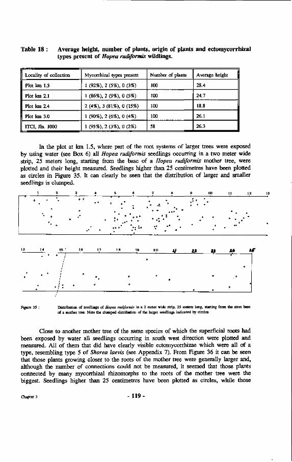
Table 18 : Average height, number of plants, origin of plants and ectomycorrhizal types present of Hopea mdiformis Wildlings.
Locality of collection
Plot km 1.5
Plot km 2.1
Plot km 2.4
Plot km 3.0
ITCI, Jin. 1000
Mycorrhizal types present
1 (92%), 2 (5%), 0 (3%)
1 (86%), 2 (9%), 0 (5%)
2 (4%), 3 (81%), 0 (15%)
1 (90%), 2 (6%), 0 (4%)
1 (95%), 2 (3%), 0 (2%)
Number of plants
100
100
100
100
58
Average height
28.4
24.7
18.8
26.1
26.3
In the plot at km 1.5, where part of the root systems of larger trees were exposed by using water (see Box 6) all Hopea mdiformis seedlings occurring in a two meter wide strip, 25 meters long, starting from the base of a Hopea rudiformis mother tree, were plotted and their height measured. Seedlings higher than 25 centimetres have been plotted as circles in Figure 35. It can clearly be seen that the distribution of larger and smaller seedlings is clumped.
1
+
« +
2
+ +-
3
*• +
+
t*
4
° +
0
5
+ +°
6
: +
7
+• +
8
+
• % * * +
••
9
-
10
+ +
+ D 4
+ +
11
+
12 13
+
3
f
14
*
+
+
+
*
15 1
• /
;
*/: *
16
•
17
•
*
18
+•
+•
19
•
2 0
+
it
o
U
0
M
o
*•
14 +
* •
*•
V
+
+
Figure 35 : Distribution of seedlings of Hopea rudiformis in a 2 meter wide strip, 25 meteis long, starting from the stem base of a mother tree. Note the clumped distribution of the larger seedlings indicated by circles.
Close to another mother tree of the same species of which the superficial roots had been exposed by water all seedlings occurring in south west direction were plotted and measured. All of them that did have clearly visible ectomycorrhizae which were all of a type, resembling type 5 of Shorea laevis (see Appendix 7). From Figure 36 it can be seen that those plants growing closer to the roots of the mother tree were generally larger and, although the number of connections could not be measured, it seemed that those plants connected by many mycorrhizal rhizomorphs to the roots of the mother tree were the biggest. Seedlings higher than 25 centimetres have been plotted as circles, while those
Chapter 3 -119
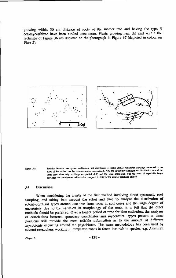
growing within 30 cm distance of roots of the mother tree and having the type 5 ectomycorrhizae have been circled once more. Plants growing near the part within the rectangle of Figure 36 are depicted on the photograph in Figure 37 (depicted in colour on Plate 2).
Figure 36 : Relation between root system architecture and distribution of larger Hopea rudiformis seedlings connected to the roots of the mother tree by ectomycorrhizal connections. Note the apparently homogenous distribution around the stem base when only seedlings are plotted (left) and the close correlation with the roots of especially larger seedlings that are depicted with circles compared to dots for the smaller seedlings plotted.
3.4 Discussion
When considering the results of the first method involving direct systematic root sampling, and taking into account the effort and time to analyze the distribution of ectomycorrhizal types around one tree from roots in soil cores and the large degree of uncertainty due to the variation in morphology of the roots, it is felt that the other methods should be preferred. Over a longer period of time for data collection, the analyses of correlations between sporocarp coordinates and mycorrhizal types present at these positions will provide the most reliable information as to the amount of different mycobionts occurring around the phytobionts. This same methodology has been used by several researchers working in temperate zones in forest less rich in species, e.g. Ammirati
Chapter 3 -120
et al. (1987) and Malajczuk (1987). The low number of ectomycorrhizal types encountered here is probably due to the poor equipment available for the research, which for example did not allow for checking hyphal characteristics in so-called squash samples of the different mycorrhizae.
In Figure 38 the sporocarp producing mycobionts are plotted over the positions of the different ectomycorrhizal types encountered. As in Figure 34 it is clear that there is some difference between the area near the crown periphery and the area nearer to the stem base. This difference may be partly explained by differences in sporocarp formation near the crown periphery and closer to the stem, possibly because of the different substances transported by stem flow and peripheral crown drip. Additional analyses of nutrients in stem flow and throughfall may yield further insights.
The results of the work with method four showed a remarkable sharp boundary between the two types of ectomycorrhizae on roots of Shorea laevis encountered. However, the boundaries drawn present best guesses because only very few ectomycorrhizae were found, most of them probably having been washed away with the strong water current. One possibility is that certain types of ectomycorrhizae break of more easily. It may therefore very well be that more types should have been encountered with other distributions when using other methods. Interesting is the close correlation between presence of roots of mother trees, ectomycorrhizal connections and growth of the seedlings. This apparent nursing role by the mother tree is studied further by Yasman (in preparation).
The fifth method, consisting of lifting seedlings resulting from natural regeneration, gave some clear results for Hopea rudiformis. Average height of the Wildlings, that all originated from the same mast flowering, was almost constant for the plants having the white pyramidical ectomycorrhizae. The plants with the pink type were clearly smaller, indicating varying mycorrhizal effects on the growth.
All these plants were collected as small seedlings with three to five leaves and grown for four months in the greenhouse under equal conditions and in the same potting medium. The differences in height therefore are not caused by variations in site conditions at the time of collecting. At the time of collecting only a small number of seedlings was investigated superficially, and this yielded only the white and the pinkish types of mycorrhizae and no Telephora terrestris mycorrhizae were noted in this initial sample. There is a good chance that the infection with Telephora terrestris got established later in the greenhouse, where it was found to be present on the roots of plants already growing there This fungus is very common in nurseries in Indonesia (Smits and Leppe, 1988). Much of the variation in height of the seedlings within one group could possibly be the result of differences in time of infection by ectomycorrhizal fungi from neighbouring already infected plants on the tables in the greenhouse, the conditions in the greenhouse were such that cross infections could have occurred later on although the results of table 18 do not indicate that this has been of major importance.
Chapter 3 - 121 -
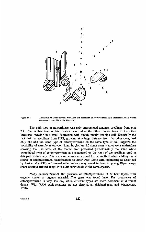
1 s
Figure 38 Appearance of ectomycorrhizal sporocarps and distribution of ectomycorrhizal types encountered under Shorea /aevw.(tree number 224 in plot Wartono).
The pink type of mycorrhizae was only encountered amongst seedlings from plot 2.4. The mother tree in this location was unlike the other mother trees in the other locations, growing in a small depression with muddy poorly draining soil. Especially the fact that the seedlings from ITCI, growing at a large distance from the other ones, had only one and the same type of ectomycorrhizae on the same type of soil supports the possibility of specific ectomycorrhizae. In plot km 1.5 some more studies were undertaken showing that the roots of the mother tree possessed predominantly the same white pyramidical type of ectomycorrhizae as encountered on the roots of the seedlings used in this part of the study. This also can be seen as support for the method using Wildlings as a source of ectomycorrhizal identification for older trees. Long term monitoring as described by Last et al (1992) and several other authors may reveal in how far young Dipterocarps share ectomycorrhizal fungi with older individuals of the same species.
Many authors mention the presence of ectomycorrhizae in or near layers with organic matter or organic material. The same was found here. The occurrence of ectomycorrhizae is very shallow, while different types are more dominant at different depths. With VAM such relations are not clear at all (Mohankumar and Mahadevan, 1988).
Chapter 3 122
Chapter 4 : Infection processes
4.1 Introduction
In this section the outcome of various inoculation experiments will be described and their results presented. Attention has been paid to 1) the infection process, 2) when it takes place, 3) how it takes place and 4) where. Moreover some results are presented on 5) the influence of substrate and physical conditions upon establishment of dipterocarp ectomycorrhizae. Because of the overlap with other chapters some parts are only mentioned here and discussed in more detail in the respective chapters.
Practical results concern the compatibility of the different fungus-Dipterocarp combinations, and growth stimulation resulting from inoculation.
4.2 Methods
Several methods were used. First the natural infection process in the forest itself was studied by collecting seeds and seedlings from the forest, sometimes by planting non-mycorrhizal seedlings in natural forest and harvesting them after a period of time to look for the presence of ectomycorrhizae. These results are only provided in the discussion for one location with Shorea laevis in the plot "Wartono Kadri".
Then the infection resulting from inoculations was studied in greater detail under more or less strictly controlled conditions in nursery and greenhouse. One specific example of the more controlled conditions is presented in chapter 7, where the results are described of some inoculation experiments conducted in gnotobiotic systems.
Seeds were collected from the branches, from nets hanging above the ground and from the forest floor and after that germinated in semi sterile soil in the greenhouse. Spontaneous development of ectomycorrhizae was monitored in the Wanariset greenhouse. Materials and methods for the collection of the material were described by Leppe (1986).
Seedlings grown from surface sterilized seeds, that had germinated in the greenhouse and that did not possess ectomycorrhizae were planted in the natural forest in several forest types and under several Dipterocarpaceae. They were lifted in small samples after 4 weeks, 3 months and 6 months, and their roots were checked for ectomycorrhizal presence.
Other non-mycorrhizal seedlings were inoculated during transplanting to polybags or to clay pots, with either parts of ectomycorrhizal mushrooms, spores or pure mycelial cultures of ectomycorrhizal fungi. Establishment of ectomycorrhizae was regularly checked.
Young plants were planted in perforons (see Box 7) and roots, passing through the perforations, were brought into contact with pieces of ectomycorrhizal mushrooms, spores
Chapter 4 - 123 -
of some Gasteromycetes, and with ectomycorrhizal root pieces from several origins. A few times mycelial growth from pure cultures was brought into contact with the roots. The infection process and the effect of the inoculations were monitored with the methods described in chapter 5. These results have been reported by Hendromono et al. (1988).
Wildlings that were collected under their respective mother trees were checked for mycorrhizal presence and, if ectomycorrhizal, planted in groups near other large dipterocarp trees of varying species. Their growth and development was monitored for one year, and part of the surviving seedlings were excavated and their root systems checked for presence of ectomycorrhizae of other types than the original ones.
4.2.1 Infection of non-mycorrhizal dipterocarp seedlings related to distance from large dipterocarp trees.
Non-mycorrhizal seedlings of Shorea assamica were planted at intervals of 2 meter along lines radiating North-South and East-West from the stem base of several trees until 20 meters from the stem base. The growth and morphological development of the seedlings was regularly checked during a period of one year. After this period the surviving seedlings were lifted and their root systems checked for the presence of ectomycorrhizae as described in chapter 3. The seedlings were planted in a lightly burned forest which was still dominated by many Dipterocarpaceae. Light intensity at the forest floor was estimated to be half of full light intensity. The light intensity was estimated from the diaphragm opening of a Minolta camera when holding it pointed at a white piece of paper at 50 cm distance both in the open and under the vegetation during a cloudy day around noon. Other dipterocarp seedlings of Hopea sp. and Dipterocarpus confertus were present and grew very well under these conditions. The seedlings were planted around Shorea pauciflora and Shorea laevis. Within a circle of 20 meters around these trees several other, smaller, Dipterocarpaceae of various species, also other species than the ones included in the analyses, were present. The extent of their root systems was not checked but it is very probable that some of the planted seedlings have been in close contact with some ectomycorrhizal roots of these trees as well (compare Figure 32).
4.2.2 Influence of the seed collecting method upon mycorrhizal infection after transplanting to a medium without inoculum.
Seeds of Shorea pauciflora, S. assamica, Dipterocarpus confertus, D. cornutus and D. tempehes were collected by shaking the seeds off the branches and then directly picking them up, and by collecting naturally fallen seeds. Seeds of Shorea assamica were collected in the ITCI concession, while all other seeds were collected in the Wanariset forest. These two classes of seeds were kept apart and sown separately. Some of the seeds that had already germinated when they were picked up from the forest floor were directly planted in plastic containers. The seeds that had not yet germinated were sown in sand that had been heated to about 50 degrees Celsius for three hours, a treatment which was meant to exclude spores of ectomycorrhizal fungi. The same treatment was applied to the medium (cf. Chapter 6), a 1:1 topsoil/sand mixture in which the already germinated seedlings were planted.
Chapter 4 - 124 -
The heating was done by placing a thin layer of the medium to be heated on a concrete floor, previously heated by sunlight. Then the layer of medium was covered with plastic during periods with full sunlight. Temperatures were regularly measured. The method is described in more detail in Smits et al. (1988). The seedlings that grew out of these seeds were transplanted to plastic containers after at least two leaves above the cotyledons had unfolded. These containers were filled with a loose medium that had been exposed to the same heat treatment. Before transplanting, the roots were checked for ectomycorrhizal presence. Of the many thousands of seedlings, only 100 of each treatment and each species were checked for presence of ectomycorrhizae. No detailed microscopical classification of types was made.
4.2.3 Inoculation of non-mycorrhizal dipterocarp seedlings with chopped sporocarps of potential ectomycorrhizal fungi
Seeds of Shorea lamellata, S. leprosula, S. pauciflora and S. ovalis were germinated in white sand that had been heated previously to eliminate any ectomycorrhizal inoculum. When the resulting seedlings had produced three leaves the seedlings were transplanted to plastic containers. All roots were checked to assure that no ectomycorrhizae had formed yet. The medium, consisting of a 1:1 topsoil/sand mixture, in the plastic containers also previously was treated with heat to eliminate potential ectomycorrhizal inoculum. During transplanting the plants were inoculated with fungal material of Amanita similis, Lactarius subpiperatus, Phylloporus bogoriensis and Russula eburneoareolata, in the planting hole. The fungal material consisted of freshly collected sporocarps of the fungi. The sporocarps were cut in pieces of circa 2 x 2 x 2 millimetre. The pieces were brought in direct contact with most of the root systems of the seedlings. The inoculated plants were put in a wind free environment under plastic covers in the nursery, on concrete floors, with 30 cm distance between groups of plants that were inoculated by one fungus and those inoculated by another. This was done to prevent, as far as possible, mycorrhizal infection from air-born inoculum or splashing of water during the daily watering of the plants. 500 plants were used for all phytobiont-mycobiont combinations. Growth was monitored for two years.
After one year five plants were carefully taken out of the plastic containers. The root systems were washed out with water and checked for ectomycorrhizal presence under a 25x dissecting microscope. The ectomycorrhizal types encountered were classified based upon mantle structure, presence of rhizomorphs, colour, thickness, branching pattern, visible layering within the mantle, mycorrhizal tip morphology and other characteristics when present.
4.3 Results
4.3.1 Correlation between mycorrhizal infection and distance from large dipterocarp trees.
All of the non-mycorrhizal seedlings showed deficiencies. Very few new leaves were produced and if so, they were yellowish. Most new leaves were produced at the somewhat lighter spots. Some 28% of the seedlings planted died within one year. Survival was not related to distance from the stem bases as can be seen from Table 19 and Figure 39.
Chapter 4 - 125 -
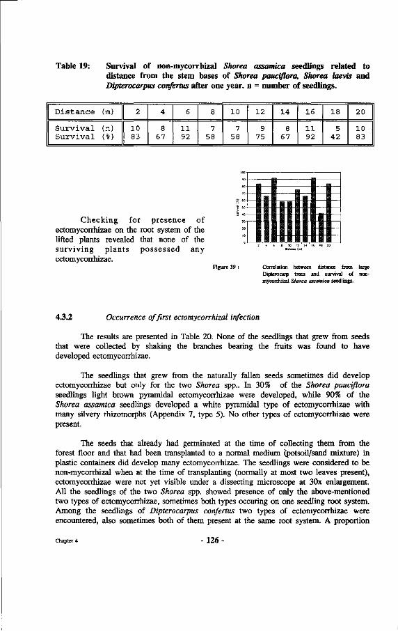
Table 19: Survival of non-mycorrhizal Shorea assamica seedlings related to distance from the stem bases of Shorea pauciflora, Shorea laevis and Dipterocarpus confertus after one year, n = number of seedlings.
Distance (m)
Survival (n) Survival (%)
2
10 83
4
8 67
6
11 92
8
7 58
10
7 58
12
9 75
14
8 67
16
11 92
18
5 42
20
10 83
Checking for presence of ectomycorrhizae on the root system of the lifted plants revealed that none of the surviving plants possessed any ectomycorrhizae.
Figure 39 : Correlation between distance from large Dipterocarp trees and survival of non-mycorrhizal Shorea assamica seedlings.
4.3.2 Occurrence of first ectomycorrhizal infection
The results are presented in Table 20. None of the seedlings that grew from seeds that were collected by shaking the branches bearing the fruits was found to have developed ectomycorrhizae.
The seedlings that grew from the naturally fallen seeds sometimes did develop ectomycorrhizae but only for the two Shorea spp.. In 30% of the Shorea pauciflora seedlings light brown pyramidal ectomycorrhizae were developed, while 90% of the Shorea assamica seedlings developed a white pyramidal type of ectomycorrhizae with many silvery rhizomorphs (Appendix 7, type 5). No other types of ectomycorrhizae were present.
The seeds that already had germinated at the time of collecting them from the forest floor and that had been transplanted to a normal medium (potsoil/sand mixture) in plastic containers did develop many ectomycorrhizae. The seedlings were considered to be non-mycorrhizal when at the time of transplanting (normally at most two leaves present), ectomycorrhizae were not yet visible under a dissecting microscope at 30x enlargement. All the seedlings of the two Shorea spp. showed presence of only the above-mentioned two types of ectomycorrhizae, sometimes both types occuring on one seedling root system. Among the seedlings of Dipterocarpus confertus two types of ectomycorrhizae were encountered, also sometimes both of them present at the same root system. A proportion
Chapter 4 - 1 2 6 -
of 49% of these seedlings was non-mycorrhizal at the time of checking, whereas 23% of the D. cornutus seedlings was found to have developed ectomycorrhizae of a yellowish, non-branching type with few loose hyphae. Of the D. tempehes seedlings 17% had developed purple-brown very long ectomycorrhizae that were covered with many loose hyphae. They resembled the type of ectomycorrhizae normally formed by Telephora terrestris in the greenhouse.
Table 20 : Percentages of infection by ectomycorrhizal fungi of seeds collected from branches, picked from the litter and germinated seeds.
\\ Treatment dipterocarp sp. \\
Shorea assamica Shorea pauciflora Dipterocarpus confertus Dipterocarpus cornutus Dipterocarpus tempehes
Shaken seeds
0 0 0 0 0
Fallen seeds
90 30
0 0 0
Germ, seeds
100 100
51 23 17
4.3.3 Compatibility of phytobiont-mycobiont combinations
The 500 plants each that were inoculated using chopped sporocarps per treatment for the different species were put in the nursery beds under a green screen that reduced light intensity about 50%. Shorea leprosula plants remained there, other plants were mixed up in routin planting activities and although most of the plants looked healthy after transplanting the data were lost. The Shorea leprosula plants infected with Amanita similis showed best growth reaching almost two meters average height after 18 months, still growing in small containers (6 x 15 cm). Abundant whitish, sparsely branched ectomycorrhizae, with no radiating hyphae, were present on the roots of a number of plants that were checked. The leaves were dark green.
The plants inoculated with Russula eburneoareolata showed good growth with light green leaves. The average height after 18 months was 1.20 m. The roots were infected by a mixture of Telephora terrestris ectomycorrhizae and a cream to white coloured pyramidal type with few radiating hyphae from the mantle near the base of the mycorrhizae. Plants inoculated with Lactarius subpiperatus and Phylloporus bogoriensis remained much smaller and showed yellowish leaves. Their average height was 60 cm after 18 months. The plants inoculated with lactarius subpiperatus were slightly larger on average. Plants sampled from both groups showed abundant Telephora terrestris mycorrhizae. No other well developed ectomycorrhizae were encountered.
Chapter 4 127.
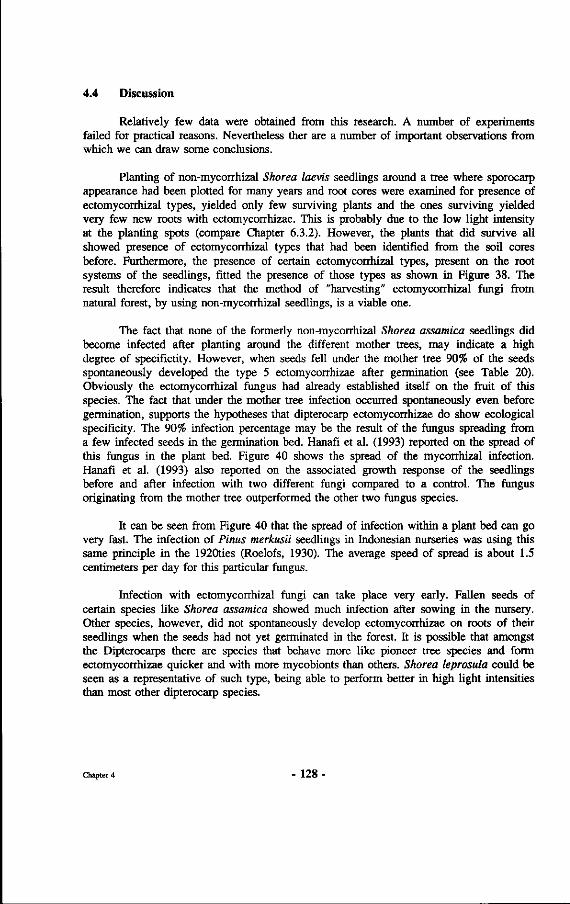
4.4 Discussion
Relatively few data were obtained from this research. A number of experiments failed for practical reasons. Nevertheless ther are a number of important observations from which we can draw some conclusions.
Planting of non-mycorrhizal Shorea laevis seedlings around a tree where sporocarp appearance had been plotted for many years and root cores were examined for presence of ectomycorrhizal types, yielded only few surviving plants and the ones surviving yielded very few new roots with ectomycorrhizae. This is probably due to the low light intensity at the planting spots (compare Chapter 6.3.2). However, the plants that did survive all showed presence of ectomycorrhizal types that had been identified from the soil cores before. Furthermore, the presence of certain ectomycorrhizal types, present on the root systems of the seedlings, fitted the presence of those types as shown in Figure 38. The result therefore indicates that the method of "harvesting" ectomycorrhizal fungi from natural forest, by using non-mycorrhizal seedlings, is a viable one.
The fact that none of the formerly non-mycorrhizal Shorea assamica seedlings did become infected after planting around the different mother trees, may indicate a high degree of specifictity. However, when seeds fell under the mother tree 90% of the seeds spontaneously developed the type 5 ectomycorrhizae after germination (see Table 20). Obviously the ectomycorrhizal fungus had already established itself on the fruit of this species. The fact that under the mother tree infection occurred spontaneously even before germination, supports the hypotheses that dipterocarp ectomycorrhizae do show ecological specificity. The 90% infection percentage may be the result of the fungus spreading from a few infected seeds in the germination bed. Hanafi et al. (1993) reported on the spread of this fungus in the plant bed. Figure 40 shows the spread of the mycorrhizal infection. Hanafi et al. (1993) also reported on the associated growth response of the seedlings before and after infection with two different fungi compared to a control. The fungus originating from the mother tree outperformed the other two fungus species.
It can be seen from Figure 40 that the spread of infection within a plant bed can go very fast. The infection of Pinus merkusii seedlings in Indonesian nurseries was using this same principle in the 1920ties (Roelofs, 1930). The average speed of spread is about 1.5 centimeters per day for this particular fungus.
Infection with ectomycorrhizal fungi can take place very early. Fallen seeds of certain species like Shorea assamica showed much infection after sowing in the nursery. Other species, however, did not spontaneously develop ectomycorrhizae on roots of their seedlings when the seeds had not yet germinated in the forest. It is possible that amongst the Dipterocarps there are species that behave more like pioneer tree species and form ectomycorrhizae quicker and with more mycobionts than others. Shorea leprosula could be seen as a representative of such type, being able to perform better in high light intensities than most other dipterocarp species.
Chapter 4 - 128 -
• • o o o « « » • • o n o o o o • o o o o o o « o o o o o « « «
Spread of infection after one month
• • • • • • • • • • • • • • o * * * « « « « * * * * « * o • • • • • • • • • • • • • o o o » » » « « « » o » « « o o • • • • • • • o » » « « « o o o o o o o « o o o o « o o o • • • • • • o « » o « « o o n o o o o o o o o o o o o o » • • o o » « o o » o o o o o o o o o o o « o « o o o o » » • o o o « « o o o o o o o o o o o o o o « « « o » « « # » • • o o o o o o o o o o o o o o o o « « « « » » » » » » » • o o o o o o o o o o o o o o o « » « « « « « « « « « « « o « « « o o o o o o « « o « o • • • • • • • • • • • • • •
Spread of infection after three months
o 0
o o o o o o o o o
o o o o o o o o o o o
o 0
o 0
o 0
o o 0
o o
• o o 0
o o o o o o 0
• • o o o o 0
o o o o
• o 0
o o o o o o o o
o o o o o o o o o o o
o o o o o o o 0
o 0
o
o 0
o o o o o o o 0 0
o 0
o 0
o 0
o o o o 0
o o o 0
o o o o 0
o 0
o o o o o o o o o o 0
o o o o o o o o 0
o o
o o 0
o 0
o o o o o o
o o o o o D o o o o o
o o o o o o o o o o o
o o o o o o o o o o o
o o o o o o o o o o o
o 0 0
0 0
0 0
0
o 0
o
• o o 0
o 0
o 0
o o o
• • o o o o o o o o o
• o o o o o o o o o o
o o o o o o o o o o o
o o o o o o o o o o 0
0
o 0
o 0
o 0
o 0
o 0
o o o o o o o o o o 0
o o 0
o o o o o o o o
o o o o o o o 0
0
o o
0
o 0
o 0
o o o o o o
Spread of infection after six months
Figure 40 : Spread of mycorrhizal infection after one, three and six months after inoculation with topsoil from beneath the mother tree. • - not infected; o - infected; D - inoculation spot..
The Shorea leprosula plants that were inoculated with chopped sporocarps showed large differences in height growth and leaf colour. Three different types of ectomycorrhizae were encountered amongst which Telephora terrestris. This fungus seemed to spread rapidly between many plants in the nursery. Obviously the precautions
Chapter 4 - 129 -
of separating the containers in different plant beds on concrete flooring were not enough to prevent infection with this "nursery" fungus.
The plants inoculated with Phylloporus bogoriensis were somewhat smaller than those inoculated with although no different type of fungus could be detected. This was probably due to the somewhat more exposed position with side light experienced by the plants in this treatment.
Chapter 4 - 130 •
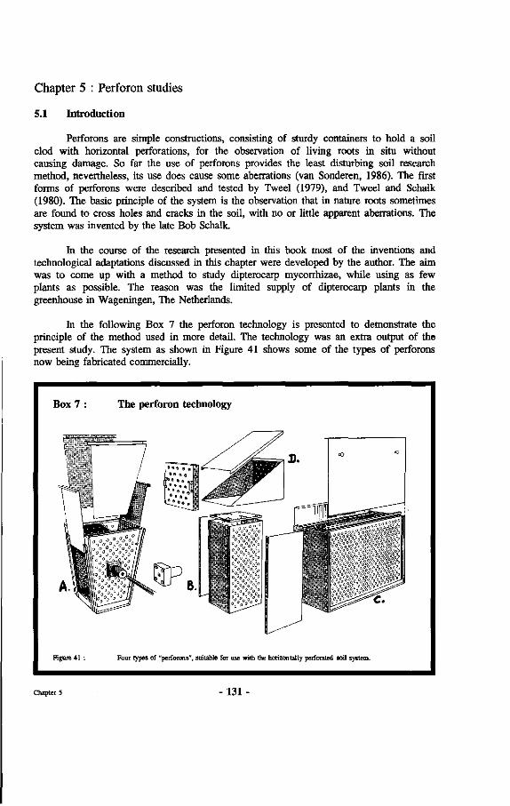
Chapter 5 : Perforon studies
5.1 Introduction
Perforons are simple constructions, consisting of sturdy containers to hold a soil clod with horizontal perforations, for the observation of living roots in situ without causing damage. So far the use of perforons provides the least disturbing soil research method, nevertheless, its use does cause some aberrations (van Sonderen, 1986). The first forms of perforons were described and tested by Tweel (1979), and Tweel and Schalk (1980). The basic principle of the system is the observation that in nature roots sometimes are found to cross holes and cracks in the soil, with no or little apparent aberrations. The system was invented by the late Bob Schalk.
In the course of the research presented in this book most of the inventions and technological adaptations discussed in this chapter were developed by the author. The aim was to come up with a method to study dipterocarp mycorrhizae, while using as few plants as possible. The reason was the limited supply of dipterocarp plants in the greenhouse in Wageningen, The Netherlands.
In the following Box 7 the perforon technology is presented to demonstrate the principle of the method used in more detail. The technology was an extra output of the present study. The system as shown in Figure 41 shows some of the types of perforons now being fabricated commercially.
Box 7 : The perforon technology
Figure 41 : Four types of "perforons", suitable for use with the horizontally perforated soil system.
Chapter 5 131
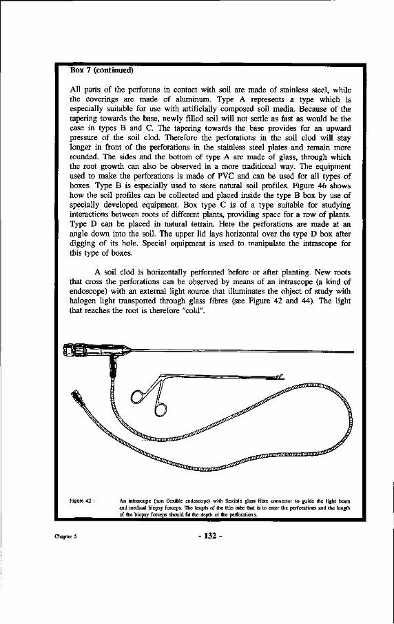
Box 7 (continued)
All parts of the perforons in contact with soil are made of stainless steel, while the coverings are made of aluminum. Type A represents a type which is especially suitable for use with artificially composed soil media. Because of the tapering towards the base, newly filled soil will not settle as fast as would be the case in types B and C. The tapering towards the base provides for an upward pressure of the soil clod. Therefore the perforations in the soil clod will stay longer in front of the perforations in the stainless steel plates and remain more rounded. The sides and the bottom of type A are made of glass, through which the root growth can also be observed in a more traditional way. The equipment used to make the perforations is made of PVC and can be used for all types of boxes. Type B is especially used to store natural soil profiles. Figure 46 shows how the soil profiles can be collected and placed inside the type B box by use of specially developed equipment. Box type C is of a type suitable for studying interactions between roots of different plants, providing space for a row of plants. Type D can be placed in natural terrain. Here the perforations are made at an angle down into the soil. The upper lid lays horizontal over the type D box after digging of its hole. Special equipment is used to manipulate the intrascope for this type of boxes.
A soil clod is horizontally perforated before or after planting. New roots that cross the perforations can be observed by means of an intrascope (a kind of endoscope) with an external light source that illuminates the object of study with halogen light transported through glass fibres (see Figure 42 and 44). The light that reaches the root is therefore "cold".
Figure 42 : An intrascope (non flexible endoscope) with flexible glass fibre connector to guide the light beam and medical biopsy forceps. The length of the thin tube that is to enter the perforations and the length of the biopsy forceps should fit the depth of the perforations.
Chapter 5 132
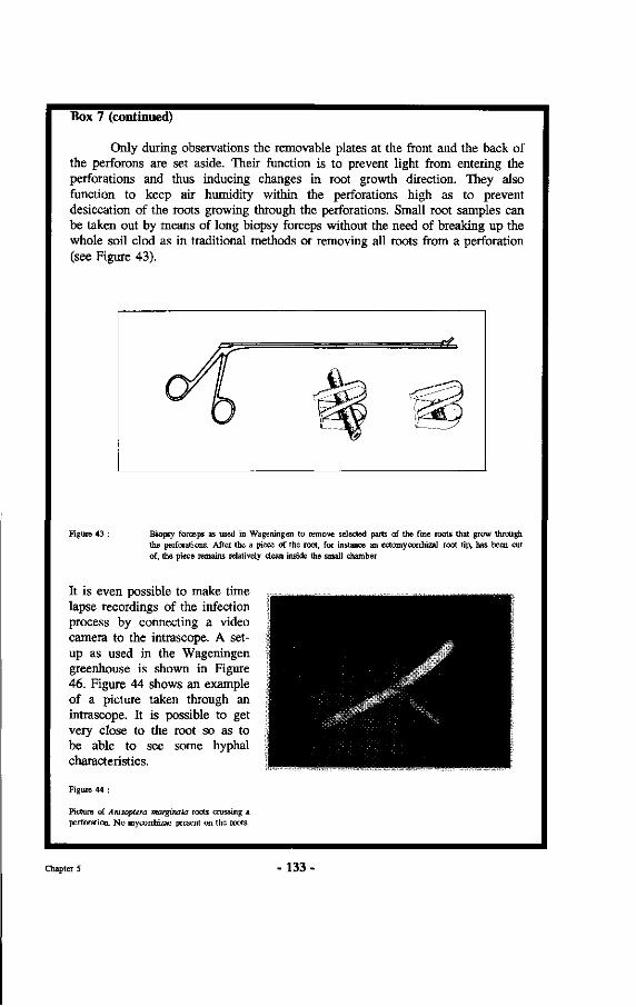
Box 7 (continued)
Only during observations the removable plates at the front and the back of the perforons are set aside. Their function is to prevent light from entering the perforations and thus inducing changes in root growth direction. They also function to keep air humidity within the perforations high as to prevent desiccation of the roots growing through the perforations. Small root samples can be taken out by means of long biopsy forceps without the need of breaking up the whole soil clod as in traditional methods or removing all roots from a perforation (see Figure 43).
Figure 43 : Biopsy forceps as used in Wageningen to remove selected parts of the fine roots that grow through the perforations. After the a piece of the root, for instance an ectomycorrhizal root tip, has been cut of, the piece remains relatively clean inside the small chamber
It is even possible to make time lapse recordings of the infection process by connecting a video camera to the intrascope. A setup as used in the Wageningen greenhouse is shown in Figure 46. Figure 44 shows an example of a picture taken through an intrascope. It is possible to get very close to the root so as to be able to see some hyphal characteristics.
Figure 44 :
Picture of Anisoptera marginala roots crossing a perforation. No mycorrhizae present on the roots.
Chapter 5 133
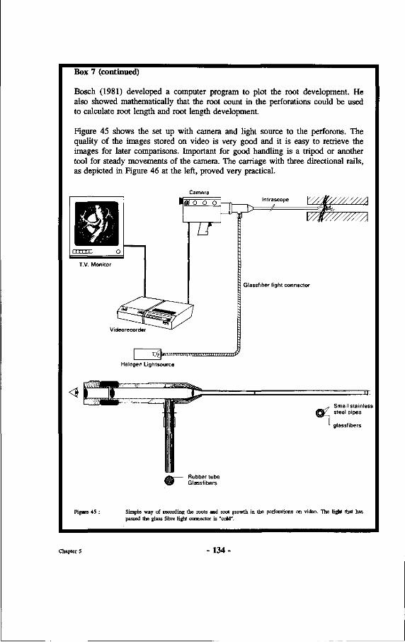
Box 7 (continued)
Bosch (1981) developed a computer program to plot the root development. He also showed mathematically that the root count in the perforations could be used to calculate root length and root length development.
Figure 45 shows the set up with camera and light source to the perforons. The quality of the images stored on video is very good and it is easy to retrieve the images for later comparisons. Important for good handling is a tripod or another tool for steady movements of the camera. The carriage with three directional rails, as depicted in Figure 46 at the left, proved very practical.
Camera
Videorecorder
Glassfiber light connector
Tnwmiitiiii/iiiiiiirin, nuimiiTTTn^
Halogen Lightsource
Small stainless steel pipes
glassfibers
Figure 45 : Simple way of recording the roots and root growth in the perforations on video. The light 1 passed the glass fibre light connector is "cold".
Chapter 5 134
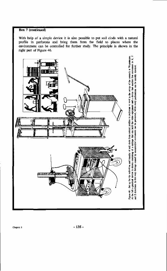
Box 7 (continued)
With help of a simple device it is also possible to put soil clods with a natural profile in perforons and bring them from the field to places where the environment can be controlled for further study. The principle is shown in the right part of Figure 46.
2 1 £
lil 'S " &
i l l? S3 s a ut S o
1.11 1*1 2 ! I alt P. -a _
* -s
§ • ' § •
Chapter 5 - 1 3 5 -
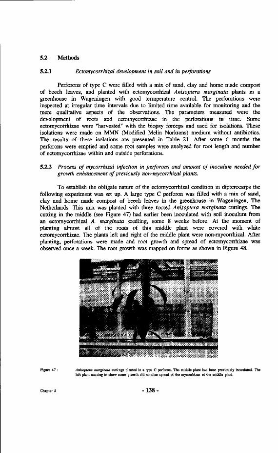
5.2 Methods
5.2.1 Ectomycorrhizal development in soil and in perforations
Perforons of type C were filled with a mix of sand, clay and home made compost of beech leaves, and planted with ectomycorrhizal Anisoptera marginata plants in a greenhouse in Wageningen with good termperature control. The perforations were inspected at irregular time intervals due to limited time available for monitoring and the mere qualitative aspects of the observations. The parameters measured were the development of roots and ectomycorrhizae in the perforations in time. Some ectomycorrhizae were "harvested" with the biopsy forceps and used for isolations. These isolations were made on MMN (Modified Melin Norkrans) medium without antibiotics. The results of these isolations are presented in Table 21. After some 6 months the perforons were emptied and some root samples were analyzed for root length and number of ectomycorrhizae within and outside perforations.
5.2.2 Process of mycorrhizal infection in perforons and amount of inoculum needed for growth enhancement of previously non-mycorrhizal plants.
To establish the obligate nature of the ectomycorrhizal condition in dipterocarps the following experiment was set up. A large type C perforon was filled with a mix of sand, clay and home made compost of beech leaves in the greenhouse in Wageningen, The Netherlands. This mix was planted with three rooted Anisoptera marginata cuttings. The cutting in the middle (see Figure 47) had earlier been inoculated with soil inoculum from an ectomycorrhizal A. marginata seedling, some 8 weeks before. At the moment of planting almost all of the roots of this middle plant were covered with white ectomycorrhizae. The plants left and right of the middle plant were non-mycorrhizal. After planting, perforations were made and root growth and spread of ectomycorrhizae was observed once a week. The root growth was mapped on forms as shown in Figure 48.
Figure 47 : Anisoptera marginata cuttings planted in a type C perforon. The middle plant had been previously inoculated. The left plant starting to show some growth did so after spread of the mycorrhizae of the middle plant.
Chapter 5 138
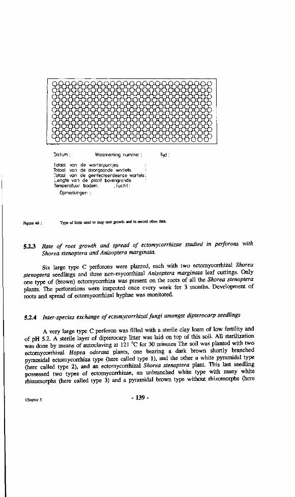
Datum : Waarneming nummer : Tijd:
Totaal van de wortelpuntjes Totaal van de doorgaande wortels Totaal van de geinfecteerdeerde wortels Lengte van de plant bovengronds Temperatuur bodem: . lucht :
Opmerkingen :
Figure 48 : Type of form used to map root growth and to record other data.
5.2.3 Rate of root growth and spread of ectomycorrhizae studied in perforons with Shorea stenoptera and Anisoptera marginata.
Six large type C perforons were planted, each with two ectomycorrhizal Shorea stenoptera seedlings and three non-mycorrhizal Anisoptera marginata leaf cuttings. Only one type of (brown) ectomycorrhiza was present on the roots of all the Shorea stenoptera plants. The perforations were inspected once every week for 3 months. Development of roots and spread of ectomycorrhizal hyphae was monitored.
5.2.4 Inter-species exchange of ectomycorrhizal fungi amongst dipterocarp seedlings
A very large type C perforon was filled with a sterile clay loam of low fertility and of pH 5.2. A sterile layer of dipterocarp litter was laid on top of this soil. All sterilization was done by means of autoclaving at 121 °C for 30 minutes The soil was planted with two ectomycorrhizal Hopea odorata plants, one bearing a dark brown shortly branched pyramidal ectomycorrhiza type (here called type 1), and the other a white pyramidal type (here called type 2), and an ectomycorrhizal Shorea stenoptera plant. This last seedling possessed two types of ectomycorrhizae, an unbranched white type with many white rhizomorphs (here called type 3) and a pyramidal brown type without rhizomorphs (here
Chapter 5 139-
Table 22: "Natural" infection of Vatica cf. bancana, Amsoptera marginata and Dipterocarpus confertus by 4 ectomycorrhizal fungi.
ECM types :
Hopea odorata
Shorea stenoptera
Vatica cf. bancana
Anisoptera marginata
Dipterocarpus confertus
1
X
0
0
0
X
2
X
0
0
X
0
3
0
X
0
0
0
4
0
X
0
0
0
Only the three originally ectomycorrhizal plants and one Dipterocarpus confertus plant showed good above ground growth. One D. confertus died before the 10th month and all V, bancana became chlorotic and stunted.
5.4 Discussion and conclusions
5.4.1 Methodology.
Normal ectomycorrhizae developed within the perforations. However, their number was on the average 20% higher inside the perforations than in the soil surrounding the perforations. This may be related to the good aeration inside the perforations. Furthermore, it must be noted that in the case of Dipterocarpaceae soil compaction might also have influenced the number of ectomycorrhizae through the greater number of available side roots for infection. These side roots are especially produced at the outer side of a bending root, as is shown in Figure 54. Groot (1987), however, found the opposite when conducting perforon studies with European Quercus robur. He found less branching of roots inside the perforations. From the figures he presents it looks as if Quercus robur does not react with formation of side roots at places where the roots get bent like all dipterocarps so far tested in perforons. Perhaps as a consequence, Groot did find less ectomycorrhizae inside the perforations than in the surrounding soil. Groot (1987) noticed that ectomycorrhizae of Quercus robur tend to be smaller inside the perforations than in the surrounding soil. With Shorea assamica type 5 mycorrhizae (Appendix 7) this was not very clear. The morphological appearance of ectomycorrhizae inside the perforations was otherwise much the same as the appearance of ectomycorrhizae in the soil.
Chapter 5 - 144
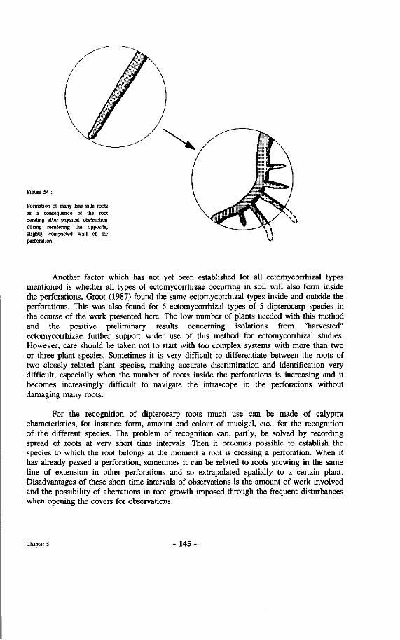
Figure 54 :
Formation of many fine side roots as a consequence of the root bending after physical obstruction during reentering the opposite, slightly compacted wall of the perforation.
Another factor which has not yet been established for all ectomycorrhizal types mentioned is whether all types of ectomycorrhizae occurring in soil will also form inside the perforations. Groot (1987) found the same ectomycorrhizal types inside and outside the perforations. This was also found for 6 ectomycorrhizal types of 5 dipterocarp species in the course of the work presented here. The low number of plants needed with this method and the positive preliminary results concerning isolations from "harvested" ectomycorrhizae further support wider use of this method for ectomycorrhizal studies. However, care should be taken not to start with too complex systems with more than two or three plant species. Sometimes it is very difficult to differentiate between the roots of two closely related plant species, making accurate discrimination and identification very difficult, especially when the number of roots inside the perforations is increasing and it becomes increasingly difficult to navigate the intrascope in the perforations without damaging many roots.
For the recognition of dipterocarp roots much use can be made of calyptra characteristics, for instance form, amount and colour of mucigel, etc., for the recognition of the different species. The problem of recognition can, partly, be solved by recording spread of roots at very short time intervals. Then it becomes possible to establish the species to which the root belongs at the moment a root is crossing a perforation. When it has already passed a perforation, sometimes it can be related to roots growing in the same line of extension in other perforations and so extrapolated spatially to a certain plant. Disadvantages of these short time intervals of observations is the amount of work involved and the possibility of aberrations in root growth imposed through the frequent disturbances when opening the covers for observations.
Chapter 5 - 1 4 5 -
specificity" encountered here therefore might indicate a higher specialization under natural conditions.
The strong specialization for substrates that had already been concluded from the sporocarp inventories in natural forest, is supported by the different degree of colonization of different substrates in the perforon with mixed species (5.3.4). Smits and Noor (1987) also found a specialization of fungi for peat medium compared to other substrates used. The findings here therefore support the conclusions of Last et al. (1992) on mycobiont substrate preferences.
Chapter 5 - 148 -

Chapter 6 : Physical influences upon ectomycorrhizae
6.1 Introduction
Smits (1983a) hypothesized that soil compaction, soil oxygen and soil temperature might be factors contributing to the presence and performance of Dipterocarp mycorrhizae. From the observations in Chapter 2.1, it was already suspected that slight changes in soil temperature result in large differences in numbers of sporocarps of ectomycorrhizal fungi appearing. In the following pages experiments investigating the influence of the above-mentioned physical factors upon ectomycorrhizae of Dipterocarpaceae are described.
6.2 Methods
6.2.1 Experiment 1 (controlled heating of mycorrhizal root systems)
This experiment was conducted in the greenhouse of the Department of Silviculture & Forest Ecology, Agricultural University Wageningen. For the experiment, young Wildlings varying in height from 15 to 50 centimetres were used. All of the plants originated from Wildlings collected in the Kebun Raya botanical garden at Bogor, Indonesia, except for the Shorea multiflora Wildlings that came from natural Dipterocarp forest in Sabah. Table 23 shows the species used in the experiment and the number of plants per species.
Table 23 : Dipterocarp species used in the soil heating experiment. The number between brackets presents the working code, number = number of plants, myc. type = type of ectomycorrhizae, a= amphimycorrhizae (see Chapter 7)., crit. temp, is temperature at which the type of mycorrhizae died (excluding a, see Chapter 7). Maximum variation between brackets.
Species number ecm.type crit.temp.(°C)
Dryobalanops lanceolata (Dl) 1 1, a 31 (1) Hopea bancana (H2) 7 2 30.5 (0.5) Hopea pierii (HI) 7 2 30.5 (0.5) Hopea sangal (H3) 7 3, a 31 (1) Shorea balangeran (S5) 17 4, 5, a 29.5/30.5 (1.5/1) Shorea leprosula (SI) 5 6 31.5 (0.5) Shorea multiflora (S2) 6 7 28.5 (1) Shorea pinanga (S7) 4 4, a 30 (1) Shorea robusta (S4) 5 4 30 (1) Shorea selanica (S3) 4 6, a 31.5 (1) Shorea seminis (S8) 5 6 32 (1) Shorea sp. (S6) 5 7(?), a 29.5 (0.5) Vatica bancana (VI) 6 8 30.5 (1) Vatica chinensis (V3) 3 8 30 (1) Vatica pauciflora (V2) 6 9 32 (1)
Chapter 6 - 149 -
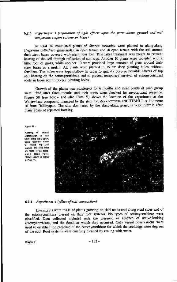
6.2.3 Experiment 3 (separation of light effects upon the parts above ground and soil temperature upon ectomycorrhizae)
In total 30 inoculated plants of Shorea assamica were planted in alang-alang (Imperata cylindrica grasslands), in open terrain and in open terrain with the soil around their stem bases covered with aluminum foil. This latter treatment was meant to prevent heating of the soil through reflection of sun rays. Another 10 plants were provided with a little roof of grass, while another 10 were provided large amounts of grass around their stem bases as a mulch. All plants were planted in 15 cm deep planting holes, without fertilizer. The holes were kept shallow in order to quickly observe possible effects of top soil heating on the ectomycorrhizae and to prevent temporary survival of ectomycorrhizal roots in loose soil in deeper planting holes.
Growth of the plants was monitored for 6 months and three plants of each group were lifted after three months and their roots were checked for mycorrhizal presence. Figure 58 (see below and also Plate V) shows the location of the experiment at the Wanawisata compound managed by the state forestry enterprise INHUTANI I, at kilometre 10 from Balikpapan. The site, dominated by the alang-alang grass, is very infertile after many years of repeated burning.
Figure 58 :
Planting of several Dipterocarps in very short alang-alang grass, using different means to reduce top soil heating. The little roofs are made of the alang-alang grass itself. Picture shown in colour in Plate V.
6.2.4 Experiment 4 (effect of soil compaction)
Inventories were made of plants growing on skid roads and along road sides and of the ectomycorrhizae present on their root systems. No types of ectomycorrhizae were classified. Data collected included only the presence or absence of active-looking ectomycorrhizae, and the depth at which they occurred. Only visual observations were used to establish the presence of the ectomycorrhizae for which the seedlings were dug out of the soil. Root systems were carefully cleaned by rinsing with water.
Chapter 6 -152
Observations were made in logged-over forest, 10 years after logging and 1 year after logging in the ITCI concession (Jalan 1000 and Meratus), on Pulau Laut within the P.T.INHUTANI II concession and in Berau within the P.T. INHUTANI I concession. The species of which seedlings were observed and investigated included Shorea ovalis, S. parvifolia, S. leprosula, S. polyandra, S. johorensis and Dryobalanops lanceolata.
6.2.5 Experiment 5 (effect of soil exposure on older trees)
Twenty Shorea laevis saplings, eight years old and on average 4 meter high, that had been planted under a secondary forest vegetation, were selected for this experiment. Seven of the trees were exposed to full light by removal of the surrounding vegetation. All the branches and litter were removed around the stem bases of these trees. Regrowth of grass and herbs was regularly removed. After three months, three of the exposed trees were given a mulch layer around the bases of their stems. Observations were made on the morphological appearance of the roots and ectomycorrhizae of the trees and the condition of the above ground parts. Temperatures of the top layer of soil (3-5 centimetres) were measured regularly.
6.3 Results
6.3.1 Results of experiment 1
Table 23 presents the results of the effect of heating upon the various associated fungi of the Dipterocarpaceae used in the experiment. It was noted that, depending upon the associated fungus, effects of the temperature treatment started to become visible on the root system after lifting the pots, after one week. Formerly fresh ectomycorrhizae with relatively bright colours became grey brown. After four weeks the newly produced leaves of plants whose ectomycorrhizae had disappeared fully started becoming yellowish (Figure 59, see plate IV). Later growth of these plants stopped completely and the plants that were not reinoculated with suitable fungi died. All of the ectomycorrhizal types either disappeared completely or showed a clear boundary between the zone where the ectomycorrhizae had died and where they were still looking healthy. Microscopic sections made through some dead ectomycorrhizae after 2 weeks, showed shrunk hyphae around normal looking cortex cells.
6.3.2 Results of experiment 2
Table 24 shows the top soil temperatures under the various planting conditions and light conditions. Table 25 presents the results (height growth, morphological appearance of the plants and survival percentages for the different planting sites) of experiment 2 after six months.
Chapter 6 - 153 •
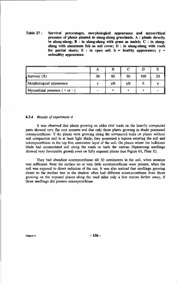
Table 27 : Survival percentages, morphological appearance and mycorrhizal presence of plants planted in alang-alang grasslands. A : plants directly in alang-alang; B : in alang-alang with grass as mulch; C : in alang-alang with aluminum foil as soil cover; D : in alang-alang with roofs for partial shade; E : in open soil, h = healthy appearance; y = unhealthy appearance.
Survival (%)
Morphological appearance
Mycorrhizal presence ( + or - )
A
30
y
-
B
80
y/h
+
C
90
y/h
+
D
100
h
+
E
20
y
-
6.3.4 Results of experiment 4
It was observed that plants growing on older skid roads on the heavily compacted parts showed very flat root systems and that only those plants growing in shade possessed ectomycorrhizae. If the plants were growing along the compacted trails on places without soil compaction and in at least light shade, they possessed a taproot entering the soil and ectomycorrhizae in the top five centimetre layer of the soil. On places where the bulldozer blade had accumulated soil along the roads or trails the various Dipterocarp seedlings showed very favourable growth even on fully exposed places (see Figure 61, Plate ÏÏ).
They had abundant ectomycorrhizae till 30 centimetres in the soil, when aeration was sufficient. Near the surface no or very little ectomycorrhizae were present, when the soil was exposed to direct radiation of the sun. It was also noticed that seedlings growing closer to the mother tree in the shadow often had different ectomycorrhizae from those growing on the exposed places along the road sides only a few meters farther away, if those seedlings did possess ectomycorrhizae.
Chapter 6 156
6.3.5 Results of experiment 5
All the 13 trees in the control treatment continued growing normal with healthy green leaves. The soil temperature was a constant 26.7 °C. Although the density of ectomycorrhizae seemed to vary slightly from month to month, healthy looking ectomycorrhizae were always present. The seven trees that were exposed to full sunlight soon dropped many leaves while the newly produced leaves became smaller and yellowish and, especially when still developing, were much more reddish/purplish.
The condition of their ectomycorrhizae changed very quickly. After one month the mantle colour had become brownish-black and these deteriorated ectomycorrhizae would break of very easily. All saplings reacted similarly. When the soil around three of the exposed saplings was covered with mulching material the condition of these trees gradually improved reflected in the appearance of many new leaves of light green colour and the reappearance of healthy looking ectomycorrhizae. The new green leaves of these mulched saplings remained smaller than those of the control saplings and the colour was not as green as the leaves of the control plants. The four remaining exposed saplings stayed yellowish and one of the saplings died.
6.4 Discussion
Although much variation existed in approach between the various experiments and the numbers of plants, as well as the species involved, all the results showed the same trend. Seedlings on exposed places do not perform as well as seedlings growing in light shade, and ectomycorrhizal development was worst in heavy shade and in full sunlight except when the soil temperature was lower than 30 °C in the latter case, for instance as can be seen for the plants with aluminum foil around their stem bases in Table 27.
Sasaki reported that most dipterocarp seedlings reach their optimal photosynthesis at about 50-70% of full light intensity. Full sunlight can cause stress for the plants. This is clear for instance for the Shorea laevis saplings in experiment 5. Full sudden sunlight exposure leads to significant loss of leaves and newly formed leaves are smaller than the former "shade leaves". Figure 62 (shown in colour in Plate VI) shows the dramatic yellowing of well established Shorea polyandra trees (location Pulau Laut, P.T. LNHUTANI II) planted underneath Paraserianthes falcataria. When the overstorey trees were removed all the leaves turned yellow, demonstrating the severe stress brought about by the exposure to full sunlight. After about four months normal growth resumed.
Voogd (1933) reports similar findings for Shorea platyclados planted in Djoepi Besar (600 m altitude, Sumatra). His pictures on p. 707 show the good performance of plants planted under a protective crown layer compared to those in open terrain. Mauricio (1956) also reports the poor performance of Dipterocarps in open terrain by either Wildlings or direct seeding. Voogd (1933) also reports the failure to perform direct seeding, although he provides many reasons other than just the exposure to light, for instance, seed lobes being eaten by squirrels and mice, ants damaging the seeds and a dry spell. Plants in the nursery would show good survival but almost all turned yellow.
Chapter 6 - 157 -
Voogd mentions specifically that the largest plants often had clearly visible ectomycorrhizae at the end of their roots. Therefore he not only noticed their presence but also, more than half a century ago, seemed to understand about their importance. Voogd also mentions that healthy growing plants from the nursery did perform well after planting compared to the seeds germinating directly in the planting holes. This seems a clear result of the ectomycorrhizal presence.
Voogd (1933) also reports the planting of Shorea platyclados in alang-alang grasslands. He found very low survival percentages, but noted that some plants, covered by the grass, still showed good growth. The experiment with planting in the alang-alang grass described here confirms his observation. Voogd reports that best growth of the seedlings took place when the seedlings were planted under secondary forest in the neighbourhood of mother trees. Voogd (1932) reports good results with planting of Hopea mengerawan by local population, using Wildlings and planting under secondary forest, near Boerai (Palembang, Sumatra). Schuitemaker (1933) reports that in West Kalimantan the local population has taken up planting of various dipterocarp species, for instance in Meliau, Sanggau, Tajan and Landak, for illipe nut and resin production. Planting is done by using Wildlings that are again planted close to mother trees in light shade. In these cases both aspects of applying light shade and planting close to mother trees are mixed. These observations in combination with the distribution of Hopea rudiformis seedlings along roots of the mother tree (Chapter 3) and death of non-mycorrhizal plants indicate the strongly obligate character of the dipterocarp seedlings as well as a possible assistance by the mother tree for the seedlings through a mycorrhizal guild.
Soil compaction mainly prevents development of roots and mycorrhizae into deeper layers, which makes the seedlings more susceptible to a rise in temperature occurring in the top soil layer which has a negative impact upon the Dipterocarp ectomycorrhizae. Probably the seedlings growing on these compacted parts of the skidroads are also more prone to drought because of their shallow root system. It seems that the physical resistance to root and fungal penetration may be a factor but that aeration is at least as important. Coster (1933, p.45) states that the average concentration of oxygen in soil air is lower in tropical areas compared with temperate zones. Nopamornbodi et al. (1987) report the effect of inundation upon survival of VA mycorrhizal fungi. Stenström and Unestam (1987) report the differences in ectomycorrhizal fungi getting established with inundation. For certain species daily flooding for 2 minutes already prevented mycorrhiza formation.
As shown in this chapter, soil temperatures in the top soil layer can raise dramatically depending upon the condition of the shade. Kramer (1932) also reports on top soil temperature increases. He measured increases from 26.5 °C in the morning to 42.5 °C at 13.00 h. in the top 5 centimetres. The daily average was 37 °C. Top soil temperatures in alang-alang grasslands were about as high as in completely open terrain, devoid of vegetation. Kramer never measured temperatures above 29.5 °C under any form of shrub or tree vegetation. Here it was found that most ectomycorrhizae do not survive when exposed to high top soil temperatures for longer periods. Parke et al. (1983) reported that when soil temperatures exceeded 27 °C no ectomycorrhizae would form on the roots of Douglas-fir seedlings.
Chapter 6 - 158 -
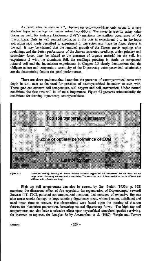
As could also be seen in 2.2, Dipterocarp ectomycorrhizae only occur in a very shallow layer in the top soil under natural conditions. The same is true in many other places as well, for instance Linderman (1987a) mentions the shallow occurrence of VA mycorrhizae. Only in well aerated media, as in the pots in experiment 1 or in the loose soil along skid roads described in experiment 4, can ectomycorrhizae be found deeper in the soil. It may be claimed that the regained growth of the Shorea laevis saplings after mulching, and the better performance of the Shorea assamica seedlings under primary and secondary forest, may be related to the presence of organic material on the soil, but experiment 2 with the aluminum foil, the seedlings growing in shade on compacted mineral soil and the inoculation experiments in Chapter 2.3 clearly demonstrate that the obligate nature and temperature sensitivity of the Dipterocarp ectomycorrhizal relationship are the determining factors for good performance.
There are three gradients that determine the presence of ectomycorrhizal roots with depth in soil, next to the need for presence of ectomycorrhizal inoculum to start with. These gradient concern soil temperature, soil oxygen and soil compaction. Under normal conditions the first two will be of most importance. Figure 63 presents schematically the conditions for thriving dipterocarp ectomycorrhizae.
Top soil temperature influence
Zone of optimal performance of ECM
Imk of oxygen
Figure 63 : Schematic drawing showing the relation between available oxygen and soil temperature and soil depth and the range where dipterocarp ectomycorrhizae can thrive. The values for each of these conditions can be different with different soils, climates and fungi.
High top soil temperatures can also be caused by fire. Endert (1933b, p. 398) mentions the disastrous effect of fire especially for regeneration of Dipterocarps. Suwardi Suwasa (PT. ITCI, personal communication) mentions that presence of extensive fire can also cause smoke damage to large standing dipterocarp trees, which become defoliated and need much time to recover. His observations were based upon the burning of clearcut forests for plantation preparation, bordering natural dipterocarp forest. The high top soil temperatures can also have a selective effect upon mycorrhizal inoculum species surviving, for instance as reported for Douglas fir by Amaranthus et al. (1987). Wright and Tarrant
Chapter 6 - 159 -
(1958) already demonstrated that intense bums negatively effect the mycorrhizal inocula potential of a site. Schramm (1966) showed the effect of high soil temperatures upon roots and ectomycorrhizae. Temperature of the top soil layers depends upon many factors. In general a dry soil tends to become hottest. In dry soils Schramm recorded top soil temperatures up to 67 °C (maximum 75), quickly lowering with depth (49.5 °C at one inch depth). Schramm found chlorotic seedlings of pines to be non-mycorrhizal. Mycorrhizal plants were healthy looking and always growing on the places with lower top soil temperatures (Schramm, 1966; Table 28, p. 114). Schramm, however concluded that not the temperature effect upon roots or associated fungi was the cause for poorly performing ectomycorrhizae but the reduced amounts of sugar transported to the roots because of heat damage to the phloem just above the soil surface. The present author is of the opinion that the effect of heat directly upon the ectomycorrhizae is the real cause.
Muchovej and Kasuya (1987) showed vast reductions in the percentages of colonization in all fungi except Pisolithus tinctorius when the medium temperature was increased from 28 to 32 °C. VAM seem to be less susceptible than ECM for (short term) exposure to high top soil temperatures (Klopatek et al., 1987) under fire. Nadarajah and Nawawi (1987) found more or less the same, testing the effect of temperature on germination and growth of VAM. They found that most of their strains showed a wide temperature tolerance. They also mention the need for this tolerance in cocoa plantations where surface soil temperatures can exceed 35 °C. Moawad (1980) describes differences in performance by VAM at different temperatures.
Taking in account all the aforementioned aspects it can be concluded that the performance of dipterocarp planting stock will depend upon a number of conditions. First of all presence ectomycorrhizal inoculum, for instance ectomycorrhizal roots, or established ectomycorrhizae, of suitable fungi. Linderman (1987b, p. 73) mentions that many of the fungi that were effective ectomycorrhizae formers in the nursery do not survive after outplanting. He suggested that therefore we may need other fungi to be inoculated at outplanting that can withstand the rigors of stressful sites. The same was found by Stenström and Unestam (1987) where their "forest fungi" performed better than the "nursery fungi" after outplantings.
The second condition concerns the normal stress factors such as évapotranspiration and damage by biotic causes. The third aspect refers to the conditions for the performance of ectomycorrhizae, which include the top soil temperature, top soil oxygen availability and opportunities for physical penetration of the soil by roots and fungal hyphae. In addition perhaps competition and general predisposition of the associated plants could be mentioned.
For practical application in large scale forestry this means that the number of actions that can be taken is limited to regulation of shade and production of planting stock with best adapted mycorrhizae. Sometimes, for instance in alang-alang grasslands, mulching can be an option as well.
Chapter 6 - 160 -
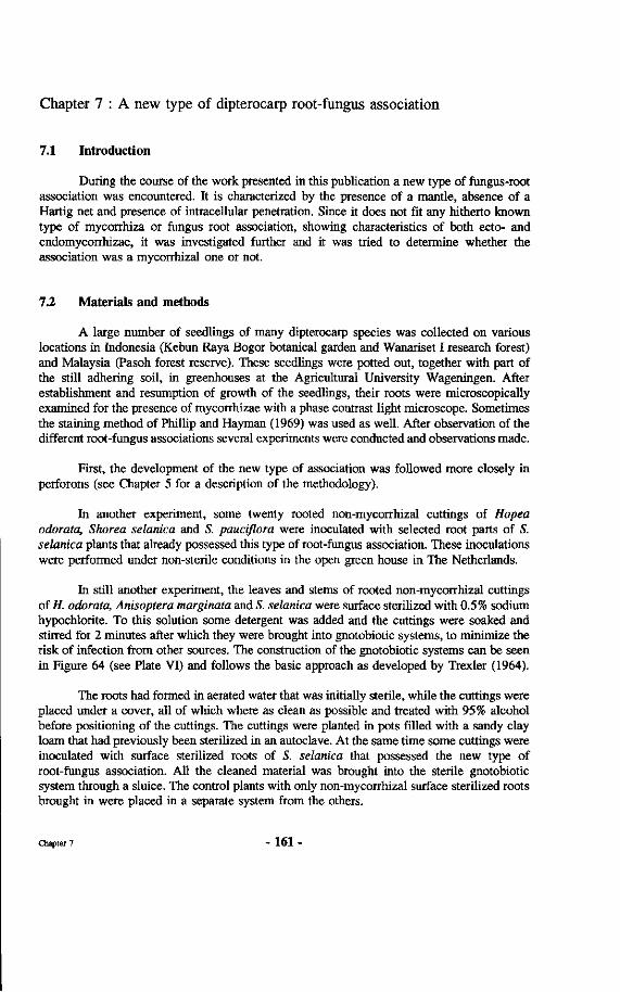
Chapter 7 : A new type of dipterocarp root-fungus association
7.1 Introduction
During the course of the work presented in this publication a new type of fungus-root association was encountered. It is characterized by the presence of a mantle, absence of a Hartig net and presence of intracellular penetration. Since it does not fit any hitherto known type of mycorrhiza or fungus root association, showing characteristics of both ecto- and endomycorrhizae, it was investigated further and it was tried to determine whether the association was a mycorrhizal one or not.
7.2 Materials and methods
A large number of seedlings of many dipterocarp species was collected on various locations in Indonesia (Kebun Raya Bogor botanical garden and Wanariset I research forest) and Malaysia (Pasoh forest reserve). These seedlings were potted out, together with part of the still adhering soil, in greenhouses at the Agricultural University Wageningen. After establishment and resumption of growth of the seedlings, their roots were microscopically examined for the presence of mycorrhizae with a phase contrast light microscope. Sometimes the staining method of Phillip and Hayman (1969) was used as well. After observation of the different root-fungus associations several experiments were conducted and observations made.
First, the development of the new type of association was followed more closely in perforons (see Chapter 5 for a description of the methodology).
In another experiment, some twenty rooted non-mycorrhizal cuttings of Hopea odorata, Shorea selanica and S. pauciflora were inoculated with selected root parts of S. selanica plants that already possessed this type of root-fungus association. These inoculations were performed under non-sterile conditions in the open green house in The Netherlands.
In still another experiment, the leaves and stems of rooted non-mycorrhizal cuttings of H. odorata, Anisoptera marginata and S. selanica were surface sterilized with 0.5% sodium hypochlorite. To this solution some detergent was added and the cuttings were soaked and stirred for 2 minutes after which they were brought into gnotobiotic systems, to minimize the risk of infection from other sources. The construction of the gnotobiotic systems can be seen in Figure 64 (see Plate VI) and follows the basic approach as developed by Trexler (1964).
The roots had formed in aerated water that was initially sterile, while the cuttings were placed under a cover, all of which where as clean as possible and treated with 95% alcohol before positioning of the cuttings. The cuttings were planted in pots filled with a sandy clay loam that had previously been sterilized in an autoclave. At the same time some cuttings were inoculated with surface sterilized roots of S. selanica that possessed the new type of root-fungus association. All the cleaned material was brought into the sterile gnotobiotic system through a sluice. The control plants with only non-mycorrhizal surface sterilized roots brought in were placed in a separate system from the others.
Chapter 7 - 161 -
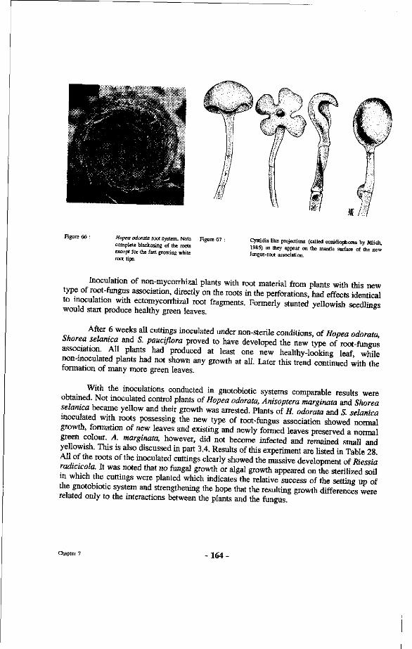
Figure 66 : Hopea odorata root system. Note complete blackening of the roots except for the fast growing white root tips.
Figure 67 : Cystidia like projections (called conidiophores by Jülich, 1985) as they appear on the mantle surface of the new fungus-root association.
Inoculation of non-mycoirhizal plants with root material from plants with this new type of root-fungus association, directly on the roots in the perforations, had effects identical to inoculation with ectomycorrhizal root fragments. Formerly stunted yellowish seedlings would start produce healthy green leaves.
After 6 weeks all cuttings inoculated under non-sterile conditions, of Hopea odorata Shorea selanica and S. pauciflora proved to have developed the new type of root-fungus association. All plants had produced at least one new healthy-looking leaf while non-inoculated plants had not shown any growth at all. Later this trend continued with the formation of many more green leaves.
. W"h the inoculations conducted in gnotobiotic systems comparable results were obtained. Not moculated control plants of Hopea odorata, Anisoptera margimta and Shorea selanica became yellow and their growth was arrested. Plants of H. odorata and S selanica inoculated with roots possessing the new type of root-fungus association showed normal growth, formation of new leaves and existing and newly formed leaves preserved a normal green colour. A. marginata, however, did not become infected and remained small and ye lowish. This is also discussed in part 3.4. Results of this experiment are listed in Table 28 All of the roots of the inoculated cuttings clearly showed the massive development of Riessia radicicola.lt was noted that no fungal growth or algal growth appeared on the sterilized soil in which the cuttings were planted which indicates the relative success of the setting up of the gnotobiotic system and strengthening the hope that the resulting growth differences were related only to the interactions between the plants and the fungus
Chapter 7 -164
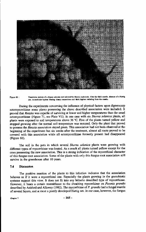
Figure 68 : Transverse section of a Hopea odorata root infected by Riessia radicicola. Note the thick mantle, absence of a Hartig net, intracellular hyphae bearing clamp connections and thick hyphae radiating from the mantle.
During the experiments concerning the influence of physical factors upon dipterocarp ectomycorrhizae some plants possessing the above described association were included. It proved that Riessia was capable of surviving at lower and higher temperatures than the usual ectomycorrhizae (Figure 71, see Plate VII). In one case with six Shorea selanica plants, all plants were exposed to soil temperatures above 36 °C. Five of the plants turned yellow and stopped growing after the normal soil temperature was restored. Only the plant that proved to possess the Riessia association stayed green. This association had not been observed at the beginning of the experiment but six weeks after the treatment, almost all roots proved to be covered with this association while all ectomycorrhizae formerly present had disappeared (Figure 66).
The soil in the pots in which several Shorea selanica plants were growing with different types of mycorrhizae was heated. As a result all plants turned yellow except for the ones possessing the new association. This is a strong indication of the mycorrhizal character of this fungus-root association. Some of the plants with only this fungus-root association still survive in the greenhouse after 10 years.
7.4 Discussion
The positive reaction of the plants to this infection indicates that the association behaves as if it were a mycorrhizal one. Especially the plants growing in the gnotobiotic systems support this view. It does not fit into any hitherto described type of mycorrhizae. However, it bears a certain resemblance to the sheathing mycorrhizae on Pisonia grandis described by Ashford and Allaway (1982). The mycorrhizae of P. grandis had a fungal mantle of several layers, and at most a poorly developed Hartig net. In our case, however, the fungus
Chapter 7 - 1 6 5 -
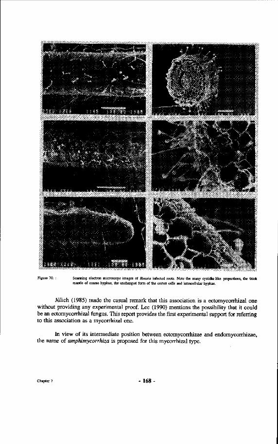
Figure 70. : Scanning electron microscope images of Riessia infected roots. Note the many cystidia-like projections, the thick mantle of coarse hyphae, the unchanged form of the cortex cells and intracellular hyphae.
Jülich (1985) made the casual remark that this association is a ectomycorrhizal one without providing any experimental proof. Lee (1990) mentions the possibility that it could be an ectomycorrhizal fungus. This report provides the first experimental support for referring to this association as a mycorrhizal one.
In view of its intermediate position between ectomycorrhizae and endomycorrhizae, the name of amphimycorrhiza is proposed for this mycorrhizal type.
Chapter 7 - 1 6 8 -
Box 8 : Amphimycorrhizae
The proposed name is based upon the Greek word amphi, meaning both, because the type possesses both characteristics of ecto as well as endomycorrhizae. The type is different from ectendomycorrhizae, for which a clear definition has already been accepted and which involves a mycorrhizal type showing both ecto- and endomycorrhizal features, but no thick mantle nor haustoria-like intracellular structures like the type of mycorrhiza described here. Therefore a new definition has to be made. A proposal for the definition of amphimycorrhizae is :
Amphimycorrhizae are mycorrhizae in which plant roots are infested with intracellular hyphae, often with distinctive clamp connections and minute haustoria-like organs, and all of the root surface is covered with a dense mantle of hyphae, at certain times bearing cystidia-like structures, without any apparent change in root morphology or cortex cell morphology. The species involved thus far belong to the genera Riessia and Riessiella.
Chapter 7 - 169 -
Kartawinata et al. (1981) showed the great number of tree species present in the Wanariset forest and Whitmore (1984) mentions the Wanariset forest as the species richest forest known in the world today. Although such claims follow each other rapidly these days, for tropical rain forest sites in Africa (Gabon), tropical America (Peru) and tropical Asia, the specific field site of this research belonging to this category eminently suits the study of dipterocarp mycorrhizal specificity.
8.3.2 Methods and approach used
Most of the techniques used have been discussed in the respective chapters. Concerning the method of estimating the degree of specificity through inventories of sporocarps it should be noticed that until much more detailed descriptions and classifications of the ectomycorrhizae present in the soil under natural conditions and of the species of fungi involved become available it can only be assumed that the number of non-hypogeous sporocarp forming species is a fairly good reflection of the actual number of mycobiont species present.
To obtain a more complete picture of and to understand the presence of dipterocarp ectomycorrhizal specificity better, it is necessary to establish more plots and in them perform permanent inventories for mycobionts as has been described in this work. These plots should preferentially represent the whole range of site conditions of one dipterocarp Speeres and be located in various areas of the geographical distribution range of the species. The results of these future studies will provide more conclusive clues as to whether dipterocarp ectomycorrhizae did indeed contribute to the speciation among the Dipterocarpoideae, leading to the large degree of endemism on the island of Borneo.
In the course of this work strong indications have been obtained that not all phytobionts freely enter into symbiosis with all mycobionts. On the contrary, the present analysis represent a probably lesser specific scenario than the one that will result from further, more detailed, studies. Other ongoing studies in the field have not yet provided contra-indications for a high degree of mycorrhizal ecological specificity under natural conditions for Dipterocarps. Although the hypothesis on mycorrhizal specificity (Smits, 1983a) has not yet been completely proven the present results have strengthened it. It is hoped that these results will lead to more future work on this aspect.
8.4 Evidence provided by the current research
What can be said about specificity of dipterocarp ectomycorrhizae based upon the results presented in this publication? What do these results contribute to support the hypotheses of enhanced spatial isolation through the obligate nature and specificity of dipterocarp mycorrhizae ? These questions are discussed below.
Chapter 8 - 176
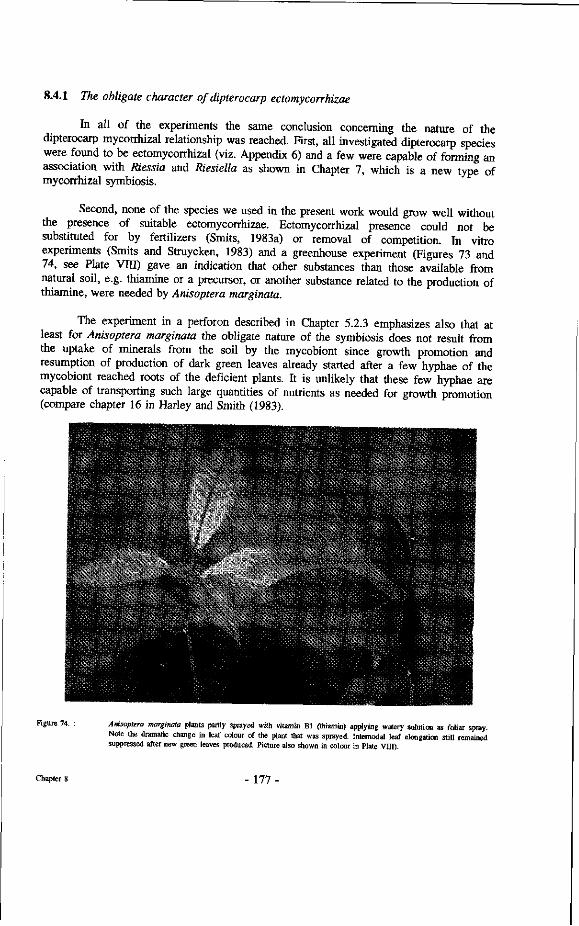
8.4.1 The obligate character of dipterocarp ectomycorrhizae
In all of the experiments the same conclusion concerning the nature of the dipterocarp mycorrhizal relationship was reached. First, all investigated dipterocarp species were found to be ectomycorrhizal (viz. Appendix 6) and a few were capable of forming an association with Riessia and Riesiella as shown in Chapter 7, which is a new type of mycorrhizal symbiosis.
Second, none of the species we used in the present work would grow well without the presence of suitable ectomycorrhizae. Ectomycorrhizal presence could not be substituted for by fertilizers (Smits, 1983a) or removal of competition. In vitro experiments (Smits and Struycken, 1983) and a greenhouse experiment (Figures 73 and 74, see Plate VIII) gave an indication that other substances than those available from natural soil, e.g. thiamine or a precursor, or another substance related to the production of thiamine, were needed by Anisoptera marginata.
The experiment in a perforon described in Chapter 5.2.3 emphasizes also that at least for Anisoptera marginata the obligate nature of the symbiosis does not result from the uptake of minerals from the soil by the mycobiont since growth promotion and resumption of production of dark green leaves already started after a few hyphae of the mycobiont reached roots of the deficient plants. It is unlikely that these few hyphae are capable of transporting such large quantities of nutrients as needed for growth promotion (compare chapter 16 in Harley and Smith (1983).
Figure 74. Anisoptera marginata plants partly sprayed with vitamin Bl (thiamin) applying watery solution as foliar spray. Note the dramatic change in leaf colour of the plant that was sprayed Intemodal leaf elongation still remained suppressed after new green leaves produced. Picture also shown in colour in Plate VIII).
Chapter S 177-
Specificity should hence be defined as ecological specificity : "the degree of specificity found under a certain set of environmental conditions". The use of this definition implies that this "certain set of environmental conditions" needs to be very well specified before endeavouring on any speculations about comparisons of specificity between different sites. The dipterocarp mycorrhizal specificity therefore as discussed here, refers to the degree of interdependency in undisturbed primary mixed dipterocarp rain forest on well draining soil, of which the Wanariset I research forest as described in Chapter 1.4. presents a good example. Alexander (1988) speaks of mycorrhizal dependency as "the degree to which a plant is dependent upon mycorrhizal infection to produce maximum growth under a given set of environmental, particularly edaphic, conditions" (Alexander, 1988). This definition comes very close to our definition of ecological specificity, although growth need not necessarily be maximum. It may be possible that for instance a survival strategy may result in different fungi being present at the root system than those yielding maximum growth. Brown and Mathews (1914) reported on the very old age of small Parashorea seedlings, confirming the well known strategy of dipterocarp seedlings of surviving long periods of waiting in shade while responding well to later sudden opening of the canopy. These waiting seedlings do probably not have the mycobiont leading to highest growth.
Discussing specificity of dipterocarp mycorrhizae it is possible to draw some preliminary conclusions based upon the results of the present research. 1) The inoculation experiments described in Chapter 4 demonstrate that under greenhouse conditions more mycobionts are capable of forming mycorrhizae than in the natural forest. 2) Also some inventories in a stand of Dryobalanops lanceolata (Misier, 1990) indicate that under disturbed conditions more mycobionts are found to be present. These conclusions were reached, however, on the basis of visual classification of the ectomycorrhizae encountered at 20 x enlargements. Becker (1983) suggested that classification of ectomycorrhizal types would be feasible in view of the great constancy in morphology of one particular ectomycorrhizal fungus forming ectomycorrhizae with roots of several phytobionts (viz. Foster and Marks, 1967; Marks and Foster, 1967; Zak, 1971). Although the detail of descriptions used here does not permit a comparison with other types, described by other authors, it is felt that the differences observed between the different types encountered do validate the conclusion that at least more mycobionts were encountered under more or less artificial conditions than under the undisturbed natural forest conditions. 3) There seems to be a strong preference for certain mycobionts even under the more artificial conditions.
The observation in the perforon (Chapter 5.2.4) showed that Vatica pauciflora (Korth.) would not accept any of the seven different available mycobionts associated with three other dipterocarps in the same perforon. In the rare instances that Pisolithus tinctorius was capable of forming ectomycorrhizae with Hopea odorata the effect even seemed negative resulting in a quicker death of the phytobiont (Chapter 4). It should therefore be questioned whether the statement by Marx (pers. comm.) that any ectomycorrhizae are better than no mycorrhizae also holds true for the Dipterocarpaceae.
The total number of inocula tested is still very low and it is also still unclear in how far one may expect the same degree of infection when using inoculum of temperate zone ectomycorrhizal fungi and of tropical material, as was done in a greenhouse by Smits (1983) and Muayanziza (1994, in preparation), notwithstanding the fact that the main
Chapter 8 - 180 -
groups of ectomycorrhizal fungi are the same (viz. Chapter 2.3.2).
The inoculations under natural conditions, involving the planting of non-mycorrhizal seedlings and cuttings on various places in the natural forest, give a better indication for the degree of specificity and compatibility under these circumstances. The experiments described in Chapter 4.1.1 and in Chapter 5.3.4 clearly indicate that even in the presence of many ectomycorrhizal roots comprising several types of ectomycorrhizae, many of the non-mycorrhizal plants would not form any mycorrhizae under natural undisturbed forest conditions. Many of these plants died within a short period, others would remain small and show no growth.
There is one factor, however, which may invite some caution about the interpretation of these results, namely the light condition during these experiments. Perhaps most of the plants planted in the shade of the undisturbed forest were growing in a light intensity below the compensation point. Photosynthesis may have produced less carbohydrate than is needed for respiration. According to the carbohydrate surplus theory of Björkman (1953) the formation of ectomycorrhizae becomes less favourable or even impossible under such conditions.
There is a chance that even very slight differences in the period that seedling leaves passed in a sun fleck might account for the formation of the ectomycorrhizae. Therefore the question arises what is more important for the establishment of the infection, the carbohydrate surplus of the seedling resulting from photosynthesis or the infection supported by hyphae connected to roots of phytobionts that do have a carbohydrate surplus. The results of the inventory of naturally established Hopea nervosa seedlings show that the seedlings connected with the roots of the mother tree through ectomycorrhizal hyphae outperform all the other seedlings and that probably as a consequence mortality among these seedlings is smaller. There may therefore exist transport of substances from mother trees to seedlings through mycorrhizal hyphae.
The planting of non-mycorrhizal seedlings may not be providing an accurate representation of the natural infection process since most of the seeds of the Dipterocarpaceae are rather large and contain much reserves in the form of carbohydrate in their cotyledons and endosperm, especially before ripening, which might enhance exudation from the newly developing roots and therefore attract infection with available inoculum. Moreover the roots are entering undisturbed soil while the seedlings in our experiments were planted in holes at the limits of which all the roots of other plants had been severed. This must have postponed the direct presence of suitable inoculum material. So the question partly remains to determine if the main cause of mortality was either the carbohydrate supply by the seedlings and therefore the opportunity to begin some ectomycorrhizal association with available inoculum, or the absence of suitable inoculum.
These aspects are now being studied in more detail by Yasman (in preparation). For the moment it can be stated that the above-mentioned experiments at least do not indicate the absence of specificity. The differences observed between the groups of plants in the experiments described in Chapter 3.2.7, although involving only small numbers of plants, indeed indicate some specificity.
Chapter 8 - 181
as follows : "At the same time it is perfectly true that the otherwise "obligate fungi" can be grown in culture apart from their usual symbionts; yet the laboratory and the woodland are two different things, and without being too cynical we may say that our experience with natural woodlands is that they are notably deficient in supplies of Erlenmeyer flasks and culture media". Therefore, when we limit specificity to ecological specificity, meaning that few mycobionts form ectomycorrhizae with few particular phytobionts under natural conditions as referred to by Kelley, specificity may exist.
Various authors take up this issue of defining the different degrees of specificity. Janos (1980; p.62) presented the figure which combined facultative and obligate symbioses between mycobiont and phytobiont with specificity. His scheme does not include ecological specificity. Molina et al. (1992a) recognize six phenomena that they consider encompass the spectrum of specificity in mycorrhizal associations : 1) Dependency vs. independency; 2) Facultative vs. obligate symbionts; 3) Fidelity to a class of mycorrhizae; 4) Host range of mycorrhizal fungi; 5) Host receptivity and finally 6) Ecological specificity. Janos therefore refers to points 1, 2, 4 and 5 only of the points mentioned by Molina et al.
As mentioned in Chapter 2.3.3 in this study the following definition of ecological specificity was used : "the degree to which phytobiont-mycobiont combinations occur in undisturbed natural ecosystems".
The degree of ecological specificity is important for the type of regeneration processes taking place. Molina et al (1992a) discuss "linkage potential", meaning that less specificity of phytobionts results in higher linkage potential and therefore theoretically better long term prospects of survival, especially for colonizing disturbed environments when different phytobionts share one or a few mycobionts. Read et al. (1985) and Newman (1988) have reviewed the consequences of plants being connected by mycorrhizal fungi, especially the movement of carbohydrates and minerals via shared hyphae and potential dependence of understorey seedlings on adult trees.
More work is needed before we can determine more precisely the degree of ecological specificity of dipterocarp mycorrhizae. For instance the mushroom inventories in this study indicate that over very small distances, in meters or sometimes decimeters, different mycobionts occur on different substrates. When more data become available it may emerge that, when different substrates are considered, the mycobionts may be more specific in the sense that they are specific in a kind of "triangular specificity" of mycobionts, phytobionts and soil types.
Certainly, if site factors like soil texture, aeration etc. are considered as being part of separate eco-units the degree of specificity within one such eco-unit would increase (see Chapter 2.3.3). Again, if succession of different mycobionts with the ageing of the phytobiont does occur, for one age class the degree of mycorrhizal specificity may narrow down further. The appearance of many rare ectomycorrhizal mushroom species in young Pinus sylvestris stands in The Netherlands (Termorshuizen, 1990), partly reflecting the low acreage of new forest plantations in The Netherlands, clearly indicates the importance of this aspect. Romell (1930) already noticed that Lactarius delicosus is found in great quantities under thick-set spruce trees when the trees are young, but disappears completely
Chapter 8 - 184 -
after the trees have reached a certain age. Again only further study will be able to provide more reliable answers. Some more indications may be obtained from other locations.
Anivat Chalermpongse (pers. comm., 1990) conducted inventories of sporocarps of potential dipterocarp mycobionts. He suspects that the number of associated mycobionts is rather limited for each dipterocarp species occurring in Thailand, and that the mycobiont-phytobiont combinations encountered are rather constant. Chalermpongse (1987) mentions several mycobionts encountered near Dipterocarps from the seasonal zone in Thailand that also appear in the research plot described in this research. Comparisons of the species of sporocarps encountered in the Wanariset forest with those collected by Iskandar in the Bukit Suharto forest at some 20 kilometres distance, in forest with many of the same dipterocarp species, yield a very high degree of resemblance. In Berau the same Amanita sp. indet. 2 was encountered near Shorea ovalis, as was the case in the Wartono Kadri plot. On Pulau Laut Russula sp. indet 1 was again encountered near Shorea laevis. These incidental observations and many more mentioned in Chapter 2.3.3 indicate that the mycobiont phytobiont combinations observed here may be rather constant over larger distances.
However, in semi Heath forest or Kerangas as described by Brünig (1968), many other mycobionts were encountered by the present author near Shorea leprosula within the ITCI concession (see appendix 9). It was here that a quite dense layer of reddish humus had accumulated, making this site quite different from the Wartono Kadri research plot. These observations again support the special mycobiont-phytobiont-substrate specificity.
Considering the apparent specialization of the different mycobionts for different substrates (Chapter 2.3) it should be concluded that, within the context of a single substrate and a single community type, the degree of specificity for a given dipterocarp species should be judged narrower. If the substrate or the soil were more uniform and the same number of different mycobionts had been evenly distributed around the tree, then the degree of specificity would be less. Thus a specialization for different substrates through different mycobionts supports the hypothesis that dipterocarps are capable of adapting themselves to certain niches by the association with varying mycobionts. Gardner and Malajczuk (1985) found that the distribution of sporocarps of different mycobionts near Eucalyptus resinifera was closely related to the amount of litter accumulated. Meyer (1985) found that there was a clear shift towards certain ectomycorrhizal fungi when the C/N ratio in the soil shifted. Therefore as compared with temperate zone experiences, substrate specialization, as encountered here, need not be an unlikely event.
In conclusion it can be said that several indications have been obtained tending to confirm a certain specificity of dipterocarp mycorrhizae. All results of this study indicate that the degree of specificity of dipterocarp mycorrhizae will be narrower than reported here. More plots should be established on different sites for a few closely related species e.g. Shorea curtisii, growing on ridge tops, Shorea leprosula on low hills and Shorea platycarpa growing in peat swamps. It would also be interesting to look into more detail at the following six closely related Shorea spp. that occur under similar growing conditions in Pasoh forest and elsewhere in peninsular Malaysia (Ashton, pers. comm.) : Shorea leprosula, S. acuminata, S. dasyphylla, S. lepidota, S. parvifolia and S. macroptera. More research should be initiated involving very local micro-changes in soil properties and
Chapter 8 - 185 -
distribution of different mycobionts around single trees. Also, detailed descriptions of ectomycorrhizae types are needed for classification purposes and for comparisons and investigations should be conducted involving possibly all ectomycorrhizal tree species.
8.4.3 Spatial isolation of dipterocarp clumps enhanced by mycorrhizae
The experiments in Chapter 2.3 and Chapter 3.3.3 all clearly demonstrated that non-mycorrhizal plants growing at larger distances from the mother tree stand a smaller chance to survive than the ones growing closer to the tree. This is the opposite of what has been found to be the general trend for the majority of tree species in the tropics (Ashton, 1989; Hubbell and Foster, 1990; Oldeman and Fundter, 1989). Many persons have mentioned the problems of dipterocarp seed dispersion. Merrill (1923) already mentions that dipterocarps never occur in secondary forests. Other writers like Ashton (1969) suggest that the limited regeneration of Dipterocarpaceae at larger distances from the mother tree is related to their limited means of seed dispersal. Ridley (1930) mentions that almost all seeds fall within 100 meter from the mother tree and Burgess (1972) mentions that more than 50% of the seeds fall within a circle of 20 meters around the tree, whereas only very rarely seeds will be blown farther than 500 meters. On the other hand several people have reported failures of direct seeding (Voogd, 1933; Alphen de Veer, 1949) in lines cut in secondary forest where no more dipterocarps were present. Here obviously the dispersal was not the problem. The lists of dipterocarp plantation establishment trials in Indonesia in experimental forest stands outside the natural range (Smits, in preparation) show many failures with Dipterocarpaceae established from seed. Wildlings seem to have performed much better (Ardikoesoema and Noerkamal, 1955; Alphen de Veer, 1949). So far the mycorrhizal condition of Dipterocarpaceae seems never to have been related to the limited availability of mycorrhizal inoculum at larger distances from the mother tree.
Spatial isolation seems to be even further enhanced, not only because of the presence of suitable mycorrhizal inoculum near the tree but also because of the existence of direct connections of one ectomycorrhizal fungus between mother tree and seedlings that benefits only the seedlings growing near the roots of the mother tree. The inventory of the plot with Hopea rudiformis seedlings (Chapter 3.3.3) indicates that there was some downhill inoculation of seedlings that germinated there. Farther away from the root system of the mother tree the survival of those naturally inoculated plants seemed to be mainly related to the amount of light and competition.
Although not fully proven the results of Chapter 3.3.3 might indicate some carbohydrate transport to take place between a mother tree and its seedlings. If this mother-sibling relation would be proven and turn out to be common this would be the best explanation for the limited distance from the mother tree at which the seedlings grow up, making it always possible in the field to indicate the mother tree of any dipterocarp seedling, and the typical clumped distribution of the large dipterocarp trees of one species within the forest. Yasman (in preparation) is working on the nursing role of dipterocarp mother trees for their seedlings.
Chapter 8 - 186 -
The winged fruits of Dipterocarpaceae are known sometimes to be blown over large distances. As in the case of some Dipterocarpaceae that seem to be unable to cross even rather small rivers it implies that some seeds must have been able to germinate after having travelled that far at such large distances but did not encounter suitable mycobionts there, or may not have been able to establish without the help of the mother tree. The aspect of specificity involved will be discussed below.
Relative to spatial isolation it is important, however, to look into the sources of inoculum of mycobionts involved, and their distribution and availability in space and time. Figures 22 and 23 show that there are large peaks in sporocarp production of the mycobionts. These peaks are closely correlated with the physiological condition of the phytobiont, especially the development of a sink for carbohydrates. For Dipterocarpaceae seedlings or small cuttings the appearance of the sporocarps was very strongly correlated to the production, and especially the onset of, a new growth flush (Figure 28). In the natural forest the sporocarps appeared during, and sometimes before the onset of the flowering, and somewhat less so during the onset of the production of new leaves. This correlation is so strong that one can predict when individual trees of Shorea lamellata will start flowering, by observing the appearance of specific sporocarps of mycobionts encountered in the forest.
Possibly this same "sink" for carbohydrates is important in the appearance of large drifts of ectomycorrhizal sporocarps both in spring and in autumn in the temperate zones. The mass sporocarp production starts just before the actual onset of flowering, which is almost always at the end of a long dry period ushering the start of the rainy season (Burgess,1972; Ng, 1977). One might thus also suspect the climatological factor to be the trigger for both sporocarp formation and the physiological reaction of the trees. However, in years when no flowering takes place the mass production of sporocarps does not occur during very wet periods, while some trees which started flowering during other periods still showed the correlation with the mycobiont sporocarp formation Smits et al (1987), Yasman (in press).
This correlation, between presence of sporocarps of the mycobionts and the physiological condition of the phytobiont, then holds strong consequences for the availability of mycobiont material from spores. The number of sporocarps in between these mass production periods is low while they are limited to certain species only (viz. Figure 22). There are no published data on spore dispersal of the mycobionts under intact forest canopy conditions. Wind speeds in the understorey are almost negligible. There may therefore be a chance that the spores are not blown very far and come down with the rain at close distances of the actual sporocarps.
In the mixed dipterocarp forest remarkably few animals occur that might act as vectors for the dispersal of the spores (Trappe and Maser, 1977). Rain causing surface run off without doubt transports spores downhill, but how far is unknown. Until data become available it will therefore be assumed that crossing of rivers by spores of mycobionts will not be common.
Most dipterocarp mycobionts belong to the Basidiomycetes, which possess spores of several mating types of which only certain combinations can result in the formation of
Chapter 8 - 187 -
the typical dikaryotic mycelium. The chance of a successful mycelium formation is therefore smaller. Moreover, the spores need to fall close together in the near proximity of uninfected young dipterocarp roots. The chance for this to happen becomes exponentially smaller with larger distances. Another aspect not yet studied in detail is how long the mycobiont spores can survive in the litter layer of a tropical rain forest with intense microbiological activity. It seems unlikely that they can survive periods of more than 3 months and would then still be capable of germination and infection of dipterocarp seedling roots. Storage trials of fresh top soil collected shortly after massive sporocarp appearances showed sharply decreasing infectivity of the inoculum and a shift in fungal species still establishing ectomycorrhizae after three weeks (Prayudi and Smits, in preparation).
Although flowering of many Dipterocarpaceae species does not take place at precisely the same time, most of the seeds do fall during the same short period. This was hypothesized to be a mechanism to avoid destruction by animal predators by Janzen (1974). The period of fruit fall and germination of seeds starts three months after appearance of the many mycobiont sporocarps (Smits et al., 1987). AU the above points therefore in the same direction, i.e. that natural infection of dipterocarp seedlings with spores of mycobionts is an unlikely event to occur. Therefore most natural infection has to originate from the vegetative mycobiont material available on the root systems of the mother tree. If this is the case then, as already mentioned from the results discussed above, spatial isolation between small populations of a dipterocarp species is enormously enhanced.
This would mean that the most important condition to support the hypotheses that dipterocarp mycorrhizae have contributed to speciation amongst the Dipterocarpaceae through enhanced isolation between sub groups of tree would be fulfilled.
8.4.4 Comparison of the research results with literature.
Janos (1985) proposes that "...effects of mycorrhizal interactions on competition provide an explanation for the way in which many plant species coexist in tropical communities...". In this paper he discusses the advantages of ectomycorrhizal associations over VA (Vesicular Arbuscular) mycorrhizal associations and hypothesizes that "high mycobiont cost limits ectotroph occurrence to those habitats in which ectomycorrhizae are more beneficial than VA mycorrhizae". He provides several reasons why ectomycorrhizal fungi may require more photosynthates from their phytobionts than do VA mycorrhizal fungi and why they may have morphological and physiological advantages over VA mycorrhizae. As a consequence of their potential advantages over VA phytobionts, Janos predicts that ectotrophs are most likely to compete successfully with VA mycotrophs in those habitats in which : 1) mineral nutrient and water availability is pulsed, 2) nitrification is inhibited, 3) the ability of the mineral soil to supply or retain mineral nutrients is very limited, 4) decomposition is slow and 5) rapid colonizing ability of both symbionts favours establishment and persistence. He concludes that these predictions are consistent with the then known distribution of tropical ectotrophs.
Chapter 8 - 188
Continuing on the specificity of mycorrhizae and competition among ectotrophs, he hypothesizes that the discriminatory ability of ectotrophs may lead to specificity of association. He then proposes that exclusive ectomycorrhizal specificity leads to monospecific stands because 1) temporal and spatial homogeneity of the soil would confer the greatest advantage in competition on the best adapted ectotroph, and 2) on extremely infertile soils, mineral limitation of mycobiont fruiting could limit adequate mycorrhizae formation to only those host species that are sufficiently competitive to achieve a threshold abundance or close spacing needed for vegetative transmission of infection among individuals. Furthermore he also suggests that "...growth of obligate mycotrophs is mineral limited...".
It is interesting to test the hypotheses presented by Janos in the light of the information gathered in these studies. Janos gained most of his experience from the neotropics but refers several times to trees from South East Asia. When we consider the results of the present study we note that the mixed dipterocarp forest soils are dominated by the presence of ectomycorrhizal fungi. As stated above, based upon the data presently available, some degree of mycorrhizal specificity must be assumed. Therefore, in Janos' view the forest should tend to be mono-specific. Nevertheless we find a very high degree of species richness (see Chapter 2.3.1). The appearance of sporocarps is irregular but they do appear in large quantities (see Figures 23, 24) and thus are not limited by availability of minerals. Nevertheless, we could perceive the small clumps of some dipterocarps as very small mono-specific stands, while Janos' prediction is certainly true for a species such as Shorea albida, which occupies a habitat which other species have failed to invade within its range {Shorea balangeran does so elsewhere). The general picture, however, does not relate well to Janos' hypotheses. For instance the heath forests showing litter accumulation contain fewer ectomycorrhizal trees than surrounding mixed dipterocarp forests.
The statement that growth of obligate mycotrophs (in this book Janos' mycotrophs are referred to as phytobionts) is mineral limited was not confirmed by the perforon studies presented here (see Chapter 5.3.2), indicating that the production of other compounds by the fungi may be more important. Nonetheless it is true that the relative dipterocarp species richness and abundance reach their peak near the nutrient threshold below which humus accumulation would become noticeable and nitrification becomes increasingly impeded.
The other proposed reasons for dominance of ectomycorrhizal species, like pulsed mineral nutrient and water availability, inhibited nitrification, retarded litter decomposition and rapid colonizing ability of both mycobiont and phytobiont do not apply with the general dipterocarp situation in the mixed forests in south east Asia. These completely different conditions nevertheless lead to vegetation types dominated by ectomycorrhizal species.
Some generalizations like the presence and dominance of ectomycorrhizal tree species in the temperate zones were also made by Malloch et al. (1980). Figure 76 shows a transect in virgin dipterocarp forest (Ashton and Hall, 192) in which the extreme dominance of ectomycorrhizal Dipterocarps is very obvious.. In the mean time more reports start appearing (Högberg, 1982; Högberg and Pierce, 1986) that show the relative
Chapter 8 - 189 -
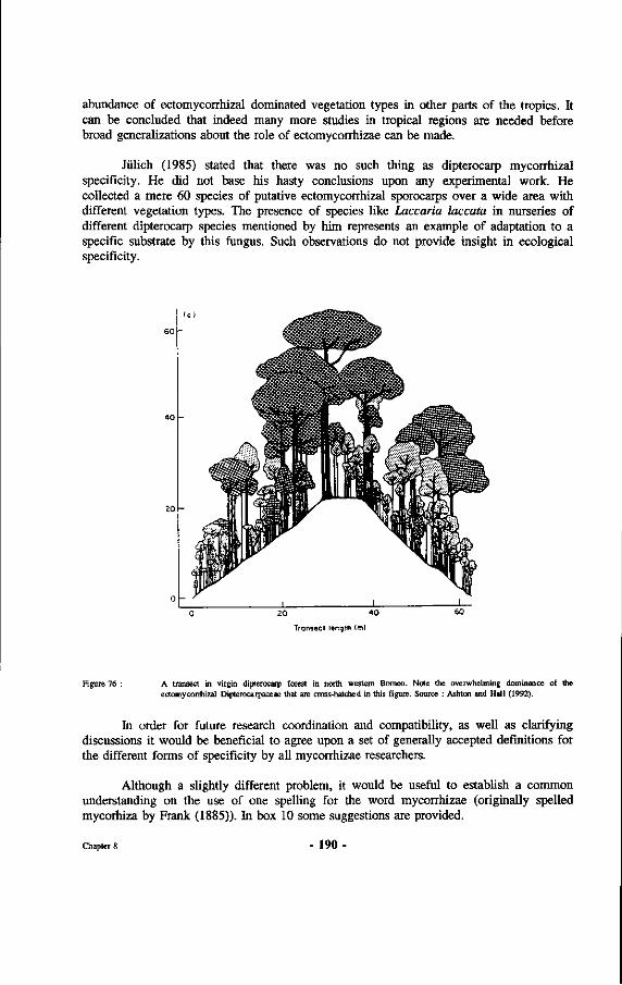
abundance of ectomycorrhizal dominated vegetation types in other parts of the tropics. It can be concluded that indeed many more studies in tropical regions are needed before broad generalizations about the role of ectomycorrhizae can be made.
Jülich (1985) stated that there was no such thing as dipterocarp mycorrhizal specificity. He did not base his hasty conclusions upon any experimental work. He collected a mere 60 species of putative ectomycorrhizal sporocarps over a wide area with different vegetation types. The presence of species like Laccaria laccata in nurseries of different dipterocarp species mentioned by him represents an example of adaptation to a specific substrate by this fungus. Such observations do not provide insight in ecological specificity.
20 4 0
Transect length (ml
Figure 76 : A transect in virgin dipterocarp forest in north western Borneo. Note the overwhelming dominance of the ectomycorrhizal Dipterocarpaceae that are cross-hatched in this figure. Source : Ashton and Hall (1992).
In order for future research coordination and compatibility, as well as clarifying discussions it would be beneficial to agree upon a set of generally accepted definitions for the different forms of specificity by all mycorrhizae researchers.
Although a slightly different problem, it would be useful to establish a common understanding on the use of one spelling for the word mycorrhizae (originally spelled mycorhiza by Frank (1885)). In box 10 some suggestions are provided.
Chapter 8 - 190-
Box 9 : Mycorrhizae, what's in a name
In 1885 Frank (p. 129) coined the term mycorhiza. In his original manuscript he spells the word with one "r" and extension "a" for the singularis form. Since his publication the word has been spelled in a variety of ways. The two most widely used forms for the pluralis form are mycorrhizae and mycorrhizas, the former generally most used in the United States and the latter in England. French authors, for instance Boullard (1968) use "mycorrhizes", Indonesians use "mikorisa", Germans "Mykorrhizen", etc. The use of mycorhizae with one "r" also still occurs. In the following these choices are discussed.
The double "r" or rho (p) is now most widely used. The word pi Ça does not seem to have been used in composite words in Old Greek, at least not as first part and probably neither as second part. However, when used in combination with adjectives the p is as a rule often doubled, for instance in : p.aKpopptÇoç (with long root) and neyaAópptCoc (with large root). In view of the Greek origin of both the words pi Ça (root) and |iuicnç (mushroom, fungus) it therefore can be considered correct to write the word with double "r" and not to follow Franks' original spelling. Also based upon the Greek origin of the words it deserves to let the first part of the word end to "o" instead of the more common "i" in Latin, when combining words. This is in concordance with Franks' spelling.
Each language has its own pluralis forms. Since the term was first coined as a scientific term it should deserve preference to write it based upon Latin spelling. In the word mycorhiza Frank, for instance, uses "y" instead of "u" (i3), thereby following Latin spelling rather than Greek. The correct pluralis form of the word should therefore be mycorrhizae. Use of other forms such as mycorrhizas places the word on a national level. The use of the scientific word should be international, based upon Latin. In view of the wide use of the form "mycorrhizae" it is felt that scientific communications should adopt this spelling as the only correct one.
8.5 Practical application of the results
In this section the practical implications of the present knowledge of dipterocarp ectomycorrhizae for the management of forests with dipterocarp trees will be discussed. First the aspects related to natural forests and natural stand management are discussed followed by mycorrhizal aspects of importance when establishing dipterocarp plantations.
8.5.1 Implications for natural stand management
It was already mentioned in Chapter 1.2.2 that under the panglong systems no attention was paid to the regeneration of the forest. When the wood resources were exhausted on one location the concession was simply moved. Sewandono (1937) mentions
Chapter 8 - 191 -
that the potential of the forest to regenerate itself should be considered good. With the non mechanical felling few small trees get damaged. Moreover alang-alang (Imperata cylindrica) and other harmful grass species are absent from the swampy areas. However, Sewandono does not discuss the regeneration of Dipterocarpaceae in detail. The volumes of wood taken out from one hectare by this type of logging was only a little less than by the present methods. Most likely, however, the average diameters of the trees were smaller, because of the difficulty of handling the larger diameter logs by hand felling methods and the higher frequency of defective logs from larger diameter trees.
If this is true, then it follows that the canopy of the forest will not be as heavily destroyed as in the case of present type of logging that only removes the larger diameter trees. These bigger trees are often colonized by many lianas that tend to interconnect the crowns of the overstorey trees, leading to more damage in the process of felling, because in their fall they tear down several other trees and damage many other ones. This leads to more damage to remaining trees as well as potentially more loss of biological diversity (Jacobs, 1988).
Another important difference between the first utilization of the dipterocarp forests and the present practice is in the method of transportation. In the earliest logging practices only those trees were felled that could be transported downhill to a river where they could be floated down. Sometimes the trees were sled down the hill, sometimes over specially constructed "knuppel roads" (see Chapter 1.1.2), using mud or some other slippery substance like pig fat to facilitate their use. These systems did not need the construction of wide roads and the use of heavy equipment. As a consequence the forest was also opened to a much lesser degree than the present forestry practice where roads go far inland and heavy bulldozers are used to pull the logs out of the forest, causing much more damage in the process. For the trees that were not mechanically felled and transported, it was also important to determine the direction of felling the tree in order to ease its further transportation. With the high powered bulldozers this is no longer necessary, so that very often the chainsaw operator will fell the tree in the direction that suits him best. This direction, however, will rarely be the best one for the bulldozer to pull out the log without damaging much of the surrounding forest. Recently attention starts to be focused again on damaged controlled logging (Hendrison, 1990) or also called reduced impact logging (Smits et al., 1992). In recent years it has become more common practice to include cutting the so called sinkers or heavier species from the forest. Clearly this trend has resulted in a still larger opening of the forest canopy.
These differences between the early days of dipterocarp forest exploitation and the present practice have therefore dramatic effects upon the degree in which they affect all kinds of biological processes in the forest and also the dipterocarp mycorrhizae. Many reports (viz. Chim, 1973) mention the large amount of dipterocarp seedlings that die as a result of the logging operations.
Other reports (viz. Miller, 1981) mention that many dipterocarps die even up to 5 years after the actual logging operations without any apparent fatal damage. The results presented in section Chapter 6 indicate that the physical influence of temperature upon dipterocarp ectomycorrhizae plays a crucial role in this mortality after logging. Contributing to the high mortality are probably also the damage caused by scraping the
Chapter 8 - 192 -
bark by machinery and logs, wounds inflicted by trees falling during and long after the logging operations, and the invisible damage to the roots of the remaining dipterocarps caused by heavy logging equipment. The effects of these damages and of increased soil temperatures are also compounded by the greater évapotranspiration experienced by the remaining and exposed trees that were only partly exposed to direct sunlight before logging operations. More evaporation from heated soil, higher transpiration from exposed crowns, fewer roots to take up water and nutrients, more difficult functioning of remaining roots and mycorrhizae and higher predisposition to biological damage all combine to create difficult conditions for the trees after logging.
What improvements can be suggested for the practice of logging by taking our present knowledge of dipterocarp ectomycorrhizae into account ? First of all it is obvious that the microclimate should be changed as little as possible by the logging operations. Ways to reach this objective are, among others, by taking out less wood per hectare, applying pre-felling climber cutting, conducting directional felling, planning of skidroads before logging operations based on maps with tree locations and use of lighter logging equipment. Especially the use of lighter logging equipment might present a very practical way to reduce damage to the forest during logging operations. The lighter bulldozers cause less soil compaction and are not capable of pulling out logs along difficult gradients. Therefore these lighter machines are forced to use one and the same skid road more than once so that more patches of forest are left undisturbed and fewer trees get wounded, yielding a better next harvest (Hendrison, 1990). It is obvious that the microclimate is disturbed less this way. The effects of the use of heavier equipment can be seen well in the EMHUTANI I concession in Berau. Near the Labanan transmigration area, up to kilometre 17 the logged over forest is in excellent condition. Ten years after the original logging operations it has already become difficult to detect the original skid roads and there are no large open places in the forest while natural regeneration is abundant. Beyond kilometre 17, when heavier equipment was introduced some fifteen years ago (Nanang, pers.comm.), the damage is much greater. Here, there is almost no young regeneration of dipterocarps and the forest is covered by a dense layer of lianas. Much attention should therefore be paid to the reduction of logging damage.
If the indications that Dipterocarpaceae tend to "nurse" their seedlings are right, it might be worthwhile not to remove older trees with thin crowns and defective boles, especially of favoured commercial species, until sufficient regeneration has established. Within this context it is very interesting to read the report of Schuitemaker (1933) who mentions planting of several Dipterocarpaceae for resin production and production of illipe nuts near Meliau, Sanggau, Tajan and Landak (West Kalimantan). He reports that planting is done with Wildlings collected from natural regeneration. These Wildlings are then planted close to some mother trees after which only occasionally removal of competing plants is performed. The same was mentioned by Roosendael and Thorenaar (1924).
8.5.2 Consequences for planting of dipterocarps
For overcoming the problems encountered in obtaining planting material for enrichment planting in logged over forest it seems best to make use of Wildlings growing near the mother tree. These seedlings can be expected to have been infected with suitable ectomycorrhizae. Furthermore they can be obtained at low cost and produced in large
Chapter 8 - 193 -
quantities after a mass flowering/fruiting season (Smits, 1986; Prayudi et al., 1992). These Wildlings should then be planted on places where the soil temperature is rather stable and does not rise above 30°C. This means therefore that they should not be directly planted on skidroads and open places like log yards as mentioned in the manual for the Indonesian selective cutting system.
Young dipterocarps seem to reach an optimal photosynthesis rate at about 50% light intensity. Mori (1980) reports that 30-50% light intensity is good for all dipterocarp seedlings, that they reach their maximum photosynthesis rate at 5-30 Klux, and that they experience growth reduction at more than 50 Klux light intensity, meaning that they grow well in light shade as has been confirmed by numerous authors. They can therefore eminently be planted under secondary forest species in lines or strips cut open to allow enough light to reach the seedlings or under the fast growing trees planted in the timber estate program in Indonesia. There is nothing new in this recommendation. There are already many examples where this system was applied as in Hulu Ketunggau, Embaloh where several tens of hectares of tengkawang (Illipe nuts yielding Shorea spp.) were planted in this way, mixed with Intsia. In Sanggau (West Kalimantan) some 300 hectares of 17 different tengkawang yielding dipterocarps were planted in this way (Darmono, pers. comm.).
If no Wildlings are available, or too few of the species intended, the production of dipterocarp planting stock through stem cuttings could be applied (Yasman and Smits, 1987). If this is done it is important to inoculate the rooted cuttings, at the time of transplanting from the rooting medium to the containers, with a suitable mycorrhizal fungus. Based upon the results obtained in this study it is advisable to inoculate the cuttings with fresh soil inoculum obtained from the direct surroundings of the roots of already infected plants/trees of the same species. A simple method for inoculation would be to use the soil of containers in which mycorrhizal Wildlings were grown for more than two months and from where these Wildlings were removed shortly before applying it as inoculum to the cuttings to be transplanted. Thus viable inoculum is always available (after the same manner in which yoghurt is produced). With this method it is not really necessary to do any preparation of the medium in which to plant the rooted cuttings, like fumigation with methyl bromide, since our experience showed that medium that has been laying around for a long time or left in the sun for even a short period of time is not likely to contain any viable inoculum of ectomycorrhizal fungi for Dipterocarpaceae. For this reason there is little chance that another mycobiont than the one intended will establish itself on the roots of the dipterocarp cuttings.
For planting of the plant material from cuttings the same recommendations are given as for the Wildlings. After the dipterocarp plants have established well, the light intensity can be increased in one or two steps. Ashton (1969) is of the opinion that selection against hybridisation takes place in Dipterocarps. Indeed hybrids do occur but are extremely rare in natural forests within the natural distribution range of the Dipterocarps.
Many Dipterocarpaceae show polyembryony. Several Dipterocarpaceae are known to be triploids or tetraploids (Kaur et al., 1978) and are known to produce seeds through apomixis. The narrowing down of the genetic variability does not seem to have limited the survival potential of these species, some being very widespread and common throughout a
Chapter 8 - 194 -
large distribution range and indeed very constant in morphological appearance throughout. A good example is Shorea ovalis.
This would indicate that there are good opportunities to produce good quality planting stock through methods of vegetative propagation. It means that there is little risk of pests and diseases since the relatively homogenous genetic make-up of adult Dipterocarps has lead to very few problems of trees being attacked in the natural forests, although their crowns in the upper canopy should provide ample pathways for spread of infection in the population. Smits et al. (1990a) reported very few disease problems with Dipterocarps. For practice this means that research on rejuvenation and methods of vegetative production as well as hedge orchards yielding the cuttings material should be continued. This may lead to increased productivity of Dipterocarps, so as to make them more attractive for commercial forestry through planting. Even though, it is recommended to make use of species mixtures as far as possible to reduce potential future risks.
Planting of Dipterocarps should be recommended in view of their good performance on nutrient poor soils. The work in this publication supports the hypotheses that ectomycorrhizae are capable of direct nutrient cycling (see for instance Figure 54). The relatively larger dominance of ectomycorrhizal trees in higher diameter classes (see Figures 15 and 16) also supports this view.
The successful planting of Dipterocarps in very short alang-alang grass (meaning very infertile soils) also indicates the good potential of these species. There is a wide variation in growth rates of different dipterocarp species and a very large variation in growth between seedlings. These differences are further amplified in plantations by the presence of different ectomycorrhizal fungi. A program on vegetative propagation of Dipterocarps would therefore offer good prospects for economical t imber production on a sustainable basis. Yasman and Smits (1988) have published a practical method for mass production of dipterocarp cuttings. Leppe and Smits (1988) published manuals for the production of hedge orchards. Smits et al. (1988) wrote a simple manual for inoculation of dipterocarp cuttings and non-mycorrhizal seedlings. Together these techniques provide ample opportunity for the production of h igh quality dipterocarp plantings stock.
The production of high quality planting stock by means of vegetative propagation provides a good way to overcome the problems of planting stock supply from seeds or Wildlings. Further selection work with Dipterocarps and fungi to suit them for different planting sites, holds great promise for production of dipterocarp timber on sustainable basis (Smits and Leppe, 1991). It is hoped that the production of Dipterocarps in plantations will make it less cost efficient to harvest poor quality dipterocarp trees from natural forest, because of their lower homogeneity and difficult sizes and location further away from processing facilities. This could lead to less intensive harvesting of natural forests and as such bring partially back post-harvesting forest conditions as was the practice during manual harvesting of these forests in the beginning of this century.
Further insight in and use of dipterocarp ectomycorrhizae holds part of the key to sustainability of the mixed dipterocarp production forests of south east Asia.
Chapter 8 - 195 -
Summary
Indonesia has been bestowed with extensive tropical rain forests making up more than 10% of the remaining tropical forests on the earth with the majority of the forest belonging to the mixed dipterocarp forest type. Some 65 million hectares out of the total forest area of 143 million hectares has been assigned as production forest, meaning that wood can be harvested from these forests under the guidelines for selective felling in mixed dipterocarp forests (MDF) set by the Indonesian Ministry of Forestry.
The family of the Dipterocarpaceae is mainly confined to South East Asia. Many tree species from this family are commercially important timber producers, e.g. Lauan, Seraya, Meranti, Keruing, Kapur, Mersawa, Bangkirai, Mengerawan, Resak, etc. Timber from this family constitutes more than 25% of the total tropical hardwood timber trade. Individuals of this tree family can make up to 80% of the trees in the upper canopy of the forest.
Despite their great economical and ecological importance there is still relatively little known about sound guidelines for sustainable management of the mixed dipterocarp forests. One of the main problems in the management of dipterocarp forests has been the production of good quality planting stock. Very recently good progress has been made in this field, through the development of practical Wildling collection methods and cuttings production systems. One of the key factors in the survival of young dipterocarp plants proved to be the presence of their ectomycorrhizae. The present publication was aimed at investigating more precisely the role of dipterocarp ectomycorrhizae in regeneration of dipterocarp forest.
The present study was partly conducted in greenhouses of the Agricultural University Wageningen in The Netherlands and partly at the Wanariset Research Station and in the nearby Wanariset Research Forest, both resorting under the Agency for Research and Development of the Indonesian Ministry of Forestry.
The first chapter of the book consists of a general introduction to the Dipterocarpaceae, information on their ecology, on the use of timber of this tropical hardwood family, as well as some general information on mycorrhizae.
Within the course of the research presented in this publication several studies were undertaken. The results of these studies are presented in chapters 2 to 7 and amongst others involve :
a) the inventarisation of ectomycorrhizal mushrooms and their relation to specific dipterocarp species in undisturbed natural MDF;
b) evaluation of the types and distribution of ectomycorrhizal roots in natural undisturbed MDF;
c) inoculation experiments with non-mycorrhizal dipterocarp planting stock both under controlled conditions and in natural vegetation;
d) investigation of the influence of physical factors upon dipterocarp ectomycorrhizae.
Summary - 196 -
The results of these studies are used to evaluate to what degree the dipterocarp mycorrhizal association may have contributed, and may still be doing so, to speciation in Dipterocarps, through enhancement of spatial isolation.
In this book a number of new techniques and inventions are presented, that may enhance further studies on mycorrhizae. Especially the technique making use of perforons has been described in detail.
Information from experimental work with a new dipterocarp fungus association between some Shorea spp. and Riessia and Riessiella spp. indicates that the association is a mycorrhizal one of a hitherto undescribed type.
It is concluded that based upon the results of the studies presented some degree of ecological specificity is present in the phytobiont-mycobiont relationships of Dipterocarps with ectomycorrhizal fungi and that this specificity may explain some of the species diversity in the family of the Dipterocarpaceae in South East Asia.
At the end of chapter 8 some practical implications of the findings obtained are presented. Dipterocarp planting stock should be mycorrhizal or otherwise be inoculated with suitable fungi for the site to be planted. Planting of young Dipterocarps should be done in such a way that high soil temperatures are avoided to ensure survival of the ectomycorrhizae.
Summary - 197 •

Samenvatting
Indonesië bezit uitgestrekte tropische regenwouden welke meer dan 10% van de nog aanwezige tropische regenwouden van deze aarde beslaan, het merendeel van deze bossen bestaande uit gemengd Dipterocarpaceae bos. Ongeveer 65 millioen hectares uit het totale bosareaal van 143 millioen hectares is bestemd als produktiebos, wat betekent dat in deze bossen hout geoogst kan worden volgens de regels voor selektieve kap die door het Indonesische ministerie voor bosbouw zijn vastgesteld.
De familie van de Dipterocarpaceae komt voornamelijk voor in zuid-oost Azië. Veel boomsoorten uit deze familie zijn belangrijke houtproducenten zoals bijvoorbeeld Lauan, Seraya, Meranti, Keruing, Kapur, Mersawa, Bangkirai, Mengerawan, Resak, etc. Hout van deze familie levert meer dan 25% van de totale wereld tropisch hardhouthandel. Het kronendak van deze bossen kan wel voor 80% bestaan uit individuen van deze familie.
Ondanks hun grote economische en ecologische belang is er relatief nog vrij weinig bekend over goede regels voor duurzaam bosbeheer van gemengde dipterocarp bossen. Een van de belangrijke problemen in het beheer van dipterocarp bossen was de produktie van goed plantgoed. Zeer recentelijk is hierin echter goede vooruitgang geboekt, door de ontwikkeling van praktische zaailing verzameltechnieken en stekproduktiesystemen. Een van de sleutelfaktoren voor het overleven van jonge dipterocarp planten bleek de aanwezigheid van ectomycorrhizae te zijn. De huidige publikatie had tot doel de rol van de ectomycorrhizae van de dipterocarp voor hun natuurlijke verjonging meer precies te onderzoeken.
Deze studie werd voor een deel uitgevoerd in de kassen van de Landbouwuniversiteit Wageningen in Nederland en gedeeltelijk op het Wanariset onderzoeksstation en in het nabij gelegen Wanariset onderzoeksbos Wanariset, beide onder de jurisdictie van het direktoraat generaal voor bosonderzoek en ontwikkeling van het Indonesische bosbouwministerie.
Het eerste hoofdstuk van dit boek bevat een algemene introduktie tot de familie van de Dipterocarpaceae, informatie betreffende hun ecology en het gebruik van het hout van deze tropische hardhoutfamilie, evenals wat algemene informatie over mycorrhizae.
Binnen het kader van dit onderzoek, zoals gepresenteerd in deze publikatie, werden verscheidene studies ondernomen. De resultaten van deze studies worden gepresenteerd in de hoofdstukken 2 tot en met 7 en betreffen onder andere :
a) de inventarisatie van ectomycorrhizae vormende paddestoelen en hun relatie tot specifieke dipterocarp soorten in ongestoord gemengd dipterocarp bos;
b) een evaluatie van types en verdeling van ectomycorrhizae in ongestoord gemengd dipterocarp bos;
c) inokulatieexperimenten met dipterocarp plantgoed zonder ectomycorrhizae, zowel onder gecontroleerde omstandigheden als in de natuurlijke vegetatie;
Summary - 198
d) onderzoek naar de invloed van fysische factoren op ectomycorrhizae van Dipterocarpaceae.
De resultaten van deze studies worden vervolgens gebruikt om te evalueren in hoeverre het samenleven van de schimmels met de Dipterocarpaceae heeft bijgedragen en mogelijk nog steeds zo doet, tot de soortsvorming in Dipterocarpaceae door het bevorderen van ruimtelijke isolatie.
In dit boek worden een aantal nieuwe technieken en uitvindingen gepresenteerd, welke mogelijk verder werk met mycorrhizae kunnen bevorderen. Vooral de technieken betreffende het gebruik van perforons zijn in detail beschreven.
Resultaten van experimenteel onderzoek met een nieuwe samenlevingsvorm tussen enkele Shorea soorten en Riessia en Riessiella soorten laten zien dat de samenlevingsvorm gezien moet worden als een mycorrhizae van een tot dusver onbekend type.
De conclusie gebaseerd op de resultaten van de studies hier beschreven is dat er een zekere mate van ecologische specificiteit aanwezig is in de phytobiont-mycobiont relatie van enige Dipterocarpaceae met ectomycorrhizae vormende schimmels en dat deze specificiteit een gedeelte van de soortenrijkdom in de familie van de Dipterocarpaceae in zuid-oost Azië kan verklaren.
Aan het eind van hoofdstuk 8 worden enige praktische consequenties van de gevonden resultaten gepresenteerd. Plantgoed van Dipterocarpaceae moet ectomycorrhizae bezitten of anders geinokuleerd worden met geschikte schimmels voor de plaatsen die beplant dienen te worden. De aanplant van nieuwe Dipterocarpaceae moet zo gedaan worden dat hogere bodemtemperaturen voorkomen worden om zo het overleven van de ectomycorrhizae te garanderen.
Summary - 199 •
Ringkasan
Indonesia dikaruniai dengan hutan tropis basah yang luas, yang merupakan lebih dari 10% dari seluruh hutan tropis basah yang masih berada di permukaan bumi. Kebanyakan hutan tersebut terdiri dari tipe hutan Dipterocarpaceae campuran. Sekitar 65 juta hektar dari luasan seluruh hutan 143 juta hektar telah ditunjuk sebagai hutan produksi, dengan arti di areal ini dapat diperkenankan pemanenan kayu sesuai petunjuk pelaksanaan sistem tebang pilih tanam Indonesia yang diterbitkan Departemen Kehutanan.
Famili Dipterocarpaceae kebanyakan terdapat di Asia tenggara. Banyak jenis pohon dari famili ini merupakan penghasil kayu penting seperti misalnya Lauan, Seraya, Meranti, Keruing, Kapur, Mersawa, Bangkirai, Mengerawan, Resak, dll. Lebih 25% dari seluruh kayu keras tropis yang diperdagangkan di dunia berasal dari famili kayu tropis keras ini. Anggota famili pohon ini dapat membentuk lebih dari 80% lapisan tajuk teratas di hutan tersebut.
Walaupun mempunyai kepentingan ekonomis dan ekologis yang amat besar, namun relatif masih kurang tersedia pengetahuan mengenai petunjuk pengelolaan hutan campuran Dipterocarpaceae secara lestari. Salah satu masalah utama dalam pengelolaan hutan Dipterocarpaceae selama ini adalah ketersediaan metode produksi bibit unggul. Belum lama ini terdapat terobosan yang baik di bidang ini melalui pengembangan metode pengumpulan anakan cabutan dan metode pembuatan stek. Salah satu faktor utama untuk daya tahan hidup anakan pohon Dipterocarpaceae temyata adalah ketersediaan ectomycorrhizae. Karya ini dimaksudkan untuk lebih seksama meneliti peranan ectomycorrhizae dalam regenerasi Dipterocarpaceae di hutan alam.
Penelitian yang dipresentasikan dalam publikasi ini dilaksanakan di dalam rumah kaca Universitas Pertanian Wageningen di Negeri Belanda dan sebagian di stasiun penelitian Wanariset dan di hutan penelitian Wanariset Samboja, Kalimantan Timur, keduanya di bawah Badan Penelitian dan Pengembangan Kehutanan dari Departemen Kehutanaan Republik Indonesia.
Bab satu dari buku ini terdiri dari introduksi umum kepada Dipterocarpaceae, informasi menenai ekologinya, penggunaan kayu dari famili pohon tropis ini dan informasi umum mengenai mikoriza.
Di dalam rangka program penelitian yang disampaikan dalam buku ini telah dilakukan berbagai jenis penelititan. Hasil penelitian ini dipresentasikan dalam bab 2 sampai dengan 7 dan melibatkan antara lain :
a) inventarisasi jamur ektomikoriza dan hubungannya dengan Dipterocarpaceae tertentu di hutan alam Dipterocarpaceae utuh;
b) evaluasi tipe dan distribusi akar yang berektomikoriza di hutan alam Dipterocarpaceae utuh;
c) percobaan inokulasi dengan bibit Dipterocarpaceae dalam keadaan terkontrol dan di dalam vegetasi alami;
Summary - 200 -
d) penelitian mengenai pengaruh beberapa faktor fïsik terhadap ektomikoriza Dipterocarpaceae.
Hasil penelitian ini selanjutnya dipakai untuk mengevaluasi sejauh mana asosiasi mikoriza dengan Dipterocarpaceae telah, dan mungkin tetap masih mempenganihi pembentukan jenis baru di Dipterocarpaceae, melalui peningkatan isolasi fïsik antara sub populasi.
Di dalam buku ini disampaikan beberapa teknik baru dan penemuan, yang diharapkan dapat mendukung penelitian mikoriza lebih lanjut. Khususnya teknologi yang menggunakan perforon disampaikan dengan banyak detil.
Hasil penelitian yang melibatkan asosiasi baru antara jamur dan Dipterocarpaceae, khususnya antara beberapa jenis Shorea dan jenis Riessia dan Riessiella menunjukkan bahwa asosiasi ini adalah suatu simbiosa mikoriza yang belum dikenal selama ini.
Berdasarkan hasil penelitian yang disampaikan disimpulkan bahwa terdapat spesifisitas ekologis tertentu di dalam hubungan phytobiont-mycobiont Dipterocarpaceae dengan jamur ektomikoriza dan bahwa spesifisitas ini dapat menjelaskan sebagian keanekaragaman jenis dalam famili Dipterocarpaceae di Asia Tenggara.
Pada akhir bab 8 disampaikan beberapa kesimpulan praktis mengenai aplikasi hasil penelitian ini. Bibit Dipterocarpaceae seharusnya memiliki mikoriza atau perlu diinokulasi dengan jamur yang sesuai untuk tempat yang akan ditanam. Penanaman bibit Dipterocarpaceae harus dilakukan demikian rupa supaya dicegah suhu tanah yang tinggi (di atas 32° C) untuk menjamin kehidupan terus dari ektomikoriza.
Summary - 201
Glossary
Apomixis :
Biotic remanence
ECM :
Ecological specificity :
Ectotroph :
Genetic drift :
Hartig net :
Mycobiont :
Mycorrhiza :
Mycorrhizal dependency:
Mycotroph :
Phytobiont :
Pyramidal :
Rhizomorphs :
Speciation :
VAM:
The production of viable seeds, without fertilization, the seedlings being identic to the mother tree.
The genetic diversity locked in the propagule bank or seed bank of a forest.
Ectomycorrhizae.
The degree of mycobiont-phytobiont specificity found under a certain set of environmental conditions.
Expression used by some authors to describe a phytobiont capable of forming ectomycorrhizae.
The shift in gene frequencies within a certain population of a species, which can lead to loss of certain genes.
The net like structure that is visible on cross sections of ectomycorrhizae, consisting of fungal hyphae entering between epidermis and/or cortex cells.
The fungal partner in the symbiosis with a higher plant
The structure composed of both plant root and infecting fungus.
"The degree to which a plant is dependent upon mycorrhizal infection to produce maximum growth under a given set of environmental, particularly edaphic, conditions" (Alexander, 1988).
Expression used by some authors to describe a phytobiont capable of forming an association with mycorrhizal fungi.
The higher plant partner in the symbiosis with another organism.
Typical shape of ectomycorrhizae, where branches of the mycorrhizae are shorter near the end of the structure than near the base and attachment with non mycorrhizal roots.
Structures made up of fungal hyphae that in their appearance resemble plant roots. Serving as transport organs for especially water from soil to ectomycorrhizal roots.
The formation of new biological species from one species to one or several other species which in the latter case may include the original species as well.
Vescicular Arbuscular Mycorrhizae
Glossary -202
References
*Abuzinadah, R.A. and Read, D.J. (1989). The role of proteins in the nitrogen nutrition of ectomycorrhizal plants. IV. The utilization of peptides by birch (Betula pendula L.) infected with different mycorrhizal fungi. New Phytologist 112: 55-60.
*Agerer, R. (1985). "Relative productivity" of mycorrhizal fungi, mycorrhizae and climatic factors. Proceedings 6th North American Conference on Mycorrhizae. (ed.R. Molina), p. 293. Corvallis: Forest Research Laboratory, Oregon, USA.
*Alexander, I.J. (1987). Ectomycorrhizas in indigenous lowland tropical forest and woodland. In: Sylvia et al. (eds.). Mycorrhizae in the next decade, Practical applications and research priorities. Proc. 7th NACOM, May 3-8, 1987, pp. 115-117.
*Alexander, I.J. (1988). Mycorrhizas of indigenous tropical forest trees: some research priorities. In: F.S.P. Ng (ed.) Trees and mycorrhiza. Proc. of the Asian Seminar, pp. 79-89.
*Allen, M.F. (1985). Phytohormone action: An integrative approach to understanding diverse mycorrhizal responses. Proceedings 6th North American Conference on Mycorrhizae. (ed.R. Molina), pp. 158-160. Corvallis: Forest Research Laboratory, Oregon, USA.
*Allen, M.F.; Moore, T.S.jr.; Christensen, M. (1980). Phytohormone changes in Bouteloua gracilis infected vesicular-arbuscular mycorrhizae: I. Cytokinin increase in the host plant. Canadian Journal of Botany 58: 371-374
*Allen, M.F.; Sexton, J.C.; Moore, T.S.jr.; Christensen, M. (1981). Influence of phosphate source on vesicular-arbuscular mycorrhizae of Bouteloua gracilis. I. Cytokinin increase in the host plant New Phytologist 87: 687-694.
*AUen, M.F.; Moore, T.S.jr.; Christensen, M. (1982). Phytohormone changes in Bouteloua gracilis infected by vesicular-arbuscular mycorrhizae: II. Altered levels of gibberellin-hke substances and abcissic acid in the host plant. Canadian Journal of Botany 60: 468-471.
*Alphen de Veer, van E.J. (1949). Tourneeverslag Negara Soematra Timoer (januari en maart 1949). (Unpublished). 17 pp., 22 flg.
*Altona T. (1926). Djati en hindoes (De Adji-Saka-legende). Tectona 19:939-1011.
*Alwis, de, D.P. and Abeynayake, K. (1980). A survey of mycorrhizae in some forests of Sri Lanka. In: P. Mikola (ed.). Tropical mycorrhiza research, pp. 146-153. Clarendon, Oxford.
*Amaranthus, M, Perry, D. and Borchers, S. (1987). Reductions in native mycorrhizae reduce growth of Douglas-fir seedlings. Sylvia et al. (eds.). p.80 Mycorrhizae in the next decade, Practical applications and research priorities. Proc. 7th NACOM, May 3-8, 1987.
*Ammirati, S., Ammirati, J. and C. Bledsoe (1987). Spatial and temporal distributions of ectomycorrhizal fungi in a douglas-plantation. Sylvia et al. (eds.). p.81 Mycorrhizae in the next decade, Practical applications and research priorities. Proc. 7th NACOM, May 3-8, 1987.
*Ardikoesoema, R.I. (1954). Tanaman Shorea javanica di Djawa. Pengumuman Balai Penjelidikan Kehutanan No. 42. 11 pp. (Reprint from Rimba Indonesia 3-4 (1954): 141-151.)
*Ardikoesoema, R.I. and M.T. Noerkamal (1955). Pertjobaan tanaman Shorea leprosula di Djawa. Pengumuman Balai Penjelidikan Kehutanan No. 48. 41 pp.
References - 203 -
*Amolds, E. (1985). Veranderingen in de paddestoelenflora (mycoflora). Wetenschappelijke Mededelingen K.N.N.V. No. 167. 101 pp.
*Ashford, A.E. and W.G. Allaway (1982). A sheathing mycorrhiza on Pisonia grandis R.Br. (Nyctoginaceae) with development of transfer cells rather than a Hartig net. New Phytologist 90: 511-519.
*Ashton, P.S. (1964). Ecological studies in the mixed dipterocarp forests of Brunei state. Oxford Forestry Memoirs 25. 74 pp.
*Ashton, P.S. (1969). Speciation among tropical forest trees: some deductions in the light of recent evidence. BiolJ.Linn.Soc. 1: 155-196.
*Ashton, P.S. (1972). The quaternary geomorphological history of western Malesia and lowland forest phytogeography. In: P. and M. Ashton (eds.), Transactions Second Aberdeen-Hull Symposium on Malesian Ecology: The Quaternary Era in Malesia. Univ. Hull Geography Dept Misc. Series 13.
*Ashton, P.S. (1980). The biological and ecological basis for the utilisation of diperocarps. Bio-Indonesia 7: 43-53. (Proc. 8th World Forestry Congress. No. FQL/26-16).
*Ashton, P.S. (1982). Dipterocarpaceae. In: Flora Malesiana ser. I, vol. 9(2): 237-552.
*Ashton, P.S. and P. Hall (1992). Comparisons of structure among mixed dipterocarp forests of north-western Borneo. Journal of Ecology 80: 459-481.
*Azizah, C, Pauziah, S. and Y.K. Chan (1987). Utilization of the VA endophyte on fruit trees grown in acid sulphate soils. Sylvia et al. (eds.). p.46 Mycorrhizae in the next decade, Practical applications and research priorities. Proc. 7th NACOM, May 3-8, 1987.
*Baillie, I.C. and J.T. Mamit (1983). Observation on rooting in Mixed Dipterocarp Forest, Central Sarawak. The Malayan Forester46(3): 369-374.
*Bakshi, B.K. (1974). Mycorrhizae and its role in forestry. P.L. 480 Project Report, Dehra Dun. 89 pp.
*Bas, C. (1969). Morphology and subdivision of Amanita and a monograph of its section Lepidella. Persoonia 5: 285-579.
*Becker, P. (1983). Ectomycorrhizae on Shorea (Dipterocarpaceae) seedlings in a lowland Malaysian rain forest Malay. Forester 46: 146-170.
*Berch, S.M., Miller, O.K. and Thiers, H.D. (1985). Evolution of mycorrhizae. Proceedings 6th North American Conference on Mycorrhizae. (ed.R. Molina), pp. 189-192. Corvallis: Forest Research Laboratory, Oregon, USA.
*Björkman, E. (1942). Über die Bedingungen der Mykorrhizabildung bei Kiefer und Fichte. Symb.Bot.Ups. 6: 1-190.
*Björkman, E. (1953). A new theory for the formation of ectotrophic mycorrhiza. Proc. 7th. Internat. Botanical Congress, Stockholm, p.439 Chronica Botanica, Waltham, Mass.
*Boer, W.A.P. de (1937). De bosexploitatie in Z.O. Borneo. Tectona 30: 243-265.
*Boedijn, K.B. (1951). Notes on Indonesian fungi. The genus Amanita. Sydowia 5: 317-327.
*Bosch, A.L. (1984). A new root observation method: the perforated soil system. Acta Oecologica Oecol. Plant. 5(19), no. 1: 61-74.
References - 204 •
*Bowen, G.D. (1973). Mineral nutrition of ectomycorrhizae. In: G.C. Marks and T.T. Kozlowski (eds.). Ectomycorrhizae: Their Ecology and Physiology, pp. 151-205. Academic Press, New York.
*Briscoe, C.B. (1959). Early results of mycorrhizal inoculation of pine in Puerto Rico. Caribb. For. 20: 73-77.
*Browne, W.H. and D.M. Mathews (1914). Philippine Dipterocarp forests. The Philippine Journal of Science Vol. IX (5/6).
*Briinig, E.F. (1968). Der Heidewald von Sarawak und Brunei. Mitteilungen der Budesforschungsanstalt für Forst- und Holzwirtschaft No. 68.
»Burck, W. (1886). Minjak Tengkawang en andere weining bekende plantaardige vetten uit Nederlandsen-Indië. Meded. 's Lands Plantentuin 3. 45 pp.
»Burck, W. (1887). Minjak Tangkawang. De Indische Gids 9(1): 60-62.
*Burgess, P.F. (1972). Studies on the regeneration of the hill forests of the Malay Peninsula. The phenology of Dipterocarps. Malay. Forester 35: 103-123.
*Butar-Butar, T. and N. Supriana (1987). Riap rata-rata dan riap berjalan diameter hutan tanaman Shorea platyclados v. SI. di Purba Tongah Sumatra Utara. In: Sumarna, K. (ed.) Presiding Simposium Hasil Penelitian Silvikultur Dipterocarpaceae. 14 pp.
»Chalermpongse, A. (1987). Mycorrhizal survey of dry deciduous and semi evergreen Dipterocarp forest ecosystems in Thailand. In: Kostermans, A.J.G.H. (ed.) Proceedings Third Round Table Conference on Dipterocarps, pp. 81-103. SEAMEO-BIOTROP, Bogor.
*Chim, L.T. and W. F. On (1973). Density, recruitment, mortality and growth of Dipterocarp seedlings in virgin and logged-over forest in Sabah. Malay. Forester 36(1): 3-15.
*Chouffot-Struycken, B. (1986). Contribution a l'étude des capacités de multiplication vegetative du Shorea curtisii Dyer ex King, Dipterocarpacee de Malaisie. Unpublished These de Docteur. L'Université Louis Pasteur de Strasbourg.
*Clements, F.E. (1924). Experimental vegetation : The relation of climaxes to climates. Washington, D.C. : Carnegie Institution of Washington, 1924. 172 pp.
*Cockburn, P.F. (1975). Phenology of dipterocarps in Sabah. Malay. Forester 38: 160-170.
»Corner, E.J.H. (1950). Clavaria and allied genera. Ann. Bot. Mem. 1: 1-740.
»Corner, E.J.H. (1970). Phylloporus Quel, and Paxillus Fr. in Malaya and Borneo. Nova Hedwigia 20: 793-822.
»Corner, E.J.H. (1972). Boletus in Malaysia. Botanic Gardens, Singapore. 263. pp.
»Corner, E.J.H.; Bas, C. (1962). The genus Amanita in Singapore and Malaya. Persoonia 2: 241-304.
»Coster, Ch. (1933). Wortelstudiën in de tropen IV. Tectona XXVI: 450-510.
»Dabral, B.G. (1983). Preliminary observations on root distribution in Shorea robusta at Dehra Dun. in Würzelökologie und ihre Nützanwendung/Root Ecology and its practical Application. Int Symp. Gumpenstein, 1982. pp. 553-566.
»Daughtridge, A.T.; Boese, S.R.; Pallardy, S.G. and Garrett, H.E. (1986). A rapid staining technique for assessment of ectomycorrhizal infection of oak roots. Canadian Journal of Botany 64: 1101-1103.
- 2 0 5 -
*Dela Cruz, R.E. (1983). The Mycorrhiza project. Annual Technical Report for 1983. National Institutes of Biotechnology and applied Microbiology (Biotech.) University of the Philippines at Loa Banos, College, Laguna. 78 pp.
*Dighton, J. and Mason, P.A. (1985). Mycorrhizal dynamics during forest tree development. In "Development of higher fungi. Edited by D. Moore, L.A. Casselton, D.A. Wood and J.C. Frankland. Cambridge University Press, pp. 117-139.
*Dighton, J., Poskitt, J.M. and Howard, D.M. (1986). Changes in occurrence of basidiomycete fruit bodies during forest stand development: with specific reference to mycorrhizal species. Trans. Br. mycol. Soc. 87: 163-171.
*Dighton, J.; Thomas, E.D.; latter, P.M. (1987). Interactions between tree roots, mycorrhizas, a saprotrophic fungus and the decomposition of organic substrates in a microcosm. Biol. Fert. Soils 4.
*Dittrich, G. (1923). Über Auftreten und Wachstumsbedingungen von höheren Pilzen. Ber. Deut Bot. Ges. 41: 128-134.
*Duddridge, J.A and DJ. Read (1984). The development and ultrastructure of ectomycorrhizas. II. Ectomycorrhizal development on pine in vitro. New Phytologist 96: 575-582.
*Edelin, C. (1984). L'architecture monopodiale : l'exemple de quelques arbres d'Asie tropicale. Academie de Montpellier, Thèse présentée a l'Université des Sciences et Techniques du Languedoc. 258 pp.
*Endert, F.H. (1933a). De proefbaanmetingen in Sangkoelirang (Z.O. Borneo). Tectona XXVI: 50-92.
*Endert, F.H. (1933b). Eenige resultaten van de boschverkenning in de buitengewesten. Tectona XXVI: 391-426.
*Eve, R. and A. Guigue (1988). Burung-burung di Wanariset Samboja, Kalimantan timur. Wanatrop 3(1): 62-71.
*Foster, R.C. and Marks, G.C. (1967). Observations on the mycorrhizas of forest trees. II. The rhizosphere of Pinus radiata D. Don. Aust. J. Biol. Sei. 20: 915-926.
*Frank, A.B. (1885). Ueber die auf Wurzelsymbiose beruhende Ernährung gewisser Baume durch unterirdische Pilze. Ber. Deut. Bot. Gesell. 3: 128-145.
*Fricke, K. (1904). Licht und Schattenhol zarten, ein wissenschaftliches nicht begründetes Dogma. Centralblatt für das ges. Forstwesen 30.
*Gan, Y.Y., Robertson, F.W., Ashton P.S., Soepadmo, E. and Lee, D.W. (1977). Genetic variation in wild populations of rain-forest tr3ees. Nature (269): 323-324.
*Gan, Y.Y., Robertson, F.W. and Soepadmo, E. (1981). Isozyme variation in some rain forest trees. Biotropica 13 (1): 20-28.
*Gardner, J.H. and Malajczuk, N. (1985). Succession of ectomycorrhizal fungi associated with Eucalypts on rehabilitated bauxite mines in south western Australia. Proceedings 6th North American Conference on Mycorrhizae. (ed.R. Molina), p. 265. Corvallis: Forest Research Laboratory, Oregon, USA.
*Gardner and Malajczuk (1988). Recolonisation of rehabilitated bauxite mine sites in Western Australia by mycorrhizal fungi. Forest Ecology and Management 24: 27-42.
*Giovanetti, M.; Mosse, B. (1980). An evaluation of techniques for measuring vesicular-arbuscular mycorrhizal infection in roots. New Phytologist 84: 489-500.
206
*Groot (1987). The perforated soil system. Unpublished M.Sc. thesis. Agricultural University Wageningen, Wageningen.
*Hacskaylo, E. (1967). Inoculation of Pinus caribea with pure cultures of mycorrhizal fungi in Puerto Rico. Proceedings 14th. IUFRO-congress, Munich, Part V, Sect. 24: 139-148.
*Hacskaylo, E. (1971). Inoculation of Pinus caribea with ectomycorrhizal fungi in Puerto Rico. Forest Science 17 : 239-245.
*Hacskaylo, E. (1973). Dependance of mycorrhizal fungi on hosts. Bulletin of the Torrey Botanical Club 100: 217-223.
*Hadi, S. (1987). The association of fungi with Dipterocarps. In: Kostermans, A.J.G.H. (ed.) Proceedings Third Round Table Conference on Dipterocarps, pp. 20-34. SEAMEO-BIOTROP, Bogor.
*Hallé, F. (1979). Premières données architecturales sur les Dipterocarpaceae. Mémoires du Muséum National d'Histoire Naturelle, Serie B, Botanique, Tome XXVI : 20-34.
*Hallé, F. and R.A.A. Oldeman (1970). Essai sur l'architecture et la dynamique de croissance des arbres tropicaux. Masson, Paris.
*Hallé, F. and R.A.A. Oldeman (1975). An essay on the architecture and dynamics of growth of tropical trees, (translation from French by B.J. Stone) Penerbit Universiti Malaya. 156 pp.
*Hallé, F. and H. Kamil (1981). Vegetative propagation of dipterocarps by stem cuttings and air layering. Malay. Forester 44(2&3): 314-318.
*Hallé, F. and F.S.P. Ng (1981). Crown construction in mature Dipterocarp trees. Malay. Forester 44: 222-232.
*Hallé, F.; Oldeman, R.A.A. and Tomlinson, B. (1978). Tropical trees and forests. An architectural analysis. Springer Verlag, Berlin. 441 pp.
*Hanafi, Z., Smits, W.T.M., Hernady, A. (in press). Percobaan inokulasi ektomikoriza pada Shorea cf. polyandra. In Wanatrop, Samarinda, Indonesia.
*Harley, J.L. (1969). The biology of mycorrhiza. Leonard Hill, London. 334 pp.
*Harley, J.L. and S.E. Smith (1983). Mycorrhizal Symbiosis. Academic Press, London-New York. 483 pp.
*Haselwandter, K., Bonn, G. and D.J. Read (1987). Degradation and utilization of lignin by mycorrhizal fungi. Sylvia et al. (eds.). p.331. Mycorrhizae in the next decade, Practical applications and research priorities. Proc. 7th NACOM, May 3-8, 1987.
*Hendrison, J. (1990). Damage-Controlled Logging in Managed Tropical Rain Forest in Suriname. Doctoral thesis, Wageningen Agricultural University, the Netherlands. 204 pp.
*Hendromono, Smits, W.T.M., and Noor, M. (1988). Inokulasi berbagai macam jamur pembentuk mycorrhiza pada akar bibit Dipterocarpaceae dengan menggunakan sistem pengamatan tanah berperforasi. In: Jurnal penelitian dan pengembangan kehutanan 1988: pp. 1-9.
*Hijner, J.A.; Arditti, J. (1973). Orchid mycorrhiza: vitamin production and requirements by the symbionts. American Journal of Botany 60: pp. 829-835
*Hildebrand, F.H. (1949-1954). Rapp. B.P.S.; Serie boomnamenlijsten nummers 15-40.
207-
*Högberg, P. (1982). Mycorrhizal associations in some woodland and forest trees and shrubs in Tanzania New Phytologist 97:591-599.
*Högberg, P. and G.D. Pearce (1986). Mycorrhizas in Zambian trees in relation to host taxonomy, vegetation type and successional patterns. J. Ecol. 74: 775-785.
*Hong, L.T. (1979). A note on dipterocarp mycorrhizal fungi. Malay. Forester 42:280-283.
*Hongo, T. and K. Yokoyama (1978). Mycofloristic ties of Japan to the continents. Mem. Shiga Univ. 28: 76-80.
*Hubbell, S.P. and R.B. Foster (1990). The fate of juvenile trees in a neotropical forest: Implications for the natural maintenance of tropical tree diversity. In Bawa, K.S. and M. Hadley (eds.), Reproductive Ecology of Tropical Forest Plants, MAB series Vol. 7: 325-349.
*Iskandar, E. (1983). Mycorrhiza dari Dipterocarpaceae. Thesis Fakultas Kehutanan, Universitas Mulawarman, Samarinda, Indonesia, 64 pp.
* Jacobs, M. (1981). Dipterocarpaceae: the taxonomie and distributional framework. Malay. Forester 44: 168-189.
*Jacobs, M. (1988). The tropical rain forest, a first encounter. Springer, Berlin, Heidelberg, New York, Tokyo. 295 pp.
*Janos, D.P. (1980). Mycorrhizae influence tropical succession. Tropical succession 1980: 56-64.
*Janos, D.P. (1985). Mycorrhizal fungi: Agents or symptoms of tropical community composition. Proceedings 6th North American Conference on Mycorrhizae. (ed.R. Molina), pp. 98-103. Corvallis: Forest Research Laboratory, Oregon, USA.
*Jansen, A.E. (1986). Identification of natural mycorrhizae, including a provisional key. Agricultural University Wageningen, Wageningen (unpublished).
*Jansen, A.E. (1991). The mycorrhizal status of Douglas Fir inthe Netherlands: its relation with stand age, regional factors, atmospheric pollutants and tree vitality. Agriculture, Ecosystems and Environment 35: 191-208.
*Janzen, D.H. (1974). Tropical blackwater rivers, animals, and mast fruiting by the Dipterocarpaceae. Biotropica 6(2): 69-103.
* Jülich, W. (1985). Fungi associated with Dipterocarpaceae in southeast Asia. I. The genera Riessia and Riessiella. Int J. Mycol. Lichenol. 2(1): 123-140.
* Jülich, W. (1988). Dipterocarpaceae and mycorrhizae. GFG Report 9: 1-103.
*Kahn, G. (1983). Architecture comparée de forets tropicales humides et dynamique de la rhizosphere. These d'Etat. Montpellier. 426 pp.
*Kalshoven, L.G.E. (1934). Topbeschadigingen door insecten in boschculturen. Tectona Dl. XXVII: 740.
*Kamienski, F.M. (1882). Les organes végétatifs du Monotropa hypopitys L.. Men. Soc. Nat. Sei. Nat. Math. Cherbourg 24: 5-40.
*Kartawinata, K.; Abdulhadi, R; Partomihardjo, T. (1981). Composition and structure of a lowland dipterocarp forest at Wanariset, East-Kalimantan. Malay. Forester 44: 397-406.
208
*Kaur, A.; Ha, CO.; Jong, K.; Sands, V.E.; Chan, H.T.; Soepadmo, E.; Ashton, P.S. (1978). Apomixis may be widespread among trees of the climax rain forest. Nature 271 (5644): 440-442.
*Kelley, A.P. (1950). Mycotrophy in plants : lectures on the biology of mycorrhizae and related structures. Waltham, Mass. : Chronica Botanica Company. 206 pp.
*Kerbert, H.J. (1909). Moet met een technisch beheer van onze wildhoutbosschen in de Buitenbezittingen nog langer gewacht worden? Tectona II: 365-369.
*Kessler, J.A.; Kade Sidiyasa; Ambriansyah and Srifin Zainal (1992). Checklist for a tree flora of the Balikpapan-Samarinda area, East-Kalimantan, Indonesia. Tropenbos Technical Series 8. 79 pp.
*Kessler, J.A. and Kade Sidiyasa. (1994). Trees of the Balikpapan-Samarinda Area, East-Kalimantan, Indonesia. Tropenbos Series 7. 446 pp.
*Khemnark, C. (1980). The role of mycorrhiza in forestry development in Thailand. In P. Mikola (ed.). Tropical mycorrhiza research, pp. 107-109. Clarendon, Oxford.
*Klopatek Coe, C; Debano, L.; Klopatek, J. (1987). Effects of fire on vesicular-arbuscular mycorrhizae in pinyon-juniper woodlands, in Sylvia et al. (eds.). Mycorrhizae in the next decade, Practical applications and research priorities. Proc. 7th NACOM, May 3-8, 1987. p. 155.
*Kools, J.F.(1949). De ontwikkeling van de exploitatie der tropische bossen speciaal in Indonesië. Veenman & Zonen, Wageningen. 15 pp.
*Koop, H. (1989). Forest dynamics. Silvi-star: a comprehensive monitoring system. Springer Verlag, Berlin, 229 p.
*Kramer, F. (1932). Onderzoek naar de kieming van den Djati. Tectona XXV: 1-24.
*Kuiper, L.C. (1994). Architectural analysis of Douglas-fir forests. Ph.D. thesis, Wageningen Agricultural University, The Netherlands, 186 pp.
*Kuyper, Th.W. (1986). A revision of the genus Inocybe in Europe-I. Subgenus Inosperma and the smooth-spored species of subgenus Inocybe. Persoonia Suppl. 3: 1-247.
*Laan, E. van der. (1927). De bosschen van Poeloe Laut en Seboekoe. Tectona 20: 19-36.
*Largent, D.L. (1977). How to identify mushrooms to genus I: Macroscopic features. Mad River Press Inc. 86 pp.
*Laiho, O. (1970). Paxillus involutus as a mycorrhizal symbiont of forest trees. Acta For. Fennica 106: 1-72.
*Last, F.T., Mason, P.A., Ingelby, K. and Fleming, L.V. (1984). Succession of fruit bodies of sheathing mycorrhizal fungi associated with Betula pendula. For. Ecol. Manage. 9: 229-234.
*Last, F.T. (1991). Mycorrhizas. Amsterdam : Elsevier. 201 pp.
*Last, F.T., Natarajan, K., Mohan, V. and P.A. Mason (1992). Sequences of sheathing (Ecto-) mycorrhizal fungi associated with man-made forests, temperate and tropical. In Read, D.J. et al (eds.). Mycorrhizas in ecosystems, pp. 214-219.
*Lee, S.S. (1988). The ectomycorrhizas of Shorea leprosula. in Ng, F.S.P. "Trees and Mycorrhiza" Proceedings of the Asian Seminar, 13-17 April 1987, Kuala Lumpur, pp. 189-206.
209
*Lee, S.S. (1990). Presentation during the fourth round table conference on Dipterocarps, December 1989, Bogor, Indonesia.
*Leppe, D. (1986). Pengaruh cara-cara pemungutan dan pembenihan terhadap pertumbuhan Shorea cf. polyandra. Wanatrop 1(1): 11-26.
*Leppe, D. and W.T.M. Smits (1988). Metoda pembuatan dan pemeliharaan kebun pangkas Dipterocarpaceae. BPK-Samarinda, Edisi Khusus 04. 44 pp..
*Limonard, T. and W.T.M. Smits (1985). The perforated soil system, a new method of root research. Proceedings 6th North American Conference on Mycorrhizae. (ed.R. Molina), p. 254. Corvallis: Forest Research Laboratory, Oregon, USA.
*Limonard, T.; Ruissen, M.A. (1989). The significance of VA-Mycorrhiza to future arable farming in the Netherlands. Netherlands Journal of Plant Pathology 95 (Supplement 1): 129-135.
*Linderman, R.G. (1987a). Response of shade tree seedlings, VA mycorrhizal fungi and Pythiurn to soil fumigation with VAPAM or methyl bromide. Sylvia et al. (eds.). p.30 Mycorrhizae in the next decade, Practical applications and research priorities. Proc. 7th NACOM, May 3-8, 1987.
*Linderman, R.G. (1987b). Perspectives on ectomycorrhiza research in teh Northwest. Sylvia et al. (eds.). pp. 72-74 Mycorrhizae in the next decade, Practical applications and research priorities. Proc. 7th NACOM, May 3-8, 1987.
*Lodge, D.J. (1987). Resurvey of mycorrhizal associations in the El Verde rain forest Puerto Rico. Sylvia et al. (eds.). p. 127. Mycorrhizae in the next decade, Practical applications and research priorities. Proc. 7th NACOM, May 3-8, 1987.
*Louis, I. and E. Scott (1987). In vitro synthesis of mycorrhizae in root organ cultures of a tropical dipterocarp species. Trans.Br.mycol.Soc. 88(4): 565-568.
*Maguire, B.; Ashton, P.S.; Zeeuw, C. de; Gainassi, D.E. and K.J. Niklas (1977). Pakaraimoideae, Dipterocarpaceae of the Western hemisphere. Taxon 26 (4): 341.
*Maguire, B. and P.S. Ashton. (1980). Pakaraimaea dipterocarpaceae II. Taxon 29 : 225-231.
*Malajczuk, N. (1987). Ecology and management of ectomycorrbizal fungi in regenerating forest ecosystems in Australia. Sylvia et al. (eds.). pp. 118-120. Mycorrhizae in the next decade, Practical applications and research priorities. Proc. 7th NACOM, May 3-8, 1987.
*Malloch, D.W.; Pirozynski, K.A.; Raven, P.H. (1980). Ecological and evolutionary significance of mycorrhizal symbioses in vascular plants. Proceedings of the National Academy of Science, USA 77: 2113-2118.
»Manning, C. (1971). The timber boom with special reference to East-Kalimantan. Bulletin of Indonesian Economic Studies 7(3).
*Marks, CG. ; Foster, R.C. (1967). Succession of mycorrhizal associations on individual roots of radiata pine. Australian Forestry 31: 193-201.
*Marsh, B. (1971). Measurement of length in random arrangements of lines. Journal of Applied Ecology 8: 265-267.
*Marx, D.H. (1969a). The influence of ectotrophic mycorrhizal fungi on the resistance of pine roots to pathogenic infections. I. Antagonism of mycorrhizal fungi to root pathogenic fungi and soil bacteria. Phytopathology 59: 153-163.
References - 2 1 0 -
*Maix, D.H. (1969b). The influence of ectotrophic mycorrhizal fungi on the resistance of pine roots to pathogenic infections.II.Production, identification, and biological activity of antibiotics produced by Leucopaxillus cerealis var. Phytopathology 59: 411-417.
*Marx, D.H. (1970). The influence of ectotrophic mycorrhizal fungi on the resistance of pine roots to pathogenic infections. V. Resistance of mycorrhizae to infection by vegetative mycelium of Phytophthora cinnamoni. Phytopathology 60: 1472-1473.
*Marx, D.H. and Davey, C.B. (1967). Ectotrophic mycorrhizae as deterrents to pathogenic root infections. Nature 213: 1139.
*Marx, D.H. and W.C. Bryan (1971). formation of ectomycorrhizae on half-sib progenies of slash pine in aseptic culture. For. Sei. 17: 488-492.
*Marx, D.H. and Hatchell, G.E. (1986). Root stripping of ectomycorrhizae decreases field performance of loblolly and longleaf pine seedlings. South. J. Appl. For. 10: 173-179.
*Mason, P.A., Wilson, J., and F.T. Last (1986). Dynamics of sheathing (ecto-) mycorrhizal fungi in woodland, in Fanta, J. (ed.), Forest dynamics research in Western and Central Europe.
*Mauricio, F.P. (1956). Preliminary study on the behaviour of wild dipterocarp seedlings when transplanted in forest. Unpublished BSF thesis. Los Banos.
*Mayr, E. (1958). Populations, species and evolution. Cambridge : Belknap Press. 453 pp.
*Merril, E.D. (1923). Philipine Dipterocarps. Philip. J. Sc. 23: 1-33.
*Messer, A.C. (1985). Fresh Dipterocarp resins gathered by Megachilid bees inhibit growth of pollen-associated fungi. Biotropica 17(2): 175-176.
*Meyer, F.H. (1985). Einfluss des Stickstoff-Faktors auf den Mykorrhizabesatz von Fichtensàmlingen in Humus einer Waldschadensflache. Algemeine Forst Zeitschrift 9/10: 208-219.
*Misier, S.K. (1990). Biotic factors, site and increment : a case study on Dryobalanops aromatica in East Kalimantan. Thesis for the Master of Science course in Tropical Forestry. Wageningen: Wageningen Agricultural University, Dep. of Forestry. 48 pp..
*Moawad, M. (1980). Ecophysiology of vesicular-arbuscular mycorrhiza. In Mikola (ed.) Tropical Mycorrhiza Research. Clarendon Press Oxford, pp. 203-205.
*Mohankumar, V.; Mahadevan, A. (1988). Vesicular-arbuscular (VA) mycorrhizal distribution with respect to organic matter content of the soil in a tropical forest. Tropical Ecology 29: 55-62.
*Molina, R. (1979). Pure culture synthesis and host specificity of red alder mycorrhizae. Canadian Journal of botany 57: 1223-1228.
*Molina, R., Massicotte, H.B., Trappe, J.M. (1992a). Ecological role of specificity phenomena in ectomycorrhizal plant communities: Potentials for interplant linkages and guild development. In Read et al. (eds.): Mycorrhizas in Ecosystems, pp. 106-112. C.A.B. International.
*Molina, R, Massicotte, H.B., Trappe, J.M. (1992b). Specificity phenomena in mycorrhizal symbioses: Copmmunity-ecological consequences and practical implication. In: Allen, M.F. (ed.), Mycorrhizal functioning: An integrative plant-fungal process, pp. 357-423. Chapman and Hall, New York.
-211
*Mori, T. (1980). Physiological studies on some dipterocarp species of peninsular Malaysia as a basis for artificial regeneration. FRI Kepong Research Pamphlet No. 78. 76 pp.
*Muchovej, R.M.C. and M.C.M. Kasuya (1987). Temperature or aluminum level on mycorrhizal formation in Pinus caribea. Sylvia et al. (eds.). p. 130 Mycorrhizae in the next decade, Practical applications and research priorities. Proc. 7th NACOM, May 3-8, 1987.
*Nadarajah, P and A. Nawawi (1987). Effect of temperature on germination and growth of vesicular-arbuscular mycorrhizal fungi. Sylvia et al. (eds.). p.214 Mycorrhizae in the next decide, Practical applications and research priorities. Proc. 7th NACOM, May 3-8, 1987.
*Newman, E.I. (1966). A method of estimating the total length of root in a sample. Journal of Applied Ecology 3: 139-145.
*Newman, E.I. (1988). Mycorrhizal links between plants: Functioning and ecological significance. Advances in Ecological Research 18: 243-270.
*Ng, F.S.P. (1966). Age at first flowering in Dipterocarps. Malay. Forester 20: 290-295.
*Ng, F.S.P. (1977). Gregarious flowering in Dipterocarps in Kepong, 1976. Malay. Forester 40: 126-137.
*Ng, F.S.P. and S. Loh (1974). Flowering to fruiting periods of Malaysian trees. Malay. Forester 37(2): 127-132.
*Nieuwenhuis, A.W. (1900). In centraal Borneo. Parts I and II. E.J.Bril, Leiden. 308 and 369 pp.
*Noor, M. dan W.T.M. Smits (1987). Pengaruh intensitas cahaya dan suhu tanah terhadap ektomikoriza dan pertumbuhan anakan Shorea cf. polyandra. In Soemarna, K. et al. (eds.). Presiding Simposium hasil penelitian silvikultur Dipterocarpaceae. pp. 11-31.
*Nopamornbode, S, Thumsurakul, S. and Y. Vasuvat (1987). Survival of VA mycorrhizal fungi after paddy rice. Sylvia et al. (eds.). p.30 Mycorrhizae in the next decade, Practical applications and research priorities. Proc. 7th NACOM, May 3-8, 1987.
*Nuhamara, S.T.; Hadi, S.; De La Cruz, R.E.; Hendromono (1985). Growth of some Dipterocarp seedlings in different soil types and inoculated with different ectomycorrhizal fungi. BIOTROP publication. 17 pp.
*Nuhamara, S.T.; Hadi, S.; De La Cruz, R.E.; Hendromono (1985). Growth of some Dipterocarp seedlings in different soil types and inoculated with different ectomycorrhizal fungi. BIOTROP publication. 17 pp.
*01deman, R.A.A. (1974). L'architecture de la forêt Guyanaise. Mém. No. 73. ORSTOM, Paris. 204 pp.
*01deman, R.A.A. (1990). Forests : Elements of Silvology. Springer Verlag, Berlin, Heidelberg, New York, London, Paris, Tolyo, Hong Kong, Barcelona. 624 pp.
*01deman, R.A.A.; Fundter, J.M. (1989). On Diptero-Vochysian tree strategies. Naturalia Montpeliensa. Spec. Issue, Colloq. Int. Arbre, pp. 191-208.
*Parke, J.L.; Linderman, R.G.; Black, C.H. (1983). The role of ectomycorrhizas in drought tolerance of Douglas-fir seedlings. New Phytol. 95: 83-95.
*Philip and Hayman (1970). Improved procedure for clearing roots and staining parasitic and vesicular-arbuscular mycorrhizal fungi for rapid assessment of infections. Transactions of the British Mycological Society 55: 158-161.
212
*Prayudi, H., Smits, W.T.M. and Blom, M. (1992). - Pengembangan pembuatan model komputer perbandingan dan analisa biaya untuk mendukung pelaksanaan program HTI (The development of a computer model comparison and cost analysis to support the establishment of timber estates). In: Media Persaki, edisi : I-II/MP-6/92: 59-65.
*Rayner (1970). A mycological colour card. Commonwealth Mycological Institute Kew, Surrew and British Mycological Society.
*Read, D.J.; Francis, R.; Finlay, R.D. (1985). Mycorhrizal myceha and nutrient cycling in plant communities. In: Fitter et al. (eds.), Ecological interactions in soil, pp. 193-217. Blackwell Scientific Publications, Oxford.
*Ridley, H.N. (1930). The dispersal of plants troughout the world. Ashford: Reeve. 744 pp.
*Roelofs, J.W.jr. (1930). Over kunstmatige verjonging van Pinus merkusii Jungh. et de Vr. en Pinus khasya Roy le. Tectona XXIII: 874-907.
*Romell, L.G. (1930). Blodriskan, en Granens RFoljesvamp (Lactarius deUcosus, a fungus following spruce). Svensk Bot. Tidskr. 24: 524-530.
*Roosendael van, J. en A. Thorenaar (1924). De natuurlijke verjonging van Ngerawan (Hopea mengarawan Miq.) in Zuid-Sumatera. Tectona XVI: 519-567.
*Roux, J. (1968). Sur le comportement des axes aériens chez quelques plantes à rameaux végétatifs polymorphes. Le concept de rameau plagiotrope. Ann. Sei. NaL Botan. (Paris) Sér. 12(9): 109-256.
*Ruissen, M.A. (1982). The development and significance of vesicular-arbuscular mycorrhizas as influenced by agricultural practices. Ph.D Dissertation Agricultural University Wageningen, 111 pp.
*Sasaki, S. (1980). Storage and germination of dipterocarp seeds. Malay. Forester 43(3): 290-308.
*Schenck, N.C. (1982). Methods and principles of mycorrhizal research. The American Phytopathological Society, St Paul. Minnesota. 244 pp.
*Schmidt, F.H. and J.H.A. Ferguson (1951). Rainfall types based on wet and dry period ratios for Indonesia with Western Guinea. Verhandelingen Djawatan Meteorologi dan Geofisik No. 42. 77 pp.
*Schramm, J.R. (1966). Plant colonization studies on black wastes from anthracite mining in Pennsylvania. Amer. Phil. Soc. Trans. 56: 1-194.
*Schuitemaker, J.P. (1933). Damar onderzoek in West-Borneo. Tectona XXVI: 230-265.
*Sewandono, M. (1937). Inventaries en inrichting van de veenmoerasbosschen in het panglonggebied van Sumatra's Oostkust. Tectona XXX: 266-285.
*Shamsuddin, M.N. (1979). Mycorrhizas of tropical forest trees. In J.I. Furtado (Ed.) Abstracts V international symposium of tropical ecology, Kuala Lumpur, Malaysia, 16-21 April 1979. p. 173. Univ. Malaya, Kuala Lumpur.
*Singh, K.G. (1966). Ectotrophic mycorrhiza in equatorial rain forest Malay. Forester 29(1): 13-19.
*Smith, D.C. and Douglas, A.E. (1987). The biology of symbiosis. 1th ed. London: Edward Arnold Ltd. 302 pp.
*Smits, W.T.M. (1982). Dipterocarpaceae, vegetative propagation and mycorrhiza. M.Sc. thesis. Wageningen. 93 pp.
- 2 1 3 -
*Smits, W.T.M. (1983a). Dipterocarps and mycorrhiza, an ecological adaptation and a factor in forest regeneration. Flora Malesiana Bulletin 36: 3926-3937
*Smits, W.T.M. (1983b). Vegetative propagation of Shorea cf. obtusa and Agathis dammara by means of leaf cuttings and stem cuttings. Malay. Forester 46(2): 175-186.
*Smits, W.T.M. (1983c). Kepentingan mycorrhiza untuk Dipterocarpaceae. Duta Rimba No. 65-66/IX/1983: 16-17.
*Smits, W.T.M. (1986). Pedoman sistem cabutan bibit Dipterocarpaceae. In Priasukmana S. dan J. Tangketaasik (eds.). Edisi khusus No 02. 37 pp.
*Smits, W.T.M. (1992). Mycorrhizal studies in dipterocarp forests in Indonesia. In Read et al. (eds.) Mycorrhizas in ecosystems, pp. 283-292. CAB.
*Smits, W.T.M. and B. Struycken (1983). Some preliminary results of experiments with in vitro culture of Dipterocarps. Netherlands Journal of Agricultural Science 31: 233-238.
*Smits, W.T.M. dan M. Noor (1987). Pemakaian pot-tray dengan gambut untuk penyapihan stek Hopea sp. In Soemama, K. et al. (eds.). Presiding Simposium hasil penelitian silvikultur Dipterocarpaceae. pp. 32-44.
*Smits, W.T.M., R.A.A. Oldeman and T. Limonard (1987). Mycorrhizae and Dipterocarpaceae in East-Kalimantan rain forests. WOTRO-report for the year 1986. pp. 67-78.
*Smits, W.T.M.; Leppe, D.; Noor, M. (1988). Metoda inokulasi mikoriza untuk persemaian Dipterocarpaceae. BPK Samarinda, Edisi Khusus 05. 41 pp..
*Smits, W.T.M., Leppe, D., Yasman, I and Noor, M. (1991). Some observations on diseases of Dipterocarpaceae. Soerianegara et al. Proc. 4th Round Table Conference on Dipterocarps. Biotrop Special Publication 41: 147-164.
*Smits, W.T.M. and Leppe, D., (1991). Prospek penanaman jenis pohon Dipterocarpaceae melalui peranan kerjasama penelitian dan pengembangan. Rimba Indonesia Vol. XXVI(l-2): 50-52.
*Smits, W.T.M., Leppe, D., Yasman, I and Noor, M. (1992). Ecological approaches to commercial Dipterocarp forestry. In: Proceedings of the International Conference on Forest biology and conservation in Borneo, July 30 - August 3, 1990, Kota Kinabalu, Sabah, Malysia. Center for Borneo Studies Publication No. 2: 423-435.
*Smits, W.T.M., Watulangkow, J.R., Rachman, R., Kusnaryono, B., and Prayudi, H. (1992). The APHI forest inventory - end product linking program : the improvement of accuracy in on the ground forest cruising (Part 1). Rimba Indonesia Vol. 27(3-4): 56-60.
*Sonderen, van, L. (1986). Studying root-systems with perforated soil systems. AUW-Silviculture Wageningen 86-13. 25 pp.
*Srivastava, P.B.L. (1977). Precoccious flowering (paedogenesis) in Dipterocarpaceae. Malay. Forester 40: 251-253.
*Srivastava, P.B.L. and P. Manggil (1981). Vegetative propagation of some dipterocarps by cuttings. Malay. Forester 44(2&3): 144-160.
*Stark, N.M. (1971). Mycorrhizae and nutrient cycling in the tropics. In Hacskaylo, E. (ed.) Proc. 1st NACOM. Mycorrhizae, Misc. Publ. #1189, USDA, Forest Service, pp. 228-229.
214
*Stensttöm, E and T. Unestam (1987). Ecological variation in pine mycorrhiza. Sylvia et al. (eds.). p.104 Mycorrhizae in the next decade, Practical applications and research priorities. Proc. 7th NACOM, May 3-8, 1987.
*Stephan and Fung (1971). Vitamin requirements of the fungal endophytes of Arundina chinensis. Canadian Journal of Botany 49: 411-415.
*Strzelczyk, E.; Leniarska, U. (1985). Production of a B-group vitamins by mycorrhizal fungi and actinomycetes isolated from the root zone of pine, Pinus sylvestris. Plant and Soil 84:283-286
*Strzelczyk, E., E.; Rozycki, H.; Pokojska-Burdziej, A.; Kampert, M. (1986). Production of some organic substances by mycorrhizal fungi of pine (Pinus sylvestris L.) Gianinazzi-Pearson, V.; Gianinazzi S. (eds.), pp. 547-550. Physiological and genetical aspects of mycorrhizae, Proc. lst.Eur.Symp.on Mycorrhizae, Dijon, 1-5 July, 1985, Institut National de la Recherche Agronomique (INRA), Paris, 832pp.
*Suhardi, Faridah, E., Iskandar, E. and Rahayu, S. (1993). Mycorrhiza formation and growth of Shorea leprosula after using charcoal and rockphsphate in Bukit Suharto. Workshop Yogyakarta, Sept 20-23, 1993, pp. 196-200. BIOREFOR Japan, Tokyo.
*Tamari, C. (1976). Phenology and seed storage trials of dipterocarps. FRI Kepong Research Pamphlet No. 69. 73 pp.
*Tamas, F.R. (1985). The nematode-trapping effects of mycorrhiza in tropical soils. Proceedings 6th North American Conference on Mycorrhizae. (ed.R. Molina), p. 334. Corvallis: Forest Research Laboratory, Oregon, USA.
*Termorshuizen, A.J. (1990). Decline of carpophores of mycorrhizal fungi in stands of Pinus sylvestris. Ph.D.-thesis, Agricultural University Wageningen, Netherlands. 128 pp.
*Tompsett, P.B. (1985). The influence of moisture content and temperature on the viability of Shorea almon, S. robusta and S. roxbwghii seed. Canadian Journal of Forest Research 15: 1074-1079.
*Tompsett, P.B. (1987). Desiccation and storage studies on Dipterocarpus seeds. Ann. appl. biol. 110: p. 371-379.
*Torquebiau, E. (1984). Man-made Dipterocarp forest in Sumatra. Agroforestry Systems 2: 103-127.
*Trappe, J.M. (1962). Fungus associates of ectotrophic mycorrhizae. botanical Review 28: 538-606.
*Trappe, J.M. (1977). Selection of fungi for ectomycorrhizal inoculation in nurseries. Annual Review of Phytopathology 22: 203-222.
*Trappe, J.M. and C. Maser (1977). Ectomycorrhizal fungi. Interactions of mushrooms and truffles with beasts and trees. In Walters (ed.) Mushrooms and Man, an interdisciplinary approach to mycology. Forest Service Washington D.C., USDA, pp 165-179.
*Trexler, P.C. (1964). Germ-free isolators. Scientific American 1964 (7): 78-88.
*Tweel, van den, P.A. (1979). Methoden van onderzoek, getoetst aan Castanea sativa. Student thesis, Agricultural University Wageningen. 52 pp.
*Tweel, van den, P.A. (1981). The horizontally perforated soil system: a new root observation method. Plant and Sou 59: 163-165.
215-
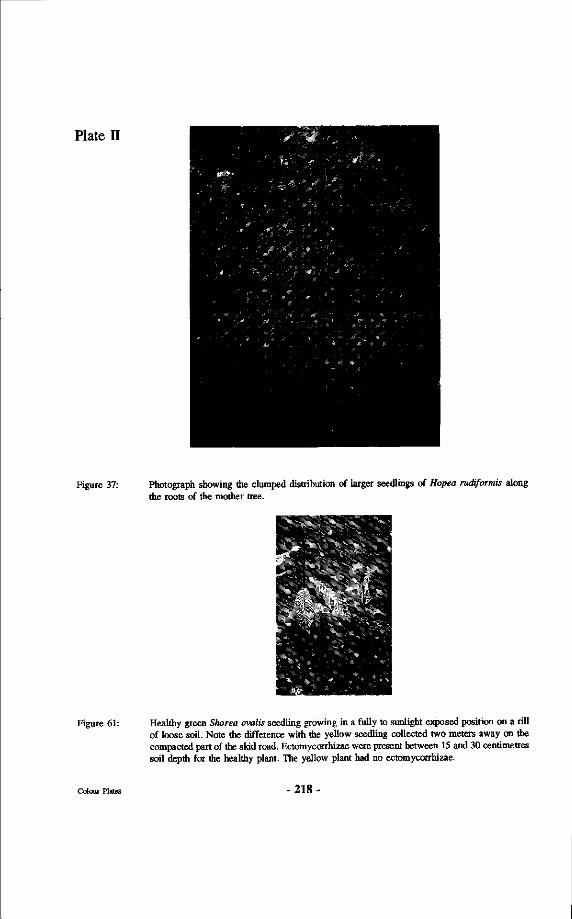
Plate E
Figure 37: Photograph showing the clumped distribution of larger seedlings of Hopea rudiformis along the roots of the mother tree.
->y*H
Figure 61: Healthy green Shorea ovalis seedling growing in a fully to sunlight exposed position on a rill of loose soil. Note the difference with the yellow seedling collected two meters away on the compacted part of the skid road. Ectomycorrhizae were present between 15 and 30 centimetres soil depth for the healthy plant. The yellow plant had no ectomycorrhizae.
Colour Plates -218-
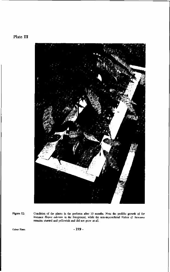
Plate HI
Figure 52: Condition of the plants in the perforon after 10 months. Note the prolific growth of for instance Hopea odorata in the foreground, while the non-mycorrhizal Vatica cf. bancana remains stunted and yellowish and did not grow at all.
Colour Plates -219-
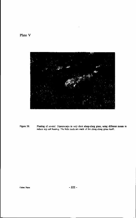
Plate V
Figure 58: Planting of several Dipterocarps in very short alang-alang grass, using different means to reduce top soil heating. The little roofs are made of the alang-alang grass itself.
- 2 2 2 -
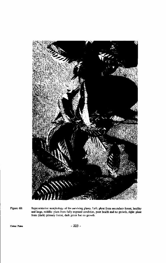
' -il,' ' V V >'l 'v '
' A'\ v - • ;>V> VVt/. H' \ >\«
4t ':; ' * & " • • •
i n ; , ' . .- .>. ..
Sb
Figure 60: Representative morphology of the surviving plants. Left: plant from secondary forest, healthy and large, middle: plant from fully exposed condition, poor health and no growth, right: plant from (dark) primary forest, dark green but no growth.
223-
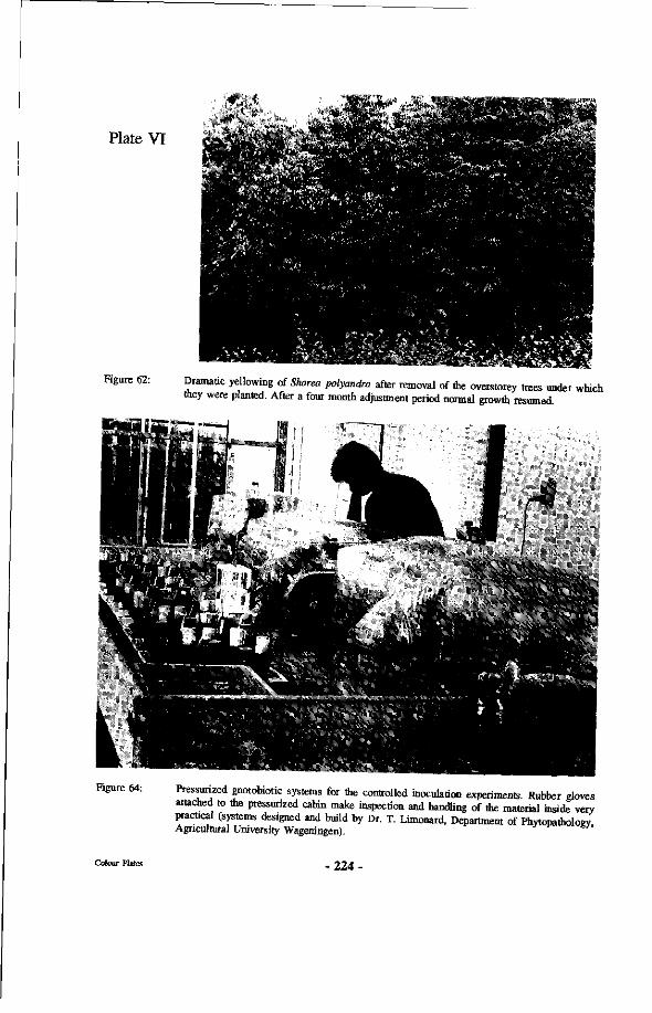
• • * * . . *
Plate VI ÄfiiÄ ter# •• •"y -
Figure 62: Dramatic yellowing of Shorea polyandra after removal of the overstorey trees vinder which they were planted. After a four month adjustment period normal growth resumed.
Figure 64: Pressurized gnotobiotic systems for the controlled inoculation experiments. Rubber gloves attached to the pressurized cabin make inspection and handling of the material inside very practical (systems designed and build by Dr. T. Limonard, Department of Phytopathology Agricultural University Wageningen).
224-
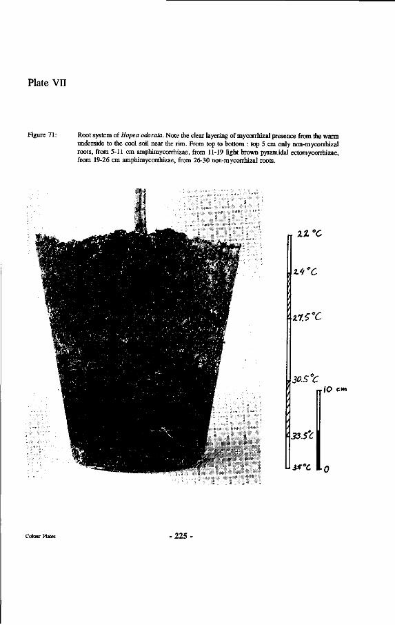
Plate VH
Figure 71 : Root system of Hopea odorata. Note the clear layering of mycorrhizal presence from the warm underside to the cool soil near the rim. From top to bottom : top 5 cm only non-mycorrhizal roots, from 5-11 cm amphimycorrhizae, from 11-19 light brown pyramidal ectomycorrhizae, from 19-26 cm amphimycorrhizae, from 26-30 non-mycorrhizal roots.
rr 2Z°C
%.^'C
'• zlS'C
u
30.S°C nIO cm
'• 33.S'C
is°c
- 2 2 5 -
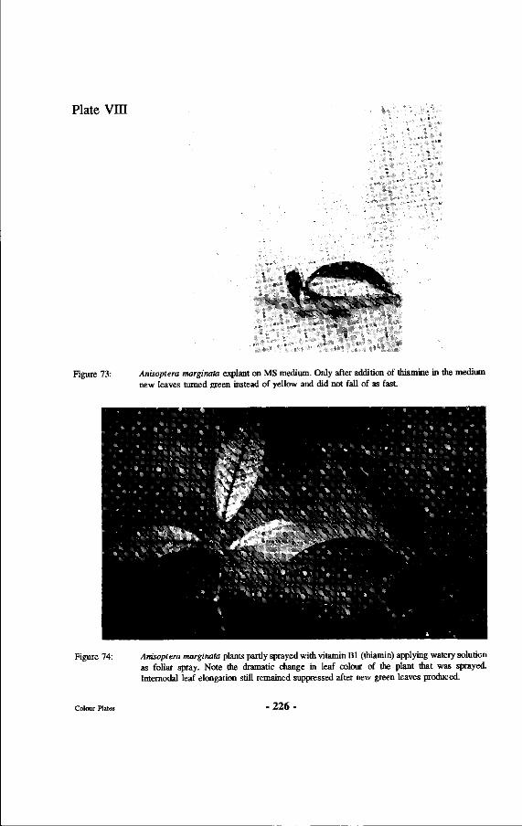
Plate Vm
\
.#*&&$t
Ȁ
Figure 73: Anisoptera marginata explant on MS medium. Only after addition of thiamine in the medium new leaves turned green instead of yellow and did not fall of as fast.
Figure 74: Anisoptera marginata plants partly sprayed with vitamin Bl (thiamin) applying watery solution as foliar spray. Note the dramatic change in leaf colour of the plant that was sprayed. Internodal leaf elongation still remained suppressed after new green leaves produced.
226
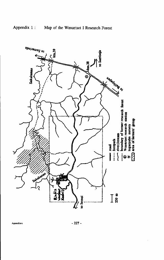
Appendix 1 : Map of the Wanariset I Research Forest
tPU!J'«'So, 3
Appendices -227-
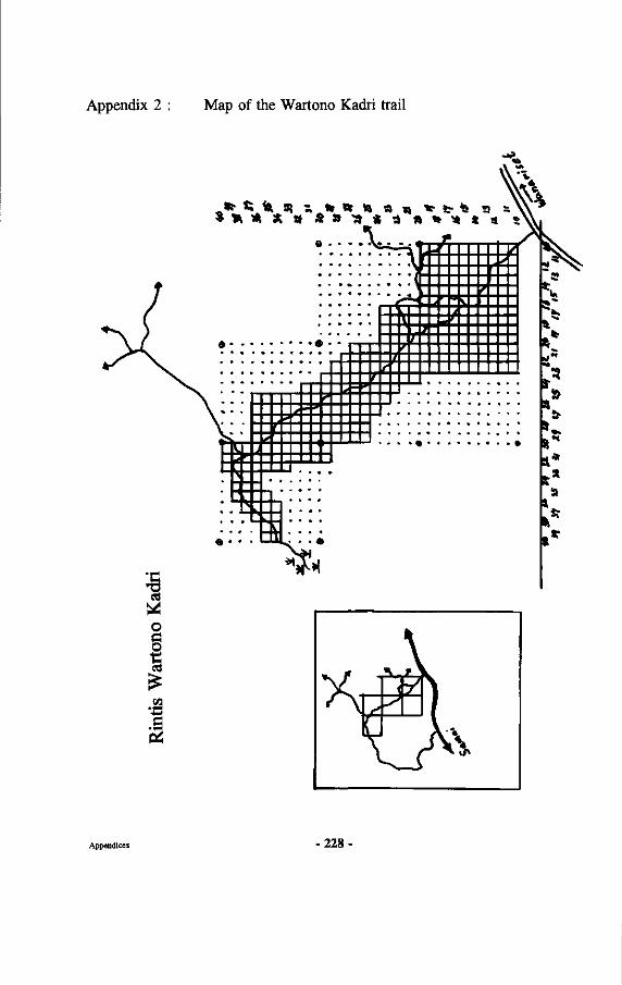
Appendix 2 : Map of the Wartono Kadri trail
a
o
I .S
S * * * a S a -a * a » * * * a
ft
SI* s*
a
m. Appendices - 2 2 8 -
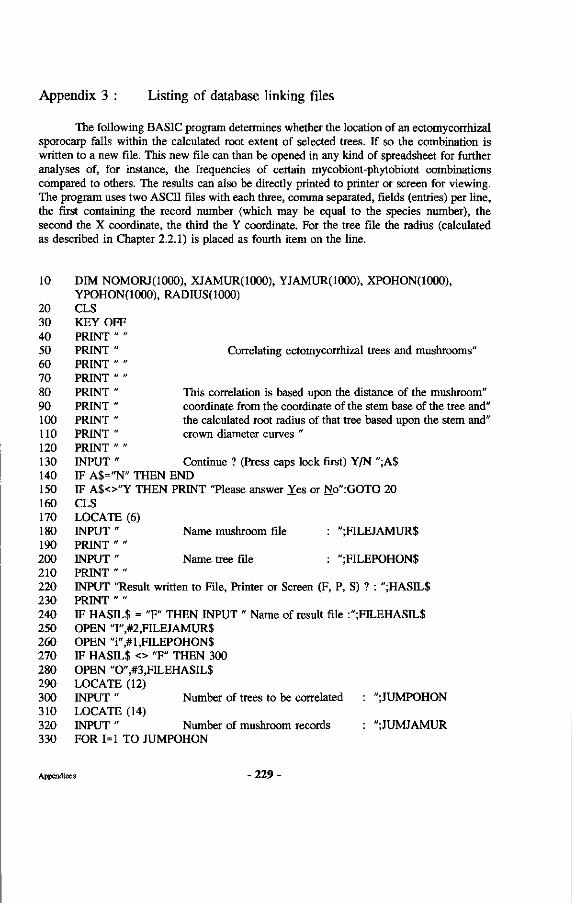
Appendix 3 : Listing of database linking files
The following BASIC program determines whether the location of an ectomycorrhizal sporocarp falls within the calculated root extent of selected trees. If so the combination is written to a new file. This new file can than be opened in any kind of spreadsheet for further analyses of, for instance, the frequencies of certain mycobiont-phytobiont combinations compared to others. The results can also be directly printed to printer or screen for viewing. The program uses two ASCII files with each three, comma separated, fields (entries) per line, the first containing the record number (which may be equal to the species number), the second the X coordinate, the third the Y coordinate. For the tree file the radius (calculated as described in Chapter 2.2.1) is placed as fourth item on the line.
10 DIM NOMORJ(IOOO), XIAMUR(IOOO), YJAMUR(IOOO), XPOHON(IOOO), YPOHON(IOOO), RADIUS(IOOO)
20 CLS 30 KEY OFF 40 PRINT " " 50 PRINT " Correlating ectomycorrhizal trees and mushrooms" 60 PRINT " " 70 PRINT " " 80 PRINT " This correlation is based upon the distance of the mushroom" 90 PRINT " coordinate from the coordinate of the stem base of the tree and" 100 PRINT " the calculated root radius of that tree based upon the stem and" 110 PRINT " crown diameter curves " 120 PRINT " " 130 INPUT " Continue ? (Press caps lock first) Y/N ";A$ 140 IF A$="N" THEN END 150 IF A$<>"Y THEN PRINT "Please answer Yes or No":GOTO 20 160 CLS 170 LOCATE (6) 180 INPUT " Name mushroom file : ";FILEJAMUR$ 190 PRINT " " 200 INPUT " Name tree file : ";FILEPOHON$ 210 PRINT " " 220 INPUT "Result written to File, Printer or Screen (F, P, S) ? : ";HASIL$ 230 PRINT " " 240 IF HASIL$ = "F" THEN INPUT " Name of result file :";FILEHASIL$ 250 OPEN T',#2,FILEJAMUR$ 260 OPEN "i",#l,FILEPOHON$ 270 IF HASIL$ <> "F" THEN 300 280 OPEN "0",#3,FILEHASIL$ 290 LOCATE (12) 300 INPUT " Number of trees to be correlated : ";JUMPOHON 310 LOCATE (14) 320 INPUT " Number of mushroom records : ";JUMJAMUR 330 FOR 1=1 TO JUMPOHON
Appendices - 2 2 9 -
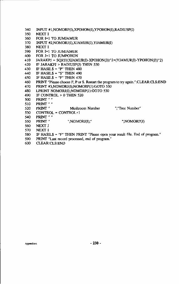
340 INPUT #l,NOMORP(I),XPOHON(I),YPOHON(I),RADIUSP(I) 350 NEXT I 360 FOR 1=1 TO JUMJAMUR 370 INPUT #2,NOMORJ(I),XJAMUR(I),YJAMUR(I) 380 NEXT I 390 FOR 1=1 TO JUMJAMUR 400 FOR J=l TO JUMPOHON 410 JARAKPJ = SQRT((XJAMUR(I)-XPOHON(J))'2+(YJAMUR(I)-YPOHON(J))"2) 420 IF JARAKPJ > RADIUSP(J) THEN 530 430 IF HASIL$ = "P" THEN 480 440 IF HASIL$ = "S" THEN 490 450 IF HASIL$ = "F" THEN 470 460 PRINT "Please choose F, P or S. Restart the program to try again.":CLEAR:CLS:END 470 PRINT #3,NOMORJ(I);NOMORP(J):GOTO 530 480 LPRINT NOMORJ(I);NOMORP(J):GOTO 530 490 IF CONTROL > 0 THEN 520 500 PRINT " " 510 PRINT " " 520 PRINT " Mushroom Number ";"Tree Number" 530 CONTROL = CONTROL+1 540 PRINT " " 550 PRINT " ";NOMORJ(I);" ";NOMORP(J) 560 NEXT J 570 NEXT I 580 IF HASIL$ = "F" THEN PRINT "Please open your result file. End of program." 590 PRINT "Last record processed, end of program." 600 CLEAR:CLS:END
Appendices - 2 3 0 •
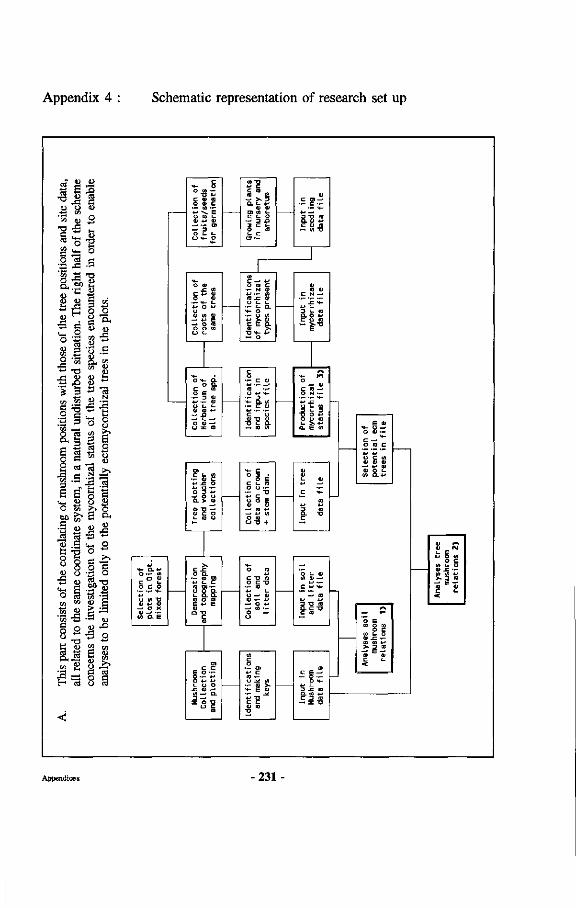
Appendix 4 Schematic representation of research set up
-S I «3 o)
•a rS
o ^ S S3 O ci '-C ^
o S
SJ
x-i .2
» 1 a'* •5.2 | | "5s ,—<
&g o e
a S3 o o
<u "3 .S
2 •-
o |
S Ë 3 O 'S o
G
D g
° 8
K l W
'S « '53 -S § 2 " • o
J3 ca
11 ra o •"
•8-°
3 ti S 4)
2 E a ^ H 'M
'M
c
O <D C
u n L 0» +J V
—• u
O <L>
O O) — i+- [_ •M O *->
*-> •— 0)
o O —'
»?
^»s V 0) Ui « u
1 o ra c
fi
ca
t or
rhi
pr
es
Ï I & —. O
^ o . w
- o c a
O
— c •—
aï 2
1 1—
<U
C N - . £ 4 -
j
O N —
'— '— **-
«- > .4- .
ï ï o
*- a*-> O •— W
o a> o c o
—« o x <D — ' • -» o. E
+j —«
t/i Q . +
(0 O l l i O Q. t- a. a mora
c c E o — o — *-• O 4J +J
tli CO
Q. « *-> C D CO — E "C
O c I U O > .e •-
Appendices -231 -
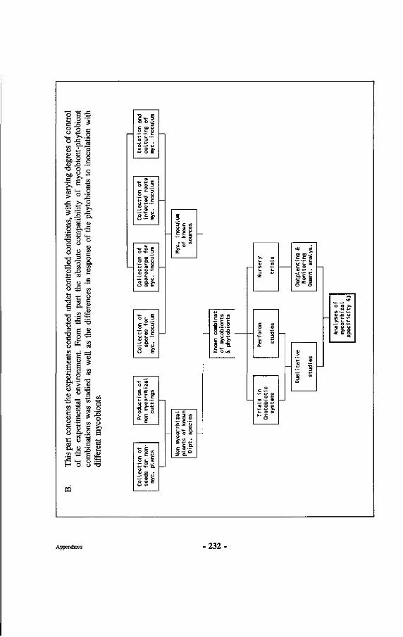
led c
ondi
tions
, with
vary
ing d
egre
es of
cont
rol
olut
e co
mpa
tibili
ty o
f m
ycob
iont
-phy
tobi
ont
ipon
se o
f the
phy
tobi
onts
to in
ocul
atio
n w
ith
cont
rol
the
abs
s in
re
3.
Thi
s par
t con
cern
s the
expe
rim
ents
cond
ucte
d und
er
of th
e ex
peri
men
tal en
viro
nmen
t. Fr
om th
is p
art
com
bina
tions
was
stud
ied
as w
ell a
s the
dif
fere
nce
diff
eren
t m
ycob
iont
s.
?,-§ ra o —•
3 c o> o o c o
. - . - c at 3 "~
"o —' o W 3 > . — u E
Col
lect
ion
of
infe
cted
roo
ts
myc
. in
ocul
um
Col
lect
ion
of
spor
ocar
ps f
or
myc
. in
ocuI
urn
Col
lect
ion
of
spor
es f
or
myc
. in
ocul
um
«4- (0 O N
c ! c w o i - o>
— (_ c *-* o — Ü U *-> 3 >•*->
T> E 3 O U (_ C Q. O
C
Col
lect
ion
of
seed
s fo
r non
-m
yc. p
lants
i " 3 c « U 3 <I) 0 0 u c c t_
— . * 3 O
• «4- ( 0 u 0
Non m
ycor
rhiz
al
plan
ts o
f kn
own
Dip
t, s
peci
es
Know
n co
mbi
nat
of m
ycob
iont
s & p
hyto
bion
ts
« •-3 4->
C (A O <D
£ Ü I - 4-1 ID U)
U C —
• r *J «1 O E
« •— tu - . J3 4-» ( D O «
— 4-> > i - O cn t - C
u
Out
plan
ting
&
Mon
itor
ing
Qua
nt, a
nal
ys.
> .- « ra •—
rz 1 ra « 3 0
Ana
lyse
s of
myc
orrh
izal
sp
ecif
icit
y 4)
Appendices 232-
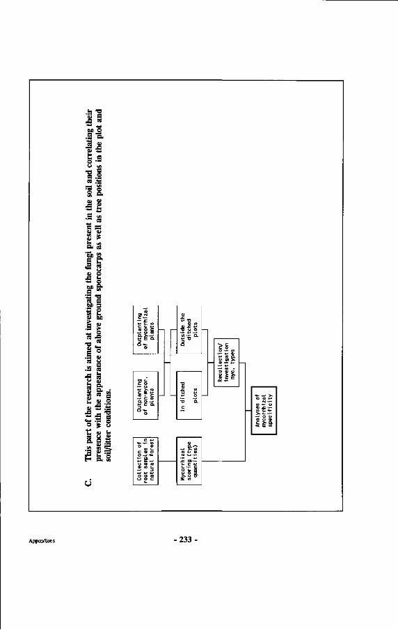
•a 'S 33 a -C «
* ^ - * j BS O
.E o. <*--•S-S u ~ t n o — U «1
• a l « Ä
P-N 1 »
'3 & M »-hl
« & 5 2 c «
• "" OS • J
e s tu CU
O" CS
"Sb g. S E* SM 3 V O
A 1-— O 6» S"
a ^ •a « A n DD s S P VI t * tu et
1 S a£ _. « ^ *_ » 's g o •5 tu
M a •s S
esea
rc
appe
; m
s.
c tu »
the
] th
th
ndit
i
o s S
is par
t se
nce
/litt
er
S I's
Ü
"(D D> N C —
*-> i- W C t - *-• ra o c
— o ra *J e a 3 O «*-
O
at (-c o
•— u
(0 . C —» c ta û. o -^ *-• c a.
o
0) . £
a! <fl <1> - C 4-<
" D U O
« — a. *-< "O
o
'S U -M
*-> o •o a c
V c c o O • • - </)
. - + j m 4-1 co p .
—' ( 0 • O Cl u a c E cc ••-
C * J
o a> c o o o -> <*-*> E — U (D (Q <D </) t -
— ' D
O O 10
o o c
<-
(U
—• >» « (O 4-" <Ü N v ^ —
- E O» — i - c +•>
(_ — c O f- (D O O D >» O O -
SE en
1 co -•- u | Gl -C •— H « t . H- 1
-> o u CD u a H c £• «- \ < Ë vi I
s
Appendices 233
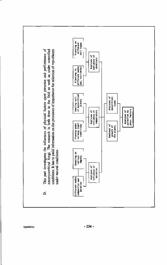
D.
Thi
s pa
rt i
nves
tigat
es th
e in
flue
nces
of p
hysi
cal
fact
ors
upon
pre
senc
e an
d pe
rfor
man
ce o
f ec
tom
ycor
rhiz
al f
ungi
. T
he r
esea
rch
is b
oth
done
in th
e fie
ld a
s w
ell
as u
nder
con
trol
led
cond
ition
s. It
has t
o yie
ld in
form
atio
n on t
he pr
oces
ses o
f im
port
ance
for s
elec
tion o
f myc
obio
nts
unde
r nat
ural
con
ditio
ns.
Pla
ntin
g on
di
ffer
ent
soi I
typ
es
Pla
ntin
g in
di
ffer
ent
med
ia
(pH
, n
utr
ien
ts)
Hea
ting
soi
I of
myc
orrh
izal
pl
ants
(_ 4-» 0> JZ to
•Ö o> c c * - o
3 -> —
O) to • -
C • - O (O L U
—• tO Û. >
Sam
plin
g at
va
riou
s de
pths
ra — XI •a c tu ra E c S •M O • -c ••- *-< 0) -H ra [_ O t_ 0) ro tu
H - o . ra "4- E a u
o o to
(A 0) —' o u to (A C U > . 0> —
-> 3 E co —« a C «4- .C < c u
Ana
lyse
s of
in
flue
nce
of
tem
pera
ture
•4- >4-O O
c to 01 o 0) U — to c *-> > 0) ra
_ - D t_ to —' 0) c-*- ta < c
Ana
lyse
s of
myc
orrh
izal
ty
pes
Ana
lyse
s of
gr
owth
of
the
plan
ts
M- > . 1-o x» o
Ana
lyse
s se
lect
ion
phys
. fac
Appendices 234
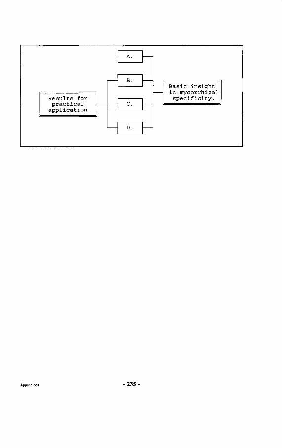
Results for practical
application
A.
B.
C.
D.
Basic insight in mycorrhizal
specificity.
Appendices - 2 3 5 -
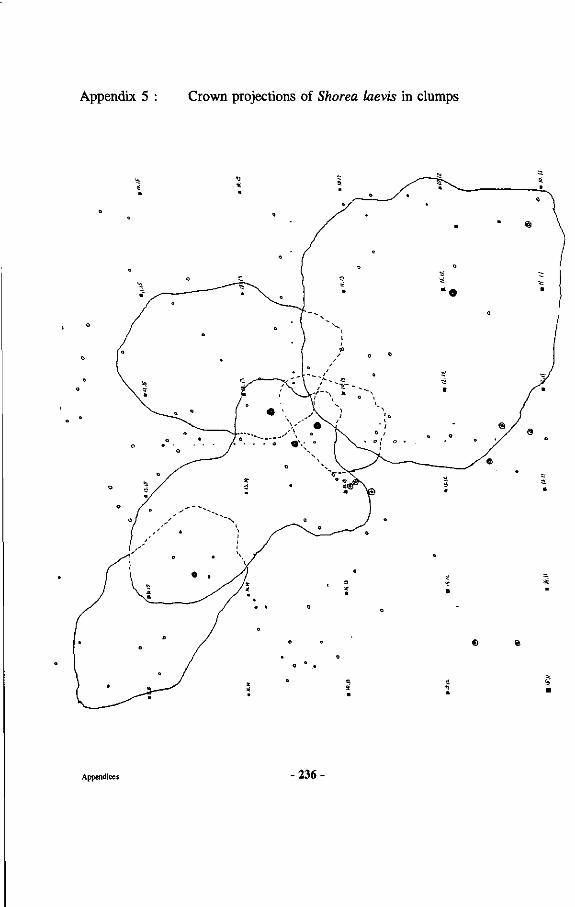
Appendix 5 : Crown projections of Shorea laevis in clumps
Appendices 236
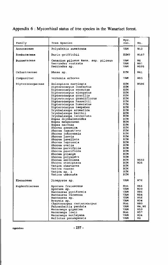
Appendix 6 : Mycorrhizal status of tree species in the Wanariset forest.
Family
Annonaceae
Bombacaceae
Burseraceae
Celastraceae
Compositae
Dipterocarpaceae
Ebenaceae
Euphorbiaceae
Tree Species
Polyalthia sumatrana
Durio griffithii
Canarium pilosum Benn. ssp. pilosum Dacryodes rostrata Dacryodes sp.
Bhesa sp.
Vernonia arborea
Anisoptera marginata Dipterocarpus confertus Dipterocarpus cornutus Dipterocarpus elongatus Dipterocarpus gracilis Dipterocarpus grandiflorus Dipterocarpus hasseltii Dipterocarpus humeratus Dipterocarpus tempehes Dryobalanops aroraatica Dryobalanops keithii Dryobalanops lanceolata Hopea dryobalanoides Hopea raengerawan Hopea nervosa Shorea assamica Shorea faguetiana Shorea johorensis Shorea laevis Shorea lamellata Shorea leprosula Shorea ovalis Shorea parvifolia Shorea pauciflora Shorea pinanga Shorea polyandra Shorea smithiana Shorea stenoptera Vatica chartacea Vatica rassac Vatica sp. 1 Vatica umbonata
Diospyros sp.
Aporusa frutescens Aporusa sp. Baccaurea pyriformis Baccaurea racemosa Baccaurea sp. Breynia sp. Chaetocarpus castanocarpus Fahrenheitia pendula Macaranga gigantea Macaranga lowii Macaranga motleyana Mallotus penangensis
Myc. con.
VAM
ECM?
VAM VAM VAM
ECM
VAM
ECM ECM ECM ECM ECM ECM ECM ECM ECM ECM ECM ECM ECM ECM ECM ECM ECM ECM ECM ECM ECM ECM ECM ECM ECM ECM ECM ECM ECM ECM ECM ECM
VAM
Non VAM VAM VAM VAM VAM Non VAM VAM Non VAM VAM
No.
W12
W107
W6 W81 W595
W41
W85
W596
W550 W25
W76
W66 W2 0 W86 W84 W46 W34 W80 W4,W6 W10 W57 W24 W2
Appendices 237-
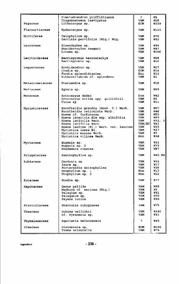
Fagaceae
Flacourtiaceae
Guttiferae
Lauraceae
Lecythidaceae
Leguminosae
Melastomataceae
Meliaceae
Moraceae
Myristicaceae
Myrtaceae
Polygalaceae
Rubiaceae
Rutaceae
Sapotaceae
Sterculiaceae
Theaceae
Thymelaeaceae
Ulmaceae
Pimelodrendron griffithianura Trigonostemon laevigatus Lithocarpus sp.
Hydnocarpus sp.
Calophyllum sp. Garcinia parvifolia (Miq.) Miq.
Alseodaphne sp. Eusideroxylon zwageri Litsea sp.
Barringtonia macrostachya Barringtonia sp.
Archidendron sp. Crudia sp. Fordia splendidissima Pithecellobium cf. splendens
Pternandra sp.
Aglaia sp.
Artocarpus dadah Artocarpus nitida spp. griffithii Ficus sp
Horsfieldia grandis (Hook, f.) Warb. Horsfieldia reticulata Warb. Knema cf. furfuracea Knema latericia Elm ssp. albifolia Knema latifolia Warb. Knema latifolia Warb. Knema laurina (Bl.) Warb. var. laurina Myristica iners Bl. Myristica maxima Warb. Myristica villosa Warb.
Eugenia sp. Eugenia sp. 2 Rhodamnia cinerea
Xanthophyllum sp.
Gardenia sp Ixora sp. Porterandia anisophyllea Urophyllum sp. 1 Urophyllum sp. 2
Euodia sp.
Ganua pallida Madhuca cf. sericea (Miq.) Palaqium sp. Palaquium sp. Payena lucida
Sterculia rubiginosa
Schima wallichii cf. Pyrenaria sp.
Aquilaria malaccensis
Gironniera sp. Trema orientalis
•p
VAM ECM
VAM
VAM VAM
VAM VAM VAM
VAM VAM
VAM ECM Rhi VAM
VAM
VAM
Non VAM VAM
VAM VAM VAM VAM VAM VAM/EC VAM VAM VAM Non
VAM VAM VAM
VAM
VAM VAM VAM Non Non
VAM
VAM VAM VAM VAM VAM
VAM
VAM VAM
?
ECM VAM
W5 W28 W359
W125
W70 W52
W94 W47 W106
W68
wie
W27 W14 W33 Wl
W18
W69
W42 W3 Wil
W67 W48 W55 W49 W32, 5 W43 W2 9 W37 W7 W38
W21 W79 W87
W40,W2
W44 W17 W36 W13 W22
W77
W88 W9 W51 W90 W50
W75
W140 W31
W45
W105 W74
Appendices -238
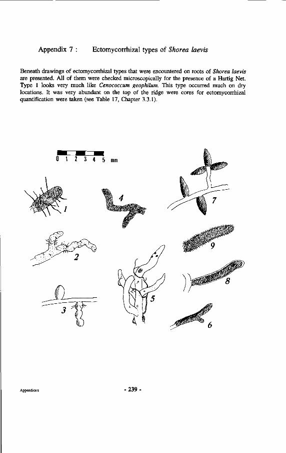
Appendix 7 Ectomycorrhizal types of Shorea laevis
Beneath drawings of ectomycorrhizal types that were encountered on roots of Shorea laevis are presented. All of them were checked microscopically for the presence of a Hartig Net. Type 1 looks very much like Cenococcum geophilum. This type occurred much on dry locations. It was very abundant on the top of the ridge were cores for ectomycorrhizal quantification were taken (see Table 17, Chapter 3.3.1).
Appendices 239
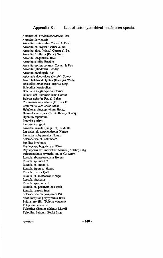
Appendix 8 : List of ectomycorrhizal mushroom species
Amanita cf. avellaneosquamosa Imai Amanita borneensis Amanita centunculus Comer & Bas Amanita cf. duplex Comer & Bas Amanita elata (Mass.) Comer & Bas Amanita fritillaria (Berk.) Sacc. Amanita longistriata Imai Amanita similis Boedijn Amanita sychnopyramis Comer & Bas Amanita tjibodensis Boedijn Amanita xanthogala Bas Aphelaria dendroides (Jungh.) Comer Austrobeletus dictyotus (Boedijn) Wolfe Boletellus emodensis (Berk.) Sing. Boletellus longicollus Boletus ferruginosporus Comer Boletus aff. olivaceirubens Comer Boletus spinifer Pat. & Baker Cortinarius anomalous (Fr.: Fr.) Fr. Craterellus vermcosus Mass. Hebeloma vinosophyllum Hongo Heimiella retispora (Pat & Baker) Boedijn Hydnum repandum Inocybe godeyi Inocybe mangayi Laccaria laccata (Scop.: Fr) B. & Br. Lactarius cf. austrovolemus Hongo Lactarius subpiperatus Hongo Scleroderma cf. columnare. Paxillus involutus Phyllopoms bogoriensis Höhn. Phylloporus aff. infundibuliformis (Cleland) Sing. Pulveroboletus ravenelii (B. & C.) Murril Russula ebumeoareolata Hongo Russula sp. indet. 2. Russula sp. indet. 7. Russula japonica Hongo Russula lilacea Quel. Russula cf. metachroa Hongo Russula nigricans Russula spec. nov. ? Russula cf. pectinatoides Peck Russula senecis Imai Scleroderma dictyosporum Pat Strobilomyces polypyramis Berk. Suillus grevillii (Boletus elegans) Telephora terrestris Tylopilus alboater (Schw.) Murrill Tylopilus ballouii (Peck) Sing.
Appendices - 2 4 0 -
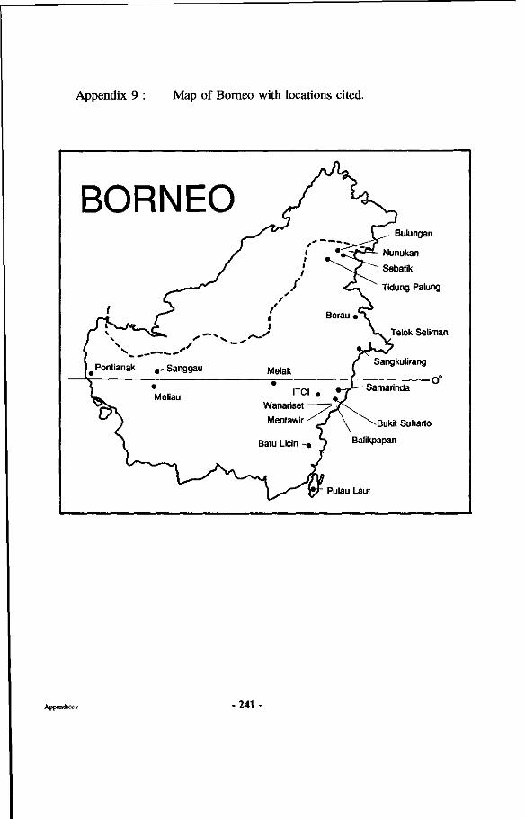
Appendix 9 : Map of Borneo with locations cited.
BORNEO
f 3
i Pontianak ,,-Sanggau
« • ^ Meliau
1
1
/ /
* / 1 1
Melak
• ITCI a
Wanariset Mentawir ^
Batu Licin -«
V \ , Bulungan
' • - C j f j ^ Nunukan
^ " \ j T ^ " Sebatik
^ - . Tidung Palung
Berau »^T
\ ^ Telok Seliman
/ Sangkulirang
i o° %-J— Samarinda
J \ \Buk i t Suharto
\ Balikpapan
" Pulau Laut
Appendices -241-
Curriculum Vitae
Willie Smits werd geboren op 22 februari 1957 te Weurt (gem. Beuningen). Na het behalen van het Gymnasium-B diploma aan het Monseigneur Zwijssen College te Veghel, begon hij de propaedeuse aan de toenmalige Landbouwhogeschool te Wageningen in 1975/1976. In 1978 koos hij voor de studierichting Bosbouw en behaalde in 1980 het kandidaatsexamen. In 1980 bracht hij zeven maanden praktijktijd door in een houtconcessie op Borneo, Indonesië, waar hij nauw betrokken was bij onderzoek aan lokale hardhoutsoorten. In deze periode trouwde hij met Syennie Watoelangkow, een Indonesische van Noord-Sulawesi.
De doctoraalstudie omvatte de vakken Bosteelt en Bosoecologie, Genetica en Tropische Bodemkunde. Binnen het vak Bosteelt verrichtte hij onder begeleiding van Prof. Oldeman onderzoek naar vegetatieve vermeerdering van Dipterocarpaceae waarbij hij enige maanden onder begeleiding van Prof. Pierik van de Landbouwuniversiteit en samen met Dr. Evers van IBN-DLO aan in-vitro kuituur van Dipterocarpaceae werkte. Daarnaast omvatte de doctoraalscriptie werk over vegetatieve vermeerdering van Agathis bomeenis en enig werk over ectomycorrhizae van Dipterocarpaceae. Binnen het doctoraalvak Genetica maakte hij een skriptie over genetische aspekten van Dipterocarpaceae, terwijl hij binnen het vak Tropische Bodemkunde aan organische stof in oxisolen werkte.
Na zijn afstuderen in 1982 werkte hij tot 1985 als wetenschappelijk onderzoeker bij de vakgroepen Bosbouw en Fytopathologie van de huidige LU aan mycorrhizae van Dipterocarpaceae, aan wortelonderzoekstechnieken en aan de invloed van zure regen op mycorrhizae. Tussendoor maakte hij studiereizen naar Frankrijk, Engeland, Spanje, Maleisië, Indonesië en de Verenigde Staten, waarbij vele lezingen werden gegeven en gezamenlijk onderzoek werd uitgevoerd.
In 1985 werd hij uitgenodigd door het Indonesische Ministerie van Bosbouw om naar Indonesië te komen om behaalde onderzoeksresultaten op praktijkschaal uit te testen. Met toestemming van de Landbouwuniversiteit vertrok hij naar Indonesië, waar hij in 1985 begon het onderzoeksstation Wanariset Samboja uit te bouwen. Dit station is inmiddels een van de belangrijkste bosbouwonderzoekslokaties in Indonesië geworden en de resultaten hebben geleid tot het opleiden van vele honderden bosbouwers van binnen en buiten Indonesië en grootschalige aanplanten van Dipterocarpaceae.
Vanaf November 1987 werd hij leider van het Tropenbos-Kalimantan onderzoeksprogramma op dezelfde lokatie, werkend voor het Instituut voor Bos- en Natuuronderzoek te Wageningen. Sinds 1991 bekleedt hij diverse funkties in commitees voor duurzaam bosbeheer, verrichtte hij consultancies, onder andere voor de wereldbank en sinds 1993 is hij persoonlijk adviseur van de Indonesische Minister voor Bosbouw.
Curriculum Vitae - 2 4 3 -
Related Documents