Journal of Sustainability Science and Management Volume 12 Number 2, December 2017: 52-62 ISSN: 1823-8556 © Penerbit UMT DEVELOPMENT OF MUD CRAB CRABLET, THE IDENTIFICATION OF CILIATES AND THE BIOEFFICACY OF LEAF EXTRACT OF Rhizophora Apiculata AS ANTI- PROTOZOAL AGENT NGUYAN KHANH LINH 2 , TRAN NGUYEN DUY KHOA 3 , SANDRA CATHERINE ZAINATHAN 2,4 , NADIRAH MUSA 2 , NAJIAH MUSA 2 , FAIZAH SHAHAROM-HARRISON* 1 Kenyir Research Institute, 2 School of Fisheries and Aquaculture Sciences , 3 Institute of Tropical Aquaculture, 4 Institute of Marine Biotechnology, Universiti Malaysia Terengganu, 21030 Kuala Nerus, Terengganu, Malaysia. *Corresponding author: [email protected] Introduction Aquaculture produced one-quarter of fish and shell fish supplied as human food. It releases the pressure of fishery (Naylor et al., 2000). In 2012, the total of aquaculture production was 90.4 million tonnes. In which, the food fish (included finfishes, crustaceans, molluscs etc.) occupied 66.6 million tonnes (FAO, 2014). In many Asian countries, the mud crab is a valuable species for fisheries (Keenan, 1999). More than 100 years ago, the mud crab cultures have been developed in China and more than 30 years in many Asian countries (Holme et al., 2006). Mud crab is an important cultured species after shrimp which can yield high economic value for fishermen and farmers (Shelley & Lovatelli, 2011). These species can be found in mangrove forest around the Pacific and India Ocean (Keenan, 1999). Tan (1997) reported that in Malaysia, the mud crab larvae rearing have not been successful which led to low mud crab production. The mud crab production in Malaysia decreased from 623 tonnes in 1995 to 162 tonnes in 2005 (Shelley, 2008). Due to their high demand and market price in Malaysia, thus, this species is highly potential as an aquaculture species. Scylla olivacea, S. transquebarica, S. paramamosain are three dominant mud crab species in Setiu Wetland, Malaysia (Zaidi et al., 2011). In this study, the green mud crab, S. paramamosain was chosen. S. paramamosain can reach the market size (200 to 300g) after 3 months (Christensen et al., 2004). However, mud crab seed captured from the wild is not sufficient to sustain the current status of mudcrab production. Therefore, economic seed production of mud crab need to be developed (Holme et al., 2006). In addition, most of diseases studies on crustaceans are focused on fish and shrimp (Jithendran et al., 2010). In Malaysia, mud crab has been exploited by local fishermen based on the mangrove forest in estuaries and coastal area (Ikhwanuddin et al., 2011). The mud crab culture in Malaysia started in 1991 and they were cultured in ponds or pen Abstract: Wild mud crabs of the genus, Scylla paramamosain were acclimatized in tanks in the AKUATROP hatchery. Eye stalk ablation was applied on the mud crabs before transferring them into a recirculating aquaculture system, where they were fed marine fish, squid and cockles for a period of one month until they produced eggs. The larvae which hatched out were placed in different larval tanks with continuous aeration. Larvae were fed daily with artemia. The density of larvae culture was 100-300 individuals per liter. The peritrich ciliates found on megalopa larva of mud crabs, Scylla paramamosain were Zoothamnium alrashedi and Myoschiston duplicatum and an unidentified peritrich ciliate. Mangrove leaf extract of Rhizophora apiculata showed that it is capable of being an anti- protozoan product as the zooids of the peritrich ciliates dropped off after treatment with the extract. The breeding tanks were kept clean, probiotics was introduced with plenty of aeration. Green water system was activated to ensure plenty of natural food namely rotifers to ensure moulting of megalopa larva into crablet larva. Keywords: Scylla sp, megalopa larvae, peritrich ciliates, Zoothamnium alrashedi, Myoschiston duplicatum.

Welcome message from author
This document is posted to help you gain knowledge. Please leave a comment to let me know what you think about it! Share it to your friends and learn new things together.
Transcript

Journal of Sustainability Science and Management Volume 12 Number 2, December 2017: 52-62
ISSN: 1823-8556© Penerbit UMT
DEVELOPMENT OF MUD CRAB CRABLET, THE IDENTIFICATION OF CILIATES AND THE BIOEFFICACY OF LEAF EXTRACT OF Rhizophora
Apiculata AS ANTI- PROTOZOAL AGENT
NGUYAN KHANH LINH2, TRAN NGUYEN DUY KHOA3, SANDRA CATHERINE ZAINATHAN2,4, NADIRAH MUSA2, NAJIAH MUSA2, FAIZAH SHAHAROM-HARRISON* 1Kenyir Research Institute, 2School of Fisheries and Aquaculture Sciences , 3Institute of Tropical Aquaculture, 4Institute of Marine Biotechnology, Universiti Malaysia Terengganu, 21030 Kuala Nerus, Terengganu, Malaysia.
*Corresponding author: [email protected]
IntroductionAquaculture produced one-quarter of fish and shell fish supplied as human food. It releases the pressure of fishery (Naylor et al., 2000). In 2012, the total of aquaculture production was 90.4 million tonnes. In which, the food fish (included finfishes, crustaceans, molluscs etc.) occupied 66.6 million tonnes (FAO, 2014). In many Asian countries, the mud crab is a valuable species for fisheries (Keenan, 1999). More than 100 years ago, the mud crab cultures have been developed in China and more than 30 years in many Asian countries (Holme et al., 2006). Mud crab is an important cultured species after shrimp which can yield high economic value for fishermen and farmers (Shelley & Lovatelli, 2011). These species can be found in mangrove forest around the Pacific and India Ocean (Keenan, 1999).
Tan (1997) reported that in Malaysia, the mud crab larvae rearing have not been successful which led to low mud crab production. The mud crab production in Malaysia decreased from 623
tonnes in 1995 to 162 tonnes in 2005 (Shelley, 2008). Due to their high demand and market price in Malaysia, thus, this species is highly potential as an aquaculture species. Scylla olivacea, S. transquebarica, S. paramamosain are three dominant mud crab species in Setiu Wetland, Malaysia (Zaidi et al., 2011). In this study, the green mud crab, S. paramamosain was chosen. S. paramamosain can reach the market size (200 to 300g) after 3 months (Christensen et al., 2004). However, mud crab seed captured from the wild is not sufficient to sustain the current status of mudcrab production. Therefore, economic seed production of mud crab need to be developed (Holme et al., 2006). In addition, most of diseases studies on crustaceans are focused on fish and shrimp (Jithendran et al., 2010).
In Malaysia, mud crab has been exploited by local fishermen based on the mangrove forest in estuaries and coastal area (Ikhwanuddin et al., 2011). The mud crab culture in Malaysia started in 1991 and they were cultured in ponds or pen
Abstract: Wild mud crabs of the genus, Scylla paramamosain were acclimatized in tanks in the AKUATROP hatchery. Eye stalk ablation was applied on the mud crabs before transferring them into a recirculating aquaculture system, where they were fed marine fish, squid and cockles for a period of one month until they produced eggs. The larvae which hatched out were placed in different larval tanks with continuous aeration. Larvae were fed daily with artemia. The density of larvae culture was 100-300 individuals per liter. The peritrich ciliates found on megalopa larva of mud crabs, Scylla paramamosain were Zoothamnium alrashedi and Myoschiston duplicatum and an unidentified peritrich ciliate. Mangrove leaf extract of Rhizophora apiculata showed that it is capable of being an anti-protozoan product as the zooids of the peritrich ciliates dropped off after treatment with the extract. The breeding tanks were kept clean, probiotics was introduced with plenty of aeration. Green water system was activated to ensure plenty of natural food namely rotifers to ensure moulting of megalopa larva into crablet larva.
Keywords: Scylla sp, megalopa larvae, peritrich ciliates, Zoothamnium alrashedi, Myoschiston duplicatum.
DEVELOPMENT OF MUDCRAB CRABLET, THE IDENTIFICATION OF CILIATES 53
J. Sustain. Sci. Manage. Volume 12(2) 2017: 52-62
among the mangrove trees (Tan, 1997). Zaidi et al. (2011) demonstrated that the mud crab in Setiu, Malaysia provide good resources for local fishermen. However, artificial breeding and larval rearing are difficult techniques (Keenan, 1999) because at the hatchery phase, three main issues need to be encountered including disease outbreak, incomplete rearing techniques, and lack of nutrition requirement (Sorgeloos & Léger, 1992). In the hatchery phase, bacteria and fungus infection appear to be the major problems. Moreover, ciliate protozoan has been recorded to cause problem during the larvae stage of mud crab (Jithendran et al., 2010).
Fernandez-Leborans (2009) recorded that ciliate protozoan are known to colonize as the epibiont on many crustacean species. Wahl (1989) defined that epibiosis is the relationship between basibiont (host organisms) and epibiont (colonized organisms). Epibiotic relationship is abundant in fresh, estuarine and marine water and in many organisma such as protozoan, bacteria and rotifers. Although their distribution is widespread, they are poorly known for their ecological implications for both basibionts and the epibionts. In addition, most of the studies of epibiosis on crustacean have been reported in fresh water, and very few are focused in marine water (Carman & Dobbs, 1997).
Protozoan has caused high mortality on many crustacean species. Roegge et al. (1977) recorded that Zoothamnium sp. found on Macrobrachium acanthurus caused mass mortality on the larvae. In addition, Zoothamnium sp. also caused abnormal morphology and led to the mortality of Chinese mitten crab larvae (Eriocheir sinensis) (Wu & Feng, 2004). Moreover, protozoan infections can affect the survival rate and normal activities of Penaeus monodon larvae (Babu, 2013). The presence of ciliate protozoans depends on the water quality and the abundance of bacteria and microorganisms. In a high nutrition and organic matter environment, the population of ciliate protozoan was found to be higher than the oligotrophic condition (Jithendran et al., 2010; Jayakumar & Ramasamy, 1999).
Ciliate protozoan mainly feed on bacteria and microalgae in eutrophic environment, thus, they tend to play an important role in the ecosystem (Bernard & Rassoulzadegan, 1999; Fenchel, 1987). Epistylis sp. has been observed in a low oxygen environment (Jithendran et al., 2010).
In Malaysia, there are many chemicals used as antiprotozoal such as saponins, formalin, acriflavine, malachite green, copper sulphate, organophosphates, and benzaklonium chloride. However, most of the chemicals used are released to the natural environment via waste water and it can cause negative effect for other aquatic animals. Moreover, use of chemicals can be harmful to human once in contact in a longer term (Mohamed et al., 2000). Therefore, the research to find the alternative natural medicine in order to reduce the impact of chemicals in aquaculture, environment and human health is very important. The use of natural medicine will be low in cost, environment friendly and produces positive effect in aquaculture (Novriadi & Haw, 2015; Citarasu, 2010). Natural/Alternative medicine has been reported as antibacterial, antiviral, antifungal, anti-stress agent (Citarasu, 2010; Ramudu & Dash, 2013) and antiparasitic (Reverter et al., 2014). Halophytes contained plants can live in a high salinity environment such as algae, sea grass and mangrove (Kumar et al., 2009). In that, mangrove has been shown to possess the ability to control pathogens in aquaculture. Choudhury et al. (2005) indicated that marine algae and mangrove have the ability to control bacterial infections in fish and shrimp (Suryati & Hala, 2002). Arivuselvan et al. (2011) reported that mangrove can be utilized as antibacterial in both fish and shrimp. In addition, the selected mangroves from Novriadi & Haw (2015) can control the iridovirus infections in tiger grouper.
Thus, this paper describes the production of mudcrab crablets and the identification of ciliates during the process. The mangrove extract of Rhizophora apiculata was also tested for its efficacy on treatment of ciliate protozoans.

Nguyan Khanh Linh et al. 54
J. Sustain. Sci. Manage. Volume 12(2) 2017: 52-62
Material and MethodsMethodology: Development of Mud Crab CrabletSix female mud crabs were bought from the farmer in mangrove forest of Setiu Wetland, Terengganu, Malaysia. The mature mud crab females (S. paramamosain) which weighted about 350-500g, healthy (body had enough claws and leg, the shell was clean) and had mature ovaries were chosen for this study. Then, the brood stocks were transferred to tanks in the AKUATROP (Institute of Tropical Aquaculture) hatchery, Universiti Malaysia Terengganu for acclimation.
The brood stocks were kept in sand tank about two days. After that, the artificial breeding was done by eye stock ablation (cut 1 eye or both eye), then they were disinfected with 20 ppm formalin and put into the sand tank. The brood stocks without 1 or 2 eyes were fed with marine fish, squid, shrimp or blood cockle (high nutrition food). The brood stock laid the egg after 7-10 days (Figure 3) and they were transferred from sand tank into the new tank with clean water. Hatching time was about 10 to 12 days. At that time, eggs of the brood stocks were observed under compound microscope to find the parasite. After hatching, zoae 1 larvae were transferred from aquarium into 6 tanks (1 m3/tank) with density about 200,000 larvae per tank.
The mud crab larvae (from zoae 1 to zoae 3) were fed with umbrella Artemia after hatching two times per day at 9 am and 9 pm. After that, at zoae 4, 5, megalopa stage, the larvae were fed by enrich artemia. Besides, water exchange every two days about 20-30%. At megalopa stage, the artificial substrates were put into the tank to reduce the cannibalism. In addition, the bacteria, fungus disease were controlled. In that time, every stage of the larvae was observed under the compound microscope for the parasite. The larvae were collected using plankton net (Figure 5), after the larvae were brought into parasitology lab. Before the larvae was placed onto glass slide, deep the larvae into fresh marine water. After that, put the larvae onto glass slide and put 1 drop of fresh saline water and covered
with cover slip and observed under the low and high magnification of the compound microscope (Figure 6). It took a month to complete the life cycle of a mudcrab. During crablet stage, the crabs were fed with fish/pellet and 10% cannbalismwas observed.The crabs were then transferred to ponds once harvesting, counting, packing and farm acclimation was carried out.
Identification of CiliatesLive samples were drawn to illustrate the morphology of the ciliate parasite. The drawing was done using a camera Lucida connected to a compound microscope. The advanced microscope was used in this study to take the micrographs from live samples. As for the Scanning Electron Microscope examination, the samples were fixed once in 2.5% Glutaraldehyde in 0.1 sodium cacodylate buffer, pH 7.2 at 0-4°C for 24 hours. After 24 hours, the samples were rinsed three times with 0.1M sodium cacodylate buffer, pH 7.2 at room temperature 3 times (15 minutes between each change). The samples were put once in 1% osimium tetraoxide in 0.1M sodium cacodylate buffer, pH 7.2 at 0-40C for 2 hours.The samples were rinsed three times (15 minutes between each change) with 0.1M sodium cacodylate buffer, pH 7.2. The samples were dehydrated once in series of ethanol: 35%, 50%, 60%, 70%, 80%, 90%, 95% and 2 times in 100% ethanol with few seconds between each change. After the preparation, the samples were placed into the specimen basket and continued in the critical point dryer (CPD) machine for about 30 minutes. The samples were mounted onto the stub by using double sided tape and coated with gold by sputter coater. The samples were scanned using SEM machine model JEOL JSM-6360LA. The images were adjusted by using brightness, contrast, magnification and capture.
Mangrove Extraction The leaves of Rhizophora apiculata were dipped in methanol for three days in the dark condtion. The dark green solution was filtered by filter papers and evaporated using the rotary

DEVELOPMENT OF MUDCRAB CRABLET, THE IDENTIFICATION OF CILIATES 55
J. Sustain. Sci. Manage. Volume 12(2) 2017: 52-62
evaporator machine until the solution became dense and dark in colour. The dense solution was stored at – 80°C for 24 hours and dried by freeze dryer machine for three days (Bele et al., 2009). Phytochemical screening method were used to analysze the compound in R. apiculata extract based on the methods of Yadav and Agarwala (2011).
Protozoan Treatment Experimental DesignThe experiment included six treatments (0, 0.2, 0.4, 0.6, 0.8, 1 g R. apiculata leaf extract per liter of marine water) in triplicates (Figure 1). Twenty megalopa larvae were put into 500 ml plastic jars with aeration. The new marine water was filtered by plankton net. Then the extract was applied for 24 hours. After that, the megalopa survival rate were calculated based on Ricker (1975) equation. The total number of protozoans was recorded. The survival was calculated as followed:
S = Nt + 1 NtWhere S is survival rate of megalopa after treatment Nt is the number of megalopa before treatmentNt + 1 is the number of megalopa after treatment
Data AnalysisThe data were analyzed using Excel and one way ANOVA, probit analysis from SPSS version 16.0 Protozoans were identified based on the morphology decribed by Lynn (2008) & Sun et al. ( 2012).
Results and DiscussionProduction of Mud Crab CrabletEye stalk ablation was conducted on mature crabs, placed individually in tanks and fed daily till it produced eggs. After spawning, the larvae were placed in larval tanks with continuous aeration (1 ton= ±200,000 larvae) and fed with Artemia. Water quality and health of larvae were monitored daily. At megalopa stage density of larvae was decreased and water salinity was controlled. Artificial substrate was introduced to prevent cannibalism. When megalopa develop into crablets they were fed with trash fish and shrimp pellets.
The larval stages of mud crabs consists of 5 zoeal stages (Figure 2). Each stage moults to the next stage in 3 to 4 days. Hence, the duration of zoea to megalopa stage takes 15 to 20 days. Megalopa develops into juvenile crab after 8 to 11 days. Zoea 1 to zoea 5 measures to about 1.2 to 4.5 mm in body length. They are planktonic and photopositive. A newly hatched zoea larva of crab measures to 1.2 mm and it consists of a cephalothorax and a 5 segmented abdomen and t two-branched telson. The carapace bears 4 spines, one dorsal spine bent backwards, rostral spine bent forwards and 2 lateral spine closely pressed against the sides of the body. The eyes are not stalked. From zoea II onwards, the eyes are stalked (for distinguishing characteristics of larva stages of crab). The megalopa stage is 4.0 mm length and it feeds actively using its claw. This period has a duration of 7 to 8 days before it metamorphose into crablet stage. These crablets
Figure 1: Experimental design of protozoan treatment with different concentrations of R. apiculata extract

Nguyan Khanh Linh et al. 56
J. Sustain. Sci. Manage. Volume 12(2) 2017: 52-62
Figure 2: The life stages of mud crab: zoea 1, zoea 2, zoea 3, zoea 4, zoae 5, megalopa stage and crablet
have a carapace width and length of 3 mm and are cannibalistic.
Parasites Identification in S. paramamosain Field observation showed presence of the parasite: Octolasmis sp on gills of the mud crabs (Figure 3).
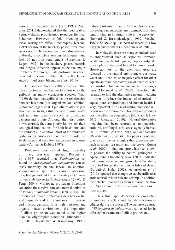
Parasites Found on Megalopa of S. paramamosain in the HatcheryIn the experiment, the results showed the presence of three species of protozoans colonized on larvae exoskeleton such as Myoschiston duplicatum (Figure 4), Zoothamnium alrasheidi was also found on the eggs of mud crab (Figure 6). In that, 100% of the larvae were colonized with Myoschiston duplicatum (Figure 5) and Zoothamnium alrasheidi (Figure 7).
Figure 3: Barnacle of Octolasmis sp infested on the gill of the crab
In this study, Myoschiston sp. was found on the exoskeleton of zoae 5 and megalopa of S. paramamosain larvae. As described in Sun et al. (2012), Zoothamniidae family include three genus with similar morphology which are

DEVELOPMENT OF MUDCRAB CRABLET, THE IDENTIFICATION OF CILIATES 57
J. Sustain. Sci. Manage. Volume 12(2) 2017: 52-62
Zoothamnopis, Myoschiston and Zoothamnium genus. However, the basal stalk of Myoschiston lacks spasmoneme. No study has been reported on the occurrence of Myoschiston sp. known to affect the larvae stage of S. paramamosain. However, M. duplicatum was found on the legs of the Hemigrapsus sp. crab collected form the beach in Gijang, Busan, Korea (Sun et al., 2012).
Figure 4: Scanning electron microscopy photomicrograph of the Myoschiston duplicatum
colony of on the mud crab exoskeleton
Zoothamnium sp. was found on zoae 5 and megalopa stage of S. paramamosain larvae. The Zoothamnium sp. in this study was similar with the description in Lynn (2008). In that, Zoothamnium sp have contractile stalk, bearing a single zooids or branched, bearing colonies of many zooids. The Zoothamnium sp. infection was recorded in shrimp pond farm near Qingdao, China that was described by Ji et al. (2009). Zoothamnium sp. has been found in mud crab (S. serrata) and caused problem on the larvae (Cholik, 1997; Jithendran et al., 2010). Zoothamnium sp. have been known to affect others crustacean species such as shrimp and prawn (Roegge et al., 1977; Babu, 2013) and in Malaysia, they are commonly found in marine shrimp hatchery in Malaysia (Sayuthi, 1993). In some crustacean species (Macrobrachium acanthurus, Eriocheir sinensis, Penaeus monodon), egg and early stage of larvae were infected by ciliate protozoans (Roegge et al., 1977; Wu & Feng, 2004; Babu, 2013). Jithendran et al. (2010) investigated that
protozoans including Epistylis sp, Zoothamnium sp, Acineta sp and Vorticella sp can cause problem mainly on egg and larvae of mud crab culture at hatchery phase. Zoothamnium sp. was found attached on prawn larvae (M. acanthurus) (Roegge et al., 1977), Chinese bitten crab larvae (E. sinensis) (Wu & Feng, 2004) and white leg shimp larvae (P. monodon) (Babu, 2013). Moreover, Zoothamnium sp. caused mass mortality of mud crab (S. paramamosain) larvae (Cholik, 1997).
Figure 5: Scanning electron microscopy photomicrograph of Myoschiston duplicatum.
Single contractile vacuole (A) bar 10µm. Lack of spasmoneme at basal stalk (B) bar 50µm. Contracted M. duplicatum (C) bar 10 µm. Ridges of stalk when
contracted (D) bar 5 µm. Rough stalk because of occurrence of bacteria and small organisms (F).
Double folded peristomial lip (F). Trochal band (G). Vertically striated basal stalk (H)
Nguyan Khanh Linh et al. 58
J. Sustain. Sci. Manage. Volume 12(2) 2017: 52-62
Figure 6: Scanning electron microscopy photomicrograph of Zoothamnium alrasheidi
colonized on mud crab eggs
Treatment of Ciliate Protozoans on Mud Crab (S. paramamosain) by Using Mangrove Leaf Extract (R. apiculata)The results showed the presence of protein, carbohydrates, phenols, tannins, flavonoids, saponins, glycosides, steroids, terpenoids, and alkaloids (Table 1). These compounds were believed to increase the immune system, as well as to control bacteria, fungus, parasite and virus. In the experiment, the extract was first applied as antiparasitic in mud crab megalopa at concentrations of 0, 0.2, 0.4, 0.6, 0.8 and 1 g/l for 24 hours to control the protozoan.
The results showed that the survival rate of megalopa after the application of R. apiculata extract fluctuated (Figure 8). After 24 hours of treatment, none of he treatment that demonstrated 100% survival rate. The survival rate at control was higher than the other treatments (90% compared to 83.3, 85 and 86.7%) (Figure 8). However, the survival rate showed no significance difference among treatments (p>0.508)
The tendency number of average zooid per megalopa after treatment with extract was decreased compared to the control treatment. However, the treatment of 0.6 and 0.8, the number of zooids (16 and 1 zooid per megalopa) showed a significant difference compared to the control (41 zooid per megalopa). At the 1 g
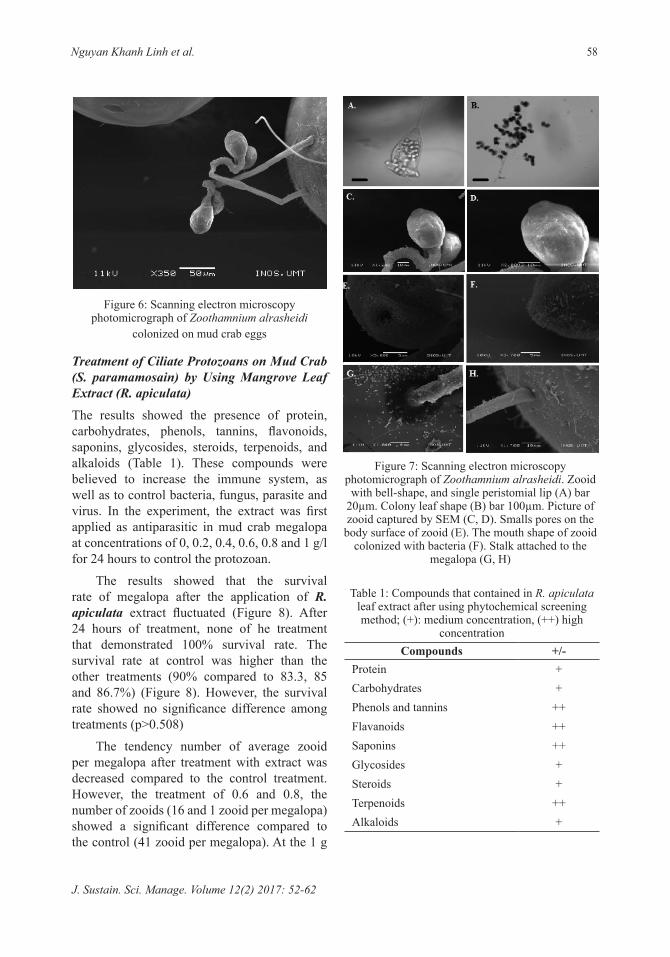
Figure 7: Scanning electron microscopy photomicrograph of Zoothamnium alrasheidi. Zooid
with bell-shape, and single peristomial lip (A) bar 20µm. Colony leaf shape (B) bar 100µm. Picture of zooid captured by SEM (C, D). Smalls pores on the
body surface of zooid (E). The mouth shape of zooid colonized with bacteria (F). Stalk attached to the
megalopa (G, H)
Table 1: Compounds that contained in R. apiculata leaf extract after using phytochemical screening method; (+): medium concentration, (++) high
concentrationCompounds +/-
Protein +Carbohydrates +Phenols and tannins ++Flavanoids ++Saponins ++Glycosides +Steroids +Terpenoids ++Alkaloids +

DEVELOPMENT OF MUDCRAB CRABLET, THE IDENTIFICATION OF CILIATES 59
J. Sustain. Sci. Manage. Volume 12(2) 2017: 52-62
extract per liter, the number of protozoan was nearly 0 (Figure 9).
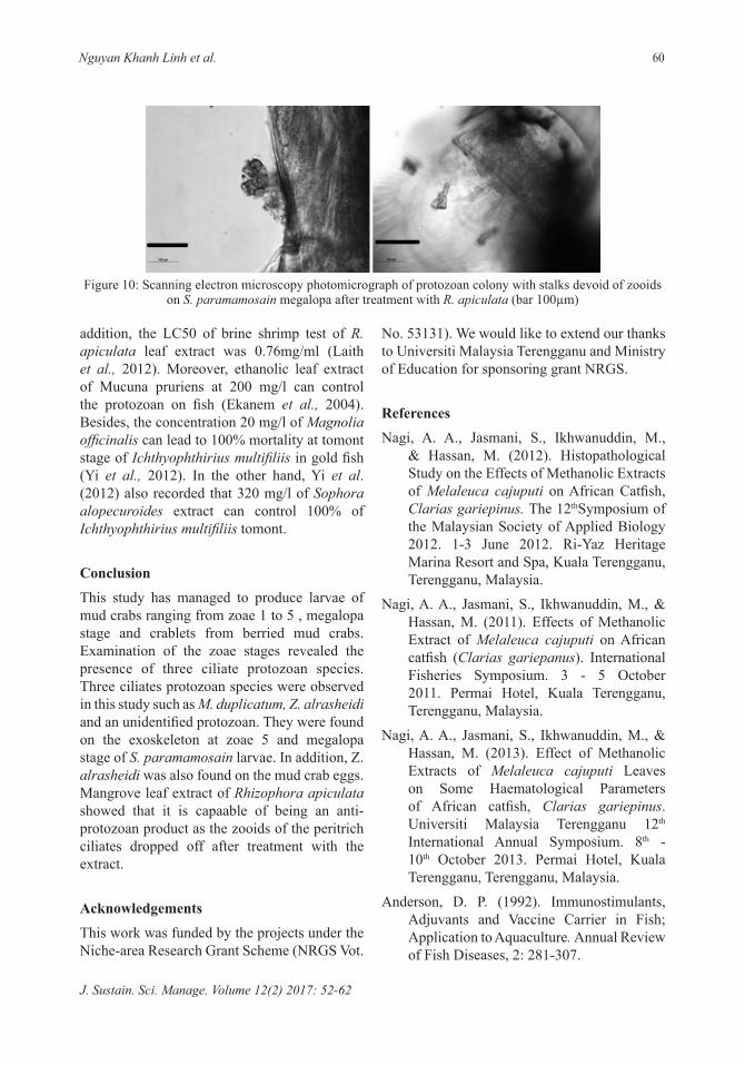
After treatment with R. apiculata, the megalopa were covered with stalks without the zooids. The megalop became light orange in color which is the color of the extract (Figure 10).
This study had demonstrated that 1g/l of R. apiculata leaf extract was sufficient to prevent protozoan infection on S. paramosain
Figure 8: Survival rate (%) of S. paramamosain megalopa after treatment with R. apiculata extract
Figure 9: Average zooid per megalopa of S. paramamosain after treating with R. apiculata
larvae. Part of plant (bark, leaf, root), method of extraction and concentration of extract are three main things that affect health of cultured fish (Reverter et al., 2014). In some studies, methanolic leaf extract of R. apiculata had positive effect on on antimicrobial ativity. For example, in-vitro test of R. apiculata leaf extract on antibacterial of fish pathogenic bacteria gave the result that the minimum inhibitory concentration (MIC) was 12.5-25 mg/ml. In
Nguyan Khanh Linh et al. 60
J. Sustain. Sci. Manage. Volume 12(2) 2017: 52-62
addition, the LC50 of brine shrimp test of R. apiculata leaf extract was 0.76mg/ml (Laith et al., 2012). Moreover, ethanolic leaf extract of Mucuna pruriens at 200 mg/l can control the protozoan on fish (Ekanem et al., 2004). Besides, the concentration 20 mg/l of Magnolia officinalis can lead to 100% mortality at tomont stage of Ichthyophthirius multifiliis in gold fish (Yi et al., 2012). In the other hand, Yi et al. (2012) also recorded that 320 mg/l of Sophora alopecuroides extract can control 100% of Ichthyophthirius multifiliis tomont.
Conclusion This study has managed to produce larvae of mud crabs ranging from zoae 1 to 5 , megalopa stage and crablets from berried mud crabs. Examination of the zoae stages revealed the presence of three ciliate protozoan species. Three ciliates protozoan species were observed in this study such as M. duplicatum, Z. alrasheidi and an unidentified protozoan. They were found on the exoskeleton at zoae 5 and megalopa stage of S. paramamosain larvae. In addition, Z. alrasheidi was also found on the mud crab eggs. Mangrove leaf extract of Rhizophora apiculata showed that it is capaable of being an anti-protozoan product as the zooids of the peritrich ciliates dropped off after treatment with the extract.
AcknowledgementsThis work was funded by the projects under the Niche-area Research Grant Scheme (NRGS Vot.
Figure 10: Scanning electron microscopy photomicrograph of protozoan colony with stalks devoid of zooids on S. paramamosain megalopa after treatment with R. apiculata (bar 100µm)
No. 53131). We would like to extend our thanks to Universiti Malaysia Terengganu and Ministry of Education for sponsoring grant NRGS.
ReferencesNagi, A. A., Jasmani, S., Ikhwanuddin, M.,
& Hassan, M. (2012). Histopathological Study on the Effects of Methanolic Extracts of Melaleuca cajuputi on African Catfish, Clarias gariepinus. The 12thSymposium of the Malaysian Society of Applied Biology 2012. 1-3 June 2012. Ri-Yaz Heritage Marina Resort and Spa, Kuala Terengganu, Terengganu, Malaysia.
Nagi, A. A., Jasmani, S., Ikhwanuddin, M., & Hassan, M. (2011). Effects of Methanolic Extract of Melaleuca cajuputi on African catfish (Clarias gariepanus). International Fisheries Symposium. 3 - 5 October 2011. Permai Hotel, Kuala Terengganu, Terengganu, Malaysia.
Nagi, A. A., Jasmani, S., Ikhwanuddin, M., & Hassan, M. (2013). Effect of Methanolic Extracts of Melaleuca cajuputi Leaves on Some Haematological Parameters of African catfish, Clarias gariepinus. Universiti Malaysia Terengganu 12th International Annual Symposium. 8th - 10th October 2013. Permai Hotel, Kuala Terengganu, Terengganu, Malaysia.
Anderson, D. P. (1992). Immunostimulants, Adjuvants and Vaccine Carrier in Fish; Application to Aquaculture. Annual Review of Fish Diseases, 2: 281-307.

DEVELOPMENT OF MUDCRAB CRABLET, THE IDENTIFICATION OF CILIATES 61
J. Sustain. Sci. Manage. Volume 12(2) 2017: 52-62
Bagg A. J., Jackson, M. S., Sweeney M. P., Ramag G., & Davies A. N. (2006). Susceptibility to Melaleucaalternifolia (tea tree) Oil of Yeasts Isolated from the Mouths of Patients with Advanced Aancer. Oral Oncology, 42: 487-492.
Bandaranayake, W. M. (1995). Survey of Mangrove Plants from Northern Australia for Phytochemical Constituents and Uv-absorbing Compounds. Current Topics in Phytochemistry (Life Science Advances), 14: 69-78
Beula, M. B., Ravikumar, S., Gnanadesigan, M., Rajakumar, B., & Anand, M. (2012b). Antiviral, Antioxidant and Toxicological Evaluation of Mangrove Associate from South East Coast of India. Asian Pacific Journal of Tropical Biomedicine, S1775-S1779.
Biswas, S. K., & Rahman, I. (2009). Environmental Toxicity, Redox Signalling and Lung Inflammation: The Role of Glutathione. Molecular Aspects of Medicine, 30(1-2): 60-76.
Bonami, J-R., & Zhang S. (2011). Viral Diseases in Commercially Exploited Crabs: A Review. Journal of Invertebrate Pathology, 106: 6-17.
Chen, J., Xoing, J., Yang, J., Mao, Z., & Chen, X. (2011). Nucleotide Sequences of Four RNA Segments of a Reovirus Isolated from the Mud Crab Scylla serrata Provide Evidence That This Virus Belongs to a New Genus in the family Reoviridae. Archives of Virology, 156: 523-528.
Deng, X-X., Lu, L., Qu, Y-J., Su, H-J., Li, G., Guo, Z-X., Zhang, R., Zheng, P-R., Chen, Y-G., He, J-G, & Weng, S. P. Sequence Analysis of 12 Genome Segments of Mud Crab Reovirus (MCRV) 2012. Virology, 422: 185-194.
Flegel T. W. (2006). Detection of Major Penaeid Shrimp Viruses in Asia, a Historical Perspective with Emphasis on Thailand. Aquaculture, 258:1-33.
Garozzo, A., Timpanaro, R., Stivala, A., Bisignano, G., & Castro, A. (2011). Activity of Melaleucaalternifolia (tea tree) Oil on Influenza Virus A/PR/8: Study on the Mechanism of action. Antiviral Research, 89: 83-88
Govindasamy, C. (2011). What is the Significance of Mangrove Forests. A Note Current Botany, 2(2): 50-55.
Hussein, S. A. M., Hashim, A. N. M., El-Sharawy, R. T., Seliem, M. A., Linscheid, M., Lindequist, U., & Nawwar, M. A. M. (2007). Ericifolin: Aneugenol 5-O-galloylglucoside and other Phenolics from Melaleucaericifolia. Phytochemistry, 68: 1464-1470.
Lightner, D.V. (1988). Vibrio Disease of Penaeid Shrimp. In: Sinderman. C.J., Lightner, D.V. (Eds), Disease Diagnosis and Control in North American Marine Aquaculture. InElsevier. Amsterdam. The Netherlands, 42-47.
Lin, C.K. (1995). Progression of Intensive Marine Shrimp Culture in Thailand. In: Browdy, C.L., Hpkins, J.S. (Eds). Swimming through Troubled Water. Proceeding of the Special Session on Shrimp Farming. Aquaculture ’95. InWorld Aquaculture Society. Baton Rouge, LA. 13-23.
Liu, W., Qian, D., & Yan, X. J. (2011). Studies on Pathogenicity and Prevalence of White Spot.
Lo, C. F., Ho, C. H., Peng, S. E., Chen, C. H., Hsu, H. C., Chiu, Y. L., Chang, C. F., Liu, K. F., Su, M. S., Wang, C. H., & Kou G. H. (1996). White Spot Syndrome Baculovirus (WSBV) Detected in Cultured and Captured Shrimp, Crabs and Other Arthropods. Diseases of Aquatic Organisms, 27: 215-225.
Martin, L., Liu, Z., Chen, K., Price, A. C., Pan, Y., Swaby, J. A., & Golden, W. C. (2007). Motor Neuron Degeneration in Amyotrophoc Lateral Sclerosis Mutant Superoxide Dismutase-1 Transgenic Mice:

Nguyan Khanh Linh et al. 62
J. Sustain. Sci. Manage. Volume 12(2) 2017: 52-62
Mechanisms of Mitochondriopathy and Cell Death. Journal of Comparative Neurology, 500: 20-46.
Münoz, M., Cedeno, R. Rodriguez, J., Van der Knap, W. P. W., Mialhe, E., & Bachere, E. (2001). Measurement of Reactive Oxygen Intermediate Production in Haemocytes of the Penaeid Shrimp. Penaeus vannamei. Aquaculture, 191: 89-107.
Najiah M, Nadirah M, Wan Nurhafizah I, Mohd Zahrol, A.S. Arief, I. Z. Mohd R.A. Tee, LW. Mariam, M., Laith, R. Amar S., & Awang S. (2011). Methanolic Activities of Selected Weeds on Bacteria Isolated from Macrobrachium rosenberghii Larvae. Thai J. Vet. Med. 41(4): 535-539.
Nazirah, O. (2013). Effect of melaleuca cajuputi Extracts as a Feed Additive on Growth Performance of African Catfish, Clarias gariepinus. Undergraduate Thesis, Universiti Malaysia Terengganu.
Nor Aini N.N.M.N. (2013). Wound Healing Activity of the Methanolic Extract of Melaleuca cajuputi leaves on Lates Calcarifer. Undergraduate thesis, Universiti Malaysia Terengganu.
Owens, L., Liessmann, L., La Fauce, K., Nyugen, T., & Zeng, C. (2010). Intranuclear Bacilliform Virus and Hepatopancreatic Parvovirus (PmergDNV) in the Mud Crab Scylla serrata (Forskal) of Australia. Aquaculture, 310: 47-50.
Rattiwanich, T., Karuhapattana, B., Kittirattrakarn, T., & Anantachoke, C. (1992). Melaleucacajuputi Powell Leaves Oil from Toedaeng Swamp Forest, pp.361-367. InAnnual Conference of Forestry 1992. Bangkok.
Reynertson K. A., Yang H., Jiang B., Basile J. M., & Kennelly E. J. (2008). Quantitative Analysis of Antiradical Phenolic Constituents from Fourteen Edible Myrtaceae fruits. Food Chemistry, 109:883-890.
Ruangrungsi, N., & Tontiwat, P. 1991. Madicinal Plant. Odianstorekarnpim, Bangkok. 244 pp.
Samathi, C. (1988). Study of Natural of Compensation of Melaleucacajuputi Powell Forest Community, Narathiwat province. 51 pp.
Sciarrone D., Ragonese C., Carnovale C., Piperno A., Dugoa P., Dugo G., & Mondello L. (2010). Evaluation of Tea Tree Oil Quality and Ascaridole: A Deep Study by Means of Chiral and Multi Heartcuts Multidimensional Gas Chromatography System Coupled to Mass Spectrometry Detection. Journal of Chromatography, A 1217: 6422-6427.
Shahrul-Hafiz, G. (2013). Behaviour Study of Lates Calcarifer and Caligus Sp. Treated with Methanolic Extract of Melalueca Cajuputi. Undergraduate thesis, Universiti Malaysia Terengganu.
Somboonna, N., Mangkalanan, S., Udompetcharaporn, A., Krittanai, C., Sritunyalucksana, K., & Flegel, T. W. (2010). Mud Crab Susceptibility to Disease from White Spot Syndrome Virus is Species-dependent. BMC Research Notes, 3: 315.
Sudheer, N. S., Philip R., & Bright Singh I. S. (2011). In Vivo Screening of Mangrove Plants for Anti WSSV Activity in Penaeus monodon and Evaluation of Ceriops tagal as a Potential Source of Antiviral Molecules. Aquaculture, 301: 36-41.
Weng, S. P., Guo, Z. X., Sun, J. J., Chan, S. M., & He, J. G., (2007). A Reovirus Disease in Cultured Westerheide, S.D., Bosman, J. D., Mbadugha, B. N. A., Kawahara, T. L. A., Matsumoto, G., Kim, S., Gu, W., Devlin, J. P., Silverman, R. B., Morimoto, R. I. (2004). Celastrols as Inducers of the Heat Shock Response and Cytoprotection. Journal of Biological Chemistry 279: 56053-56060.
Related Documents