ResearchArticle Corneal Epithelial Damage and Impaired Tear Functions in Patients with Inflamed Pinguecula ErkutK¨ uç¨ uk , 1 U˘ gurYılmaz, 2 andK¨ ursadRamazanZor 1 1 Ophthalmology Department, Ni˘ gde ¨ Omer Halisdemir University, Faculty of Medicine, 51240 Ni˘ gde,Turkey 2 Ophthalmology Department, Pamukkale University Faculty of Medicine, 20160 Denizli, Turkey Correspondence should be addressed to Erkut K¨ uç¨ uk; [email protected] Received 30 July 2018; Revised 27 September 2018; Accepted 9 October 2018; Published 31 October 2018 Academic Editor: Jes´ us Pintor Copyright©2018ErkutK¨ uç¨ uketal.isisanopenaccessarticledistributedundertheCreativeCommonsAttributionLicense,which permits unrestricted use, distribution, and reproduction in any medium, provided the original work is properly cited. Purpose. In this study, we evaluated corneal epithelial integrity and tear film parameters in patients with inflamed pinguecula and compared these findings with their fellow eyes and with healthy controls. Methods. We evaluated the fluorescein staining properties and performed the tear break-up time (TBUT) test and Schirmer 2 test (ST2) measurements of 32 patients who had symptomatic unilateral inflamed pinguecula and compared the results with their fellow eyes and also with an age- and sex-matched control group. Results. Twenty-three eyes (72%) in the inflamed pinguecula group and 1 eye (3.1%) in the fellow eyes group had punctate epithelial staining (PES) or epithelial defect on the nasal cornea (p < 0.001). ere was no PES or epithelial defect in the control group. Eyes with inflamed pinguecula (n � 32) had lower TBUT and ST2 values compared to the control group (n � 32) (p < 0.001 for both). Fellow eyes (n � 32) also had lower TBUT and ST2 values compared to the control group (p � 0.003 for both). ere was no difference in the TBUT and ST2 results between the eyes with inflamed pinguecula and fellow eyes (p � 0.286 and p � 0.951, respectively). Conclusion. A high percentage of eyes with inflamed pinguecula had nasal corneal epithelial staining or epithelial defect. We also found lower TBUT and ST2 results in eyes with inflamed pinguecula and the fellow eyes compared to the control group. ese findings may be important in pathogenesis of pinguecula and pterygium and also in uncovering their relation. 1.Introduction Pinguecula is a yellowish elevated mass commonly located on the nasal bulbar conjunctiva close to the limbus [1]. Its prevalence increases with age, and ultraviolet radiation (UVR) is a risk factor in its pathogenesis [2, 3]. Male gender and diabetes mellitus are also reported risk factors [4]. Histological studies reported abnormal differentiation and squamous metaplasia of the conjunctival epithelium, ex- aggeration and distortion in the production of elastic fibers, and abnormality of their organization in the subepithelial connective tissue [5–7]. It was reported that 22.5% to 70.1% of the population has pinguecula [4, 8]. is heterogeneity in the prevalence may be due to differences in age, geo- graphic location, and ethnicity of participants. Pinguecula may be inflamed, causing hyperemia, pain, and foreign body sensation. Pterygium is a triangular growth of conjunctival fibro- vascular tissue onto the cornea, usually located at the nasal cornea. Its prevalence is lower than that of pinguecula. It can cause decreased visual acuity, irritation, and pain due to inflammation and cosmetic problems. Although surgery is effective in its treatment, the risk of recurrence is still an important problem. Ultraviolet radiation (UVR) is thought to be a factor in the development of both pinguecula and pterygium. It is hypothesized that UVR causes conjunctival degeneration and the formation of pinguecula. With in- creased exposure, corneal epithelial and stem cells may be affected and lead to the formation of pterygium [9, 10]. But it is still unknown if pinguecula is a precursor of pterygium or if so, what causes its progress to pterygium. Several studies reported abnormalities of tear function tests in pinguecula patients [2, 11]. e abnormality of the tear film and mechanical trauma may cause inflammation of pinguecula [12]. Inflamed pinguecula has attracted little attention in the ophthalmic community. In this study, we investigated the fluorescein staining properties and tear film parameters in patients with inflamed pinguecula. We also Hindawi Journal of Ophthalmology Volume 2018, Article ID 2474173, 5 pages https://doi.org/10.1155/2018/2474173

Welcome message from author
This document is posted to help you gain knowledge. Please leave a comment to let me know what you think about it! Share it to your friends and learn new things together.
Transcript
Research ArticleCorneal Epithelial Damage and Impaired Tear Functions inPatients with Inflamed Pinguecula
Erkut Kuçuk ,1 Ugur Yılmaz,2 and Kursad Ramazan Zor1
1Ophthalmology Department, Nigde Omer Halisdemir University, Faculty of Medicine, 51240 Nigde, Turkey2Ophthalmology Department, Pamukkale University Faculty of Medicine, 20160 Denizli, Turkey
Correspondence should be addressed to Erkut Kuçuk; [email protected]
Received 30 July 2018; Revised 27 September 2018; Accepted 9 October 2018; Published 31 October 2018
Academic Editor: Jesus Pintor
Copyright© 2018ErkutKuçuk et al.-is is an open access article distributed under theCreativeCommonsAttribution License,whichpermits unrestricted use, distribution, and reproduction in any medium, provided the original work is properly cited.
Purpose. In this study, we evaluated corneal epithelial integrity and tear film parameters in patients with inflamed pinguecula andcompared these findings with their fellow eyes and with healthy controls.Methods. We evaluated the fluorescein staining propertiesand performed the tear break-up time (TBUT) test and Schirmer 2 test (ST2) measurements of 32 patients who had symptomaticunilateral inflamed pinguecula and compared the results with their fellow eyes and also with an age- and sex-matched control group.Results. Twenty-three eyes (72%) in the inflamed pinguecula group and 1 eye (3.1%) in the fellow eyes group had punctate epithelialstaining (PES) or epithelial defect on the nasal cornea (p< 0.001). -ere was no PES or epithelial defect in the control group. Eyeswith inflamed pinguecula (n � 32) had lower TBUT and ST2 values compared to the control group (n � 32) (p< 0.001 for both).Fellow eyes (n � 32) also had lower TBUT and ST2 values compared to the control group (p � 0.003 for both). -ere was nodifference in the TBUT and ST2 results between the eyes with inflamed pinguecula and fellow eyes (p � 0.286 and p � 0.951,respectively).Conclusion. A high percentage of eyes with inflamed pinguecula had nasal corneal epithelial staining or epithelial defect.We also found lower TBUT and ST2 results in eyes with inflamed pinguecula and the fellow eyes compared to the control group.-ese findings may be important in pathogenesis of pinguecula and pterygium and also in uncovering their relation.
1. Introduction
Pinguecula is a yellowish elevated mass commonly locatedon the nasal bulbar conjunctiva close to the limbus [1]. Itsprevalence increases with age, and ultraviolet radiation(UVR) is a risk factor in its pathogenesis [2, 3]. Male genderand diabetes mellitus are also reported risk factors [4].Histological studies reported abnormal differentiation andsquamous metaplasia of the conjunctival epithelium, ex-aggeration and distortion in the production of elastic fibers,and abnormality of their organization in the subepithelialconnective tissue [5–7]. It was reported that 22.5% to 70.1%of the population has pinguecula [4, 8]. -is heterogeneityin the prevalence may be due to differences in age, geo-graphic location, and ethnicity of participants. Pingueculamay be inflamed, causing hyperemia, pain, and foreignbody sensation.
Pterygium is a triangular growth of conjunctival fibro-vascular tissue onto the cornea, usually located at the nasal
cornea. Its prevalence is lower than that of pinguecula. It cancause decreased visual acuity, irritation, and pain due toinflammation and cosmetic problems. Although surgery iseffective in its treatment, the risk of recurrence is still animportant problem. Ultraviolet radiation (UVR) is thoughtto be a factor in the development of both pinguecula andpterygium. It is hypothesized that UVR causes conjunctivaldegeneration and the formation of pinguecula. With in-creased exposure, corneal epithelial and stem cells may beaffected and lead to the formation of pterygium [9, 10]. But itis still unknown if pinguecula is a precursor of pterygium orif so, what causes its progress to pterygium.
Several studies reported abnormalities of tear functiontests in pinguecula patients [2, 11]. -e abnormality of thetear film and mechanical trauma may cause inflammation ofpinguecula [12]. Inflamed pinguecula has attracted littleattention in the ophthalmic community. In this study, weinvestigated the fluorescein staining properties and tear filmparameters in patients with inflamed pinguecula. We also
HindawiJournal of OphthalmologyVolume 2018, Article ID 2474173, 5 pageshttps://doi.org/10.1155/2018/2474173
discussed the role of these parameters in the possible evo-lution of the inflamed pinguecula to pterygium.
2. Materials and Methods
-is controlled multicenter study was performed in theOphthalmology Department of Nigde Omer HalisdemirUniversity (Nigde, Turkey) and Ophthalmology Departmentof Pamukkale University Hospital (Denizli, Turkey). Bothcities are located at the same latitude (38°), and they have thesame distance from the equator. Denizli is located approxi-mately 124 km from the Aegean Sea, and Nigde is located130 km from the Mediterranean Sea. Although regionaldifferences can exist, these two cities show similar climaticcharacteristics. -irty-two consecutive patients who appliedto these clinics between July 2017 and September 2017 andhad symptomatic unilateral inflamed pinguecula were in-cluded. Twelve of these patients were from PamukkaleUniversity Hospital and 20 from Nigde Omer HalisdemirUniversity Ophthalmology Department. Symptomaticinflamed pinguecula was described as a combination ofvascular congestion and hyperemia of the pinguecula andadjacent conjunctiva in biomicroscopic examination togetherwith patients’ description of a recent increase in ocularredness and one or more of the following symptoms: pho-tophobia, pain, foreign-body sensation, discomfort, andtearing. Two independent experienced ophthalmologists (EKand UY) diagnosed the patients for inclusion criteria. Acontrol group (n � 32) was formed from age-matched in-dividuals that did not have any ophthalmic disease other thanrefractive problems. Subjects who had corneal pathologies,allergic conditions, previous corneal and/or conjunctivalsurgery, meibomian gland dysfunction, active ocular in-fection, and contact lens users were excluded. All participantsunderwent complete ophthalmologic examination. To ensurereproductivity, all patients diagnosed with inflamed pin-guecula were reexamined, and tests of the tear function wereperformed on the following day in the morning in theophthalmologists’ dimly lit examination room. Cornealstaining properties were evaluated using fluorescein sodiumsolution 2% (Fluorescite®; Alcon Laboratories, Inc., FortWorth, Texas 76134, USA). For TBUT test measurements,a drop of 2% fluorescein solution was applied to the lateralinferior fornix.-e patient was asked to blink several times foruniform distribution of fluorescein and then instructed tolook ahead without blinking. -e time from the last blink tothe appearance of the first dry spot on the cornea wasrecorded using the cobalt blue filter of the biomicroscope anda stopwatch. -ree consecutive measurements were made,and the mean of measurements was recorded. -irty minuteslater, in the dimly lit examining room, a topical anestheticagent proparacaine hydrochloride 0.5% drop (Alcaine®;Alcon, FortWorth, TX) was applied to the inferior fornix, andthree minutes later, a standard Schirmer test filter strip (BioSchirmer®; Bio-Tech Vision Care, Ahmedabad, Gujarat,India) was inserted into the lateral inferior fornix at thejunction of the middle and lateral thirds of the lower eyelid,taking care not to touch cornea.-e patient was asked to keepeyes open and blink as necessary. After five minutes, the filter
strip was removed and wetting was recorded. -is study wasperformed according to the tenets of Declaration of Helsinki,and the study received approval from Pamukkale UniversityEthics Committee. Written informed consent and verbalinformed consent were taken from patients and controls.
Statistical analysis was performed using SPSS version 20.0(IBM Corporation, Armonk, NY). Test results were expressedas mean ± standard deviation (SD). -e distribution of thevariables was tested using the Kolmogorov–Smirnov test. -echi-square test was used to compare groups for gender andnasal corneal epithelial staining. Independent-samples T testwas used to compare the groups for age. For BUT and ST2values, the Kruskal–Wallis one-way test was used to test thedifference among groups andMann–WhitneyU test was usedto compare groups. In all analyses, p values <0.05 wereconsidered as statistically significant.
3. Results
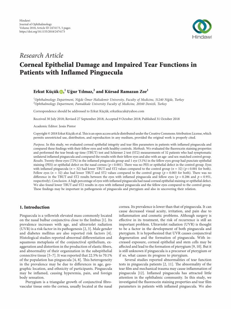
-ere was no significant difference in age and gender be-tween inflamed pinguecula and control groups (p � 0.862and p � 0.794, respectively) (Table 1). -irty-two eyes of 32patients had inflamed pinguecula. All inflamed pingueculaewere on the nasal conjunctiva (Figure 1). -ere were pin-guecula in 13 (40 %) and pterygium in 3 (9%) of the felloweyes (n � 32). -ere was no pinguecula or pterygium in thecontrol group.
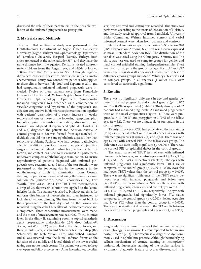
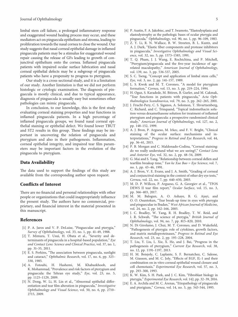
Twenty-three eyes (72%) had punctate epithelial staining(PES) or epithelial defect on the nasal cornea in eyes withinflamed pinguecula (Figures 2(a) and 2(b)). -ere was oneeye (3.1%) with corneal PES in the fellow eyes group. -edifference was statistically significant (p< 0.001). -ere wasno corneal PES or epithelial defect in the control group.
-e mean values of TBUT tests of eyes with inflamedpinguecula, fellow eyes, and control eyes were 8.1 ± 3.9 s, 9.3 ±4.3 s, and 13.5 ± 4.9 s, respectively (Table 2). -e eyes withinflamed pinguecula had significantly lower TBUT valuescompared to the control group (p< 0.001). Fellow eyes alsohad lower TBUT values than the control group (p � 0.003).-ere was no significant difference in the TBUT results be-tween eyes with inflamed pinguecula and fellow eyes(p � 0.286). -e mean values of ST2 results of eyes withinflamed pinguecula, fellow eyes, and control eyes were 11.6 ±5.1 s, 11.6 ± 5.3 s, and 17.6 ± 7.8 s, respectively. -e eyes withinflamed pinguecula had significantly lower ST2 valuescompared to the control group (p< 0.001). Fellow eyes alsohad lower ST2 values than the control group (p � 0.003).-ere was no significant difference in the ST2 results betweenthe eyes with inflamed pinguecula and fellow eyes (p � 0.951).
4. Discussion
Pinguecula is a common disease of the conjunctiva whoseexact etiology is unknown. UVR is reported to be an im-portant factor [2, 3]. Fluorescein is a diagnostic dye com-monly used in ophthalmic practice. Although the underlyingcellular mechanism of corneal staining is incompletelyunderstood, fluorescein staining of the ocular surface isa common diagnostic feature of ocular diseases, and it is
2 Journal of Ophthalmology
Table 1: Demographic characteristics of groups.
Inflamed pinguecula group (n � 32) Control group (n � 32) p
Age (years) (mean ± SD) 32.78 ± 10.35 32.31 ± 11.07 0.862a
Sex Female, n (%) 21 (65.6%) 20 (62.5%) 0.794bMale, n (%) 11 (34.4%) 12 (37.5%)aIndependent-samples T test; bchi-square test; p value <0.05 is statistically significant.
Figure 1: An inflamed pinguecula.
(a) (b)
Figure 2: (a) Epithelial defect and (b) fluorescent staining in a patient with inflamed pinguecula.
Table 2: Schirmer 2 and TBUT test results of the groups.
Patient eyes withinflamed pinguecula(n � 32) (Group 1)
Patient eyes withoutinflamed pinguecula
(n � 32)(Group 2)
Control eyes(n � 32)(Group 3)
p∗
p# for intergroupcomparisons
Groups1 vs 2
Groups1 vs 3
Groups2 vs 3
BUT (s)Mean ± SD 8.1 ± 3.9 9.3 ± 4.3 13.5 ± 4.9
<0.005 0.286 <0.001 0.003Median 8.0 8.0 14.0Range 3–19 3–18 4–25
ST2 (mm)Mean ± SD 11.6 ± 5.1 11.6 ± 5.3 17.6 ± 7.8
<0.005 0.951 <0.001 0.003Median 11.0 10.5 20.0Range 3–22 2–21 4–30
∗p value for comparison among three groups (Kruskal–Wallis one-way test). #p values for intergroup comparisons (Mann–Whitney U test). p value <0.05 isstatistically significant.
Journal of Ophthalmology 3
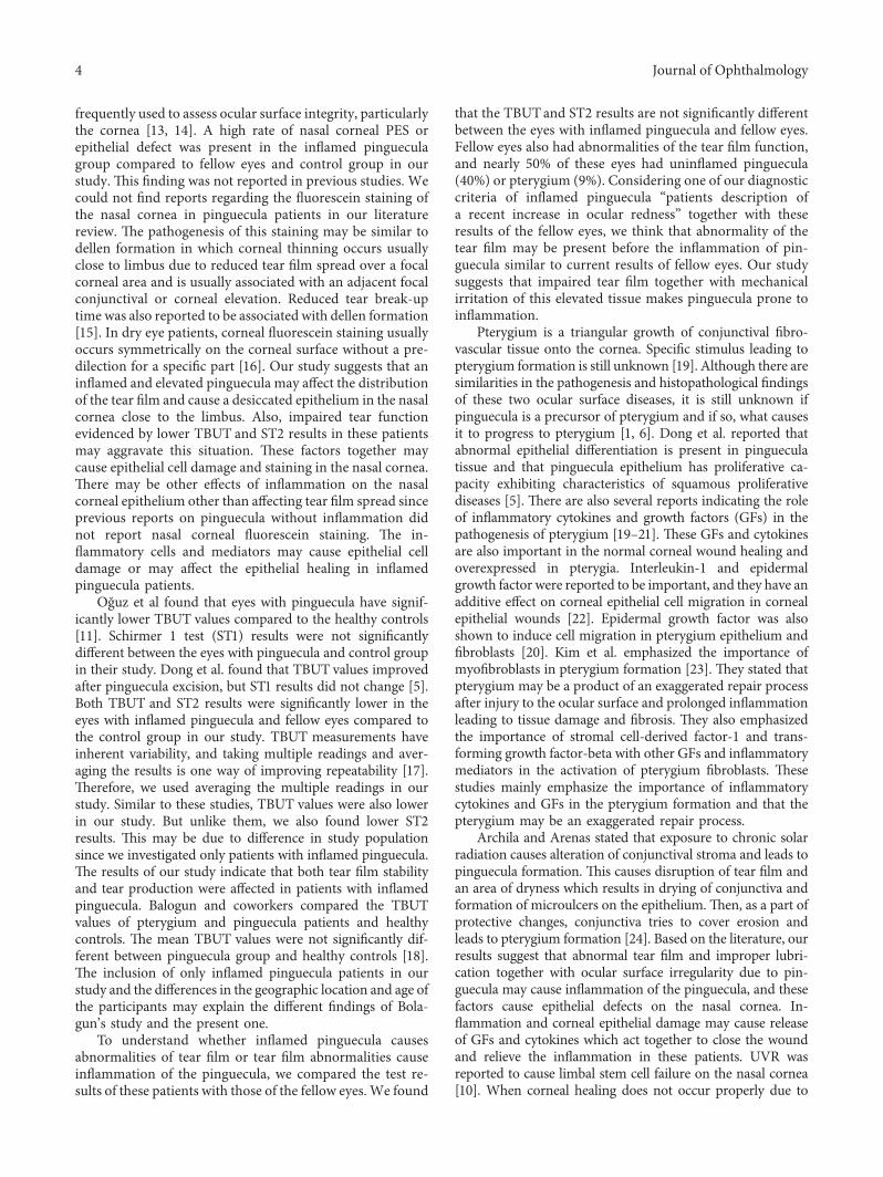
frequently used to assess ocular surface integrity, particularlythe cornea [13, 14]. A high rate of nasal corneal PES orepithelial defect was present in the inflamed pingueculagroup compared to fellow eyes and control group in ourstudy. -is finding was not reported in previous studies. Wecould not find reports regarding the fluorescein staining ofthe nasal cornea in pinguecula patients in our literaturereview. -e pathogenesis of this staining may be similar todellen formation in which corneal thinning occurs usuallyclose to limbus due to reduced tear film spread over a focalcorneal area and is usually associated with an adjacent focalconjunctival or corneal elevation. Reduced tear break-uptime was also reported to be associated with dellen formation[15]. In dry eye patients, corneal fluorescein staining usuallyoccurs symmetrically on the corneal surface without a pre-dilection for a specific part [16]. Our study suggests that aninflamed and elevated pinguecula may affect the distributionof the tear film and cause a desiccated epithelium in the nasalcornea close to the limbus. Also, impaired tear functionevidenced by lower TBUT and ST2 results in these patientsmay aggravate this situation. -ese factors together maycause epithelial cell damage and staining in the nasal cornea.-ere may be other effects of inflammation on the nasalcorneal epithelium other than affecting tear film spread sinceprevious reports on pinguecula without inflammation didnot report nasal corneal fluorescein staining. -e in-flammatory cells and mediators may cause epithelial celldamage or may affect the epithelial healing in inflamedpinguecula patients.
Oguz et al found that eyes with pinguecula have signif-icantly lower TBUT values compared to the healthy controls[11]. Schirmer 1 test (ST1) results were not significantlydifferent between the eyes with pinguecula and control groupin their study. Dong et al. found that TBUT values improvedafter pinguecula excision, but ST1 results did not change [5].Both TBUT and ST2 results were significantly lower in theeyes with inflamed pinguecula and fellow eyes compared tothe control group in our study. TBUT measurements haveinherent variability, and taking multiple readings and aver-aging the results is one way of improving repeatability [17].-erefore, we used averaging the multiple readings in ourstudy. Similar to these studies, TBUT values were also lowerin our study. But unlike them, we also found lower ST2results. -is may be due to difference in study populationsince we investigated only patients with inflamed pinguecula.-e results of our study indicate that both tear film stabilityand tear production were affected in patients with inflamedpinguecula. Balogun and coworkers compared the TBUTvalues of pterygium and pinguecula patients and healthycontrols. -e mean TBUT values were not significantly dif-ferent between pinguecula group and healthy controls [18].-e inclusion of only inflamed pinguecula patients in ourstudy and the differences in the geographic location and age ofthe participants may explain the different findings of Bola-gun’s study and the present one.
To understand whether inflamed pinguecula causesabnormalities of tear film or tear film abnormalities causeinflammation of the pinguecula, we compared the test re-sults of these patients with those of the fellow eyes.We found
that the TBUTand ST2 results are not significantly differentbetween the eyes with inflamed pinguecula and fellow eyes.Fellow eyes also had abnormalities of the tear film function,and nearly 50% of these eyes had uninflamed pinguecula(40%) or pterygium (9%). Considering one of our diagnosticcriteria of inflamed pinguecula “patients description ofa recent increase in ocular redness” together with theseresults of the fellow eyes, we think that abnormality of thetear film may be present before the inflammation of pin-guecula similar to current results of fellow eyes. Our studysuggests that impaired tear film together with mechanicalirritation of this elevated tissue makes pinguecula prone toinflammation.
Pterygium is a triangular growth of conjunctival fibro-vascular tissue onto the cornea. Specific stimulus leading topterygium formation is still unknown [19]. Although there aresimilarities in the pathogenesis and histopathological findingsof these two ocular surface diseases, it is still unknown ifpinguecula is a precursor of pterygium and if so, what causesit to progress to pterygium [1, 6]. Dong et al. reported thatabnormal epithelial differentiation is present in pingueculatissue and that pinguecula epithelium has proliferative ca-pacity exhibiting characteristics of squamous proliferativediseases [5]. -ere are also several reports indicating the roleof inflammatory cytokines and growth factors (GFs) in thepathogenesis of pterygium [19–21]. -ese GFs and cytokinesare also important in the normal corneal wound healing andoverexpressed in pterygia. Interleukin-1 and epidermalgrowth factor were reported to be important, and they have anadditive effect on corneal epithelial cell migration in cornealepithelial wounds [22]. Epidermal growth factor was alsoshown to induce cell migration in pterygium epithelium andfibroblasts [20]. Kim et al. emphasized the importance ofmyofibroblasts in pterygium formation [23]. -ey stated thatpterygium may be a product of an exaggerated repair processafter injury to the ocular surface and prolonged inflammationleading to tissue damage and fibrosis. -ey also emphasizedthe importance of stromal cell-derived factor-1 and trans-forming growth factor-beta with other GFs and inflammatorymediators in the activation of pterygium fibroblasts. -esestudies mainly emphasize the importance of inflammatorycytokines and GFs in the pterygium formation and that thepterygium may be an exaggerated repair process.
Archila and Arenas stated that exposure to chronic solarradiation causes alteration of conjunctival stroma and leads topinguecula formation. -is causes disruption of tear film andan area of dryness which results in drying of conjunctiva andformation of microulcers on the epithelium.-en, as a part ofprotective changes, conjunctiva tries to cover erosion andleads to pterygium formation [24]. Based on the literature, ourresults suggest that abnormal tear film and improper lubri-cation together with ocular surface irregularity due to pin-guecula may cause inflammation of the pinguecula, and thesefactors cause epithelial defects on the nasal cornea. In-flammation and corneal epithelial damage may cause releaseof GFs and cytokines which act together to close the woundand relieve the inflammation in these patients. UVR wasreported to cause limbal stem cell failure on the nasal cornea[10]. When corneal healing does not occur properly due to
4 Journal of Ophthalmology
limbal stem cell failure, a prolonged inflammatory responseand exaggerated wound healing process may occur, and thesemediators act on pinguecula epithelium and stroma, leading toproliferation towards the nasal cornea to close the wound. Ourstudy suggests that nasal corneal epithelial damage in inflamedpinguecula patients may be a stimulus for exaggerated woundrepair causing the release of GFs leading to growth of con-junctival epithelium onto the cornea. Inflamed pingueculapatients with impaired ocular surface lubrication and nasalcorneal epithelial defects may be a subgroup of pingueculapatients who have a propensity to progress to pterygium.
Our study is a cross-sectional study, and it is a limitationof our study. Another limitation is that we did not performhistologic or cytologic examination. -e diagnosis of pin-guecula is mostly clinical, and due to typical appearance,diagnosis of pinguecula is usually easy but sometimes otherpathologies can mimic pinguecula.
In conclusion, to our knowledge, this is the first studyevaluating corneal staining properties and tear functions ininflamed pinguecula patients. In a high percentage ofinflamed pinguecula groups, we found nasal corneal epi-thelial staining or epithelial defect. We found lower TBUTand ST2 results in this group. -ese findings may be im-portant in uncovering the relation of pinguecula andpterygium and also in their pathogenesis. Inflammation,corneal epithelial integrity, and impaired tear film param-eters may be important factors in the evolution of thepinguecula to pterygium.
Data Availability
-e data used to support the findings of this study areavailable from the corresponding author upon request.
Conflicts of Interest
-ere are no financial and personal relationships with otherpeople or organizations that could inappropriately influencethe present study. -e authors have no commercial, pro-prietary, and financial interest in the material presented inthis manuscript.
References
[1] P. A. Jaros and V. P. DeLuise, “Pingueculae and pterygia,”Survey of Ophthalmology, vol. 33, no. 1, pp. 41–49, 1988.
[2] T. Mimura, T. Usui, H. Obata et al., “Severity and de-terminants of pinguecula in a hospital-based population,” Eyeand Contact Lens: Science and Clinical Practice, vol. 37, no. 1,pp. 31–35, 2011.
[3] E. S. Perkins, “-e association between pinguecula, sunlightand cataract,” Ophthalmic Research, vol. 17, no. 6, pp. 325–330, 1985.
[4] A. Fotouhi, H. Hashemi, M. Khabazkhoob, andK. Mohammad, “Prevalence and risk factors of pterygium andpinguecula: the Tehran eye study,” Eye, vol. 23, no. 5,pp. 1125–1129, 2009.
[5] N. Dong, W. Li, H. Lin et al., “Abnormal epithelial differ-entiation and tear film alteration in pinguecula,” InvestigativeOphthalmology and Visual Science, vol. 50, no. 6, pp. 2710–2715, 2009.
[6] P. Austin, F. A. Jakobiec, and T. Iwamoto, “Elastodysplasia andelastodystrophy as the pathologic bases of ocular pterygia andpinguecula,” Ophthalmology, vol. 90, no. 1, pp. 96–109, 1983.
[7] Z. Y. Li, R. N. Wallace, B. W. Streeten, B. L. Kuntz, andA. J. Dark, “Elastic fiber components and protease inhibitorsin pinguecula,” Investigative Ophthalmology and Visual Sci-ence, vol. 32, no. 5, pp. 1573–1585, 1991.
[8] T. Q. Pham, J. J. Wang, E. Rochtchina, and P. Mitchell,“Pterygium/pinguecula and the five-year incidence of age-related maculopathy,” American Journal of Ophthalmology,vol. 139, no. 3, pp. 536-537, 2005.
[9] S. C. Tseng, “Concept and application of limbal stem cells,”Eye, vol. 3, no. 2, pp. 141–157, 1989.
[10] L. S. Kwok and M. T. Coroneo, “A model for pterygiumformation,” Cornea, vol. 13, no. 3, pp. 219–224, 1994.
[11] H. Oguz, S. Karadede, M. Bitiren, B. Gurler, and M. Cakmak,“Tear functions in patients with pinguecula,” Acta Oph-thalmologica Scandinavica, vol. 79, no. 3, pp. 262–265, 2001.
[12] J. Frucht-Pery, C. S. Siganos, A. Solomon, T. Shvartzenberg,C. Richard, and C. Trinquand, “Topical indomethacin solu-tion versus dexamethasone solution for treatment of inflamedpterygium and pinguecula: a prospective randomized clinicalstudy,” American Journal of Ophthalmology, vol. 127, no. 2,pp. 148–152, 1999.
[13] A. J. Bron, P. Argueso, M. Irkec, and F. V. Bright, “Clinicalstaining of the ocular surface: mechanisms and in-terpretations,” Progress in Retinal and Eye Research, vol. 44,pp. 36–61, 2015.
[14] P. B. Morgan and C. Maldonado-Codina, “Corneal staining:do we really understand what we are seeing?,” Contact Lensand Anterior Eye, vol. 32, no. 2, pp. 48–54, 2009.
[15] G. Mai and S. Yang, “Relationship between corneal dellen andtearfilm breakup time,” Yan ke Xue Bao � Eye Science, vol. 7,no. 1, pp. 43–46, 1991.
[16] A. J. Bron, V. E. Evans, and J. A. Smith, “Grading of cornealand conjunctival staining in the context of other dry eye tests,”Cornea, vol. 22, no. 7, pp. 640–650, 2003.
[17] M. D. P. Willcox, P. Argueso, G. A. Georgiev et al., “TFOSDEWS II tear film report,” Ocular Surface, vol. 15, no. 3,pp. 366–403, 2017.
[18] M. M. Balogun, A. O. Ashaye, B. G. Ajayi, andO. O. Osuntokun, “Tear break-up time in eyes with pterygiaand pingueculae in Ibadan,”West African Journal of Medicine,vol. 24, no. 2, pp. 162–166, 2005.
[19] J. C. Bradley, W. Yang, R. H. Bradley, T. W. Reid, andI. R. Schwab, “-e science of pterygia,” British Journal ofOphthalmology, vol. 94, no. 7, pp. 815–820, 2010.
[20] N. Di Girolamo, J. Chui, M. T. Coroneo, and D. Wakefield,“Pathogenesis of pterygia: role of cytokines, growth factors,and matrix metalloproteinases,” Progress in Retinal and EyeResearch, vol. 23, no. 2, pp. 195–228, 2004.
[21] T. Liu, Y. Liu, L. Xie, X. He, and J. Bai, “Progress in thepathogenesis of pterygium,” Current Eye Research, vol. 38,no. 12, pp. 1191–1197, 2013.
[22] H. M. Boisjoly, C. Laplante, S. F. Bernatchez, C. Salesse,M. Giasson, and M. C. Joly, “Effects of EGF, IL-1 and theircombination on in vitro corneal epithelial wound closure andcell chemotaxis,” Experimental Eye Research, vol. 57, no. 3,pp. 293–300, 1993.
[23] K. W. Kim, S. H. Park, and J. C. Kim, “Fibroblast biology inpterygia,” Experimental Eye Research, vol. 142, pp. 32–39, 2016.
[24] E. A. Archila and M. C. Arenas, “Etiopathology of pingueculaand pterigium,” Cornea, vol. 14, no. 5, pp. 543-544, 1995.
Journal of Ophthalmology 5

Stem Cells International
Hindawiwww.hindawi.com Volume 2018
Hindawiwww.hindawi.com Volume 2018
MEDIATORSINFLAMMATION
of
EndocrinologyInternational Journal of
Hindawiwww.hindawi.com Volume 2018
Hindawiwww.hindawi.com Volume 2018
Disease Markers
Hindawiwww.hindawi.com Volume 2018
BioMed Research International
OncologyJournal of
Hindawiwww.hindawi.com Volume 2013
Hindawiwww.hindawi.com Volume 2018
Oxidative Medicine and Cellular Longevity
Hindawiwww.hindawi.com Volume 2018
PPAR Research
Hindawi Publishing Corporation http://www.hindawi.com Volume 2013Hindawiwww.hindawi.com
The Scientific World Journal
Volume 2018
Immunology ResearchHindawiwww.hindawi.com Volume 2018
Journal of
ObesityJournal of
Hindawiwww.hindawi.com Volume 2018
Hindawiwww.hindawi.com Volume 2018
Computational and Mathematical Methods in Medicine
Hindawiwww.hindawi.com Volume 2018
Behavioural Neurology
OphthalmologyJournal of
Hindawiwww.hindawi.com Volume 2018
Diabetes ResearchJournal of
Hindawiwww.hindawi.com Volume 2018
Hindawiwww.hindawi.com Volume 2018
Research and TreatmentAIDS
Hindawiwww.hindawi.com Volume 2018
Gastroenterology Research and Practice
Hindawiwww.hindawi.com Volume 2018
Parkinson’s Disease
Evidence-Based Complementary andAlternative Medicine
Volume 2018Hindawiwww.hindawi.com
Submit your manuscripts atwww.hindawi.com
Related Documents