Copyright 1980by The Journalof Bone and Joint Surgery, Incorporated Complications of Colles’ Fractures* BY WILLIAM P. COONEY, III, M.D.t, JAMES H. DOBYNS, M.D.J’, AND RONALD L. LINSCHEID, M.D.S’, ROCHESTER, MINNESOTA From the Department of Orthopedics, Mayo Cl’inic and Foundation, Rochester ABSTRACT: Patients with Colles’ fractures have se- rious complications morefrequently than is generally appreciated. A study of 565 fractures revealed 177 (31 per cent) with such complications as persistent _neuropathies of the median, ulnar, or radial nerves (forty-five cases), radiocarpal or radio-ulnar arthrosis (thirty-seven cases), and malposition-malunion (thirty cases). Other complications included tendon ruptures (seven), unrecognized associated injuries (twelve), Volkmann’s ischemia (four cases), finger stiffness (nine cases), and shoulder, hand syndrome (twenty cases). many patients, incomplete restoration of radial length or secondary loss of the reduction position caused the complications. Current opinion seems to be that there are no impor- tant problems relating to the treatment of Colles’ frac- tures z,lz,15,zl, despite admonitions r,~.l~ that many patients whohave had such a fracture are found to have permanent disability and poor function of the hand and wrist. In our hospitals, we have seen a steady flow of complications which has significantly sharpened our awareness of the many difficulties associated with treatment. These difficulties are not commonly appreciated. In the present study of patients referred to us for early and late manage- menlO,we have accumulated sufficient material to report figures on the incidence of complications from Colles’ fracture. Treatment of these complications is a separate consideration that will not be discussed or analyzed, ex- cep~ in delineating a general approach to a specific compli- cation. Clinical Material All patients treated for Colles’ fractures at the Mayo Clinic from January 1968 through December 1975 were .studied. There was a total of 565 patients. Of these, 356 (63 per cent) were seen primarily at our hospitals for treatment of the Colles fracture, while the others were re- ferred for evaluation and treatment because of complica- tions, either early (during the acute treatment of the frac- ture) or .late (with specific complications). All patients whowere referred had had primary treatment of the frac- ture elsewhere, and any patient with a recognized compli- * Read at the Annual Meeting of The American Academy of Or- thopaedic Surgeons, New Orleans, Louisiana, February 2, 1976. ? Department of Orthopedics, Mayo Clinic, Rochester, Minnesota 55901. Please address reprint requests to Dr. Cooney. cation whowas sent to us for treatment was also consid- ered to be a referral. The case histories, roentgenograms, and :follow-up data on all 565 patients were assessed with respect to the mechanism of the injury and associated in- Juries. In evaluating and tabulating the results (Table I), used the Frykman classification of the fracture. Type I was an extra-articular radial fracture; Type II, an extra- articular radial fracture plus an ulnar fracture; Type III, a fracture into the radiocarpal joint; and Type IV, a fracture into the radiocarpal joint plus an ulnar fracture. Type V was a fracture into the radio-ulnar joint; Type VI, a frac- ture into the radio-ulnar joint plus an ulnar fracture; Type VII, a fracture into both joints; and Type VIII, a fracture into both joints plus an ulnar fracture. When there were complications, we especially studied the method of reduction, the anesthesia, the type of immobilization, and the post-fracture care, and wetried to correlate each with the type of complication. Observations In the total of 565 cases, there were 177 serious complications in 128 patients, as categorized into these eight major types: compression neuropathy (forty-five case:s), arthrosis after fracture (thirty-seven cases), malun- ion after loss of reduction (thirty cases), tendon rupture (seven cases), unrecognized associated injuries (twelve cases), complications of fixation (thirteen cases), Volk- ma~tn’s ischemic contracture (four cases), arthrofibrosis the fingers (nine .cases), and shoulder-hand syndrome (upper-limb dystrophy) (twenty causes). Some patients had more than one complication. Pa- tients with shoulder-hand syndrome often had two or more presenting complications that contributed to the dys- trophy. A tenth complication, early loss of reduction (forty-one cases), was not included in the. analysis, except to record its occurrence when it produced a symptomatic arthrosis or malunion. Minorcomplications were not recorded in this Study. They included transitory radial and median neuritis; flexor and extensor tendinitis; cast-pressure sores; pin-site irrita- tion; and stiffness of the wrist, elbow;and shoulder joints. Conservative treatment, applied early, relieved most of these minor complications. Complications were encountered whatever the form of fracture treatment used. Among the 356 patients who were primarily treated at our institution, sixty-eight pa- 613

Welcome message from author
This document is posted to help you gain knowledge. Please leave a comment to let me know what you think about it! Share it to your friends and learn new things together.
Transcript
Copyright 1980 by The Journal of Bone and Joint Surgery, Incorporated
Complications of Colles’ Fractures*BY WILLIAM P. COONEY, III, M.D.t, JAMES H. DOBYNS, M.D.J’, AND RONALD L. LINSCHEID, M.D.S’,
ROCHESTER, MINNESOTA
From the Department of Orthopedics, Mayo Cl’inic and Foundation, Rochester
ABSTRACT: Patients with Colles’ fractures have se-rious complications more frequently than is generallyappreciated. A study of 565 fractures revealed 177 (31per cent) with such complications as persistent_neuropathies of the median, ulnar, or radial nerves(forty-five cases), radiocarpal or radio-ulnar arthrosis(thirty-seven cases), and malposition-malunion (thirtycases). Other complications included tendon ruptures(seven), unrecognized associated injuries (twelve),Volkmann’s ischemia (four cases), finger stiffness (ninecases), and shoulder, hand syndrome (twenty cases). many patients, incomplete restoration of radial lengthor secondary loss of the reduction position caused thecomplications.
Current opinion seems to be that there are no impor-tant problems relating to the treatment of Colles’ frac-turesz,lz,15,zl, despite admonitions r,~.l~ that many patientswho have had such a fracture are found to have permanentdisability and poor function of the hand and wrist. In ourhospitals, we have seen a steady flow of complicationswhich has significantly sharpened our awareness of themany difficulties associated with treatment. Thesedifficulties are not commonly appreciated. In the presentstudy of patients referred to us for early and late manage-menlO, we have accumulated sufficient material to reportfigures on the incidence of complications from Colles’fracture. Treatment of these complications is a separateconsideration that will not be discussed or analyzed, ex-cep~ in delineating a general approach to a specific compli-cation.
Clinical Material
All patients treated for Colles’ fractures at the MayoClinic from January 1968 through December 1975 were.studied. There was a total of 565 patients. Of these, 356(63 per cent) were seen primarily at our hospitals fortreatment of the Colles fracture, while the others were re-ferred for evaluation and treatment because of complica-tions, either early (during the acute treatment of the frac-ture) or .late (with specific complications). All patientswho were referred had had primary treatment of the frac-ture elsewhere, and any patient with a recognized compli-
* Read at the Annual Meeting of The American Academy of Or-thopaedic Surgeons, New Orleans, Louisiana, February 2, 1976.
? Department of Orthopedics, Mayo Clinic, Rochester, Minnesota55901. Please address reprint requests to Dr. Cooney.
cation who was sent to us for treatment was also consid-ered to be a referral. The case histories, roentgenograms,and :follow-up data on all 565 patients were assessed withrespect to the mechanism of the injury and associated in-Juries.
In evaluating and tabulating the results (Table I), used the Frykman classification of the fracture. Type I wasan extra-articular radial fracture; Type II, an extra-articular radial fracture plus an ulnar fracture; Type III, afracture into the radiocarpal joint; and Type IV, a fractureinto the radiocarpal joint plus an ulnar fracture. Type Vwas a fracture into the radio-ulnar joint; Type VI, a frac-ture into the radio-ulnar joint plus an ulnar fracture; TypeVII, a fracture into both joints; and Type VIII, a fractureinto both joints plus an ulnar fracture.
When there were complications, we especiallystudied the method of reduction, the anesthesia, the type ofimmobilization, and the post-fracture care, and we tried tocorrelate each with the type of complication.
Observations
In the total of 565 cases, there were 177 seriouscomplications in 128 patients, as categorized into theseeight major types: compression neuropathy (forty-fivecase:s), arthrosis after fracture (thirty-seven cases), malun-ion after loss of reduction (thirty cases), tendon rupture(seven cases), unrecognized associated injuries (twelvecases), complications of fixation (thirteen cases), Volk-ma~tn’s ischemic contracture (four cases), arthrofibrosis the fingers (nine .cases), and shoulder-hand syndrome(upper-limb dystrophy) (twenty causes).
Some patients had more than one complication. Pa-tients with shoulder-hand syndrome often had two or morepresenting complications that contributed to the dys-trophy. A tenth complication, early loss of reduction(forty-one cases), was not included in the. analysis, exceptto record its occurrence when it produced a symptomaticarthrosis or malunion.
Minor complications were not recorded in this Study.They included transitory radial and median neuritis; flexorand extensor tendinitis; cast-pressure sores; pin-site irrita-tion; and stiffness of the wrist, elbow; and shoulder joints.Conservative treatment, applied early, relieved most ofthese minor complications.
Complications were encountered whatever the formof fracture treatment used. Among the 356 patients whowere primarily treated at our institution, sixty-eight pa-
613
614 w.P. COONEY, III, J. H. DOBYNS, AND R. L. LINSCHEID
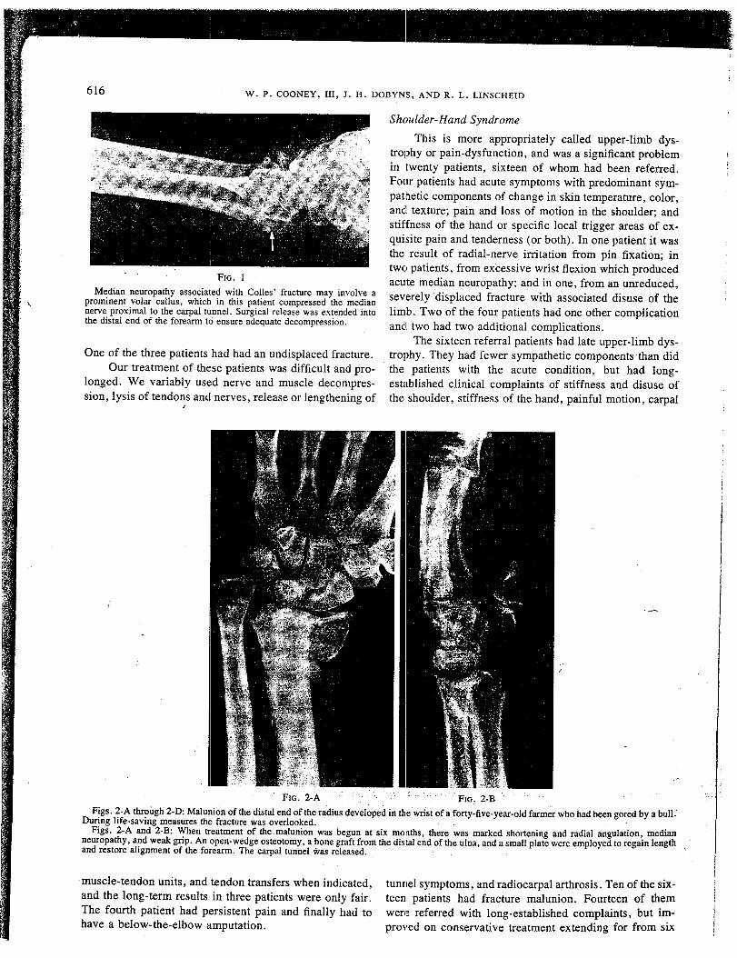
TABLE I
COMPLICATIONS OF COLLES’ FRACTURE ACCORDING TO TREATMENT METHODS
Frykman No. of No. of AnesthesiaType Complications Patients Local Block/General Unknown
ImmobilizationCast Pins Unknown
I 12 9 3 3 3 8 1 0II 14 10 7 1 2 9 1 0III 2 2 1 1 0 1 1 0IV 16 12 6 6 0 9 2 1V 19 12 8 4 0 11 1 0
VI 24 19 11 5 3 16 2 1VII 27 18 10 6 2 14 3 1
VIII 42 32 20 12 0 26 6 0Unknown 16 14 12 0 2 12 0 2
tients (19 per cent) had seventy-eight complications.Among the referred patients, sixty had ninety-nine com-plications. Of the 128 patients with complications, seventy-eight had had local anesthesia and thirty-eight had hadblock or general anesthesia; in twelve cases, the type ofanesthesia was not recorded. Eighty-six patients hadclosed reduction and~ immobilization in a cast, seventeenhad primary external pin fixation, and twenty had failureof cast immobilization with secondary pin fixation. Forfive patients the types of reduction and immobilizationwere not specified. The comminuted displaced intra-articular fractures (the unstable ones, Frykman Types IVthrough VIII) were associated with an increased number ofcomplications, especially the more comminuted Type-VIIand VIII fractures (sixty-nine of the 177 complications).For sixteen fractures, the Frykman class could not be de-termined. The largest number of complications (74 percent) was in patients who had had injection of a localanesthetic into the fracture site, although that method ofanesthesia was used in only 56 per cent of the patijents whohad tr.eatment for Colles’ fracture. Complications after the
reduction of displaced comminuted fractures were lesslikely to occ.ur if either general anesthesia or an axillaryblock was given, followed bY sustained traction (ten min-utes) and gentle reduction. After primary external pin-fixation techniques in seventy-five fractures there weretwenty-one complications, while after failed closed reduc-tion and secondary external pin fixation in forty fractures,twenty-eight complications were encountered. There were128 complications in patients who were treated by closedreduction and plaster-cast fixation, but that routine wasfollowed three times more frequently than the othermethods of treatment combined. The age of the patient,sex, and mechanism of injury seemed to have no relation-ship to the incidence of complications.
Compressive Neuropathy
This was the most frequent single complication (7.9per cent), occurring in twenty-one patients treated locallyand in twenty-four who were referred to us for treatment.It was observed both acutely and late after the injury hadoccurred. Median neuropathy developed early in thirty-one patients, usually associated with reduction of the frac-ture in the emergency room under local anesthesia (twenty
patients). This complication was less frequent after bra-chial block or general anesthesia (eleven patients). Radialneuropathy, attributable to improper immobilization (castcompression at the spiral groove of the humerus or on thedorsum of the hand), was diagnosed in three patients. Irri-tation from external pin fixation caused a severe radialneuropathy in two patients. Ulnar neuropathy occurred insix patients as a result of cast compression. All but five ofthe early neuropathies required no treatment and resolvedafter the offending compressing agent (cast or pin) wasremoved. The five exceptions were patients who had aneuropathy as a result of initial injury. They had im-mediate carpal-tunnel release and no permanent sequelae.
Late neuropathy of the median nerve occurred inforty-one patients. All had persistent symptoms. In fouradditional patients, the median neuropathy was combinedwith ulnar neuropathy. There were no late radialneuropathies. Thirty-one of the forty-five patients requiredrelease of the carpal tunnel or Guyon’s canal, or both, andextensive exploration through an appropriate palmar orforearm incision was essential for adequate decompres~s!on(Fig. 1). In six patients, volar fracture fragments werefound compressing both the ulnar and median nerves andwere removed. Excessive callus formation (seven pa-tients), persistent hematoma (six patients), and localizedswelling (twelve patients), usually the result of the forcedvolar flexion-ulnar deviation position (Cotton-Loder),were considered to be responsible for most of the other lateneuropathies. Eleven of the twenty-four patients who werereferred to us and seven of the twenty-one treated primar-ily had one additional complication associated with a com-pressive neuropathy, and one referral patient had threeassociated complications.
Arthrosis after Fracture
When either painful motion of the wrist or forearmwas evident or there was a mechanical obstruction tomotion after fracture, we diagnosed the condition as ar-throsi~. It was observed in thirty-seven patients and repre-sented. 20 per cent of the complications. Radio-ulnar ar-throsi.,; (twenty-seven patients) was more common thanradiocarpal arthrosis (ten patients). Frykman Type-VI,VII, and VIII fractures most often elicited this complica-tion. When external pin,fixation techniques that restored
COMPLICATIONS OF COLLES’ FRACTURES 615
the radial length were used to treat those fractures therewas a lower incidence of arthrosis (four patients), despitethe fact that pin fixation was the preferred treatment for themore comrninuted fractures.
Of the ten patients with radiocarpal arthrosis, ninewere treated surgically: three by dorsal ostectomy, two byproximal row carpectomy, two by arthrodesis, and two bytotal prosthetic arthroplasty of the wrist. Of the twenty-seven patients with radio-ulnar arthrosis, nineteen had apainful radio-ulnar joint that required a Darrach resectionof the distal end of the ulna (fourteen patients), a Milchprocedure (one patient), or a Silastic replacement arthro-plasty (four patients). Six other patients had symptomaticradio-ulnar subluxation with a mechanical obstruction tomotion and required Darrach excision of the distal end ofthe ulna. All twenty of the patients who had the Darrachprocedure had improvement in motion of the wrist and, inparticular, in pronation and supination of the forearm.
Ten of the twenty-one referral patients and seven ofthe sixteen primary patients had an additional complica-tion.
Malposition- Malunion 1
Thirty patients had this complication, the majorityhaving been referred for treatment. Five patients had frac-tures that were not yet fully united when they were seen fortreatment. They required early open reduction. The othertwenty-five patients required osteotomy. Malunion wasmost commonly related to loss of the reduction position,which commonly occurs when the fracture is unstable andcomminuted. This loss of reduction early in the treatment
¯ period was a frequent problem. In this series, treatment forloss of reduction was required in 27 per cent of the 565patients. A corrective reduction was usually performed bydistraction and gentle manipulation, the patient having hadbrachial b!ock or general anesthesia. It was successful inmost patients (more than 92 per cent) when accomplishedwithin two week.s of the fracture and when the reductionwas maintained with some form of external pin fixation.Our preference was to insert in the base of the second andthird metacarpals two 2.0-millimeter (5/64-inch) Stein-mann pins oriented at 60 to 90 degrees to each other. Twoslightly larger pins(2.3 millimeters, 3/32 inch) wereplaced in the middle third of the radius. A Roger Andersonexternal-fixation apparatus attached to these pins main-tained the reduction and provided stabilization. Additionalpins or Kirschner wires were. used, as required, to secureloose fragments. When the pins applied above and belowwere inadequate to maintain ~eduction open reduction wasdone (five patients), with sa.tisfactory results.
After inadequate treatment of the fresh fracture wasfollowed by malunion, the complaints of significant pain,deformity, and limitation of motion present in twenty-fivepatients led to recommendations for corrective osteotomy.Fourteen patients had that operation at our institution.Three patients accepted the deformity or preferred not tohave surgery, and six had the operation in their home
community. Two were lost to follow-up. In most of ourfourteen patients the corrective osteotomy was supplemen-ted by bone-grafting (Figs. 2-A through 2-D). Improve-ment in grip strength and motion was achieved in all butone of these fourteen patients, and that patient required ar-throdesis.
Nine of the referral patients in this group had anadditional complication, as did one of the twelve primarypatients. Two referral patients had two additional compli-cations.
Tendon Rupture
Rupture of the extensor pollicis longus was noted infive patients, and rupture of the index flexor digitorum pro-fundus or flexor pollicis longus was noted in one patienteach. The: rupture was primarily related to bone fragmentsfrom displaced fractures that abraded the tendon during theweeks after healing of the fracture. All five patients withloss of the extensor pollicis longus tendon had rupturewithin two months from the initial injury (two, two, three,four, and eight weeks), while in the two patients withflexor tendon rupture the rupture occurred after threemonths. All patients had either a tendon transfer or a ten-don graft.. Direct tendon repair was not possible becauseseveral centimeters of tendon substance had been lost.
Associated Injuries Unrecognized Primarily
These included scaphoid fractures (four patients),radial head fractures (two patients), Bennett’s fracture(one patient), and intercarpal ligament injuries (five tients), which were recognized between two days and onemonth from the time of the original injury. These injuriesusually were caused by the same mechanism that causedthe Colles fracture. In our series, ligament instability ofthe wrist required operative reconstruction of thescapholunate ligament in four patients. Scaphoid fracturesrequired open reduction in two patients, and radial head-fractures required excision of the radial head in two pa-tients.
Complications of Fixation
Three patients with pin fixation had pin breakage thatrequired operative removal of the pins. Pin loosening withpurulent drainage occurred in eight patients, and an ulcer-ation of the area around a pin occurred in one patient. Onepatient sustained a fracture through the pin site in the distalend of the radius. Two patients had nerve irritation causedby the casL which led to sympathetic dystrophy (as will bediscussed). Casts caused other complications, as describedin the paragraphs on compressive neuropathies, Voik-mann’s ischemia, and shoulder-hand syndrome.
Volkmann’s Ischemic Contracture
This was seen in four referral patients, three of whomhad had a constricting cast that was retained despite thepatient’s complaints of persisting pain. Continued use ofanalgesics in two patients further masked the symptoms.
VOL. 62-A, NO. 4, JUNE 1980
616 w.P. COONEY, III, I. H. DOBYNS, AND R. L. LINSCHEID
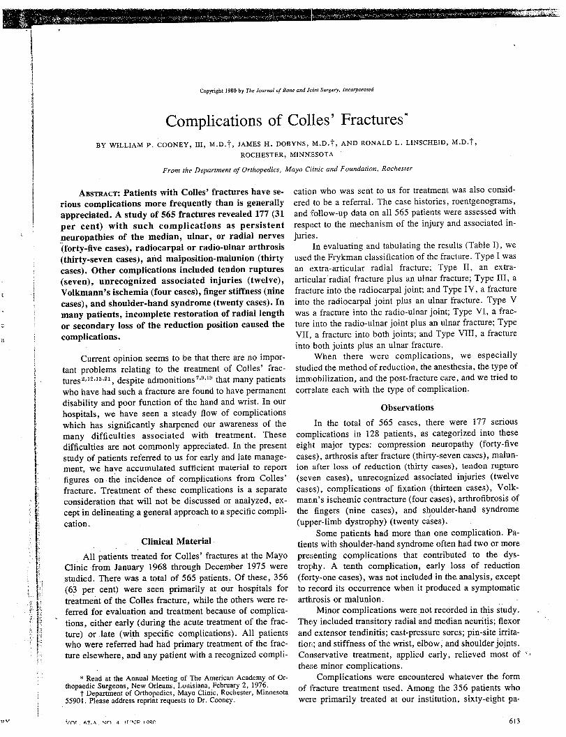
Fro. 1Median neuropathy associated with Colles’ fracture may involve a
prominent volar callus, which in this patient compressed the mediannerve proximal to the. carpal tunnel. Surgical release was extended intothe distal end of the forearm tO ensure adequate decompression.
One of the three patients had had an undisplaced fracture.Our treatment of these patients was difficult and pro-
longed. We variably used nerve and muscle decompres-sion, lysis of tendons and nerves, release or lengthening of
Shoulder-Hand Syndrome
This is more appropriately called upper-limb dys-trophy or pain-dysfunction, and was a significant problemin twenty patients, sixteen of whom had been referred.Four patients had acute symptoms with predominant sym-pathetic components of change in skin temperature, color,and texture; pain and loss of motion in the shoulder; andstiffness of the hand or specific local trigger areas of ex-quisite pain and tenderness (or both). In one patient it wasthe result of radial-nerve irritation from pin fixation; intwo patients, from excessive wrist flexion which producedacute median neuropathy; and in one, from an unreduced,severely displaced fracture with associated disuse of thelimb. Two of the four patients had one other complicationand two had two additional complications.
The sixteen referral patients had late upper-limb dys-trophy. They had fewer sympathetic components than didthe patients with the acute condition, but had long-established clinical complaints of stiffness and disuse ofthe shoulder, stiffness of the hand, painful motion, carpal
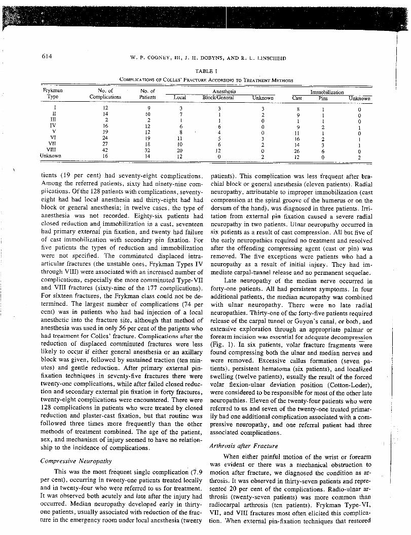
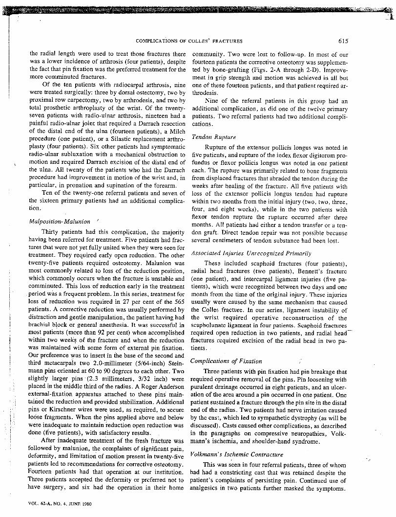
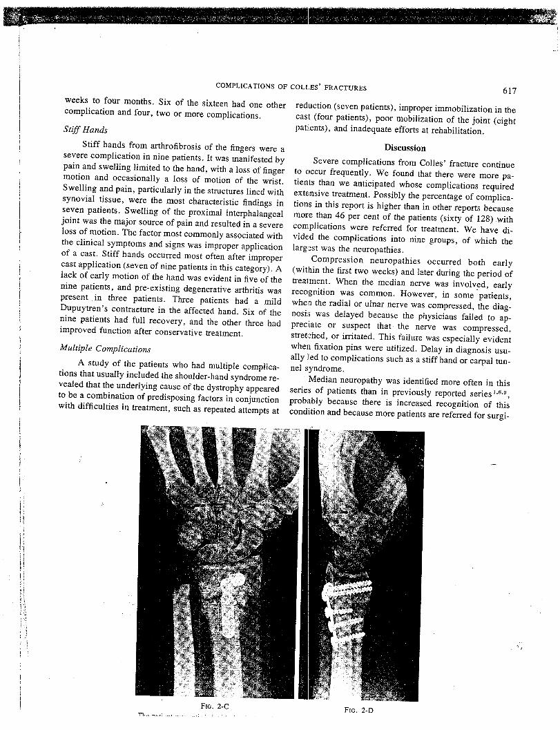
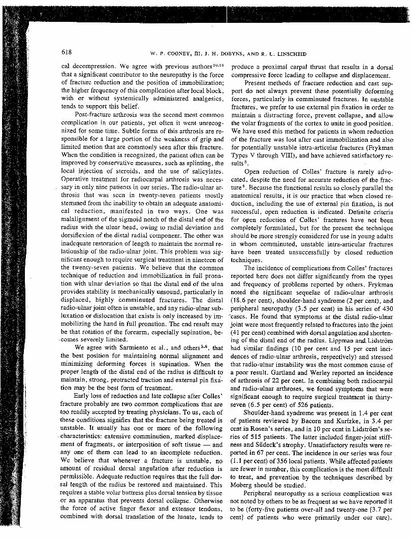
Figs. 2-A throi~gti 2-D: Maluni0n of the distal end of the radius developed in the wrist of a forty-five-year-old farmer who had been gored by a bull.During life-saving measures the fracture was overlooked.
Figs. 2-A and 2-B: When treatment of the malunion was begun at six months, there was marked shortening and radial angulation, medianneuropathy, and weak grip. An open-wedge osteotomy, a bone graft from the distal end of the ulna, and a small plate were employed to regain lengthand restore alignment of the forearm. The carpal tunnel ~,as released.
muscle-tendon units, and tendon transfers when indicated,and the long-term results in three patients were only fair.The fourth patient had persistent pain and finally had tohave a below-the-elbow amputation.
tunnel symptoms, and radiocarpal arthrosis. Ten of the six-teen patients had fracture malunion. Fourteen of themwere referred with long-established complaints, but im-proved on conservative treatment extending for from six
COMPLICATIONS OF COLLES’ FRACTURES
weeks to four months. Six of the sixteen had one othercomplication and four, two or more complications.
Stiff Hands
Stiff hands from arthrofibrosis of the fingers were asevere complication in nine patients. It was manifested bypain and swelling limited to the hand, with a loss of fingermotion and occasionally a loss of motion of the wrist.Swelling and pain, particularly in the structures lined withsynovial tissue, were the most characteristic findings inseven patients. Swelling of the proximal interphalangealjoint was the major source of pain and resulted in a severeloss of motion. The factor most commonly associated withthe clinical symptoms and signs was improper applicationof a cast. Stiff hands occurred most often after impropercast application (seven of nine patients in this category). lack of early motion of the hand was evident in five of thenine patients, and pre-existing degenerative arthritis waspresent in three patients. Three patients had a mildDupuytren’s contraeture in the affected hand. Six of thenine patients had full recovery, and the other three hadimproved function after conservative treatment.
Multiple Complications
A study of the patients who had multiple complica-tions that usually included the shoulder-hand syndrome re-vealed that the underlying cause of the dystrophy appearedto be a combination of predisposing factors in conjunctionwith difficulties in treatment, such as repeated attempts at
617
reduction (seven patients), improper immobilization in thecast (four patients), poor mobilization of the joint (eightpatiients), and inadequate efforts at rehabilitation.
DiscussionSevere complications from Colles’ fracture continue
to occur frequently. We found that there were more pa-tients than we anticipated whose complications requiredexte.nsive treatment. Possibly the percentage of complica-tions in this report is higher than in other reports becausemore than 46 per cent of the patients (sixty of 128):withcomplications were referred for treatment. We have di-vide, d the complications into nine groups, of which thelargest was the neuropathies.
Compression neuropathies occurred both early(within the first two weeks) and later during the period treatment. When the median nerve was involved, earlyrecognition was common. However, in some "patients,when the radial or ulnar nerve was compressed, the diag-nosis was delayed because the physicians failed to ap-preciate or suspect that the nerve was compressed,stretched, or irritated. This failure was especially evidentwhen fixation pins were utilized. Delay in diagnosis usu-ally lied to complications such as a stiff hand or carpal tun-nel syndrome.
Median neuropathy was identified more often in thisseries of patients than in previously reported series1,6,’~,
probably because there is increased recognition of thiscondition and because more patients are referred for surgi-
FIG. 2-C F~G. 2-D
618 W. P. COONEY, tlI, J. H. DOBYNS, AND R. L. LINSCHEID
cal decompression. We agree with previous authors1°,16
that a significant contributor to the neuropathy is the forceof fracture reduction and the position of immobilization;the higher frequency of this complication after local block,with or without systemically administered analgesics,tends to support this belief.
Post-fracture arthrosis was the second most commoncomplication in our patients, yet often it went unrecog-nized for some time. Subtle forms of this arthrosis are re-sponsible for a large portion of the weakness of grip andlimited motion that are commonly seen after this fracture.When the condition is recognized, the patient often can beimproved by conservative measures, such as splinting, thelocal injection of steroids, and the use of salicylates.Operative treatment for radiocarpal arthrosis Was neces-sary in only nine patients in our series. The radio-ulnar ar-throsis that was seen in twenty-seven patients mostlystemmed from the inability to obtain an adequate anatomi-cal reduction, manifested in two ways. One wasmalalignment of the sigmoid notch of the distal end of theradius with the ulnar head, owing to radial deviation anddorsiflexion of the distal radial component. The other wasinadequate restoration of length to maintain the normal re-lationship of the radio-ulnar joint. This problem was sig-nificant enough to require surgical treatment in nineteen ofthe twenty-seven patients. We believe that the commontechnique of reduction and immobilization in full prona-tion with ulnar deviation so that the distal end of the ulnaprovides stability is mechanically unsound, particularly indisplaced, highly Comminuted fractures. The distalradio-ulnar joint often is unstable, and any radio-ulnar sub-luxation or dislocation that exists is only increased by im-mobilizing the hand in full pronation. The end result maybe that rotation of the forearm, especially supination, be-
/comes severely limited.We agree with Sarmiento et al., and others~,6, that
the best position for maintaining normal alignment andminimizing deforming forces is supination. When theproper length of the distal end of the radius is difficult tomaintain, strong, protracted traction and external pin fixa-tion may be the best form of treatment.
Early loss of reduction and late collapse after Colles’fracture probably are two common complications that aretoo readily accepted by treating physicians. To us, each ofthese conditions signifies that the fracture being treated isunstable. It usually has one or more of the following
¯ characteristics: extensive comminution, marked displace-ment of fragments, or interposition of soft tissue -- andany one of them can lead to an incomplete reduction.We believe that whenever a fracture is unstable, noamount of residual dorsal angulation after reduction ispermissible. Adequate reduction requires that the full dor-sal length of the radius be restored and maintained. Thisrequires a stable volar buttress plus dorsal tension by tissueor an apparatus that prevents dorsal collapse. Otherwisethe force of active finger flexor and extensor tendons,combined with dorsal translation of the lunate, tends to
produce a proximal carpal thrust that results in a dorsalcompressive force leading to collapse and displacement.
Present methods of fracture reduction and cast sup-port do not always prevent these potentially deformingforces, particularly in comminuted fractures. In unstablefractures, we prefer to use external pin fixation in order tomaintain a distracting force, prevent collapse, and allowthe volar fragments of the cortex to unite in good position.We have used this method for patients in whom reductionof the fracture was lost after cast immobilization and alsofor potentially unstable intra-articular fractures (FrykmanTypes V through VIII), and have achieved satisfactory re-sults 3. ~
Open reduction of Coltes’ fracture is rarely advo-cated, despite the need for accurate reduction of the frac-ture s. Because the functional results so closely parallel theanatomical results, it is our practice that when closed re-duction, including the use of external pin fixation, is notsuccessful, open reduction is indicated. Definite criteriafor open reduction of Colles’ fractures have not beencompletely formulated, but for the present the techniqueshould be more strongly considered for use in young adultsin whom comminuted, unstable intra-articular fractureshave been treated unsuccessfully by closed reductiontechniques.
The incidence of complications from Colles’ fracturesreported here does not differ significantly from the typesand frequency of problems reported by others. Frykmannoted the significant sequelae of radio-ulnar arthrosis(18.6 per cent), shoulder-hand syndrome (2 per cent), peripheral neuropathy (315 per cent) in his series of 430
"’cases. He found that symptoms at the distal radio-ulnarjoint were most frequently related to fractures into the joint(41 per cent) combined with dorsal angulation and shorten-ing of the distal end of the radius. Lippman and. Lidstr6mhad similar findings (10 per cent and 15 per cent inci-dences of radio-ulnar arthrosis, respectively) and stressedthat radio-ulnar instability was the most common cause ofa poor result. Gartland and Werley reported an incidenceof arthrosis of 22 per cent. In combining both radiocarp~land radio-ulnar arthroses, we found symptoms that weresignificant enough to require surgical treatment in thirty-seven (6.5 per cent) of 526 patients.
Shoulder-hand syndrome was present in 1.4 per centof patients reviewed by Bacorn and Kurizke, in 3.4 percent in Rosen’s series, and in 10 per cent in Lidstr6m’s se-ries of 515 patients. The latter included finger-joint stiff-hess and Siideck’s atrophy. Unsatisfactory results were re-ported in 67 per cent. The incidence in our series was four.(1.1 per cent) of 356 local patients. While affected patientsare fewer in number, this complication is the most difficultto treat, and prevention by the techniques described byMoberg should be studied.
Peripheral neuropathy as a serious complication wasnot noted by others to be as frequent as we have reported itto be (forty-five patients over-all and twenty-one [3.7 percent] of patients who were primarily under our care).
COMPLICATIONS OF COLLES ’ FRACTURES 619
Lidstr6m believed that nerve injuries are rare after frac-tures of the distal end of the radius (slightly more than per cent). Bacorn and Kurtzke reported an incident of 0.2per cent and Schlesinger and Liss noted only one case per1,000 fractures. We believe that these negative reportswere due in part to a lack of recognition and possibly moreconcern with treatment of the fracture than with potentialsequelae. Lynch and Lipscomb, Frykman, Robbins, andothers5 have placed proper emphasis on the causes of me-dian neuropathy and the need for aggressive treatment incertain acute as well as late cases.
Complications related to more than one factor were
common whenever three complications -- neuropathy, ar-throsis, and shoulder-hand syndrome -- were present.Frykman found that of eighty patients with radio-ulnar ar-throsis, five (6.3 per cent) had median neuropathy and fivehad shoulder-hand syndrome. Of our twenty patients withshoulder-hand syndrome, sixteen had one or more as-sociated complications (arthrosis in ten patients, medianneuropathy in nine patients, malunion in ten patients, andsympathetic dystrophy in five patients). Evidently, thesecomplications and others contribute directly to the 24 to 27per cent incidence of poor functional results that has beenreported3"6"~ from the treatment of Colles’ fractures.
References1. BACORN, R. W., and KURTZ~:E, J. F.: Colles’ Fracture: A Study of Two Thousand Cases from the New York State Workmen’s Compensation
Board. J. Bone and Joint Surg., 35-A: 643-658, July 1953.2. BOSACCO, D. N., and TRABULSL L. R.: The Colles’ Fracture -- Treatment by Closed Reduction, Internal Fixation and Short Arm Cast Applica-
tion. In Proceedings of The American Academy of Orthopaedic Surgeons. J. Bone and Joint Surg., 57-A: 1030, Oct. 1975.3. COONEY, W. P., III; LINSCHEID, R. L.; and DOBYNS, J. H.: External Pin Fixation for Unstable Colles’ Fractures. J. Bone and Joint Surg.,
61-A: 840-845, Sept. 1979.4. DARRACH, W~LL~AM: Partial Excision of Lower Shaft of Ulna for Deformity following Colles’s Fracture. Ann. Surg., 57: 764-765, 1913.5. DoBYNS, J. H., and LINSCHEID, R. L.: Complications of Treatment of Fractures and Dislocations of the Wrist. In Complications in Orthopaedic
Surgery, edited by C. H. Epps, Jr. Vol. 1, pp. 271-352. Philadelphia, J. B. Lippincott, 1978.6. FRYKMAN, G.: Fractur§ of the Distal Radius Including Sequelae -- Shoulder-Hand-Finger Syndrome Disturbance of the Distal Radio-Ulnar
Joint and Impairment df Nerve Function. A Clinical and Experimental Study. Acta Ortt~op. gcandina~ica, Suppl¢mentum 108, 1967.7. GARTLAND, J. J., JR., and WERLEY, C. W.: Evaluation of Healed Colles’ Fractures. J. Bone and Joint Surg., 33-A: 895-907, Oct. 1951.8. KRISTIANSEN, AMUND, and G~ERSOE, E~NAR: Colles’ Fracture. Operative Treatment, Indications and Results. Acta Orthop. Scandinavica, 39:
33-46, 1968.9. LIDSTR6M, ANDERS: Fractures of the Distal End of the Radius. A Clinical and Statistical Study of End Results. Acta Orthop. Scandinavica,
Supplementum 41, 1959.10. LIPPMAN, R. K.: Laxity of the Radio-ulnar Joint following Colles’ Fracture. Arch. Surg., 35: 772-786, 1937.11. LYNCH, A. C., and LIPSCOMI~, P. R.: The Carpal Tunnel Syndrome and Colles’ Fractures. J. Am. Med. Assn., 185: 363-366, 1963.12. MARVEL, J. R., JR.: Comminuted Fractures of the Distal End of the Radius Treated by Pins and Plaster Technique. In Proceedings of The
American Academy of Orthopaedic Surgeons. J. Bone and Joint Surg., 57-A: 1030, Oct. 1975.13. M~L¢8, HENRY: Cuff Resection of the Ulna for Malunited Colles’ Fracture. J. Bone and Joint Surg., 23:311-313, April 1941.14. MOSERG, ERIK: Shoulder-Hand-Finger Syndrome, Reflex Dystrophy, Causalgia [Abstract]. Acta Chir. Scandinavica, 125: 523, 1963.15. POOL, CHRISTOPHER: Colles’ Fracture. A Prospective Study of Treatment. J. Bone and Joint Surg., 55-B: 540-544, Aug. 1973.16. RO88INS, J. V.: Logical Reduction of Displaced Colles’ Fractures. New York State J. Med., 50: 2959-2962, 1950.17. ROSEN, ERIK: Fractura Extremitatis Distalis Radii. Ugeskr. Laeger., 109: 603-610, 1947.18. SARMIE~qTO, At~GUSTO; PRATT, G. W.; BERRY, N. C.; and S~NCLAm, W. F.: Cg~lles’ Fractures. Functional Bracing in Supination. J. Bone and
Joint Surg., 57-A: 311-317, April 1975.19. S¢I-IECK, MAx: Long-Term Follow-up of Treatment of Comminuted Fractures of the Distal End of the Radius by Transfixation with Kirschner
Wires and Cast. J. Bone and Joint Surg., 44-A: 337-351, March 1962.20. SC8LES~NGER, E. B., and L~ss, H. R.: Fundamentals, Fads and Fallacies in the Carpal Tunnel Syndrome. Am. J. Surg., 97: 466-470, 1959.21. SMAI.LL, G. B.: Long-Term Follow-up of Colles’ Fracture. J. Bone and Joint Surg., 47-B: 80-85, Feb. 1965.
Related Documents