248 Copyright © SLACK Incorporated ORIGINAL ARTICLE Comparison of the Visual Results After SMILE and Femtosecond Laser-Assisted LASIK for Myopia Fangyu Lin, MD; Yesheng Xu, MD; Yabo Yang, PhD, MD ABSTRACT PURPOSE: To perform a comparative clinical analysis of the safety, efficacy, and predictability of two surgi- cal procedures (ie, small incision lenticule extraction [SMILE] and femtosecond laser-assisted LASIK [FS- LASIK]) to correct myopia. METHODS: Sixty eyes of 31 patients with a mean spher- ical equivalent of -5.13 ± 1.75 diopters underwent myopia correction with the SMILE procedure. Fifty-one eyes of 27 patients with a mean spherical equivalent of -5.58 ± 2.41 diopters were treated with the FS-LASIK procedure. Postoperative uncorrected and corrected distance visual acuity, manifest refraction, and higher- order aberrations were analyzed statistically at 1 and 3 months postoperatively. RESULTS: No statistically significant differences were found at 1 and 3 months in parameters that included the percentage of eyes with an uncorrected distance visual acuity of 20/20 or better (P = .556, .920) and mean spherical equivalent refraction (P = .055, .335). At 1 month, 4 SMILE-treated eyes and 1 FS-LASIK– treated eye lost one or more line of visual acuity (P = .214, chi-square test). At 3 months, 2 SMILE-treated eyes lost one or more line of visual acuity, whereas all FS-LASIK–treated eyes had an unchanged or cor- rected distance visual acuity. Higher-order aberrations and spherical aberration were significantly lower in the SMILE group than the FS-LASIK group at 1 (P = .007, .000) and 3 (P = .006, .000) months of follow-up. CONCLUSIONS: SMILE and FS-LASIK are safe, effec- tive, and predictable surgical procedures to treat myo- pia. SMILE has a lower induction rate of higher-order aberrations and spherical aberration than the FS-LASIK procedure. [J Refract Surg. 2014;30(4):248-254.] ASIK is the most commonly used refractive surgical procedure to treat myopia and astigmatism world- wide. However, the induction of higher-order aber- rations (HOAs) is a problem after LASIK. HOAs often cause significant night vision problems such as glare, haze, and ha- los. In addition, decreasing postoperative contrast sensitivity may compromise patient satisfaction with the procedure. 1-3 Femtosecond lasers have become available for clinical use in the past few years. They have the ability to cut tissue with great precision and are increasingly used to create flaps for LASIK. The greater precision of femtosecond lasers helps maintain the morphological stability of the flap. This leads to smaller corneal biomechanical changes and the subsequent induction of fewer HOAs. 4 Despite the development of femtosecond lasers, HOAs have shown an increase postoperatively in previous studies. 3,5 It has been postulated that the creation of a corneal flap and the in- duction of localized inflammation associated with excimer la- ser use may play an important role in producing these changes. An all-in-one femtosecond laser procedure, known as small incision lenticule extraction (SMILE), was described by Sekundo et al. 6 and became clinically available in 2011. The procedure does not require the creation of a flap. The intrastro- mal lenticule is taken out through a small 2- to 5-mm incision. This reduces the risks associated with flap creation, such as dry eye, traumatic flap dislocation, and epithelial ingrowth. 7,8 The aim of this study was to evaluate the comparative safety, efficacy, and predictability of SMILE with that of fem- tosecond laser-assisted LASIK (FS-LASIK) at 1 and 3 months postoperatively. Comparisons were made between the two treatment arms of the study using uncorrected distance visual From the Department of Ophthalmology, Second Affiliated Hospital, College of Medicine, Zhejiang University, Hangzhou, China. Submitted: October 30, 2013; Accepted: January 27, 2014; Posted online: April 4, 2014 The authors have no financial or proprietary interest in the materials pre- sented herein. Correspondence: Yabo Yang, PhD, MD, Department of Ophthalmology, Second Affiliated Hospital, College of Medicine, Zhejiang University, Hangzhou 310009, China. E-mail: [email protected] doi:10.3928/1081597X-20140320-03 L

Welcome message from author
This document is posted to help you gain knowledge. Please leave a comment to let me know what you think about it! Share it to your friends and learn new things together.
Transcript
248 Copyright © SLACK Incorporated
O R I G I N A L A R T I C L E
Comparison of the Visual Results After SMILE and Femtosecond Laser-Assisted LASIK for MyopiaFangyu Lin, MD; Yesheng Xu, MD; Yabo Yang, PhD, MD
ABSTRACT
PURPOSE: To perform a comparative clinical analysis of the safety, efficacy, and predictability of two surgi-cal procedures (ie, small incision lenticule extraction [SMILE] and femtosecond laser-assisted LASIK [FS-LASIK]) to correct myopia.
METHODS: Sixty eyes of 31 patients with a mean spher-ical equivalent of -5.13 ± 1.75 diopters underwent myopia correction with the SMILE procedure. Fifty-one eyes of 27 patients with a mean spherical equivalent of -5.58 ± 2.41 diopters were treated with the FS-LASIK procedure. Postoperative uncorrected and corrected distance visual acuity, manifest refraction, and higher-order aberrations were analyzed statistically at 1 and 3 months postoperatively.
RESULTS: No statistically significant differences were found at 1 and 3 months in parameters that included the percentage of eyes with an uncorrected distance visual acuity of 20/20 or better (P = .556, .920) and mean spherical equivalent refraction (P = .055, .335). At 1 month, 4 SMILE-treated eyes and 1 FS-LASIK–treated eye lost one or more line of visual acuity (P = .214, chi-square test). At 3 months, 2 SMILE-treated eyes lost one or more line of visual acuity, whereas all FS-LASIK–treated eyes had an unchanged or cor-rected distance visual acuity. Higher-order aberrations and spherical aberration were significantly lower in the SMILE group than the FS-LASIK group at 1 (P = .007, .000) and 3 (P = .006, .000) months of follow-up.
CONCLUSIONS: SMILE and FS-LASIK are safe, effec-tive, and predictable surgical procedures to treat myo-pia. SMILE has a lower induction rate of higher-order aberrations and spherical aberration than the FS-LASIK procedure.
[J Refract Surg. 2014;30(4):248-254.]
ASIK is the most commonly used refractive surgical procedure to treat myopia and astigmatism world-wide. However, the induction of higher-order aber-
rations (HOAs) is a problem after LASIK. HOAs often cause significant night vision problems such as glare, haze, and ha-los. In addition, decreasing postoperative contrast sensitivity may compromise patient satisfaction with the procedure.1-3
Femtosecond lasers have become available for clinical use in the past few years. They have the ability to cut tissue with great precision and are increasingly used to create flaps for LASIK. The greater precision of femtosecond lasers helps maintain the morphological stability of the flap. This leads to smaller corneal biomechanical changes and the subsequent induction of fewer HOAs.4
Despite the development of femtosecond lasers, HOAs have shown an increase postoperatively in previous studies.3,5 It has been postulated that the creation of a corneal flap and the in-duction of localized inflammation associated with excimer la-ser use may play an important role in producing these changes.
An all-in-one femtosecond laser procedure, known as small incision lenticule extraction (SMILE), was described by Sekundo et al.6 and became clinically available in 2011. The procedure does not require the creation of a flap. The intrastro-mal lenticule is taken out through a small 2- to 5-mm incision. This reduces the risks associated with flap creation, such as dry eye, traumatic flap dislocation, and epithelial ingrowth.7,8
The aim of this study was to evaluate the comparative safety, efficacy, and predictability of SMILE with that of fem-tosecond laser-assisted LASIK (FS-LASIK) at 1 and 3 months postoperatively. Comparisons were made between the two treatment arms of the study using uncorrected distance visual
From the Department of Ophthalmology, Second Affiliated Hospital, College of Medicine, Zhejiang University, Hangzhou, China.
Submitted: October 30, 2013; Accepted: January 27, 2014; Posted online: April 4, 2014
The authors have no financial or proprietary interest in the materials pre-sented herein.
Correspondence: Yabo Yang, PhD, MD, Department of Ophthalmology, Second Affiliated Hospital, College of Medicine, Zhejiang University, Hangzhou 310009, China. E-mail: [email protected]
doi:10.3928/1081597X-20140320-03
L
249Journal of Refractive Surgery • Vol. 30, No. 4, 2014
Visual Results After SMILE and FS-LASIK for Myopia/Lin et al
acuity (UDVA) and corrected distance visual acuity (CDVA), refractive diopters (D), and HOAs.
PATIENTS AND METHODSPatient PoPulation
This prospective comparative study involved 58 patients (111 eyes) who were scheduled for myopic correction at the Second Affiliated Hospital of Zheji-ang University, Hangzhou, China, between May and December 2012. All patients were randomly allocated into one of two treatment groups (SMILE or FS-LASIK) per their own choice.
Inclusion criteria consisted of a minimum age of 18 years, a stable refractive error for at least 2 years (< -0.25 D), a CDVA of 20/25 or better, sphere ranging from -0.00 to -10.00 D, cylinder ranging from -0.00 to -4.00 D, minimum corneal thickness of 500 µm, and the absence of systemic or localized ocular disease. This study was approved by an ethics committee and followed the tenets of the Declaration of Helsinki. Writ-ten informed consent was received from all patients.
PreoPerative examinationPreoperative examination included UDVA and
CDVA, slit-lamp examination, noncontact tonometry, refraction (ie, manifest and cycloplegic), ultrasonic pachymetry, funduscopy, endothelial cell count, B-scan ocular ultrasound corneal fluorescein staining, corneal topography (Orbscan II; Bausch & Lomb, Roch-ester, NY), and wavefront aberrations (CRS-Master sys-tem; Carl Zeiss Meditec, Jena, Germany). In this study, CDVA measurement was recorded with an upper limit of 20/20. Patients had to discontinue wearing contact lenses for at least 2 weeks (for soft lenses) or 3 weeks (for hard lenses) prior to the assessment.
Surgical techniqueSMILE Procedure. All surgical procedures were
performed by the same surgeon (YY). Eyes were pre-operatively anesthetized with 0.4% oxybuprocaine eye drops. After standard sterilization and sterile draping, an eyelid speculum was used to keep the eye open. A VisuMax femtosecond laser (Carl Zeiss Med-itec) was used for SMILE. The patient’s eye was posi-tioned under the interface cone, and the patient was asked to fixate on a blinking green target. The cone was connected to suction ports. When the surgeon confirmed that an appropriate centration (ie, center of pupil) had been achieved, suction was applied to the cone. Once suction was on, delivery of the femtosec-ond laser pulses was initiated to begin the entire len-ticule creation. During this period, the contact glass of the cone remained fixed on the cornea, and the patient
was required to observe the blinking target. Four sub-sequent femtosecond incisions were performed. The first incision created the posterior surface of the lenti-cule (from periphery to the center). The second inci-sion consisted of a vertical side cut made all around the lenticule (the depth of the cut was the cap thick-ness). The third incision created the anterior surface of the lenticule (from center to periphery). Finally, the fourth was a small incision of 50° to create access to the lenticule. It was located at 120° off the corneal sur-face, with a circumferential length of 4.0 to 5.0 mm and a side-cut angle of 90°. Thickness of the lenticule depended on the refractive error to be corrected, with a diameter of 6.1 to 6.6 mm in all cases. Thickness of the cap was 100 µm, and its diameter was 7.0 to 7.5 mm. All steps were created with the femtosecond la-ser using energy of 120 nJ. Once the lenticule creation was completed, suction was released automatically. A thin spatula was inserted through the side cut to sepa-rate the lenticule (first the anterior and then the pos-terior surface), and it was subsequently grasped with non-toothed serrated forceps.
FS-LASIK Procedure. A VisuMax femtosecond laser (Carl Zeiss Meditec) was used for FS-LASIK flaps and subsequent photoablation was performed with a MEL-80 excimer laser (Carl Zeiss Meditec). The steps for preoperative preparation, confirmation of appro-priate centration, and initiation of suction were done using the same procedures described above for the SMILE group. Surgical parameters were as follows: flap diameter was 7.9 mm and its intended thickness was 85 to 100 µm, with the hinge at the 12-o’clock po-sition. The side-cut angle was 90°; spot spacing and track spacing were between 2 and 3 mm. For excimer ablation, aspheric ablation profiles (ie, aspheric smart ablation) using a personal adjustment nomogram were applied. Once the excimer ablation was completed, the flap was repositioned.
At the end of the two procedures, the cornea was irrigated with saline and all patients received one drop of tobramycin/dexamethasone. The postoperative regi-men included loteprednol and levofloxacin eye drops for 1 week. The patients were encouraged to use lubri-cating drops as needed to improve comfort.
PoStoPerative Follow-uPPostoperative follow-up was at 1 day and 1 week
after the surgery. Complete ophthalmic evaluations were conducted at 1 and 3 months postoperatively. The following parameters were obtained in the latter two examinations: UDVA and CDVA, manifest refrac-tion, noncontact tonometry, ultrasonic pachymetry, corneal topography, and wavefront aberrations.
250 Copyright © SLACK Incorporated
Visual Results After SMILE and FS-LASIK for Myopia/Lin et al
StatiStical analySiSAll statistical analyses were performed with SPSS
version 19.0 (SPSS, Inc., Chicago, IL). Chi-square tests were used to compare the results of various groups, in-dependent t tests were used to compare data between the two groups, and paired t tests were used for sta-tistical analysis to compare data before and after each treatment. All values are given as the mean ± standard deviation. A P value of less than .05 was considered statistically significant.
RESULTSAt 1 and 3 months, 111 eyes were observed. The
SMILE group included 60 eyes of 31 patients (15 men and 16 women) with ages ranging from 18 to 40 years. Intended myopic spherical correction ranged from -1.75 to -7.75 D and cylinder ranged from -0.00 to -2.00 D. The FS-LASIK group included 51 eyes of 27 patients (18 men and 9 women) with ages ranging from 18 to 39 years. Intended myopic spherical correction ranged from -1.00 to -9.75 D and cylinder ranged from -0.00 to -3.75 D. Detailed clinical data are shown in Table 1.
uDvaAll eyes had emmetropia as the target refraction.
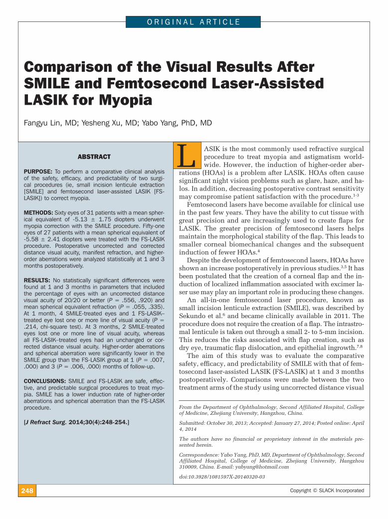
The efficacy index (mean postoperative UDVA/mean preoperative CDVA) was 0.99 ± 0.15 and 1.04 ± 0.20 at 1 and 3 months, respectively, in the SMILE group and 1.02 ± 0.17 and 1.10 ± 0.23, respectively, in the FS-LASIK group. Figure 1 shows the UDVA of the SMILE and FS-LASIK groups at 1 and 3 months. The
eyes with a postoperative UDVA of 20/20 or better all reached preoperative CDVA, and the differences at 1 (chi-square test2 = 0.347; P = .556) and 3 (chi-square test2 = 0.010; P = .920) months were not statistically significant.
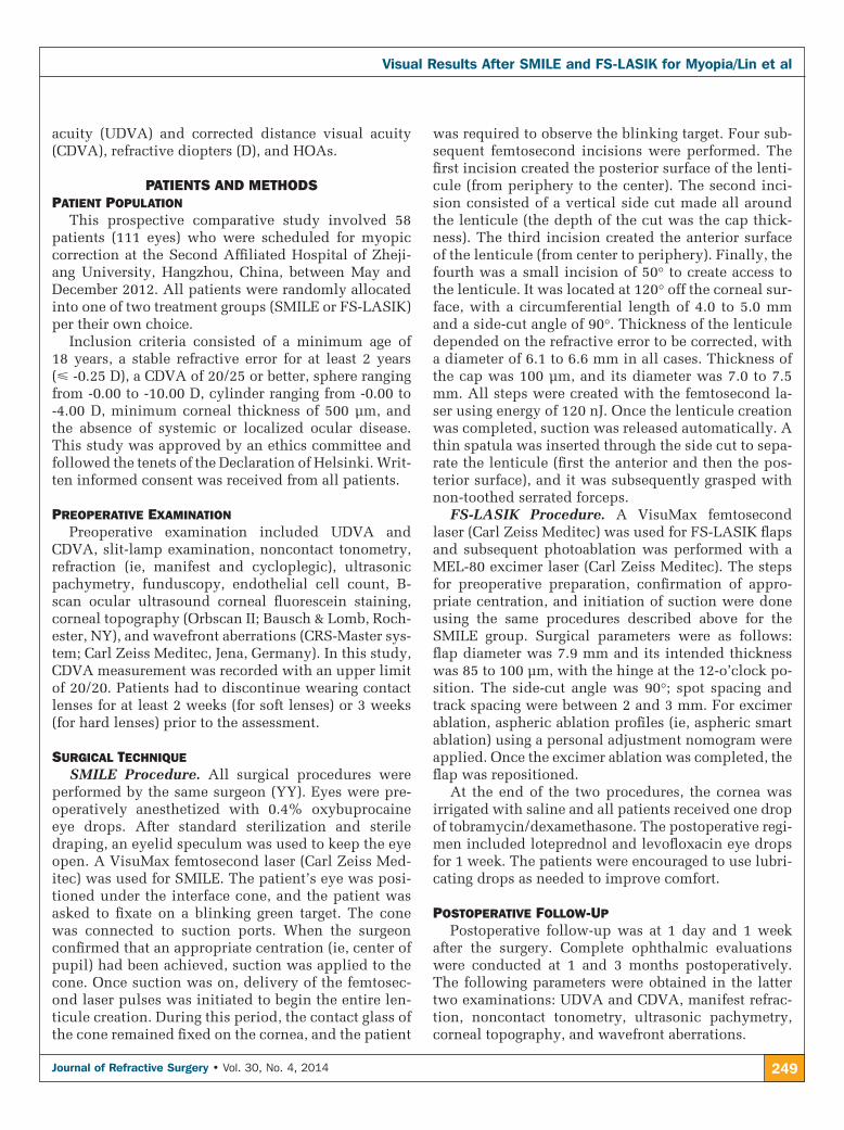
cDvaThe safety index (mean postoperative CDVA/mean
preoperative CDVA) was 1.00 ± 0.06 and 1.01 ± 0.05 at 1 and 3 months, respectively, in the SMILE group and 1.00 ± 0.03 and 1.01 ± 0.03, respectively, in the FS-LASIK group. Figure 2 shows the percentage of eyes in which there was a gain or loss of Snellen visual acuity lines compared with preoperative levels at 1 and 3 months. No suction loss or other complications during surgery and no signs of postoperative ectasia were noted.
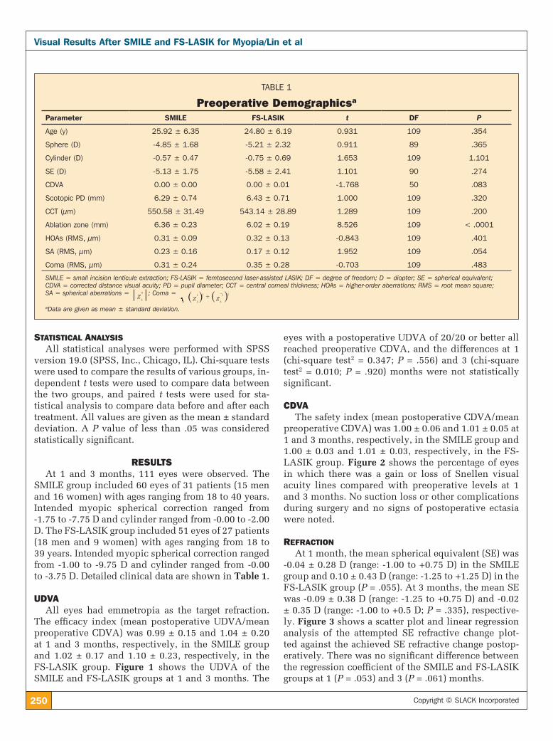
reFractionAt 1 month, the mean spherical equivalent (SE) was
-0.04 ± 0.28 D (range: -1.00 to +0.75 D) in the SMILE group and 0.10 ± 0.43 D (range: -1.25 to +1.25 D) in the FS-LASIK group (P = .055). At 3 months, the mean SE was -0.09 ± 0.38 D (range: -1.25 to +0.75 D) and -0.02 ± 0.35 D (range: -1.00 to +0.5 D; P = .335), respective-ly. Figure 3 shows a scatter plot and linear regression analysis of the attempted SE refractive change plot-ted against the achieved SE refractive change postop-eratively. There was no significant difference between the regression coefficient of the SMILE and FS-LASIK groups at 1 (P = .053) and 3 (P = .061) months.
TABLE 1
Preoperative Demographicsa
Parameter SMILE FS-LASIK t DF P
Age (y) 25.92 ± 6.35 24.80 ± 6.19 0.931 109 .354
Sphere (D) -4.85 ± 1.68 -5.21 ± 2.32 0.911 89 .365
Cylinder (D) -0.57 ± 0.47 -0.75 ± 0.69 1.653 109 1.101
SE (D) -5.13 ± 1.75 -5.58 ± 2.41 1.101 90 .274
CDVA 0.00 ± 0.00 0.00 ± 0.01 -1.768 50 .083
Scotopic PD (mm) 6.29 ± 0.74 6.43 ± 0.71 1.000 109 .320
CCT (µm) 550.58 ± 31.49 543.14 ± 28.89 1.289 109 .200
Ablation zone (mm) 6.36 ± 0.23 6.02 ± 0.19 8.526 109 < .0001
HOAs (RMS, µm) 0.31 ± 0.09 0.32 ± 0.13 -0.843 109 .401
SA (RMS, µm) 0.23 ± 0.16 0.17 ± 0.12 1.952 109 .054
Coma (RMS, µm) 0.31 ± 0.24 0.35 ± 0.28 -0.703 109 .483
SMILE = small incision lenticule extraction; FS-LASIK = femtosecond laser-assisted LASIK; DF = degree of freedom; D = diopter; SE = spherical equivalent; CDVA = corrected distance visual acuity; PD = pupil diameter; CCT = central corneal thickness; HOAs = higher-order aberrations; RMS = root mean square; SA = spherical aberrations =
Z4
0 ; Coma = Z Z3
1 2
3
1 2+
-^ ^h haData are given as mean ± standard deviation.
251Journal of Refractive Surgery • Vol. 30, No. 4, 2014
Visual Results After SMILE and FS-LASIK for Myopia/Lin et al
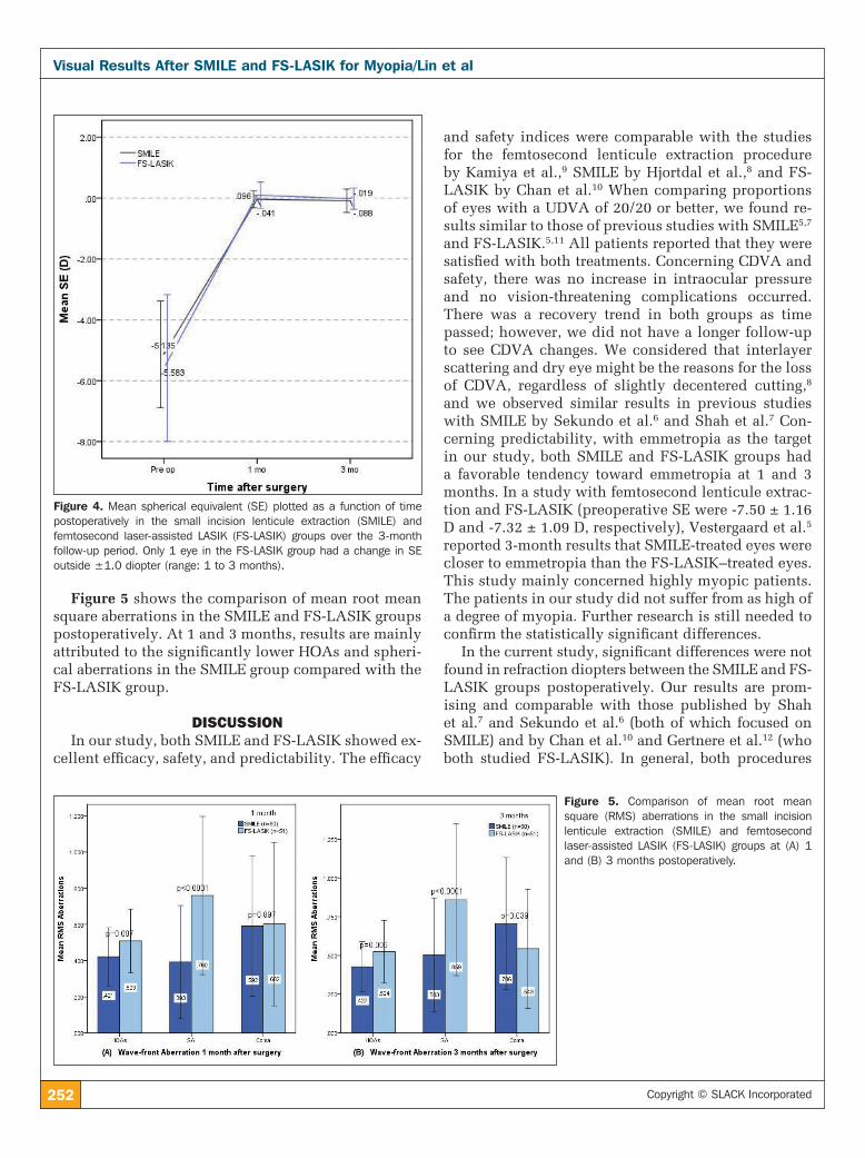
Additionally in Figure 4, the mean SE plotted as a function of time in both groups shows the sta-bility over the 3-month follow-up period. When the number of eyes with a change within ±0.5 D in SE between 1 and 3 months was compared in the two groups, no significant difference was found (x 2=0.862; P = .353).
ocular hoaSThe total number of HOAs, including spherical ab-
erration and coma, was not statistically significantly different preoperatively in the SMILE and FS-LASIK groups (Table 1). Data that compared these parameters before and after treatment demonstrated that they in-creased significantly (P < .05) in each group.
Figure 2. The percentage of eyes in which there was a gain/loss of Snellen visual acuity lines com-pared with preoperative levels for different postop-erative periods. (A) At 1 month, 4 eyes (6.67%) in the small incision lenticule extraction (SMILE) group and 1 eye (1.96%) in the femtosecond laser-assisted LASIK (FS-LASIK) group lost one or more line. (B) At 3 months, 2 eyes (3.33%) in the SMILE group lost one or more line, whereas all FS-LASIK–treated eyes had an unchanged cor-rected distance visual acuity (CDVA).
Figure 3. Attempted spherical equivalent (SE) refractive change plotted against the achieved SE refractive change. (A) At 1 month, 60 (100%) eyes in the small incision lenticule extraction (SMILE) group and 48 (94.12%) eyes in the fem-tosecond laser-assisted LASIK (FS-LASIK) group had a SE within ±1.0 diopter. (B) At 3 months, it was 59 (98.33%) and 51 (100%), respectively.
Figure 1. A distribution of uncorrected distance visual acuity (UDVA) at (A) cumulative 1 month and (B) 3 months postoperatively. Forty-eight (80.00%) of 60 eyes in the small incision lenti-cule extraction (SMILE) group and 43 (84.31%) of 51 eyes in the femtosecond laser-assisted LASIK (FS-LASIK) group had 20/20 or better at 1 month, whereas 51 eyes (85.00%) in the SMILE group and 43 eyes (84.31%) in the FS-LASIK group had 20/20 or better at 3 months.
252 Copyright © SLACK Incorporated
Visual Results After SMILE and FS-LASIK for Myopia/Lin et al
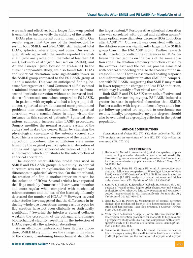
Figure 5 shows the comparison of mean root mean square aberrations in the SMILE and FS-LASIK groups postoperatively. At 1 and 3 months, results are mainly attributed to the significantly lower HOAs and spheri-cal aberrations in the SMILE group compared with the FS-LASIK group.
DISCUSSIONIn our study, both SMILE and FS-LASIK showed ex-
cellent efficacy, safety, and predictability. The efficacy
and safety indices were comparable with the studies for the femtosecond lenticule extraction procedure by Kamiya et al.,9 SMILE by Hjortdal et al.,8 and FS-LASIK by Chan et al.10 When comparing proportions of eyes with a UDVA of 20/20 or better, we found re-sults similar to those of previous studies with SMILE5,7 and FS-LASIK.5,11 All patients reported that they were satisfied with both treatments. Concerning CDVA and safety, there was no increase in intraocular pressure and no vision-threatening complications occurred. There was a recovery trend in both groups as time passed; however, we did not have a longer follow-up to see CDVA changes. We considered that interlayer scattering and dry eye might be the reasons for the loss of CDVA, regardless of slightly decentered cutting,8 and we observed similar results in previous studies with SMILE by Sekundo et al.6 and Shah et al.7 Con-cerning predictability, with emmetropia as the target in our study, both SMILE and FS-LASIK groups had a favorable tendency toward emmetropia at 1 and 3 months. In a study with femtosecond lenticule extrac-tion and FS-LASIK (preoperative SE were -7.50 ± 1.16 D and -7.32 ± 1.09 D, respectively), Vestergaard et al.5 reported 3-month results that SMILE-treated eyes were closer to emmetropia than the FS-LASIK–treated eyes. This study mainly concerned highly myopic patients. The patients in our study did not suffer from as high of a degree of myopia. Further research is still needed to confirm the statistically significant differences.
In the current study, significant differences were not found in refraction diopters between the SMILE and FS-LASIK groups postoperatively. Our results are prom-ising and comparable with those published by Shah et al.7 and Sekundo et al.6 (both of which focused on SMILE) and by Chan et al.10 and Gertnere et al.12 (who both studied FS-LASIK). In general, both procedures
Figure 4. Mean spherical equivalent (SE) plotted as a function of time postoperatively in the small incision lenticule extraction (SMILE) and femtosecond laser-assisted LASIK (FS-LASIK) groups over the 3-month follow-up period. Only 1 eye in the FS-LASIK group had a change in SE outside ±1.0 diopter (range: 1 to 3 months).
Figure 5. Comparison of mean root mean square (RMS) aberrations in the small incision lenticule extraction (SMILE) and femtosecond laser-assisted LASIK (FS-LASIK) groups at (A) 1 and (B) 3 months postoperatively.
253Journal of Refractive Surgery • Vol. 30, No. 4, 2014
Visual Results After SMILE and FS-LASIK for Myopia/Lin et al
were safe and effective, but a longer follow-up period is essential to further verify the stability of the results.
HOAs play an important role in visual quality. Our results suggest that the use of the femtosecond la-ser (in both SMILE and FS-LASIK) still induced total HOAs, spherical aberrations, and coma. Our results qualitatively agree with the results reported by Shah et al.7 (who analyzed a pupil diameter of less than 5.0 mm), Sekundo et al.6 (who focused on SMILE), and Au and Krueger11 (who focused on FS-LASIK). In the current comparative study, we also found total HOAs and spherical aberration were significantly lower in the SMILE group compared to the FS-LASIK group at 1 and 3 months. This was an anticipated finding, be-cause Vestergaard et al.5 and Gertnere et al.12 also noted a minimal increase in spherical aberration in femto-second lenticule extraction without an increased inci-dence of increased coma when compared to FS-LASIK.
In patients with myopia who had a larger pupil di-ameter, spherical aberration caused more pronounced problems than coma-like aberration. Thus, spherical aberration was a crucial factor in night vision dis-turbance in this subset of patients.13 Spherical aber-rations commonly increase after LASIK procedures. Surgery modifies the normal aspheric system of the cornea and makes the cornea flatter by changing the physiological curvature of the anterior corneal sur-face. This is a necessary consequence of the refractive correction procedure. The equilibrium state main-tained by the original positive spherical aberration of cornea and negative spherical aberration of the lens is destroyed, which contributes to the introduction of spherical aberration.
The aspheric smart ablation profile was used in SMILE and FS-LASIK groups in our study, so corneal curvature was not an explanation for the significant differences in spherical aberration. On the other hand, the creation of a flap is another important reason for the induction of HOAs. Several articles have reported that flaps made by femtosecond lasers were smoother and more regular when compared with mechanical microkeratomes and that use of the lasers significantly decreased the number of HOAs.14-16 However, several other studies have suggested that the differences in in-ducing whole-eye aberrations among various types for flap creation have not been clinically or statistically significant.17 Severing the interlayer corneal collagen weakens the cross-links of the collagen and changes biomechanical stability, resulting in the induction of HOAs, especially the spherical aberrations.
As an all-in-one femtosecond laser flapless proce-dure, SMILE likely minimizes the change in the shape of the cornea, maintaining biomechanical stability to
the largest extent.18 Postoperative spherical aberration also was correlated with optical and ablation zones.19 Large optical zone diameters decrease HOA induction after LASIK.20,21 Our result was consistent with this; the ablation zone was significantly larger in the SMILE group than in the FS-LASIK group. Further research is still needed to confirm the differences in HOAs be-tween the two groups on the basis of the same abla-tion zone. The ablation efficiency reduction caused by the excimer laser and the healing process associated with corneal remodeling are also the reasons for the in-creased HOAs.22 There is less wound healing response and inflammatory infiltration after SMILE in compari-son with FS-LASIK, suggesting that SMILE may result in fewer topographic changes and less HOA induction, which may favorably affect visual results.23
Both SMILE and FS-LASIK were safe, effective, and predictable for treating myopia. FS-LASIK induced a greater increase in spherical aberration than SMILE. Further studies with larger numbers of eyes and a lon-ger follow-up period are required to corroborate our results. Ideally, preoperative myopia degrees should also be evaluated as a grouping criterion in the patient population.
AUTHOR CONTRIBUTIONSConception and design (FL, YX, YY); data collection (FL, YX,
YY); analysis and interpretation of data (FL, YX, YY); writing the
manuscript (FL, YX, YY)
REFERENCES 1. Hashemi H, Nazari R, Amoozadeh J, et al. Comparison of post-
operative higher-order aberrations and contrast sensitivity: tissue-saving versus conventional photorefractive keratectomy for low to moderate myopia. J Cataract Refract Surg. 2010; 36:1732-1740.
2. Moshirfar M, Betts BS, Churgin DS, et al. A prospective, ran-domized, fellow eye comparison of WaveLight Allegretto Wave Eye-Q versus VISX CustomVue STAR S4 IR in laser in situ ker-atomileusis (LASIK): analysis of visual outcomes and higher order aberrations. Clin Ophthalmol. 2011;5:1339-1347.
3. Kamiya K, Shimizu K, Igarashi A, Kobashi H, Komatsu M. Com-parison of visual acuity, higher-order aberrations and corneal asphericity after refractive lenticule extraction and wavefront-guided laser-assisted in situ keratomileusis for myopia. Br J Ophthalmol. 2013;97:968-975.
4. Ortiz D, Alió JL, Piñero D. Measurement of corneal curvature change after mechanical laser in situ keratomileusis flap cre-ation and femtosecond laser flap creation. J Cataract Refract Surg. 2008;34:238-242.
5. Vestergaard A, Ivarsen A, Asp S, Hjortdal JØ. Femtosecond (FS) laser vision correction procedure for moderate to high myopia: a prospective study of ReLEx flex and comparison with a retro-spective study of FS-laser in situ keratomileusis. Acta Ophthal-mol. 2013;91:355-362.
6. Sekundo W, Kunert KS, Blum M. Small incision corneal re-fractive surgery using the small incision lenticule extraction (SMILE) procedure for the correction of myopia and myopic
254 Copyright © SLACK Incorporated
Visual Results After SMILE and FS-LASIK for Myopia/Lin et al
astigmatism: results of a 6 month prospective study. Br J Oph-thalmol. 2011;95:335-339.
7. Shah R, Shah S, Sengupta S. Results of small incision lenticule extraction: all-in-one femtosecond laser refractive surgery. J Cataract Refract Surg. 2011;37:127-137.
8. Hjortdal JØ, Vestergaard AH, Ivarsen A, Ragunathan S, Asp S. Predictors for the outcome of small-incision lenticule extrac-tion for myopia. J Refract Surg. 2012;28:865-871.
9. Kamiya K, Igarashi A, Ishii R, Sato N, Nishimoto H, Shimizu K. Early clinical outcomes, including efficacy and endotheli-al cell loss, of refractive lenticule extraction using a 500 kHz femtosecond laser to correct myopia. J Cataract Refract Surg. 2012;38:1996-2002.
10. Chan A, Ou J, Manche EE. Comparison of the femtosecond laser and mechanical keratome for laser in situ keratomileusis. Arch Ophthalmol. 2008;126:1484-1490.
11. Au JD, Krueger RR. Optimized femto-LASIK maintains preex-isting spherical aberration independent of refractive error. J Re-fract Surg. 2012;28:S821-S825.
12. Gertnere J, Solomatin I, Sekundo W. Refractive lenticule extrac-tion (ReLEx flex) and wavefront-optimized Femto-LASIK: com-parison of contrast sensitivity and high-order aberrations at 1 year. Graefes Arch Clin Exp Ophthalmol. 2013;251:1437-1442.
13. Oshika T, Tokunaga T, Samejima T, Miyata K, Kawana K, Kaji Y. Influence of pupil diameter on the relation between ocular higher-order aberration and contrast sensitivity after laser in situ keratomileusis. Invest Ophthalmol Vis Sci. 2006;47:1334-1338.
14. Tran DB, Sarayba MA, Bor Z, et al. Randomized prospective clinical study comparing induced aberrations with IntraLase and Hansatome flap creation in fellow eyes: potential impact on wavefront-guided laser in situ keratomileusis. J Cataract Re-fract Surg. 2005;31:97-105.
15. Medeiros FW, Stapleton WM, Hammel J, Krueger RR, Netto MV, Wilson SE. Wavefront analysis comparison of LASIK outcomes
with the femtosecond laser and mechanical microkeratomes. J Refract Surg. 2007;23:880-887.
16. Buzzonetti L, Petrocelli G, Valente P, et al. Comparison of cor-neal aberration changes after laser in situ keratomileusis per-formed with mechanical microkeratome and IntraLase femto-second laser: 1-year follow-up. Cornea. 2008;27:174-179.
17. Calvo R, McLaren JW, Hodge DO, Bourne WM, Patel SV. Corne-al aberrations and visual acuity after laser in situ keratomileu-sis: femtosecond laser versus mechanical microkeratome. Am J Ophthalmol. 2010;149:785-793.
18. Reinstein DZ, Archer TJ, Randleman JB. Mathematical model to compare the relative tensile strength of the cornea after PRK, LASIK and small incision lenticule extraction. J Refract Surg. 2013;29:454-460.
19. Vega-Estrada A, Alió JL, Arba Mosquera S, Moreno LJ. Corneal higher order aberrations after LASIK for high myopia with a fast repetition rate excimer laser, optimized ablation profile, and femtosecond laser-assisted flap. J Refract Surg. 2012;28:689-696.
20. Bühren J, Kuhne C, Kohnen T. Influence of pupil and optical zone diameter on higher-order aberrations after wavefront-guided myopic LASIK. J Cataract Refract Surg. 2005;31:2272-2280.
21. Mok KH, Lee VW. Effect of optical zone ablation diameter on LASIK-induced higher order optical aberrations. J Refract Surg. 2005;21:141-143.
22. Kwon Y, Bott S. Postsurgery corneal asphericity and spheri-cal aberration due to ablation efficiency reduction and corneal remodeling in refractive surgeries. Eye (Lond). 2009;23:1845-1850.
23. Riau AK, Angunawela RI, Chaurasia SS, Lee WS, Tan DT, Meh-ta JS. Early corneal wound healing and inflammatory responses after refractive lenticule extraction (ReLEx). Invest Ophthalmol Vis Sci. 2011;52:6213-6221.
ERRATUMThis article has been amended to include a factual correction. An error was identified subsequent to its original printing. On page 250 of the article “Comparison of the Visual Results After SMILE and Femtosecond Laser-Assisted LASIK for Myopia” by Lin et al., which was published in the April 2014 issue of the Journal of Refractive Surgery, the P value of 1.101 should be .101. This error was acknowledged on page 582, volume 30, is-sue 9. The online article and its erratum are considered the version of record.doi:10.3928/1081597X-20140819-01
Related Documents