Pašagić et al. Scr Med 2020;51(3):209-14. CASE REPORT DOI:10.5937/scriptamed51-28407 Combined Orthodontic and Surgical Treatment of Impacted Maxillary Canine in Young Patient with Class II Malocclusion: a Case Report Ljubica Pašagić, 1 Ivana Ilić, 1 Vesna Kecman, 2 Marko Bulajić, 2 Nina Zubović, 3 Branislav Glišić 4 This article should be cited as follows: Pašagić Lj, Ilić I, Kecman V, Bulajić M, Zubović N, Glišić B. Combined orthodontic and surgical treatment of impacted maxillary canine in young patient with class II malocclusion: a case report. Scr Med 2020;51(3):209-14. Received: 14 September 2020 Revision received: 20 September 2020 Accepted: 20 September 2020 ARTICLE INFO (1) (2) (3) (4) Abstract The impaction of maxillary canines is one of the biggest challenges in orthodontics practice. This case report describes successful surgical and orthodontic approach to the treatment of palatally impacted maxillary canine in a 14-year-old boy. Intra- oral clinical examination revealed an absence of the upper right canine, an ectopic position of the upper left canine and crowding in the maxillary arch. The impaction of right maxillary canine and class II malocclusion were confirmed by lateral ceph- alogram, orthopantomogram and cone beam computed tomography. In the first phase, a transpalatal arch to the upper first molar teeth was applied, first premo- lars were extracted, and brackets were placed on all teeth and nickel-titanium arch wire was applied. The initial orthodontic phase was soon thereafter followed by the surgical exposure and orthodontic traction of the impacted canine using ligature wire attached from the button with chain to the open coil on the arch wire. The orthodontic treatment took two years with satisfactory aesthetic and functional results at the end. This clinical case has shown that adequate treatment of impacted maxillary canine can be achieved by using combined surgical technique and appro- priate orthodontic approach. Key words: Impacted canine; Malocclusion, Class II; Orthodontic-surgical ap- proach; Canine traction. Introduction The presence of canines in dental arch are ex- tremely important for facial harmony, smile aes- thetic and stabile occlusion. Many factors can influence the functional and aesthetic balance, including the canine impaction. The impaction of the maxillary permanent canines is the second most common form of tooth impaction after the third molars with the rate that varies from 0.2 to 2.8 %. 1, 2 Girls are much more frequently affected than boys, with the prevalence ratio from 1.3 : 1 to 3.2 : 1, and with bilateral occlusion occurring in 8 % of cases. 3 The impacted canines occur 10- Copyright © 2020 Pašagić et al. This is an open access article distributed under the Creative Commons Attribution License (CC BY), which permits unrestricted use, distribution, and reproduction in any medium, provided the original work is properly cited. Correspondence: LJUBICA PAŠAGIĆ E: [email protected] M: +387 65 950 044 20 times more frequently in the upper jaw than in the lower jaw4 and the impaction occurs more frequently in the palatine region than in the buc- cal one (85 % vs 15 %). 5 The causes of the maxillary canine impaction can be classified into several distinct groups: local tissue obstruction, local pathology, disturbance of normal development and hereditary or genetic factors. 4, 6 Canine impaction is very often accom- panied by the persistence of deciduous teeth, cysts or dental ankyloses. 7 Department of Orthodontics, Dental Clinic, Faculty of Medicine, University of Banja Luka, Banja Luka, the Repub- lic of Srpska, Bosnia and Herzegovina. Department of Oral Surgery, Dental Clinic, Faculty of Medicine, University of Banja Luka, Banja Luka the Repub- lic of Srpska, Bosnia and Herzegovina. Department of Periodontal Diseases, Dental Clinic, Faculty of Medicine, Uni- versity of Banja Luka, Banja Luka, the Republic of Srpska, Bosnia and Her- zegovina. Department of Orthodontics, School of Dental Medicine, University of Bel- grade, Belgrade, Serbia.

Welcome message from author
This document is posted to help you gain knowledge. Please leave a comment to let me know what you think about it! Share it to your friends and learn new things together.
Transcript
Pašagić et al. Scr Med 2020;51(3):209-14.
CASE REPORTDOI:10.5937/scriptamed51-28407
Combined Orthodontic and Surgical Treatment of Impacted Maxillary Canine in Young Patient with Class II Malocclusion: a Case Report
Ljubica Pašagić,1 Ivana Ilić,1 Vesna Kecman,2 Marko Bulajić,2 Nina Zubović,3 Branislav Glišić4
This article should be cited as follows: Pašagić Lj, Ilić I, Kecman V, Bulajić M, Zubović N, Glišić B. Combined orthodontic and surgical treatment of impacted maxillary canine in young patient with class II malocclusion: a case report. Scr Med 2020;51(3):209-14.
Received: 14 September 2020Revision received: 20 September 2020Accepted: 20 September 2020
ARTICLE INFO
(1)
(2)
(3)
(4)
AbstractThe impaction of maxillary canines is one of the biggest challenges in orthodontics practice. This case report describes successful surgical and orthodontic approach to the treatment of palatally impacted maxillary canine in a 14-year-old boy. Intra-oral clinical examination revealed an absence of the upper right canine, an ectopic position of the upper left canine and crowding in the maxillary arch. The impaction of right maxillary canine and class II malocclusion were confirmed by lateral ceph-alogram, orthopantomogram and cone beam computed tomography. In the first phase, a transpalatal arch to the upper first molar teeth was applied, first premo-lars were extracted, and brackets were placed on all teeth and nickel-titanium arch wire was applied. The initial orthodontic phase was soon thereafter followed by the surgical exposure and orthodontic traction of the impacted canine using ligature wire attached from the button with chain to the open coil on the arch wire. The orthodontic treatment took two years with satisfactory aesthetic and functional results at the end. This clinical case has shown that adequate treatment of impacted maxillary canine can be achieved by using combined surgical technique and appro-priate orthodontic approach.
Key words: Impacted canine; Malocclusion, Class II; Orthodontic-surgical ap-proach; Canine traction.
Introduction
The presence of canines in dental arch are ex-tremely important for facial harmony, smile aes-thetic and stabile occlusion. Many factors can influence the functional and aesthetic balance, including the canine impaction. The impaction of the maxillary permanent canines is the second most common form of tooth impaction after the third molars with the rate that varies from 0.2 to 2.8 %.1, 2 Girls are much more frequently affected than boys, with the prevalence ratio from 1.3 : 1 to 3.2 : 1, and with bilateral occlusion occurring in 8 % of cases.3 The impacted canines occur 10-
Copyright © 2020 Pašagić et al. This is an open access article distributed under the Creative Commons Attribution License (CC BY), which permits unrestricted use, distribution, and reproduction in any medium, provided the original work is properly cited.
Correspondence:LJUBICA PAŠAGIĆE: [email protected]: +387 65 950 044
20 times more frequently in the upper jaw than in the lower jaw4 and the impaction occurs more frequently in the palatine region than in the buc-cal one (85 % vs 15 %).5
The causes of the maxillary canine impaction can be classified into several distinct groups: local tissue obstruction, local pathology, disturbance of normal development and hereditary or genetic factors.4, 6 Canine impaction is very often accom-panied by the persistence of deciduous teeth, cysts or dental ankyloses.7
Department of Orthodontics, Dental Clinic, Faculty of Medicine, University of Banja Luka, Banja Luka, the Repub-lic of Srpska, Bosnia and Herzegovina.Department of Oral Surgery, Dental Clinic, Faculty of Medicine, University of Banja Luka, Banja Luka the Repub-lic of Srpska, Bosnia and Herzegovina.Department of Periodontal Diseases, Dental Clinic, Faculty of Medicine, Uni-versity of Banja Luka, Banja Luka, the Republic of Srpska, Bosnia and Her-zegovina.Department of Orthodontics, School of Dental Medicine, University of Bel-grade, Belgrade, Serbia.
210 Pašagić et al. Scr Med 2020;51(3):209-14.
Case history
There are several options for treatment of maxil-lary canine impaction. Some of them include ex-traction followed by an implant support or a pros-thetic replacement procedure. However, if the position of impacted canine in the bone allows an orthodontic-surgical method, the orthodontic treatment should be started as soon as possible to avoid secondary problems.8 The treatment proce-
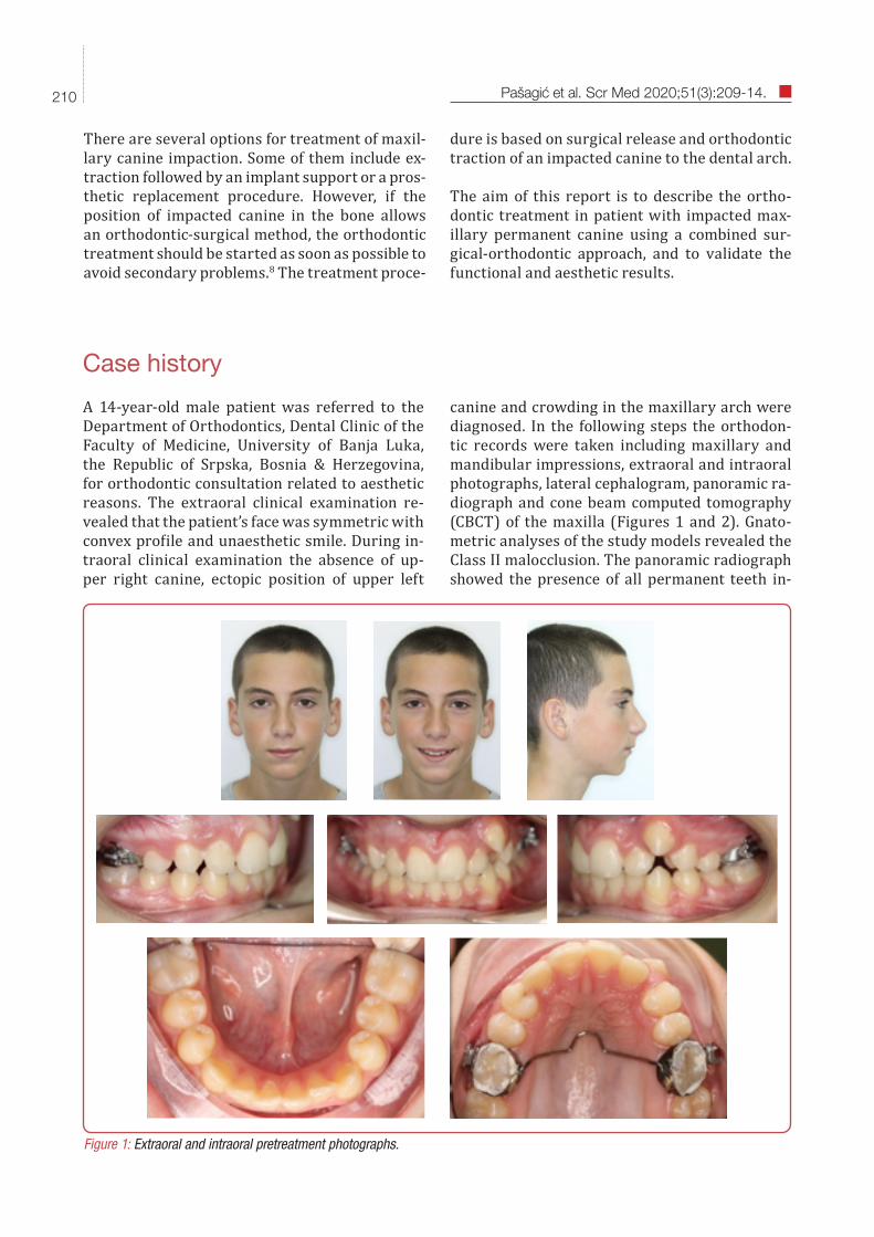
A 14-year-old male patient was referred to the Department of Orthodontics, Dental Clinic of the Faculty of Medicine, University of Banja Luka, the Republic of Srpska, Bosnia & Herzegovina, for orthodontic consultation related to aesthetic reasons. The extraoral clinical examination re-vealed that the patient’s face was symmetric with convex profile and unaesthetic smile. During in-traoral clinical examination the absence of up-per right canine, ectopic position of upper left
dure is based on surgical release and orthodontic traction of an impacted canine to the dental arch.
The aim of this report is to describe the ortho-dontic treatment in patient with impacted max-illary permanent canine using a combined sur-gical-orthodontic approach, and to validate the functional and aesthetic results.
Figure 1: Extraoral and intraoral pretreatment photographs.
canine and crowding in the maxillary arch were diagnosed. In the following steps the orthodon-tic records were taken including maxillary and mandibular impressions, extraoral and intraoral photographs, lateral cephalogram, panoramic ra-diograph and cone beam computed tomography (CBCT) of the maxilla (Figures 1 and 2). Gnato-metric analyses of the study models revealed the Class II malocclusion. The panoramic radiograph showed the presence of all permanent teeth in-
211Pašagić et al. Scr Med 2020;51(3):209-14.
Figure 3: Orthodontic traction of the canine into the dental arch.
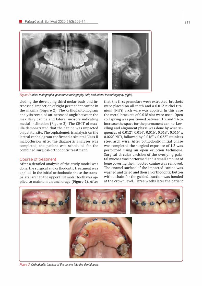
Figure 2: Initial radiographs; panoramic radiography (left) and lateral teleradiography (right).
cluding the developing third molar buds and in-traosseal impaction of right permanent canine in the maxilla (Figure 2). The orthopantomogram analysis revealed an increased angle between the maxillary canine and lateral incisors indicating mesial inclination (Figure 2). The CBCT of max-illa demonstrated that the canine was impacted on palatal site. The cephalometric analysis on the lateral cephalogram confirmed a skeletal Class II malocclusion. After the diagnostic analyses was completed, the patient was scheduled for the combined surgical-orthodontic treatment.
Course of treatmentAfter a detailed analysis of the study model was done, the surgical and orthodontic treatment was applied. In the initial orthodontic phase the trans-palatal arch to the upper first molar teeth was ap-plied to maintain an anchorage (Figure 1). After
that, the first premolars were extracted, brackets were placed on all teeth and a 0.012 nickel-tita-nium (NiTi) arch wire was applied. In this case the metal brackets of 0.018 slot were used. Open coil spring was positioned between 1.2 and 1.4 to increase the space for the permanent canine. Lev-elling and alignment phase was done by wire se-quences of 0.012", 0.014", 0.016", 0.018", 0.016" x 0.022" NiTi, followed by 0.016" x 0.022" stainless steel arch wire. After orthodontic initial phase was completed the surgical exposure of 1.3 was performed using an open eruption technique. Surgical circular excision of the overlying pala-tal mucosa was performed and a small amount of bone covering the impacted canine was removed. The enamel surface of the impacted canine was washed and dried and then an orthodontic button with a chain for the guided traction was bonded at the crown level. Three weeks later the patient
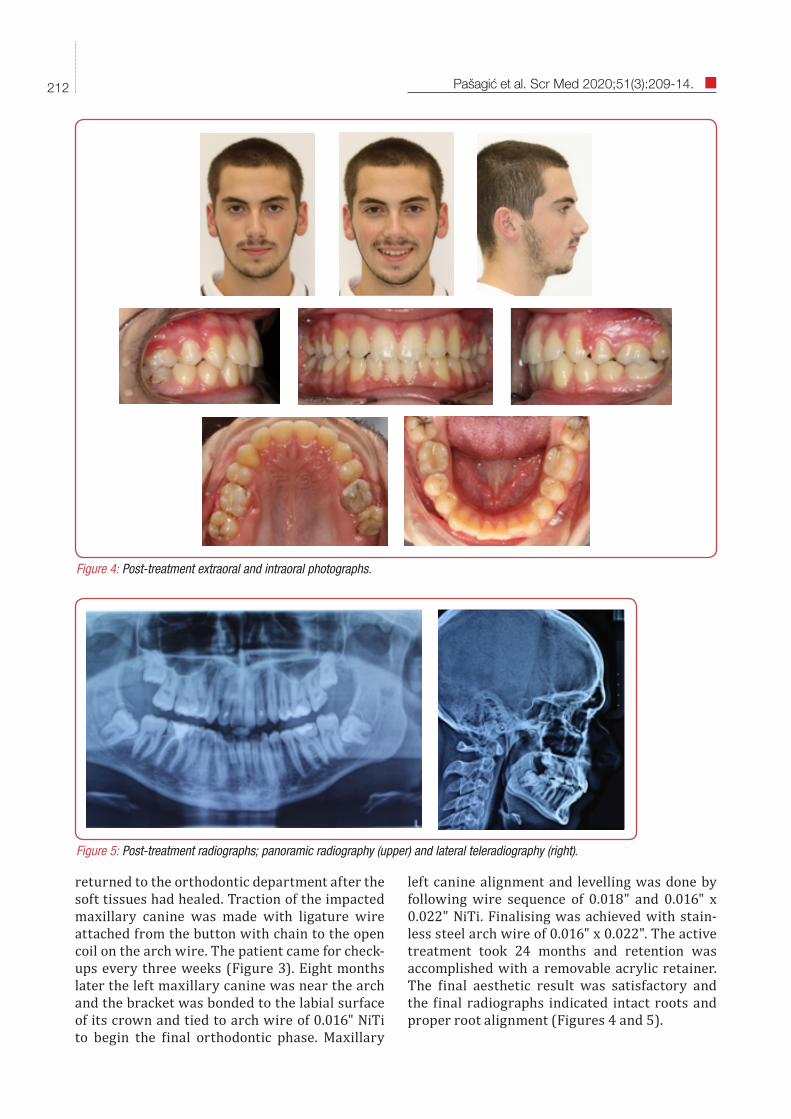
returned to the orthodontic department after the soft tissues had healed. Traction of the impacted maxillary canine was made with ligature wire attached from the button with chain to the open coil on the arch wire. The patient came for check-ups every three weeks (Figure 3). Eight months later the left maxillary canine was near the arch and the bracket was bonded to the labial surface of its crown and tied to arch wire of 0.016" NiTi to begin the final orthodontic phase. Maxillary
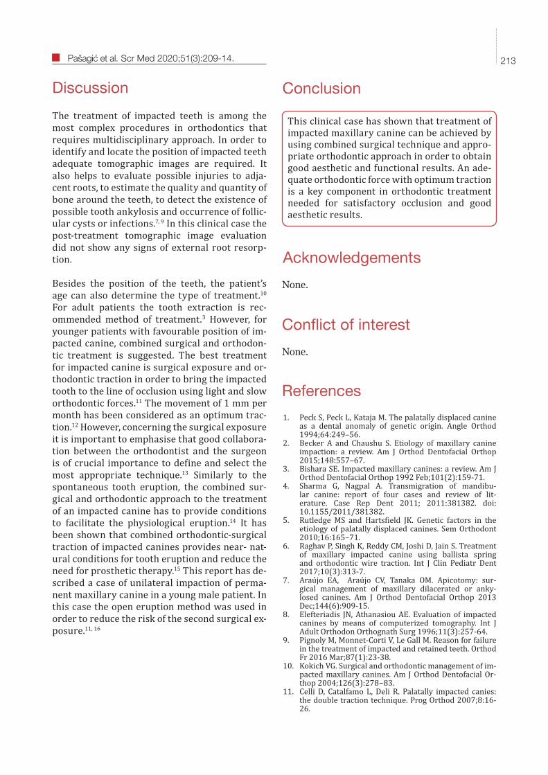
Figure 4: Post-treatment extraoral and intraoral photographs.
Figure 5: Post-treatment radiographs; panoramic radiography (upper) and lateral teleradiography (right).
left canine alignment and levelling was done by following wire sequence of 0.018" and 0.016" x 0.022" NiTi. Finalising was achieved with stain-less steel arch wire of 0.016" x 0.022". The active treatment took 24 months and retention was accomplished with a removable acrylic retainer. The final aesthetic result was satisfactory and the final radiographs indicated intact roots and proper root alignment (Figures 4 and 5).
212 Pašagić et al. Scr Med 2020;51(3):209-14.
This clinical case has shown that treatment of impacted maxillary canine can be achieved by using combined surgical technique and appro-priate orthodontic approach in order to obtain good aesthetic and functional results. An ade-quate orthodontic force with optimum traction is a key component in orthodontic treatment needed for satisfactory occlusion and good aesthetic results.
Conclusion
Conflict of interest
None.
None.
Acknowledgements
References
1. Peck S, Peck L, Kataja M. The palatally displaced canine as a dental anomaly of genetic origin. Angle Orthod 1994;64:249–56.
2. Becker A and Chaushu S. Etiology of maxillary canine impaction: a review. Am J Orthod Dentofacial Orthop 2015;148:557–67.
3. Bishara SE. Impacted maxillary canines: a review. Am J Orthod Dentofacial Orthop 1992 Feb;101(2):159-71.
4. Sharma G, Nagpal A. Transmigration of mandibu-lar canine: report of four cases and review of lit-erature. Case Rep Dent 2011; 2011:381382. doi: 10.1155/2011/381382.
5. Rutledge MS and Hartsfield JK. Genetic factors in the etiology of palatally displaced canines. Sem Orthodont 2010;16:165–71.
6. Raghav P, Singh K, Reddy CM, Joshi D, Jain S. Treatment of maxillary impacted canine using ballista spring and orthodontic wire traction. Int J Clin Pediatr Dent 2017;10(3):313-7.
7. Araújo EA, Araújo CV, Tanaka OM. Apicotomy: sur-gical management of maxillary dilacerated or anky-losed canines. Am J Orthod Dentofacial Orthop 2013 Dec;144(6):909-15.
8. Elefteriadis JN, Athanasiou AE. Evaluation of impacted canines by means of computerized tomography. Int J Adult Orthodon Orthognath Surg 1996;11(3):257-64.
9. Pignoly M, Monnet-Corti V, Le Gall M. Reason for failure in the treatment of impacted and retained teeth. Orthod Fr 2016 Mar;87(1):23-38.
10. Kokich VG. Surgical and orthodontic management of im-pacted maxillary canines. Am J Orthod Dentofacial Or-thop 2004;126(3):278−83.
11. Celli D, Catalfamo L, Deli R. Palatally impacted canies: the double traction technique. Prog Orthod 2007;8:16-26.
Discussion
The treatment of impacted teeth is among the most complex procedures in orthodontics that requires multidisciplinary approach. In order to identify and locate the position of impacted teeth adequate tomographic images are required. It also helps to evaluate possible injuries to adja-cent roots, to estimate the quality and quantity of bone around the teeth, to detect the existence of possible tooth ankylosis and occurrence of follic-ular cysts or infections.7, 9 In this clinical case the post-treatment tomographic image evaluation did not show any signs of external root resorp-tion.
Besides the position of the teeth, the patient’s age can also determine the type of treatment.10 For adult patients the tooth extraction is rec-ommended method of treatment.3 However, for younger patients with favourable position of im-pacted canine, combined surgical and orthodon-tic treatment is suggested. The best treatment for impacted canine is surgical exposure and or-thodontic traction in order to bring the impacted tooth to the line of occlusion using light and slow orthodontic forces.11 The movement of 1 mm per month has been considered as an optimum trac-tion.12 However, concerning the surgical exposure it is important to emphasise that good collabora-tion between the orthodontist and the surgeon is of crucial importance to define and select the most appropriate technique.13 Similarly to the spontaneous tooth eruption, the combined sur-gical and orthodontic approach to the treatment of an impacted canine has to provide conditions to facilitate the physiological eruption.14 It has been shown that combined orthodontic-surgical traction of impacted canines provides near- nat-ural conditions for tooth eruption and reduce the need for prosthetic therapy.15 This report has de-scribed a case of unilateral impaction of perma-nent maxillary canine in a young male patient. In this case the open eruption method was used in order to reduce the risk of the second surgical ex-posure.11, 16
213Pašagić et al. Scr Med 2020;51(3):209-14.
12. Ferreira JTL, Romano FL, Sasso Stuani MB, Assed Car-neiro FC, Nakane Matsumoto MA. Traction of impacted canines in a skeletal Class III malocclusion: A challenging orthodontic treatment. Am J Orthod Dentofacial Orthop 2017 Jun;151(6):1159-68.
13. Cruz RM. Orthodontic traction of impacted canines: concepts and clinical application. Dental Press J Orthod 2019 Jan-Feb;24(1):74-87.
14. Figliuzzi MM, Altilia M, Mannarino L, Giudice A, Fortuna-
to L. Minimally invasive surgical management of impact-ed maxillary canines. Ann Ital Chir 2018;89(5):443-7.
15. Pavlović J, Tabaković SZ, Simić S, Vujačić A, VukićevićV. Orthodontic-surgical treatment of four impacted ca-nines in an adult patient: a case report. Vojnosanit Pregl 2016;73(7):682–5.
16. Ferguson J, Parvizi F. Eruption of palatal canines follow-ing surgical exposure: a review of outcomes in a series of consecutively treated cases. Br Dent J 1997;24:203-7.
214 Pašagić et al. Scr Med 2020;51(3):209-14.
Related Documents