1 Clock Controlled Endogenous Melatonin Rhythms in Nile Tilapia (Oreochromis niloticus niloticus) and African Catfish (Clarias gariepinus) Martinez-Chavez Carlos C. 1† , Al-Khamees S. 1† , Campos-Mendoza A. 2 , Penman D. J 1 , Migaud H. *1 1 Institute of Aquaculture, University of Stirling, Stirling, FK9 4LA. UK. 2 Laboratorio de Acuicultura, Facultad de Biología, Universidad Michoacana de San Nicolás de Hidalgo, Morelia, Michoacán, México. Endogenous melatonin rhythms Corresponding author: Dr. Herve Migaud * [email protected] Tel. +44 1786467878 Fax. +44 1786472133 Keywords: Melatonin; Endogenous rhythm; Oscillator; Pineal; Tilapia; Catfish. † Joint first authors *To whom all correspondence should be addressed

Welcome message from author
This document is posted to help you gain knowledge. Please leave a comment to let me know what you think about it! Share it to your friends and learn new things together.
Transcript
1
Clock Controlled Endogenous Melatonin Rhythms in Nile Tilapia (Oreochromis
niloticus niloticus) and African Catfish (Clarias gariepinus)
Martinez-Chavez Carlos C.1†, Al-Khamees S.1†, Campos-Mendoza A.2, Penman D. J1,
Migaud H.*1
1Institute of Aquaculture, University of Stirling, Stirling, FK9 4LA. UK.
2Laboratorio de Acuicultura, Facultad de Biología, Universidad Michoacana de San
Nicolás de Hidalgo, Morelia, Michoacán, México.
Endogenous melatonin rhythms
Corresponding author: Dr. Herve Migaud*
Tel. +44 1786467878
Fax. +44 1786472133
Keywords: Melatonin; Endogenous rhythm; Oscillator; Pineal; Tilapia; Catfish.
† Joint first authors
*To whom all correspondence should be addressed
2
Abstract
The purpose of this work was to investigate the circadian melatonin system in two
tropical teleost species characterised by different behavioural habits, Nile tilapia
(diurnal) and African catfish (nocturnal). To do so, fish were subjected to either a
control photoperiod (12L:12D), continuous light (LL) or darkness (DD) or a 6L:6D
photoperiod. Under 12L:12D, plasma melatonin levels were typically low during the
photophase and high during the scotophase in both species. Interestingly, in both
species melatonin levels significantly decreased prior to the onset of light, which in
catfish, reached similar basal levels to those during the day, demonstrating that
melatonin production can anticipate photic changes probably through circadian
clocks. Further evidence for the existence of such pacemaker activity was obtained
when fish were exposed to DD, as a strong circadian melatonin rhythm was
maintained. Such an endogenous rhythm was sustained for at least 18 days in Nile
tilapia. A similar rhythm was shown in catfish although DD was only tested for four
days. Under LL, the results confirmed the inhibitory effect of light on melatonin
synthesis already reported in other species. Finally, when acclimatised to a short
photo-cycle (6L:6D), no endogenous melatonin rhythm was observed in tilapia under
DD, with melatonin levels remaining high. This could suggest that the circadian
clocks cannot entrain to such a short photo-cycle. Additional research is clearly
needed to further characterise the circadian axis in teleost species, identify and
localize the circadian clocks and better understand the environmental entrainment of
fish physiology.
3
Introduction
Melatonin is known to be a biological time keeping hormone or “zeitgeber” which is
entrained by light and displays circadian and circannual rhythms in vertebrates
(Menaker et al. 1997). These rhythms can also be self-sustained and are under the
control of circadian clocks (Falcon 1999; Fukada and Okano 2002; Holzberg and
Albrecht 2003; Falcon et al. 2007). As such melatonin seems to play a major role in
synchronising many behavioural (locomotor, feeding, shoaling, and migration
activities) and physiological (growth, reproduction, immunity) processes across the
animal kingdom. Key components of the circadian system (photoreceptors involved in
the light reception, clock mechanisms that regulate the rhythms and neuroendocrine
regulation of physiological functions) have been investigated and characterized in
mammals (Malpaux et al. 2001; Herzog and Tosini 2001; Simonneaux and Ribelayga
2003). However, in teleosts, circadian organization and clock controlled rhythms are
still poorly characterized with studies focusing on very few species such as zebrafish
Danio reiro (Cahill 2002; Vallone et al. 2005; Lopez-Olmeda et al. 2006; Carr et al.
2006; Ziv and Gothilf 2006). Furthermore, no clear pathway between the hormone
melatonin and the seasonality of fish physiology has been demonstrated in fish
(Mayer et al. 1997; Falcon et al. 2007) as opposed to higher vertebrates (Arendt
1998). This is due to the inconsistency of the results reported so far which could be
partly explained by the numerous factors which have been shown to affect melatonin
production in fish (light, temperature, size, age and previous photoperiod
entrainment), highlighting the complexity of the melatonin system in fish and the
diversity of experimental procedures used (melatonin administrated by injection, in
feed or in water, pinealectomy) (Mayer et al. 1997; Falcon et al. 2007). Ultimately,
these conflicting findings could also result from the highly divergent nature of the
4
systems at work in fish which might have evolved due to the multitude of
environments they inhabit (Hardeland et al. 2006; Migaud et al. 2007).
Circadian rhythms are a conserved feature observed from photosynthetic prokaryotes
to mammals (Menaker et al. 1997; Ekstrom and Miessl 2003). At the core of any
circadian rhythm is a network of autonomous endogenous oscillators, also called
biological clocks or circadian pacemakers, which in the case of mammals feed
information to a master clock found in the Suprachasmatic Nucleus (SCN),
synchronizing their physiology to the photic conditions (Foulkes et al. 1997; Vígh et
al. 2002; Holzberg and Albrecht 2003; Iuvone et al. 2005). Importantly, in mammals,
photo-entrainment is exclusively mediated by retinal photoreceptors and as such
pineal photoreceptors have lost their direct light sensory abilities (Ekstrom and Miessl
2003). In teleosts, research has predominantly focused on temperate, annual breeding
species such as salmonids, pike Esox lucius and sea bass Dicentrarchus labrax (Iigo
et al. 1998; Bayarri et al. 2002; Bayarri et al. 2003). Results to date in these species
have suggested a more decentralized organization in fish compared to that found in
other vertebrates, where the pineal gland is light sensitive and independent of the SCN
(or similar structure still to be found) or eyes (retina) and may contain, depending on
the species, an endogenous oscillator that can sustain in vitro melatonin rhythms
(Zachmann et al. 1992a; Cahill 1996; Okimoto and Stetson 1999b; Iigo et al. 2004).
However, a recent study suggested a different type of circadian organisation in Nile
tilapia (O. niloticus niloticus) and African catfish (C. gariepinus), characterised by a
pineal gland which is not light sensitive or far less sensitive than in previously studied
teleost species and no independent circadian pacemaker regulating melatonin
production (Migaud et al. 2007). Studies from pineal glands in culture performed in
both temperate and tropical teleosts have commonly demonstrated rhythmic melatonin
5
production under light and dark (LD) periods (Gern and Greenhouse 1988; Kezuka et
al. 1989; Iigo et al. 1991; Migaud et al. 2006). However, some species such as Nile
tilapia and African catfish seem to be exceptions to this generalised model, where
melatonin production was shown to rely on photic information perceived by the eyes
while in sea bass and cod, both the eyes and the pineal gland are required to sustain
full amplitude melatonin rhythms (Bayarri et al. 2003; Migaud et al. 2007). This
clearly illustrates the diversity of adaptations present in fish. Intrapineal oscillators,
capable of self-sustaining melatonin rhythms in vitro in the absence of light stimuli,
have been found in numerous species including pike, Esox lucius (Falcon et al. 1989),
goldfish, Carassius auratus (Kezuka et al. 1989; Iigo et al. 1991), white sucker,
Catostomus commersoni (Zachmann et al. 1992b), zebrafish (Cahill 1996), sailfin
molly, Poecilia velifera (Okimoto and Stetson 1999a, b), golden rabbitfish, Siganus
guttatus (Takemura et al. 2006), ayu, Plecoglossus altivelis (Iigo et al. 2004) and sea
bass (Bolliet et al. 1996; Ron 2004; Bayarri et al. 2004a; Migaud et al. 2006).
However, no such endogenous circadian system have been shown to exist in
salmonids (Gern and Greenhouse 1988; Migaud et al. 2006; Iigo et al. 2007) and
common dentex, Dentex dentex (Pavlidis et al. 1999).
Such endogenous rhythms are clearly entrained by circadian clocks which have not
yet been fully characterised in fish. In higher vertebrates, the molecular basis of the
circadian clock has been shown to consist of feedback loop mechanisms involving a
number of clock genes (mainly BMAL, Clock, Per’s, Cry’s) entrained by light which
maintain and synchronise self-sustained rhythms (Zordan et al. 2001; Stehle et al.
2003; Iuvone et al. 2005). Understanding these endogenous rhythms in fish is still in
its infancy. However, teleosts could provide very useful models in the field of
chronobiology, not only for their plasticity but also for their diversity.
6
The present studies were carried out on two different tropical species occupying
different niches and displaying different reproductive and feeding strategies: the Nile
tilapia, an omnivorous batch spawner fish with diurnal habits, and the African catfish,
a carnivorous seasonal breeder with nocturnal habits (Bromage and Roberts 1995).
Furthermore, photoperiodic manipulations have recently been shown to exert effects
on growth performances, sexual maturation (timing of spawning, fecundity) and fry
survival in both species (Appelbaum and McGeer 1998; Ridha and Cruz 2000;
Campos-Mendoza et al. 2004; Almazan-Rueda et al. 2004; Biswas et al. 2005; Rad et
al. 2006) although the mechanisms by which photoperiod act on reproduction are still
unknown. Therefore, to better understand the basis of such photoperiodic
physiological effects, this work aimed to investigate circadian endogenous melatonin
rhythms. To do so, a series of trials were carried out, to firstly confirm circadian
melatonin rhythms, secondly examine endogenous melatonin rhythms under constant
photic conditions - continuous light (LL) and darkness (DD) and determine whether
these rhythms are circadian in nature and thirdly to determine the effects of short
photo-cycles on the entrainment of these endogenous melatonin rhythms.
Materials and Methods
Mixed sex red Nile tilapia (O. niloticus niloticus) and African catfish (C. gariepinus)
(mean weight ranging from 150-200 g) were obtained from the tropical aquarium
facilities at the Institute of Aquaculture, University of Stirling. All fish were raised
from first feeding under 12L:12D conditions and were acclimated for two to three
weeks in the experimental rearing systems prior to the start of the experiments to
12L:12D or 6L:6D photoperiod. In all experiments, to exclude feed as an
environmental input variable fish were fed to satiation with commercial trout pellets

7
(Standard Expanded, Skretting, Cheshire, UK) delivered continuously throughout the
24 h period with automatic feeders (Fish-mate F14 Pet-Mate, Surrey, UK). In all
experiments under a 12L:12D photoperiod, lights were switched on at 08:00 h and off
at 20:00 h. Similarly, lights for 6L:6D where switched on at 08:00 h and then
switched off and on at 6 hour intervals. For each species experiments were done in
closed water recirculation systems (27 ± 1 oC) as previously described (Campos-
Mendoza et al. 2004) unless stated otherwise. Nitrate, nitrite, ammonia and pH were
monitored throughout the experiments with aquarium water quality kits (C-Test kits,
New Aquarium Systems, Mentor, USA) and remained within safe limits. In all
experiments, fish were either anesthetized (0.1-0.15 g/l) or killed by a lethal dose (0.5-
0.8 g/l) of benzocaine solution (SIGMA, Poole, UK) and blood sampled by
venipuncture of the caudal vein using heparinised syringes. Rearing tanks were lit
using standard 60 W GLS bulbs (CPC, Leeds, UK) providing a light intensity of
approximately 0.75 Wm-2 at the water surface (measured by a single channel light
sensor, Skye instruments, Powys, UK). Extreme care was made to the experimental
lighting regimes and sampling to avoid potential light pollution. In all experiments
light meter readings showed no detectable penetration of external light into the tank
system. However, during all experiments the main laboratory lights were left on
constantly to prevent the possibility of any background photoperiod affecting the
system (except during night time sampling when lights were switched off to have
access to the fish under DD and sampling was performed using a red dim light).
Furthermore the rearing systems for tilapia and catfish include 4 and 8 light-proof
individual compartments with two tanks in each, respectively. Tank size is 40x120x40
cm (200 L) and 46x46x41 cm (86 L) for tilapia and catfish, respectively. Fish under a
given experimental photoperiodic treatment can therefore not be entrained to the
8
regime of the other compartments. All trials were carried out according to
international ethical standards (Touitou et al. 2006).
Experiment 1: Diel plasma melatonin profiles in Nile tilapia and African catfish.
To determine the diel plasma melatonin profile, fish (n=10 and 5 individuals at each
sampling point for tilapia and catfish, respectively) exposed to 12L:12D photoperiod
were sacrificed and blood sampled during two consecutive light periods at 14:00 h,
19:00 h (first day) and 09:00 h, 14:00 h (second day) and every two hours (tilapia) or
every three hours (catfish) during the scotophase. In this trial Nile tilapia were held
constant at 24 ± 1 oC due to heater malfunction during acclimation and sampling. A
follow up trial was then performed to determine whether both species could anticipate
the onset of light by reducing melatonin production before the lights were switched on
(i.e. dawn). To do so, fish (n=5) were sacrificed and blood sampled during night
(02:30, 05:30, 06:15, 07:00 and 07:45 h) and day (08:30 and 14:30 h).
Experiment 2: Endogenous melatonin rhythms in Nile tilapia exposed to LL and DD
conditions.
The effects of both LL and DD regimes on plasma melatonin levels were studied
firstly to confirm the inhibitory effect of LL on melatonin production and determine
the profile of return to normal melatonin levels and secondly to establish whether or
not circadian rhythmic melatonin production remained in fish exposed to DD for 18
days. For the LL trial, a total of 32 mixed sex tilapia were placed in 8 tanks (4
fish/tank, isolated by Perspex sheets with flow through holes). Fish from each tank
were blood sampled every 12 hours (mid-photophase or subjective photophase at
14.00 h and mid-scotophase or subjective scotophase at 02:00 h). Fish from the three
9
first tanks were randomly selected and first blood sampled under anaesthesia during
the 12L:12D ambient photoperiodic regime (day-night-day) after which continuous
light treatment began in all tanks (day two of the trial). Fish were returned to a
12L:12D photoperiod on day nine. The sampling regime consisted in blood sampling
fish under anaesthesia from all tanks once (corresponding to 8 sampling points,
randomised design, fish were then allowed to recover in aerated water and returned to
their tanks) and then sacrificing fish from each tank (same order) for a second blood
sampling (sampling 9 to 16 from days 8 to 12). For the DD trial, a similar approach
was taken although in this case, 44 fish in total (n=4) were exposed to DD for 18
consecutive days.
Experiment 3: Endogenous melatonin rhythms in African catfish exposed to 12L:12D,
LL and DD conditions.
The aims of this study were similar to the previous experiment performed on tilapia
except that the duration of exposure to LL and DD was shorter (4 days), a batch of
fish was exposed in parallel to a 12L:12D (control) photoperiod and the experimental
design differed as described below. A total of 180 mixed sex catfish were stocked in 6
tanks (30 fish/tank) corresponding to the three experimental photoperiodic regimes in
duplicate (control, LL and DD). Sampling consisted in sacrificing 3 fish per tank (6
per treatment) every 12 hours during the middle of the photophase or subjective
photophase at 14:00 h and scotophase or subjective scotophase at 02:00 h by lethal
anaesthesia and blood sampling. Each fish was thus only sampled once.
Experiment 4: Circadian plasma melatonin rhythm in Nile tilapia and African catfish
under DD
10
The aim of this experiment was to determine whether the endogenous melatonin
rhythms previously observed in experiment 2 and 3 under DD (for tilapia and catfish,
respectively) was circadian in nature. To do so, fish of both species (previously
acclimated to 12L:12D) were subjected to DD for three consecutive days before
performing a 24 h sampling. Four fish of each species were blood sampled every 4
hours during the subjective photophase at 14:00, 18:00 h (first day) and 10:00, 14:00
(second day) and subjective scotophase at 22:00, 02:00, 06:00 h.
Experiment 5: Endogenous melatonin rhythms in Nile tilapia exposed to DD and
previously acclimated to a 6L:6D photo-cycle
This short term trial was designed to test the oscillator capacity to synchronize to a
short photo-cycle and entrain rhythmic endogenous melatonin production under DD
in tilapia. Mixed sex fish were acclimated to a 6L:6D photoperiod for two weeks
before being exposed to DD. Sampling (n=4) took place during 3 consecutive
subjective photo-cycles under DD, at the middle of the subjective photophase (14.00
h) and scotophase (02.00 h). A control photophase sample was taken before placing
fish under DD.
Melatonin assay
Blood samples were centrifuged at 1200 x g for 15 min at 40C (Jouan CT422,
Buckinghamshire, UK) and plasma stored at -70 0C until analysed for melatonin using
a commercially available ELISA kit (IBL, Hamburg, Germany). Prior to the analyses,
the kit has been validated by confirming the parallelism between serial dilutions of
night-time pooled plasma from both species to the standard curve (data not presented).
All standards and samples were assayed in duplicate. The sensitivity of the assay,
defined as the smallest quantity of melatonin statistically distinguishable from the
11
zero standard, was 3 pgml-1. Intra-assay coefficient of variation was 5.5% (n=4) and
inter-assay coefficient of variation was 9.4% (n=3). Pooled rainbow trout plasma with
a melatonin content of 211.6 ± 2.3 pgml-1, sampled during the night, was used to
check the reproducibility of measurements between assays, i.e. for quality control.
Statistical analysis
All data was analysed using MINITAB® Release 14.13 (Minitab Ltd., UK). When
necessary data was transformed using the natural logarithm to conform to normality
and homogeneity of variance (Kolmogorov-Smirnov and Bartlett’s tests). Melatonin
levels were analysed using a General Linear Model (Zar 1999) followed by Tukey’s
post-hoc tests to identify significant differences. In the case of the replicated trial
(experiment 3), data was pooled as no significant differences between duplicates was
observed. Data is expressed as mean ± S.E.M values. Significant differences were
determined at p ≤ 0.05.
Results
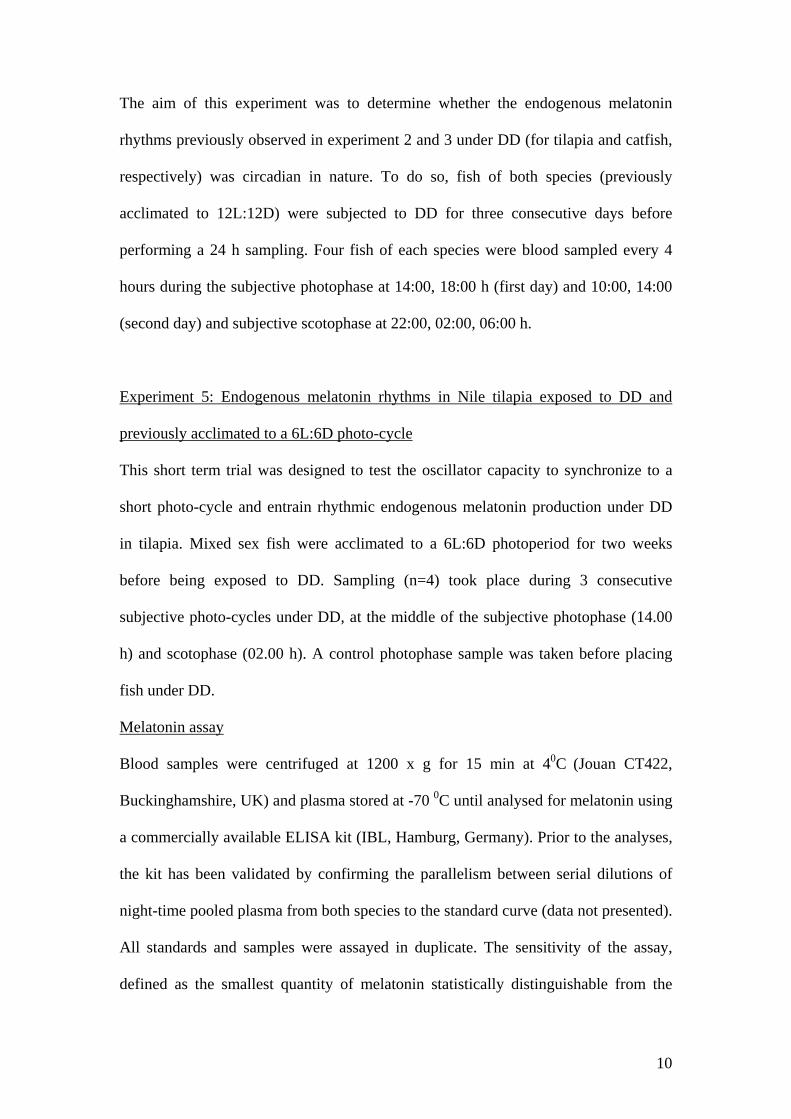
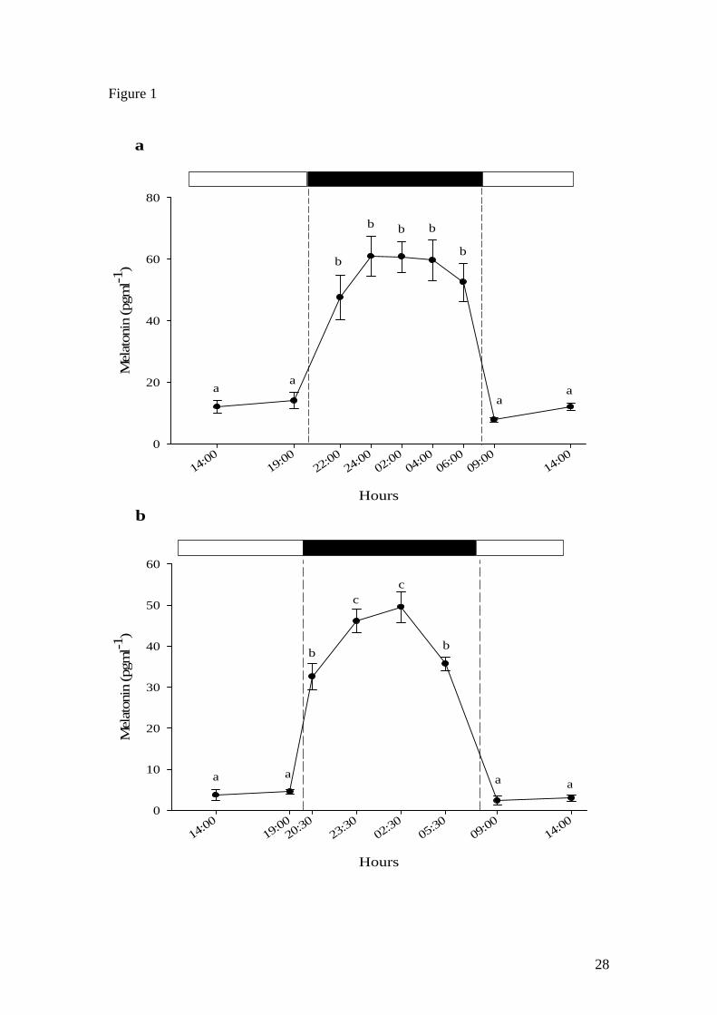
A clear diel rhythm was observed in Nile tilapia (Fig. 1a) and African catfish (Fig. 1b)
with basal levels (<20 pg ml-1 and <10 pg ml-1, respectively) during the photophase
and high melatonin levels (>45 pg ml-1 and >30 pg ml-1, respectively) during the
scotophase. In tilapia, peak melatonin levels were observed 2 hours after the light was
switched off and were maintained (plateau) until the last scotophase sampling point
(06:00 h) after which levels returned to basal levels in the following day sampling
points (09:00 and 14:00 h) (Fig. 1a). Similarly, melatonin levels in catfish remained
basal during the photophase (14:00 and 19:00 h), then significantly increased 30
minutes after the start of the scotophase (20:30 h) to peak 3 hours (23:30 h) later and
remain high at the following sampling point (02:30 h) (Fig. 1b). Thereafter, melatonin
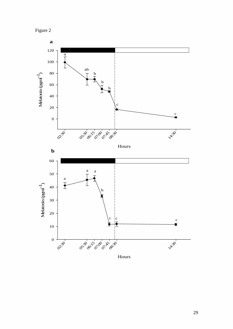
12
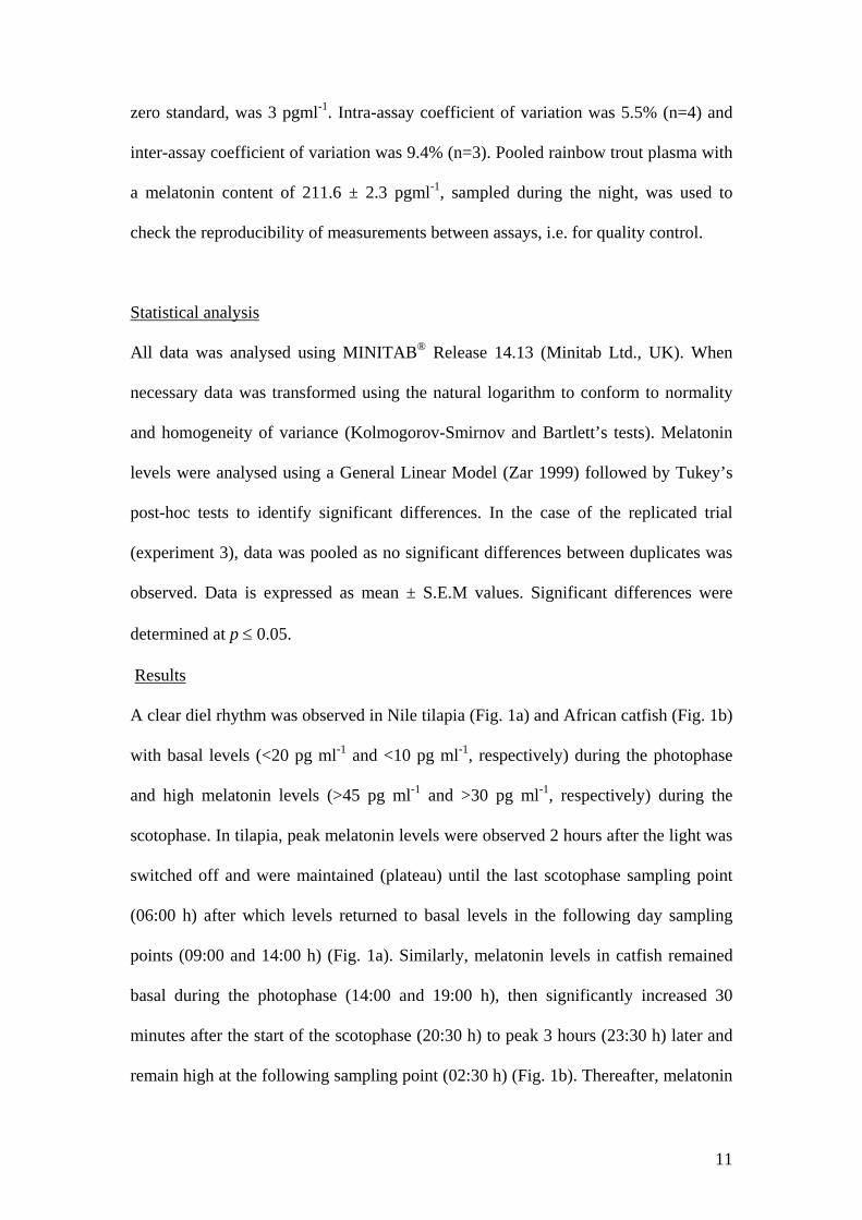
significantly decreased at the last scotophase sampling point (05:30 h) before
returning to basal photophase levels after the light was switched on (08:30 and 11:30
h). Scotophase melatonin levels (sampling points from 02:30 h to 07:45 h) in Nile
tilapia were significantly reduced although basal levels were only reached after the
onset of photophase at 08:30 h and 14:30 h (Fig. 2a). On the other hand, catfish
melatonin levels were shown to decrease significantly during the scotophase 1 hour
prior to the start of the photophase (07:00 h) and reached basal levels shortly before
the start of the photophase at 08:00 h (Fig. 2b).
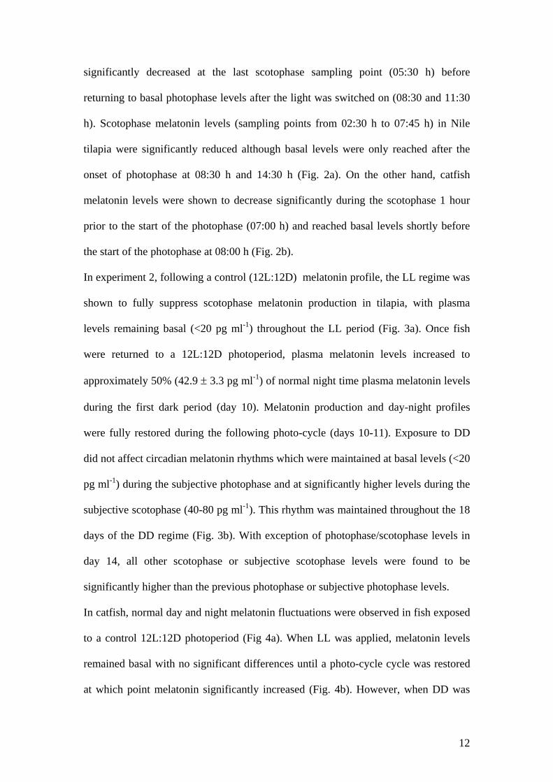
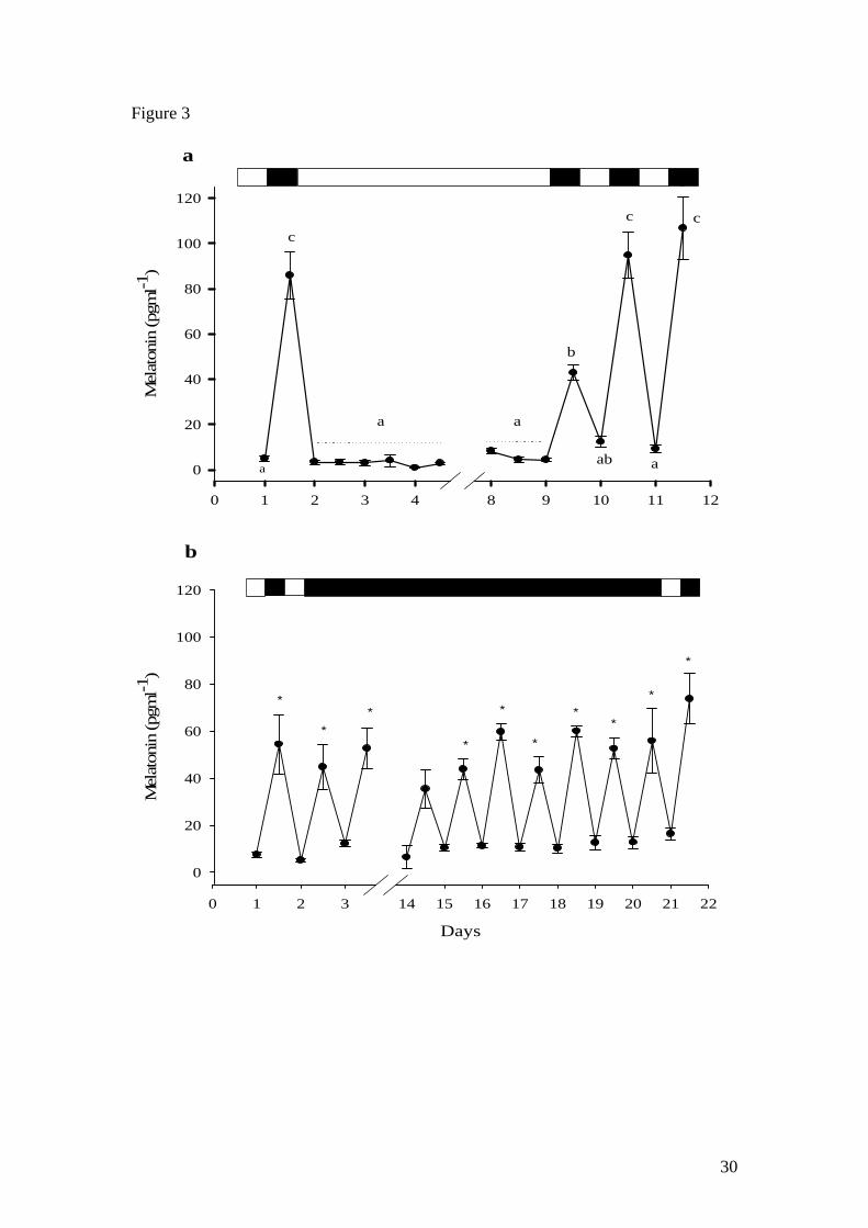
In experiment 2, following a control (12L:12D) melatonin profile, the LL regime was
shown to fully suppress scotophase melatonin production in tilapia, with plasma
levels remaining basal (<20 pg ml-1) throughout the LL period (Fig. 3a). Once fish
were returned to a 12L:12D photoperiod, plasma melatonin levels increased to
approximately 50% (42.9 ± 3.3 pg ml-1) of normal night time plasma melatonin levels
during the first dark period (day 10). Melatonin production and day-night profiles
were fully restored during the following photo-cycle (days 10-11). Exposure to DD
did not affect circadian melatonin rhythms which were maintained at basal levels (<20
pg ml-1) during the subjective photophase and at significantly higher levels during the
subjective scotophase (40-80 pg ml-1). This rhythm was maintained throughout the 18
days of the DD regime (Fig. 3b). With exception of photophase/scotophase levels in
day 14, all other scotophase or subjective scotophase levels were found to be
significantly higher than the previous photophase or subjective photophase levels.
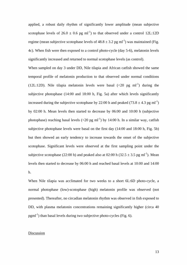
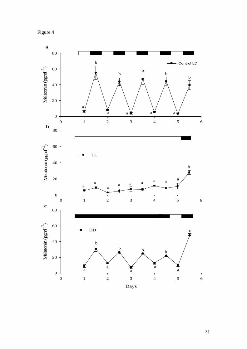
In catfish, normal day and night melatonin fluctuations were observed in fish exposed
to a control 12L:12D photoperiod (Fig 4a). When LL was applied, melatonin levels
remained basal with no significant differences until a photo-cycle cycle was restored
at which point melatonin significantly increased (Fig. 4b). However, when DD was
13
applied, a robust daily rhythm of significantly lower amplitude (mean subjective
scotophase levels of 26.0 ± 0.6 pg ml-1) to that observed under a control 12L:12D
regime (mean subjective scotophase levels of 48.8 ± 3.2 pg ml-1) was maintained (Fig.
4c). When fish were then exposed to a control photo-cycle (day 5-6), melatonin levels
significantly increased and returned to normal scotophase levels (as control).
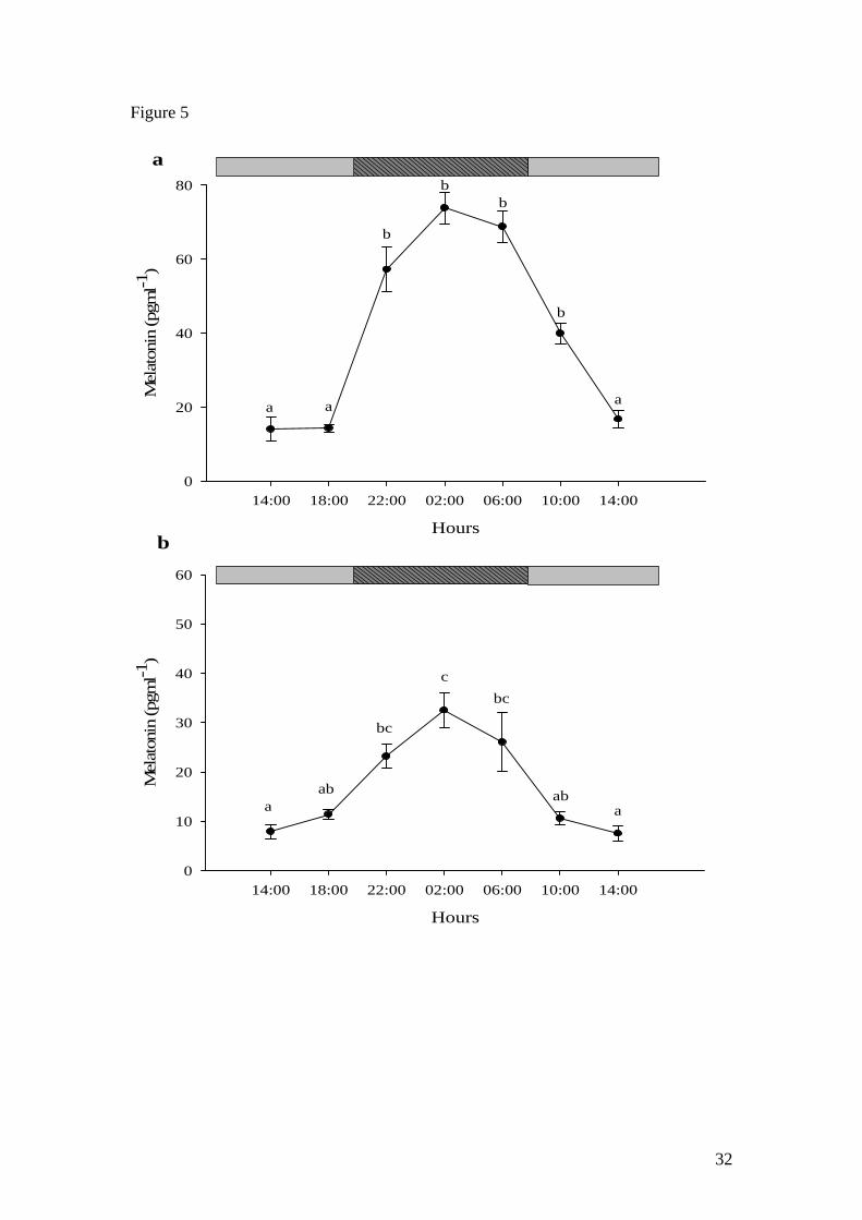
When sampled on day 3 under DD, Nile tilapia and African catfish showed the same
temporal profile of melatonin production to that observed under normal conditions
(12L:12D). Nile tilapia melatonin levels were basal (<20 pg ml-1) during the
subjective photophase (14:00 and 18:00 h, Fig. 5a) after which levels significantly
increased during the subjective scotophase by 22:00 h and peaked (73.8 ± 4.3 pg ml-1)
by 02:00 h. Mean levels then started to decrease by 06:00 and 10:00 h (subjective
photophase) reaching basal levels (<20 pg ml-1) by 14:00 h. In a similar way, catfish
subjective photophase levels were basal on the first day (14:00 and 18:00 h, Fig. 5b)
but then showed an early tendency to increase towards the onset of the subjective
scotophase. Significant levels were observed at the first sampling point under the
subjective scotophase (22:00 h) and peaked also at 02:00 h (32.5 ± 3.5 pg ml-1). Mean
levels then started to decrease by 06:00 h and reached basal levels at 10:00 and 14:00
h.
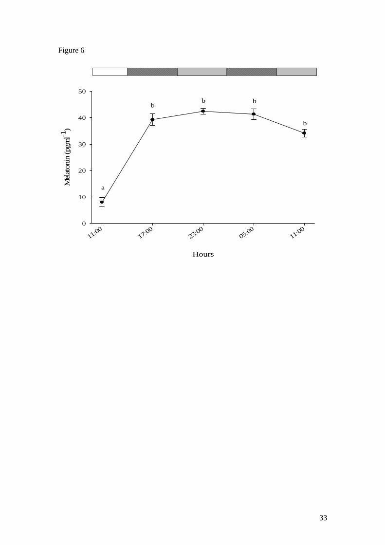
When Nile tilapia was acclimated for two weeks to a short 6L:6D photo-cycle, a
normal photophase (low)-scotophase (high) melatonin profile was observed (not
presented). Thereafter, no circadian melatonin rhythm was observed in fish exposed to
DD, with plasma melatonin concentrations remaining significantly higher (circa 40
pgml-1) than basal levels during two subjective photo-cycles (Fig. 6).
Discussion
14
Seasonal breeders rely on environmental factors such as photoperiod to synchronize
their physiology (Arendt 1998; Pevet 2003). In teleosts, this has been thoroughly
documented in temperate species which are exposed to marked seasonal changes of
day-length and temperature (Falcon et al. 2007). Recently, a number of reproductive
and growth performance studies have shown that tropical teleosts such as Nile tilapia
and African catfish can also be responsive to photoperiodic changes (Campos-
Mendoza et al. 2004; Almazan-Rueda et al. 2004; Biswas et al. 2005; Rad et al.
2006). However, importantly, the role of melatonin and circadian endogenous
rhythms in these tropical species had not been previously studied.
The present results firstly showed a similar diel plasma melatonin profile in Nile
tilapia and African catfish to that previously reported in most vertebrate species
(Reiter 1988; Cassone 1990; Mayer et al. 1997; Pavlidis et al. 1999; Hardeland et al.
2006; Migaud et al. 2006). Although still to be demonstrated in fish, these typical
photophase (low) - scotophase (high) circadian plasma melatonin fluctuations may
provide both species, as in other vertebrates, with daily and calendar time which
entrain the endogenous time keeping system. Interestingly, the present study showed a
significant decrease in plasma melatonin levels in both species, more so in catfish
which reached basal day levels before the start of the photophase. These observations
clearly suggest the involvement of a clock controlled system of melatonin secretion
which is capable of anticipating the next photophase period. If so, the output
(melatonin) of this system is likely to be regulated by arylalkylamine-N-
acetyltransferase (AANAT) or hydroxyindole-O-methyl-transferase (HIOMT)
synthesis at the transcriptional and/or translational level as previously suggested in
other species (Klein et al. 1997; Falcon et al. 2001; Appelbaum et al. 2005). Indeed,
light has been shown to regulate the expression of several circadian clock genes (i.e.
15
Per2) and photoreceptor conserved elements (PCEs) that are capable of regulating E-
box and promoter regions of genes such as AANAT which will ultimately affect the
rate and production of melatonin synthesis (Klein et al. 1997; Zordan et al. 2001;
Pando et al. 2001; Appelbaum and Gothilf 2006).
In order to further characterise the circadian control of melatonin production in tilapia,
melatonin rhythms under constant photic conditions and its entrainment were
investigated. Constant photoperiod (i.e. LL/DD) has commonly been used to describe
rhythmic melatonin production in many vertebrates species including teleosts (Gern
and Greenhouse 1988; Falcon et al. 1989; Kezuka et al. 1989; Okimoto and Stetson
1999b; Migaud et al. 2006; Takemura et al. 2006). In the current study constant LL
regimes resulted in a clear suppression of day-night plasma melatonin rhythms in both
species, as already reported in many other fish species (Falcon et al. 2007).
Interestingly, following 7-8 days under LL, normal rhythmic melatonin production
was restored during the second night period in fish exposed to day-night cycles. The
initial surge (50% of full night-time melatonin) might be explained by a
desensitisation of the melatonin production system after having been suppressed for
seven complete days under LL. On the other hand, when exposed to DD, a strong
endogenous melatonin rhythm was maintained for 18 days (duration of DD exposure)
in tilapia previously acclimatised to a 12L:12D photoperiod. A similar endogenous
rhythm was also able to sustain itself for at least 4 days (duration of DD exposure) in
African catfish. Importantly, results also demonstrated that melatonin rhythm in both
species exposed to DD for three days was circadian (i.e. cycling over approximately
24 hours). However, from these trials it is not possible to determine whether there was
a phase-shift in the circadian rhythm later on as a single sampling point was
performed in the middle of the subjective photophase/scotophase throughout the DD
16
trials. Furthermore, if full amplitude of melatonin oscillations were shown in tilapia
under DD, melatonin levels during mid subjective scotophase (02:00 h) were only 50-
60 % of normal scotophase levels under 12L:12D in catfish. It is difficult at this stage
to explain these findings however, it is possible that the oscillator driving the
endogenous melatonin production rhythm might be ‘desenzitised’ during prolonged
exposure to DD resulting in a near 50% output signal. These results raise interesting
questions as to whether these robust clock controlled melatonin rhythms may
eventually dampen and/or free run or continue.
In support to the present results, many in vitro studies suggested that intrapineal
oscillators exist in fish (Bolliet et al. 1996) with the exception of salmonids (Iigo et al.
2007) in which the pineal gland would either not contain such oscillators or these
would not control melatonin production (still to be demonstrated). However, in vivo
data on endogenous melatonin rhythms are clearly lacking in fish with only very few
species studied (Randall et al. 1995; Pavlidis et al. 1999; Kazimi and Cahill 1999;
Bayarri et al. 2004b; Migaud et al. 2006; Vera et al. 2007; Oliveira et al. 2007). A
recent in vivo study performed on temperate fish species (sea bass, Atlantic salmon
and Atlantic cod) has shown that when acclimatised to a 12L:12D photoperiod at two
different temperatures (10 and 180C) and thereafter exposed to DD, no circadian
endogenous melatonin rhythm was maintained, with levels remaining as high as
during night-time (Migaud et al. unpublished). These results obtained in sea bass and
Atlantic cod appear to not support previous in vitro findings obtained in the same
species (Bolliet et al. 1996; Ron 2004) in which endogenous melatonin rhythms from
isolated pineal glands were reported although rearing temperatures were different than
in the in vivo trials (24 and 20-220C for sea bass and cod respectively). This could
therefore highlight the importance of studying the circadian system as a whole (in
17
vivo). Finally, it could be hypothesized that such strong endogenous rhythms in
tropical species may reflect an adaptation to the rather steady photic environment they
inhabit as compared to the strong seasonal variations experienced by temperate
species. Two main differences were observed between tilapia and catfish; a clearer
melatonin anticipation to photic changes and a reduced melatonin amplitude under
DD as compared to 12L:12D in catfish. It is difficult at this stage to determine
whether these differences are related to specific behavioural adaptations.
The present studies also reported interesting results on the entrainment of the
endogenous system to short photo-cycles. Indeed, when acclimated to a short 6L:6D
photoperiod, no melatonin rhythms were observed under DD in tilapia with levels
remaining high although only two subjective photo-cycles have been studied. This
suggests that, irrespective of its location (pineal, retina and/or brain), the endogenous
melatonin oscillator was either a) not able to entrain to such short photo-cycles or b)
able to entrain but the coupling with the output (melatonin) became dissociated
resulting in constant high plasma melatonin levels when subsequently exposed to DD.
Further studies with longer exposure to DD are needed to determine if free running
melatonin rhythms occur as observed in humans (Foster and Kreitzman 2005).
In the last decade or so understanding of the molecular bases of circadian clock
mechanisms has substantially progressed and has been shown to involve
transcriptional and translational feedback loops involving a highly conserved set of
“clock genes” across vertebrates (Iuvone et al. 2005). It could thus be hypothesised
that this circadian clockwork requires a minimal time/lapse of integration of
environmental signals for the gene expression of the positive and negative
components (transcriptional/translational factors) and the synthesis and activation of
the final products (protein, metabolite and molecular signals) to ultimately entrain the
18
physiology of the animal. Thus, according to this hypothesis, the 6L:6D photoperiod
under which fish were acclimatized in this study could be too short for the circadian
clock to entrain an endogenous melatonin rhythm output that would continue under
DD conditions, as observed in fish previously acclimatised to a 12L:12D photoperiod.
These results could thus explain why eggs from broodstock subjected to the same
6L:6D photoperiod were shown to be not viable as compared to control 12L:12D
(Biswas et al. 2005). However, these preliminary results can only suggest that the
entrainment of the melatonin rhythm may have been affected by the previous
acclimation to 6L:6D as only four samplings over two subjective 6L:6D photo-cycles
were performed. In order to conclude that the entrainment is truly disturbed, further
studies in a range of teleosts species raised under various short photo-cycles and
sampled over longer periods under DD are needed to confirm such a hypothesis and
determine the critical minimal period required for the system to be entrained.
Taken together these results further enhance our knowledge of light perception and
circadian rhythmicity in tropical teleosts and show the potential for these species to
become interesting models in chronobiology. Irrespective of their localisation which
still needs to be determined, these studies have demonstrated the presence of circadian
melatonin oscillators which can anticipate daily photic changes and maintain strong
circadian rhythmic melatonin production under darkness. However, the results have
shown that short photo-cycles appear to disrupt these endogenous melatonin rhythms,
possibly by affecting the transcriptional-translational feedback loops of the circadian
clock which might not be able to entrain over such short periods. Further studies are
needed to confirm this hypothesis and better characterise the circadian axis in fish.
Acknowledgements

19
This study was possible through the financial support from CONACyT (Consejo
Nacional de Ciencia y Tecnologia, Mexico), the Saudi Arabian government and the
University of Stirling.
20
References
Almazan-Rueda P, Schrama JW, Verreth JAJ. (2004). Behavioural responses under
different feeding methods and light regimes of the African catfish (Clarias
gariepinus) juveniles. Aquaculture 231:347-359.
Appelbaum L, Anzulovich A, Baler R, Gothilf Y. (2005). Homeobox-clock protein
interaction in zebrafish: A shared mechanism for pineal-specific and circadian
gene expression. J. Biol. Chem. 280:11544-11551.
Appelbaum L, Gothilf Y. (2006). Mechanism of pineal-specific gene expression: The
role of E-box and photoreceptor conserved elements. Mol. Cell. Endocrinol.
252:27-33.
Appelbaum S, McGeer JC. (1998). Effect of diet and light regime growth and survival
of African catfish (Clarias gariepinus) larvae and early juveniles. Aquacult.
Nutr. 4:157-164.
Arendt J. (1998). Melatonin and the pineal gland: influence on mammalian seasonal
and circadian physiology. Rev. Reprod. 3:13-22.
Bayarri MJ, Madrid JA, Sanchez-Vazquez FJ. (2002). Influence of light intensity,
spectrum and orientation on sea bass plasma and ocular melatonin. J. Pineal
Res. 32:34-40.
Bayarri MJ, Rol de Lama MA, Madrid JA, Sanchez-Vasquez FJ. (2003). Both pineal
and lateral eyes are needed to sustain daily circulating melatonin rhythms in sea
bass. Brain Res. 969:175-182.
Bayarri MJ, Garcia-Allegue R, Lopez-Olmeda JF, Madrid JA, Sanchez-Vazquez FJ.
(2004a). Circadian melatonin release in vitro by European sea bass pineal. Fish
Physiol. Biochem. 30:87-89.
Bayarri MJ, Munoz-Cueto JA, Lopez-Olmeda JF, Vera LM, Rol de Lama MA,
Madrid JA, Sanchez-Vazquez FJ. (2004b). Daily locomotor activity and
melatonin rhythms in Senegal sole (Solea senegalensis). Physiol. Behav.
81:577-583.
Biswas AK, Morita T, Yoshizaki G, Maita M, Takeuchi T. (2005). Control of
reproduction in Nile tilapia Oreochromis niloticus (L.) by photoperiod
manipulation. Aquaculture 243:229-239.
Bolliet V, Ali MA, Lapointe FJ, Falcon J. (1996). Rhythmic melatonin secretion in
different teleost species: an in vitro study. J. Comp. Physiol. B 165:677-683.
21
Bromage N, Roberts JR. (1995). Broodstock Management and Egg and Larval
Quality. Edinburgh, UK: Blackwell Science.
Cahill GM. (1996). Circadian regulation of melatonin production in cultured zebrafish
pineal and retina. Brain Res. 708:177-181.
Cahill G. (2002). Clock mechanisms in zebrafish. Cell Tissue Res. 309:27-34.
Campos-Mendoza A, McAndrew BJ, Coward K, Bromage N. (2004). Reproductive
response of Nile tilapia (Oreochromis niloticus) to photoperiodic manipulation;
effects on spawning periodicity, fecundity and egg size. Aquaculture 231:299-
314.
Carr AJ, Tamai TK, Young LC, Ferrer V, Dekens MP, Whitmore D. (2006). Light
reaches the very heart of the zebrafish clock. Chronobiol. Int. 23:91-100.
Cassone VM. (1990). Effects of melatonin on vertebrate circadian systems. Trends
Neurosci. 13:457-464.
Ekstrom P, Miessl H. (2003). Evolution of photosensory pineal organs in new light:
the fate of neuroendocrine receptors. Phil. Trans. R. Soc. Lond. B 358:1679-
1700.
Falcon J, Marmillon JB, Claustrat B, Collin JP. (1989). Regulation of melatonin
secretion in a photoreceptive pineal organ: an in vitro study in the pike. J.
Neurosci. 9:1943-1950.
Falcon J. (1999). Cellular circadian clocks in the pineal. Prog. Neurobiol. 58:121-162.
Falcon J, Galarneau KM, Weller JL, Ron B, Chen G, Coon SL, Klein DC. (2001).
Regulation of arylalkylamine N-acetyltransferase-2 (AANAT2, EC 2.3.1.87) in
the fish pineal organ: Evidence for a role of proteasomal proteolysis.
Endocrinology 142:1804-1813.
Falcon J, Besseau L, Sauzet Sa, Boeuf G. (2007). Melatonin effects on the
hypothalamo-pituitary axis in fish. Trends Endocrinol. Metab. 18:81-88.
Foster R, Kreitzman L. (2005). Rhythms of Life. London, UK: Profile Book.
Foulkes NS, Whitmore D, Sassone-Corsi P. (1997). Rhythmic transcription: The
molecular basis of circadian melatonin synthesis. Biol. Cell 89:487-494.
Fukada Y, Okano T. (2002). Circadian clock system in the pineal gland. Mol.
Neurobiol. 25:19-30.
Gern WA, Greenhouse SS. (1988). Examination of in vitro melatonin secretion from
superfused trout (Salmo gairdneri) pineal organs maintained under diel
illumination or continuous darkness. Gen. Comp. Endocrinol. 71:163-174.
22
Hardeland R, Pandi-Perumal SR, Cardinali DP. (2006). Melatonin. Int. J. Biochem.
Cell Biol. 38:313-316.
Herzog ED, Tosini G. (2001). The mammalian circadian clock shop. Sem. Cell Dev.
Biol. 12:295-303.
Holzberg D, Albrecht U. (2003). The circadian clock: a manager of biochemical
processes within the organism. J. Neuroendocrinol. 15:339-343.
Iigo M, Kezuka H, Aida K, Hanyu I. (1991). Circadian rhythms of melatonin
secretion from superfused goldfish (Carassius auratus) pineal glands in vitro.
Gen. Comp. Endocrinol. 83:152-158.
Iigo M, Kitamura S, Ikuta K, Sanchez-Vazquez FJ, Ohtani-Kaneko R, Hara M, Hirata
K, Tabata M, Aida K. (1998). Regulation by light and darkness of melatonin
Secretion from the superfused masu salmon (Oncorhynchus masou) pineal
organ. Biol. Rhythm Res. 29:86-97.
Iigo M, Fujimoto Y, Gunji-Suzuki M, Yokosuka M, Hara M, Ohtani-Kaneko R,
Tabata M, Aida K, Hirata K. (2004). Circadian rhythm of melatonin release
from the photoreceptive pineal organ of a teleost, ayu (Plecoglossus altivelis) in
flow-thorough culture. J. Neuroendocrinol. 16:45-51.
Iigo M, Abe T, Kambayashi S, Oikawa K, Masuda T, Mizusawa K, Kitamura S,
Azuma T, Takagi Y, Aida K, Yanagisawa T. (2007). Lack of circadian
regulation of in vitro melatonin release from the pineal organ of salmonid
teleosts. Gen. Comp. Endocrinol. 154:91-97.
Iuvone PM, Tosini G, Pozdeyev N, Haque R, Klein DC, Chaurasia SS. (2005).
Circadian clocks, clock networks, arylalkylamine N-acetyltransferase, and
melatonin in the retina. Prog. Ret. Eye Res. 24:433-456.
Kazimi N, Cahill GM. (1999). Development of a circadian melatonin rhythm in
embryonic zebrafish. Dev. Brain Res. 117:47-52.
Kezuka H, Aida K, Hanyu I. (1989). Melatonin secretion from goldfish pineal gland
in organ culture. Gen. Comp. Endocrinol. 75:217-221.
Klein DC, Coon SL, Roseboom PH, Weller JL, Bernard M, Gastel JA, Zatz M,
Iuvone PM, Rodriguez IR, Begay V, Falcon J, Cahill GM, Cassone VM, Baler
R. (1997). The melatonin rhythm-generating enzyme: molecular regulation of
serotonin N-acetyltransferase in the pineal gland. Rec. Prog. Horm. Res. 52:307-
356.
23
Lopez-Olmeda JF, Madrid JA, Sanchez-Vasquez FJ. (2006). Light and temperature
cycles as zeitgebers of zebrafish (Danio rerio) circadian activity rhythms.
Chronobiol. Int. 23:537-550.
Malpaux B, Migaud M, Tricoire H, Chemineau P. (2001). Biology of mammalian
photoperiodism and the critical role of the pineal gland and melatonin. J. Biol.
Rhythms 16:336-347.
Mayer I, Bornestaf C, Borg B. (1997). Melatonin in non-mammalian vertebrates:
Physiological role in reproduction? Comp. Biochem. Physiol. A 118:515-531.
Menaker M, Moreira LF, Tosini G. (1997). Evolution of circadian organization in
vertebrates. Braz. J. Med. Biol. Res. 30:305-313.
Migaud H, Davie A, Martinez-Chavez CC, Al-Khamees S. (2007). Evidence for
differential photic regulation of pineal melatonin synthesis in teleosts. J. Pineal
Res. 43:327-335
Migaud H, Taylor JF, Taranger GL, Davie A, Cerda-Reverter JM, Carrillo M, Hansen
T, Bromage N. (2006). A comparative ex vivo and in vivo study of day and night
perception in teleosts species using the melatonin rhythm. J. Pineal Res. 41:42-
52.
Okimoto DK, Stetson MH. (1999a). Properties of the melatonin-generating system of
the sailfin molly, Poecilia velifera. Gen. Comp. Endocrinol. 114:293-303.
Okimoto DK, Stetson MH. (1999b). Presence of an intrapineal circadian oscillator in
the teleostean family Poeciliidae. Gen. Comp. Endocrinol. 114:304-312.
Oliveira C, Ortega A, Lopez-Olmeda J, Vera LM, Sanchez-Vasquez FJ. (2007).
Influence of constant light and darkness, light intensity, and light spectrum on
plasma melatonin rhythms in Senegal sole. Chronobiol. Int. 24:615-627.
Pando MP, Pinchak AB, Cermakian N, Sassone-Corsi P. (2001). A cell-based system
that recapitulates the dynamic light-dependent regulation of the vertebrate clock.
PNAS 98:10178-10183.
Pavlidis M, Greenwood L, Paalavuo M, Molsa H, Laitinen JT. (1999). The effect of
photoperiod on diel rhythms in serum melatonin, cortisol, glucose, and
electrolytes in the common Dentex, Dentex dentex. Gen. Comp. Endocrinol.
113:240-250.
Pevet P. (2003). Melatonin: from seasonal to circadian signal. J. Neuroendocrinol.
15:422-426.
24
Rad F, Bozaoglu S, Ergene Gozukara S, Karahan A, Kurt G. (2006). Effects of
different long-day photoperiods on somatic growth and gonadal development in
Nile tilapia (Oreochromis niloticus L.). Aquaculture 255:292-300.
Randall CF, Bromage N, Thorpe JE, Miles MS, Muir JS. (1995). Melatonin rhythms
in Atlantic salmon (Salmo salar) maintained under natural and out-of-phase
photoperiods. Gen. Comp. Endocrinol. 98:73-86.
Reiter RJ. (1988). Comparative aspects of pineal melatonin rhythms in mammals.
Anim. Plant Sci. 1:111-116.
Ridha MT, Cruz EM. (2000). Effect of light intensity and photoperiod on Nile tilapia
Oreochromis niloticus L. seed production. Aquacult. Res. 31:609-617.
Ron B. (2004). In vitro melatonin rhythm reveals a clock controlled pineal in the
European sea bass, Dicentrarchus labrax. Israeli J. Aquaculture 56:281-285.
Simonneaux V, Ribelayga C. (2003). Generation of the melatonin endocrine message
in mammals: A review of the complex regulation of melatonin synthesis by
norepinephrine, peptides, and other pineal transmitters. Pharmacol. Rev.
55:325-395.
Stehle JH, von Gall C, Korf H-W. (2003). Melatonin: A clock-output, a clock-input.
J. Neuroendocrinol. 15:383-389.
Takemura A, Ueda S, Hiyakawa N, Nikaido Y. (2006). A direct influence of
moonlight intensity on changes in melatonin production by cultured pineal
glands of the golden rabbitfish, Siganus guttatus. J. Pineal Res. 40:236-241.
Touitou Y, Smolensky MH., Portaluppi F. (2006). Ethics, standards and procedures
in human and animal research in chronobiology. Chronobiol. Int. 23:1083-1096.
Vallone D, Lahiri K, Dickmeis T, Foulkes NS. (2005). Zebrafish cell clocks feel the
heat and see the light! Zebrafish 2:171-187.
Vera LM, De Oliveira C, Lopez-Olmeda JF, Ramos J, Mananos E, Madrid JA,
Sanchez-Vazquez FJ. (2007). Seasonal and daily plasma melatonin rhythms and
reproduction in Senegal sole kept under natural photoperiod and natural or
controlled water temperature. J. Pineal Res. 43:50-55.
Vígh B, Manzano MJ, Zádori A, Frank CL, Lukáts A, Röhlich P, Szél A, Dávid C.
(2002). Nonvisual photoreceptors of the deep brain, pineal organs and retina.
Histol. Histopathol. 17:555-590.
25
Zachmann A, Ali MA, Falcon J. (1992a). Melatonin and its effects in fishes: an
overview. In Ali MA. (Ed) Rhythms in Fishes. London: Plenum Publishing
Corporation, pp. 149.
Zachmann A, Falcon J, KniJff SCM, Bolliet V, Ali MA. (1992b). Effects of
photoperiod and temperature on rhythmic melatonin secretion from the pineal
organ of the White Sucker (Catostomus commersoni) in vitro. Gen. Comp.
Endocrinol. 86:26-33.
Zar JH. (1999). Biostatistical Analysis. Upper Saddle River, New Jersey, USA:
Prentice Hall.
Ziv L, Gothilf Y. (2006). Period2 expression pattern and its role in the development of
the pineal circadian clock in zebrafish. Chronobiol. Int. 23:101-112.
Zordan MA, Rosato E, Piccin A, Foster R. (2001). Photic entrainment of the circadian
clock: from Drosophila to mammals. Sem. Cell . Dev. Biol. 12:317-328.
26
Figure Legends
Figure 1. Circadian plasma melatonin profile in a) Nile tilapia (n=10) and b) African
catfish (n=5). All values shown are mean ± SEM. Letters indicate significant (p<0.05)
difference between sample points. Open and filled boxes indicate photophase and
scotophase respectively.
Figure 2. Anticipatory decrease of plasma melatonin levels prior to light onset in a)
Nile tilapia (n=5) and b) African catfish (n=5). Values shown are mean ± SEM.
Letters indicate significant (p<0.05) difference between sampling points. Open and
filled boxes indicate photophase and scotophase respectively.
Figure 3. Plasma melatonin profile of Nile tilapia subjected to a) LL and b) DD
regimes. Values shown are mean ± SEM (n= 4). Letters indicate significant (p<0.05)
difference between sampling points. Symbols (*) indicate significant differences with
previous sampling point. Open and filled boxes indicate photophase and scotophase
respectively.
Figure 4. Plasma melatonin profile of African catfish subjected to a) 12L:12D, b) LL
and c) DD regimes. Values shown are mean ± SEM (n= 6). Letters indicate significant
(p<0.05) difference between sampling points. Open and filled boxes indicate
photophase and scotophase respectively.
Figure 5. Circadian plasma melatonin profile in a) Nile tilapia and b) African catfish
on the third day under DD. All values shown are mean ± SEM (n=4). Letters indicate
significant (p<0.05) differences between sample points. Grey boxes indicate the
subjective photophase periods and darker filled box the subjective scotophase period.
Figure 6. Melatonin levels of Nile tilapia acclimatised for two weeks to a 6L:6D
photoperiod and sampled before and under DD for 2 following subjective day-night
cycles. Each point represents the mean ± SEM (n= 4). Letters indicate significant

27
(p<0.05) differences between sampling points. Open and filled boxes indicate
photophase and scotophase respectively.
28
Figure 1
Hours
14:0019:00
22:0024:00
02:0004:00
06:0009:00
14:00
Mel
aton
in (p
gml-1
)
0
20
40
60
80
aa
b
b b b
b
aa
a
Hours
14:0019:00
20:3023:30
02:3005:30
09:0014:00
Mel
aton
in (p
gml-1
)
0
10
20
30
40
50
60
a a a a
bb
cc
b
29
Figure 2
Hours
02:30
05:30
06:15
07:00
07:45
08:30
14:30
Mel
aton
in (p
gml-1
)
0
20
40
60
80
100
120
Hours
02:30
05:30
06:15
07:00
07:45
08:30
14:30
Mel
aton
in (p
gml-1
)
0
10
20
30
40
50
60
a
b
a
a a
b
c c c
a
abb
b
b
c
c
30
Figure 3
0 1 2 3 4 8 9 10 11 12
Mel
aton
in (p
gml-1
)
0
20
40
60
80
100
120*
a
a
a a
b
ab
cc
c
a
Days
0 1 2 3 14 15 16 17 18 19 20 21 22
Mel
aton
in (p
gml-1
)
0
20
40
60
80
100
120
b
*
**
*
*
*
**
*
*
31
Figure 4
0 1 2 3 4 5 6
Mel
aton
in (p
gml-1
)
0
20
40
60
80
LL
0 1 2 3 4 5 6
Mel
aton
in (p
gml-1
)
0
20
40
60
80
Control LD
Days
0 1 2 3 4 5 6
Mel
aton
in (p
gml-1
)
0
20
40
60
80
DD
a
b
c
a
b
a
b
a
b
a
b
a
b
a
b
a
b
a
b
a
b
a
c
aa
a a a a a aa
b
32
Figure 5
Hours
14:00 18:00 22:00 02:00 06:00 10:00 14:00
Mel
aton
in (p
gml-1
)
0
20
40
60
80
Hours
14:00 18:00 22:00 02:00 06:00 10:00 14:00
Mel
aton
in (p
gml-1
)
0
10
20
30
40
50
60
a
b
a
b
a
bb
b
a
aab
bc
cbc
aba
33
Figure 6
Hours
11:0017:00
23:0005:00
11:00
Mel
aton
in (p
gml-1
)
0
10
20
30
40
50
bb b
b
a
Related Documents