Clinician’s Guide National Osteoporosis Foundation Bone Source Knowledge. Competence. Results. Developed by the National Osteoporosis Foundation to Prevention and Treatment of Osteoporosis 2014 Issue, Version 1 Released April 1, 2014 © National Osteoporosis Foundation (NOF). All rights reserved.

Welcome message from author
This document is posted to help you gain knowledge. Please leave a comment to let me know what you think about it! Share it to your friends and learn new things together.
Transcript
CHAPTER 1 ■ OSTEOPOROSIS: WHAT IT IS AND WHO IS AT RISK 5
Clinician’s Guide
NationalOsteoporosis Foundation
Bone SourceKnowledge. Competence. Results.
Developed by the National Osteoporosis Foundation
to Prevention and Treatment of Osteoporosis
2014 Issue, Version 1
Released April 1, 2014
© National Osteoporosis Foundation (NOF). All rights reserved.
2014 CLINICIAN’S GUIDE TO PREVENTION AND TREATMENT OF OSTEOPOROSIS
1
2014 Issue, Version 1 Release Date: April 1, 2014
The Clinician’s Guide to Prevention and Treatment of Osteoporosis was developed by an expert committee of the National Osteoporosis Foundation (NOF) in collaboration with a multi-specialty council of medical experts in the field of bone health convened by NOF. Readers are urged to consult current prescribing information on any drug, device or procedure discussed in this publication. Funding for the development of this document was provided by NOF. National Osteoporosis Foundation 1150 17th St., NW, Suite 850, Washington, DC 20036 © National Osteoporosis Foundation (NOF). All rights reserved. Single copies of this document, in its entirety or in part, may be printed and distributed for educational use. Unmodified excerpts of the text may be used for educational presentations and publications, in electronic form and in print, provided the source is attributed to the National Osteoporosis Foundation. No part of this Guide may be reproduced with modified content without advance written permission from the National Osteoporosis Foundation. To request permission, contact NOF at (202) 721-6348. BoneSource® is a registered trademark of the National Osteoporosis Foundation. Suggested citation: National Osteoporosis Foundation. Clinician’s Guide to Prevention and Treatment of Osteoporosis. Washington, DC: National Osteoporosis Foundation; 2014.
2014 CLINICIAN’S GUIDE TO PREVENTION AND TREATMENT OF OSTEOPOROSIS
2
2014 Clinician’s Guide Update Committee and Organizations Represented Felicia Cosman, MD, Chair, National Osteoporosis Foundation
Robert Lindsay, MD, PhD, Co-chair, National Osteoporosis Foundation Meryl S. LeBoff, MD, National Osteoporosis Foundation Suzanne Jan de Beur, MD, American Society for Bone and Mineral Research Bobo Tanner, MD, International Society for Clinical Densitometry NOF acknowledges the following individuals for their prior contribution to the project:
Members of the 2008 Clinician’s Guide Development Committee Bess Dawson-Hughes, MD, Chair, National Osteoporosis Foundation Robert Lindsay, MD, PhD, Co-chair, National Osteoporosis Foundation Sundeep Khosla, MD, National Osteoporosis Foundation L. Joseph Melton, III, MD, National Osteoporosis Foundation Anna N.A. Tosteson, ScD, National Osteoporosis Foundation Murray Favus, MD, American Society for Bone and Mineral Research Sanford Baim, MD, International Society for Clinical Densitometry
Consultants to the 2014 Update Committee
Karl Insogna, MD Douglas Kiel, MD, MPH E. Michael Lewiecki, MD Harold Rosen, MD
John Schousboe, MD
National Osteoporosis Foundation Staff
Susan Randall, MSN, FNP-BC, Senior Director, Science and Education
The Clinician’s Guide to Prevention and Treatment of Osteoporosis 2014 is endorsed by the following organizations*:
American Academy of Pain Medicine (AAPM) American Society for Bone and Mineral Research (ASBMR) International Society for Clinical Densitometry (ISCD)
*The list of endorsing organizations will be updated as additional endorsements are received.
2014 CLINICIAN’S GUIDE TO PREVENTION AND TREATMENT OF OSTEOPOROSIS
3
CLINICIAN’S GUIDE TO PREVENTION AND TREATMENT OF OSTEOPOROSIS Disclosure Policy It is the policy of NOF to ensure balance, independence, objectivity and scientific rigor in all sponsored publications and programs. NOF requires disclosure of any financial interest or any other relationship that the Committee members have with the manufacturer(s) of any commercial product(s). All contributors to this publication have disclosed any real or apparent interest that may have direct bearing on the subject matter of this program. All potential conflicts have been resolved to the satisfaction of the NOF. Medication information included in this guidance follows the U.S. Food and Drug Administration (FDA)-approved label. Note to Readers The Clinician’s Guide is designed to serve as a basic reference on the prevention, diagnosis and treatment of osteoporosis in the U.S. It is based largely on updated information on the U.S. incidence and cost of osteoporosis. For those with low bone mass (in whom more than 50 percent of fractures occur), the Guide incorporates an analysis from the World Health Organization (WHO) that assesses 10-year fracture risk. The Guide utilizes an economic analysis prepared by the National Osteoporosis Foundation in collaboration with the WHO (Dr. J. Kanis), the American Society for Bone and Mineral Research, the International Society for Clinical Densitometry and a broad multidisciplinary coalition of clinical experts, to indicate the level of risk at which it is cost-effective to consider treatment. This information combined with clinical judgment and patient preference should lead to more appropriate testing and treatment of those at risk of fractures attributable to osteoporosis. This Guide is intended for use by clinicians as a tool for clinical decision-making in the treatment of individual patients. While the guidance for testing and risk evaluation comes from an analysis of available epidemiological and economic data, the treatment information in this Guide is based mainly on evidence from randomized, controlled clinical trials. The efficacy (fracture risk reduction) of medications was used in the analysis to help define levels of risk at which it is cost effective to treat. The Guide addresses postmenopausal women and men age 50 and older. The Guide also addresses secondary causes of osteoporosis which should be excluded by clinical evaluation. Furthermore, all individuals should follow the universal recommendations for osteoporosis prevention and management outlined in this Guide. The recommendations herein reflect an awareness of the cost and effectiveness of both diagnostic and treatment modalities. Some effective therapeutic options that would be prohibitively expensive on a population basis might remain a valid choice in individual cases under certain circumstances. This Guide cannot and should not be used to govern health policy decisions about reimbursement or availability of services. Its recommendations are not intended as rigid standards of practice. Clinicians should tailor their recommendations and, in consultation with their patients, devise individualized plans for osteoporosis prevention and treatment.
2014 CLINICIAN’S GUIDE TO PREVENTION AND TREATMENT OF OSTEOPOROSIS
4
Update Information This document was originally written and approved in 1999. It was updated in 2008, 2010 and 2013. The 2013 issues of NOF’s Clinician’s Guide contain updated guidance on vertebral fracture assessment and the use of biochemical markers of bone turnover, as well as updated information on calcium, vitamin D and medications. The 2013 issue was first released on March 1, 2013 with additional edits released in April 2013 (2013 version 2) and November 2013 (2013 version 3). The current version (2014) was released April 1, 2014. The 2014 version of the Clinician’s Guide stresses the importance of screening vertebral imaging to diagnose asymptomatic vertebral fractures; provides updated information on calcium, vitamin D and osteoporosis medications; addresses duration of treatment; and includes an expanded discussion on the utility of biochemical markers of bone turnover and an evaluation of secondary causes of osteoporosis.
2014 CLINICIAN’S GUIDE TO PREVENTION AND TREATMENT OF OSTEOPOROSIS
5
Contents 1. EXECUTIVE SUMMARY .............................................................................................................................. 7
Synopsis of Major Recommendations to the Clinician ............................................................................. 7
2. OSTEOPOROSIS: IMPACT AND OVERVIEW .............................................................................................. 10
Scope of the Problem .............................................................................................................................. 10
Medical Impact ....................................................................................................................................... 10
Economic Toll .......................................................................................................................................... 11
3. BASIC PATHOPHYSIOLOGY ...................................................................................................................... 12
4. APPROACH TO THE DIAGNOSIS AND MANAGEMENT OF OSTEOPOROSIS ............................................. 14
Risk Assessment ...................................................................................................................................... 14
Clinical Evaluation ................................................................................................................................... 17
Diagnosis ................................................................................................................................................. 18
Bone Mineral Density Measurement and Classification ......................................................................... 18
Who Should be Tested? .......................................................................................................................... 20
Vertebral Imaging ................................................................................................................................... 21
Biochemical Markers of Bone Turnover ................................................................................................. 22
Use of WHO Fracture Risk Algorithm (FRAX®) in the U.S. ...................................................................... 22
5. UNIVERSAL RECOMMENDATIONS FOR ALL PATIENTS ............................................................................ 25
Adequate Intake of Calcium and Vitamin D ............................................................................................ 25
Regular Weight-Bearing and Muscle-Strengthening Exercise ................................................................ 27
Fall Prevention ........................................................................................................................................ 27
6. PHARMACOLOGIC THERAPY ................................................................................................................. 229
Who Should Be Considered for Treatment? ........................................................................................... 29
U.S. FDA-Approved Drugs for Osteoporosis ........................................................................................... 31
Bisphosphonates ................................................................................................................................. 31
Calcitonin ............................................................................................................................................ 33
Estrogen/Hormone Therapy (ET/HT) .................................................................................................. 34
Estrogen Agonist/Antagonist (formerly known as SERMs): Raloxifene .............................................. 35
Tissue Selective Estrogen Complex: Conjugated estrogens/ bazedoxifene (Conjugated estrogens paired with estrogen agonist/antagonist) .......................................................................................... 36
2014 CLINICIAN’S GUIDE TO PREVENTION AND TREATMENT OF OSTEOPOROSIS
6
Parathyroid Hormone: Teriparatide ................................................................................................... 37
Receptor Activator of Nuclear Factor kappa-B (RANK) Ligand (RANKL)/ RANKL Inhibitor: Denosumab ............................................................................................................................................................ 37
Sequential and Combination Therapy .................................................................................................... 38
Duration of Treatment ............................................................................................................................ 38
Monitoring Effectiveness of Treatment .................................................................................................. 39
Implementation of Fracture Liaison Service (FLS) Secondary Fracture Prevention Programs ............... 41
7. PHYSICAL MEDICINE AND REHABILITATION ........................................................................................... 42
CONCLUSIONS AND REMAINING QUESTIONS ............................................................................................ 43
GLOSSARY.................................................................................................................................................... 45
KEY REFERENCES ......................................................................................................................................... 49
2014 CLINICIAN’S GUIDE TO PREVENTION AND TREATMENT OF OSTEOPOROSIS
7
1. EXECUTIVE SUMMARY
Osteoporosis is a silent disease until it is complicated by fractures— fractures that occur following minimal trauma or, in some cases, with no trauma. Fractures are common and place an enormous medical and personal burden on the aging individuals who suffer them and take a major economic toll on the nation. Osteoporosis can be prevented, diagnosed and treated before fractures occur. Importantly, even after the first fracture has occurred, there are effective treatments to decrease the risk of further fractures. Prevention, detection and treatment of osteoporosis should be a mandate of primary care providers. Since NOF first published the Guide in 1999, it has become increasingly clear that many patients are not being given appropriate information about prevention and many patients are not receiving appropriate testing to diagnose osteoporosis or establish osteoporosis risk. Most importantly, many patients who have osteoporosis-related fractures are not being diagnosed with osteoporosis and are not receiving any of the FDA-approved, effective therapies. This Guide offers concise recommendations regarding prevention, risk assessment, diagnosis and treatment of osteoporosis in postmenopausal women and men age 50 and older. It includes indications for bone densitometry and fracture risk thresholds for intervention with pharmacologic agents. The absolute risk thresholds at which consideration of osteoporosis treatment is recommended were guided by a cost-effectiveness analysis. Synopsis of Major Recommendations to the Clinician Recommendations apply to postmenopausal women and men age 50 and older. Universal recommendations:
• Counsel on the risk of osteoporosis and related fractures. • Advise on a diet that includes adequate amounts of total calcium intake (1,000 mg per day for men
50-70; 1,200 mg per day for women 51 and older and men 71 and older), incorporating dietary supplements if diet is insufficient.
• Advise on vitamin D intake (800-1,000 IU per day), including supplements if necessary for individuals age 50 and older.
• Recommend regular weight-bearing and muscle-strengthening exercise to improve agility, strength, posture and balance; maintain or improve bone strength; and reduce the risk of falls and fractures.
• Assess risk factors for falls and offer appropriate modifications (e.g. home safety assessment, balance training exercises, correction of vitamin D insufficiency, avoidance of CNS depressant medications, careful monitoring of anti-hypertensive medication and visual correction when needed).
• Advise on cessation of tobacco smoking and avoidance of excessive alcohol intake. • Measure height annually, preferably with a wall mounted stadiometer.
2014 CLINICIAN’S GUIDE TO PREVENTION AND TREATMENT OF OSTEOPOROSIS
8
Diagnostic assessment: • BMD testing should be performed:
o In women age 65 and older and men age 70 and older, o In postmenopausal women and men above age 50-69, based on risk factor profile. o In post menopausal women and men over age 50 who have had an adult age fracture, to
diagnose and determine degree of osteoporosis. o At DXA facilities using accepted quality assurance measures.
• Vertebral imaging should be performed:
o In all women age 70 and older and all men age 80 and older if BMD T-score is < -1.0 at the spine,
total hip or femoral neck. o In women age 65 to 69 and men age 70 to 79 if BMD T-score is < -1.5 at the spine, total hip or
femoral neck. o In postmenopausal women and men age 50 and older with specific risk factors:
Low trauma fracture during adulthood (age 50+) Historical height loss of 1.5 inches or more (4 cm)
Defined as the difference between the current height and peak height at age 20 Prospective height loss of 0.8 inches or more (2 cm)
Defined as the difference between the current height and a previously documented height measurement
Recent or ongoing long term glucocorticoid treatment o If bone density testing is not available, vertebral imaging may be considered based on age alone.
• Check for secondary causes of osteoporosis.
• Biochemical markers of bone turnover can aid in risk assessment and serve as an additional
monitoring tool when treatment is initiated. Monitoring patients: • Perform BMD testing one to two years after initiating medical therapy for osteoporosis and every
two years thereafter. o More frequent BMD testing may be warranted in certain clinical situations. o The interval between repeat BMD screenings may be longer for patients without major risk
factors and who have an initial T-score in the normal or upper low bone mass range. • Biochemical markers can be repeated to determine if treatment is producing expected effect.
2014 CLINICIAN’S GUIDE TO PREVENTION AND TREATMENT OF OSTEOPOROSIS
9
Pharmacologic treatment recommendations: After appropriate evaluation: • Initiate pharmacologic treatment in those with hip or vertebral (clinical or asymptomatic) fractures.
• Initiate therapy in those with T-scores < -2.5 at the femoral neck, total hip or lumbar spine by dual-
energy x-ray absorptiometry (DXA). • Initiate treatment in postmenopausal women and men age 50 and older with low bone mass (T-
score between -1.0 and -2.5, osteopenia) at the femoral neck, total hip or lumbar spine by DXA and a 10-year hip fracture probability > 3 percent or a 10-year major osteoporosis-related fracture probability > 20 percent based on the U.S.-adapted WHO absolute fracture risk model (FRAX®; www.NOF.org and www.shef.ac.uk/FRAX).
• Current FDA-approved pharmacologic options for osteoporosis are bisphosphonates (alendronate,
ibandronate, risedronate and zoledronic acid), calcitonin, estrogen agonist/antagonist (raloxifene), estrogens and/or hormone therapy, tissue-selective estrogen complex (conjugated estrogens/bazedoxifene), parathyroid hormone 1-34 (teriparatide) and RANK ligand inhibitor (denosumab).
• No pharmacologic therapy should be considered indefinite in duration. After the initial treatment
period, which depends on the pharmacologic agent, a comprehensive risk assessment should be performed. There is no uniform recommendation that applies to all patients and duration decisions need to be individualized.
• In adults age 50 and older, after a fracture, institute appropriate risk assessment and treatment
measures for osteoporosis as indicated. An alternative in many centers is a fracture liaison service (FLS) program where patients with recent fractures may be referred for care coordination and transition management.
2014 CLINICIAN’S GUIDE TO PREVENTION AND TREATMENT OF OSTEOPOROSIS
10
2. OSTEOPOROSIS: IMPACT AND OVERVIEW
Scope of the Problem Osteoporosis is the most common bone disease in humans, representing a major public health problem as outlined in Bone Health and Osteoporosis: A Report of the Surgeon General (2004).1 It is characterized by low bone mass, deterioration of bone tissue and disruption of bone architecture, compromised bone strength and an increase in the risk of fracture. According to the WHO diagnostic classification, osteoporosis is defined by BMD at the hip or lumbar spine that is less than or equal to 2.5 standard deviations below the mean BMD of a young-adult reference population. Osteoporosis is a risk factor for fracture just as hypertension is for stroke. The risk of fractures is highest in those with the lowest BMD; however, the majority of fractures occur in patients with low bone mass rather than osteoporosis, because of the large number of individuals with bone mass in this range. Osteoporosis affects an enormous number of people, of both sexes and all races, and its prevalence will increase as the population ages. Based on data from the National Health and Nutrition Examination Survey III (NHANES III), NOF has estimated that more than 9.9 million Americans have osteoporosis and an additional 43.1 million have low bone density.2 About one out of every two Caucasian women will experience an osteoporosis-related fracture at some point in her lifetime, as will approximately one in five men.1 Although osteoporosis is less frequent in African Americans, those with osteoporosis have the same elevated fracture risk as Caucasians.
Medical Impact Fractures and their complications are the relevant clinical sequelae of osteoporosis. The most common fractures are those of the vertebrae (spine), proximal femur (hip) and distal forearm (wrist). However, most fractures in older adults are due at least in part to low bone mass, even when they result from considerable trauma. A recent fracture at any major skeletal site in an adult older than 50 years of age should be considered a significant event for the diagnosis of osteoporosis and provides a sense of urgency for further assessment and treatment. The most notable exceptions are those of the fingers, toes, face and skull, which are primarily related to trauma rather than underlying bone strength. Fractures may be followed by full recovery or by chronic pain, disability and death.5 Hip fractures are associated with an 8.4 to 36 percent excess mortality within one year, with a higher mortality in men than in women3; additionally, hip fractures are followed by a 2.5-fold increased risk of future fractures.4 Approximately 20 percent of hip fracture patients require long-term nursing home care, and only 40 percent fully regain their pre-fracture level of independence.1 Although the majority of vertebral fractures are initially clinically silent, these fractures are often associated with symptoms of pain, disability, deformity and mortality.5 Postural changes associated with kyphosis may limit activity, including bending and reaching. Multiple thoracic fractures may result in restrictive lung disease and lumbar fractures may alter abdominal anatomy, leading to constipation, abdominal pain, distention, reduced appetite and premature satiety. Vertebral fractures, whether clinically apparent or silent, are major predictors of future fracture risk, up to 5-fold for subsequent vertebral fracture and 2- to 3-fold for fractures at other sites. Wrist fractures are less disabling but can interfere with some activities of daily living as much as hip or vertebral fractures.

2014 CLINICIAN’S GUIDE TO PREVENTION AND TREATMENT OF OSTEOPOROSIS
11
Pelvic fractures and humerus fractures are also common and contribute to increased morbidity and mortality. Fractures can also cause psychosocial symptoms, most notably depression and loss of self-esteem, as patients grapple with pain, physical limitations, and lifestyle and cosmetic changes.
Economic Toll Annually, two million fractures are attributed to osteoporosis, causing more than 432,000 hospital admissions, almost 2.5 million medical office visits and about 180,000 nursing home admissions in the U.S.1 Medicare currently pays for approximately 80 percent of these fractures, with hip fractures accounting for 72 percent of fracture costs. Due in part to an aging population, the cost of care is expected to rise to $25.3 billion by 2025. 6 Despite the availability of cost effective and well-tolerated treatments to reduce fracture risk, only 23 percent of women age 67 or older who have an osteoporosis-related fracture receive either a bone mineral density test or a prescription for a drug to treat osteoporosis in the six months after the fracture.7
2014 CLINICIAN’S GUIDE TO PREVENTION AND TREATMENT OF OSTEOPOROSIS
12
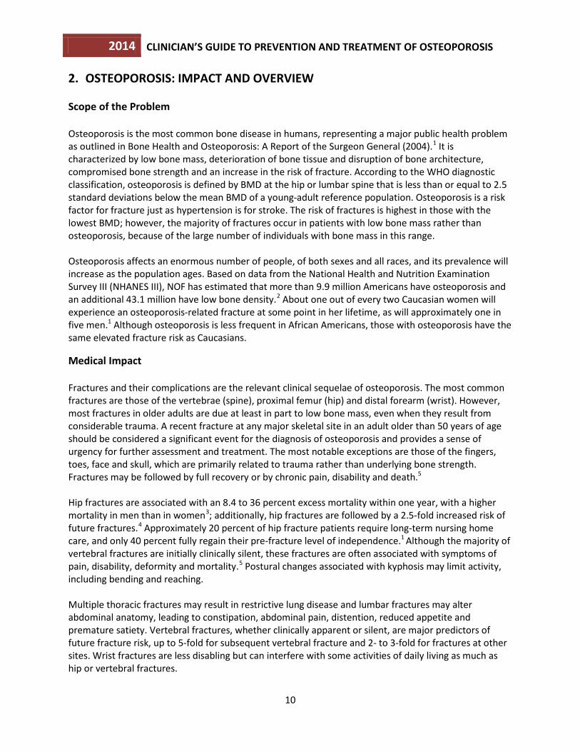
3. BASIC PATHOPHYSIOLOGY Bone mass in older adults equals the peak bone mass achieved by age 18-25 minus the amount of bone subsequently lost. Peak bone mass is determined largely by genetic factors, with contributions from nutrition, endocrine status, physical activity and health during growth.8 The process of bone remodeling that maintains a healthy skeleton may be considered a preventive maintenance program, continually removing older bone and replacing it with new bone. Bone loss occurs when this balance is altered, resulting in greater bone removal than replacement. The imbalance occurs with menopause and advancing age. With the onset of menopause, the rate of bone remodeling increases, magnifying the impact of the remodeling imbalance. The loss of bone tissue leads to disordered skeletal architecture and an increase in fracture risk. Figure 1 shows the changes within cancellous bone as a consequence of bone loss. Individual trabecular plates of bone are lost, leaving an architecturally weakened structure with significantly reduced mass. Increasing evidence suggests that rapid bone remodeling (as measured by biochemical markers of bone resorption or formation) increases bone fragility and fracture risk. FIGURE 1. Micrographs of Normal vs. Osteoporotic Bone9
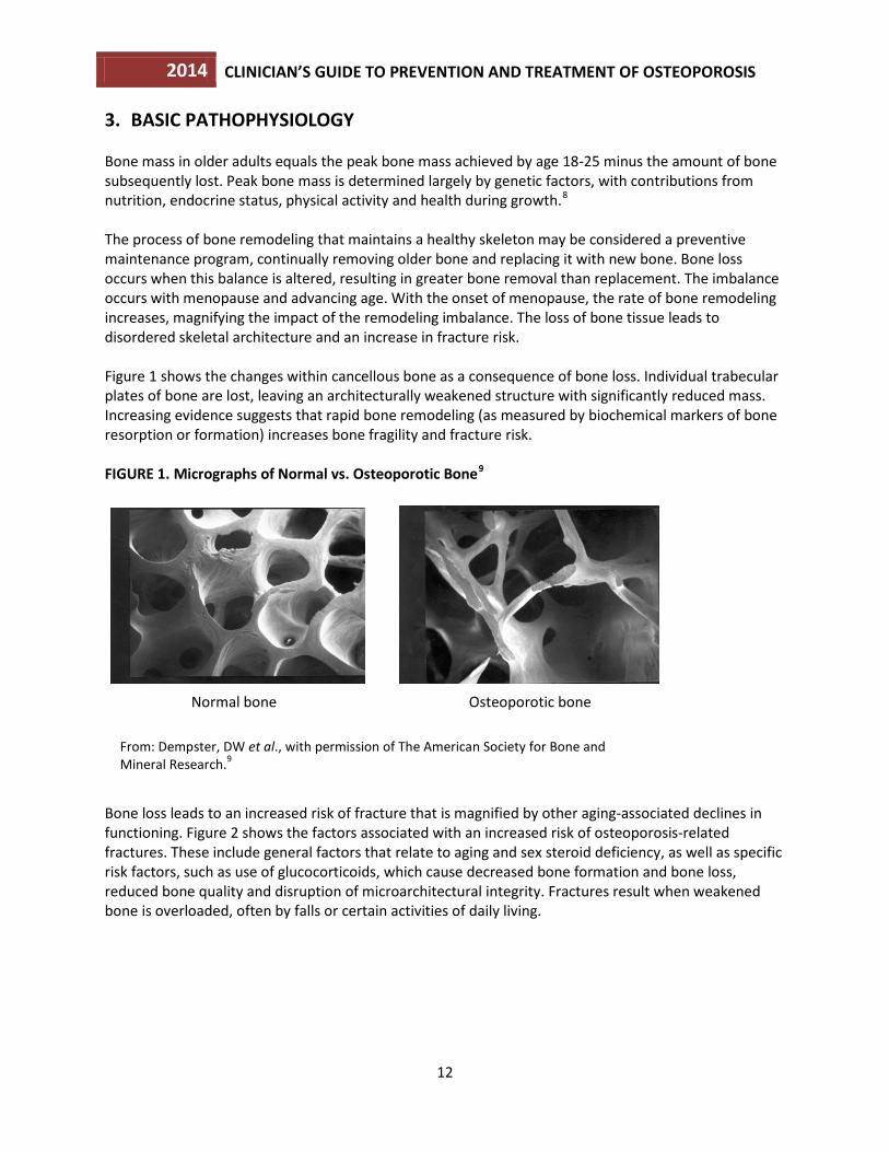
Bone loss leads to an increased risk of fracture that is magnified by other aging-associated declines in functioning. Figure 2 shows the factors associated with an increased risk of osteoporosis-related fractures. These include general factors that relate to aging and sex steroid deficiency, as well as specific risk factors, such as use of glucocorticoids, which cause decreased bone formation and bone loss, reduced bone quality and disruption of microarchitectural integrity. Fractures result when weakened bone is overloaded, often by falls or certain activities of daily living.
From: Dempster, DW et al., with permission of The American Society for Bone and Mineral Research.9
Normal bone Osteoporotic bone
2014 CLINICIAN’S GUIDE TO PREVENTION AND TREATMENT OF OSTEOPOROSIS
13
FIGURE 2. Pathogenesis of Osteoporosis-Related Fractures
From: Cooper C and Melton LJ, with modification.10
2014 CLINICIAN’S GUIDE TO PREVENTION AND TREATMENT OF OSTEOPOROSIS
14
4. APPROACH TO THE DIAGNOSIS AND MANAGEMENT OF OSTEOPOROSIS
NOF recommends a comprehensive approach to the diagnosis and management of osteoporosis. A detailed history and physical examination together with BMD assessment, vertebral imaging to diagnose vertebral fractures, and, when appropriate, the WHO 10-year estimated fracture probability are utilized to establish the individual patient’s fracture risk.11 Therapeutic intervention thresholds are based on NOF’s economic analysis that takes into consideration the cost-effectiveness of treatments and competition for resources in the U.S.12,13 The clinician’s clinical skills and past experience, incorporating the best patient-based research available, is used to determine the appropriate therapeutic intervention. The potential risks and benefits of all osteoporosis interventions should be reviewed with patients and the unique concerns and expectations of individual patients considered in any final therapeutic decision.
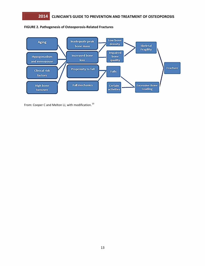
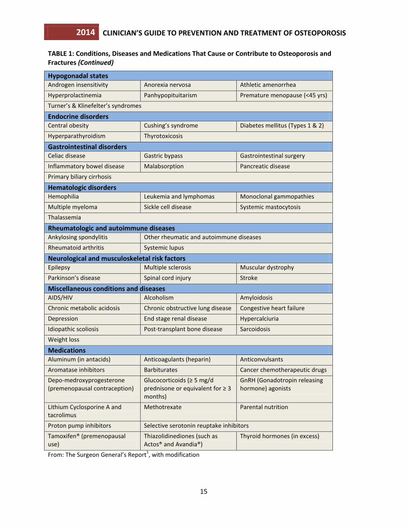
Risk Assessment All postmenopausal women and men age 50 and older should be evaluated for osteoporosis risk in order to determine the need for BMD testing and/or vertebral imaging. In general, the more risk factors that are present, the greater the risk of fracture. Osteoporosis is preventable and treatable, but because there are no warning signs prior to a fracture, many people are not being diagnosed in time to receive effective therapy during the early phase of the disease. Many factors have been associated with an increased risk of osteoporosis-related fracture (Table 1).
TABLE 1: Conditions, Diseases and Medications That Cause or Contribute to Osteoporosis and Fractures
Lifestyle factors
Alcohol abuse Excessive thinness Excess Vitamin A
Frequent falling High salt intake Immobilization
Inadequate physical activity Low calcium intake Smoking (active or passive)
Vitamin D insufficiency
Genetic diseases
Cystic fibrosis Ehlers-Danlos Gaucher’s disease
Glycogen storage diseases Hemochromatosis Homocystinuria
Hypophosphatasia Marfan syndrome Menkes steely hair syndrome
Osteogenesis imperfecta Parental history of hip fracture Porphyria
Riley-Day syndrome
2014 CLINICIAN’S GUIDE TO PREVENTION AND TREATMENT OF OSTEOPOROSIS
15
TABLE 1: Conditions, Diseases and Medications That Cause or Contribute to Osteoporosis and Fractures (Continued)
Hypogonadal states Androgen insensitivity Anorexia nervosa Athletic amenorrhea Hyperprolactinemia Panhypopituitarism Premature menopause (<45 yrs) Turner’s & Klinefelter’s syndromes
Endocrine disorders Central obesity Cushing’s syndrome Diabetes mellitus (Types 1 & 2) Hyperparathyroidism Thyrotoxicosis
Gastrointestinal disorders Celiac disease Gastric bypass Gastrointestinal surgery Inflammatory bowel disease Malabsorption Pancreatic disease Primary biliary cirrhosis
Hematologic disorders Hemophilia Leukemia and lymphomas Monoclonal gammopathies Multiple myeloma Sickle cell disease Systemic mastocytosis Thalassemia
Rheumatologic and autoimmune diseases Ankylosing spondylitis Other rheumatic and autoimmune diseases Rheumatoid arthritis Systemic lupus
Neurological and musculoskeletal risk factors Epilepsy Multiple sclerosis Muscular dystrophy Parkinson’s disease Spinal cord injury Stroke
Miscellaneous conditions and diseases AIDS/HIV Alcoholism Amyloidosis Chronic metabolic acidosis Chronic obstructive lung disease Congestive heart failure Depression End stage renal disease Hypercalciuria Idiopathic scoliosis Post-transplant bone disease Sarcoidosis Weight loss
Medications Aluminum (in antacids) Anticoagulants (heparin) Anticonvulsants Aromatase inhibitors Barbiturates Cancer chemotherapeutic drugs Depo-medroxyprogesterone (premenopausal contraception)
Glucocorticoids (≥ 5 mg/d prednisone or equivalent for ≥ 3 months)
GnRH (Gonadotropin releasing hormone) agonists
Lithium Cyclosporine A and tacrolimus
Methotrexate Parental nutrition
Proton pump inhibitors Selective serotonin reuptake inhibitors Tamoxifen® (premenopausal use)
Thiazolidinediones (such as Actos® and Avandia®)
Thyroid hormones (in excess)
From: The Surgeon General’s Report1, with modification
2014 CLINICIAN’S GUIDE TO PREVENTION AND TREATMENT OF OSTEOPOROSIS
16
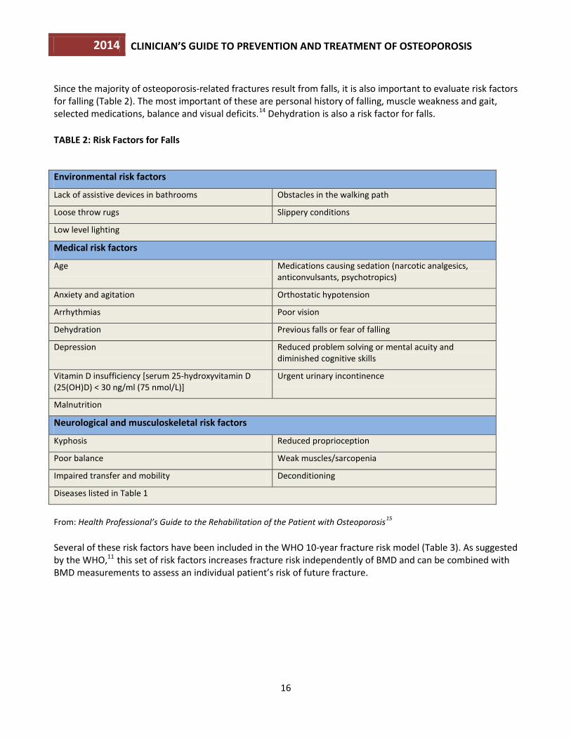
Since the majority of osteoporosis-related fractures result from falls, it is also important to evaluate risk factors for falling (Table 2). The most important of these are personal history of falling, muscle weakness and gait, selected medications, balance and visual deficits.14 Dehydration is also a risk factor for falls. TABLE 2: Risk Factors for Falls
Environmental risk factors
Lack of assistive devices in bathrooms Obstacles in the walking path
Loose throw rugs Slippery conditions
Low level lighting
Medical risk factors
Age Medications causing sedation (narcotic analgesics, anticonvulsants, psychotropics)
Anxiety and agitation Orthostatic hypotension
Arrhythmias Poor vision
Dehydration Previous falls or fear of falling
Depression Reduced problem solving or mental acuity and diminished cognitive skills
Vitamin D insufficiency [serum 25-hydroxyvitamin D (25(OH)D) < 30 ng/ml (75 nmol/L)]
Urgent urinary incontinence
Malnutrition
Neurological and musculoskeletal risk factors
Kyphosis Reduced proprioception
Poor balance Weak muscles/sarcopenia
Impaired transfer and mobility Deconditioning
Diseases listed in Table 1
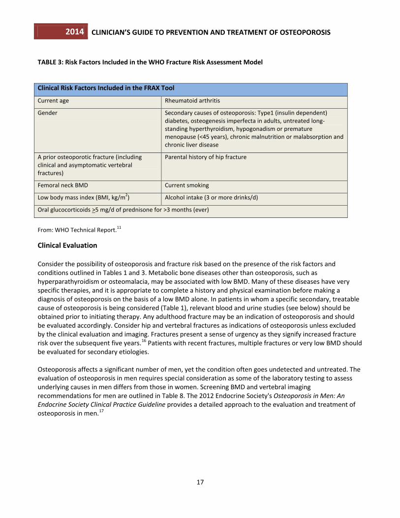
From: Health Professional’s Guide to the Rehabilitation of the Patient with Osteoporosis15 Several of these risk factors have been included in the WHO 10-year fracture risk model (Table 3). As suggested by the WHO,11 this set of risk factors increases fracture risk independently of BMD and can be combined with BMD measurements to assess an individual patient’s risk of future fracture.
2014 CLINICIAN’S GUIDE TO PREVENTION AND TREATMENT OF OSTEOPOROSIS
17
TABLE 3: Risk Factors Included in the WHO Fracture Risk Assessment Model
Clinical Risk Factors Included in the FRAX Tool
Current age Rheumatoid arthritis
Gender Secondary causes of osteoporosis: Type1 (insulin dependent) diabetes, osteogenesis imperfecta in adults, untreated long-standing hyperthyroidism, hypogonadism or premature menopause (<45 years), chronic malnutrition or malabsorption and chronic liver disease
A prior osteoporotic fracture (including clinical and asymptomatic vertebral fractures)
Parental history of hip fracture
Femoral neck BMD Current smoking
Low body mass index (BMI, kg/m2) Alcohol intake (3 or more drinks/d)
Oral glucocorticoids >5 mg/d of prednisone for >3 months (ever)
From: WHO Technical Report.11
Clinical Evaluation Consider the possibility of osteoporosis and fracture risk based on the presence of the risk factors and conditions outlined in Tables 1 and 3. Metabolic bone diseases other than osteoporosis, such as hyperparathyroidism or osteomalacia, may be associated with low BMD. Many of these diseases have very specific therapies, and it is appropriate to complete a history and physical examination before making a diagnosis of osteoporosis on the basis of a low BMD alone. In patients in whom a specific secondary, treatable cause of osteoporosis is being considered (Table 1), relevant blood and urine studies (see below) should be obtained prior to initiating therapy. Any adulthood fracture may be an indication of osteoporosis and should be evaluated accordingly. Consider hip and vertebral fractures as indications of osteoporosis unless excluded by the clinical evaluation and imaging. Fractures present a sense of urgency as they signify increased fracture risk over the subsequent five years.16 Patients with recent fractures, multiple fractures or very low BMD should be evaluated for secondary etiologies. Osteoporosis affects a significant number of men, yet the condition often goes undetected and untreated. The evaluation of osteoporosis in men requires special consideration as some of the laboratory testing to assess underlying causes in men differs from those in women. Screening BMD and vertebral imaging recommendations for men are outlined in Table 8. The 2012 Endocrine Society's Osteoporosis in Men: An Endocrine Society Clinical Practice Guideline provides a detailed approach to the evaluation and treatment of osteoporosis in men.17
2014 CLINICIAN’S GUIDE TO PREVENTION AND TREATMENT OF OSTEOPOROSIS
18
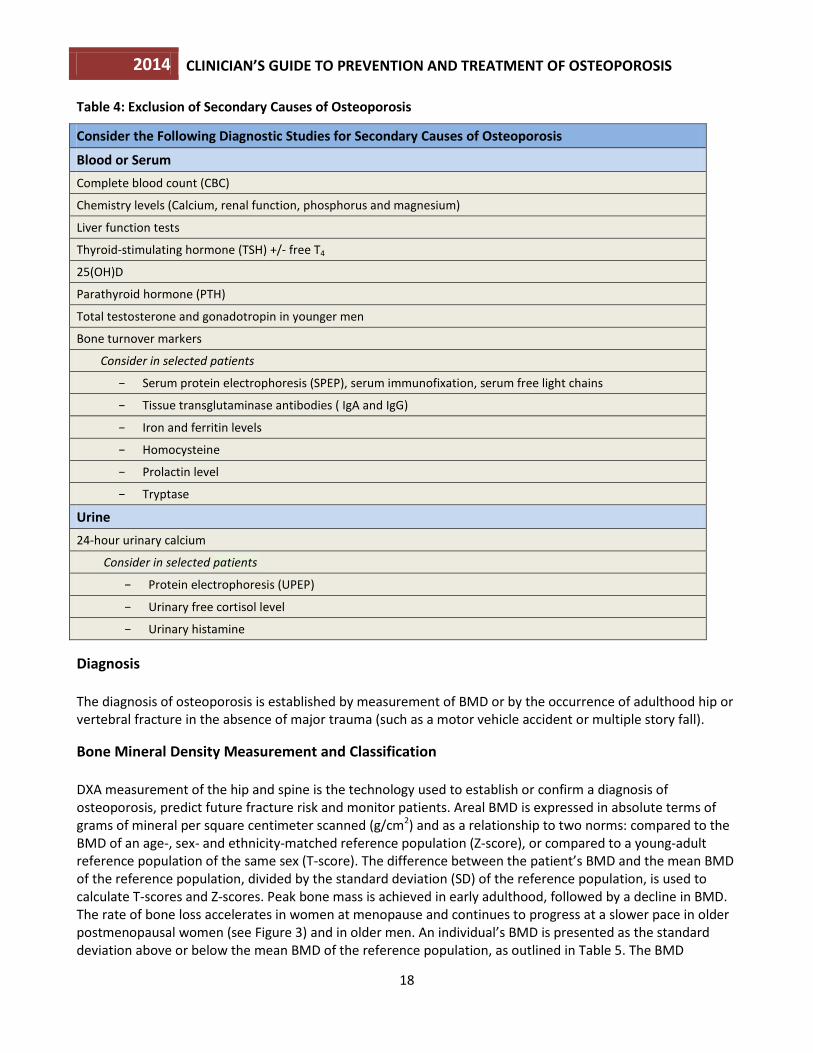
Table 4: Exclusion of Secondary Causes of Osteoporosis
Consider the Following Diagnostic Studies for Secondary Causes of Osteoporosis
Blood or Serum Complete blood count (CBC)
Chemistry levels (Calcium, renal function, phosphorus and magnesium)
Liver function tests
Thyroid-stimulating hormone (TSH) +/- free T4
25(OH)D
Parathyroid hormone (PTH)
Total testosterone and gonadotropin in younger men
Bone turnover markers
Consider in selected patients
− Serum protein electrophoresis (SPEP), serum immunofixation, serum free light chains
− Tissue transglutaminase antibodies ( IgA and IgG)
− Iron and ferritin levels
− Homocysteine
− Prolactin level
− Tryptase
Urine 24-hour urinary calcium
Consider in selected patients
− Protein electrophoresis (UPEP)
− Urinary free cortisol level
− Urinary histamine
Diagnosis The diagnosis of osteoporosis is established by measurement of BMD or by the occurrence of adulthood hip or vertebral fracture in the absence of major trauma (such as a motor vehicle accident or multiple story fall).
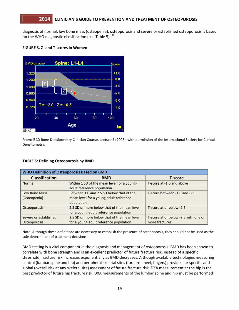
Bone Mineral Density Measurement and Classification DXA measurement of the hip and spine is the technology used to establish or confirm a diagnosis of osteoporosis, predict future fracture risk and monitor patients. Areal BMD is expressed in absolute terms of grams of mineral per square centimeter scanned (g/cm2) and as a relationship to two norms: compared to the BMD of an age-, sex- and ethnicity-matched reference population (Z-score), or compared to a young-adult reference population of the same sex (T-score). The difference between the patient’s BMD and the mean BMD of the reference population, divided by the standard deviation (SD) of the reference population, is used to calculate T-scores and Z-scores. Peak bone mass is achieved in early adulthood, followed by a decline in BMD. The rate of bone loss accelerates in women at menopause and continues to progress at a slower pace in older postmenopausal women (see Figure 3) and in older men. An individual’s BMD is presented as the standard deviation above or below the mean BMD of the reference population, as outlined in Table 5. The BMD
2014 CLINICIAN’S GUIDE TO PREVENTION AND TREATMENT OF OSTEOPOROSIS
19
diagnosis of normal, low bone mass (osteopenia), osteoporosis and severe or established osteoporosis is based on the WHO diagnostic classification (see Table 5). 18 FIGURE 3. Z- and T-scores in Women
From: ISCD Bone Densitometry Clinician Course. Lecture 5 (2008), with permission of the International Society for Clinical Densitometry. TABLE 5: Defining Osteoporosis by BMD WHO Definition of Osteoporosis Based on BMD
Classification BMD T-score Normal Within 1 SD of the mean level for a young-
adult reference population T-score at -1.0 and above
Low Bone Mass (Osteopenia)
Between 1.0 and 2.5 SD below that of the mean level for a young-adult reference population
T-score between -1.0 and -2.5
Osteoporosis 2.5 SD or more below that of the mean level for a young-adult reference population
T-score at or below -2.5
Severe or Established Osteoporosis
2.5 SD or more below that of the mean level for a young-adult reference population
T-score at or below -2.5 with one or more fractures
Note: Although these definitions are necessary to establish the presence of osteoporosis, they should not be used as the sole determinant of treatment decisions. BMD testing is a vital component in the diagnosis and management of osteoporosis. BMD has been shown to correlate with bone strength and is an excellent predictor of future fracture risk. Instead of a specific threshold, fracture risk increases exponentially as BMD decreases. Although available technologies measuring central (lumbar spine and hip) and peripheral skeletal sites (forearm, heel, fingers) provide site-specific and global (overall risk at any skeletal site) assessment of future fracture risk, DXA measurement at the hip is the best predictor of future hip fracture risk. DXA measurements of the lumbar spine and hip must be performed
2014 CLINICIAN’S GUIDE TO PREVENTION AND TREATMENT OF OSTEOPOROSIS
20
by appropriately trained technologists on properly maintained instruments. DXA scans are associated with exposure to trivial amounts of radiation. In postmenopausal women and men age 50 years and older, the WHO diagnostic T-score criteria (normal, low bone mass and osteoporosis) are applied to BMD measurement by central DXA at the lumbar spine and femoral neck.18 BMD measured by DXA at the one-third (33 percent) radius site can be used for diagnosing osteoporosis when the hip and lumbar spine cannot be measured or are unusable or uninterpretable.19 In premenopausal women, men less than 50 years of age and children, the WHO BMD diagnostic classification should not be applied. In these groups, the diagnosis of osteoporosis should not be made on the basis of densitometric criteria alone. The International Society for Clinical Densitometry (ISCD) recommends that instead of T-scores, ethnic or race adjusted Z-scores should be used, with Z-scores of -2.0 or lower defined as either “low bone mineral density for chronological age” or “below the expected range for age” and those above -2.0 being “within the expected range for age.” 19
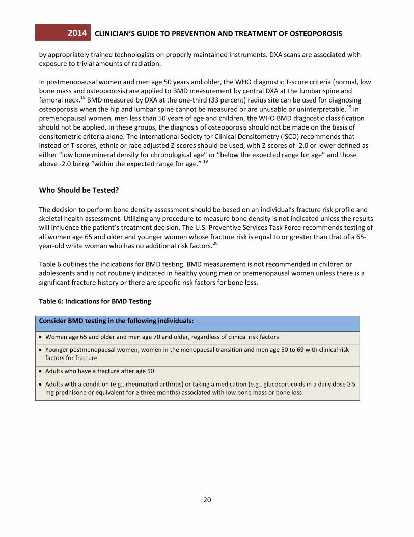
Who Should be Tested? The decision to perform bone density assessment should be based on an individual’s fracture risk profile and skeletal health assessment. Utilizing any procedure to measure bone density is not indicated unless the results will influence the patient’s treatment decision. The U.S. Preventive Services Task Force recommends testing of all women age 65 and older and younger women whose fracture risk is equal to or greater than that of a 65-year-old white woman who has no additional risk factors.20 Table 6 outlines the indications for BMD testing. BMD measurement is not recommended in children or adolescents and is not routinely indicated in healthy young men or premenopausal women unless there is a significant fracture history or there are specific risk factors for bone loss. Table 6: Indications for BMD Testing Consider BMD testing in the following individuals:
• Women age 65 and older and men age 70 and older, regardless of clinical risk factors
• Younger postmenopausal women, women in the menopausal transition and men age 50 to 69 with clinical risk factors for fracture
• Adults who have a fracture after age 50
• Adults with a condition (e.g., rheumatoid arthritis) or taking a medication (e.g., glucocorticoids in a daily dose ≥ 5 mg prednisone or equivalent for ≥ three months) associated with low bone mass or bone loss
2014 CLINICIAN’S GUIDE TO PREVENTION AND TREATMENT OF OSTEOPOROSIS
21
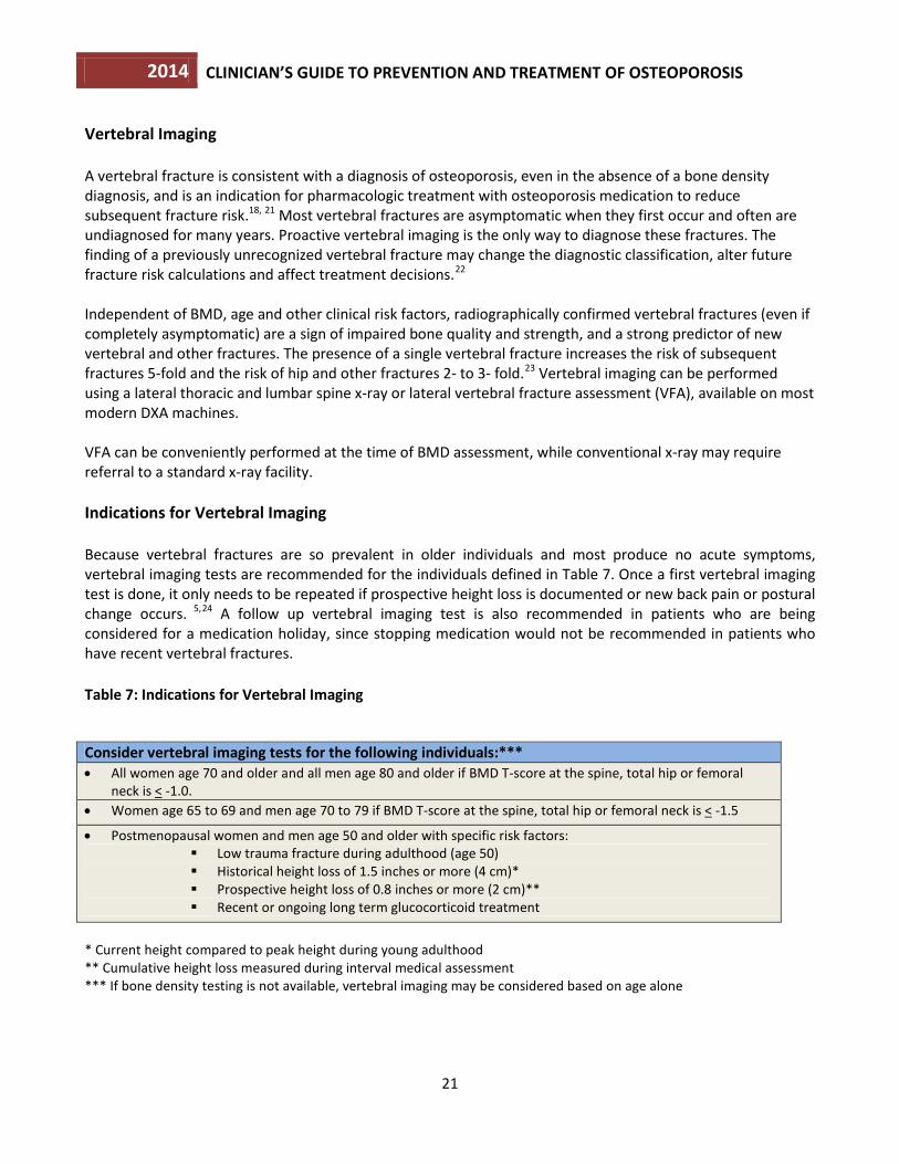
Vertebral Imaging A vertebral fracture is consistent with a diagnosis of osteoporosis, even in the absence of a bone density diagnosis, and is an indication for pharmacologic treatment with osteoporosis medication to reduce subsequent fracture risk.18, 21 Most vertebral fractures are asymptomatic when they first occur and often are undiagnosed for many years. Proactive vertebral imaging is the only way to diagnose these fractures. The finding of a previously unrecognized vertebral fracture may change the diagnostic classification, alter future fracture risk calculations and affect treatment decisions.22 Independent of BMD, age and other clinical risk factors, radiographically confirmed vertebral fractures (even if completely asymptomatic) are a sign of impaired bone quality and strength, and a strong predictor of new vertebral and other fractures. The presence of a single vertebral fracture increases the risk of subsequent fractures 5-fold and the risk of hip and other fractures 2- to 3- fold.23 Vertebral imaging can be performed using a lateral thoracic and lumbar spine x-ray or lateral vertebral fracture assessment (VFA), available on most modern DXA machines. VFA can be conveniently performed at the time of BMD assessment, while conventional x-ray may require referral to a standard x-ray facility. Indications for Vertebral Imaging Because vertebral fractures are so prevalent in older individuals and most produce no acute symptoms, vertebral imaging tests are recommended for the individuals defined in Table 7. Once a first vertebral imaging test is done, it only needs to be repeated if prospective height loss is documented or new back pain or postural change occurs. 5,24 A follow up vertebral imaging test is also recommended in patients who are being considered for a medication holiday, since stopping medication would not be recommended in patients who have recent vertebral fractures. Table 7: Indications for Vertebral Imaging
Consider vertebral imaging tests for the following individuals:*** • All women age 70 and older and all men age 80 and older if BMD T-score at the spine, total hip or femoral
neck is < -1.0. • Women age 65 to 69 and men age 70 to 79 if BMD T-score at the spine, total hip or femoral neck is < -1.5
• Postmenopausal women and men age 50 and older with specific risk factors: Low trauma fracture during adulthood (age 50) Historical height loss of 1.5 inches or more (4 cm)* Prospective height loss of 0.8 inches or more (2 cm)** Recent or ongoing long term glucocorticoid treatment
* Current height compared to peak height during young adulthood ** Cumulative height loss measured during interval medical assessment *** If bone density testing is not available, vertebral imaging may be considered based on age alone
2014 CLINICIAN’S GUIDE TO PREVENTION AND TREATMENT OF OSTEOPOROSIS
22
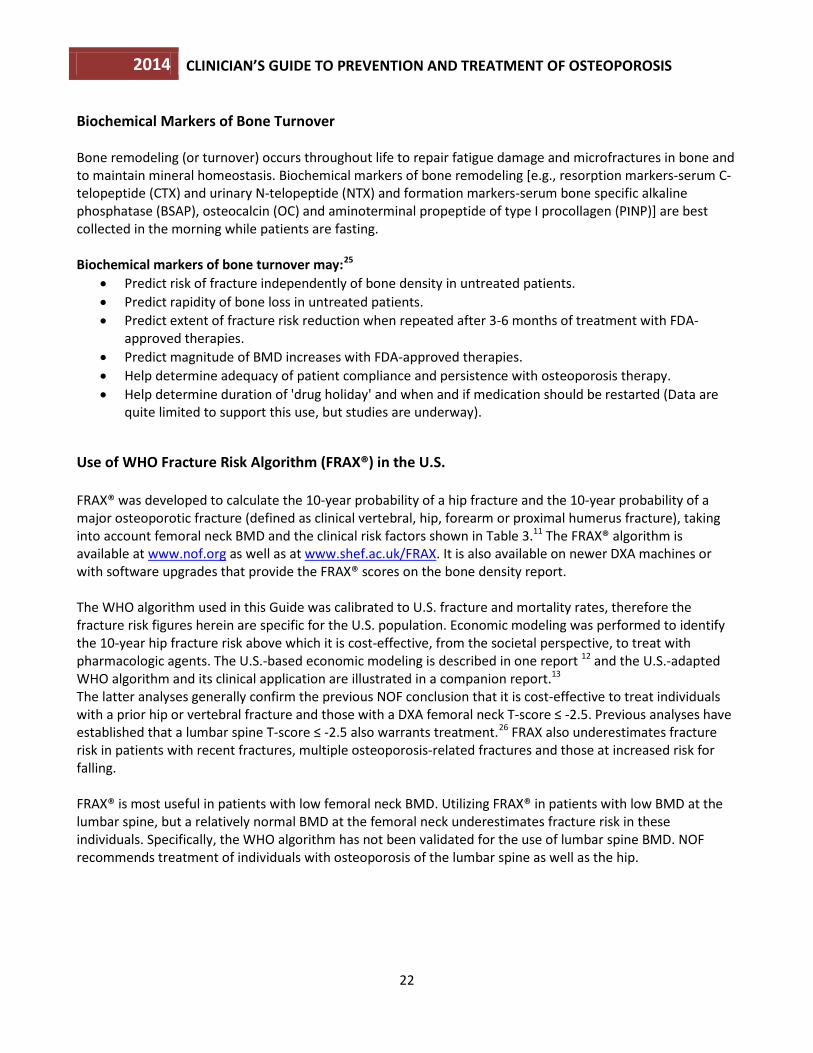
Biochemical Markers of Bone Turnover Bone remodeling (or turnover) occurs throughout life to repair fatigue damage and microfractures in bone and to maintain mineral homeostasis. Biochemical markers of bone remodeling [e.g., resorption markers-serum C-telopeptide (CTX) and urinary N-telopeptide (NTX) and formation markers-serum bone specific alkaline phosphatase (BSAP), osteocalcin (OC) and aminoterminal propeptide of type I procollagen (PINP)] are best collected in the morning while patients are fasting. Biochemical markers of bone turnover may:25
• Predict risk of fracture independently of bone density in untreated patients. • Predict rapidity of bone loss in untreated patients. • Predict extent of fracture risk reduction when repeated after 3-6 months of treatment with FDA-
approved therapies. • Predict magnitude of BMD increases with FDA-approved therapies. • Help determine adequacy of patient compliance and persistence with osteoporosis therapy. • Help determine duration of 'drug holiday' and when and if medication should be restarted (Data are
quite limited to support this use, but studies are underway).
Use of WHO Fracture Risk Algorithm (FRAX®) in the U.S. FRAX® was developed to calculate the 10-year probability of a hip fracture and the 10-year probability of a major osteoporotic fracture (defined as clinical vertebral, hip, forearm or proximal humerus fracture), taking into account femoral neck BMD and the clinical risk factors shown in Table 3.11 The FRAX® algorithm is available at www.nof.org as well as at www.shef.ac.uk/FRAX. It is also available on newer DXA machines or with software upgrades that provide the FRAX® scores on the bone density report. The WHO algorithm used in this Guide was calibrated to U.S. fracture and mortality rates, therefore the fracture risk figures herein are specific for the U.S. population. Economic modeling was performed to identify the 10-year hip fracture risk above which it is cost-effective, from the societal perspective, to treat with pharmacologic agents. The U.S.-based economic modeling is described in one report 12 and the U.S.-adapted WHO algorithm and its clinical application are illustrated in a companion report.13 The latter analyses generally confirm the previous NOF conclusion that it is cost-effective to treat individuals with a prior hip or vertebral fracture and those with a DXA femoral neck T-score ≤ -2.5. Previous analyses have established that a lumbar spine T-score ≤ -2.5 also warrants treatment.26 FRAX also underestimates fracture risk in patients with recent fractures, multiple osteoporosis-related fractures and those at increased risk for falling.
FRAX® is most useful in patients with low femoral neck BMD. Utilizing FRAX® in patients with low BMD at the lumbar spine, but a relatively normal BMD at the femoral neck underestimates fracture risk in these individuals. Specifically, the WHO algorithm has not been validated for the use of lumbar spine BMD. NOF recommends treatment of individuals with osteoporosis of the lumbar spine as well as the hip.
2014 CLINICIAN’S GUIDE TO PREVENTION AND TREATMENT OF OSTEOPOROSIS
23
Application of U.S.-Adapted FRAX® in the U.S. • FRAX® is intended for postmenopausal women and men age 50 and older; it is not intended for use in
younger adults or children. • The FRAX® tool has not been validated in patients currently or previously treated with pharmacotherapy
for osteoporosis. In such patients, clinical judgment must be exercised in interpreting FRAX® scores. Patients who have been off osteoporosis medications for one to two years or more might be considered untreated. 27
• FRAX® can be calculated with either femoral neck BMD or total hip BMD, but, when available, femoral
neck BMD is preferred. The use of BMD from non-hip sites is not recommended. • The WHO determined that for many secondary causes of osteoporosis, fracture risk was mediated
primarily through impact on BMD.28 For this reason, when femoral neck BMD is inserted into FRAX®, the secondary causes of osteoporosis button is automatically inactivated.
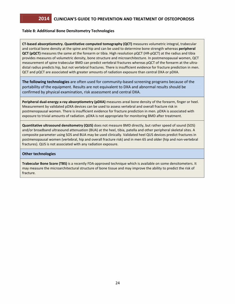
The therapeutic thresholds proposed in this Guide are for clinical guidance only and are not rules. All treatment decisions require clinical judgment and consideration of individual patient factors, including patient preferences, comorbidities, risk factors not captured in the FRAX® model (e.g., frailty, falls), recent decline in bone density and other sources of possible under- or over-estimation of fracture risk by FRAX®. The therapeutic thresholds do not preclude clinicians or patients from considering intervention strategies for those who do not have osteoporosis by BMD (WHO diagnostic criterion of T-score ≤ -2.5), do not meet the cut points after FRAX® or are not at high enough risk of fracture despite low BMD. Conversely, these recommendations should not mandate treatment, particularly in patients with low bone mass above the osteoporosis range. Decisions to treat must still be made on a case-by-case basis. Additional Bone Densitometry Technologies The following bone mass measurement technologies included in Table 8 are capable of predicting both site-specific and overall fracture risk. When performed according to accepted standards, these densitometric techniques are accurate and highly reproducible.19 However, T-scores from these technologies cannot be used according to the WHO diagnostic classification because they are not equivalent to T-scores derived from DXA.
2014 CLINICIAN’S GUIDE TO PREVENTION AND TREATMENT OF OSTEOPOROSIS
24
Table 8: Additional Bone Densitometry Technologies
CT-based absorptiometry. Quantitative computed tomography (QCT) measures volumetric integral, trabecular and cortical bone density at the spine and hip and can be used to determine bone strength whereas peripheral QCT (pQCT) measures the same at the forearm or tibia. High resolution pQCT (HR-pQCT) at the radius and tibia provides measures of volumetric density, bone structure and microarchitecture. In postmenopausal women, QCT measurement of spine trabecular BMD can predict vertebral fractures whereas pQCT of the forearm at the ultra- distal radius predicts hip, but not vertebral fractures. There is insufficient evidence for fracture prediction in men. QCT and pQCT are associated with greater amounts of radiation exposure than central DXA or pDXA.
The following technologies are often used for community-based screening programs because of the portability of the equipment. Results are not equivalent to DXA and abnormal results should be confirmed by physical examination, risk assessment and central DXA.
Peripheral dual-energy x-ray absorptiometry (pDXA) measures areal bone density of the forearm, finger or heel. Measurement by validated pDXA devices can be used to assess vertebral and overall fracture risk in postmenopausal women. There is insufficient evidence for fracture prediction in men. pDXA is associated with exposure to trivial amounts of radiation. pDXA is not appropriate for monitoring BMD after treatment.
Quantitative ultrasound densitometry (QUS) does not measure BMD directly, but rather speed of sound (SOS) and/or broadband ultrasound attenuation (BUA) at the heel, tibia, patella and other peripheral skeletal sites. A composite parameter using SOS and BUA may be used clinically. Validated heel QUS devices predict fractures in postmenopausal women (vertebral, hip and overall fracture risk) and in men 65 and older (hip and non-vertebral fractures). QUS is not associated with any radiation exposure.
Other technologies
Trabecular Bone Score (TBS) is a recently FDA-approved technique which is available on some densitometers. It may measure the microarchitectural structure of bone tissue and may improve the ability to predict the risk of fracture.
2014 CLINICIAN’S GUIDE TO PREVENTION AND TREATMENT OF OSTEOPOROSIS
25
5. UNIVERSAL RECOMMENDATIONS FOR ALL PATIENTS
Several interventions to preserve bone strength can be recommended to the general population. These include an adequate intake of calcium and vitamin D, lifelong participation in regular weight-bearing and muscle-strengthening exercise, cessation of tobacco use, identification and treatment of alcoholism, and treatment of risk factors for falling.
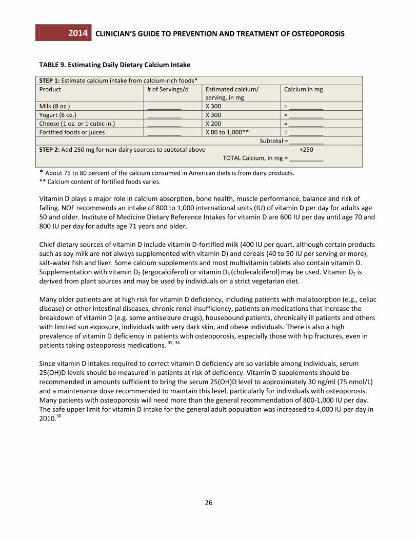
Adequate Intake of Calcium and Vitamin D Advise all individuals to obtain an adequate intake of dietary calcium. Providing adequate daily calcium and vitamin D is a safe and inexpensive way to help reduce fracture risk. Controlled clinical trials have demonstrated that the combination of supplemental calcium and vitamin D can reduce the risk of fracture.29 A balanced diet rich in low-fat dairy products, fruits and vegetables provide calcium as well as numerous nutrients needed for good health. If adequate dietary calcium cannot be obtained, dietary supplementation is indicated up to the recommended daily intake. Lifelong adequate calcium intake is necessary for the acquisition of peak bone mass and subsequent maintenance of bone health. The skeleton contains 99 percent of the body’s calcium stores; when the exogenous supply is inadequate, bone tissue is resorbed from the skeleton to maintain serum calcium at a constant level. NOF supports Institute of Medicine (IOM) recommendations that men age 50-70 consume 1,000 mg per day of calcium and that women age 51 and older and men age 71 and older consume 1,200 mg per day of calcium.30 There is no evidence that calcium intake in excess of these amounts confers additional bone strength. Intakes in excess of 1,200 to 1,500 mg per day may increase the risk of developing kidney stones, cardiovascular disease and stroke. The scientific literature is highly controversial in this area.31,32,33,34 Table 9 illustrates a simple method for estimating the calcium content of a patient’s diet. The average daily dietary calcium intake in adults age 50 and older is 600 to 700 mg per day. Increasing dietary calcium is the first-line approach, but calcium supplements should be used when an adequate dietary intake cannot be achieved.
2014 CLINICIAN’S GUIDE TO PREVENTION AND TREATMENT OF OSTEOPOROSIS
26
TABLE 9. Estimating Daily Dietary Calcium Intake STEP 1: Estimate calcium intake from calcium-rich foods* Product # of Servings/d Estimated calcium/
serving, in mg Calcium in mg
Milk (8 oz.) __________ X 300 = __________ Yogurt (6 oz.) __________ X 300 = __________ Cheese (1 oz. or 1 cubic in.) __________ X 200 = __________ Fortified foods or juices __________ X 80 to 1,000** = __________ Subtotal = __________ STEP 2: Add 250 mg for non-dairy sources to subtotal above +250 TOTAL Calcium, in mg = __________
* About 75 to 80 percent of the calcium consumed in American diets is from dairy products. ** Calcium content of fortified foods varies. Vitamin D plays a major role in calcium absorption, bone health, muscle performance, balance and risk of falling. NOF recommends an intake of 800 to 1,000 international units (IU) of vitamin D per day for adults age 50 and older. Institute of Medicine Dietary Reference Intakes for vitamin D are 600 IU per day until age 70 and 800 IU per day for adults age 71 years and older. Chief dietary sources of vitamin D include vitamin D-fortified milk (400 IU per quart, although certain products such as soy milk are not always supplemented with vitamin D) and cereals (40 to 50 IU per serving or more), salt-water fish and liver. Some calcium supplements and most multivitamin tablets also contain vitamin D. Supplementation with vitamin D2 (ergocalciferol) or vitamin D3 (cholecalciferol) may be used. Vitamin D2 is derived from plant sources and may be used by individuals on a strict vegetarian diet. Many older patients are at high risk for vitamin D deficiency, including patients with malabsorption (e.g., celiac disease) or other intestinal diseases, chronic renal insufficiency, patients on medications that increase the breakdown of vitamin D (e.g. some antiseizure drugs), housebound patients, chronically ill patients and others with limited sun exposure, individuals with very dark skin, and obese individuals. There is also a high prevalence of vitamin D deficiency in patients with osteoporosis, especially those with hip fractures, even in patients taking osteoporosis medications. 35, 36 Since vitamin D intakes required to correct vitamin D deficiency are so variable among individuals, serum 25(OH)D levels should be measured in patients at risk of deficiency. Vitamin D supplements should be recommended in amounts sufficient to bring the serum 25(OH)D level to approximately 30 ng/ml (75 nmol/L) and a maintenance dose recommended to maintain this level, particularly for individuals with osteoporosis. Many patients with osteoporosis will need more than the general recommendation of 800-1,000 IU per day. The safe upper limit for vitamin D intake for the general adult population was increased to 4,000 IU per day in 2010.30

2014 CLINICIAN’S GUIDE TO PREVENTION AND TREATMENT OF OSTEOPOROSIS
27
Treatment of Vitamin D Deficiency Adults who are vitamin D deficient may be treated with 50,000 IU of vitamin D2 or vitamin D3 once a week or the equivalent daily dose (7,000 IU vitamin D2 or vitamin D3) for 8-12 wks to achieve a 25(OH)D blood level of approximately 30 ng/ml. This regimen should be followed by maintenance therapy of 1,500–2,000 IU/d or whatever dose is needed to maintain the target blood level. 37, 38
Regular Weight-Bearing and Muscle-Strengthening Exercise Recommend regular weight-bearing and muscle-strengthening exercise to reduce the risk of falls and fractures.39,40, 41, 42Among the many health benefits, weight-bearing and muscle-strengthening exercise can improve agility, strength, posture and balance, which may reduce the risk of falls. In addition, exercise may modestly increase bone density. NOF strongly endorses lifelong physical activity at all ages, both for osteoporosis prevention and overall health, as the benefits of exercise are lost when people stop exercising. Weight-bearing exercise (in which bones and muscles work against gravity as the feet and legs bear the body’s weight) includes walking, jogging, Tai-Chi, stair climbing, dancing and tennis. Muscle-strengthening exercise includes weight training and other resistive exercises, such as yoga, Pilates and boot camp programs. Before an individual with osteoporosis initiates a new vigorous exercise program, such as running or heavy weight-lifting, a clinician’s evaluation is appropriate.
Fall Prevention Major risk factors for falling are shown in Table 2. In addition to maintaining adequate vitamin D levels and physical activity, as described above, several strategies have been demonstrated to reduce falls. These include, but are not limited to, multifactorial interventions such as individual risk assessment, Tai Chi and other exercise programs, home safety assessment and modification especially when done by an occupational therapist and gradual withdrawal of psychotropic medication if possible. Appropriate correction of visual impairment may improve mobility and reduce risk of falls. Hip protectors may protect an individual from injuring the hip in the event of a fall, although evidence regarding anti-fracture benefits is inconclusive.43 There is additional uncertainty as to which hip protector to use, as most of the marketed products have not been tested in randomized clinical trials.

2014 CLINICIAN’S GUIDE TO PREVENTION AND TREATMENT OF OSTEOPOROSIS
28
Cessation of Tobacco Use and Avoidance of Excessive Alcohol Intake Advise patients to stop tobacco smoking. The use of tobacco products is detrimental to the skeleton as well as to overall health.44,45,46, 47 NOF strongly encourages a smoking cessation program as an osteoporosis intervention. Recognize and treat patients with excessive alcohol intake. Moderate alcohol intake has no known negative effect on bone and may even be associated with slightly higher bone density and lower risk of fracture in postmenopausal women. However, alcohol intake of more than two drinks per day for women or three drinks a day for men may be detrimental to bone health, increases the risk of falling and requires further evaluation for possible alcoholism.48
2014 CLINICIAN’S GUIDE TO PREVENTION AND TREATMENT OF OSTEOPOROSIS
29
6. PHARMACOLOGIC THERAPY
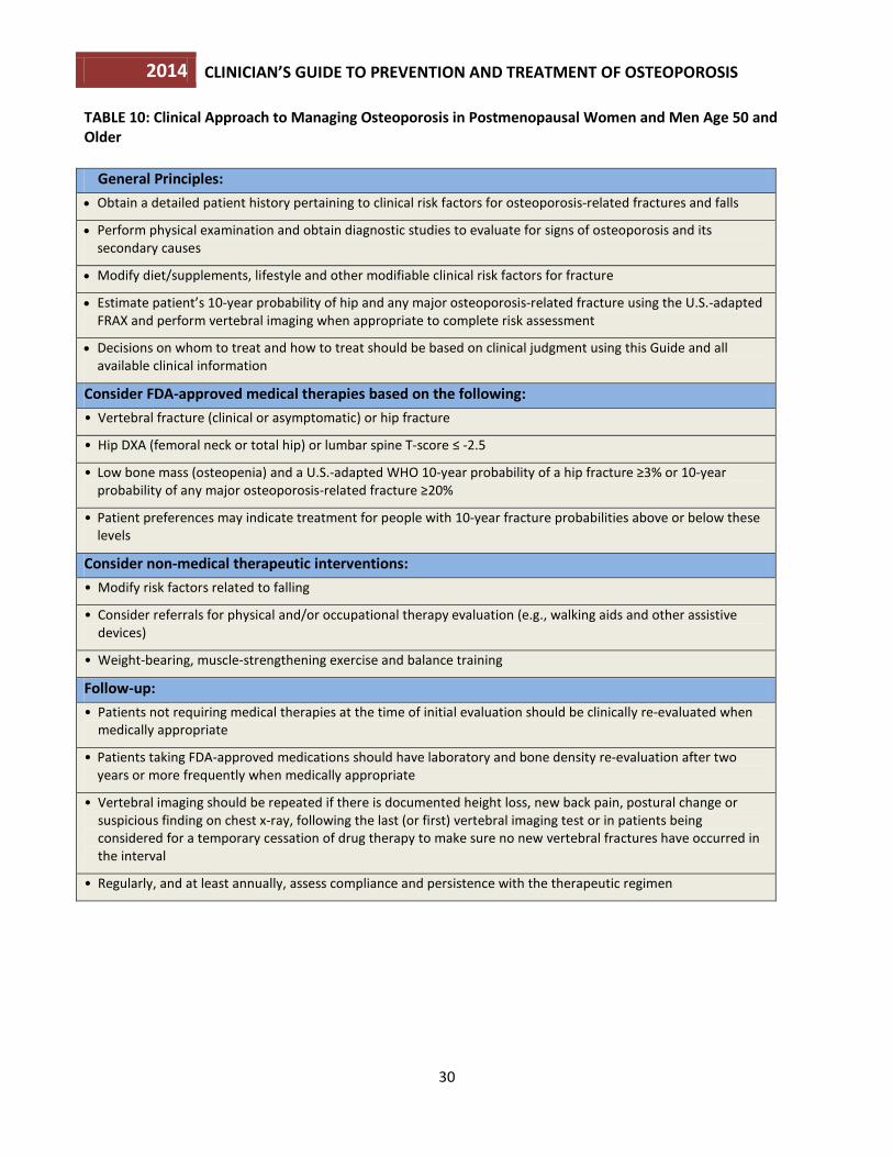
All patients being considered for treatment of osteoporosis should also be counseled on risk factor reduction including the importance of calcium, vitamin D and exercise as part of any treatment program for osteoporosis. Prior to initiating treatment, patients should be evaluated for secondary causes of osteoporosis and have BMD measurements by central DXA, when available, and vertebral imaging studies when appropriate. Biochemical marker levels should be obtained if monitoring of treatment effects is planned. An approach to the clinical assessment of individuals with osteoporosis is outlined in Table 10. The percentage of risk reductions for vertebral and non-vertebral fractures cited below are those cited in the FDA-approved Prescribing Information. In the absence of head-to-head trials, direct comparisons of risk reduction among drugs should be avoided.
Who Should Be Considered for Treatment? Postmenopausal women and men age 50 and older presenting with the following should be considered for treatment: • A hip or vertebral fracture (clinically apparent or found on vertebral imaging). There are abundant data that patients with spine and hip fractures will have reduced fracture risk if treated with pharmacologic therapy. This is true for fracture patients with BMD in both the low bone mass and osteoporosis range. 49,50,51,52,53,54,55,56,57,58 In patients with a hip or spine fracture, the T-score is not as important as the fracture itself in predicting future risk of fracture and antifracture efficacy from treatment. • T-score ≤ -2.5 at the femoral neck, total hip or lumbar spine. There is abundant evidence that the elevated risk of fracture in patients with osteoporosis by BMD is reduced with pharmacotherapy.59,60,61,62,63,64,65,66,67,68,69,70,71,72 • Low bone mass (T-score between -1.0 and -2.5 at the femoral neck or lumbar spine) and a 10-year probability of a hip fracture ≥3 percent or a 10-year probability of a major osteoporosis-related fracture ≥20 percent based on the U.S.-adapted WHO algorithm. 13, 14, 73, 74, 75 Although FRAX calculated fracture risk prediction has been confirmed in multiple studies, there are relatively few data confirming fracture risk reductions with pharmacotherapy in this group of patients.
2014 CLINICIAN’S GUIDE TO PREVENTION AND TREATMENT OF OSTEOPOROSIS
30
TABLE 10: Clinical Approach to Managing Osteoporosis in Postmenopausal Women and Men Age 50 and Older
General Principles: • Obtain a detailed patient history pertaining to clinical risk factors for osteoporosis-related fractures and falls
• Perform physical examination and obtain diagnostic studies to evaluate for signs of osteoporosis and its secondary causes
• Modify diet/supplements, lifestyle and other modifiable clinical risk factors for fracture
• Estimate patient’s 10-year probability of hip and any major osteoporosis-related fracture using the U.S.-adapted FRAX and perform vertebral imaging when appropriate to complete risk assessment
• Decisions on whom to treat and how to treat should be based on clinical judgment using this Guide and all available clinical information
Consider FDA-approved medical therapies based on the following: • Vertebral fracture (clinical or asymptomatic) or hip fracture
• Hip DXA (femoral neck or total hip) or lumbar spine T-score ≤ -2.5
• Low bone mass (osteopenia) and a U.S.-adapted WHO 10-year probability of a hip fracture ≥3% or 10-year probability of any major osteoporosis-related fracture ≥20%
• Patient preferences may indicate treatment for people with 10-year fracture probabilities above or below these levels
Consider non-medical therapeutic interventions: • Modify risk factors related to falling
• Consider referrals for physical and/or occupational therapy evaluation (e.g., walking aids and other assistive devices)
• Weight-bearing, muscle-strengthening exercise and balance training
Follow-up: • Patients not requiring medical therapies at the time of initial evaluation should be clinically re-evaluated when
medically appropriate
• Patients taking FDA-approved medications should have laboratory and bone density re-evaluation after two years or more frequently when medically appropriate
• Vertebral imaging should be repeated if there is documented height loss, new back pain, postural change or suspicious finding on chest x-ray, following the last (or first) vertebral imaging test or in patients being considered for a temporary cessation of drug therapy to make sure no new vertebral fractures have occurred in the interval
• Regularly, and at least annually, assess compliance and persistence with the therapeutic regimen
2014 CLINICIAN’S GUIDE TO PREVENTION AND TREATMENT OF OSTEOPOROSIS
31
U.S. FDA-Approved Drugs for Osteoporosis Current FDA-approved pharmacologic options for the prevention and/or treatment of postmenopausal osteoporosis include, in alphabetical order: bisphosphonates (alendronate, alendronate plus D, ibandronate, risedronate and zoledronic acid), calcitonin, estrogens (estrogen and/or hormone therapy), estrogen agonist/antagonist (raloxifene), tissue-selective estrogen complex (conjugated estrogens/bazedoxifene), parathyroid hormone (PTH[1-34], teriparatide) and the RANKL inhibitor denosumab. Please see Prescribing Information for specific details of their use. The anti-fracture benefits of FDA-approved drugs have mostly been studied in women with postmenopausal osteoporosis. There are limited fracture data in glucocorticoid-induced osteoporosis and in men. FDA-approved osteoporosis treatments have been shown to decrease fracture risk in patients who have had fragility fractures and/or osteoporosis by DXA. Pharmacotherapy may also reduce fractures in patients with low bone mass (osteopenia) without fractures, but the evidence supporting this isn’t as strong. Thus, the clinician should assess the potential benefits and risks of therapy in each patient and the effectiveness of a given osteoporosis treatment on reduction of vertebral and nonvertebral fractures. Note that the intervention thresholds do not take into account the non-skeletal benefits or risks associated with specific drug use. NOF does not advocate the use of drugs not approved by the FDA for prevention and treatment of osteoporosis. Examples of these drugs are listed in Table 11 for information only.
Bisphosphonates Drug efficacy: Alendronate, brand name: Fosamax®, Fosamax Plus D, Binosto™ and generic alendronate. Alendronate sodium is approved by the FDA for the prevention (5 mg daily and 35 mg weekly tablets) and treatment (10 mg daily tablet, 70 mg weekly tablet, 70 mg weekly tablet with 2,800 IU or 5,600 IU of vitamin D3 and 70 mg effervescent tablet) of postmenopausal osteoporosis. Alendronate is also approved for treatment to increase bone mass in men with osteoporosis and for the treatment of osteoporosis in men and women taking glucocorticoids.76 Alendronate reduces the incidence of spine and hip fractures by about 50 percent over three years in patients with a prior vertebral fracture or in patients who have osteoporosis at the hip site.77,78 It reduces the incidence of vertebral fractures by 48 percent over three years in patients without a prior vertebral fracture. Ibandronate, brand name: Boniva® and generic ibandronate. Ibandronate sodium is approved by the FDA for the treatment (150 mg monthly tablet and 3 mg every three months by intravenous injection) of postmenopausal osteoporosis. Ibandronate is available as a generic preparation in the U.S. The oral preparations are also approved for the prevention of postmenopausal osteoporosis. Ibandronate reduces the incidence of vertebral fractures by about 50 percent over three years, but reduction in risk of nonvertebral fracture with ibandronate has not been documented. Risedronate, brand name: Actonel®, Atelvia™ and generic risedronate. Risedronate sodium is approved by the FDA for the prevention and treatment (5 mg daily tablet; 35 mg weekly tablet; 35 mg weekly delayed release tablet; 35 mg weekly tablet packaged with 6 tablets of 500 mg calcium carbonate; 75 mg tablets on two consecutive days every month; and 150 mg monthly tablet) of postmenopausal osteoporosis. Risedronate is
2014 CLINICIAN’S GUIDE TO PREVENTION AND TREATMENT OF OSTEOPOROSIS
32
also approved for treatment to increase bone mass in men with osteoporosis and for the prevention and treatment of osteoporosis in men and women who are either initiating or taking glucocorticoids.79 Risedronate reduces the incidence of vertebral fractures by 41 to 49 percent and non-vertebral fractures by 36 percent over three years, with significant risk reduction occurring within one year of treatment in patients with a prior vertebral fracture. Zoledronic acid, brand name: Reclast®. Zoledronic acid is approved by the FDA for the prevention and treatment (5 mg by intravenous infusion over at least 15 minutes once yearly for treatment and once every two years for prevention) of osteoporosis in postmenopausal women. It is also approved to improve bone mass in men with osteoporosis, and for the prevention and treatment of osteoporosis in men and women expected to be on glucocorticoid therapy for at least 12 months. Zoledronic acid is also indicated for the prevention of new clinical fractures in patients (both women and men) who have recently had a low-trauma (osteoporosis-related) hip fracture. Zoledronic acid reduces the incidence of vertebral fractures by 70 percent (with significant reduction at one year), hip fractures by 41 percent and non-vertebral fractures by 25 percent over three years in patients with osteoporosis defined by prevalent vertebral fractures and osteoporosis by BMD of the hip. Drug administration: Alendronate (generic and Fosamax) and risedronate (Actonel) tablets must be taken on an empty stomach, first thing in the morning, with 8 ounces of plain water (no other liquid). Binosto must be dissolved in 4 ounces of room temperature water taken on an empty stomach, first thing in the morning. Delayed release risedronate (Atelvia) tablets must be taken immediately after breakfast with at least 4 ounces of plain water (no other liquid). After taking these medications, patients must wait at least 30 minutes before eating, drinking or taking any other medication. Patients should remain upright (sitting or standing) during this interval. Ibandronate must be taken on an empty stomach, first thing in the morning, with 8 ounces of plain water (no other liquid). After taking this medication, patients must remain upright and wait at least 60 minutes before eating, drinking or taking any other medication. Ibandronate, 3 mg per 3 ml prefilled syringe, is given by intravenous injection over 15 to 30 seconds, once every three months. Serum creatinine should be checked before each injection. Zoledronic acid, 5 mg in 100 ml, is given once yearly or once every two years by intravenous infusion over at least 15 minutes. Patients should be well hydrated and may be pre-treated with acetaminophen to reduce the risk of an acute phase reaction (arthralgia, headache, myalgia, fever). These symptoms occurred in 32 percent of patients after the first dose, 7 percent after the second dose and 3 percent after the third dose. Drug safety: Side effects are similar for all oral bisphosphonate medications and include gastrointestinal problems such as difficulty swallowing, inflammation of the esophagus and stomach. All bisphosphonates can affect renal function and are contraindicated in patients with estimated GFR below 30-35 ml/min. Zoledronic acid is contraindicated in patients with creatinine clearance less than 35 mL/min, or in patients with evidence of acute renal impairment. Healthcare professionals should screen patients prior to administering zoledronic acid in order to identify at-risk patients and should assess renal function by
2014 CLINICIAN’S GUIDE TO PREVENTION AND TREATMENT OF OSTEOPOROSIS
33
monitoring creatinine clearance prior to each dose of zoledronic acid.80 Eye inflammation can also occur. Any such complication should be reported to the healthcare provider as soon as possible. There have been rare reports of osteonecrosis of the jaw (ONJ) with long term use of bisphosphonates for osteoporosis, though ONJ is much more common following high dose intravenous bisphosphonate treatment for patients with cancer. The risk of ONJ appears to increase with duration of treatment beyond five years.81 Although rare, low trauma atypical femur fractures may be associated with the long-term use of bisphosphonates (e.g. >5 years of use). Pain in the thigh or groin area, which can be bilateral, often precedes these unusual fractures. Patients should be evaluated closely for these unusual fractures, including proactive questioning regarding thigh and groin pain. For patients with thigh and groin pain, a stress fracture in the subtrochanteric region or femoral shaft of the femur may be present. Bilateral x-ray of the femurs should be ordered when an atypical femur fracture is suspected, followed by an MRI or a radionuclide bone scan when clinical suspicion is high enough.82 Surgical fixation is required in some cases whereas medical conservative treatment is appropriate in other cases. Bisphosphonates should be stopped if atypical femur fractures have occurred.
Calcitonin Drug efficacy: Brand name: Miacalcin® or Fortical® and generic calcitonin. Salmon calcitonin is FDA-approved for the treatment of osteoporosis in women who are at least five years postmenopausal when alternative treatments are not suitable. Calcitonin reduces vertebral fracture occurrence by about 30 percent in those with prior vertebral fractures but has not been shown to reduce the risk of nonvertebral fractures.54, 83 Drug administration: 200 IU delivered as a single daily intranasal spray. Subcutaneous administration by injection also is available. Drug safety: Intranasal calcitonin can cause rhinitis, epistaxis and allergic reactions, particularly in those with a history of allergy to salmon. The FDA has reviewed long-term post marketing data concerning calcitonin and the very small increase in the risk of certain cancers. A meta-analysis of 21 randomized, controlled clinical trials with calcitonin-salmon (nasal spray and investigational oral forms) suggests an increased risk of malignancies in calcitonin-salmon treated patients compared to placebo-treated patients. The overall incidence of malignancies reported in the 21 trials was higher among calcitonin-salmon treated patients (4.1 percent) compared with placebo-treated patients (2.9 percent). The data were not sufficient for further analyses by specific type of malignancy. Although a definitive causal relationship between the calcitonin-salmon use and malignancies cannot be established from this meta-analysis, the benefits for the individual patient should be carefully evaluated against all possible risks. 84, 85
2014 CLINICIAN’S GUIDE TO PREVENTION AND TREATMENT OF OSTEOPOROSIS
34
Estrogen/Hormone Therapy (ET/HT) Drug efficacy: ET brand names: e.g. Climara®, Estrace®, Estraderm®, Estratab®, Ogen®, Premarin®, Vivelle®; HT brand names: e.g. Activella®, Femhrt®, Premphase®, Prempro®. Estrogen/hormone therapy is approved by the FDA for the prevention of osteoporosis, relief of vasomotor symptoms and vulvovaginal atrophy associated with menopause. Women who have not had a hysterectomy require HT, which also contains progestin to protect the uterine lining. The Woman’s Health Initiative (WHI) found that five years of HT (Prempro®) reduced the risk of clinical vertebral fractures and hip fractures by 34 percent and other osteoporotic fractures by 23 percent.86 Drug administration: ET/HT is available in a wide variety of oral as well as transdermal preparations including estrogen only, progestin only and combination estrogen-progestin. ET/HT dosages include cyclic, sequential and continuous regimens. If and when treatment is stopped, bone loss can be rapid and alternative agents should be considered to maintain BMD. Drug safety: The Women’s Health Initiative (WHI) reported increased risks of myocardial infarction, stroke, invasive breast cancer, pulmonary emboli and deep vein thrombosis during five years of treatment with conjugated equine estrogen and medroxyprogesterone acetate (Prempro®).86 Subsequent analyses of these data showed no increase in cardiovascular disease in women starting treatment within 10 years of menopause. In the estrogen only arm of WHI, no increase in breast cancer incidence was noted over 7.1 years of treatment. Other doses and combinations of estrogen and progestins were not studied and, in the absence of comparable data, their risks should be assumed to be comparable. Because of the risks, ET/HT should be used in the lowest effective doses for the shortest duration to treat moderately severe menopausal symptoms and should be considered primarily for women within the first few years of menopause. When ET/HT use is considered solely for prevention of osteoporosis, the FDA recommends that approved non-estrogen treatments should first be carefully considered. When ET/HT treatments are stopped, bone loss can be rapid and alternative agents should be considered to maintain BMD.
2014 CLINICIAN’S GUIDE TO PREVENTION AND TREATMENT OF OSTEOPOROSIS
35
Estrogen Agonist/Antagonist (formerly known as SERMs): Raloxifene Drug efficacy: Raloxifene, brand name: Evista® and generic raloxifene. Raloxifene is approved by the FDA for both prevention and treatment of osteoporosis in postmenopausal women. Raloxifene reduces the risk of vertebral fractures by about 30 percent in patients with a prior vertebral fracture and by about 55 percent in patients without a prior vertebral fracture over three years. Reduction in risk of nonvertebral fracture with raloxifene has not been documented. Raloxifene is also indicated for the reduction in risk of invasive breast cancer in postmenopausal women with osteoporosis. 87,88,89,90 Raloxifene does not reduce the risk of coronary heart disease. Drug administration: Available in a 60 mg tablet form to be taken with or without food. Drug safety: Raloxifene increases the risk of deep vein thrombosis to a degree similar to that observed with estrogen. It can also increase hot flashes and cause leg cramps.

2014 CLINICIAN’S GUIDE TO PREVENTION AND TREATMENT OF OSTEOPOROSIS
36
Tissue-Selective Estrogen Complex: Conjugated estrogens/ bazedoxifene (Conjugated estrogens paired with estrogen agonist/antagonist) Drug efficacy:
Conjugated estrogens/bazedoxifene, brand name: Duavee®. Conjugated estrogens/bazedoxifene is approved by the FDA for women who suffer from moderate-to-severe hot flashes (vasomotor symptoms) associated with menopause and to prevent osteoporosis after menopause.
The medication combines conjugated estrogen with an estrogen agonist/antagonist (bazedoxifene). The bazedoxifene component reduces the risk of endometrial hyperplasia (excessive growth of the lining of the uterus) that can occur with the estrogen component of the drug. Therefore progestins do not need to be taken with conjugated estrogens/bazedoxifene.
Use of this combination drug significantly increased mean lumbar spine BMD (treatment difference, 1.51 percent), at 12 months compared to placebo in women who had been postmenopausal between one and five years. Treatment with conjugated estrogens/bazedoxifene also increased total hip BMD. The treatment difference in total hip BMD at 12 months was 1.21 percent.91,92,93 ,94
Drug Administration:
Available as a tablet containing conjugated estrogens and bazedoxifene 0.45mg / 20mg, to be taken once daily without regard to meals.
Drug Safety:
Conjugated estrogens/bazedoxifene is intended only for postmenopausal women who still have a uterus. Like other products containing estrogen, it should be used for the shortest duration consistent with treatment goals and risks for the individual woman. When using this drug only for the prevention of osteoporosis, such use should be limited to women who are at significant risk of osteoporosis and only after carefully considering alternatives that do not contain estrogen.
Side effects of conjugated estrogens/bazedoxifene include muscle spasms, nausea, diarrhea, dyspepsia, upper abdominal pain, oropharyngeal pain, dizziness and neck pain. Because this product contains estrogen, it is approved with the same Boxed Warning and other Warnings and Precautions that have been approved with estrogen products.

2014 CLINICIAN’S GUIDE TO PREVENTION AND TREATMENT OF OSTEOPOROSIS
37
Parathyroid Hormone: Teriparatide
Drug efficacy: PTH(1-34), teriparatide, brand name: Forteo®. Teriparatide is approved by the FDA for the treatment of osteoporosis in postmenopausal women and men at high risk for fracture. It is also approved for treatment in men and women at high risk of fracture with osteoporosis associated with sustained systemic glucocorticoid therapy.95 Teriparatide reduces the risk of vertebral fractures by about 65 percent and non-vertebral fragility fractures by about 53 percent in patients with osteoporosis, after an average of 18 months of therapy. Drug administration: Teriparatide is an anabolic (bone-building) agent administered by 20 µg daily subcutaneous injection. If and when treatment is stopped, bone loss can be rapid and alternative agents should be considered to maintain BMD. Treatment duration is recommended not to exceed 18 to 24 months. Drug safety: Side effects of teriparatide include leg cramps, nausea and dizziness. Because it caused an increase in the incidence of osteosarcoma in rats (high doses, long duration treatment in the rodent), patients with an increased risk of osteosarcoma (e.g., patients with Paget’s disease of bone and those having prior radiation therapy of the skeleton), bone metastases, hypercalcemia, or a history of skeletal malignancy should not receive teriparatide therapy. It is common practice to follow teriparatide treatment with an antiresorptive agent, usually a bisphosphonate, to maintain or further increase BMD.
Receptor Activator of Nuclear Factor kappa-B (RANK) Ligand (RANKL)/ RANKL Inhibitor: Denosumab Drug efficacy: Denosumab, brand name Prolia®. Denosumab is approved by the FDA for the treatment of osteoporosis in postmenopausal women at high risk of fracture. Denosumab reduces the incidence of vertebral fractures by about 68 percent, hip fractures by about 40 percent and non-vertebral fractures by about 20 percent over three years. Denosumab is also indicated to increase bone mass in men at high risk of fracture, treat bone loss in women with breast cancer on aromatase inhibitor therapies and to treat bone loss in men receiving gonadatropin-reducing hormone treatment for prostate cancer who are at high risk for fracture. Drug administration: Administered by a health professional, 60 mg every six months as a subcutaneous injection. Drug safety: Denosumab may cause hypocalcemia. Hypocalcemia must be corrected before starting denosumab. Denosumab increased the risk of serious skin infections (cellulitis) and skin rash. Denosumab has been rarely associated with the development of ONJ, both when used to treat osteoporosis and to treat patients with cancer (at much higher doses), although it is much more common in the latter setting. Denosumab has also

2014 CLINICIAN’S GUIDE TO PREVENTION AND TREATMENT OF OSTEOPOROSIS
38
been associated rarely with the development of atypical femur fractures. If and when denosumab treatment is stopped, bone loss can be rapid and alternative agents should be considered to maintain BMD. Sequential and Combination Therapy Sequential treatment with anabolic therapy followed by an antiresorptive agent is generally preferred. Combination therapy with teriparatide and an antiresorptive can be considered in a few clinical settings in patients with very severe osteoporosis such as spine and hip fractures. There are few indications for combining two antiresorptive treatments, but such options could be considered in the short-term in women who are experiencing active bone loss while on low dose HT for menopausal symptoms or raloxifene for breast cancer prevention.
Duration of Treatment No pharmacologic therapy should be considered indefinite in duration. All non-bisphosphonate medications produce temporary effects that wane upon discontinuation. If these treatments are stopped, benefits rapidly disappear. In contrast, bisphosphonates may allow residual effects even after treatment discontinuation. Therefore, it may be possible to discontinue bisphosphonates and retain residual benefits against fracture at least for several years. Evidence of efficacy beyond five years is limited, whereas rare safety concerns such as ONJ and atypical femur fractures become more common beyond five years. 68,96 Since there is no extensive evidence base to guide treatment duration decisions, duration decisions need to be individualized.97 After the initial three to five year treatment period, a comprehensive risk assessment should be performed. This should include interval clinical history, particular with respect to intercurrent fracture history and new chronic diseases or medications, as well as height measurement, BMD testing and vertebral imaging if there has been any documented height loss during the treatment period. It is reasonable to discontinue bisphosphonates after three to five years in people who appear to be at modest risk of fracture after the initial treatment period. In contrast, for those who appear to be at high risk for fracture, continued treatment with a bisphosphonate or an alternative therapy should be considered.98
2014 CLINICIAN’S GUIDE TO PREVENTION AND TREATMENT OF OSTEOPOROSIS
39
Table 11: Non-FDA-Approved Drugs for Osteoporosis.
These drugs are listed for information only. Non-approved agents include:
Calcitriol. This synthetic vitamin D analogue, which promotes calcium absorption, has been approved by the FDA for managing hypocalcemia and metabolic bone disease in renal dialysis patients. It is also approved for use in hypoparathyroidism, both surgical and idiopathic, and pseudohypoparathyroidism. No reliable data demonstrate a reduction of risk for osteoporotic fracture.
Genistein. An isoflavone phytoestrogen which is the main ingredient in the prescription “medical food” product Fosteum® and generally regarded as safe by the FDA. Genistein may benefit bone health in postmenopausal women but more data are needed to fully understand its effects on bone health and fracture risk.
Other bisphosphonates (etidronate, pamidronate, tiludronate). These medications vary chemically from alendronate, ibandronate, risedronate and zoledronic acid but are in the same drug class. At this time, none is approved for prevention or treatment of osteoporosis. Most of these medications are currently approved for other conditions (e.g. Paget's disease, hypercalcemia of malignancy, myositis ossificans).
PTH (1-84). This medication is approved in some countries in Europe for treatment of osteoporosis in women. In one clinical study PTH(1-84) effectively reduced the risk of vertebral fractures at a dose of 100mcg/d.
Sodium fluoride. Through a process that is still unclear, sodium fluoride stimulates the formation of new bone. The quality of bone mass thus developed is uncertain, and the evidence that fluoride reduces fracture risk is conflicting and controversial.
Strontium ranelate. This medication is approved for the treatment of osteoporosis in some countries in Europe. Strontium ranelate reduces the risk of both spine and non-vertebral fractures, but the mechanism is unclear. Incorporation of strontium into the crystal structure replacing calcium may be part of its mechanism of effect. These effects have only been documented with the pharmaceutical grade agent produced by Servier. This effect has not been studied in nutritional supplements containing strontium salts.
Tibolone. Tibolone is a tissue-specific, estrogen-like agent that may prevent bone loss and reduce menopausal symptoms. It is indicated in Europe for the treatment of vasomotor symptoms of menopause and for prevention of osteoporosis, but it is not approved for use in the U.S.
Monitoring Effectiveness of Treatment It is important to ask patients whether they are taking their medications and to encourage continued and appropriate compliance with their osteoporosis therapies to reduce fracture risk. It is also important to review their risk factors and encourage appropriate calcium and vitamin D intakes, exercise, fall prevention and other lifestyle measures. Furthermore, the need for continued medication to treat osteoporosis should be reviewed annually. Duration of treatment must be individualized. Some patients may be able to discontinue treatment temporarily after several years of therapy, particularly after bisphosphonate administration. Other patients will need to continue treatment. If treatment is discontinued, serial monitoring can include clinical assessment for fractures, falling, any interval chronic disease occurrence and consideration of serial BMD testing, use of biochemical markers and vertebral imaging in some patients. Accurate yearly height measurement is a critical determination of osteoporosis treatment efficacy. Patients who lose 2 cm (or 0.8 inches) or more in height either acutely or cumulatively should have a repeat vertebral imaging test to determine if new or additional vertebral fractures have occurred since the prior vertebral imaging test.
2014 CLINICIAN’S GUIDE TO PREVENTION AND TREATMENT OF OSTEOPOROSIS
40
Serial central DXA testing is an important component of osteoporosis management. Measurements for monitoring patients should be performed in accordance with medical necessity, expected response and in consideration of local regulatory requirements. NOF recommends that repeat BMD assessments generally agree with Medicare guidelines of every two years, but recognizes that testing more frequently may be warranted in certain clinical situations. The following techniques may be used to monitor the effectiveness of treatment: Central DXA. Central DXA assessment of the hip or lumbar spine is the “gold standard” for serial assessment of BMD. Biological changes in bone density are small compared to the inherent error in the test itself, and interpretation of serial bone density studies depends on appreciation of the smallest change in BMD that is beyond the range of error of the test. This least significant change (LSC) varies with the specific instrument used, patient population being assessed, measurement site, technologist’s skill with patient positioning and test analysis, and the confidence intervals used.99 Changes in the BMD of less than 3-6 percent at the hip and 2-4 percent at the spine from test to test may be due to the precision error of the testing itself. Information on how to assess precision and calculate the LSC is available at www.ISCD.org. QCT. Volumetric BMD of the lumbar spine can be used to monitor age-, disease- and treatment-related BMD changes in men and women. Precision of acquisition should be established by phantom data and analysis precision by re-analysis of patient data. pDXA, pQCT and QUS. Peripheral skeletal sites do not respond with the same magnitude as the spine and hip to medications and thus are not appropriate for monitoring response to therapy at this time. Biochemical markers of bone turnover. Suppression of biochemical markers of bone turnover after 3-6 months of treatment with specific antiresorptive therapies, and biochemical marker increases after 1-3 months of specific anabolic therapies, have been predictive of greater BMD responses and in some cases fracture risk reduction in large clinical trials. Biochemical marker changes in individuals must exceed the LSC in order to be clinically meaningful. The LSC is specific to the biomarker being utilized, which is calculated by multiplying the “precision error” of the specific biochemical marker (laboratory provided) by 2.77 (95 percent confidence level). Biological variability can be reduced by obtaining samples in the early morning after an overnight fast. Serial measurements should be made at the same time of day at the same laboratory. Vertebral Imaging: Once the first vertebral imaging test has been performed to determine prevalent vertebral fractures (indications above), repeat testing should be performed to identify incident vertebral fractures if there is a change in the patient’s status suggestive of new vertebral fracture, including documented height loss, undiagnosed back pain, postural change, or a possible finding of new vertebral deformity on chest x-ray. If patients are being considered for a temporary cessation of drug therapy, vertebral imaging should be repeated to determine that no vertebral fractures have occurred in the interval off treatment. A new vertebral fracture on therapy indicates a need for more intensive or continued treatment rather than treatment cessation.
2014 CLINICIAN’S GUIDE TO PREVENTION AND TREATMENT OF OSTEOPOROSIS
41
Implementation of Fracture Liaison Service (FLS) Secondary Fracture Prevention Programs FLS programs have been implemented successfully in a number of closed and open settings over the last 15 years, both in the U.S. (most notably at Kaiser Permanente and Geisinger Health System) as well as abroad. These programs have accomplished a reduction in secondary fracture rates as well as health care cost savings.100,101 In the U.S., Kaiser Permanente’s Healthy Bones program has reduced the expected hip fracture rate by 38 percent since 1998 102; Geisinger Health System achieved $7.8 million in cost savings over five years.103 A Fracture Liaison Service is a coordinated care system headed by an FLS coordinator (a nurse practitioner, physician’s assistant, nurse or other health professional) who ensures that individuals who suffer a fracture receive appropriate diagnosis, treatment and support. The FLS uses established protocols to find and assess fracture patients. The program creates a population database of fracture patients and establishes a process and timeline for patient assessment and follow-up care. An FLS manager is frequently based in a hospital and requires support from a qualified physician or physician team.
2014 CLINICIAN’S GUIDE TO PREVENTION AND TREATMENT OF OSTEOPOROSIS
42
7. PHYSICAL MEDICINE AND REHABILITATION
Physical medicine and rehabilitation can reduce disability, improve physical function and lower the risk of subsequent falls in patients with osteoporosis. Rehabilitation and exercise are recognized means to improve function, such as activities of daily living. Psychosocial factors also strongly affect functional ability of the patient with osteoporosis who has already suffered fractures. Recommendations from the Health Professional’s Guide to Rehabilitation of the Patient with Osteoporosis15 • Evaluate and consider the patient’s physical and functional safety as well as psychological and social status,
medical status, nutritional status and medication use before prescribing a rehabilitation program. • Evaluate the patient and her/his current medication use and consider possible interactions and risk for
altered mental status. Intervene as appropriate. • Provide training for the performance of safe movement and safe activities of daily living, including posture,
transfers, lifting and ambulation in populations with or at high risk for osteoporosis. Intervene as appropriate, e.g., with prescription for assistive device for improved balance with mobility.
• Implement steps to correct underlying deficits whenever possible, i.e., improve posture and balance and
strengthen quadriceps muscles to allow a person to rise unassisted from a chair; promote use of assistive devices to help with ambulation, balance, lifting and reaching.
• Evaluate home environment for risk factors for falls and intervene as appropriate. • Based on the initial condition of the patient, provide a complete exercise recommendation that includes
weight-bearing aerobic activities for the skeleton, postural training, progressive resistance training for muscle and bone strengthening, stretching for tight soft tissues and joints and balance training.
• Advise patients to avoid forward bending and exercising with trunk in flexion, especially in combination
with twisting. • As long as principles of safe movement are followed, walking and daily activities, such as housework and
gardening, are practical ways to contribute to maintenance of fitness and bone mass. Additionally, progressive resistance training and increased loading exercises, within the parameter of the person’s current health status, are beneficial for muscle and bone strength. Proper exercise may improve physical performance/function, bone mass, muscle strength and balance, as well as reduce the risk of falling.
• Avoid long-term immobilization and recommend partial bed rest (with periodic sitting and ambulating) only
when required and for the shortest periods possible. • In patients with acute vertebral fractures or chronic pain after multiple vertebral fractures, the use of trunk
orthoses (e.g., back brace, corset, posture training support devices) may provide pain relief by reducing the loads on the fracture sites and aligning the vertebra. However, long-term bracing may lead to muscle weakness and further de-conditioning.
2014 CLINICIAN’S GUIDE TO PREVENTION AND TREATMENT OF OSTEOPOROSIS
43
• Effective pain management is a cornerstone in rehabilitation from vertebral fractures. Pain relief may be obtained by the use of a variety of physical, pharmacological and behavioral techniques with the caveat that the benefit of pain relief should not be outweighed by the risk of side effects such as disorientation or sedation which may result in falls.
• Individuals with recent, painful vertebral fractures that fail conservative management may be candidates
for interventions, such as kyphoplasty or vertebroplasty, when performed by experienced practitioners.
CONCLUSIONS AND REMAINING QUESTIONS
The Guide has focused on the prevention, diagnosis and treatment of osteoporosis in postmenopausal women and men age 50 and older using the most common existing diagnostic and treatment methods available. Much is known about osteoporosis in this population. However, many additional issues urgently need epidemiologic, clinical and economic research. For example: • How can we better assess bone strength using non-invasive technologies and thus further refine or identify
patients at high risk for fracture? • There is a need to expand the WHO FRAX™ algorithm to incorporate information on lumbar spine BMD and
to consider multiple fractures and regency of fractures in quantitative risk assessment.
• There is a need to develop a fracture risk calculator for patients who have already initiated pharmacologic therapy.
• How can children, adolescents and young adults maximize peak bone mass? • What are the precise components (type, intensity, duration, frequency) of an effective exercise program for
osteoporosis prevention and treatment? • What should be done to identify and modify risk factors for falling, and what would be the magnitude of
effect on fracture risk in a population? • How effective are different FDA-approved treatments in preventing fractures in patients with moderately
low bone mass? Do benefits exceed risks? • What approaches are most effective in treating osteoporosis in disabled populations? • How can we make the diagnosis of vertebral fractures more accurate and consistent, particularly mild
fractures? • How long should antiresorptive therapies be continued, and are there long-term side effects as yet
unknown? • Are combination therapies useful and, if so, which drug combinations are useful and when should they be
used?
2014 CLINICIAN’S GUIDE TO PREVENTION AND TREATMENT OF OSTEOPOROSIS
44
• Can we identify agents that will significantly increase bone mass and return bone structure to normal? • Should we treat patients to a certain goal and then reconsider type and/or dose of therapy? If so, what
should that goal be? • How should therapeutic agents be sequentially prescribed in order to maximize benefits and minimize risks
over the lifespan of the patient? NOF is committed to continuing the effort to answer these and other questions related to this debilitating disease, with the goal of eliminating osteoporosis as a threat to the health of present and future generations. For additional resources on osteoporosis and bone health, visit www.nof.org.
2014 CLINICIAN’S GUIDE TO PREVENTION AND TREATMENT OF OSTEOPOROSIS
45
GLOSSARY
Alendronate (Fosamax®, Binosto™): A bisphosphonate approved by the U.S. Food and Drug Administration for prevention and treatment of osteoporosis; accumulates and persists in the bone. Studies indicate about a 50 percent reduction in vertebral and hip fractures in patients with osteoporosis. Atypical femur fractures: Low or no trauma fractures which are characterized by distinct radiographic (transverse fracture line, periosteal callus formation at the fracture site, little or no comminution) and clinical features (prodromal pain, bilaterality) that resemble stress fractures. These fractures are thought to be associated with long-term use of potent antiresorptive medications and are distinguished from ordinary osteoporotic femoral diaphyseal fractures. Biochemical markers of bone turnover: Biochemical markers of bone remodeling [e.g., resorption markers - serum C-telopeptide (CTX) and urinary N-telopeptide (NTX) and formation markers - serum bone specific alkaline phosphatase (BSAP), osteocalcin (OC) and aminoterminal propeptide of type 1 procollagen (P1NP)] can be measured in the serum and urine. Elevated levels of markers of bone turnover may predict bone loss, and declines in the levels of markers after 3-6 months of treatment may be predictive of fracture risk reduction. Bone mineral density (BMD): A risk factor for fractures. By DXA, BMD is expressed as the amount of mineralized tissue in the area scanned (g/cm2); with QCT, BMD is expressed as the amount per volume of bone (mg/cm3). Hip BMD by DXA is considered the best predictor of hip fracture; it appears to predict other types of fractures as well as measurements made at other skeletal sites. Lumbar spine BMD may be preferable to assess changes early in menopause and after bilateral ovariectomy and may be better than hip BMD in predicting risk of spine fractures especially in women in their 50s and 60s. . Calcitonin (Miacalcin® or Fortical®): A polypeptide hormone that inhibits the resorptive activity of osteoclasts. Calcitriol: A synthetic form of 1,25-dihydroxyvitamin D3, a hormone that aids calcium absorption and mineralization of the skeleton. Its effectiveness as a treatment for osteoporosis is still uncertain. Calcitriol is not FDA approved. Calcium: A mineral that plays an essential role in development and maintenance of a healthy skeleton. The vast majority of the body’s calcium is stored in bone. If intake is inadequate, calcium is mobilized from the skeleton to maintain a normal blood calcium level. In addition to being a substrate for bone mineralization, calcium has an inhibitory effect on bone remodeling through suppression of circulating parathyroid hormone. Cancellous bone: The spongy, or trabecular, tissue in the middle of bone (e.g., vertebrae) and at the end of the long bones. Cortical bone: The dense outer layer of bone. Cost-effectiveness analysis: As utilized in this Guide, a quantitative analysis that considers the value of treatment by comparing average costs and average health outcomes (quality-adjusted life expectancy) for patients who are treated for osteoporosis relative to untreated patients. Denosumab: A human antibody to RANK ligand approved by the FDA for the treatment of osteoporosis in postmenopausal women at high risk of fracture. Denosumab reduces the incidence of vertebral fractures by

2014 CLINICIAN’S GUIDE TO PREVENTION AND TREATMENT OF OSTEOPOROSIS
46
about 68 percent, hip fractures by about 40 percent and non-vertebral fractures by about 20 percent over three years. Dual-energy x-ray absorptiometry (DXA): A diagnostic test used to assess bone density at various skeletal sites using radiation exposure about one-tenth that of a standard chest x-ray. Central DXA (lumbar spine, hip) is the preferred measurement for definitive diagnosis of osteoporosis and for monitoring the effects of therapy. Estrogen: One of a group of steroid hormones that control female sexual development; directly affects bone mass through estrogen receptors in bone, reducing bone turnover and bone loss. Indirectly increases intestinal calcium absorption and renal calcium conservation and, therefore, improves calcium balance. See hormone therapy. Estrogen agonists/antagonists: A group of compounds that are selective estrogen receptor modulators, formerly known as SERMs. Examples are the pharmaceutical agents raloxifene and bazedoxifene. Exercise: An intervention long associated with healthy bones, despite limited evidence for significant beneficial effect on bone mineral density or fracture risk reductions. Studies evaluating exercise are ongoing; however, enough is known about the positive effect of exercise on fall prevention to support its inclusion in a comprehensive fracture prevention program. Fluoride: A compound that stimulates the formation of new bone by enhancing the recruitment and differentiation of osteoblasts. Studies show varying effects on BMD depending upon the preparation, dose, measurement site and outcomes assessed. Fracture: Breakage of a bone, either complete or incomplete. Incomplete fractures include stress fractures which are often related to repetitive stress on bones, such as metatarsals and tibia, and are not generally thought to be osteoporosis-related. Most studies of osteoporosis focus on hip, vertebra and/or distal forearm fractures. Vertebral fractures include morphometric as well as clinical fractures. FRAX®: The World Health Organization Fracture Risk Assessment Tool. www.NOF.org and www.shef.ac.uk/FRAX Hormone/estrogen therapy (HT/ET) (HT – Activella®, Femhrt®, Premphase®, Prempro®; ET – Climara®, Estrace®, Estraderm®, Estratab®, Ogen®, Ortho-Est®, Premarin®, Vivelle®): HT is a general term for all types of estrogen replacement therapy when given along with progestin, cyclically or continuously. HT is generally prescribed for women after natural menopause or bilateral ovariectomy with progestin required to protect the uterus from unopposed estrogen. ET is prescribed for postmenopausal women who have had a hysterectomy. Studies indicate that five years of HT may decrease vertebral fractures by 35 to 50 percent and non-vertebral fractures by about 25 percent. Ten or more years of use might be expected to decrease the rate of all fractures by about 50 percent. Ibandronate (Boniva®): A bisphosphonate approved by the FDA for the prevention and treatment of postmenopausal osteoporosis. Ibandronate reduces the incidence of vertebral fractures by about 50 percent over three years. Least significant change (LSC): A measure utilized as part of DXA precision assessment that helps to determine if a BMD change can be ascribed to treatment effects or is due to measurement error.
2014 CLINICIAN’S GUIDE TO PREVENTION AND TREATMENT OF OSTEOPOROSIS
47
Low bone mass (osteopenia): The designation for bone density between 1.0 and 2.5 standard deviations below the mean BMD of a young-adult reference population (T-score between -1.0 and -2.5). Modeling: The term for skeletal processes that occur during growth and fracture repair (e.g., linear growth, cortical apposition and cancellous modification) and increase bone mass. Modeling occurs on bone surfaces without prior bone resorption. Non-vertebral fractures: Fractures of the hip, wrist, forearm, leg, ankle, foot and other sites. Normal bone mass: The designation for bone density within 1 standard deviation of the mean BMD of a young-adult reference population (T-score at -1.0 and above). Osteopenia: See low bone mass. Osteoporosis: A chronic, progressive disease characterized by low bone mass, microarchitectural deterioration of bone tissue and decreased bone strength, bone fragility and a consequent increase in fracture risk; bone density 2.5 or more standard deviations below the mean BMD of a young-adult reference population (T-score at or below -2.5). Peak bone mass: The maximum bone mass accumulated during young adult life (late teens to early 20s). Peripheral DXA: A DXA test used to assess bone density in the forearm, finger and heel. Physiatrist: A physician who specializes in medicine and rehabilitation, or physiatry. Previous fracture: A risk factor for future fractures, defined here as a history of a previous fracture after age 40. PTH (1-34), teriparatide, (Forteo®): An anabolic therapy approved for the treatment of osteoporosis. The pivotal study indicates a 65 percent reduction in vertebral fractures and a 40 to 50 percent reduction in non-vertebral fractures after 18 months of therapy in patients with osteoporosis. Quantitative computed tomography (QCT): A diagnostic test used to assess bone density; reflects three-dimensional BMD. Usually used to assess the lumbar spine, but has been adapted for other skeletal sites. It is also possible to measure trabecular and cortical bone density in the periphery by peripheral QCT (pQCT). Quantitative ultrasound densitometry (QUS): A diagnostic test used to assess bone density at the calcaneus or patella. Ultrasound measurements correlate only modestly with other assessments of bone density in the same patient, yet some prospective studies indicate that ultrasound may predict fractures as well as other measures of bone density. Raloxifene (Evista®): An estrogen agonist/antagonist (or selective estrogen receptor modulator) approved by the FDA for prevention and treatment of osteoporosis. It lowers the risk of vertebral fracture by about 30 percent in patients with and about 55 percent in patients without prior vertebral fracture. Raloxifene is also approved for the prevention of breast cancer. RANKL: Receptor Activator of Nuclear Factor kappa-B (RANK) Ligand (RANKL) Remodeling: The ongoing lifelong dual processes of bone resorption (breakdown) and formation.
2014 CLINICIAN’S GUIDE TO PREVENTION AND TREATMENT OF OSTEOPOROSIS
48
Resorption: The loss or breakdown of bone tissue during bone remodeling. Risedronate (Actonel®, Atelvia®): A bisphosphonate approved by the FDA for prevention and treatment of osteoporosis. It lowers the risk of vertebral fracture by about 41-49 percent and non-vertebral fractures by about 36 percent. Risk factors: For osteoporotic fractures, includes low BMD, parental history of hip fracture, low body weight, previous fracture, smoking, excess alcohol intake, glucocorticoid use, secondary causes of osteoporosis (e.g., rheumatoid arthritis) and history of falls. These readily accessible and commonplace factors are associated with the risk of hip fracture and, in most cases, with that of vertebral and other types of fracture as well. Secondary causes of osteoporosis: Osteoporosis that is drug-induced or caused by disorders such as hyperthyroidism, renal disease or chronic obstructive pulmonary disease. Severe or “established” osteoporosis: Osteoporosis characterized by bone density that is 2.5 standard deviations or more below the young normal mean (T-score at or below -2.5), accompanied by the occurrence of at least one fragility-related fracture. Standard deviation (SD): A measure of variation of a population measurement. T-score: In describing BMD, the number of standard deviations above or below the mean BMD of a young-adult reference population of the same sex. Teriparatide: See PTH (1-34), teriparatide, (Forteo®). Vitamin D: A group of fat-soluble sterol compounds that includes ergocalciferol (vitamin D2) and cholecalciferol (vitamin D3). These compounds are ingested from plant and animal sources; cholecalciferol is also formed in skin on exposure to ultraviolet light. When activated in the liver and then the kidney, vitamin D promotes calcium absorption and bone mass. Vitamin D replacement also increases muscle strength and lowers risk of falling. A 25(OH)D level of approximately 30 ng/ml (75 nmol/L) is considered by many to be optimal. Zoledronic acid (Reclast®): A bisphosphonate approved by the FDA for treatment of postmenopausal osteoporosis and to reduce risk of subsequent fracture in those with prior hip fracture. It lowers risk of vertebral fractures by about 70 percent, hip fractures by about 41 percent and non-vertebral fractures by about 25 percent. Z-score: In describing BMD, the number of standard deviations above or below the mean BMD for persons of the same age, sex and ethnicity.
2014 CLINICIAN’S GUIDE TO PREVENTION AND TREATMENT OF OSTEOPOROSIS
49
KEY REFERENCES
1 Office of the Surgeon General (US). Bone Health and Osteoporosis: A Report of the Surgeon General. Rockville (MD): Office of the Surgeon General (US); 2004. Available from: http://www.ncbi.nlm.nih.gov/books/NBK45513/. Accessed March 2014. 2 Wright NC, Looker A, Saag K, Curtis JR, Dalzell ES, Randall S, Dawson-Hughes B. The recent prevalence of osteoporosis and low bone mass based on bone mineral density at the femoral neck or lumbar spine in the United States. Forthcoming 2014. 3 Abrahamsen B, van Staa T, Ariely R, Olson M, Cooper C. Excess mortality following hip fracture: a systematic epidemiological review. Osteoporos Int. 2009; 20(10):1633–1650. 4 Colón-Emeric C, Kuchibhatla M, Pieper C, et al. The contribution of hip fracture to risk of subsequent fractures: data from two longitudinal studies. Osteoporos Int. 2003; 11:879-883. 5 Lewiecki EM, Laster AJ. Clinical review: Clinical applications of vertebral fracture assessment by dual-energy x-ray absorptiometry. J Clin Endo Metab. 2006;91(11):4215-4222. 6 Burge R, Dawson-Hughes B, Solomon DH, Wong JB, King AB, Tosteson A. Incidence and economic burden of osteoporosis-related fractures in the United States, 2005-2025. J Bone Min Res. 2007;22(3):465-475. 7 National Committee for Quality Assurance. HEDIS & Performance Measurement. Proposed Changes to Existing Measure for HEDIS®1 2015: Osteoporosis Management in Women Who Had a Fracture (OMW). Washington, DC. Available at www.ncqa.org/HEDISQualityMeasurement.aspx. Accessed March 2, 2014. 8 Khosla S, Riggs BL. Pathophysiology of age-related bone loss and osteoporosis. Endocrinol Metab Clin N Am. 2005;34:1015-1030. 9 Dempster DW, Shane E, Horbert W, Lindsay R. A simple method for correlative light and scanning electron microscopy of human iliac crest bone biopsies: qualitative observations in normal and osteoporotic subjects. J Bone Miner Res. 1986;1(1):15-21. 10 Cooper C, Melton LJ 3rd. Epidemiology of osteoporosis. Trends Endocrinol Metab. 1992;3(6):224-229. 11 Kanis JA on behalf of the World Health Organization Scientific Group (2007). Assessment of osteoporosis at the primary health care level. Technical Report. World Health Organization Collaborating Center for Metabolic Bone Diseases. University of Sheffield, UK; 2007. 12 Tosteson ANA, Melton LJ 3rd, Dawson-Hughes B, et al. National Osteoporosis Foundation Guide Committee. Cost-effective osteoporosis treatment thresholds: The United States perspective. Osteoporos Int. 2008;19(4):437-447. 13 Dawson-Hughes B, Tosteson ANA, Melton LJ 3rd, et al. National Osteoporosis Foundation Guide Committee. Implications of absolute fracture risk assessment for osteoporosis practice guidelines in the U.S.A. Osteoporos Int. 2008;19(4):449-458. 14 American Geriatrics Society, British Geriatrics Society and American Academy of Orthopaedic Surgeons Panel on Falls Prevention. Guideline for the prevention of falls in older persons. J Am Geriatr Soc. 2001;(49):664-672. 15 National Osteoporosis Foundation. Health Professional’s Guide to Rehabilitation of the Patient with Osteoporosis. Washington, DC: National Osteoporosis Foundation; 2003.
2014 CLINICIAN’S GUIDE TO PREVENTION AND TREATMENT OF OSTEOPOROSIS
50
16 van den Bergh JP, van Geel TA, Geusens PP. Osteoporosis, frailty and fracture: implications for case finding and therapy. Nat Rev Rheumatol. 2012; 8(3):163-172. 17 Watts NB, Adler RA, Bilezikian JP, et al. Endocrine Society. Osteoporosis in men: an Endocrine Society clinical practice guideline. J Clin Endocrinol Metab. 2012;97(6):1802-22. (Also available on the Endocrine Society’s website http://www.endo-society.org/guidelines/Current-Clinical-Practice-Guidelines.cfm) 18 Kanis JA, Melton LJ 3rd, Christiansen C, Johnston CC, Khaltaev N. The diagnosis of osteoporosis. J Bone Miner Res. 1994;9(8):1137-1141. 19 International Society for Clinical Densitometry. 2013 Official Positions - Adult. http://www.iscd.org/ official-positions/2013-iscd-official-positions-adult/. Accessed February 2014. 20 Nelson HD, Haney EM, Chou R, Dana T, Fu R, Bougatsos C. Screening for Osteoporosis: Systematic Review to Update the 2002 US Preventive Services Task Force Recommendation. Evidence Syntheses No. 77. AHRQ Publication No. 10-05145-EF-1. Rockville, MD: Agency for Healthcare Research and Quality, (US); July 2010. 21 Melton LJ, Chrischilles EA, Cooper C, Lane AW, Riggs BL. How many women have osteoporosis? J Bone Miner Res. 2005; 20(5): 886–892. 22 Lenchik L, Rogers LF, Delmas PD, Genant HK. Diagnosis of osteoporotic vertebral fractures: Importance of recognition and description by radiologists. Am J Roetgenology. 2004; 183(4): 949-958. 23 Ross PD, Davis JW, Epstein RS, Wasnich RD. Pre-existing fractures and bone mass predict vertebral fracture incidence in women. Ann Intern Med.1991; 114(11): 919-923. 24 Schousboe JT, Ensrud KE, Nyman JA, Kane RL, Melton LJ 3rd. Potential cost-effective use of spine radiographs to detect vertebral deformity and select osteopenic post-menopausal women for amino-bisphosphonate therapy. Osteoporos Int. 2005;16(12):1883-1893. 25 Burch J, Rice S, Yang H, Neilson A, Stirk L, Francis R, Holloway P, Selby P & Craig, D. (2014). Systematic review of the use of bone turnover markers for monitoring the response to osteoporosis treatment: the secondary prevention of fractures, and primary prevention of fractures in high-risk groups. Health technology assessment (Winchester, England), 2014; 18(11): 1-206. 26 National Osteoporosis Foundation. Physician’s Guide to Prevention and Treatment of Osteoporosis. Washington, DC: National Osteoporosis Foundation; 2005. 27 National Osteoporosis Foundation (NOF) and International Society for Clinical Densitometry (ISCD). Recommendations to DXA Manufacturers for FRAX® Implementation. Available at http://www.nof.org/files/nof/public/content/resource/862/files/392.pdf. Accessed January 28, 2013. 28 Kanis JA, Johnell O, Oden A, Johansson H and McCloskey E. FRAX and the assessment of fracture probability in men and women from the UK. Osteoporos Int. 2008;19(4):385–397. 29 Larsen ER, Mosekilde L, Foldspang A. Vitamin D and calcium supplementation prevents osteoporotic fractures in elderly community dwelling residents: a pragmatic population-based 3-year intervention study. J Bone Miner Res. 2004;19(3):370-378. 30 Institute of Medicine (US) Committee to Review Dietary Reference Intakes for Vitamin D and Calcium; Ross AC, Taylor CL, Yaktine AL, et al., editors. Dietary Reference Intakes for Calcium and Vitamin D. Washington (DC): National Academies Press (US); 2011. Available from: http://www.ncbi.nlm.nih.gov/books/NBK56070/. Accessed March 2014.
2014 CLINICIAN’S GUIDE TO PREVENTION AND TREATMENT OF OSTEOPOROSIS
51
31 Prentice RL, Pettinger MB, Jackson RD, et al. Health risks and benefits from calcium and vitamin D supplementation: Women's Health Initiative clinical trial and cohort study. Osteoporos Int. 2013;24(2): 567-580. 32 Reid IR, Bolland MJ. Calcium supplements: bad for the heart? Heart. 2012;98(12):895-896. 33 Bolland MJ, Grey A, Avenell A, Gamble GD, Reid IR. Calcium supplements with or without vitamin D and risk of cardiovascular events: reanalysis of the Women's Health Initiative limited access dataset and meta-analysis. BMJ. 2011;19;342. 34 Moyer VA; U.S. Preventive Services Task Force*.Vitamin D and calcium supplementation to prevent fractures in adults: U.S. Preventive Services Task Force recommendation statement. Ann Intern Med. 2013;158(9):691-696. 35 LeBoff MS, Hawkes WG, Glowacki J, Yu-Yahiro J, Hurwitz S, Magaziner J. Vitamin D-deficiency and post-fracture changes in lower extremity function and falls in women with hip fractures. Osteoporos Int. 2008;19(9):1283-1290. 36 Holick MF, Siris ES, Binkley N, et al. Prevalence of Vitamin D Inadequacy among postmenopausal north American women receiving osteoporosis therapy, J Clin Endocrinol Metab. 2005;90(6):3215-3224. 37 Looker AC, Pfeiffer CM, Lacher DA, Schleicher RL, Picciano MF, Yetley EA. Serum 25-hydroxyvitamin D status of the U.S.US population: 1988–1994 compared to 2000–-2004. Am J Clin Nutr. 2008;88:(6):1519-1527. 38 Wortsman J, Matsuoka LY, Chen TC, Lu Z, Holick MF. 2000 Decreased bioavailability of vitamin D in obesity. Am J Clin Nutr. 72:(3):690–693. 39 Granacher U, Gollhofer A, Hortobágyi T, Kressig RW, & Muehlbauer T. The importance of trunk muscle strength for balance, functional performance and fall prevention in seniors: a systematic review. Sports Med. 2013; 43(7): 627-641. 40 Sherrington C, Whitney JC, Lord SR, Herbert RD, Cumming RG, Close JC. Effective exercise for the prevention of falls: a systematic review and meta-analysis. J Am Geriatr Soc. 2008;56(12):2234-43. 41 Choi M, Hector M. Effectiveness of intervention programs in preventing falls: a systematic review of recent 10 years and meta-analysis. J Am Med Dir Assoc. 2012;13(2):188.13-21. 42 Gillespie LD, Robertson MC, Gillespie WJ, Sherrington C, Gates S, Clemson LM, Lamb SE. Interventions for preventing falls in older people living in the community. Cochrane Database Syst Rev. 2012;12(9):CD007146. doi: 10.1002/14651858.CD007146.pub3. 43 Gillespie WJ, Gillespie LD, Parker MJ. Hip protectors for preventing hip fractures in older people. Cochrane Database Syst Rev. 2010;10:CD001255. 44 Ayo-Yusuf O A, Olutola B G. Epidemiological association between osteoporosis and combined smoking and use of snuff among South African women. Niger J Clin Pract. 2014;17:174-7. 45 Waugh EJ, Lam MA, Hawker GA, McGowan J, Papaioannou A, Cheung AM, et al. Risk factors for low bone mass in healthy 40-60 year old women: A systematic review of the literature. Osteoporos Int. 2009;20:1-21.
2014 CLINICIAN’S GUIDE TO PREVENTION AND TREATMENT OF OSTEOPOROSIS
52
46 Benson BW, Shulman JD. Inclusion of tobacco exposure as a predictive factor for decreased bone mineral content. Nicotine Tob Res. 2005;7:719-24. 47 Krall EA, Dawson-Hughes B. Smoking increases bone loss and decreases intestinal calcium absorption. J Bone Miner Res. 1999;14:215-20. 48 Mikosch P. Alcohol and bone. Wiener medizinische Wochenschrift).2014; 164(1-2): 15-24. 49 Black DM, Cummings SR, Karpf DB, et al. Randomised trial of effect of alendronate on risk of fracture in women with existing vertebral fractures. Fracture Intervention Trial Research Group. Lancet. 1996;348(9041):1535-1541. 50 Chesnut CH 3rd, CH, Skag A, Christiansen C. et al. Effects of oral ibandronate administered daily or intermittently on fracture risk in postmenopausal osteoporosis. J Bone Miner Res. 2004;19(18):1241-1249. 51 Harris, ST, Watts NB, Genant HK, et al. Effects of risedronate treatment on vertebral and nonvertebral fractures in women with postmenopausal osteoporosis: a randomized controlled trial. Vertebral Efficacy with Risedronate Therapy (VERT) Study Group. JAMA. 1999;282(14):1344-1352. 52 Reginster J, Minne HW, Sorensen OH, et al. Randomized trial of the effects of risedronate on vertebral fractures in women with established postmenopausal osteoporosis. Vertebral Efficacy with Risedronate Therapy (VERT) Study Group. Osteoporos Int. 2000;11(1):83-91. 53 Kanis JA, Barton IP, Johnell O. Risedronate decreases fracture risk in patients selected solely on the basis of prior vertebral fracture. Osteoporos Int. 2005;16(5):475-482. 54 Chesnut CH 3rd Silverman S, Andriano K, et al. A randomized trial of nasal spray salmon calcitonin in postmenopausal women with established osteoporosis: the prevent recurrence of osteoporotic fractures study. PROOF Study Group. Am J Med. 2000;109:(4):267-276. 55 Ettinger B, Black DM, Mitlak BH, et al. Reduction of vertebral fracture risk in postmenopausal women with osteoporosis treated with raloxifene: results from a 3-year randomized clinical trial. Multiple Outcomes of Raloxifene Evaluation (MORE) Investigators. JAMA. 1999;18;282(7):637-645. (Erratum in: JAMA. 1999;282[22]:2124). 56 Cummings, SR, San Martin J, McClung MR, et al. Denosumab for prevention of fractures in postmenopausal women with osteoporosis. N Engl J Med. 2009;361(8):756-765. (Erratum in: N Engl J Med. 2009;361[19]:1914). 57 Neer RM, Arnaud CD, Zanchetta JR, et al. Effect of parathyroid hormone (1-34) on fractures and bone mineral density in postmenopausal women with osteoporosis. N Engl J Med. 2001;344(19):1434-1441. 58 Lyles KW, Colón-Emeric CS, Magaziner JS, et al. Zoledronic acid and clinical fractures and mortality after hip fracture. N Eng J Med . 2007;357(18):1799-1809. 59 Black DM, Thompson DE, Bauer DC, et al. Fracture risk reduction with alendronate in women with osteoporosis: the Fracture Intervention Trial. FIT Research Group. J Clin Endocrinol Metab. 2000;85(11):4118-4124. 60 Black DM, Schwartz AV, Ensrud KE, et al. Effects of continuing or stopping alendronate after 5 years of treatment: the Fracture Intervention Trial Long-term Extension (FLEX): a randomized trial. JAMA. 2006;296(24):2927-2938.
2014 CLINICIAN’S GUIDE TO PREVENTION AND TREATMENT OF OSTEOPOROSIS
53
61 Reginster J, Minne HW, Sorensen OH, et al. Randomized trial of the effects of risedronate on vertebral fractures in women with established postmenopausal osteoporosis. Vertebral Efficacy with Risedronate Therapy (VERT) Study Group. Osteoporos Int. 2000;11(1):83-91. 62 Bone HG, Hosking D, Devogelaer JP, et al. Alendronate Phase III Osteoporosis Treatment Study Group. Ten years’ experience with alendronate for osteoporosis in postmenopausal women. N Eng J Med. 2004;350(12):1189-1199. 63 Miller, PD, McClung MR, Macovei L, et al. Monthly oral ibandronate therapy in postmenopausal osteoporosis: 1-year results from the MOBILE study. J Bone Miner Res. 2005;20(8):1315-1322. 64 Reginster JY, Adami S, Lakatos P, et al. Efficacy and tolerability of once-monthly oral ibandronate in postmenopausal osteoporosis: 2 year results from the MOBILE study. Ann Rheu Dis. 2006;65(5):654-661. 65 Eisman JA, Civitelli R, Adami S, et al. Efficacy and tolerability of intravenous ibandronate injections in postmenopausal osteoporosis: 2-year results from the DIVA study. J Rheumatol. 2008;35(3):488-497. 66 McClung MR, Geusens P, Miller PD, et al. Hip Intervention Program Study Group. Effect of risedronate on the risk of hip fracture in elderly women. Hip Intervention Program Study Group. N Engl J Med. 2001;344(5):333-340. 67 Black DM, Delmas PD, Eastell R, et al. HORIZON Pivotal Fracture Trial. Once-yearly zoledronic acid for treatment of postmenopausal osteoporosis. N Eng J Med .2007;356:(18):1809-1822. 68 Sorensen OH, Crawford GM, Mulder H, et al. Long-term efficacy of risedronate: a 5-year placebo-controlled clinical experience. Bone. 2003;32:(2):120-126. 69 Cummings, SR, San Martin J, McClung MR, et al. FREEDOM Trial. Denosumab for prevention of fractures in postmenopausal women with osteoporosis. N Engl J Med. 2009;361(8):756-765. (Erratum in: N Engl J Med. 2009;36[10]:191) 70 Neer RM, Arnaud CD, Zanchetta JR, et al. Effect of parathyroid hormone (1-34) on fractures and bone mineral density in postmenopausal women with osteoporosis. N Engl J Med. 2001;344(19):1434-1441. 71 Rossouw JE, Anderson GL, Prentice RL, et al. Writing Group for the Women's Health Initiative Investigators. Risks and benefits of estrogen plus progestin in healthy postmenopausal women: principal results From the Women's Health Initiative randomized controlled trial. JAMA. 2002;288(3):321-333. 72 Anderson GL, Limacher M, Assaf AR, et al. Women's Health Initiative Steering Committee. Effects of conjugated equine estrogen in postmenopausal women with hysterectomy: the Women's Health Initiative randomized controlled trial. JAMA. 2004;291:(14):1701-1712. 73 Kanis JA, Johansson H, Oden A, Dawson-Hughes B, Melton LJ 3rd, McCloskey EV. The effects of a FRAX revision for the USA. Osteoporos Int. 2010;21(1):35-40. 74 Ettinger B, Black DM, Dawson-Hughes B, Pressman AR, Melton LJ 3rd. Updated fracture incidence rates for the US version of FRAX. Osteoporos Int. 2010;21(1):25-33. 75 Dawson-Hughes B, Tosteson AN, Melton LJ 3rd, et al. National Osteoporosis Foundation Guide Committee. Implications of absolute fracture risk assessment for osteoporosis practice guidelines in the USA. Osteoporos Int. 2008;19(4):449-458. 76 Saag KG, Emkey R, Schnitzer TJ, et al. Alendronate for the prevention and treatment of glucocorticoid-induced osteoporosis. Glucocorticoid-Induced Osteoporosis Intervention Study Group. NEJM. 1998;339(5):292-299.

2014 CLINICIAN’S GUIDE TO PREVENTION AND TREATMENT OF OSTEOPOROSIS
54
77 Black DM, Cummings SR, Karpf DB, et al. Randomised trial of effect of alendronate on risk of fracture in women with existing vertebral fractures. Fracture Intervention Trial Research Study Group. Lancet. 1996;348(9041):1535-1541. 78 Cummings SR, Black DM, Thompson DE, et al. Effect of alendronate on risk of fracture in women with low bone density but without vertebral fractures: results from the Fracture Intervention Trial. JAMA. 1998;280(24):2077–2082 79 Eastell R, Devogelaer JP, Peel NF, et al. Prevention of bone loss with risedronate in glucocorticoid-treated rheumatoid arthritis patients. Osteoporos Int. 2000;11:(4):331-337. 80 U.S. Food and Drug Administration. Reclast (zoledronic acid): Drug Safety Communication - New Contraindication and Updated Warning on Kidney Impairment. Posted 09/01/2011. Available at: http://www.fda.gov/Safety/MedWatch/SafetyInformation/SafetyAlertsforHumanMedicalProducts/ucm270464.htm. Accessed January 28, 2013. 81 Khosla S, Burr D, Cauley J, et al. American Society for Bone and Mineral Research. Bisphosphonate-associated osteonecrosis of the jaw: Report of a task force of the American Society for Bone and Mineral Research. J Bone Miner Res. 2007;22(10):1470-1491. 82 Shane E, Burr D, Abrahmsen B, et al. American Society for Bone and Mineral Research. Atypical subtrochanteric and diaphyseal femoral fractures: Second report of a task force of the American Society for Bone and Mineral Research. J Bone Miner Res. 2014;29(1):1-23. 83 Binkley N, Bolognese M, Sidorowicz-Bialynicka A, Vally T, Trout R, Miller C, Buben CE, Gilligan JP, Krause, DS and for the Oral Calcitonin in Postmenopausal Osteoporosis (ORACAL) Investigators (2012), A phase 3 trial of the efficacy and safety of oral recombinant calcitonin: The oral calcitonin in postmenopausal osteoporosis (ORACAL) trial. J Bone Miner Res. 2012;27(8): 1821–1829. 84 Overman RA, Borse M, Gourlay ML. Salmon Calcitonin Use and Associated Cancer Risk. Ann Pharmacother. 2013;47(12): 1675-1684. 85 Food and Drug Administration. Questions and Answers: Changes to the Indicated Population for Miacalcin (calcitonin-salmon). March 11, 2013. Available online: http://www.fda.gov/Drugs/DrugSafety/PostmarketDrugSafetyInformationforPatientsandProviders/ucm388641.htm 86 Rossouw JE, Anderson GL, Prentice RL, et al. Writing Group for the Women’s Health Initiative Investigators. Risks and benefits of estrogen plus progestin in healthy postmenopausal women: principle results from the Women’s Health Initiative randomized controlled trial. JAMA. 2002;288(3):321-333. 87 Cauley JA, Norton L, Lippman ME, et al. Continued breast cancer risk reduction in postmenopausal women treated with raloxifene: 4-year results from the MORE trial. Multiple outcomes of raloxifene evaluation. Breast Cancer Res Treat. 2001:65(2):125-134. 88 Martino S, Cauley, JA, Barrett-Connor E, et al. CORE Investigators. Continuing outcomes relevant to Evista: breast cancer incidence in postmenopausal osteoporotic women in a randomized trial of raloxifene. J Natl Cancer Inst. 2004;96(23):1751-1761.
2014 CLINICIAN’S GUIDE TO PREVENTION AND TREATMENT OF OSTEOPOROSIS
55
89 Vogel VG, Costantino JP, Wickerham DL, et al. National Surgical Adjuvent Breast and Bowel Project (NSABP). Effects of tamoxifen vs. raloxifene on the risk of developing invasive breast cancer and other disease outcomes: the NSABP Study of Tamoxifen and Raloxifene (STAR) P-2 trial. JAMA. 2006;295(23):2727-2741. 90 Barrett-Connor E, Mosca L, Collins P, et al. Raloxifene Use for The Heart (RUTH) Trial Investigators. Effects of raloxifene on cardiovascular events and breast cancer in postmenopausal women. N Engl J Med. 2006;355(2):125-137. 91 Gennari L, Merlotti D, De Paola V, Martini G, Nuti R. Bazedoxifene for the prevention of postmenopausal osteoporosis. Ther Clin Risk Manag. 2008;4(6):1229–1242. 92 Lindsay R, Gallagher JC, Kagan R, Pickar JH, Constantine, G. Efficacy of tissue-selective estrogen complex of bazedoxifene/conjugated estrogens for osteoporosis prevention in at-risk postmenopausal women. Fertil Steril. 2009;92(3):1045-1052. 93 Mirkin S , Komm BS, Pan K, Chines, AA. Effects of bazedoxifene/conjugated estrogens on endometrial safety and bone in postmenopausal women. Climacteric. 2013;16(3):338-346. 94 Pinkerton JV, Pickar JH, Racketa J, Mirkin S. Bazedoxifene/conjugated estrogens for menopausal symptom treatment and osteoporosis prevention. Climacteric. 2012;15(5):411-418. 95 Saag K, Shane E, Boonen S, et al. Teriparatide or alendronate in glucocorticoid-induced osteoporosis. N Engl J Med. 2007;357:(20):2028-2039.
96 Black DM, Reid IR, Boonen S, et al. The effect of 3 versus 6 years of Zoledronic acid treatment of osteoporosis: A randomized extension to the HORIZON-Pivotal Fracture Trial (PFT). J Bone Miner Res. 2012;27:243–254. (Erratum in: J Bone Miner Res. 2012 Dec;27[12]:2612.) 97 Boonen S, Ferrari S, Miller PD. Postmenopausal osteoporosis treatment with antiresorptives: Effects of discontinuation or long-term continuation on bone turnover and fracture risk—a perspective. J Bone Miner Res. 2012;27(5):963–974 98 Black DM, Bauer DC, Schwartz AV, Cummings SR, Rosen CJ. Continuing bisphosphonate treatment for osteoporosis--for whom and for how long? N Engl J Med. 2012;366(22):2051-2053. 99 Bonnick SL Dual-energy x-ray absorptiometry: interpreting reports and serial measurements. Clinical obstetrics and gynecology. 2013;56(4): 677-685. 100 Dell R, Greene D. Is osteoporosis disease management cost effective? Curr Osteoporos Rep. 2010;8(1):49-55. 101 McLellan AR, Wolowacz SE, Zimovetz EA, et al. Fracture liaison services for the evaluation and management of patients with osteoporotic fracture: a cost-effectiveness evaluation based on data collected over 8 years of service provision. Osteoporos Int. 2011;22(7):2083-2098. 102 Dell R, Greene D, Schelkun SR, Williams K. Osteoporosis disease management: the role of the orthopaedic surgeon. J Bone Joint Surg Am. 2008;90(Suppl 4):188–194. 103 Newman ED. A schema for effective osteoporosis management: Outcomes of the Geisinger Health System Osteoporosis Program. Dis Manage Health Outcome. 2003; 11(10):611-616.
Related Documents