DOI: 10.1002/cmdc.201200479 Hydrosoluble Benzo[e]pyridoindolones as Potent Inhibitors of Aurora Kinases Ly-Thuy-Tram Le, [a] Hong-Lien Vu, [a] Delphine Naud-Martin, [b] Marianne Bombled, [b] Chi- Hung Nguyen,* [b] and Annie Molla* [a] Introduction Aurora kinases are a family of serine/threonine protein kinases that play key roles in mitotic progression. [1, 2] In humans, three aurora kinases have been identified: A, B, and C. Aurora A is in- itially associated with centrosomes and then with spindle mi- crotubules, whereas aurora B is a chromosomal passenger pro- tein travelling from centromeres to microtubules. Aurora A is required for centrosome duplication, entry into mitosis, forma- tion of bipolar spindles, and mitotic checkpoints. [3–5] Aurora B is essential for chromosome condensation, kinetochore func- tions, spindle checkpoint activation, and cytokinesis comple- tion. [2, 6–8] Aurora C is poorly described and is likely to be involved in spermatogenesis. [1] Aurora kinases A and B are overexpressed in many cancers, including primary colon and breast cancers. [1, 9] In light of these observations, aurora kinases have emerged as potential drug- gable targets for anticancer therapy, and many small molecule inhibitors of aurora kinases have been developed. [10–14] Several of these ATP-competitive inhibitors are currently in clinical de- velopment. Preliminary data from clinical trials generally indi- cate disease stabilization, with the best responses achieved in patients with solid tumors or in those suffering from refractory chronic myeloid leukemia. [15–17] In a previous study, we identified benzo[e]pyridoindoles (BePI), represented in Scheme 1 A, as novel potent inhibitors of the aurora kinases. [14] The best hit, compound 1, is an ATP- competitive inhibitor with nanomolar in vitro aurora kinase in- hibitory activity. In cells, it prevented the phosphorylation of histone H3 and induced mitotic exit without chromosome seg- regation, a known phenomenon associated with aurora B inac- tivation. [6] In addition, it prevented the growth of cell lines de- rived from different carcinomas. However, these BePI com- pounds were poorly soluble in water and therefore have been limited in use. The BePI structure contains a privileged scaffold to which multiple biological activities have been ascribed. BePI was orig- inally identified as a synthetic triple helix-specific ligand [18, 19] and as an agent inhibiting both topoisomerases I and II with the selection of an intoplicine compound for clinical trials. [20, 21] More recently, protein kinase casein kinase 2 inhibition by BePI derivatives has been implicated in antitumor activity. [22] The specificity of the biological action in the latter case was a result of hydroxy and chloro substituents at the 3- and 11- positions, respectively. As the scaffold is highly modifiable, we designed new BePI compounds with the added key advantage of water solubility and tested their capacity to inhibit the func- tion of aurora kinase B. Our results are described below. Our previous set of BePI derivatives 1–7 was expanded with the new analogues 8–13 described in this study. The aim of this study was to improve the activity and solubility of hit compound 1. [14] Upon evaluating the ability of these benzo[e]- Aurora kinases play an essential role in mitotic progression and are potentially druggable targets in cancer therapy. We identi- fied benzo[e]pyridoindoles (BePI) as powerful aurora kinase in- hibitors. Their efficiency was demonstrated both in enzymatic inhibition studies and in cell culture assays. New BePI mole- cules were synthesized, and a structure–activity relationship study was conducted with the aim of improving the activity and solubility of the lead compound. Tetracyclic BePI deriva- tives are characterized by a particular curved shape, and the presence of an oxo group on the pyridine ring was found to be required for aurora kinase B inhibition. New hydrosoluble benzo[e]pyridoindolones were subsequently designed, and their efficacy was tested by a combination of cell-cycle analysis and time-lapse experiments in live cells. The most active BePI derivative, 13 b, inhibited the cell cycle, drove cells to poly- ploidy, and eventually induced apoptosis. It exhibited high an- tiproliferative activity in HeLa cells with an IC 50 value of 63 nm. Relative to compounds tested in clinical trials, this antiprolifer- ative potency places 13 b among the top 10 aurora kinase in- hibitors. Our results justify further in vivo evaluation in preclini- cal animal models of cancer. [a] L.-T.-T. Le, + Dr. H.-L. Vu, ++ Dr. A. Molla Biology Laboratory, CRI-INSERM/UJF U823, Institut Albert Bonniot UniversitȖ Joseph Fourier, 38706 La Tronche (France) E-mail : [email protected] [b] D. Naud-Martin, M. Bombled, Dr. C.-H. Nguyen Chemistry Laboratory, UMR 176 CNRS, Institut Curie Bat 110 Centre Universitaire, 91405 Orsay (France) E-mail : [email protected] [ + ] Permanent address: DaNang University of Technology (Vietnam) [ ++ ] Permanent address: Ho Chi Minh University of Natural Sciences (Vietnam) Supporting information for this article is available on the WWW under http://dx.doi.org/10.1002/cmdc.201200479. # 2013 Wiley-VCH Verlag GmbH & Co. KGaA, Weinheim ChemMedChem 2013, 8, 289 – 296 289 CHEMMEDCHEM FULL PAPERS

Welcome message from author
This document is posted to help you gain knowledge. Please leave a comment to let me know what you think about it! Share it to your friends and learn new things together.
Transcript
DOI: 10.1002/cmdc.201200479
Hydrosoluble Benzo[e]pyridoindolones as PotentInhibitors of Aurora KinasesLy-Thuy-Tram Le,[a] Hong-Lien Vu,[a] Delphine Naud-Martin,[b] Marianne Bombled,[b] Chi-Hung Nguyen,*[b] and Annie Molla*[a]
Introduction
Aurora kinases are a family of serine/threonine protein kinasesthat play key roles in mitotic progression.[1, 2] In humans, threeaurora kinases have been identified: A, B, and C. Aurora A is in-itially associated with centrosomes and then with spindle mi-crotubules, whereas aurora B is a chromosomal passenger pro-tein travelling from centromeres to microtubules. Aurora A isrequired for centrosome duplication, entry into mitosis, forma-tion of bipolar spindles, and mitotic checkpoints.[3–5] Aurora Bis essential for chromosome condensation, kinetochore func-tions, spindle checkpoint activation, and cytokinesis comple-tion.[2, 6–8] Aurora C is poorly described and is likely to beinvolved in spermatogenesis.[1]
Aurora kinases A and B are overexpressed in many cancers,including primary colon and breast cancers.[1, 9] In light of theseobservations, aurora kinases have emerged as potential drug-gable targets for anticancer therapy, and many small moleculeinhibitors of aurora kinases have been developed.[10–14] Severalof these ATP-competitive inhibitors are currently in clinical de-velopment. Preliminary data from clinical trials generally indi-
cate disease stabilization, with the best responses achieved inpatients with solid tumors or in those suffering from refractorychronic myeloid leukemia.[15–17]
In a previous study, we identified benzo[e]pyridoindoles(BePI), represented in Scheme 1 A, as novel potent inhibitors ofthe aurora kinases.[14] The best hit, compound 1, is an ATP-competitive inhibitor with nanomolar in vitro aurora kinase in-hibitory activity. In cells, it prevented the phosphorylation ofhistone H3 and induced mitotic exit without chromosome seg-regation, a known phenomenon associated with aurora B inac-tivation.[6] In addition, it prevented the growth of cell lines de-rived from different carcinomas. However, these BePI com-pounds were poorly soluble in water and therefore have beenlimited in use.
The BePI structure contains a privileged scaffold to whichmultiple biological activities have been ascribed. BePI was orig-inally identified as a synthetic triple helix-specific ligand[18, 19]
and as an agent inhibiting both topoisomerases I and II withthe selection of an intoplicine compound for clinical trials.[20, 21]
More recently, protein kinase casein kinase 2 inhibition byBePI derivatives has been implicated in antitumor activity.[22]
The specificity of the biological action in the latter case wasa result of hydroxy and chloro substituents at the 3- and 11-positions, respectively. As the scaffold is highly modifiable, wedesigned new BePI compounds with the added key advantageof water solubility and tested their capacity to inhibit the func-tion of aurora kinase B. Our results are described below.
Our previous set of BePI derivatives 1–7 was expanded withthe new analogues 8–13 described in this study. The aim ofthis study was to improve the activity and solubility of hitcompound 1.[14] Upon evaluating the ability of these benzo[e]-
Aurora kinases play an essential role in mitotic progression andare potentially druggable targets in cancer therapy. We identi-fied benzo[e]pyridoindoles (BePI) as powerful aurora kinase in-hibitors. Their efficiency was demonstrated both in enzymaticinhibition studies and in cell culture assays. New BePI mole-cules were synthesized, and a structure–activity relationshipstudy was conducted with the aim of improving the activityand solubility of the lead compound. Tetracyclic BePI deriva-tives are characterized by a particular curved shape, and thepresence of an oxo group on the pyridine ring was found tobe required for aurora kinase B inhibition. New hydrosoluble
benzo[e]pyridoindolones were subsequently designed, andtheir efficacy was tested by a combination of cell-cycle analysisand time-lapse experiments in live cells. The most active BePIderivative, 13 b, inhibited the cell cycle, drove cells to poly-ploidy, and eventually induced apoptosis. It exhibited high an-tiproliferative activity in HeLa cells with an IC50 value of 63 nm.Relative to compounds tested in clinical trials, this antiprolifer-ative potency places 13 b among the top 10 aurora kinase in-hibitors. Our results justify further in vivo evaluation in preclini-cal animal models of cancer.
[a] L.-T.-T. Le,+ Dr. H.-L. Vu,++ Dr. A. MollaBiology Laboratory, CRI-INSERM/UJF U823, Institut Albert BonniotUniversit� Joseph Fourier, 38706 La Tronche (France)E-mail : [email protected]
[b] D. Naud-Martin, M. Bombled, Dr. C.-H. NguyenChemistry Laboratory, UMR 176 CNRS, Institut CurieBat 110 Centre Universitaire, 91405 Orsay (France)E-mail : [email protected]
[+] Permanent address : DaNang University of Technology (Vietnam)
[++] Permanent address : Ho Chi Minh University of Natural Sciences (Vietnam)
Supporting information for this article is available on the WWW underhttp://dx.doi.org/10.1002/cmdc.201200479.
� 2013 Wiley-VCH Verlag GmbH & Co. KGaA, Weinheim ChemMedChem 2013, 8, 289 – 296 289
CHEMMEDCHEMFULL PAPERS
pyridoindoles to inhibit aurora kinase B, we found that the oxogroup on ring D is required for aurora kinase B inhibition. Theinhibitory activity increased with the bulkiness of the R8 alkylgroup (Me, Et) on ring D. A dialkylaminoalkoxy substituent atthe 3-position of ring A enabled the possibility of synthesizinga hydrosoluble counterpart. Both the free base and the male-ate salt of each hydrosoluble BePI were evaluated and wereshown to exhibit high antiproliferative activity in HeLa cells.Based on these studies, we conclude that these hydrosolubleBePI are potent aurora kinase inhibitors that are worthy ofin vivo evaluation in preclinical animal models of cancer.
Results and Discussion
New benzo[e]pyridoindoles
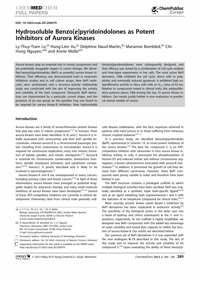
We previously identified benzo[e]pyridoindoles 1 and 2 aspotent inhibitors of aurora kinases by high throughput screen-ing, and their activities were confirmed both in vitro and incells.[14] Major features of 1 and 2 are described in table S1(Supporting Information). In the order to improve hit 1, we de-cided to define the important requirements for aurora kinase Binhibition. We therefore increased the number of availablebenzo[e]pyridoindoles by varying positions 3, 8, and 11(Scheme 1 A); syntheses illustrated in Scheme 1 B.
Requirements for aurora kinaseB inhibition in cells
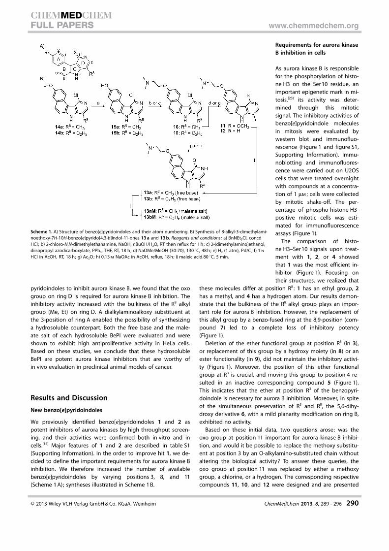
As aurora kinase B is responsiblefor the phosphorylation of histo-ne H3 on the Ser 10 residue, animportant epigenetic mark in mi-tosis,[23] its activity was deter-mined through this mitoticsignal. The inhibitory activities ofbenzo[e]pyridoindole moleculesin mitosis were evaluated bywestern blot and immunofluo-rescence (Figure 1 and figure S1,Supporting Information). Immu-noblotting and immunofluores-cence were carried out on U2OScells that were treated overnightwith compounds at a concentra-tion of 1 mm ; cells were collectedby mitotic shake-off. The per-centage of phospho-histone H3-positive mitotic cells was esti-mated for immunofluorescenceassays (Figure 1).
The comparison of histo-ne H3–Ser 10 signals upon treat-ment with 1, 2, or 4 showedthat 1 was the most efficient in-hibitor (Figure 1). Focusing ontheir structures, we realized that
these molecules differ at position R8 : 1 has an ethyl group, 2has a methyl, and 4 has a hydrogen atom. Our results demon-strate that the bulkiness of the R8 alkyl group plays an impor-tant role for aurora B inhibition. However, the replacement ofthis alkyl group by a benzo-fused ring at the 8,9-position (com-pound 7) led to a complete loss of inhibitory potency(Figure 1).
Deletion of the ether functional group at position R3 (in 3),or replacement of this group by a hydroxy moiety (in 8) or anester functionality (in 9), did not maintain the inhibitory activi-ty (Figure 1). Moreover, the position of this ether functionalgroup at R3 is crucial, and moving this group to position 4 re-sulted in an inactive corresponding compound 5 (Figure 1).This indicates that the ether at position R3 of the benzopyri-doindole is necessary for aurora B inhibition. Moreover, in spiteof the simultaneous preservation of R3 and R8, the 5,6-dihy-droxy derivative 6, with a mild planarity modification on ring B,exhibited no activity.
Based on these initial data, two questions arose: was theoxo group at position 11 important for aurora kinase B inhibi-tion, and would it be possible to replace the methoxy substitu-ent at position 3 by an O-alkylamino-substituted chain withoutaltering the biological activity? To answer these queries, theoxo group at position 11 was replaced by either a methoxygroup, a chlorine, or a hydrogen. The corresponding respectivecompounds 11, 10, and 12 were designed and are presented
Scheme 1. A) Structure of benzo[e]pyridoindoles and their atom numbering. B) Synthesis of 8-alkyl-3-dimethylami-noethoxy-7H-10H-benzo[e]pyrido[4,3-b]indol-11-ones 13 a and 13 b. Reagents and conditions : a) BnNEt3Cl, concdHCl; b) 2-chloro-N,N-dimethylethanamine, NaOH, nBuOH/H2O, RT then reflux for 1 h; c) 2-(dimethylamino)ethanol,diisopropyl azodicarboxylate, PPh3, THF, RT, 18 h; d) NaOMe/MeOH (30:70), 130 8C, 48 h; e) H2 (1 atm), Pd/C; f) 1 n
HCl in AcOH, RT, 18 h; g) Ac2O; h) 0.13 m NaOAc in AcOH, reflux, 18 h; i) maleic acid.80 8C, 5 min.
� 2013 Wiley-VCH Verlag GmbH & Co. KGaA, Weinheim ChemMedChem 2013, 8, 289 – 296 290
CHEMMEDCHEMFULL PAPERS www.chemmedchem.org
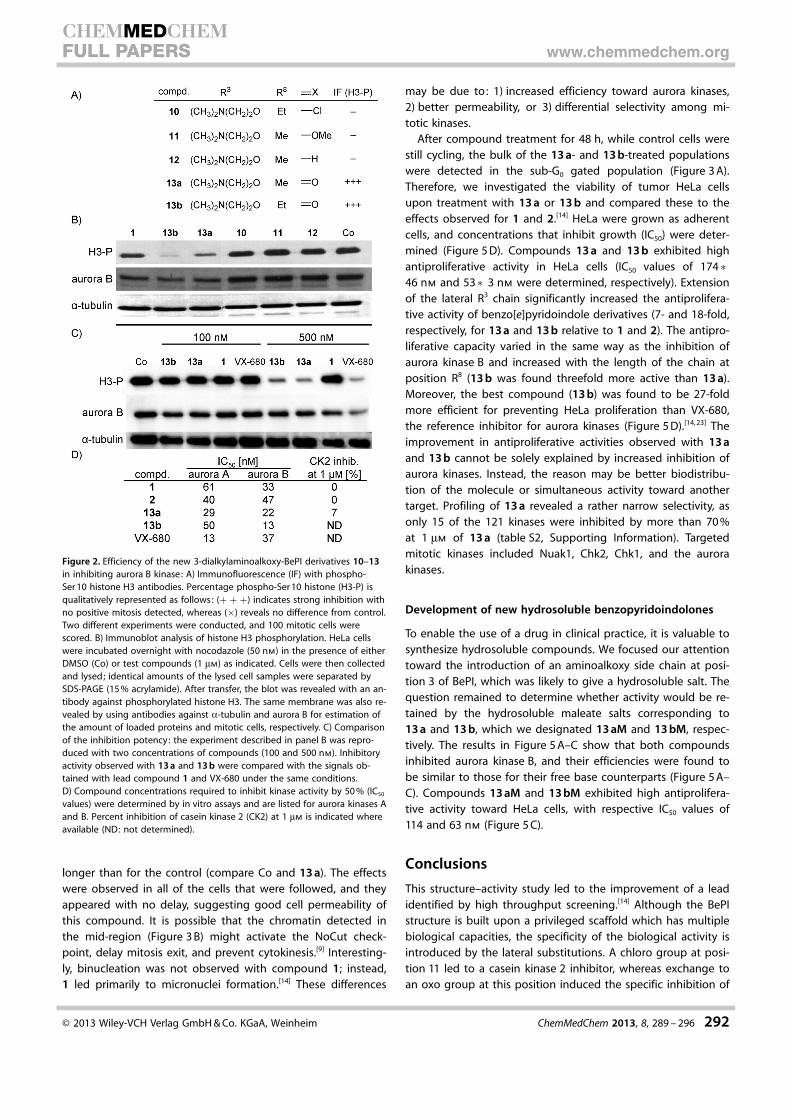
in Figure 2 A. These compounds 10–12 and their oxo counter-parts 13 a and 13 b, fitted with a lateral chain [-O-CH2CH2N-(CH3)2] at position 3, allowed us to study the SAR within thisfamily of aurora inhibitors. However, substitution of the pyrroleamine with a methyl group resulted in an inactive compound(data not shown).
The ability of these benzo[e]pyridoindole molecules to inhib-it histone H3 phosphorylation in mitosis was evaluated by im-munofluorescence (Figure 2 A) and western blotting (Fig-ure 2 B). Two compounds, 13 a and 13 b, were found to bemore potent than lead compound 1 (Figure 2 B and C). A com-parison of the activities of these five new molecules (10–13 aand 13 b) clearly revealed that the oxo group at position 3 isrequired for aurora kinase B inhibition. Indeed, compounds 11and 12, which differ from 13 a by replacement of the oxogroup with an ether group (OMe) and a hydrogen atom, re-spectively, had no inhibitory activity (Figure 2). Similarly, chloroderivative 10 was inactive when we compared its activity with13 b, the most potent aurora B inhibitor in this series. Evalua-tion of these molecules confirmed the correlation between in-hibitory potency and the bulkiness of the R8 alkyl substituentobserved in the former series (13 b was slightly superior to13 a, while 1 was better than 2 and 4).[14] The evaluation of H3phosphorylation signals at different compound concentrationsrevealed that in cells, 13 b inhibited aurora kinase B at a levelsimilar to VX-680, the reference aurora kinase inhibitor.[24]
Inhibitory activities toward recombinant aurora kin-ases confirmed that 13 b is a more powerful aurora Binhibitor than 13 a (IC50 values of 13 nm versus22 nm, see Figure 2 D). Neither compound exhibitedselectivity toward the aurora kinases, with aurora Aalso inhibited at nanomolar levels in vitro (Figure 2 D).Compound 13 b was found to be approximatelythreefold more potent than lead compound1 toward aurora B. Benzo[e]pyridoindoles bearinga methyl at position 8 (2 and 13 a) inhibited aurorakinases A and B to the same extent, whereas 1 and13 b, with an ethyl substituent at this position, weremore efficient toward aurora kinase B than aurora A(Figure 2 D). Conversely, VX-680 inhibited aurora Athreefold more efficiently than aurora B (Figure 2 D).Moreover, casein kinase 2, targeted by BePIs with hy-droxy and chloro substituents at the 3- and 11-posi-tions, respectively[14, 22] was not affected by 1 or 13 a(Figure 2 D). In summary, within the BePI family, therequirements for an aurora kinase B inhibitor are asfollows: an oxo group at position 11, a large alkylsubstituent at R8, and an ether group at R3. The R3
site enables addition of a long chain without affect-ing aurora kinase activity, a possibility that was laterexploited for deriving hydrosoluble salts correspond-ing to 13 a and 13 b.
Outcome of cells treated by dialkylaminoalkoxy-benzo[e]pyridoindolones 13 a and 13 b
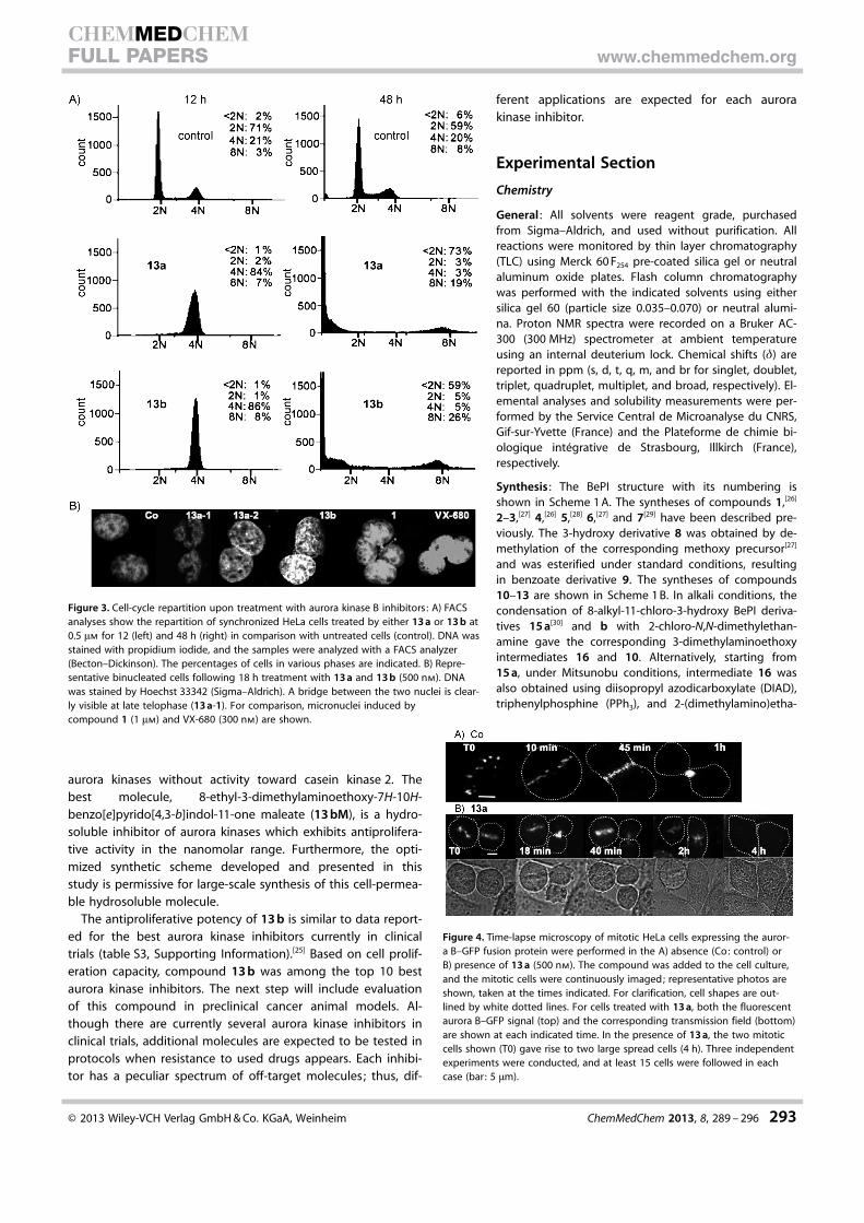
Next, we tested the behavior of cells treated withcompounds 13 a and 13 b. HeLa cells were synchronized at theS phase, and the compounds were added upon release. The re-sults of experiments following 12 and 48 h of compound treat-ment are presented in Figure 3 A. After 12 h, cells in the controlentered a new cycle (71 % in G1) whereas 13 a- and 13 b-treat-ed cells were primarily arrested in G2/M populations (84 and86 %, respectively). These 4N populations were identified byimmunofluorescence as binucleate cells (Figure 3 B). The con-nection between the two nuclei was clearly visible in late telo-phase (Figure 3 B, 13 a-1). Conversely, compound 1 and VX-680gave rise to micronuclei as a result of mitotic exit following de-layed metaphase (Figure 3 B).[14] At micromolar concentrations,13 a and 13 b prevented ongoing mitosis, thus confirming theinhibition of mitotic target(s).
To understand how these binucleate cells arose, we per-formed time-lapse microscopy on HeLa cells stably expressingaurora B–GFP. Cells were treated with either DMSO (control) orwith compound (500 nm) and were imaged immediately. Inthe control experiments, aurora B–GFP localized on alignedcentromeres (Figure 4, 10 min), followed by relocation to themidzone (~45 min) and final concentration at the mid-body(1 h). In the presence of 13 a, the centromeres aligned quitenormally (T0 and 18 min), and aurora B appeared to localize inthe midzone at anaphase onset (40 min, 2 h). However, thetwo daughter cells did not separate, and each mitotic cell gaverise to a large spread cell (4 h). The duration of mitosis was
Figure 1. Inhibition of aurora kinase B by benzo[e]pyridoindole molecules 1–9 with anoxo group at position 11: U2OS cells were treated overnight with the test compounds at1 mm in the presence of nocodazole (50 nm). Aurora kinase B activity was evaluated bothby western blots and by immunofluorescence (IF) with a specific antibody against phos-phoserine 10 histone H3 (H3-P). For western blots, the efficiency of the test molecule (M)is compared with control (Co, DMSO), and actin is used as an internal loading control.Data are reported as follows: (+ + +): strong inhibition, (+ +): significant inhibition, and(+ /�): a slight decrease in signal, whereas (�) reveals no difference from control. Thefull western blots are given in figure S1 (Supporting Information). For immunofluores-cence, the percentage of positive phospho-Ser 10 histone H3 is indicated. Two differentexperiments were conducted, and 100 mitotic cells were scored.
� 2013 Wiley-VCH Verlag GmbH & Co. KGaA, Weinheim ChemMedChem 2013, 8, 289 – 296 291
CHEMMEDCHEMFULL PAPERS www.chemmedchem.org
longer than for the control (compare Co and 13 a). The effectswere observed in all of the cells that were followed, and theyappeared with no delay, suggesting good cell permeability ofthis compound. It is possible that the chromatin detected inthe mid-region (Figure 3 B) might activate the NoCut check-point, delay mitosis exit, and prevent cytokinesis.[9] Interesting-ly, binucleation was not observed with compound 1; instead,1 led primarily to micronuclei formation.[14] These differences
may be due to: 1) increased efficiency toward aurora kinases,2) better permeability, or 3) differential selectivity among mi-totic kinases.
After compound treatment for 48 h, while control cells werestill cycling, the bulk of the 13 a- and 13 b-treated populationswere detected in the sub-G0 gated population (Figure 3 A).Therefore, we investigated the viability of tumor HeLa cellsupon treatment with 13 a or 13 b and compared these to theeffects observed for 1 and 2.[14] HeLa were grown as adherentcells, and concentrations that inhibit growth (IC50) were deter-mined (Figure 5 D). Compounds 13 a and 13 b exhibited highantiproliferative activity in HeLa cells (IC50 values of 174�46 nm and 53�3 nm were determined, respectively). Extensionof the lateral R3 chain significantly increased the antiprolifera-tive activity of benzo[e]pyridoindole derivatives (7- and 18-fold,respectively, for 13 a and 13 b relative to 1 and 2). The antipro-liferative capacity varied in the same way as the inhibition ofaurora kinase B and increased with the length of the chain atposition R8 (13 b was found threefold more active than 13 a).Moreover, the best compound (13 b) was found to be 27-foldmore efficient for preventing HeLa proliferation than VX-680,the reference inhibitor for aurora kinases (Figure 5 D).[14, 23] Theimprovement in antiproliferative activities observed with 13 aand 13 b cannot be solely explained by increased inhibition ofaurora kinases. Instead, the reason may be better biodistribu-tion of the molecule or simultaneous activity toward anothertarget. Profiling of 13 a revealed a rather narrow selectivity, asonly 15 of the 121 kinases were inhibited by more than 70 %at 1 mm of 13 a (table S2, Supporting Information). Targetedmitotic kinases included Nuak1, Chk2, Chk1, and the aurorakinases.
Development of new hydrosoluble benzopyridoindolones
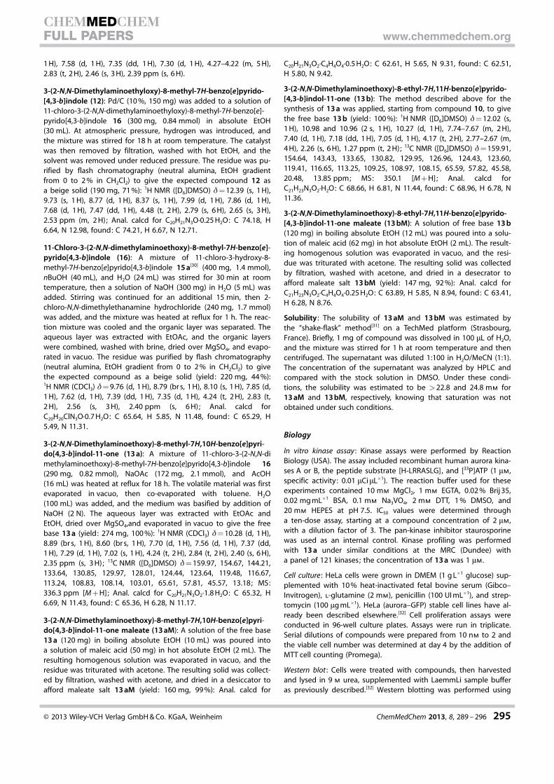
To enable the use of a drug in clinical practice, it is valuable tosynthesize hydrosoluble compounds. We focused our attentiontoward the introduction of an aminoalkoxy side chain at posi-tion 3 of BePI, which was likely to give a hydrosoluble salt. Thequestion remained to determine whether activity would be re-tained by the hydrosoluble maleate salts corresponding to13 a and 13 b, which we designated 13 aM and 13 bM, respec-tively. The results in Figure 5 A–C show that both compoundsinhibited aurora kinase B, and their efficiencies were found tobe similar to those for their free base counterparts (Figure 5 A–C). Compounds 13 aM and 13 bM exhibited high antiprolifera-tive activity toward HeLa cells, with respective IC50 values of114 and 63 nm (Figure 5 C).
Conclusions
This structure–activity study led to the improvement of a leadidentified by high throughput screening.[14] Although the BePIstructure is built upon a privileged scaffold which has multiplebiological capacities, the specificity of the biological activity isintroduced by the lateral substitutions. A chloro group at posi-tion 11 led to a casein kinase 2 inhibitor, whereas exchange toan oxo group at this position induced the specific inhibition of
Figure 2. Efficiency of the new 3-dialkylaminoalkoxy-BePI derivatives 10–13in inhibiting aurora B kinase: A) Immunofluorescence (IF) with phospho-Ser 10 histone H3 antibodies. Percentage phospho-Ser 10 histone (H3-P) isqualitatively represented as follows: (+ + +) indicates strong inhibition withno positive mitosis detected, whereas (�) reveals no difference from control.Two different experiments were conducted, and 100 mitotic cells werescored. B) Immunoblot analysis of histone H3 phosphorylation. HeLa cellswere incubated overnight with nocodazole (50 nm) in the presence of eitherDMSO (Co) or test compounds (1 mm) as indicated. Cells were then collectedand lysed; identical amounts of the lysed cell samples were separated bySDS-PAGE (15 % acrylamide). After transfer, the blot was revealed with an an-tibody against phosphorylated histone H3. The same membrane was also re-vealed by using antibodies against a-tubulin and aurora B for estimation ofthe amount of loaded proteins and mitotic cells, respectively. C) Comparisonof the inhibition potency: the experiment described in panel B was repro-duced with two concentrations of compounds (100 and 500 nm). Inhibitoryactivity observed with 13 a and 13 b were compared with the signals ob-tained with lead compound 1 and VX-680 under the same conditions.D) Compound concentrations required to inhibit kinase activity by 50 % (IC50
values) were determined by in vitro assays and are listed for aurora kinases Aand B. Percent inhibition of casein kinase 2 (CK2) at 1 mm is indicated whereavailable (ND: not determined).
� 2013 Wiley-VCH Verlag GmbH & Co. KGaA, Weinheim ChemMedChem 2013, 8, 289 – 296 292
CHEMMEDCHEMFULL PAPERS www.chemmedchem.org
aurora kinases without activity toward casein kinase 2. Thebest molecule, 8-ethyl-3-dimethylaminoethoxy-7H-10H-benzo[e]pyrido[4,3-b]indol-11-one maleate (13 bM), is a hydro-soluble inhibitor of aurora kinases which exhibits antiprolifera-tive activity in the nanomolar range. Furthermore, the opti-mized synthetic scheme developed and presented in thisstudy is permissive for large-scale synthesis of this cell-permea-ble hydrosoluble molecule.
The antiproliferative potency of 13 b is similar to data report-ed for the best aurora kinase inhibitors currently in clinicaltrials (table S3, Supporting Information).[25] Based on cell prolif-eration capacity, compound 13 b was among the top 10 bestaurora kinase inhibitors. The next step will include evaluationof this compound in preclinical cancer animal models. Al-though there are currently several aurora kinase inhibitors inclinical trials, additional molecules are expected to be tested inprotocols when resistance to used drugs appears. Each inhibi-tor has a peculiar spectrum of off-target molecules; thus, dif-
ferent applications are expected for each aurorakinase inhibitor.
Experimental Section
Chemistry
General : All solvents were reagent grade, purchasedfrom Sigma–Aldrich, and used without purification. Allreactions were monitored by thin layer chromatography(TLC) using Merck 60 F254 pre-coated silica gel or neutralaluminum oxide plates. Flash column chromatographywas performed with the indicated solvents using eithersilica gel 60 (particle size 0.035–0.070) or neutral alumi-na. Proton NMR spectra were recorded on a Bruker AC-300 (300 MHz) spectrometer at ambient temperatureusing an internal deuterium lock. Chemical shifts (d) arereported in ppm (s, d, t, q, m, and br for singlet, doublet,triplet, quadruplet, multiplet, and broad, respectively). El-emental analyses and solubility measurements were per-formed by the Service Central de Microanalyse du CNRS,Gif-sur-Yvette (France) and the Plateforme de chimie bi-ologique int�grative de Strasbourg, Illkirch (France),respectively.
Synthesis : The BePI structure with its numbering isshown in Scheme 1 A. The syntheses of compounds 1,[26]
2–3,[27] 4,[26] 5,[28] 6,[27] and 7[29] have been described pre-viously. The 3-hydroxy derivative 8 was obtained by de-methylation of the corresponding methoxy precursor[27]
and was esterified under standard conditions, resultingin benzoate derivative 9. The syntheses of compounds10–13 are shown in Scheme 1 B. In alkali conditions, thecondensation of 8-alkyl-11-chloro-3-hydroxy BePI deriva-tives 15 a[30] and b with 2-chloro-N,N-dimethylethan-amine gave the corresponding 3-dimethylaminoethoxyintermediates 16 and 10. Alternatively, starting from15 a, under Mitsunobu conditions, intermediate 16 wasalso obtained using diisopropyl azodicarboxylate (DIAD),triphenylphosphine (PPh3), and 2-(dimethylamino)etha-
Figure 3. Cell-cycle repartition upon treatment with aurora kinase B inhibitors: A) FACSanalyses show the repartition of synchronized HeLa cells treated by either 13 a or 13 b at0.5 mm for 12 (left) and 48 h (right) in comparison with untreated cells (control). DNA wasstained with propidium iodide, and the samples were analyzed with a FACS analyzer(Becton–Dickinson). The percentages of cells in various phases are indicated. B) Repre-sentative binucleated cells following 18 h treatment with 13 a and 13 b (500 nm). DNAwas stained by Hoechst 33342 (Sigma–Aldrich). A bridge between the two nuclei is clear-ly visible at late telophase (13 a-1). For comparison, micronuclei induced bycompound 1 (1 mm) and VX-680 (300 nm) are shown.
Figure 4. Time-lapse microscopy of mitotic HeLa cells expressing the auror-a B–GFP fusion protein were performed in the A) absence (Co: control) orB) presence of 13 a (500 nm). The compound was added to the cell culture,and the mitotic cells were continuously imaged; representative photos areshown, taken at the times indicated. For clarification, cell shapes are out-lined by white dotted lines. For cells treated with 13 a, both the fluorescentaurora B–GFP signal (top) and the corresponding transmission field (bottom)are shown at each indicated time. In the presence of 13 a, the two mitoticcells shown (T0) gave rise to two large spread cells (4 h). Three independentexperiments were conducted, and at least 15 cells were followed in eachcase (bar: 5 mm).
� 2013 Wiley-VCH Verlag GmbH & Co. KGaA, Weinheim ChemMedChem 2013, 8, 289 – 296 293
CHEMMEDCHEMFULL PAPERS www.chemmedchem.org
nol. These methods provide flexibility for the introduction of vari-ous side chains. The conversion of 11-chloro intermediate 16 tothe final pyridone 13 a was initially carried out in a two-step trans-formation via demethylation by HCl/AcOH of methoxy derivative11. Alternatively, experiments using either acetic anhydride oracetic acid in the presence of NaOAc were found to be convenient,as the final products, 13 a and 13 b, were obtained in a single stepfrom chloro intermediates 16 or 10 (Scheme 1 B). However, thelatter method is preferred, as it resulted in higher yields, and thepurification of the final compounds was easier. Finally, 11-unsubsti-tuted compound 12 was synthesized from chloro derivative 16 bypalladium-catalyzed hydrogenation. All compounds were fully char-acterized by MS and NMR. Microanalyses ensured the purity ofcompounds.
3-Hydroxy-8-methyl-7H,10H-benzo[e]pyrido[4,3-b]indol-11-one(8): A mixture of 3-methoxy-8-methyl-7H,10H-benzo[e]pyrido[4,3-b]indol-11-one[27] (83 mg, 0.3 mmol), benzyltriethylammonium chlo-ride (480 mg, 2.1 mmol) and 37 % HCl (4 mL) was heated in a 5 mLsealed tube in an oil bath at 140 8C for 24 h. The reaction mixturewas evaporated in vacuo, then H2O (10 mL) was added. The
medium was rendered basic by addition of 28 % NH4OH (1 mL),and the resulting solid was collected by filtration, washed withH2O, and then dried using a vacuum desiccator. The intermediate(3-hydroxy-8-methyl-7H,10H-benzo[e]pyrido[4,3-b]indol-11-one) wasobtained as a brown powder (yield: 50 mg, 63 % yield): 1H NMR([D6]DMSO) d= 11.95 (br s, 1 H), 10.88 and 10.86 (2 br s, 1 H), 10.18(d, 1 H), 9.38 (br s, 1 H), 7.68–7.50 (m, 2 H), 7.21–7.16 (m, 1 H), 7.12–7.03 (m, 2 H), 2.29 ppm (s, 3 H); MS: 265.2 [M + H]; Anal. calcd forC16H12N2O2·0.5 H2O: C 70.33, H 4.76, N 10.25, found: C 70.81, H 4.89,N 9.96.
3-Benzoyloxy-8-methyl-7H,10H-benzo[e]pyrido[4,3-b]indol-11-one (9): A mixture of 3-hydroxy-8-methyl-7H,10H-benzo[e]pyrido-[4,3-b]indol-11-one (35 mg, 0.13 mmol), benzoic anhydride(140 mg, 0.6 mmol), and pyridine (1 mL) was heated in an oil bathat 135 8C for 1 h. The volatile material was co-evaporated with tolu-ene in vacuo. H2O (5 mL) was added, and the medium alkalinizedwith solid NaHCO3. The liquid was discarded, and the residue waswashed with H2O (2 � 2 mL), then boiling EtOH was added (4 mL).The resulting precipitate was filtered, washed with a minimalamount of EtOH, and then dried in vacuo to give the expectedcompound (yield: 20 mg, 41 %): 1H NMR ([D6]DMSO) d= 12.22 (s,1 H), 11.03 & 11.01 (2 s, 1 H), 10.48 (d, 1 H), 9.26–8.19 (m, 2 H), 7.87(d, 1 H), 7.85–7.75 (m, 3 H), 7.68–7.61 (m, 2 H), 7.78 (dd, 1 H), 7.14 (d,1 H), 2.32 ppm (s, 3 H); MS: 367.1 [M�H]; Anal. calcd forC23H16N2O3·0.25 H2O: C 74.09, H 4.43, N 7.52, found: C 73.76, H 4.46,N 7.29.
11-Chloro-3-(2-N,N-dimethylaminoethoxy)-8-ethyl-7H-benzo[e]pyrido[4,3-b]indole (10): Step 1: In a sealed 50 mL tube,a mixture of 11-chloro-8-ethyl-3-methoxy-7H-benzo[e]pyrido[4,3-b]indole 14 b[26] (600 mg, 1.9 mmol), benzyltriethylammonium chlo-ride (2.80 g, 12 mmol), and 37 % HCl (45 mL) was heated in an oilbath at 140 8C for 24 h. The reaction mixture was evaporated in va-cuo, then H2O (10 mL) was added. The medium was basified by ad-dition of 28 % NH4OH (2 mL), and the resulting solid was collectedby filtration, then washed with H2O and dried using a vacuum des-iccator to yield intermediate 11-chloro-8-ethyl-3-hydroxy-7H-benzo[e]pyrido[4,3-b]indole (15 b) as a brown powder (yield:450 mg, 78 %); MS: 296.1/298.1 [M + H]; Anal. calcd forC17H13ClN2O·0.25 H2O: C 67.78, H 4.52, N 9.30, found: C 67.90, H4.62, N 9.56.
Step 2: The protocol described for the synthesis of compound 11-chloro-3-(2-N,N-dimethylaminoethyloxy)-8-methyl-7H-benzo[e]pyri-do[4,3-b]indole 16 by method 1 was applied, starting from 11-chloro-3-hydroxy-8-ethyl-7H-benzo[e]pyrido[4,3-b]indole and using2-chloro-N,N-dimethylethanamine hydrochloride to give the titlecompound (yield: 37 % yield): 1H NMR (CDCl3) d= 9.76 (d, 1 H), 8.73(br s, 1 H), 8.13 (s, 1 H), 7.86 (d, 1 H), 7.62 (d, 1 H), 7.43–7.34 (m, 2 H),4.25 (t, 2 H), 2.97 (q, 2 H), 2.84 (t, 2 H), 2.40 (s, 6 H), 1.46 ppm (t, 3 H);MS: 368.2 and 370.2 [M + H].
11-Methoxy-3-(2-N,N-dimethylaminoethoxy)-8-methyl-7H-benzo-[e]pyrido[4,3-b]indole (11): A mixture of 11-chloro-3-(2-N,N-di-methylaminoethoxy)-8-methyl-7H-benzo[e]pyrido[4,3-b]indole 16(80 mg, 0.22 mmol) and 30 % NaOMe in MeOH (12 mL) was heatedin a 25 mL sealed tube in an oil bath at 130 8C for 48 h. The reac-tion mixture was cooled, poured into H2O (30 mL), and extractedwith CH2Cl2. The organic layer was washed with brine, dried overMgSO4, and evaporated in vacuo. The residue was purified by flashchromatography (neutral alumina, EtOAc gradient from 0 to 20 %in CH2Cl2) to give the methoxy compound 11 (yield: 75 mg, 94 %):1H NMR (CDCl3) d= 9.64 (d, 1 H), 8.82 (br s, 1 H), 7.85 (s, 1 H), 7.73 (d,
Figure 5. Effect of hydrosoluble benzo[e]pyridoindolones (13 a and 13 b) incells : A) The maleate salts corresponding to benzo[e]pyridoindolones 13 aand 13 b are shown as 13 aM and 13 bM, respectively. Their potency to in-hibit aurora kinase B at 1 mm was evaluated in B) U2OS and C) HeLa cells (Te:Control). Blots were revealed by using an antibody against phospho-Ser 10histone H3 (H3-P) ; the same membrane was also revealed by using an anti-body against aurora kinase B for estimation of the number of mitotic cells.D) Cell growth and viability were tested under standard conditions in 96-well culture plates with the MTT cell viability assay (Promega). Average IC50
values �SD of three independent experiments are listed; data for 1, 2, andVX-680 were reported previously by Hoang et al. ,[14, 16] and are indicated forcomparison. The lateral chain of each molecule is noted, with the six mole-cules bearing an oxo group at position 11.
� 2013 Wiley-VCH Verlag GmbH & Co. KGaA, Weinheim ChemMedChem 2013, 8, 289 – 296 294
CHEMMEDCHEMFULL PAPERS www.chemmedchem.org
1 H), 7.58 (d, 1 H), 7.35 (dd, 1 H), 7.30 (d, 1 H), 4.27–4.22 (m, 5 H),2.83 (t, 2 H), 2.46 (s, 3 H), 2.39 ppm (s, 6 H).
3-(2-N,N-Dimethylaminoethyloxy)-8-methyl-7H-benzo[e]pyrido-[4,3-b]indole (12): Pd/C (10 %, 150 mg) was added to a solution of11-chloro-3-(2-N,N-dimethylaminoethyloxy)-8-methyl-7H-benzo[e]-pyrido[4,3-b]indole 16 (300 mg, 0.84 mmol) in absolute EtOH(30 mL). At atmospheric pressure, hydrogen was introduced, andthe mixture was stirred for 18 h at room temperature. The catalystwas then removed by filtration, washed with hot EtOH, and thesolvent was removed under reduced pressure. The residue was pu-rified by flash chromatography (neutral alumina, EtOH gradientfrom 0 to 2 % in CH2Cl2) to give the expected compound 12 asa beige solid (190 mg, 71 %): 1H NMR ([D6]DMSO) d= 12.39 (s, 1 H),9.73 (s, 1 H), 8.77 (d, 1 H), 8.37 (s, 1 H), 7.99 (d, 1 H), 7.86 (d, 1 H),7.68 (d, 1 H), 7.47 (dd, 1 H), 4.48 (t, 2 H), 2.79 (s, 6 H), 2.65 (s, 3 H),2.53 ppm (m, 2 H); Anal. calcd for C20H21N3O·0.25 H2O: C 74.18, H6.64, N 12.98, found: C 74.21, H 6.67, N 12.71.
11-Chloro-3-(2-N,N-dimethylaminoethoxy)-8-methyl-7H-benzo[e]-pyrido[4,3-b]indole (16): A mixture of 11-chloro-3-hydroxy-8-methyl-7H-benzo[e]pyrido[4,3-b]indole 15 a[30] (400 mg, 1.4 mmol),nBuOH (40 mL), and H2O (24 mL) was stirred for 30 min at roomtemperature, then a solution of NaOH (300 mg) in H2O (5 mL) wasadded. Stirring was continued for an additional 15 min, then 2-chloro-N,N-dimethylethanamine hydrochloride (240 mg, 1.7 mmol)was added, and the mixture was heated at reflux for 1 h. The reac-tion mixture was cooled and the organic layer was separated. Theaqueous layer was extracted with EtOAc, and the organic layerswere combined, washed with brine, dried over MgSO4, and evapo-rated in vacuo. The residue was purified by flash chromatography(neutral alumina, EtOH gradient from 0 to 2 % in CH2Cl2) to givethe expected compound as a beige solid (yield: 220 mg, 44 %):1H NMR (CDCl3) d= 9.76 (d, 1 H), 8.79 (br s, 1 H), 8.10 (s, 1 H), 7.85 (d,1 H), 7.62 (d, 1 H), 7.39 (dd, 1 H), 7.35 (d, 1 H), 4.24 (t, 2 H), 2.83 (t,2 H), 2.56 (s, 3 H), 2.40 ppm (s, 6 H); Anal. calcd forC20H20ClN3O·0.7 H2O: C 65.64, H 5.85, N 11.48, found: C 65.29, H5.49, N 11.31.
3-(2-N,N-Dimethylaminoethoxy)-8-methyl-7H,10H-benzo[e]pyri-do[4,3-b]indol-11-one (13 a): A mixture of 11-chloro-3-(2-N,N-dimethylaminoethoxy)-8-methyl-7H-benzo[e]pyrido[4,3-b]indole 16(290 mg, 0.82 mmol), NaOAc (172 mg, 2.1 mmol), and AcOH(16 mL) was heated at reflux for 18 h. The volatile material was firstevaporated in vacuo, then co-evaporated with toluene. H2O(100 mL) was added, and the medium was basified by addition ofNaOH (2 N). The aqueous layer was extracted with EtOAc andEtOH, dried over MgSO4,and evaporated in vacuo to give the freebase 13 a (yield: 274 mg, 100 %): 1H NMR (CDCl3) d= 10.28 (d, 1 H),8.89 (br s, 1 H), 8.60 (br s, 1 H), 7.70 (d, 1 H), 7.56 (d, 1 H), 7.37 (dd,1 H), 7.29 (d, 1 H), 7.02 (s, 1 H), 4.24 (t, 2 H), 2.84 (t, 2 H), 2.40 (s, 6 H),2.35 ppm (s, 3 H); 13C NMR ([D6]DMSO) d= 159.97, 154.67, 144.21,133.64, 130.85, 129.97, 128.01, 124.44, 123.64, 119.48, 116.67,113.24, 108.83, 108.14, 103.01, 65.61, 57.81, 45.57, 13.18; MS:336.3 ppm [M + H]; Anal. calcd for C20H21N3O2·1.8 H2O: C 65.32, H6.69, N 11.43, found: C 65.36, H 6.28, N 11.17.
3-(2-N,N-Dimethylaminoethoxy)-8-methyl-7H,10H-benzo[e]pyri-do[4,3-b]indol-11-one maleate (13 aM): A solution of the free base13 a (120 mg) in boiling absolute EtOH (10 mL) was poured intoa solution of maleic acid (50 mg) in hot absolute EtOH (2 mL). Theresulting homogenous solution was evaporated in vacuo, and theresidue was triturated with acetone. The resulting solid was collect-ed by filtration, washed with acetone, and dried in a desiccator toafford maleate salt 13 aM (yield: 160 mg, 99 %): Anal. calcd for
C20H21N3O2·C4H4O4·0.5 H2O: C 62.61, H 5.65, N 9.31, found: C 62.51,H 5.80, N 9.42.
3-(2-N,N-Dimethylaminoethoxy)-8-ethyl-7H,11H-benzo[e]pyrido-[4,3-b]indol-11-one (13 b): The method described above for thesynthesis of 13 a was applied, starting from compound 10, to givethe free base 13 b (yield: 100 %): 1H NMR ([D6]DMSO) d= 12.02 (s,1 H), 10.98 and 10.96 (2 s, 1 H), 10.27 (d, 1 H), 7.74–7.67 (m, 2 H),7.40 (d, 1 H), 7.18 (dd, 1 H), 7.05 (d, 1 H), 4.17 (t, 2 H), 2.77–2.67 (m,4 H), 2.26 (s, 6 H), 1.27 ppm (t, 2 H); 13C NMR ([D6]DMSO) d= 159.91,154.64, 143.43, 133.65, 130.82, 129.95, 126.96, 124.43, 123.60,119.41, 116.65, 113.25, 109.25, 108.97, 108.15, 65.59, 57.82, 45.58,20.48, 13.85 ppm; MS: 350.1 [M + H]; Anal. calcd forC21H23N3O2·H2O: C 68.66, H 6.81, N 11.44, found: C 68.96, H 6.78, N11.36.
3-(2-N,N-Dimethylaminoethoxy)-8-ethyl-7H,11H-benzo[e]pyrido-[4,3-b]indol-11-one maleate (13 bM): A solution of free base 13 b(120 mg) in boiling absolute EtOH (12 mL) was poured into a solu-tion of maleic acid (62 mg) in hot absolute EtOH (2 mL). The result-ing homogenous solution was evaporated in vacuo, and the resi-due was triturated with acetone. The resulting solid was collectedby filtration, washed with acetone, and dried in a desecrator toafford maleate salt 13 bM (yield: 147 mg, 92 %): Anal. calcd forC21H23N3O2·C4H4O4·0.25 H2O: C 63.89, H 5.85, N 8.94, found: C 63.41,H 6.28, N 8.76.
Solubility : The solubility of 13 aM and 13 bM was estimated bythe “shake-flask” method[31] on a TechMed platform (Strasbourg,France). Briefly, 1 mg of compound was dissolved in 100 mL of H2O,and the mixture was stirred for 1 h at room temperature and thencentrifuged. The supernatant was diluted 1:100 in H2O/MeCN (1:1).The concentration of the supernatant was analyzed by HPLC andcompared with the stock solution in DMSO. Under these condi-tions, the solubility was estimated to be >22.8 and 24.8 mm for13 aM and 13 bM, respectively, knowing that saturation was notobtained under such conditions.
Biology
In vitro kinase assay : Kinase assays were performed by ReactionBiology (USA). The assay included recombinant human aurora kina-ses A or B, the peptide substrate [H-LRRASLG], and [33P]ATP (1 mm,specific activity: 0.01 mCi mL�1). The reaction buffer used for theseexperiments contained 10 mm MgCl2, 1 mm EGTA, 0.02 % Brij 35,0.02 mg mL�1 BSA, 0.1 mm Na3VO4, 2 mm DTT, 1 % DMSO, and20 mm HEPES at pH 7.5. IC50 values were determined througha ten-dose assay, starting at a compound concentration of 2 mm,with a dilution factor of 3. The pan-kinase inhibitor staurosporinewas used as an internal control. Kinase profiling was performedwith 13 a under similar conditions at the MRC (Dundee) witha panel of 121 kinases; the concentration of 13 a was 1 mm.
Cell culture : HeLa cells were grown in DMEM (1 g L�1 glucose) sup-plemented with 10 % heat-inactivated fetal bovine serum (Gibco–Invitrogen), l-glutamine (2 mm), penicillin (100 UI mL�1), and strep-tomycin (100 mg mL�1). HeLa (aurora–GFP) stable cell lines have al-ready been described elsewhere.[32] Cell proliferation assays wereconducted in 96-well culture plates. Assays were run in triplicate.Serial dilutions of compounds were prepared from 10 nm to 2 andthe viable cell number was determined at day 4 by the addition ofMTT cell counting (Promega).
Western blot : Cells were treated with compounds, then harvestedand lysed in 9 m urea, supplemented with LaemmLi sample bufferas previously described.[32] Western blotting was performed using
� 2013 Wiley-VCH Verlag GmbH & Co. KGaA, Weinheim ChemMedChem 2013, 8, 289 – 296 295
CHEMMEDCHEMFULL PAPERS www.chemmedchem.org
the following antibodies: rabbit anti-phospho-histone H3 (Ser 10)(Upstate, 1:2000), rabbit anti-aurora B (Epitomics, 1:4000), mouseanti-actin (Sigma, 1:5000) and mouse anti-tubulin (Sigma, 1:5000).Bands were visualized using horseradish peroxidase-labeled anti-bodies and the ECL technique (Amersham Bioscience).
Cell-cycle analysis : HeLa cells were synchronized at the S phase ofthe cell cycle by serum starvation for 48 h and by subsequent thy-midine treatment (3 mm) for 20 h. After release, cells were treatedwith 13 a and 13 b (0.5 mm) for 12 h and 48 h. For determination ofcell-cycle profiles, cells were fixed by ice-cold 70 % EtOH for 1 h,then incubated with propidium iodide solution (50 mL�1) in thepresence of 0.2 mg mL�1 RNase for 15 min at 37 8C. DNA contentwas measured using a FACS flow cytometer (Becton–Dickinson,San Diego, CA, USA) and CellQuest software.
Immunofluorescence : U2OS cells were seeded on glass coverslipsand treated with compounds at a concentration of 1 mm for 20 h.Immunofluorescence was performed as described in Ref. [33]. Theprimary antibody used was phospho-histone H3 (Ser 10; Upstate,1:2000). Specific staining was revealed using Hylite Fluor 488-conju-gated secondary antibodies (Anaspec). DNA was visualized with0.1 mm Hoechst 33342 (Sigma–Aldrich). Images were collected witha ZEISS 510 laser scanning confocal microscope with a 63 � oil-im-mersion objective. Slices of 0.5 mm are shown.
Time-lapse experiments : Ex vivo experiments were conducted onHeLa cells grown on Lab-Tek chambered cover glass (Nalge NuncInternational) and maintained under standard culture conditions asdescribed in Ref. [14, 33] . Images were acquired on a Zeiss LSM510system using a Planapochromat 40 � water-immersion objective.GFP was excited with a 488 nm Argon 2 laser (power varying from0.1 to 2 %). In each experiment, approximately 15 mitotic cellswere followed, and three independent experiments were per-formed. Representative confocal slices are shown.
Acknowledgements
L.-T.-T.L. and H.-L.V. were supported by a Vietnam/France pro-gram. This work was supported by INSERM, UJF, CNRS, and Insti-tut Curie, and we also acknowledge La Ligue Nationale contre leCancer (Equipe labelis�e La Ligue, St�fan Dimitrov). Microscopyexperiments were conducted at the IBISA platform of the CRIINSERM/UJF U823. We thank Thierry Pagnier and Kiran Padma-nabhan (CNRS and INSERM, respectively) for editorial assistance.The authors declare no competing financial interests.
Keywords: antitumor agents · aurora kinases · cancer ·inhibitors · structure–activity relationships
[1] G. Vader, S. M. A. Lens, Biochim. Biophys. Acta. Rev. Cancer 2008, 1786,60 – 72.
[2] M. Carmena, S. Ruchaud, W. C. Earnshaw, Curr. Opin. Cell Biol. 2009, 21,796 – 805.
[3] T. Sardon, I. Peset, B. Petrova, I. Vernos, EMBO J. 2008, 27, 2567 – 2579.[4] E. Hannak, M. Kirkham, A. A. Hyman, K. Oegema, J. Cell Biol. 2001, 155,
1109 – 1116.
[5] A. Seki, J. A. Coppinger, C.-Y. Jang, J. R. Yates, G. Fang, Science 2008,320, 1655 – 1658.
[6] S. M. A. Lens, R. H. Medema, Cell Cycle 2003, 2, 507 – 510.[7] A. T. Saurin, M. S. van der Waal, R. H. Medema, S. M. A. Lens, G. J. P. L.
Kops, Nat. Commun. 2011, 2, 316.[8] M. Mendoza, C. Norden, K. R. Durrer, H. F. Uhlmann, Y. Barral, Nat. Cell
Biol. 2009, 11, 477 – 483.[9] S. Sen, H. Zhou, R. A. White, Oncogene 1997, 14, 2195 – 2200.
[10] L. Garuti, M. Roberti, G. Bottegoni, Curr. Med. Chem. 2009, 16, 1949 –1963.
[11] F. Girdler, K. E. Gascoigne, P. A. Eyers, S. Hartmuth, C. Crafter, K. M.Foote, N. J. Keen, S. S. Taylor, J. Cell Sci. 2006, 119, 3664 – 3675.
[12] J. R. Jackson, D. R. Patrick, M. M. Dar, P. S. Huang, Nat. Rev. Cancer 2007,7, 107 – 117.
[13] C. Soncini, P. Carpinelli, L. Gianellini, D. Fancelli, P. Vianello, L. Rusconi, P.Storici, P. Zugnoni, E. Pesenti, V. Croci, Clin. Cancer Res. 2006, 12, 4080 –4089.
[14] T. M.-N. Hoang, B. Favier, A. Valette, C. Barette, C. H. Nguyen, L. Lafane-ch�re, D. S. Grierson, S. Dimitrov, A. Molla, Cell Cycle 2009, 8, 765 – 772.
[15] G. M. T. Cheetham, P. A. Charlton, J. M. C. Golec, J. R. Pollard, Cancer Lett.2007, 251, 323 – 329.
[16] N. T. M. Hoang, M. Delacour-Larose, A. Molla, Curr. Enzyme Inhib. 2008,4, 153 – 159.
[17] H.-L. Vu, T. M. N. Hoang, B. Favier, A. Molla, Curr. Enzyme Inhib. 2010, 6,19 – 25.
[18] J. L. Mergny, G. Duval-Valentin, C. H. Nguyen, L. Perrouault, B. Faucon,M. Roug�e, T. Montenay-Garestier, E. Bisagni, C. H�l�ne, Science 1992,256, 1681 – 1684.
[19] C. Escud�, C. H. Nguyen, S. Kukreti, Y. Janin, J.-S. Sun, E. Bisagni, T. Gar-estier, C. H�l�ne, Proc. Natl. Acad. Sci. USA 1998, 95, 3591 – 3596.
[20] M. C. Bissery, C. H. Nguyen, E. Bisagni, P. Vrignaud, F. Lavelle, Invest. NewDrugs 1993, 11, 263 – 277.
[21] J. F. Riou, P. Foss�, C. H. Nguyen, A. K. Larsen, M. C. Bissery, L. Grondard,J. M. Saucier, E. Bisagni, F. Lavelle, Cancer Res. 1993, 53, 5987 – 5993.
[22] R. Prudent, V. Moucadel, C.-H. Nguyen, C. Barette, F. Schmidt, J.-C. Flor-ent, L. Lafanech�re, C. F. Sautel, E. Duchemin-Pelletier, E. Spreux, CancerRes. 2010, 70, 9865 – 9874.
[23] S. H. Baek, Mol. Cell 2011, 42, 274 – 284.[24] J. Bain, L. Plater, M. Elliott, N. Shpiro, C. J. Hastie, H. McLauchlan, I. Kle-
vernic, J. S. C. Arthur, D. R. Alessi, P. Cohen, Biochem. J. 2007, 408, 297 –315.
[25] M. Kollareddy, D. Zheleva, P. Dzubak, P. S. Brahmkshatriya, M. Lepsik, M.Hajduch, Invest. New Drugs 2012, 30, 2411 – 2432.
[26] C. H. Nguyen, E. Bisagni, F. Lavelle, M. C. Bissery, C. Huel, AnticancerDrug Des. 1992, 7, 219 – 233.
[27] C. H. Nguyen, J. M. Lhoste, F. Lavelle, M. C. Bissery, E. Bisagni, J. Med.Chem. 1990, 33, 1519 – 1528.
[28] C. Escud�, C. H. Nguyen, J.-L. Mergny, J.-S. Sun, E. Bisagni, T. Garestier, C.H�l�ne, J. Am. Chem. Soc. 1995, 117, 10212 – 10219.
[29] C. H. Nguyen, C. Marchand, S. Delage, J.-S. Sun, T. Garestier, C. H�l�ne,E. Bisagni, J. Am. Chem. Soc. 1998, 120, 2501 – 2507.
[30] S. Vinogradov, V. Roig, Z. Sergueeva, C. H. Nguyen, P. Arimondo, N. T.Thuong, E. Bisagni, J.-S. Sun, C. H�l�ne, U. Asseline, Bioconjugate Chem.2003, 14, 120 – 135.
[31] A. Avdeef, Adv. Drug Delivery Rev. 2007, 59, 568 – 590.[32] M. Delacour-Larose, M.-N. H. Thi, S. Dimitrov, A. Molla, Cell Cycle 2007, 6,
1878 – 1885.[33] M. Delacour-Larose, A. Molla, D. A. Skoufias, R. L. Margolis, S. Dimitrov,
Cell Cycle 2004, 3, 1418 – 1426.
Received: October 19, 2012
Published online on December 28, 2012
� 2013 Wiley-VCH Verlag GmbH & Co. KGaA, Weinheim ChemMedChem 2013, 8, 289 – 296 296
CHEMMEDCHEMFULL PAPERS www.chemmedchem.org
Related Documents