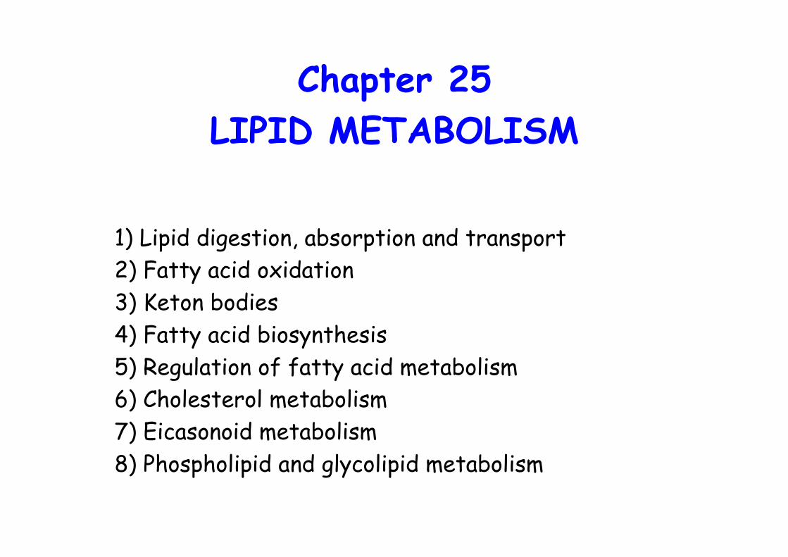
Chapter 25 LIPID METABOLISM 1) Lipid digestion, absorption and transport 2) Fatty acid oxidation 3) Keton bodies 4) Fatty acid biosynthesis 5) Regulation of fatty acid metabolism 6) Cholesterol metabolism 7) Eicasonoid metabolism 8) Phospholipid and glycolipid metabolism

Welcome message from author
This document is posted to help you gain knowledge. Please leave a comment to let me know what you think about it! Share it to your friends and learn new things together.
Transcript
Chapter 25LIPID METABOLISM
1) Lipid digestion, absorption and transport2) Fatty acid oxidation3) Keton bodies4) Fatty acid biosynthesis5) Regulation of fatty acid metabolism6) Cholesterol metabolism7) Eicasonoid metabolism8) Phospholipid and glycolipid metabolism
Lipid Digestion, Absorption andTransport
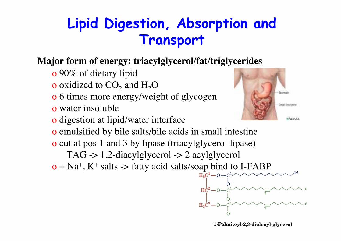
Major form of energy: triacylglycerol/fat/triglycerideso 90% of dietary lipido oxidized to CO2 and H2Oo 6 times more energy/weight of glycogeno water insolubleo digestion at lipid/water interfaceo emulsified by bile salts/bile acids in small intestineo cut at pos 1 and 3 by lipase (triacylglycerol lipase)
TAG -> 1,2-diacylglycerol -> 2 acylglycerolo + Na+, K+ salts -> fatty acid salts/soap bind to I-FABP
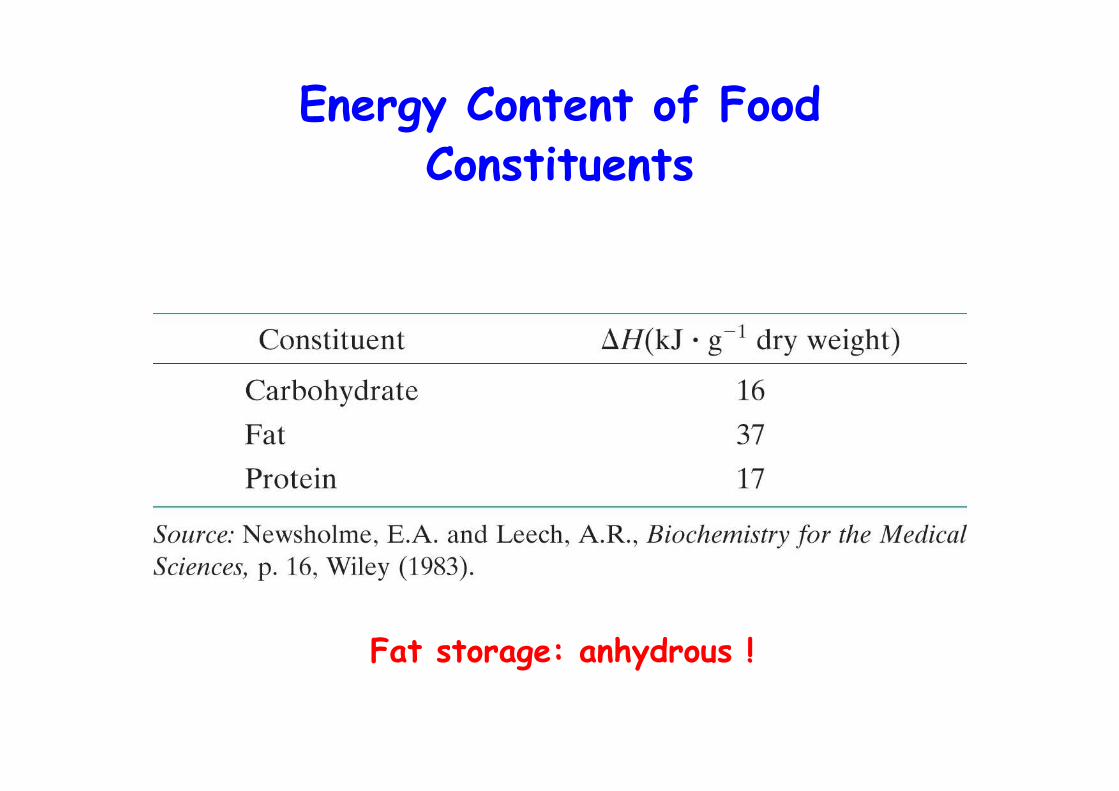
Energy Content of FoodConstituents
Fat storage: anhydrous !
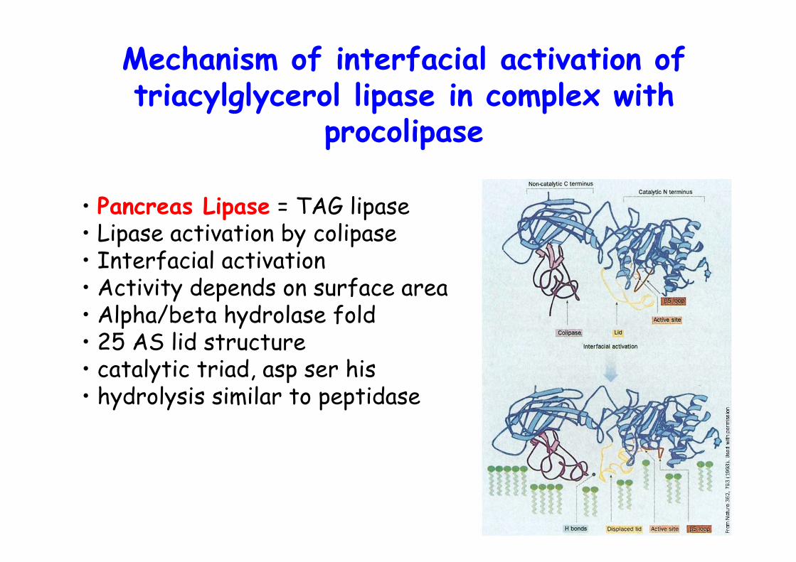
Mechanism of interfacial activation oftriacylglycerol lipase in complex with
procolipase
• Pancreas Lipase = TAG lipase• Lipase activation by colipase• Interfacial activation• Activity depends on surface area• Alpha/beta hydrolase fold• 25 AS lid structure• catalytic triad, asp ser his• hydrolysis similar to peptidase
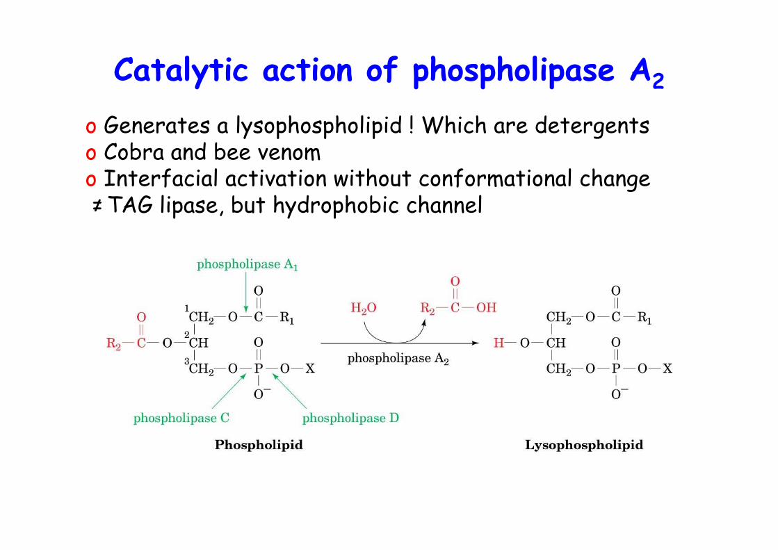
Catalytic action of phospholipase A2
o Generates a lysophospholipid ! Which are detergentso Cobra and bee venomo Interfacial activation without conformational change ≠TAG lipase, but hydrophobic channel
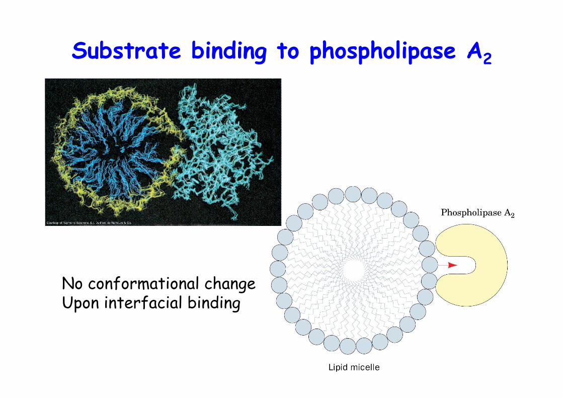
Substrate binding to phospholipase A2
No conformational changeUpon interfacial binding
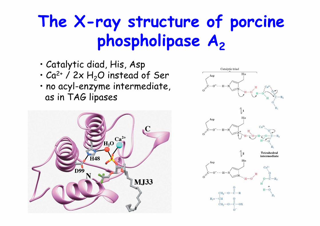
The X-ray structure of porcinephospholipase A2
• Catalytic diad, His, Asp• Ca2+ / 2x H2O instead of Ser• no acyl-enzyme intermediate, as in TAG lipases
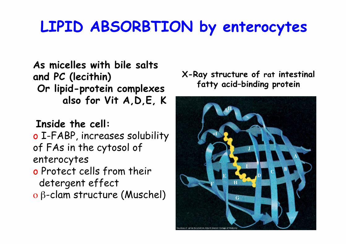
X-Ray structure of rat intestinalfatty acid–binding protein
As micelles with bile saltsand PC (lecithin) Or lipid-protein complexes
also for Vit A,D,E, K
Inside the cell:o I-FABP, increases solubilityof FAs in the cytosol ofenterocyteso Protect cells from their detergent effecto β-clam structure (Muschel)
LIPID ABSORBTION by enterocytes
Lipid transport
• Hydrolyzed lipids are absorbed by the intestinal mucosa• Converted back to triglycerides !• Packed into lipoprotein particles, chylomicrons• Released into lymph/blood -> delivered to tissue• Triglyceride made by liver is packaged into VLDL part. -> Released into blood• TAG hydrolyzed in periphery by lipoprotein lipase ->• FA uptake but glycerol back transport to liver and kidney• TAG in adipose tissue is mobilized by hormone-sensitive lipase -> free FA enter blood, bound to serum albumin
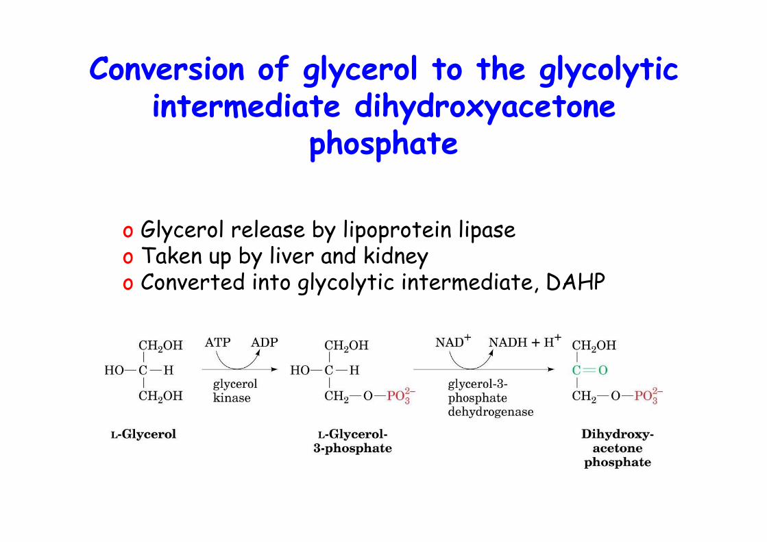
Conversion of glycerol to the glycolyticintermediate dihydroxyacetone
phosphate
o Glycerol release by lipoprotein lipaseo Taken up by liver and kidneyo Converted into glycolytic intermediate, DAHP
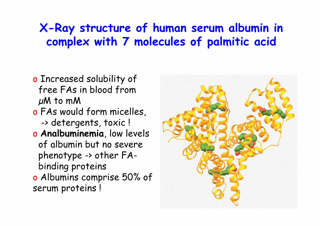
X-Ray structure of human serum albumin incomplex with 7 molecules of palmitic acid
o Increased solubility of free FAs in blood from µM to mMo FAs would form micelles, -> detergents, toxic !o Analbuminemia, low levels of albumin but no severe phenotype -> other FA- binding proteinso Albumins comprise 50% ofserum proteins !
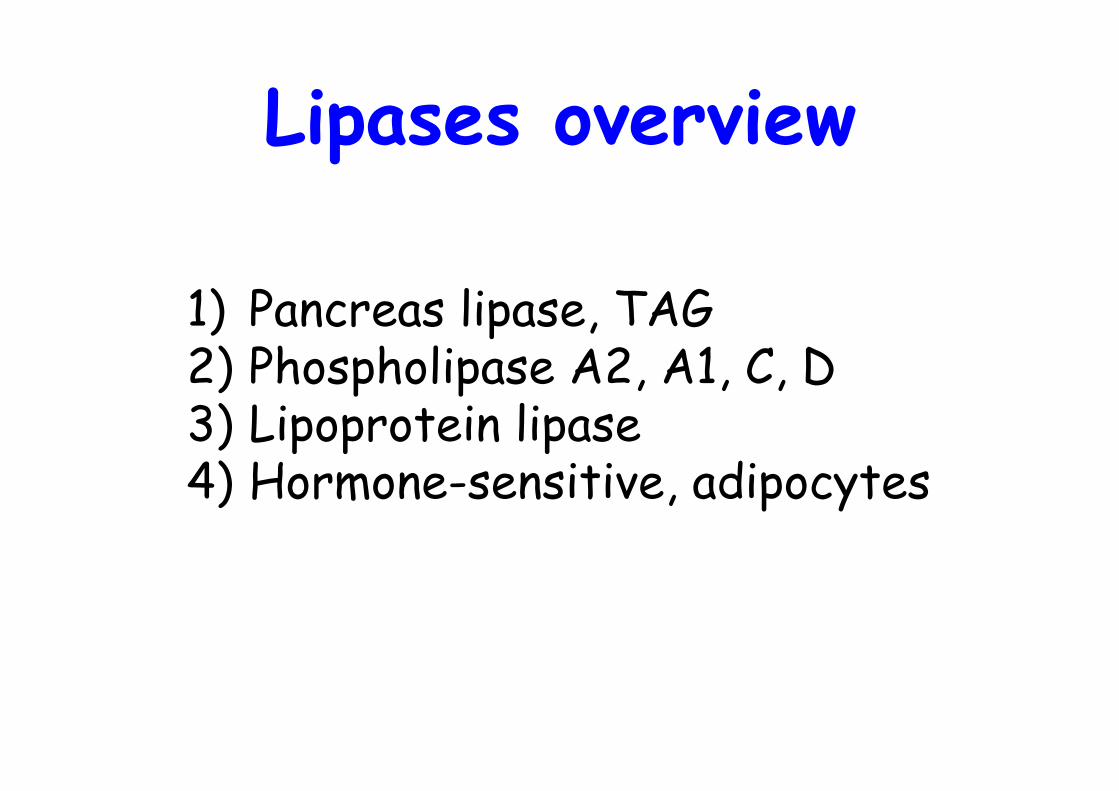
Lipases overview
1) Pancreas lipase, TAG2) Phospholipase A2, A1, C, D3) Lipoprotein lipase4) Hormone-sensitive, adipocytes
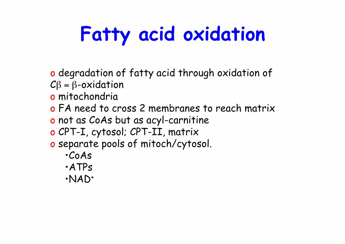
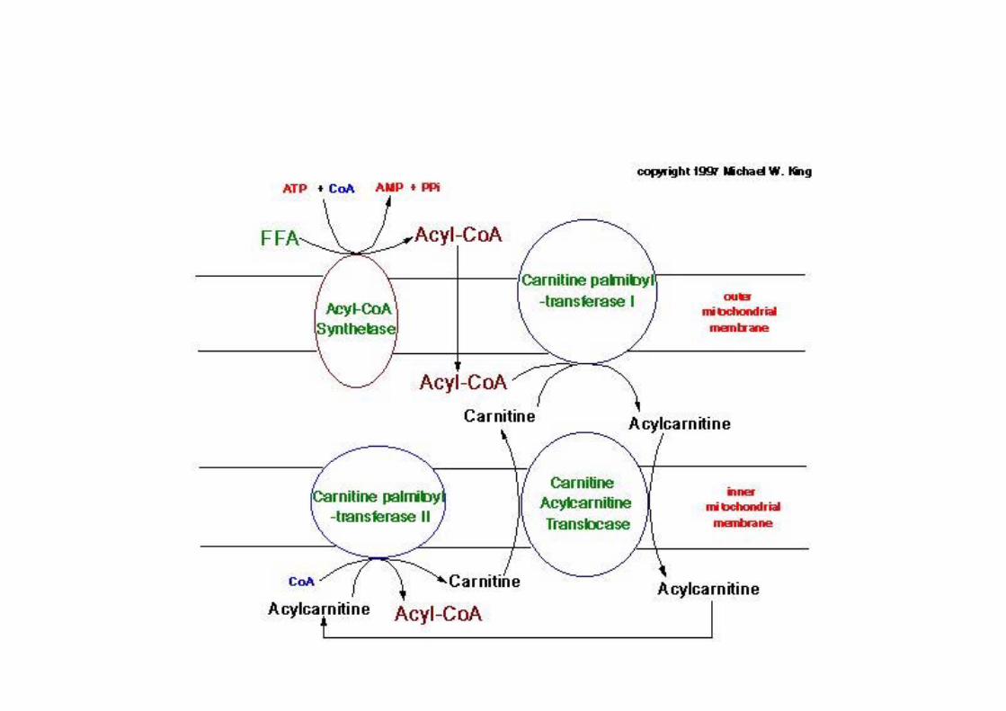
Fatty acid oxidation
o degradation of fatty acid through oxidation ofCβ = β-oxidationo mitochondriao FA need to cross 2 membranes to reach matrixo not as CoAs but as acyl-carnitineo CPT-I, cytosol; CPT-II, matrixo separate pools of mitoch/cytosol.
•CoAs•ATPs•NAD+

Franz Knoop’s classic experiment indicatingthat fatty acids are metabolically oxidized at
their β-carbon atom
o Phenyl-labelled even- or odd-numbered fatty acidso Feed to dogs -> what product appears in urine ?
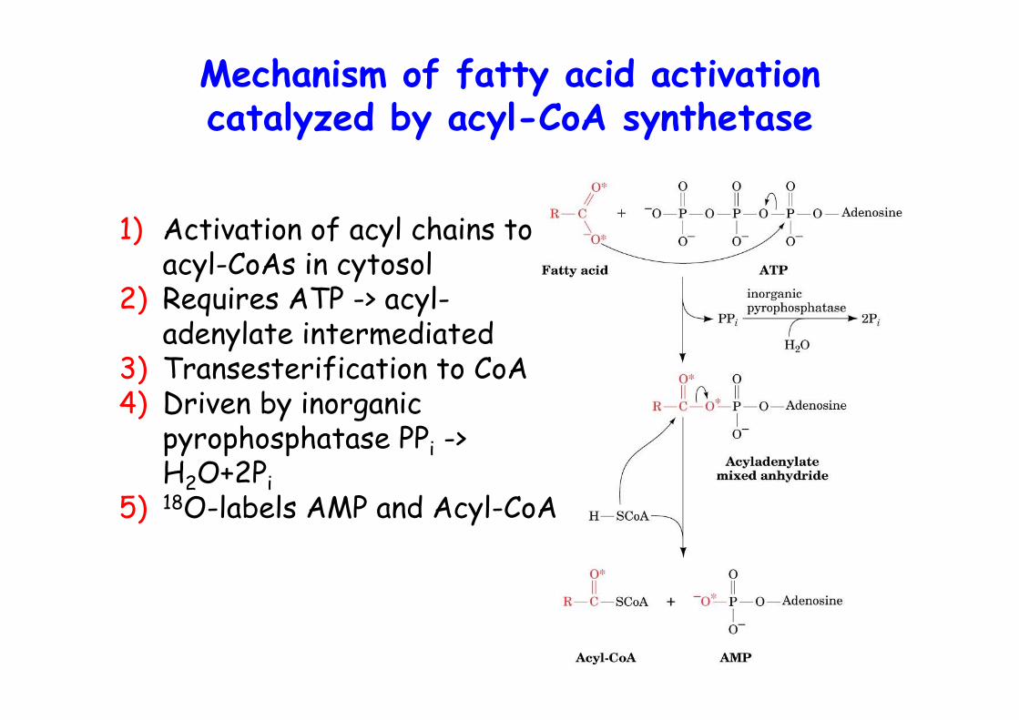
Mechanism of fatty acid activationcatalyzed by acyl-CoA synthetase
1) Activation of acyl chains toacyl-CoAs in cytosol
2) Requires ATP -> acyl-adenylate intermediated
3) Transesterification to CoA4) Driven by inorganic
pyrophosphatase PPi ->H2O+2Pi
5) 18O-labels AMP and Acyl-CoA
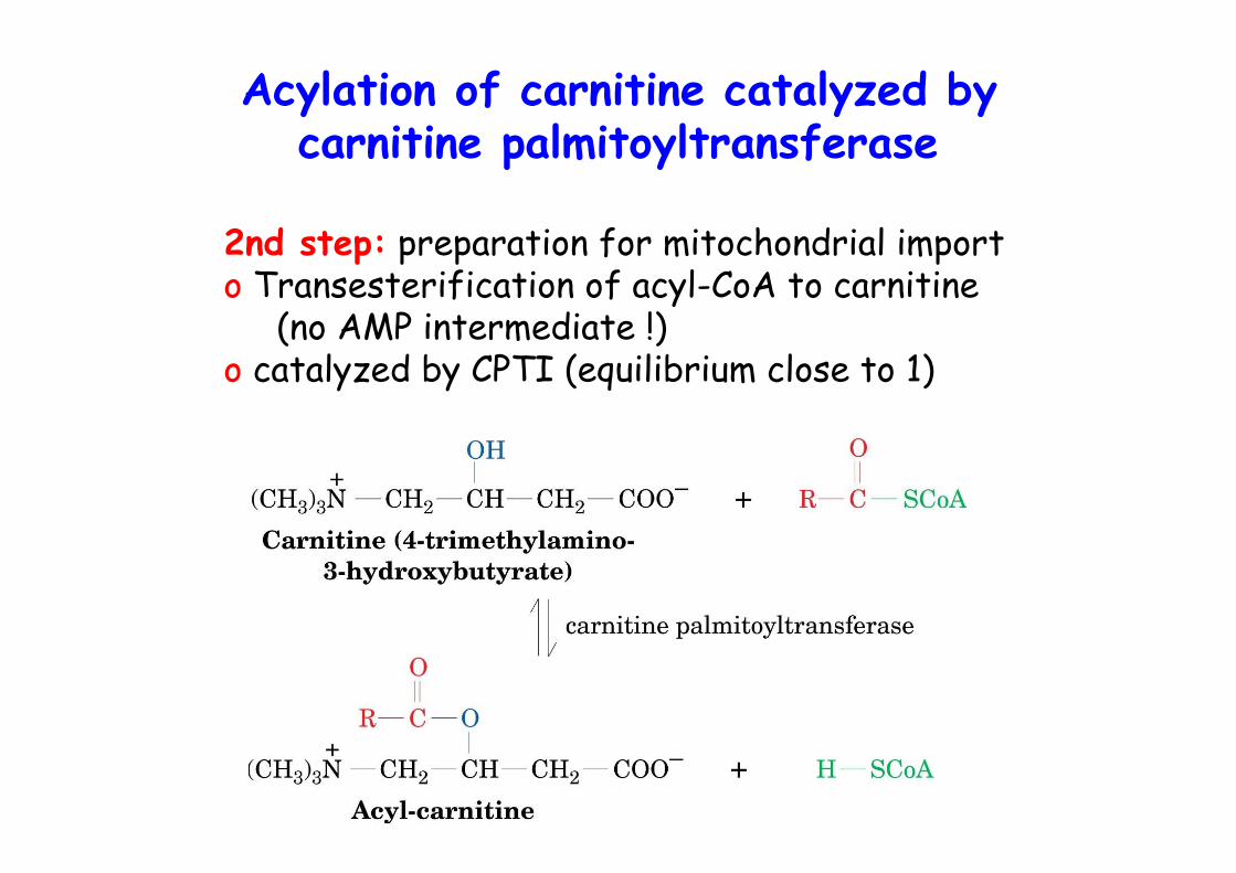
Acylation of carnitine catalyzed bycarnitine palmitoyltransferase
2nd step: preparation for mitochondrial importo Transesterification of acyl-CoA to carnitine
(no AMP intermediate !)o catalyzed by CPTI (equilibrium close to 1)
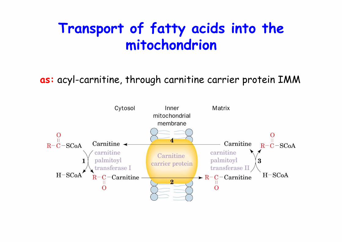
Transport of fatty acids into themitochondrion
as: acyl-carnitine, through carnitine carrier protein IMM
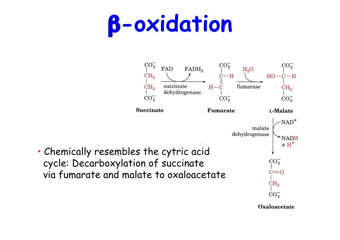
β-oxidation
• Chemically resembles the cytric acid cycle: Decarboxylation of succinate via fumarate and malate to oxaloacetate
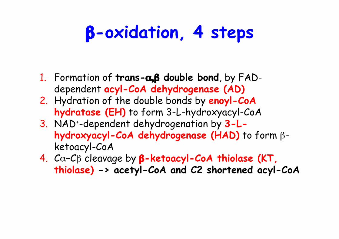
β-oxidation, 4 steps
1. Formation of trans-α,β double bond, by FAD-dependent acyl-CoA dehydrogenase (AD)
2. Hydration of the double bonds by enoyl-CoAhydratase (EH) to form 3-L-hydroxyacyl-CoA
3. NAD+-dependent dehydrogenation by 3-L-hydroxyacyl-CoA dehydrogenase (HAD) to form β-ketoacyl-CoA
4. Cα–Cβ cleavage by β-ketoacyl-CoA thiolase (KT,thiolase) -> acetyl-CoA and C2 shortened acyl-CoA

The β-oxidationpathway of fatty
acyl-CoA
Long chain versions of EH, HAD and KTs in α4β4ocatmeric protein, mitochobdrial trifunctionalprotein -> chanelling, no detectable intermediates
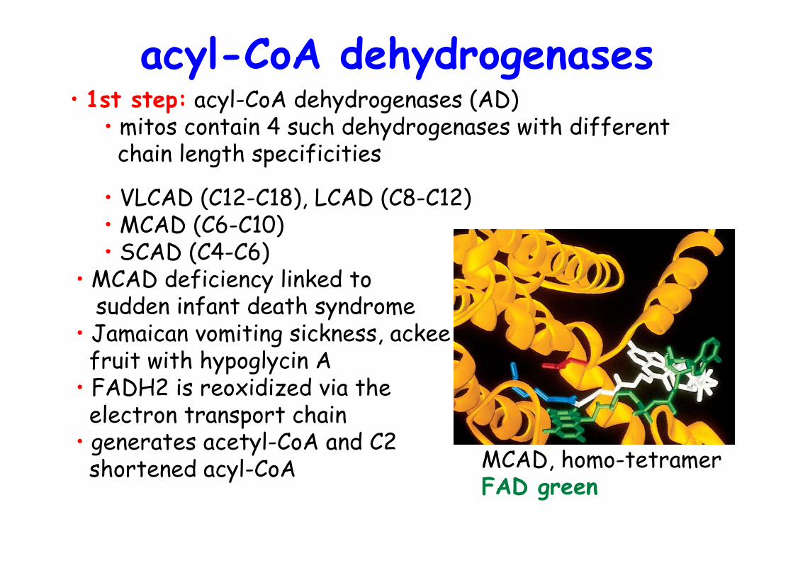
acyl-CoA dehydrogenases
• VLCAD (C12-C18), LCAD (C8-C12)• MCAD (C6-C10)• SCAD (C4-C6)
• MCAD deficiency linked to sudden infant death syndrome• Jamaican vomiting sickness, ackee fruit with hypoglycin A• FADH2 is reoxidized via the electron transport chain• generates acetyl-CoA and C2 shortened acyl-CoA
• 1st step: acyl-CoA dehydrogenases (AD)• mitos contain 4 such dehydrogenases with different chain length specificities
MCAD, homo-tetramerFAD green
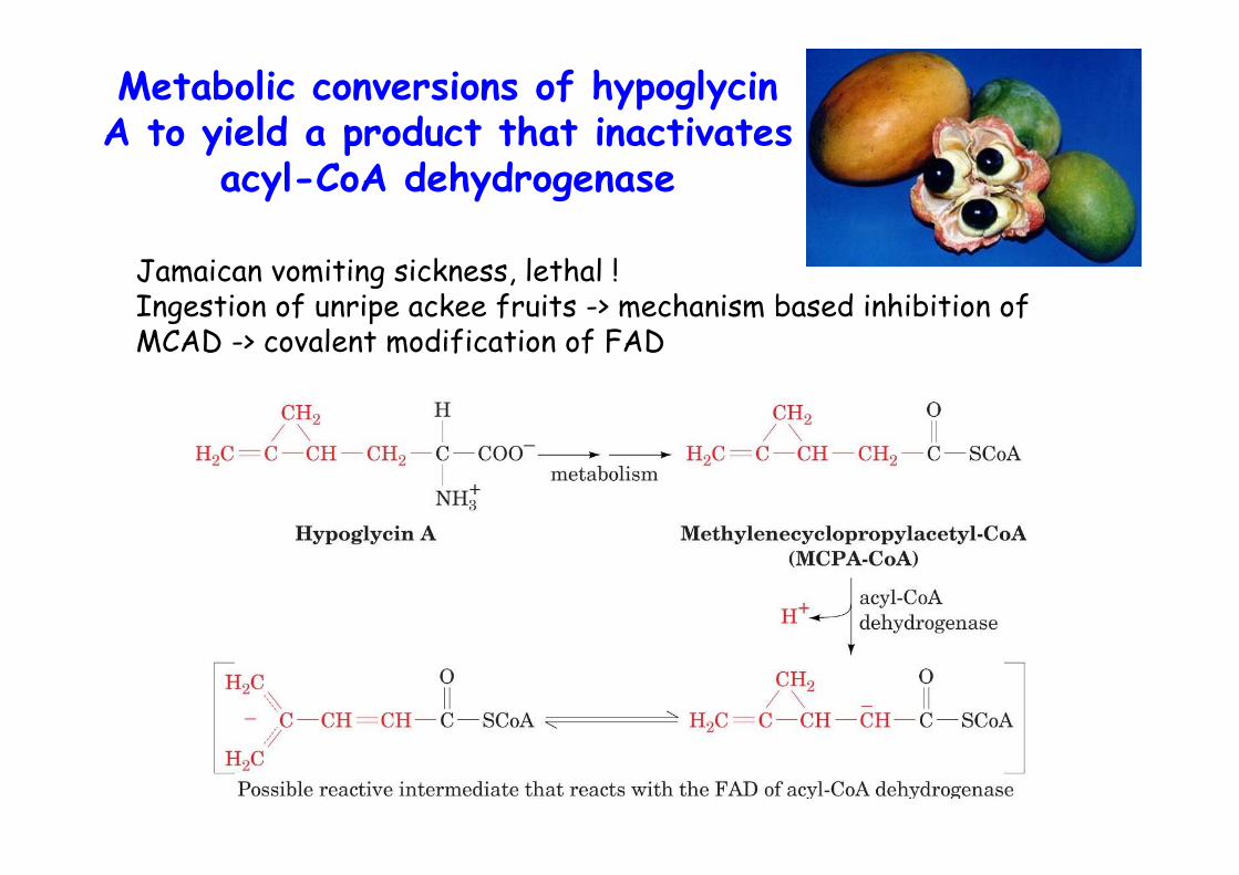
Metabolic conversions of hypoglycinA to yield a product that inactivates
acyl-CoA dehydrogenase
Jamaican vomiting sickness, lethal !Ingestion of unripe ackee fruits -> mechanism based inhibition ofMCAD -> covalent modification of FAD
Mitochondrial trifunctionalprotein
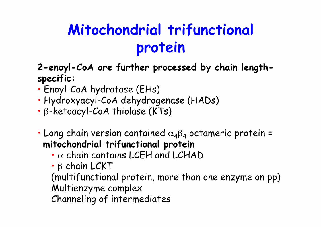
2-enoyl-CoA are further processed by chain length-specific:• Enoyl-CoA hydratase (EHs)• Hydroxyacyl-CoA dehydrogenase (HADs)• β-ketoacyl-CoA thiolase (KTs)
• Long chain version contained α4β4 octameric protein = mitochondrial trifunctional protein
• α chain contains LCEH and LCHAD• β chain LCKT(multifunctional protein, more than one enzyme on pp)Multienzyme complexChanneling of intermediates
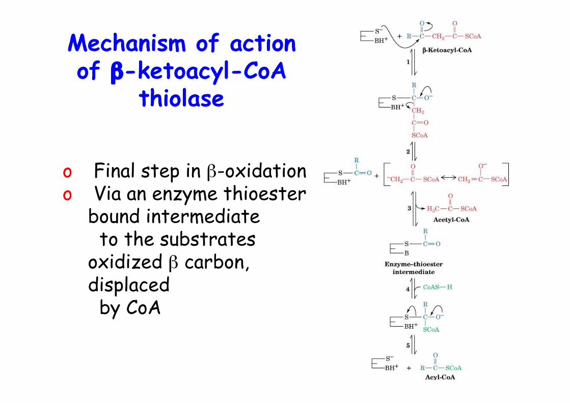
Mechanism of actionof β-ketoacyl-CoA
thiolase
o Final step in β-oxidationo Via an enzyme thioester
bound intermediate to the substratesoxidized β carbon,displaced by CoA
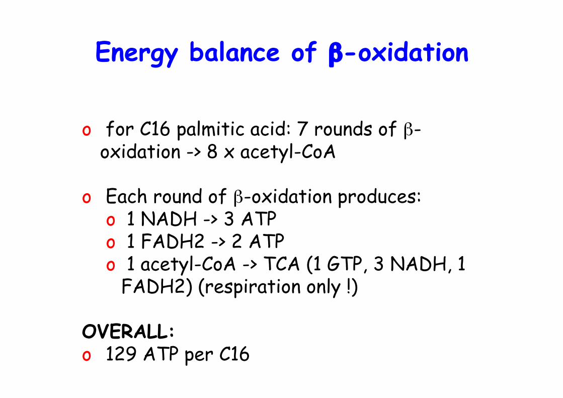
Energy balance of β-oxidation
o for C16 palmitic acid: 7 rounds of β-oxidation -> 8 x acetyl-CoA
o Each round of β-oxidation produces:o 1 NADH -> 3 ATPo 1 FADH2 -> 2 ATPo 1 acetyl-CoA -> TCA (1 GTP, 3 NADH, 1
FADH2) (respiration only !)
OVERALL:o 129 ATP per C16
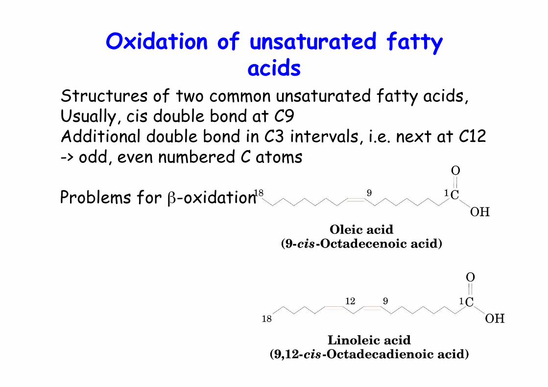
Oxidation of unsaturated fattyacids
Structures of two common unsaturated fatty acids,Usually, cis double bond at C9Additional double bond in C3 intervals, i.e. next at C12-> odd, even numbered C atoms
Problems for β-oxidation
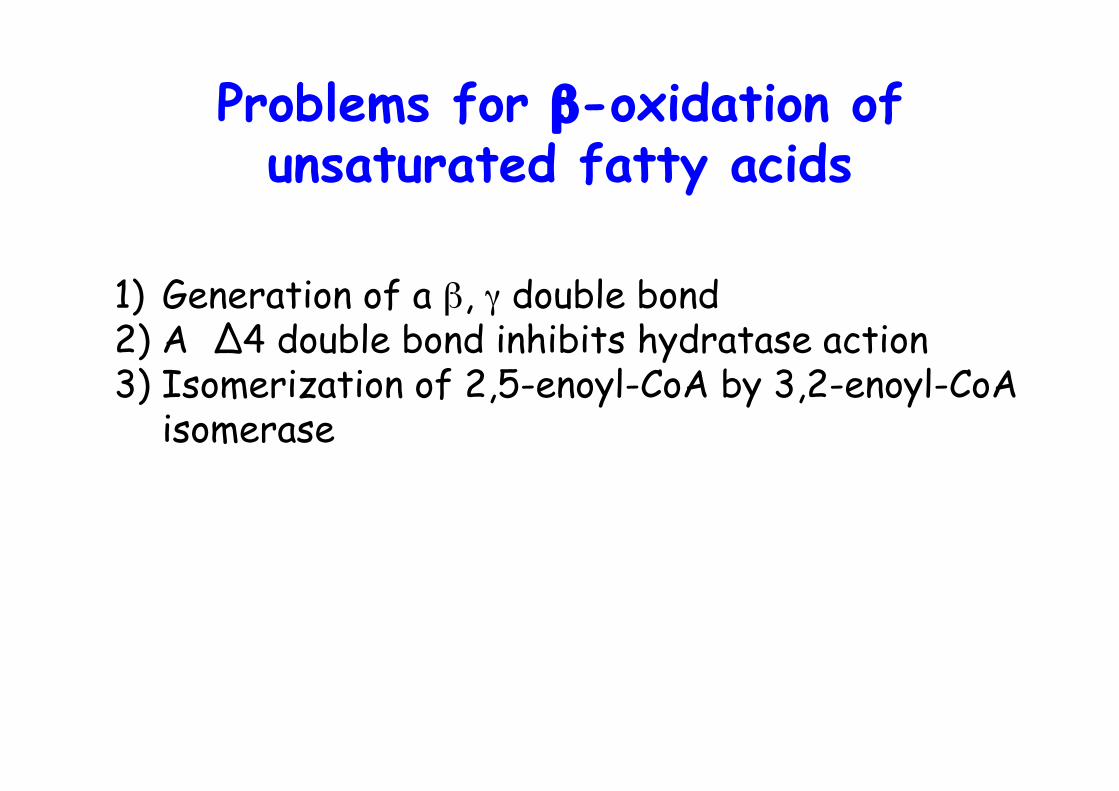
Problems for β-oxidation ofunsaturated fatty acids
1) Generation of a β, γ double bond2) A ∆4 double bond inhibits hydratase action3) Isomerization of 2,5-enoyl-CoA by 3,2-enoyl-CoA
isomerase
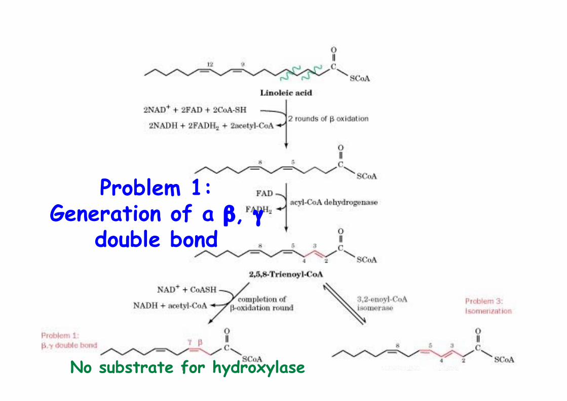
Problem 1:Generation of a β, γ
double bond
No substrate for hydroxylase
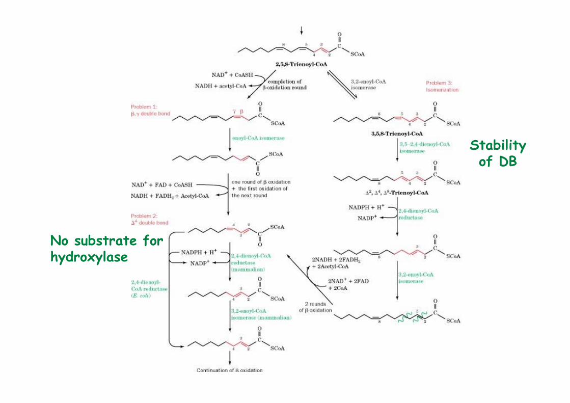
No substrate for hydroxylase
Stabilityof DB
Oxidation of odd chainfatty acids
o Most naturally FA are even numberedo Odd numbered FA are rare, some plants and marine organismso Final round of b-oxidation yields propionyl-CoAo Propionyl-CoA is converted to succinyl-CoA -> TCAo Propionate is also produced by oxidation of Ile, Val, Meto Ruminant animals, most caloric intake from acetate and propionate produced by microbial fermentation of carbohydrates in their stomach
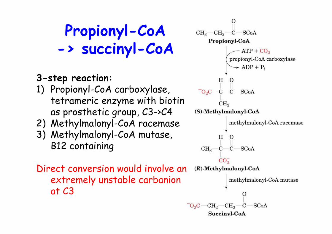
Propionyl-CoA-> succinyl-CoA
3-step reaction:1) Propionyl-CoA carboxylase,
tetrameric enzyme with biotinas prosthetic group, C3->C4
2) Methylmalonyl-CoA racemase3) Methylmalonyl-CoA mutase,
B12 containing
Direct conversion would involve anextremely unstable carbanionat C3
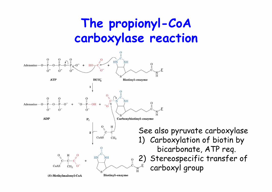
The propionyl-CoAcarboxylase reaction
See also pyruvate carboxylase1) Carboxylation of biotin by
bicarbonate, ATP req.2) Stereospecific transfer of
carboxyl group

The rearrangement catalyzedby methylmalonyl-CoA mutase
Vit B12-dependentHighly stereospecific (R-methylmalonyl-CoA) -> racemase
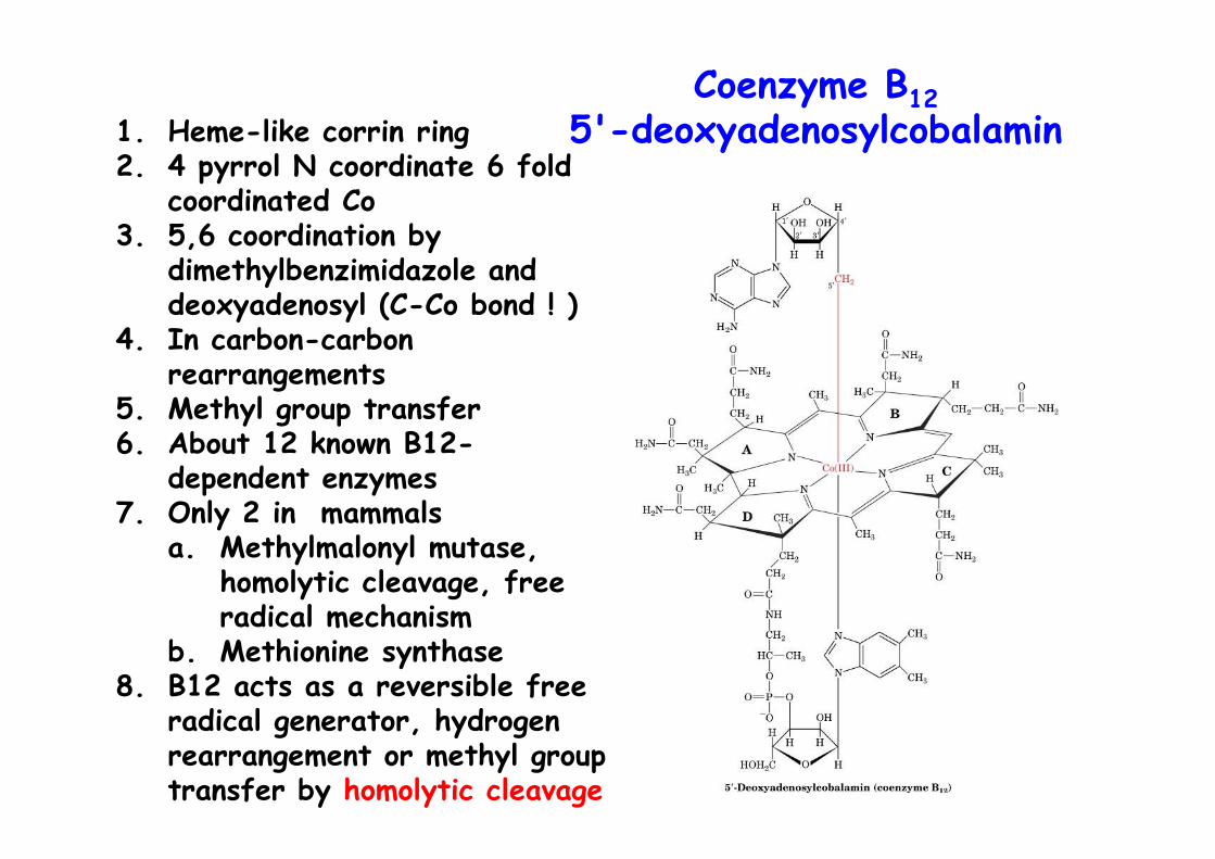
Coenzyme B125'-deoxyadenosylcobalamin1. Heme-like corrin ring
2. 4 pyrrol N coordinate 6 foldcoordinated Co
3. 5,6 coordination bydimethylbenzimidazole anddeoxyadenosyl (C-Co bond ! )
4. In carbon-carbonrearrangements
5. Methyl group transfer6. About 12 known B12-
dependent enzymes7. Only 2 in mammals
a. Methylmalonyl mutase,homolytic cleavage, freeradical mechanism
b. Methionine synthase8. B12 acts as a reversible free
radical generator, hydrogenrearrangement or methyl grouptransfer by homolytic cleavage
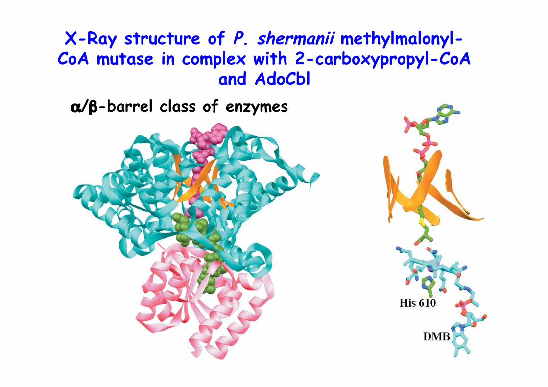
X-Ray structure of P. shermanii methylmalonyl-CoA mutase in complex with 2-carboxypropyl-CoA
and AdoCblα/β-barrel class of enzymes
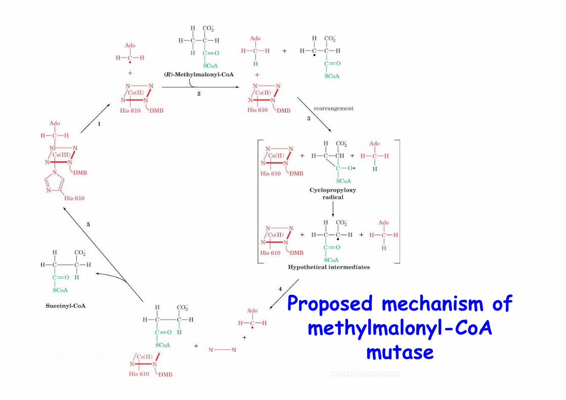
Proposed mechanism ofmethylmalonyl-CoA
mutase
Vit B12 deficiency
Pernicious anemiao in elderlyo decreased number of red blood cellso treated by daily consumption of raw liver (1926) -> (1948)o only few bacteria synthesize B12, plants and mammals noto human obtain it from meato Vit. B12 is specifically bound in intestine by intrinsic factoro complex absorbed in intestinal mucosa -> bloodo bound to transcobalamins in blood for uptake by tissueo not usually a dietary disease but result from insufficient secretion of intrinsic factor
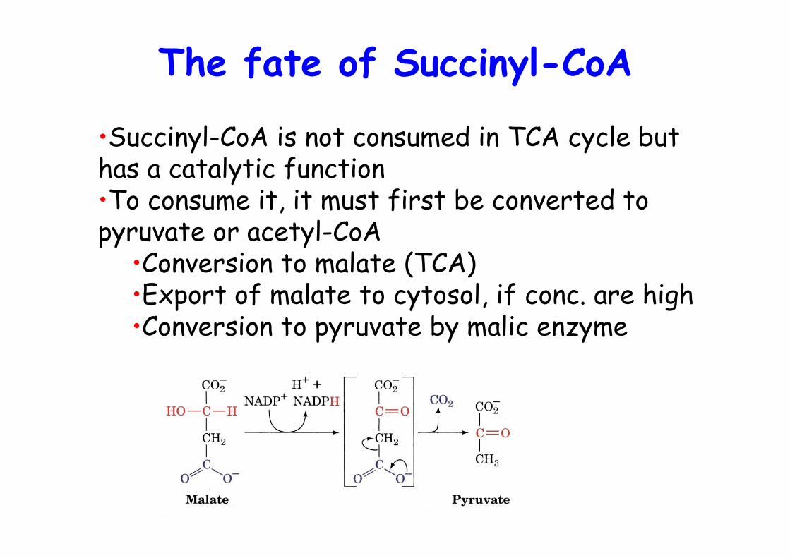
The fate of Succinyl-CoA
•Succinyl-CoA is not consumed in TCA cycle buthas a catalytic function•To consume it, it must first be converted topyruvate or acetyl-CoA
•Conversion to malate (TCA)•Export of malate to cytosol, if conc. are high•Conversion to pyruvate by malic enzyme
Peroxisomal β oxidation
• β-oxidation occurs both in mitochondria and in peroxisomes• Peroxisomes: Shortening of very-long chain fatty acids (VLCFA) for subsequent transport and oxidation in mitochondria• ALD protein to transport VLCFA into peroxisomes, no carnitine required, VLCFA-CoA synthetase• X-adrenoleukodystrophy caused by defects in ALD, lethal in young boys, 13% reduced efficiency of lignoceric acid (C24:0) to lignoceryl-CoA conversion• first step in perox. oxid. Acyl-CoA oxidase generates H2O2 -> name ! Catalase• carnitine for transport of chain shortened FAs out of perox. and into mito.
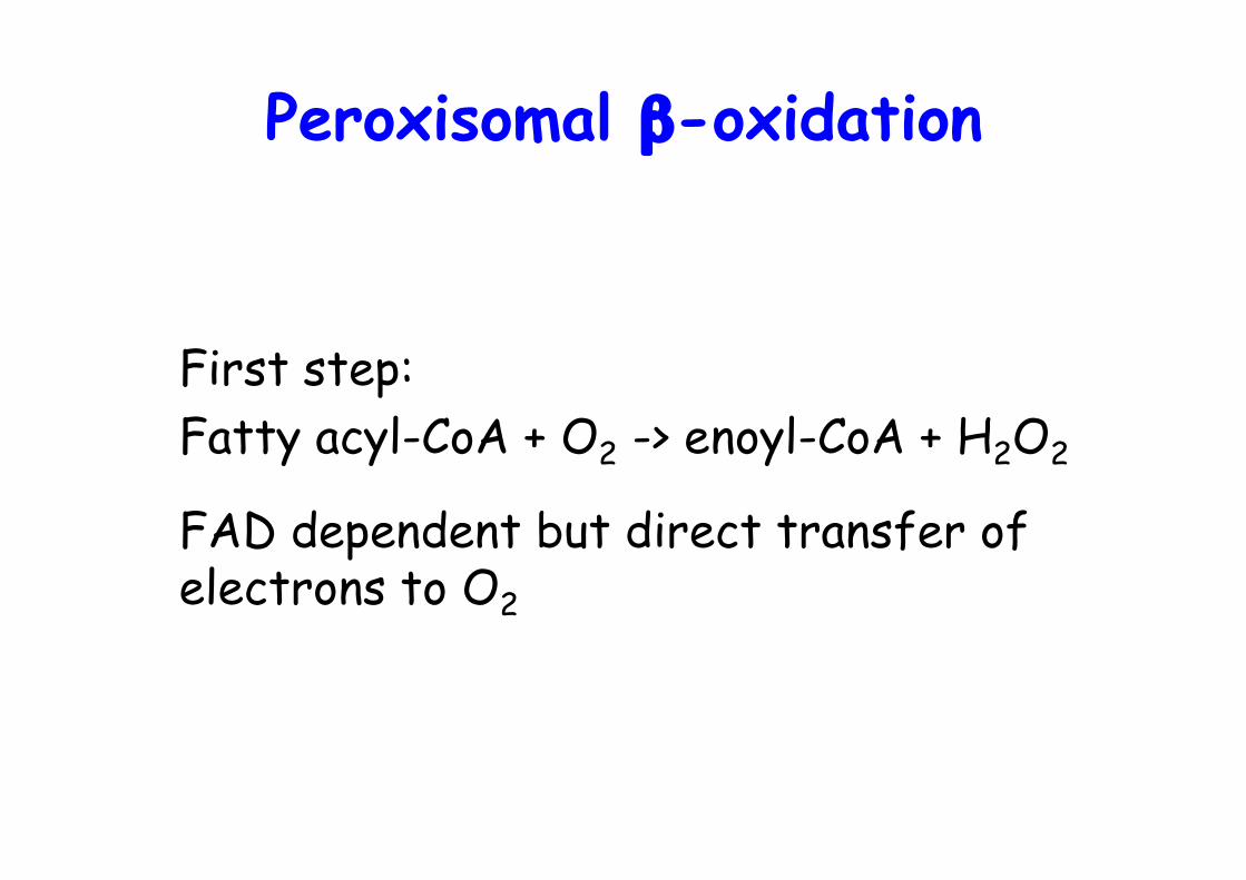
Peroxisomal β-oxidation
First step:Fatty acyl-CoA + O2 -> enoyl-CoA + H2O2
FAD dependent but direct transfer of electrons to O2
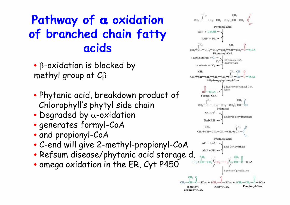
• β-oxidation is blocked by methyl group at Cβ
• Phytanic acid, breakdown product of Chlorophyll’s phytyl side chain• Degraded by α-oxidation• generates formyl-CoA• and propionyl-CoA• C-end will give 2-methyl-propionyl-CoA• Refsum disease/phytanic acid storage d.• omega oxidation in the ER, Cyt P450
Pathway of α oxidationof branched chain fatty
acids
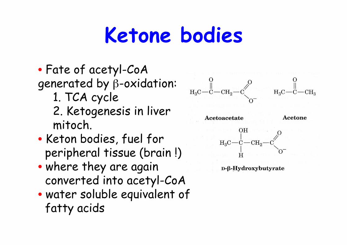
Ketone bodies• Fate of acetyl-CoAgenerated by β-oxidation:
1. TCA cycle2. Ketogenesis in livermitoch.
• Keton bodies, fuel for peripheral tissue (brain !)• where they are again converted into acetyl-CoA• water soluble equivalent of fatty acids
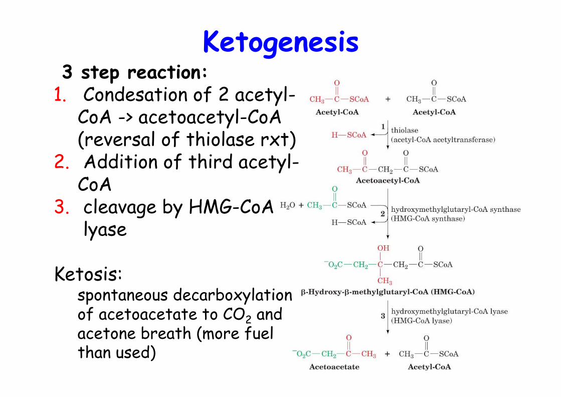
Ketogenesis 3 step reaction:1. Condesation of 2 acetyl-
CoA -> acetoacetyl-CoA(reversal of thiolase rxt)
2. Addition of third acetyl-CoA
3. cleavage by HMG-CoA lyase
Ketosis:spontaneous decarboxylationof acetoacetate to CO2 andacetone breath (more fuelthan used)
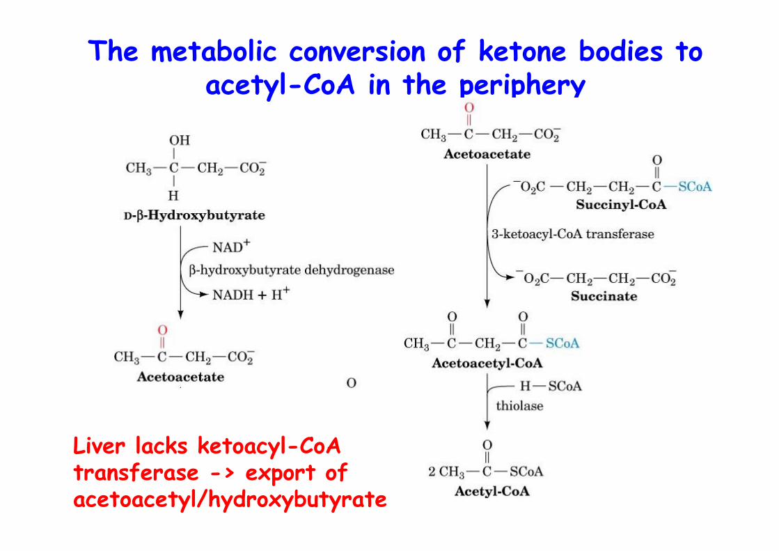
The metabolic conversion of ketone bodies toacetyl-CoA in the periphery
Liver lacks ketoacyl-CoA transferase -> export of acetoacetyl/hydroxybutyrate
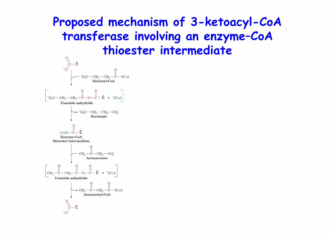
Proposed mechanism of 3-ketoacyl-CoAtransferase involving an enzyme–CoA
thioester intermediate
Fatty acid SynthesisSynthesis of FA through condensation of C2 units ->reversal of β-oxidationCytosolic, NADPH <-> mitochondrial, FAD, NADDifference in stereochemistryC3 unit for growth (malonyl-CoA) <-> C2 for oxidation(acetyl-CoA)
Growing chain esterified to acyl-carrier protein (ACP)Esterified to phosphopantetheine group as in CoA whichitself is bound to a Ser on ACPACP synthase transfers phosphopantetheine to apo-ACPto form a holo-ACP
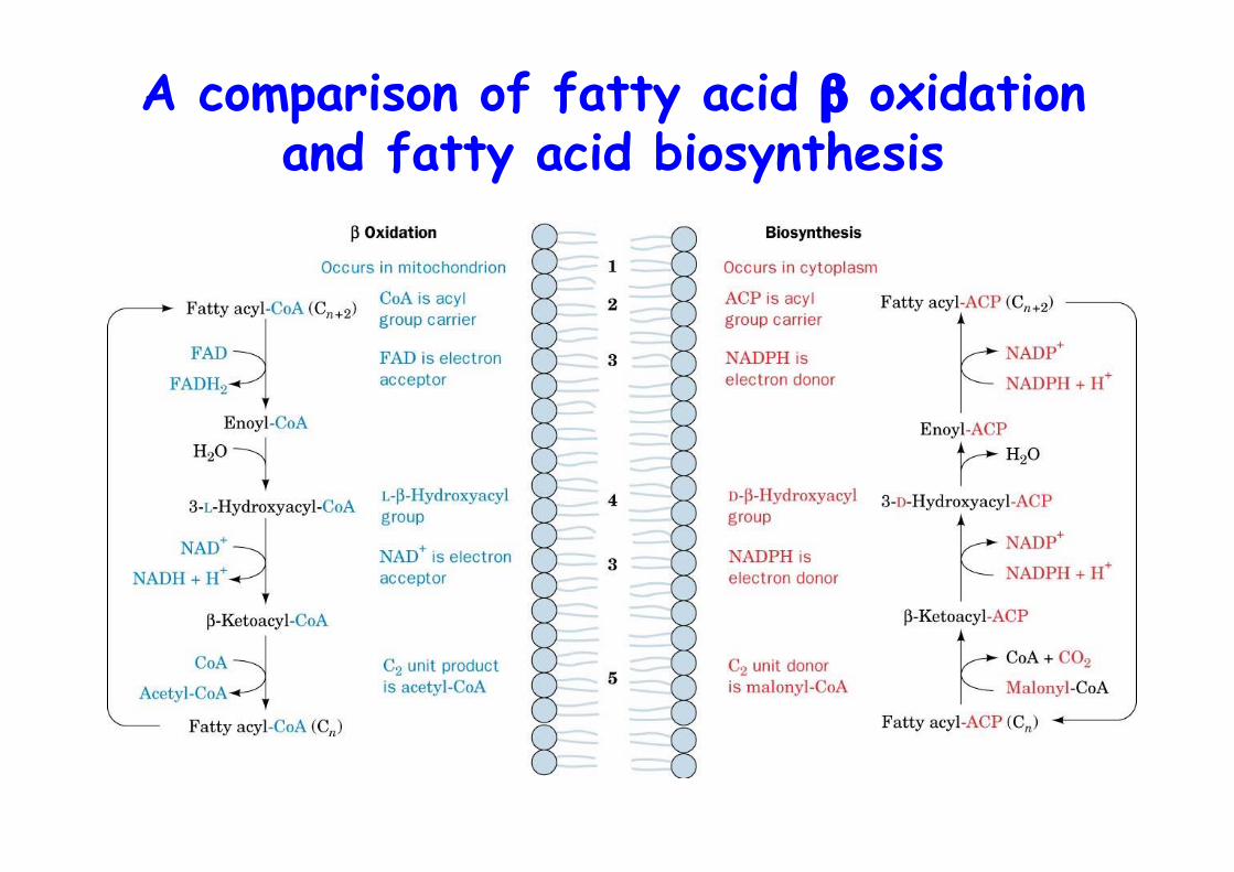
A comparison of fatty acid β oxidationand fatty acid biosynthesis
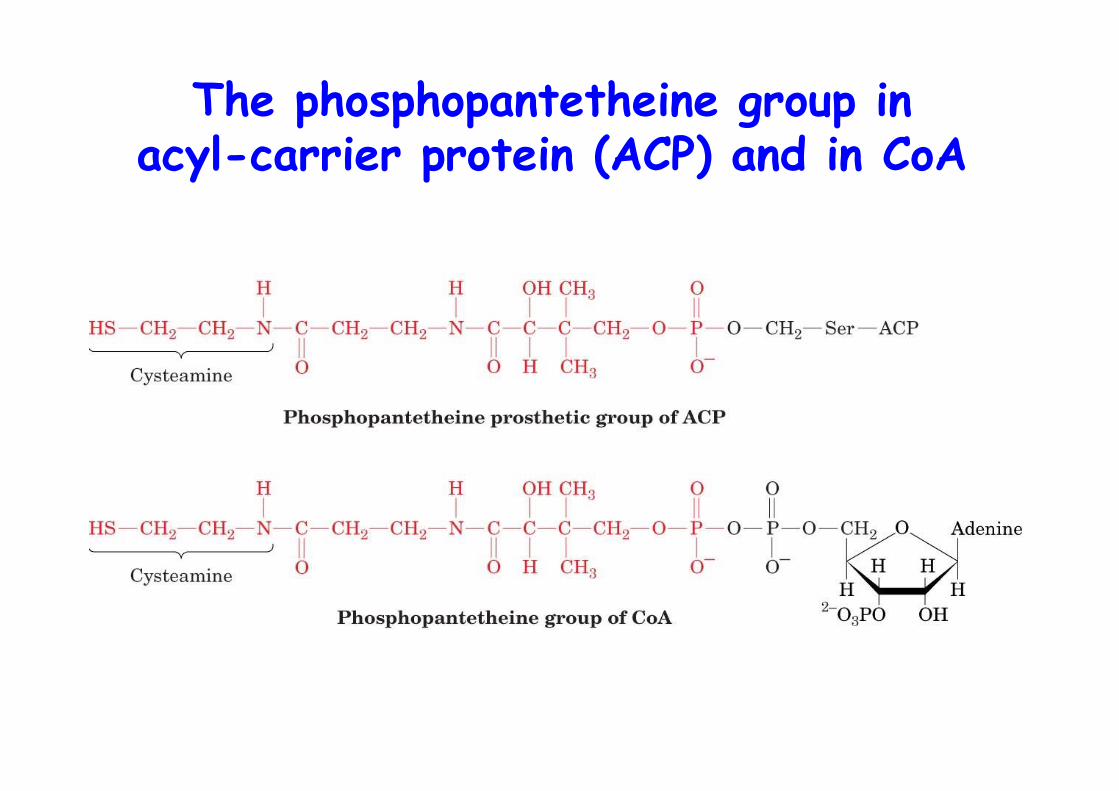
The phosphopantetheine group inacyl-carrier protein (ACP) and in CoA
Acetyl-CoA carboxylase• Catalyzes first and committed step of FA synthesis• Biotin-dependent (see propionyl-CoA carboxylase)• Hormonally regulated• Glucagon -> cAMP up -> PKA -> ACC is phosphorylated -> inactive, inhibited by palmitate• AMPK, AMP-dependent kinase activates ACC• ACC undergoes polymerization during activation• Mammals two isoforms:
α-ACC, adipose tissue β-ACC, tissue that oxidize FA, heart muscle, regulates β-ox. as malonyl-CoA inhibits CPT-I
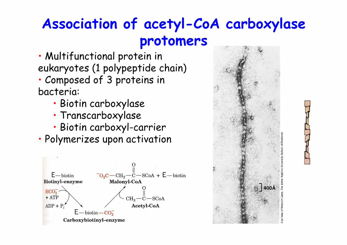
Association of acetyl-CoA carboxylaseprotomers
• Multifunctional protein ineukaryotes (1 polypeptide chain)• Composed of 3 proteins inbacteria:
• Biotin carboxylase• Transcarboxylase• Biotin carboxyl-carrier
• Polymerizes upon activation
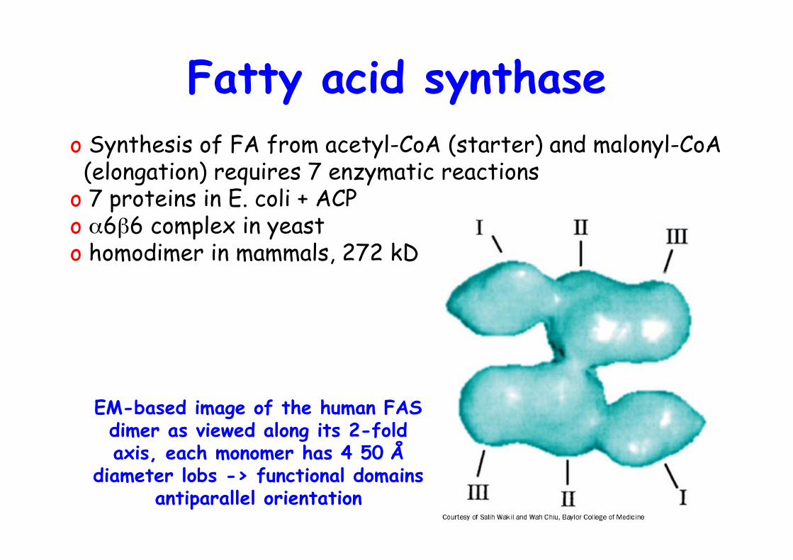
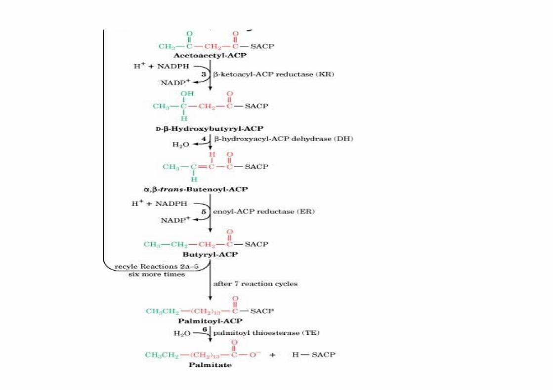
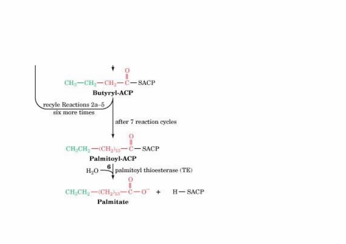
Fatty acid synthaseo Synthesis of FA from acetyl-CoA (starter) and malonyl-CoA (elongation) requires 7 enzymatic reactionso 7 proteins in E. coli + ACPo α6β6 complex in yeasto homodimer in mammals, 272 kD
EM-based image of the human FASdimer as viewed along its 2-foldaxis, each monomer has 4 50 Å
diameter lobs -> functional domainsantiparallel orientation
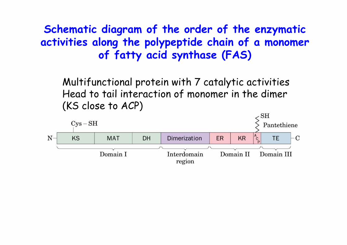
Schematic diagram of the order of the enzymaticactivities along the polypeptide chain of a monomer
of fatty acid synthase (FAS)
Multifunctional protein with 7 catalytic activitiesHead to tail interaction of monomer in the dimer(KS close to ACP)
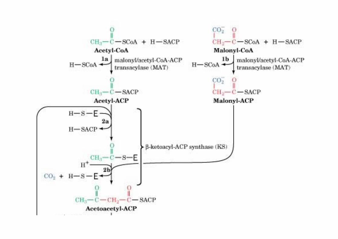
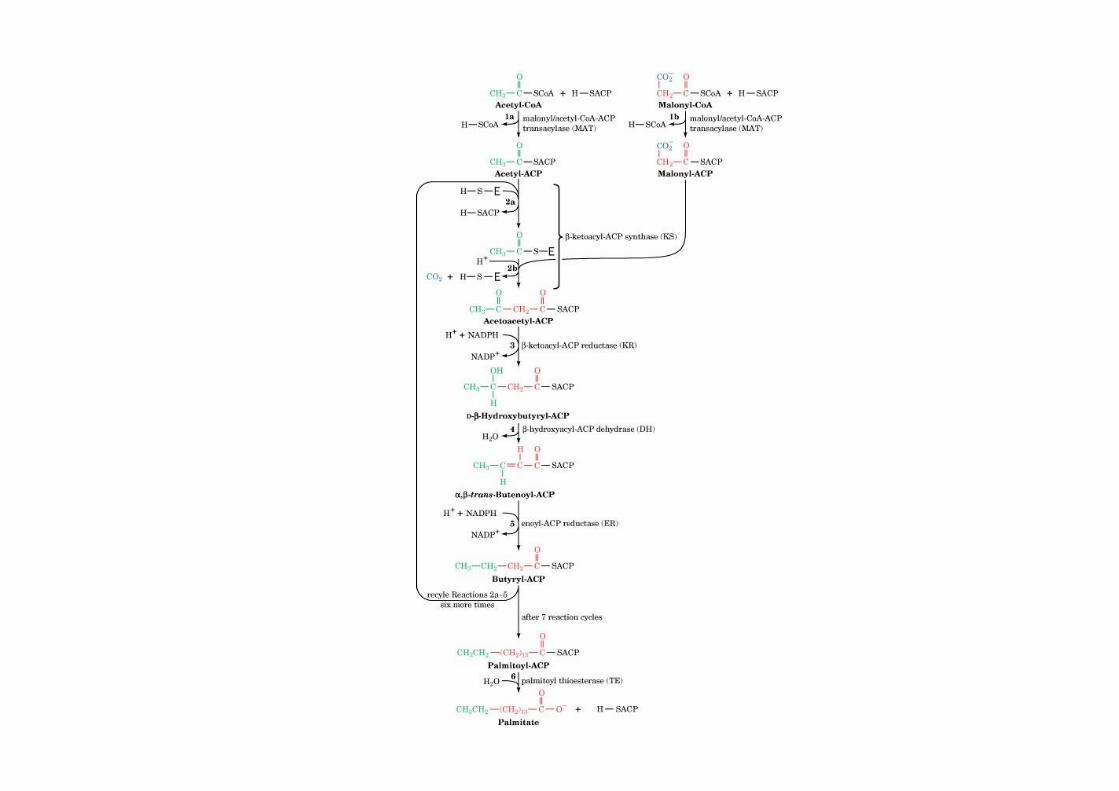
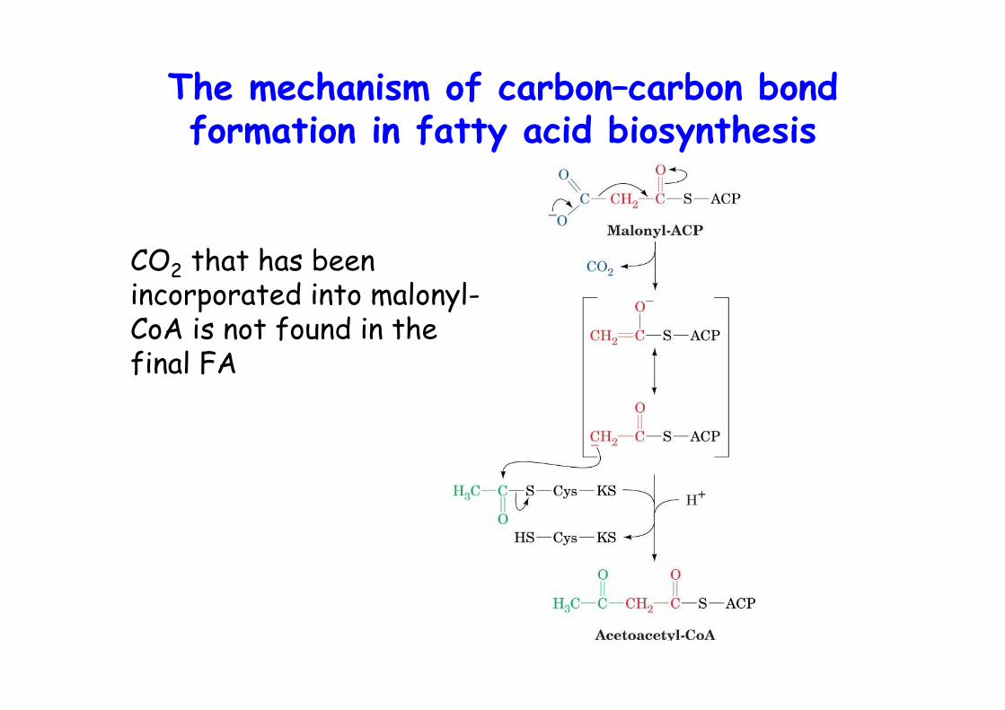
The mechanism of carbon–carbon bondformation in fatty acid biosynthesis
CO2 that has beenincorporated into malonyl-CoA is not found in thefinal FA
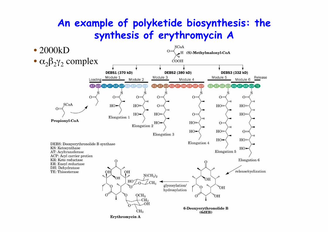
An example of polyketide biosynthesis: thesynthesis of erythromycin A
• 2000kD• α2β2γ2 complex
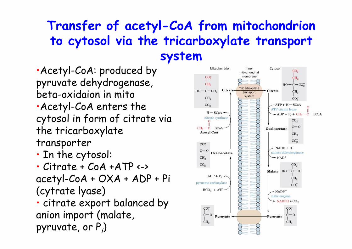
Transfer of acetyl-CoA from mitochondrionto cytosol via the tricarboxylate transport
system•Acetyl-CoA: produced bypyruvate dehydrogenase,beta-oxidaion in mito•Acetyl-CoA enters thecytosol in form of citrate viathe tricarboxylatetransporter• In the cytosol:• Citrate + CoA +ATP <->acetyl-CoA + OXA + ADP + Pi(cytrate lyase)• citrate export balanced byanion import (malate,pyruvate, or Pi)
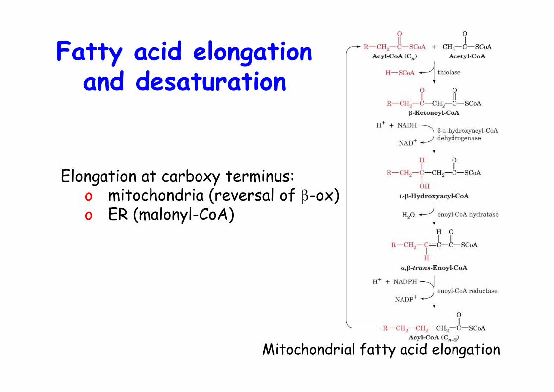
Fatty acid elongationand desaturation
Mitochondrial fatty acid elongation
Elongation at carboxy terminus:o mitochondria (reversal of β-ox)o ER (malonyl-CoA)

FA desaturation
The electron-transfer reactions mediated by the ∆9-fatty acyl-CoA desaturase complex
Properties:o Cis, ∆9 first, not conjugatedo membrane-bound, nonheme iron enzymes, cyt b5-dependento mammals front end desaturation (∆9, 6, 5/4)o essential FA, linoleic (C18:2n-6, ∆9,12), linolenic (C18:3n-3, ∆9,12,15)o some made by combination of desaturation and elongationo PUFAs, fish oil, n-3, n-6 (omega)o vision, cognitive functions
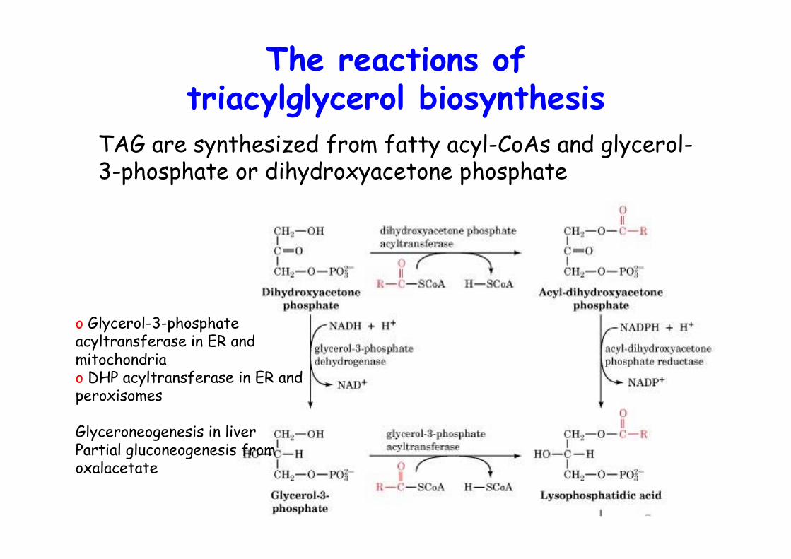
The reactions oftriacylglycerol biosynthesis
o Glycerol-3-phosphateacyltransferase in ER andmitochondriao DHP acyltransferase in ER andperoxisomes
Glyceroneogenesis in liverPartial gluconeogenesis fromoxalacetate
TAG are synthesized from fatty acyl-CoAs and glycerol-3-phosphate or dihydroxyacetone phosphate
Metabolic controlDifferences in energy needs:
o between resting and activated muscle 100xo feed <-> fasting
o Breakdown of glycogen and fatty acids concern the whole organismo organs and tissues connected by blood stream
o Blood glucose levels sensed by pancreatic α cells, glucosedown -> secrete glucagono β cells, glucose up -> insulin
o These hormones also control fatty acid synthesis <-> β oxidation
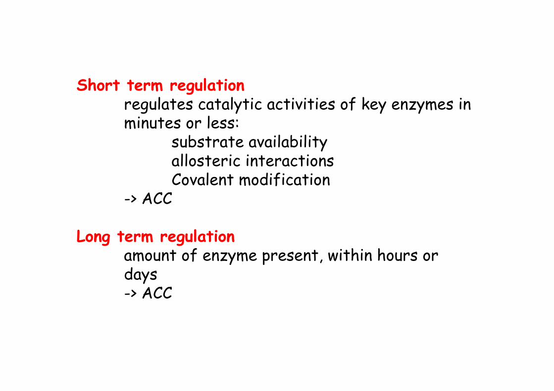
Short term regulationregulates catalytic activities of key enzymes inminutes or less:
substrate availabilityallosteric interactionsCovalent modification
-> ACC
Long term regulationamount of enzyme present, within hours or days-> ACC
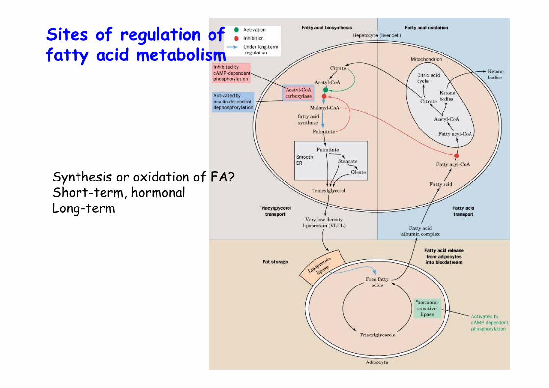
Sites of regulation offatty acid metabolism
Synthesis or oxidation of FA?Short-term, hormonalLong-term
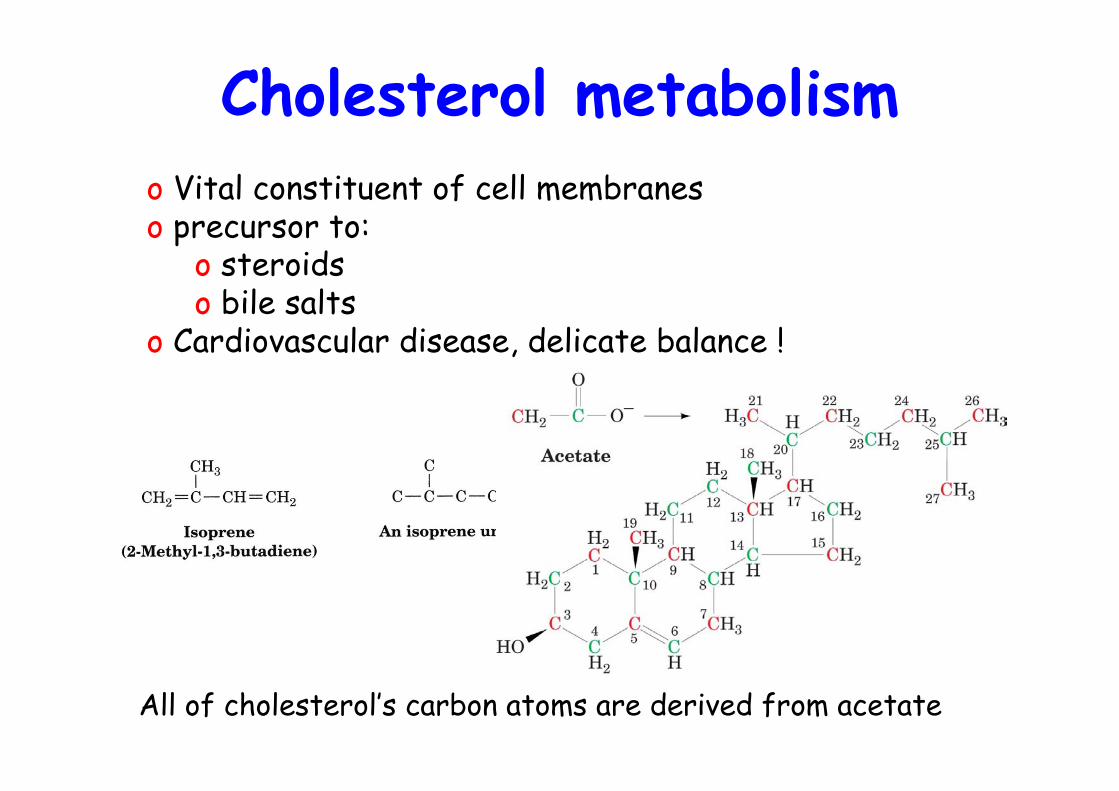
Cholesterol metabolism
All of cholesterol’s carbon atoms are derived from acetate
o Vital constituent of cell membraneso precursor to:
o steroidso bile salts
o Cardiovascular disease, delicate balance !
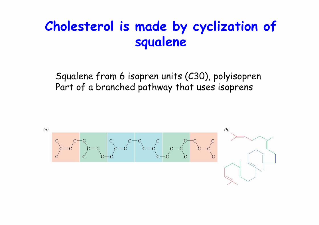
Cholesterol is made by cyclization ofsqualene
Squalene from 6 isopren units (C30), polyisoprenPart of a branched pathway that uses isoprens
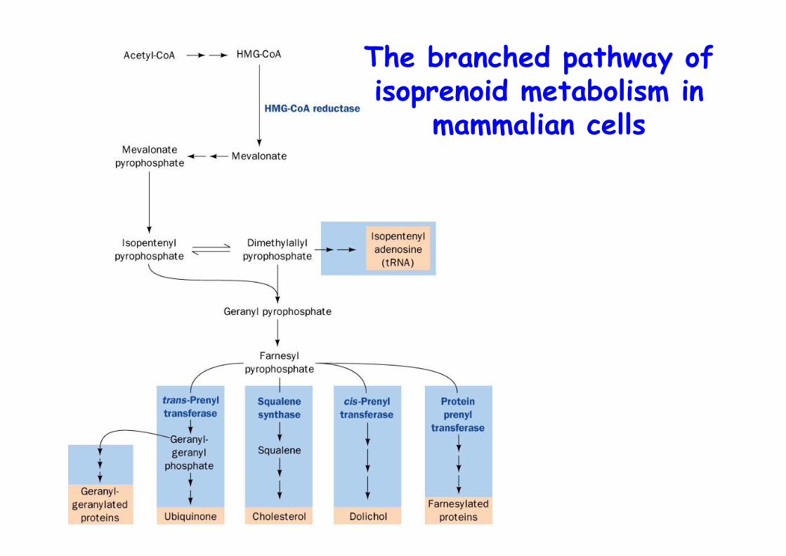
The branched pathway ofisoprenoid metabolism in
mammalian cells
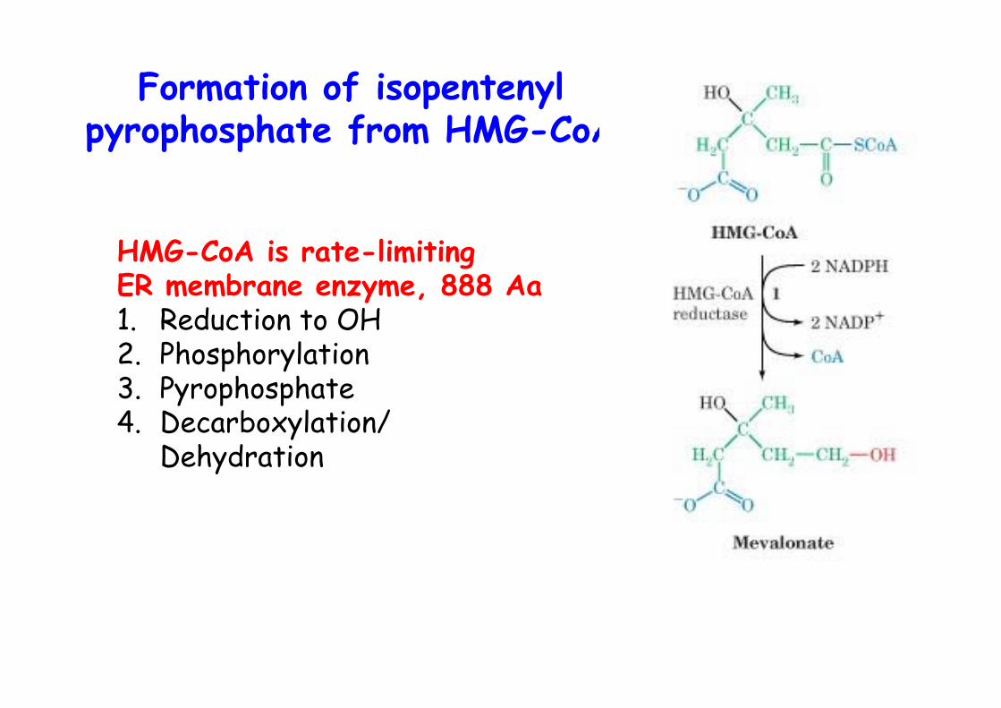
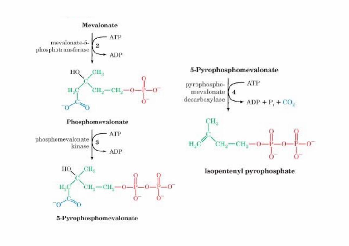
Formation of isopentenylpyrophosphate from HMG-CoA
HMG-CoA is rate-limitingER membrane enzyme, 888 Aa1. Reduction to OH2. Phosphorylation3. Pyrophosphate4. Decarboxylation/
Dehydration
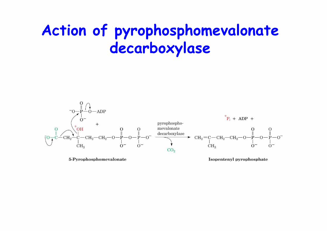
Action of pyrophosphomevalonatedecarboxylase
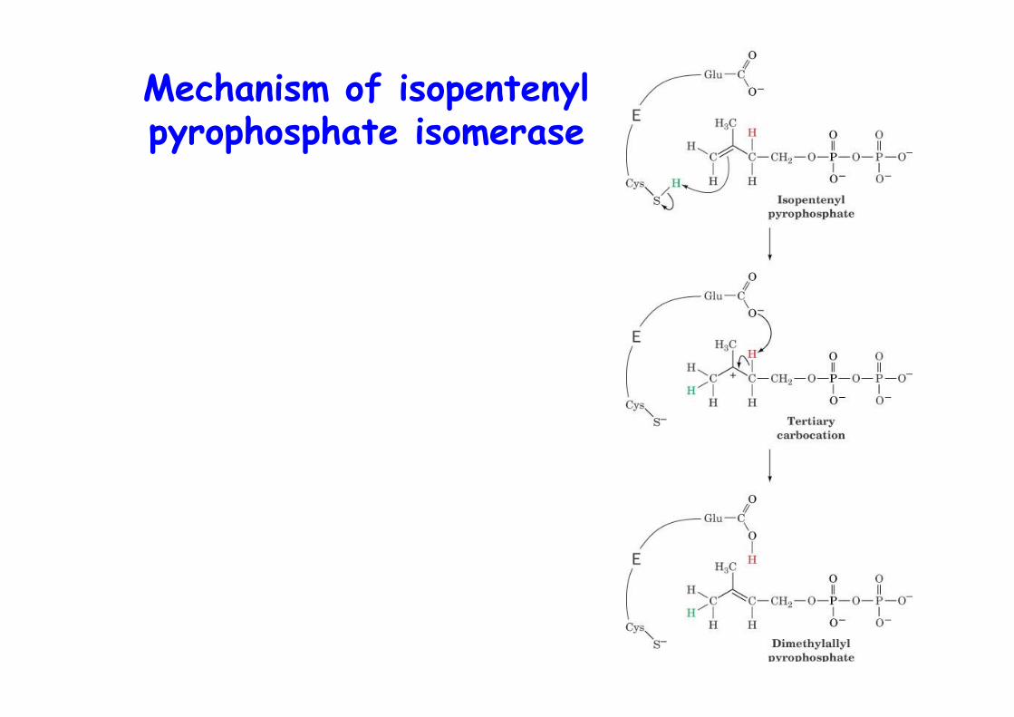
Mechanism of isopentenylpyrophosphate isomerase
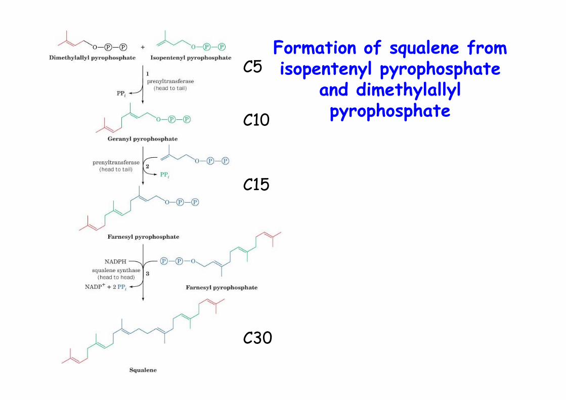
Formation of squalene fromisopentenyl pyrophosphate
and dimethylallylpyrophosphate
C5
C10
C15
C30
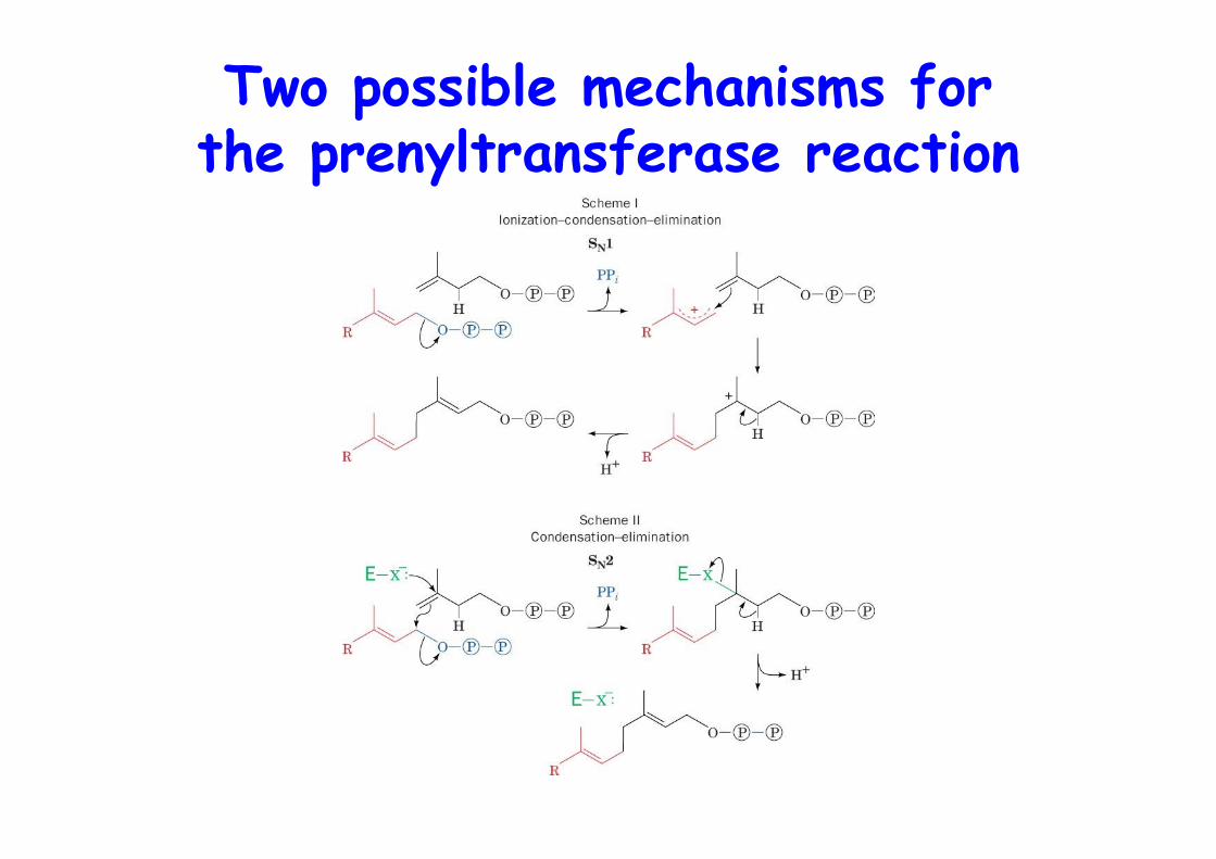
Two possible mechanisms forthe prenyltransferase reaction

Action of squalene synthase
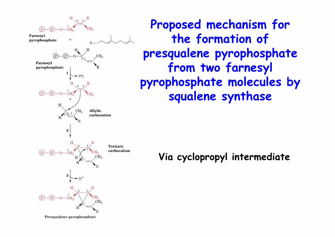
Proposed mechanism forthe formation of
presqualene pyrophosphatefrom two farnesyl
pyrophosphate molecules bysqualene synthase
Via cyclopropyl intermediate
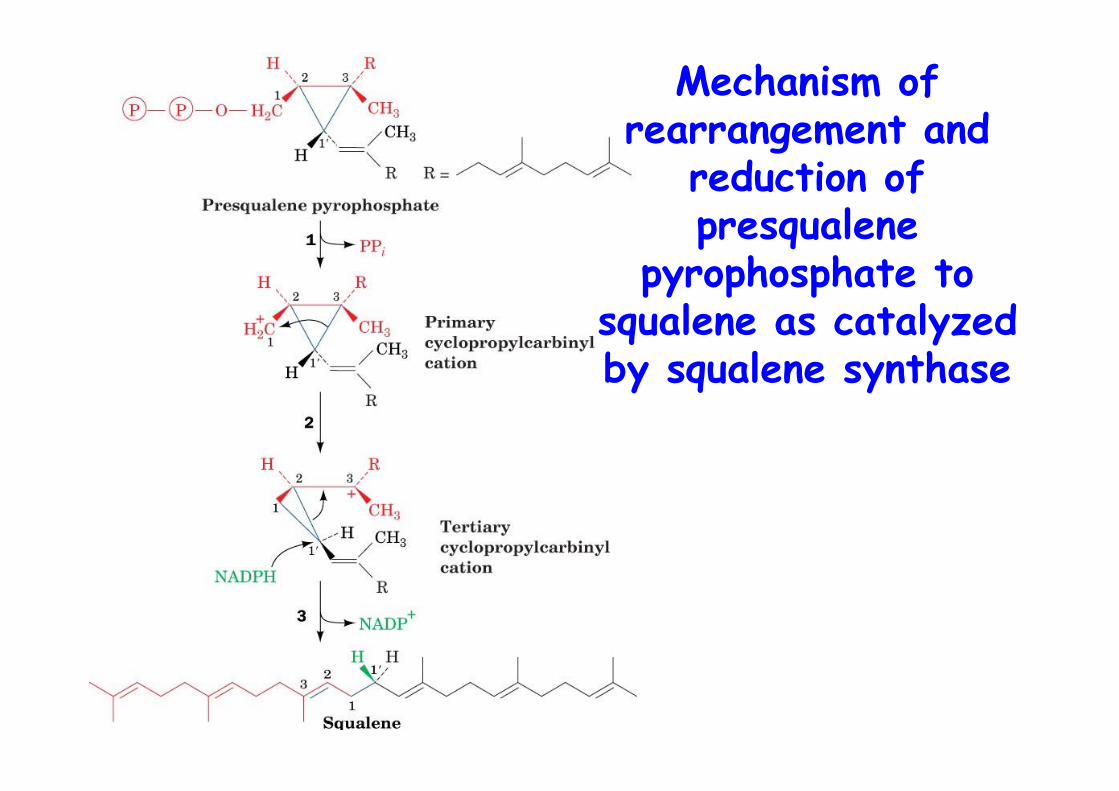
Mechanism ofrearrangement and
reduction ofpresqualene
pyrophosphate tosqualene as catalyzedby squalene synthase

Squalene synthase
o ER anchoredo Monomer single domain
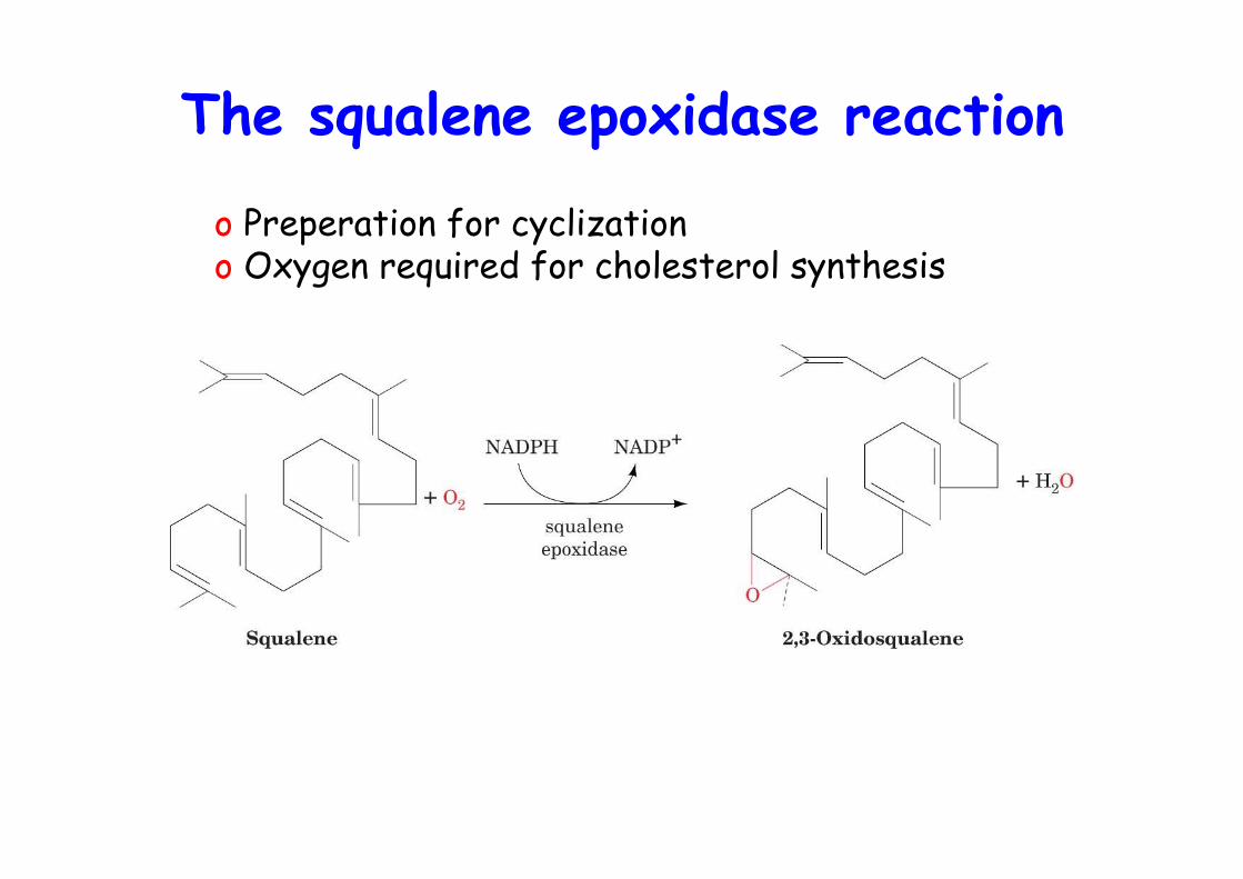
The squalene epoxidase reaction
o Preperation for cyclization o Oxygen required for cholesterol synthesis
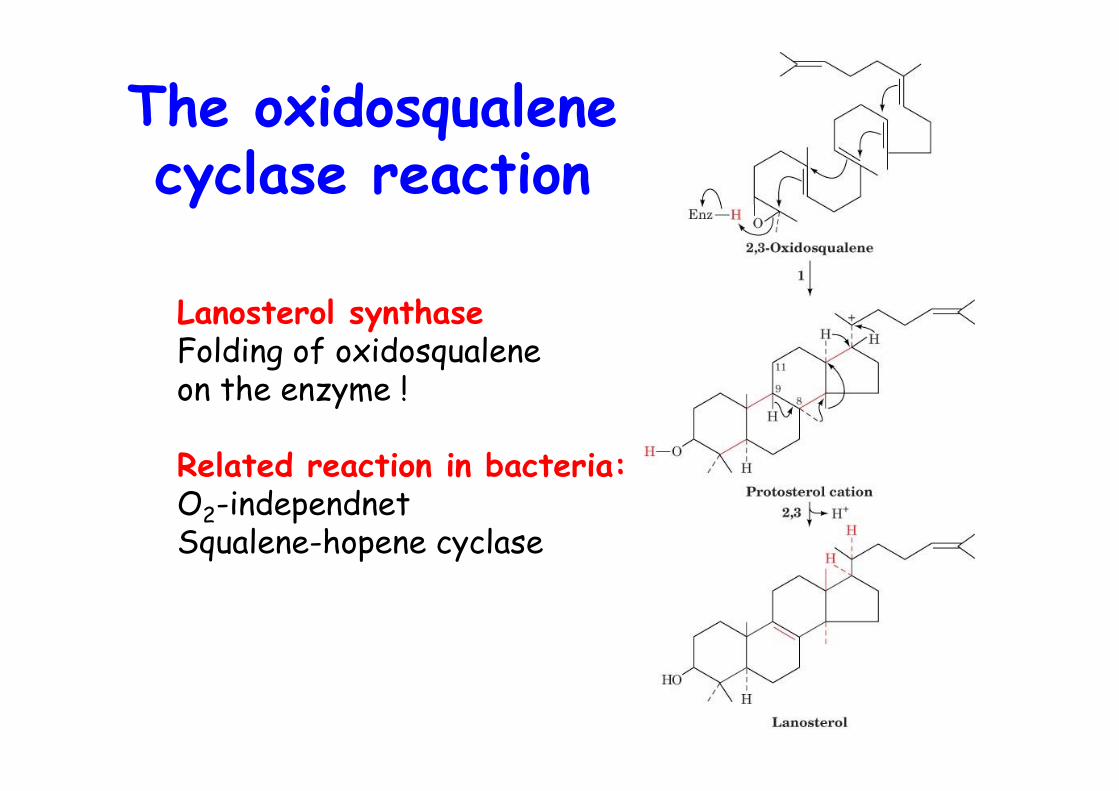
The oxidosqualenecyclase reaction
Lanosterol synthaseFolding of oxidosqualene on the enzyme !
Related reaction in bacteria:O2-independnetSqualene-hopene cyclase
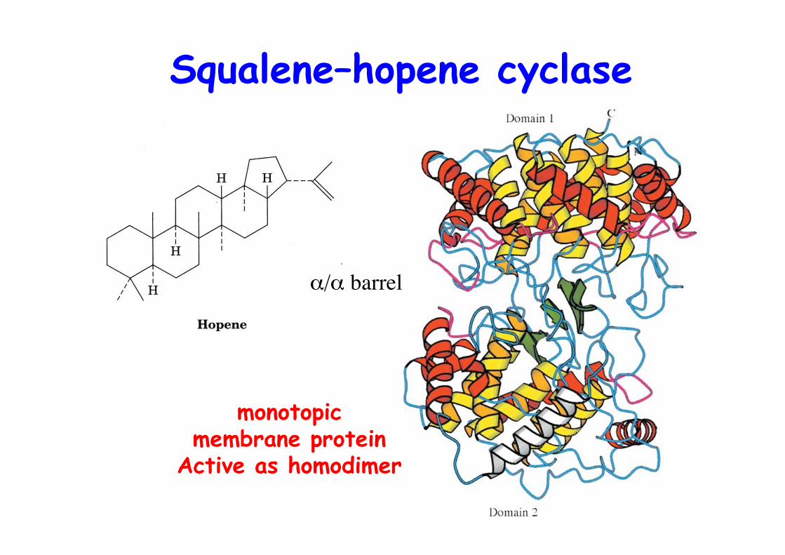
Squalene–hopene cyclase
α/α barrel
monotopicmembrane protein
Active as homodimer
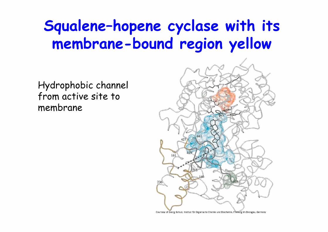
Squalene–hopene cyclase with itsmembrane-bound region yellow
Hydrophobic channelfrom active site tomembrane
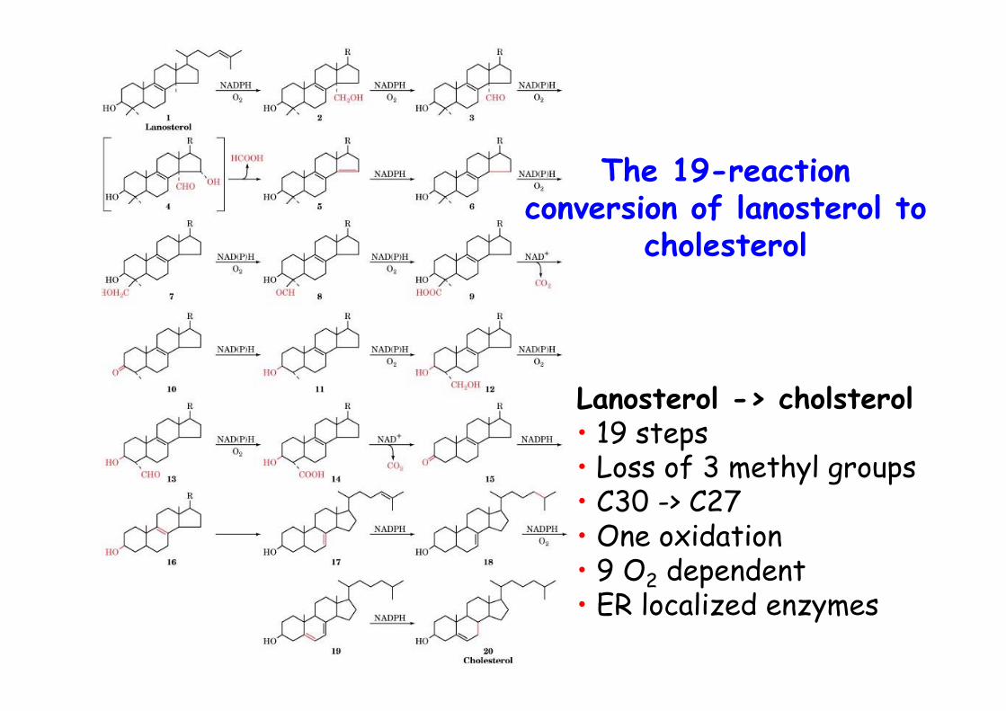
The 19-reactionconversion of lanosterol to
cholesterol
Lanosterol -> cholsterol• 19 steps• Loss of 3 methyl groups• C30 -> C27• One oxidation• 9 O2 dependent• ER localized enzymes
CholesterolLiver synthesized cholesterol is:
o converted to bile saltso esterified to cholesteryl ester, ACAT
which are then packaged into lipoprotein complexes, VLDLand taken up by the tissue by LDL receptor mediatedendocytosis
Mammalian cells thus have 2 ways to acquire cholesterol:de novo synthesis or via LDL uptake
Dietary sterols are absorbed in small intestine andtransported as chylomicrons in lymph to tissue/liver
HDL transports cholesterol from the peripheral tissue tothe liver
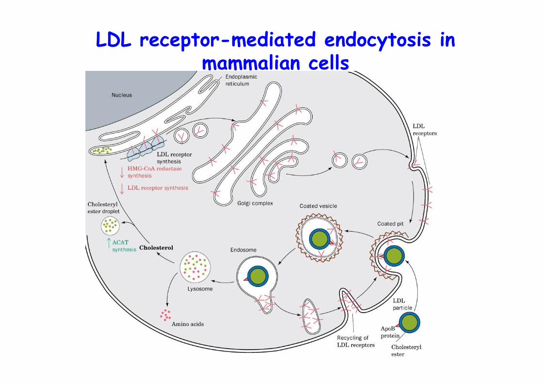
LDL receptor-mediated endocytosis inmammalian cells
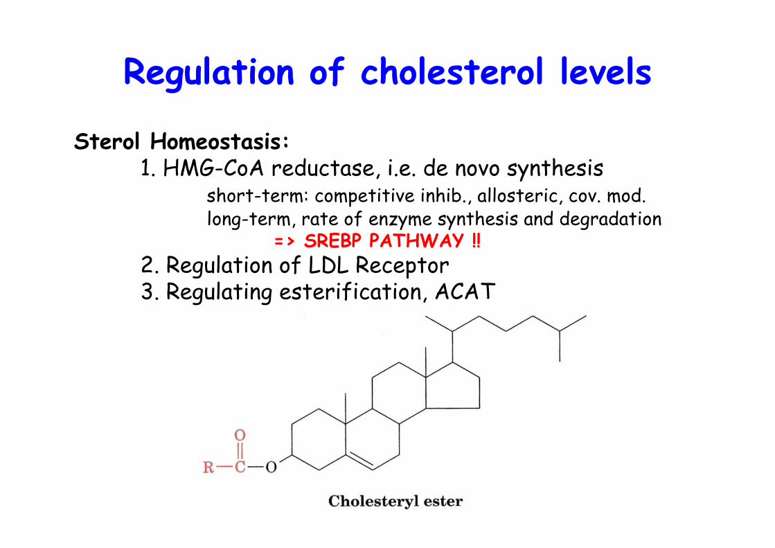
Regulation of cholesterol levels
Sterol Homeostasis:1. HMG-CoA reductase, i.e. de novo synthesis
short-term: competitive inhib., allosteric, cov. mod.long-term, rate of enzyme synthesis and degradation
=> SREBP PATHWAY !!2. Regulation of LDL Receptor3. Regulating esterification, ACAT
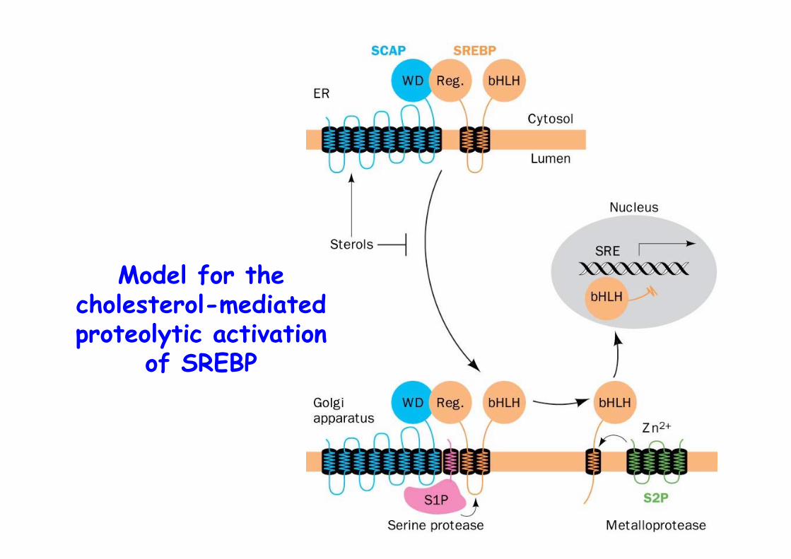
Model for thecholesterol-mediatedproteolytic activation
of SREBP
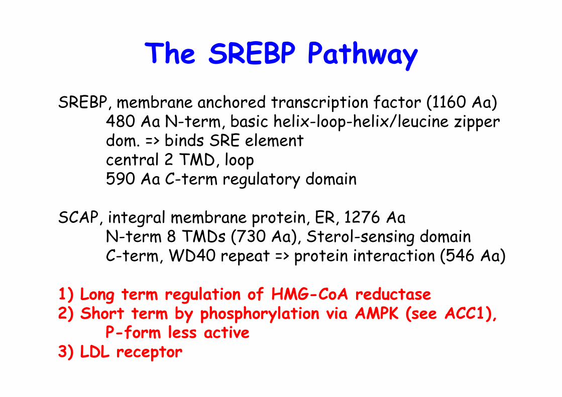
The SREBP PathwaySREBP, membrane anchored transcription factor (1160 Aa)
480 Aa N-term, basic helix-loop-helix/leucine zipper dom. => binds SRE elementcentral 2 TMD, loop590 Aa C-term regulatory domain
SCAP, integral membrane protein, ER, 1276 AaN-term 8 TMDs (730 Aa), Sterol-sensing domainC-term, WD40 repeat => protein interaction (546 Aa)
1) Long term regulation of HMG-CoA reductase2) Short term by phosphorylation via AMPK (see ACC1),
P-form less active3) LDL receptor
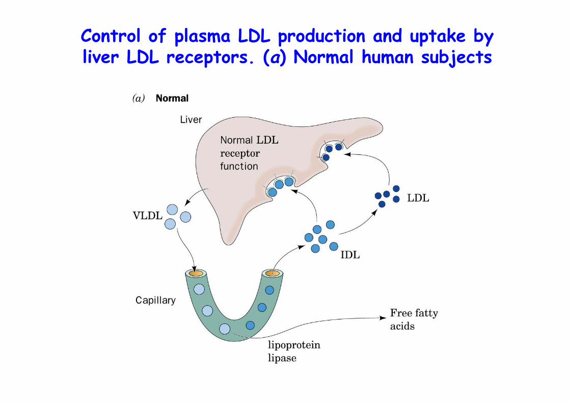
Control of plasma LDL production and uptake byliver LDL receptors. (a) Normal human subjects
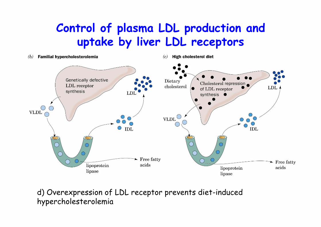
Control of plasma LDL production anduptake by liver LDL receptors
d) Overexpression of LDL receptor prevents diet-inducedhypercholesterolemia
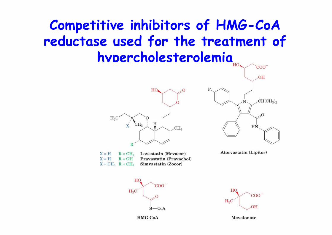
Competitive inhibitors of HMG-CoAreductase used for the treatment of
hypercholesterolemia
COMBINATORIAL THERAPHY
1) Anion exchanger,cholestyramine, reduced uptake of dietary cholesterol => 15-20% drop
2) HMG-CoA inhibitorstatins
Combined => 50-60% reduction of bloodcholesterol levels
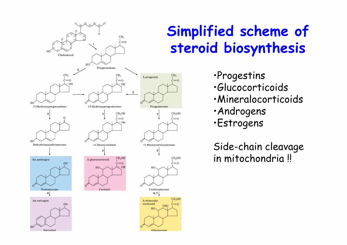
Simplified scheme ofsteroid biosynthesis
•Progestins•Glucocorticoids•Mineralocorticoids•Androgens•Estrogens
Side-chain cleavagein mitochondria !!
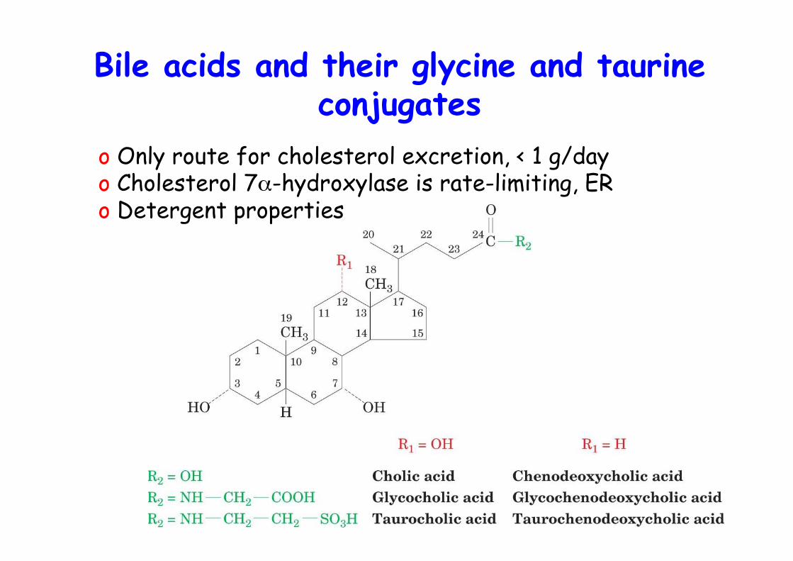
Bile acids and their glycine and taurineconjugates
o Only route for cholesterol excretion, < 1 g/dayo Cholesterol 7α-hydroxylase is rate-limiting, ERo Detergent properties
Prostaglandins (PGs)o 1930, Ulf von Euler: human semen extract
stimulates uteruscontraction and lower blood pressure
o Thought to originate in prostata -> name
o mid 50s, isolated from body fluids in etherextract (PGE)
o Made by all cells except RBC
Eicosanoid metabolism:Prostaglandinsm prostacyclins,
thromboxanes, leukotriens, and lipoxins
o Collectively: eicosanoids, C20 compounds- profound physiological effects at very low conc.- hormone-like but paracrine- bind to G-coupled receptors, affect cAMP- signal as hormones do- arachidonic acid C20:4
o What you inhibit by aspirin !!NSAIDs, nonsteroidal anti-inflammatory drugs
o What you inhibit by cortisol !!
Eicosanoids
Mediate:1) inflammation2) production of pain and fever3) regulate blood pressure4) induction of blood clotting5) reproductive functions6) sleep/wake cycle
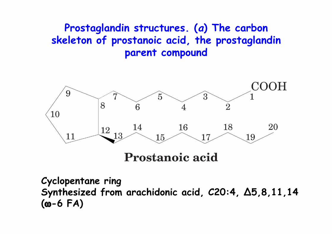
Prostaglandin structures. (a) The carbonskeleton of prostanoic acid, the prostaglandin
parent compound
Cyclopentane ringSynthesized from arachidonic acid, C20:4, ∆5,8,11,14(ω-6 FA)
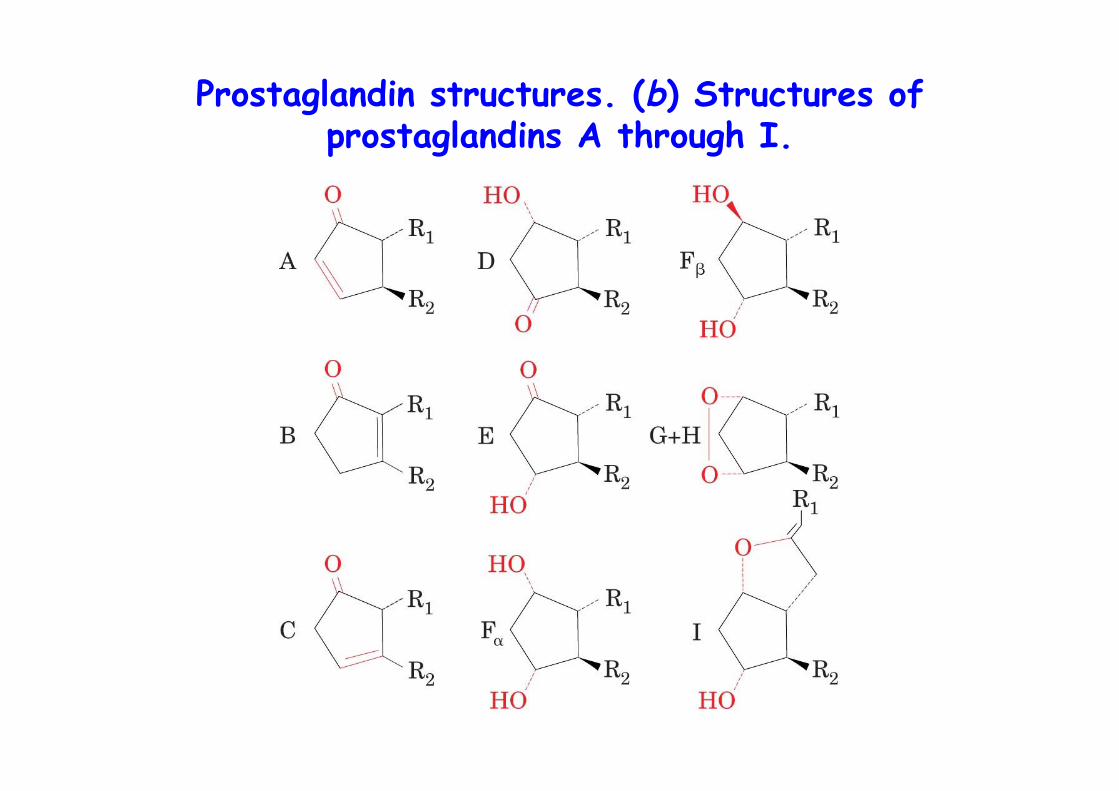
Prostaglandin structures. (b) Structures ofprostaglandins A through I.
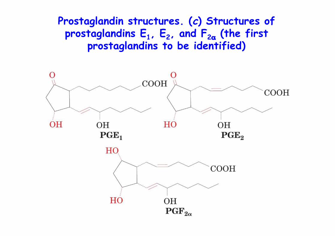
Prostaglandin structures. (c) Structures ofprostaglandins E1, E2, and F2α (the first
prostaglandins to be identified)
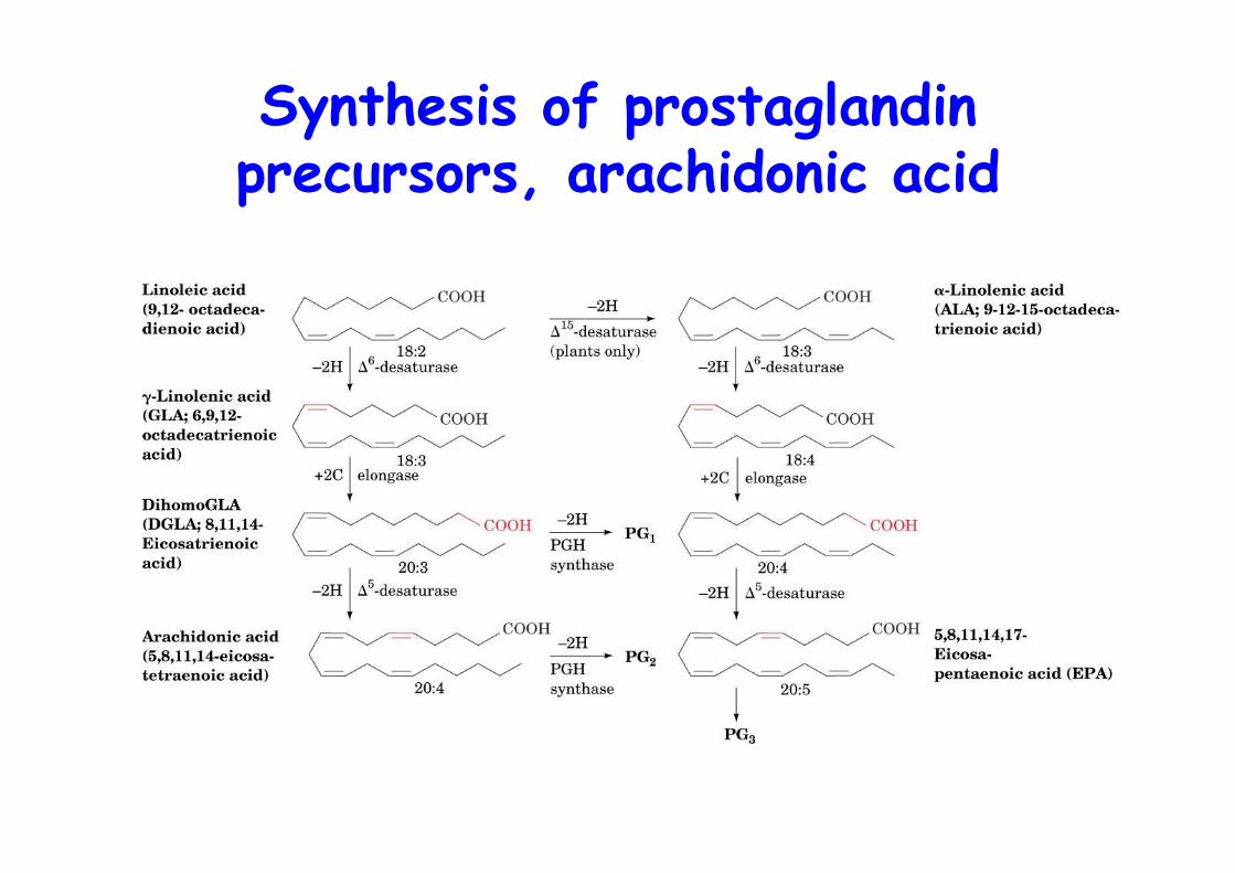
Synthesis of prostaglandinprecursors, arachidonic acid
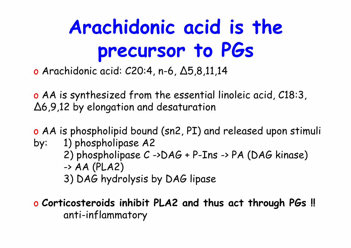
Arachidonic acid is theprecursor to PGs
o Arachidonic acid: C20:4, n-6, ∆5,8,11,14
o AA is synthesized from the essential linoleic acid, C18:3, ∆6,9,12 by elongation and desaturation
o AA is phospholipid bound (sn2, PI) and released upon stimuli by: 1) phospholipase A2
2) phospholipase C ->DAG + P-Ins -> PA (DAG kinase) -> AA (PLA2)3) DAG hydrolysis by DAG lipase
o Corticosteroids inhibit PLA2 and thus act through PGs !!anti-inflammatory
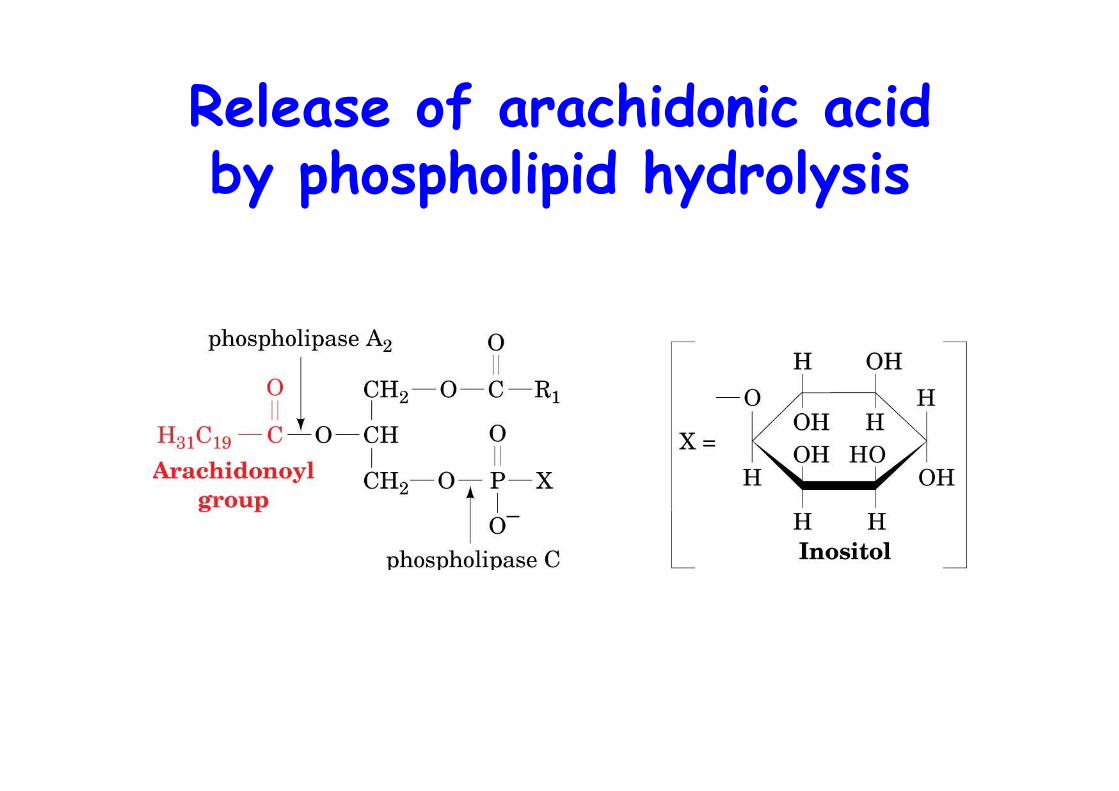
Release of arachidonic acidby phospholipid hydrolysis

Pathways of arachidonic acidliberation from phospholipids
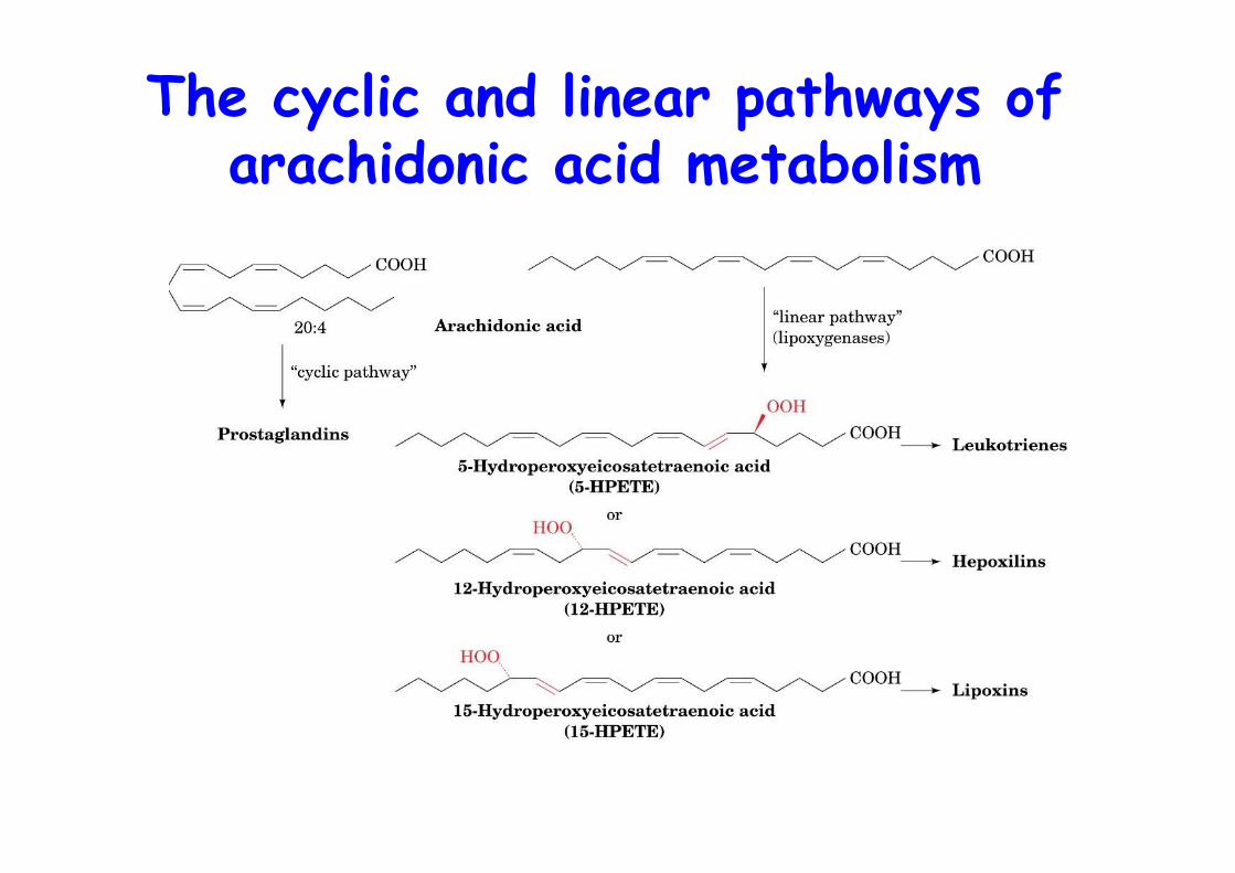
The cyclic and linear pathways ofarachidonic acid metabolism
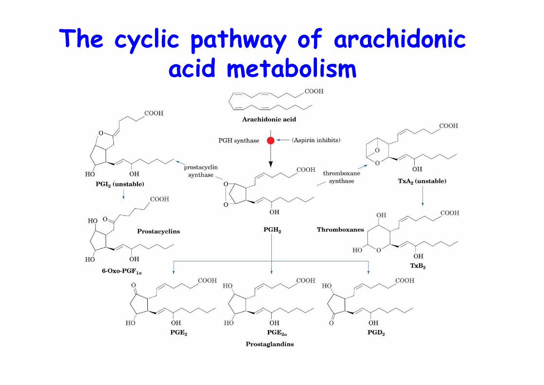
The cyclic pathway of arachidonicacid metabolism
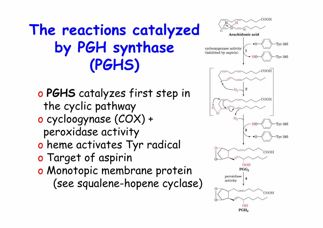
The reactions catalyzedby PGH synthase
(PGHS)
o PGHS catalyzes first step inthe cyclic pathway
o cycloogynase (COX) +peroxidase activity
o heme activates Tyr radicalo Target of aspirino Monotopic membrane protein (see squalene-hopene cyclase)
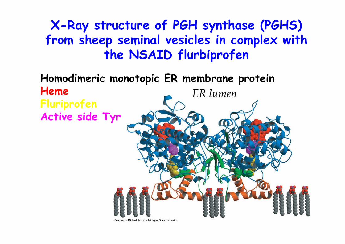
X-Ray structure of PGH synthase (PGHS)from sheep seminal vesicles in complex with
the NSAID flurbiprofen
Homodimeric monotopic ER membrane proteinHemeFluriprofenActive side Tyr
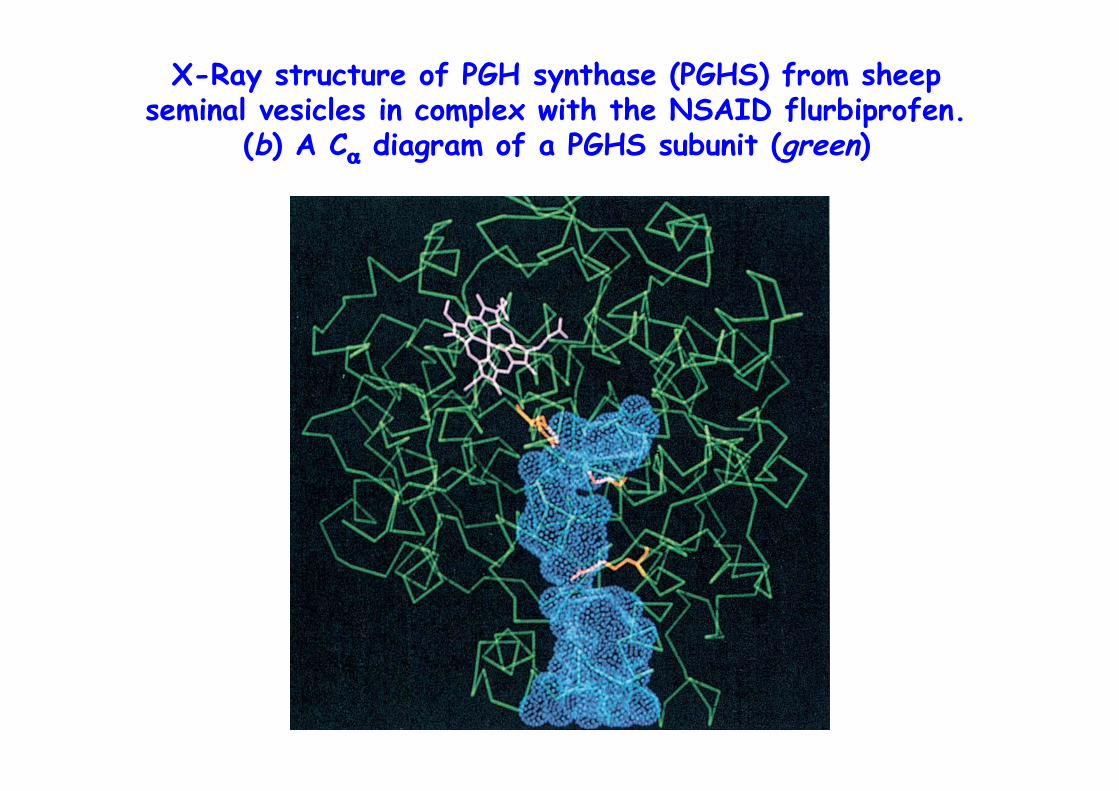
X-Ray structure of PGH synthase (PGHS) from sheepseminal vesicles in complex with the NSAID flurbiprofen.
(b) A Cα diagram of a PGHS subunit (green)
ASPIRIN
o Acetylsalicylic acido Inhibits cyclooxygenase activity of PGHSo Acetylates Ser 530o Flurbiprofen blocks channelo Low dose of aspirin reduce heart-attack
risk, inhibits platelet aggregation(enucleated cells, 10 days lifetime, cannotresynthesize enzyme
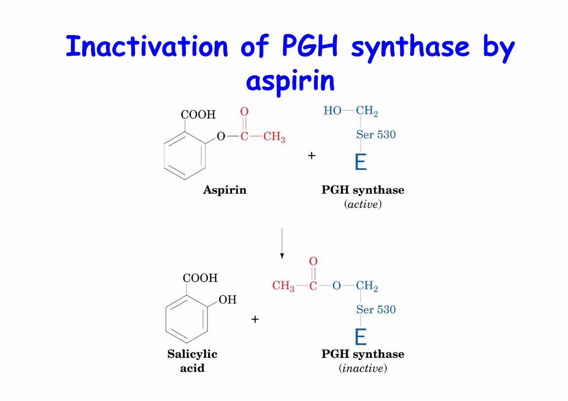
Inactivation of PGH synthase byaspirin
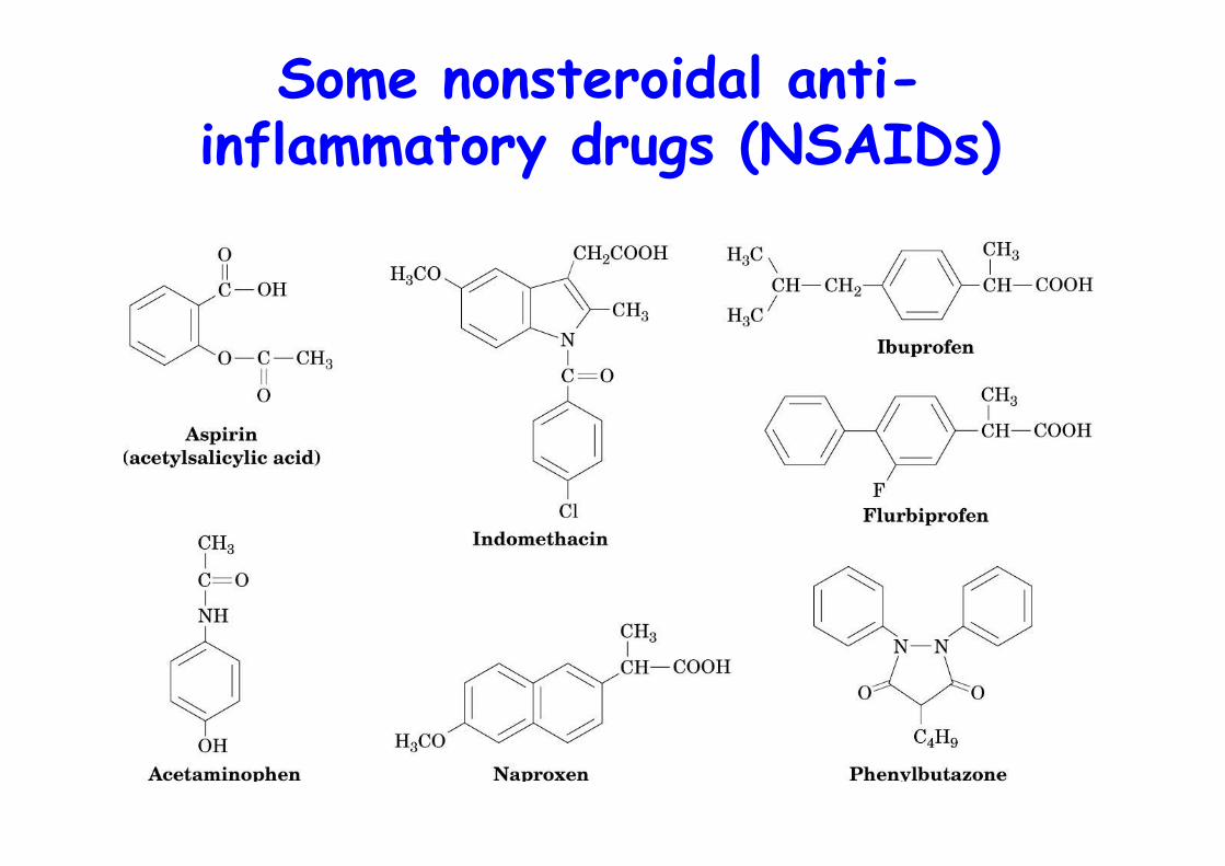
Some nonsteroidal anti-inflammatory drugs (NSAIDs)
Vioxxo 2 PGH synthase isoforms, COX1, COX-2o COX-1 is constitutively expressed in most tissues, including the gastrointestinal mucosao COX-2 only in certain tissues expressed in response to inflammatory stimuli
Aspirin can induce gastrointestinal ulceration
⇒Search for selective COX-2 inhibitors (coxibs) forlong-term treatment, i.e. arthritis
COX-3 may be the target of acetaminophen, widelyused analgesic/antipyretic drug -> treat pain & fever
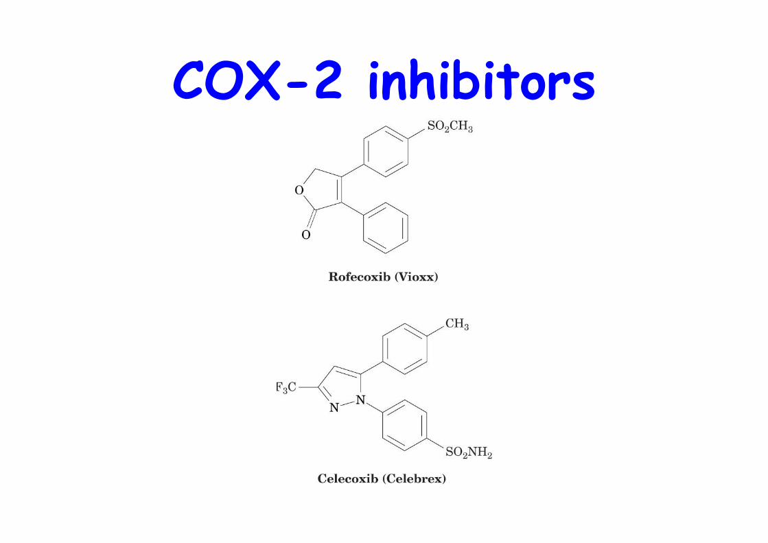
COX-2 inhibitors
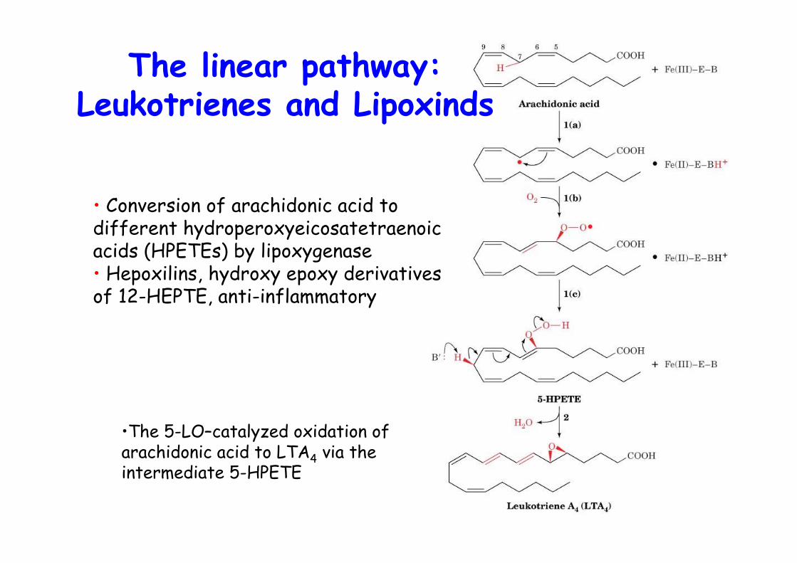
•The 5-LO–catalyzed oxidation ofarachidonic acid to LTA4 via theintermediate 5-HPETE
The linear pathway:Leukotrienes and Lipoxinds
• Conversion of arachidonic acid todifferent hydroperoxyeicosatetraenoicacids (HPETEs) by lipoxygenase• Hepoxilins, hydroxy epoxy derivativesof 12-HEPTE, anti-inflammatory
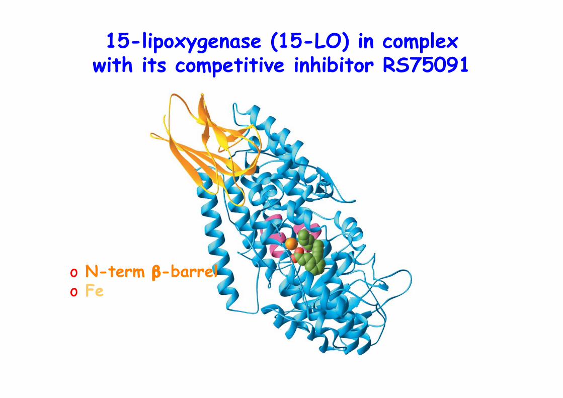
15-lipoxygenase (15-LO) in complexwith its competitive inhibitor RS75091
o N-term β-barrelo Fe
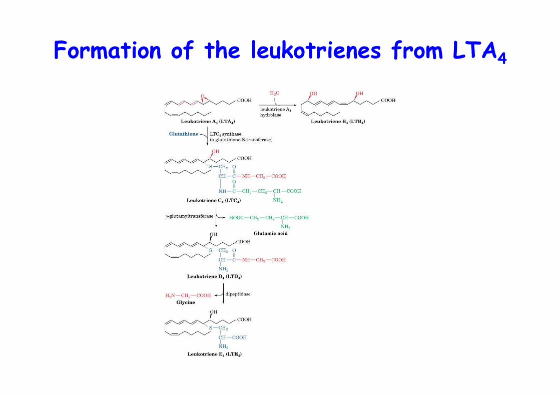
Formation of the leukotrienes from LTA4
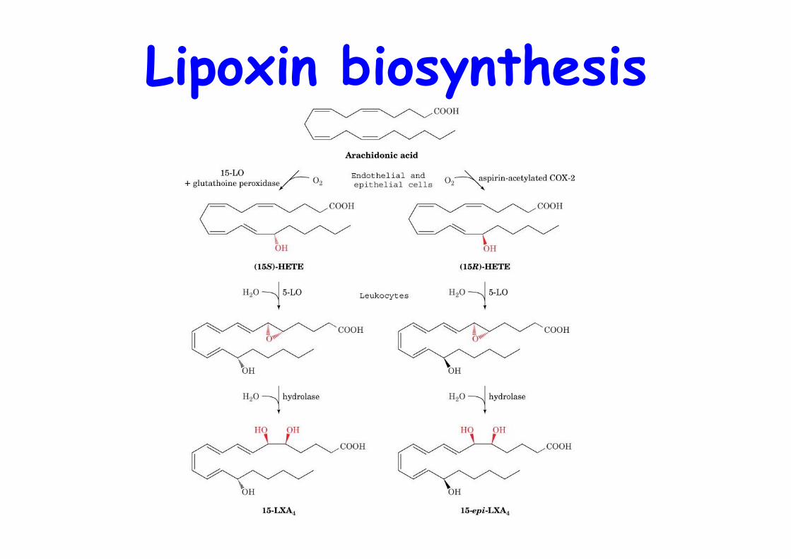
Lipoxin biosynthesis
ESKIMOS
o Low risk of cardiovascular diseasedespite the fact that they eat a lotof fat, why?
o Are healthy because they eat fish, PUFAs, n-3, n-6
o Reduce cholesterol, leukotriene and PG levels
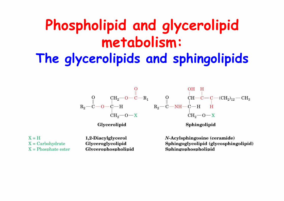
Phospholipid and glycerolipidmetabolism:
The glycerolipids and sphingolipids
Membrane lipidsAmphipathic: hydrophobic tail / hydrophilic head• glycerol, 1,2-diacyl-sn-glycerol• N-acylsphingosine (ceramide)• Head:
• phosphate ester• carbohydrate
• 2 categories of phospholipids: Glycerophospholipids, sphingophospholipids
• 2 categories of glycolipids Glyceroglycolipids, sphingoglycolipids/glycosphingolipids
Glycerophospholipidbiosynthesis
o sn-1: saturated FAo sn-2: unsaturated FA
o Biosynthesis of diacylglycerophospholipidso from DAG and PA as TAG synthesis
o Head group addition:o PC/PE
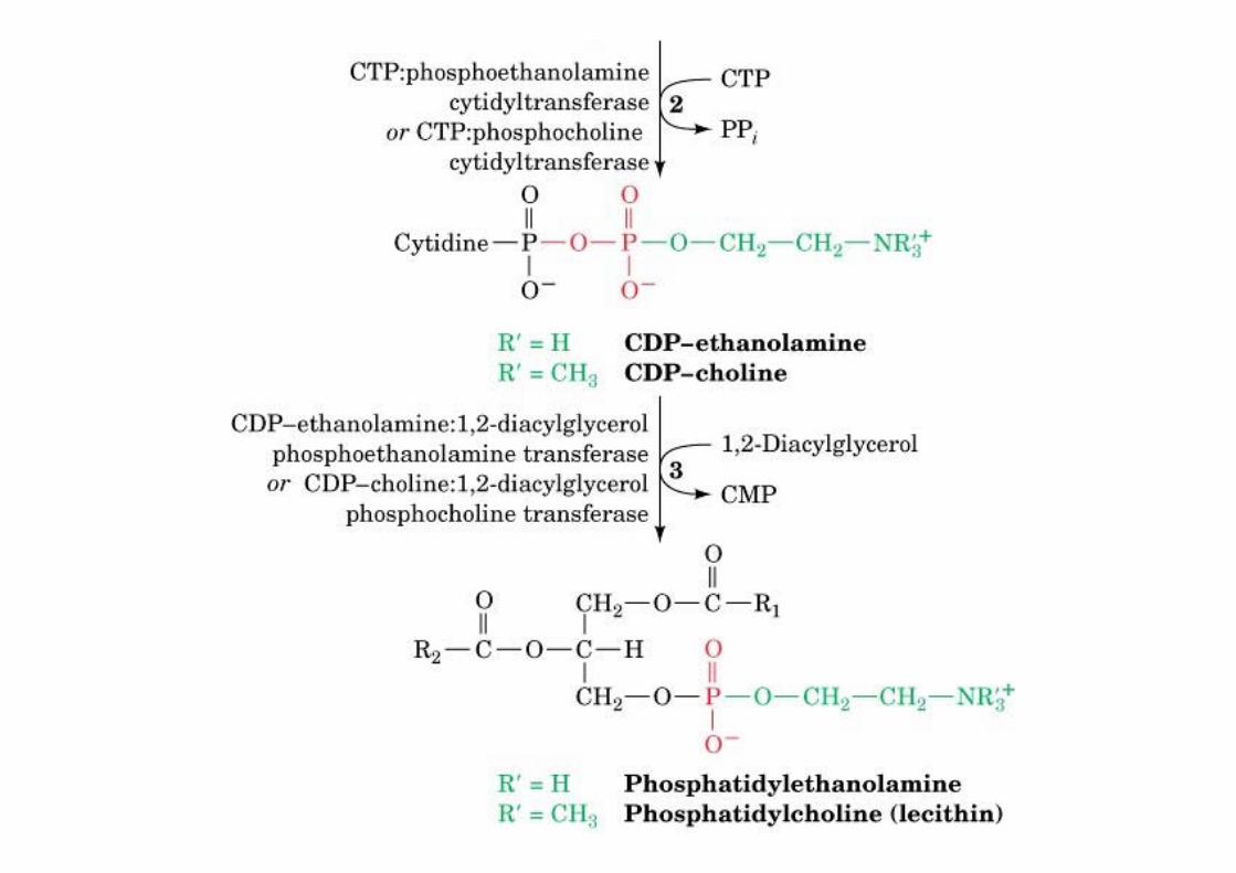
o P-activated Etn or Choo -> CDP-activated Etn or Cholo -> transfer on DAG
o PS, head-group exchange with PEo PI/PG, CDP-DAG
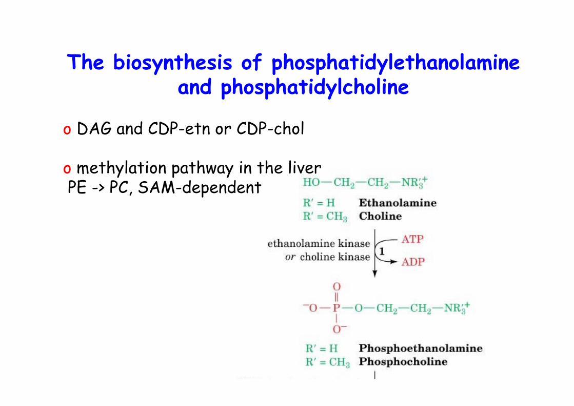
o DAG and CDP-etn or CDP-chol
o methylation pathway in the liver PE -> PC, SAM-dependent
The biosynthesis of phosphatidylethanolamineand phosphatidylcholine
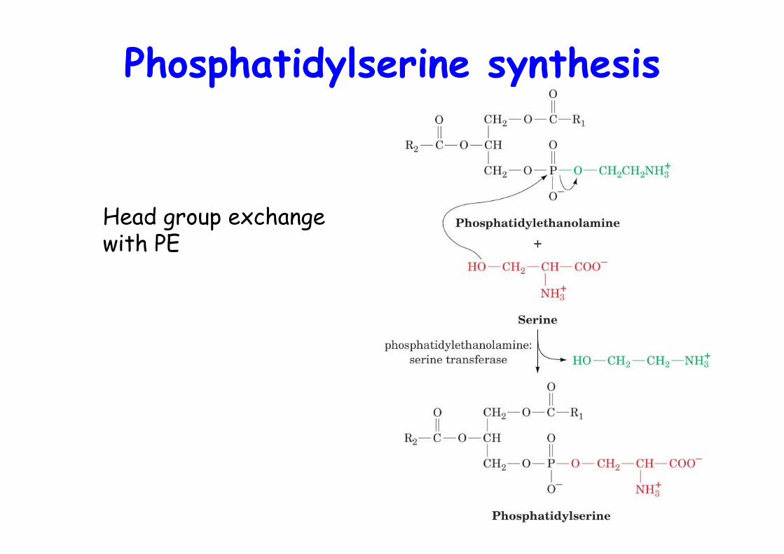
Phosphatidylserine synthesis
Head group exchange with PE
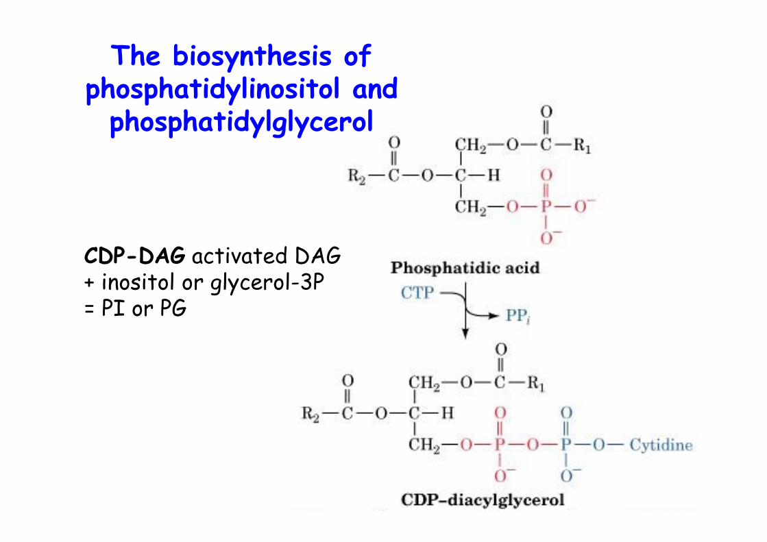
The biosynthesis ofphosphatidylinositol and
phosphatidylglycerol
CDP-DAG activated DAG+ inositol or glycerol-3P= PI or PG
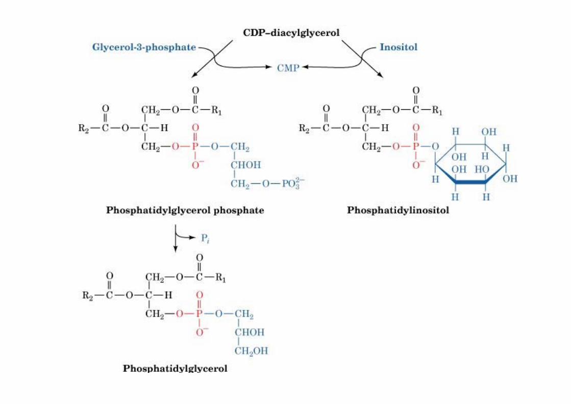
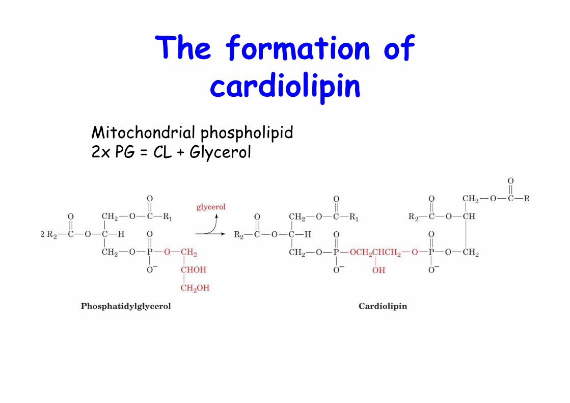
The formation ofcardiolipin
Mitochondrial phospholipid2x PG = CL + Glycerol
FA Remodeling
Tissue and cell-type specific introduction of definedFA into lipids
Examples:o 80% of brain PI contains C18:0 in sn-1 and C20:4 in sn-2o 40% of lung PC has C16:0 in both positions, surfactant
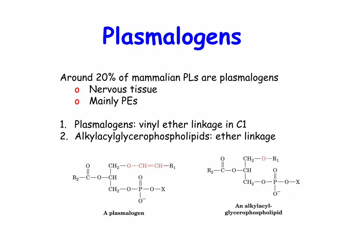
PlasmalogensAround 20% of mammalian PLs are plasmalogens
o Nervous tissueo Mainly PEs
1. Plasmalogens: vinyl ether linkage in C12. Alkylacylglycerophospholipids: ether linkage
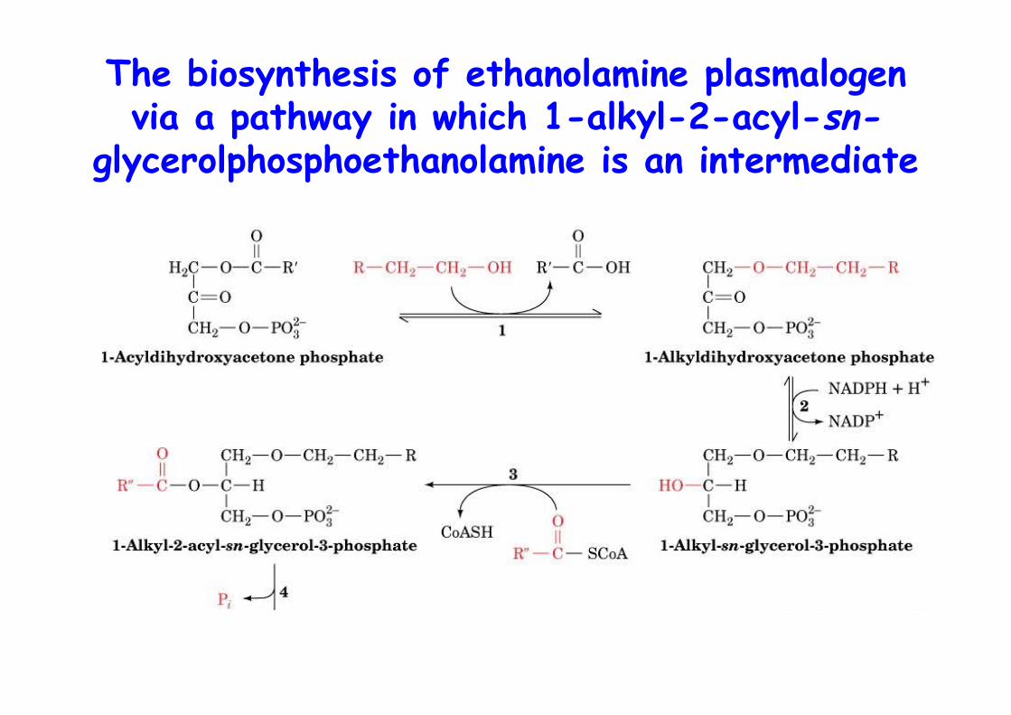
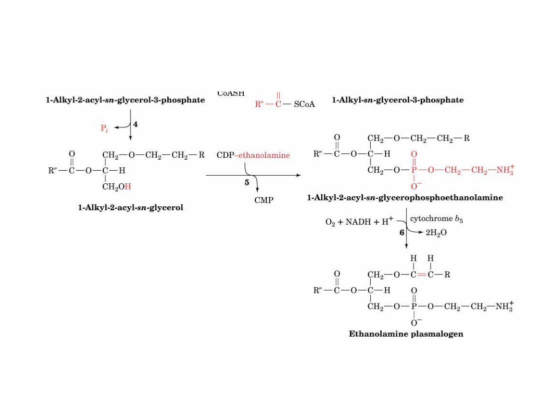
The biosynthesis of ethanolamine plasmalogenvia a pathway in which 1-alkyl-2-acyl-sn-
glycerolphosphoethanolamine is an intermediate
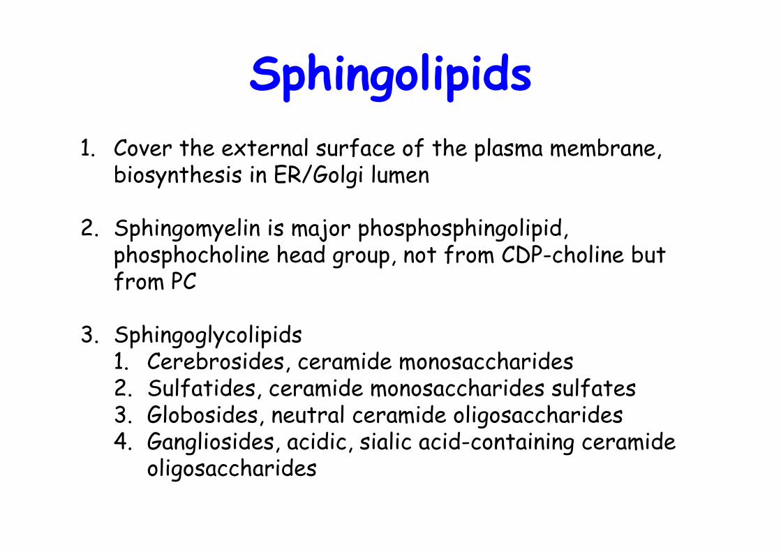
Sphingolipids1. Cover the external surface of the plasma membrane,
biosynthesis in ER/Golgi lumen
2. Sphingomyelin is major phosphosphingolipid,phosphocholine head group, not from CDP-choline butfrom PC
3. Sphingoglycolipids1. Cerebrosides, ceramide monosaccharides2. Sulfatides, ceramide monosaccharides sulfates3. Globosides, neutral ceramide oligosaccharides4. Gangliosides, acidic, sialic acid-containing ceramide
oligosaccharides
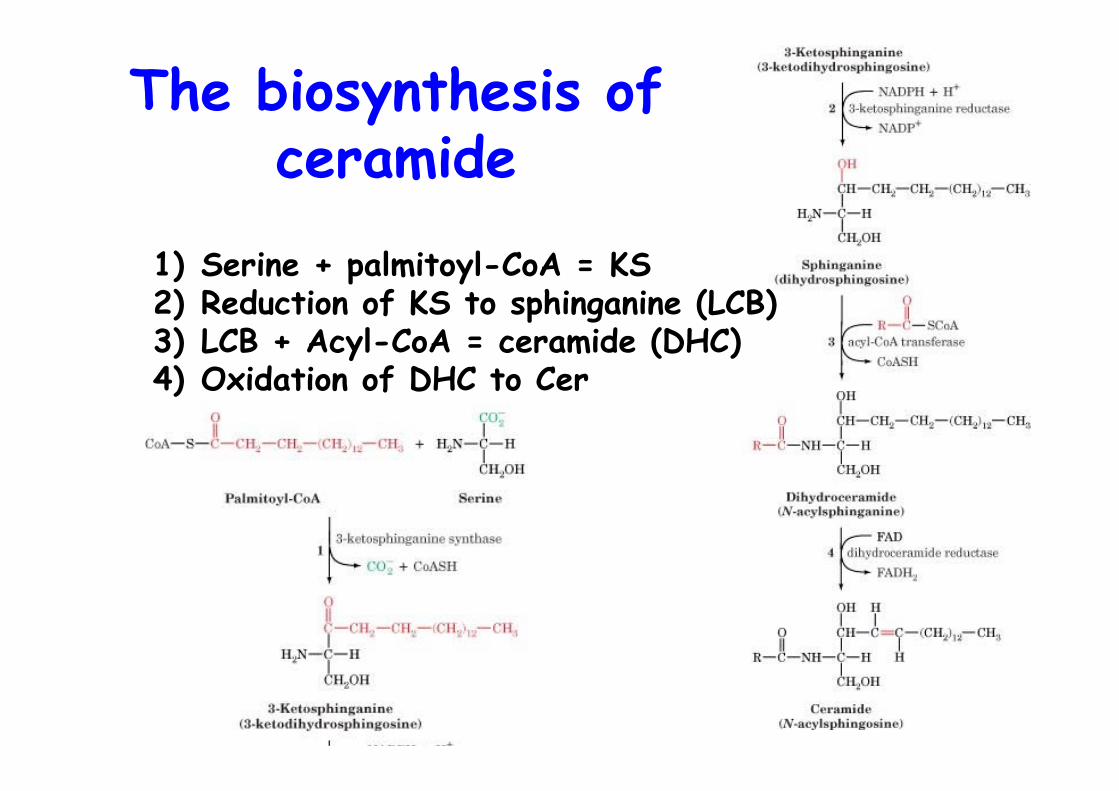
The biosynthesis ofceramide
1) Serine + palmitoyl-CoA = KS2) Reduction of KS to sphinganine (LCB)3) LCB + Acyl-CoA = ceramide (DHC)4) Oxidation of DHC to Cer
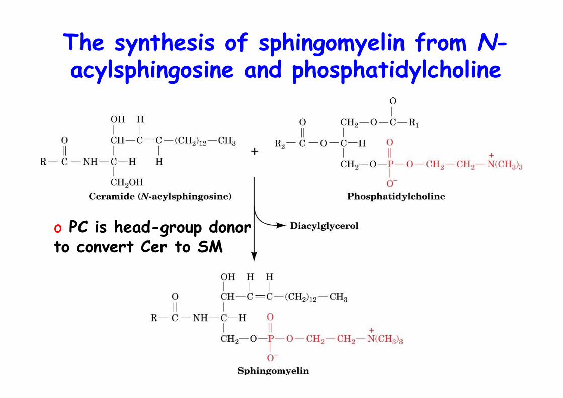
The synthesis of sphingomyelin from N-acylsphingosine and phosphatidylcholine
o PC is head-group donorto convert Cer to SM
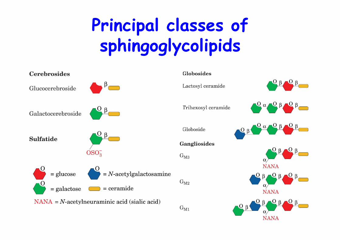
Principal classes ofsphingoglycolipids
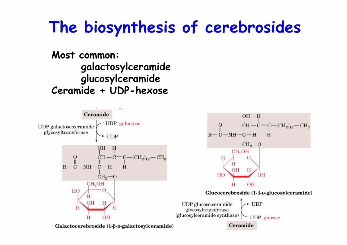
The biosynthesis of cerebrosidesMost common:
galactosylceramideglucosylceramide
Ceramide + UDP-hexose
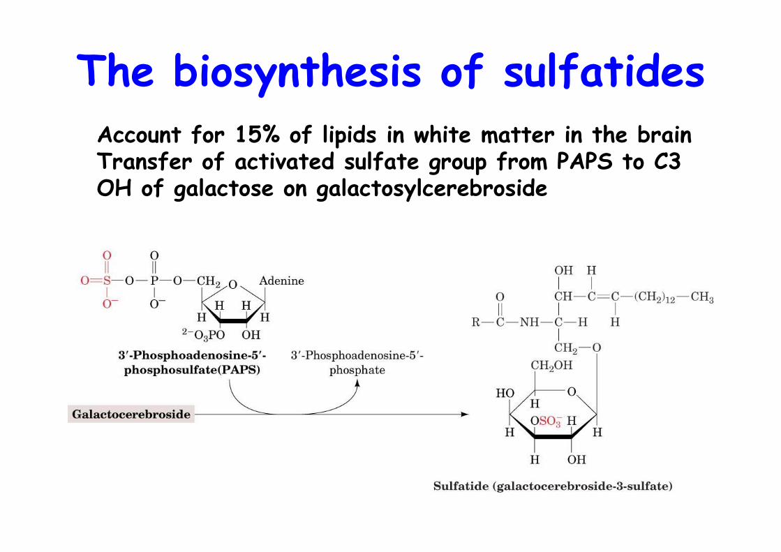
The biosynthesis of sulfatidesAccount for 15% of lipids in white matter in the brainTransfer of activated sulfate group from PAPS to C3 OH of galactose on galactosylcerebroside
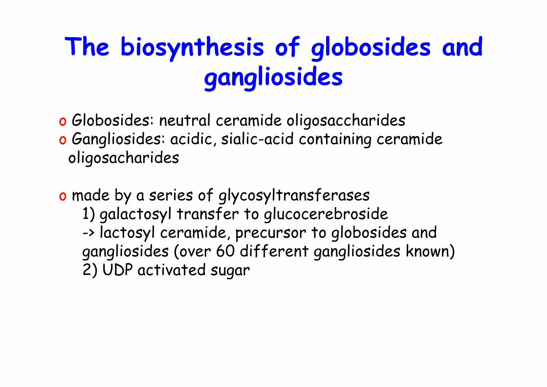
The biosynthesis of globosides andgangliosides
o Globosides: neutral ceramide oligosaccharideso Gangliosides: acidic, sialic-acid containing ceramide oligosacharides
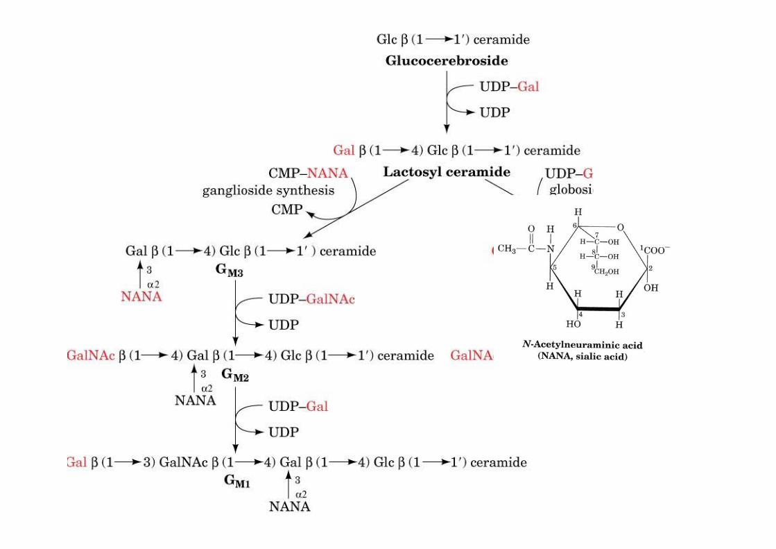
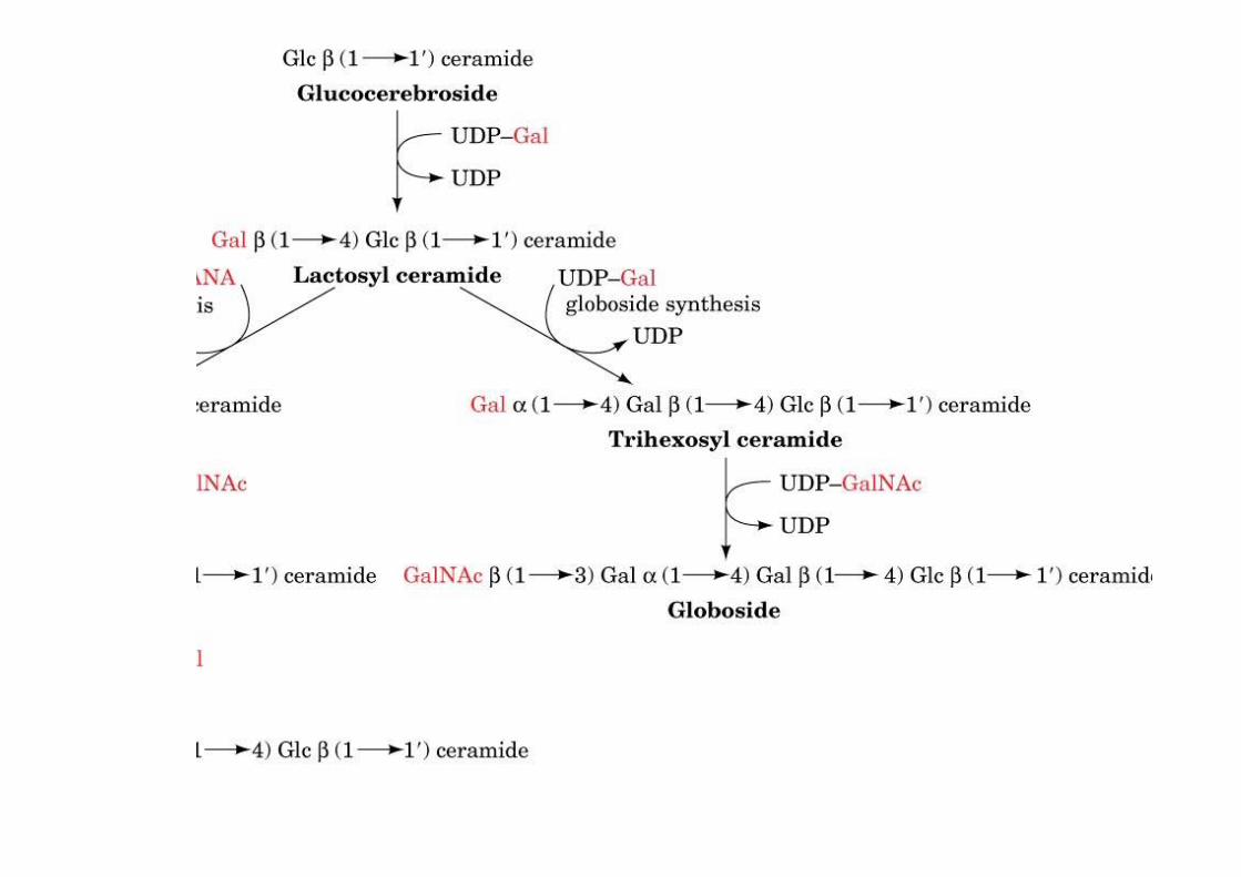
o made by a series of glycosyltransferases1) galactosyl transfer to glucocerebroside -> lactosyl ceramide, precursor to globosides andgangliosides (over 60 different gangliosides known)2) UDP activated sugar
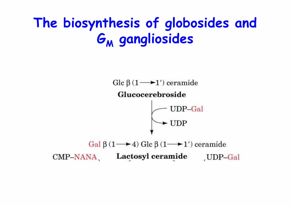
The biosynthesis of globosides andGM gangliosides
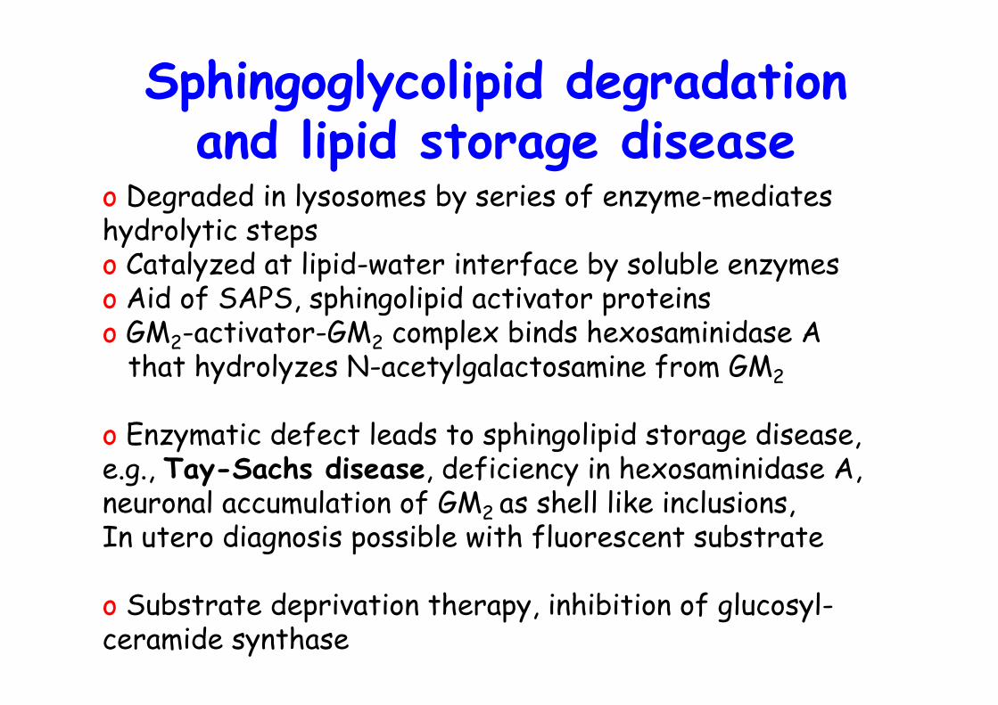
Sphingoglycolipid degradationand lipid storage disease
o Degraded in lysosomes by series of enzyme-mediates hydrolytic stepso Catalyzed at lipid-water interface by soluble enzymeso Aid of SAPS, sphingolipid activator proteinso GM2-activator-GM2 complex binds hexosaminidase A that hydrolyzes N-acetylgalactosamine from GM2
o Enzymatic defect leads to sphingolipid storage disease,e.g., Tay-Sachs disease, deficiency in hexosaminidase A,neuronal accumulation of GM2 as shell like inclusions,In utero diagnosis possible with fluorescent substrate
o Substrate deprivation therapy, inhibition of glucosyl-ceramide synthase
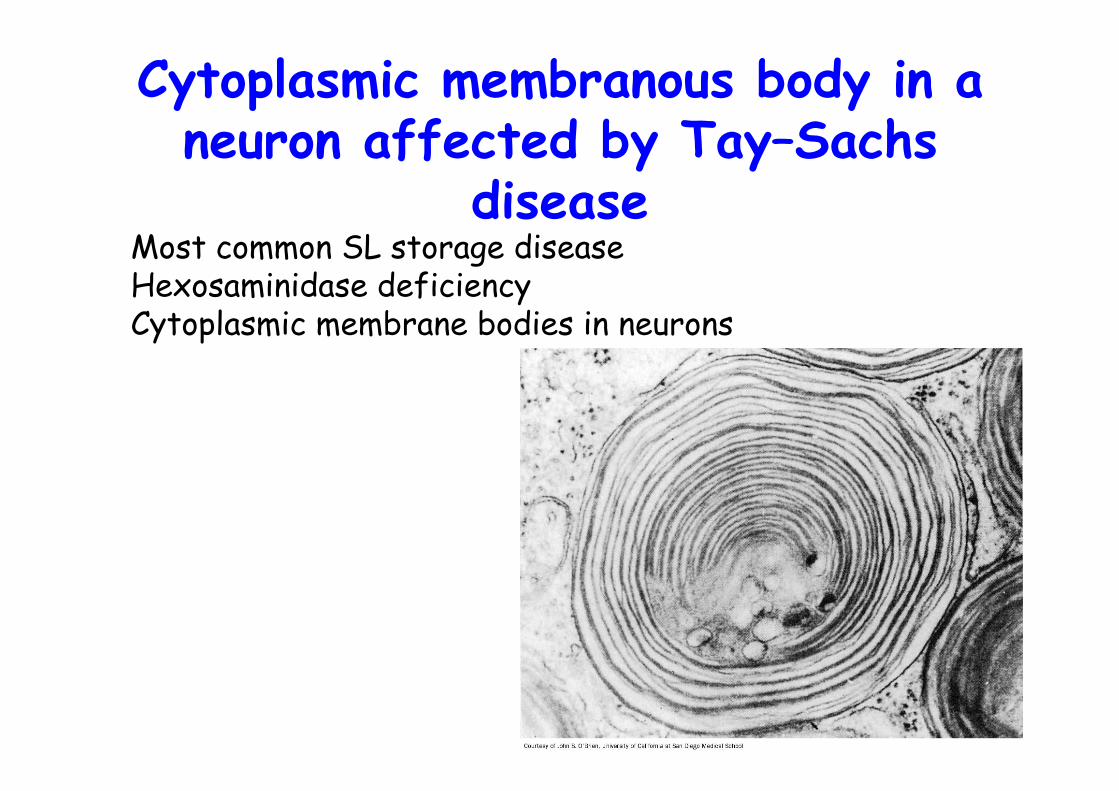
Cytoplasmic membranous body in aneuron affected by Tay–Sachs
diseaseMost common SL storage diseaseHexosaminidase deficiencyCytoplasmic membrane bodies in neurons
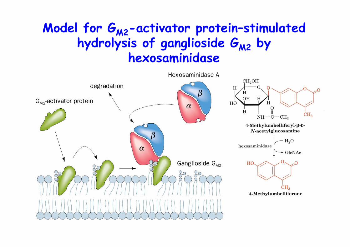
Model for GM2-activator protein–stimulatedhydrolysis of ganglioside GM2 by
hexosaminidase
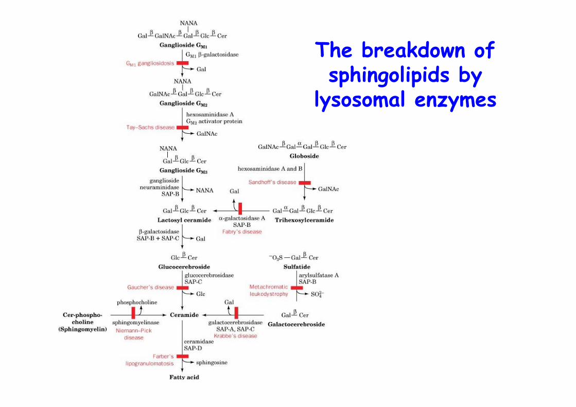
The breakdown ofsphingolipids by
lysosomal enzymes
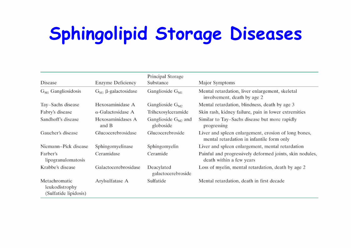
Sphingolipid Storage Diseases
Related Documents