5 Contents 2.1 Introduction ......................................................................................................................... 6 2.2 Incidence ............................................................................................................................. 6 2.3 FAP...................................................................................................................................... 7 2.4 Etiology ............................................................................................................................... 8 2.5 Clinical Presentation ........................................................................................................... 8 2.6 Clinical Considerations ....................................................................................................... 9 2.6.1 Risk Factors ............................................................................................................ 9 2.6.2 Unique Tumor Locations ........................................................................................ 10 2.6.3 FAP vs. Non-FAP .................................................................................................... 11 2.6.4 Multicentricity ........................................................................................................ 11 2.7 Clinical Course.................................................................................................................... 11 2.8 Conclusions ......................................................................................................................... 12 References .................................................................................................................................... 13 Abstract Desmoid tumors (DT) constitute a rare fibroblastic proliferative disease. They present sporadically or as a manifestation of a hereditary syndrome such as Familial Adenomatous Polyposis (FAP). Despite the absence of metastatic poten- tial, DT may cause debilitating symptoms and in some cases life-threatening organ damage because of their locally invasive nature. DT may range from small slow- growing masses to rapidly enlarging aggressive tumors. The clinical course of the disease is unpredictable but available data suggest an initial phase of growth may be followed by a long period of growth arrest with tumor stabilization or even regres- sion. FAP-related DT are preferentially located in the abdomen whereas sporadic DT tend to involve mostly the extremities, although the abdomen and the thorax may also be affected. Antecedent trauma, pregnancy and estrogens play a role in the genesis of some desmoid tumors. Surgery is the favored current approach in the treatment of most desmoid tumors. Definitive protocols are not available as C. Litchman (ed.), Desmoid Tumors, DOI 10.1007/978-94-007-1685-8_2, © Springer Science+Business Media B.V. 2011 Chapter 2 Clinical Presentation of Desmoid Tumors Anastasia Constantinidou, Michelle Scurr, Ian Judson and Charisse Litchman C. Litchman () Department of Neurology, The Stamford Hospital, Stamford, CT 06904, USA e-mail: [email protected]

Welcome message from author
This document is posted to help you gain knowledge. Please leave a comment to let me know what you think about it! Share it to your friends and learn new things together.
Transcript
5
Contents
2.1 Introduction ......................................................................................................................... 62.2 Incidence ............................................................................................................................. 62.3 FAP ...................................................................................................................................... 72.4 Etiology ............................................................................................................................... 82.5 Clinical Presentation ........................................................................................................... 82.6 Clinical Considerations ....................................................................................................... 9
2.6.1 Risk Factors ............................................................................................................ 92.6.2 Unique Tumor Locations ........................................................................................ 102.6.3 FAP vs. Non-FAP .................................................................................................... 112.6.4 Multicentricity ........................................................................................................ 11
2.7 Clinical Course .................................................................................................................... 112.8 Conclusions ......................................................................................................................... 12References .................................................................................................................................... 13
Abstract Desmoid tumors (DT) constitute a rare fibroblastic proliferative disease. They present sporadically or as a manifestation of a hereditary syndrome such as Familial Adenomatous Polyposis (FAP). Despite the absence of metastatic poten-tial, DT may cause debilitating symptoms and in some cases life-threatening organ damage because of their locally invasive nature. DT may range from small slow-growing masses to rapidly enlarging aggressive tumors. The clinical course of the disease is unpredictable but available data suggest an initial phase of growth may be followed by a long period of growth arrest with tumor stabilization or even regres-sion. FAP-related DT are preferentially located in the abdomen whereas sporadic DT tend to involve mostly the extremities, although the abdomen and the thorax may also be affected. Antecedent trauma, pregnancy and estrogens play a role in the genesis of some desmoid tumors. Surgery is the favored current approach in the treatment of most desmoid tumors. Definitive protocols are not available as
C. Litchman (ed.), Desmoid Tumors, DOI 10.1007/978-94-007-1685-8_2, © Springer Science+Business Media B.V. 2011
Chapter 2Clinical Presentation of Desmoid Tumors
Anastasia Constantinidou, Michelle Scurr, Ian Judson and Charisse Litchman
C. Litchman ()Department of Neurology, The Stamford Hospital, Stamford, CT 06904, USAe-mail: [email protected]

6
most studies have been retrospective, small and comprised of mixed populations of FAP and non-FAP as well as of mixed populations of extra-abdominal and intra-abdominal patients.
Keywords FAP • Musculoaponeurotic • Sporadic • Primary tumor • β-catenin • Abdominal • Extra-abdominal • Intra-abdominal • Pregnancy • Head and neck • Trauma
2.1 Introduction
Desmoid tumors (DT) also known as aggressive fibromatosis (AF) constitute a rare fibroblastic proliferative disease. As suggested by their name (desmoid from the Greek word “δεσμος” meaning band-like) DT may occur in any musculoaponeu-rotic or fascial tissue [1]. Usually the masses are firm and fixed to surrounding tis-sue. It is uncommon to note lymphadenopathy, overlying skin changes, erythema, or dilated veins.
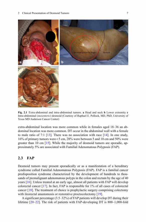
Desmoid tumors can occur anywhere in the body and are generally divided by anatomic designation as extra-abdominal, abdominal, or intra-abdominal (see Fig. 2.1). The behaviors of the tumors, including growth rates, age predilection and recurrence rates often vary with the location of the tumor [2, 3]. The most common locations are the extremities (around the limb girdles or the proximal extremities), the abdominal wall (most commonly in women during or after pregnancy), and intra-abdominal or mesenteric. Depending on their location, they tend to infiltrate adjacent organs, extend along fascial planes, compress blood vessels and nerves, erode bones or obstruct organs such as the bowel.
Though they have a benign histologic appearance, lacking the nuclear and cyto-plasmic features of a malignancy and a metastatic potential, DT may cause debili-tating symptoms such as pain, deformity and in some cases life-threatening organ damage because of their locally invasive nature. DT may range from small slow-growing masses to rapidly enlarging aggressive tumors. The clinical course of the disease is unpredictable but increasing information suggests that an initial phase of growth may be followed by a long period of growth arrest with tumor stabilization or even regression [4–6].
2.2 Incidence
Though the actual incidence is likely significantly higher due to misdiagnosis, mul-tiple and confusing pathologic nomenclature and underreporting, the current esti-mate is an incidence of 2–4 per million per year. Desmoid tumors are undisputedly very rare, with only 900 new cases diagnosed each year in the US. These tumors constitute 0.03% of all biopsy-analyzed neoplasms and < 3% of all biopsy-analyzed soft-tissue tumors [7]. These tumors have been documented in patients between 3 and 67 years [8], with a peak incidence of 25–35. The female to male ratio ranges from 1.4 to 1.8 [9–12]. Reitamo et al. noted that in females under the age of 15 an
A. Constantinidou et al.
7
extra-abdominal location was more common while in females aged 18–36 an ab-dominal location was more common. DT occur in the abdominal wall with a female to male ratio of 7:1 [13]. There was no association with race [14]. In one study, 16% of primary tumors were < 5 cm, 28% were between 5 and 10 cm and 50% were greater than 10 cm [15]. While the majority of desmoid tumors are sporadic, ap-proximately 5% are associated with Familial Adenomatous Polyposis (FAP).
2.3 FAP
Desmoid tumors may present sporadically or as a manifestation of a hereditary syndrome called Familial Adenomatous Polyposis (FAP). FAP is a familial cancer predisposition syndrome characterized by the development of hundreds to thou-sands of premalignant adenomatous polyps in the colon and rectum by the age of 40 years [16]. Unless treated at an early age, almost all patients with FAP will develop colorectal cancer [17]. In fact, FAP is responsible for 1% of all cases of colorectal cancer [18]. The treatment of choice is prophylactic surgery comprising colectomy with ileorectal anastomosis or restorative proctocolectomy [19].
A significant percentage (3.5–32%) of FAP patients will develop DT during their lifetime [20–22]. The risk of patients with FAP-developing DT is 800–1,000-fold
Fig. 2.1 Extra-abdominal and intra-abdominal tumors. a Head and neck b Lower extremity c Intra-abdominal ( mesenteric) desmoid (Courtesy of Raphael E. Pollock, MD, PhD, University of Texas MD Anderson Cancer Center)
2 Clinical Presentation of Desmoid Tumors
8
higher compared to the general population [23]. The peak incidence of DT in FAP is between the second and the third decade [24]. In the majority of cases DT occur following prophylactic surgery for FAP [25, 26] with surgical trauma identified as a trigger for the development of DT in FAP. However, in some cases, DT may be the first manifestation of FAP with about 4% of cases of DT appearing as an incidental finding at the time of primary surgery [27]. Family history is a predisposing factor for DT formation in FAP patients [28, 29], with an observed increased risk of 2.5 times in first-degree relatives [29].
2.4 Etiology
Desmoid tumors are the result of deregulation of connective tissue growth. In-creased nuclear expression of β-catenin, a protein responsible for regulation of gene expression, proliferation and survival, is the characteristic feature in both sporadic and FAP-associated DT. Familial Adenomatous Polyposis is a hereditary (autoso-mal dominant) disease characterized by a germ-line mutation in the adenomatous polyposis coli gene (APC). In FAP-driven DT, inactivation of the APC gene leads to accumulation of β-catenin whereas in the sporadic setting, in approximately 85% of cases, mutations in the β-catenin gene CTNNBi lead to increased activity of β-catenin [30].
Desmoid tumors are viewed as a nonneoplastic process by some authors and as a well-differentiated low-grade sarcoma by others [31]. The characterization of desmoid tumors as a neoplastic process rather than as an inflammatory fibrous reac-tion has been bolstered by the molecular studies of X-chromosome inactivation that confirmed that DT are the result of a clonal process [31, 32]. Nonrandom X-chro-mosome inactivation, trisomy 8 and/or 20 was demonstrated in greater than 30% of sporadic DT [33]. DT behave aggressively as locally infiltrating mesenchymal monoclonal proliferations that lack metastatic potential [34].
2.5 Clinical Presentation
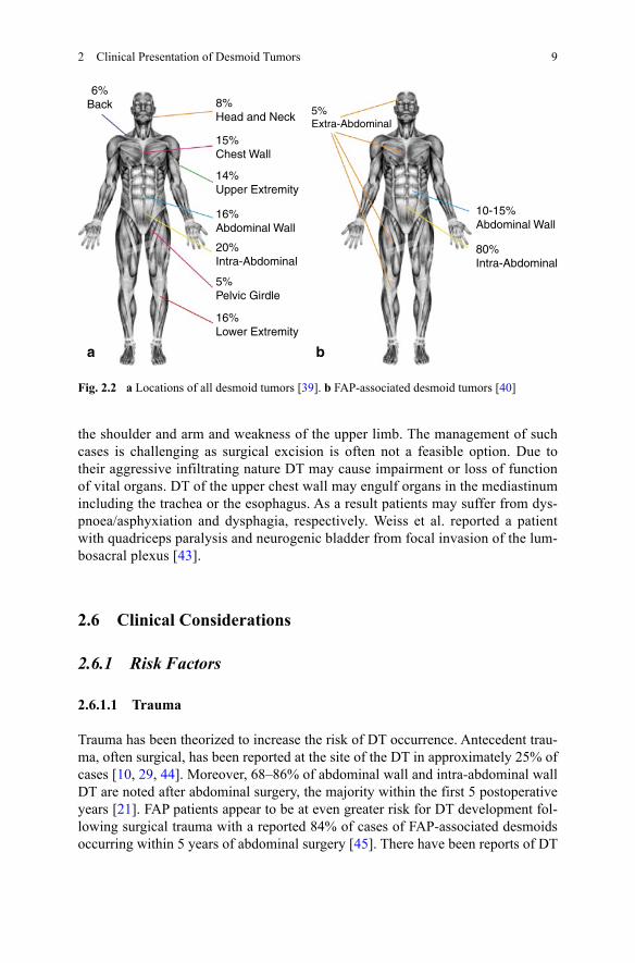
In sporadic desmoids, between 37 and 50% of DT arise in the abdominal region [35–37]. The most common extra-abdominal sites are the shoulder girdle, chest wall and inguinal regions [38] (see Fig. 2.2).
Patients with intra-abdominal desmoids may have asymptomatic masses which silently enlarge and infiltrate into adjacent structures [2] or may have symptoms of weight loss, cachexia, malaise, compression of ureters, renal failure, small bowel compression, perforation and peritonitis [35, 41, 42 ]. In sporadic DT, infiltration of intestinal or visceral structures is less common but muscle, nerve and vessel involvement may result in debilitating symptoms such as pain, restricted mobil-ity or deformity. A characteristic example of such presentation is the infiltration of the brachial plexus by a shoulder girdle tumor which may result in pain in
A. Constantinidou et al.
9
the shoulder and arm and weakness of the upper limb. The management of such cases is challenging as surgical excision is often not a feasible option. Due to their aggressive infiltrating nature DT may cause impairment or loss of function of vital organs. DT of the upper chest wall may engulf organs in the mediastinum including the trachea or the esophagus. As a result patients may suffer from dys-pnoea/asphyxiation and dysphagia, respectively. Weiss et al. reported a patient with quadriceps paralysis and neurogenic bladder from focal invasion of the lum-bosacral plexus [43].
2.6 Clinical Considerations
2.6.1 Risk Factors
2.6.1.1 Trauma
Trauma has been theorized to increase the risk of DT occurrence. Antecedent trau-ma, often surgical, has been reported at the site of the DT in approximately 25% of cases [10, 29, 44]. Moreover, 68–86% of abdominal wall and intra-abdominal wall DT are noted after abdominal surgery, the majority within the first 5 postoperative years [21]. FAP patients appear to be at even greater risk for DT development fol-lowing surgical trauma with a reported 84% of cases of FAP-associated desmoids occurring within 5 years of abdominal surgery [45]. There have been reports of DT
Fig. 2.2 a Locations of all desmoid tumors [39]. b FAP-associated desmoid tumors [40]
6%Back 8%
Head and Neck
15%Chest Wall
14%Upper Extremity
16%Abdominal Wall
20%Intra-Abdominal
5%Pelvic Girdle
16%Lower Extremity
a
10-15%Abdominal Wall
80%Intra-Abdominal
5%Extra-Abdominal
b
2 Clinical Presentation of Desmoid Tumors
10
in laparoscopic port sites [46], following a total hip replacement [47], around sili-cone implants [48], at the site of an internal jugular catheter [49] and at the site of a previous rib fracture [50].
2.6.1.2 Estrogen and Pregnancy
There are several lines of evidence to support a role for estrogen in modulating the behavior of DT. Several studies have shown that DT in females of childbearing age have a greater growth rate than that of those in males or in pre- or postmenopausal women [3, 51]. Further, an increased frequency rate was demonstrated during preg-nancy [9, 51] and in females taking oral contraceptives [28, 52]. Additionally, there have been reports of tumor regression during menarche and menopause [51, 53, 54] and with Tamoxifen treatment [55].
In the lab, fibrous tumors have been induced in animal models following the administration of exogenous estrogen [53] and estrogen was shown to exert a mi-togenic influence on many cell types, including fibroblasts [56]. Additionally, in a study of human DT, estrogen receptors (ER) were observed in 33% of all DT exam-ined, with an equal incidence in males and females and with antiestrogen binding sites found in 79% of samples, including some which were ER negative [57].
In pregnancy-associated DT, the mass is most frequently located within one of the two rectus muscles of the abdominal wall without involving the midline [58, 59]. Pregnancy-associated DT may develop during any trimester or postpartum.
While the history of antecedent trauma is 28% of sporadic DT [60], such a his-tory is ostensibly missing in pregnant DT patients. It has been theorized that the combination of an altered hormonal milieu and the trauma of stretching of the ab-dominal aponeurosis during the advancement of pregnancy are contributing factors [61]. There has been one report of a DT that developed at the site of a prior caesar-ean section scar during a subsequent pregnancy [16].
A study of FAP patients revealed no association between the female gender or pregnancy and the risk of the development of DT [62]. After examining the diver-gent natural histories and behaviors of pregnancy-associated DT and FAP-associat-ed DT, one group of investigators concluded that these two types of DT are separate entities [61].
2.6.2 Unique Tumor Locations
2.6.2.1 Head and Neck DT
Head and neck DT are a more aggressive disorders that affect a younger population. Twelve percent of extra-abdominal DT arise in the head and neck [63]. The mean age is 16.87 years, with 57.32% of cases under 11 years. Children with DT of the head and neck are younger at the time of diagnosis than children with DT at other
A. Constantinidou et al.
11
sites [64–66]. There is a 30% local recurrence (LR) with a male to female ratio of 1:1 [67]. One explanation for the often difficult clinical course is the restricted anatomy containing crucial neural and vascular structures [67].
2.6.2.2 Breast
Desmoid tumors are rarely seen in the breast and can simulate breast carcinoma [68].
2.6.3 FAP vs. Non-FAP
Anatomic locations differ between FAP and sporadic DT, with more intra-abdominal or abdominal than extra-abdominal wall tumors. In a Mayo clinic review from 1976 to 1999, 67% of FAP-associated DT were abdominal as compared to 11% sporadic. Limb DT accounted for 1.4% in FAP patients and 34.7% in non-FAP patients [69]. While one large study reported a female to male ratio of 3.0 in FAP patients with DT [28], some studies failed to show the female predominance in FAP-associated DT that has been shown in sporadic DT [29, 44]. Additionally, desmoid develop-ment occurred an average of 6 years earlier in FAP patients [22]. Eighty to 90% of FAP individuals will carry an alteration in the APC gene on chromosome number 5. The majority will have a family history of colorectal cancer and polyposis. But, up to 33% of FAP patients with DT will have a de novo mutation within the APC gene and therefore no family history of DT [69].
2.6.4 Multicentricity
There have been 10–20 reports of multicentric extra-abdominal DT, mostly in FAP patients [70–73]. These usually recur in the same limb in proximity to the site of the primary tumor. They do not grow simultaneously, with the second growth generally occurring years later [74].
2.7 Clinical Course
DT remains an enigmatic disease with a variable course that can range from an incidental small tumor that can remain small and stable or become large and grow rapidly, causing death in a matter of months or years. The morbidity and mortality is largely determined by the location of the tumor and therefore the adjacent structures the tumor may infiltrate or compress. According to Church, 10% of DT will resolve
2 Clinical Presentation of Desmoid Tumors
12
spontaneously, 30% will undergo cycles of progression and resolution, 50% will remain stable after diagnosis and 10% will progress rapidly [75].
Some of the local recurrence (LR) rates are determined by tumor location. For example, extremity tumors are considered locally aggressive and have LR ranging from 24 to 77% [76–80]. LR rates for intra-abdominal tumors are higher than for extra-abdominal tumors, reported to be 57–86% [28, 81, 82]. One review found LR to be 24% for abdominal wall, 43% for extra-abdominal and 77% for intra-abdom-inal tumors [2]. In a study of 78 FAP patients that studied progression-free survival rates after surgery versus conservative care, it was determined that extra-abdominal and abdominal wall DT had better outcomes and more benefit overall from surgical intervention than intra-abdominal tumors [22].
Gender has been shown not to be a prognostic factor for LR [4, 83]. There is disagreement about whether age may play a role in recurrence. Some studies have shown that younger age was associated with increased local treatment failure [39, 84] while others did not [75, 85]. One study found the recurrence rate in children to be 88%, twice that of adults (38%) [10]. Also controversial is the role of age in LR risk. Some studies show increased risk of LR in female patients older than 30 [88] while others show increased risk in patients under 30 [9]. One larger study of 103 patients over 26 years found no correlation with recurrence to age, gender, or site [83]. There is some suggestion that size of the primary tumor is an important predic-tor for recurrence [40] but that a single recurrence did not significantly increase the likelihood of a subsequent recurrence [10].
There is ongoing controversy over the significance of margin status in predicting LR. In one series, response rates of 72% and 41% were reported for tumor-free and tumor-positive margins, respectively [86]. Other studies show no correlation with margin status. The MSKCC (Memorial Sloan-Kettering Cancer Center) and Insti-tuto Nazionale Tumori experiences showed no significant difference (22% negative vs. 24% positive [76] and 21% positive vs. 18% negative) [87].
The limitations in the studies stem from the small subject numbers and the mix of intra- and extra-abdominal tumors as well as primary and recurrent lesions, lead-ing to conflicting results about the biology of these elusive tumors [9, 70, 76–81, 88–90]. The difficulties of interpretation of the data are compounded by the un-predictable natural course of this tumor that can apparently regress even without treatment [75].
2.8 Conclusions
Desmoid tumors are an enigmatic, elusive disease that continue to defy definition. Due to their rarity and the practical limitations in their study, these tumors often evade accurate characterization. As they can arise in many locations throughout the body, thereby presenting unique challenges to physicians in many different fields, the most appropriate and fruitful approach to caring for any individual desmoid tumor patient is a multidisciplinary one.
A. Constantinidou et al.

13
References
1. Goldblum J, Fletcher JA (2002) Desmoid-type fibromatoses. In: Fletcher CDM, Unni KK, Mertens F (Eds) World Health Organization classification of tumours. Pathology and Genet-ics of Tumours of Soft Tissue and Bone. IARC Press, Lyon, pp 83–84
2. Easter DW, Halasz NA (2010) Recent trends in the management of desmoid tumors. Sum-mary of 19 cases and review of the literature. Ann Surg 210:765–769
3. Hayry P, Reitamo JJ, Totterman S et al (1982) The desmoid tumor. II. Analysis of factors possibly contributing to the etiology and growth behavior. Am J Clin Pathol 77:674–680
4. Phillips SR, A’Hern R, Thomas JM (2004) Aggressive fibromatosis of the abdominal wall, limbs and limb girdles. Br J Surg 91(12):1624–1629
5. Bonvalot S, Eldweny H, Haddad V et al (2008) Extra-abdominal primary fibromatosis: aggressive management could be avoided in a subgroup of patients. Eur J Surg Oncol 34(4):462–468
6. Stoeckle E, Coindre JM, Longy M et al (2009) A critical analysis of treatment strategies in desmoid tumours: a review of a series of 106 cases. Eur J Surg Oncol 35:129–134
7. Micke O, Seegenschmiedt MH (2005) Radiation therapy for aggressive fibromatosis (des-moid tumors): results of a national Patterns of Care Study. Int J Radiat Oncol Biol Phys 61:882–891
8. Brodsky IT, Gordan MS, Hajdu SI, Burt M (1992) Desmoid tumors of the chest wall. A lo-cally recurrent problem. J Thorac Cardiovasc Surg 104:900–903
9. Posner MC, Shiu MH, Newsome JL, Hajdu SI, Gaynor JJ, Brennan MF (1989) The desmoid tumor. Not a benign disease. Arch Surg 124:191–196
10. Lopez R, Kemalyan N, Moseley HS, Dennis D, Vetto RM (1990) Problems in diagnosis and management of desmoid tumors. Am J Surg 159:450–453
11. Jarvinen HJ (1987) Desmoid disease as a part of familial adenomatous polyposis coli. Acat Chir Scand 153:379–383
12. Klemmer S, Pascoe L, DeCosse J (1987) Occurrence of desmoids in patients with familial adenomatous polyposis of the colon. Am J Med Genet 28:385–392
13. Pack GT, Ehrlich HE (1944) Neoplasms of the anterior abdominal wall with special consid-eration to desmoid tumours. Int Abstr Surg 79:177–198
14. Wong SL (2008) Diagnosis and management of desmoid tumors and fibrosarcoma. J Surg Onc 97:554–558
15. De Camargo VP, Keohan ML, D’Adamo DR, Antonescu CR, Brennan MF, Singer S, Ahn LS, Maki RG (2010) Clinical outcomes of systemic therapy for patients with deep fibromatosis (desmoid tumor). Cancer 116:2258–2265
16. De Cian F, Delay E, Rudigoz RC, Rachere D, Rivoire M (1999) Desmoid tumor arising in a cesarean section scar during pregnancy: monitoring and management. Gynecol Oncol 75:145–148
17. Herman K, Marcinek A (1996) Abdominal desmoid in a 28 year-old pregnant woman. Ginekol Pol 67:374–375
18. Burke AP, Sobin LH, Shekitka KM et al (1990) Intra-abdominal fibromatosis: a pathologic analysis of 130 tumors with comparison of clinical subgroups. Am J Surg Pathol 14:335–341
19. Suarez V, Hall C (1985) Mesenteric fibromatosis. Br J Surg 72:976–97820. Bertario L, Russo A, Sala P et al (2003) Multiple approach to the exploration of genotype-
phenotype correlations in familial adenomatous polyposis. J Clin Oncol 21:1698–170721. Clark SK, Phillips RK (1996) Desmoids in familial adenomatous polyposis. Br J Surg
83:1494–150422. Nieuwenhuis MH, Casparie M, Mathus-Vliegen LM, Dekkers OM, Hogendoorn PC, Vasen
HF (2010) A nation-wide study comparing sporadic and familial adenomatous polyposis-related desmoid-type fibromatoses. Int J Cancer 129(1):256–261
23. Fong Y, Rosen PP, Brennan MF (1999) Multifocal desmoids. Surgery 114:902–906
2 Clinical Presentation of Desmoid Tumors
14
24. Godwin Y, McCulloch TA, Scully L (2001) Extra-abdominal desmoid tumour of the breast: review of the primary management and the implications for breast reconstruction. Br J Plast Surg 54:268–271
25. Corsten M, Donald P, Boggan J et al (1998) Extra-abdominal fibromatosis (desmoid tumor) arising in the infratemporal fossa: a case report. Skull Base Surg 8(4):237–241
26. Heinimann K, Mullhaupt B, Weber W et al (1998) Phenotypic differences in familial adeno-matous polyposis based on APC germline mutation status. Gut 43:675–679
27. Sleijfer S (2009) Management of aggressive fibromatosis: can we unravel the maze of treat-ment options? Eur J Cancer 45(17):2928–2929
28. Jones IT, Jagelman DG, Fazio VW, Lavery IC, Weakley FL, McGannon E (1986) Desmoid tumors in familial polyposis coli. Ann Surg 204:94–97
29. Gurbuz AK, Giardiello FM, Petersen GM, Krush AJ, Offerhaus GJ, Booker SV, Kerr MC, Hamilton SR (1994) Desmoid tumours in familial adenomatous polyposis. Gut 35:377–381
30. De Bree E, Keus R, Melissas J, Tsiftsis D, van Coevorden F (2009) Desmoid tumors: need for an individualized approach. Expert Rev Anticancer Ther 9:525–535
31. Li M, Cordon-Cardo C, Gerald WL, Rosai J (1996) Desmoid fibromatosis is a clonal process. Hum Pathol 27:939–943
32. De Wever I, Dal Cin P, Fletcher CD et al (2000) Cytogenetic, clinical and morphologic cor-relations in 78 cases of fibromatosis: a report from the CHAMP Study Group. Chromosomes and morphology. Mod Pathol 13:1080–1085
33. Fletcher JA, Naeem R, Xiao S, Corson JM (1995) Chromosome aberrations in desmoid tu-mors: trisomy 8 may be a predictor of recurrence. Cancer Genet Cytogenet 63:527–529
34. Alman BA, Pajerski ME, Diaz-Cano S et al (1997) Aggressive fibromatosis (desmoid tumor) is a monoclonal disorder. Diagn Mol Pathol 6:98–101
35. Lewis JJ, Boland PJ, Leung DH, Woodruff JM, Brennan MF (1999) The enigma of desmoid tumor. Ann Surg 229:866–873
36. Weiss S, Goldblum JR (Eds) (2001) Enzinger and Weiss’s soft tissue tumors, 4th edn. Mobis, St Louis, pp 641–693
37. Bruce JM, Bradley EL 3rd, Satchidanand SK (1996) A desmoid tumor of the pancreas. Spo-radic intra-abdominal desmoid revisited. Int J Pancreatol 19:197–203
38. Khorsand J, Karakousis CP (1985) Desmoid tumours and their management. Am J Surg 149:215–218
39. Lev D, Kotilingam D, Wei C, Ballo MT, Zagars GK, Pisters PW, Lazar AA, Patel SR, Benjamin RS, Pollock RE (2007) Optimizing treatment of desmoid tumors. J Clin Oncol 25(13):1785–1791
40. Sturt JNH, Clark SK (2006) Current ideas in desmoid tumors. Familial Cancer 5:275–28541. Corbel L, Souissi M, Chretien Y, Dufour B (1992) Desmoid tumor of the mesentery. An un-
common cause of ureteral obstruction. J Radiol 73:669–67242. Anthony T, Rodriquez-Bigas MA, Weber TK, Petrelli NJ (1996) Desmoid tumor. J Am Coll
Surg 182:369–37743. Weiss AJ, Lackman RD (1989) Low-dose chemotherapy of desmoid tumors. Cancer
64:1192–119444. McAdams WA, Goligher JC (1970) The occurrence of desmoids in patients with familial
polyposis coli. Cr J Surg 57:618–63145. Bertario L, Russo A, Sala P et al (2001) Genotype and phenotype factor as determinant of
desmoid tumors in patients with familial adenomatous polyposis. Int J Cancer 95:102–10746. Lynch HT, Fitzgibbons R Jr (1996) Surgery, desmoid tumors and familial adenomatous pol-
yposis: case report and literature review. Am J Gastroenterol 91:2598–260147. Gebhart M, Fourmarier M, Heymans O, Alexiou J, Yengue P, De Saint-Aubain N (1999)
Development of a desmoid tumor at the site of a total hip replacement. Acta Orthop Belg 65:230–234
48. Reitamo JJ, Hayry P, Nykyri E, Saxen E (1982) The desmoid tumor. I. Incidence, sex, age, and anatomical distribution in the Finnish population. Am J Clin Pathol 77:665–673
A. Constantinidou et al.

15
49. Skhiri H, Zellama D, Ameur FM, Moussa A, Gmar BS, Achour A, Ben Dhia N, Zakhama A, Elmay M (2004) Desmoid cervical tumor following the placing of an internal jugular cath-eter. Presse Med 33:95–97 (French)
50. Wiel Marin A, Romagnoli A, Carlucci I, Veneziani A, Mercui M, Destito C (1995) Thoracic desmoid tumors: a rare evolution of rib fracture. Etiopathogenesis and therapeutic consider-ations. G Chir 16:341–344
51. Reitamo JJ, Scheinin TM, Hayry P (1986) The desmoid syndrome. New aspects in the cause, pathogenesis and treatment of the desmoid tumor. Am J Surg 151:230–237
52. Waddell WR (1975) Treatment of intra-abdominal and abdominal wall desmoid tumors with drugs that affect the metabolism of cyclic 3″.5″-adenosine monophosphate. Ann Surg 181:299–302
53. Dahn I, Johnsson N, Lundh G (1963) Desmoid tumors. A series of 33 cases. Acta Chir Scand 126:305–314
54. Lofti AM, Dozois RR, Gordon H, Hruska LS, Weiland LH, Carryer PW, Hurt RD (1989) Mesenteric fibromatosis complicating familial adenomatous polyposis: predisposing factors and results of treatment. Int J Colorectal Dis 4:30–36
55. Wilcken N, Tattersall MH (1991) Endocrine therapy for desmoid tumors. Cancer 68:1384–1388
56. Dhingra K (1999) Antiestrogens-tamoxifen, SERMS and beyond. Invest New Drugs 17:285–311
57. Lim CL, Walker MJ, Mehta RR et al (1986) Estrogen and antiestrogen binding sites in des-moid tumors. Eur J Cancer Clin Oncol 22:583
58. Gansar GF, Markowitz IP, Cerise EJ (1987) Thirty years of experience with desmoid tumors at Charity Hospital. Surg 53(6):318–319
59. Galetotti F, Facci E, Bianchin E (2006) Desmoid tumour involving the abdominal rectus muscle: report of a case. Hernia 10:278–281
60. Enzinger FM, Weiss SW (1995) Soft tissue tumors, 3rd Edn. Mosby Year Book Inc., Saint Louis.
61. Johner A, Tiwari P, Zetler P, Wiseman SM (2009) Abdominal wall desmoid tumors associated with pregnancy: current concepts. Expert Rev Anticancer Ther 9(11):1675–1682
62. Nieuwenhuis MH, De Vos tos Nederveen Cappel W, Botma A et al (2008) Desmoid tumors in a Dutch cohort of patients with familial adenomatous polyposis. Clin Gastroenterol Hepatol 6:215–219
63. Conley J, Healey WV, Stout AP (1966) Fibromatosis of the head and neck. Am J Surg 112(4):609–614
64. Ayala AG, Ro JY, Goepfert H, Cangir A Khorsand J, Flake G (1986) Desmoid fibromatosis: a clinicopathologic study of 25 children. Semin Diagn Pathol 3:138–150
65. Scougall P, Staheli LT, Chew DE, Taylor TKF, Almquist EE (1987) Desmoid tumors in child-hood. Orthop Rev 16:481–488
66. Spiegel DA, Dormans JP, Meyer JS et al (1999) Aggressive fibromatosis from infancy to adolescence. J Pediatr Oprthop 19:776–784
67. Kruse AL, Luebber HT, Gratz KW, Obwegeser JA (2010) Aggressive fibromatosis of the head and neck: a new classification based on a literature review over 40 years (1968–2008). Oral Maxillofac Surg 14(40):227–232
68. Greenberg D, McIntyre H, Ramsaroop R, Artyr J, Harman J (2002) Aggressive fibromatosis of the breast: a case report and literature review. Breast J 8:55–57
69. Fallen T, Wilson M, Morlan B, Lindor NL (2006) Desmoid tumors-a characterization of patients seen at Mayor Clinic 1976–1999. Fam Cancer 5:191–194
70. Rock MG, Pritchard DJ, Reiman HM et al (1984) Extra-abdominal desmoid tumors. J Bone Joint Surg 66A:1369–1373
71. Wagstaff MJD, Raurell A, Perks AGB (2004) Multicentric extra-abdominal desmoid tu-mours. Br Assoc of Plastic Surgeons 57:362–365
72. Antal I, Szendroi M, Kovacs G et al (1994) Multicentric extra-abdominal desmoid tumour: a case report. J Cancer Res Clin Oncol 120:490–494
2 Clinical Presentation of Desmoid Tumors
16
73. Maurer F, Horst F, Pfannenberg C et al (1996) Multifocal extra-abdominal desmoid tumour-diagnostic and therapeutic problems. Arch Orthop Trauma Surg 115:359–362
74. Barber HM, Galasko CSB, Woods CG (1973) Multicentric extra-abdominal desmoid tu-mours. Report of two cases. J Bone Joint Surg 55:858–863
75. Church JM (1995) Desmoid tumours in patients with familial adenomatous polyposis. Semin Colon Rectal Surg 6:29–32
76. Merchant NP, Lewis JJ, Leung DH, Woodruff JM, Brennan MF (1999) Extremity and trunk desmoid tumors: a multifactorial analysis of outcome. Cancer 86:2045–2052
77. Wold LE, Weiland LH (1983) Tumefactive fibro-inflammatory lesions of the head and neck. Am J Surg Pathol 7:477–482
78. Exelby PR (1981) Surgery of soft tissue sarcomas in children. Natl Cancer Inst Monogr 153–157
79. Scott RJ, Taeschner W, Heinimann K et al (1997) Association of extracolonic manifestations of familial adenomatous polyposis with acetylation phenotype in a large FAP kindred. Eur J Hum Genet 5:43–49
80. Thomas JA, Kothare SN (1972) Desmoid tumors of the abdominal wall. Indian J Cancer 9:66–69
81. Rodriguez-Bigas MA, Mahoney MC, Karakousis CP, Petrelli NJ (1994) Desmoid tumors in patients with familial adenomatous polyposis. Cancer 74:1270–1274
82. Penna C, Tiret E, Parc R et al (1993) Operation and abdominal desmoid tumors in familial adenomatous polyposis. Surg Gyencol Obstet 177:263–268
83. Pignatti G, Barbanti-Brodano G, Ferrari D, Gherlinzoni F, Bertoni F, Bacchini P, Barbieri E, Giunti A, Campanacci M (2000) Extraabdominal desmoid tumor: a study of 83 cases. Clini-cal Orthop and Related Research 375:207–213
84. Sorensen A, Keller J, Nielsen OS, Jensen OM (2002) Treatment of aggressive fibromatosis. A retrospective study of 72 patients followed for 1–27 years. Acta Orthop Scan 73:213–219
85. De Bree E, van Coevorden F, Keus RB, Tsiftsis DD (2004) Treatment of extremity desmoid tumours. Eur J Surg Oncol 30:1141–1142
86. Nuyttens JJ, Rust PF, Thomas CR, Turrisi III (2000) Surgery versus radiation therapy for pa-tients with aggressive fibromatosis or desmoid tumors. A comparative review of 22 articles. Cancer 88:1517–1523
87. Gronchi A, Casali PG, Mariani L et al (2003) Quality of surgery and outcome in extra-ab-dominal aggressive fibromatosis: a series of patients surgically treated at a single institution. J Clin Oncol 21:190–197
88. Pritchard DJ, Nascimento AG, Petersen IA (1996) Local control of extra-abdominal desmoid tumors. J Bone Joint Surg 78:848–854
89. Miralbell R, Suit HD, Mankin HJ, Zuckerberg LR, Stracher MA, Rosenberg AE (1990) Fi-bromatoses: from postsurgical surveillance to combined surgery and radiation therapy. Int J Radiat Oncol BIol Phys 18:535–540
90. Anthony T, Rodriguez-Bigas MA, Weer TK, Petrelli NJ (1996) Desmoid tumors. J Am Coll Surg 182:369–377
A. Constantinidou et al.

http://www.springer.com/978-94-007-1684-1
Related Documents