Int J Clin Exp Med 2016;9(11):22458-22462 www.ijcem.com /ISSN:1940-5901/IJCEM0032882 Case Report Use of a fully-resorbable, biomimetic composite hydroxyapatite as bone graft substitute for posterolateral spine fusion: a case report Brunella Grigolo 1,2 , Paolo Dolzani 2 , Christian Giannetti 3 , Miria Tenucci 3 , Giuseppe Calvosa 3 1 SSD Laboratory RAMSES, Italy; 2 Laboratory of Immunorheumatology and Tissue Regeneration, Rizzoli Orthopaedic Institute, Via di Barbiano1/10, 40136, Bologna, Italy; 3 Orthopaedic Division, S. Maria Maddalena Hospital, Borgo S. Lazzero, 56048, Volterra, Italy Received May 27, 2016; Accepted August 3, 2016; Epub November 15, 2016; Published November 30, 2016 Abstract: Introduction: A successful spinal fusion is the result of the proper integration between the fixation device, used to stabilize and correct the column posture, and the bone graft applied to replace or integrate the bone com- ponents. Background: In the recent past, the use of bone graft substitutes has been widely employed in spinal sta- bilization surgery, with the aim of improving the amount of bone graft material available, as well as to avoid the use of autograft and allograft bone, with related drawbacks and side-effects. However, since standardized non-invasive techniques (X-rays, CT scan images) are no sufficiently accurate in predicting whether bony fusion occurred, cur- rently the only option to evaluate the bone graft osteointeointegration is through a revision surgery, due to the need of correcting the previous surgical procedure. Here, we present the osteointegrative properties of a third-generation hydroxyapatite bone graft substitute applied on a 65-year-old patient, who underwent a spinal stabilization surgery and, one year later, a revision surgery due to the rupture of a metallic bar. Materials and Methods: Bony fusion was evaluated by histological and immunohistochemical analysis performed on a biopsy sample one year after surgery. For this purpose, histological and immunohistochemical analysis were performed. Results: Histological analysis clearly evidenced the complete osteointegration of the device, together with new bone formation. Discussion and Conclusions: Third-generation bone graft substitutes allow bone ingrowth and tissue remodelling by providing three- dimensional structural support, thus resulting as ideal scaffolds for bone replacement and osteointegration. Keywords: Posterolateral spine fusion, spinal stabilization, arthrodesis, bone grafts, hydroxyapatite, osteointegra- tion Introduction A successful spinal fusion (or arthrodesis) mainly depends on the proper integration between fixation devices (metal implants, screws with rods or plates, cages), used to sta- bilize and correct the spine column, and bone grafts applied to replace or integrate the bone components [1, 2]. Currently, autograft and allograft materials are considered the “gold standard” options to achieve spinal fusion. Autologous bone, usually harvested from non-essential bones (spinous processes) or from the iliac crest (pelvis), has osteoinductive, osteoconductive and osteogen- ic properties, no concerns for diseases trans- mission and no risks of immunogenicity. However, side effects (such as donor-side mor- bidity, post-operative chronic pain and the risk of relatively limited quantity) can reduce its use [3]. As well as autograft, allograft material shows osteoinductive and osteoconductive properties and, being harvested from cadaveric tissue donors, it shows reduced patients-relat- ed side-effects. Nonetheless, limitations (e.g. risks of disease transfer and poor osteogenic properties) restrict its applicability [3]. To circumvent all these issues, the use of bone graft substitutes has been extensively investi- gated for surgical applications [1, 4]. These materials can be artificially created from ceram- ics (or other naturally occurring and biocompat- ible substances) and hold mechanical proper- ties similar to the bone component. Their imme- diate availability and unlimited quantity makes these products of relevant interest for surgical

Welcome message from author
This document is posted to help you gain knowledge. Please leave a comment to let me know what you think about it! Share it to your friends and learn new things together.
Transcript
Int J Clin Exp Med 2016;9(11):22458-22462www.ijcem.com /ISSN:1940-5901/IJCEM0032882
Case Report Use of a fully-resorbable, biomimetic composite hydroxyapatite as bone graft substitute for posterolateral spine fusion: a case report
Brunella Grigolo1,2, Paolo Dolzani2, Christian Giannetti3, Miria Tenucci3, Giuseppe Calvosa3
1SSD Laboratory RAMSES, Italy; 2Laboratory of Immunorheumatology and Tissue Regeneration, Rizzoli Orthopaedic Institute, Via di Barbiano1/10, 40136, Bologna, Italy; 3Orthopaedic Division, S. Maria Maddalena Hospital, Borgo S. Lazzero, 56048, Volterra, Italy
Received May 27, 2016; Accepted August 3, 2016; Epub November 15, 2016; Published November 30, 2016
Abstract: Introduction: A successful spinal fusion is the result of the proper integration between the fixation device, used to stabilize and correct the column posture, and the bone graft applied to replace or integrate the bone com-ponents. Background: In the recent past, the use of bone graft substitutes has been widely employed in spinal sta-bilization surgery, with the aim of improving the amount of bone graft material available, as well as to avoid the use of autograft and allograft bone, with related drawbacks and side-effects. However, since standardized non-invasive techniques (X-rays, CT scan images) are no sufficiently accurate in predicting whether bony fusion occurred, cur-rently the only option to evaluate the bone graft osteointeointegration is through a revision surgery, due to the need of correcting the previous surgical procedure. Here, we present the osteointegrative properties of a third-generation hydroxyapatite bone graft substitute applied on a 65-year-old patient, who underwent a spinal stabilization surgery and, one year later, a revision surgery due to the rupture of a metallic bar. Materials and Methods: Bony fusion was evaluated by histological and immunohistochemical analysis performed on a biopsy sample one year after surgery. For this purpose, histological and immunohistochemical analysis were performed. Results: Histological analysis clearly evidenced the complete osteointegration of the device, together with new bone formation. Discussion and Conclusions: Third-generation bone graft substitutes allow bone ingrowth and tissue remodelling by providing three-dimensional structural support, thus resulting as ideal scaffolds for bone replacement and osteointegration.
Keywords: Posterolateral spine fusion, spinal stabilization, arthrodesis, bone grafts, hydroxyapatite, osteointegra-tion
Introduction
A successful spinal fusion (or arthrodesis) mainly depends on the proper integration between fixation devices (metal implants, screws with rods or plates, cages), used to sta-bilize and correct the spine column, and bone grafts applied to replace or integrate the bone components [1, 2].
Currently, autograft and allograft materials are considered the “gold standard” options to achieve spinal fusion. Autologous bone, usually harvested from non-essential bones (spinous processes) or from the iliac crest (pelvis), has osteoinductive, osteoconductive and osteogen-ic properties, no concerns for diseases trans-mission and no risks of immunogenicity. However, side effects (such as donor-side mor-
bidity, post-operative chronic pain and the risk of relatively limited quantity) can reduce its use [3]. As well as autograft, allograft material shows osteoinductive and osteoconductive properties and, being harvested from cadaveric tissue donors, it shows reduced patients-relat-ed side-effects. Nonetheless, limitations (e.g. risks of disease transfer and poor osteogenic properties) restrict its applicability [3].
To circumvent all these issues, the use of bone graft substitutes has been extensively investi-gated for surgical applications [1, 4]. These materials can be artificially created from ceram-ics (or other naturally occurring and biocompat-ible substances) and hold mechanical proper-ties similar to the bone component. Their imme-diate availability and unlimited quantity makes these products of relevant interest for surgical
Hydroxyapatite-derived bone graft substitute for spine fusion
22459 Int J Clin Exp Med 2016;9(11):22458-22462
applications requiring a large amount of bone graft, therefore representing an immediate and ready-to-use alternative to autograft and allo-graft bone.
Among these, hydroxyapatite bone substitutes have been used as graft extenders to foster bone regeneration. Several experiments on ani-mal models [5] and clinical studies on humans [6] have demonstrated that these biomaterials possess a chemico-physical structure which supports cell migration and scaffold coloniza-tion, sustaining new tissue formation and bone
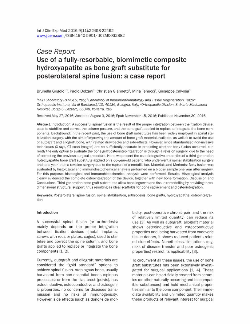
A smoking-free, 64-year-old woman presented with a chronic low back pain in March 2012. The patient was diagnosed on 2011 with idio-pathic lumbar spine scoliosis (Figure 1A, 1B), which worsened during the last year, rising to a left sciatic irritating pain. VAS (Visual Analog Scale for pain) score was 9. Oswestry Low Back Pain Disability Index was 50. The patient under-went spinal stabilization surgery through poste-rior approach performed by the use of rods sta-bilization system and pedicle screws, which were inserted bilaterally on L4-L5 levels, on L3 (left side), on D11 and D12 (right side) and on
Figure 1. (A) Antero-posterior and (B) lateral view of pre-operative radiograph showing lumbar spine scoliosis. (C) Antero-posterior and (D) lateral view of post-operative radiograph showing lumbar spine scoliosis correction.
regeneration, and therefore supporting their usability also in the spinal field. However, as standardized noninvasive evaluations (X-rays, CT scan images) are not 100% accu-rate in predicting whether a bony fusion has occurred [2, 7], only secondary surgical procedures (e.g. revision sur-gery for spinal stabilization) can provide information about successful joint ossifications.
RegenOss (provided by Fin-Ceramica Faenza S.p.A., Fa- enza, Italy) is a fully-biomimet-ic and resorbable third-gen- eration hydroxyapatite bone graft substitute, which resem-bles the process occurring during biological neo-ossifi- cation, through the patented process of nucleation of the Mg-Ha nanocrystals into type I Collagen fibers [4]. These features allow the product to serve as scaffold to guide effective bone regeneration, fostering cell attachment and proliferation and promoting a reduction of the osteointegra-tion time periods [4, 8, 9].
Here we report osteointegra-tion evidences of the Mg-en- riched HA bone graft substi-tute RegenOss, ascertained after revision surgery for spi-nal stabilization.
Case report
Hydroxyapatite-derived bone graft substitute for spine fusion
22460 Int J Clin Exp Med 2016;9(11):22458-22462
D10 (left side), followed by neurological left decompression and scoliosis curve correction (Figure 1C, 1D).
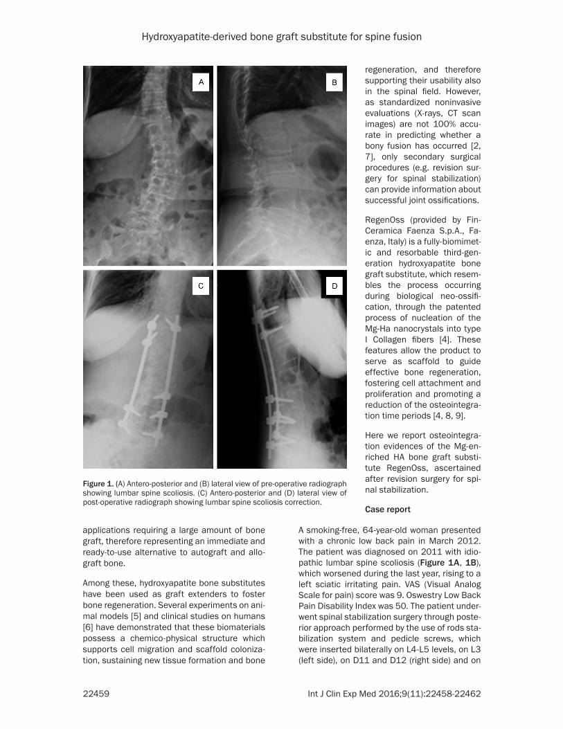
The screw-based system was integrated with the apposition of autologous bone (60%) plus a synthetic bone graft substitute (40%) (Regen- Oss, provided by Fin-Ceramica Faenza S.p.A., Faenza, Italy) along the rods and the articular processes on the right side (Figure 2A, 2B) in order to achieve bony fusion and stabilize the spine column. Patient was discharged from the hospital after routine postoperative period.
After one and a half years (August 2013) with-out any symptoms, the patient went back to the hospital with dorsalgy due to rupture of the left rod. We therefore proceeded for a spinal stabi-lization revision surgery, removing the D10 screw (left side) and positioning another pedi-cle screw in D11 (left side). During surgery,
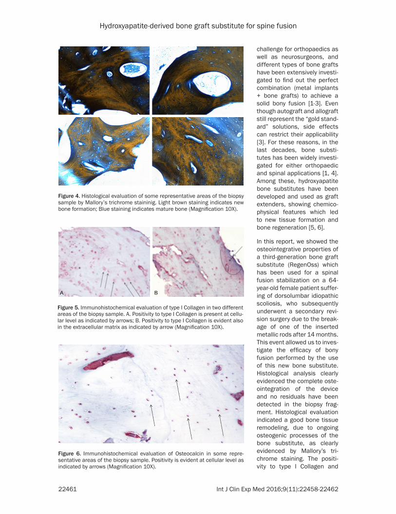
tion areas within mature bone (Figure 4). Im- munohistochemical evaluation confirmed the good quality of the bone tissue as shown by a positive staining for typical osteogenic markers such as type I Collagen and Osteocalcin. In par-ticular, we noticed a strong positivity for type I Collagen at both cellular and extracellular lev-els in mature and new bone areas (Figure 5). Similar results have been observed also for osteocalcin, but in this case the positivity was limited to the cells (Figure 6).
Discussion
Arthrodesis is a surgical procedure employed in a large number of traumatic, degenerative and oncological spinal diseases, in order to perma-nently fuse together two or more vertebrae and stabilize the spine column [1]. The need of metal implants to be integrated with vertebral bones of the spine has always represented a
Figure 2. Images from a spinal stabilization surgery theatre. A. Autologous bone harvested from the laminar processes of the vertebrae and trimmed for spinal fusion. B. During spinal fusion, the composite bone substitute RegenOss (red circle) will be mixed with autologous bone and applied along the rods and the pedicle screws to improve spinal stabilization.
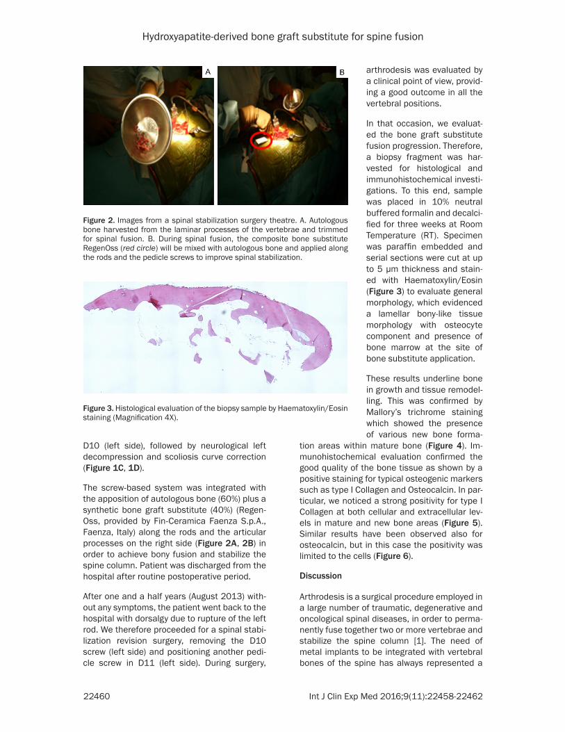
Figure 3. Histological evaluation of the biopsy sample by Haematoxylin/Eosin staining (Magnification 4X).
arthrodesis was evaluated by a clinical point of view, provid-ing a good outcome in all the vertebral positions.
In that occasion, we evaluat-ed the bone graft substitute fusion progression. Therefore, a biopsy fragment was har-vested for histological and immunohistochemical investi-gations. To this end, sample was placed in 10% neutral buffered formalin and decalci-fied for three weeks at Room Temperature (RT). Specimen was paraffin embedded and serial sections were cut at up to 5 µm thickness and stain- ed with Haematoxylin/Eosin (Figure 3) to evaluate general morphology, which evidenced a lamellar bony-like tissue morphology with osteocyte component and presence of bone marrow at the site of bone substitute application.
These results underline bone in growth and tissue remodel-ling. This was confirmed by Mallory’s trichrome staining which showed the presence of various new bone forma-
Hydroxyapatite-derived bone graft substitute for spine fusion
22461 Int J Clin Exp Med 2016;9(11):22458-22462
challenge for orthopaedics as well as neurosurgeons, and different types of bone grafts have been extensively investi-gated to find out the perfect combination (metal implants + bone grafts) to achieve a solid bony fusion [1-3]. Even though autograft and allograft still represent the “gold stand-ard” solutions, side effects can restrict their applicability [3]. For these reasons, in the last decades, bone substi-tutes has been widely investi-gated for either orthopaedic and spinal applications [1, 4]. Among these, hydroxyapatite bone substitutes have been developed and used as graft extenders, showing chemico-physical features which led to new tissue formation and bone regeneration [5, 6].
In this report, we showed the osteointegrative properties of a third-generation bone graft substitute (RegenOss) which has been used for a spinal fusion stabilization on a 64- year-old female patient suffer-ing of dorsolumbar idiopathic scoliosis, who subsequently underwent a secondary revi-sion surgery due to the break-age of one of the inserted metallic rods after 14 months. This event allowed us to inves-tigate the efficacy of bony fusion performed by the use of this new bone substitute. Histological analysis clearly evidenced the complete oste-ointegration of the device and no residuals have been detected in the biopsy frag-ment. Histological evaluation indicated a good bone tissue remodeling, due to ongoing osteogenic processes of the bone substitute, as clearly evidenced by Mallory’s tri-chrome staining. The positi- vity to type I Collagen and
Figure 4. Histological evaluation of some representative areas of the biopsy sample by Mallory’s trichrome staininig. Light brown staining indicates new bone formation; Blue staining indicates mature bone (Magnification 10X).
Figure 5. Immunohistochemical evaluation of type I Collagen in two different areas of the biopsy sample. A. Positivity to type I Collagen is present at cellu-lar level as indicated by arrows; B. Positivity to type I Collagen is evident also in the extracellular matrix as indicated by arrow (Magnification 10X).
Figure 6. Immunohistochemical evaluation of Osteocalcin in some repre-sentative areas of the biopsy sample. Positivity is evident at cellular level as indicated by arrows (Magnification 10X).
Hydroxyapatite-derived bone graft substitute for spine fusion
22462 Int J Clin Exp Med 2016;9(11):22458-22462
Osteocalcin markers confirmed this process. Our results suggest how the fully-biomimetic and resorbable third-generation hydroxyapatite bone substitute RegenOss could be easily adapted as filling material during spinal fusion procedure from either biomechanical and his-tological perspectives.
Disclosure of conflict of interest
None.
Address correspondence to: Brunella Grigolo, SSD Laboratory RAMSES, Italy; Laboratory of Immu- norheumatology and Tissue Regeneration, Rizzoli Orthopaedic Institute, Via di Barbiano1/10, 40136, Bologna, Italy. Tel: +39-051-6366090; Fax: +39-051-6366807; E-mail: [email protected]
References
[1] Gupta A, Kukkar N, Sharif K, Main BJ, Albers CE, El-Amin III SF. Bone graft substitutes for spine fusion: A brief review. World J Orthop 2015; 6: 449-56.
[2] Boden SD. Overview of the biology of lumbar spine fusion and principles for selecting a bone graft substitute. Spine 2002; 27 Suppl 1: S26-31.
[3] Park JJ, Hershman SH, Kim YH. Updates in the use of bone grafts in the lumbar spine. Bull Hosp Jt Dis 2013 2013; 71: 39-48.
[4] Barbanera A, Longo GP, Vitali M, Sprio S, Tampieri A. Potential applications of synthetic bioceramic bone graft substitute in spinal sur-gery. Progress in Neuroscience 2013; 97-104.
[5] Bròdano GB, Giavaresi G, Lolli F, Salamanna F, Parrilli A, Martini L, Griffoni C, Greggi T, Arcangeli E, Pressato D, Boriani S, Fini M. Hydroxyapatite-Based Biomaterials vs. Auto- logous Bone Graft in Spinal Fusion: An in Vivo Animal Study. Spine 2014; 39: E661-8.
[6] Marcacci M, Kon E, Zaffagnini S, Giardino R, Rocca M, Corsi A, Benvenuti A, Bianco P, Quarto R, Martin I, Muraglia A, Cancedda R. Reconstruction of extensive long-bone defects in sheep using porous hydroxyapatite sponges. Calcif Tissue Int 1999; 64: 83-90.
[7] Brodsky AE, Kovalsky ES, Khalil MA. Correlation of radiologic assessment of lumbar spine fu-sions with surgical exploration. Spine 1991; 16 Suppl: S261-5.
[8] Bertinetti L, Drouet C, Combes C, Rey C, Tampieri A, Coluccia S, Martra G. Surface Characteristics of Nanocrystalline Apatites: Effect of Mg Surface Enrichment on Mor- phology, Surface Hydration Species, and Cationic Environments. Langmuir 2009; 25: 5647-54.
[9] Crespi R, Capparè P, Gherlone E. Magnesium-enriched hydroxyapatite compared to calcium sulfate in the healing of human extraction sockets: radiographic and histomorphometric evaluation at 3 months. J Periodontol 2009; 80: 210-8.
Related Documents